Embed Size (px)
Citation preview

REVIEW ARTICLE
Neuroimaging and the functional neuroanatomy of psychotherapy
JOSHUA L. ROFFMAN*, CARL D. MARCI , DEBRA M. GLICK,DARIN D. DOUGHERTY AND SCOTT L. RAUCH
Department of Psychiatry, Massachusetts General Hospital and Harvard Medical School, Boston, MA, USA
ABSTRACT
Background. Studies measuring the effects of psychotherapy on brain function are under-rep-resented relative to analogous studies of medications, possibly reflecting historical biases. However,psychological constructs relevant to several modalities of psychotherapy have demonstrable neuro-biological correlates, as indicated by functional neuroimaging studies in healthy subjects. Thisreview examines initial attempts to measure directly the effects of psychotherapy on brain functionin patients with depression or anxiety disorders.
Method. Fourteen published, peer-reviewed functional neuroimaging investigations of psycho-therapy were identified through a MEDLINE search and critically reviewed. Studies were comparedfor consistency of findings both within specific diagnostic categories, and between specific mod-alities of psychotherapy. Results were also compared to predicted neural models of psychother-apeutic interventions.
Results. Behavioral therapy for anxiety disorders was consistently associated with attenuation ofbrain-imaging abnormalities in regions linked to the pathophysiology of anxiety, and with acti-vation in regions related to positive reappraisal of anxiogenic stimuli. In studies of major depressivedisorder, cognitive behavioral therapy and interpersonal therapy were associated with markedlysimilar changes in cortical–subcortical circuitry, but in unexpected directions. For any given psy-chiatric disorder, there was only partial overlap between the brain-imaging changes associated withpharmacotherapy and those associated with psychotherapy.
Conclusions. Despite methodological limitations, initial neuroimaging studies have revealed con-vergent and mechanistically sensible effects of psychotherapy on brain function across a range ofpsychiatric disorders. Further research in this area may take advantage of emerging neuroimagingtechniques to explore a broader range of psychotherapies, with the ultimate goal of improvingclinical decision-making and treatment.
INTRODUCTION
Functional neuroimaging studies provide ameans to observe and characterize changes inbrain function related to psychiatric interven-tions. Since the introduction of this researchtool, investigators have devoted considerably
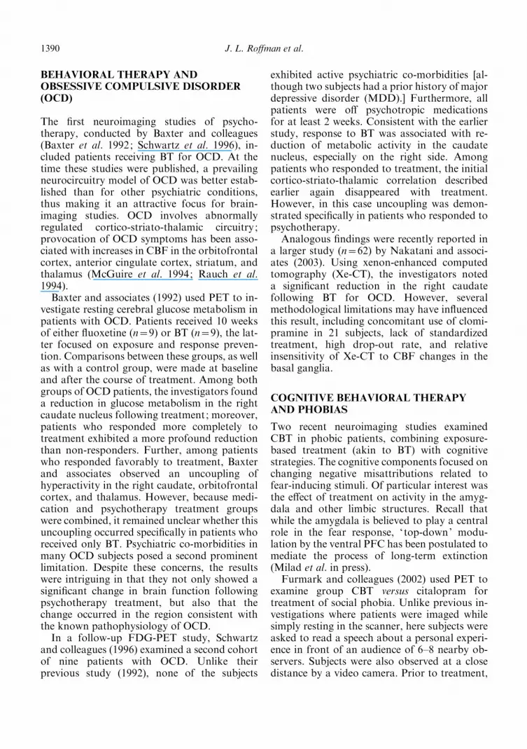
more effort towards understanding the neuralmechanisms of medication than of psycho-therapy (Fig. 1). This disparity has persisteddespite the similar costs and clinical efficacy ofmedication and psychotherapy for commonpsychiatric disorders (Antonuccio et al. 1995;Goldman et al. 1998; Satcher, 1999). Historicalbias toward medications as being a clearlydefined ‘biological ’ intervention, compared withthe more complex psychosocial intervention ofpsychotherapy (Westen et al. 2004), has likely
* Address for correspondence: Joshua L. Roffman, M.D.,Department of Psychiatry, Massachusetts General Hospital andHarvard Medical School, Boston, MA 02114, USA.(Email : [email protected])
Psychological Medicine, 2005, 35, 1385–1398. f 2005 Cambridge University Pressdoi:10.1017/S0033291705005064 Printed in the United Kingdom
1385

contributed to this imbalance. However, as firstsuggestedwell before the era of functional neuro-imaging, the changes in affect, behavior, andcognition that are mediated by psychotherapiesundoubtedly have biological underpinnings(Freud, 1895). In recent years, the number ofstudies using neuroimaging to explicitly evalu-ate neural correlates of psychotherapy hassteadily increased. These studies answer the callfor more biologically rigorous approaches topsychotherapy research (Kandel, 1998).
In this review, we will address how neuro-imaging research is beginning to reveal therelationship between psychotherapy and brainfunction. We shall first give examples of howpsychological constructs relevant to psycho-therapy, such as extinction, cognitive restruc-turing, and repression, have been associatedwith discrete brain activity. While long con-sidered the ‘building blocks ’ of psychotherapyon a theoretical level, these constructs appear tohave parallel meaning on the level of neuro-anatomy. Second, we will evaluate the emergingliterature on psychotherapy-related changes inbrain activity profiles, drawing some preliminarycomparisons among different psychotherapies,and between psychotherapeutic and psycho-pharmacological approaches. Finally, we will
discuss how future research efforts may refineour physiological understanding of how psycho-therapy works, and why this knowledge may beclinically useful.
PUTATIVE NEURONAL MECHANISMSOF PSYCHOTHERAPY
Many potentially unhealthy cognitive andemotional patterns targeted by psychotherapyappear to have measurable biological analogs(Beutel et al. 2003). One salient example in-volves repression, or the unconscious ‘forget-ting’ of threatening ideas or experiences. In arecent investigation by Anderson and associates(2004), healthy subjects were instructed either toremember or ‘forget ’ target words. In a recalltask, presentation of ‘forget ’ words was asso-ciated not only with poor recall of those words,but also with increased activation of the pre-frontal cortex (PFC) and decreased hippo-campal activation. These results replicated aprevious study conducted by Bunge and col-leagues (2001), who also found that the an-terior cingulate gyrus directs attention awayfrom unwanted memories. Thus, active ‘ for-getting’ modulated by the PFC and anterior
40
35
30
25
20
15
10
5
01990 1991 1992 1993 1994 1995 1996 1997
Year
Num
ber
of s
tudi
es
1998 1999 2000 2001 2002 2003
FIG. 1. Imaging/medication (%) and imaging/psychotherapy (&) studies by year. Method: An Ovid MEDLINE search was com-pleted using key words related to neuroimaging (e.g. PET, fMRI, SPECT), and medication (e.g. psychotropic) to find studiesincluding both neuroimaging and medication between the years 1966 and 2003. Based on the abstracts generated from this search,we selected studies based on four criteria: included studies were published in English between 1990 and 2003, used human subjects,and investigated psychiatric (e.g. depression) rather than neurological (e.g. Parkinson’s Disease) disorders. A similar search wasconducted using key words related to neuroimaging and psychotherapy (e.g. psychotherapy, intrapersonal therapy, cognitivebehavioral therapy, and psychodynamic therapy).
1386 J. L. Roffman et al.

cingulate may have relevance to the psycho-dynamic concept of repression.
The process of psychotherapy may alsoengage dedicated neural circuitries that are par-ticularly responsive to a discrete mode of treat-ment. We find examples of this in studies relatedto psychodynamic, cognitive, and behavioraltherapy; such studies have used healthy subjectsto investigate analogs of specific therapy pro-cesses. When a psychodynamically orientedtherapist ‘ takes a history’, this elicits episodicmemories from the patient in a focused way.However, when the same therapist observes thepatient ‘free associate ’, episodic recall occurs ina less organized, more random way. Andreasenand co-workers (1995) observed that whilerandom memories engaged assocation cortex infrontal, parietal, and temporal regions, focusedmemories selectively activated verbal areas(including Broca’s area and the left frontaloperculum). Thus, the less ‘censored’ process offree association may engage wider networksof association cortex, facilitating exploration oflatent aspects of the patient’s symptomatologyor personality.
In cognitive behavioral therapy (CBT) fordepression, patients are sometimes asked torevisit bad or painful memories and explicitlyre-evaluate their negativity toward the memory.Using a related paradigm in healthy subjects,Ochsner and colleagues (2002) observed a re-lationship among reappraisal of negative stim-uli, improvement in mood, and brain activitypatterns. Subjects rated their mood before andafter being asked to ‘re-interpret ’ highly nega-tive scenes in a more positive light. Reappraisalwas associated with both improved mood andincreased activity in dorsolateral and dorsome-dial PFC, but decreased activity in the amygdalaand orbitofrontal cortex. These findings suggesta model of cognitive therapy: while limbic andventral prefrontal structures generate negativeaffect in response to a certain stimulus, dorsalprefrontal circuitry may be engaged throughreappraisal techniques to dampen this outflowfrom more ventral structures (see also Ochsner& Feldman Barrett, 2001; Scherer et al. 2001).
As a final example, we turn to behavioraltherapy (BT). BT for anxiety disorders oftenrelies on desensitization or extinction of learnedresponses to anxiety-provoking stimuli. Con-verging evidence from animal and human
studies implicates the ventral PFC and amyg-dala in this process (Milad et al. in press).Targeted lesions or specific pharmacologicalinhibition of the amygdala interfere with fearconditioning in rodents (see Maren & Quirk,2004 for a review), while functional neuro-imaging studies in humans have consistentlyassociated amygdalar activation with con-ditioned fear responses (Buchel et al. 1999;Fischer et al. 2000; Charney, 2003; Cheng et al.2003). Analogous studies in rats (Lebron et al.2004) and humans (Gottfried & Dolan, 2004;Phelps et al. 2004) suggest that the ventral PFCmediates the retention and recall of extinctionfor conditioned fear responses by keepingthe amygdala in check. One might, therefore,expect extinction-based behavioral therapies inhumans to work either by potentiating ventralPFC activity or attenuating the amygdala (orboth).
Collectively, the growing number of studiesrelated to the psychotherapy process point to aplausible neuronal substrate for psychother-apeutic interventions. In this setting, we havealso seen emerge a critical mass of studies thatdirectly evaluate the neurobiological effects ofpsychotherapy in patients with mood and anxi-ety disorders.
THE PROBLEM OF VARIABLEMETHODOLOGY
Before examining the literature on psycho-therapy and neuroimaging in detail, we mustrecognize that heterogeneities across thesestudies often limit our ability to compare themdirectly (Table 1). There exist many specificdifferences in rationale, technique, and efficacyof the various psychotherapeutic modalities inuse today. Several of these modalities, includ-ing BT, CBT, and interpersonal therapy (IPT)lend themselves well to controlled experi-mental design by using manual-based treatmentin a time-limited setting. However, even amongmanualized treatment programs, adherence toa given framework is often far from absolute(Ablon & Jones, 2002). In several of thereviewed studies, although the therapeuticmodality is called one thing (e.g. CBT), thedescription of the psychotherapeutic processmore closely resembles another (e.g. BT).
The functional neuroanatomy of psychotherapy 1387

Table 1. Studies examining the functional neuroanatomy of psychotherapy
Study Therapy type Subjects Comparisons Imaging Post-treatment findings Comments
Baxter et al.(1992)
BT, 10 weeks 9 patients withOCD
9 patients with OCD,taking fluoxetine
FDG-PET,resting
1. Decreased metabolism in R caudate in bothgroups
1. Multiple co-morbidities
2. Uncoupling of cortico-striato-thalamiccircuit in combined subject pool
2. Unclear if uncouplingoccurred as effect of BT
Schwartzet al. (1996)
CBT, 10 weeks 9 patients withOCD, plusearlier cohort
None FDG-PET,resting
1. Decreased metabolism in R caudate 1. Some patients alsoreceived group CBT2. Uncoupling of cortico-striato-thalamic
circuit
Nakataniet al. (2003)
BT, varyingduration
31 patients withtreatment-refractory OCD
31 healthy controls Xe-CT, resting 1. Decreased CBF in R caudate 1. 21 patients also tookclomipramine
2. Lack of standardizedtreatment
3. Poor sensitivity to basalganglia
Furmarket al. (2002)
Group CBT, 8weeks
6 treatment-naivepatients withsocial phobia
6 waitlist patients and 6citalopram patients
PET, whileobserved readingscript
1. In CBT and citalopram groups, decreasedlimbic metabolism
1. Small numbers in eachgroup
2. In CBT group, decreased periaqueductalgray metabolism
3. In citalopram group, decreased thalamicmetabolism
Paquette et al.(2003)
Group CBT, four3-hour sessions
12 patients withspider phobia
13 healthy controls fMRI, whileviewing spiders
1. Decreased activation in parahippocampalgyrus and dorsolateral prefrontal cortex
1. Extent of post-CBTanxiety testing unclear
Goldappleet al. (2004)
CBT, 15–20sessions
17 patients withMDD
Post-hoc comparison to13 patients givenparoxetine
FDG-PET,resting/avoiding‘ruminating’
1. In CBT group, decreased metabolism inmultiple frontal regions, increased in limbicregions
1. 3 patients dropped out
2. In paroxetine group, changes in oppositedirection
2. No imaging controlgroup
Martin et al.(2001)
IPT, 6 weeks 13 patients withMDD
15 patients with MDDgiven venlafaxine
SPECT, resting 1. In each group, increased CBF in right basalganglia
1. Semi-random design
2. In IPT group, increased CBF in R posteriorcingulate
2. Short duration oftreatment
Brody et al.(2001a)
IPT, 12 weeks 14 patients withMDD
10 patients with MDDgiven paroxetine and16 healthy controls
FDG-PET,resting
1. In both MDD groups, decreasedmetabolism in prefrontal cortex, increased ininferior temporal cortex and insula
1. Non-randomized withbaseline differencesbetween groups
Brody et al.(2001b)
IPT, 12 weeks 14 patients withMDD
25 patients with MDDgiven paroxetine
FDG-PET,resting
1. Correlation of symptom clusterimprovement with reduced frontal lobemetabolism
1. Paroxetine and IPTgroups apparentlycombined
2. Positive correlation for cognitive symptoms 2. No correction formultiple comparisons
Penades et al.(2002)
Group ‘neuro-psychologicalrehab, ’ 12 weeks
8 patients withschizophrenia,on olanzapine
None SPECT, duringTower ofLondon task
1. Weakly increased CBF in frontal lobe,correlated with improvement in test score
1. No control intervention
2. Non-specific imaging measure
1388
J.L.Roffmanet
al.

Methodological inconsistencies created by theuse of single versus multiple therapists, differingnumbers of sessions, and varying milieus (e.g.individual versus group therapy) should also beconsidered when comparing studies.
The neuroimaging modalities that have beenemployed in psychotherapy research also vary.These techniques provide indices of brain activityby measuring glucose metabolism or cerebralblood flow (CBF). Although the relationshipbetween glucose metabolism or CBF andneuronal activity remains controversial (Gioveet al. 2003), it is commonplace to use the term‘brain activity ’ essentially interchangeably with‘metabolic activity ’ or ‘hemodynamic activity’.Among the studies reviewed below, the prin-cipal imaging techniques applied include func-tional magnetic resonance imaging (fMRI),18fluorodeoxyglucose positron emission tom-ography (FDG-PET), and 99mtechnetium hexa-methylpropyleneamineoxime single photon emis-sion computed tomography (99mTc-HMPAOSPECT). These imaging modalities differ withrespect to mechanism, image resolution, andpatient-related limitations (see Dougherty et al.2004 for a detailed review). Moreover, there areseveral methods for examining regional brainactivity, including voxel-based techniques (suchas SPM) and region-of-interest (ROI)-basedapproaches. ROI-based methods enable fewermultiple comparisons and respect anatomicalboundaries. Alternatively, voxelwise methodscan be more data driven (and hence less biased),and may ultimately provide a better measureof functional connectivity (Dougherty et al.2004).
Several additional design considerationsshould be taken into account when comparingthese investigations. In most imaging studiesof treatment effects, subjects are scanned beforeand after a trial of psychotherapy (or a com-parison intervention), and in some cases, acti-vation differences over time are comparedwith a group of healthy or waitlist control sub-jects. Some functional neuroimaging studiesexamine brain activity while the subject is rest-ing in the scanner, others while the subject isengaged in a cognitive or affective task relatedto the psychiatric condition (e.g. exposure to ananxiety-provoking stimulus). Recognizing thesedesign concerns, we now turn to the studiesthemselves.W
ykes
etal.
(2002)
‘Cognitive
remediation
therapy,’12
weeks
6patients
with
schizophrenia,
on
antipsychotics
6patients
with
schizophrenia
given
occupationaltherapy,
6healthycontrols
fMRI,during
workingmem
ory
andvigilance
tasks
1.In
cognitivetherapygroup,increasedCBF
inR
inferiorfrontalcortex
andbilateral
occipitalcortex
1.Smallnumbersin
each
group
Laatsch
etal.
(1999)
‘Cognitiverehab
therapy,’6–36
sessions
5patients
with
traumaticbrain
injury
None
SPECT,
presumably
resting
1.Globalincreasesin
CBFmost
apparent
duringtreatm
entphase
in3of5patients
1.Nospecificregional
hypotheses
2.Noattem
ptto
correlate
imagingwithneuropsych
measures
Levin
etal.
(1999)
EMDR,3sessions
1patientwith
PTSD
None
SPECT,while
beingread
scripts
1.IncreasedCBFin
anteriorcingulate,L
frontallobe
1.Singlecase,nofollow-up
Brodyet
al.
(1998)
BT,8–12weeks
18patients
with
OCD
9patients
withOCD
given
fluoxetine
FDG-PET,
resting(at
baselineonly)
1.ForBTpatients,Lorbitofrontalcortex
metabolism
positivelycorrelatedwith
treatm
entresponse
1.Groupassignmentbased
onpatientpreference
2.Fluoxetinepatients
exhibited
anegative
correlation
2.Multipleco-m
orbidities
BT,Behavioraltherapy;CBT,cognitivebehavioraltherapy;IPT,interpersonaltherapy;EMDR,eyemovem
entdesensitizationandreprocessing;OCD,obsessive–compulsivedisorder;
MDD,majordepressivedisorder;PTSD,post-traumaticstress
disorder;FDG-PET,
18fluorodeoxyglucose
positronem
issiontomography;Xe-CT,xenon-enhancedcomputedtomography;
CBF,cerebralbloodflow;fM
RI,functionalmagnetic
resonance
imaging;SPECT,99mtechnetium
hexamethylpropyleneamineoxim
esingle
photonem
issioncomputedtomography;R,right;
L,left.
The functional neuroanatomy of psychotherapy 1389
BEHAVIORAL THERAPY ANDOBSESSIVE COMPULSIVE DISORDER(OCD)
The first neuroimaging studies of psycho-therapy, conducted by Baxter and colleagues(Baxter et al. 1992; Schwartz et al. 1996), in-cluded patients receiving BT for OCD. At thetime these studies were published, a prevailingneurocircuitry model of OCD was better estab-lished than for other psychiatric conditions,thus making it an attractive focus for brain-imaging studies. OCD involves abnormallyregulated cortico-striato-thalamic circuitry ;provocation of OCD symptoms has been asso-ciated with increases in CBF in the orbitofrontalcortex, anterior cingulate cortex, striatum, andthalamus (McGuire et al. 1994; Rauch et al.1994).
Baxter and associates (1992) used PET to in-vestigate resting cerebral glucose metabolism inpatients with OCD. Patients received 10 weeksof either fluoxetine (n=9) or BT (n=9), the lat-ter focused on exposure and response preven-tion. Comparisons between these groups, as wellas with a control group, were made at baselineand after the course of treatment. Among bothgroups of OCD patients, the investigators founda reduction in glucose metabolism in the rightcaudate nucleus following treatment; moreover,patients who responded more completely totreatment exhibited a more profound reductionthan non-responders. Further, among patientswho responded favorably to treatment, Baxterand associates observed an uncoupling ofhyperactivity in the right caudate, orbitofrontalcortex, and thalamus. However, because medi-cation and psychotherapy treatment groupswere combined, it remained unclear whether thisuncoupling occurred specifically in patients whoreceived only BT. Psychiatric co-morbidities inmany OCD subjects posed a second prominentlimitation. Despite these concerns, the resultswere intriguing in that they not only showed asignificant change in brain function followingpsychotherapy treatment, but also that thechange occurred in the region consistent withthe known pathophysiology of OCD.
In a follow-up FDG-PET study, Schwartzand colleagues (1996) examined a second cohortof nine patients with OCD. Unlike theirprevious study (1992), none of the subjects
exhibited active psychiatric co-morbidities [al-though two subjects had a prior history of majordepressive disorder (MDD).] Furthermore, allpatients were off psychotropic medicationsfor at least 2 weeks. Consistent with the earlierstudy, response to BT was associated with re-duction of metabolic activity in the caudatenucleus, especially on the right side. Amongpatients who responded to treatment, the initialcortico-striato-thalamic correlation describedearlier again disappeared with treatment.However, in this case uncoupling was demon-strated specifically in patients who responded topsychotherapy.
Analogous findings were recently reported ina larger study (n=62) by Nakatani and associ-ates (2003). Using xenon-enhanced computedtomography (Xe-CT), the investigators noteda significant reduction in the right caudatefollowing BT for OCD. However, severalmethodological limitations may have influencedthis result, including concomitant use of clomi-pramine in 21 subjects, lack of standardizedtreatment, high drop-out rate, and relativeinsensitivity of Xe-CT to CBF changes in thebasal ganglia.
COGNITIVE BEHAVIORAL THERAPYAND PHOBIAS
Two recent neuroimaging studies examinedCBT in phobic patients, combining exposure-based treatment (akin to BT) with cognitivestrategies. The cognitive components focused onchanging negative misattributions related tofear-inducing stimuli. Of particular interest wasthe effect of treatment on activity in the amyg-dala and other limbic structures. Recall thatwhile the amygdala is believed to play a centralrole in the fear response, ‘ top-down’ modu-lation by the ventral PFC has been postulated tomediate the process of long-term extinction(Milad et al. in press).
Furmark and colleagues (2002) used PET toexamine group CBT versus citalopram fortreatment of social phobia. Unlike previous in-vestigations where patients were imaged whilesimply resting in the scanner, here subjects wereasked to read a speech about a personal experi-ence in front of an audience of 6–8 nearby ob-servers. Subjects were also observed at a closedistance by a video camera. Prior to treatment,
1390 J. L. Roffman et al.
subjects exhibited prominent activation in lim-bic structures, including the amygdala, hippo-campus, and adjacent temporal cortex. Patientsin the CBT arm received eight weekly grouptherapy sessions that specifically targeted anxi-ety associated with public speaking. In contrastto waitlist control subjects who did not changeover time, patients in both treatment armsdemonstrated a significant reduction of activityin these limbic and paralimbic regions. Of note,while no change in activity was observed inventral PFC in the CBT group, patients receiv-ing citalopram exhibited activity reductions inthis region after treatment. Additional differ-ences were also evident between treatmentgroups. Patients receiving cognitive therapyshowed decreased CBF in the periaqueductalgray area, which has been associated with de-fense behaviors (Behbehani, 1995). Subjectstaking citalopram exhibited thalamic reductionsin CBF, potentially reflecting reductions in sen-sory input to the amygdala (Charney & Deutch,1996). These results imply that CBT and citalo-pram therapy for social phobia might dampenlimbic response by different mechanisms, even ifthe ventral prefrontal components of these me-chanisms remain unclear. It should be noted,however, that this study included subjects withslightly varying diagnoses (e.g. specific andgeneralized social phobias).
Another provocation study by Paquette andcolleagues (2003) related similar findings, in thiscase by exposing non-medicated, spider-phobicpatients to pictures of spiders during fMRIscans. Compared to non-phobic control sub-jects, patients initially exhibited significant acti-vation in the parahippocampal gyrus and rightdorsolateral PFC#. After successful group CBTsessions using exposure therapy, patients dem-onstrated significantly less activation in both theparahippocampal gyrus and right dorsolateralPFC, and increased activation in the right ven-tral PFC. Paquette and associates linked theabatement of the parahippocampal gyrus re-sponse to a dampening of contextual memory(believed to be mediated by this structure). Theyfurther suggested that the dorsolateral PFC
changes might reflect the restructuring of con-scious cognitive defenses; with the completionof successful psychotherapy, less demand wasplaced on the dorsolateral PFC to plan a reac-tion to the perceived threat. This notion is con-sistent with Ochsner et al.’s recent (2004) ob-servation that the dorsal PFC may play a rolein up-regulating negative affect and limbic out-flow under conditions where an aversive stimu-lus must assume personal salience. Followingtherapy, a shift of activity to the ventral PFCcould prompt down-regulation of limbic ac-tivity, and consequently dampen the fear reac-tion. This pattern is again consistent with thefindings of Ochsner et al. (2004), who correlatedright ventral PFC activity with down-regulationof negative affect and limbic outflow when sub-jects were asked to de-emphasize personal con-nections to aversive stimuli.
Taken together, these studies are consistentwith the model of BT-related desensitization toaversive stimuli described earlier in our review.Modulation of limbic activation following fearprovocation may involve either reductions ofCBF in limbic and adjoining paralimbic regions,or enhancement of CBF in ventral PFC, orperhaps both (as suggested by Paquette et al.2003). The contributions of the cognitive partsof CBT to hemodynamic changes in thesestudies remain unclear – however, it should benoted that in both cases, the description of thetherapy process clearly points more towardsbehavioral approaches (e.g. extinction-basedprocesses).
COGNITIVE BEHAVIORAL THERAPYAND DEPRESSION
MDD has often been associated with alterationsin prefrontal brain activity in untreated patients(Drevets, 1998), with dorsal areas (including thedorsolateral PFC) exhibiting decreased activity,and ventral frontal regions demonstrating in-creased activity (Dougherty & Rauch, 1997,Mayberg, 1997; Drevets, 2000; Rauch, 2003). Asingle functional neuroimaging study of CBT inmedication-free, depressed patients has beenreported by Goldapple and colleagues (2004).Neuroimaging was conducted with FDG-PETbefore and after the psychotherapy trial, withpatients being instructed to ‘avoid ruminatingon any one topic’ during scanning. A post-hoc
# It is worth noting that the lack of activation of the amygdala,while surprising, has been replicated in several other studies of spiderphobia (Fredrikson et al. 1995; Rauch et al. 1995; Johanson et al.1998), possibly reflecting the interaction of specific phobias and spe-cific patterns of limbic hyperactivity.
The functional neuroanatomy of psychotherapy 1391

comparison was made to a second group of pa-tients who had been given paroxetine.Surprisingly, in the CBT group, metabolism inmultiple frontal regions including the dorso-lateral PFC decreased after therapy. Given theassociation of depression with reduced dorso-lateral PFC activity at baseline, and the modelof CBT elaborated earlier in this review, thisfinding is somewhat counterintuitive. Theauthors related the reduction in prefrontalmetabolism to treatment-related diminution of‘active rethinking and reappraisal of emotionalideas ’. This interpretation is somewhat akin tothat offered by Paquette and colleagues (2003)for reduced dorsal prefrontal demand in suc-cessfully treated spider phobia. At the sametime, however, it contradicts the notion thatCBT enhances patients’ abilities to reappraiseaffect-generating stimuli (e.g. Ochsner et al.2002). Notably, among patients who receivedparoxetine in the Goldapple investigation, PFCmetabolism increased with medication therapy,even though efficacy was comparable to CBT,again implying that medication and CBT maywork through different mechanisms.
In addition to changes in the PFC, Gold-apple and associates also described changes inlimbic and paralimbic activity after treatment.However, once again a differential patternemerged for subjects receiving CBT and parox-etine. Subjects in the CBT group exhibitedsignificant increases in activity in the hippo-campus, parahippocampal gyrus, and dorsalcingulate gyrus. In the paroxetine group, sub-jects exhibited less activity in hippocampal andparahippocampal regions, as well as decreasedactivity in the posterior cingulate and ventralsubgenual cingulate. Based on these results, andon known functional and anatomic relation-ships between implicated brain regions (Friston,1994; Horwitz et al. 1999), Goldapple andcolleagues proposed a modality-specific modelof treatment response in depression. Anti-depressant medications appear to have exerted‘bottom-up’ effects by disengaging ventralfrontal and limbic regions. In contrast, CBT ef-fected ‘top-down’ changes by reducing corticalprocessing in favor of ventral and limbic regionsmediating attention to personally relevantemotional and environmental stimuli. Again,this result (and interpretation) oppose the emo-tion regulation model of Ochsner and colleagues
(2002), who posit that ventral frontal and limbichyperactivity exacerbate negative affect. Toreconcile these differences, we invoke the notionthat brain activation represents an interactionbetween treatment protocol and underlyingbrain state (Seminowicz et al. 2004). In this case,while both investigations involved cognitive re-structuring, Goldapple et al. examined patientswith depression, and Ochsner et al. looked athealthy controls. Bearing this same interactionin mind, we may consider whether psy-chotherapies other than CBT induce similarchanges in brain activity among patients withdepression.
IPT AND DEPRESSION
IPT, like CBT, is a manualized, time-limitedtherapy that lends itself well to controlled trials.However, in contrast to CBT, IPT emphasizesimproving interpersonal relationships, oftendrawing directly on the relationship betweenpatients and their therapists. Several recent stud-ies have examined changes in CBF associatedwith the treatment of depression with IPT. Ineach case, neuroimaging comparisons weremade to a second group of depressed patientsreceiving pharmacotherapy.
In a 6-week study of 28 patients with MDD,Martin and associates (2001) compared the ef-fects of IPT and venlafaxine (37.5 mg daily) onregional CBF using 99mTc-HMPAO SPECT.Subjects had been drug-naive or drug-free forthe 6 months preceding the study. After baselinescans, subjects in the IPT group received 6weeks of psychotherapy by the same therapist,while those in the venlafaxine group were seenfor 15 minutes every 2 weeks. Both groups im-proved clinically, and in both groups Martinand co-workers observed an increase in bloodflow in the right basal ganglia. However, sub-jects in the IPT group also exhibited an increasein right posterior cingulate activity. Consistentwith Goldapple and colleagues (2004), Martinet al. underscored the importance of limbic andparalimbic recruitment in psychotherapy-medi-ated changes; however, it should be noted thatMartin and colleagues described changes only inone specific paralimbic region.
Bearing this single finding in mind, it is im-portant to note several methodological limita-tions of this study. Martin and colleagues
1392 J. L. Roffman et al.
employed a semi-randomized design, in whichsubjects with a strong preference for IPT orvenlafaxine could choose that treatment ; foursubjects pre-selected venlafaxine, while onechose IPT. Of note, striatal perfusion appearedgreater at baseline among subjects in the IPTgroup, potentially reflecting this design limi-tation. Additional problems included a lack ofcomparison to healthy control subjects, failureto exclude co-morbid anxiety disorders, and therelatively poor resolution of subcortical struc-tures by SPECT. Finally, it is important to notethat patients in the venlafaxine group demon-strated a more robust response to treatment. Itmay be argued, though that both treatmentswere sub-optimal, given the relatively low doseof venlafaxine and the brief duration of IPT.
A longer, 12-week study of similar design wasconducted by Brody and co-workers (2001a),who used PET to examine 24 patients who re-ceived IPT or paroxetine. While this study in-cluded a control group of healthy subjects, allpatients were allowed to self-select into the drugor therapy groups. Of note, subjects in the par-oxetine cohort were less ill at baseline, andexhibited greater improvement over time thanthose in the IPT group. However, these designlimitations aside, Brody et al. found a decreasein dorsal and ventral prefrontal cortical metab-olism with IPT treatment directly analogous tothat described by Goldapple et al. (2004). Inaddition, the authors described an increase inmetabolism in limbic and paralimbic regions(in this case, the right insula and left inferiortemporal lobe) in both treatment groups com-pared to controls. Unlike Goldapple, though,Brody and associates reported a decrease inPFC activation with paroxetine.
In a follow-up study using a larger cohort of39 patients receiving either paroxetine or IPTfor MDD, Brody and colleagues (2001b) at-tempted to correlate treatment-related changesin brain activity with improvement in specificmood symptom clusters. In all subjects, reduc-tions of ventral and dorsal frontal lobe metab-olism were associated with improvements in theanxiety/somatization and psychomotor retar-dation symptom clusters of the HamiltonDepression Rating Scale, and in the tension/anxiety and fatigue clusters of the Profile ofMood States. Interestingly, improvement incognitive disturbance positively correlated with
changes in dorsolateral PFC metabolism. Thisfinding is especially germane in light of thenegative correlation between activity in thedorsolateral PFC and improvement on globaldepression scores after CBT. This distinctionsuggests that while CBT may specifically dam-pen ‘over-thinking’ and rumination aspects ofdorsolateral PFC function in depression, IPTpotentially improves general cognitive abilitiesmediated by this region. Again these findingsmust be interpreted cautiously pending repli-cation, and in consideration of design limita-tions: no effort was made to separate patientsreceiving IPT or paroxetine for correlationswith hemodynamic changes, and no correctionfor multiple comparisons was implemented de-spite a total of six symptom clusters being as-sessed in each of 12 ROIs.
WHERE DO WE STAND? THEMES ANDLIMITATIONS
Several themes emerge when considering thesestudies in aggregate. First, psychotherapy-re-lated changes in brain activation appear strik-ingly similar within patients who share the samepsychiatric diagnosis. For example, despite theirmethodological limitations and heterogeneities(e.g. patients receiving concomitant pharma-cotherapy or multiple types of psychothera-peutic interventions), each of the reportsexamining psychotherapeutic interventions forOCD yielded comparable results. In each case,BT resulted in decreased metabolism in thecaudate nucleus, a finding consistent with thewell-established pathophysiology of the dis-order (Saxena et al. 1998). Moreover, bothfluoxetine and psychotherapy for OCD appearto uncouple dysfunctional cortico-striato-thalamic circuitry. In two studies examiningpatients with phobias, a reduction in limbic orparalimbic activity was observed followingtreatment, again consistent with the hypothe-sized pathophysiology. Among studies of de-pression, following successful psychotherapywith CBT or IPT, patients surprisingly – butconsistently – exhibited decreased activity indorsal frontal regions and increased activity inventral frontal and subcortical regions (notablyincluding limbic and paralimbic structures).
It is particularly intriguing that in MDD,both CBT and IPT have been associated with a
The functional neuroanatomy of psychotherapy 1393
similar pattern of CBF alterations. Given thedifferent theoretical approaches of these twotreatments, how might we account for thiscommon pattern? As suggested above, whenconsidering the interacting effects of psychiatriccondition and therapeutic approach on brainactivity, perhaps underlying pathophysiologyis the predominant factor. Another possibility,as suggested by Caspar (2003), is that whileneuroimaging technologies allow us to visualizea ‘final common pathway’ of sorts, the morerelevant changes in brain physiology attribu-table to psychotherapy may involve moremicroscopic phenomena. It is conceivable, al-though presently difficult to confirm, that CBTand IPT exert differing effects on cellular or evenmolecular levels. A third possible explanation issuggested by the work of Ablon & Jones (2002).Studying patients with MDD, these authorsused a standardized measure (the PsychotherapyQ-sort) to compare psychotherapy processvariables in transcripts of CBT versus IPT ses-sions. These process variables were related tothe contributions of the patient, therapist, andthe patient–therapist interaction. Ablon & Jonesobserved that successful IPT and CBT sessionsboth conformed strongly to the same set ofprocess variables, implying that despite thera-pists’ differing theoretical orientations, the workof therapy remained quite similar. It is temptingto speculate that the similarity of CBF changesseen after IPT and CBT reflects a neural corre-late of this phenomenon.
Regardless of what mechanism might bestexplain the observed similarities of CBT andIPT for depression, these findings point to theneed for adherence measures when attemptingto ascribe treatment effects to any particularpsychotherapeutic approach. This notion be-comes critical when attempting to match specificpsychotherapeutic interventions with specificchanges in brain activation profiles. For ex-ample, in both studies of CBT for phobic dis-orders (Furmark et al. 2002; Paquette et al.2003), it appeared that behavioral interventionspredominated over cognitive ones – thus, it isnot surprising that the CBF correlates of symp-tom improvement so closely matched thosepredicted by animal and healthy human analogmodels of behavioral therapy.
A second recurring theme of this review,especially among studies of MDD, is that while
psychotherapy and pharmacotherapy achievedsimilar efficacy, they were associated with over-lapping – but not identical – changes in brain-imaging profiles. Moreover, patients who re-ceived pharmacotherapy exhibited less reliablepatterns of brain activation than those who weretreated with psychotherapy, even when the samemedication (paroxetine) was used in differentstudies (Brody et al. 2001a ; Goldapple et al.2004). This discrepancy has also been observedamong studies examining exclusively psycho-pharmacological interventions (e.g. Brody et al.1999; Kennedy et al. 2001). Seminowicz andcolleagues (2004) have attempted to explainthese differential effects through a path-model-ing meta-analysis of studies involving medi-cation and CBT to treat depression. Theiranalysis, which includes subjects from theGoldapple et al. (2004) study, assumes thattreatment response is dependent not just onspecific interventions but rather on the interac-tion of pre-treatment brain state, brain respon-siveness, and treatment choice. They suggestthat among responders to CBT, fronto-frontalprocessing abnormalities appear to predomi-nate, while those who ultimately respond tomedications exhibit altered fronto-limbic dys-regulation. However, the predictive validity ofthis model has yet to be tested prospectively.The use of such general descriptors ‘ limbic’ and‘frontal ’ should also be viewed with caution,given the considerable structural and functionalheterogeneity of these regions.
A third theme is that each study used tworounds of neuroimaging – one before treatment,and the other afterward – and the majority in-volved patients resting passively during scans. Apotential confound of this approach is thatwhile changes in brain activation over time mayreflect correlates of symptom improvement,they do not necessarily imply a mechanism oftreatment action, on either neuroanatomical orcellular/molecular levels. This is problematiceven among those studies where subjects wereactively participating in cognitive or behavioraltasks related to their underlying disorder. Forexample, in the Furmark et al. (2002) study, thelack of ventral PFC activation following thecompleted course of treatment was surprisinggiven its proposed role in modulating limbicoutflow; however, it is possible that such in-creased activity occurred during the treatment,
1394 J. L. Roffman et al.
and was missed because the second scan oc-curred after the completion of therapy. (Of note,neuroimaging studies involving drug interven-tions suffer from a similar limitation, since it isdifficult to differentiate brain activity changesthat occur while an individual is actively takinga medication from those that occur as a conse-quence of taking the medication.) Thus, whilethis review strongly suggests that significant,and in many cases, unique changes in the brainare associated with psychotherapy, little can beconcluded about the precise neurobiology andmechanisms involved in these changes.
FUTURE STUDIES OF PSYCHOTHERAPYAND BRAIN FUNCTION
A logical next step in addressing the problem ofmechanism would involve, at the least, ad-ditional functional scans interpolated duringthe course of therapy, and at best, real-time imag-ing during psychotherapy sessions themselves.Technical and logistical limitations of fMRI,PET, and SPECT preclude the naturalistic useof these imaging tools in this manner. However,novel neuroimaging technologies hold somepromise for these applications. One such tool,near-infrared spectroscopy (NIRS), permitsmeasurement of cortical CBF less invasivelythan fMRI, and is both more portable and lessexpensive. Safe and practical for repeated meas-ures, NIRS has been employed to measureCBF in patients with a variety of neuro-psychiatric conditions (for a review see Strang-man et al. 2002) and in research involving basicauditory and cognitive processing (Sato et al.1999). A second optical technique currently indevelopment, two-photon microscopy, can po-tentially image deeper brain activity in vivo evenon the cellular level (Miller, 2003) ; it has beensuggested that in the future this technique mayfind its way into psychotherapy research aswell (Zabarenko, 2004). Just as psychophysio-logical recording methods have been used topermit simultaneous measurements from bothpatient and therapist (e.g. Marci et al. 2004), theuse of next-generation, non-invasive opticaltechniques could also permit simultaneousmeasurements in clinical settings, providing apowerful assay of patient–therapist interactions.
On the more immediate horizon, investi-gators have already begun to use functional
neuroimaging to predict treatment outcome.This work is akin to recent studies that haveused electroencephalography (EEG) within 48 hof starting treatment to predict whether patientswill ultimately respond to antidepressants (e.g.Cook et al. 2002). Brody and associates (1998)reported differential responses to behavioraltherapy and fluoxetine for OCD based on pre-treatment PET scans. Among patients who re-ceived BT, Brody observed a positive corre-lation between treatment response and baselinemetabolism in the left orbitofrontal cortex;however, the inverse pattern was seen amongpatients in the fluoxetine group. This finding in-volves a region implicated not only in the patho-physiology of OCD (McGuire et al. 1994;Rauch et al. 1994), but also in desensitization ofanxiety-provoking stimuli as described earlier(Gottfried & Dolan, 2004; Phelps et al. 2004).Brody and colleagues thus propose that patientswith increased orbitofrontal cortex activity atbaseline may be predisposed to benefit from ex-tinction-based therapy for OCD. However, aspatients in the Brody et al. study were allowed toselect their own treatment group, this interpret-ation must be viewed with caution. While ad-ditional prospective studies in this area areneeded, the ability to construct individualizedtreatment plans using biological markers (suchas neuroimaging) could ultimately allow pa-tients and clinicians to move away from thecurrent ‘trial-and-error’ approach, minimizingfrustration as well as lost time and expense.
Thus far neuroimaging studies of psycho-therapy have focused on manual-driven, time-limited treatments. However, much is to begained by subjecting other commonly usedtherapeutic techniques to investigation withfunctional neuroimaging methods. For ex-ample, a single case report illustrates the nor-malization of serotonin receptor binding, asdetermined by SPECT, in a patient with bor-derline personality disorder following 1 year ofpsychodynamic psychotherapy (Viinamaki et al.1998). Still, at present there are no publishedreports of brain activation changes associatedwith either psychodynamic psychotherapy ordialectical behavioral therapy, two empiricallysupported treatments widely in use. Anothercase report describes increased CBF in the an-terior cingulate gyrus and frontal lobe followingeye movement desensitization and reprocessing
The functional neuroanatomy of psychotherapy 1395

(EMDR), a novel (although not yet empiricallyvalidated) treatment for post-traumatic stressdisorder (Levin et al. 1999). Several studies havedemonstrated increased frontal lobe metabolismfollowing cognitive rehabilitation therapies forschizophrenia (Penades et al. 2002; Wykes et al.2002) and traumatic brain injury (Laatsch et al.1999; Strangman et al. in press). Finally, with arenewed focus on psychotherapy as an alterna-tive treatment to medications in child and ado-lescent patients, extension of neuroimagingstudies to include this population is warranted.This work could also elucidate the unique effectsof age-appropriate treatments on the developingbrain. Child imaging will presumably rely onimaging methods that avoid ionizing radiation;the safety of repeated measures in children isanother important consideration.
CONCLUSION
While functional neuroimaging techniques haverevolutionized biological psychiatry researchover the past decade, the potential of neu-roscientific tools to explore and refine psycho-social interventions remains largely untapped.With efforts to understand basic psychologicalconstructs in neurological terms well underway,initial forays into the neuroimaging of psycho-therapy have suggested plausible and apparentlyconvergent mechanisms by which therapychanges the brain. The specific implications ofthis research on clinical practice remain uncer-tain, but it is likely that additional work in thisarea will further demystify and validatepsychotherapy in the eyes of patients and clin-icians alike (Gabbard, 2000; Beutel et al. 2003).Functional neuroimaging also offers the prom-ise to improve clinical outcomes in two ways :first, by helping to inform treatment selection,and second, by providing an enhanced vocabu-lary for discussing psychological and thera-peutic concepts central to psychotherapy. Theadded perspective of functional brain imaging,when used to its full potential, may thusstrengthen the credibility and utility of a time-honored mainstay in psychiatric treatment.
ACKNOWLEDGMENTS
The authors are grateful to the PsychotherapyResearch Group and the Psychiatry Writing
Seminar at MGH for their contributions to thismanuscript.
DECLARATION OF INTEREST
None.
REFERENCES
Ablon, J. S. & Jones, E. E. (2002). Validity of controlled trials ofpsychotherapy: findings from the NIMH Treatment of DepressionCollaborative Research Program. American Journal of Psychiatry159, 775–783.
Anderson, M. C., Ochsner, K. N., Kuhl, B., Cooper, J., Robertson, E.,
Gabrieli, S. W., Glover, G. H. & Gabrieli, J. D. (2004). Neuralsystems underlying the suppression of unwanted memories.Science 303, 232–235.
Andreasen, N. C., O’Leary, D. S., Cizadlo, T., Arndt, S., Rezai, K.,
Watkins, G. L., Ponto, L. L. & Hichwa, R. D. (1995).Remembering the past : two facets of episodic memory exploredwith positron emission tomography. American Journal of Psy-chiatry 152, 1576–1585.
Antonuccio, D. O., Danton, W. G. & DeNelsky, G. Y. (1995).Psychotherapy versus medication for depression: challenging theconventional wisdom with data. Professional Psychology:Research and Practice 26, 574–585.
Baxter, L. R., Schwartz, J. M., Bergman, K. S., Szuba, M. P., Guze,
B. H., Mazziotta, J. C., Alazraki, A., Selin, C. E., Ferng, H. K.,
Munford, P. & Phelps, M. E. (1992). Caudate glucose metabolicrate changes with both drug and behavior therapy for obsessive-compulsive disorder. Archives of General Psychiatry 49, 681–689.
Behbehani, N. M. (1995). Functional characteristics of the midbrainperiaqueductal gray. Progress in Neurobiology 46, 575–605.
Beutel, M. E., Stern, E. & Silbersweig, D. A. (2003). The emergingdialogue between psychoanalysis and neuroscience : neuroimagingperspectives. Journal of the American Psychoanalytic Association51, 773–801.
Brody, A. L., Saxena, S., Mandelkern, M. A., Fairbanks, L. A., Ho,
M. L. & Baxter, L. R. (2001b). Brain metabolic changes associatedwith symptom factor improvement in major depressive disorder.Biological Psychiatry 50, 171–178.
Brody, A. L., Saxena, S., Schwartz, J. M., Stoessel, P. W.,
Maidment, K., Phelps, M. E. & Baxter, L. R. (1998). FDG-PETpredictors of response to behavioral therapy and pharmacother-apy in obsessive compulsive disorder. Psychiatry Research 84, 1–6.
Brody, A. L., Saxena, S., Silverman, D. H., Alborzian, S., Fairbanks,
L. A., Phelps., M. E., Huang, S. C., Wu, H. M., Maidment, K. &
Baxter Jr., L. R. (1999). Brain metabolic changes in major de-pressive disorder from pre- to post-treatment with paroxetine.Psychiatry Research 91, 127–139.
Brody, A. L., Saxena, S., Stoessel, P., Gillies, L. A., Fairbanks, L. A.,
Alborzian, S., Phelps, M. E., Huang, S.-C., Wu, H.-M., Ho, M. L.,
Ho, M. K., Au, S. C., Maidment, K. & Lewis, R (2001a). Regionalbrain metabolic changes in patients with major depression treatedwith either paroxetine or interpersonal therapy: preliminary find-ings. Archives of General Psychiatry 58, 631–640.
Buchel, C., Dolan, R. J., Armony, J. L. & Friston, K. J. (1999).Amygdala-hippocampal involvement in human averse trace con-ditioning revealed through event-related functional magnetic res-onance imaging. Journal of Neuroscience 19, 10869–10876.
Bunge, S. A., Ochsner, K. N., Desmond, J. E., Glover, G. H. &
Gabrieli, J. D. (2001). Prefrontal regions involved in keeping in-formation out of mind. Brain 124, 2074–2086.
Caspar, F. (2003). Psychotherapy research and neurobiology: chal-lenge, chance, or enrichment? Psychotherapy Research 13, 1–23.
Charney, D. S. (2003). Neuroanatomical circuits modulating fearand anxiety behaviors. Acta Psychiatrica Scandinavica Supplemen-tum 417, 38–50.
1396 J. L. Roffman et al.
Charney, D. S. & Deutch, A. (1996). Functional neuroanatomy ofanxiety and fear: implications for the pathophysiology and treat-ment of anxiety disorders. Critical Reviews in Neurobiology 10,419–446.
Cheng, D. T., Knight, D. C., Smith, C. N., Stein, E. A. & Helmstetter,
F. J. (2003). Functional MRI of human amgydala activity duringPavlovian fear conditioning: stimulus processing versus responseexpression. Behavioral Neuroscience 117, 3–10.
Cook, I. A., Leuchter, A. F., Morgan, M., Witte, E., Stubberman,
W. F., Abrams, M., Rosenberg, S. & Uijtdehaage, S. H. (2002).Early changes in prefrontal cortical activity characterize clinicalresponders to antidepressants. Neuropsychopharmacology 27,120–131.
Dougherty, D. D. & Rauch, S. L. (1997). Neuroimaging and clinicalmodels of depression. Harvard Review of Psychiatry 5, 138–159.
Dougherty, D. D., Rauch, S. L. & Rosenbaum, J. F. (2004). Essentialsof neuroimaging for clinical practice. American PsychiatricAssociation : Washington.
Drevets, W. C. (1998). Functional neuroimaging studies of de-pression: the anatomy of melancholia. Annual Review of Medicine49, 341–361.
Drevets, W. C. (2000). Functional anatomical abnormalities in limbicand prefrontal cortical structures in major depression. Progress inBrain Research 126, 413–431.
Fischer, H., Andersson, J. L., Furmark, T., Fredrikson, M. (2000).Fear conditioning and brain activity: a positron emission tom-ography study in humans. Behavioral Neuroscience 114, 671–680.
Fredrikson, M., Wik, G., Annas, P., Ericson, K. & Stone-Elander, S.
(1995). Functional neuroanatomy of visually elicited simple pho-bic fear: additional data and theoretical analysis. Psycho-physiology 32, 43–48.
Freud, S. (1895). Project for a scientific psychology. Standard Edition1, 295–397.
Friston, K. (1994). Functional and effective connectivity in neuroi-maging: a synthesis. Human Brain Mapping 2, 58–78.
Furmark, T., Tillfors, M., Marteinsdottir, I., Fischer, H., Pissiota, A.,
Langstrom, B. & Fredrikson, M (2002). Common changes in cer-ebral blood flow in patients with social phobia treated with cit-alopram or cognitive-behavioral therapy. Archives of GeneralPsychiatry 59, 425–433.
Gabbard, G. O. (2000). A neurobiologically informed perspective ofpsychotherapy. British Journal of Psychiatry 177, 117–122.
Giove F., Mangia, S., Bianciardi, M., Garreffa, G., DiSalle, F.,
Morrone, R. & Maraviglia, B. (2003). The physiology and metab-olism of neuronal activation: in vivo studies by NMR and othermethods. Magnetic Resonance Imaging 21, 1283–1293.
Goldapple, K., Segal, Z., Garson, C., Lau, M., Bieling, P.,
Kennedy, S. & Mayberg, H. (2004). Modulation of cortical-limbicpathways in major depression. Archives in General Psychiatry 61,34–41.
Goldman, W., McCulloch, J., Cuffel, B., Zarin, D. A., Suarez, A. &
Burns, B. J. (1998). Outpatient utilization patterns of integratedand split psychotherapy and psychopharmacology for depression.Psychiatric Services 49, 477–482.
Gottfried, J. A. & Dolan, R. J. (2004). Human orbitofrontal cortexmediates extinction learning while accessing conditioned re-presentations of value. Nature Neuroscience 7, 1144–1152.
Horwitz, B., Tagamets, M. A. & McIntosh, A. R. (1999). Neuralmodeling, functional brain imaging, and cognition. Trends inCognitive Sciences 3, 91–98.
Johanson, A., Gustafson, L., Passant, U., Risberg, J., Smith, G.,
Warkentin, S. & Tucker, D. (1998). Brain function in spider pho-bia. Psychiatry Research: Neuroimaging 84, 101–111.
Kandel, E. R. (1998). A new intellectual framework for psychiatry.American Journal of Psychiatry 155, 457–469.
Kennedy, S. H., Evans, K. R., Kruger, S., Mayberg, H. S.,
Meyer, J. H., McCann, S., Arifuzzman, A. I., Houle, S. &
Vaccarino, F. J. (2001). Changes in regional brain glucose metab-olism measured with positron emission tomography after parox-etine treatment of major depression. American Journal of Psy-chiatry 158, 899–905.
Laatsch, L., Pavel, D., Jobe, T., Lin, Q. & Quintana, J.-C.
(1999). Incorporation of SPECT imaging in a longitudinalcognitive rehabilitee therapy programme. Brain Injury 13, 555–570.
Lebron, K., Milad, M. R. & Quirk, G. J. (2004). Delayed recall offear extinction in rats with lesions of the ventral medial prefrontalcortex. Learning and Memory 11, 544–548.
Levin, P., Lazrove, S. & van der Kolk, B. (1999). What psychologicaltesting and neuroimaging tell us about the treatment of posttrau-matic stress disorder by eye movement desensitization and re-processing. Journal of Anxiety Disorders 13, 159–172.
Marci, C. D., Moran, E. K. & Orr, S. P. (2004). Physiologic evidencefor the interpersonal role of laughter during psychotherapy.Journal of Nervous and Mental Disorders 192, 689–695.
Maren, S. & Quirk, G. J. (2004). Neuronal signaling of fear memory.Nature Reviews Neuroscience 5, 844–852.
Martin, S. D., Martin, R. M. N., Rai, S. S., Richardson, M. A.,
Royall, R. & Eng, I. E. E. (2001). Brain blood flow changes in de-pressed patients treated with interpersonal psychotherapy or ven-lafaxine hydrochloride : preliminary findings. Archives of GeneralPsychiatry 58 : 641–648.
Mayberg, H. S. (1997). Limbic-cortical dysregulation: a proposedmodel of depression. Journal of Neuropsychiatry and ClinicalNeuroscience 9, 471–481.
McGuire, P. K., Bench, C. J., Frith, C. D., Marks, I. M., Frackowiak,
R. S. J. & Dolan, R. J. (1994). Functional anatomy of obsessive-compulsive phenomena. British Journal of Psychiatry 164, 459–468.
Milad, M. R., Rauch, S. L., Pittman, R. K. & Quirk, G. J. (in press).Fear extinction in rats : implications for human brain imaging andanxiety disorders. Biological Psychiatry.
Miller, G. (2003). Spying on the brain, one neuron at a time. Science300, 78–79.
Nakatani, E., Nakgawa, A., Ohara, Y., Goto, S., Uozumi, N., Iwakiri,
M., Yamamoto, Y., Motomura, K., Iikura, Y. & Yagagami, T.
(2003). Effects of behavior therapy on regional cerebral blood flowin obsessive-compulsive disorder. Psychiatry Research 124, 113–120.
Ochsner, K. N., Bunge, S. A., Gross, J. J. & Gabrieli, J. D.
(2002). Rethinking feelings : an FMRI study of the cognitiveregulation of emotion. Journal of Cognitive Neuroscience 14,1215–1229.
Ochsner, K. N. & Feldman Barrett, L. (2001). A multi-processperspective on the neuroscience of emotion. In Emotion:Current Issues and Future Directions (ed. R. J. Davidson,H. Goldsmith and K. R. Scherer). Oxford University Press :New York.
Ochsner, K. N., Ray, R. D., Cooper, J. C., Robertson, E. R., Chopra,
S., Gabrieli, J. D. & Gross, J. J. (2004). For better or worse:neural systems supporting the cognitive down- and up-regulationof negative emotion. Neuroimage 23, 483–499.
Paquette, V., Levesque, J., Mensour, B., Leroux, J.-M.,
Beaudoin, G., Bourgouin, P. & Beauregard, M. (2003). ‘Change themind and you change the brain’: effects of cognitive-behavioraltherapy on the neural correlates of spider phobia. Neuroimage 18,401–409.
Penades, R., Boget, T., Lomena, F., Mateos, J. J., Catalan, R.,
Gasto, C. & Salamero, M (2002). Could the hypofrontalitypattern in schizophrenia be modified through neuropsycholo-gical rehabilitation? Acta Psychiatrica Scandinavica 105, 202–208.
Phelps, E. A., Delgado, M. R., Nearing, K. I. & LeDoux, J. E. (2004).Extinction learning in humans: role of the amygdala and vmPFC.Neuron 43, 897–905.
Rauch, S. L. (2003). Neuroimaging and neurocircuitry models per-taining to the neurosurgical treatment of psychiatric disorders.Neurosurgery Clinics of North America 14, 213–223.
Rauch, S. L., Jenike, M. J., Alpert, N. A., Baer., L., Breiter, H. C. R.,
Savage, C. R. & Fischman, A. J. (1994). Regional cerebral bloodflow measured during symptom provocation in obsessive-compulsive disorder using oxygen 15-labelled carbon dioxide and
The functional neuroanatomy of psychotherapy 1397

positron emission tomography. Archives of General Psychiatry 51,62–70.
Rauch, S. L., Savage, C. R., Alpert, N. M., Miguel, E. C., Baer, L.,
Breiter, H. C., Fischman, A. J., Manzo, P. A., Moretti, C. &
Jenike, M. A. (1995). A positron emission tomographic study ofsimple phobic symptom provocation. Archives of General Psy-chiatry 52, 20–28.
Satcher, D. (1999). Mental health: a report of the surgeon general.United States Public Health Service.
Sato, H., Takeuchi, T. & Sakai, K. L. (1999). Temporal cortex acti-vation during speech recognition: an optical topography study.Cognition 73, B55–B66.
Saxena S., Brody, A. L., Schwartz, J. M. & Baxter, L. R. (1998).Neuroimaging and frontal-subcortical circuitry in obsessive-compulsive disorder. British Journal of Psychiatry 173 (Suppl. 35),26–37.
Scherer, K. R., Schorr, A. & Johnstone, T. (2001).Appraisal Processesin Emotion. Oxford University Press : New York.
Schwartz, J. M., Stoessel, P. W., Baxter, L. R., Martin, K. M. &
Phelps, M. E. (1996). Systematic changes in cerebral glucosemetabolic rate after successful behavior modification treatment ofobsessive-compulsive disorder. Archives of General Psychiatry 53,109–113.
Seminowicz, D. A., Mayberg, H. S., McIntosh, A. R., Goldapple, K.,
Kennedy, S., Segal, Z. & Rafi-Tari, S. (2004). Limbic-frontal
circuitry in major depression: a path modeling metanalysis.Neuroimage 22, 409–418.
Strangman, G., Boas, D. A. & Sutton, J. P. (2002). Non-invasiveneuroimaging using near-infrared light. Biological Psychiatry 52,679–693.
Strangman, G., O’Neil-Pirozzi, T. M., Burke, D., Cristina, D.,
Goldstein, R., Rauch, S. L., Savage, C. R., Glenn, M. B. (in press).Functional neuroimaging and cognitive rehabilitation for peoplewith traumatic brain injury.American Journal of Physical Medicineand Rehabilitation.
Viinamaki, H., Kuikka, J. & Tiihonen, J. (1998). Change in mono-amine transporter density related to clinical recovery: a case-control study. Nordic Journal of Psychiatry 52, 39–44.
Westen, D., Novotny, C. M. & Thompson-Brenner, H. (2004). Theempirical status of empirically supported psychotherapies : as-sumptions, findings, and reporting in controlled clinical trials.Psychological Bulletin 130, 631–663.
Wykes, T., Brammer, M., Mellers, J., Bray, P., Reeder, C., Williams,
S. & Corner, J (2002). Effects on the brain of a psychologicaltreatment: cognitive remediation therapy: functional magneticresonance imaging in schizophrenia. British Journal of Psychiatry181, 144–152.
Zabarenko, L. M. (2004). Psychoanalysis, neuroscience, and cog-nitive psychology: some samples from recent research. Psycho-analytic Psychology 21, 488–491.
1398 J. L. Roffman et al.


