Embed Size (px)
Citation preview

The
Journ
al o
f Exp
erim
enta
l M
edic
ine
ARTICLE
© 2008 Willimsky et al.
The Rockefeller University Press $30.00
J. Exp. Med. Vol. 205 No. 7 1687-1700 www.jem.org/cgi/doi/10.1084/jem.20072016
1687
Cancer usually occurs sporadically, is clonal in origin, and is the result of a stepwise accumula-tion of mutations that progressively lead to malignancy. Between the initiating oncogenic event and tumor progression, there is a variable and sometimes very long premalignant phase that is clinically unapparent. Most if not all tu-mors evoke immune reactivity at some point, but when is not clear. Analysis of the tumor-induced CTL response has shown contradic-tory results in that tumors alternatively induced functional CTLs, anergized or deleted CTLs, induced functional CTLs followed by their inactivation, or were ignored by CTLs ( 1 – 8 ). These data were obtained by exposing the host to a large number of tumor cells at a single time point (e.g., by tumor transplantation experi-ments or in mice expressing a tumor antigen/oncogene in an organ-specifi c fashion), which leaves the question of the relevance for sporadic tumors open. Because in the current sporadic tumor models no defi ned tumor antigen is known, the adaptive immune response to spo-
radic premalignant lesions (PMLs) has not been previously analyzed.
Defective CTL responses against antigens not expressed by the tumor have been shown in several tumor models. This tumor-induced immune suppression has been attributed to tu-mor burden ( 9 – 11 ). Tumor burden also in-duces the expansion of CD11b/Gr-1 + cells, termed immature myeloid cells (iMCs) ( 12, 13 ). iMCs have been shown to inhibit CTL responses to unrelated antigens in vitro and support tu-mor growth in vivo, e.g., by promoting tumor angiogenesis ( 14 – 21 ). iMCs are thought to directly inhibit CTL responses by their increased frequency and production of immune-sup-pressive molecules, but more complex mecha-nisms have also been proposed ( 22 – 25 ). Again, the role these fi ndings have for sporadic cancer is unknown.
We have previously described a transgenic mouse model of sporadic cancer. The mice, termed LoxP-Tag, harbor the SV40 T-antigen (Tag), which is silent because of a stop cassette separating a ubiquitously active promoter and the oncogene ( 26 ). By rare stochastic events,
CORRESPONDENCE
Thomas Blankenstein:
Abbreviations used: iMC, im-
mature myeloid cell; LTB, large
tumor bearing; MRI, magnetic
resonance imaging; PML, pre-
malignant lesion; Tag, T-anti-
gen; TIL, tumor-infi ltrating
lymphocyte.
The online version of this article contains supplemental material.
Immunogenicity of premalignant lesions is the primary cause of general cytotoxic T lymphocyte unresponsiveness
Gerald Willimsky , 1,4 Melinda Cz é h , 1 Christoph Loddenkemper , 2,3 Johanna Gellermann , 5 Karin Schmidt , 1 Peter Wust , 5 Harald Stein , 2 and Thomas Blankenstein 1,4
1 Institute of Immunology, 2 Institute of Pathology, and 3 Research Center ImmunoSciences, Charit é Campus Benjamin Franklin,
12200 Berlin, Germany
4 Max-Delbr ü ck-Center for Molecular Medicine, 13122 Berlin, Germany
5 Clinic for Radiation Medicine, Charit é Campus Berlin Buch, 13125 Berlin, Germany
Cancer is sporadic in nature, characterized by an initial clonal oncogenic event and usually
a long latency. When and how it subverts the immune system is unknown. We show, in a
model of sporadic immunogenic cancer, that tumor-specifi c tolerance closely coincides with
the fi rst tumor antigen recognition by B cells. During the subsequent latency period until
tumors progress, the mice acquire general cytotoxic T lymphocyte (CTL) unresponsiveness,
which is associated with high transforming growth factor (TGF) � 1 levels and expansion of
immature myeloid cells (iMCs). In mice with large nonimmunogenic tumors, iMCs expand
but TGF- � 1 serum levels are normal, and unrelated CTL responses are undiminished. We
conclude that (a) tolerance to the tumor antigen occurs at the premalignant stage,
(b) tumor latency is unlikely caused by CTL control, and (c) a persistent immunogenic tumor
antigen causes general CTL unresponsiveness but tumor burden and iMCs per se do not.
1688 IMMUNOGENIC CANCER CAUSES GENERAL CTL UNRESPONSIVENESS | Willimsky et al.
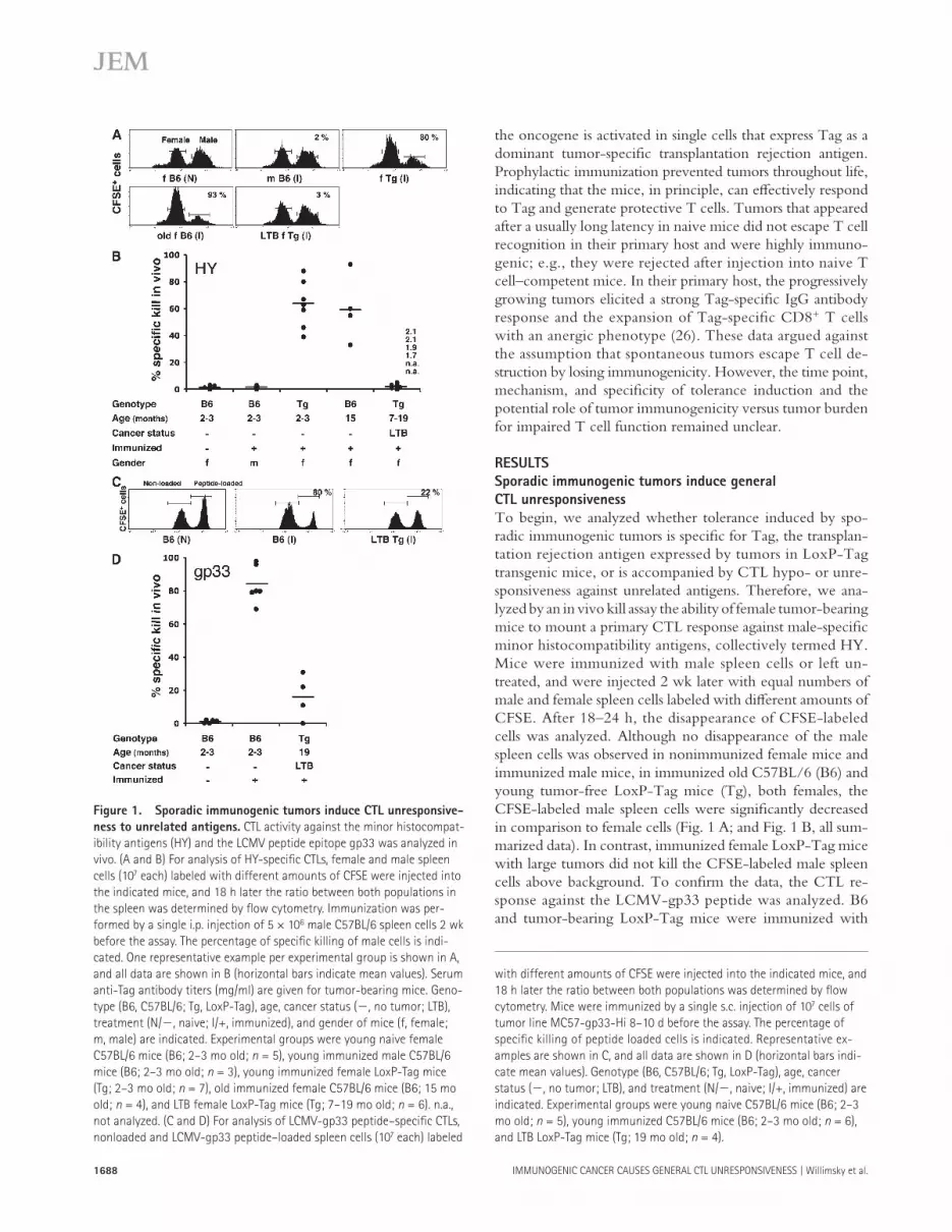
the oncogene is activated in single cells that express Tag as a dominant tumor-specifi c transplantation rejection antigen. Prophylactic immunization prevented tumors throughout life, indicating that the mice, in principle, can eff ectively respond to Tag and generate protective T cells. Tumors that appeared after a usually long latency in naive mice did not escape T cell recognition in their primary host and were highly immuno-genic; e.g., they were rejected after injection into naive T cell – competent mice. In their primary host, the progressively growing tumors elicited a strong Tag-specifi c IgG antibody response and the expansion of Tag-specifi c CD8 + T cells with an anergic phenotype ( 26 ). These data argued against the assumption that spontaneous tumors escape T cell de-struction by losing immunogenicity. However, the time point, mechanism, and specifi city of tolerance induction and the potential role of tumor immunogenicity versus tumor burden for impaired T cell function remained unclear.
RESULTS
Sporadic immunogenic tumors induce general
CTL unresponsiveness
To begin, we analyzed whether tolerance induced by spo-radic immunogenic tumors is specifi c for Tag, the transplan-tation rejection antigen expressed by tumors in LoxP-Tag transgenic mice, or is accompanied by CTL hypo- or unre-sponsiveness against unrelated antigens. Therefore, we ana-lyzed by an in vivo kill assay the ability of female tumor-bearing mice to mount a primary CTL response against male-specifi c minor histocompatibility antigens, collectively termed HY. Mice were immunized with male spleen cells or left un-treated, and were injected 2 wk later with equal numbers of male and female spleen cells labeled with diff erent amounts of CFSE. After 18 – 24 h, the disappearance of CFSE-labeled cells was analyzed. Although no disappearance of the male spleen cells was observed in nonimmunized female mice and immunized male mice, in immunized old C57BL/6 (B6) and young tumor-free LoxP-Tag mice (Tg), both females, the CFSE-labeled male spleen cells were signifi cantly decreased in comparison to female cells ( Fig. 1 A ; and Fig. 1 B , all sum-marized data). In contrast, immunized female LoxP-Tag mice with large tumors did not kill the CFSE-labeled male spleen cells above background. To confi rm the data, the CTL re-sponse against the LCMV-gp33 peptide was analyzed. B6 and tumor-bearing LoxP-Tag mice were immunized with
Figure 1. Sporadic immunogenic tumors induce CTL unresponsive-
ness to unrelated antigens. CTL activity against the minor histocompat-
ibility antigens (HY) and the LCMV peptide epitope gp33 was analyzed in
vivo. (A and B) For analysis of HY-specifi c CTLs, female and male spleen
cells (10 7 each) labeled with different amounts of CFSE were injected into
the indicated mice, and 18 h later the ratio between both populations in
the spleen was determined by fl ow cytometry. Immunization was per-
formed by a single i.p. injection of 5 × 10 6 male C57BL/6 spleen cells 2 wk
before the assay. The percentage of specifi c killing of male cells is indi-
cated. One representative example per experimental group is shown in A,
and all data are shown in B (horizontal bars indicate mean values). Serum
anti-Tag antibody titers (mg/ml) are given for tumor-bearing mice. Geno-
type (B6, C57BL/6; Tg, LoxP-Tag), age, cancer status ( � , no tumor; LTB),
treatment (N/ � , naive; I/+, immunized), and gender of mice (f, female;
m, male) are indicated. Experimental groups were young naive female
C57BL/6 mice (B6; 2 – 3 mo old; n = 5), young immunized male C57BL/6
mice (B6; 2 – 3 mo old; n = 3), young immunized female LoxP-Tag mice
(Tg; 2 – 3 mo old; n = 7), old immunized female C57BL/6 mice (B6; 15 mo
old; n = 4), and LTB female LoxP-Tag mice (Tg; 7 – 19 mo old; n = 6). n.a.,
not analyzed. (C and D) For analysis of LCMV-gp33 peptide – specifi c CTLs,
nonloaded and LCMV-gp33 peptide – loaded spleen cells (10 7 each) labeled
with different amounts of CFSE were injected into the indicated mice, and
18 h later the ratio between both populations was determined by fl ow
cytometry. Mice were immunized by a single s.c. injection of 10 7 cells of
tumor line MC57-gp33-Hi 8 – 10 d before the assay. The percentage of
specifi c killing of peptide loaded cells is indicated. Representative ex-
amples are shown in C, and all data are shown in D (horizontal bars indi-
cate mean values). Genotype (B6, C57BL/6; Tg, LoxP-Tag), age, cancer
status ( � , no tumor; LTB), and treatment (N/ � , naive; I/+, immunized) are
indicated. Experimental groups were young naive C57BL/6 mice (B6; 2 – 3
mo old; n = 5), young immunized C57BL/6 mice (B6; 2 – 3 mo old; n = 6),
and LTB LoxP-Tag mice (Tg; 19 mo old; n = 4).

JEM VOL. 205, July 7, 2008
ARTICLE
1689
MC57 cells that express large amounts of the gp33 peptide ( 27 ), and an in vivo kill assay with gp33 peptide – loaded spleen cells was performed 8 – 10 d later. Although a substantial kill of peptide-loaded spleen cells was observed in B6 mice, no signifi cant kill was observed in tumor-bearing LoxP-Tag mice ( Fig. 1 C ; and Fig. 1 D , all summarized data). These data show that sporadic immunogenic tumors induce general CTL unresponsiveness.
Tag-specifi c tolerance in mice with PMLs
We asked when Tag-specifi c tolerance occurred relative to the time point of the fi rst measurable tumor antigen recogni-tion by the adaptive immune system. This way, we also indi-rectly addressed the question of whether LoxP-Tag mice developed concomitant immunity ( 28 ) at an early stage of tu-mor growth. Thus far, the best surrogate marker for tumor development in LoxP-Tag mice are Tag-specifi c IgG anti-bodies ( 26 ). Approximately 90% of the mice had developed anti-Tag IgG antibodies at the time of obvious tumor burden. Usually, these antibodies occurred many months before tumor burden, indicating a long latency between the initiating onco-genic event and tumor progression. By measuring anti-Tag IgG antibodies in the serum of LoxP-Tag mice, we identifi ed a group of mice between 6 – 14 mo of age that had recently developed low anti-Tag IgG antibody titers ( Fig. 2 A ). Be-cause macroscopically visible tumors could rarely be detected in such mice, they were termed PML mice. As a control, 15 – 19-mo-old mice were used that had high anti-Tag IgG anti-body titers and macroscopically detectable tumors, termed large tumor – bearing (LTB) mice. Because in most cases tu-mors grew in internal organs, a fact that makes the judgment of tumor status diffi cult, the mice were routinely analyzed at the end of the experiment for PML or LTB status. The mice were subjected to an in vivo kill assay with spleen cells loaded with peptide IV, the dominant epitope of Tag ( 29, 30 ). As a further control, young (tumor-free) LoxP-Tag mice were im-munized by a challenge with 16.113 cells, a Tag-positive re-gressor tumor derived from LoxP-Tag mice ( 26 ). Immunized but not naive young LoxP-Tag mice showed a substantial kill of Tag peptide IV – loaded spleen cells ( Fig. 2 B ). Immunized old (6 – 19 mo) B6 mice also effi ciently lysed Tag peptide IV – loaded target cells. The PML mice with low anti-Tag IgG an-tibodies ( Fig. 2 A ) were unable to kill Tag peptide – loaded spleen cells above background ( Fig. 2 B ). Assuming that the time of recognition of the tumor antigen by CTLs and induc-tion of anti-Tag IgG antibodies do not substantially differ from each other, the data suggest that Tag-specifi c CTL toler-ance occurred almost simultaneously with the fi rst detection of anti-Tag IgG antibodies. This also indicates that we failed to detect a phase of concomitant immunity, during which ini-tially functional CTLs were generated. Even after immuniza-tion with 16.113 cells, LTB LoxP-Tag mice were unable to kill Tag peptide IV – loaded spleen cells ( Fig. 2 B ). This was not caused by the advanced age of the mice, because immu-nized old (6 – 19 mo) B6 mice showed lytic activity compara-ble to that of young (2 – 3 mo) LoxP-Tag mice ( Fig. 2 B ).
Figure 2. Tag-specifi c CTL cell tolerance occurs almost simul-
taneously with the development of Tag-specifi c antibodies. (A) To
identify LoxP-Tag mice that had most recently immunologically rec-
ognized PMLs (open symbols; n = 6), anti-Tag IgG antibodies were
measured as indicated in individual mice. As a control, mice that al-
ready had detectable anti-Tag antibodies for several months (LTB
mice; closed symbols; n = 4) were used. (B) CTL activity against the
Tag-specific peptide IV was analyzed in vivo in PML mice (6 – 14 mo
old) and LTB mice (16 – 19 mo old) shown in A. Therefore, nonloaded
and peptide IV – loaded splenocytes labeled with different amounts of
CFSE were injected into the respective mice and as a control into
young naive (2 – 3 mo old; n = 2) and immunized LoxP-Tag transgenic
mice (2 – 3 mo old; n = 3), and aged C57BL/6 mice (B6; 6 – 19 mo old;
n = 5). 18 h later the ratio between both populations was determined
by flow cytometry of spleen cells. The percentage of specific killing of
peptide-loaded cells is indicated (horizontal bars indicate mean val-
ues). Genotype (B6, C57BL/6; Tg, LoxP-Tag), age, cancer status ( � , no
tumor; PML; LTB), and treatment ( � , naive; +, immunized) are indi-
cated. (C) LoxP-Tag mice at � 6 – 9 mo of age have developed Tag-
specific tolerance. 10 6 cells of Tag-expressing tumor line 16.113 were
s.c. injected into 3-mo-old Rag-2 � / � mice ( n = 4), as well as 3-mo-old
( n = 4), 6-mo-old ( n = 4), 9-mo-old ( n = 4), and 13-mo-old ( n = 3)
LoxP-Tag mice, and tumor growth was observed. Error bars represent
SD. The age and number of mice with tumor per number of mice in
the experiment are shown in parenthesis.

1690 IMMUNOGENIC CANCER CAUSES GENERAL CTL UNRESPONSIVENESS | Willimsky et al.
To confi rm these data, LoxP-Tag mice of diff erent ages (6, 9, and 13 mo) were injected with 16.113 cells that are regularly rejected in 3-mo-old LoxP-Tag mice ( Fig. 2 C ). In all mice of 9 and 13 mo of age (4 out of 4 and 3 out of 3, respectively) and in 2 out of 4 mice injected at the age of 6 mo, the transplanted tumor was not rejected. In the 6-mo-old mice, it was those with anti-Tag IgG antibodies that did not reject 16.113 cells (unpublished data). These data demon-strate that in the majority of LoxP-Tag mice, Tag-specifi c tolerance occurred around 6 – 9 mo of age, coinciding with the initial detection of anti-Tag IgG antibodies. Because in these PML mice usually no macroscopic tumors could be detected, and because of the long latency until obvious tu-mor burden (due to the stochastic nature of the tumor model between 12 and > 24 mo), we conclude that Tag-specifi c tolerance occurred in mice with PMLs. This also indicates that the tumor latency is unlikely caused by Tag-specifi c CTL-mediated mechanisms.
Persistent tumor antigen causes general CTL
unresponsiveness subsequent to Tag-specifi c tolerance
We next analyzed when general CTL unresponsiveness oc-curred relative to Tag-specifi c tolerance. LoxP-Tag mice of diff erent ages (7, 10 – 12, and 17 mo) were analyzed for HY-specifi c CTLs and retrospectively anti-Tag IgG antibodies in the blood were determined. Immunized but not naive female B6 mice specifically killed the CFSE-labeled male spleen cells. Immunized 7-mo-old female LoxP-Tag mice had es-sentially normal anti-HY CTL responses even though some of the mice already had anti-Tag IgG antibodies ( Fig. 3 A ). At 10 – 12 mo of age, anti-HY CTL responses were decreased in several LoxP-Tag mice, mostly those with anti-Tag anti-bodies in the serum. However, another portion of this group,
Figure 3. Tag-specifi c tolerance and general CTL unresponsiveness
are induced consecutively. (A) HY-specifi c CTL activity 2 wk after immu-
nization of female LoxP-Tag mice of different ages (7, 10 – 12, and 17 mo)
with 5 × 10 6 male spleen cells was determined by in vivo kill assay, as
described in Fig. 1 . Female and male spleen cells were injected into the
indicated mice, and 18 h later the ratio between both populations in the
spleen was determined by fl ow cytometry. The percentage of specifi c kill-
ing of male cells is indicated (horizontal bars indicate mean values), and
numbers give the anti-Tag IgG antibody titers (mg/ml) in the serum of
corresponding mice. Anti-Tag IgG antibody titers in the serum (mg/ml) are
indicated if mice were positive. n.a., not analyzed. (B) 6 – 13-mo-old LoxP-
Tag mice were fi rst immunized with 5 × 10 6 male spleen cells and 1 wk
later challenged s.c. with 10 6 Tag-expressing 16.113 cells. Mice in which
challenge tumors were not rejected and that had a tumor size of 8 – 10 mm
in diameter (open symbols; n = 8) were boosted with 5 × 10 6 male spleen
cells and subjected to an HY-specifi c in vivo kill assay 2 wk later. Identically
treated 2-mo-old C57BL/6 mice (B6; n = 4) and LoxP-Tag mice (Tg; n = 2)
were used as controls (closed symbols). The percentage of specifi c killing
of male cells is indicated (horizontal bars indicate mean values). Anti-Tag
IgG antibody titers in the serum (mg/ml) are indicated if mice were posi-
tive. n.d., not detectable. (C) 6- and 12-mo-old LoxP-Tag mice reject re-
gressor tumor cells expressing gp33. 10 6 cells of gp33-expressing tumor
line MC57-gp33-Hi were s.c. injected into 3-mo-old Rag-2 � / � ( n = 3),
6-mo-old LoxP-Tag ( n = 3), and 12-mo-old LoxP-Tag ( n = 3) mice, and
tumor growth was observed. The age and number of mice with tumor per
number of mice in the experiment is shown in parenthesis. (D) LoxP-Tag
mice shown in C that rejected MC57-gp33-Hi regressor tumor cells were
immunized with Tag + 16.113 cells, and 9 d later were analyzed for CTL
activity against the Tag-specifi c peptide IV in vivo. Therefore, nonloaded
and peptide IV – loaded splenocytes labeled with different amounts of CFSE
were injected into the respective mice and as a control into young naive
and immunized C57BL/6 mice (2 mo old; n = 2, respectively). 18 h later,
the ratio between both populations was determined by fl ow cytometry of
spleen cells. The percentage of specifi c killing of peptide-loaded cells is
indicated (horizontal bars indicate mean values).

JEM VOL. 205, July 7, 2008
ARTICLE
1691
not only those without anti-Tag antibodies, showed a normal HY-specifi c kill. In 17-mo-old LoxP-Tag mice, no HY-spe-cifi c kill was detected. In another experiment, LoxP-Tag mice between 6 – 13 mo of age were challenged with 16.113 cells, and those mice that did not reject the transplanted tumor and, thus, had developed Tag-specifi c tolerance were sub-jected to an in vivo anti-HY kill assay. Several mice with 16.113 tumors had decreased anti-HY CTL activity, but an-other group of mice that was unable to reject 16.113 cells had normal anti-HY lytic CTL activity ( Fig. 3 B ). These data in-dicate that general CTL unresponsiveness occurred subse-quent to Tag-specifi c tolerance and was probably caused by the persistent strong tumor antigen. If this assumption was correct, LoxP-Tag mice that recently had developed Tag-specifi c tolerance might still be able to reject antigenically unrelated regressor tumor cells. To address this question, LoxP-Tag mice at 6 or 12 mo of age were challenged with MC57-gp33-Hi cells that are rejected in naive B6 mice and elicit gp33-specifi c CTLs ( Fig. 1, C and D ) ( 27 ). As shown in Fig. 3 C , tumors grew in Rag-2 � / � mice but were rejected in LoxP-Tag mice. The latter mice were then immunized by injection of 16.113 cells and analyzed by in vivo kill assay for Tag peptide IV – specifi c CTLs. Two out of three LoxP-Tag mice at 6 mo of age and three out of three mice at 12 mo of age did not show signifi cant peptide IV – specifi c CTL activity ( Fig. 3 D ). Thus, shortly after having developed Tag-specifi c CTL tolerance, LoxP-Tag mice appear to have normal CTL responses against unrelated targets.
Primary but not memory CTL responses are impaired
in old LoxP-Tag mice
To distinguish whether anti-HY CTL responses were inhib-ited in the priming/expansion or eff ector phase, the frequency of D b /UTY-tetramer – binding CD8 + T cells was analyzed in LoxP-Tag mice with diff erent anti-HY CTL activity, de-scribed in Fig. 3 (A and B) . UTY is an HY antigen for which the H2-D b – presented peptide sequence is known ( 31 ). In most immunized female B6 and 2-mo-old LoxP-Tag mice, � 2 – 4% of the CD8 + T cells were D b /UTY-tetramer positive, correlating with high cytolytic activity ( Fig. 4, A and B ). Non-immunized mice contained < 0.03% double-positive cells. In most of the older LoxP-Tag mice, especially the 6 – 7- and 10 – 13-mo-old groups, the decrease of anti-HY CTL activity was proportional to the decrease of D b /UTY-tetramer – binding cells, suggesting that the priming/expansion of anti-HY CTLs was inhibited. However, regardless of tumor burden, in some 17 – 19-mo-old mice a slight increase in the frequency of D b /UTY-tetramer – positive CD8 + T cells was detected, even though no anti-HY cytolytic activity was detected in these mice ( Fig. 4 B ). We cannot exclude, therefore, that CTL eff ector function was also inhibited.
The aforementioned experiments addressed the question of whether primary unrelated CTL responses were impaired in old LoxP-Tag mice. Next, we asked whether memory CTL responses were also inhibited in old LoxP-Tag mice. A group of female LoxP-Tag mice were immunized at the age
Figure 4. Primary but not memory CTL responses are suppressed in
old LoxP-Tag mice. (A) HY-immunized mice shown in Fig. 3 (A and B) were
analyzed for the presence of UTY-specifi c T cells by staining spleen cells with
anti-CD8 mAbs and H-2D b /UTY tetramers. One representative example of
fl ow cytometric analysis for each experimental group is shown. The per-
centage of UTY-specifi c tetramer-positive CD8 + T cells is indicated. (B) The
percentage of HY-specifi c killing is plotted against the percentage of
UTY-specifi c tetramer-positive CD8 + T cells from experiments shown in Fig. 3
(A and B) . (C) Memory CTL responses against unrelated antigens are not
generally inhibited in aged LoxP-Tag mice. 2-mo-old female LoxP-Tag and
C57BL/6 mice were immunized three times with 5 × 10 6 male spleen cells
(prime). At the age of 18 – 19 mo, these mice were reimmunized with 5 × 10 6
male spleen cells (boost), and HY-specifi c CTL activity was determined 2 wk
later by in vivo kill assay, as described in Fig. 1 . CFSE-labeled female and male
spleen cells were injected into the indicated mice, and as a control into
2-mo-old naive C57BL/6 (B6; n = 2), immunized C57BL/6 mice (B6; n = 2),
and immunized 18-mo-old LoxP-Tag mice (Tg; n = 2). 18 h later, the ratio
between both populations was determined by fl ow cytometry. The percent-
age of specifi c killing of male cells is indicated (horizontal bars indicate mean
values). Genotype (B6, C57BL/6; Tg, LoxP-Tag), age, cancer status ( � , tumor-
free; PML), and treatment ( � , naive; +/ � , immunized once at the indicated
age; +/+, primed at 2 mo/boosted at the indicated age) are indicated.

1692 IMMUNOGENIC CANCER CAUSES GENERAL CTL UNRESPONSIVENESS | Willimsky et al.
LoxP-Tag mice that had not been immunized at the age of 2 mo failed to mount anti-HY CTL responses after primary immunization at the age of 18 mo ( Fig. 4 C ). In contrast, old LoxP-Tag mice that had been immunized at 2 mo showed normal anti-HY cytolytic activity, comparable to age-matched nontransgenic control mice. Thus, the persis-tent tumor antigen inhibits primary but not memory CTL responses against unrelated antigens.
Detection of premalignant immunogenic lesions
in Tag-tolerant mice
In those mice designated as LTB mice, obvious tumor burden was detected by magnet resonance tomography ( Fig. 5 A ) that was confi rmed by immunohistology with an anti-Tag anti-body ( Fig. 5 B ). However, in the majority of mice that were tolerant for Tag and/or had shown general CTL unrespon-siveness, no macroscopically visible or magnetic resonance imaging (MRI) – detectable primary tumor could be detected. Detection of PMLs was diffi cult; because of the stochastic na-ture of the model, not only the tumor latency but also tumor location can be variable from mouse to mouse. Because LoxP-Tag mice frequently develop renal cancer, the kidneys of sev-eral mice were analyzed for PMLs. To not miss any small PMLs, the entire kidneys of seven mice were cross sectioned in 4- μ m steps; every 10th section was analyzed for pathological alterations and, if early lesions were detected, consecutive sec-tions were stained with diff erent antibodies. Comparable to the frequency of renal cell carcinoma in four out of seven mice, microscopically small cell clusters that stained positive with an anti-Tag antibody were detected in the kidneys ( Fig. 5 C ; and Fig. S1, available at http://www.jem.org/cgi/content/full/jem.20072016/DC1). These cell clusters that were not larger than 1 mm in diameter were defi ned as premalig-nant because they additionally stained positive for the prolifer-ation marker Ki-67 ( Fig. 5 C and Fig. S1). In the kidneys of the four mice, between two and fi ve Tag-positive lesions were detected, for which we do not know whether they were clon-ally related or independent lesions. According to the age of the analyzed mice (6 – 7 mo) and the mean time until obvious tu-mor burden (between 12 and > 24 mo), these premalignant le-sions may have been unapparent for a long time. Remarkably, proportionally to the size of the PMLs a strong T cell infi ltrate (CD3 + ) was detected ( Fig. 5 C and Fig. S1). Because these mice were shown either to fail to reject 16.113 tumor cells or to be unable to kill Tag peptide IV – loaded spleen cells and, therefore, were tolerant for Tag, the role of these T cells, whose phenotype and antigen specifi city are unknown, re-mains to be analyzed. However, the T cells infi ltrating PMLs were negative for FoxP3, a marker for regulatory T cells ( Fig. 5 C , Fig. S1, and Fig. S2). Even though we cannot exclude that further Tag-positive lesions in other organs escaped our attention, the data demonstrate that the PMLs have the ap-pearance of being highly immunogenic despite Tag-specifi c CTL tolerance, because they contain a strong T cell infi ltrate and the mice produce anti-Tag IgG antibodies. Furthermore, general CTL unresponsiveness appears to be initiated in mice
of 2 mo with male spleen cells, as above. At 18 mo, when most LoxP-Tag mice were unable to respond to male spleen cell immunization in the previous experiments ( Fig. 1 B and Fig. 3 A ), the mice were subjected to an in vivo anti-HY CTL kill assay 2 wk after booster immunization. As before,
Figure 5. Detection of tumors and PMLs in LoxP-Tag mice. (A) Tumors
in LTB mice were detected by MRI. Shown are representative transversal and
coronal MR images of a kidney tumor (#1) and a tumor in the bone (#2);
tumors are indicated by white circles. (B) Immunohistology of tissue sections
in LTB mice. Tissues were stained for Tag, Ki-67, CD3 (red), FoxP3 (brown),
and F4/80 (red) expression and counterstained with hematoxylin. Bar, 200 μ m.
(C) Representative PMLs in mice that recently developed anti-Tag anti-
bodies but without macroscopically detectable tumors. Kidneys of 6 – 7-mo-
old LoxP-Tag mice, which were shown to have developed tumor-induced
tolerance, were cross sectioned in 4- μ m steps and examined for the presence
of PMLs by staining with hematoxylin and eosin (not depicted). Consecutive
sections of identifi ed PMLs were stained with mAbs as in B. In four out of
seven mice analyzed, PMLs were detected in the kidney. Lymph nodes ob-
tained from tumor-free mice served as a positive control for FoxP3 staining
(Fig. S2, available at http://www.jem.org/cgi/content/full/jem.20072016/DC1).
Staining of additional PMLs is shown in Fig. S1. Bar, 100 μ m.

JEM VOL. 205, July 7, 2008
ARTICLE
1693
mice were crossed to Alb-Cre transgenic mice that express the Cre recombinase in a liver-specifi c fashion. Because of the early deletion of the stop cassette and Tag expression in a Cre-LoxP – mediated fashion, the mice are tolerant for Tag. They do not produce anti-Tag antibodies, accept Tag-posi-tive tumor grafts that are rejected in young tumor-free LoxP-Tag mice ( 26 ), and cannot kill Tag peptide IV – loaded spleen cells after immunization ( Fig. 7 A ). At approximately weeks 12 – 16, they have developed large liver tumors, as analyzed by MRI ( Fig. 7 B ) and immunohistology ( Fig. 7 C ), that are non- (or low-) immunogenic, because Tag is the dominant transplantation rejection antigen ( 26 ). Female LoxP-Tag × Alb-Cre double-transgenic mice were analyzed for HY-spe-cific CTLs as above. Immunized mice with large tumors killed transferred CFSE-labeled male spleen cells almost as effi ciently as normal B6 mice ( Fig. 7 D ). These data demon-strate that although small immunogenic PMLs induce general CTL unresponsiveness, large nonimmunogenic tumors do not. Surprisingly, LoxP-Tag × Alb-Cre double-transgenic mice had by far the highest frequency of iMCs in the spleen (means of 45% CD11b/Gr-1 – and 22% c-kit/Gr-1 – positive; Fig. 6, A and B ), but they had normal serum TGF- � 1 concentra-tions ( Fig. 6 C ). These data support the notion that tumor burden increases iMCs ( 15, 32 ), but also show that large numbers of tumor-induced iMCs per se are not suffi cient to inhibit CTL responses against unrelated antigens. Based on a few additional indicative markers (F4/80, IL-4 receptor, and Fc � receptor), no signifi cant phenotypic diff erence of CD11b-positive cells from LoxP-Tag compared with LoxP-Tag × Alb-Cre mice was observed, with the exception that based on higher F4/80 expression, CD11b-positive cells from aged single-transgenic mice appeared to have a more mature phe-notype (Fig. S4, available at http://www.jem.org/cgi/content/full/jem.20072016/DC1). The tumor-induced expansion of iMCs without an obvious need to inhibit CTL responses in-dicates that their primary function is the direct support of tu-mor growth, as previously suggested ( 16 ).
DISCUSSION
When are cancer cells recognized by adaptive immune cells? What is the outcome of the initial recognition? And what are the consequences of long-term tumor antigen reactivity? Answers to these questions are the key to the question of whether T cells spontaneously control autochthonous tu-mors, termed cancer immunosurveillance. Previous studies did not allow these questions to be addressed because of the lack of suitable models ( 33 ).
Antigen-defi ned sporadic cancer model
Most data on antitumor T cell responses have been obtained by exposing the host to an artifi cially large number of tumor cells at a single time point, e.g., by tumor transplantation ex-periments or in transgenic models with tissue-specifi c onco-gene/tumor antigen expression. In other models (e.g., chemical carcinogenesis), it is not possible to analyze specifi c T cell responses because no tumor antigen is known. Additionally,
with PMLs, with no indication of local FoxP3 + T cell involve-ment. PMLs were also infi ltrated by F4/80-positive cells ( Fig. 5 C and Fig. S1), representing the major infl ammatory com-ponent of the developing stroma in these lesions.
General CTL unresponsiveness is associated
with an increase of iMCs and TGF- � 1
To search for a mechanism of the general CTL unresponsive-ness we analyzed iMCs, a cell population known to nonspe-cifi cally inhibit CTL responses ( 15, 18, 24 ). An analysis of spleens from LTB LoxP-Tag mice showed that iMCs, de-tected by antibodies specifi c for CD11b/Gr-1 or Gr-1/c-kit, were signifi cantly expanded in comparison to young tumor-free LoxP-Tag and old B6 mice ( Fig. 6 A ; and Fig. 6 B , all summarized data). Spleen cells from young LoxP-Tag and old B6 mice contained 5 – 7% CD11b/Gr-1 – positive and 0.5 – 3% c-kit/Gr-1 – positive cells, with those from LTB mice containing means of 22 and 8%, respectively. iMCs were al-ready slightly increased in mice with PMLs (mean of 10% CD11b/Gr-1 – positive cells). Despite variability between mice of the same group, it appeared that iMC expansion correlated with progressive tumor growth ( Fig. 6 B ). Substantial num-bers of iMCs were also detected in tumors ( Fig. 6, A and B ). LTB LoxP-Tag mice had substantially elevated serum TGF- � 1 concentrations of between 90 – 150 ng/ml, with a mean of 120 ng/ml ( Fig. 6 C ) ( 26 ). Young tumor-free mice had 50 ± 10 ng/ml TGF- � 1 in the serum. Old B6 mice had slightly increased TGF- � 1 levels compared with young LoxP-Tag mice. LoxP-Tag mice with PMLs (defi ned by the presence of anti-Tag IgG antibodies in the serum) revealed TGF- � 1 concentrations almost as high as LTB mice (106 ± 25 ng/ml; Fig. 6 C ). In essence, tumor-induced tolerance and CTL un-responsiveness against unrelated antigens is associated with tumor-specifi c IgG antibodies, high serum levels of TGF- � 1, and large numbers of iMCs in the spleen and tumor. The source of the elevated TGF- � 1 is diffi cult to identify because many cell types are able to express TGF- � 1. Because we found a good correlation between serum Tag-specifi c IgG antibodies and elevated TGF- � 1 levels, and TGF- � 1 – IgG complexes have been implicated in CTL suppression ( 23, 24 ), we evaluated TGF- � 1 – producing IgG + B cells. By immuno-histology, a substantially increased number of IgG/TGF- � 1 double-positive cells were detected in perihilar lymph nodes of kidneys of tumor-bearing LoxP-Tag compared with young tumor-free LoxP-Tag mice ( Fig. 6 D ; and Fig. S3, available at http://www.jem.org/cgi/content/full/jem.20072016/DC1). However, we also found anti – TGF- � 1 immune reactivity that did not overlap with anti-IgG reactivity. This result sug-gested that IgG-producing B cells are at least one potential source for the elevated TGF- � 1 levels in tumor-bearing LoxP-Tag mice.
Large nonimmunogenic tumors do not induce general
CTL unresponsiveness
Finally, we asked whether nonimmunogenic tumors also in-duce CTL unresponsiveness to unrelated antigens. LoxP-Tag

1694 IMMUNOGENIC CANCER CAUSES GENERAL CTL UNRESPONSIVENESS | Willimsky et al.
Figure 6. Tumors in LoxP-Tag mice induce an increase in iMCs and serum TGF- � 1 levels. (A and B) For analysis of iMCs, single-cell prepara-
tions of spleen and tumor were double stained with antibodies against Gr-1 and CD11b or Gr-1 and c-kit, respectively, to assess CD11b + /Gr-1 + (top)
and c-kit + /Gr-1 + (bottom) cells in spleens of 2 – 3-mo-old LoxP-Tag mice (young Tg), 14 – 17-mo-old wild-type mice (old B6), 6 – 13-mo-old tumor-
bearing LoxP-Tag mice with PMLs, 7 – 19-mo-old LTB LoxP-Tag mice, and 3 – 4-mo-old LoxP-Tag × Alb-Cre double-transgenic mice (TB Tg × Alb-Cre)
with large liver tumors. Numbers show the percentage of double-positive cells of nonlymphocytes. One representative example per experimental
group is shown in A, and all data for splenic CD11b + /Gr-1 + and c-kit + /Gr-1 + cells ( � ) and tumor-infi ltrating CD11b + /Gr-1 + cells ( � ) are shown in B
(horizontal bars indicate mean values). (C) Serum obtained from individual young LoxP-Tag (Tg; 3 mo old; n = 5), old C57BL/6 mice (B6; 12 – 23 mo
old; n = 5), LoxP-Tag transgenic mice with PMLs (PML Tg; 6 – 13 mo old; n = 4), LTB LoxP-Tag transgenic mice (LTB Tg; 7 – 19 mo old; n = 4), and LTB

JEM VOL. 205, July 7, 2008
ARTICLE
1695
we assume abundant cell death of Tag-expressing cells ( 40 ) and chronic tumor antigen release under CTL tolero-genic conditions.
Split tolerance and no necessity of immune escape
The time point of fi rst detection of anti-Tag IgG antibodies in the serum of LoxP-Tag mice marked the time point of tolerance. This sounds paradoxical. Tolerance was measured by the inability to kill Tag peptide IV – loaded spleen cells and the failure to reject Tag-positive tumor cells that were re-jected in young LoxP-Tag mice. On the other hand, the anti-Tag IgG antibodies, the generation of which likely involves functional CD4 + T cell help, indicates a productive anti-Tag immune response. Therefore, it is debatable whether Tag in LoxP-Tag mice is immunogenic or antigenic. The split toler-ance is remarkable because some investigators proposed that nascent tumors are unable to induce functional T cells (e.g., because of the lack of co-stimulation; reference 41 ), whereas others suggested that nascent tumors are able to do so ( 42 ). Even though the Tag-specifi c CD4 + T cells have to be ana-lyzed, our data support the former hypothesis with regards to CTLs and the latter hypothesis with regards to CD4 + T cells, yet they are deviated to a phenotype that does not allow them to support CTL generation. What is often considered as tu-mor immunity should in fact be regarded as innocent tumor reactivity. Thus, the net eff ect of the split tolerance is tolerance of the cancer cells: it is unlikely that the humoral immune re-sponse has tumoricidal activity, because Tag is a nuclear anti-gen and not accessible to antibodies.
The long latency is probably caused by the multistage process of carcinogenesis and not CTL-mediated tumor con-trol, because the mice had acquired Tag-specifi c tolerance upon or close to the fi rst recognition of the PMLs. As has been mentioned before, the anti-Tag IgG antibodies might involve CD4 + T cell help; therefore, their occurrence might indicate the time point when CTLs become aware of Tag. Because we failed to detect a window of functional CTL ac-tivation, our data suggest that sporadic cancer in our model is unable to induce functional CTLs. Instead, CTLs with an anergic phenotype expand in tumor-bearing LoxP-Tag mice ( 26 ). This may explain why highly immunogenic tumors pro-gressively grow. Our data argue strongly against immunoed-iting or immune escape ( 42 ), direct evidence of which is lacking ( 33, 43, 44 ). If spontaneous tumors, even if very immuno-genic, are unable to induce a protective T cell response, then there is no need for tumor cells to escape T cell recognition or destruction. Potentially immune-suppressive mechanisms like increased levels of serum TGF- � 1 or expansion of iMCs could be the consequence, not the cause, of the default im-mune response.
chemical carcinogenesis is strongly infl uenced by nonspecifi c infl ammatory responses that may be altered in immune-defi cient mice. Unfortunately, altered chemical carcinogen-esis in immune-defi cient compared with immune-competent mice has been erroneously attributed to T cell – specifi c anti-tumor eff ects ( 33 ). Several sporadic cancer models have been developed, e.g., by oncogene activation in the adult mouse through Cre recombinase – mediated deletion of a stop cas-sette ( 34 ) or by spontaneous recombinations (hit and run) ( 35 ). In these models, the antitumor immune response has not been analyzed. LoxP-Tag mice provide a unique spo-radic cancer model because the oncogene is stochastically activated in individual cells without experimental manipula-tion. Because young LoxP-Tag mice eff ectively respond to Tag, the initiating oncogene (e.g., shown by the rejection of transplanted Tag-positive tumor cells in young mice), the immune response against the developing tumor can be ana-lyzed throughout the long process leading to malignancy. Tumor initiation and progression may be variable from mouse to mouse because of the stochastic nature of the model. It showed, as based on the induction of anti-Tag antibodies and verifi ed in several cases histologically, that LoxP-Tag mice contain PMLs at around 6 – 9 mo but obvious tumor burden occurred between 12 and > 24 mo. In this regard, sporadic cancer in LoxP-Tag mice closely resembles cancer in humans that may persist in a premalignant state long be-fore clinical appearance ( 36, 37 ). However, a caveat of the model is that because of the stochastic nature of oncogene activation, we do not know in how many cells and at which level Tag is expressed in the lifetime of LoxP-Tag mice. One can assume, as verified in several mice, that several PMLs occur in the same mouse, but only few or often only one fi -nally progressed. Even though multiple cancers have been observed in humans ( 38 ) (e.g., induced by chemical carcino-gens, a phenomenon termed fi eld cancerization; reference 39 ), these lesions unlikely share the same tumor-specifi c antigens. In contrast, PMLs in LoxP-Tag mice share Tag as antigen. The early time point of Tag-specifi c tolerance ( � 6 – 9 mo), when mice reveal PMLs, could therefore be caused by the multiplicity of Tag-positive lesions. Whether mice with a single cancerous lesion develop tolerance at a later time point remains to be analyzed. It should be noted that the number of PMLs in the kidney, a frequent site of tumor develop-ment, at the time when Tag-specifi c tolerance had already manifested was low. By complete sectioning of the kidneys of 6 – 7-mo-old Tag-tolerant LoxP-Tag mice, between two and fi ve lesions were detected in four out of seven ana-lyzed mice, all of which were microscopically small. Because the premalignant cells appeared to proliferate (i.e., were Ki-67 + ) without apparent progression for an extended time period,
LoxP-Tag × Alb-Cre double-transgenic mice (LTB Tg × Alb-Cre; 3 – 4 mo old; n = 4) was analyzed for TGF- � 1 by ELISA. Error bars represent SD. (D) Perihilar
lymph nodes of kidneys obtained from young tumor-free LoxP-Tag mice and mice with PMLs were double stained for TGF- � 1 (red) and IgG (green);
nuclei were counterstained with DAPI. Cells that are double positive for IgG antibodies and TGF- � 1 are shown in orange (merge). Additional stainings
for PML and LTB mice are shown in Fig. S3 (available at http://www.jem.org/cgi/content/full/jem.20072016/DC1). Bar, 20 μ m.

1696 IMMUNOGENIC CANCER CAUSES GENERAL CTL UNRESPONSIVENESS | Willimsky et al.
unexpected, because the double-transgenic mice had the highest frequency of iMCs that was considered to be suffi -cient to inhibit CTL activation. Because LoxP-Tag × Alb-Cre mice had developed tolerance for Tag early in life and Tag is the dominant transplantation rejection antigen, tumors in the double-transgenic mice are nonimmunogenic in comparison to tumors that grow in single transgenic LoxP-Tag mice. Immunogenicity, not tumor burden is, therefore, the pri-mary cause for the general CTL unresponsiveness. Tumor bur-den in double-transgenic LoxP-Tag occurred very fast because Tag was activated in the whole liver. We cannot exclude, therefore, that the longer latency with the continuously pre-sent Tag expression in single-transgenic LoxP-Tag mice con-tributed to the general CTL unresponsiveness. Collectively, we suggest that the degree of general CTL unresponsiveness directly refl ects the degree of tumor immunogenicity.
At the current time, we can only speculate about the cellular and molecular mechanisms of how general CTL un-responsiveness is mediated. Recently, iMCs were suggested to be renamed as myeloid-derived suppressor cells, a defi ni-tion that implies that they always act immune suppressive ( 48 ).
General CTL unresponsiveness
Over time and subsequent to Tag-specifi c tolerance, LoxP-Tag mice developed a profound general CTL unresponsive-ness. It was associated with increased serum levels of TGF- � 1, expansion of iMCs, and the presence of anti-Tag IgG anti-bodies. Because we have seen in the previous paragraph that escape from immune surveillance is likely not the reason for the immune suppression, the question arises why and by which mechanism is the general CTL unresponsiveness in-duced? Our data suggest that the chronic and persistently immunogenic tumor antigen sustains the default CTL re-sponse that cannot be turned down, resulting in general CTL unresponsiveness. Whether TGF- � 1, which has consistently been shown to suppress CTL responses ( 45 – 47 ), is the cause of the Tag-specifi c tolerance or the symptom of the persistent tumor antigen remains unclear. However, because TGF- � 1 levels were already elevated in mice with PMLs and persisted at high levels for a very long time, it likely contributed to the general CTL unresponsiveness. This is supported by the nor-mal anti-HY CTL responses and normal TGF- � 1 levels in Tag-tolerant LoxP-Tag × Alb-Cre mice. This was rather
Figure 7. Large nonimmunogenic tumors in LoxP-Tag × Alb-Cre double-transgenic mice do not induce general CTL unresponsiveness.
(A) LoxP-Tag × Alb-Cre double-transgenic mice are tolerant for Tag. LoxP-Tag × Alb-Cre double-transgenic mice (1 – 2 mo old; n = 3) were immunized s.c.
with 10 6 Tag + 16.113 cells and subjected to a Tag-specifi c peptide IV in vivo kill assay 2 wk later. As a control, naive mice (2 mo old; n = 2) and immunized
wild-type mice (2 mo old; n = 2) were used. The percentage of specifi c killing of Tag-specifi c peptide IV – loaded spleen cells is shown (horizontal bars indi-
cate mean values). (B) LoxP-Tag × Alb-Cre double-transgenic mice (Tg × Alb-Cre) develop large tumors in the liver at 3 – 4 mo of age. MRI analysis re-
vealed livers to be four to fi ve times larger in size in comparison to livers from 3 – 4-mo-old LoxP-Tag mice (Tg). (C) Immunohistochemical analysis using
Tag-specifi c antibody shows strong Tag expression in liver tumors that consist of hepatocellular and cholangiocellular cell carcinoma (HCC and CCC, re-
spectively). Bar, 200 μ m. (D) LoxP-Tag × Alb-Cre mice with large liver tumors at 3 – 4 mo of age ( n = 3) were immunized with 5 × 10 6 male spleen cells and
subjected to an HY-specifi c in vivo kill assay 2 wk later, as described in Fig. 1 . Naive and HY-immunized C57BL/6 (B6) mice served as controls. The percent-
age of specifi c killing of male cells is shown (horizontal bars indicate mean values).

JEM VOL. 205, July 7, 2008
ARTICLE
1697
the best indication that human cancers are not dramatically diff erent in terms of immunogenicity compared with those in LoxP-Tag mice comes from the observation that PMLs in LoxP-Tag mice and primary carcinomas in humans are both often infi ltrated by T cells. In humans, these tumor-infi ltrating lymphocytes (TILs) have been associated with a good prog-nosis, which has been interpreted as a tumoricidal T cell re-sponse ( 57, 58 ). Our data support the observation but not the interpretation. Mice with PMLs and a strong T cell infi l-trate at the age of 6 – 7 mo had a comparably good prognosis, yet they had already acquired Tag-specifi c tolerance. The TILs, therefore, probably refl ect a cancer-promoting infl ammatory response ( 59, 60 ) induced by the PMLs, not a tumor-eradi-cating T cell response. Whether the TILs in PMLs of LoxP-Tag mice are innocent bystander cells of the infl ammatory response or cancer-promoting remains to be determined.
In conclusion, sporadic cancers do not sneak through and do not induce concomitant immunity but are recognized at the premalignant stage and induce a default immune response. The data argue strongly against immunoediting ( 42 ), direct evidence of which is lacking ( 33, 43, 44 ). Provided that similar mechanisms that we observe in the mouse model also occur in cancer patients, our data have to be seen as a cautionary note for therapeutic cancer vaccines: the more immunogenic the target antigen, the higher the preexisting immune suppression. However, they also have to be seen as a positive note for adoptive T cell therapy: the best target antigens are retained on the cancer cells; methods to eliminate immune suppression before T cell transfer have been developed ( 61, 62 ).
MATERIALS AND METHODS Animals. LoxP-Tag transgenic mice were described previously ( 26 ).
C57BL/6 mice were obtained from Charles River Laboratories, Alb-Cre
transgenic mice (B6.Cg-Tg(Alb-cre)21Mgn/J) were purchased from the
Jackson Laboratory, and Rag-2 � / � mice (129S6(B6)-Rag2tm1Fwa) mice
were obtained from Taconic. Mice were housed at the animal facilities of
Charit é (Campus Benjamin Franklin) and Max-Delbr ü ck-Center for Mo-
lecular Medicine. All experiments were in accordance with institutional,
state, and federal (Landesamt f ü r Arbeitsschutz, Gesundheitsschutz und tech-
nische Sicherheit, Berlin) guidelines.
Immunization/tumor transplantation. Mice were immunized at the
ages indicated in the fi gures either by s.c. injection of 10 6 Tag + 16.113 cells
(Tag-specifi c immunization; reference 26 ) or 10 7 MC57-gp33-Hi cells
(LCMV-gp33 peptide – specifi c immunization; reference 27 ), or i.p. injec-
tion of 5 × 10 6 male spleen cells (HY-specifi c immunization). In tumor
challenge experiments, 10 6 Tag + 16.113 or MC57-gp33-Hi cells were in-
jected s.c. Animals that rejected 16.113 cells were monitored for at least 60 d.
In the tumor transplantation experiments, mice were scored tumor bearing
when the tumor size was ≥ 10 mm in diameter. At this size, tumors had a mean
volume of 425 ± 75 mm 3 , which is best approximated by use of the formula
for hemi-ellipsoids: vol = lwh(1/2).
In vivo kill assays. Tag-specifi c peptide IV (VVYDFLKL; reference 5 ) and
gp33-41 peptide – specifi c (KAVYNFATM; reference 63 ) in vivo cytotoxic-
ity assays were performed as previously described ( 26 ). For gp33 peptide, as-
says were performed 8 – 10 d after immunization. For analysis of CTL activity
against the HY antigens, spleen cells from male and female C57BL/6 mice
were labeled with CSFE in a fi nal concentration of 0.75 μ M (CFSE high ) or
0.075 μ M (CFSE low ), respectively, for 15 min at room temperature. Cells were
The experimental systems to analyze their immune-suppres-sive potential were restricted to an in vitro analysis in which the addition of iMCs inhibited the activation of CTLs against tumor and unrelated antigens ( 15, 17, 49 ). Indeed, the iMCs appeared to consistently inhibit CTL responses in vitro ( 18 ). However, the dramatic expansion of iMCs and tumor burden in LoxP-Tag × Alb-Cre mice did not cause CTL unrespon-siveness in vivo. Whether iMCs in our model suppress CTL responses in vitro is not known, but even if they do, the in vivo data may be more relevant. Therefore, we prefer to call them iMCs rather than myeloid-derived suppressor cells, be-cause they obviously do not always suppress unrelated CTLs.
The question remains as to why CTL responses against unrelated antigens were suppressed in LoxP-Tag but not in LoxP-Tag × Alb-Cre mice even though iMCs were ex-panded to high frequencies in both mouse lines. One possibility is that the iMCs, known to be a heterogenous cell population, are phenotypically diff erent in the two mouse lines. They could, for instance, diff er in the production of immune-suppressive molecules such as nitric oxide or arginase ( 17, 19, 50 ). How-ever, an alternative possibility is one reminiscent of a mecha-nism suggested by Rowley et al. ( 22 – 24 ). They proposed that chronic exposure of a fi rst antigen, in this case sheep red blood cells, induced B cells to secrete IgG-latent TGF- � 1 complexes. Through Fc receptors, this complex is taken up by myeloid-derived cells that activate the latent form of TGF- � 1, which then, in turn, is used to inhibit activation of CTLs against unrelated antigens ( 22 – 24 ). Specifi c antibodies, non-dissociated IgG – TGF- � 1 complexes, monocytes/macrophages, and Fc receptors were all necessary for inhibition of CTL priming in vitro. Several lines of evidence indicate that gen-eral CTL unresponsiveness in LoxP-Tag mice is mediated by a similar mechanism: (a) iMCs alone were not suffi cient to inhibit unrelated CTL responses, as observed in LoxP-Tag × Alb-Cre mice; (b) inhibition of unrelated CTL responses cor-related with an increase in TGF- � 1 and anti-Tag IgG anti-bodies, as seen in LoxP-Tag but not LoxP-Tag × Alb-Cre mice; (c), iMCs expressed Fc-receptors; and (d) IgG – TGF- � 1 double-positive cells were detected in Tag-tolerant LoxP-Tag mice. Future studies are needed to elucidate the precise mechanism of general CTL unresponsiveness in old LoxP-Tag mice.
Relevance of LoxP-Tag mice for human cancer
It is diffi cult to judge how the immunogenicity of tumors in humans compares with that in our tumor model. On the one hand, human tumors are often assumed to be less immuno-genic than mouse tumors. On the other hand, increased iMC frequencies ( 32 ), elevated TGF- � 1 serum levels ( 51 ), tumor-reactive antibodies ( 52, 53 ), and strongly increased tumor-reactive CTLs without apparent regression ( 54 – 56 ) have all been detected in cancer patients but are not yet analyzed in the same patients. Additionally, memory CTL responses that might be the most frequent in (usually elderly) cancer pa-tients are normal in tumor-bearing LoxP-Tag mice. Therefore, one would not expect frequent general CTL defi ciency or cancer-induced opportunistic infections in humans. Perhaps

1698 IMMUNOGENIC CANCER CAUSES GENERAL CTL UNRESPONSIVENESS | Willimsky et al.
ters: TR, 308 ms; TE, 4.7 and 10 ms; fl ip angle, 70 ° , NEX, 22; FOV, 150 ×
75 mm; matrix, 256 × 128; and slice thickness, 3 mm with a resolution of
0.7 × 0.6 mm for the CP Breast Array Coil and 1 mm with a resolution of
0.2 × 0.2 mm for the Loop Flex Coil (small surface).
Online supplemental material. Fig. S1 shows additional representative
PMLs in mice that recently developed anti-Tag antibodies but without
macroscopically detectable tumors. Fig. S2 shows FoxP3 control staining of
lymph nodes. Fig. S3 shows the analysis of IgG and TGF- � 1 – expressing
cells in draining lymph nodes of mice with PMLs and tumors. Fig. S4 shows
a phenotypic analysis of CD11b-positive cells obtained from young LoxP-
Tag mice, LoxP-Tag mice with PMLs, and tumor-bearing LoxP-Tag ×
Alb-Cre mice. Online supplemental material is available at http://www
.jem.org/cgi/content/full/jem.20072016/DC1.
We thank S. Horn, K. Borgwald, D. Barthel, S. Spieckermann, M. Roesch, A. Gaertner,
and C. Westen for excellent technical assistance.
This work was supported by grants from the Deutsche
Forschungsgemeinschaft (SFB633, TR36, and TR54), the European Community (FP6
grant “ ATTACK ” ), and the clinical cooperation program of the Max-Delbr ü ck-Center
for Molecular Medicine (to G. Willimsky and J. Gellermann).
The authors declare that they have no competing fi nancial interests.
Submitted: 18 September 2007
Accepted: 21 May 2008
REFERENCES 1 . Ye , X. , J. McCarrick , L. Jewett , and B.B. Knowles . 1994 . Timely im-
munization subverts the development of peripheral nonresponsiveness and suppresses tumor development in simian virus 40 tumor antigen-transgenic mice. Proc. Natl. Acad. Sci. USA . 91 : 3916 – 3920 .
2 . Speiser , D.E. , R. Miranda , A. Zakarian , M.F. Bachmann , K. McKall-Faienza , B. Odermatt , D. Hanahan , R.M. Zinkernagel , and P.S. Ohashi . 1997 . Self antigens expressed by solid tumors do not effi ciently stimulate naive or activated T cells: implications for immunotherapy. J. Exp. Med. 186 : 645 – 653 .
3 . Wick , M. , P. Dubey , H. Koeppen , C.T. Siegel , P.E. Fields , L. Chen , J.A. Bluestone , and H. Schreiber . 1997 . Antigenic cancer cells grow progressively in immune hosts without evidence for T cell exhaustion or systemic anergy. J. Exp. Med. 186 : 229 – 238 .
4 . Ochsenbein , A.F. , P. Klenerman , U. Karrer , B. Ludewig , M. Pericin , H. Hengartner , and R.M. Zinkernagel . 1999 . Immune surveillance against a solid tumor fails because of immunological ignorance. Proc. Natl. Acad. Sci. USA . 96 : 2233 – 2238 .
5 . Schell , T.D. , B.B. Knowles , and S.S. Tevethia . 2000 . Sequential loss of cytotoxic T lymphocyte responses to simian virus 40 large T antigen epitopes in T antigen transgenic mice developing osteosarcomas. Cancer Res. 60 : 3002 – 3012 .
6 . Nguyen , L.T. , A.R. Elford , K. Murakami , K.M. Garza , S.P. Schoenberger , B. Odermatt , D.E. Speiser , and P.S. Ohashi . 2002 . Tumor growth enhances cross-presentation leading to limited T cell activation without tolerance. J. Exp. Med. 195 : 423 – 435 .
7 . Lyman , M.A. , S. Aung , J.A. Biggs , and L.A. Sherman . 2004 . A spontane-ously arising pancreatic tumor does not promote the diff erentiation of naive CD8+ T lymphocytes into eff ector CTL. J. Immunol. 172 : 6558 – 6567 .
8 . Otahal , P. , T.D. Schell , S.C. Hutchinson , B.B. Knowles , and S.S. Tevethia . 2006 . Early immunization induces persistent tumor-infi ltrating CD8+ T cells against an immunodominant epitope and promotes lifelong control of pancreatic tumor progression in SV40 tumor antigen transgenic mice. J. Immunol. 177 : 3089 – 3099 .
9 . Mullen , C.A. , J.L. Urban , C. Van Waes , D.A. Rowley , and H. Schreiber . 1985 . Multiple cancers. Tumor burden permits the outgrowth of other cancers. J. Exp. Med. 162 : 1665 – 1682 .
10 . Levey , D.L. , and P.K. Srivastava . 1995 . T cells from late tumor-bearing mice express normal levels of p56lck, p59fyn, ZAP-70, and CD3 zeta despite suppressed cytolytic activity. J. Exp. Med. 182 : 1029 – 1036 .
11 . Horiguchi , S. , M. Petersson , T. Nakazawa , M. Kanda , A.H. Zea , A.C. Ochoa , and R. Kiessling . 1999 . Primary chemically induced tumors
washed once in ice-cold RPMI 1640 medium with 10% FCS and twice in
ice-cold PBS. A total of 2 × 10 7 mixed male and female cells at a 1:1 ratio
were injected i.v. into the mice indicated in the fi gures that were either left
untreated (naive) or that had been immunized 2 wk earlier with 5 × 10 6 male
spleen cells. 18 h later, CFSE-labeled cells in the spleens of recipient mice
were analyzed by fl ow cytometry. Naive controls were set as 0%, and the
specifi c cytolytic activity was calculated as previously described ( 26 ).
ELISA. Serum samples were collected from the mice indicated in the fi g-
ures. For detection of anti-Tag antibodies, ELISA plates coated with SV40
Tag protein were used as described previously ( 26 ). Mouse anti-SV40 large
Tag antibody (pAb 100; BD Biosciences) was used as the standard. TGF- � 1
serum levels were determined by commercial ELISA (R & D Systems) ac-
cording to the manufacturer ’ s instructions.
Histology and immunohistochemistry. Complete autopsies of LoxP-
Tag mice at the end of the experiments were performed, and whole organs or
macroscopically detectable tumor tissues were embedded in paraffi n. 2 – 4 μ m
serial sections were mounted on slides and stained with hematoxylin/eosin.
For the detection of PMLs, whole organs were step sectioned, and hema-
toxylin/eosin-stained sections were analyzed for pathological alterations at
a distance of � 50 μ m from each other. For immunostaining, consecutive
slides were subjected to a heat-induced epitope retrieval step before incuba-
tion with the following antibodies: mouse anti-SV40 large T, small t anti-
gen (pAb 108; BD Biosciences); Ki-67 (TEC-3; Dako); CD3 (N1580;
Dako); FoxP3 (FJK-16s; eBioscience); and F4/80 (BM8; eBioscience). For
detection, the streptavidin AP kit (Dako) alone, or biotinylated donkey
anti – rat (Dianova) or rabbit anti – rat (Dako) secondary antibodies were used,
followed by the streptavidin AP kit or the EnVision peroxidase kit (Dako).
Alkaline phosphatase was revealed by Fast Red (Dako) as chromogen, and
peroxidase was developed with a highly sensitive diaminobenzidine chro-
mogenic substrate. For double immunofl uorescence labeling, sections were
incubated fi rst with goat anti – mouse IgG antibody (Sigma-Aldrich), fol-
lowed by Alexa Fluor 488 – conjugated anti – goat antibody (Invitrogen). Af-
ter washing three times in PBS, sections were incubated with monoclonal
rabbit anti – TGF- � antibody (56E4; Cell Signaling Technology), followed
by Alexa Fluor 555 – conjugated anti – rabbit antibody (Invitrogen). Nuclei
were counterstained with DAPI (Roche), and slides were mounted in Flu-
oromount-G (SouthernBiotech). Images were acquired using a fl uorescence
microscope (AxioImager Z1) equipped with a charge-coupled device cam-
era (AxioCam MRm) and processed with Axiovision software (all from
Carl Zeiss, Inc.).
Flow cytometric analysis. Single-cell suspensions of spleen cells were
stained with antibodies against Gr-1 (RB6-8C5; BD Biosciences) and CD11b
(M1/70; BD Biosciences) or c-kit (ack45; BD Biosciences), or APC-labeled
D b /UTY tetramers (WMHHNMDLI [ 31 ]; Beckman Coulter) and FITC-
labeled rat anti – mouse CD8a (53-6.7; BD Biosciences). For analysis of
tumor-infi ltrating myeloid cells, tumors were collagenase digested (1 mg/ml
for 4 h at 37 ° C), washed twice, and subsequently incubated with Gr-1 and
CD11b antibodies. For phenotypic analysis of iMCs, spleens were positively
selected using CD11b MACS beads (Miltenyi Biotec). Purifi ed iMCs ( > 95%
CD11b-positive cells) were then stained with antibodies against Gr-1, c-kit,
F4/80 (A3-1; AbD Serotec), CD124 (IL-4 receptor � chain; mIL4R-M1;
BD Biosciences), and CD16/CD32 (Fc � III/II receptor; 2.4G2; BD Biosci-
ences), and isotype-matched control mAbs.
In vivo MRI. MRI experiments were performed with a 1.5-T clinical
MRI instrument (MAGNETOM Symphony Maestro; Siemens) with either
a CP Breast Array Coil or a Loop Flex Coil (small surface; Siemens). For
T2-weighted MR imaging, a turbo spin echo sequence with the following
parameters was used: resolution, 0.7 × 0.6 mm; section thickness, 2 mm;
repetition time (TR), 3,900 ms; time to echo (TE), 92 ms; number of ac-
quisitions (NEX), 8; and fi eld of view (FOV), 150. T1-weighted images
were performed with gradient echo sequences with the following parame-

JEM VOL. 205, July 7, 2008
ARTICLE
1699
nants expressing full-length T antigen or epitope minigenes. J. Virol. 74 : 6922 – 6934 .
30 . Schell , T.D. , L.M. Mylin , I. Georgoff , A.K. Teresky , A.J. Levine , and S.S. Tevethia . 1999 . Cytotoxic T-lymphocyte epitope immunodomi-nance in the control of choroid plexus tumors in simian virus 40 large T antigen transgenic mice. J. Virol. 73 : 5981 – 5993 .
31 . Greenfi eld , A. , D. Scott , D. Pennisi , I. Ehrmann , P. Ellis , L. Cooper , E. Simpson , and P. Koopman . 1996 . An H-YDb epitope is encoded by a novel mouse Y chromosome gene. Nat. Genet. 14 : 474 – 478 .
32 . Almand , B. , J.I. Clark , E. Nikitina , J. van Beynen , N.R. English , S.C. Knight , D.P. Carbone , and D.I. Gabrilovich . 2001 . Increased produc-tion of immature myeloid cells in cancer patients: a mechanism of immunosuppression in cancer. J. Immunol. 166 : 678 – 689 .
33 . Blankenstein , T. 2007 . Do autochthonous tumors interfere with eff ec-tor T cell responses? Semin. Cancer Biol. 17 : 267 – 274 .
34 . Meuwissen , R. , S.C. Linn , M. van der Valk , W.J. Mooi , and A. Berns . 2001 . Mouse model for lung tumorigenesis through Cre/lox controlled sporadic activation of the K-Ras oncogene. Oncogene . 20 : 6551 – 6558 .
35 . Johnson , L. , K. Mercer , D. Greenbaum , R.T. Bronson , D. Crowley , D.A. Tuveson , and T. Jacks . 2001 . Somatic activation of the K-ras on-cogene causes early onset lung cancer in mice. Nature . 410 : 1111 – 1116 .
36 . Nielsen , M. , J. Jensen , and J. Andersen . 1984 . Precancerous and cancer-ous breast lesions during lifetime and at autopsy. A study of 83 women. Cancer . 54 : 612 – 615 .
37 . Welch , H.G. , and W.C. Black . 1997 . Using autopsy series to estimate the disease “ reservoir ” for ductal carcinoma in situ of the breast: how much more breast cancer can we fi nd? Ann. Intern. Med. 127 : 1023 – 1028 .
38 . Moertel , C.G. 1977 . Multiple primary malignant neoplasms: historical perspectives. Cancer . 40 : 1786 – 1792 .
39 . Garcia , S.B. , H.S. Park , M. Novelli , and N.A. Wright . 1999 . Field cancerization, clonality, and epithelial stem cells: the spread of mutated clones in epithelial sheets. J. Pathol. 187 : 61 – 81 .
40 . Casanovas , O. , J.H. Hager , M.G. Chun , and D. Hanahan . 2005 . Incomplete inhibition of the Rb tumor suppressor pathway in the con-text of inactivated p53 is suffi cient for pancreatic islet tumorigenesis. Oncogene . 24 : 6597 – 6604 .
41 . Fuchs , E.J. , and P. Matzinger . 1996 . Is cancer dangerous to the immune system? Semin. Immunol. 8 : 271 – 280 .
42 . Dunn , G.P. , A.T. Bruce , H. Ikeda , L.J. Old , and R.D. Schreiber . 2002 . Cancer immunoediting: from immunosurveillance to tumor escape. Nat. Immunol. 3 : 991 – 998 .
43 . Qin , Z. , and T. Blankenstein . 2004 . A cancer immunosurveillance con-troversy. Nat. Immunol. 5 : 3 – 4 .
44 . Blankenstein , T. , and Z. Qin . 2003 . Chemical carcinogens as foreign bod-ies and some pitfalls regarding cancer immune surveillance. Adv. Cancer Res. 90 : 179 – 207 .
45 . Torre-Amione , G. , R.D. Beauchamp , H. Koeppen , B.H. Park , H. Schreiber , H.L. Moses , and D.A. Rowley . 1990 . A highly immunogenic tumor trans-fected with a murine transforming growth factor type beta 1 cDNA escapes immune surveillance. Proc. Natl. Acad. Sci. USA . 87 : 1486 – 1490 .
46 . Gorelik , L. , and R.A. Flavell . 2001 . Immune-mediated eradication of tumors through the blockade of transforming growth factor-beta signal-ing in T cells. Nat. Med. 7 : 1118 – 1122 .
47 . Thomas , D.A. , and J. Massague . 2005 . TGF-beta directly targets cyto-toxic T cell functions during tumor evasion of immune surveillance. Cancer Cell . 8 : 369 – 380 .
48 . Gabrilovich , D.I. , V. Bronte , S.H. Chen , M.P. Colombo , A. Ochoa , S. Ostrand-Rosenberg , and H. Schreiber . 2007 . The terminology issue for myeloid-derived suppressor cells. Cancer Res. 67 : 425 .
49 . Gabrilovich , D.I. , M.P. Velders , E.M. Sotomayor , and W.M. Kast . 2001 . Mechanism of immune dysfunction in cancer mediated by im-mature Gr-1+ myeloid cells. J. Immunol. 166 : 5398 – 5406 .
50 . Rodriguez , P.C. , D.G. Quiceno , J. Zabaleta , B. Ortiz , A.H. Zea , M.B. Piazuelo , A. Delgado , P. Correa , J. Brayer , E.M. Sotomayor , et al . 2004 . Arginase I production in the tumor microenvironment by mature myeloid cells inhibits T-cell receptor expression and antigen-specifi c T-cell responses. Cancer Res. 64 : 5839 – 5849 .
51 . Pasche , B. 2001 . Role of transforming growth factor beta in cancer. J. Cell. Physiol. 186 : 153 – 168 .
induce profound immunosuppression concomitant with apoptosis and alterations in signal transduction in T cells and NK cells. Cancer Res. 59 : 2950 – 2956 .
12 . Serafi ni , P. , C. De Santo , I. Marigo , S. Cingarlini , L. Dolcetti , G. Gallina , P. Zanovello , and V. Bronte . 2004 . Derangement of immune responses by myeloid suppressor cells. Cancer Immunol. Immunother. 53 : 64 – 72 .
13 . Kusmartsev , S. , and D.I. Gabrilovich . 2006 . Role of immature my-eloid cells in mechanisms of immune evasion in cancer. Cancer Immunol. Immunother. 55 : 237 – 245 .
14 . Seung , L.P. , D.A. Rowley , P. Dubey , and H. Schreiber . 1995 . Synergy between T-cell immunity and inhibition of paracrine stimulation causes tumor rejection. Proc. Natl. Acad. Sci. USA . 92 : 6254 – 6258 .
15 . Melani , C. , C. Chiodoni , G. Forni , and M.P. Colombo . 2003 . Myeloid cell expansion elicited by the progression of spontaneous mammary car-cinomas in c-erbB-2 transgenic BALB/c mice suppresses immune re-activity. Blood . 102 : 2138 – 2145 .
16 . Yang , L. , L.M. DeBusk , K. Fukuda , B. Fingleton , B. Green-Jarvis , Y. Shyr , L.M. Matrisian , D.P. Carbone , and P.C. Lin . 2004 . Expansion of myeloid immune suppressor Gr+CD11b+ cells in tumor-bearing host directly promotes tumor angiogenesis. Cancer Cell . 6 : 409 – 421 .
17 . Bunt , S.K. , P. Sinha , V.K. Clements , J. Leips , and S. Ostrand-Rosenberg . 2006 . Infl ammation induces myeloid-derived suppressor cells that facilitate tumor progression. J. Immunol. 176 : 284 – 290 .
18 . Gallina , G. , L. Dolcetti , P. Serafi ni , C. De Santo , I. Marigo , M.P. Colombo , G. Basso , F. Brombacher , I. Borrello , P. Zanovello , et al . 2006 . Tumors induce a subset of infl ammatory monocytes with immuno-suppressive activity on CD8+ T cells. J. Clin. Invest. 116 : 2777 – 2790 .
19 . Mazzoni , A. , V. Bronte , A. Visintin , J.H. Spitzer , E. Apolloni , P. Serafi ni , P. Zanovello , and D.M. Segal . 2002 . Myeloid suppressor lines inhibit T cell responses by an NO-dependent mechanism. J. Immunol. 168 : 689 – 695 .
20 . Bronte , V. , P. Serafi ni , C. De Santo , I. Marigo , V. Tosello , A. Mazzoni , D.M. Segal , C. Staib , M. Lowel , G. Sutter , et al . 2003 . IL-4-induced arginase 1 suppresses alloreactive T cells in tumor-bearing mice. J. Immunol. 170 : 270 – 278 .
21 . Kusmartsev , S. , Y. Nefedova , D. Yoder , and D.I. Gabrilovich . 2004 . Antigen-specifi c inhibition of CD8+ T cell response by immature my-eloid cells in cancer is mediated by reactive oxygen species. J. Immunol. 172 : 989 – 999 .
22 . Stach , R.M. , and D.A. Rowley . 1993 . A fi rst or dominant immuniza-tion. II. Induced immunoglobulin carries transforming growth factor beta and suppresses cytolytic T cell responses to unrelated alloantigens. J. Exp. Med. 178 : 841 – 852 .
23 . Rowley , D.A. , and R.M. Stach . 1998 . B lymphocytes secreting IgG linked to latent transforming growth factor-beta prevent primary cyto-lytic T lymphocyte responses. Int. Immunol. 10 : 355 – 363 .
24 . Beck , C. , K. Schreiber , H. Schreiber , and D.A. Rowley . 2003 . C-kit+ FcR+ myelocytes are increased in cancer and prevent the prolifera-tion of fully cytolytic T cells in the presence of immune serum. Eur. J. Immunol. 33 : 19 – 28 .
25 . Terabe , M. , S. Matsui , J.M. Park , M. Mamura , N. Noben-Trauth , D.D. Donaldson , W. Chen , S.M. Wahl , S. Ledbetter , B. Pratt , et al . 2003 . Transforming growth factor- � production and myeloid cells are an eff ector mechanism through which CD1d-restricted T cells block cytotoxic T lymphocyte-mediated tumor immunosurveillance: abroga-tion prevents tumor recurrence. J. Exp. Med. 198 : 1741 – 1752 .
26 . Willimsky , G. , and T. Blankenstein . 2005 . Sporadic immunogenic tumours avoid destruction by inducing T-cell tolerance. Nature . 437 : 141 – 146 .
27 . Zhang , B. , N.A. Bowerman , J.K. Salama , H. Schmidt , M.T. Spiotto , A. Schietinger , P. Yu , Y.X. Fu , R.R. Weichselbaum , D.A. Rowley , et al . 2007 . Induced sensitization of tumor stroma leads to eradication of established cancer by T cells. J. Exp. Med. 204 : 49 – 55 .
28 . Gorelik , E. 1983 . Concomitant tumor immunity and the resistance to a second tumor challenge. Adv. Cancer Res. 39 : 71 – 120 .
29 . Mylin , L.M. , T.D. Schell , D. Roberts , M. Epler , A. Boesteanu , E.J. Collins , J.A. Frelinger , S. Joyce , and S.S. Tevethia . 2000 . Quantitation of CD8(+) T-lymphocyte responses to multiple epitopes from simian virus 40 (SV40) large T antigen in C57BL/6 mice immunized with SV40, SV40 T-antigen-transformed cells, or vaccinia virus recombi-

1700 IMMUNOGENIC CANCER CAUSES GENERAL CTL UNRESPONSIVENESS | Willimsky et al.
52 . Sahin , U. , O. Tureci , H. Schmitt , B. Cochlovius , T. Johannes , R. Schmits , F. Stenner , G. Luo , I. Schobert , and M. Pfreundschuh . 1995 . Human neoplasms elicit multiple specifi c immune responses in the au-tologous host. Proc. Natl. Acad. Sci. USA . 92 : 11810 – 11813 .
53 . Chen , Y.T. , M.J. Scanlan , U. Sahin , O. Tureci , A.O. Gure , S. Tsang , B. Williamson , E. Stockert , M. Pfreundschuh , and L.J. Old . 1997 . A tes-ticular antigen aberrantly expressed in human cancers detected by autolo-gous antibody screening. Proc. Natl. Acad. Sci. USA . 94 : 1914 – 1918 .
54 . Lee , P.P. , C. Yee , P.A. Savage , L. Fong , D. Brockstedt , J.S. Weber , D. Johnson , S. Swetter , J. Thompson , P.D. Greenberg , et al . 1999 . Characterization of circulating T cells specifi c for tumor-associated an-tigens in melanoma patients. Nat. Med. 5 : 677 – 685 .
55 . Lurquin , C. , B. Lethe , E. De Plaen , V. Corbiere , I. Theate , N. van Baren , P.G. Coulie , and T. Boon . 2005 . Contrasting frequencies of antitumor and anti-vaccine T cells in metastases of a melanoma patient vaccinated with a MAGE tumor antigen. J. Exp. Med. 201 : 249 – 257 .
56 . Germeau , C. , W. Ma , F. Schiavetti , C. Lurquin , E. Henry , N. Vigneron , F. Brasseur , B. Lethe , E. De Plaen , T. Velu , et al . 2005 . High frequency of antitumor T cells in the blood of melanoma patients before and after vaccination with tumor antigens. J. Exp. Med. 201 : 241 – 248 .
57 . Zhang , L. , J.R. Conejo-Garcia , D. Katsaros , P.A. Gimotty , M. Massobrio , G. Regnani , A. Makrigiannakis , H. Gray , K. Schlienger ,
M.N. Liebman , et al . 2003 . Intratumoral T cells, recurrence, and sur-vival in epithelial ovarian cancer. N. Engl. J. Med. 348 : 203 – 213 .
58 . Galon , J. , A. Costes , F. Sanchez-Cabo , A. Kirilovsky , B. Mlecnik , C. Lagorce-Pages , M. Tosolini , M. Camus , A. Berger , P. Wind , et al . 2006 . Type, density, and location of immune cells within human colo-rectal tumors predict clinical outcome. Science . 313 : 1960 – 1964 .
59 . Prehn , R.T. 1994 . Stimulatory eff ects of immune reactions upon the growths of untransplanted tumors. Cancer Res. 54 : 908 – 914 .
60 . Prehn , R.T. 2007 . Does the immune reaction cause malignant trans-formation by disrupting cell-to-cell or cell-to-matrix communications? Theor. Biol. Med. Model. 4 : 16 .
61 . Greenberg , P.D. , D.E. Kern , and M.A. Cheever . 1985 . Therapy of disseminated murine leukemia with cyclophosphamide and immune Lyt-1+,2 � T cells. Tumor eradication does not require participation of cytotoxic T cells. J. Exp. Med. 161 : 1122 – 1134 .
62 . Dudley , M.E. , J.R. Wunderlich , P.F. Robbins , J.C. Yang , P. Hwu , D.J. Schwartzentruber , S.L. Topalian , R. Sherry , N.P. Restifo , A.M. Hubicki , et al . 2002 . Cancer regression and autoimmunity in patients after clonal repopulation with antitumor lymphocytes. Science . 298 : 850 – 854 .
63 . Pircher , H. , D. Moskophidis , U. Rohrer , K. Burki , H. Hengartner , and R.M. Zinkernagel . 1990 . Viral escape by selection of cytotoxic T cell-resistant virus variants in vivo. Nature . 346 : 629 – 633 .


