Embed Size (px)
Citation preview

Functional MRI Using Regularized Parallel ImagingAcquisition
Fa-Hsuan Lin,1* Teng-Yi Huang,1,2 Nan-Kuei Chen,3 Fu-Nien Wang,1,4
Steven M. Stufflebeam,1 John W. Belliveau,1 Lawrence L. Wald,1 andKenneth K. Kwong1
Parallel MRI techniques reconstruct full-FOV images from un-dersampled k-space data by using the uncorrelated informationfrom RF array coil elements. One disadvantage of parallel MRIis that the image signal-to-noise ratio (SNR) is degraded be-cause of the reduced data samples and the spatially correlatednature of multiple RF receivers. Regularization has been pro-posed to mitigate the SNR loss originating due to the latterreason. Since it is necessary to utilize static prior to regulariza-tion, the dynamic contrast-to-noise ratio (CNR) in parallel MRIwill be affected. In this paper we investigate the CNR of regu-larized sensitivity encoding (SENSE) acquisitions. We proposeto implement regularized parallel MRI acquisitions in functionalMRI (fMRI) experiments by incorporating the prior from com-bined segmented echo-planar imaging (EPI) acquisition intoSENSE reconstructions. We investigated the impact of regular-ization on the CNR by performing parametric simulations atvarious BOLD contrasts, acceleration rates, and sizes of theactive brain areas. As quantified by receiver operating character-istic (ROC) analysis, the simulations suggest that the detectionpower of SENSE fMRI can be improved by regularized reconstruc-tions, compared to unregularized reconstructions. Human motorand visual fMRI data acquired at different field strengths and arraycoils also demonstrate that regularized SENSE improves the de-tection of functionally active brain regions. Magn Reson Med 54:343–353, 2005. © 2005 Wiley-Liss, Inc.
Key words: fMRI; SENSE; EPI; parallel MRI; brain
To study the human functional brain, functional MRI(fMRI) was introduced to map brain activity (1–3). Thecombination of high spatial resolution (millimeters), theuse of cerebral blood as an endogenous contrast agent(4,5), and the ease of imaging underlying anatomy hasmade fMRI a widely used tool for mapping brain function.Typically, echo-planar imaging (EPI) (6) is used in fMRI toachieve sufficient spatiotemporal resolution. Owing to ad-
vances in MRI facilities, including high slew-rate gradi-ents, high-quality radiofrequency (RF) coils, dedicatedpulse sequence design, and image reconstruction algo-rithms, a temporal resolution of 1–2 s and a spatial reso-lution of 3–5 mm can be achieved simultaneously forwhole-brain fMRI. However, the spatiotemporal resolutionof fMRI is limited by technological challenges, safety con-cerns regarding acoustic noise, peripheral nerve stimula-tion, and the specific absorption rate (SAR) of tissue (7).
In 1990 the RF coil array was introduced to improve thesignal-to-noise ratio (SNR) of an image (8). In practice,multiple sets of data are obtained from an RF coil arraythat consist of spatially distinct observations of MRI sig-nals, modulated by individual coil sensitivity profiles.Therefore, instead of combining individual coil images forhigher SNR or larger field of view (FOV) in MRI, it ispossible to use multiple receivers in the array to recon-struct full-FOV images from the incomplete k-space acqui-sition. To restore full-FOV images from undersampled k-space data or aliased images, approaches involving boththe k-space domain (simultaneous acquisition of spatialharmonics (SMASH)) (9) and the image domain (sensitiv-ity encoding (SENSE)) (10) have been proposed. Such par-allel MRI techniques can reduce the imaging scan time andthus improve the temporal resolution. Alternatively, par-allel MRI can be used to increase the image spatial reso-lution within the same amount of acquisition time. Addi-tional benefits of the parallel MRI technique include re-duced susceptibility artifact due to reduced readoutduration, decreased geometrical distortion due to in-creased phase-encoding bandwidth (11,12), and lower EPIacoustic noise due to reduced gradient switching (13).Several groups have applied SENSE using EPI (14–16) orprinciples of echo-shifting with a train of observation(PRESTO) (17) pulse sequences to fMRI experiments togain the advantages of parallel MRI.
However, parallel MRI also has some disadvantages. Forexample, the SNR of an image is usually lower due to 1)the reduced data sample and 2) the instability involved inthe unfolding operation. The loss of SNR in parallel imag-ing due to the first reason is inevitable because of thereduced data samples compared to the traditional k-spaceacquisitions. Reason 2 is closely related to the geometriclayout of the elements of an RF array, which may not beoptimal to provide sufficient independent informationfrom all elements in the array to unfold the aliased image.Since the formation and reconstruction of parallel MRIwere formulated as a linear equation (18), we could em-ploy the Tikhonov regularization technique (19) to mini-mize the noise amplification in the reconstruction of afull-FOV image with an L-curve based regularization
1Massachusetts General Hospital, Department of Radiology, MGH-HMS-MIT,Athinoula A. Martinos Center for Biomedical Imaging, Charlestown, Massa-chusetts, USA.2Department of Electrical Engineering, National Taiwan University of Scienceand Technology, Taipei, Taiwan.3Department of Radiology, Brigham and Women’s Hospital, Boston, Massa-chusetts, USA.4Department of Electrical Engineering, National Taiwan University, Taipei,Taiwan.Grant sponsor: National Institutes of Health; Grant numbers: R01 HD040712;R01 NS037462; P41 RR14075; Grant sponsor: Mental Illness and Neuro-science Discovery Institute (MIND).*Correspondence to: Fa-Hsuan Lin, Athinoula A. Martinos Center for Biomed-ical Imaging, Bldg. 149, 13th Street, Mailcode 149-2301, Charlestown, MA02129. E-mail: [email protected] 12 October 2004; revised 25 February 2005; accepted 26 February2005.DOI 10.1002/mrm.20555Published online in Wiley InterScience (www.interscience.wiley.com).
Magnetic Resonance in Medicine 54:343–353 (2005)
© 2005 Wiley-Liss, Inc. 343

(20,21). Our previous study of regularized SENSE imagingfocused on mitigating the SNR loss in anatomical/struc-tural images, especially at high acceleration rate cases, butnot in dynamic studies. In this paper we focus on the effectof the regularization process on dynamic imaging and anfMRI time series. In our previous paper (20), we noted thatreplicates of prior image features were present in theSENSE reconstructed images when the regularization washigh. Replication of prior images may hinder our ability todistinguish brain activation from baseline brain activity,because fMRI relies on the dynamic information obtainedfrom different imaging time points to differentiate brainactivity. Since the blood oxygen level-dependent (BOLD)contrast in fMRI is usually low (e.g., only 10% on a 7Thigh-field scanner) (22), a reduction of the functional con-trast-to-noise ratio (CNR) by regularization is undesirable.On the other hand, the noise associated with the SENSEunfolding is also reduced in the regularized SENSE recon-struction (20). A direct comparison of the CNR perfor-mance of SENSE functional MRI with and without regu-larization has not been systematically investigated. In thiswork we performed parametric simulations to study theeffect of regularization on parallel MRI acquisitions in thecontext of fMRI. We also used visual and motor fMRI dataacquired at 1.5T and 3T scanners to quantitatively studythe impact of regularized SENSE reconstruction on thefMRI detection power. A regularized SENSE fMRI strategyis proposed to obtain high detection power in fMRI studies.
MATERIALS AND METHODS
Regularized and Unregularized SENSE ImageReconstruction Using In Vivo Sensitivity
Mathematically, the formation of aliased images from mul-tiple receivers can be formulated as a linear operation to“fold” the full-FOV spin density images (18):
y� � S��full [1]
where y� is the vector formed from vertical concatenation ofthe pixel intensities recorded by each receiver (foldedimage), and ��full is the vector formed from the full-FOVimage. The encoding matrix S consists of the verticalstacks of the product of the aliasing operation due tosubsampling of the k-space data and the coil-specific sen-sitivity modulation over the image. The goal of the parallelMRI reconstruction is to solve for ��full given our knowledgeof S. While Eq. [1] is expressed in the image domainSENSE approach (10), similar linear relationships areformed in the k-space-based SMASH (9,23) method.
Instead of using the traditional SENSE formulationshown in Eq. [1], we adopted an in vivo sensitivity recon-struction (23) to avoid potential errors due to misestima-tion of the coil sensitivity. Using in vivo sensitivity recon-struction, we estimated the “spin density ratio” in thefull-FOV reference scan and SENSE accelerated scan:
y� � Ax� x� ���acc
��full[2]
where the encoding matrix A is the vertical stacks of theproduct of the aliasing operation and full-FOV coil ele-ment images, and ��acc and ��full indicate the full-FOV spindensity in the SENSE accelerated scan and full-FOV refer-ence scan, respectively. Practically, in vivo sensitivity for-mulation only estimates the ratio of spin density betweenaccelerated and full-FOV reference scans. Thus, it avoidsthe estimation of the coil sensitivity profile, as required inconventional SENSE reconstruction. Since fMRI experi-ments mostly focus on the within-voxel dynamic BOLDcontrast, which is a balance between T*2 changes betweenoxy-/deoxyhemoglobin and oxyhemoglobin inflow effects,an in vivo sensitivity formulation serves to differentiatethe active from the baseline brain states. Note that noadditional body coil scanning is used to acquire the coilsensitivity profiles when the in vivo sensitivity method isemployed. The full-FOV reference images are used as thecoil sensitivity. The experimental procedure used to ac-quire such reference images is presented in the followingsection.
To solve for the full-FOV image, the encoding matrix isinverted by means of a least-squares estimation (10). Toquantify the noise sensitivity of the parallel imaging re-construction, the amplification of the noise power (g-fac-tor) is derived (10). In a recent study (20) we proposed theuse of full-FOV prior information to stabilize the recon-struction of a SENSE image. In the following section wepresent a segmented EPI acquisition scheme to acquire thecomposition of all EPI segments as the prior information.Given the prior information x�0 for the solution x�, we canuse Tikhonov regularization to minimize the followingcost function to solve for the full-FOV regularized recon-structed image:
x� � � arg minx�
��Ax� � y�2 � �2�L�x� � x�0��2� [3]
where �2 is the regularization parameter, L is a positivesemidefinite linear transformation, �2 denotes the priorinformation about the solution x�0, and �•�2 represents theL-2 norm. The regularization parameter can be estimatedby the L-curve technique (21). The solution of Eq. [3] andthe associated g-factor were derived previously.
Reference and Accelerated Image Acquisition UsingSegmented EPI
We used a segmented gradient-echo EPI sequence (24,25)to acquire dynamic data. Figure 1 shows a schematic k-space trajectory with an EPI factor of 4. In segmented EPI,each EPI segment is acquired individually and sequen-tially with a distinct prephase shift. For each EPI segment,due to insufficient sampling along the phase-encoding di-rection to satisfy the Nyquist sampling criterion, the Fou-rier reconstruction of an individual EPI segment yields analiased image. To avoid aliasing, multiple EPI segmentscan be combined by appropriate prephasing in each EPIsegment in the phase-encoding direction, such that thek-space gap between the nearest two EPI phase-encodinglines is equal to the inverse of the FOV. This image recon-struction approach is termed “composition reconstruc-
344 Lin et al.
tion” in this paper, in order to differentiate it from theSENSE reconstructions described in the following text.
In SENSE MRI, instead of composition reconstruction,each EPI segment yields an aliased image from the indi-vidual element of the RF array. Subsequent SENSE unfold-ing can restore a full-FOV image from single EPI segmentin multiple RF array coils using the TSENSE or UNFOLDtechnique to achieve temporal resolution enhancement(25,26). In regularized SENSE imaging reconstruction, theprior full-FOV reference image is crucial, since it biasesthe reconstruction of the SENSE acquisitions. Here wepropose to use the composition reconstruction at the first nTR of the n-segment segmented EPI sequence to acquirethe reference image. The n-segment segmented EPI is suit-able for achieving n-fold acceleration, as shown in Fig. 1.The full-FOV reference image should be acquired duringthe baseline period of the fMRI experiment without pre-senting any stimulus in order to avoid a reduction ofcontrast between active and baseline brain states in thereconstructed image. Such a full-FOV reference image willhave image distortion and susceptibility artifacts identicalto those of the SENSE reconstructed image, since bothhave identical bandwidths and readout times.
Simulations
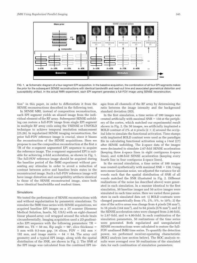
We tested the performance of SENSE reconstructions withand without regularization by parametric simulations. Tosimulate the fMRI time series with SENSE acquisitions, weacquired baseline EPI images on a 3T scanner (SiemensMedical Solution, Inseln, NJ, USA) with an eight-channellinear phased-array coil wrapped around the whole braincircumferentially. Imaging acquisition used a 2D gradient-echo EPI sequence with the following parameters: TR �2000 ms, TE � 50 ms, flip angle � 90°, slice thickness �5 mm with 0.5-mm gap, 14 slices, FOV � 192 mm �192 mm, and image matrix � 64 � 64. The array coilgeometry and a typical EPI image, along with the spatialdistribution of the SNR, are shown in Fig. 2. The SNR ofthe EPI image was calculated from the combined EPI im-
ages from all channels of the RF array by determining theratio between the image intensity and the backgroundstandard deviation (SD).
In the first simulation, a time series of 100 images wascreated artificially with maximal SNR � 150 at the periph-ery of the cortex, which matched our experimental resultshown in Fig. 2. On 50 images, we artificially implanted aBOLD contrast of 2% at 4 pixels (2 � 2) around the occip-ital lobe to simulate the functional activation. Time stampswith implanted BOLD contrast were used as the paradigmfile in calculating functional activation using a t-test (27)after SENSE unfolding. The k-space data of the imageswere decimated to simulate 2.67-fold SENSE acceleration(keeping three k-space lines in eight contiguous k-spacelines), and 4.00-fold SENSE acceleration (keeping everyfourth line in four contiguous k-space lines).
In the second simulation, a time series of 100 imageswas created synthetically with maximal SNR � 150. Usingzero-mean Gaussian noise, we adjusted the variance for allvoxels such that the spatial distribution of SNR of allvoxels matched the SNR illustrated in Fig. 2. Differentrealizations of the noise (as described above) were gener-ated in each simulation. In a manner identical to the firstsimulation, 50 baseline images and 50 active images weresimulated in each time series. Here we varied three param-eters in each simulated data set: 1) BOLD contrasts werechanged parametrically from 1%, 2%, 5%, to 10%; 2) thesize of the active areas was change from 4 pixels (36 mm2),to 16 pixels (144 mm2), and to 64 pixels (576 mm2); and 3)the SENSE acceleration rates were changed from 2.00-fold,to 2.67-fold, and to 4.00-fold. In each combination of thesimulation parameters, 50 realizations of the time serieswere generated. Both regularized and unregularizedSENSE reconstructions were calculated to restore the full-FOV unaliased fMRI time series. To quantify the detectionpower, we performed receiver-operating characteristic(ROC) analysis (see section below). The ROC analysis re-sults were averaged over 50 realizations of the simulateddata for each combination of simulation parameters.
FIG. 1. a: Schematic diagram of a four-segment EPI acquisition. In the baseline acquisition, the combination of all four EPI segments makesthe prior for the subsequent SENSE reconstructions with identical bandwidth and read-out time and associated geometrical distortion andsusceptibility artifact. In the actual fMRI experiment, each EPI segment generates a full-FOV image using SENSE reconstruction.
fMRI Using Regularized Parallel Imaging 345
3T Visual fMRI Experiment
One set of blocked-design visual fMRI data was acquiredfrom a 3T scanner with an eight-channel phased-array coilas described above. A healthy subject was recruited for thestudy after approval was obtained from the institutionalreview board, and the subject gave informed consent. Avisual stimulus (4-Hz checkerboard) was presented usingE-PRIME software (Psychology Software Tools, Inc., Pitts-burgh, PA, USA). The visual stimulus was designed todisplay either continuous checkerboards at 4 Hz flashingfor 30 s (“on” condition), or 30 s fixation (“off” condition).Three “off” conditions and two “on” conditions were al-ternatively presented to the subject, starting with the “off”condition. The imaging acquisition used a 2D gradient-echo EPI sequence with the following parameters: TR �2000 ms, TE � 50 ms, flip angle � 90°, slice thickness �5 mm with 0.5-mm gap, 14 slices, FOV � 200 mm �200 mm, and image matrix � 128 � 128. In addition tosingle-shot full phase-encoding data (60 volumes with 24“on” and 36 “off” conditions; 10 dummy scans), we col-lected three-segment EPI data (20 volumes in compositionreconstruction with 8 “on” and 12 “off” conditions; 10dummy scans) and four-segment EPI data (15 volumes incomposition reconstruction with six “on” and nine “off”conditions; 10 dummy scans). The phase encoding wasalong the anterior–posterior direction.
1.5T Motor fMRI Experiment
Blocked-design motor fMRI data were acquired from a1.5T (GE Medical, Milwaukee, WI, USA) scanner using afour-channel head array (Nova Medical, Wakefield, MA,USA). The array was wrapped around the periphery of thewhole head with four equally curved rectangular surfacecoils. One subject participated in the study after givingsigned informed consent, and the experiment was ap-proved by the institutional review board. The subject wasasked to perform the finger flexion task using his thumband other fingers of his right hand alternatively during the“on” block for 30 s continuously. The finger flexion wasself-paced, and the subject randomly chose the fingers forthe flexion task. Following the “on” block, the subject wasasked to remain still without making any hand movementduring the “off” block for 30 s. Two “on” and two “off”blocks were recorded. We used a 2D gradient-echo EPIsequence with the following parameters: TR � 2500 ms,TE � 50 ms, flip angle � 90°, slice thickness � 6 mmwithout gap, eight slices, FOV � 240 mm � 240 mm, andimage matrix � 128 � 128. Two sets of phase-encodingdata were collected: full phase-encoding data (60 volumes
with 24 “on” and 36 “off” conditions; 10 dummy scans),and two-segment EPI data (30 volumes with 12 “on” and18 “off” conditions; 10 dummy scans).
Quantification of fMRI Detection Power Using ROCAnalysis
Given the fMRI stimulus paradigm, a t-test (27) was per-formed on the reconstructed SENSE fMR images to con-trast the “on” and the “off” conditions. The detectionpowers of the regularized and unregularized SENSE recon-structions were computed using the ROC curves (28). Forthe experimental data, due to the lack of a “gold standard”for fMRI activation, we used the segmented EPI data withcomposition reconstruction to generate the relative goldstandard of functional activation loci. The use of compo-sition reconstruction for segmented EPI data ensured thatthe geometric distortion and susceptibility artifact wereidentical to the parallel MRI acquisition. All voxels witht-statistics above 4.0 (P 0.0001) were considered to bethe gold standard of functional activation. Note that oncewe decided the gold standard of functional activation, wedid not change it during the ROC analysis. After we de-fined the gold standard of functional activation, we variedthe threshold of t-statistics calculated from different
FIG. 2. Left: The layout of the eight-channelarray coil. Middle: A typical EPI image usinga sum-of-squares combination from theeight-channel array. Right: The spatial dis-tribution of the SNR of the EPI image.
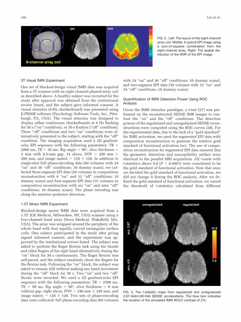
FIG. 3. The t-statistic maps from regularized and unregularized2.67-fold/4.00-fold SENSE accelerations. The blue box indicatesthe location of the simulated fMRI BOLD contrast of 2%.
346 Lin et al.

SENSE reconstructions to calculate TPR and FPR. Here thetrue-positive rate TPR() and false-positive rate FPR() ofthe detection thresholded at level were calculated as
TPR�� �size�area�XSENSE � � � area�Xref � ��
size�area�Xref � ��
FPR��
�size�area�XSENSE � � � �area�brain� � area�Xref � ���
size�area�brain� � area�Xref � ��
[4]
where area(Xref � ) indicates the t-statistic that is larger than from the full-FOV reference fMRI time series, and area(XSENSE � ) indicates the t-statistic that is larger than fromthe SENSE fMRI reconstructions; area(brain) indicates thebrain tissue in the image. � and - denote the Boolean ANDand NOT operators, respectively. The operator size(●) calcu-lates the size of the area. ROC curves for both regularized andunregularized SENSE fMRI were calculated separately. Theareas under each ROC curve were used to quantify the de-tection power. The false-positive rate was controlled below10%. The area under the ROC curve was multiplied by 10.0such that the maximum of the ROC area was 1.0. TPRscontrolled at 1%, 5%, and 10% FPRs were also reported. EPIacquisitions with different EPI factors were calculated sepa-rately to generate ROC curves for different SENSE accelera-tions.
RESULTS
Simulations
Figure 3 shows the t-statistic maps of the simulation data (2%BOLD CNR) including both 2.67-fold and 4.00-fold SENSEaccelerations with/without regularization. Both t-statisticmaps were overlaid on the identical full-FOV reference EPIimages. Note that with both acceleration rates, regularizationimproved the sensitivity of detection by achieving highert-statistic values. At 2.67-fold acceleration with regulariza-tion, all four voxels of the implanted functional activationgave t-statistics above 12.0. With unregularized reconstruc-tions, however, only three voxels had t-statistics above thevalue of 12.0. At 4.00-fold acceleration, the regularizedSENSE reconstructions showed all four voxels with t-statis-tics above 7.0, while unregularized reconstructions had onlythree voxels exceeding the t � 7.0 threshold.
For more complete simulations, we varied the BOLDcontrasts from 1%, 2%, 5%, to 10%. Figure 4a shows theaveraged ROC analysis at different BOLD contrasts with a4.00-fold SENSE acceleration, and 576 mm2 simulated anactive brain area (64 voxels). Note that higher BOLD con-trasts yielded higher TPRs at 1%, 5%, and 10% controlledFPRs. At all BOLD contrasts, the regularized reconstruc-tions had higher TPRs than unregularized reconstructions.At 10% BOLD contrast, the differences in TPRs were aslarge as 5% when FPRs were controlled below 10%. Figure4b shows the simulation results with different sizes ofsimulated brain area with 5% BOLD contrast and 4.00-foldSENSE acceleration. Interestingly, the larger the simulatedactive brain area, the lower the TPR we calculated. This is
because more specific and sensitive reconstructions arerequired to yield good TPRs in simulations with moresimulated active brain voxels. Thus, when using the iden-tical regularized and unregularized reconstructions, it is dif-ficult to achieve simultaneous enhancement of specificityand sensitivity when more brain active voxels are simulated.Nevertheless, a comparison of the regularized and unregular-ized SENSE reconstructions indicated that regularizationhelped improve the detection power by providing higherTPRs at each controlled FPR. Lastly, Fig. 4c shows the ROCanalysis results for different SENSE acceleration rates with a5% BOLD contrast and a 576 mm2 simulated active brainarea (64 voxels). As expected, the higher SENSE acceleration
FIG. 4. a: ROC analysis for 1%, 2%, 5%, and 10% BOLD contrastin 4.00-fold SENSE acceleration and 576 mm2 simulated activebrain area. b: ROC analysis for 36 mm2, 144 mm2, and 576 mm2
simulated active brain area with 4.00-fold SENSE acceleration and5% BOLD contrast. c: ROC analysis for 2.00-, 2.67-, and 4.00-foldSENSE accelerations in 5% BOLD contrast and 576 mm2 simulatedactive brain area.
fMRI Using Regularized Parallel Imaging 347

rate corresponded to a lower TPR at each FPR, potentiallydue to reduced data samples. A comparison of regularizedand unregularized reconstructions showed that TPR was ac-tually improved when regularization was employed. Regu-larization can improve TPR by 2–5%, depending on theSENSE acceleration rates and controlled FPRs.
3T Visual fMRI Experiment
For the 3T visual fMRI experiment, Fig. 5a shows theg-factor maps for 3.00-fold and 4.00-fold SENSE EPI. At3.00-fold SENSE acceleration, without regularization, theaveraged g-factor is 2.62 � 1.47 (�SD). Regularizationreduced the average g-factor to 2.21 � 1.04. In 4.00-foldacceleration, SENSE reconstructions without regulariza-tion gave the averaged g-factors of 3.67 � 1.82. Regular-ization reduced the averaged g-factor to 3.01 (SD � 1.26).Note that the g-factor maps are derived from the encodingmatrix in the SENSE reconstruction. Using in vivo sensi-tivity, the coil maps in the encoding matrix are actually thefull-FOV anatomical images. Thus it is possible that someresidual ghosting artifacts in the anatomical image willaffect the g-factor maps. However, g-factor maps containmore than just the coil sensitivity information. g-factormaps also include the spatial distribution of the condition-
ing of the encoding matrix, which is determined by thek-space trajectory in the accelerated scan and the physicallocations of the array coils. Thus, we show EPI images inFig. 5b to contrast the difference between g-factor mapsand residual ghosting artifacts. A comparison of the g-factor maps and EPI images illustrates the difference be-tween the two. Figure 6 shows the t-statistic maps of a 3Tvisual fMRI experiment using SENSE EPI with/withoutregularization at 3.00- and 4.00-fold accelerations. At 3.00-fold SENSE acceleration, regularized reconstructionsyielded a larger functional activated area than the unregu-larized reconstructions around the occipital lobe (regular-ized: 2327 mm2; unregularized: 2139 mm2). Such an in-crease in sensitivity by regularization was also observed inthe 4.00-fold SENSE acceleration (regularized: 896 mm2;unregularized: 735 mm2). Figure 7a shows the threealiased regions with 3.00-fold acceleration. One of thealiased regions is the visual cortex shown in Fig. 6. Thecorresponding time courses for all three aliased regions aredepicted in Fig. 7b. Note that only activated visual cortexshowed a time course that matched the stimulus paradigm.Two other aliased regions showed either low BOLD con-trasts in all conditions or a time course that was mis-matched to the stimulus paradigm. To illustrate both the
FIG. 5. a: g-Factor maps for 3.00- and 4.00-fold SENSE accelerations. An image maskcovering the brain parenchyma was used tomask the g-factor maps to illustrate the spa-tial distribution of g-factors inside the brain.b: Reconstructed EPI SENSE images at3.00-fold acceleration.
FIG. 6. The t-statistic maps of the visualfMRI experiment from a 3T scanner using aneight-channel head array, calculated with3.00- or 4.00-fold SENSE acceleration.
348 Lin et al.

sensitivity and specificity of the SENSE reconstructions,Fig. 8 shows the ROC curves from t-statistic maps using3.00- or 4.00-fold SENSE EPI acquisitions, including bothregularized (solid lines) and unregularized (dashed lines)unfolding. We found that the detection power decreasedwhen the SENSE acceleration increased from 3.00-fold to4.00-fold. Using regularization to unfold the identicalSENSE acceleration can improve the detection power inboth 3.00- and 4.00-fold accelerations, as the ROC curvesshift toward the upper-left corner. At FPR � 1%, regular-ization improved TPR by 6.7% and 28.2% for 3.00-foldand 4.00-fold accelerations, respectively. Regularizationimproved TPRs by 3.9% and 18.7% at FPR � 5% for 3.00-and 4.00-fold accelerations, respectively. Table 1 summa-rizes the ROC areas and TPRs at FPR � 1%, 5%, and 10%.
At 3.00-fold SENSE acceleration, the use of regularizationcan increase the ROC area maximally by 4.8% in SENSEunfolding. This implies that regularized SENSE recon-struction has a higher detection power than unregularizedSENSE reconstructions. The same advantage of regular-ized SENSE reconstructions over unregularized ones wasalso observed in the 4.00-fold SENSE acquisitions. The useof regularization can increase the ROC area maximally by16.4% in 4.00-fold acceleration.
1.5T Motor fMRI Experiment
For the 1.5T motor fMRI experiment, the t-statistic mapswith full phase-encoding data and 2.00-fold SENSE accel-eration are shown in Fig. 9. The contralateral motor cortex
FIG. 7. a: EPI image showing the three aliasedregions with 3.00-fold acceleration. One of thealiased regions is the visual cortex. b: The timecourses of the three regions. Note that only thevisual cortex region time course corresponded wellwith the stimulus paradigm.
Table 1The Areas Under the ROC Curves and TPRs for the SENSE Reconstructions Using Traditional Algorithm (Unregularized) or RegularizedReconstructions in Visual fMRI Experiment at 3T with 3.0-fold or 4.0 Fold SENSE Acceleration*
SENSEacceleration
ReconstructionTPR
ROC areaFPR � 1% FPR � 5% FPR � 10%
3.0Unregularized 0.517 0.675 0.732 0.653Regularized 0.552 0.702 0.755 0.685
4.0Unregularized 0.168 0.344 0.457 0.331Regularized 0.216 0.408 0.506 0.385
*The false positive rate of the ROC was controlled at 10%. The area was scaled by 10.0 such that the maximum of the area is 1.0.
fMRI Using Regularized Parallel Imaging 349

was found to be activated in both the full phase-encodingdata and the 2.00-fold SENSE unfolding. Additional de-tected activation loci included the supplementary motorarea and ipsilateral frontal cortex. Activation in these areaswas consistently detected in both of the 2.00-fold SENSEreconstructions with/without regularization. Regulariza-tion increased the size of the active area in the t-statisticmaps as compared to the unregularized SENSE reconstruc-tions (regularized: 3751 mm2; unregularized: 3227 mm2).ROC curves for regularized and unregularized 2.00-foldSENSE accelerations are shown in Fig. 10. Using ROCanalysis to quantify detection power, we found that regu-larization improved the TPR by 9.5% when FPR was con-trolled at 1% (unregularized TPR: 0.84, regularized TPR:0.92). Regularization increased the area under the ROCcurve 3.6% in 2.00-fold acceleration, compared to unregu-larized SENSE reconstructions. Table 2 summarizes theROC areas from regularized and unregularized SENSE re-constructions in 2.00-fold acceleration.
DISCUSSION
A major finding of the current study is that regularizationimproves the CNR of an fMRI time series as the result ofreduced contrast and a further reduced noise level. Aconcern with using prior information in dynamic imagingis that the replication of prior image features in time-seriesmeasurements may reduce the functional contrast. A reg-
ularized SENSE-EPI fMRI acquisition protocol was foundto provide a robust CNR improvement in various scenar-ios, across field strengths (1.5T and 3T), across differentcoil array geometries (four- or eight-element circumferen-tial head array), across different SENSE accelerations(from 2.00-fold to 4.00-fold accelerations), and across dif-ferent functional modalities (visual and motor). From sim-ulations and experimental data, we found that the func-tional CNR was actually improved by the regularizedSENSE reconstruction compared to unregularized SENSEreconstructions. This suggests that the proposed regular-ization process suppresses the noise amplification inSENSE reconstruction to a greater extent than the possiblefunctional contrast loss, by partially replicating prior im-age features. It was previously reported that SENSE acqui-sitions produced an SNR that was lower than full-FOVacquisitions but higher than theoretical predictions(14,16). This was partly explained by the influence ofsignal-dependent physiological noise (31,32). The im-provement of the CNR reported in this work, however, wasnot related to physiological noise. This is because theSENSE reconstructions with/without regularization werecalculated from identical segmented EPI data sets. Thusthe improvement in CNR solely reflects the benefit of in-corporating prior information in SENSE unfolding. In fact,the use of prior information can lead to a reduction ofcontrast between two functional states. However, the pre-sented regularization technique further reduced the noise.Overall, the CNR was improved.
FIG. 8. The ROC curves for the 3.00- and 4.00-fold SENSE recon-structions using the traditional algorithm (unreg.) or regularized re-construction (reg.) in a 3T visual fMRI experiment. The ROC curveswere calculated based on the thresholded (t � 4.0) statistical valuesfrom composition reconstruction of the segmented EPI data withthree and four EPI segments, respectively.
FIG. 9. The t-statistic maps of themotor fMRI experiment from a1.5T scanner using a four-channelhead array. The t-statistic mapswere calculated from full phase-encoding data and 2.00-foldSENSE acceleration.
FIG. 10. The ROC curves for the 2.00-fold SENSE reconstructionsusing the traditional algorithm (unreg.) or regularized reconstruction(reg.) in a 1.5T motor fMRI experiment. The ROC curves werecalculated based on the thresholded (t � 4.0) statistical values fromcomposition reconstruction of two-segment segmented EPI.
350 Lin et al.

The major advantage of using parallel MRI (specificallySENSE MRI (10)) is that it can improve the spatiotemporalresolution of images (14,17). Additionally, it can reducesusceptibility artifacts and geometrical distortions due tothe shortened readout window and increased phase-en-coding bandwidth (15,16). Nevertheless, the price forthese benefits is a reduced SNR and CNR in the functionalimages, as reported in previous visual (15,17) and motor(14–17) system studies. In this paper, we propose to mit-igate the SNR and CNR loss in SENSE acquisitions byemploying the information in full-FOV reference scans. Ina previous study (20) we showed that the g-factors in aregularized SENSE reconstruction can be smaller than 1.0.This indicates that the regularized unfolding operationdecreases the stochastic variability of the estimated spindensity compared to the full-FOV reference scans. Thisnoise reduction results from the prior knowledge used inthe regularization process. Since prior information is em-ployed in a regularized SENSE reconstruction, less vari-ability of the estimated spin density from parallel acquisi-tions is expected. Thus, using regularization can partiallycompensate for the SNR loss due to the reduced samples inthe accelerated acquisition.
The dependency of the static image in the regularizedSENSE reconstruction on the dynamic time series, such asfMRI data, is similar to that in the keyhole imaging tech-nique (29,30), which replicates the center part of the k-space in the dynamic scan in order to enhance the tempo-ral resolution of data acquisition. In contrast to keyholeimaging, regularized SENSE reconstruction focuses on thestabilization of the matrix inversion during restoration ofthe full-FOV unaliased image. Keyhole imaging cannot beapplied directly to reduce the noise amplification, as inregularized SENSE reconstruction, because the keyholetechnique applies the static lower-resolution image to thefull-FOV unaliased image, which is not available until thenecessary image unfolding process is completed. Wewould also like to clarify that the inclusion of prior infor-mation in SENSE reconstruction does not represent a mereDC shift in all time series images. The matrix solution ofthe prior-informed regularized reconstruction (20) indi-cates that the prior information is not superimposed on thetime series directly. Actually, the prior is used as a com-bination of the DC shift (from the prior information) andmatrix conditioning (on the inversion of encoding matrix).
Previous studies applied parallel MRI acquisition proto-cols to fMRI experiments using the PRESTO (17) and gra-dient-echo EPI (14–16) sequences. The purpose of usingSENSE MRI as the fMRI data acquisition protocol is toenhance spatiotemporal resolution by reducing the timerequired to traverse k-space. Thus, single-shot EPI or re-
duced phase-encoding PRESTO have become feasible onhigh-field (�3T) scanners, where the T*2 relaxation time ofhuman gray matter may be shorter than 30 ms in areas withpronounced field inhomogeneities (33,34). Traditionally,such a short T*2 made single-shot EPI unsuitable for suffi-cient spatiotemporal resolution because of the insufficientacquisition time to completely traverse k-space. With par-allel imaging, the time required to traverse k-space is re-duced by “unfolding” the aliased images from individualreceivers in the array. The reduced k-space traversing timealso helps reduce susceptibility artifacts and geometricdistortions originating from the local magnetic field inho-mogeneity. This is due to the shortened readout time of thedata acquisition, which contributes less to local spindephasing within individual voxels. Geometric distortionis also improved by a higher bandwidth in the phase-encoding direction. Another significant benefit of SENSEis that the SENSE EPI may reduce the acoustic noise byreduced switching of the gradients to complete k-spacetrajectory traversing (13).
In this study we utilized the “in vivo sensitivity” recon-struction approach described by Sodickson (23). Thismethod substitutes full-FOV array element images fromthe reference scan in place of the estimated coil profileused by SENSE (10). Thus a misestimation of the coilsensitivity profile is avoided. One major disadvantage ofthe in vivo sensitivity reconstruction approach is that itcannot reconstruct the image features that are not presentin the full-FOV reference scans. This situation occurswhen the reference scan and the SENSE-accelerated scansinvolve time-varying anatomical features, such as cardiacimaging at different phases. However, this issue is lesscritical in brain imaging, since it has been reported that thecerebral cortex stays relatively stationary even in the pres-ence of cardiac pulsation (35). Thus, the in vivo sensitivityreconstruction approach is capable of revealing most of theanatomical and functional features in each time point ofthe dynamic scan of the brain.
One advantage of using in vivo sensitivity is the sim-plicity of prior image specifications. The difference in theprior image for regularized SENSE reconstructions usingeither in vivo sensitivity or conventional coil sensitivitymap estimates (e.g., the ratio between array coil imagesand body coil images) is that by using the in vivo sensi-tivity, all pixels in the prior image have values of 1.0. Withconventional coil sensitivity map estimates, the prior im-age is a full-FOV image with anatomical features. In theconventional coil sensitivity approach, such a prior imageshould contain only anatomical features, without spatialmodulation from the coil sensitivity profile. Even thoughwe can use the traditional sum-of-squares to combine all
Table 2The Areas Under the ROC Curves, and TPRs for the SENSE Reconstructions Using Traditional Algorithm (Unregularized) or RegularizedReconstructions in Motor fMRI Experiment at 1.5T with 2.0-fold SENSE Acceleration*
SENSEacceleration
ReconstructionTPR
ROC areaFPR � 1% FPR � 5% FPR � 10%
2.0Unregularized 0.844 0.952 0.973 0.934Regularized 0.925 0.976 0.983 0.968
*The false positive rate of the ROC was controlled at 10%. The area was scaled by 10.0 such that the maximum of the area is 1.0.
fMRI Using Regularized Parallel Imaging 351

channels of the array coils to yield a full-FOV prior image,such a combined image will still contain residual spatialmodulation of the coil sensitivity maps, and thus it cannotserve as a good prior image. However, with the in vivosensitivity approach, we do not require such a full-FOVimage with anatomical features and without spatial mod-ulation of the coil sensitivity maps. In in vivo sensitivitymethod, the use of a constant prior image with a pixelvalue equal to 1.0 can avoid potential errors and bias fromthe specification of prior information in the SENSE recon-struction.
Using regularization in SENSE reconstructions, we re-quire aliased accelerated image, prior full-FOV image, andcoil sensitivity maps to compute the regularization param-eters. Investigators traditionally have used a full-FOV ref-erence image to estimate the coil sensitivity maps. How-ever, in our approach we do not estimate the coil sensitiv-ity profile because, as indicated in Eq. [2], fMRIexperiments mostly focus on the dynamic BOLD contrastwithin an image voxel to differentiate between active andbaseline brain states. The in vivo sensitivity method servesthis purpose. In fact, this is another advantage of using invivo sensitivity in fMRI experiments, in addition to theadvantages of using prior image specification and avoidingcoil map estimation errors, as described above. Note thatwhen we use in vivo sensitivity, the coil sensitivity mapsare identical to the full-FOV reference image. However, ifinvestigators use the conventional coil sensitivity esti-mated from the ratio between the body- and array-coilfull-FOV reference images, the regularization parameterswill differ from those of the in vivo sensitivity method.
The acquisition protocol presented in this paper is basedon the segmented EPI sequence, similarly to the temporalSENSE (TSENSE) (16,25) and unaliasing by Fourier-en-coding the overlaps using the temporal dimension(UNFOLD) (36,37) techniques. We demonstrated with sim-ulations and experimental data that each interleaved seg-ment of EPI can be unfolded into a full-FOV image withspatial resolution that is identical to an image recon-structed by combining all segments. Using parallel imag-ing reconstruction, we can increase the temporal resolu-tion when the individual EPI segment is reconstructed intoa full-FOV image. The increment in the number of obser-vations can improve the detection power. It should benoted that the proposed regularization procedure can alsobe incorporated using a number of variations in the seg-mented EPI sequences, including variable density sam-pling (25,38,39), to improve the SNR and CNR in fMRIstudies.
One practical concern with applying the regularizedSENSE reconstruction is the computational load. Cur-rently, the bottleneck in the computational performance ofSENSE fMRI is caused by the unfolding of the SENSEaccelerated scans, not the time required to estimate theregularization parameters. We noticed that once the regu-larization parameters were determined, the computationalreconstruction time was identical to that of reconstructionwithout regularization. Thus the additional computationaltime required for the proposed technique is not excessive.In dynamic imaging, the impact on the increased imagereconstruction time due to regularization estimation canbe minimized by estimating regularizations only once and
then applying identical regularization parameters to mul-tiple measurements. The computational performance canbe further improved with the use of 1) compiled languagefor the code, 2) the direct regularization approach to esti-mate the time required to estimate the regularization pa-rameters, and 3) the complex conjugated approach forSENSE unfolding. In addition to the L-curve techniqueused in this study, other automatic regularization estima-tion methods, such as generalized cross validation (GCV)(40,41), potentially can be used to estimate appropriateregularization parameters.
CONCLUSIONS
In comparison with conventional EPI scans, parallel imag-ing techniques can reduce susceptibility artifacts and im-age geometric distortions, and improve the spatiotemporalresolution in fMRI studies. In this paper we have pre-sented a regularized SENSE fMRI method based on Tik-honov regularization to mitigate the SNR and CNR lossdue to geometric correlation in the spatial informationfrom the array coil elements. With the use of an eight-channel head array, parametric simulations with differentBOLD contrasts (1–10%), SENSE acceleration rates (2.00-to 4.00-fold), and sizes of the active brain area (36–576 mm2) showed that the detection power was improvedwhen regularization was employed in SENSE fMRI acqui-sitions. Additional experimental data at 1.5T and 3T withfour- and eight-channel arrays for motor and visual fMRIexperiments validate that the proposed regularization pro-cedure can improve the sensitivity and specificity ofSENSE fMRI to detect brain activation.
ACKNOWLEDGMENT
We thank Dr. Walid Kyriakos for providing the RF arraycoil at Brigham and Women’s Hospital.
REFERENCES
1. Belliveau JW. Functional NMR imaging of the brain. Ph.D. dissertation,Harvard University, Cambridge, MA, 1990.
2. Belliveau JW, Kennedy DN, McKinstry RC, Buchbinder BR, WeisskoffRM, Cohen MS, Vevea JM, Brady TJ, Rosen BR. Functional mapping ofthe human visual cortex by magnetic resonance imaging. Science 1991;254:716–719.
3. Belliveau JW, Kennedy DN, McKinstry RC, Buchbinder BR, WeisskoffRM, Vevea JM, Nadeau K, Cohen MS, Brady TJ, Rosen BR. Functionalmapping of the human visual cortex with susceptibility-contrast MRimaging. J Magn Reson Imaging 1991;1:202.
4. Ogawa S, Lee TM, Kay AR, Tank DW. Brain magnetic resonance imag-ing with contrast dependent on blood oxygenation. Proc Natl Acad SciUSA 1990;87:9868–9872.
5. Kwong KK, Belliveau JW, Chesler DA, Goldberg IE, Weisskoff RM,Poncelet BP, Kennedy DN, Hoppel BE, Cohen MS, Turner R, ChengHM, Brady TJ, Rosen BR. Dynamic magnetic resonance imaging ofhuman brain activity during primary sensory stimulation. Proc NatlAcad Sci USA 1992;89:5675–5679.
6. Mansfield P. Multi-planar image formation using NMR spin echos. JPhysics 1977;C10:L55–L58.
7. Schmitt F, Stehling MK, Turner R, Bandettini PA. Echo-planar imaging:theory, technique, and application. Berlin/New York: Springer; 1998.xiv, 662 p.
8. Roemer PB, Edelstein WA, Hayes CE, Souza SP, Mueller OM. The NMRphased array. Magn Reson Med 1990;16:192–225.
352 Lin et al.
9. Sodickson DK, Manning WJ. Simultaneous acquisition of spatial har-monics (SMASH): fast imaging with radiofrequency coil arrays. MagnReson Med 1997;38:591–603.
10. Pruessmann KP, Weiger M, Scheidegger MB, Boesiger P. SENSE: sen-sitivity encoding for fast MRI. Magn Reson Med 1999;42:952–962.
11. Bammer R, Keeling SL, Augustin M, Pruessmann KP, Wolf R, Stoll-berger R, Hartung HP, Fazekas F. Improved diffusion-weighted single-shot echo-planar imaging (EPI) in stroke using sensitivity encoding(SENSE). Magn Reson Med 2001;46:548–554.
12. Weiger M, Pruessmann KP, Boesiger P. 2D SENSE for faster 3D MRI.MAGMA 2002;14:10–19.
13. de Zwart JA, van Gelderen P, Kellman P, Duyn JH. Reduction ofgradient acoustic noise in MRI using SENSE-EPI. Neuroimage 2002;16:1151–1155.
14. Preibisch C, Pilatus U, Bunke J, Hoogenraad F, Zanella F, LanfermannH. Functional MRI using sensitivity-encoded echo planar imaging(SENSE-EPI). Neuroimage 2003;19(2 Pt 1):412–421.
15. Weiger M, Pruessmann KP, Osterbauer R, Bornert P, Boesiger P, JezzardP. Sensitivity-encoded single-shot spiral imaging for reduced suscep-tibility artifacts in BOLD fMRI. Magn Reson Med 2002;48:860–866.
16. de Zwart JA, van Gelderen P, Kellman P, Duyn JH. Application ofsensitivity-encoded echo-planar imaging for blood oxygen level-depen-dent functional brain imaging. Magn Reson Med 2002;48:1011–1020.
17. Golay X, Pruessmann KP, Weiger M, Crelier GR, Folkers PJ, Kollias SS,Boesiger P. PRESTO-SENSE: an ultrafast whole-brain fMRI technique.Magn Reson Med 2000;43:779–786.
18. Sodickson DK, McKenzie CA. A generalized approach to parallel mag-netic resonance imaging. Med Phys 2001;28:1629–1643.
19. Tikhonov AN, Arsenin VI. Solutions of ill-posed problems. Washing-ton/New York: Winston/Halsted Press; 1977. xiii, 258 p.
20. Lin FH, Kwong KK, Belliveau JW, Wald LL. Parallel imaging recon-struction using automatic regularization. Magn Reson Med 2004;51:559–567.
21. Hansen PC. Rank-deficient and discrete ill-posed problems: numericalaspects of linear inversion. Philadelphia: SIAM; 1998. xvi, 247 p.
22. Yacoub E, Shmuel A, Pfeuffer J, Van De Moortele PF, Adriany G,Andersen P, Vaughan JT, Merkle H, Ugurbil K, Hu X. Imaging brainfunction in humans at 7 Tesla. Magn Reson Med 2001;45:588–594.
23. Sodickson DK. Tailored SMASH image reconstructions for robust invivo parallel MR imaging. Magn Reson Med 2000;44:243–251.
24. McKinnon GC. Ultrafast interleaved gradient-echo-planar imaging on astandard scanner. Magn Reson Med 1993;30:609–616.
25. Kellman P, Epstein FH, McVeigh ER. Adaptive sensitivity encodingincorporating temporal filtering (TSENSE). Magn Reson Med 2001;45:846–852.
26. Madore B. UNFOLD-SENSE: a parallel MRI method with self-calibra-tion and artifact suppression. Magn Reson Med 2004;52:310–320.
27. Friston KJ, Holmes AP, Poline JB, Grasby PJ, Williams SC, FrackowiakRS, Turner R. Analysis of fMRI time-series revisited. Neuroimage 1995;2:45–53.
28. Chow S-C, Shao J. Statistics in drug research: methodologies and recentdevelopments. New York: M. Dekker; 2002. xiv, 367 p.
29. van Vaals JJ, Brummer ME, Dixon WT, Tuithof HH, Engels H, NelsonRC, Gerety BM, Chezmar JL, den Boer JA. “Keyhole” method for accel-erating imaging of contrast agent uptake. J Magn Reson Imaging 1993;3:671–675.
30. Hu X. On the “keyhole” technique. J Magn Reson Imaging 1994;4:231.31. Kruger G, Kastrup A, Glover GH. Neuroimaging at 1.5 T and 3.0 T:
comparison of oxygenation-sensitive magnetic resonance imaging.Magn Reson Med 2001;45:595–604.
32. Kruger G, Glover GH. Physiological noise in oxygenation-sensitivemagnetic resonance imaging. Magn Reson Med 2001;46:631–637.
33. Chen NK, Egorova S, Guttmann CR, Panych LP. Functional MRI withvariable echo time acquisition. Neuroimage 2003;20:2062–2070.
34. Barth M, Metzler A, Klarhofer M, Roll S, Moser E, Leibfritz D. Func-tional MRI of the human motor cortex using single-shot, multiplegradient-echo spiral imaging. Magn Reson Imaging 1999;17:1239–1243.
35. Poncelet BP, Wedeen VJ, Weisskoff RM, Cohen MS. Brain parenchymamotion: measurement with cine echo-planar MR imaging. Radiology1992;185:645–651.
36. Madore B, Glover GH, Pelc NJ. Unaliasing by Fourier-encoding theoverlaps using the temporal dimension (UNFOLD), applied to cardiacimaging and fMRI. Magn Reson Med 1999;42:813–828.
37. Madore B. Using UNFOLD to remove artifacts in parallel imaging andin partial-Fourier imaging. Magn Reson Med 2002;48:493–501.
38. Jakob PM, Griswold MA, Edelman RR, Sodickson DK. AUTO-SMASH:a self-calibrating technique for SMASH imaging. Simultaneous acqui-sition of spatial harmonics. MAGMA 1998;7:42–54.
39. Heidemann RM, Griswold MA, Haase A, Jakob PM. VD-AUTO-SMASHimaging. Magn Reson Med 2001;45:1066–1074.
40. Golub GH, Heath MT, Wahba G. Generalized cross-validation as amethod for choosing a good ridge parameter. Technometrics 1979;21:215–223.
41. Wahba G. Practical approximate solutions to linear operator equationswhen the data are noisy. SIAM J Numer Anal 1977;14:651–667.
fMRI Using Regularized Parallel Imaging 353