Embed Size (px)
Citation preview

Digestive system
1. Microscopic anatomy of esophagus, stomach, small and large intestine
2. Microscopic anatomy of liver, pancreas and salivary glands. Embryonic development of GIT
Petr Vaňhara, PhD
Department of Histology and Embryology LF MU

1. Development and general structure of hollow organs/gut tube
2. Esophagus (Oesophagus)
3. Stomach (Ventriculus, Gaster)
4. Small and large intestine (Duodenum, Ileum, Jejunum, Colon)
- Tunica mucosa
- Tela submucosa
- Tunica muscularis externa
- Serosa/adventitia
- Microscopic anatomy
- Gl. oesophageae propriae
- Microscopic anatomy
- Functional modification of gastric mucosa and gastric glands
- Enteroendocrinne system
- Microscopic anatomy
- Functional modification of intestinal mucosa and Lieberkühn crypts
- Enteroendocrinne system

Alimentary canal

General architecture of
hollow organs

General architecture of hollow organs incl. gut tube
1. Mucosa (Tunica mucosa)
2. Submucosa (Tela submucosa)
3. Tunica muscularis externa
4. Serosa/adventitiaDonna Myers © 2007
Lumen
Serosa/Adventitia
Muscularis
externa
Submucosa
Mucosa
Four layers
Lumen
1
2
3
4


General architecture of hollow organs incl. digestive tube

Mucosa (Tunica mucosa)
- inner layer of gut tube
- protective, absorption and resorption
- microscopic structure depending on localization
- Lamina epithelialis mucosae
- Lamina propria mucosae
- Lamina muscularis mucosae
- Lamina epithelialis mucosae
- epithelium type corresponding to function of gut tube
- oral cavity, pharynx, esophagus, anus – stratified squamous ep.
- stomach, intestine – simple columnar
- mucus - secreted by mucosal or submucosal glands (oral cavity,
esophagus), secretory epithelium (stomach) or goblet cells (intestine)
- Lamina propria mucosae
- Layer of mucosal connective tissue – loose collagen
- Fenestrated blood capillaries – transport of metabolite (intestine)
- mucosal glands in some regions /esophagus)
- innervations, immune system
- Lamina muscularis mucosae
- smooth muscles in two layers (inner circular, outer longitudinal)
- small mechanical movements of mucosa facilitating secretion and
absorption independently on peristaltic movements.
Mucosa (Tunica mucosa)

Submucosa (Tela submucosa)
Submucosal connective tissue
- distinct layer of loose connective tissue
- defines shape of mucosa (rugae, plicae)
- larger blood and lymph veins nourishing mucosa, muscularis
externa and serosa
- innervations – nerve plexus - plexus submucosus Meissneri
= groups of multipolar neurons and small ganglions,
visceral sensory fibers (sympaticus) and fibers and terminal
ganglions of parasympaticus (enteric nerve system)
- glands – different in different regions
- protective function

Outer muscular layers (Tunica muscularis externa)
- Two concentric, thick layers of smooth muscle, separated by thin layer of connective tissue
- Inner – circular, outer – longitudinal (spiral)
- Myenteric (Auerbach) plexus
- Peristaltic – passage through the gut tube
- Local modifications of m.e.
- pharyngoesophagal sphincter + external anal sphincter – skeletal muscles
- stomach – third - oblique - layer
- taenie coli – thickened part of longitudinal layer in colon
Circular Longitudinal

Serosa/Adventitia (Tunica serosa/adventitia)- outermost layer of gut tube
- Serosa
- serous membrane of loose connective tissue (Lamina propria serosae) and single layer
squamous epithelium (L. epithelialis serosae)
- syn. mesothelium, visceral peritoneum
- continuous with mesenterium
- barrier against various pathogens , antiadhesive properties – intracoelomic movements,
immune functions (Ag presentation), ECM production, etc.
- Adventitia
- some parts of the tube are not covered with epithelium
- esophagus in thorax, parts of digestive system in peritoneal cavity in sites of fixation to the
walls (duodenum, part of colon, rectum, anal canal)
- connective tissue only continuous with connective tissue of the walls

S.E. Mutsaers / The International Journal of Biochemistry & Cell Biology 36 (2004) 9–16
Serosa/Adventitia (Tunica serosa/adventitia)
1.4m

Innervation of the digestive tube
Enteric nervous system
- self-contained nervous system
- numerous ganglia, 100 x106 neurons (more than in spinal
cord)
- Meissner submucosal plexus and Aurebach myenteric plexus
- peristaltic motility, secretory function, mucosal movements,
regulation of blood flow
- sensory components
Parasympathetic and sympathetic supply
• parasympathetic supply mostly by vagus nerve (cranial X),
colon and rectum by sacral spinal nerves
- vagus nerve – mostly sensory fibers (information from
mucosa and back)
- secretion from glands, smooth muscle contractions
- inhibits sphincters, stimulates peristaltics and secretion
• sympathetic supply by splanchnic nerves
- vasomotor fibers – control of blood flow
- activates sphincters, inhibits peristaltics and secretion

Pharynx- pars nasalis
- pseudostratified columnar ciliated epithelium
- seromucous glands
- pars oralis et laryngea
- nonkeratinized stratified squamous epithelium
- mucous glands
- collagen c.t. (lamina propria), typical tela submucosa absent
- skeletal muscles

Esophagus (Oesophagus)- Mucosa
- nonkeratinized stratified squamous epithelium mechanically protects esophagal tissue
- l. propria contains cardial glands (tubular mucinous) and diffuse lymphatic tissue
- Submucosa
- loose collagen connective tissue, defines shape of mucosa
- blood and lymph veins, plexus submucosus Meissneri
- submucosal glands (tubular mucinous)
- diffuse lymphatic tissue
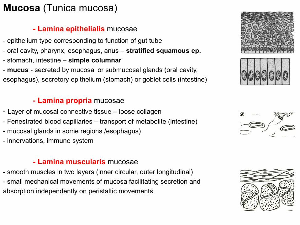
Esophagus (Oesophagus)- Muscularis externa
- inner circular and outer longitudinal layer
- plexus myentericus Auerbachi
- upper third – skeletal muscle, mid third – mixed smooth and skeletal, lower third –
smooth muscles only
- Adventitia
- neck and chest – connects esophagus with surrounding tissue
- loose connective tissue
- in peritoneal cavity - serosa

Cardia of stomach – connection with esophagus
Nonkeratinized stratified squamous epithelium simple columnar epithelium
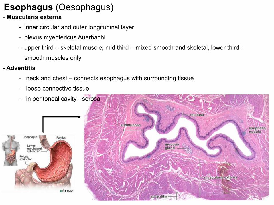
Stomach (Ventriculus, Gaster)
- general anatomy of hollow tube
- anatomical regions differ also in histologic structure
- rugae gastricae (submucosa)
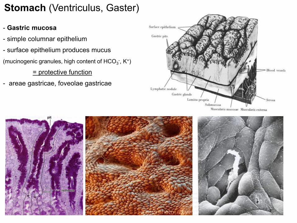
Stomach (Ventriculus, Gaster)
- Gastric mucosa
- simple columnar epithelium
- surface epithelium produces mucus
(mucinogenic granules, high content of HCO3-, K+)
= protective function
- areae gastricae, foveolae gastricae

Stomach (Ventriculus, Gaster)
- Gastric mucosa
- L. propria contains large amount of glands
- Gl. cardiacae
- Gl. pyloricae
- Gl. gastricae propriae
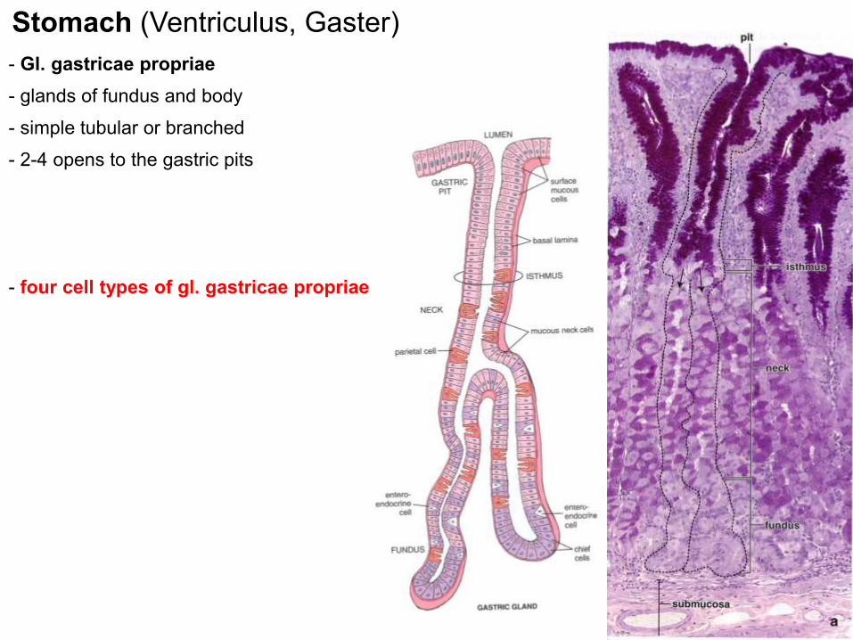
Stomach (Ventriculus, Gaster)
- Gl. gastricae propriae
- glands of fundus and body
- simple tubular or branched
- 2-4 opens to the gastric pits
- four cell types of gl. gastricae propriae

chief- most abundant, lower part of body and fundus of the gland
- pyramidal shape, basophilic cytoplasm, RER, pepsinogenic granules
parietal- neck-body junction
- eosinophilic cytoplasm, high numbers of mtch., SER
- complex and dynamic ultrastructure
- intracellular canals in apical part with microvilli – membrane bound
enzyme complexes producing H+ a Cl- (HCl originates extracelullarly)
neck cells- cubic, mucinous
- capable of regeneration of all cell types in gastric epithelium
Stomach(Ventriculus, Gaster)
Gl. gastricae propriae

Stomach (Ventriculus, Gaster)
Gl. gastricae propriae
Type Hormone Localization/Function
D cells Somatostatin - Stomach, intestine, hepatic and pancreatic ducts
EC cells Serotonin - Stomach, gallbladder, intestine
- Peristaltics
ECL cells Histamin - Stomach
- HCl secretion
G cells Gastrin - Pars pylorica, duodenum
- HCl, pepsin secretion
L (EG) cells Enteroglucagon - Stomach, intestine
- attenuates secretion of pancreatic enzymes and peristaltics
(entero)endocrine
- minor, secretion
- granules
- different cell types with different sensitivity to various histological stainings
- secretion of various biologically active compounds
- DNES/APUD
- GIT chemosensing
- see lesson spring semester 2012 - Epithelial tissue

Break
http://luminaryvisuals.com/

General architecture of hollow organs incl. gut tube
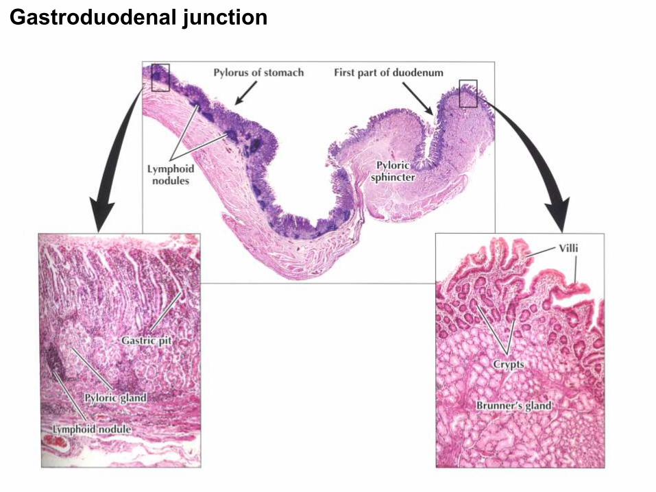
Gastroduodenal junction

General architecture of the intestine

Four basic layers: mucosa, submucosa, muscularis externa, serosa
mucosa and submucosa maximise the resorptive area
• plicae circulares (Kerckringi) – mucosa + submucosa, ca 800, increase 2-3x, distal region of duodenum
• villae (villi intestinales) – mucosa (l. propria + epithelium) 0,5-1,5 mm long, 10-40/mm2 , 4 000 000, increase 5-10x
• microvillae – apical part of enterocytes – 1- 2 μm long, 0,1 μm wide, 100 mil./mm2, increase 20x
Small intestine – adaptation to efficient resorption

plicae circulares (Kerckringi)
– 2-3x
villi (villi intestinales)
– 5-10x
microvilli (striated border)
– 20x
Small intestine – adaptation to effective resorption
Simple columnar
epithelium
- enterocytes
- goblet cells
- Paneth cells
- enteroendocrine cells
- M-cells
Intestinal mucosa
Crypts of Lieberkühn
200-600x
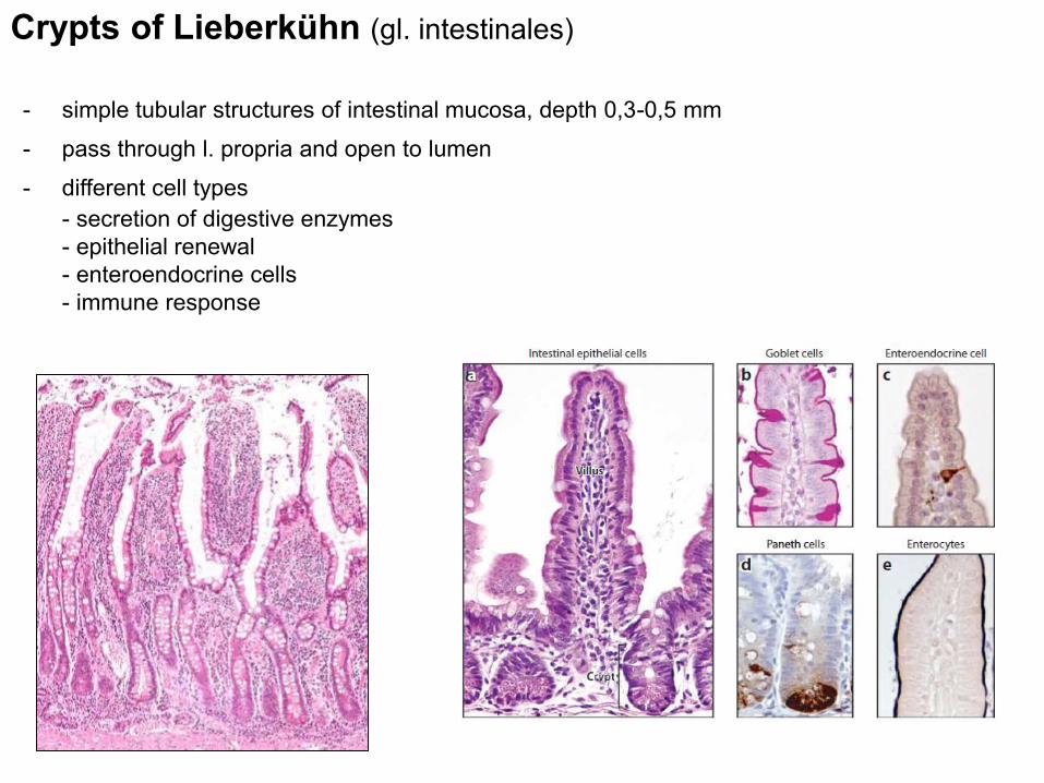
Crypts of Lieberkühn (gl. intestinales)
- simple tubular structures of intestinal mucosa, depth 0,3-0,5 mm
- pass through l. propria and open to lumen
- different cell types
- secretion of digestive enzymes
- epithelial renewal
- enteroendocrine cells
- immune response

Enterocytes
Intestinal mucosa
- tall, columnar cells
- nucleus located in basis of the cell
- apical surface modified- microvilli
(3000) + glycocalyx (0,5m) =
striated border (cuticle)
- tight intercellular connections,
interdigitations
Function:
- digestion – enzymatic complexes
on microvilli membrane
- absorption and transport –
passive, facilitated i active
- lipid uptake - chylomicrons
1m
0,1m

Microvilli

Transportion and resorption
Transport of glucose from intestinal lumen to blood
stream
Na+/K+ ATPase - basolateral surface - concentration
gradient Na+ and K+
K+ gradient generates negative membrane potential
Na+/glucose symport on apical surface
Facilitated diffusion by glucose uniporter (GLUT2) in
basolateral membrane
Acidification of stomach fluid by parietal cells
Apical membrane - H+/K+ ATPase + Cl− a K+ canals
Basolateral membrane – anion antiporter HCO3− and
Cl− ions
Combined activity of ion channels a cells keeps the
electroneutrality and neutral cytoplasmic pH while
reaching high extracellular concentration of H+ and Cl− in
lumen of stomach
http://www.ncbi.nlm.nih.gov/books/NBK21502/

Jádra
F-aktin
Mucin v
sekrečních
granulech
- Cylindrical glandular epithelial cells
- Apical surface – apocrine/merocrine secretion of mucin
- Basal part – RER, GA, nucleus, mitochondria
- Mucinogenic granules
- see lesson spring semester 2015 - Epithelial tissue
Goblet cells
Intestinal mucosa

Goblet cells
Intestinal mucosa
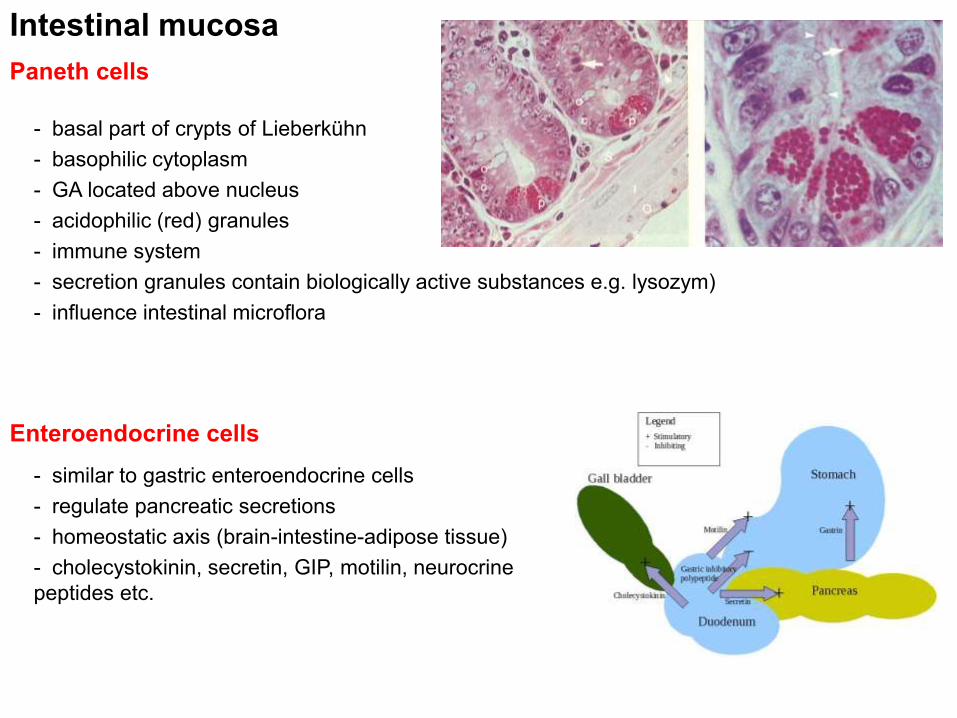
Paneth cells
Intestinal mucosa
- basal part of crypts of Lieberkühn
- basophilic cytoplasm
- GA located above nucleus
- acidophilic (red) granules
- immune system
- secretion granules contain biologically active substances e.g. lysozym)
- influence intestinal microflora
Enteroendocrine cells
- similar to gastric enteroendocrine cells
- regulate pancreatic secretions
- homeostatic axis (brain-intestine-adipose tissue)
- cholecystokinin, secretin, GIP, motilin, neurocrine
peptides etc.

M cells (microfold)
Intestinal mucosa
- epithelial cells above Peyer’s patches
and lymphatic nodules
- no microvilli
- induces immune response
- MHCII
- antigen presentation to dendritic cells
and lymphocytes
„Microfold“
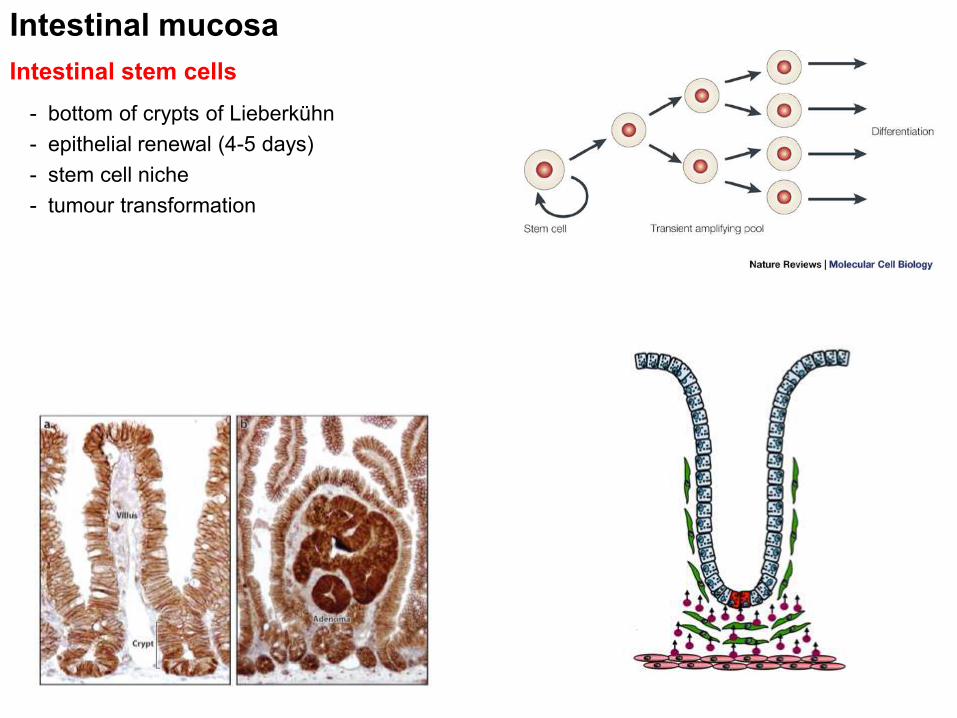
Intestinal stem cells
Intestinal mucosa
- bottom of crypts of Lieberkühn
- epithelial renewal (4-5 days)
- stem cell niche
- tumour transformation

L. propria
Intestinal mucosa
- immune system – GALT
- immunologic barrier
- Peyer’s patches
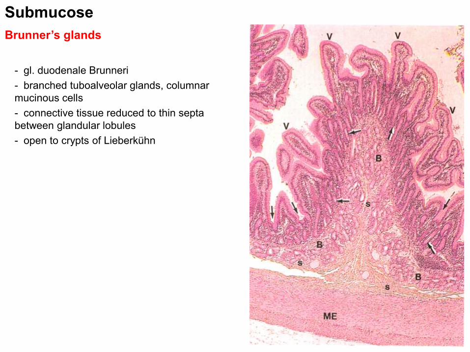
Brunner’s glands
Submucose
- gl. duodenale Brunneri
- branched tuboalveolar glands, columnar
mucinous cells
- connective tissue reduced to thin septa
between glandular lobules
- open to crypts of Lieberkühn

Muscularis externa
- two layers of smooth muscle (inner circular, outer longitudinal)
- plexus myentericus Auerbachi
Serosa
- loose collagen connective tissue + simple squamous epithelium (mesothelium)

Colon
- no plicae of Kerckring, villi
- muscularis externa – longitudinal layer forms taenie coli
- surface serosa forms appendices epiploicae (adipose)
Small intestine Colon

Colon
- absorption of water, electrolytes
- deeper crypts of Lieberkühn, no Paneth cells
- abundant goblet cells
- abundant lymphatic follicles in l. propria (GALT)

Apendix
- develops from and is connected to caecum 8-10 cm
(0,5-1cm)
- continuous longitudinal layer of m. externa
- lymphatic follicles reaching submucosa
- irregular crypts of Lieberkühn with Paneth cells

Rectum and anal canal
- Pars pelvina
- plicae transversae recti
- histological architecture identical to colon
- Canalis analis
- anulus hemorhoidalis – no L. crypts, simple columnar epithelium replaced by stratified
squamous epithelium
- rich venous plexus
- columnae rectales
- sinus rectales and valvulae rectales
- zona cutanea – typical skin

Rectum and anal canal

Microscopic anatomy of the gut tubesee also the requirements for exam
Summary GIT 1
- General architecture of hollow organs and gut tube: mucosa (l. epithelialis m ., l. propria, l.
muscularis m.), submucosa, t. muscularis externa, serosa (l. propria s., l. epith. s.), adventitia
- Pharynx – structure and microscopic anatomy
- Esophagus - structure, epithelium, mucosal and submucosal glands, differences in t. muscularis
ext.
- Stomach – anatomical and histological structure, mucosa - areae gastricae, foveolae gastricae,
gastric glands (pyloricae vs. propriae), localization, ultrastructure and function of gl. gastricae
propriae and its cells (chief, parietal, neck, enteroendocrine
- Small and large intestine, appendix - anatomical and histological structure, mucosa, glands
(crypts of Lieberkühn, Brunner’s glands), cell types of intestinal mucosa, lymphatic system,
modifications of intestinal wall
- Rectum and anal canal - anatomical and histological structure, mucosa, epithelium, description
of associated structures

Thank you for attention
http://www.med.muni.cz/histology/education/
http://www.med.muni.cz/histology/petr-vanhara/