Embed Size (px)
Citation preview
USMLE Step 1 Session
Pathology 122.10.2014, Klub A. Trýba
FB: USMLE @ Masaryk
Marek Čierny (324602 at mail.muni.cz)
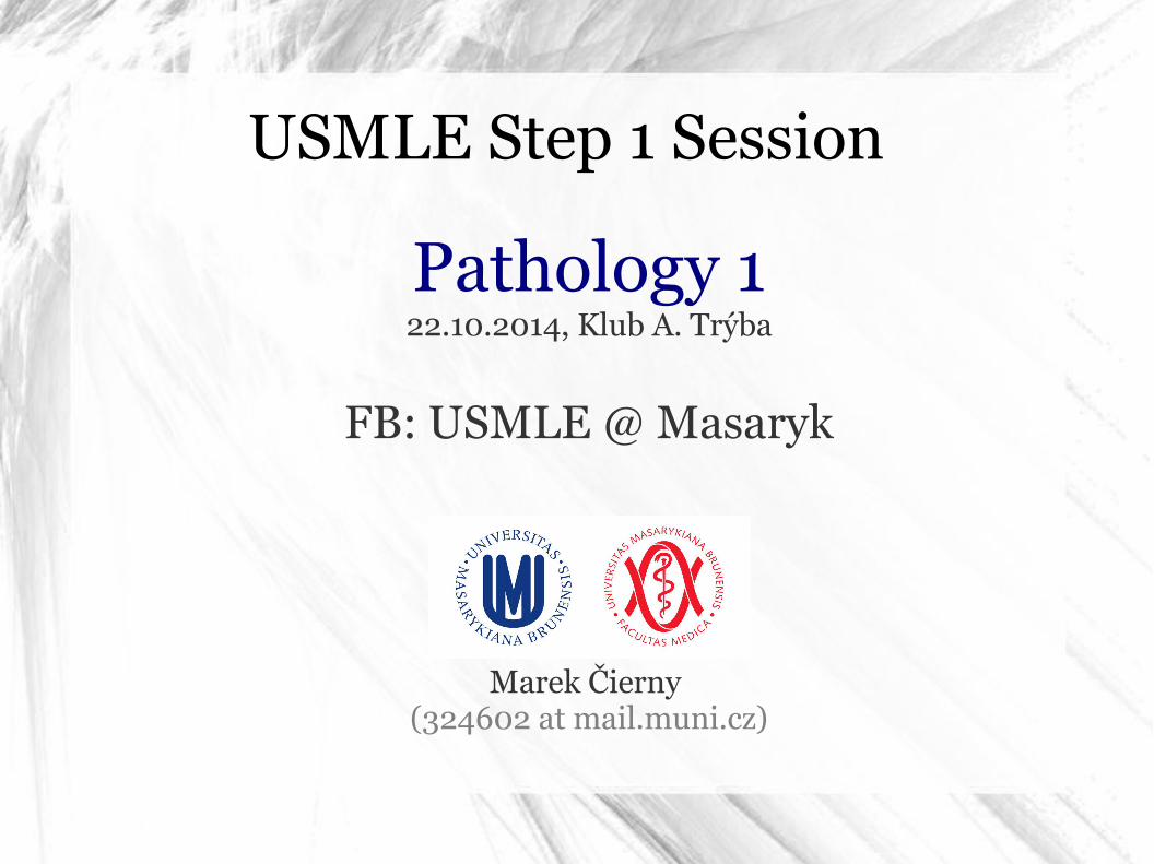
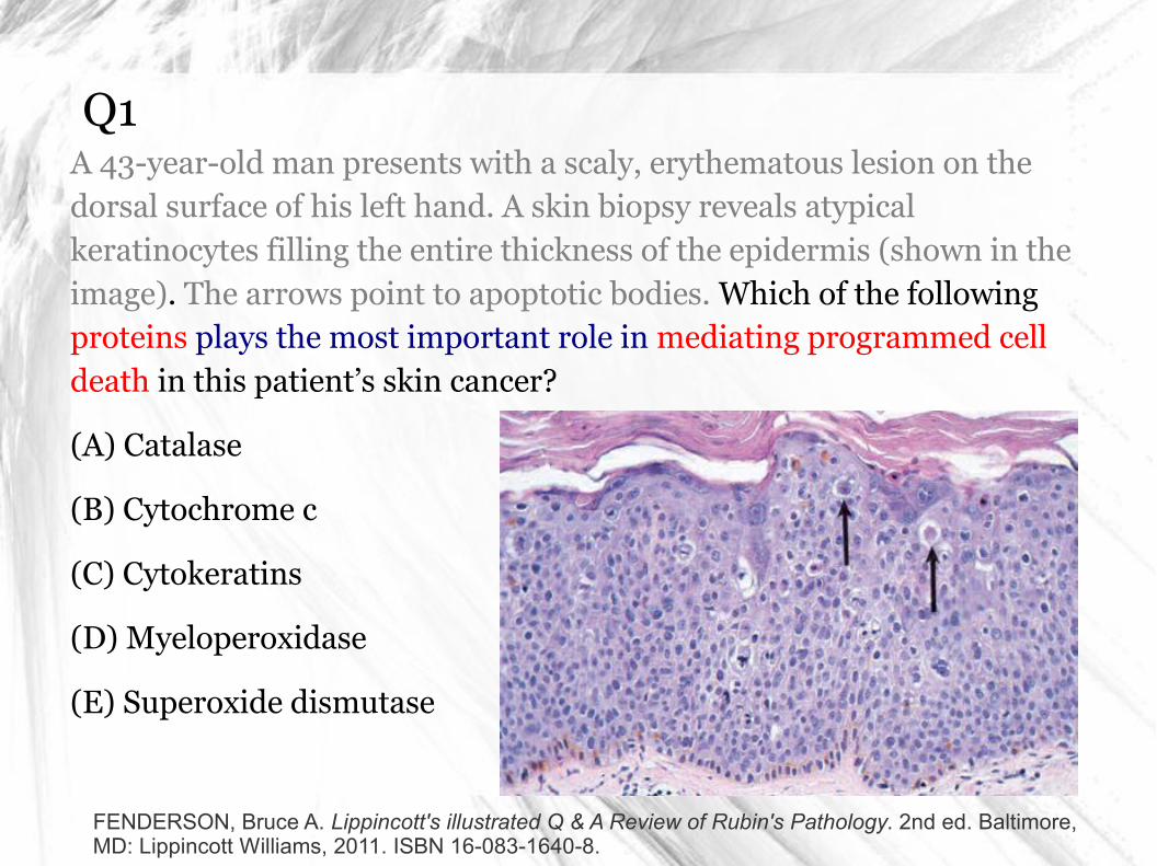
Q1A 43-year-old man presents with a scaly, erythematous lesion on the dorsal surface of his left hand. A skin biopsy reveals atypical keratinocytes filling the entire thickness of the epidermis (shown in the image). The arrows point to apoptotic bodies. Which of the following proteins plays the most important role in mediating programmed cell death in this patient’s skin cancer?
(A) Catalase
(B) Cytochrome c
(C) Cytokeratins
(D) Myeloperoxidase
(E) Superoxide dismutase
FENDERSON, Bruce A. Lippincott's illustrated Q & A Review of Rubin's Pathology. 2nd ed. Baltimore, MD: Lippincott Williams, 2011. ISBN 16-083-1640-8.
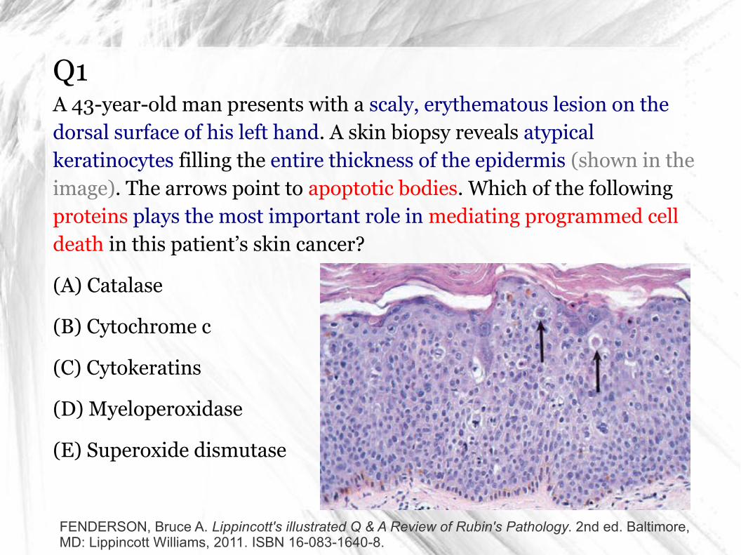
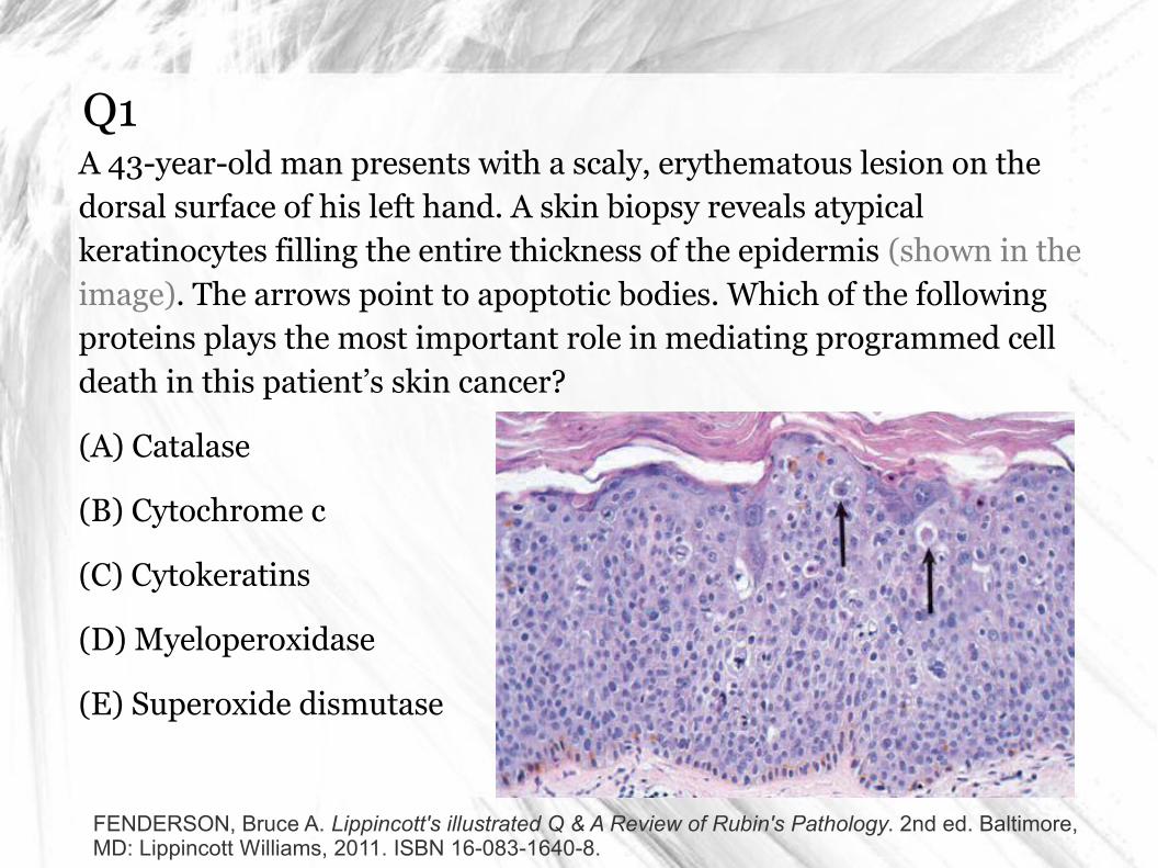
A1: what is apoptosis?
Tao Le, Vikas Bhushan: First Aid for the USMLE Step 1 2014, McGraw Hill Professional, 2014, ISBN 0071831436.
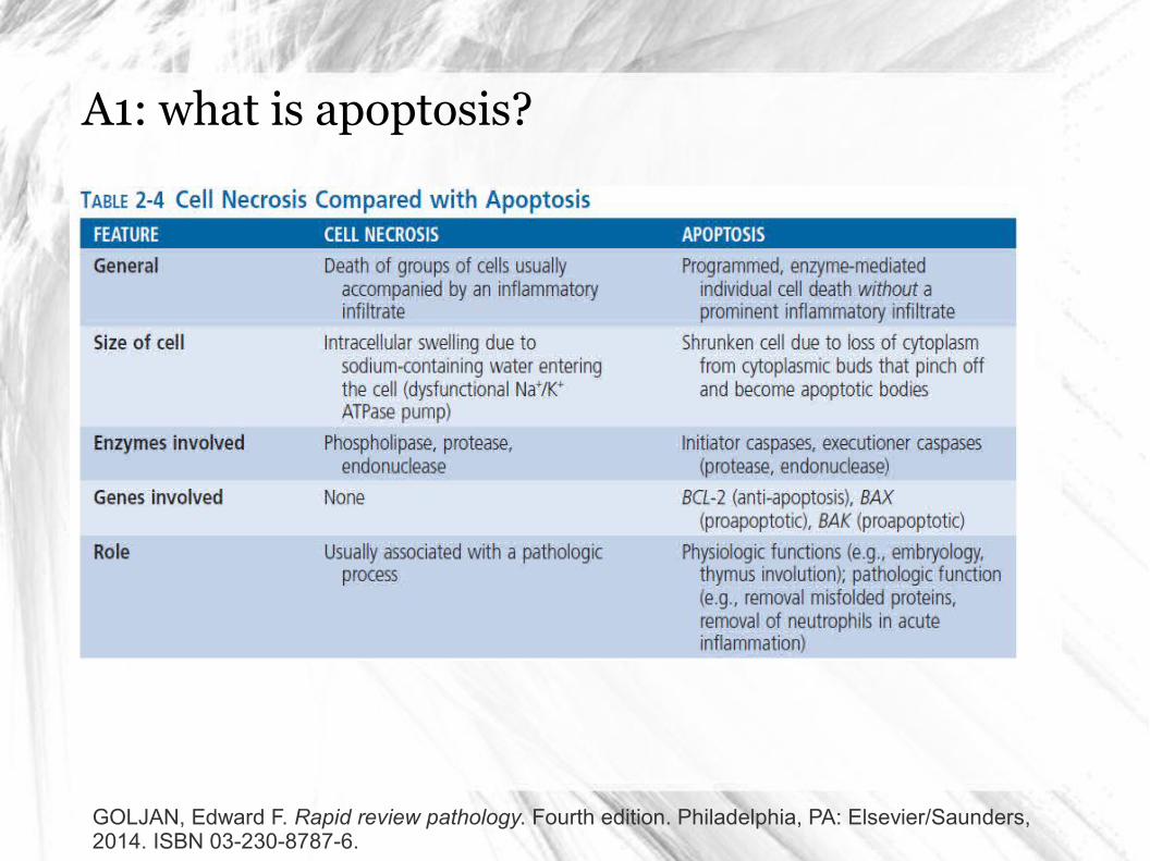
A1: what is apoptosis?
GOLJAN, Edward F. Rapid review pathology. Fourth edition. Philadelphia, PA: Elsevier/Saunders, 2014. ISBN 03-230-8787-6.
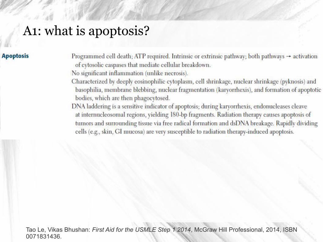
A1: what is apoptosis?
Tao Le, Vikas Bhushan: First Aid for the USMLE Step 1 2014, McGraw Hill Professional, 2014, ISBN 0071831436.
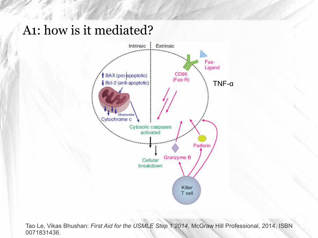
A1: how is it mediated?
Tao Le, Vikas Bhushan: First Aid for the USMLE Step 1 2014, McGraw Hill Professional, 2014, ISBN 0071831436.
TNF-α
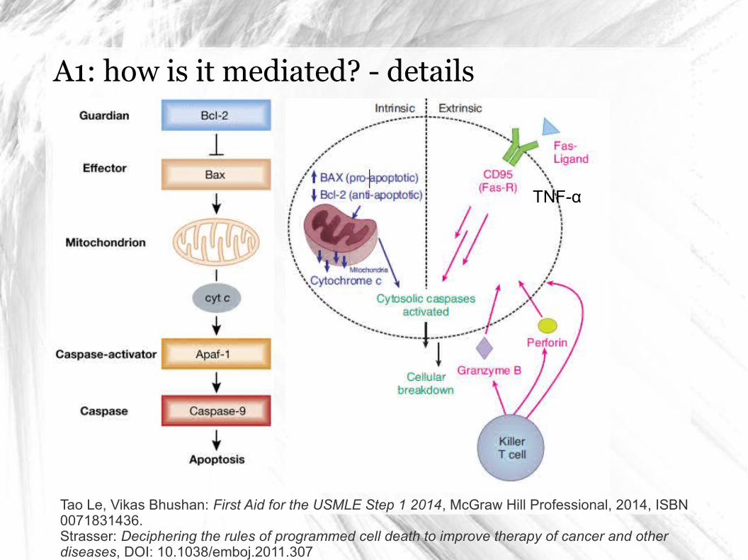
A1: how is it mediated? - details
Tao Le, Vikas Bhushan: First Aid for the USMLE Step 1 2014, McGraw Hill Professional, 2014, ISBN 0071831436.Strasser: Deciphering the rules of programmed cell death to improve therapy of cancer and other diseases, DOI: 10.1038/emboj.2011.307
TNF-α
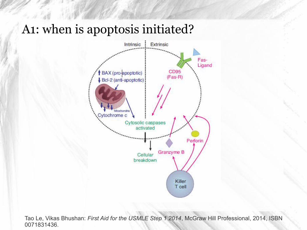
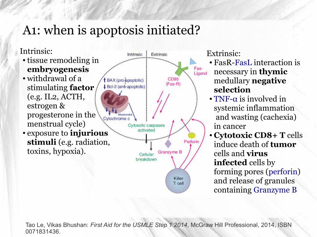
A1: when is apoptosis initiated?
Tao Le, Vikas Bhushan: First Aid for the USMLE Step 1 2014, McGraw Hill Professional, 2014, ISBN 0071831436.
A1: when is apoptosis initiated?
Tao Le, Vikas Bhushan: First Aid for the USMLE Step 1 2014, McGraw Hill Professional, 2014, ISBN 0071831436.
Intrinsic:● tissue remodeling in
embryogenesis● withdrawal of a
stimulating factor (e.g. IL2, ACTH, estrogen & progesterone in the menstrual cycle)
● exposure to injurious stimuli (e.g. radiation, toxins, hypoxia).
Extrinsic:● FasR-FasL interaction is
necessary in thymic medullary negative selection
● TNF-α is involved in systemic inflammation and wasting (cachexia) in cancer
● Cytotoxic CD8+ T cells induce death of tumor cells and virus infected cells by forming pores (perforin) and release of granules containing Granzyme B
Q1A 43-year-old man presents with a scaly, erythematous lesion on the dorsal surface of his left hand. A skin biopsy reveals atypical keratinocytes filling the entire thickness of the epidermis (shown in the image). The arrows point to apoptotic bodies. Which of the following proteins plays the most important role in mediating programmed cell death in this patient’s skin cancer?
(A) Catalase
(B) Cytochrome c
(C) Cytokeratins
(D) Myeloperoxidase
(E) Superoxide dismutase
FENDERSON, Bruce A. Lippincott's illustrated Q & A Review of Rubin's Pathology. 2nd ed. Baltimore, MD: Lippincott Williams, 2011. ISBN 16-083-1640-8.
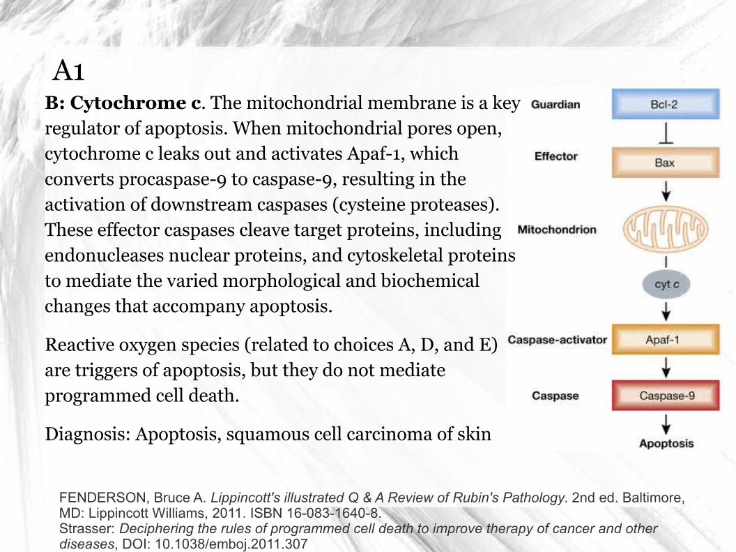
A1B: Cytochrome c. The mitochondrial membrane is a key regulator of apoptosis. When mitochondrial pores open, cytochrome c leaks out and activates Apaf-1, which converts procaspase-9 to caspase-9, resulting in the activation of downstream caspases (cysteine proteases). These effector caspases cleave target proteins, including endonucleases nuclear proteins, and cytoskeletal proteins to mediate the varied morphological and biochemical changes that accompany apoptosis.
Reactive oxygen species (related to choices A, D, and E) are triggers of apoptosis, but they do not mediate programmed cell death.
Diagnosis: Apoptosis, squamous cell carcinoma of skin
FENDERSON, Bruce A. Lippincott's illustrated Q & A Review of Rubin's Pathology. 2nd ed. Baltimore, MD: Lippincott Williams, 2011. ISBN 16-083-1640-8.Strasser: Deciphering the rules of programmed cell death to improve therapy of cancer and other diseases, DOI: 10.1038/emboj.2011.307
Q2A 28-year-old man with a history of radiation/bone marrow transplantation for leukemia presents with severe diarrhea. He subsequently develops septic shock and expires. Microscopic examination of the colon epithelium at autopsy reveals numerous acidophilic bodies and small cells with pyknotic nuclei. Which of the following proteins most likely played a key role in triggering radiation-induced cell death in this patient’s colonic mucosa?
(A) Cytochrome P450
(B) β-Catenin
(C) E-Cadherin
(D) P-Selectin
(E) p53
FENDERSON, Bruce A. Lippincott's illustrated Q & A Review of Rubin's Pathology. 2nd ed. Baltimore, MD: Lippincott Williams, 2011. ISBN 16-083-1640-8.
A2
GOLJAN, Edward F. Rapid review pathology. Fourth edition. Philadelphia, PA: Elsevier/Saunders, 2014. ISBN 03-230-8787-6.
A2
GOLJAN, Edward F. Rapid review pathology. Fourth edition. Philadelphia, PA: Elsevier/Saunders, 2014. ISBN 03-230-8787-6.
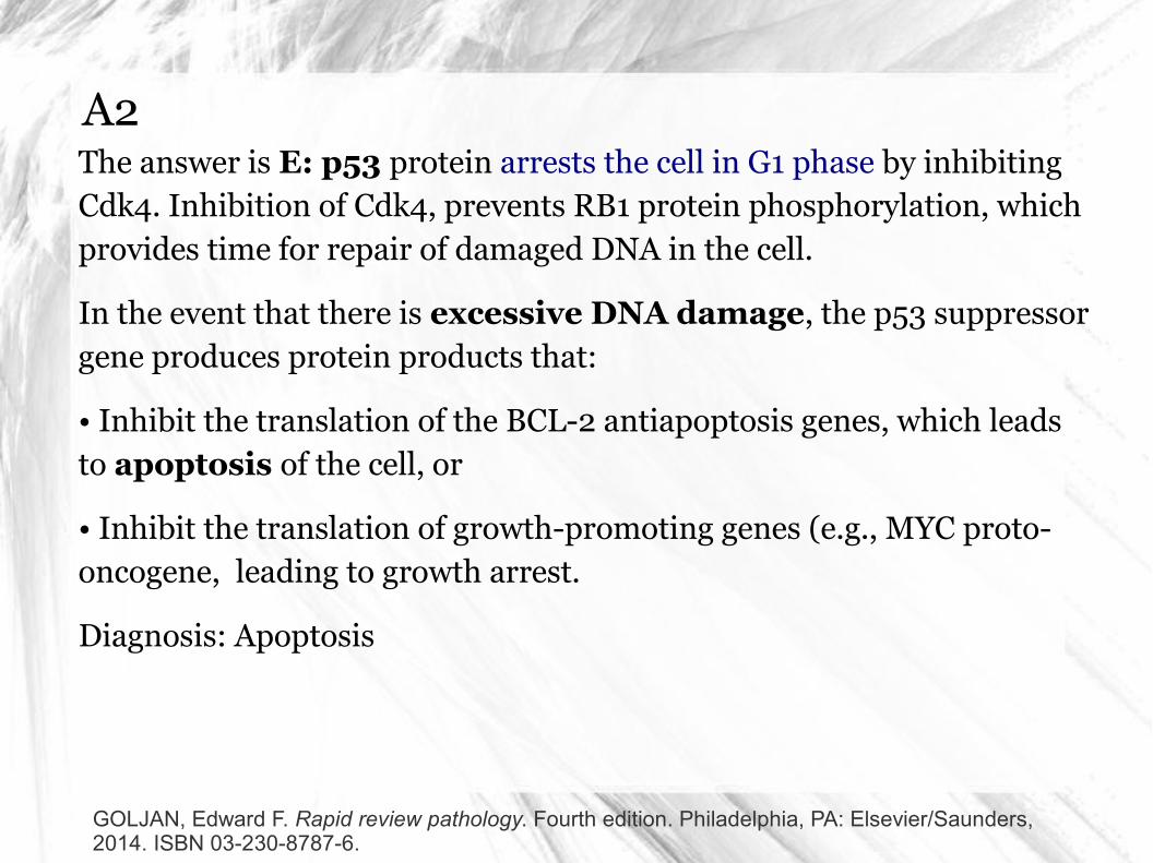
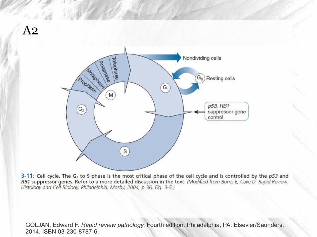
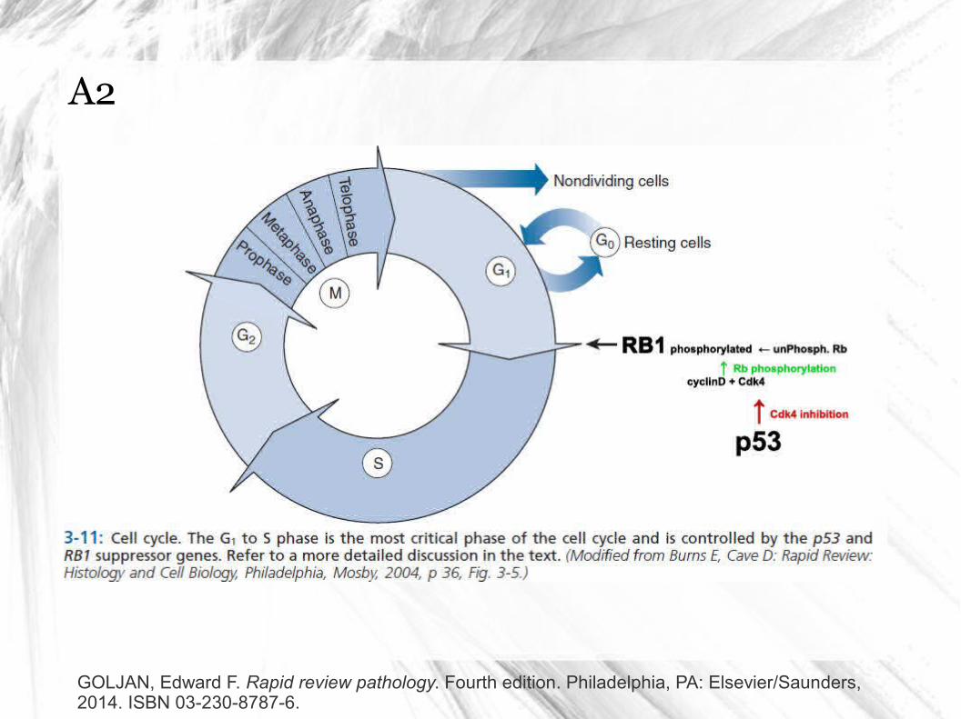
A2The answer is E: p53 protein arrests the cell in G1 phase by inhibiting Cdk4. Inhibition of Cdk4, prevents RB1 protein phosphorylation, which provides time for repair of damaged DNA in the cell.
In the event that there is excessive DNA damage, the p53 suppressor gene produces protein products that:
• Inhibit the translation of the BCL-2 antiapoptosis genes, which leads to apoptosis of the cell, or
• Inhibit the translation of growth-promoting genes (e.g., MYC proto-oncogene, leading to growth arrest.
Diagnosis: Apoptosis
GOLJAN, Edward F. Rapid review pathology. Fourth edition. Philadelphia, PA: Elsevier/Saunders, 2014. ISBN 03-230-8787-6.
Q3A 22-year-old construction worker sticks himself with a sharp, rusty nail. Within 24 hours, the wound has enlarged to become a 1-cm sore that drains thick, purulent material. This skin wound illustrates which of the following morphologic types of necrosis?
(A) Caseous necrosis
(B) Coagulative necrosis
(C) Fat necrosis
(D) Fibrinoid necrosis
(E) Liquefactive necrosis
FENDERSON, Bruce A. Lippincott's illustrated Q & A Review of Rubin's Pathology. 2nd ed. Baltimore, MD: Lippincott Williams, 2011. ISBN 16-083-1640-8.
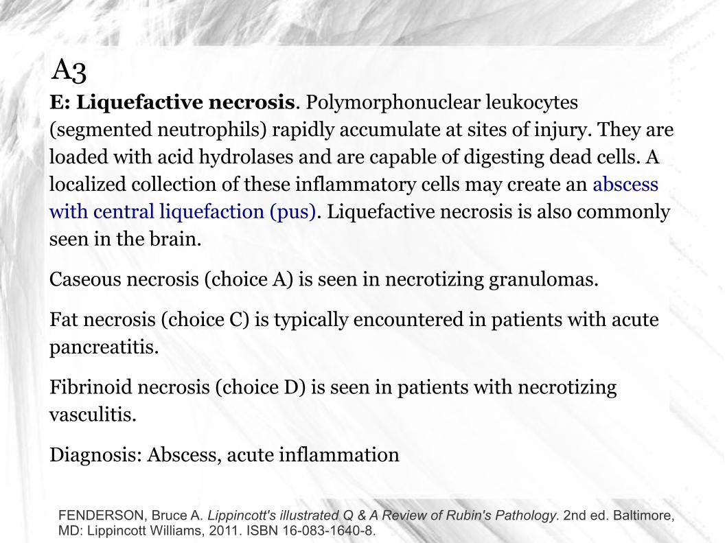
A3E: Liquefactive necrosis. Polymorphonuclear leukocytes (segmented neutrophils) rapidly accumulate at sites of injury. They are loaded with acid hydrolases and are capable of digesting dead cells. A localized collection of these inflammatory cells may create an abscess with central liquefaction (pus). Liquefactive necrosis is also commonly seen in the brain.
Caseous necrosis (choice A) is seen in necrotizing granulomas.
Fat necrosis (choice C) is typically encountered in patients with acute pancreatitis.
Fibrinoid necrosis (choice D) is seen in patients with necrotizing vasculitis.
Diagnosis: Abscess, acute inflammation
FENDERSON, Bruce A. Lippincott's illustrated Q & A Review of Rubin's Pathology. 2nd ed. Baltimore, MD: Lippincott Williams, 2011. ISBN 16-083-1640-8.
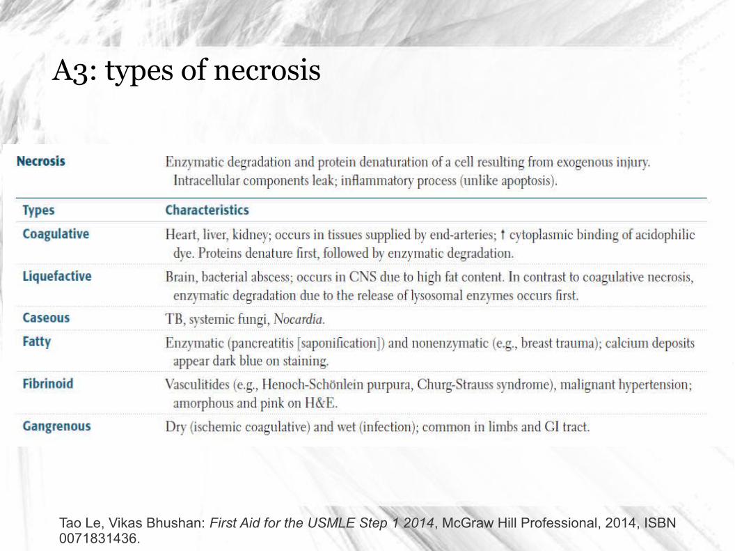
A3: types of necrosis
Tao Le, Vikas Bhushan: First Aid for the USMLE Step 1 2014, McGraw Hill Professional, 2014, ISBN 0071831436.
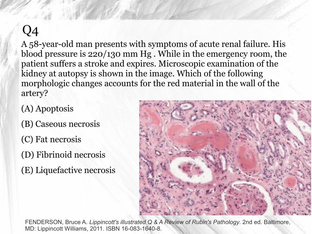
Q4A 58-year-old man presents with symptoms of acute renal failure. His blood pressure is 220/130 mm Hg . While in the emergency room, the patient suffers a stroke and expires. Microscopic examination of the kidney at autopsy is shown in the image. Which of the following morphologic changes accounts for the red material in the wall of the artery?
(A) Apoptosis
(B) Caseous necrosis
(C) Fat necrosis
(D) Fibrinoid necrosis
(E) Liquefactive necrosis
FENDERSON, Bruce A. Lippincott's illustrated Q & A Review of Rubin's Pathology. 2nd ed. Baltimore, MD: Lippincott Williams, 2011. ISBN 16-083-1640-8.
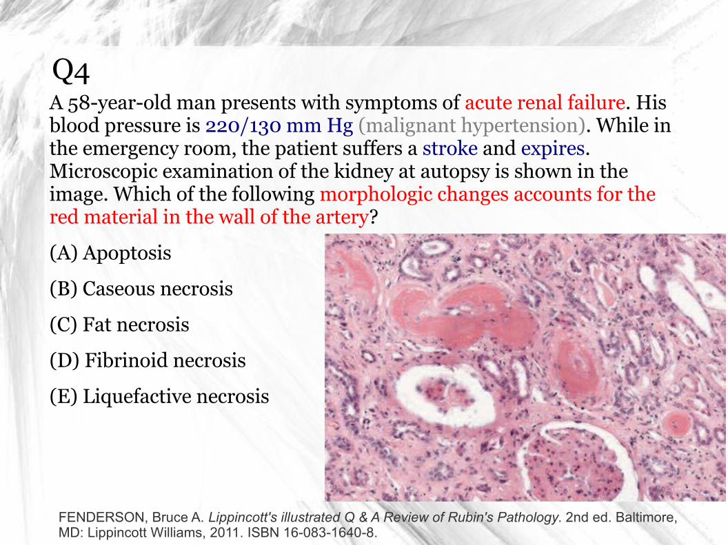
Q4A 58-year-old man presents with symptoms of acute renal failure. His blood pressure is 220/130 mm Hg (malignant hypertension). While in the emergency room, the patient suffers a stroke and expires. Microscopic examination of the kidney at autopsy is shown in the image. Which of the following morphologic changes accounts for the red material in the wall of the artery?
(A) Apoptosis
(B) Caseous necrosis
(C) Fat necrosis
(D) Fibrinoid necrosis
(E) Liquefactive necrosis
FENDERSON, Bruce A. Lippincott's illustrated Q & A Review of Rubin's Pathology. 2nd ed. Baltimore, MD: Lippincott Williams, 2011. ISBN 16-083-1640-8.
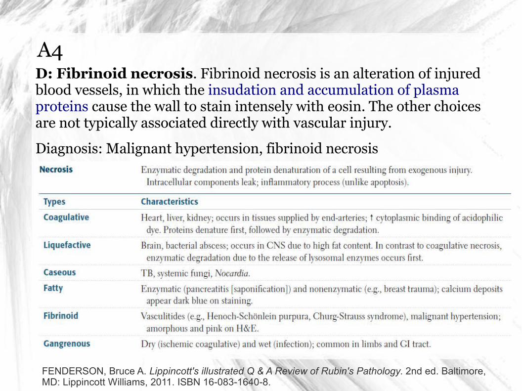
A4D: Fibrinoid necrosis. Fibrinoid necrosis is an alteration of injured blood vessels, in which the insudation and accumulation of plasma proteins cause the wall to stain intensely with eosin. The other choices are not typically associated directly with vascular injury.
Diagnosis: Malignant hypertension, fibrinoid necrosis
FENDERSON, Bruce A. Lippincott's illustrated Q & A Review of Rubin's Pathology. 2nd ed. Baltimore, MD: Lippincott Williams, 2011. ISBN 16-083-1640-8.
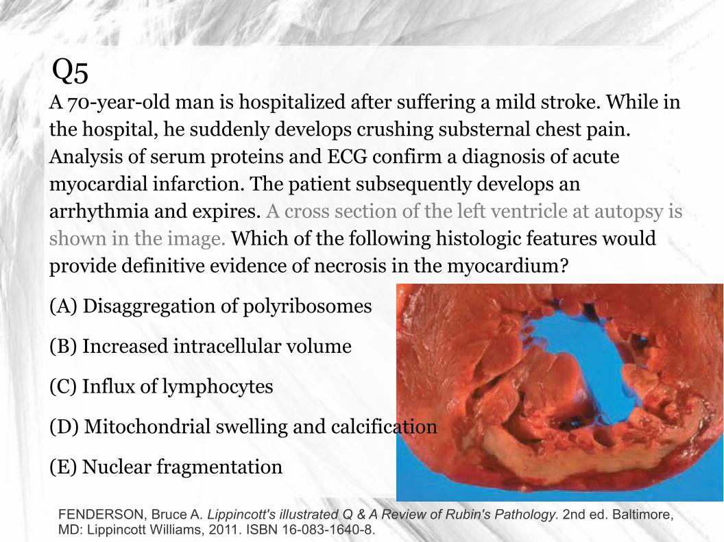
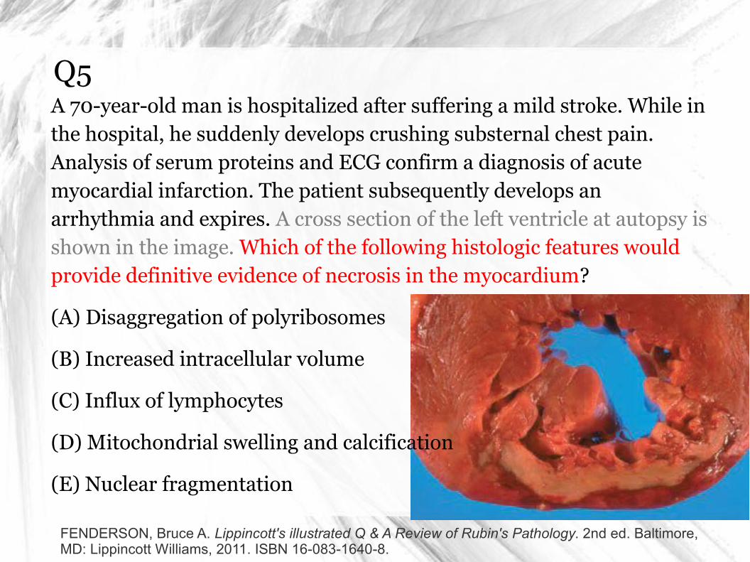
Q5A 70-year-old man is hospitalized after suffering a mild stroke. While in the hospital, he suddenly develops crushing substernal chest pain. Analysis of serum proteins and ECG confirm a diagnosis of acute myocardial infarction. The patient subsequently develops an arrhythmia and expires. A cross section of the left ventricle at autopsy is shown in the image. Which of the following histologic features would provide definitive evidence of necrosis in the myocardium?
(A) Disaggregation of polyribosomes
(B) Increased intracellular volume
(C) Influx of lymphocytes
(D) Mitochondrial swelling and calcification
(E) Nuclear fragmentation
FENDERSON, Bruce A. Lippincott's illustrated Q & A Review of Rubin's Pathology. 2nd ed. Baltimore, MD: Lippincott Williams, 2011. ISBN 16-083-1640-8.
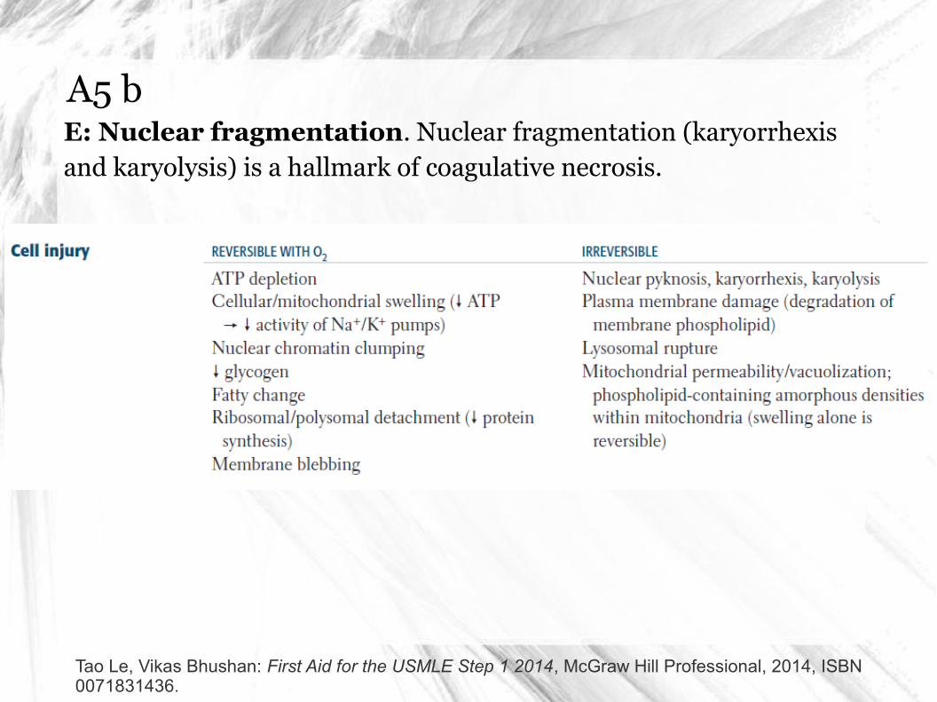
A5 bE: Nuclear fragmentation. Nuclear fragmentation (karyorrhexis and karyolysis) is a hallmark of coagulative necrosis.
Tao Le, Vikas Bhushan: First Aid for the USMLE Step 1 2014, McGraw Hill Professional, 2014, ISBN 0071831436.
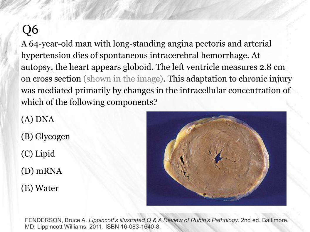
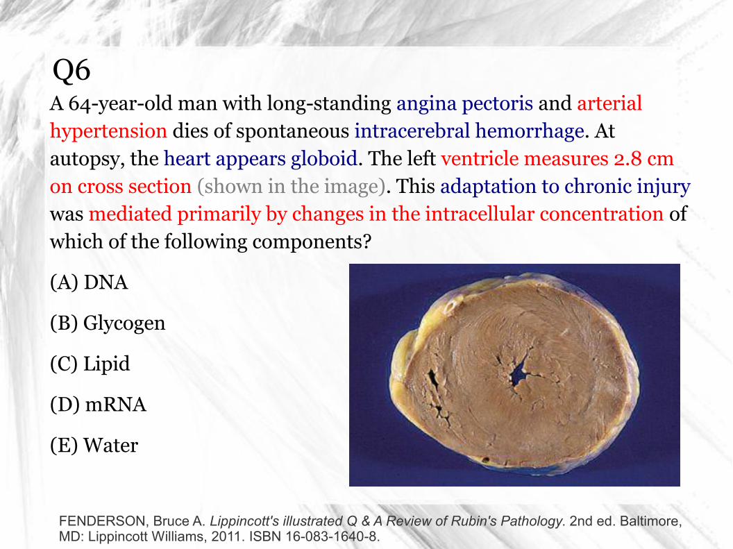
Q6A 64-year-old man with long-standing angina pectoris and arterial hypertension dies of spontaneous intracerebral hemorrhage. At autopsy, the heart appears globoid. The left ventricle measures 2.8 cm on cross section (shown in the image). This adaptation to chronic injury was mediated primarily by changes in the intracellular concentration of which of the following components?
(A) DNA
(B) Glycogen
(C) Lipid
(D) mRNA
(E) Water
FENDERSON, Bruce A. Lippincott's illustrated Q & A Review of Rubin's Pathology. 2nd ed. Baltimore, MD: Lippincott Williams, 2011. ISBN 16-083-1640-8.
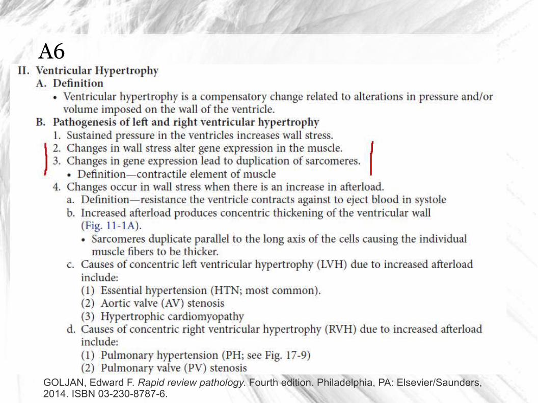
A6What is the inderlying mechanism of concentric thickening of the ventricular wall?
Proliferation?
Hypertrophy?
Deposition of a molecule?
FENDERSON, Bruce A. Lippincott's illustrated Q & A Review of Rubin's Pathology. 2nd ed. Baltimore, MD: Lippincott Williams, 2011. ISBN 16-083-1640-8.
A6D: mRNA. Hypertrophic cardiac myocytes have more cytoplasm and larger nuclei than normal cells. The final steps of pathogenesis include increases in mRNA, rRNA, and protein. Hypertrophy results from transcriptional regulation.
Aneuploidy (choice A) is not a feature of myofiber hypertrophy. Water influx (choice E), which is typical of hydropic swelling in acute injury, is not a common feature of hypertrophy.
Diagnosis: Hypertrophic heart disease, hypertrophy
FENDERSON, Bruce A. Lippincott's illustrated Q & A Review of Rubin's Pathology. 2nd ed. Baltimore, MD: Lippincott Williams, 2011. ISBN 16-083-1640-8.
A6
GOLJAN, Edward F. Rapid review pathology. Fourth edition. Philadelphia, PA: Elsevier/Saunders, 2014. ISBN 03-230-8787-6.
Q7A 24-year-old woman contracts toxoplasmosis during her pregnancy and delivers a neonate at 37 weeks of gestation with a severe malformation of the central nervous system. MRI studies of the neonate reveal porencephaly and hydrocephalus. An X-ray film of the head shows irregular densities in the basal ganglia. These X-ray findings are best explained by which of the following mechanisms of disease?
(A) Amniotic fluid embolism
(B) Dystrophic calcification
(C) Granulomatous inflammation
(D) Metastatic calcification
(E) Organ immaturity
FENDERSON, Bruce A. Lippincott's illustrated Q & A Review of Rubin's Pathology. 2nd ed. Baltimore, MD: Lippincott Williams, 2011. ISBN 16-083-1640-8.
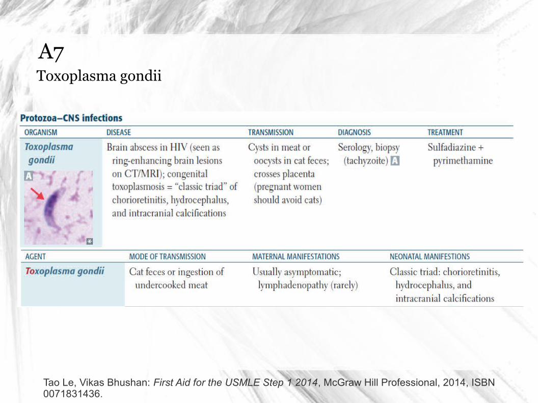
A7Toxoplasma gondii
Tao Le, Vikas Bhushan: First Aid for the USMLE Step 1 2014, McGraw Hill Professional, 2014, ISBN 0071831436.
A7B: Dystrophic calcification. Dystrophic calcification reflects underlying cell injury. Serum levels of calcium are normal, and the calcium deposits are located in previously damaged tissue. Intrauterine Toxoplasma infection affects approximately 0.1% of all pregnancies. Acute encephalitis in the fetus afflicted with TORCH syndrome may be associated with foci of necrosis that become calcified. Microcephaly, hydrocephalus, and microgyria are frequent complications of these intrauterine infections.
Metastatic calcification (choice D) reflects an underlying disorder in calcium metabolism. Example?
Diagnosis: Dystrophic calcification
FENDERSON, Bruce A. Lippincott's illustrated Q & A Review of Rubin's Pathology. 2nd ed. Baltimore, MD: Lippincott Williams, 2011. ISBN 16-083-1640-8.
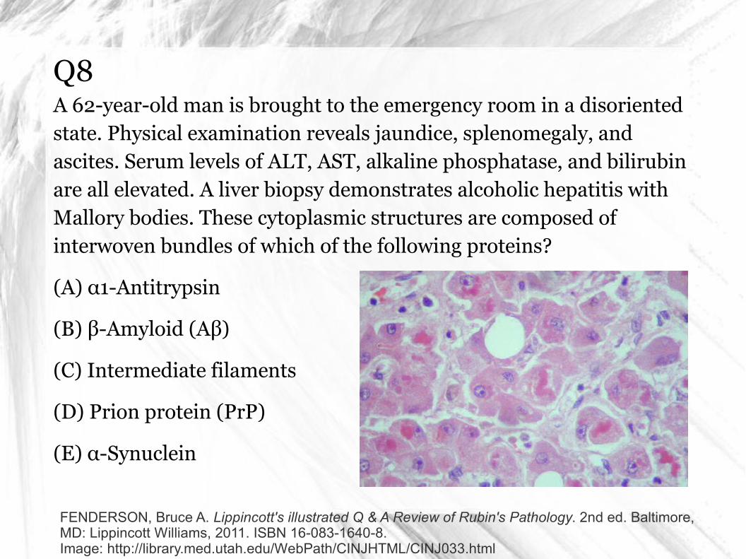
Q8A 62-year-old man is brought to the emergency room in a disoriented state. Physical examination reveals jaundice, splenomegaly, and ascites. Serum levels of ALT, AST, alkaline phosphatase, and bilirubin are all elevated. A liver biopsy demonstrates alcoholic hepatitis with Mallory bodies. These cytoplasmic structures are composed of interwoven bundles of which of the following proteins?
(A) α1-Antitrypsin
(B) β-Amyloid (Aβ)
(C) Intermediate filaments
(D) Prion protein (PrP)
(E) α-Synuclein
FENDERSON, Bruce A. Lippincott's illustrated Q & A Review of Rubin's Pathology. 2nd ed. Baltimore, MD: Lippincott Williams, 2011. ISBN 16-083-1640-8.Image: http://library.med.utah.edu/WebPath/CINJHTML/CINJ033.html
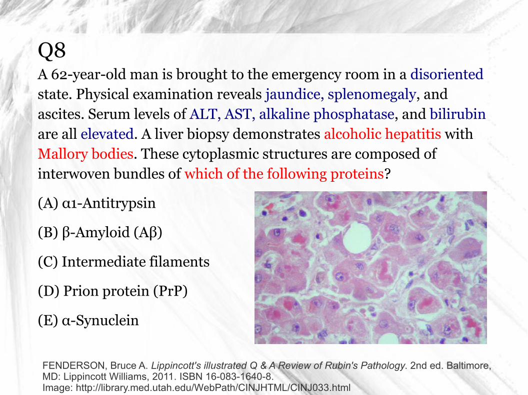
A8: alcoholic liver disease
GOLJAN, Edward F. Rapid review pathology. Fourth edition. Philadelphia, PA: Elsevier/Saunders, 2014. ISBN 03-230-8787-6.
C: Intermediate filaments. Hyaline is a term that refers to any material that exhibits a reddish, homogeneous appearance when stained with hematoxylin and eosin (H&E). Standard terminology includes hyaline arteriolosclerosis, alcoholic hyaline in the liver (Mallory bodies), hyaline membranes in the lung, and hyaline droplets in various cells. Alcoholic (Mallory) hyaline is composed of cytoskeletal intermediate filaments (cytokeratins), whereas pulmonary hyaline membranes consist of plasma proteins deposited in alveoli.
Structurally abnormal α1-antitrypsin molecules (choice A) accumulate in the liver of patients with α1-antitrypsin deficiency.
α-Synuclein (choice E) accumulates in neurons in the substantia nigra of patients with Parkinson disease.
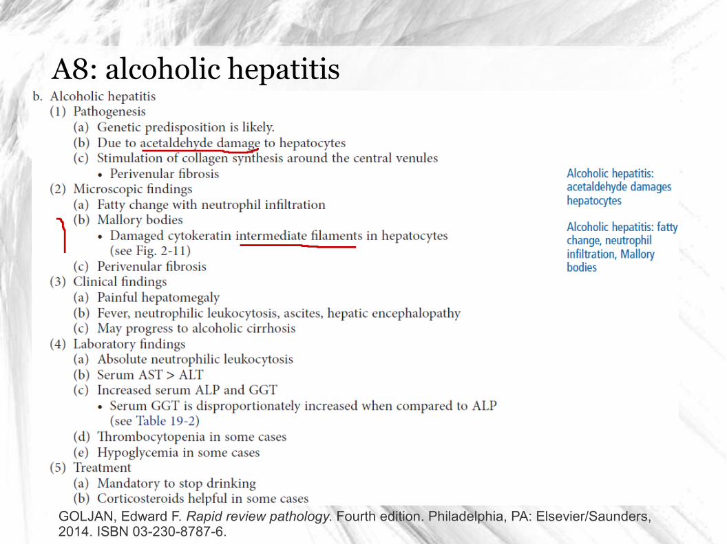
A8: alcoholic hepatitis
GOLJAN, Edward F. Rapid review pathology. Fourth edition. Philadelphia, PA: Elsevier/Saunders, 2014. ISBN 03-230-8787-6.
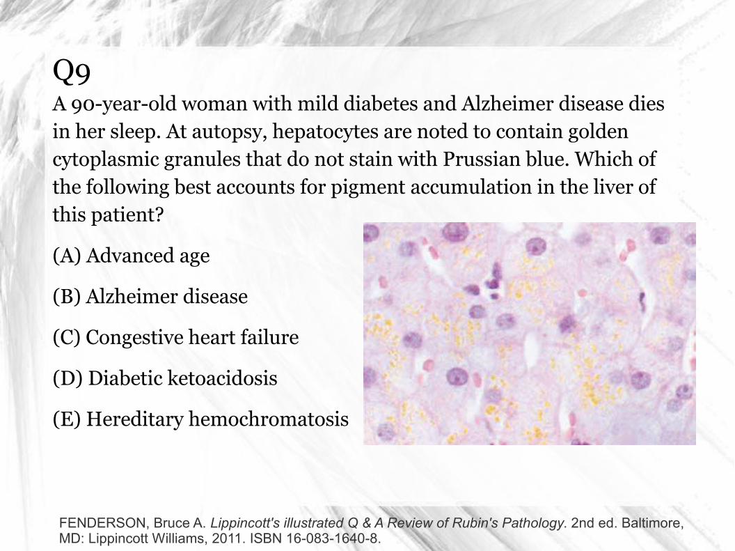
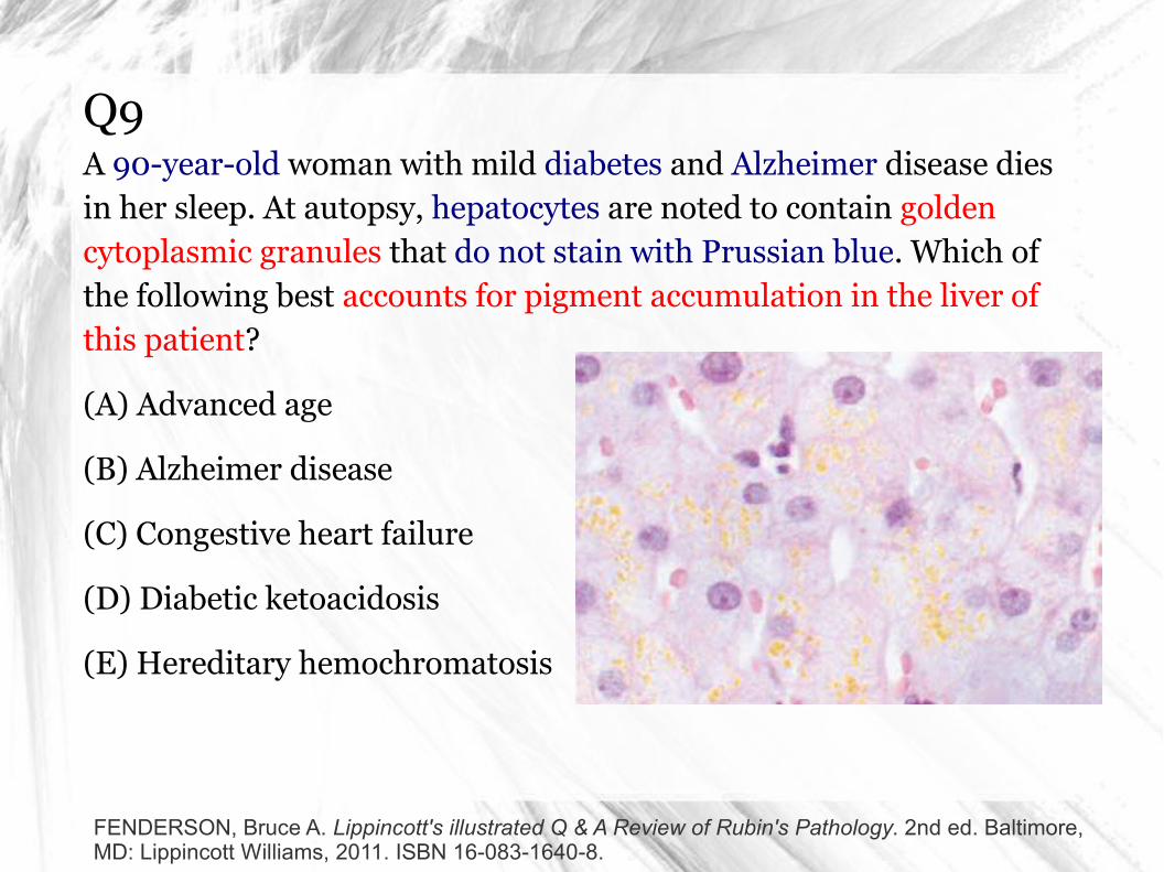
Q9A 90-year-old woman with mild diabetes and Alzheimer disease dies in her sleep. At autopsy, hepatocytes are noted to contain golden cytoplasmic granules that do not stain with Prussian blue. Which of the following best accounts for pigment accumulation in the liver of this patient?
(A) Advanced age
(B) Alzheimer disease
(C) Congestive heart failure
(D) Diabetic ketoacidosis
(E) Hereditary hemochromatosis
FENDERSON, Bruce A. Lippincott's illustrated Q & A Review of Rubin's Pathology. 2nd ed. Baltimore, MD: Lippincott Williams, 2011. ISBN 16-083-1640-8.
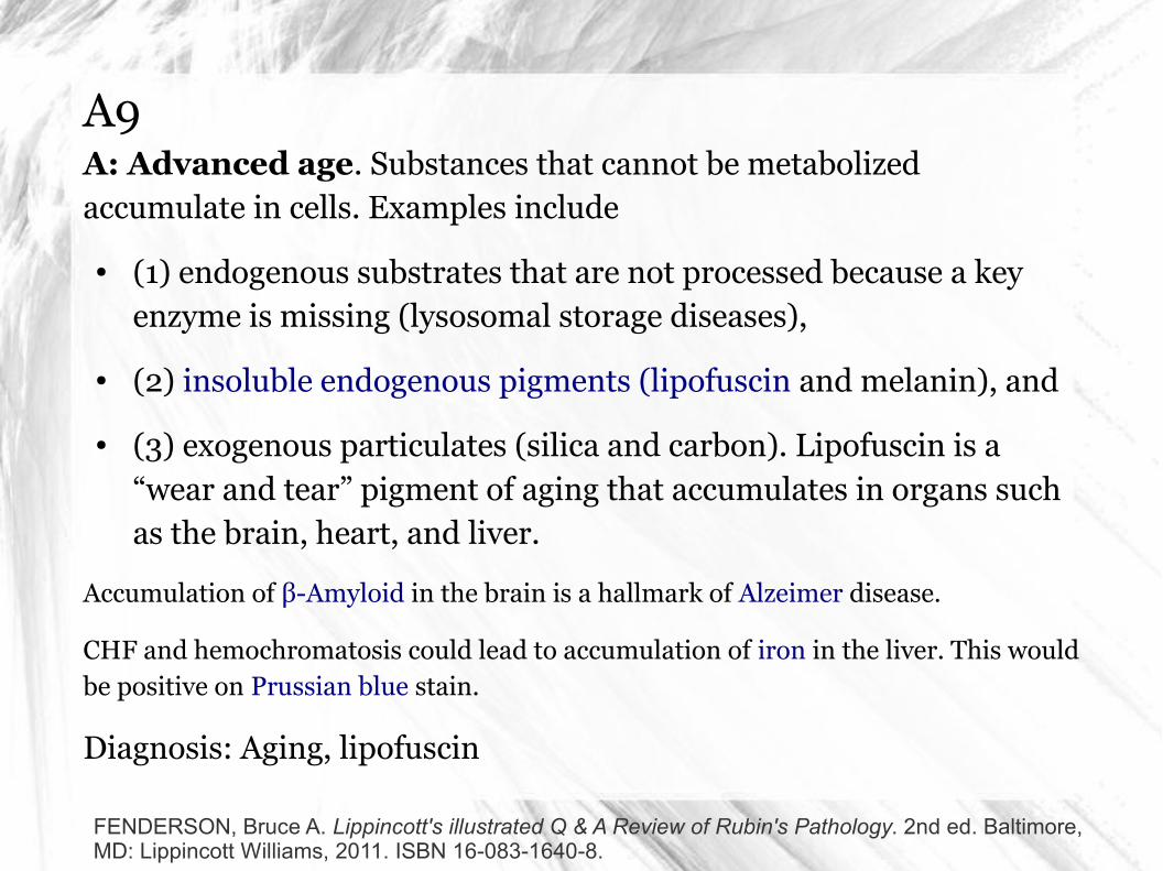
A9A: Advanced age. Substances that cannot be metabolized accumulate in cells. Examples include
● (1) endogenous substrates that are not processed because a key enzyme is missing (lysosomal storage diseases),
● (2) insoluble endogenous pigments (lipofuscin and melanin), and
● (3) exogenous particulates (silica and carbon). Lipofuscin is a “wear and tear” pigment of aging that accumulates in organs such as the brain, heart, and liver.
Accumulation of β-Amyloid in the brain is a hallmark of Alzeimer disease.
CHF and hemochromatosis could lead to accumulation of iron in the liver. This would be positive on Prussian blue stain.
Diagnosis: Aging, lipofuscin
FENDERSON, Bruce A. Lippincott's illustrated Q & A Review of Rubin's Pathology. 2nd ed. Baltimore, MD: Lippincott Williams, 2011. ISBN 16-083-1640-8.
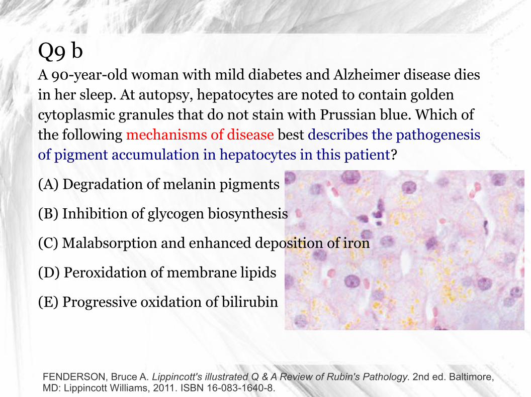
Q9 bA 90-year-old woman with mild diabetes and Alzheimer disease dies in her sleep. At autopsy, hepatocytes are noted to contain golden cytoplasmic granules that do not stain with Prussian blue. Which of the following mechanisms of disease best describes the pathogenesis of pigment accumulation in hepatocytes in this patient?
(A) Degradation of melanin pigments
(B) Inhibition of glycogen biosynthesis
(C) Malabsorption and enhanced deposition of iron
(D) Peroxidation of membrane lipids
(E) Progressive oxidation of bilirubin
FENDERSON, Bruce A. Lippincott's illustrated Q & A Review of Rubin's Pathology. 2nd ed. Baltimore, MD: Lippincott Williams, 2011. ISBN 16-083-1640-8.
A9 bD: Peroxidation of membrane lipids. Lipofuscin is found in lysosomes and contains peroxidation products of unsaturated fatty acids. The presence of this pigment is thought to reflect continuing lipid peroxidation of cellular membranes as a result of inadequate defenses against activated oxygen radicals.
FENDERSON, Bruce A. Lippincott's illustrated Q & A Review of Rubin's Pathology. 2nd ed. Baltimore, MD: Lippincott Williams, 2011. ISBN 16-083-1640-8.
Q10A 60-year-old man is rushed to the hospital with acute liver failure. He undergoes successful orthotopic liver transplantation; however, the transplanted liver does not produce much bile for the first 3 days. Poor graft function in this patient is thought to be the result of “reperfusion injury.” Which of the following substances was the most likely cause of reperfusion injury in this patient’s transplanted liver?
(A) Cationic proteins
(B) Free ferric iron
(C) Hydrochlorous acid
(D) Lysosomal acid hydrolases
(E) Reactive oxygen species
FENDERSON, Bruce A. Lippincott's illustrated Q & A Review of Rubin's Pathology. 2nd ed. Baltimore, MD: Lippincott Williams, 2011. ISBN 16-083-1640-8.
A10E: Reactive oxygen species. Ischemia/reperfusion (I/R) injury is a common clinical problem that arises in the setting of occlusive cardiovascular disease, infection, transplantation, shock, and many other circumstances. The genesis of I/R injury relates to the interplay between transient ischemia and the re-establishment of blood flow (reperfusion). Initially, ischemia produces a type of cellular damage that leads to the generation of free radical species. Subsequently, reperfusion provides abundant molecular oxygen (O2) to combine with free radicals to form reactive oxygen species. Oxygen radicals are formed inside cells through the xanthine oxidase pathway and released from activated neutrophils.
FENDERSON, Bruce A. Lippincott's illustrated Q & A Review of Rubin's Pathology. 2nd ed. Baltimore, MD: Lippincott Williams, 2011. ISBN 16-083-1640-8.
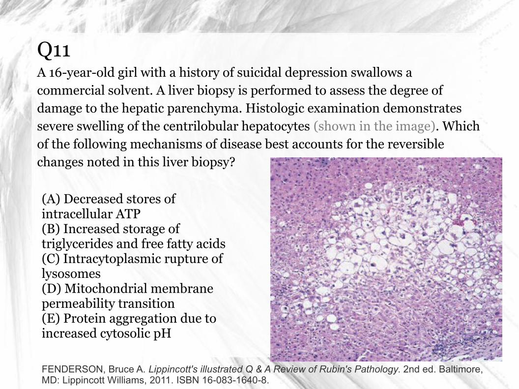
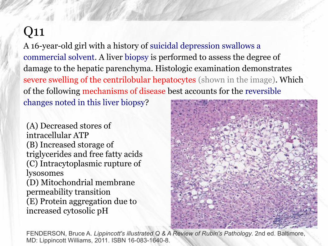
Q11A 16-year-old girl with a history of suicidal depression swallows a commercial solvent. A liver biopsy is performed to assess the degree of damage to the hepatic parenchyma. Histologic examination demonstrates severe swelling of the centrilobular hepatocytes (shown in the image). Which of the following mechanisms of disease best accounts for the reversible changes noted in this liver biopsy?
FENDERSON, Bruce A. Lippincott's illustrated Q & A Review of Rubin's Pathology. 2nd ed. Baltimore, MD: Lippincott Williams, 2011. ISBN 16-083-1640-8.
(A) Decreased stores of intracellular ATP(B) Increased storage of triglycerides and free fatty acids(C) Intracytoplasmic rupture of lysosomes(D) Mitochondrial membrane permeability transition(E) Protein aggregation due to increased cytosolic pH
A11A: Decreased stores of intracellular ATP. Hydropic swelling may result from many causes, including chemical and biologic toxins, infections, and ischemia. Injurious agents cause hydropic swelling by
(1) increasing the permeability of the plasma membrane to sodium;
(2) damaging the membrane sodium-potassium ATPase (pump); or
(3) interfering with the synthesis of ATP, thereby depriving the pump of its fuel.
Diagnosis: Hydropic swelling, hepatotoxicity
FENDERSON, Bruce A. Lippincott's illustrated Q & A Review of Rubin's Pathology. 2nd ed. Baltimore, MD: Lippincott Williams, 2011. ISBN 16-083-1640-8.
A11
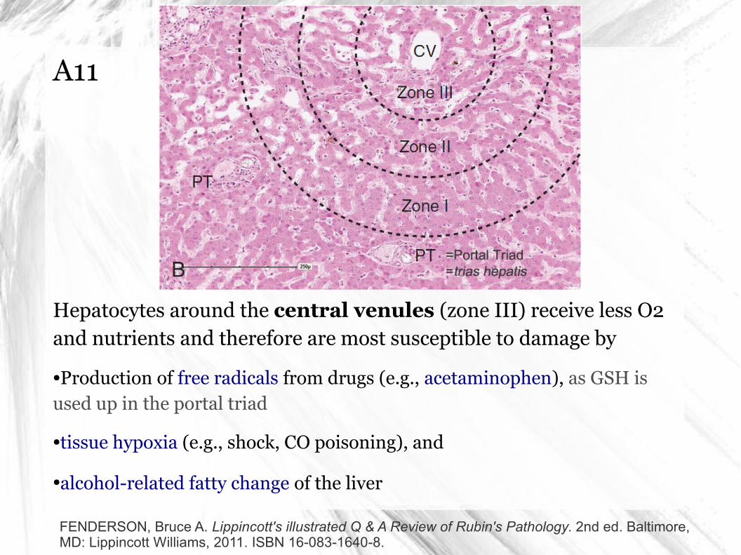
Hepatocytes around the central venules (zone III) receive less O2 and nutrients and therefore are most susceptible to damage by
●Production of free radicals from drugs (e.g., acetaminophen), as GSH is used up in the portal triad
●tissue hypoxia (e.g., shock, CO poisoning), and
●alcohol-related fatty change of the liver
FENDERSON, Bruce A. Lippincott's illustrated Q & A Review of Rubin's Pathology. 2nd ed. Baltimore, MD: Lippincott Williams, 2011. ISBN 16-083-1640-8.
Q12A 32-year-old woman with poorly controlled diabetes mellitus delivers a healthy boy at 38 weeks of gestation. As a result of maternal hyperglycemia during pregnancy, pancreatic islets in the neonate would be expected to show which of the following morphologic responses to injury?
(A) Atrophy
(B) Dysplasia
(C) Hyperplasia
(D) Metaplasia
(E) Necrosis
FENDERSON, Bruce A. Lippincott's illustrated Q & A Review of Rubin's Pathology. 2nd ed. Baltimore, MD: Lippincott Williams, 2011. ISBN 16-083-1640-8.
A12C: Hyperplasia. Infants of diabetic mothers show a 5% to 10% incidence of major developmental abnormalities, including anomalies of the heart and great vessels and neural tube defects. The frequency of these lesions relates to the control of maternal diabetes during early gestation. During fetal development, the islet cells of the pancreas have proliferative capacity and respond to increased demand for insulin by undergoing physiologic hyperplasia. Fetuses exposed to hyperglycemia in utero may develop hyperplasia of the pancreatic β cells, which may secrete insulin autonomously and cause hypoglycemia at birth.
Diagnosis: Diabetes mellitus
FENDERSON, Bruce A. Lippincott's illustrated Q & A Review of Rubin's Pathology. 2nd ed. Baltimore, MD: Lippincott Williams, 2011. ISBN 16-083-1640-8.
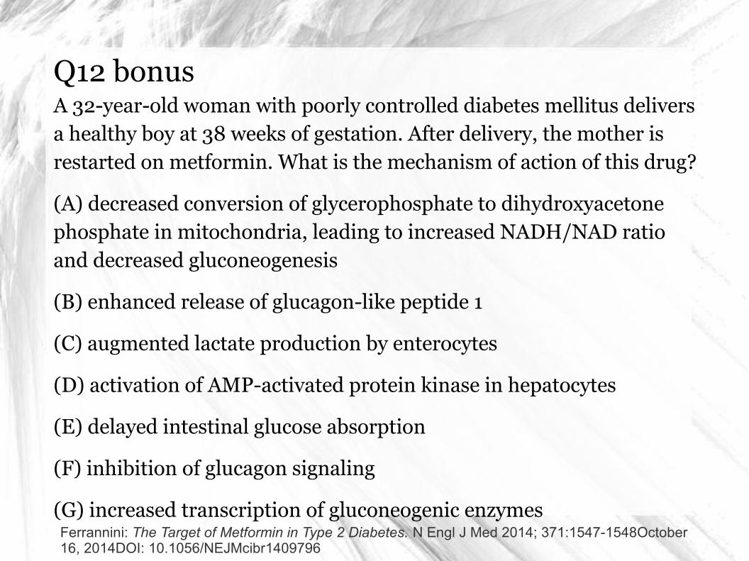
Q12 bonusA 32-year-old woman with poorly controlled diabetes mellitus delivers a healthy boy at 38 weeks of gestation. After delivery, the mother is restarted on metformin. What is the mechanism of action of this drug?
(A) decreased conversion of glycerophosphate to dihydroxyacetone phosphate in mitochondria, leading to increased NADH/NAD ratio and decreased gluconeogenesis
(B) enhanced release of glucagon-like peptide 1
(C) augmented lactate production by enterocytes
(D) activation of AMP-activated protein kinase in hepatocytes
(E) delayed intestinal glucose absorption
(F) inhibition of glucagon signaling
(G) increased transcription of gluconeogenic enzymesFerrannini: The Target of Metformin in Type 2 Diabetes. N Engl J Med 2014; 371:1547-1548October 16, 2014DOI: 10.1056/NEJMcibr1409796
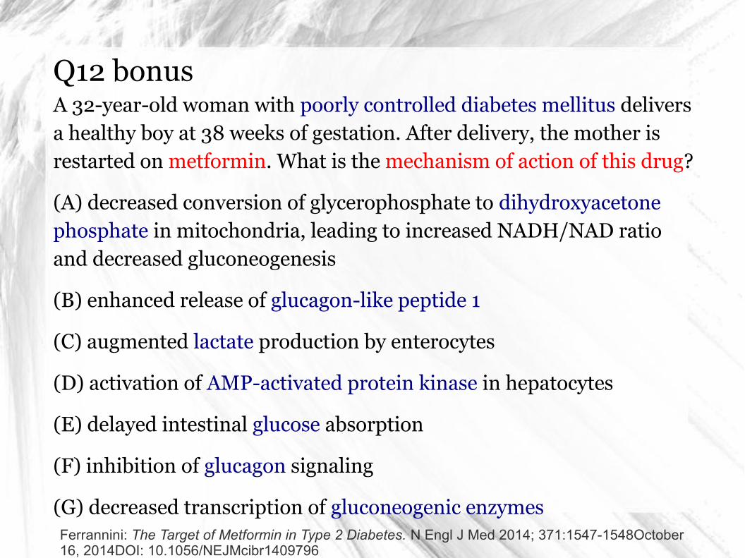
Q12 bonusA 32-year-old woman with poorly controlled diabetes mellitus delivers a healthy boy at 38 weeks of gestation. After delivery, the mother is restarted on metformin. What is the mechanism of action of this drug?
(A) decreased conversion of glycerophosphate to dihydroxyacetone phosphate in mitochondria, leading to increased NADH/NAD ratio and decreased gluconeogenesis
(B) enhanced release of glucagon-like peptide 1
(C) augmented lactate production by enterocytes
(D) activation of AMP-activated protein kinase in hepatocytes
(E) delayed intestinal glucose absorption
(F) inhibition of glucagon signaling
(G) decreased transcription of gluconeogenic enzymesFerrannini: The Target of Metformin in Type 2 Diabetes. N Engl J Med 2014; 371:1547-1548October 16, 2014DOI: 10.1056/NEJMcibr1409796
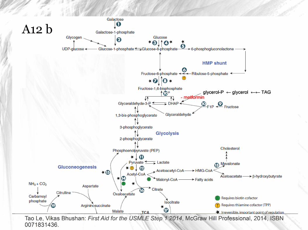
A12 b
Ferrannini: The Target of Metformin in Type 2 Diabetes. N Engl J Med 2014; 371:1547-1548October 16, 2014DOI: 10.1056/NEJMcibr1409796
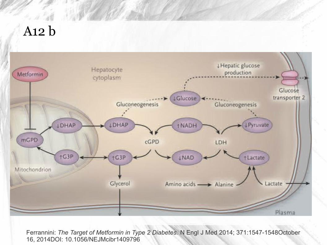
A12 b
Ferrannini: The Target of Metformin in Type 2 Diabetes. N Engl J Med 2014; 371:1547-1548October 16, 2014DOI: 10.1056/NEJMcibr1409796
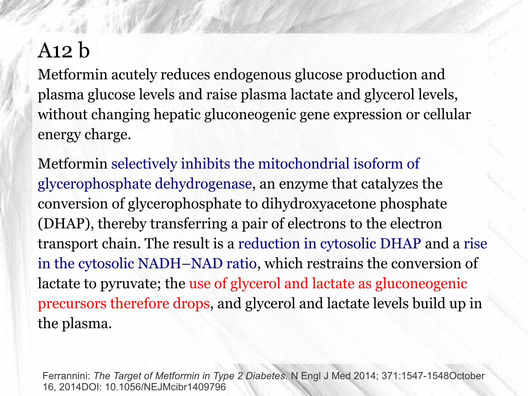
Metformin acutely reduces endogenous glucose production and plasma glucose levels and raise plasma lactate and glycerol levels, without changing hepatic gluconeogenic gene expression or cellular energy charge.
Metformin selectively inhibits the mitochondrial isoform of glycerophosphate dehydrogenase, an enzyme that catalyzes the conversion of glycerophosphate to dihydroxyacetone phosphate (DHAP), thereby transferring a pair of electrons to the electron transport chain. The result is a reduction in cytosolic DHAP and a rise in the cytosolic NADH–NAD ratio, which restrains the conversion of lactate to pyruvate; the use of glycerol and lactate as gluconeogenic precursors therefore drops, and glycerol and lactate levels build up in the plasma.
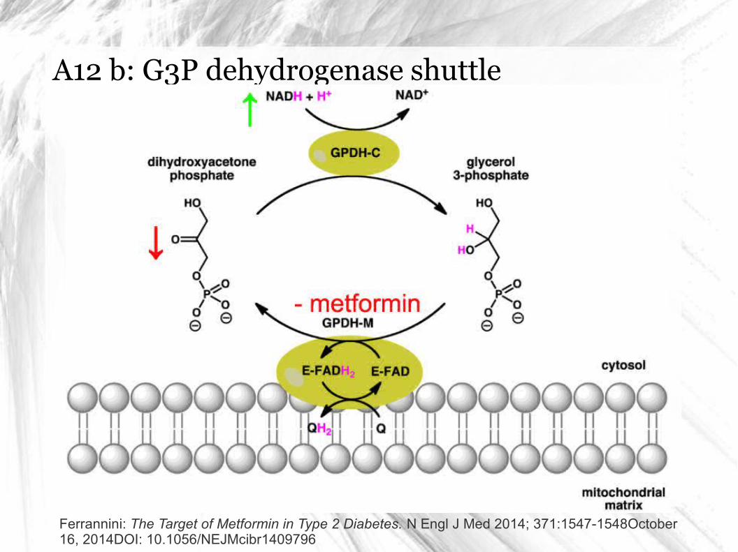
A12 b: G3P dehydrogenase shuttle
Ferrannini: The Target of Metformin in Type 2 Diabetes. N Engl J Med 2014; 371:1547-1548October 16, 2014DOI: 10.1056/NEJMcibr1409796
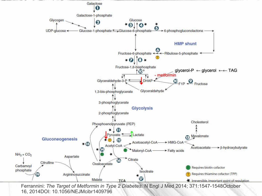
A12 b
Ferrannini: The Target of Metformin in Type 2 Diabetes. N Engl J Med 2014; 371:1547-1548October 16, 2014DOI: 10.1056/NEJMcibr1409796
Děkuji za pozornost a diskusi
FB: USMLE @ Masaryk
Marek Čierny (324602 at mail.muni.cz)
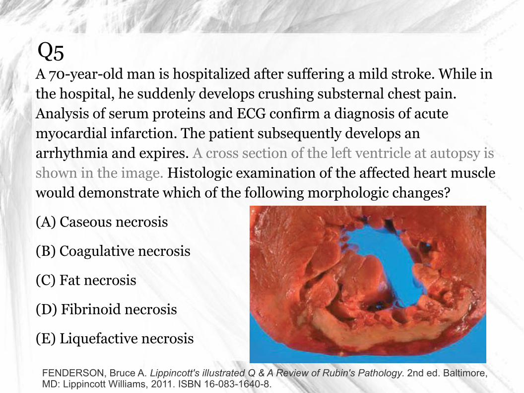
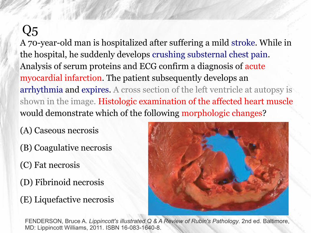
Q5A 70-year-old man is hospitalized after suffering a mild stroke. While in the hospital, he suddenly develops crushing substernal chest pain. Analysis of serum proteins and ECG confirm a diagnosis of acute myocardial infarction. The patient subsequently develops an arrhythmia and expires. A cross section of the left ventricle at autopsy is shown in the image. Histologic examination of the affected heart muscle would demonstrate which of the following morphologic changes?
(A) Caseous necrosis
(B) Coagulative necrosis
(C) Fat necrosis
(D) Fibrinoid necrosis
(E) Liquefactive necrosis
FENDERSON, Bruce A. Lippincott's illustrated Q & A Review of Rubin's Pathology. 2nd ed. Baltimore, MD: Lippincott Williams, 2011. ISBN 16-083-1640-8.
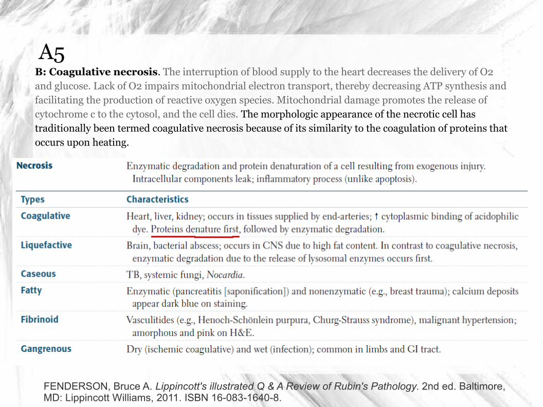
A5B: Coagulative necrosis. The interruption of blood supply to the heart decreases the delivery of O2 and glucose. Lack of O2 impairs mitochondrial electron transport, thereby decreasing ATP synthesis and facilitating the production of reactive oxygen species. Mitochondrial damage promotes the release of cytochrome c to the cytosol, and the cell dies. The morphologic appearance of the necrotic cell has traditionally been termed coagulative necrosis because of its similarity to the coagulation of proteins that occurs upon heating.
FENDERSON, Bruce A. Lippincott's illustrated Q & A Review of Rubin's Pathology. 2nd ed. Baltimore, MD: Lippincott Williams, 2011. ISBN 16-083-1640-8.