Embed Size (px)
Citation preview

Developmental Biology 347 (2010) 1–8
Contents lists available at ScienceDirect
Developmental Biology
j ourna l homepage: www.e lsev ie r.com/deve lopmenta lb io logy
Review
Developing in vivo biophysics by fishing for single molecules
Xi Wang a, Thorsten Wohland a,⁎, Vladimir Korzh b,c,⁎a Department of Chemistry, National University of Singapore, Singaporeb Cancer and Developmental Cell Biology Division, Institute of Molecular and Cell Biology, A-STAR, Singaporec Department of Biological Sciences, National University of Singapore, Singapore
⁎ Corresponding authors. V. Korzh is to be contactedCell Biology Division, Institute of Molecular and Cell Bio
E-mail address: [email protected] (V. Korzh).
0012-1606/$ – see front matter © 2010 Elsevier Inc. Adoi:10.1016/j.ydbio.2010.08.004
a b s t r a c t
a r t i c l e i n f oArticle history:Received for publication 30 April 2010Revised 27 July 2010Accepted 3 August 2010Available online 10 August 2010
Keywords:SMTFCSFRETTIRFZebrafishMedakaC. elegans
Single-molecule techniques (SMT) provide the possibility to quantitatively analyze the action of singlemolecules. SMTs can resolve the distribution of states of an ensemble of molecules, collecting informationthat is otherwise not accessible by typical ensemble techniques. Until now, the application of SMTs indevelopmental biology was limited. Several recent studies illustrate the possibility to investigate thebehavior of single biological molecules in invertebrates such as Caenorhabditis elegans and transparentembryos of model teleosts. These studies have paved the way for the application of fluorescence-based SMTs,e.g. fluorescence correlation spectroscopy, fluorescent energy transfer, or single particle tracking, indevelopmental biology. This review aims to define SMTs applicable in developmental biology, and discussproperties of an ideal animal model.
at Cancer and Developmentallogy, A-STAR, Singapore.
ll rights reserved.
© 2010 Elsevier Inc. All rights reserved.
Introduction
Until recently, studying the biological properties of proteins waslimited to the analysis of molecules in complexes; crystallography orelectron microscopy, for example, could only reveal the structuraldetails of molecules in a static state without any kinetic or dynamicinformation. With the recent inception of single-molecule techniques(SMT), there is now the possibility to analyze the properties ofindividual molecules with good spatial and temporal resolution. Theapplication of SMTs has been extremely successful in vitro, howeverthese methods are only beginning to be applied in vivo. Previously,even when SMT approaches are applied to analysis of biologicalphenomena in living cells, theywere used to study events taking placeunder in vitro conditions, such as in test tubes or in cell cultures.
The full potential of SMT for live cell imaging, particularly in liveanimals, remains underutilized. To a large extent, this derived fromthe interdisciplinary gap previously isolating physicists and biolo-gists: physicists develop sophisticated instruments that permitanalysis at a single-molecule level, but lack the knowledge of livingsystems; and biologists, capable of maintaining and manipulatingliving cells or model animals, are unable to adapt these models to SMTdue to the lack of access and/or skills to use these instruments. Now,
as the interdisciplinary gap between physics and biology is beingbridged, the possibility to accurately assess and measure the behaviorof biological molecules in vivo is within reach. The critical demand tojoin the two disciplines was likely aided by the fact that many SMTsare based on fluorescence, which is particularly suitable for studyingliving organisms.
Several important discoveries have been made since SMT firstbecame available. In the 1970s, a single fluorescent molecule wasdetected in solution (Hirschfeld, 1976) and provided the first proof-of-principle that single-molecule analysis was possible. This wasfollowed by the application of natural fluorescent proteins (FP) fromthe jellyfish Aequorea Victoria, and their genetically modifiedderivatives, for cell biological studies (Shimomura et al., 1962; Chalfieet al., 1994; Ormo et al., 1996). This later led to a major effort indeveloping novel types of FPs from other marine organisms, such ascorals and sea anemones, with improved or different spectralcharacteristics (Campbell et al., 2002; Labas et al., 2002; Shaneret al., 2004; Shcherbo et al., 2007). As a result, the usefulness of FP forSMTs, such as fluorescence correlation spectroscopy (FCS), was soonrecognized (Terry et al., 1995).
When kept in culture, even genetically identical cells are ratherheterogeneous. Thus, it is evident that cells from multicellularorganisms are very distinct in terms of their biological activity.What remains less clear is whether the intrinsic basic properties ofbiological macromolecules vary as much as the biological activity ofcells maintained under in vitro conditions versus those cells in the

2 X. Wang et al. / Developmental Biology 347 (2010) 1–8
living organism. In order to better understand this, a comparativeanalysis using SMTs on cells in culture and in the multicellularorganismwas required. This was achieved using several fluorescence-based single-molecule detection techniques to study the biologicalprocesses taking place in cell culture in vitro and in vivo in transparentanimals, such as worms (Caenorhabditis elegans), or transparentembryos of model vertebrates, such as zebrafish (Danio rerio) andmedaka (Oryzias latipes). These pioneering studies were largelyfocused on establishing the possibility to use SMT approaches for invivo studies. Only now is the complexity of biological processes at thesingle-molecule level in multicellular organisms beginning to beunraveled. Here, we review the applications for which SMTs areemployed in vivo.
SMT for in vivo developmental studies
Fluorescence correlation spectroscopy
Fluorescence correlation spectroscopy (FCS, Fig. 1) was developedalmost 40 years ago (Magde et al., 1972). FCS is a very sensitivetechnique that depends on the statistical analysis of fluctuations influorescence measured from a small observation volume. Thefluctuations contain the information about the dynamics of themolecules responsible for the fluorescence emission. Any molecularprocess which changes the fluorescence in the observation volumecan thus be observed and can be analyzed via correlation functions.The normalized autocorrelation function (ACF) is defined as:
G τð Þ = hF tð Þ⋅F t + τð ÞihF tð Þi2
Fig. 1. Fluorescence correlation spectroscopy (FCS). (A) The laser focus of a confocal microexcitation this can be achieved up to 50–100 μm depth. (B) Fluorescently labeled molecules mfluorescence signal thus contains information about the number of molecules and their mtreatment of the data) is calculated to extract the average behavior of the molecules. Theconcentration, respectively. The resulting autocorrelation curve is fitted to an appropriate m(D, E, F) Typical changes seen in an ACF for the change in different parameters. (D) Diffcharacteristic changes of the shape of the ACF. (E) A change in concentration will change thcharacteristic time for a motion will change the width of the ACF; the slower the movemen
where h:::i denotes time average and F(t) is the fluorescence intensityat time t. Once autocorrelation analysis is performed, the experimen-tal ACF curves are properly fitted using the appropriate mathematicalmodel for the process under investigation (Krichevsky and Bonnet,2002; Bacia and Schwille, 2007; Shi and Wohland, 2010). Commonly,the diffusion of particles is observed, for which the appropriate fittingmodel for FCS is well known. Adjustments in the diffusion coefficientdue to changes in the local environment of a particle or its binding canthen be monitored and evaluated.
Bymeasuring fluctuations in fluorescence of single molecules, datadescribing the dynamic behavior of these emitters is obtained andused to calculate their concentration and diffusion coefficients. Theseparameters provide enough information to derive secondary para-meters including dissociation constants and stoichiometry of binding(Rauer et al., 1996; Van Craenenbroeck and Engelborghs, 1999;Wohland et al., 1999), aggregation and transient binding times(Starchev et al., 1998; Eggeling et al., 2009), and protein interactionwith membrane components and, in particular, its lipids (Pramanikand Rigler, 2001; Bacia et al., 2004; Kahya et al., 2004; Briddon andHill, 2007).
Recently, FCS has been applied to in vivo analyses of variousproblems of developmental biology and physiology. It was firstintroduced to investigate molecular dynamics in live organisms, aswell as to determine flow direction and velocities in zebrafish bloodvessels (Pan et al., 2007b). The early use of FCS analysis to determinediffusion coefficients began with ex vivo experiments. Medakaembryos were dissected and the dynamic properties of greenfluorescent protein (GFP)-labeled nuage proteins were analyzed inprimordial germ cells using FCS and fluorescence recovery afterphotobleaching (FRAP) (Nagao et al., 2008). These approaches werethen employed to study the localization and redistribution of
scope is positioned at a point of interest within the living sample. Using one-photonoving through the confocal volume give rise to fluorescence intensity fluctuations. Theovement. (C) The autocorrelation function (ACF) of the intensity signal (a statisticalwidth and amplitude of the ACF provide information about molecular movement andodel function to extract the characteristic time scales and concentration of the system.erent modes of motion, including different modes of diffusion or flow will result ine amplitude of the ACF; the higher the concentration, the lower the amplitude. (F) Thet, the wider the ACF.
3X. Wang et al. / Developmental Biology 347 (2010) 1–8
GFP-labeled NMY-2 and PAR-2 proteins during the asymmetric firstdivision of C. elegans embryos (Petrasek et al., 2008), and later, toinvestigate chromatin binding protein dynamics during developmentof the Drosophila embryo (Bhattacharya et al., 2009).
In parallel, attempts were made to adapt this approach for the invivo analysis of blood flow and other biological processes intransparent zebrafish embryos. A combined laser scanning confocalmicroscopy (LSCM) and FCS system for uncompromised imaging andspectroscopy measurements was developed. Scanning the laser beamwith a defined speed and direction was used to measure the flowdirection in developing capillaries of zebrafish embryos at an optimalresolution of at least 3 μm (Pan et al., 2007a,b; Korzh et al., 2008; Panet al., 2009). Subsequently, this approach was extended to determineblood flow velocities with high spatial resolution, and together, thediffusion coefficients of cytosolic and membrane-bound enhancedGFP (EGFP)-labeled proteins in muscle and neurons of living embryoswas evaluated (Shi et al., 2009b).
Finally, FCS has been used to investigate in vivo protein-proteininteractions in two different applications. First, single wavelengthfluorescence cross-correlation spectroscopy was used to measure theinteraction of Cdc42, a small Rho-GTPase, and IQGAP1, an actin-bindingscaffolding protein, in cultured cells and zebrafish embryos. The resultsemphasise that bimolecular interactions depend on thebiological systemunder investigation, and that analyses need to be performed underphysiologically-relevant conditions (Shi et al., 2009a). In the secondapplication, static-volume, two-focus and dual-color scanning fluores-cence correlation spectroscopywas combined to quantify themobility offibroblast growth factor receptors, Fgfr1 and Fgfr4, in cell membranes ofliving zebrafish embryos and determine their in vivo binding affinities toFgf8. Furthermore, this technique was used to demonstrate that a freelydiffusing morphogen (FGF8) can set up concentration gradients in acomplex multicellular tissue by a simple source-sink mechanism (Rieset al., 2009; Yu et al., 2009).
Up to now, the application of FCS in living organisms is stillchallenging due to the thick tissue induced light scattering. Takingadvantage of the recently developed microscopic techniques, a numberof custom-made FCS microscopic systems have been realized toovercome this problem. One of these extensions to FCS is the use oftwo-photon excitation. In this case, the penetration depth can be greatlyincreased. Combined with FCS, it may enable detection of proteindynamics at the singlemolecule level up to several hundreds ofmicronsinto the tissue of living organisms (Helmchen and Denk, 2005) withadvantages in respect to reduced out-of-focus photobleaching and lessphototoxicity to cells. Another method capable of monitoring biomo-lecular behavior within thick tissue is selective plane illuminationmicroscopy (SPIM), which visualizes a specimen with a thin light sheetorthogonal to the objective and parallel to the focal image plane(Huisken et al., 2004). Unlike conventional confocal microscopy inwhich the entire specimen is exposed for each plane, only one plane isilluminated and observed at a time in SPIM, thus greatly reducing theradiation exposure to the specimen and significantly increasing theperiod of observation. Recently, Wohland and colleagues have reportedSPIM-FCS to be used for concentration and diffusion coefficient imagingin non-homogeneous samples and zebrafish embryos (Wohland et al.,2010).
Single particle tracking
Single particle tracking (SPT) is the observation of the motion ofindividual particles within a medium. The coordinate of a certainparticle over a series of time steps is referred to as a trajectory. Theinformation garnered from this trajectory can then provide usefulinformation about the mechanisms that drive and constrain theparticle motions.
By now, in vitro SPT techniques have been well established aspowerful biochemical tools that can reveal the dynamic nature of
proteins and nucleic acids (Joo et al., 2008). Although possessing greatadvantages for the detection of subpopulation behaviors in complexenvironments, the application of SPT in vivo is still limited. One majorholdback is the low signal-to-noise ratio (SNR), which is primarilycaused by cellular autofluorescence. This optically challengingsituation can be improved by applying particular illuminationschemes such as total internal reflection or selective planeillumination.
In Total Internal Reflection (TIR) microscopy, light exits themicroscope objective as a highly inclined, parallel light beam. Whenthe beam encounters the glass-water interface of the cover slide, all ofthe light will be reflected if the angle of inclination is higher than thecritical angle θc; this angle is determined by the refractive indices ofwater and glass (sinθc=nwater/nglass). However, at the glass-waterinterface, a thin evanescent wave is formed. This evanescent wave isused in Total Internal Reflection Fluorescence (TIRF) (Fig. 2) toilluminate and excite fluorophores immediately adjacent to the glass-water interface (Axelrod, 2001). When TIRF is applied to a biologicalspecimen, it illuminates a very narrow layer above the glass slide (lessthan 100 nm) where a biological specimen is attached, such as a cellmembrane. In addition, the adjacent cytoplasmic region and anyprotein movement in this area is accessible for analysis. This allowsthe user to observe the fluorescence of a single molecule as it moveswithin the membrane and/or adjacent region, or as it interacts withother labeled molecules.
YFP-C10H-Ras is widely used as a model molecule for proteinsanchored in the cytoplasmic leaflet of the plasma membrane and,because it is biologically inert, it serves as a probe for detectingchanges in the organization of the plasma membrane. In combina-tion with wide-field microscopy, TIRF microscopy was used in acomparative analysis of membrane distribution of this molecule in azebrafish cell line (ZF4, in vitro), in primary embryonic stem cells(ex vivo), and cells of epithelium of 2-day-old zebrafish embryos (invivo) to reveal the size variation of microdomains (lipid rafts) in allthese different cells (Schaaf et al., 2009). Here a home-builtmicroscope equipped with a 100x oil-immersion TIRF objective(NA 1.45, Nikon, Tokyo, Japan) was used. This provided a proof-of-principle that TIRF could be used for in vivo studies of superficialcells of the zebrafish embryo. Based on the results of this study, theauthors proposed that this method can be applied to othermembrane proteins, and can even be extended to cytoplasmic ornuclear proteins by using a variant of TIRF based on the highlyinclined thin illumination technology (Konopka and Bednarek,2008; Tokunaga et al., 2008).
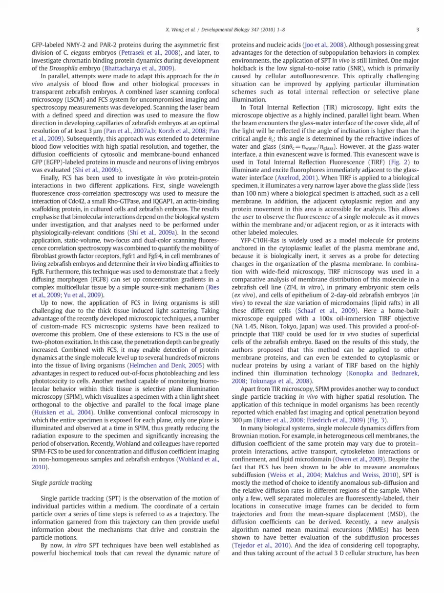
Apart from TIR microscopy, SPIM provides another way to conductsingle particle tracking in vivo with higher spatial resolution. Theapplication of this technique in model organisms has been recentlyreported which enabled fast imaging and optical penetration beyond300 μm (Ritter et al., 2008; Friedrich et al., 2009) (Fig. 3).
In many biological systems, single molecule dynamics differs fromBrownianmotion. For example, in heterogeneous cell membranes, thediffusion coefficient of the same protein may vary due to protein–protein interactions, active transport, cytoskeleton interactions orconfinement, and lipid microdomain (Owen et al., 2009). Despite thefact that FCS has been shown to be able to measure anomaloussubdiffusion (Weiss et al., 2004; Malchus and Weiss, 2010), SPT ismostly the method of choice to identify anomalous sub-diffusion andthe relative diffusion rates in different regions of the sample. Whenonly a few, well separated molecules are fluorescently-labeled, theirlocations in consecutive image frames can be decided to formtrajectories and from the mean-square displacement (MSD), thediffusion coefficients can be derived. Recently, a new analysisalgorithm named mean maximal excursions (MMEs) has beenshown to have better evaluation of the subdiffusion processes(Tejedor et al., 2010). And the idea of considering cell topography,and thus taking account of the actual 3 D cellular structure, has been

Fig. 2. Total internal reflection fluorescence (TIRF) microscopy. (A) A TIRF microscopy is used in which an evanescent excitation field at the coverglass-specimen interface excitesmolecules within b100 nm of the coverglass. Since the evanescent wave does not reach into the bulk of the tissue, no other molecules are excited, resulting in a low background forTIRF. (B) Schaaf et al. recently mounted a living embryo to posit the apical membranes of the outer cell layer of the embryo's skin within the evanescent field (Schaaf et al., 2009). (C)The trajectory of single fluorescent molecules can be tracked over time in the embryo. (D) By connectivity analysis between consecutive images, the squared displacements of themolecules were determined for each time lag used. From this graph the mobility and changes in mobility can be evaluated for each molecule.
4 X. Wang et al. / Developmental Biology 347 (2010) 1–8
introduced to achieve higher accuracy and to account for artifactsintroduced by an unwarranted assumption of sample flatness which isoften not fulfilled on cell membranes (Adler et al., 2010). These 3D SPTtechniques promise to give a more accurate picture of single moleculetrajectories and thus provide more information on the underlyingmechanisms.
Compared to FCS, SPT provides direct information about trajecto-ries of individual molecules which can yield essential clues to revealthe mechanism causing subdiffusion in biological systems. However,SPT is only suitable for relatively low fluorescent protein expressionlevels and the statistical analysis is limited unless a large number ofmolecules are tracked in each experiment. Finally, SPT in threedimensions remains difficult due to the generally rapid diffusion aswell as limitations in determination of particle z positions. In this area,SPIM has shown great potential (Fig. 3). In future, by achieving therequirements of high spatial and temporal resolution, high contrastand high imaging rates, SPIM may allow the direct tracking of singleparticles within living organisms in full three dimensions (Ritter et al.,2008).
Single-molecule Fluorescence Resonance Energy Transfer (smFRET)
Fluorescence Resonance Energy Transfer (FRET) takes place via adipole-dipole interaction between a donor and an acceptor fluor-ophore when they are within 10 nm of each other (Fig. 4). Theefficiency of energy transfer, E, between these two flurophores isgiven by:
E =R60
R60 + R6
where R is the distance between the donor and acceptor, and R0 is thedistance at which 50% of the energy is transferred, and is a function ofthe properties of the dyes that are used (Liu et al., 2008). A smallchange in distance between the two sites of a biologicalmoleculewheredonor and acceptor are attached can result in a sizeable change in E.Therefore, structural changes of biological molecules or relative motionbetween two different molecules can be detected via FRET changes.
In biological studies, a key application of the FRET principle isthrough the use of FRET-biosensors, which are usually fusion proteinsof enhanced cyan fluorescent protein (ECFP) and enhanced yellowfluorescent protein (EYFP) (or other appropriate pairs, such as EGFPand amonomeric red fluorescent protein) linked by a sensory domain.This domain responds to changes in certain cellular parameters by aconformational modification, leading to an altered FRET signal. Usingappropriate sensory domains, several second messenger moleculesand signaling processes have been studied in vivo and ex vivoincluding: recording cAMP signaling (Lissandron et al., 2007; Shaferet al., 2008); determining the spatiotemporal pattern of caspaseactivation in Drosophila (Takemoto et al., 2007); imaging dynamicintracellular Ca2+ changes during gastrulation of the zebrafish(Tsuruwaka et al., 2007); monitoring cation channel activation inzebrafish (Richler et al., 2008); and analyzing the activity of GTPasesin Xenopus (Matthews et al., 2008). In addition, FRET has also beensuccessfully used to monitor protein-protein interactions in living C.elegans (Wagner et al., 2009) and conformational changes in the Na+/Ca2+ exchanger in zebrafish (Xie et al., 2008).
While FRET has proven to be a powerful tool to measureinteractions and conformations, it only provides the ensembleaverage of many molecules. Therefore, extending this approach tothe single-molecule level would reveal more information; forexample, it would provide information on heterogeneous
Fig. 3. Selective plane illumination microscopy (SPIM). In SPIM, fluorescence excitationanddetectionare split into twodistinctoptical paths. The illuminationaxis is orthogonal tothe detection axis. A microscope objective lens and common widefield optics are used toimage the sample onto a CCD camera. A typical SPIM setup consists of the samplemounting, illumination and detection units. Samplemounting and positioning are neededto position the biological sample within the illumination and detection optics. Theillumination optics are designed to illuminate a very thin sheet around the focal plane ofthe detection objective. The fluorescence from the sample is imaged onto one or morecameras by the detection optics. The advantage of SPIM is that all parts of the embryoilluminated are actually observed, thus limiting the light dose necessary for imaging awhole specimen. Compare, for example, confocal microscopy in which for each imagepoint a large portion of the specimen has to be illuminated. This allows SPIM to takemoremeasurements per time unit, aswell asmore overall measurements per sample comparedto confocal microscopy. When using a fast camera single particle tracking, FCS and othersingle-molecule techniques can be realized with this setup.
5X. Wang et al. / Developmental Biology 347 (2010) 1–8
subpopulations of molecules or their intermediate states duringdynamic transitions that would usually be hidden by ensembleaveraging. Recent advances in illumination, detection and lasertechnologies allow for the observation of biological processes at thesingle-molecule level using FRET (smFRET). This technique mayprovide the answer to various biological questions for both intramo-lecular interactions, such as conformational dynamics of DNA, RNA,and proteins, as well as intermolecular interactions, such as proteinswith DNA, RNA, and other ligands. However, most of these studies areconducted using purified components in vitro and, as such, thesemeasurements may lack physiological relevance. Applying smFRET inan in vivo setting could address this issue, but such live-cellapplications are challenging. Recently, smFRET was combined withsingle-particle tracking in live cells to detect the conformation ofindividual proteins using TIRF microscopy with spectrally-resolveddetection for smFRET analysis (Sakon and Weninger, 2010). Thisrevealed how individual SNARE proteins, fluorescently-labeled withFRET donor and acceptor, rapidly incorporate into folded complexes atthe cell membrane. A similar approach was used to monitor proteinfolding stability and dynamics in cell culture, and these findingsallowed the authors to detect the influence of a crowded cellenvironment on protein stability and folding rate (Ebbinghaus et al.,2010). It seems that the next logical step would be to extend thisapproach to the transparent embryos of model teleosts, similar towhat has previously been employed for FCS.
A comparison between the fluorescence techniques
To date by far not all possible fluorescence techniques have beenadvanced into animals. Therefore we restrict our comparison here, to
the above mentioned three SMTs, FCS, SPT and smFRET, which havebeen used in organisms. FRET or smFRET is a very popular techniqueto measure molecular interactions. It has a range of advantages notmatched by the other two techniques. Firstly, FRET allows not only themeasurement of interactions but can as well yield information on thedistance of the two interacting proteins and thus on structural detailsof the complex. In addition, FRET can be conducted on widelycommercially available microscopes and does not require customizedinstrumentation. And this is even partly true for smFRET which can beconducted on a total internal reflection fluorescence (TIRF) micros-copy setup (Sakon andWeninger, 2010). However, for measurementsinside an embryo some customization might be advantageous. Thedisadvantage of FRET is that it is dependent on the distance andorientation of the donor and acceptor fluorophores. It thus can lead tofalse negative results due to unfavorable fluorophore configurations.This is not a problem in fluorescence cross-correlation spectroscopy, atwo-color modality of FCS, which requires only the concertedmovement of two labeled particles to determine their interactionand is independent of fluorophore distance and orientation. Inaddition, FCS yields information on concentrations and moleculardynamics, in particular the diffusion coefficient of labeled particles. Bymeasuring these parameters many secondary parameters, includingligand–receptor binding, protein association, aggregation events, ortransport and biomolecule trafficking have been determined for awide range of biological problems (Mutze et al., 2010). Themeasurements are fast (b 1 min down to seconds), have singlemolecules sensitivity and commercial instruments are widely avail-able. It should be noted though that despite its sensitivity, FCS doesnot follow single molecules, but due to its inherent data treatment bycorrelation functions gives statistical averages of diffusion coeffi-cients. If it is necessary to access the distribution of diffusioncoefficients for one particle in time or for many particles, SPT is thebetter option. SPT truly follows a single particle, with resolutions onthe order of 10 nm or less. It can monitor changes in diffusioncoefficient over time and space, which would be averaged in FCS, andcan give more detailed information about the molecular system(Pinaud et al., 2009; Leptihn et al., 2009). It should be noted that iftrajectories can be followed for sufficiently long times, the trajectoriescould even give a high resolution picture of the underlying biologicalstructure (if it is sufficiently static and does not change over the sametime as the single molecule movement) without the need of highlabeling densities as required in super-resolution techniques. Thedisadvantage of SPT is that it has to work at very low concentrationsand needs very photostable fluorophores, which often excludes theuse of the less photostable genetically encoded fluorescent proteins,which are commonly used in FRET and FCS. When these conditionsare fulfilled then SPT, especially the 3D implementation, has greatpotential to yield new insights into biology.
The interested user has to carefully balance the need forfluorophores and labeling strategies with the need for temporal andspatial resolution to decide on which technique is best for hisparticular experiments. However, an approach using several comple-mentary techniques to obtain a consistent overall picture of thesystem under investigation is advisable.
Will other SMTs be applicable for in vivo studies?
Some non-optical SMTs, such as electron microscopy or atomicforce microscopy, depend on the fixation of biological materials, andthese techniques cannot be used for in vivo studies. Other non-opticaltechniques, such as scanning ion conductance microscopy (SICM),may at least for now remain limited to the analysis of cells in culture(Korchev et al., 2000; Novak et al., 2009) due to the spatial limitationsimposed by in vivo analysis. Since this technique is not limited by thetransparent nature of the embryos, it may be adapted for studying the

6 X. Wang et al. / Developmental Biology 347 (2010) 1–8
membranes of superficial cells of the more robust Xenopus embryos asopposed to the fragile teleost embryos.
The recent invention of super-resolution fluorescence microscopytechniques allows locating fluorescently-labeled probes with anaccuracy of just tens of nanometers, far surpassing the traditionaldiffraction resolution limit. The rapid pace of development in thesetechniques gives them the ability to image three-dimensionalstructures, measure interactions by multicolor colocalization, andrecord dynamic processes in living cells at the nanometer scale(Huang et al., 2009). However, it should be noted that to reach super-resolution a concomitant high labeling density has to be reachedwhose effects on the biology of organisms has not been investigatedyet. Nevertheless, it is anticipated that these super-resolutionfluorescence microscopy techniques may soon find their way intothe area of in vivo studies at the single-molecule level.
Is there an ideal model vertebrate animal for SMTs?
The ideal invertebrate species for SMT applications is alreadyavailable—C. elegans. Based on its properties as a model species, onecould instantly define the properties for an ideal vertebrate species forSMTs. For it to be reminiscent of C. elegans, it would need to berelatively small and transparent, even as an adult. Ideally, similar tozebrafish, it must be able to be manipulated for genetic anddevelopmental studies, easy to maintain, and able to produce alarge number of externally-fertilized eggs by natural spawning incaptivity. As mentioned earlier, even the wild-type zebrafish, despiteits limitations, is useful for some SMT applications. The situationcertainly improves with introduction of the compound pigmentation
Fig. 4. FRET based biosensor. FRET is based on the nonradiative energy transfer from adonor fluorophore to an acceptor fluorophore through dipole–dipole interaction. Anecessary condition for FRET is that the emission spectrum of the donor overlaps withthe absorption spectrum of the acceptor. Due to the strong distance and orientationdependence of the dipole-dipole interaction, FRET pairs are conformationally sensitivein the Angstrom range. This sensitivity can be used, e.g., in the detection of protein-protein interactions if the two proteins are labeled with a donor-acceptor pair. In FRETbased biosensors, a ligand-induced conformational change alters the distance ororientation between two fluorescent proteins (donor and acceptor) linking with thesensory domain. Often fluorescent protein pairs, e.g. cyan and yellow fluorescentprotein (CFP and YFP), are used as donor-acceptor pairs. These FRET-capable FPs canthen convert themolecular event of interest into a detectable change in FRET efficiency.
mutant zebrafish which are transparent, as compared with its wild-type relative (White et al., 2008).
But is this the limit for transparent vertebrate species? Fortunately,the class of Teleostei includes the largest number of vertebrate species,and as adults they can typically be much smaller than zebrafish (andtherefore completely transparent), or predominantly transparent. Thesmallest vertebrate species of Teleostei from the peat marshes ofSumatra, the Paedocypris progenetica, belongs to the family Cyprinidae,(similar to zebrafish), and measure only 7.9 mm in length as an adult.According to its discoverers, this species has "a very rudimentaryskull", which leaves a large portion of the brain exposed (Kottelatet al., 2006), making it very promising for neurobiological studies invivo. This exposure of the brain is due to the fact that the skeleton ofPaedocypris is characterized by reduction and loss of the later-developing skeletal elements. Therefore, in many aspects, it resemblesthat of a larval/early juvenile stage of its close relatives, such aszebrafish (Britz and Conway, 2009). Currently, a crucial shortcomingin the use of this species is a lack of knowledge about its laboratorymaintenance, and an absence of any data that describes its geneticsand developmental biology.While it is obvious that a wealth of data inthese areas about the zebrafish could be used as a guide, the studies ofPaedocypris remain limited to a few initial morphologicalobservations.
The second smallest vertebrate, also belonging to Cyprinidae, isDanionella, which is represented by three rather similar species—D.mirifica, D. translucida, and D. dracula. All are found in Burma, and onaverage are 10 mm in length (Britz and Conway, 2009). These speciesare even closer in evolutionary terms to zebrafish, and there arehobbyists’ reports of D. translucida reproduction in captivity. Duringmating, the female lays several eggs that are probably about the samesize as that of zebrafish (Roberts, 1986). Unfortunately, similar toPaedocypris, these species of Danionella are currently not used in thelaboratory.
In the lineup of potential candidates for a species of choice forSMTs, there are several so called glass fishes, including Pristellamaxillaries (or the x-ray tetra), several species from a familyAmbassidae, including the Indian glassy fish, Parambassis ranga, anda less likely candidate (due to its aggressiveness), the glass catfish(Kryptopterus bicirrhis) (http://www.fishbase.org/home.htm).
Potential problems
As with any emerging field, in vivo developmental and physiolog-ical studies based on SMT face many challenges. The first problem isgrowth of the field. The link between physicists and biologists is stillrelatively fragile. This is reflected by the fact that most publishedstudies illustrate proof-of-principle and therefore out of necessity relyheavily on well-established microscopic systems and biologicalparadigms. Most of these techniques are far from being routinetechniques in developmental biology, and so far have not beeninstrumental in changing existing dogmas based on in vitro findings.This may change as a new breed of “developmental biophysicists”start to feel equally comfortable in the “devbio” and opticslaboratories, and begin to integrate knowledge generated in bothfields of science.
The second problem is the intracellular delivery of labeledcompounds. From a point of view of cell economics and in theabsence of compelling quantitative data, it could be assumed that theintracellular systems operate at almost full capacity. If any marginexists, it should be limited. Even with the embryo of anamniotes,which is provided with a great excess of various molecules and evenorganelles, only some of this material is ready for immediate use. Forexample, while the DNA injected into the embryo multiplies, thesituation appears different at the protein level. An evaluation of thebinding sites for housekeeping enzymes available in a teleost embryoblastoderm demonstrated 20% surplus capacity; that is, at any

7X. Wang et al. / Developmental Biology 347 (2010) 1–8
developmental stage, it seems to be somewhat limited (Klyachkoet al., 1982).
Therefore, one should be aware that in many instances an artificialsituation is created when there is a rapid increase in the amount of abiologicalmolecule followingmicroinjection. Often it results in a gain-of-function (GOF) phenotype, which creates an abnormal shift inestablished and physiological proportions, or in the stoichiometricratio of various biological molecules. Thus, projects aiming toquantitatively measure the ratio of various biologicals (e.g. ligandand receptor) based on introduction of some of these componentsinside the cell (usually labeled or tagged) should aim to maintain thephysiological ratio between the molecules being measured. This canbe achieved by proportionately depleting a pool of endogenousmolecules in parallel with the introduction of exogenous material. Inembryos of amniotes, this could be achieved by the loss-of-functionapproach (LOF) using morpholino-based antisense oligonucleotides.Maintenance of a “normal” or “wild type” phenotype after suchexperimental procedure could be used to gauge whether, underspecific experimental conditions, a ratio of molecules remains withina physiological range. Luckily, these fears may be unsubstantiated to alarge extent as the SMTs predominantly rely on as little labeledmaterial as possible, and in other situations no labeled material isrequired at all as measurement is based on autofluorescence (Korzhet al., 2008).
Acknowledgments
The V.K. laboratory in the IMCB is supported by the institutionalgrant from the Agency for Science, Technology and Research ofSingapore. T.W. was funded by the Biomedical Research Council ofSingapore (BMRC 07/1/21/19/488—R-143-000-351-305).
References
Adler, J., Shevchuk, A.I., Novak, P., Korchev, Y.E., Parmryd, I., 2010. Plasma membranetopography and interpretation of single-particle tracks. Nat. Methods 7, 170–171.
Axelrod, D., 2001. Selective imaging of surface fluorescence with very high aperturemicroscope objectives. J. Biomed. Opt. 6, 6–13.
Bacia, K., Schwille, P., 2007. Practical guidelines for dual-color fluorescence cross-correlation spectroscopy. Nat. Protoc. 2, 2842–2856.
Bacia, K., Scherfeld, D., Kahya, N., Schwille, P., 2004. Fluorescence correlationspectroscopy relates rafts in model and native membranes. Biophys. J. 87,1034–1043.
Bhattacharya, D., Talwar, S., Mazumder, A., Shivashankar, G.V., 2009. Spatio-temporalplasticity in chromatin organization in mouse cell differentiation and duringDrosophila embryogenesis. Biophys. J. 96, 3832–3839.
Briddon, S.J., Hill, S.J., 2007. Pharmacology under the microscope: the use offluorescence correlation spectroscopy to determine the properties of ligand–receptor complexes. Trends Pharmacol. Sci. 28, 637–645.
Britz, R., Conway, K.W., 2009. Osteology of Paedocypris, a miniature and highlydevelopmentally truncated fish (Teleostei: Ostariophysi: Cyprinidae). J. Morphol.270, 389–412.
Campbell, R.E., Tour, O., Palmer, A.E., Steinbach, P.A., Baird, G.S., Zacharias, D.A., Tsien, R.Y., 2002. A monomeric red fluorescent protein. Proc. Natl. Acad. Sci. U. S. A. 99,7877–7882.
Chalfie, M., Tu, Y., Euskirchen, G., Ward, W.W., Prasher, D.C., 1994. Green fluorescentprotein as a marker for gene expression. Science 263, 802–805.
Ebbinghaus, S., Dhar, A., McDonald, J.D., Gruebele, M., 2010. Protein folding stability anddynamics imaged in a living cell. Nat. Methods 7, 319–323.
Eggeling, C., Ringemann, C., Medda, R., Schwarzmann, G., Sandhoff, K., Polyakova, S.,Belov, V.N., Hein, B., von Middendorff, C., Schonle, A., Hell, S.W., 2009. Directobservation of the nanoscale dynamics of membrane lipids in a living cell. Nature457, 1159–1162.
Friedrich, M., Nozadze, R., Gan, Q., Zelman-Femiak, M., Ermolayev, V., Wagner, T.U.,Harms, G.S., 2009. Detection of single quantum dots in model organisms with sheetillumination microscopy. Biochem. Biophys. Res. Commun. 390, 722–727.
Helmchen, F., Denk, W., 2005. Deep tissue two-photon microscopy. Nat. Methods 2,932–940.
Hirschfeld, T., 1976. Optical microscopic observation of single small molecules. Appl.Opt. 15, 2965–2966.
Huang, B., Bates, M., Zhuang, X., 2009. Super-resolution fluorescence microscopy. Annu.Rev. Biochem. 78, 993–1016.
Huisken, J., Swoger, J., Del Bene, F., Wittbrodt, J., Stelzer, E.H., 2004. Optical sectioningdeep inside live embryos by selective plane illumination microscopy. Science 305,1007–1009.
Joo, C., Balci, H., Ishitsuka, Y., Buranachai, C., Ha, T., 2008. Advances in single-moleculefluorescence methods for molecular biology. Annu. Rev. Biochem. 77, 51–76.
Kahya, N., Scherfeld, D., Bacia, K., Schwille, P., 2004. Lipid domain formation anddynamics in giant unilamellar vesicles explored by fluorescence correlationspectroscopy. J. Struct. Biol. 147, 77–89.
Klyachko, O.S., Korzh, V.P., Gorgolyuk, S.I., Timofeev, A.V., Neyfakh, A.A., 1982.Nonuniform distribution of enzymes in fish eggs. J. Exp. Zool. 222, 137–148.
Konopka, C.A., Bednarek, S.Y., 2008. Variable-angle epifluorescence microscopy: a newway to look at protein dynamics in the plant cell cortex. Plant J. 53, 186–196.
Korchev, Y.E., Negulyaev, Y.A., Edwards, C.R., Vodyanoy, I., Lab, M.J., 2000. Functionallocalization of single active ion channels on the surface of a living cell. Nat. Cell Biol.2, 616–619.
Korzh, S., Pan, X., Garcia-Lecea, M., Winata, C.L., Wohland, T., Korzh, V., Gong, Z., 2008.Requirement of vasculogenesis and blood circulation in late stages of liver growthin zebrafish. BMC Dev. Biol. 8, 84.
Kottelat, M., Britz, R., Hui, T.H., Witte, K.E., 2006. Paedocypris, a new genus of SoutheastAsian cyprinid fish with a remarkable sexual dimorphism, comprises the world'ssmallest vertebrate. Proc. Biol. Sci. 273, 895–899.
Krichevsky, O., Bonnet, G., 2002. Fluorescence correlation spectroscopy: the techniqueand its applications. Rep. Prog. Phys. 65, 251–297.
Labas, Y.A., Gurskaya, N.G., Yanushevich, Y.G., Fradkov, A.F., Lukyanov, K.A., Lukyanov, S.A., Matz, M.V., 2002. Diversity and evolution of the green fluorescent proteinfamily. Proc. Natl. Acad. Sci. U. S. A. 99, 4256–4261.
Leptihn, S., Har, J.Y., Chen, J., Ho, B., Wohland, T., Ding, J.L., 2009. Single moleculeresolution of the antimicrobial action of quantum dot-labeled sushi peptide on livebacteria. BMC Biol. 7, 22.
Lissandron, V., Rossetto, M.G., Erbguth, K., Fiala, A., Daga, A., Zaccolo, M., 2007.Transgenic fruit-flies expressing a FRET-based sensor for in vivo imaging of cAMPdynamics. Cell Signal. 19, 2296–2303.
Liu, P., Ahmed, S., Wohland, T., 2008. The F-techniques: advances in receptor proteinstudies. Trends Endocrinol. Metab. 19, 181–190.
Magde, D., Elson, E., Webb, W., 1972. Thermodynamic fluctuations in a reacting system—measurement by fluorescence correlation spectroscopy. Phys. Rev. Lett. 29,705–708.
Malchus, N., Weiss, M., 2010. Elucidating anomalous protein diffusion in living cells withfluorescence correlation spectroscopy—facts and pitfalls. J. Fluoresc. 20, 19–26.
Matthews, H.K., Marchant, L., Carmona-Fontaine, C., Kuriyama, S., Larrain, J., Holt, M.R.,Parsons, M., Mayor, R., 2008. Directional migration of neural crest cells in vivo isregulated by Syndecan-4/Rac1 and non-canonical Wnt signaling/RhoA. Develop-ment 135, 1771–1780.
Mutze, J., Ohrt, T., Schwille, P., 2010. Fluorescence correlation spectroscopy in vivo.Laser Photon. Rev. (epub ahead of print).
Nagao, I., Aoki, Y., Tanaka, M., Kinjo, M., 2008. Analysis of the molecular dynamics ofmedaka nuage proteins by fluorescence correlation spectroscopy and fluorescencerecovery after photobleaching. FEBS J. 275, 341–349.
Novak, P., Li, C., Shevchuk, A.I., Stepanyan, R., Caldwell, M., Hughes, S., Smart, T.G.,Gorelik, J., Ostanin, V.P., Lab, M.J., Moss, G.W., Frolenkov, G.I., Klenerman, D.,Korchev, Y.E., 2009. Nanoscale live-cell imaging using hopping probe ionconductance microscopy. Nat. Methods 6, 279–281.
Ormo, M., Cubitt, A.B., Kallio, K., Gross, L.A., Tsien, R.Y., Remington, S.J., 1996. Crystalstructure of the Aequorea victoria green fluorescent protein. Science 273, 1392–1395.
Owen, D.M., Williamson, D., Rentero, C., Gaus, K., 2009. Quantitative microscopy:protein dynamics and membrane organisation. Traffic 10, 962–971.
Pan, X., Foo, W., Lim, W., Fok, M.H., Liu, P., Yu, H., Maruyama, I., Wohland, T., 2007a.Multifunctional fluorescence correlation microscope for intracellular and micro-fluidic measurements. Rev. Sci. Instrum. 78, 053711.
Pan, X., Yu, H., Shi, X., Korzh, V., Wohland, T., 2007b. Characterization of flow directionin microchannels and zebrafish blood vessels by scanning fluorescence correlationspectroscopy. J. Biomed. Opt. 12, 014034.
Pan, X., Shi, X., Korzh, V., Yu, H., Wohland, T., 2009. Line scan fluorescence correlationspectroscopy for three-dimensional microfluidic flow velocity measurements. J.Biomed. Opt. 14, 024049.
Petrasek, Z., Hoege, C., Mashaghi, A., Ohrt, T., Hyman, A.A., Schwille, P., 2008.Characterization of protein dynamics in asymmetric cell division by scanningfluorescence correlation spectroscopy. Biophys. J. 95, 5476–5486.
Pinaud, F., Michalet, X., Iyer, G., Margeat, E., Moore, H.P., Weiss, S., 2009. Dynamicpartitioning of a glycosyl-phosphatidylinositol-anchored protein in glycosphingo-lipid-rich microdomains imaged by single-quantum dot tracking. Traffic 10 (6),691–712.
Pramanik, A., Rigler, R., 2001. Ligand–receptor interactions in themembrane of culturedcells monitored by fluorescence correlation spectroscopy. Biol. Chem. 382,371–378.
Rauer, B., Neumann, E., Widengren, J., Rigler, R., 1996. Fluorescence correlationspectrometry of the interaction kinetics of tetramethylrhodamin alpha-bungar-otoxin with Torpedo californica acetylcholine receptor. Biophys. Chem. 58, 3–12.
Richler, E., Chaumont, S., Shigetomi, E., Sagasti, A., Khakh, B.S., 2008. Trackingtransmitter-gated P2X cation channel activation in vitro and in vivo. Nat. Methods5, 87–93.
Ries, J., Yu, S.R., Burkhardt, M., Brand, M., Schwille, P., 2009. Modular scanning FCSquantifies receptor–ligand interactions in living multicellular organisms. Nat.Methods 6, 643–645.
Ritter, J.G., Veith, R., Siebrasse, J.P., Kubitscheck, U., 2008. High-contrast single-particletracking by selective focal plane illumination microscopy. Opt. Express 16,7142–7152.
Roberts, T.R., 1986. Danionella translucida, a new genus and species of cyprinid fishfrom Burma, one of the smallest living vertebrates. Environ. Biol. Fish. 16, 231–241.

8 X. Wang et al. / Developmental Biology 347 (2010) 1–8
Sakon, J.J., Weninger, K.R., 2010. Detecting the conformation of individual proteins inlive cells. Nat. Methods 7, 203–205.
Schaaf, M.J., Koopmans, W.J., Meckel, T., van Noort, J., Snaar-Jagalska, B.E., Schmidt, T.S.,Spaink, H.P., 2009. Single-molecule microscopy reveals membrane microdomainorganization of cells in a living vertebrate. Biophys. J. 97, 1206–1214.
Shafer, O.T., Kim, D.J., Dunbar-Yaffe, R., Nikolaev, V.O., Lohse, M.J., Taghert, P.H., 2008.Widespread receptivity to neuropeptide PDF throughout the neuronal circadian clocknetworkofDrosophila revealedbyreal-timecyclicAMP imaging.Neuron58, 223–237.
Shaner, N.C., Campbell, R.E., Steinbach, P.A., Giepmans, B.N., Palmer, A.E., Tsien, R.Y.,2004. Improved monomeric red, orange and yellow fluorescent proteins derivedfrom Discosoma sp. red fluorescent protein. Nat. Biotechnol. 22, 1567–1572.
Shcherbo, D., Merzlyak, E.M., Chepurnykh, T.V., Fradkov, A.F., Ermakova, G.V., Solovieva, E.A., Lukyanov, K.A., Bogdanova, E.A., Zaraisky, A.G., Lukyanov, S., Chudakov, D.M., 2007.Bright far-red fluorescent protein for whole-body imaging. Nat. Methods 4, 741–746.
Shi, X., Wohland, T., 2010. Fluorescence correlation spectroscopy. In: Diaspro, A. (Ed.),Nanoscopy andMultidimensional Optical FluorescenceMicroscopy. CRC Press, Genoa.
Shi, X., Foo, Y.H., Sudhaharan, T., Chong, S.W., Korzh, V., Ahmed, S., Wohland, T., 2009a.Determination of dissociation constants in living zebrafish embryos with singlewavelength fluorescence cross-correlation spectroscopy. Biophys. J. 97, 678–686.
Shi, X., Teo, L.S., Pan, X., Chong, S.W., Kraut, R., Korzh, V., Wohland, T., 2009b. Probingevents with single molecule sensitivity in zebrafish and Drosophila embryos byfluorescence correlation spectroscopy. Dev. Dyn. 238, 3156–3167.
Shimomura, O., Johnson, F.H., Saiga, Y., 1962. Extraction, purification and properties ofaequorin, a bioluminescent protein from the luminous hydromedusan, Aequorea. J.Cell Comp. Physiol. 59, 223–239.
Starchev, K., Zhang, J., Buffle, J., 1998. Applications of Fluorescence CorrelationSpectroscopy—Particle Size Effect, 203, pp. 189–196.
Takemoto, K., Kuranaga, E., Tonoki, A., Nagai, T., Miyawaki, A., Miura, M., 2007. Localinitiation of caspase activation in Drosophila salivary gland programmed cell deathin vivo. Proc. Natl. Acad. Sci. U. S. A. 104, 13367–13372.
Tejedor, V., Benichou, O., Voituriez, R., Jungmann, R., Simmel, F., Selhuber-Unkel, C.,Oddershede, L.B., Metzler, R., 2010. Quantitative analysis of single particletrajectories: mean maximal excursion method. Biophys. J. 98, 1364–1372.
Terry, B.R., Matthews, E.K., Haseloff, J., 1995. Molecular characterisation of recombinantgreen fluorescent protein by fluorescence correlation microscopy. Biochem.Biophys. Res. Commun. 217, 21–27.
Tokunaga, M., Imamoto, N., Sakata-Sogawa, K., 2008. Highly inclined thin illuminationenables clear single-molecule imaging in cells. Nat. Methods 5, 159–161.
Tsuruwaka, Y., Konishi, T., Miyawaki, A., Takagi, M., 2007. Real-time monitoring ofdynamic intracellular Ca(2+) movement during early embryogenesis throughexpression of yellow cameleon. Zebrafish 4, 253–260.
Van Craenenbroeck, E., Engelborghs, Y., 1999. Quantitative characterization of thebinding of fluorescently labeled colchicine to tubulin in vitro using fluorescencecorrelation spectroscopy. Biochemistry 38, 5082–5088.
Wagner, O.I., Esposito, A., Kohler, B., Chen, C.W., Shen, C.P., Wu, G.H., Butkevich, E.,Mandalapu, S., Wenzel, D., Wouters, F.S., Klopfenstein, D.R., 2009. Synapticscaffolding protein SYD-2 clusters and activates kinesin-3 UNC-104 in C. elegans.Proc. Natl. Acad. Sci. U. S. A. 106, 19605–19610.
Weiss, M., Elsner, M., Kartberg, F., Nilsson, T., 2004. Anomalous subdiffusion is ameasure for cytoplasmic crowding in living cells. Biophys. J. 87, 3518–3524.
White, R.M., Sessa, A., Burke, C., Bowman, T., LeBlanc, J., Ceol, C., Bourque, C., Dovey, M.,Goessling, W., Burns, C.E., Zon, L.I., 2008. Transparent adult zebrafish as a tool for invivo transplantation analysis. Cell Stem Cell. 2, 183–189.
Wohland, T., Friedrich, K., Hovius, R., Vogel, H., 1999. Study of ligand–receptorinteractions by fluorescence correlation spectroscopy with different fluorophores:evidence that the homopentameric 5-hydroxytryptamine type 3As receptor bindsonly one ligand. Biochemistry 38, 8671–8681.
Wohland, T., Shi, X., Sankaran, J., Stelzer, E.H.K., 2010. Single plane illuminationfluorescence correlation spectroscopy (SPIM-FCS) probes inhomogeneous three-dimensional environments. Opt. Express 18, 10627–10641.
Xie, Y., Ottolia, M., John, S.A., Chen, J.N., Philipson, K.D., 2008. Conformational changes ofa Ca2+-binding domain of the Na+/Ca2+ exchanger monitored by FRET intransgenic zebrafish heart. Am. J. Physiol. Cell Physiol. 295, 388–393.
Yu, S.R., Burkhardt, M., Nowak, M., Ries, J., Petrasek, Z., Scholpp, S., Schwille, P., Brand,M., 2009. Fgf8 morphogen gradient forms by a source-sink mechanism with freelydiffusing molecules. Nature 461, 533–536.