Embed Size (px)
Citation preview

9th EBSA
European Biophysics Congress
July 13th – 17th 2013, Lisbon, Portugal
This supplement was not sponsored by outside commercial interests. It was funded entirely by the publisher.
Eur Biophys J (2013) 42 (Suppl 1):S1–S236DOI 10.1007/s00249-013-0917-x
S2 Eur Biophys J (2013) 42:S1–S2
Acknowledgements
Sponsors:
• Springer-Verlag
• JPK Instruments
• Avanti Polar Lipids Inc.
• Leica Microsystems
• Nanospot
• European Grid Infrastructure
• Bio-Logic
• PicoQuant GmbH
• Anton Paar GmbH
• K.I.T. Group GmbH
• Routledge Journals Taylor & Francis
• SARSTEDT
• Royal Society Publishing
• GE Healthcare
• NanoTemper Technologies
• Photometrics&Qimaging
• Luso–American Foundation
• Turismo de Lisboa
Eur Biophys J (2013) 42 (Suppl 1):S1–S236
Eur Biophys J (2013) 42:S3–S14 S3
9th EBSA European Biophysics Congress
Lisbon (Portugal), July 13th – 17th, 2013
Organized by The Portuguese Biophysical Society.
ORGANISING COMMITTEE
Chairpersons: Manuela M. Pereira (ITQB – UNL) and Manuel Prieto (IST – UTL)
SPBf Committee
Andreia S. Fernandes (IBMC – UP)
Joao Carlos Marcos (CQ – UM)
Lıgia O. Martins (ITQB – UNL)
Armindo Salvador (CNC – UC)
Nuno C. Santos (IMM/FMUL – UL)
Claudio M. Soares (ITQB – UNL)
Graca Soveral (FF – UL)
Marco Domingues (SPBf)
SCIENTIFIC COMMITTEE
Jose L. Carrascosa, Centro Nacional de Biotecnologıa, Madrid, Spain.
Ana Margarida Damas, University of Oporto, Oporto, Portugal.
Alberto Diaspro, The Italian Institute of Technology, Genoa, Italy.
Erick J. Dufourc, CBMN, University of Bordeaux, Bordeaux, France.
Yves Engelborghs, University of Leuven, Leuven, Belgium.
Michael A. Ferenczi, Imperial College London, London, United Kingdom.
Felix Goni, University of the Basque Country, Bilbao, Spain.
Helmut Grubmuller, Max-Planck-Institut fur biophysikalische Chemie, Gottingen, Germany.
J. Antoinette Killian, Utrecht University, Utrecht, The Netherlands.
Alexander Konstantinov, University of Moscow, Moscow, Russia.
Laszlo Matyus, Medical and Health Science Center, Debrecen, Hungary.
Satyajit Mayor, National Centre for Biological Sciences, Bangalore, India.
Lennart Nilsson, Karolinska Institute, Stockholm, Sweden.
Manuela M. Pereira, New University of Lisbon, Oeiras, Portugal.
Manuel Prieto, Technical University of Lisbon, Lisbon, Portugal.
Catherine Royer, University of Montpellier, Montpellier, France.
Michael Savageau, University of California, Davis, USA.
Petra Schwille, Max Planck Institute of Biochemistry, Martinsried, Germany.
Ilpo Vattulainen, Aalto University School of Science and Technology, Finland.
Anthony Watts, University of Oxford, Oxford, United Kingdom.
Anthony J. Wilkinson, University of York, York, United Kingdom.
123
Eur Biophys J (2013) 42 (Suppl 1):S1–S236
S4 Eur Biophys J (2013) 42:S3–S14
Welcome to the 9th European Biophysics Congress
Dear Colleagues,
On behalf of the Portuguese Biophysical Society and EBSA, it is our pleasure to
welcome you to Lisbon, for the “9th European Biophysics Congress - EBSA2013”.
EBSA 2013 results from four years of planning and organization, in interaction
with national societies and congress chairs. Certainly your lively participation will
be essential for attaining a high scientific level. The excellent conditions of the
congress venue will foster a most pleasant atmosphere.
The biennial EBSA congresses are the flagship of this federation of learned societies,
together with the EBSA Journal, the “European Biophysics Journal”. In addition
to the expected participants from 31 European countries, it is a great pleasure to
also welcome biophysicists from 16 other nations outside the European area, with
a significant participation of Asia, North and South Americas, and also the nearby
African countries.
For this congress, all EBSA member societies were contacted in order to suggest
speakers. Also for the first time, reduced fees were considered for participants
members of any biophysical society, and we would like to stress the excellent links
of EBSA and the Biophysical Society (USA).
The Portuguese Biophysical Society (www.spbf.pt) is a very active and young en-
tity with 300 members, most of them being middle career or students, who attained
international visibility and certainly it is a great pleasure for the Portuguese bio-
physicists to meet their friends from abroad at this western part of Europe.
The organization of a congress is always a major task, and this is still more complex
in these times of such a troubled economic situation in Europe. Because of this,
we would like to thank the speakers who accepted the invitation under significant
restrictions of financial support.
Our thanks to the symposia chairs for the selection of speakers and the oral pre-
sentations, to the sponsors that contributed financially, to the “Societe Francaise
de Biophysique” that allowed us to use the platform for abstracts submission, with
123
Eur Biophys J (2013) 42 (Suppl 1):S1–S236
Eur Biophys J (2013) 42:S3–S14 S5
a special word to Eric Quiniou. We would like to extend our acknowledgements to
“Turismo de Lisboa” and specifically Ana Mendes, for their support, and to FLAD
(Luso-American Foundation).
A significant number of bursaries were provided by EBSA (60 grants) to European
students, and the Biophysical Society (USA) also attributed 12 grants to students
of any nationality.
And at last, we thank to the members of the Organizing Committee, with a special
mention for the fantastic help of Marco Domingues, to the EBSA Executive Com-
mittee who followed this process during these four years, and among other duties
collaborated in the process of poster awards, a special word to the continued sup-
port from Antony Watts for his permanent help along the congress preparation,
Avanti for starting the European Award of an already reputed scientific prize, and
Springer for the continuous support of these congresses.
Once the congress is finished, and you have been living these days by the banks of
the Tagus River learning and teaching biophysics, we expect you to stay in Lisbon
for some time. This city has a most beautiful urban geography, in addition to its
fantastic light. And of course there is the Atlantic, the incredible fine arts and
gastronomy, a very specific late gothic architecture, and why not seek out some
translations of our beautiful literature and poetry?
And now, let’s start working, and see you again at the next EBSA Congress in
Dresden, Germany, on 18-22 July 2015.
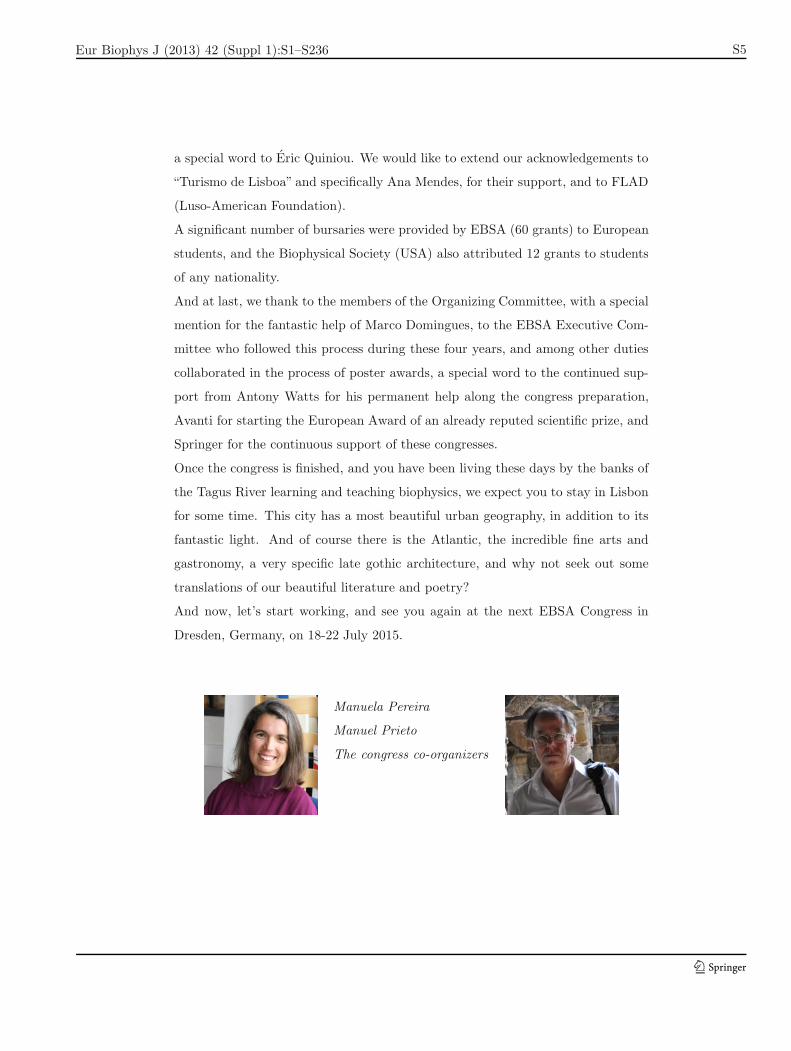
Manuela Pereira
Manuel Prieto
The congress co-organizers
123
Eur Biophys J (2013) 42 (Suppl 1):S1–S236
S6 Eur Biophys J (2013) 42:S3–S14
EBSA Welcome
Ladies and Gentlemen,
On behalf of the Executive Committee of the European Biophysical Societies’ Asso-
ciation, I am pleased and honoured to welcome you at the 9th European Biophysics
Congress in Lisbon.
This congress is the 6th since EBSA decided to organize congresses biennially. We
are able to organize congresses more frequently since EBSA has a secure annual
income from the European Biophysics Journal. Anthony Watts, the managing
editor of the journal has done a magnificent job in the past several years. We
are much obliged for his editorial activity and for his pivotal role in the Executive
Committee. Last year we renewed the contract with Springer for another 6 years,
thereby we are confident that we can continue our mission to support “Biophysics
in Europe”.
In addition to congresses, another main activity of EBSA is to organize schools and
Biophysics Courses to engage and train younger scientists in the area. The 2nd
EBSA School on Membrane Biophysics held in Lacanao, France in June 2012, was
again a great success and heavily oversubscribed. EBSA supported the Croatian
International School of Biophysics in Primosten, also in 2012, and we plan to
contribute to the next school in 2014. In the past two years we awarded many
bursaries to make the participation of young scientist possible at a number of
relevant international meetings. Additionally, we supported several visits between
European laboratories to outstanding young biophysicists.
We continue our tradition with the EBSA Young Investigators’ Prize. This time
we received 8 nominations from 6 member societies (Austria, France, Hungary,
Italy, Spain and United Kingdom). In a very strong competition, the Executive
Committee selected Dr. Michael Karl Sixt from Austria. He will receive e2000, an
engraved medal and a certificate, and has been invited to present a plenary lecture
at the Congress.
As a very new initiative, through the very generous contribution of Walt Shaw,
CEO of Avanti Polar Lipids Inc. has established a biennial award – The Avanti
123
Eur Biophys J (2013) 42 (Suppl 1):S1–S236
Eur Biophys J (2013) 42:S3–S14 S7
Polar Lipids / EBSA Award - to be given by EBSA. The award will be presented
at the EBSA Congress to an investigator for outstanding contributions to our
understanding of lipid biophysics. Several outstanding nominations arrived with
the Executive Committee for this Award and Professor Felix M. Goni Head of the
Biophysics Unit (Joint Center of the Spanish National Research Council (CSIC)
and the University of the Basque Country) is the first winner of this prestigious
prize. The winner receives an honorarium of US$ 3,000 and will deliver a plenary
lecture at the Congress.
The financial support from EBSA and Springer was pivotal in organizing this
congress. This is the second time when 60 bursaries were offered for young tal-
ented colleagues to facilitate their participation. EBSA is important in supporting
“Biophysics in Europe”. Please be involved in the activities of EBSA, please attend
the General Assembly on July 15, even if you are not the voting representative of
your country.
As President, and on behalf of the Executive Committee of EBSA and the 32
Member Societies, I would like thank our hosts, the Portuguese Biophysical Soci-
ety. Special thanks are due to Manuela Pereira (President, Portuguese Biophysical
Society) and Manuel Prieto and to all the Members of the Organising and Scien-
tific Committees, who have worked so tirelessly and conscientiously to make this
congress a success. The members of the Executive Committee and the Secretary of
EBSA, Antoinette Killian, also played an essential role in helping the organizers,
and give their unreserved support to the local organizers.
I am confident that this Congress will yet again be a success, as were the previ-
ous congresses, and I am looking forward to meeting you at the 10th European
Biophysics Congress to be held in Dresden in 2015 organized by the German Bio-
physical Society.
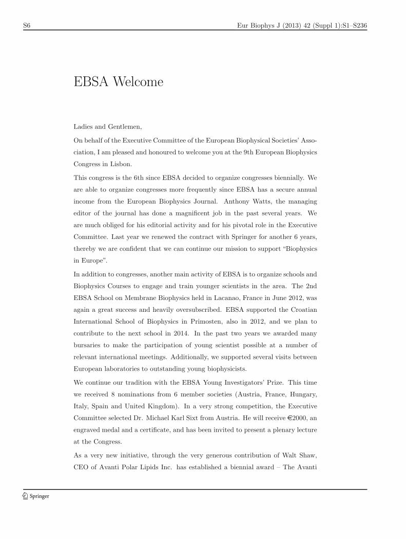
Laszlo Matyus
President of EBSA
123
Eur Biophys J (2013) 42 (Suppl 1):S1–S236
S8 Eur Biophys J (2013) 42:S3–S14
European Biophysics Journal – YOUR Journal
The European Biophysical Societies Association, EBSA, is supported by subscrip-
tions from member societies together with a good income stream from the European
Biophysical Journal (EBJ) published by Springer and for which EBSA holds the
copyright. The Journal is therefore integral to the health of EBSA and the support
of all its activities, including this congress.
EBJ has been growing significantly in recent years, now receiving 3 times more
submissions than just 8 years ago – it is also now a monthly publication. Submis-
sions are truly international in origin, and the developments in science in Asia are
certainly reflected in our global visibility. Additionally, in 2011, EBJ published 121
papers (∼55% acceptance rate); >1900 citations for EBJ papers for the year; IF
2.139, and visibility and downloads per month numbers are significantly increasing
(∼10,000 in 2010, ∼11,000 in 2011 and >16,000 full downloaded articles on
average per month in 2012). In the face of competition from many new journals
in this area, is holding its own position (continually in the upper 50%) very well.
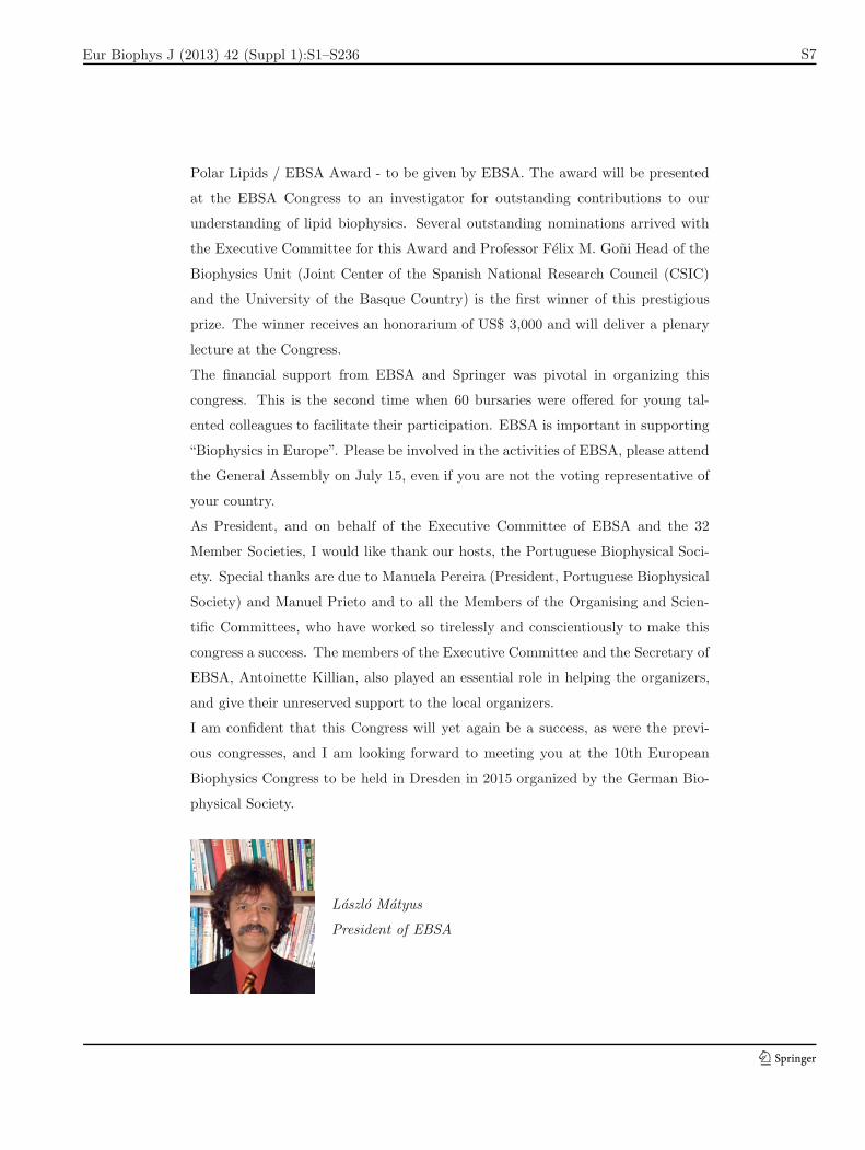
With the retirement of Phillip Kuchel, Sydney, AU last
year, Frances Separovic has been appointed as the new
Editor in the Pacific Rim. Frances is head of the School
of Chemistry, University of Melbourne, Australia, and
works on membrane peptides, using a range of biophysical
methodologies. She has extensive experience of biophysics,
having been intimately involved with the Australian So-
ciety for Biophysical and the Biophysical Society (USA),
of which she is a Fellow. Frances is also on the editorial
board of Biochim. Biophys. Acta – Biomembranes and
Acc. Chem. Res.
Frances Separovic
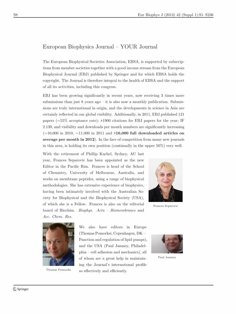
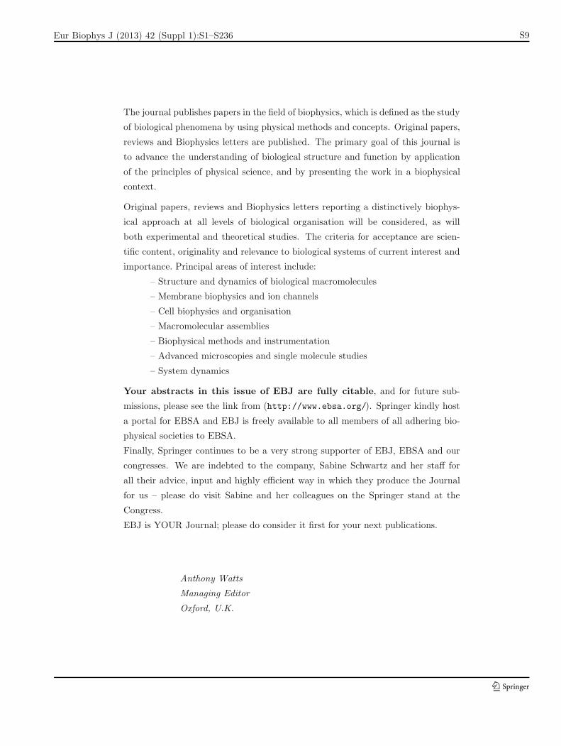
Thomas Pomorski
We also have editors in Europe
(Thomas Pomorksi, Copenhagen, DK –
Function and regulation of lipid pumps),
and the USA (Paul Janmey, Philadel-
phia – cell adhesion and mechanics), all
of whom are a great help in maintain-
ing the Journal’s international profile
so effectively and efficiently.
Paul Janmey
123
Eur Biophys J (2013) 42 (Suppl 1):S1–S236
Eur Biophys J (2013) 42:S3–S14 S9
The journal publishes papers in the field of biophysics, which is defined as the study
of biological phenomena by using physical methods and concepts. Original papers,
reviews and Biophysics letters are published. The primary goal of this journal is
to advance the understanding of biological structure and function by application
of the principles of physical science, and by presenting the work in a biophysical
context.
Original papers, reviews and Biophysics letters reporting a distinctively biophys-
ical approach at all levels of biological organisation will be considered, as will
both experimental and theoretical studies. The criteria for acceptance are scien-
tific content, originality and relevance to biological systems of current interest and
importance. Principal areas of interest include:
– Structure and dynamics of biological macromolecules
– Membrane biophysics and ion channels
– Cell biophysics and organisation
– Macromolecular assemblies
– Biophysical methods and instrumentation
– Advanced microscopies and single molecule studies
– System dynamics
Your abstracts in this issue of EBJ are fully citable, and for future sub-
missions, please see the link from (http://www.ebsa.org/). Springer kindly host
a portal for EBSA and EBJ is freely available to all members of all adhering bio-
physical societies to EBSA.
Finally, Springer continues to be a very strong supporter of EBJ, EBSA and our
congresses. We are indebted to the company, Sabine Schwartz and her staff for
all their advice, input and highly efficient way in which they produce the Journal
for us – please do visit Sabine and her colleagues on the Springer stand at the
Congress.
EBJ is YOUR Journal; please do consider it first for your next publications.
Anthony Watts
Managing Editor
Oxford, U.K.
123
Eur Biophys J (2013) 42 (Suppl 1):S1–S236
S10 Eur Biophys J (2013) 42:S3–S14
Michael Sixt – EBSA young researcher award
Michael Sixt studied human medicine at the University of Erlangen, Germany, and
received his MD in 2002. It was his time as a clinical resident at the Dermatological
Clinic Erlangen, which directed his interest to the migration of dendritic cells, a
system he is still working on. He followed his previous supervisor, Lydia Sorokin,
to Lund in Sweden, where he did a postdoc investigating the extracellular matrix
of lymph nodes, and studying how this affects transport of antigens. He then
moved to the Max Planck Institute of Biochemistry in Martinsried near Munich,
were he started as a group leader in 2005. Since 2010 he has been working as an
Assistant Professor at the Institute of Science and Technology (IST) Austria in
Klosterneuburg, near Vienna.
Michael is an enormously creative and innovative scientist, who, throughout his
young career, has been building bridges from his biomedical background to the
neighboring research fields of physics, molecular biology and biochemistry. Re-
cently he has particularly addressed how cytoskeletal dynamics and transmembrane
force coupling are fine-tuned in migrating leukocytes. He found out that these cells
can adapt their actin polymerization rate according to the adhesive nature of the
substrate. The data provide a first explanation how cells that follow a soluble
guidance cue can prioritize the soluble cue over the adhesive substrate. Recently,
he developed new assays that allowed him to address directly the distribution of
endogenous chemokines and their effect on leukocyte navigation. Using this ap-
proach he provided the first example of a functional chemokine gradient in vivo.
Notably, he found that this gradient is not soluble but immobilized to heparan sul-
fate residues, which has far reaching consequences regarding the mode how these
gradients are perceived and interpreted by the migrating cells. Michael makes a
very worthy recipient of the EBSA Young Investigator’s Award for 2013.
Gerhard Schutz, Vienna University of Technology, Austria
123
Eur Biophys J (2013) 42 (Suppl 1):S1–S236
Eur Biophys J (2013) 42:S3–S14 S11
Felix M. Goni – AVANTI award
Felix M. Goni (San Sebastian, Spain, 1951) earned his MD in Navarra (1975)
and became interested in Biophysics during summer courses at the Gulbekian
Foundation (Oeiras, Portugal) under N. van Uden. His post-doctoral work un-
der D. Chapman (Royal Free Hospital, London) involved studies of lipid-protein
interactions in membranes. From 1984 he has been a full Professor of Biochem-
istry at the University of the Basque Country (Bilbao, Spain). His research deals
with molecular interactions in membranes, with particular attention to lipids and
detergents. In 1999 he founded the Unidad de Biofısica, a joint centre of the Uni-
versity of the Basque Country and the Spanish National Research Council (CSIC),
and has been head of this institute from 2002. He has chaired the Publications
Committee of FEBS (2006-2011) and is currently Chair of the International Rela-
tions Committee of the U.S. Biophysical Society. He was President of the Spanish
Biophysical Society (1992-1998), who made him a Honorary Member in 2011.
123
Eur Biophys J (2013) 42 (Suppl 1):S1–S236
S12 Eur Biophys J (2013) 42:S3–S14
Congresses of the
EUROPEAN BIOPHYSICAL SOCIETIES’ ASSOCIATION
1st EUROPEAN BIOPHYSICS CONGRESS, 1971, BADEN, AUSTRIA2nd CONGRESS, 1997, ORLEANS, FRANCE3rd CONGRESS, 2000, MUNICH, GERMANY4th CONGRESS, 2003, ALICANTE, SPAIN5th EBSA / 15th IUPAB / SFB CONGRESS, 2005, MONTPELLIER, FRANCE6th CONGRESS, 2007, LONDON, UNITED KINGDOM7th CONGRESS, 2009, GENOA, ITALY8th CONGRESS, 2011, BUDAPEST, HUNGARY9th CONGRESS, 2013, LISBON, PORTUGAL
for your diary
10th CONGRESS, 2015, Dresden, GermanyJuly 18 – 22, 2015
—
11th congress will be in 2017 (see http://www.ebsa.org/ for venue and dates)
123
Eur Biophys J (2013) 42 (Suppl 1):S1–S236
Eur Biophys J (2013) 42:S3–S14 S13
EBSA Bursaries
Awarded to:
Aekbote, Badri – Szeged, Hungary
Antosova, Andrea – Kosice, Slovakia
Artetxe, Ibai – Leioa, Spain
Barnoud, Jonathan – Paris, France
Bednarikova, Zuzana – Kosice, Slovakia
Borile, Giulia – Padova, Italy
Cehlar, Ondrej – Bratislava, Slovakia
Coceano, Geovanna – Trieste, Italy
Covino, Roberto – Trento, Italy
Cujova, Sabına – Prague, Czech Republic
Dasanna, Anil – Toulouse, France
Deak, Robert – Budapest, Hungary
di Carlo, Maria – Palermo, Italy
Duan, Chenxi – Grenoble, France
Durka, Kamil – Lodz, Poland
Efimova, Svetlana – Saint Petersburg, Russia
Elani, Yuval – London, UK
Garaiova, Zuzana – Bratislava, Slovakia
Ghazaryan, Narine – Yerevan, Republic of Armenia
Girych, Mykhailo – Kharkiv, Ukraine
Goncharova, Iryna – Prague, Czech Republic
Graczer, Eva – Budapest, Hungary
Hadju, Kata – Szeged, Hungary
Hakobyan, Lilit – Yerevan, Republic of Armenia
Hikisz, Pawel – Lodz, Poland
Iarinca, Luiza – Cluj-Napoca, Romania
Iftemi, Sorana – Iasi, Romania
Istrate, Claudia – Bucharest, Romania
123
Eur Biophys J (2013) 42 (Suppl 1):S1–S236
S14 Eur Biophys J (2013) 42:S3–S14
Jakubowski, Rafal – Torun, Poland
Jobin, Marie-Lise – Pessac, France
Klose, Daniel – Osnabruck, Germany
Kopecka, Miroslava – Prague, Czech Republic
Kozlowska, Justyna – London, UK
Kudryashova, Ksenia – Moscow, Russia
Kwiatkowska, Marta – Lodz, Poland
Lighezan, Liliana – Timisoara, Romania
Lima, Angelica – Cachan, France
Marcuello, Carlos – Zaragoza, Spain
Martinez, Denis – Pessac, France
Mathesz, Anna – Szeged, Hungary
Michelssens, Servass – Leuven, Belgium
Muller, Jochen – Munich, Germany
Nagy, Krisztina – Szeged, Hungary
Nord, Ashley – Oxford, UK
Novotna, Pavlina – Prague, Czech Republic
Pesina, Daryna – Kharkiv, Ukraine
Piccirilli, Federica – Palermo, Italy
Prado, Pablo – Madrid, Spain
Pytel, Edyta – Lodz, Poland
Saponaro, Andrea – Milan, Italy
Shrestha, Dilip – Debrecen, Hungary
Siposova, Katarina – Kosice, Slovakia
Small, Lara – Durham, UK
Somkuti, Judit – Budapest, Hungary
Surleac, Marius – Bucharest, Romania
Szaloki, Nikoletta – Debrecen, Hungary
Takats-Nyeste, Annamaria – Budapest, Hungary
Tetryakova, Tatyana – Tiblisi, Georgia
Tripon, Carmen – Cluj-Napoca, Romania
Wypijewska, Anna – Warsaw, Poland
123
Eur Biophys J (2013) 42 (Suppl 1):S1–S236
Eur Biophys J (2013) 42:S15–S32 S15
Scientific Programme
SATURDAY, JULY 13th 2013 PAVILHAO ATLANTICORossio dos Olivais, Lote 2.13.01A, 1990 – 231 LisboaWeb site : http://www.pavilhaoatlantico.pt
15.00-16.00 Registration
17.00-17.30 Welcome Address
17.30-18.15 PLENARY LECTURELewis Kay, University of Toronto, CanadaSeeing the invisible by solution NMR spectroscopyChair: Antoinette Killian
18.30-19.30 Welcome Reception
123
Eur Biophys J (2013) 42 (Suppl 1):S1–S236
S16 Eur Biophys J (2013) 42:S15–S32
SUNDAY, JULY 14th 2013 PAVILHAO ATLANTICORossio dos Olivais, Lote 2.13.01A, 1990 – 231 LisboaWeb site : http://www.pavilhaoatlantico.pt
9.00-9.45 PLENARY LECTUREMichael Sixt, EBSA Award Speaker, Institute of Science andTechnology, AustriaCytoskeletal mechanics of chemotactic leukocytesChair: Lazlo Matyus
09.50-12.30 1. BIOLOGICAL ELECTRON AND PROTON TRANSFERChairs: R. Louro and A. Konstantinov
Invited speakers
Ulrike Alexiev, GermanyExploring the entrance of proton pathways in cytochrome c oxidase from P.denitrificans
Irene Dıaz-Moreno, SpainHow redox proteins form transient complexes in photosynthesis and respi-ration
Cyrille Costentin, FranceProton-coupled electron transfers in phenol and tryptophan oxidations: Anelectrochemical approach
Short talks
Petra Hellwig, FranceStudy of the Fe-S vibrational modes in complex I by means of Raman, FarIR and electrochemistry
Ana Sofia Fernandes Oliveira, PortugalExploring the dioxygen diffusion pathways in aa3 Cytochrome c oxidases:Insights from MD simulations
Filipa Calisto, PortugalStructural characterization of peripheral subunits from Alternative ComplexIII
09.50-12.30 2. CELL BIOPHYSICS AND SIGNALINGChairs: J. Rino and L. Matyus
Invited speakers
George Barisas, U.S.A.Nanoparticle probes of molecular rotation on cell surfaces
123
Eur Biophys J (2013) 42 (Suppl 1):S1–S236
Eur Biophys J (2013) 42:S15–S32 S17
T. W. J. (Dorus) Gadella, The NetherlandsEnhanced fluorescent proteins for FRET and for studying signaling acrossthe membrane
Maria Garcıa-Parajo, SpainBiophysics of leukocyte adhesion: nanoscale organization and dynamics ofthe integrin LFA-1
Short talks
Nils Petersen, CanadaProtein-protein-protein interactions in membranes measured by triple cor-relation of confocal images
Hetvi Gandhi, GermanyEarly events in cytokine receptor mediated signaling
Joachim Piguet, SwitzerlandTracking NK1 receptor diffusion in the membrane of living cells. Roles ofclathrin and cytoskeleton.
09.50-12.30 3. CHEMICAL AND SYNTHETIC BIOLOGYChairs: R. Eritja and A. Casini
Invited speakers
Wim Quax, The NetherlandsTurning Bacillus subtilis into a methylotrophic terpenoid synthesizing cell
Pier Luigi Luisi, ItalyTitle to be announced
Goncalo Bernardes, PortugalChemoselective transformations for bioimaging and targeted therapeutics
Short talks
Yuval Elani, U.K.Manufacturing vesicles with internal bilayer partitions: a novel unit forsynthetic biology
Vania Brissos, PortugalStabilization of FMN-dependent NADPH:dye/quinone reductase from Pseu-domonas putida by directed evolution
Sonia Mendes, PortugalCatalytic and spectroscopic characterization of two bacterial dye-decolourising peroxidases
12.30-15.00 POSTER SESSION, LUNCH & EXHIBITS
123
Eur Biophys J (2013) 42 (Suppl 1):S1–S236
S18 Eur Biophys J (2013) 42:S15–S32
15.00-15.45 PLENARY LECTUREViola Vogel, ETH Zurich, SwitzerlandMechanobiology: From molecular zippers to the recognition of nanotopogra-phiesChair: Erick Dufourc
16.15-18.30 4. MOLECULAR MOTORSChairs: M. Pereira and H. Grubmuller
Invited speakers
Jacek Czub, PolandThe mechanism of energy transmission in F1-ATPase as revealed by molec-ular dynamics simulations
Michael Borsch, GermanyMotors, gears and breaks of FoF1-ATP synthase monitored by single-molecule FRET
Julie Plastino, FranceMembrane dynamics and cytoskeleton assembly in cell motility
Short talks
Ilja Kusters, NetherlandsSingle molecule observation of protein translocation
Ashley Nord, U.K.Stepping Behavior of Rotary Molecular Motors
Stefan Balint, SpainCorrelating cargo transport with the cytoskeletal network at high resolution
16.15-18.30 5. PROTEIN FOLDING, ASSEMBLY AND STABILITYChairs: E. Melo and A. Kolinski
Invited speakers
Anne Ulrich, GermanyTransport machineries in biomembranes that utilize electrostatic “chargezippers”
Mikael Oliveberg, SwedenPrion-like aggregation in ALS
Martin Blackledge, FranceProtein conformational dynamics and molecular recognition in folded andunfolded proteins by NMR
Short talks
Birgit Habenstein, GermanyTau structure in paired helical filaments revealed by solid-state NMR
123
Eur Biophys J (2013) 42 (Suppl 1):S1–S236
Eur Biophys J (2013) 42:S15–S32 S19
Karin Hauser, GermanyFibril formation of polyglutamine repeats: a spectroscopic study
Claudio Gomes, PortugalCross talks between amyloid-forming proteins in neurodegeneration
16.15-18.30 6. SYSTEMS BIOLOGYChairs: A. Salvador and J. M. G. Vilar
Invited speaker
Oliver Ebenhoh, U.K.The role of mixing entropy in carbohydrate metabolism
Leonor Saiz, U.S.A.Negative feedback and crosstalk in the Transforming Growth Factor β sig-naling pathway
Short talks
Marina Monteiro, PortugalFRAP biophysical tool to probe nucleic acids-membrane ligand interactionsin pDNA purification
Tomas Tokar, SlovakiaBcl-2 family regulation of apoptosis by non-trivial decisioning
Jan Sielewiesiuk, PolandPositive and negative feedback loops coupled by a common promoter
123
Eur Biophys J (2013) 42 (Suppl 1):S1–S236
S20 Eur Biophys J (2013) 42:S15–S32
MONDAY, JULY 15th 2013 PAVILHAO ATLANTICORossio dos Olivais, Lote 2.13.01A, 1990 – 231 LisboaWeb site : http://www.pavilhaoatlantico.pt
9.00-9.45 PLENARY LECTUREProfessor Sir Alan Fersht, FRS, Department of Chemistry, Uni-versity of Cambridge, U.K.Tumour suppressor p53: biophysics and drug discoveryChair: Catherine Royer
09.50-12.30 7. CHANNELS AND TRANSPORTERSChairs: G. Soveral and N. Schmitt
Invited speakers
Rainer Schindl, AustriaActivation mechanism of the store-operated calcium channel complexSTIM1 and Orai1
Teresa Giraldez, SpainGating ring motions underlying function of BK channels
Michael Pusch, ItalyCLC-5, an endosomal chloride – proton exchanger mutated in Dent’s disease:a biophysical perspective
Short talks
Vicente Aguilella, SpainSARS-CoV E protein ion channel characterization by tuning the proteinand lipid charge
Gyorgy Panyi, HungaryLocked-open activation gate impedes recovery from inactivation in ShakerK+ channels
Katsumi Matsuzaki, JapanInfluenza A virus M2 protein forms a dimeric channel in biomembranes
09.50-12.30 8. BIOMOLECULAR SIMULATION: SPANNING SCALESChairs: C. Soares and I. Vattulainen
Invited speakers
Markus Deserno, U.S.A.Optimization of an elastic network augmented coarse-grained model tostudy CCMV capsid deformation
Luca Monticelli, FranceModeling the effect of nano-sized polymer particles on the properties of lipidmembranes
123
Eur Biophys J (2013) 42 (Suppl 1):S1–S236
Eur Biophys J (2013) 42:S15–S32 S21
Helmut Grubmuller, GermanyAtomistic simulation of single molecule experiments: Molecular machinesand a dynasome perspective
Short talks
Gerhard Konig, U.S.A.A hybrid quantum-chemical approach for free energy simulations.
Manuel Melo, NetherlandsMixing and matching simulations at different resolutions
Luıs Filipe, PortugalConformational determinants of peptidic tree-like molecules: insights fromMD simulations
09.50-12.30 9. IMAGING AND BIOSPECTROSCOPYChairs: M. Prieto and P. Schwille
Invited speakers
Jens Michaelis, GermanyMechanistic insight into eukaryotic gene expression from single moleculeexperiments
Catherine Royer, FranceQuantifying protein interaction networks in live cells using fluorescence fluc-tuation microscopy
David Klenerman, U.K.Single molecule studies of protein aggregates
Short talks
Axel Hochstetter, SwitzerlandTracing the microscopic motility of unicellular parasites
Maria Sarmento, PortugalPI(4,5)P2 acts as a lipid calcium sensor in the presence of physiologicalcalcium concentrations
Jacob Piehler, GermanyDynamic submicroscopic signaling zones revealed by TALM and image cor-relation analysis
12.30-15.00 POSTER SESSION, LUNCH & EXHIBITS
14.30-14.45 Company talks
NanoTemper Technologies
123
Eur Biophys J (2013) 42 (Suppl 1):S1–S236
S22 Eur Biophys J (2013) 42:S15–S32
15.00-15.45 PLENARY LECTUREMaria Joao Romao, Lisbon, PortugalUnraveling new functions and modes of action of molybdenum-dependentenzymesChair: Helmut Grubmuller
16.15-18.30 10. MOLECULAR RECOGNITION AND NANOBIOPHYSICSChairs: P. Eaton and J. Piehler
Invited speakers
Vinod Subramaniam, The NetherlandsGetting a grip on alpha-synuclein amyloid oligomers - single molecule ap-proaches
Joao Pedro Conde, PortugalLab-on-chip detection of biomolecules with integrated sensors
Daniel Muller, SwitzerlandQuantifying and localizing interactions guiding the structural and functionalproperties of GPCRs
Short talks
Peter Jonsson, U.K.Molecular nanomechanics and local stimulus of individual biomolecules onthe surface of cells
Anny Slama-Schwok, FranceRegulation of Nitric oxide synthases by fluorescent NADPH derivatives upontwo photon excitation
Agata Szuba, GermanySignal-Driven tethering system based on DNA-Origami linked to lipid bi-layers
16.15-18.30 11. MEMBRANE STRUCTURE AND DOMAINSChairs: M. J. Moreno and F. Goni
Invited speakers
Banafshe Larijani, U.K.Effects of phosphoinositides and their derivatives on membrane morphologyand function
Hans-Joachim Galla, GermanySubstrate turn and stimulated ATP-hydrolysis of the hABCC3 transportershow positive cooperativity
Jesus Perez-Gil, SpainModulation of phase coexistance and biophysical activity in pulmonary sur-factant membranes and film
123
Eur Biophys J (2013) 42 (Suppl 1):S1–S236
Eur Biophys J (2013) 42:S15–S32 S23
Short talks
Liana C. Silva, PortugalCeramide activates endocytosis and forms ordered intracellular lipid do-mains in response to TNF-α
Sebastian Finger, GermanyThe effect of cyclic antimicrobial hexapeptides on model bacteria mem-branes
Miglena I. Angelova, FranceLocal pH gradients induce polarization of Lo and Ld domains in GM1-containing giant vesicles
16.15-18.30 12. PROTEIN-NUCLEIC ACID INTERACTIONSChairs: A. Athanasiades and A. Wilkinson
Invited speakers
Anastassis Perrakis, The NetherlandsBinding J: molecular biophysics to understand the binding of a unique pro-tein to a unique DNA base
Oscar Llorca, SpainTransient geometries in nonsense-mediated mRNA decay (NMD) visualizedby cryo-EM
Dagmar Klostermeier, GermanyRNA binding and unwinding by the T. thermophilus DEAD-box helicaseHera
Short talks
Beata Vertessy, HungaryGenomic integrity of virulence genes is preserved by a dUTPase-basedmolecular switch
Carina Monico, ItalyProtein-DNA interactions probed by Ultrafast Force-clamp Spectroscopy
Nicolas Fiszman, FranceEucaryotic translation at single molecule scale
18.30-19.30 EBSA General Assembly
123
Eur Biophys J (2013) 42 (Suppl 1):S1–S236
S24 Eur Biophys J (2013) 42:S15–S32
TUESDAY, JULY 16th 2013 PAVILHAO ATLANTICORossio dos Olivais, Lote 2.13.01A, 1990 – 231 LisboaWeb site : http://www.pavilhaoatlantico.pt
9.00-9.45 PLENARY LECTUREFelix Goni, Avanti Award Speaker, Universidad del Paıs Vasco,SpainA lifetime of oily games and greasy ministrationsChair: Manuel Prieto
09.50-12.30 13. MATERIAL SCIENCE IN BIOPHYSICSChairs: J. C. Marcos and A. Turberfield
Invited speakers
Friedrich Simmel, GermanyDynamical diversity of compartmentalized in vitro transcriptional oscillators
Thomas Scheibel, GermanyProcessing of recombinant proteins for materials applications: About spidersilk and more
Rui Afonso, PortugalYoung Biophysicist Award - Portuguese Biophysical SocietyNanotube-forming hydrophobic dipeptides: structure, properties and appli-cations
Short talks
Badri L. Aekbote, HungaryOptical tools for localized fluorescence enhancement and single cell studies
Arwen Tyler, U.K.Tuning curvature in inverse micellar and bicontinuous cubic phases
Lea-Laetitia Pontani, U.S.A.Specificity, flexibility and valence of DNA bonds guide emulsion architecture
09.50-12.30 14. PROTEIN-LIPID INTERACTIONSChairs: L. Loura and A. Killian
Invited speakers
Amitabha Chattopadhyay, IndiaInteraction of membrane cholesterol with G Protein-Coupled Receptors: Amultidimensional approach
Lena Maler, SwedenLipid interactions of glycosyltransferases
123
Eur Biophys J (2013) 42 (Suppl 1):S1–S236
Eur Biophys J (2013) 42:S15–S32 S25
Daniel Otzen, DenmarkLiprotides: complexes between fatty acids and (partially denatured) pro-teins
Short talks
Yvonne Klapper, GermanyLipid coated quantum dots as a model to study lipid-protein interactionsvia FCS
Hartmut Luecke, SpainStructure, function & inhibitors of the pH-gated H. pylori urea channelessential for acid survival
Georg Pabst, AustriaModulation of ion-channel activity by cholesterol and ceramide
09.50-12.30 15. NEUROSCIENCESChairs: A. Sebastiao and M. Ferenczi
Invited speakers
Jean-Philippe Pin, FranceEnlightening allosteric properties of metabotropic glutamate
Yasser Roudi, NorwayInhibitory networks of grid cells
Karri Lamsa, U.K.Rectification of glutamate receptors set cell type –specific plasticity rules ininterneurons
Short talks
Andrea Mescola, ItalySurface strategy for regulation and control of neural cell adhesion
Megan Oliva, AustraliaDifferential modulation of NaV1.1 and NaV1.2 sodium channels by the β1auxiliary subunit
Daniele Arosio, ItalyImproving a GFP-based sensor to measure intracellular parameters like pHand ion concentrations
12.30-15.00 POSTER SESSION, LUNCH & EXHIBITS
14.30-14.45 Company talks
GE Healthcare
123
Eur Biophys J (2013) 42 (Suppl 1):S1–S236
S26 Eur Biophys J (2013) 42:S15–S32
15.00-15.45 PLENARY LECTUREPetra Fromme, Arizona State University, Tempe (AZ), U.S.A.Femtosecond crystallography: Dawn of a new era in structural biologyChair: Jose Carrascosa
16.15-18.30 16. BIOLOGICALLY ACTIVE PEPTIDESChairs: M. Bastos and E. Dufourc
Invited speakers
Karl Lohner, AustriaDisruption of bacterial membranes based on membrane curvature generationby antimicrobial peptides
Miguel Castanho, PortugalUnraveling a new actor in dengue virus-cell fusion
Huey Huang, U.S.A.Process of inducing pores in membranes by melittin
Short talks
Annika Kopp, GermanyBiophysical investigations into the effect of antimicrobial peptides on bac-terial membranes
Isabel Alves, FranceHow do a proapoptotic and a cell penetrating peptide work together to killcancer cells?
Peter Judge, U.K.Non-uniform changes in lipid order induced by the Membrane TargettingSequence of the MinD ATPase
16.15-18.30 17. NEW AND NOTABLEChairs: L. Martins and A. Watts
Invited speakers
Michael Mayer, U.S.A.Single protein characterization methods with nanopores
Joao Morais Cabral, PortugalStructure and function of the KtrAB ion transporter
Mark Dodding, U.K.Structural basis for kinesin-1: Cargo recognition
Short talks
Fabiola Gutierrez, NetherlandsAltering the torsional rigidity of proteins with surfactants
123
Eur Biophys J (2013) 42 (Suppl 1):S1–S236
Eur Biophys J (2013) 42:S15–S32 S27
Francesca Munari, GermanyConformational plasticity of the multi-domain Heterochromatin Protein 1β
Maximilian Richly, FranceInvestigating the Cell Membrane via Single Particle Tracking and Hydro-dynamic Force Application
16.15-18.30 18. PROTEIN STRUCTURE AND FUNCTIONChairs: C. Frazao and A. Ulrich
Invited speakers
Francesca Marassi, U.S.A.Structural studies of membrane proteins in membrana
Ben Berks, U.K.Moving folded proteins across membranes: structural analysis of the Tatprotein translocase
Reinhard Grisshammer, U.S.A.Structure of the agonist-bound neurotensin receptor NTS1
Short talks
Ana P. Batista, PortugalInvestigating substrate interaction on Type II NADH:quinone oxidoreduc-tase from Escherichia coli
Roslin Adamson, U.K.Probing GPCR-Gα interactions: A functional study by EM and SPR
Patrick Drucker, GermanyThe Annexin A2 core domain is identified to induce membrane curvature
19.30-24.00 Conference Dinner
123
Eur Biophys J (2013) 42 (Suppl 1):S1–S236
S28 Eur Biophys J (2013) 42:S15–S32
WEDNESDAY, JULY 17th 2013 PAVILHAO ATLANTICORossio dos Olivais, Lote 2.13.01A, 1990 – 231 LisboaWeb site : http://www.pavilhaoatlantico.pt
9.00-9.45 PLENARY LECTURENynke Dekker, Kavli Institute of Nanoscience, TU Delft, TheNetherlandssInvestigating transcription and replication at the level of single moleculesand cellsChair: Anthony Watts
09.50-12.30 19. INTRINSICALLY DISORDERED PROTEINSChairs: C. Gomes and Y. Levy
Invited speakers
Kresten Lindorff-Larsen, DenmarkCombining NMR and molecular simulations to study protein dynamics
Peter Tompa, BelgiumSupertertiary structural ensembles of proteins
Ben Schuler, SwitzerlandProbing the polymeric properties of IDPs with single-molecule spectroscopy
Short talks
Ana-Cristina Sotomayor-Perez, FranceDisorder-to-order transition in RTX proteins: Implications for toxin physi-ology
Volodymyr Shvadchak, NetherlandsRepeats in the α-synuclein sequence determine its conformation on mem-branes
Larisa Kapinos Schneider, SwitzerlandKaryopherin binding induces conformational transitions in the intrinsicallydisordered FG domains
09.50-12.30 20. RNA STRUCTURE AND FUNCTIONChairs: C. Arraiano and B. Vertessy
Invited speakers
Chirlmin Joo, The NetherlandsDefense against viral attack: single-molecule view on a bacterial adaptiveimmune system
123
Eur Biophys J (2013) 42 (Suppl 1):S1–S236
Eur Biophys J (2013) 42:S15–S32 S29
Eric Westhof, FranceRNA architectural modules, their detection in RNA Sequences and the as-sembly of large RNAs
Frederic Allain, SwitzerlandSplicing and translation regulation by small RNA binding proteins
Short talks
Gilmar F. Salgado, FranceProbing DNA G-quadruplex structures inside living cells using NMR spec-troscopy
Francesco Colizzi, ItalySymmetry and asymmetry in the unwinding of nucleic acids
Tobias Schmidt, GermanyDeciphering the RNA-binding complex NF90-NF45: complex formation fa-cilitates RNA chaperone activity
09.50-12.30 21. BIOPHYSICS IN EUROPE (TEACHING, CAREER ANDFUNDING)
Chairs: M. Castanho and C. Royer
Invited speakers
Jeremy Craven, U.K.Keeping the physics in physical biochemistry teaching
Bertrand Garcia-Moreno, U.S.A.Why, how, and whither biophysics?
David Pina, BelgiumFunding opportunities within the next European Framework Programmefor Research and Innovation — Horizon 2020
Silke Schumacher, GermanyEMBL and EMBL’s training activities
12.30-15.00 POSTER SESSION, LUNCH & EXHIBITS
14.30-14.45 Company talks
Leica Microsystems
15.00-15.45 PLENARY LECTUREMartin Hof, J. Heyrovsky Institute of Physical Chemistry of theASCR, Czech RepublicHydration, mobility, aggregation and nanodomain formation in model mem-branes studied by fluorescenceChair: Manuela Pereira
123
Eur Biophys J (2013) 42 (Suppl 1):S1–S236
S30 Eur Biophys J (2013) 42:S15–S32
16.15-18.30 22. MOLECULAR BASIS OF DISEASEChairs: C. Rodrigues and R. Ariens
Invited speakers
Albin Hermetter, AustriaSupra-molecular interactions of oxidized phospholipids in cells and lipopro-teins
Thomas Gutsmann, GermanyReconstituted micriobial lipid membranes as a tool in drug research
Javier Sancho, SpainThe LDL receptor: folding and binding events in function and in disease
Short talks
Ari Gafni, U.S.A.Synergistic interactions of neuron-bound Alzheimer’s Aβ40 and Aβ42: asingle molecule study
Markus Rudolph, SwitzerlandStructure of human α-2,6 sialyltransferase reveals mode of binding of com-plex glycans
Pascal Preira, FranceMembrane dynamic organization of HIV co-receptors analyzed by SingleParticle Tracking at the surface
16.15-18.30 23. SINGLE MOLECULE BIOPHYSICSChairs: N. Santos and Y. Engelborghs
Invited speakers
Peter Hinterdorfer, AustriaThese IgGs are made for walkin’: Random antibody movement on bacterialand viral surfaces
Marriano Carrion-Vazquez, SpainCommon characteristics in early amyloidogenesis: from single-molecules totherapy
Jasna Brujic, New York, U.S.A.Novel analysis methods in force-clamp spectroscopy shed light on proteinfolding
Short talks
Niels Zijlstra, NetherlandsAggregation conditions strongly influence the molecular composition ofalpha-synuclein oligomers
123
Eur Biophys J (2013) 42 (Suppl 1):S1–S236
Eur Biophys J (2013) 42:S15–S32 S31
Jorg Langowski, GermanyLive cell protein mobility and interaction maps by light sheet fluorescencecorrelation spectroscopy
Giovanna Coceano, ItalyBiomechanics study of cancer cells by optical tweezers and speckle mi-croscopy
16.15-18.30 24. SUPRAMOLECULAR ASSEMBLIESChairs: J. Carrascosa and M. Rappolt
Invited speakers
Jose M. Valpuesta, SpainThe protein folding pathway: a coordinated network of molecular chaper-ones
Borislav Angelov, Czech RepublicStructural analysis of tetrahedral channel formation and hydration in cubo-some nanoparticles
Giulio Caracciolo, ItalyTargeted drug delivery by nanoparticle-protein corona
Short talks
Antoine Loquet, GermanyStructure of a bacterial filament solved by solid-state NMRa: the type IIIsecretion system needle
Maite Paternostre, FrancePeptide nanotubes: structure and mechanism
Francesco Spinozzi, ItalyQuaternary structure of protein assemblies from small-angle x-ray and neu-tron scattering
19.00-21.00 Farewell Cocktail
123
Eur Biophys J (2013) 42 (Suppl 1):S1–S236

S32 Eur Biophys J (2013) 42:S15–S32
123
Eur Biophys J (2013) 42 (Suppl 1):S1–S236
Eur Biophys J (2013) 42:S33–S35 S33
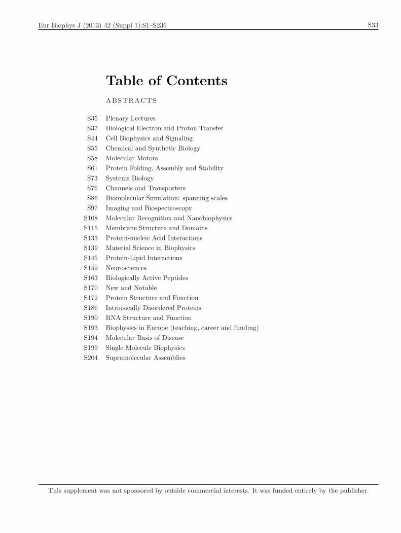
Table of Contents
ABSTRACTS
S35 Plenary Lectures
S37 Biological Electron and Proton Transfer
S44 Cell Biophysics and Signaling
S55 Chemical and Synthetic Biology
S58 Molecular Motors
S61 Protein Folding, Assembly and Stability
S73 Systems Biology
S76 Channels and Transporters
S86 Biomolecular Simulation: spanning scales
S97 Imaging and Biospectroscopy
S108 Molecular Recognition and Nanobiophysics
S115 Membrane Structure and Domains
S133 Protein-nucleic Acid Interactions
S139 Material Science in Biophysics
S145 Protein-Lipid Interactions
S159 Neurosciences
S163 Biologically Active Peptides
S170 New and Notable
S172 Protein Structure and Function
S186 Intrinsically Disordered Proteins
S190 RNA Structure and Function
S193 Biophysics in Europe (teaching, career and funding)
S194 Molecular Basis of Disease
S199 Single Molecule Biophysics
S204 Supramolecular Assemblies
This supplement was not sponsored by outside commercial interests. It was funded entirely by the publisher.
Eur Biophys J (2013) 42 (Suppl 1):S1–S236

S34 Eur Biophys J (2013) 42:S33–S35
123
Eur Biophys J (2013) 42 (Suppl 1):S1–S236
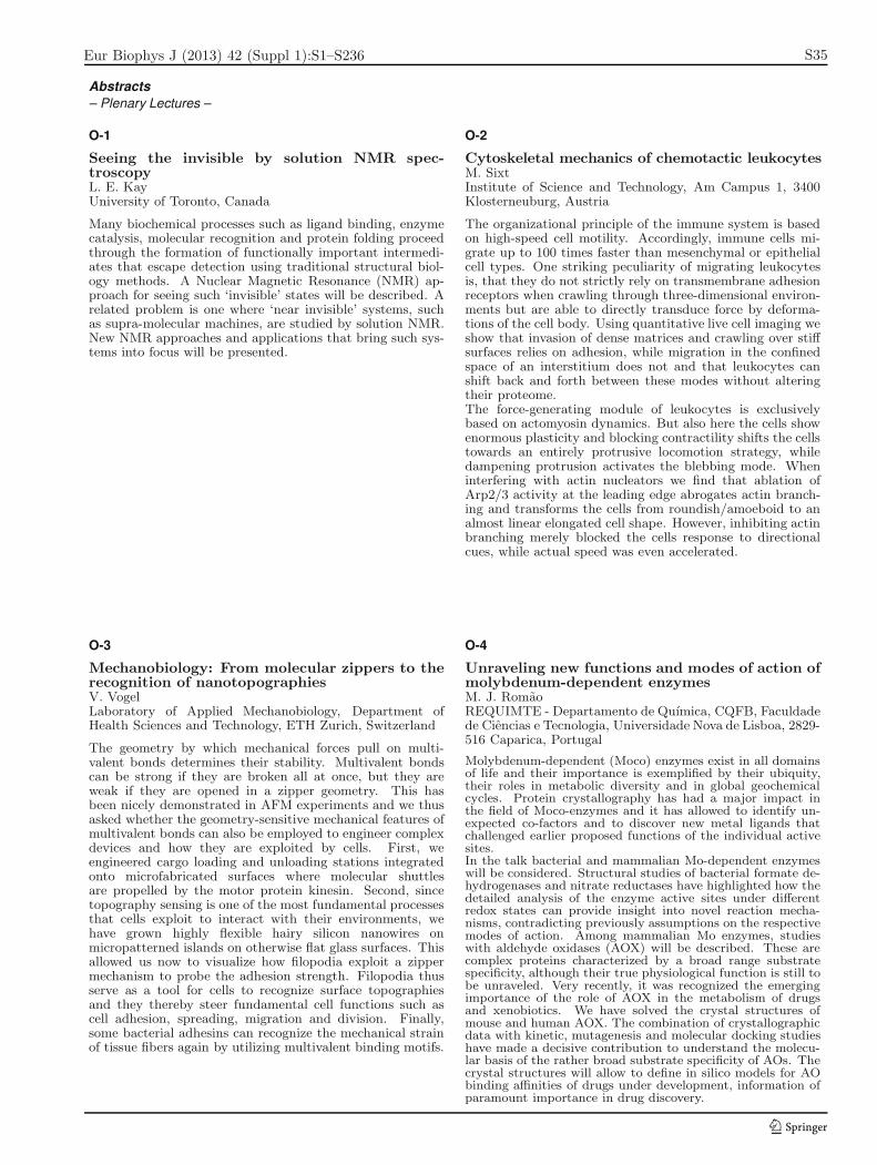
Unraveling new functions and modes of action ofmolybdenum-dependent enzymesM. J. RomaoREQUIMTE - Departamento de Quımica, CQFB, Faculdadede Ciencias e Tecnologia, Universidade Nova de Lisboa, 2829-516 Caparica, Portugal
Molybdenum-dependent (Moco) enzymes exist in all domainsof life and their importance is exemplified by their ubiquity,their roles in metabolic diversity and in global geochemicalcycles. Protein crystallography has had a major impact inthe field of Moco-enzymes and it has allowed to identify un-expected co-factors and to discover new metal ligands thatchallenged earlier proposed functions of the individual activesites.In the talk bacterial and mammalian Mo-dependent enzymeswill be considered. Structural studies of bacterial formate de-hydrogenases and nitrate reductases have highlighted how thedetailed analysis of the enzyme active sites under differentredox states can provide insight into novel reaction mecha-nisms, contradicting previously assumptions on the respectivemodes of action. Among mammalian Mo enzymes, studieswith aldehyde oxidases (AOX) will be described. These arecomplex proteins characterized by a broad range substratespecificity, although their true physiological function is still tobe unraveled. Very recently, it was recognized the emergingimportance of the role of AOX in the metabolism of drugsand xenobiotics. We have solved the crystal structures ofmouse and human AOX. The combination of crystallographicdata with kinetic, mutagenesis and molecular docking studieshave made a decisive contribution to understand the molecu-lar basis of the rather broad substrate specificity of AOs. Thecrystal structures will allow to define in silico models for AObinding affinities of drugs under development, information ofparamount importance in drug discovery.
O-4
Mechanobiology: From molecular zippers to therecognition of nanotopographiesV. VogelLaboratory of Applied Mechanobiology, Department ofHealth Sciences and Technology, ETH Zurich, Switzerland
The geometry by which mechanical forces pull on multi-valent bonds determines their stability. Multivalent bondscan be strong if they are broken all at once, but they areweak if they are opened in a zipper geometry. This hasbeen nicely demonstrated in AFM experiments and we thusasked whether the geometry-sensitive mechanical features ofmultivalent bonds can also be employed to engineer complexdevices and how they are exploited by cells. First, weengineered cargo loading and unloading stations integratedonto microfabricated surfaces where molecular shuttlesare propelled by the motor protein kinesin. Second, sincetopography sensing is one of the most fundamental processesthat cells exploit to interact with their environments, wehave grown highly flexible hairy silicon nanowires onmicropatterned islands on otherwise flat glass surfaces. Thisallowed us now to visualize how filopodia exploit a zippermechanism to probe the adhesion strength. Filopodia thusserve as a tool for cells to recognize surface topographiesand they thereby steer fundamental cell functions such ascell adhesion, spreading, migration and division. Finally,some bacterial adhesins can recognize the mechanical strainof tissue fibers again by utilizing multivalent binding motifs.
O-3
Cytoskeletal mechanics of chemotactic leukocytesM. SixtInstitute of Science and Technology, Am Campus 1, 3400Klosterneuburg, Austria
The organizational principle of the immune system is basedon high-speed cell motility. Accordingly, immune cells mi-grate up to 100 times faster than mesenchymal or epithelialcell types. One striking peculiarity of migrating leukocytesis, that they do not strictly rely on transmembrane adhesionreceptors when crawling through three-dimensional environ-ments but are able to directly transduce force by deforma-tions of the cell body. Using quantitative live cell imaging weshow that invasion of dense matrices and crawling over stiffsurfaces relies on adhesion, while migration in the confinedspace of an interstitium does not and that leukocytes canshift back and forth between these modes without alteringtheir proteome.The force-generating module of leukocytes is exclusivelybased on actomyosin dynamics. But also here the cells showenormous plasticity and blocking contractility shifts the cellstowards an entirely protrusive locomotion strategy, whiledampening protrusion activates the blebbing mode. Wheninterfering with actin nucleators we find that ablation ofArp2/3 activity at the leading edge abrogates actin branch-ing and transforms the cells from roundish/amoeboid to analmost linear elongated cell shape. However, inhibiting actinbranching merely blocked the cells response to directionalcues, while actual speed was even accelerated.
O-2
Seeing the invisible by solution NMR spec-troscopyL. E. KayUniversity of Toronto, Canada
Many biochemical processes such as ligand binding, enzymecatalysis, molecular recognition and protein folding proceedthrough the formation of functionally important intermedi-ates that escape detection using traditional structural biol-ogy methods. A Nuclear Magnetic Resonance (NMR) ap-proach for seeing such ‘invisible’ states will be described. Arelated problem is one where ‘near invisible’ systems, suchas supra-molecular machines, are studied by solution NMR.New NMR approaches and applications that bring such sys-tems into focus will be presented.
O-1
– Plenary Lectures –
Abstracts
Eur Biophys J (2013) 42:S35–S208 S35
123
Eur Biophys J (2013) 42 (Suppl 1):S1–S236
Hydration, mobility, aggregation and nan-odomain formation in model membranes studiedby fluorescenceM. HofJ. Heyrovsky Institute of Physical Chemistry, Academy ofSciences of the Czech Republic, Dolejskova 3, CZ-18223Prague 8, Czech Republic; Hof@jh-inst.
Fluorescence can be used in all kind of model membranesystems, such as monolayers, supported lipid bilayers, orunilamellar vesicles, as also in cells. Using a fluorescentreporter one can gain information on location, dynamicsand polarity of the labelled system. Although recentlysuper-resolution microscopy appeared, the combination of“conservative” techniques can still provide valuable informa-tion on questions in lipid membrane biophysics. Specifi-cally, time-dependent fluorescence shift method1 (for pro-tein applications see presentation of M. Amaro), differentvariants of fluorescence fluctuation spectroscopy2, and aMonte Carlo/Fluorescence Resonance Energy Transfer ap-proach will be discussed3. From the application of thosetechniques three membrane topics will be addressed: Influ-ence of monovalent ions (“Hofmeister”-series)4−7, impact oftruncated oxidized phospholipids8−10 and dynamics and sizeof lipid nanodomains in model membranes11.1) Jurkiewicz Biochimie 2012 94 26; 2) Machan BBA-20101798 1377; 3) Sachl Biophys J 2011 101 L60; 4) Vacha JPC A2009 113 7235; 5) Vacha JPC B 2010 114 9504; 6) JurkiewiczBBA 2012 1818 609; 7) Pokorna Farad Discus 2013 160 341;8) Beranova Langmuir 2010 26 6140; 9) Volinsky Biophys J2011 101 1376; 10) Jurkiewicz BBA 2012 1818 2388; 11) SteflBiophys. J 2012 102 9 2104
O-7
Investigating transcription and replication at thelevel of single molecules and cellsN. DekkerDepartment of Bionanoscience, TU Delft, The Netherlands
Single-molecule force and torque spectroscopy are very ver-satile techniques that allow us to shed light on genomic pro-cesses such as transcription and replication. In this talk,I will show how one can use these approaches to under-stand the mechanical properties of DNA and RNA. Then,I will highlight how we can study transcription by RNA-dependent RNA polymerases. We show how a very gen-eral approach that consists of parallel tracking to acquirehundreds of traces of individual RNA-dependent RNA poly-merases transcribing RNA in real time, combined with ananalysis method that can simultaneously probe different in-termediate states visited by the polymerases, can readily elu-cidate the mechanochemistry of these enzymes. Lastly, asthe true environment of all of these molecules is the livingcell, I will demonstrate our ability to track replication insidebacterial cells, and discuss the implications of our observa-tions on the dynamics of the replication fork.
O-6
A lifetime of oily games and greasy ministrationsF. M. GoniUnidad de Biofısica (CSIC, UPV/EHU), 48080 Bilbao, Spain
(1976-1978): In my post-doctoral years in London, underD. Chapman, I studied the interaction of intrinsic proteinswith membrane lipids, together with J.C. Gomez Fernandez,and helped to describe that, against the predominant viewat the time (“annular lipids”) all lipids were freely exchang-ing in the plane of the membrane at the relevant time-scaleof the enzyme turnover time. (1979-1989): After my re-turn to Spain I worked on detergents and the mechanism ofmembrane solubilization. Original observations at the timeincluded the ability of detergents to induce the lysis andreassembly (apparent fusion) of vesicles and the surfactant-induced release of vesicle contents at concentrations belowthose causing solubilization. (1987-2000): We were ableto produce the first model system of catalytically-promotedmembrane fusion, in which the catalyst was phospholipase C.(1994-present): At the instigation of my long-time associateAlicia Alonso we investigated the possible parallelism be-tween phospholipase C/diacylglyceride production and sph-ingomyelinase/ceramide production, with the discovery thatceramide was not primarily a fusogen, as was diacylglyc-erol, but rather destroyed the bilayer permeability barrier.(1997-present): As a result of our studies on sphingomyeli-nases, Alonso and myself have developed a systematic seriesof studies on the physical properties of ceramides and relatedsimple sphingolipids
O-5
– Plenary Lectures –
Abstracts
S36 Eur Biophys J (2013) 42:S35–S208
123
Eur Biophys J (2013) 42 (Suppl 1):S1–S236
Structural characterization of peripheral sub-units from Alternative Complex IIIF. G. Calisto, P. N. Refojo, M. M. PereiraInstituto de Tecnologia Quımica e Biologica, UniversidadeNova de Lisboa, Av. Da Republica EAN, 2780-157 Oeiras,Portugal
Respiratory chains are composed of several membrane pro-tein complexes enabling the transduction of energy from oxi-doreduction reactions to the establishment of transmembrenedifference of the membrane potential by charge transloca-tion. Central to aerobic respiratory chains is cytochrome bc1complex, which presents quinol:cytochrome c oxidoreductaseactivity. We recognize the existence of an alternative com-plex, Alternative Complex III – ACIII, performing the samefunction as the bc1 complex but structurally totally distinct.Besides four transmembrene proteins, ACIII contains threeperipheral subunits, ActA and ActE are facing the periplasmbut the orientation in relation to the membrane of the ActBremains unknown.In this work we investigated the orientation of subunit ActBand its functional implications, taking advantage of Pro-teinaseK activity on isolated membrane vesicles from R.marinus. We observed that ActB was digested by Pro-teinaseK, while ActE was present at a constant level. Asa control membranes were solublised with Triton X-100. Inits presence ActE was also sensitive to ProteinaseK.The orientation of ActB subunit towards the cytoplasm, inopposition to the other peripheral proteins which face theperiplasm has strong repercussion in the operative mode ofACIII.
O-11
Exploring the entrance of proton pathways in cy-tochrome c oxidase from P. denitrificansK. Kirchberg1, T.-Y. Kim1, H. Michel2, U. Alexiev11Freie Universitat Berlin, Physics Department, Berlin, Ger-many, 2Max-Planck Institute of Biophysics, Department ofMolecular Membrane Biology, Frankfurt a.M., Germany
Cytochrome c oxidase (CcO), the terminal oxidase of cellularrespiration, reduces molecular oxygen to water. The mecha-nism of proton pumping as well as the coupling of proton andelectron transfer is still not understood in this redox-linkedproton pump. Two proton transfer pathways have been sug-gested, originating at the N-(negative) side of the membranewith differential involvement in the redox cycle. We recentlyshowed that the first H+-uptake at the N-side of fully oxi-dized CcO coincides with single electron input into CuA atthe opposite side of the membrane. This indicates long-rangeinteractions and efficient H+-uptake mechanisms, such asproton collecting antennae switched on by electron injection[1]. To further understand the mechanism of proton pump-ing single cysteine variants are employed [2]. They provideselective binding sites for various reporter molecules, such asfluorophores for conformational changes, pH-indicator dyesfor protonation reactions, or polarity labels for sensing thelocal environment. A correlation between the differential in-volvement of the proton pathways and protein surface prop-erties will be discussed.[1] K. Kirchberg, H. Michel, U. Alexiev, J Biol Chem 2012,287, 8187[2] K. Kirchberg, H. Michel, U. Alexiev, Biochem BiophysActa 2013, 1827, 276
O-10
How redox proteins form transient complexes inphotosynthesis and respirationI. Dıaz-Moreno1, B. Moreno-Beltran1, A. Guerra-Castellano1 , K. Gonzalez-Arzola1, J. M. Garcıa-Heredia1,A. Velazquez2, P. M. Nieto3, M. Ubbink4, A. Dıaz-
Quintana1, M. A. de la Rosa11Instituto de Bioquımica Vegetal y Fotosıntesis, cicCartuja,Sevilla, Spain, 2Instituto de Biocomputacion y Fısica de Sis-temas Complejos (BIFI), Zaragoza, Spain, 3Instituto de In-vestigaciones Quımicas, cicCartuja, Sevilla, Spain, 4Instituteof Chemistry, Leiden University, The Netherlands
Protein complex formation is at least a two-step process inwhich the formation of a final, well-defined complex entails theinitial formation of a dynamic encounter complex. The life-time of the protein complex is determined by the dissociationrate. Highly transient complexes, with lifetimes on the orderof milliseconds, exhibit moderate or low binding affinities, withdissociation constants in the μM–mM range. Electron transfer(ET) reactions mediated by soluble redox proteins exchangingelectrons between large membrane complexes in photosynthesisand respiration are excellent examples of transient interactions.Here, experimental approaches based on dia and paramag-netic NMR spectroscopy are combined with NMR restraint-or charge-driven docking simulations to study the molecularrecognition processes in ET complexes, using the cyanobacte-rial Cf -Cc6 interaction in photosynthesis and the plant Cc1-Cc6 adduct in respiration, as physiological model systems.Both ET ensembles exhibit optimal coupling between the re-dox centers although they might differ in their dynamic be-havior. Needless to say that such an integrative methodologyopens new perspectives in our understanding of the dynamic,transient adducts formed between proteins beyond the modelsystems herein analyzed.
O-9
Proton-coupled electron transfers in phenol andtryptophan oxidations: An electrochemical ap-proachC. CostentinUniversite Paris Diderot, Sorbonne Paris Cite, Laboratoired´Electrochimie Moleculaire, Unite Mixte de Recherche Uni-versite - CNRS No 7591, France
Association between single electron transfer and protontransfer in many reactions of electron transfer, radicalchemistry and biochemistry is well a recognized phe-nomenon. There is some evidence that the two reactionsmight be concerted in many natural processes, notably inthe reactions of Photosystem II. Coupling proton transferto electron transfer entails an improvement of the drivingforce of the reaction. Two types of mechanisms may befollowed; mechanisms in which the two reactions occur ina stepwise manner, with proton transfer first, followed byelectron transfer (EPT) or, vice versa, electron transferfirst, followed by proton transfer (PET) and a mechanismin which proton and electron transfer occur in a concertedmanner (CPET). Only in the last case will the benefitsof the additional driving force offered by the couplingwith proton transfer be fully exploited. Electrochemistry,through techniques like cyclic voltammetry, can provide aquite effective access to CPET in terms of diagnosis andquantitative kinetic characterization. Several examples willbe presented : phenol (mimicking tyrosine) and tryptophanoxidations with a particular emphasis on the role of theproton acceptor (water or external base).
O-8
– Biological Electron and Proton Transfer –
Abstracts
Eur Biophys J (2013) 42:S35–S208 S37
123
Eur Biophys J (2013) 42 (Suppl 1):S1–S236
investigation of charge translocation by the res-piratory complex I reconstituted in liposomesP. J. Castro, A. P. Batista, M. M. PereiraInstituto de Tecnologia Quımica e Biologica, UniversidadeNova de Lisboa, Av. da Republica EAN, 2780-157 Oeiras,Portugal
Respiratory complex I plays a central role in energy pro-duction by coupling electron transfer between NADH andquinone to ions translocation across the membrane, therebyestablishing an electrochemical potential. This L-shapedenzyme consists of hydrophilic and membrane domains.The membrane domain includes seven hydrophobic subunitsand the three largest subunits, NuoL, M and N (E. colinomenclature), are homologous to each other and toNa+/H+ antiporter complex (Mrp) subunits. Previousstudies indicate that complex I from Rhodotermus marinustransduces energy by two different processes: protonpumping and Na+/H+ antiporting.This work aims at evaluating the ability of the isolated com-plex I to translocate H+ and Na+ across the membrane andto determine the stoichiometry of the process. In order toachieve our goal, the enzyme was purified from Rhodotermusmarinus and incorporated into liposomes. The proteoli-posomes were characterized by Dynamic Light Scattering(DLS). The existence of NADH:quinone oxidoreductaseactivity and the formation of membrane potential afteraddiction of the substrates proved that the incorporationwas successful. H+ and Na+ translocation were monitoredby fluorescence spectroscopy and 23Na-NMR, respectively.
P-15
Monitoring redox activity of heme proteinsby photochromic fluorescence resonance energytransferH. Bayraktar, S. ManiogluKOC UNIVERSITY, Department of Chemistry, Istanbul,Turkey
The real time imaging of redox proteins is crucial for un-derstanding their role in cellular signaling. In mitochondria,they are key regulators of cellular processes and differentactivity can be observed depending on the cell type. Thecurrent methods to measure the redox activity are limiteddue to the lack of genetically encoded probes. The activityof Cytochrome c (Cyt c), heme protein, is a regulator forapoptosis and the electron transfer. Here we present pho-tochromic fluorescent energy transfer (pcFRET) method tomeasure the redox activity of Cyt c. We have previously usedpcFRET as an ultrasensitive method measuring the photo-cycle of rodopsin membrane proteins. It is a general tool forturning absorbance changes into fluorescence. The Venus flu-orescent fragment was ligated into the Cyt c expression vec-tor with standard cloning methods. The expression of Cytc-Venus in BL21 expression cells was demonstrated. The de-tection of pcFRET for the Cyt c oxidation in BL21 cells iscurrently being studied. The expression of Cyt c-Venus willbe optimized in cells, we will investigate how frequently theoxidation state of Cyt c change. We will study the frequencychange of redox signal depending on external stimuli.
P-14
Study of the Fe-S vibrational modes in complex Iby means of Raman, Far IR and electrochemistryM. Yegres1, T. Friedrich2, P. Hellwig1
1Laboratoire de bioelectrochimie et spectroscopie, UMR7140, CNRS-Universite de Strasbourg, France., 2Institutfur Biochemie und Org.Chemie, Albert-Ludwigs-UniversitatFreiburg, Germany
Complex I couples the electron transfer from NADH toubiquinone with a translocation of protons across themembrane. It has an unusual L-shaped structure consistingof peripheral arm extending into the aqueous phase, thatincludes all know cofactors and a membrane part embeddedin the lipid bilayer, where proton translocation takes place.There is evidence that the energy released by the redoxreaction is transmitted by conformational changes of a 140
A long amphipatic helix that is aligned to the membranearm. It is proposed that the helix acts like a pistontransmitting the energy from the peripheral arm to themembrane arm. Here we study on the action of the proteinby means of the Fe-S vibrational spectroscopies in the lowfrequency range. These signals are sensitive to redox state,cluster ligation, hydrogen bonding and the conformationof bound cysteine residues. The spectra obtained from theE. coli complex I were compared to those of the solublefragment and model compounds. In addition, labeling ofthe Fe-S clusters by 54Fe was performed. A full assignmentof the spectral features was performed for the oxidized,the electrochemically and the NADH reduced form givingclear evidence on the conformational changes responsiblefor proton translocation.
O-13
Exploring the dioxygen diffusion pathways in aa3Cytochrome c oxidases: Insights from MD sim-ulationsA. S. F. Oliveira, J. M. Damas, A. M. Baptista, C. M. SoaresInstituto de Tecnologia Quımica e Biologica - UniversidadeNova de Lisboa
Cytochrome c oxidases (CCOX) are members of the haem-copper oxidase superfamily and are the terminal enzymes ofthe respiratory chain. These proteins are membrane-boundmulti-subunit redox-driven proton pumps, which couple thereduction of molecular dioxygen to water with the creationof a transmembrane electrochemical proton gradient.Over the last 20 years, most of the CCOX research focusedon the mechanisms and energetics of reduction and/or protonpumping and little emphasis has been given to the pathwaysused by dioxygen to reach the binuclear site. The main ob-jective of this work is to identify possible dioxygen pathwaysin the reduced CCOX from Rhodobacter sphaeroides[1] usingextensive Molecular Dynamics (MD) simulations. Our sim-ulations allowed the identification of two possible dioxygenchannels, whose entrances are both located in the membraneregion. The first channel is a Y-shaped hydrophobic cavitywith a constriction point near F282I and W172I , and it cor-responds to the oxygen pathway previously identified in thestructure[2]. The second channel starts near the hydroxylfarnesyl tail of haem a3 and ends near the Y288I (which iscovalently linked to the H284I imidazole group).[1]Qin et al. (2009) Biochemistry 48, 5121-5130[2]Svensson-EK et al. (2002) J.Mol.Biol. 321, 329-339
O-12
– Biological Electron and Proton Transfer –
Abstracts
S38 Eur Biophys J (2013) 42:S35–S208
123
Eur Biophys J (2013) 42 (Suppl 1):S1–S236
Role of proton motive force and ATPase activ-ity in biohydrogen production by RhodobactersphaeroidesL. Hakobyan1, L. Gabrielyan1, A. Trchounian2
1Dep. of Biophysics, Yerevan State University, Armenia,2Dep. of Microbiology & Plants and Microbes Biotechnol-ogy, Biology Faculty, Yerevan State University, Armenia
In order to examine the role of proton motive force (PMF) orthe proton H+-ATPase in H2 production by R. sphaeroides,isolated from mineral springs in Armenia, PMF and its com-ponents (the membrane potential (Δψ) and the transmem-brane pH gradient (ΔpH)) and ATPase activity were deter-mined, and the effect of hydrogenase inhibitor diphenyleneiodonium (Ph2I), was examined. Under nitrogen limitationconditions Δψ was shown to be of -98 mV and the reversedΔpH was +30 mV resulting in PMF of -68 mV. The addi-tion of Ph2I decreased Δψ to –70 mV in concentrations of20 μM and higher; lower concentrations of Ph2I had no valu-able effect on Δψ. The [pH]in determined by the quenchingof fluorescence of 9-aminoacridine was effectively blocked byPh2I. The R. sphaeroides membrane vesicles demonstratedsignificant ATPase activity. The incubation of the vesicles inthe presence of Ph2I caused to marked inhibition in ATPaseactivity revealing concentration dependence effect. The 10-20 μM Ph2I did not affect the ATPase activity, whereas 40μM Ph2I caused to marked inhibition (∼2 fold) in ATPaseactivity.The results point out a role of PMF and the H+-ATPase inhydrogenase activity involved in H2 production. Moreover,relationship of F0F1 with hydrogenase is suggested.
P-19
Structure of the membrane protein menaquinolfumerate reductase from Chloroflexus aurantia-
cus at 3 AR. Fromme1, Y. Xin2, P. Fromme1, R. Blankenship2
1Department of Chemistry and Biochemistry, Arizona StateUniversity, Tempe, AZ, USA 85287-1604, 2Department ofBiology and Chemistry, Washington University Campus Box1137, St. Louis, Mo 63130, USA
The photosynthetic green filamentus bacteria Chloroflexusaurantiacus has a quinol:fumerate reductase (QFR) thatreduces fumerate to succinate. As our study has shown thecomposition of ligands for the electron transfer process withFAD, three different iron-sulphur clusters, Fe2S2, Fe4S4 andFe3S4 beside two hemes is well conserved with for exampleWolinella succinogenes. All known protein structures ofQFR are functional dimers, therefore initially we tried tosolve the X-ray diffraction data in a dimer solution untilwe found with anomalous data(Fe edge) the convincingsolution in a trimeric arrangement in the asymmetric unit.The sequence homology of the Chloroflexus aurantiacusQFR to the closest pdb entry from E. coli is for the bigsubunit A only 29 %, the ferredoxin iron sulphur bindingsubunit B 24% and the transmembrane subunit C withoutany significant identity to known structures. The currentmodel has 3090 residues placed in the electron density at3 A resolution. For each monomer the FAD, the threeFeS clusters and two heme are found. Each C subunithas five transmembrane helices. Now with the prelimi-nary structure the identifying process of the trimer formingresidues has begun and shades a light on the evolution of thisimportant membrane protein and its functional organization.
P-18
The selectivity of type II NADH: quinone oxi-doreductase for quinones – a docking studyA. M. Duarte, F. V. Sena, A. P. Batista, M. M. PereiraInstituto de Tecnologia Quimica e Biologica, UniversidadeNova de Lisboa, Av. da Republica EAN, 2780 157 Oeiras,Portugal
Type II NADH: quinone oxidoreductases is a flavoproteinthat catalyzes the transfer of electrons from NADH toquinones. NDH-II has been detected in different microor-ganisms which produces different quinones . In E. coli thephysiological quinone is ubiquinone, while S. aureus and B.subtilis synthesis menaquinone. Recently the high resolu-tion structure of NDH-II of S. cerevisiae in complex withquinone, NAD and FAD was obtained. These studies pro-vided a crucial piece of information on the conformation andarrangement of the enzyme and its substrates. However, thereaction and selection mechanism behind this interaction isstill unknown.What is the binding hotspot between quinone and NDH-IIin the different organisms?Are these hotspots related to quinone selection by the differ-ent NDH-II?To unravel the interaction hotspots of NDH-II:quinone wesetup an in silico docking calculation based on the recentlypublished high resolution structures of NDH-II. This strat-egy uses a combination of homology modeling and dockingexperiments of different quinones interacting with NDH-IIfrom different organisms. The differences between dockingsites will shed light into the molecular selective determinantsfor different quinones by NDH-II.
P-17
Electron transfer with azurin at Au/SAM junc-tions in contact with the glass-forming environ-mentT. D. Dolidze1, D. E. Khoshtariya2, T. Tretyakova2,D. H. Waldeck3, R. van Eldik41I. Beritashvili Center of Experimental Biomedicine, 0160Tbilisi, Georgia, 2Institute for Biophysics and Bio-Nanosciences, Department of Physics, Tbilisi State Uni-versity, 0128 Tbilisi, Georgia, 3Department of Chem-istry, University of Pittsburgh, Pittsburgh, PA 5260, USA,4Department of Chemistry and Pharmacy, University ofErlangen-Nurnberg, 91058 Erlangen, Germany
Interfacial biological electron transfer under the conditionof approaching the glass-transition threshold was exploredby the method of rapid-scan protein film voltammetry.The gold-deposited alkanethiol SAM/azurin assemblies wereplaced, for the first time, in contact with a buffered proticionic melt, choline dihydrogen phosphate ([ch][dhp]), con-taining less than two, or more (up to 15) water moleculesper [ch][dhp]. The extra confinement of Az films within thesemi-solid environment allowed for the essential alteration ofthe protein’s conformational flexibility, directly controllingET in a dynamical regime, that was further tuned throughthe temperature (273–353 K) and pressure (0.1–150 MPa)variations. As the glassy state was approached, the Mar-cus theory-based data analysis revealed: (a) the transitorytraversing of a broad nonergodic ET zone, and (b) the ulti-mate breakdown of the medium’s linear response motif (i.e.,an increase in anharmonicity for the ET energy profiles).
P-16
– Biological Electron and Proton Transfer –
Abstracts
Eur Biophys J (2013) 42:S35–S208 S39
123
Eur Biophys J (2013) 42 (Suppl 1):S1–S236
NMR studies of the bioenergetic metabolism ofmetal reducing bacteriaR. O. Louro, A. Carrelo, T. M. Pereira, A. S. Alves,C. M. PaqueteITQB-UNL, Av da Republica (EAN) 2780-156 Oeiras
Metal reducing bacteria are becoming the focus of researchefforts to apply their unique metabolic potential in fields suchas bioremediation of metal contaminated environments,andbioelectrosynthesis or energy production from cheap raw ma-terials such as wastewater. These organisms have a uniqueorganization of their bioenergetic redox chains,with ATPproduction that occurs across the cytoplasmic membranecoupled to the reduction of the solid-phase electron acceptorthat occurs at the cell surface, in a process called extracel-lular respiration. Shewanella oneidensis MR-1 is a modelGram negative metal reducing bacterium, which has nu-merous multi-heme cytochromes that participate in electrontransfer across the periplasm and that are displayed on thecell surface to reduce the solid-phase electron acceptors. Thislast process can proceed by direct contact or be mediatedby small redox shuttles such as flavins. NMR spectroscopywas used to determine the protein-protein interactions thatsustain electron flow across the periplasmic space during ex-tracellular respiration and the protein-small molecule inter-actions that mediate indirect electron transfer between thecells and the solid-phase acceptors.
P-23
Lab-on-a-chip tool for bioelectronic investiga-tionsA. Kincses, S. Valkai, F. Walter, M. A. Deli, P. Ormos,A. DerInstitute of Biophysics, Biological Research Centre of theHungarian Academy of Sciences
We established a non-invasive method for the measurementof electric signals associated to membrane-coupled signal-and energy transduction phenomena of living cells. Themethod uses a pair of metal film electrodes picking up thesignals. In microscopic dimensions, the electrodes observeevents only from their close vicinity, so objects of microme-ter scale (typically cells or thin tissues) can be monitored thisway. Cell delivery can be controlled by microfluidic tools ina lab-on-a-chip measuring chamber, giving further access tomicroscopic observations. We applied the method to moni-tor fundamental cell physiological processes on different lev-els of organization (Chlamydomonas cells, neuro-epithelialtissues).We expect our method to become a highly sensitive, versatiletool for the kinetic investigation of electric phenomena asso-ciated to transport phenomena across cellular and epithelialmembranes. The financial support of Hungarian researchgrant KTIA-OTKA CK 78367 is gratefully acknowledged.
P-22
Dynamic control for short-range biological electrontransfer: insights from protein film voltammetryD. E. Khoshtariya1, T. D. Dolidze2, D. H. Waldeck3, R. van
Eldik41Institute for Biophysics and Bionanosciences, Departmentof Physics, I. Javakhisvili Tbilisi State University, 0128,Tbilisi, Georgia, 2I. Beritashvili Center of ExperimentalBiomedicine, 0160 Tbilisi, Georgia, 3Department of Chem-istry, University of Pittsburgh, Pittsburgh, PA 15260, USA,4Department of Chemistry and Pharmacy, University ofErlangen-Nurnberg, 91058 Erlangen, Germany
For the case of short-range biological electron transfer, undercertain experimental conditions, the slowest conformationalfluctuations of redox-active proteins may directly control theelectron transfer rate, through the pre-exponential term of akinetic equation. In contrast, the long-range ET is controlledby the donor-acceptor electronic coupling. Our approachimplies special design of interfacial biomimetic assemblies,encompassing Au-deposited self-assembled monolayer filmsof variable thickness and head group composition, providingdifferent interaction modes for the functionalized redox pro-teins (cytochrome c, azurin, myoglobin, etc.). Furthermore,the exterior manipulations by using highly viscous mediasuch as aqueous glucose mixtures or water-doped protic ionicmelts as a glass forming environment, further tuned by thevariations of temperature and pressure, allows for the care-ful identification of a dynamically controlled regime of ET,along with the disclosure of novel non-ergodic and nonlinearmedium response motifs.
P-21
Functional equivalence of as isolated and highenergy metastable states of cytochrome c oxidaseD. Jancura1, V. Berka2, J. Stanicova3, M. Fabian4
1Department of Biophysics, Safarik University, Kosice, Slo-vakia, 2Department of Internal Medicine, University ofTexas Health Science Center, Houston, USA, 3Institute ofBiophysics and Biomathematics, University of Veterinarymedicine, Kosice, Slovakia, 4Department of Biochemistryand Cell Biology, Rice University, Houston, USA
The current model of the proton pumping in cytochromec oxidase (CcO) presumes the existence of a ”high-energy”metastable OH state. It has been suggested that there ex-ist differences between ligation and protonation states of thecatalytic site of OH and the “resting” state of the oxidizedCcO. Our previous study did not reveal differences either inthe spectral characteristics (optical, EPR) or the kinetics ofelectron transfer to the catalytic site in these two forms ofCcO. In this work, the reactions of as “isolated” fast form(O) and (OH) of the oxidized CcO with H2O2 have been in-vestigated by stopped-flow method. The rate constants forthe binding of H2O2 to the catalytic site are almost identicalfor both forms of the fully oxidized CcO. Using phenol redas a pH indicator we have found that a relaxation of OH toO state and the binding of H2O2 to the catalytic site of bothforms are not coupled with an apparent proton uptake or re-lease. Our findings again indicate that there is no differencein the ligation and protonation states of the catalytic site ofO and OH of the oxidized CcO.Acknowledgements: This work was supported by APVV-0242-11 and by the project CELIM funded by 7.FP EU.
P-20
– Biological Electron and Proton Transfer –
Abstracts
S40 Eur Biophys J (2013) 42:S35–S208
123
Eur Biophys J (2013) 42 (Suppl 1):S1–S236
Molecular details of electron transfer in fumaratereduction by flavocytochrome c3C. M. Paquete, I. Saraiva, R. O. LouroInstituto de Tecnologia Quımica e Biologica - UNL, Oeiras,Portugal
Numerous redox enzymes catalyze two-electron processes,even though the connection from the protein surface to theactive site is made through chains of redox cofactors thatonly exchange one electron at a time. The soluble fumaratereductase from the periplasmic space of Shewanella is one ofthese proteins. The three-dimensional structure showed thatthis protein contains a chain of four hemes, which interactswith the FAD catalytic centre that performs the obligatorytwo electron-two proton reduction of fumarate to succinate.In order to investigate the role played by the redox chain inthe catalytic activity of this enzyme, transient kinetic studiesof flavocytochrome reduction in the absence and in the pres-ence of substrate were performed. The kinetic contributionof each heme for electron uptake and conduction to the cat-alytic centre was determined. This enabled the observationthat the catalytically most competent states of the enzymeare those least prevalent in a quasi-stationary condition ofturnover. Comparison of the results from fumarate reduc-tases from two different organisms enabled the identificationof the role of the redox properties of individual hemes in themodulation of the catalytic activity of the enzymes.
P-27
Inter- and intra-monomeric communication inthe cytochrome bc1 complex as revealed by MDsimulationsP. S. Orekhov1, K. V. Shaytan1, A. Y. Mulkidjanian2
1School of Biology, Moscow State University, 119899Moscow, Russia, 2School of Physics, University of Os-nabrueck, D-49069 Osnabrueck, Germany
The cytochrome bc1 complex (bc1) plays an important rolein cell bioenergetics acting as a dimeric redox-driven protontranslocase. Mitchell’s Q-cycle in general describes mecha-nism of bc1 activity: ubiquinol is oxidized in the site QP ofthe cytochrome b subunit, whereby the two released elec-trons are transferred to the heme of cyt. c1 (via the mobile“head” domain of the Rieske protein) and to a ubiquinonemolecule in the QN site of cyt. b.As it was discussed before [1] the ubiquinol reduction in theQN site should be coupled with the mobility of the Rieske“head”domain, while thermodynamics considerations [2] im-ply the coupling between the two “head” domains. However,the details of both the coupling events in bc1 remain elusive.We performed equilibrium and metadynamics MD simula-tions of the bc1 with different ligands to establish inter- andintra-monomeric coupling in bc1 dimer while application ofvarious computational techniques allowed us to track possi-ble pathways for allosteric signal propagation.References1. 1.Gopta OA et al. (1998) FEBS Lett, 431:291-296.2.2.Mulkidjanian AY (2007) Photochem Photobiol Sci,6:19-34.
P-26
Interaction mechanisms between plant GALDHand cytochrome cJ. A. Navarro1, Q. Bashir2, N. G. Leferink3, P. Ferreira4,J. B. Moreno-Beltran1, A. H. Westphal3, I. Dıaz-Moreno1,M. Medina4, M. A. de la Rosa1, M. Ubbink2, M. Hervas1,W. J. van Berkel31IBVF, CSIC & University of Sevilla, cicCartuja, Sevilla,Spain, 2Gorlaeus Laboratories, Leiden University, TheNetherlands, 3Laboratory of Biochemistry, Wageningen Uni-versity, The Netherlands, 4Department of Biochemistry andBIFI, University of Zaragoza, Spain
The flavoprotein L-galactono-γ-lactone dehydrogenase(GALDH) catalyzes the terminal step of vitamin C biosyn-thesis in plant mitochondria. Here we investigated theinteraction between Arabidopsis thaliana GALDH and itsnatural electron acceptor cytochrome c (Cc). Using laserspectroscopy we observed that GALDHSQ oxidation by Ccfollows a kinetic mechanism involving protein association,to form a transient bimolecular complex prior to electrontransfer. The kinetic analysis of both GALDHSQ andGALDHHQ oxidation suggests that GALDH reductionby its carbohydrate substrate limits the overall rate ofCc reduction. ITC analysis showed that GALDH weaklyinteracts with both oxidized and reduced Cc. Chemicalshift perturbations for 1H and 15N nuclei of Cc mapped theinteracting surface of Cc to a single surface surrounding theheme edge. In summary, the results point to a relativelylow-affinity GALDH/Cc interaction, similar for all partnerredox states, directed by electrostatic complementary forcesand involving protein-protein dynamic motions.
P-25
Ion translocation by respiratory Complex I: Therole of NuoL subunitB. C. Marreiros, A. P. Batista, M. M. PereiraITQB, Av. da Republica, EAN, 2780-157 Oeiras, Portugal
Complex I is the least understood enzyme of the respiratorychain and its deficiencies have been implicated in severalneurodegenerative diseases. It couples the oxidation ofNADH and reduction of quinone to charge translocationacross the membrane, contributing for the establishmentof the membrane potential. The membrane arm containsNa+/H+ antiporter-like subunits (NuoL, M and N), whichare homologous to Na+/H+ antiporters (Mrp), suggestingthat these subunits may participate in charge translocation.Understanding the coupling of the enzymatic reaction tocharge translocation is still a major question in complexI research. Crystallographic structural data showed thepresence of a ∼110 A long amphipathic helix part of theC-terminal of NuoL subunit, which may function as acoupling element.In this work we investigate the role of NuoL. We used anE. coli mutated strain in NuoL, obtained from the ’Keiocollection’ and monitored the ion transport. Proton translo-cation was studied by quenching of ACMA fluorescence and23Na-NMR spectroscopy was used to investigate sodiumtransport. We observed that the presence of NuoL subunitis not essential for entire proton translocation by complex I,but is determinant for sodium transport.
P-24
– Biological Electron and Proton Transfer –
Abstracts
Eur Biophys J (2013) 42:S35–S208 S41
123
Eur Biophys J (2013) 42 (Suppl 1):S1–S236
Bacteriorhodopsin and ATP-synthase create gra-dients in atto- to picoliter sized compartmentsM. Schwamborn, D. Frese, C. SteinemGeorg-August University Goettingen, Intitute for Organicand Biomolecular Chemistry, Tammannstr. 2, 37077 Goet-tingen
The light-driven proton pump bacteriorhodopsin (bR) isfound in the cell membrane of Halobacterium salinarum. Theprotein forms crystalline regions (purple membrane) consist-ing of hexagonally packed trimers. Upon light irradiation bRpumps protons out of the cytosol creating an electrochemi-cal H+-gradient that is used for ATP synthesis. This taskis carried out by the ubiquitous protein ATP-synthase thatproduces ATP if the proton electrochemical potential is largeenough to overcome the free energy of ATP hydrolysis. Theprotein is also capable of reversing the process, i.e. usingATP to create H+-gradients.Both proteins are studied to large extend in model systems.Here we present pore-spanning membranes (PSMs) as a newmodel, providing a system that consists of atto- to picolitersized compartments covered by a lipid bilayer of desiredlipid and protein composition. To obtain such a bilayer,bR and ATP-synthase are reconstituted into liposomes thatare spread on porous substrates with defined pores. ApH-sensitive fluorescent dye entrapped in the compartmentsallows to monitor the proteins’ H+-pumping activity bymeans of fluorescence microscopy.
P-31
Alternative complex III: a different architectureusing known building modulesP. N. Refojo, M. A. Ribeiro, F. Calisto, M. Teixeira,M. M. PereiraInstituto de Tecnologia Quımica e Biologica, UniversidadeNova de Lisboa, Av. da Republica EAN, 2780-157 Oeiras,Portugal
Alternative complexes III (ACIII) form a recently identifiedfamily of enzymes with quinol:electron acceptor oxidoreduc-tase activity that replace functionally cytochrome bc1/b6f.Gene clusters encoding ACIII are widespread in the Bacteriadomain and are mostly present in genomes were the codinggenes for the bc1/b6f complexes are absent. Furthermore, thegene clusters are frequently associated with those coding foroxygen reductases subunits. Biochemical and genomic anal-yses showed that ACIII is composed of six to eight subunits,most of which homologous to different proteins already ob-served in other known enzymatic complexes. In fact, ACIIIcan be seen as a combination of modules present in differentenzyme families, namely the complex iron-sulfur molybde-num containing enzymes to which enzymes like polysulfidereductase and DMSO reductase belong to. With the increas-ing number of completely sequenced genomes, a larger num-ber of gene clusters coding for ACIII were identified and anunanticipated diversity in gene clusters, both in terms of itsconstitution and organization was detected. The several un-expected gene arrangements brought new perspectives to therole of the different subunits of ACIII.
P-30
The monoheme cytochrome c subunit of alterna-tive complex III from Rhodothermus marinusP. N. Refojo, M. Ribeiro, F. Calisto, M. Teixeira,M. M. PereiraITQB-UNL, Av da Republica, Estacao Agronomica Na-cional, 2780-157 Oeiras, Portugal
In Rhodothermus marinus’ respiratory chain, quinol: elec-tron acceptor oxidoreductase activity is performed by thealternative complex III (ACIII). This seven subunit com-plex is a member of a recently identified family of enzymeswhich catalyses the same reaction as the bc1 complex, butis structurally unrelated to it. The ACIII contains 4 trans-membrane subunits without redox cofactors, one cytoplasmicsubunit with 4 iron-sulfur clusters and two periplasmic sub-units containing C-type hemes. A structural and functionalassociation between the ACIII and the caa3 heme-copper ox-idase (HCO) from R. marinus has been reported.The monoheme cytochrome c subunit (mhc) was structurallyand functionally characterized. For this purpose, a truncatedform of its coding gene was cloned and expressed in E.coli.The UV-Visible spectra of the protein are characteristic of alow spin heme with a histidine-methionine-Fe coordination.By measuring the mhc: O2 oxidoreductase activity of thecaa3 HCO, we conclude that mhc is the electron donor forthe caa3 HCO in its association with the ACIII.
P-29
Position of W104 in AppA BLUF domain re-vealed by fluorescence spectroscopyK. Raics1, K. Pirisi1, J. Orban1, R. Brust3, G. Greg4,T. Mike4, T. J. Pete3, M. R. Stephen2, A. Lukacs11Department of Biophysics, Medical School, University ofPecs, Hungary, 2School of Chemistry, University of EastAnglia, Norwich, UK, 3Department. of Chemistry, StonyBrook University, Stony Brook, USA, 4Central Laser Facil-ity, Rutherford Appleton Laboratory, Didcot, UK
AppA is a BLUF protein which serves as a transcriptionalanti-repressor found in Rhodobacter sphaeroides. Similarlyto other flavoproteins the absorption of the photon is fol-lowed by an electron transfer cascade, but the same timethe hydrogen-bond network around FAD undergoes to a re-organization which is crucial for the formation of the lightadapted state of the protein.There are several different X-ray structure for AppA, whichshow the position of W104 at a different place: in theoriginal crystal structure from Anderson et al W104 waslocated closer to the flavin forming a hydrogen bond withQ63. A different – shorter length – AppA structure hasshown a structure with W104 farer from the flavin partiallyexposed to the solvent.In order to clarify the location of W104 we made severalmutants and performed fluorescence anisotropy, fluorescenceanisotropy decay and FRET measurements. Exchangingthe tryptophan at W64 to phenylalanine caused that wecould excite selectively the W104 tryptophan. For thedark state (dW64F) we observed a slower rotational corre-lation time than in a partially solvent exposed case (lW64F).
P-28
– Biological Electron and Proton Transfer –
Abstracts
S42 Eur Biophys J (2013) 42:S35–S208
123
Eur Biophys J (2013) 42 (Suppl 1):S1–S236
Direct electrochemistry of gold-deposited self-assembled monolayers of L-cysteineT. Tretyakova1, T. D. Dolidze2, D. E. Khoshtariya1, R. van
Eldik31Institute for Biophysics and Bio-Nanosciences, Departmentof Physics, Tbilisi State University, 0128 Tbilisi, Georgia,2Department of Biophysics, I. Beritashvili Center of Exper-imental Biomedicine, 0160 Tbilisi, Georgia, 3Department ofChemistry and Pharmacy, University of Erlangen-Nurnberg,91058 Erlangen, Germany
We report on the gold-deposited self-assembled monolayerfilms (SAMs) of L-cysteine (Cys), which, when probed bythe fast scan cyclic voltammetry (CV), exhibit a pair ofwell-defined waves of electron exchange in a quasi-reversiblekinetic regime. The signal emerges in the course of suc-cessive cycling over the potential range of 0.4 to −0.4 V(phosphate buffers, pH 5.5 to 8). The formal redox potentialof the Au/Cys electrode is pH dependent with a slope44 mV/pH. The redox process, seemingly, is coupled to aproton translocation, involving the carboxylic group of Cys.Furthermore, we observed the electro-catalytic activity ofAu/Cys electrodes toward the ascorbic acid decomposition.Based on these results we suppose that all the data reportedfor different proteins immobilized at Cys SAMs should bereconsidered very carefully, in particular when successivecycling over the potential range of 0.4 to −0.4 V wasapplied, aiming a disclosure of the proteins’ voltammetricresponse.
P-32
– Biological Electron and Proton Transfer –
Abstracts
Eur Biophys J (2013) 42:S35–S208 S43
123
Eur Biophys J (2013) 42 (Suppl 1):S1–S236
Protein-protein-protein interactions in mem-branes measured by triple correlation of confocalimagesM. Anikovsky2, N. O. Petersen1
1University of Alberta, Edmonton, Canada, 2University ofCalgary, Calgary, Canada
Protein-protein interactions have been measured suc-cessfully and quantitatively by fluorescence correlationspectroscopy in solution (Elson, BJ 101, 2855-2870 (2011))and image correlation spectroscopy on cell surfaces (Kolinand Wiseman, Cell Biochem Biophys 49, 141-164 (2007)).These tools fail to provide information about about protein-protein-protein interactions. It has been known for sometime that higher order moments or correlations containthe relevant information (Palmer and Thompson, BJ 52,257-270 (1987); Heinze, Jahnz, and Schwille, BJ 86, 506-516(2004)) but it is only recently that triple correlation func-tions of ternary complexes have been measured in solution(Ridgeway, Millar, and Williamson, J. Phys Chem B 116,1908-1919 (2012) and PNAS 109, 13614-13619 (2012)). Wedemonstrate how complete and quantitative information forternary complexes of membrane proteins can be obtainedfrom three confocal images from each of three distinctlylabeled proteins from a combination of image correlationspectroscopy, image cross-correlation spectroscopy, and im-age triple cross-correlation spectroscopy. This is illustratedwith simulations and measurements of interactions betweenselected proteins. The simulation and analysis programs forthese will be presented and will be available for interestedparties.
O-36
Nanoparticle probes of molecular rotation on cellsurfacesD. Zhang1, P. W. Winter2, I. Pecht3, D. A. Roess4,B. G. Barisas11Dept. of Chemistry, Colorado State Univ., 2Cell & Molec-ular Biology Prog., Colorado State Univ., 3Dept. of Im-munology, Weizmann Inst. of Science, 4Dept. of BiomedicalSciences, Colorado State Univ.
Rotation of membrane proteins is a sensitive measure of theiraggregation state and their interactions with other mem-brane species. We have used nanoparticles including asym-metric quantum dots (QD) as non-bleaching probes of the ro-tation of individual cell surface proteins. Invitrogen QD655sconjugated to A2 DNP-specific IgE allow examination of slowrotation of the Type I Fcε receptor (FcεRI) on RBL-2H3cells. For individual QDs in image pair sequences, we cal-culate the time-autocorrelation function for fluorescence po-larization fluctuations (PFTAC). PFTAC decay extends wellinto the ms timescale, as implied by time-resolved phospho-rescence anisotropy results. Treatment effects suggest thatsuch slow decay may be a property of the membrane itself,perhaps reflecting large-scale fluctuations of mesoscale mem-brane regions. To examine PFTAC decays faster than imag-ing measurements permit, we have applied time-correlatedsingle photon counting to individual QD fluorescence and an-alyzed data down to correlation times of 1ns. These measure-ments should thus include the 80μs hydrodynamic rotationof single FcεRI molecules, but significance of current datais limited by QD emission rates. Other probes are thereforebeing explored. Supported by NSF grant MCB-1024668.
O-35
Enhanced fluorescent proteins for FRET and forstudying signaling across the membraneT. Gadella, J. Goedhart, L. van Weeren, L. Joosen,M. Adjobo-Hermans, M. HinkSection of Molecular Cytology & van Leeuwenhoek Centrefor Advanced Microscopy, Swammerdam Institute for LifeSciences, Amsterdam, The Netherlands
A screening method is reported that, in addition to fluores-cence intensity, quantifies the excited state lifetime of a fluo-rescent protein, providing a direct measure for the quantumyield of the fluorescent protein. A library of cyan fluorescentprotein (CFP) variants was screened eventually yielding thebright cyan fluorescent protein variant, mTurquoise2 whichhas a very high quantum yield of 0.93, and a seriously in-creased fluorescence lifetime of 4 ns. mTurquoise2 is thepreferred donor to YFP variants (like mVenus or mCitrine)with an R0 of 5.83 nm.We apply the different probes for the study of GPCR-triggered signaling across the membrane in single mam-malian cells. A new Gq FRET sensor was made to monitoractivation of the G protein, and downstream signaling eventslike generation of lipid-derived second messengers, PtdInsP2-dependent PLC relocalization, and RhoGEF activation witha variety of FRET-, ratio imaging- and TIRF-microscopicapplications will be presented.Goedhart J, von Stetten D, Noirclerc-Savoye M, LelimousinM, Joosen L, Hink MA, van Weeren L, Gadella TWJ, Roy-ant A. (2012) Structure-guided evolution of cyan fluorescentproteins towards a quantum yield of 93%. Nat Commun.3:751.
O-34
Biophysics of leukocyte adhesion: nanoscale or-ganization and dynamics of the integrin LFA-1K. Borgman1, C. Manzo1, I. Piechocka1, A. Sosa-Costa1,T. S. van Zanten1, M. F. Garcia-Parajo2
1ICFO- Institute of Photonic Sciences, Mediterranean Tech-nology Park, 08860 Castelldefels, Spain, 2ICREA- InstitucioCatalana de Recerca i Estudis Avancats, Barcelona, Spain
Integrins are cell membrane adhesion receptors involved inmorphogenesis, immunity, tissue healing and metastasis. Acentral, yet unresolved question regarding the function ofintegrins is how these receptors regulate their conformationand dynamic nanoscale organization on the membrane togenerate adhesion-competent microclusters upon ligandbinding. We exploit single molecule fluroescence approaches(nm accuracy & ms temporal resolution) to investigatemolecular conformation, lateral organization and dynamicsof the β2-integrin LFA-1 on immune cells. Our results indi-cate that LFA-1 forms stable and non-mixable nanoclustersin the neighborhood of GPI-anchored protein nanodomainsprior to ligand activation [1]. Mobility of LFA-1 depends onits conformational state and anchoring to the cytoskeleton[2,3]. Furthermore, lateral mobility resulted crucial formicrocluster formation upon ligand binding and for stableleukocyte adhesion under shear stress conditions [3]. Ourongoing research centers on the role that mechanical stimuli(shear-stress and isotropic mechanical stretching) have onboth activation and lateral mobility of LFA-1 on monocytes.[1] T.S. van Zanten et al PNAS 106, 18557, 2009.[2] Diez-Ahedo et al, Small 5, 1258, 2009.[3] G.J. Bakker et al PNAS, 109, 4869, 2012.
O-33
– Cell Biophysics and Signaling –
Abstracts
S44 Eur Biophys J (2013) 42:S35–S208
123
Eur Biophys J (2013) 42 (Suppl 1):S1–S236
The investigation of cellular adhesion in the con-text of external stimuliT. Baronsky, A. JanshoffGeorg-August-University, Institute of Physical Chemistry,Tammannstr. 6, 37077 Goettingen, Germany
Cellular dynamics as observed in cell migration strongly de-pends on the adhesion to the underlying surfaces. Thereby,it is indisputable that cells respond to different substratesmorphologies and mechanical properties by changing theircellular properties such as cell locomotion, adhesion,growth, differentiation and elasticity. Here, we investigatedsubstrates with different physical and chemical propertiesvarying in rigidity or porosity. By means of single cell forcespectroscopy (SCFS) we investigated the adhesion strengthof epithelial cells as a function of substrate properties. SCFSis a modified version of the atomic force spectroscopy (AFM)and is used to measure the strength of overall cell adhesiondown to single-molecule levels. Concretely, cellular adhesionof the epithelial cell line MDCK II (Madin-Darby CanineKidney) to PDMS (Polydimethylsiloxane) substrates withdifferent elasticity and on normal Petri dish was investigated.
P-40
Relationship between Bax and Bak distributionand apoptosis onset in U-87 MG cells upon Hyp-PDTL. Balogova, M. Maslanakova, P. Miskovsky, K. StroffekovaDepartment of Biophysic, Institute of Physical Sciences,Pavol Jozef Safarik University in Kosice
Apoptosis is a key process in the development and mainte-nance of tissue homeostasis, which is tightly regulated bybalance between cell survival and damage signals. Our workfocused on the intrinsic mitochondrial apoptotic pathway,where Bcl-2 family of proteins plays the major role. We wereparticularly interested in two pro-apoptotic players Bak andBax, and investigated their role in apoptosis triggered byhypericin photodynamic therapy (HypPDT). We show thedistribution of Bax and Bak in U-87 MG human gliomacells incubated with Hyp before and after PDT. One hourpost HypPDT there is a significant Bax translocation intomitochondria, however our results indicate that in U-87MG cells there are two populations of mitochondria. One,almost exclusively localized near the plasma membrane,contains Bax and Bak simultaneously, and the other whichcontains Bax only and is distributed throughout the cell.The different protein content and spatial distribution ofthese two populations suggest that they can play differ-ent roles in response to apoptotic stimuli. Further weinvestigated relationships between onset of apoptosis andeither Bax translocation, or metabolic flux rate in U87MG cells. Supported by EU grants (ITMS: 26110230013;26220120040) and by APVV-0242-11.
P-39
Tracking NK1 receptor diffusion in the mem-brane of living cells. Roles of clathrin and cy-toskeletonJ. Piguet, L. Veya, H. VogelEcole Polytechnique Federale de Lausanne, Switzerland
NK1 receptor is a very potent target for treatment of depres-sion and attenuation of side effects in cancer therapy. In thiswork, we describe the impact of impaired endocytosis andcytoskeleton function on the receptor’s mobility pattern inthe plasma membrane of living cells.We found that non-activated receptors diffuse freely andin confined domains, which are directly related to clathrin-mediated endocytosis (CME). Inhibitors of different steps ofCME, lead to an accumulation of NK1R in confined mem-brane domains, and a diminution of NK1R mediated Ca2+
release.Depolymerisation of actin and microtubules does not signifi-cantly modify freely diffusing receptors diffusion parameters,indicating absence of direct interaction of NK1R with the cy-toskeleton. Nevertheless, actin depolymerisation triggers theapparition of fast receptors in circular domains in the absenceof activation. This effect is correlated with the formation ofcell membrane blebs. Microtubule depolymerisation also sig-nificantly increases the fraction of receptors slowly diffusingin domains. This increase in confinement is related to earlyendocytosis inhibition.Our results point to the central importance of clathrin inNK1R membrane homeostasis and regulation of the recep-tor’s activity.
O-38
Early events in cytokine receptor mediated sig-nalingH. R. Gandhi1, K. Kurgonaite1, T. Kurth1, P. Schwille2,C. Bokel1, T. Weidemann2
1CRTD, TU Dresden, Germany, 2Max PIanck Institute ofBiochemistry, Martinsried, Germany
The cytokine Interleukin-4 (IL-4) is believed to induceheterodimeric IL-4 receptor (IL-4R) complexes at thesurface of target cells. Here we study at the biophysicaland cell biological level how these signals are transducedto the downstream transcription factor STAT6. We showthat individual receptor subunits accumulate in a noveltype of early sorting endosome stably anchored within theactin cortex (cortical endosomes), and characterize theirtrafficking by FRAP and quantitative imaging. In additionwe found that pharmacological inhibition of receptorinternalization blocks STAT6 activation. Endocytosisthus acts upstream of JAK/STAT signaling. Fluorescencecross-correlation spectroscopy (FCCS) analysis of IL-4Rsubunits in their native plasma membrane environmentrevealed that complex formation is indeed ligand induced,however the affinities for recruitment of a second receptorchain are comparably low. In addition, fluorescence lifetimeimaging (FLIM) showed that the complexes preferentiallyform within the cortical endosomes, where the subunitsare enriched. Thus, subcellular concentration of cytokinereceptor subunits within signaling endosomes constitutesa novel, thermodynamic prerequisite for IL-4R signaltransduction not yet described in other pathways.
O-37
– Cell Biophysics and Signaling –
Abstracts
Eur Biophys J (2013) 42:S35–S208 S45
123
Eur Biophys J (2013) 42 (Suppl 1):S1–S236
Single particle tracking reveals that amyloid ag-gregates alter the mobility of GM1 gangliosideM. Calamai, F. S. PavoneLENS - European Laboratory for Non-Linear Spectroscopy,Sesto Fiorentino, Italy
Neuronal impairment in Alzheimer’s disease (AD) is cur-rently attributed to a complex cascade of events triggeredby the interaction of amyloid oligomers, constituted primar-ily by Aβ1-42 peptide, with the plasma membrane. Amongstthe variety of toxic mechanisms proposed, one involves thebinding of amyloid species to GM1 gangliosides. GM1 takespart into the formation of membrane rafts, dynamic and spe-cialized membrane microdomains responsible for the com-partmentalization of cellular processes such as signalling andprotein trafficking. The interaction with GM1 has beendemonstrated to be a crucial factor also in mediating the ag-gregation and toxicity of other amyloidogenic proteins andpeptides, such as amylin (also known as human islet polypep-tide, hIAPP), whose aggregation is associated to the devel-opment of type II diabetes. Here we take advantage of singleparticle tracking (SPT) techniques to monitor in real-timein living cells the dynamics of GM1 following the bindingof amyloid aggregates of Aβ1-42 and amylin to the plasmamembrane. We demonstrate that a direct interaction takesplace in vivo, heavily affecting the diffusion properties of asubpopulation of GM1 molecules. Our results might implyan additional mechanism of toxicity, where amyloid aggre-gates alter cellular processes dependent on membrane raftmobility and clustering.
P-44
Challenging cellular mechanics - tension home-ostasis by surface area regulationB. R. Bruckner, A. Pietuch, A. JanshoffGeorg August University of Gottingen, Institute of PhysicalChemistry, Tammannstr. 6, 37077 Gottingen, Germany
This study describes a mechanical model to map elasticproperties of epithelial cells. In contrast to the commonlyused contact model devised by Hertz, we used an approachrelying on a modified liquid droplet model that also takesthe architecture of the apical membrane into account.Force spectroscopy comprising indentation and tetherpulling experiments was applied to map the membrane’sin-plane tension as a set-point for regulation and theavailable surface area at the same location. Evidencefor the validity of our model is given by obtaining thesame mechanical properties mainly independent from thegeometry of the indenter. Thereby, we can show how cellsquickly adapt to a mechanically challenging situation asexperienced during migration, cell division or exposure toosmotic stress. Various stimuli target membrane reservoiravailability, actomyosin-integrity, membrane-cytoskeletonattachment sites, and hydrostatic pressure to provoke atension-driven response of the apparent area compressibilitythat can readily be transformed into the available membranesurface area. We found substantial changes in membranetension and area compressibility modulus due to interferingwith membrane-cytoskeleton attachment sites and availablemembrane surface area.
P-43
Multispot multiphoton Ca2+ imaging in acutemyocardial slices of CPVT heartsG. Borile1, A. Urbani1, C. de Mauro2, D. Alfieri2,J. W. Lederer4, F. Pavone3, M. Mongillo51Dept. of Biomedical Sciences, Univ. of Padua and VIMM,Italy, 2Light4Tech Firenze s.r.l., Scandicci, Italy, 3Dept. ofPhysics,Univ. of Florence, Italy, 4UMBI, Univ. of Maryland,Baltimore, U.S.A., 5CNR Neuroscience, Italy
Rationale: Alterations in cardiomyocyte (CM) Ca2+ han-dling play a role in initiating and sustaining arrhythmias.Aim: We aimed to establish a multicellular cardiac modelto investigate Ca2+ dynamics in CM obtained from mice har-bouring a single a.a. mutation in the cardiac ryanodine re-ceptor linked to inherited stress triggered arrhythmia. Re-sults: Acute thick (450 μm) ventricular slices were obtainedfrom young WT and CPVT mice. Slices were loaded withthe Ca2+ indicator Fluo4-AM and imaged with a multispotmultiphoton microscope (MMM) to optically monitor intra-cellular Ca2+ fluctuations during electrical pacing upon β-adrenergic stimulation. Heart slices from CPVT mice de-veloped Ca2+ alternans and increased propensity to spon-taneous diastolic Ca2+ release when adrenergically stimu-lated. These cell-wide Ca2+ waves originated typically fromthe same Ca2+ release hotspot. Conclusions: We devel-oped a powerful close-to-in vivo model of Ca2+ imaging inacute heart slices that allows to observe several cells simul-taneously in their own tissue environment. Moreover theconcurrent high spatial and temporal resolution afforded bythe parallel scanning in MMM can be exploited to assesssubcellular Ca2+ dynamics in a wide tissue region.
P-42
Effect of dinitrosyl iron complexes on mechani-cal properties of red blood cellsV. D. Bichan1, E. V. Shamova1, L. S. Drozd2,D. V. Grigorieva1, I. V. Gorudko1, S. A. Chizhik2,K. B. Shumaev3, S. N. Cherenkevich1, A. F. Vanin4
1Belarusian State Univiversity, Minsk, Belarus, 2Luikov Heatand Mass Transfer Institute of National Academy of Sciencesof Belarus, Minsk, Belarus, 3Russian Cardiology ResearchComplex, Moscow, Russia, 4N.N. Semenov Inst. of ChemicalPhysics, Russian Academy of Sciences, Moscow, Russia
Dinitrosyl iron complexes (DNICs) are generated in livingsystems endowed with the ability to synthesize nitric oxide.DNICs have every reason to be regarded as signaling agentsresponsible for regulation of biochemical and functional ac-tivity of blood cells. The aim of our study was to investigatethe action of DNICs on the mechanical properties of redblood cells. DNICs were synthesized by A. Vanin method.Atomic force microscopy researches were carried out usingatomic-force microscope NT-206 (”MicroTestMachines”,Belarus) working in contact mode. The Young’s moduluswas calculated using the Hertz model describing the elasticdeformation of the two bodies in contact under load. Itwas obtained that DNICs at concentration (50 nM-2.5 μM)caused a decrease of the elastic modulus of red blood cells,the largest effect was observed at concentration of 50 nM.These results are in good agreement with obtained changesin parameters of osmotic and acidic hemolysis of red bloodcells treated with DNICs. The totality of the experimentaldata suggests that DNICs improve deformability of redblood cells that can be important for blood microcirculation.
P-41
– Cell Biophysics and Signaling –
Abstracts
S46 Eur Biophys J (2013) 42:S35–S208
123
Eur Biophys J (2013) 42 (Suppl 1):S1–S236
Modulation of cone photoresponse by whole-celldelivery of zGCAP3 and its monoclonal antibodyA. Fasoli1, M. Benedusi1, M. Aquila1, D. Dell´ Orco2,R. Fries3, A. Scholten3, E. Kremmer4, K. W. Koch3,G. Rispoli11University of Ferrara, Department of Life Sciences andBiotechnology, Ferrara, IT, 2University of Verona, De-partment of Life Sciences and Reproduction, Verona, IT,3University of Oldenburg, Institute of Biology and Environ-mental Sciences, Oldenburg, DE, 4Institute of Molecular Im-munology, Helmholtz Zentrum Munchen, Munich, DE
The physiological function of guanylate cyclase-activatingprotein 3 (zGCAP3) was investigated in zebrafish cones byrecording the effect on the photoresponse by cytosol injec-tion of exogenous zGCAP3 and its monoclonal antibody.These proteins were delivered via whole-cell by an internalperfusion system coupled to a pressure-polished patchpipette. Whole-cell recordings had stable light sensitivity,dark current amplitude, response kinetics and light adapta-tion. The rising phase of the response to saturating flasheswas particularly fast (current fell to 0 within ∼12 ms), whilethe recovery phase of the response to sub-saturating flasheswas monotonic or biphasic suggesting the existence of twotypes of cones having similar spectral sensitivity. Injectionof anti-zGCAP3 produced current fall to zero level in ∼5min, and progressively slowing down kinetics of responsesdelivered on decaying current; however, control antibodygave similar results as anti-zGCAP3. Purified zGCAP3 didnot alter the photoresponse, indicating that the target GCwas already saturated with endogenous zGCAP.
P-48
Red Blood Cell Membrane Properties InfluenceShape Transition CriticallyB. Eggart, S. Braunmuller, T. FrankeAugsburg University, Germany
Upon moderate acceleration in capillary flow red blood cells(RBC) transit from their biconcave discocyte rest shape to anaxisymmetric parachute shape with an intermediate slipper-like shape. Those shape transitions have been described ascontinuous phase transitions and are governed on the onehand by membrane viscoelastic properties and on the otherhand by the imposed flow forces. From a physiological pointof view shape transitions reduce the slip velocity and by thislower flow resistance. In addition changing shapes supportstank-treading ability of the RBC membrane.In our experiments we identified the slipper shape as asteady and not purely transient dynamic shape and definetwo critical velocities to characterize the discocyte toslipper and the slipper to parachute transition, respectively.Additionally we selectively increase the RBC membraneproperties by applying formaldehyde and diamide, respec-tively. These modifications affect the critical velocities byan increasing number of cross-links in the cytoskeleton.Furthermore we use cholesterol to change the lipid area ofthe membrane. We then compare these model systems toreal anemia such as hereditary spherocytosis. In severalwidespread diseases deformability is altered and causescritical symptoms because oxygen transportation efficiencyis affected.
P-47
Biological activity of extract of Actinida arqutaleavesS. M. Cyboran1, J. Oszmianski2, H. M. Kleszczynska1
1Department of Physics and Biophysics, Wroc�law Univer-sity of Environmental and Life Sciences, Wroc�law, Poland,2Department of Fruit, Vegetable and Cereals Technology,Wroclaw University of Environmental and Life Sciences
The aim of the reported study was to determine the polyphe-nol composition and biological activity of kiwi leaves extract.Plant extracts are known for their beneficial health and pro-phylactic effects. But the molecular mechanism of the inter-action between an extract component and the living organ-ism has not yet been explained. The health-boosting activityof the extracts is ascribed mainly to the antioxidant activitytowards biological systems of polyphenolic compounds con-tained in the extracts.The study comprised a quantitative and qualitative analysisof the extract from kiwi leaves, using the UPLC-DAD andUPLC-ESI-MS methods, it’s antioxidant activity towardsthe erythrocyte membrane subjected to physicochemical ox-idizing agents, and an examination of the extract’s effecton the properties of the erythrocyte membrane.The resultsobtained have shown that the extract under study is rich inpolyphenols, mostly flavonoids, inhibits membrane oxidationinduced by UVB radiation, UVC and AAPH. It also causes adecrease in membrane fluidity and packing order of the polarheads of membrane lipids.This work was sponsored by the Ministry of Science and Ed-ucation, sci. project no. NN304 173840 and NN312 422340.
P-46
Chelidonine interferes with the action of IL-6 inhuman uveal melanoma cellsI. Csomos, E. Nizsaloczki, M. Lodi, L. Matyus, A. BodnarDepartment of Biophysics and Cell Biology, Research Centerfor Molecular Medicine, MHSC, University of Debrecen, H-4012 Debrecen, Hungary
There is increasing evidence suggesting that IL-6 is involvedin tumorigenesis. It can stimulate the proliferation of tumorcells as well as promote cell survival through the inhibitionof apoptosis. Chelidonine, the major alkaloid component ofChelidonium majus, was reported previously to provoke celldeath in a variety of tumor cells. One of its intracellular tar-gets is the antiapoptotic Bcl-2 protein. Expression of Bcl-2 isupregulated by STAT3 activation, which is the major mech-anism responsible for IL-6-mediated survival of tumor cells.Herein we studied the effect of chelidonine as well as its inter-ference with the action of IL-6 in a human uveal melanoma(UM) cell line, OCM-3. Our flow cytometric cell proliferationassay demonstrated the antiproliferative effect of chelidonine.Combined analysis of PI exclusion/annexin V binding andDNA fragmentation revealed chelidonine-induced apoptosisof OCM-3 cells. Pretreatment of cells with even sublethaldoses of chelidonine abolished IL-6-evoked STAT3 activa-tion. Our findings indicate the possible use of chelidoninein the therapy of UM: it can either provoke cell death or – atlower doses – weaken the antiapoptotic machinery of tumorcells fuelled by IL-6, therefore sensitizing them for apoptosis.
P-45
– Cell Biophysics and Signaling –
Abstracts
Eur Biophys J (2013) 42:S35–S208 S47
123
Eur Biophys J (2013) 42 (Suppl 1):S1–S236
Quercetin exerts its antitumoral effect whilemanifesting a strong antioxidant characterD. Ionescu, C. Ganea, A. Iftime, I. T. Tofolean, R. Irimia,A. Goicea, A. Dimancea, A. Neagu, I. Baran“Carol Davila” University of Medicine and Pharmacy, Dept.of Biophysics, 8 Eroii Sanitari, 050474 Bucharest, Romania
We found previously that the flavonoid quercetin (QC) isa strong inducer of apoptosis in human leukemia Jurkat Tcells and an activator of the ryanodine receptor/Ca2+ releasechannel. Here we show that exposure to QC for 1 h reducessignificantly the mitochondrial level of superoxide anion andthe cellular content of H2O2, and prevents the extensive in-crease in the H2O2 level induced by menadione. However,QC decreased clonogenic survival in a dose-dependent man-ner and reduced significantly the clonogenicity after exposureto menadione. By using dantrolene, a ryanodine receptor in-hibitor, we found that inhibition of Ca2+ release protectscells against QC-induced cell death. In addition, QC mani-fested a biphasic effect on the mitochondrial transmembranepotential, by first inducing near complete depolarization ofthe mitochondria, which then entered a persistent hyperpo-larized state. Menadione induced a gradual depolarization ofthe mitochondria, whereas the combination QC-menadioneelicited a depolarization kinetic pattern that was interme-diate between the two traces corresponding to the agentsapplied alone. Acknowledgements. This work was sup-ported by a grant of the Romanian National Authority forScientific Research, CNCS - UEFISCDI, project number PN-II-ID-PCE-2011-3-0800.
P-52
Nitroxides as modulators of oxidative stress gen-erated by anticancer drugsP. Hikisz1, K. Durka1, K. Kowalski1, A. Koceva-Chy�la11Department of Thermobiology, Institute of Biophysics, Fac-ulty of Biology and Environmental Protection, Universityof Lodz, Pomorska 141/143, Poland, 2Department of Or-ganic Chemistry, Institute of Chemistry, University of Lodz,Tamka 12, 91-403 Lodz, Poland
Breast cancer is the most common cancer among women inEurope. The most effective therapy is based on the combinedeffects of doxorubicin and taxanes, which carries a number ofundesirable side effects such as generation of reactive oxygenspecies (ROS) in normal cells, e.g. cardiomyocytes. Exces-sive generation of ROS causes oxidation of lipids, protein andDNA. In connection with this fact scientists are still search-ing for effective methods of adverse effect reduction. It isbelieved that nitroxides may exhibit this property.The purpose of this study was to identify parameters re-lated to oxidation of cellular components in breast cancercells (MDA-MB-231) exposed to anticancer drugs doxoru-bicin and docetaxel and to evaluate the effect of pyrroline ni-troxide derivative Pirolin on ROS generated by these drugs.After 1h -preincubation with PL, MDA-MB-231 cells weretreated for 2h with IC50 concentration of DOX, DTX orDOX-DTX. Then the cells were incubated for 3, 12, 24, 42,72 h in fresh medium and the resultant cell lysates were usedfor analysis of thiol groups and total antioxidant capacity.We have found that Pirolin alone did affect the investigatedparameters. However, preincubation of cells with Pirolinenhanced changes induced by doxorubicin.
P-51
Cytotoxic properties of ferrocenes in cancer cellsare related to the generation of oxidative stressP. Hikisz1, K. Durka1, K. Kowalski2, A. Koceva-Chy�la11Department of Thermobiology, Institute of Biophysics, Fac-ulty of Biology and Environmental Protection, Universityof Lodz, Pomorska 141/143, Poland, 2Department of Or-ganic Chemistry, Institute of Chemistry, University of Lodz,Tamka 12, 91-403 Lodz, Poland
Bioorganometallic chemistry is a branch of science who car-ries out the synthesis of organometallic compounds whichcan be potential anticancer drugs, e.g. ferrocene derivatives.Chemical stability, easy chemical modification and strongredox properties of these compounds make them attractivepotential chemotherapeutic agents.The aim of this study was to investigate the effect of fer-rocene derivatives (15 and 15CL) on the generation of reac-tive oxygen species (ROS) in HepG2 liver cancer cells andthe role of ROS production in cell cytotoxicity. Oxidation-sensitive fluorescence dye H2DCF-DA was used to measureROS production. HepG2 cells were exposed to 20, 60, 100μM of 15 and 15CL. After 0.5, 3 and 6 h of exposure, fluo-rescence was measured.Ferrocenes caused the increased intracellular generation ofROS in a dose time-dependent manner. Pretreatment ofcells with ROS scavengers N-acetyl cysteine (3mM) ??andvitamin E (50μM) decreased significantly ROS level. Ourresults show that the cytotoxic properties of the investigatedferrocene derivatives are related to their oxidative propertiesand ROS generation in cells. Greater effects were observedfor CL-substituted derivative 15CL.
P-50
Unraveling the role of neuron sub-cellular me-chanical properties in Traumatic Brain InjuriesT. Grevesse, S. GabrieleMechanobiology and Soft Matter Group, InFluX, Universityof Mons, 20 Place du Parc, 7000, Mons, Belgium
During violent head shocks, neurons can be severely dam-aged, see their very complex interactions with extracellularmatrix, other neurons and glial cells highly modified andtheir function impaired. Based on the assumption that celldeformation is the initiating event of Traumatic Brain In-juries (TBI), we investigate the mechanical properties of neu-rons at the sub-cellular level (soma vs axon).In this way, we developed a new method to control and tuneseparately matrix stiffness, cell shape and protein type anddensity, which are major environment factors in neuron func-tion and mechanotransduction. In addition, we developed amagnetic tweezers set-up to probe the neuron mechanicalbehaviour in response to a strain exerted at the sub-cellularlevel (axon vs soma).Taking advantage of these techniques, we studied themechanosensitivity of neurons deposited on laminin stripeson substrates of stiffness ranging from 3 to 500 kPa. Weshow that soma and axon structures behave differently whenmechanically stressed but also that axon is the mechanosen-sitive part of the neuron. We explain these differencesby differences in cytoskeletal organization and compositionthrough pharmacological treatments and immunostainingexperiments. These events can explain diffuse axonal injuriesin TBI
P-49
– Cell Biophysics and Signaling –
Abstracts
S48 Eur Biophys J (2013) 42:S35–S208
123
Eur Biophys J (2013) 42 (Suppl 1):S1–S236
Biological activity of selected anthocyanins to-ward the lipid and erythrocyte membraneH. M. Kleszczynska, D. Bonarska-Kujawa, H. PruchnikDepartment of Physics and Biophysics, Wroclaw Universityof Environmental and Life Sciences, Wroclaw, Poland
Anthocyanins are flavonoids that endow the plants withcolours. They possess antioxidant, antibacterial, antivirusproperties; and are helpful in treating cancer. Though thespectrum of anthocyanins action is very broad, the molec-ular mechanism of their interaction with living organismson the cell level has not yet been fully explained. Thestudy comprised: Oenin chloride (Malvidin-3-O-glucosidechloride), Kuromanin chloride (Cyanidin-3-O-glucoside chlo-ride), Myrtillin chloride (Delphinidin-3-O-glucoside chlo-ride), which occur e.g. in fruit of bilberry and blackcurrant.In the study, it was determined the anthocyanins’ antioxi-dant activity and location within the lipid and erythrocytemembrane, using the fluorimetric method with various fluo-rescent probes and observing the shapes of erythrocytes un-der a microscope.The results obtained indicate that the anthocyanins perfectlyprotect the erythrocyte membrane against oxidation, locat-ing mainly in the outer part of the membrane, significantlydecreasing the packing order of the polar heads of lipids andinducing echinocytes. However, they do not affect membranefluidity, and thus do not penetrate the membrane hydropho-bic region.This work was sponsored by the Ministry of Science andEducation, scientific project no. N N312 422340 and NN304 173840.
P-56
Cell mechanics in the context of confluent mono-layersA. JanshoffUniversity of Gottingen, Germany
Epithelial cells are exposed to a number of mechanical chal-lenges such as osmotic stress or forces exerted by the cy-toskeleton or by the extracellular environment. The ques-tion arises how polar epithelial cells respond to stimulatedinterference in the context of a confluent monolayer. How doepithelial cells respond to changes in cortical and membranetension by surface area regulation if challenged by diversemechanical cues? We answer this question by provokingchanges in tension using external stimuli directed towards thecontractile actomyosin cortex (cytochalasin D, blebbistatin)and excess surface area by cholesterol extraction (MBCD).A combination of site-specific AFM-indentation experimentswith membrane-tether pulling allowed us to simultaneouslymonitor changes in membrane tension and excess surfacearea at the same position. Generally, we observed that mem-brane tension is readily adjusted by sacrificing or produc-ing excess surface area of the plasma membrane. We alsofound that isolated apical membranes from confluent MDCKII monolayers display identical mechanical properties as theapical side of living MDCK II cells in a confluent monolayerconfirming that membrane mechanics in conjunction withcytoskeletal adhesion governs the elastic response.
P-55
Wharton Jelly human Mesenchymal Stem Cellmigration on noisy nanogratingE. Jacchetti1, C. Di Rienzo1, S. Meucci2, F. Nocchi3,F. Beltram1, M. Cecchini11NEST, Scuola Normale Superiore and Istituto Nanoscienze-CNR, Piazza San Silvestro 12, 56127 Pisa, Italy., 2Centerfor Nanotechnology Innovation @NEST, Istituto Italianodi Tecnologia, Piazza San Silvestro 12, 56127 Pisa, Italy,3Immunohematology and Transplant Biology Unit, AziendaOspedaliero-Universitaria Pisana, Cisanello Hospital ViaParadiso 2, 56127 Pisa, Italy
Securing cell adhesion and polarization between biomaterialsand cells, and driving the migration is one of key considera-tions for tissue engineering and regeneration. However, thecell–substrate interaction and the effects induced by topog-raphy has only been recently reported. Wharton Jelly hu-man Mesenchymal Stem Cells (WJ-hMSC) are an attractivesource to use in tissue engineering and tissue regenerationdue to their ability to migrate, proliferate and differentiate.In this study we observed the cell morpho-functional prop-erties induced by thopography by using nanopatterns com-posed by ridges and grooves with 500 nm width and 350 nmdeep, enriched with an increasing percentage of noise, in or-der to break the anisotropy. This study demonstrates thatthe localization of FA on well-defined nanogratings is crit-ically determined by the topographical variation present inthe nanopattern and that, their features influence stronglythe morfological cell properties and the migration features.
P-54
A fluorescence anisotropy study between gen-tamicin, kanamycin, amikacin and cell membraneor the liposomal membraneC. Istrate, T. Savopol, M. Iordache, M. Moisescu, E. KovacsUniversity of Medicine and Pharmacy “CarolDavila”,Department of Biophysics & Cellular Biotech-nology, Bucharest, Romania
Aminoglycosides are hydrophilic, polycationic, amine-containing carbohydrates that are binding both to the an-ionic outer bacterial membrane and to anionic phospholipidsin the cell membrane of mammalian renal proximal tubularcells.Aminoglycoside antibiotics, largely used in infectionswith Gram-negative bacteria, are known to induce a pro-nounced nephrotoxicity and ototoxicity. The mechanisms bywhich these antibiotics interact with cell membrane are notyet fully understood.Our aim was to study the modificationof membrane fluidity induced by these antibiotics.We testedthe effect of 3 aminoglycosides:gentamicin, amikacin andkanamicin on artificial (liposomes) and natural membranes(opossum kidney[OK] epithelia cells).Liposomes preparedfrom dimyristoyl-phosphatidylcholine (DMPC) mixed withcardiolipin, which mimick the heterogeneous charge compo-sition of the natural cell membrane, were used.Membranefluidity was assessed using fluorescence spectroscopy record-ings on TMA-DPH labeled liposomes and OK cells.The flu-orescence anisotropy of liposomes was increased at all tem-peratures (15-350C) in the presence of all antibiotics, espe-cially kanamicin. In conclusion both membrane models andcell membrane showed a decreased membrane fluidity in thepresence of the applied antibiotics.
P-53
– Cell Biophysics and Signaling –
Abstracts
Eur Biophys J (2013) 42:S35–S208 S49
123
Eur Biophys J (2013) 42 (Suppl 1):S1–S236
Measurement of weak interactions between ad-hesion molecules by means of colloidal probe mi-croscopyM. Oelkers1, B. Lorenz1, E. Kriemen2, D. B. Werz2,A. Janshoff1
1Institute of Physical Chemistry, University of Gottin-gen, 37077 Gottingen, Germany, 2Institute of Organic andBiomolecular Chemistry, University of Gottingen, 37077Gottingen, Germany
Weak interactions displayed on cell surfaces play a pivotalrole in cell adhesion, cell development, wound healing,tumorgenesis, and tissue formation. Other important in-teractions comprise proteins in docking or fusion processes.In order to get more information about such processes,we have to mimic native cell-cell contacts and the contactbetween vesicles and cells. For this reason carbohydraterecognition and synaptotagmin - PIP2 interaction processeswere explored by colloidal probe microscopy. An in situcoupling of the carbohydrate to membrane-coated surfaceswas employed to functionalize the solid supported lipidbilayer with sugar moieties and the dynamic strength of thehomomeric self-association was measured as a function ofcalcium ions and loading rate. A deterministic model wasused to estimate the basic energy landscape and number ofparticipating bonds in the contact zone.
P-60
Bacterial chemotaxis in chemical gradients cre-ated in a flow-free microfluidic deviceK. Nagy, O. Haja, A. Kerenyi, S. Valkai, P. Ormos, P. Gala-jdaInstitute of Biophysics, Biological Research Centre of theHungarian Academy of Sciences, Szeged, Hungary
Motility helps bacteria explore spatially heterogeneousenvironments. By chemotaxis bacteria constantly detect theconcentration gradient of chemoeffectors and make
”deci-
sions” on the net direction of movement. The technology ofmicrofluidics is suitable for precise manipulation of liquidsin microscopic dimensions in creating devices to generatestable and well-controlled chemical gradients. We havefabricated and experimentally characterized a microfluidicdevice that creates temporally stable chemical concentrationgradient in a flow-free environment. The majority ofconcentration gradient generating devices are based onmixing of chemical species by the help of laminar flow, ourdesign is a novel conception eliminating the disturbance offluid flow. Thus bacteria in our devices swim in a static butchemically heterogeneous environment. We have studiedthe chemotactic response of E. coli to several substances.We tested some well-known attractants and repellents,measured the effect of conditioned media, cell-cell signallingmolecules and antibiotics. In this device we are able toobserve the behavior of the culture for an extended periodof time (∼24 h) therefore we can study small variations inchemotaxis in various chemical environments.
P-59
The effect of glyphosate, its metabolites and im-purities on human acetylcholinesterase activityM. Kwiatkowska1, H. Nowacka-Krukowska2, B. Bukowska11University of �Lodz, Faculty of Biology and Environmen-tal Protection, Department of Environmental Pollution Bio-physics, �Lodz,Poland, 2Institute of Industrial Organic Chem-istry, Warsaw, Poland
Glyphosate [N -(phosphonomethyl)glycine] is used all overthe world to protect agricultural and horticultural crops andit’s not safe as it had been considered before. Poisoningsstill pose a challenge and problems for toxicological investi-gations.That is why we investigate the effect of the mostpopularly used pesticide: glyphosate, its metabolites:aminomethylphosphonic acid and methylphosphonic acid,impurities: N -(phosphonomethyl)iminodiacetic acid, N -methylglyphosate, hydroxymethylphosphonic acid and bis-(phosphonomethyl)amine on acetylcholinesterase (AChE)activity present in human erythrocytes membrane (in vitro).The analysis of noxious effects of metabolites and impuri-ties seems to be very important to evaluate the toxicologi-cal risk that is exerted by these substances (EU regulations1107/200/EC).The erythrocytes were incubated with xenobiotics at con-centrations of 0.01 to 5 mM for 1 and 4 h. Glyphosate, itsmetabolites and impurities were not able to inhibit AChEactivity, in contrast to organophosphorous compounds. Sta-tistically significant decrease in the activity of AChE (about20%) were observed only for very high concentrations of thesecompounds (0.25–5 mM).
P-58
Signaling pathways and messenger crosstalk inplants triggered by high and low temperaturesE. V. Kolesneva
”Y. S. Bakakina, L. V. Dubovskaya,
I. D. VolotovskiInstitute of Biophysics and Cell Engineering, NationalAcademy of Sciences of Belarus, Minsk, Belarus
The high and low temperatures trigger in plants the signalingprocesses coupled with formation of secondary mediators. Inthe present work the cytoplasm crosstalk among messengersystems of calcium ions, red light, cyclic GMP, H2O2 andNO is considered. The data are brought forward to showthe changes in [Ca2+]cyt are registered after cGMP introduc-
tion into plant cell, red light induces Ca2+ entry into thecytoplasm from outer space, NO increases guanylate cyclaseactivity and rises [Ca2+]cyt . Besides the messenger activity,under nanomolar concentrations NO itself acts in plant cellas a cytoprotector-antioxidant. A number of protein specificbinding sites for cGMP were identified in the cytoplasm.Their affinities for cyclic mononucleotide are controlled byCa2+/calmodulin, GTP and red light. Two temperature-triggered signaling pathways in plant cell accompanying by[Ca2+]cyt increase are considered. First one is developed di-rectly just after temperature action and is provided by theopening Ca2+ channels and second one functions via NO andcGMP located in a pathway upstream of [Ca2+]cyt . The gen-eral scheme of signaling events in plant cell to the tempera-ture action is discussed.
P-57
– Cell Biophysics and Signaling –
Abstracts
S50 Eur Biophys J (2013) 42:S35–S208
123
Eur Biophys J (2013) 42 (Suppl 1):S1–S236
Membrane binding and mobility of the A-kinaseanchoring protein AKAP18δJ. Preiner1, A. Horner2, F. Goetz3, E. Klussmann3, P. Pohl21Center for Advanced Bioanalysis GmbH, Linz, Aus-tria, 2Institute of Biophysics, Johannes Kepler UniversityLinz, Austria, 3Max-Delbruck-Centrum Berlin-Buch (MDC),Berlin, Germany
AKAP18δ is a member of the A-kinase anchoring pro-tein family of scaffolding proteins that orchestrates theacute regulation of body water balance. It is locatedon aquaporin-2 (AQP2)-bearing vesicles and binds bothprotein kinase A and phosphodiesterase to spatiotemporallydetermine cellular signaling events. We defined a hithertounobserved mechanism through which AKAP18δ can anchorto membranes via fluorescence correlation spectroscopyand the use of different AKAP18δ variants. The affinityis exclusively provided by electrostatic attraction of aminoacid residues, and although they are distantly located on thesequence, they concentrate in the tertiary structure to forma binding surface (1). High-speed atomic force microscopypermitted us to directly “watch” single AKAP18δ moleculesat sub-molecular and sub-second temporal resolution inBrownian motion on top of lipid bilayers. We propose thatthe sharp dependence of AKAP18δ’s mobility on the mem-brane composition we observed contributes to the specificrecruitment of the AKAP18δ-based signaling module tomembranes such as those of AQP2-bearing vesicles.(1) A. Horner, F. Goetz, R. Tampe, E. Klussmann, P. Pohl.2012. Mechanism for targeting the A-kinase anchoringprotein AKAP18d to the membrane. J. Biol. Chem. 287:42495-42501
P-64
Single cell adhesion on different multifunctionalsubstrates: a new quantitative approachA. Petrelli, C. Canale, A. Diaspro, S. DanteNanophysics Department, Fondazione Istituto Italiano diTecnologia, Genova, Italy
The attachment of cells to other cells plays a key role inmany biological and pathological processes and the study ofits properties is fundamental for the organization in multi-cellular organisms.A better knowledge of the affections that influence the ad-hesion capability of cells in several pathologies, could enablethe development of new therapeutical strategies.We introduce a method that allows the investigation of celladhesion at the single cell level providing the capability totest the adhesion of a single cell on multifunctional sub-strates. We applied single cell force spectroscopy (SCFS)on custom designed molecular patterns prepared by usinga cantilever based technique and we tested the adhesion ofChinese Hamster Ovary cells and Human Embrionic Kidneycells on two polyelectrolytes that are widely used as adhesivefactors for cells growth: Polyethylenimine and Poly-D-Lysine[1]. Our results confirm the hypothesis on the mechanism ofadhesion promotion by protonated molecules. The techniquehere introduced could be extended to test the adhesion on anunlimited number molecular species and could be employedalso in the study of cell-cell adhesion.[1] Canale C., Petrelli A. et al., Biosensors and Bioelectronics(2013) in press (available on line)
P-63
Biophysical characterization of human mesenchi-mal stem cellsL. Petecchia1, M. Vercellino2, L. Visai2, C. Usai1,F. Sbrana1, M. Vassalli1, P. Gavazzo11Institute of Biophysics – National Research Council, Genoa,Italy, 2Dept. of Molecular Medicine and Center for TissueEngineering (C.I.T.), University of Pavia, Pavia, Italy
Human mesenchymal stem cells (hMSC) are able to self re-new or to be committed towards cells of various lineages,such as osteoblasts. For this reason they have acquired apromising role in the field of regenerative medicine, even ifmajor limitations to their clinical application to repair bonedefects exist due to the poor availability of cells and thetime required to differentiate to a stage suitable for implanta-tion. Physical stimuli, such as pulsed electromagnetic fields(PEMFs), have been widely used in orthopedics and, at themoment, PEMF therapy is approved for bone disorders. Themechanism through which PEMF promotes the formation ofbone remains however elusive. Our activity focused on thein vitro characterization of the molecular mediators of theeffects of PEMF treatment on osteogenic differentiation ofhMSC. In particular, through a combined biochemical andbiophysical approach, we concentrated on the role of calcium,highlighting a potential role of intracellular calcium stores inthe PEMF-induced differentiation pathway.
P-62
Bacterial sensor domain that recognizes its cog-nate signals in the monomeric stateA. Ortega1, M. Rico-Jimenez1, F. Munoz-Martınez1,
C. Garcıa-Fontana1, B. Morel2, T. Krell11EEZ, CSIC, 18008, Granada. Spain, 2Dpt. de QuımicaFısica e Inst. de Biotecnologıa, UGR, 18071 Granada, Spain
Bacteria respond to environmental signals by means of threedifferent protein systems, the most complex of which, thechemoreceptor system, allows these organisms to mediatechemotaxis towards or away from particular components.We present here the characterization of PctA, PctB andPctC, three different amino acid chemoreceptors from theubiquitous and human patogen bacteria Pseudomonasputida, by analytical ultracentrifugation (AUC), isother-mal titration calorimetry (ITC) and differential scanningcalorimetry (DSC), in combination with hydrodynamicmodeling.Enterobacterial responses towards aminoacid are mediatedby chemoreceptors that possess a 4-helix bundle LBR. Thesedomains are shown to stabilize and activate the receptoras dimers upon recognition of their cognate ligands. Theligand profile of the P. aeruginosa receptors was uncoveredby ITC, and DSC studies showed that ligand recognitionstabilizes PctA, PctB and PctC LBR’s. However, unlikeenterobacterial chemoreceptors, P. aeruginosa’s adopt a“double PDC”domain that maintains the monomeric confor-mation upon activation by their cognate aminoacids. AUCstudies in combination with hydrodynamic models allowedus to confirm also the structural conformation predicted byhomology modeling of these chemotaxis receptors.
P-61
– Cell Biophysics and Signaling –
Abstracts
Eur Biophys J (2013) 42:S35–S208 S51
123
Eur Biophys J (2013) 42 (Suppl 1):S1–S236
Biophysical characterization of Neurosporacrassa membrane and its sensitivity to Stau-rosporineF. C. Santos1, A. S. Fernandes2, A. Videira2, R. F. M. deAlmeida11CQB,DQB,FCUL,Lisboa,Portugal,2IBMC/ICBAS,UP,Porto,Portugal
N. crassa is a filamentous fungus widely used as a modelmulticellular eukaryote. Staurosporine (STS) is a drugused to induce programmed cell death (PCD) in variousorganisms. In N. crassa, STS up-regulates the expression ofthe ABC transporter ABC-3, which localizes at the plasmamembrane (PM) and is responsible for STS efflux1. Tounderstand the role of PM biophysical properties in N.crassa and STS-induced PCD, we studied 3 strains (wildtype (wt), the ABC-3 deletion mutant and slime, devoidof cell wall). Our results show that N. crassa possesses ahighly ordered PM probably with distinct types of ordereddomains. As conidia grow away from a state of latency,ordered domains decrease. In the slime strain, STS inducesPM reorganization, leading to increased global rigidity atthe expense of ordered domains. An analogous relation hasbeen observed for Saccharomyces cerevisiae mutant vs. wtcells2. STS has distinct effects on wt and ABC-3 mutantstrain. Overall, our results suggest important biologicalroles of the PM biophysical properties in N. crassa andthat ordered domains might be involved in the mechanismsof STS-induced PCD. Supported by FCT grant PEst-OE/QUI/UI0612/2011;1Fernandes et al.2011. Fungal GenetBiol,1130; 2Aresta-Branco et al .2011.J.Biol.Chem,5043
P-68
Ionizing irradiation causes rapid activation ofK+ channels in A549 cells and lymphocytesB. Roth1, C. Gibhardt1, M. Gebhardt1, M. Durante2,C. Fournier2, A. Moroni3, G. Thiel11Technische Universitat Darmstadt, Darmstadt, Germany,2GSI Helmholtzzentrum fur Schwerionenforschung GmbH,Darmstadt, Germany, 3Universita degli Studi di Milano, Mi-lano, Italy
The focus of the present work is to investigate the very earlyirradiation-induced cellular effects and potential entrancepoints into radiation induced signal transduction cascades.Because of their relevance in human physiology it is hencecrucial to understand the sensitivity of ion channels toionizing irradiation. Our data show that ionizing irradiationwith x-ray as low as 0.1 Gy causes in the epithelial lungcancer cell line A549 an immediate increase in the conduc-tance of two K+ channels. This includes an activation ofhIK type channels, e.g. channels, which are effective incell cycle control. We found that the same stimulation ofchannel activity, which occurs after irradiation, was alsoinduced in response to a treatment of A549 cells with H2O2
and upon elevation of the cytosolic Ca2+ concentration.The results of these experiments imply that irradiationgenerates in cells a rapid increase in reactive oxygen speciesand in the cytosolic concentration of Ca2+. The data implythat irradiation could affect cell-cycle control by activatingcertain K+ channels via a membrane hyperpolarization. Inaddition we find that the same type of channel is activatedin resting lymphocytes after low-dose irradiation smaller 1Gy x-ray.
P-67
Raft Localization, Microdomain Confinementare Reversed During LH receptor Desensitiza-tionD. A. Roess1, P. W. Winter2, A. Wolf-Ringwall1,B. G. Barisas31Department of Biomedical Sciences, 2Cell and MolecularBiology Program, 3Department of Chemistry
W examined the involvement of membrane microdomainsduring luteinizing hormone receptor (LHR) recovery fromdesensitization. Single particle tracking experiments showedthat untreated individual LHR were confined within cell-surface membrane compartments (199 ± 17 nm) and asso-ciated with the bulk plasma membrane. After brief humanchorionic gonadotropin (hCG) exposure, LHR remained forseveral hours desensitized to hCG challenge and were con-fined within smaller diameter (<120 nm) membrane compart-ments and associated with rafts. By 5 hours, when hCGchallenge again resulted in elevated cAMP, unoccupied LHRwere found in larger 169 ± 22 nm diameter membrane com-partments and >90% of LH receptors were in the bulk plasmamembrane. Thus, during recovery from LHR desensitization,LHR are located with rafts and confined within mesoscale(80-160 nm) cell-surface compartments. This may reflecthormone-driven translocation of receptors into rafts and for-mation there of protein aggregates too large or rigid to per-mit signaling. Once bound hormone is removed, receptorstructures must dissociate before LHR can signal effectivelyin response to hCG challenge. Moreover, such larger pro-tein complexes are more easily constrained laterally and soappear resident in smaller membrane compartments.
P-66
Signal Transfer within Transmembrane ProteinComplexes Probed by FT-IR Difference Spec-troscopyI. Radu1, I. L. Budyak2, R. Schlesinger1, J. Heberle11Freie Universitat Berlin, Germany, 2University of Mas-sachusetts, Amherst, USA
The communication within the biomembrane is essential formany vital processes in any cell but little understood on theatomistic level. In this work, we investigated the molecularbasis of the signal transfer between two membrane proteins.The target was the light-sensitive transmembrane complexbetween sensory rhodopsin I (SRI) and its cognate trans-ducer (HtrI) from the archaeon Halobacterium salinarum.The photoreceptor SRI belongs to the family of seven-transmembrane proteins which carry all-trans retinal aschromophore. In the cell membrane, SRI is tightly boundto a second transmembrane protein HtrI, to form a func-tional complex. The SRI/HtrI complex functions as a color-sensitive molecular machinery to trigger the movement ofthe flagellated cells towards favourable or away from harm-ful light conditions (phototaxis). The atomic structure of theSRI/HtrI complex has not been solved yet and, therefore de-tails on the interaction which defines the binding site betweenreceptor and transducer are missing. Here, we applied FT-IR difference spectroscopy on SRI / HtrI mutants to demon-strate that at the cytoplasmic-proximity of the membrane,Tyr210 of SRI and Asn53 of HtrI interact via a hydrogenbond which is crucial for signal transfer from the receptor toits transducer.
P-65
– Cell Biophysics and Signaling –
Abstracts
S52 Eur Biophys J (2013) 42:S35–S208
123
Eur Biophys J (2013) 42 (Suppl 1):S1–S236
Multi-ion sensor system for real-time ion trans-port monitoring – new tool for investigating cys-tic fibrosis mechanism?R. Toczylowska-Maminska, M. Zajac, H. Madej, K. DolowyWarsaw University of Life Sciences - SGGW, Department ofBiophysics, Warsaw, Poland
Cystic fibrosis (CF) is the most common lethal genetic dis-ease caused by disturbed ion transport within cells is. InCF abnormal ion transport leads to increased viscosity ofthe mucus layer and chronic infections often leading to pa-tient death. There are many contradictory hypotheses ofthe ion transport through epithelial cell layer and availablemeasuring techniques give only the information about the to-tal current flowing across epithelial cell monolayer, withoutpossibility of distinguishing particular ion (K+, Na+, Cl−,HCO3
−) contributions.In this work novel potentiometric system for real-time deter-mination of K+, Cl−, Na+ ions and pH in cell monolayer isdescribed. The constructed electrodes were integrated withreference electrode in one system allowing direct measure-ment of the ion concentration changes in both apical andbasolateral face of the cell monolayer grown on porous sup-port. The described sensor system was successfully appliedto in vitro studies of ion fluxes in human bronchial cells16HBE14o-. For the first time particular ion concentrationsin apical and basolateral layers has been monitored.The work was supported by Ministry of Science and HigherEducation Grant No 1828/B/PO1/2010/39.
P-72
The studies of natural antioxidants for capacityto protect organism from cell damageA. R. Sukiasyan, A. A. KirakosyanState Engineering University of Armenia, 105 Teryan Str.,0009 Yerevan, Armenia
Erythrocyte membranes contain many polyunsaturatedfatty acid moieties and they are susceptible to variousoxidative stresses. Natural antioxidants are extensivelystudies for their capacity to protect organisms and cellfrom damage induced by oxidative stress, the latter beingconsidered a cause of ageing, degenerative diseases. In aprevious work we obtained results that shown how plantextract can influents on some electrical properties of bilayerof lipid membranes (BLM). After adding the plant extractin KCl, NaCl, CaCl2 membrane washing liquids the value ofmembrane resistance changed accordingly on two, four, oneorders. Transport of those through BLM pores dischargethe membrane potential and durations tends to cause adielectric breakdown of the BLM in presence of that plantextract. We were following of our investigations and studiedthe kinetic changes of hemolys curve under influence ofplant extract. It was measuring the RedOx potential andpH of plant extract and calculating the value of rH, whichpointed the degree of oxidation of our example. The definiteof inhibition activity of plant extracts by products of LPOtestify about a stage changes in a free radical processesin during oxidative stress. These results were statisticalprocessing with MatLab.
P-71
Cell stiffening and softening evoked by opticalstress applicationR. Stange, K. D. Nnetu, J. A. KasUniversitat Leipzig, Soft Matter Physics
The mechanical properties of living cells are largely deter-mined by the cytoskeleton, a complex bio-polymer networkconsisting of filamentous actin, microtubules and interme-diate filaments. Cell-mechanical properties are meant tobe of great importance in all kinds of tissue development(e.g. cancer). Commonly described as viscoelastic material,cells should be time translation invariant (TTI) in theremechanical response to a transient stress.As cells are continuously restructuring their cytoskeleton,time translation invariance does not need to be fulfilled. Weinvestigated the mechanical reaction of single suspended cellsto a transient stress by using the optical stretcher, an opticaldivergent dual beam trap. There was no time dependencefound in their mechanical behavior, after application of asmall stress. By increasing the transient stress over a certainpoint we observed the cells to strongly stiffen directly afterstress application, followed by softening back to its originalvalue and even further. Additionally we found the charac-teristic times describing this behavior to be cell type specific.
P-70
Determination of relations between lipid perox-idation, antioxidant defence and trace elementsin COPDD. Saribal, M. C. Akyolcu, B. AydemirIstanbul University Cerrahpasa Medical Faculty Departmentof Biophysics, Istanbul, Turkey
An imbalance between oxidants and antioxidants is proposedin the pathogenesis of COPD. The present study aimed toassess oxidative stress dependent lipid peroxidation, antiox-idant enzyme activities and the role of the trace elementsin enzyme activities and relation of them with defense sys-tem molecules were also searched in patients with COPD.26 patients with acute exacerbation and 26 controls werestudied. SOD,CAT,GSH,MDA levels were determined. Therole of the trace elements in enzyme activities and relationof them with defense system molecules were also searched.Serum concentrations of Fe , Cu and Zn were determined.MDA was found to be statistically higher but GSH,SOD andCAT enzyme activity lower in RBC of patient than controls.While decreased Fe and Zn but increased Cu concentrationswere determined in patient than that of controls. Results ofthis study indicate,there are alterations in serum concentra-tions of trace elements in COPD patients, suggesting thatthey may play a role in the pathophysiology of this diseaseby virtue of their role in oxidative stress. We recommendfurther studies on the role of trace elements in the patho-physiology of COPD, their association with markers of ox-idant/antioxidant status and on the clinical significance oftheir deficiency.
P-69
– Cell Biophysics and Signaling –
Abstracts
Eur Biophys J (2013) 42:S35–S208 S53
123
Eur Biophys J (2013) 42 (Suppl 1):S1–S236
Nanoscale EGFR separations in cells using fluo-rophore localisation confidence intervalsS. E. D. Webb, S. R. Needham, S. K. Roberts, M. Hirsch,C. J. Tynan, D. J. Rolfe, B. C. Coles, D. T. Clarke,L. C. Zanetti-Domingues, D. Korovesis, M. L. Martin-FernandezSTFC Rutherford Appleton Laboratory, Didcot, UK
The human epidermal growth factor receptor (EGFR) is akey target for anti-cancer therapeutics. EGFR activation istriggered by epidermal growth factor binding and involvesdimer formation. It may also be mediated by extra- andintracellular domain conformational changes and by cluster-ing within plasma membrane domains, both of which remainpoorly understood. This is largely because current methodshave insufficient resolution in cells to report changes in recep-tor structure or to distinguish EGFR dimers from confine-ment within lipid rafts or ‘fences’. Distinguishing dimerisa-tion from clustering depends on determining separations inthe 10-80 nm range in cells. Our method quantifies individ-ual separation confidence intervals and uses them to deter-mine the number of separations present in single moleculefluorescence images and their values. In common with othersingle molecule separation fluorescence techniques, it relieson the positional shift of the centroid of a fluorescence spotwhen one of the two molecules in it photobleaches, but isbetter suited to measurements in the plasma membrane ofintact cells. We have used it to investigate the conformationand supra-molecular organisation of wild-type EGFR anda range of EGFR mutants permanently expressed in mam-malian cells.
P-74
DC-SIGN glycosylation regulates receptorspatiotemporal membrane organization andendocytosisJ. A. Torreno-Pina1, B. M. Castro1, C. Manzo1,S. Buschow2, A. Cambi2, M. F. Garcia-Parajo31ICFO-Institut de Ciencies Fotoniques, Barcelona, Spain,2Dep. Tumour Immunology, Nijmegen Centre for Molecu-lar Life Sciences, Radbound University, Nijmegen MedicalCentre, The Netherlands, 3ICREA-Institucio Catalana deRecerca i Estudis Avancats, Barcelona, Spain
An evolving concept in cell membrane biology concerns theintimate relationship between cell membrane organizationand cellular function. Glycosylation, a post-translationmodification found in virtually all membrane proteins hasbeen described to modulate receptor membrane organiza-tion and functions. However, the molecular mechanismsgoverning these processes are still unknown. We studiedhow the single N -glycosylation of the pathogen recognitionreceptor DC-SIGN affects its spatiotemporal membraneorganization using a variety of single molecule approaches.DC-SIGN de-glycosylation was shown to decrease receptormembrane lateral diffusion. At larger spatial scales (μmscale), glycosylated DC-SIGN was corralled into particularmembrane areas, which decreased receptor encounter withregions of transient confinement (TCZ). Dual-colour SPTmeasurements revealed that TCZ were mainly due to DC-SIGN association with clathrin. This was further confirmedby the co-localization of clathrin with gp120-coated beadsbound to DC-SIGN. Overall, we show that DC-SIGN mem-brane organization, dynamics and antigen internalizationare highly dependent on its glycosylation state, highlightingthe crucial role of glycans in the modulation of membranespatiotemporal arrangement.
P-73
– Cell Biophysics and Signaling –
Abstracts
S54 Eur Biophys J (2013) 42:S35–S208
123
Eur Biophys J (2013) 42 (Suppl 1):S1–S236
Manufacturing vesicles with internal bilayer par-titions: a novel unit for synthetic biologyY. Elani1, A. Gee2, R. V. Law1, O. Ces11Imperial College London, U.K., 2King´s College London,U.K.
Vesicles serve crucial functional roles as models of artificialcells, as drug-delivery vehicles, and asmodels to gain insights into the biophysical behaviourof membranes. To date, manufacturing strategies havefocused on uni-compartment structures, resulting in vesi-cles with homogenous internal contents. This imposescertain limitations on their use as models of biologicalsystems, which by their nature are not homogenous andhave a complex spatio-dynamic organisation. We bridgethis divide by fabricating networked multi-compartmentvesicles, with spanning bilayers segregating the vesicle intodistinct regions. These were generated by encasing multiplewater-in-oil droplets with an external bilayer, using aprocess of gravity-mediated phase-transfer. We were able toexert control over the content of individual compartments,and could define the vesicle architecture. In addition,by inserting protein pores, material transfer between thecompartments was achieved, and the possibility of insertingcomplex inter- and intra-vesicle network communicationnetworks demonstrated. The membrane bound structurescan be viewed as an enabling technology which allows thecomplex organization seen in real cells to be introduced intoartificial ones for the first time.
O-78
Stabilization of FMN-dependent NADPH:dye/quinonereductase from Pseudomonas putida by directedevolutionV. Brissos1, N. Goncalves1, A. M. Goncalves1, E. Pinho EMelo2, I. Bento1, L. O. Martins11Instituto de Tecnologia Quımica e Biologica, UniversidadeNova de Lisboa, Av. da Republica, 2780-157 Oeiras, Portu-gal, 2Institute for Biotechnology and Bioengineering, Centerfor Molecular and Structural Biomedicine, Universidade doAlgarve, 8005-139, Faro, Portugal
Thermostability is a key parameter in bioprocesses involvingenzymes. Consequently, improving the stability and under-standing the mechanisms that confer enzyme stability is amajor objective of many protein engineering studies. PpA-zoR is a FMN-dependent NADPH:dye/quinone reductasefrom Pseudomonas putida, showing broad substrate speci-ficity making it very attractive for bioremediation, how-ever, its thermal stability represents a serious drawbackwith a half life of 13 min at 50◦C. Therefore directed evolu-tion approaches were followed in order to improve PpAzoRthermostability. A combined strategy of error-prone PCRand DNA shuffling was used to generate mutant libraries,which were screened to identify enzymes with increased ther-mostability. Five rounds of mutagenesis/recombination andscreenings (≈ 10,000 clones) yielded 26 improved variants.Characterisation of the best hits and X-ray crystal studiesprovide a clear distinction between 1) mutant enzymes withan increase thermal unfolding temperature and thus withincreased optimal temperature and 2) mutants that hardlysuffer irreversible denaturation through aggregation. There-fore two sets of mutations were clearly identified, those thatincrease the thermal stability of the native state and thosethat prevent aggregation of the unfolded state.
O-77
Turning Bacillus subtilis into a methylotrophicterpenoid synthesizing cellD. Xue, M. J. Sibbald, W. J. QuaxDepartment of Pharmaceutical Biology, University ofGroningen, the Netherlands.
Due to its amazing synthesizing capacity B. subtilis is thehost of choice for the production of enzymes and vitaminsrepresenting 2 million tons of fermentation broth everyyear. Both classical mutagenesis and metabolic engineeringhave resulted in a wealth of genetic tools that are beingused to build efficient cell factories for small molecules(riboflavin) and secreted enzymes. All industrial B. subtilisfermentations are glucose based fed-batch processes. Aswitch to methanol as substrate would not only stop thecompetition with the food value of sugars, but couldalso lead to a very significant cost reduction. Recentlyobtained knowledge on the C1-assimilation pathway fromthe related methylotrophic B. methanolicus, renders B.subtilis into an ideal starting point for a synthetic biologyendeavour of creating a versatile methanol cell factory.C1- assimilation genes have been functionally expressed inB. subtilis resulting in the incorporation of methanol intothe ribulose monophosphate pathway (RUMP). One of theend products of RUMP is pyruvate, which is in B. subtilisthe starting compound for the methyl-erythritol phosphate(MEP) pathway leading to isoprene. On its turn isopreneis the general precursor for terpenoids, which form afunctionally and structurally highly varied group of naturalproducts including numerous medicines. In this study theeffect of introducing several essential MEP pathway geneswas investigated and it was shown that various carotenoidcompounds novel to B. subtilis can be produced efficiently.
O-76
Chemoselective transformations for bioimagingand targeted therapeuticsG. J. L. Bernardes1,21Department of Chemistry, University of Cambridge, Lens-field Road, Cambridge CB2 1EW, UK, 2Instituto de Medic-ina Molecular, Faculdade de Medicina da Universidade deLisboa, Av. Prof. Egas Moniz, 1649-028 Lisboa, Portugal
Covalent protein modification is a key instrument in Chemi-cal Biology: attachment of biophysical probes enable proteintracking and imaging; attachment of lipids to small therapeu-tic peptides extends their half-life in circulation; and proteinconjugates are important biologic targets for the treatmentof human diseases. In addition, proteins are modified aftertranslation, increasingly significantly their structural com-plexity and functional capability. The use of efficient, com-plete, chemo- & regioselective methods in benign aqueoussystems offers ways to redesign the structure and function ofproteins of biological and therapeutic interest.This abstract features examples of (a) precise and controlledmethods for site-specific protein labeling in vitro and invivo[1,2]; (b) development of chemically-defined, tracelesstherapeutic proteins for cancer therapy[3,4] and (c) con-trolled delivery of therapeutic CO using chemically-definedcarbonyl metalloproteins[5,6].[1] G. J. L. Bernardes et. al., J. Am. Chem. Soc., 2008,130, 5052-5053; [2] J. M. Chalker et. al., Acc. Chem. Res.,2011, 44, 730-741; [3] G. J. L. Bernardes et. al., Angew.Chem. Int. Ed., 2012, 51, 941-944; [4] M. Steiner et. al.,Chem. Sci., 2013, 4, 297-302; [5] C. C. Romao et. al.,Chem. Soc. Rev., 2012, 41, 3571-3583; [6] T. Santos-Silvaet. al., J. Am. Chem. Soc., 2011, 133, 1192-119.
O-75
– Chemical and Synthetic Biology –
Abstracts
Eur Biophys J (2013) 42:S35–S208 S55
123
Eur Biophys J (2013) 42 (Suppl 1):S1–S236
Cytochrome P450 for the conversion of deoxy-podophyllotoxin to podophyllotoxinC. L. Seegers, R. Setroikromo, W. J. QuaxUniversity of Groningen, Groningen, The Netherlands
Podophyllotoxin is a pharmaceutically highly relevant pre-cursor for the anticancer drugs etoposide and teniposide.The natural sources of podophyllotoxin are limited andchemical synthesis is not economically feasible. The rhizomesof the abundantly growing plant Anthriscus sylvestris con-tain deoxypodophyllotoxin and in trace amounts podophyl-lotoxin.The conversion of deoxypodophyllotoxin to podophyllotoxinwas achieved by us with cytochrome P450 3A4, heterolo-gously expressed in E. coli Dh5α. Cytochrome P450 3A4is an external heme containing monooxygenase, which ismainly expressed in the human liver and gastrointestinaltract. Cytochrome p450 3A4 is responsible for detoxifica-tion and clearance of xenobiotics. Therefore this enzyme iscapable of oxidizing a broad range of substrates. The spa-cious binding site has been shown to fit and metabolize atleast 422 drugs.Cytochrome P450 3A4 hydroxylates deoxypodophyllotoxinat the C-7-position to form stereoselectively the diastereomerof podophyllotoxin, epipodophyllotoxin, which can also beused as precursor for etoposide and teniposide.Based on the crystal structure of cytochrome P450 3A4
(2.05 A) a model was made for the docking of deoxy-podophyllotoxin and epipodophyllotoxin into the enzyme.Future plans include the engineering of the enzyme towardshigher efficiency.
P-82
Novel peptide biosurfactants for pharmaceuticalformulationI. N. Knyght, M. J. Lawrence, D. J. BarlowInstitute of Pharmaceutical Science, King’s College London,London, United Kingdom
With the aim to develop biosustainable, environmentally-friendly surfactants for pharmaceutical and biomedical use,we have designed and synthesized peptidyl biosurfactantsbased on the dimeric plant defensin SPE10 (an antifungalisolated from Pachyrrhizus seeds). Molecular modellingstudies were used to guide amino acid mutations in thedefensin sequence such that the (disulfide-stabilised) foldedpeptides would possess distinct hydrophobic and hydrophilicsurfaces and present as amphiphilic surfactants. Defensinand two engineered mutants (incorporating C-terminalHis tags) were cloned into a modified pET-24b vector andtransformed into E. coli Shuffle cells. Expression of thepeptides in the whole cell lysates was demonstrated byimmuno-blotting with an anti-His tag antibody and theiridentity confirmed by mass spectrometric analyses of thebands excised from SDS-PAGE gels.
P-81
CURVIGAMI – Minimal membrane curvature-inducing scaffoldsH. G. Franquelim1, V. Linko2, A. Czogalla3 , H. Dietz2,
P. Schwille11Max-Planck Institute of Biochemistry, Martinsried nearMunich, Germany, 2Technical University of Munich, Garch-ing near Munich, Germany, 3Paul Langerhans Institute Dres-den, Medical Faculty TU Dresden, Germany
Biological membranes are dynamic cellular barriers that suf-fer deformation and bending. Despite huge effort in iden-tifying the general elements involved in membrane curva-ture, the physical-chemical basis of curvature induction isstill poorly understood. Here we fill this gap by engineeringa minimal curvature-inducing system. Due to its exclusivenanoengineering properties, DNA origami technology will beutilized to build minimal curvature-inducing scaffolds. Thisstate-of-the-art technology enables the folding of long strandsof DNA into nano-objects with defined shapes by usingsequence-specific short DNA staples. Hybrid origami scaf-folds with specific functional membrane-attachment groupsbound at defined positions on the scaffolds will be produced.The interaction of our structures with lipid model systemswill be studied and their capability of inducing membranecurvature evaluated. Fluorescence microscopy and atomicforce microscopy methods will be give information on theextent, localization and forces involved in the interactions.At the end, this quantitative characterization of minimalmembrane-inducing scaffolds will help understand the role ofcooperativity in membrane deformation and the rules thatgovern the induction of membrane curvature.
P-80
Catalytic and spectroscopic characterization oftwo bacterial dye-decolourising peroxidasesS. Mendes, M. Sezer, A. Santos, V. Brissos, S. Todorovic,L. O. MartinsInstituto de Tecnologia Quımica e Biologica, UniversidadeNova de Lisboa, Av. da Republica, 2780-157 Oeiras, Portu-gal
We have established catalytic and spectroscopic fingerprintsof two recombinant DyP decolourising peroxidases from thesoil bacteria Bacillus subtilis (BsDyP) and Pseudomonasputida (PpDyP). DyPs are a family of heme-containing per-oxidases with wide substrate specificity and a high poten-tial for biotechnological applications. The genes encodingBsDyP and PpDyP, belonging to subfamilies A and B, re-spectively, were cloned and heterologously expressed in Es-cherichia coli. The purified BsDyP is a single 48 kDamonomer (Topt 20 – 30◦C) whereas PpDyP is a 120 kDahomotetramer with a peculiar flat and broad temperatureprofile (Topt 10-30◦C). PpDyP exhibits higher activities anda wider scope of substrates, oxidizing with increased speci-ficity high-redox anthraquinonic or azo dyes, phenolic, non-phenolic lignin units and manganese and ferrous ions. Ourspectroscopic data suggest that subtle differences in the ac-tive sites might account for the distinct catalytic behaviour.In particular, resonance Raman spectroscopy reveals hetero-geneous and distinct spin populations, with catalytically rel-evant 5-coordinated species being more abundant in PpDyP.Our results will guide future optimization of these biocata-lysts towards their application in biodegradation and/or bio-conversion processes.
O-79
– Chemical and Synthetic Biology –
Abstracts
S56 Eur Biophys J (2013) 42:S35–S208
123
Eur Biophys J (2013) 42 (Suppl 1):S1–S236
The tumbleweed hub: characterisation and dy-namicsL. S. R. Small, E. H. C. BromleyDurham University, U.K.
The Tumbleweed is a designed synthetic protein motorwhich has 3 dimeric coiled coils at its centre, which, whendisulphide-bonded together, form a hub for the other motorcomponents. This bottom-up approach to motor design al-lows us to exploit the complexity of protein building blocks,and through the process of building a synthetic motor wehope to gain insight on natural biological motors and theirfunction. For this poster, the Tumbleweed’s central hub isdiscussed.We use FRET and other biophysical techniques, such as Cir-cular Dichroism (for secondary structure and stability data)and Dynamic Light Scattering (for hydrodynamic diame-ters), to gain information on our coiled coil peptide hubsystem. FRET (Fluorescence or F?rster Resonance EnergyTransfer) is highly dependent on the distance between thedonor and acceptor molecules involved (the rate of energytransfer is inversely proportional to the separation to the 6thpower), and hence is useful to look at the hub’s dimensions,and variations in the separation between known locations inthe system. This poster details the results we have gainedusing these techniques, and the conclusions we can draw fromthem about the structure and dynamics of the Tumbleweedhub.Reference:E H C Bromley et al., HFSP J. 3(3), 204-212 (2009)
P-83
– Chemical and Synthetic Biology –
Abstracts
Eur Biophys J (2013) 42:S35–S208 S57
123
Eur Biophys J (2013) 42 (Suppl 1):S1–S236
Correlating cargo transport with the cytoskeletalnetwork at high resolutionS. Balint, I. V. Vilanova, A. S. Alvarez, M. LakadamyaliICFO - Institut de Ciencies Fotoniques, 08860 Castelledefels(Barcelona), Spain
Intracellular transport plays an essential role on fundamentalprocesses inside the cell. Motor proteins move cargo throughthe microtubule network to the required location of func-tion. To understand how cargo-bound motors navigate andovercome the physical barriers set up by this complex net-work, we have developed a correlative imaging method basedon single particle tracking and superresolution microscopy.This allows us to map the transport trajectories of cargosto individual microtubules with high spatiotemporal resolu-tion. We use this method to study the behavior of lysosomesat microtubule–microtubule intersections. We conclude thatmicrotubule intersections represent a source of hindrance forcargo transport that leads to long pausing periods when theseparation is smaller than 100 nm.These results give novel insights into the effect of the cy-toskeletal geometry on cargo transport in vivo and have im-portant implications for the mechanisms that cargo-boundmotors use to maneuver through the obstructions set up bythe complex cytoskeletal network. Furthermore, this methodcan also be used for a wide range of applications in biology,where putting dynamics into the context of nanoscale ultra-structural or molecular information is important.
O-87
Membrane dynamics and cytoskeleton assemblyin cell motilityJ. PlastinoInstitut Curie, Paris, France
Many cell movements proceed via a crawling mechanism,where cytoskeleton assembly beneath the leading edge mem-brane pushes out the front of the cell to form a lamellipodia.The goal of our research is to study the interplay betweenmembrane and cytoskeleton dynamics during motility. In afirst part, we use a simple system of crawling cell motility, theCaenorhabditis elegans sperm cell, to probe the relationshipbetween membrane tension and cytoskeleton dynamics, andfind that increased tension enhances motility by increasinglamellipodia organization. We then turn to a simple model ofcell motility in vivo, the ventral enclosure event of C. elegansembryogenesis, coupled with in vitro bead motility assays, tostudy how Ena/VASP proteins enhance lamellipodial pro-trusion. Based on results from this combined approach, wepropose that VASP acts by retaining nascent filaments at thesurface, thus potentially enhancing Arp2/3 complex activity,leading to an increase in actin network growth and motility.Overall these studies highlight how small alterations in themechanical properties of the cell membrane or in cytoskele-ton biochemistry have large effects on motility.
O-86
The mechanism of energy transmission in F1-ATPase as revealed by molecular dynamics sim-ulationsJ. CzubGdansk University of Technology, Poland
FoF1-ATPase is a motor protein that synthesize ATP usingthe proton gradientas a free energy source. The proton flowthrough the membrane-embedded Fo generates the rotarytorque that drives the rotation of the F1 asymmetric shaft.The energy of the rotating shaft is used by the catalyticsubunit of F1 to synthesize ATP. The microscopic mechanismof this energy conversion is still not fully understood.To investigate this mechanism we used atomistic MD simu-lations. Analysis of the rotational fluctuations revealed thatthe elasticity of the F1 shaft, as sensed by Fo, arises from twodistinct contributions: the intrinsic elasticity and an effectivepotential imposed by the catalytic subunit. Separation ofthese two contributions provided a quantitative descriptionof the dynamic coupling between the rotor and the stator andenabled us to propose a minimal model of the F1 energeticsnear the crystal structure resting state.To directly study the energy transmission between the rotorand stator subunits of F1 during the catalytic cycle we em-ployed a force-probe MD in which the central shaft is drivento rotate by externally applied torque. This approach al-lowed to propose the mechanism by which the rotating shaftinduces a sequence of conformational changes at the activesites.
O-85
Motors, gears and breaks of FoF1-ATP synthasemonitored by single-molecule FRETM. BorschSingle-Molecule Microscopy Group, Jena Univ.Hosp., Nonnenplan 2 - 4, 07743 Jena, [email protected], http://www.m-boersch.org
Catalytic activities of enzymes are associated with elasticconformational changes of the protein backbone. FoF1-ATPsynthase is a rotary molecular machine which catalyzes theformation of adenosine triphosphate (ATP). The Escherichiacoli enzyme consists of a membrane-bound Fo motor whereproton translocation through Fo drives a 10-stepped rotarymotion [1]. An internal central stalk transduces the energyof this rotation to the F1 motor where ATP is synthesizedin an 120◦ rotary stepping cycle [2,3]. To prevent waste-ful hydrolysis of ATP, FoF1-ATP synthase utilizes differ-ent autoinhibitory mechanisms including mechanical block-ing of subunit rotation. These conformational changes canbe monitored in real time by single-molecule Forster res-onance energy transfer (FRET). The rotary mechanics ofproton-driven FoF1-ATP synthase will be discussed and asingle-molecule FRET approach to observe both rotations si-multaneously in a triple-labeled single FoF1-ATP synthase[4]at work will be presented.[1] M.G. Duser et. al., EMBO J 28 (2009) 2689-2696.[2] M. Diez et. al., Nature Struc. Mol. Biol. 11 (2004) 135-141.[3] B. Zimmermann et. Al., EMBO J 24 (2005) 2053-2063.[4] S. Ernst et. al., J Biomed Opt 17 (2012) 011004.
O-84
– Molecular Motors –
Abstracts
S58 Eur Biophys J (2013) 42:S35–S208
123
Eur Biophys J (2013) 42 (Suppl 1):S1–S236
Investigating the coupling mechanism of complexI: Is a long amphipathic α-helix involved?A. P. Batista, B. C. Marreiros, M. M. PereiraInstituto de Tecnologia Quımica e Biologica, UniversidadeNova de Lisboa, Oeiras, Portugal
Complex I plays a central role in energy transduction. It cat-alyzes the oxidation of NADH and the reduction of quinone,coupled to ion translocation across the membrane. The re-search on this enzyme has gained a new enthusiasm, espe-cially after the resolution of the crystallographic structuresof bacterial and mitochondrial complexes. Most attentionis now dedicated to the investigation of the energy couplingmechanism.In this work, we made a thorough investigation of complexI and group 4 [NiFe] hydrogenases and established a thirdmember of this family of proteins: the energy-converting hy-drogenase related complex. We observed that four subunits(NuoB, D, H and antiporter-like) are common to the 3 typesof complexes and we have denominated these subunits as theuniversal adaptor.We further explored the proprieties of the adaptor by in-vestigating the structural characteristics of the antiporter-like subunit. We observed that the adaptor contains anantiporter-like subunit with a long amphipathic α-helix. Thelong helix is a common denominator that has been conservedthrough evolution. This should reflect a key role of such helixin the coupling mechanism of this family of enzymes. Thesefindings are a step forward in the investigation of the cou-pling mechanism of complex I.
P-91
Design and characterisation of a soluble variantof the flagella motor protein MotBD. A. Andrews1, M. Xie2, V. Hughes1, M. C. Wilce1,A. Roujeinikova11Department of Biochemistry and Molecular Biology,Monash University, Clayton, VIC, Australia, 2ManchesterInterdisciplinary Biocentre, Faculty of Life Sciences, Univer-sity of Manchester, Manchester, UK
The bacterial flagellum is a complex nanomachine thatprovides motility through its action as an ion driven screwpropeller. The flagellum is an essential virulence factorof many bacteria, including H. pylori, where motility isrequired to initiate and maintain a robust infection of thehuman epithelial gastric cells resulting in diseases suchas gastric cancer and duodenal ulcers. Motor rotation isgoverned by the stator complex, which includes motilityproteins A and B (MotB2MotB4) and forms a protonconduction channel. An active stator MotB componentanchors to the peptidoglycan layer and the cytoplasmicmembrane simultaneously, via its C-terminal domain andthe N-terminal transmembrane (TM) helix, respectively. Asoluble chimeric variant of MotB was constructed by replac-ing the native transmembrane helical region with a GCN4leucine zipper. Several techniques were used to characterisechimeric MotB including small angle X-ray scattering(SAXS). A full-length model of soluble MotB was derivedfrom SAXS data using previously solved crystal structuresof the C-terminal domain and the GCN4 zipper. This initialstructural characterisation of the full-length protein will aidfurther understand the activation mechanism.
P-90
Stepping Behavior of Rotary Molecular MotorsA. L. Nord, B. C. Steel, R. M. BerryUniversity of Oxford, U.K.
A large number of biological processes are governed by theaction of molecular motors, which convert chemical energyinto mechanical motion. While the combination of structuraldata and single-molecule experiments have begun to uncoverthe nature and mechanisms underlying motor-protein func-tion, achieving sufficient spatial and temporal resolution toresolve kinetics and discrete stepping behaviour of rotary mo-tors remains a challenge. We are using a backscattering laserdark-field microscope to track gold nanoparticles attached toflagellar motors and F1-ATPase with sub-nanometer and mi-crosecond resolution. We have found this setup to be usefulfor both in vitro and in vivo experiments due to low back-ground scattering from cells and high back scattering fromgold nanoparticles, allowing for arbitrary precision in local-ization, provided sufficient collected photons. We presentpreliminary data from the application of this technique inan attempt to characterise the ATP-binding, catalytic, andinhibited states of yeast F1-ATPase and the stepping be-haviour of the bacterial flagellar motor.
O-89
Single molecule observation of protein transloca-tionI. Kusters1, M. C. Punter2, J.-P. Birkner2, S. Tans3, A. vanOijen2, A. J. Driessen1
1Department of Microbiology, GBB and Zernike Insti-tute for Advanced Materials,University of Groningen, TheNetherlands, 2Department of Single Molecule Biophysicsand Zernike Institute for Advanced Materials, Universityof Groningen, The Netherlands, 3FOM Institute for Atomicand Molecular Physics (AMOLF), Amsterdam, The Nether-lands
Bacterial protein secretion is mediated by the ATPaseSecA that translocates proteins targeted for export througha protein-conducting membrane channel, termed SecYEG.The mechanism of protein translocation has so far mostlybeen addressed by conventional biochemical assays that in-volve ensemble averaging. Here, we present an in vitro pro-tein translocation assay with single molecule sensitivity us-ing surface-immobilized membrane vesicles. Using TIRF mi-croscopy, formation of a specific protein translocation inter-mediate is observed by single molecule FRET. This assayrepresents a step towards the real time observation of singlepreproteins being translocated by the bacterial translocon.
O-88
– Molecular Motors –
Abstracts
Eur Biophys J (2013) 42:S35–S208 S59
123
Eur Biophys J (2013) 42 (Suppl 1):S1–S236
Effect of phosphorylation on the motility of thebiological nano motor Cin8O. Shapira1, L. Gheber1
1Department of Chemistry Ben-Gurion University of theNegev, Beer-Sheva, Israel, 2Ilze Katz Institute for NanoscaleScience and Technology Ben-Gurion University of the Negev,Beer-Sheva, Israel
Saccharomyces cerevisiaes Cin8, a member of the kinesin-5 family of motors that performs important functions inmitotic spindle dynamics such as spindle assembly andanaphase spindle elongation. Resent work has shown thatCin8 is a bidirectional motor and moves in vitro towardsthe minus-end of microtubules (MTs) and changes direc-tionality as a function on ionic strength conditions and MTbinding geometry (Gerson-Gurwitz et al., 2011). Previouswork from our laboratory had also indicated that Cin8 isdifferentially phosphorylated during late anaphase at threecyclin-dependent kinase 1 (Cdk1) specific sites located in itsmotor domain. In vivo, this phosphorylation causes Cin8 de-tachment from the spindles, reduces spindle elongation rateand aids in maintaining proper spindle morphology (Avunie-Masala et al., 2011).Here, we examined the motile properties of Cin8 by asingle-molecule fluorescence motility assay. To study theeffect of phosphorylation, we examined the activity ofphosphorylation-deficient and phosphorylation-mimic mu-tant of Cin8. We found that addition of negative chargein the phospho-mimic mutant weakens the MT-motor inter-action and affects the motile properties of Cin8. Results willbe presented.
P-95
Simulations of neck linker modified and one headloaded kinesinN. Orgovan, I. DerenyiELTE-MTA ”Lendulet”Biophysics Research Group, Depart-ment of Biological Physics, Eotvos University, Hungary
Recently, Czovek et al. established a complete, thermody-namically consistent kinetic model for the two-headed ho-modimeric motor protein, kinesin. Computational simula-tions based on the model justified the crucial role of theconformational changes of the neck linkers (NLs, the peptidechains connecting the two motor domains to the stalk) in thedirectional movement and force-generation of conventionalkinesin. The model was able to reproduce a large number ofexperimental data (speed, dwell time distribution, random-ness, processivity, hydrolysis rate, etc.) astonishingly wellunder normal as well as under highly unphysiological condi-tions. Moreover, it enabled a more detailed deconvolutionof the mechanochemical cycle than it is experimentally pos-sible. Having such a powerful model, we have applied it tomodified versions of the wild-type kinesin, and reproduced(i) the speeds, processivities, and ATP consumption ratesof NL modified kinesin; and (ii) the force-velocity relation-ship of the one-head-pulled kinesin. The good agreementbetween the simulations and the experiments further justifythe legitimacy of the model, which thus provides a detailedunderstanding of the experimental observations and the ba-sic mechanism of the operation of kinesin.
P-94
Covalent cargo loading to molecular shuttles viacopper-free ”click chemistry”S. M. Fruh1, D. Steuerwald1, U. Simon2, V. Vogel11Laboratory of Applied Mechanobiology, Department ofHealth Sciences and Technology, ETH Zurich, Zurich,Switzerland, 2Institute of Inorganic Chemistry, RWTHAachen, Aachen, Germany
Molecular motors such as the motor protein kinesin are en-visioned to drive future nanomachines. Powered by ATPconsumption, kinesins actively propel microtubules as trans-porters in the well-established molecular shuttle system. Inorder to harness this biological transport system for the as-sembly of nanoscale hybrid devices, the development of ade-quate cargo loading strategies for molecular shuttles are animportant prerequisite.In this work, we introduce copper-free ”click chemistry” rep-resented by the strain-promoted azide-alkyne cycloadditionas a compelling cargo loading strategy. The bioorthogonalreaction is easy to implement in the motor protein driventransport of microtubules and has pronounced advantagesover existing cargo loading strategies. In addition, it offersthe possibility to combine with one or more of the existingapproaches for multiple and orthogonal cargo loading.Copper-free ”click chemistry” is a most useful conjugationmethod for many kinds of motility assays especially forcargo loading on actively propelled microtubules to designnanoscale biosensors or assembly lines.
P-93
Kinesin KIFC1 actively transports bare double-stranded DNAF. Farina1, P. Pierobon2, C. Delevoye3, J. Monnet1,F. Dingli4, D. Loew4, M. Quanz5, M. Dutreix5, G. Cappello11Institut Curie, CNRS, UMR168, Paris 75005, France,2Immunity and Cancer/U932 Inserm, Institut Curie, Paris75005, France, 3Structure and Membrane Compartments/CNRS - UMR144, Institut Curie, Paris 75005, France,4Laboratory of Proteomic Mass Spectrometry, InstitutCurie, Paris 75005, France, 5Normal and Pathological Sig-naling/UMR3347, Institut Curie, Paris 75005, France
Over the last years, exogenous DNA molecules have beenused in gene and molecular therapy. These therapies implythe delivery of DNA into cells: at present, it is not knownhow these DNA molecules reach the cell nucleus. We usedan in-cell single molecule approach to observe the motionof exogenous short DNA molecules in the cytoplasm of eu-karyotic cells. Our observations suggest an active transportof the DNA along the cytoskeleton filaments. We devel-oped an in-vitro motility assay, in which the motion of singleDNA molecules along cytoskeleton filaments in cell extractsis monitored; we demonstrate that microtubule-associatedmotors are involved in this transport. Precipitation of DNAbound proteins and mass spectrometry analyses reveal thepreferential binding of the kinesin KIFC1 on DNA. Cell ex-tract depletion of kinesin KIFC1 significantly decreases DNAmotion confirming the active implication of this molecularmotor in the intracellular DNA transport. Moreover, usingFsrster Resonance Energy Transfer microscopy we showedthe interaction between KIFC1 and DNA molecules.
P-92
– Molecular Motors –
Abstracts
S60 Eur Biophys J (2013) 42:S35–S208
123
Eur Biophys J (2013) 42 (Suppl 1):S1–S236
Tau structure in paired helical filaments revealedby solid-state NMRB. Habenstein1, V. Daebel1, J. Biernat2, A. Loquet1,K. Tepper2, S. Chinnathambi2, E. Mandelkow2, V. Vijayan1,A. Lange11MPI BPC, Am Fassberg 11, D 37077 Goettingen, 2DZNEand CAESER center, Ludwig-Erhard-Allee 2, D 53175 Bonn
Alzheimer’s disease and other tauopathies are accompaniedby the intracellular self-assembly of the protein tau into fib-rils termed “paired helical filaments” (PHFs). Tau is associ-ated to the microtubules and can, upon hyperphosphoryla-tion, detach and assemble into PHFs, the major componentof the diseased brain deposits. So far, atomic details onthe structural basis of PHFs are missing due to the intrinsicinsolubility and lack of long-range order of the assemblies,making studies by solution NMR and X-ray difficult. Here,we present a solid-state NMR spectroscopic study of PHFsfrom two variants of a truncated tau isoform (K19 wild-typeand K19 C322A mutant), known to form PHFs comparableto those found in diseased brains (1).The results on K19 wild-type and C322A mutant PHFs dis-close an intermolecular disulfide bridge (DSB) formation anda DSB-dependant polymorphism in the core structure (2).Our results further reveal the rigid -structured core of bothfibrils to comprise the same short stretch of 20-30 residuescontaining several -strands connected by short kinks. Thelong-range distances obtained from solid-state NMR unveiltwo different possible structures of Tau in PHFs.(1) von Bergen M et al. Biochemistry 2006, 45, 6446(2) Daebel V et al. JACS 2012, 134, 13982
O-99
Cross talks between amyloid-forming proteins inneurodegenerationS. B. Carvalho1, S. S. Leal1, H. M. Botelho1, I. Cardoso2,G. Fritz3, C. M. Gomes11Instituto Tecnologia Quimica e Biologica, UniversidadeNova de Lisboa, Oeiras, Portugal, 2Instituto Biologia Molec-ular e Celular, Porto, Portugal, 3Department of Neu-ropathology, University of Freiburg, Germany
Protein misfolding and conformational changes are a corner-stone of neurodegenerative diseases involving formation anddeposition of toxic protein oligomers. Although mutationsfavor protein aggregation, physiological factors such aslabile metal ions within the cellular environment are playalso a prominent role in aggregation processes [1]. Here Iwill explore our recent progresses on the analysis of amyloidformation by S100 proteins overexpressed in the brain inamyloid diseases (AD and ALS) under conditions mimickingthe synaptic environment. These proteins form amyloid-likestructures [2] characterized by the typical Thioflavin Tfluorescence and FT-IR fingerprints. We have screened theeffect of calcium and zinc, two major players in the chemicalbiology of the glutamatergic synapse, in the amyloidogenesispathway, finding a strong dependence on the amyloidformation kinetics and morphology as inferred from TEM.Also, we have recently [3] elicited that S100A6, which isoverexpressed in AD and ALS, is itself amyloidogenic andseeds SOD1 aggregation, shortening its nucleation process.[1] Leal et al (2012) Coord Chem Reviews 256:2253-2270 [2]Fritz et al (2010) FEBS J 277:4578-4590 [3] Botelho et al(2012) J Biol Chem 287:42233-42
O-98
Transport machineries in biomembranes thatutilize electrostatic “charge zippers”A. S. Ulrich1, T. Walther1, C. Gottselig1, S. L. Grage1,M. Berditsch1, B. Zimpfer1, J. Reichert1, M. Wolf2,T. Steinbrecher3, S. Prock1, J. Burck1, A. Vargiu4,P. Ruggerone4, W. Wenzel21Institute of Biological Interfaces, Karlsruhe Institute ofTechnology (KIT), Germany, 2Institute of Nanotechnology,KIT, 3Institute of Theoretical Biophysics, KIT, 4SLACS,University of Cagliari, Italy
Membrane proteins are engaged in diverse transport pro-cesses of moving hydrophilic material across hydrophobiclipid bilayers, involving e.g. pore formation or more subtlecatalytic mechanisms. We demonstrate here a new structuralprinciple for the folding and self-assembly of membrane pro-teins, based on electrostatic interactions. In these so-called“charge zippers”, long ladders of salt bridges form betweenamphiphilic transmembrane segments, running all the wayacross the lipid bilayer. The role of this functionally impor-tant structural motif will be illustrated for two case stud-ies with pharmaceutical and biotechnological relevance: (i)the biofilm-inducing peptide TisB , which enables the con-trolled passage of protons across bacterial membranes; and(ii) the TatA translocase, which drives the export of fullyfolded proteins through a pore with variable diameter.
O-97
Protein conformational dynamics and molecularrecognition in folded and unfolded proteins by NMRP. Guerry, R. Schneider, J.-R. Huang, E. Delaforge, D. Mau-rin, V. Ozenne, G. Communie, L. Mollica, M. Jensen,M. BlackledgeProtein Dynamics & Flexibility, Inst. de Biologie StructuraleJ.-P. Ebel, CNRS; CEA; UJF; UMR 5075, Grenoble, France
Proteins are inherently dynamic, exhibiting conformationalfreedom on timescales from picoseconds to seconds, im-plicating structural rearrangements essential for function.NMR is sensitive to conformational fluctuations occurringup to the millisecond and we have recently developed meth-ods to quantitatively describe motions occurring in proteinson timescales from picoseconds to millisecond that can beused to study the role of protein dynamics in molecularrecognition. 1−4
Intrinsically disordered proteins (IDPs) represent extremeexamples where protein flexibility plays a determining rolein function. The development of meaningful descriptionsof the behaviour of IDPs is a key challenge for structuralbiology.5,6 We present approaches to determine structuralbehaviour in IDPs from NMR data, and apply these to de-scribe the formation of dynamic complexes in viral IDPs.7−9
1. Salmon et al Angew Chem Int Ed. 48, 4154, 20092. Markwick et al J.Am.Chem.Soc. 131, 16968, 20093. Salmon et al Angew Chem Int Ed. 51, 6103, 20124. Guerry et al Angew Chem Int Ed 52, 3181, 20135. Jensen et al. J.Am.Chem.Soc. 132, 1270, 20106. Jensen et al J.Am.Chem.Soc. 130, 8055, 20087. Salmon et al J.Am.Chem.Soc. 132, 8407, 20108. Ozenne et al J.Am.Chem.Soc. 134,15138, 20129. Jensen et al PNAS 108, 9839 2011
O-96
– Protein Folding, Assembly and Stability –
Abstracts
Eur Biophys J (2013) 42:S35–S208 S61
123
Eur Biophys J (2013) 42 (Suppl 1):S1–S236
Stabilization of bacterial luciferase andNADH:FMN-oxidoreductase in a gelatinousenvironmentA. Bezrukikh1, E. Esimbekova2, V. Kratasyuk11Siberian Federal University, Krasnoyarsk, Russia, 2Instituteof Biophysics, Krasnoyarsk, Russia
A coupled enzymatic system of luminous bacteriaNADH:FMN-oxidoreductase-luciferase, widely used inbioluminescent analysis, gradually becomes inactive duringstorage and application in vitro. The aim of our work wasto increase the stability of these enzymes by varying thegel-like gelatine and starch environment.It was shown that in the presence of gelatin the temperatureoptimum of enzymes was 25?C, the same as that of thebuffer solution, and in the presence of starch it was 33?C.Gelatin did not influence the thermal inactivation rate ofthe coupled enzymatic system, while starch reduced itsdenaturation rate. The starch environment decreased thethermal inactivation rate constant of 43?C 12 times. Thusstarch is the better additive for improving the thermalstability of the enzymes.The coupled enzymatic system surrounded by both gelatinand starch increased its resistance in an alkaline pH regionand its optimum ionic force expanded to lower values.Furthermore, a reagent composed of luciferase and oxidore-ductase, immobilized in gelatine gel by dosing in drops anddrying, was prepared. The active conformation of enzymesin the reagent is thus stabilized, so the enzymes remainstable when exposed to negative factors and long-termstorage of more than two years.
P-103
Influence of nanosphere size and structure on in-sulin amyloid aggregationZ. Bednarikova1, V. Zavisova2, K. Siposova1, M. Koneracka2,A. Antosova2, P. Kopcansky2, Z. Gazova21Department of Biochemistry, P. J. Safarik University,Moyzesova 11, Kosice, Slovakia, 2Institute of ExperimentalPhysics, Slovak Academy of Sciences, Watsonova 47, Kosice,Slovakia
Insulin fibrils are highly ordered protein aggregates associ-ated with insulin localized amyloidosis. We have investigatedthe influence of two types of spherical nanospheres formedfrom polylactic acid dosed by Fe3O4 nanoparticles and thesenanospheres modified by bovine serum albumin on insulinamyloid aggregation in vitro. The size of nanospheres wascharacterized by SEM and dynamic light scattering measure-ments giving hydrodynamic diameters of 60 nm and 196 nm,respectively. The anti-amyloidogenic activity of nanosphereswas monitored by ThT assay and AFM. Our results sug-gested that magnetite nanospheres are able to inhibit insulinfibrillization and destroy the insulin amyloid aggregates. Wehave found that the inhibition was more effective for largernanospheres than for smaller ones. The opposite dependencewas observed for depolymerization-small nanospheres weremore effective than larger nanospheres. We conclude thatthe extent of anti-amyloidogenic activity is affected bythe size of nanospheres (due to steric inability to affectbonds involved in insulin fibrillization) and the presenceof Fe3O4 nanoparticles. This work was supported by ESF26110230061, VEGA 0181, 0041, APVV 0171-10 and VVGS38/12-13.
P-102
Natural tetrahydrofuran derivatives reduce in-sulin amyloid aggregationA. Antosova1, K. Siposova1, Z. Bednarikova1, A. Lasikova2,J. Dohanosova2, T. Gracza2, Z. Gazova11Department of Biophysics, Institute of ExperimentalPhysics SAS, Watsonova 47, 04353 Kosice, 2Department ofOrganic Chemistry, Slovak University of Technology, Radlin-skeho 9, SK-812 37 Bratislava, Slovakia
The clearance of protein amyloid self-assemblies representsan attractive strategy to develop therapies for amyloido-sis. Insulin amyloids are formed in the patient with dia-betes type II at the sites of frequent insulin injections. Weinvestigated anti-amyloid ability of natural tetrahydrofuran(THF) derivatives (W10-W14) using both the spectral andmicroscopic techniques. By ThT assay and atomic forcemicroscopy it was found that structure of THFs is factorsignificantly affecting their depolymerizing activity. Whilediastereomeric mixtures of 2,3,4-trisubstituted THF W12-W13 display no disassembly activity, optically pure THFW10, W11 and W14 with substituent at C-5 are able todestroy insulin fibrils more effectively. The highest anti-amyloid activity of derivative l-altro-W14 in comparisonwith its D-galacto diastereomer is the most probably dueto the advantageous configuration. The observed featuresmake THF compound W14 of potential interest as a naturaltherapeutical agent targeting insulin-associated amyloidosis.(This work was supported by ESF 2622012033, 26110230061,APVV:0171-10, 0203-10, SK-RO-0016-12,VEGA 0181).
P-101
Fibril formation of polyglutamine repeats: aspectroscopic studyB. S. Heck, F. Doll, A. Popp, K. HauserBiophysical Chemistry, Department of Chemistry, Universityof Konstanz, Germany
The formation of β-sheets plays an important role in pro-tein folding, but also in fibril formation due to their propen-sity for aggregation. PolyQ repeats are found in proteinsassociated with many neurodegenerative diseases and it isagreed that the length of the polyQ sequence is critical for in-ducing fibril formation, although the molecular mechanismsare still poorly understood. Polyglutamine (polyQ) peptideshave been analyzed in dependence of the sequence lengths(K2QnK2 with n=10,20,30) by IR- and CD-spectroscopy.K2Q10K2 reveals a temperature-stable random structure in awide concentration range. Increase of the peptide length toK2Q20K2 results in the formation of different β-structures,clearly seen in IR bands representative for intra- and inter-molecular β-sheets. Intermolecular β-sheet and the aggrega-tion tendency are significantly enhanced in K2Q30K2. Time-resolved IR studies have been performed by a laser-excitedtemperature-jump technique. Rapid heating of the solventis induced by a Raman-shifted Nd:YAG pulse and ns-to-μspeptide dynamics is monitored at single wavelengths using aquantum cascade laser tunable in the amide I region. TheT-jump studies indicate different conformational dynamicsfor intra- and intermolecular β-structures.
O-100
– Protein Folding, Assembly and Stability –
Abstracts
S62 Eur Biophys J (2013) 42:S35–S208
123
Eur Biophys J (2013) 42 (Suppl 1):S1–S236
Structure-based design of antimicrobial peptai-bols bearing unnatural α,α-dialkylglycinesD. R. Cruz1, T. G. Castro1, C. M. Soares2, N. M. Micaelo11Centro de Quımica, Universidade do Minho, Braga, Portu-gal, 2Instituto de Tecnologia Quımica e Biologica, Universi-dade Nova de Lisboa, Oeiras, Portugal
Peptaibols are a class of natural antibiotics bearing a highcontent of natural α,α-dialkylglycines (α,α-dags), such as theα-aminoisobutyric (AIB), α,α-diethylglycine (DEG) and iso-valine (IVA). The concept behind the antibiotic activity ofpeptaibols is associated with their ability to form pores inlipidic membrane bilayers, leading to leakage of cytoplasmicmaterial, and, ultimately, to cell death. These pores areformed mainly due to the helical structure of peptaibols inapolar environments, which is thought to be induced by thesteric constraints of α,α-dag residues.In the present work we focus on two peptides from this class:zervamicin IIB and antiamoebin I. Zervamicin IIB and an-tiamoebin I are 16-residue peptides, with different α,α-dagcontents. We perform a computational molecular dynamicsstudy of antiamoebin and zervamicin analogs bearing non-natural α,α-dags, to understand the effect of these residuesin the secondary structure, and their ability to restrain theconformational space of the peptides where they are inserted.The aim of this study is to propose novel antibiotic pep-tides that are structurally pre-organised and less prone tometabolic degradation in order to increase their activity.
P-107
Modeling of non-covalent complexes of the cell-penetrating peptide CADY and its siRNA cargoJ.-M. Crowet1, L. Lins1, S. Deshayes2, G. Divita2,M. Morris2, R. Brasseur1, A. Thomas31CBMN, University of Liege, Gembloux, Belgium, 2CRBM,University of Montpellier, Montpellier, France, 3IPBS, Uni-versity of Toulouse III, Toulouse, France
CADY is a cell-penetrating peptide spontaneously makingnon-covalent complexes with short interfering RNAs (siR-NAs) in water. Neither the structure of CADY nor that ofthe complexes is resolved. We have calculated and analyzed3D models of CADY and of the non-covalent CADY-siRNAcomplexes in order to understand their formation and sta-bilization. Data from the ab initio calculations and molec-ular dynamics support that, in agreement with the exper-imental data, CADY is a polymorphic peptide partly heli-cal. We calculated and compared several complexes withpeptide/siRNA ratios of up to 40. The initial binding ofCADYs is essentially due to the electrostatic interactions ofthe arginines with siRNA phosphates. Due to a repetitivearginine motif (XLWR(K)), CADYs can adopt multiple po-sitions at the siRNA surface. Nevertheless, several complexproperties are common: an average of 14 ± 1 CADYs isrequired to saturate a siRNA. The 40 CADYs/siRNA thatis the optimal ratio for vector stability always correspondsto two layers of CADYs per siRNA and the peptide cageis stabilized by hydrophobic CADY-CADY contacts. Theanalysis demonstrates that the hydrophobicity, the positivecharges and the polymorphism of CADY are mandatory tomake stable the CADY-siRNA complexes.
P-106
Protein Folding Pathways with Realistic Atom-istic Force FieldsR. Covino1, S. A Beccara2, T. Skrbic3, C. Micheletti4,P. Faccioli11University of Trento, Department of Physics & INFN –Gruppo collegato di Trento, Italy, 2Bruno Kessler Founda-tion, LISC, Italy, 3Bruno Kessler Foundation, ECT*, Italy,4SISSA, Statistical and Biological Physics Sector, Italy
It is still under debate whether proteins fold through fewwell defined pathways or through a large multitude of inde-pendent ways. Answering these questions is made difficultby the fact that standard molecular dynamics (MD) simula-tions are often impracticable. The Dominant Reaction Path-way (DRP) approach permits to efficiently study the ther-mally activated conformational dynamics of biomolecules inatomistic detail and to characterize the folding pathways ofa protein once. We first apply the DRP studying the foldingpathways of the Fip35 WW Domain, a 35 amino-acids longprotein. Performing all atom simulations, we can show thatthis small protein folds following only two pathways. Ourresults are compatible with ultra long MD simulations andconsistent with experimental available data on the foldingkinetics. We apply then our simulation scheme to a muchmore challenging task: performing an all-atom folding sim-ulation of a 82 amino-acids long protein displaying a topo-logical knot in its native conformation. We can portray thefolding mechanism and identify the essential key contactsleading to the proper formation of this knot. We show thatnon native contacts can sensibly enhance the probability ofcorrectly forming the knot.
P-105
Analysis of GPCR-Ligand Interaction by NMR:NPY and Y1RM. Bosse1, P. Schmidt1, A. Kaiser2, L. Thomas1, P. Muller1,A. G. Beck-Sickinger2, D. Huster11Institute of Medical Physics and Biophysics, University ofLeipzig, Germany, 2Institute of Biochemistry, University ofLeipzig, Germany
G protein-coupled receptors (GPCRs) form the backbone ofcellular communication and allow cells to detect externalsignals, such as light or molecules, or to communicate witheach other. With these tasks, GPCRs are involved in mostphysiological processes and have become of high interest forthe research community as well as for the pharmaceuticalindustry. Unfortunately, molecular characterisation ofthese large transmembrane proteins is difficult. Solutionnuclear magnetic resonance (NMR) spectroscopy offers theopportunity to study structural and dynamical aspects inthe interaction of ligand and receptor. In our research, weare interested in the interaction between the neuropeptideY (NPY) and one of its GPCR, the neuropeptide Yreceptor type 1 (Y1R). The activity of this system plays animportant role in the circuitry of energy homeostasis and inthe onset of anxiety. The Y1R was produced recombinantlyin Escherichia coli as inclusion bodies, solubilised in SDS,refolded and incorporated in DMPC/DHPC bicelles. Several
differently 15N-labelled NPY variants were synthesized bysolid phase peptide synthesis and studied by NMR in thepresence and in the absence of the receptor to map theamino acids involved in receptor binding, indicated by achange of their chemical shifts.
P-104
– Protein Folding, Assembly and Stability –
Abstracts
Eur Biophys J (2013) 42:S35–S208 S63
123
Eur Biophys J (2013) 42 (Suppl 1):S1–S236
pep14-23, a peptide drug lead against denguevirus, binds anionic lipids becoming α-helicalA. F. Faustino1, G. M. Guerra1, A. Hollmann1,M. A. R. B. Castanho1, F. C. L. Almeida2, A. T. da Poian2,N. C. Santos1, I. C. Martins11Instituto de Medicina Molecular, Faculdade de Medicina,Universidade de Lisboa, Lisbon, Portugal, 2UniversidadeFederal do Rio de Janeiro, Rio de Janeiro, Brazil
Dengue virus (DENV) causes a mosquito-borne disease thataffects millions, causing 20,000 deaths worldwide every year.With outbreaks occurring in tropical and increasingly in tem-perate regions and without effective therapies available, abetter understanding of DENV life cycle is of utmost im-portance. DENV capsid (C) protein interaction with hostlipid droplets (LDs) is essential for viral replication. Westudied this interaction before [1,2], developing pep14-23, apatented peptide inhibitor of this interaction. In this study,we evaluated pep14-23 (and the corresponding C protein re-gion) interaction with phospholipids. The combination ofbioinformatics analysis with experimental tensiometry, cir-cular dichroism and zeta potential data shows that pep14-23binds anionic phospholipids, acquiring α-helical structure.Given LDs negative charge, pep14-23 inhibition of DENVC may involve a similar interaction mechanism with LDsphospholipids (which may also occur in the correspondingintrinsically disordered region of DENV C protein). Thisdata may help to design future pep14-23 based therapies forDENV and related flaviviruses.1. Carvalho et al., 2012, J Virol, 86:20962. Martins et al., 2012, Biochem J, 444:405
P-111
An intermediate state for folding and aggrega-tion elucidates amyloidogenicity of β2m Δn6S. G. Estacio1, H. Krobath1, D. Vila-Vicosa2,M. Machuqueiro2, E. I. Shakhnovich3, P. F. N. Faısca11Centro de Fısica da Materia Condensada, Departamento deFısica, Faculdade de Ciencias, Universidade de Lisboa, Por-tugal, 2Centro de Quımica e Bioquımica, Departamento deQuımica e Bioquımica, Faculdade de Ciencias, Universidadede Lisboa, Portugal, 3Department of Chemistry and Chem-ical Biology, Harvard University, Cambridge (MA), U.S.A.
One of the major components of amyloid plaques of patientswith dialysis-related amyloidosis (DRA) is a cleaved variantof protein β2-microglubulin (hβ2m), ΔN6, which lacks sixN-terminal residues. In vitro experiments showed that con-trary to the wild-type form, ΔN6 is able to form amyloid fib-rils at physiological pH. Furthermore, a mild acidification ofthe medium (pH 6.2), such as that occurring in the synovialfluid of DRA patients, strongly enhances fibril formation byΔN6. Such behavior has been linked with a supposed abilityof ΔN6 to populate one or more amyloidogenic conformers.We present a computational approach to investigate the denovo amyloid assembly by ΔN6. We show that deletion ofthe N-terminal hexapeptide triggers the formation of an in-termediate state for folding and aggregation exhibiting anunstructured strand A detached from a well-preserved core.Strand A plays a pivotal role in dimerization by acting asa sticky hook. The detachment of strand A from the coreis maximized at pH 6.2 making the identified amyloidogenicstate the key player in ΔN6 aggregation at this pH value.Based on an analysis of the dimer interfaces we predict thatresidues Tyr10, His13, Phe30 and His84 may be importantplayers in ΔN6 amyloid assembly.
P-110
The internal friction of proteins determined byMD simulationsR. Deak1, I. Derenyi1,21ELTE-MTA ”Lendulet” Biophysics Research Group, Bu-dapest, Hungary, 2Department of Biological Physics, EotvosUniversity, Budapest, Hungary
The rates of protein conformational changes are usually notonly limited by external but also internal friction, how-ever, the origin and significance of this latter phenomenon ispoorly understood. It is often found experimentally that alinear fit to the reciprocal of the reaction rate as a functionof the viscosity of the external medium has a non-zero valueat zero viscosity, signifying the presence of internal friction.To better understand this phenomenon, we have performedmolecular dynamics simulations of a conformation changeof tripsin, where we could separately control the friction ofthe surface and the interior of the protein. Here we presentthe results of our simulations, and also compare them to theexperimental data obtained for the activation of tripsin.
P-109
Transthyretin aggregation into amyloid: molec-ular species, mechanism and kineticsZ. L. de Almeida1, T. Q. Faria1, C. S. H. Jesus2, P. F. Cruz2,R. M. M. Brito21Center for Neuroscience and Cell Biology (CNC), Universityof Coimbra (UC), 2CNC & Chemistry Department, UC
Amyloidoses are clinical disorders caused by extracellular de-position of insoluble amyloid fibrils, derived from the aggre-gation of misfolded peptides or proteins which, under normalconditions, are soluble. Transthyretin (TTR), a 55 kDa ho-motetramer, is one of more than twenty human proteins thatcause amyloidogenic diseases.Several studies on TTR aggregation suggest that amyloidfibril formation is a multi-step process initiated by the disso-ciation of the tetramer to non-native monomeric intermedi-ates that assemble into non-fibrillar and cytotoxic oligomerswhich further aggregate to form fibrils.In order to understand the molecular basis of aggregationand also investigate new compounds able to inhibit/disruptamyloid fibrils, we studied the kinetics of TTR aggregationusing several spectroscopic techniques. Light Scattering andTransmission Electron Microscopy were used to character-ize the molecular species present along the TTR aggregationpathway.Our data shows that WT-TTR assembly from acid-unfoldedmonomers can be described as a 2-step process with a fasterprotein concentration-dependent first step and a second con-centration independent step. Furthermore, the TTR amy-loid pathway involves the accumulation of an intermediatecomposed of 8 to 10 monomeric units.
P-108
– Protein Folding, Assembly and Stability –
Abstracts
S64 Eur Biophys J (2013) 42:S35–S208
123
Eur Biophys J (2013) 42 (Suppl 1):S1–S236
Amyloid aggregation triggers: hydrophobic sur-faces, metal ions, intramolecular interactions,lipidsM. Hoernke1, V. Knecht2, B. Koksch3, G. Brezesinski21MPI of Colloids and Interfaces, Potsdam, Germany cur-rently at Unitversity of Gothenburg, Sweden, 2MPI of Col-loids and Interfaces, Potsdam, Germany, 3Freie UniversitaetBerlin, Germany
Amyloid aggregates that form after conversion of peptidesto structures rich in beta-sheets are related to various neu-rodegenerative diseases. Recently, beta-sheet formation asthe early stage of amyloid formation is considered to be in-volved in neurotoxicity. Here, we demonstrate in a system-atic overview how various factors enhance or inhibit beta-sheet formation. We identified the effect of interfaces on theonset of aggregation dominating over other factors in a set oftailor-made model peptides with defined secondary structurepropensities and binding sites for metal ions. Various sophis-ticated surface-sensitive infrared and X-ray methods werecomplemented with molecular dynamics simulations. Tran-sitions of a helical intermediate state to beta-sheet were char-acterized and manipulated. The delicate balance of stabiliz-ing interactions with metal ions, intramolecular salt bridges,nematic order, hydrophobic interactions with surfaces andconformational energies as well as their relation to each otherwill be discussed. In conclusion, our study implies that tran-sient air-water interfaces occurring during in vitro experi-ments may lead to amyloid seeding. We also show that min-imal changes in the peptide sequence may reverse effects ofmetal ions or internal stabilizing interactions.
P-115
Polymorphism of lysozyme amyloid fibrils andcell viabilityZ. Gazova1, K. Siposova1, A. Filippi2, E. Demjen1,A. Antosova1, Z. Bednarikova1, C. Ganea2, M.-M. Mocanu2
1Department of Biophysics, Institute of ExperimentalPhysics Slovak Academy of Sciences, Kosice, Slovakia,2Department of Biophysics, ”Carol Davila” University ofMedicine and Pharmacy, Bucharest, Romania
Lysozyme amyloid aggregates play important role insystemic amyloidosis associated with presence of protein de-posits in various organs leading to the failure of the affectedorgans. Recent studies suggest that cytotoxicity of amyloidaggregates is due to generic structural properties commonto oligomeric and certain fibrillar aggregates. We studiedcytotoxic effect of lysozyme amyloid fibrils formed in vitroat two different experimental conditions causing diversityof fibril morphology. The fibril structural characteristicswere evaluated by spectroscopic methods, atomic forcemicroscopy and image analysis. The cytotoxic effect of bothtypes of lysozyme amyloid fibrils on the renal LLC-PK1cells was judged employing different methods. Our findingssupport the hypothesis that mature amyloid fibrils areactively involved in the cytotoxic processes. However, theinhibition of renal cell growth, promotion of the cell deathand accumulation of the cell in late apoptotic/ necroticstages significantly depended on the structural properties ofstudied fibrils. This work was supported by projects VEGA0181, APVV-0171-10, SK-RO-0016-12, ESF 26110230061,grant of the Romanian National Authority for ScientificResearch, CNCS–UEFISCDI, PN-II-RU-TE-2011-3-0204.
P-114
Structure and dynamics of heat unfolded ribonu-clease A investigated using neutron scatteringJ. Fischer1, R. Biehl1, B. Hoffmann2, D. Richter11Forschungszentrum Julich, JCNS-1 and ICS-1, Julich, Ger-many, 2Forschungszentrum Julich, ICS-7, Julich, Germany
Structure and dynamics play the key role in protein func-tion, but roughly 30% of eukaryotic proteins are partially oreven completely unfolded [1]. Nevertheless, intrinsically un-folded proteins are functional and involved in several biolog-ical processes. To get further insight into disordered struc-tures and their dynamics, we use Ribonuclease A (RNase A)as a model system, as it is a well known protein denaturingreversibly upon heating. Additionally, the unfolding transi-tion, as well as stability and protein-protein interaction areinfluenced by the pH. A detailed study of the structure anddynamics of RNase A at several pH values using Small AngleNeutron and X-ray Scattering (SANS, SAXS) as well as Neu-tron Spin Echo Spechtroscopy (NSE) and Circular DichroismSpectroscopy is presented. The combination of these tech-niques allows us to observe large-scale internal dynamics ofsubdomains or of unfolded protein strands that operate onthe same length scale as rotational diffusion. However, thetimescale can be different and depends on the protein struc-ture and internal interactions. [1] A. L. Fink, Current Opin-ion in Structural Biology 2005,15:35-41
P-113
The impact of room temperature ionic liquids onlysozyme amyloid fibrillizationD. Fedunova, A. Antosova, K. Siposova, Z. Bednarikova,Z. GazovaDepartment of Biophysics, Institute of ExperimentalPhysics, SAS, Watsonova 47, 040 01 Kosice, Slovakia
Accumulation of amyloid fibrils in tissues is a hallmark ofvarious diseases and amyloids are also used as novel bio-materials. The direct amyloid self-assembly in vitro servesas useful tool for study the mechanism of amyloid fibril-lization and helps to search active antiamyloidogenic drugs.Therefore, finding the effective conditions for amyloid fab-rication is of great importance. Room temperature ionicliquids have been found to alter self-assembly of proteins.We have studied the effect of imidazolium-based ionic liq-uids (ILs) on kinetics of lysozyme amyloid formation at var-ious temperatures by Thioflavin T assay, CD spectroscopyand AFM. We have found that the efficiency of ILs strictlydepends on their concentrations and anion type. For 1-butyl-3-methyl-imidazolium acetate, the acceleration of lysozymeamyloid formation kinetics is observed at concentrations upto 30% (v/v) IL in 250 mM glycine buffer. At higher ILconcentrations, the inhibition of lysozyme fibrillization wasobserved. We have found that lysozyme amyloid propertiescan be modulated by chemical structures of ILs. The workwas supported by Slovak grant agency VEGA n. 0155, 0181and APVV 0171-10 as well as ESF project 26110230061.
P-112
– Protein Folding, Assembly and Stability –
Abstracts
Eur Biophys J (2013) 42:S35–S208 S65
123
Eur Biophys J (2013) 42 (Suppl 1):S1–S236
Role of ionizable amino acid residues on α-helixconformational stability and folding dynamicsC. S. H. Jesus1, P. F. Cruz2, R. M. M. Brito1, C. Serpa21Center for Neuroscience and Cell Biology & Chemistry De-partment, University of Coimbra, Portugal, 2Chemistry De-partment, University of Coimbra, Portugal
Although the formation of α-helix represents one of the sim-plest scenarios in protein folding, it has been a subject ofgreat interest in recent years. Recent experimental studiessuggest that the stability of native helical conformations inamyloid-forming peptides and proteins, such as amyloid β-peptide (Aβ) and lung surfactant protein C (SP-C), play acrucial role in the aggregation behaviour of such proteins,reducing their propensity for amyloid fibril formation.We performed time-resolved photoacoustic calorimetry (TR-PAC) experiments associated with a laser-pulsed pH-jumptechnique to examine the early folding dynamics and to iden-tify the key factors governing the folding mechanism of anα-helical peptide model with 13 amino acid residues. In ad-dition, CD and NMR techniques have been applied to obtainstructural details. Our results have provided strong evidencethat two pairs of side chain interactions stabilize the heli-cal conformation: a salt bridge between Glu2 and Arg10,and a π-stacking interaction between His8 and Tyr12. Usinga laser-pulsed pH-jump we induce destabilization on site-specific regions of the α-helix peptide, namely due to pro-tonation of His8 and Glu2. Conformational and dynamicsparameters, such as volume changes, enthalpy, and kineticswere obtained.
P-119
Minichaperone protect and reactivation of mal-todextrin glucosidase enzyme against thermalstressN. Jain, T. K. ChaudhuriKusuma School of Biological Science, Indian Institute ofTechnology, Delhi-110016, India
The chaperonin GroEL plays an essential role in promotingprotein folding and in protecting against mis-folding and ag-gregation in the cellular environment. Here we demonstratethat its isolated apical domain part known as mini-GroEL (21 kDa) was able to provide statistically significant and spe-cific protection of MalZ, a 69 kda monomeric protein fromE.coli, against thermal inactivation at stoichiometrical con-centrations which was monitored by means of light scatteringand measurement of enzyme activity. The thermally-stressedMalz at 50◦C showed a complete loss of structure and wasprone to aggregation. Mini-GroEL was able to bind to thisstate and suppress its aggregation, thereby preventing irre-versible denaturation of the enzyme. Moreover, mini-GroELis also able to promote refolding of protein after thermal de-naturation. The mini-GroEl bound Malz exhibited native-like secondary and tertiary structure showing the interactionof mini with the MG state of the Malz was confirmed byintrinsic fluorescence measurements. 8-Anilinonaphthalenesulphonate (ANS) binding studies revealed the involvementof hydrophobic interactions in the formation of the complex.SDS/PAGE analysis confirmed that mini-GroEL had formeda soluble complex with MalZ after a period of thermal stress.
P-118
Brownian dynamics simulation of clathrin cageformationI. M. Ilie, W. K. Den Otter, W. J. BrielsComputational Biophysics, University of Twente, Enschede,the Netherlands
The processes of endo- and exocytosis are associated withthe transport of nutrients, hormones and proteins in to andout of living cells. When these molecules enter a cell, theyare collected and encapsulated in vesicles for further trans-port to a destination with the cell. Likewise, the products oforganelles are encapsulated before being transported to theedge of the cell. The central protein in the formation processof these vesicles is clathrin.Clathrins have three long legs that enable them self-assmebleinto polyhedral cages. We investigate the formation andstructure of clathrin cages by means of computer simulations.To achieve this, we have developed a highly coarse-grainedpatchy particle model by representing a clathrin protein as arigid triskelion with interaction sites on the legs. To simulatetheir dynamics, we have implemented a Brownian Dynamicsalgorithm to describe their translational and rotational mo-tion.We present validation tests of the algorithm to show thatboth static and dynamic properties are consistent with the-ory. The model clathrins are observed to self-assemble intocages within several second. These cages are structurallysimilar to those observed by in vitro experiments.
P-117
Molecular structure changes of Aβ(1-16) peptideinduce by transition metalsS. E. Iftemi1, A. Asandei2, T. Luchian1
1Department of Physics, Laboratory of Molecular Biophysicsand Medical Physics, Alexandru Ioan Cuza University, Blvd.Carol I, No. 11, Iasi, Romania, 2Department of Interdis-ciplinary Research, Alexandru Ioan Cuza University, Blvd.Carol I, No. 11, Iasi 700506, Romania
It is generally accepted that metal ions play an importantrole in neurological pathologies. Metal ions were shown toenhance amyloid beta peptides aggregation or to induce amisfolding in their secondary structure that promotes fibrilformation. In this work we used α-HL protein pores insertedinto planar lipid membranes as model biological nanoreactorssuited to study structural and functional details that governthe interactions manifested between peptide monomers andthe interior of the pore lumen. Using this system we wereable to investigate at the single molecule level the changesinduced by either Cu2+ or Zn2+ complexation in the con-formation of a truncated amyloid of human origin, Aβ1−16.The experimental data suggests that the Zn2+ modulationof the human Aβ1−16 peptide is less effective as compared tothe Cu2+. In addition, this study points out the potentialof such nanopores as highly sensitive real-time conformationsensors. The differences between Zn2+ and Cu2+ bindingmay serve as a basis for understanding the role of metal-induced misfolding of amyloid peptide in AD pathogenesis.Acknowlegments:PN-II-ID PCCE-2011-2-0027/01.06.2012 and PN II-PT–PCCA-2011-3.1.0595 Nr. 123/2012
P-116
– Protein Folding, Assembly and Stability –
Abstracts
S66 Eur Biophys J (2013) 42:S35–S208
123
Eur Biophys J (2013) 42 (Suppl 1):S1–S236
Conformational Selection Underlies Recognitionof a Molybdoenzyme by Its Dedicated Chaper-oneE. Mileo1, M. Lorenzi1, G. Gerbaud1, L. Sylvi2, F. Halgand1,A. Walburger2, A. Magalon2, B. Guigliarelli1 , V. Belle11Aix-Marseille Universite, CNRS, BIP UMR 7281, 2LCBUMR 7283, Marseille, France
Molecular recognition mechanisms between proteins areessential in many biological processes. Understandingthe key role played by dedicated chaperones in metallo-protein folding and assembly requires the knowledge oftheir conformational ensembles. In this study, the NarJchaperone dedicated to the assembly of the membrane-bound respiratory nitrate reductase complex NarGHI, amolybdenum-iron containing metalloprotein, was takenas a model of dedicated chaperone. The combination oftwo techniques ie site-directed spin labeling followed byEPR spectroscopy and ion mobility mass spectrometry, wasused to get information about the structure and conforma-tional dynamics of the NarJ chaperone upon binding theN-terminus of the NarG metalloprotein partner. The studyof singly spin-labeled proteins revealed the location of thebinding site. Inter-label distance measurements by pulseddouble electron-electron resonance (DEER) techniques andion mobility mass spectrometry experiments demonstratedthe existence of several conformers and the selection of oneof them upon complex formation. Taken together theseresults suggest that the recognition mechanism between thededicated chaperone and the metalloprotein is governed bya conformational selection mechanism.
P-123
Mapping induced folding of viral nucleoproteinsusing SDSL combined with EPR spectroscopyM. Martinho1, L. Nesme1, A. Fournel1, Z. El Habre2,J. Habchi2, S. Longhi2, B. Guigliarelli1 , V. Belle11Aix-Marseille Universite, CNRS, BIP UMR 7281, 13402,Marseille Cedex 20, France, 2Aix-Marseille Universite,CNRS, AFMB UMR 6098, 13288 Marseille Cedex 09, France
Site-directed spin-labeling (SDSL) combined to ElectronParamagnetic Resonance (EPR) spectroscopy has emergedas a valuable tool for studying these conformational changes.In particular, SDSL has been shown to be well-suited tostudy induced folding events within intrinsically disorderedproteins (IDPs) that are not readily amenable to X-ray crys-tallography. In SDSL method, a nitroxide side chain is in-troduced via cysteine substitution mutagenesis followed bymodification of the unique sulfhydryl group with a specificnitroxyde reagent. Measurements of the EPR spectral prop-erties of the label provide a wealth of information on itsenvironment in the protein. This study aims at mappingthe disorder-to-order transition of the nucleoprotein (N) ofHenipah Viruses within the Paramyxoviridae family. Theunstructured Cterm region of the N protein, Ntail , interactswith its partner, PXD (X domain of the phosphoprotein part-ner), through an α-helical induced folding. Comparison withthe well-characterized Measles virus NTAIL–PXD system, al-lowed us to specify the structural models of Henipah viruses
NTAIL–PXD complexes proposed previously.[1]
[1]M. Martinho et al, J. Biomol. Struct. Dyn. 2013.
P-122
Thermodynamical studies of the peptide con-taining MABA acid which promotes bends inproteinsJ. Makowska, D. Uber, D. Lubowiecka, L. Chmurzynski1Faculty of Chemistry, University of Gdansk, Sobieskiego 18,80-952 Gdansk, Poland
For the peptide Ac-DDATKT-NH2 (Dag1), derived fromthe immunoglobulin binding protein G from Streptococcus,and its mutant (Ac-DD-(MABA)-KT-NH2 [Dag1 M]), thepKa values of the charged amino-acids groups present inthe above-mentioned sequences were determined by poten-tiometric titration measurements in the temperature rangefrom 25oC to 50oC. The temperature range for the phasetransition of these peptides was determined by using differ-ential scanning calorimetry. CD spectra were carried out ina temperature range of 10oC-90oC. Our earlier studies sug-gested that the presence of like-charged residues at the end ofa short polypeptide chain composed of nonpolar residues caninduce a chain reversal. To decrease mobility of peptide un-der study (Dag1) and ensure a better stability of its structure(better stiffness), the amino acids from turn region of Dag1in the original sequences was replaced by rest of the meta-aminobenzoic acid (MABA), which played role of β-mimetic.Presented data show that the charged residues present inboth sequences have a similar environment (similar confor-mational preferences) which suggest that some short peptidefragments excised from proteins can fold in aqueous solutioninto conformations with shape similar to that they assumein the parent protein.
P-121
CABS-flex: a server for fast simulation of proteindynamicsS. KmiecikFaculty of Chemistry, University of Warsaw, Pasteura 1, 02-093 Warsaw, Poland
The CABS-flex server [1] implements CABS-model basedprotocol for the fast simulations of near-native dynamicsof globular proteins. In this application, the CABS modelwas shown to be a computationally efficient alternative toall-atom molecular dynamics (MD) [2]. The simulationmethod has been validated on a large set of MD sim-ulation and NMR ensemble data. Using a single input(user-provided file in PDB format), the server outputsan ensemble of protein models (in all-atom PDB format)reflecting the flexibility of the input structure, together withthe accompanying analysis (residue mean-square-fluctuationprofile and others). The ensemble of predicted models canbe used in structure-based studies of protein functions andinteractions. The CABS-flex server is freely accessible athttp://biocomp.chem.uw.edu.pl/CABSflexReferences1. M. Jamroz, A. Kolinski, S. Kmiecik. Nucleic Acids Res,2013, doi: 10.1093/nar/gkt332.2. M. Jamroz, M Orozco, A. Kolinski, S. Kmiecik. J ChemTheory Comput, 2013, 9 (1), 119–125.
P-120
– Protein Folding, Assembly and Stability –
Abstracts
Eur Biophys J (2013) 42:S35–S208 S67
123
Eur Biophys J (2013) 42 (Suppl 1):S1–S236
Monitoring the early stages of protein fibrilla-tion: time-resolved small-angle x-ray scatteringstudiesM. G. Ortore1, S. Vilasi2, F. Librizzi2, D. Bulone2,A. Palumbo Piccionello4, F. Spinozzi1, R. Itri3, P. L. SanBiagio2, P. Mariani11Univ. Politecnica Marche, Ancona, Italy, 2IBF, CNR,Palermo, Italy, 3Univ. de Sao Paulo, Sao Paulo, Brazil,4Univ. di Palermo, Palermo, Italy
Many human pathologies are associated with proteinaggregation and fibrillation, hence the possibility to struc-turally describe this phenomena is a necessary conditionto develop successfully therapy strategies. Small-AngleX-ray Scattering experiments raise the advantage thatstructural analysis can be performed on proteins directlyin solution, without disturbing the intrinsic equilibria, andthe use of synchrotron radiation enables to investigatethe kinetic process from its very first stages, with a timeresolution of milliseconds. Prefibrillar intermediates area key issue in amyloid researches, because they show thehighest cytotoxicity with respect to mature fibrils. However,the early aggregation process is still largely unknown, sinceit is very fast. We present the structural analysis of thevery first aggregation stages of three proteins: a mutantapomyoglobin, the neuronal-specific septin-3 protein andthe amyloid beta peptide. The achieved results demonstratethat SAXS can successfully monitor the first stages ofamyloidogenic process, hence it can be also exploited tomonitor effects of pharmaceutical agents in modifying orpreventing the early amyloid aggregation patterns.
P-127
Amyloid Disruption by Hypersonic Wave inNon-equilibrium Molecular Dynamics Simula-tionsH. Okumura1, S. G. Itoh1
1Institute for Molecular Science, 2The Graduate Universityfor Advanced Studies
Amyloids are insoluble and misfolded fibrous proteinaggregates and associated with more than 20 serious humandiseases. Recently, there are some experimental reportsthat cavitation disrupts amyloid fibrils. However, it is stillunknown how the cavitation or bubble in water disruptsthe amyloid fibrils at atomic level. In order to answerthis problem, we performed isobaric-isothermal moleculardynamics simulations of an amyloid-β oligomer in explicitwater, which constitutes an amyloid fibril. Amyloid-β fibrilsare known to be associated with the Alzheimer’s disease.We put twelve amyloid-β peptide molecules, 10169 watermolecules, and twelve sodium ions as counter ions. Thesimulation was started from the experimentally-knownamyloid oligomer structure in the amyloid fibril. To expresssupersonic wave, sinusoidal pressure was applied between-100 MPa and 300 MPa. When the pressure was decreasedto a negative value of -100 MPa from a room pressure,a bubble formation was observed around the C-terminalregion, the amino acid residues of which were hydrophobic.Even after the bubble size increased, the secondary struc-tures of the oligomer were maintained. When the pressurewas increased to a positive value, the bubble shrank, andthe oligomer was disrupted.
P-126
Replica-exchange molecular dynamics simula-tions of the amyloid-beta(16-22) fragmentsN. Nishikawa1, P. Nguyen2, P. Derreumaux2, Y. Okamoto11Department of Physics, Graduate School of Science, NagoyaUniversity, 2Laboratoire de Biochimie Theorique, Institut deBiologie Physico-Chimique
Folding and self-assembly of the 42-residue amyloid-β pep-tide (Aβ) are linked to Alzheimer’s disease (AD). This self-assembly process is however difficult to observe experimen-tally. Simulations thus play an important role in overcomingthis problem.We have performed replica-exchange molecular dynamicssimulations of the monomer and some oligomers of Aβ pep-tide fragments with the OPEP simulation program [2,3].This program package is a coarse-grained model of protein.It consists a detailed representation of the backbone, mod-eled by its N, H, Cα, C’, O atoms and in one bead or centroidfor all side-chains.Aβ normally makes the helix structures. However, it is be-lieved that Aβ will form the sheet structure when they aregathered together, and this self-assembly process is the causeof AD. We focused on the points which sequences tend to ag-gregate, and we simulated in various fragments of this pro-tein. This time, we will announce about the Amyloid-β(16-22) fragments which is a well-studied system numerically.References[1] Y. Sugita and Y. Okamoto, Chemical Physics Letters,314, 141 (1999).[2] J. Maupetit, P. Tuffery, and P. Derreumaux, Proteins 69,394-408 (2007).[3] G. Wei, W. Song, P. Derreumaux, and N. Mousseau.Frontiers in Bioscience 13, 5681-5692 (2008).
P-125
Human septins SEPT5-SEPT7-SEPT8 interactin a hetero-complexS. T. B. Morais, J. N. A. Macedo, E. Crusca, A. P. U. AraujoUniversidade de Sao Paulo, Brazil
Septins are proteins belonging to the GTPases family,which are able to form hetero-oligomeric complexes andhigher-order structures as filaments. Septins are involvedin important cellular processes, such as cytokinesis andmembrane trafficking, and some of them have been foundto be involved in neurological disorders, as the Alzheimer’sdisease. The human genome encodes 13 septins whichare divided into 4 subgroups based on sequence similarity.Since proteins from different subgroups interact amongthemselves to form filaments, many different combinationscould be assembled, but only a few have been actuallydescribed so far. Based on our previous results of septininteractions from Yeast Two-Hybrid experiments, we havestarted biochemical and biophysical studies on putativeheterocomplexos, particularly human septins 5, 7 and 8.In order to verify if those septins interact, a co-expressionsystem was designed. SEPT7 was fused with His-tag andcoexpressed with SEPT5 and SEPT8 in E. coli. All theseptins were recovered after co-purification by nickel affinitychromatography, thus validating the interaction. Thepresence of human septins 5, 7 and 8 in the complex wasconfirmed by mass spectroscopy. Current efforts are aimingto check if this complex is able to form filaments.
P-124
– Protein Folding, Assembly and Stability –
Abstracts
S68 Eur Biophys J (2013) 42:S35–S208
123
Eur Biophys J (2013) 42 (Suppl 1):S1–S236
Fibrillation of Human Serum Albumin at physi-ological pH is inhibited by oxidationG. Sancataldo1, V. Vetri1, V. Fodera2, V. Militello1,L. Maurizio11Universita di Palermo, Dipartimento di Fisica e Chimica,Palermo, 2Department of Drug Design and Pharmacology,Faculty of Health and Medical Sciences, Copenhagen
Oxidative stress and amyloid fibrils formation have been sug-gested to underlie the loss of cellular function in pathologieslike Parkinson’s and Alzheimer’s disease. The understand-ing of the temperate relationship between these two processescan be of great importance in revealing the molecular basisof neurodegeneration.Here we show that oxidation of HSA induced by H2O2 in-hibits amyloid fibrils formation. Structural properties andaggregation pathways of non-oxidized and oxidized HSA werestudied by means UV-Vis absorption and fluorescence, CD,FTIR, light scattering and TEM. The mutual interactionsof several mechanisms contribute to the formation of fibrilsin HSA through a non-nucleated process being the aggre-gation pathway regulated by the balance between attractiveand repulsive interactions in partially unfolded molecules.In the case of oxidized HSA, this equilibrium is alteredby minute variations due to protein oxidation which causeschanges in protein tertiary structure leading to protein com-paction and resulting in increased stability and reduced as-sociation propensity of oxidized molecules. HSA forms thinand straight amyloid fibrils while Oxidized HSA forms curlyand amorphous aggregates resulting from a slower processwith different intermediate states.
P-131
Characterization and optimization of a novelprotein refolding methodologyG. Roussel1, S. L. Rouse2, M. S. Sansom2, E. A. Perpete1,C. Michaux11Unit of Theoretical and Structural Physico-Chemistry, Uni-versity of Namur, Belgium, 2Structural Bioinformatics andComputational Biochemistry Unit, University of Oxford,United-Kingdom
Recently, a simple yet effective method to recover refoldedand active proteins has been developed, based on the associ-ation of an anionic detergent (sodium dodecyl sulfate, SDS,known as denaturing agent) with an amphipathic diol sol-vent. Up to now, this cosolvent effect has been observedon both soluble and bacterial membrane proteins. Noneto say, it is crucial to have a clear picture of the physic-ochemical and molecular basis of these refolding processes.We have therefore used both experimental (intrinsic fluores-cence and circular dichroısm) and theoretical (coarse-grainedand atomistic molecular dynamics) approaches to help un-derstanding such intricate phenomena. We first start withthe study of the detergent/cosolvent couple (by consideringdifferent alcohols molecules) and then investigate the pro-tein/detergent/cosolvent complex (by using peptides as mod-els for alpha- and beta- secondary structures). Such a studygives access to the nature and strength of the interactions be-tween the molecules, and the system stability as a functionof time. Original detergent/solvent pairs can be proposedin order to improve the efficiency of our refolding protocol,as well as to sharpen our understanding of its mechanism,paving the way to the full protein characterization.
P-130
Characterization of HEWL fibrillation using thefluorescence properties of Alexa 488 and NileRedJ. C. Ricardo1, A. Fedorov1, M. Prieto1, A. Coutinho21CQFM/IN, IST, UTL, Lisbon, Portugal, 2CQFM/IN, IST,UTL and DQB/FCUL, Lisbon, Portugal
Amyloid fibers contribute to the pathology of many diseases,including type II diabetes and Alzheimer’s. Recent researchhas proposed that membranes containing acidic phospho-lipids can facilitate the initial steps of amyloid fibril assem-bly, by both amyloidogenic and non-amyloidogenic proteins.To elucidate this question, we have been focusing our stud-ies on hen egg white lysozyme (HEWL) as a model non-amyloidogenic protein.1 The conformational changes and fib-rillation kinetics of HEWL in solution at pH 2.2 and 570C were monitored by following the variations in the flu-orescence properties of both HEWL fluorescently-labeledwith Alexa 488 (HEWL-A488) and using the extrinsic dyesThioflavin T (ThT) and Nile Red (NR). The characteris-tic stages of HEWL fibrillation could be clearly identified bytracking the increase in HEWL-A488 fluorescence anisotropyover time. The general use of NR as an amyloidotropic dyewas further confirmed by quantitatively measuring its bind-ing to HEWL and insulin fibrils through fitting an associa-tive model to their fluorescence anisotropy decays. Addi-tional FRET studies are planned to clarify whether ThTand NR compete for the same binding site in HEWL fib-rils. Supported by project PTDC/QUI-BIQ/099947/2008FCT/Portugal.1Melo et al. J. Phys. Chem. B 2013, 117 :2906
P-129
Studies on composition-dependent structure ofneuronal and bio-inspired supramolecular assem-blies using small angle x-ray scatteringS. Pregent, R. BeckThe Center for Nanoscience and Nanotechnology, Tel AvivUniversity, Israel
Understanding the nature of interactions between biologicalmolecules enabling the assembly of supra-molecular struc-tures is a central goal in biophysics. These structures arecritical for a wide range of cellular functions, e.g. remod-elling of the supra-molecular structure of cytoskeletal pro-teins complexes, which occurs during cell division, locomo-tion, intracellular trafficking and signal transduction.The cytoskeleton comprises three negatively charged pro-teins, including filamentous-actin, intermediate filamentsand microtubules. The supra-molecular assemblies of thesecytoskeletal filaments are mediated, in vivo, by complex in-teractions between one or more types of cross-linking pro-teins and by unstructured regions radiating from the filamentbackbone.The main objective of this project is to elucidate the natureof the structures and interactions in these supra-molecularassemblies. We aim to elucidate the dominant interactionsbetween different subunits, their critical concentration forstructure’s integrity and the consequent structural alter-ations once this concentration is not met. We also aim atstudying intermolecular interactions of interfilament proteinsadsorbed onto various surfaces for new, highly functional,highly “tunable” smart materials and surfaces.
P-128
– Protein Folding, Assembly and Stability –
Abstracts
Eur Biophys J (2013) 42:S35–S208 S69
123
Eur Biophys J (2013) 42 (Suppl 1):S1–S236
Proline-containing tripeptides are effective in-hibitors of A-beta amyloid fibrillizationK. Siposova1, M. S. Li2, M. H. Viet2, Z. Bednarikova1,
T. T. Nguyen3, A. Antosova4, Z. Gazova41Department of Biochemistry, P. J. Safarik University,Kosice, Slovakia, 2Institute of Physics, Polish Academy ofSciences Al. Lotnikow 32/46, Warsaw, Poland, 3Institutefor Computational Science and Technology, Ho Chi MinhCity, Vietnam, 4Institute of Experimental Physics SlovakAcademy of Sciences, Kosice, Slovakia
Alzheimer’s disease (AD) is the most frequent form of de-mentia among the elderly and is associated with the extra-cellular A-beta amyloid deposits in brain. One of the moststraightforward approaches finding a cure is the targetingbeta-amyloid fibrillization. In the present work we have fo-cused on tripeptides with different binding affinity to A-beta,but with mutual ability to cross the blood-brain barrier. “Insilico” screening of 8000 peptides resulted in selection of anumber of tripeptides with significant binding affinity andability to inhibit fibrillization. By both docking and MM-PBSA methods tripeptides containing Proline and aromaticamino acids were identified as potentially the most effectiveinhibitors. The inhibitory activity of the best fitted tripep-tides (WWW, WWP, WPW, PWW) was investigated alsoexperimentally using ThT fluorescence assay and AFM. Invitro experiments revealed IC50 values in micromolar rangeand, thus, fully confirmed theoretically predicted significanceof Proline in tripeptide sequence. This work was supported by2011/01/B/NZ1/01622, ESF 26110230061 , APVV 0171-10, VEGA 0181, VVGS 38/12-13 .
P-135
Structural investigation of the human septin2/6/7/9 hetero complexS. M. Silva, J. N. Macedo, J. P. Damalio, E. Crusca,J. Cheleski, J. L. S. Lopes, A. P. U. Araujo, R. C. Garratt1Instituto de Fısica de Sao Carlos, Universidade de SaoPaulo, Sao Carlos, Sao Paulo, Brazil
Septins (SEPT) are conserved guanine nucleotide bindingproteins which self-organize into heterofilaments which areinvolved in cytokinesis and a variety of cellular events. Inhumans 13 SEPT have been identified. The only crystalstructure of a complex available is that of a hexamer SEPT2-SEPT6-SEPT7. Recent studies indicate that SEPT9 occu-pies the terminal position of this complex, leading to oc-tamers. The genes corresponding to SEPT7-His/SEPT9(residues 262-568) were cloned into the bicistronic vectorpETDuet and SEPT2/ SEPT6 into pRSFDuet. Proteinswere co-expressed in E. coli, co-purified by affinity chro-matography and molecular exclusion. The resulting complexwas confirmed by LC-ESI MS/MS after tryptic digestion.SEPT9 (residues 262-568) was subcloned into the pET28a(+) vector for crystallographic and biophysical studies. Cir-cular Dichroism was performed to evaluate its stability onthermal unfolding in the presence and absence of nucleotides(GTP or GDP) and Mg2+, suggesting that protein stabil-ity is ligand dependent. Isothermal Titration Calorimetrystudies are currently being done to evaluate thermodynamicbinding parameters of SEPT9 by nucleotides. Thus, thepresent study is expected to contribute to our knowledgeof the SEPT biophysics and stability for nucleotides.
P-134
Amino acids structural volume change upon pho-toinduced pH jumpC. Serpa1, C. S. H. Jesus2, R. M. D. Nunes1, R. M. M. Brito2,
L. G. Arnaut11Chemistry Department, University of Coimbra, 3004-535Coimbra (Portugal), 2Center for Neuroscience and Cell Bi-ology, University of Coimbra, 3004-517 Coimbra (Portugal)
A major challenge in biophysical chemistry is the un-derstanding of the mechanisms of protein folding: howan unstructured polypeptide chain can rapidly adopt aunique, densely packed, three dimensional structure. Thecharge state of the ionizable groups on the side chainsof amino acids influence the polypeptide chain stability,determining protein structures. The amino acid protonationstep constitutes the earlier event in acid-induced (un)foldingprocesses in proteins. We describe the structural changesinduced by a sudden pH jump on amino acids in aqueoussolution at pH above their pKa values. A methodology inwhich a phototriggered acid generator deprotonates veryquickly, resulting in a long-lived reversible pH-jump isassociated with photoacoustic calorimetry. As the protongradient protonates amino acid residues it produces distinctcharged species. This induces the rearrangement of thestructure of surrounding water molecules. Those structuralchanges are accompanied by a variation in the overallsolution volume, inducing a pressure wave. Photoacousticcalorimetry allowed the determination of the enthalpy,kinetics and volume changes of the protonation of the aminoacids mainly involved in the ionic equilibrium of proteins.We thank FCT: PTDC/QUI-QUI/099730/2008.
P-133
Gas-phase, conformational dynamics hexa-coordinated globin proteinsE. Schenk1, R. Almeida1, L. Astudillo1, J. Miksovska1,M. Ridgeway2, M. Park2, F. Fernandez-Lima11Department of Chemistry and Biochemistry, Florida Inter-national University, 11200 SW 8th Street, Miami, FL 33199,2Bruker Daltonics, Inc., 40 Manning Road, Billerica, MA01821
The coupling of fast, gas-phase separation techniques basedon ion-neutral collisions and mass separation combined withtheoretical calculations have shown unique strength in re-solving the structure of isomers, conformers, and speciesof differing chemical class (derived from differences in func-tional groups, polarities, and atomic compositions). In thepresent talk, we describe the use of Trapped Ion Mobil-ity Spectrometry coupled to Mass Spectrometry (TIMS-MS)and theoretical calculations for the study of conformationalmotifs in hexa-coordinated globin proteins. In particular,this study focuses on the comparison of the conformationalspace of native and engineered mutants as a way to betterunderstand the ligand binding in the heme cavity. Inspectionof the TIMS-MS results showed that multiple conformationsare observed for each charge state (e.g., z = 8, 9 and 10).Comparison with the theoretical calculations showed that,the heme orientation in the heme cavity defines two majordistinct conformational groups, which can be correlated withthe results observed for z = 8 and z = 9; the higher numberof conformations observed at z = 10 maybe a consequence ofthe coulombic repulsion between the amino acid side chainsnear the heme cavity.
P-132
– Protein Folding, Assembly and Stability –
Abstracts
S70 Eur Biophys J (2013) 42:S35–S208
123
Eur Biophys J (2013) 42 (Suppl 1):S1–S236
Thermodynamic parameters of tissues collagensdenaturation - DSC measurementsH. Tr
↪ebacz1, A. Atras1, M. Szklener2
1Chair and Department of Biophysics, Medical University ofLublin, Lublin, Poland, 2St. John’s Cancer Center Lublin,Poland
Functioning of connective tissues is related to the integrityof their supporting framework of collagen. Understandingthe relationship between the structure of molecules and theenergetics of their stability is essential to understanding pro-cesses of the tissues development and degradation.In the work thermodynamic stability and conformationaltransitions of collagens in different histological types of con-nective tissues were investigated using differential scanningcalorimetry (DSC). The thermal transitions were studied inthe temperature range 25oC - 90oC in in samples of ligament,cartilage, artery, skin, nerves and peritoneum, and 25oC -160oC in bone .To characterize the thermodynamic stability of a system andtransition between the folded and unfolded state of the pro-tein, the free energy and entropy change were calculated us-ing enthalpy, transition temperature, and the heat capacitychange obtained from DSC scans. Thermodynamic parame-ters of collagen unfolding were tissue dependent. Denatura-tion of the molecules in different tissues occurred at differenttemperatures and with different cooperativity and enthalpyof the processes. Results demonstrated that even the sametype collagen molecule can exists in different thermal statesin different tissues.
P-139
Insights into the interaction of beta-2 microglob-ulin fibrils with serum amyloid p componentG. F. Taylor1, J. M. Werner2, S. P. Wood3,
P. T. F. Williamson2
1Department of Biochemistry, University of Oxford, UK,2Centre for Biological Sciences, University of Southamp-ton, UK, 3Centre of Amyloidosis and Acute Phase Proteins,Royal Free and University College Medical School, London,UK
Dialysis related amyloidosis (DRA) results in the depositionof amyloid in the joints causing pain and restricted mobilityfor sufferers. The major protein component of these amyloiddeposits is fibrillar a2-microglobulin (β2m). Serum amyloid-P component (SAP) is a protein ubiquitously present in fib-rillar deposits and is thought to play a key role in stabilisingthe fibrillar structures and preventing their clearance by thehost’s defences.Using solid and solution-state NMR we investigated thestructural transitions that result in the conversion ofmonomeric a2m into its fibrillar form and identified sites in-volved in the interaction with SAP. Magic-angle spinning(MAS) correlation experiments have permitted the substan-tial assignment of the fibrils. 2D experiments of labelledfibrils in the presence of unlabelled SAP reveal significantchanges in the spectral region corresponding to the fib-ril’s glutamate sidechains. Modification of the glutamatesidechains of amyloid fibrils to remove their charge results incomplete loss of binding between the fibrils and SAP, high-lighting the essential nature of the fibril glutamate sidechainsfor binding with SAP.
P-138
ANS fluorescence on the stability of extracellularGlossoscolex paulistus hemoglobin with denatu-rantM. Tabak, A. E. B. Barros, F. A. O. Carvalho, J. W. P. Car-valhoInstituto de Quımica de Sao Carlos, Universidade de SaoPaulo, Sao Carlos, SP, Brazil
Glossoscolex paulistus giant extracellular hemoglobin(HbGp) has a molecular mass of 3.6 MDa. The presentwork focuses on the study of HbGp oligomeric stability inthe presence of a cationic surfactant (DTAB) as well as ureaand guanidine denaturants. Anilino-naphtalene sulfonate(ANS) is used as an extrinsic fluorescence probe. It hasbeen used in recent years as a sensitive probe for proteinunfolding studies. HbGp concentration was 0.1 mg/mL,while DTAB was in the range 0-25 mmol/L. Urea andguanidine were in the range 0-5 mol/L and sodium fosfate30 mmol/L, at pH 7.0, was the buffer. Fluorescence wasmonitored through excitation at 295 and 350 nm. ANS inpure buffer has a very low emission centered at 510 nm. Inthe presence of 9 mmol/L DTAB, the emission increases50-fold and the maximum emission wavelength shifts to495 nm. Native HbGp also produces a 8-fold increase inANS emission with a shift in maximum to 480 nm. Thepresence of urea or guanidine in HbGp solution does notenhance the ANS emission observed in native HbGp. Thisobservation suggests that these denaturants do not promotean increase in accessibility of HbGp hydrophobic sites forthe binding of the probe. Acknowledgments: CNPq andFAPESP Brazilian agencies for financial support.
P-137
High pressure FTIR studies on allergens: search-ing for ways to produce hypoallergenic foodJ. Somkuti1, M. Bublin2, L. Smeller11Department of Biophysics and Radiation Biology, Semmel-weis University, Budapest, Hungary, 2Department of Patho-physiology and Allergy Research, Medical University Vienna,Vienna, Austria
Food allergy is an IgE-mediated immune disorder represent-ing a health problem of great public concern. We are lookingfor the answer whether reduction or loss of IgE-binding canbe achieved by denaturing pressure or combined pressure-temperature treatment. This would open the way to producehypoallergenic food.We performed Fourier transform infrared and tryptophanfluorescence spectroscopy to explore the p-T phase diagramof a fish allergen (Gad m 1). The phase diagram was found tobe quite complex containing partially unfolded and moltenglobule states. A pressure of 500 MPa leads to a partiallyunfolded state at 27 ◦C. The complete pressure unfoldingcould only be reached at elevated temperature (40 ◦C) andpressure 1.14 GPa. A strong correlation was found betweenCa2+-binding and the protein conformation. The heat andthe combined heat and pressure treated protein samples weretested with sera of allergic patients.In case of the apple allergen (Mal d 1) we also studied theeffect exerted on pressure stability by environmental factors(different pD, effect of sugar and ionic strength), which canbe important for the stability of the protein in the apple. Inall cases the allergen unfolded with a transition midpoint inthe range of 150-250 MPa. The unfolding was irreversible.
P-136
– Protein Folding, Assembly and Stability –
Abstracts
Eur Biophys J (2013) 42:S35–S208 S71
123
Eur Biophys J (2013) 42 (Suppl 1):S1–S236
Inactivation of trimeric purine nucleoside phos-phorylase: analytical ultracentrifugation studiesB. Wielgus-Kutrowska1, A. Modrak-Wojcik1, A. Dyzma2,
M. Zolkiewski3, A. Bzowska11Division of Biophysics, Institute of Experimental Physics,University of Warsaw, Warsaw, Poland, 2College of Inter-Faculty Individual Studies in Mathematics and Natural Sci-ence, University of Warsaw, Warsaw, Poland, 3Departmentof Biochemistry and Molecular Biophysics, Kansas StateUniversity, Manhattan, USA
Purine nucleoside phosphorylase (PNP, EC 2.4.2.1), thekey enzyme in the purine salvage pathway catalyzes thereversible phosphorolytic cleavage of the glycosidic bond ofpurine nucleosides. This protein is at the centre of researchand medical interest. The functional molecule of mammalianPNP consist of three identical monomers. It is alreadyknown that the wild type PNPs do not dissociate into activesubunits, but the question if the monomers are intermediatesof the PNP inactivation process is still open. Analyticalultracentrifugation with absorbance detection was used toanswer this question. Simultaneous measurements of theactivity decline and the presence of different PNP forms insolution show a correlation between the enzyme inactivationand the disappearance of trimers. At the same time, onlythe appearance of the species with mass much higher thanthat of the trimer was observed. We conclude that, at thetime scale of an analytical centrifugation experiment, themonomers are not significantly populated intermediates ofPNP inactivation, but the trimers probably aggregate intolarger complexes.
P-141
In vitro characterization of amyloid fibrils withnovel aminobenzanthrone dyesK. O. Vus1, V. M. Trusova1, G. P. Gorbenko1, E. Kirilova2,G. Kirilov2, I. Kalnina21V.N. Karazin Kharkiv National University, 4 Svobody Sq.,Kharkiv, 61022, Ukraine, 2Department of Chemistry and Ge-ography, Faculty of Natural Sciences and Mathematics, Dau-gavpils University, 13 Vienibas, Daugavpils LV5401, Latvia
Due to pathological and functional roles of amyloid fibrils,development of the effective fluorescent dyes for theirdetection and characterization seem to be of considerablesignificance to medicine and material science. The presentstudy provides new insights into the complexation of theprospective amyloid reporters belonging to the class ofaminobenzanthrone fluorescent dyes (BD), with lysozymeamyloid fibrils. The dye-protein binding parameters werefound to differ for morphologically distinct lysozyme fibrilsprepared either by organic solvent or acidic denaturation.This finding was interpreted in terms of the differencesin the conformation of “steric zipper” determining theprotofilament substructure of amyloid fibrils. Forsterresonance energy transfer showed that BD and classicalamyloid marker Thioflavin T (ThT) occupy distinct bindingsites in the fibril structure, presumably, uncharged surfacegrooves or dry “steric zipper” interface. This assumption iscorroborated by the fact that BD fluorescence display lowerionic strength dependence compared to ThT. It is concludedthat BD can be used as complementary to ThT amyloidreporters suitable for fibril identification and structuraldescription.
P-140
– Protein Folding, Assembly and Stability –
Abstracts
S72 Eur Biophys J (2013) 42:S35–S208
123
Eur Biophys J (2013) 42 (Suppl 1):S1–S236
Positive and negative feedback loops coupled bya common promoterJ. Sielewiesiuk, A. LopaciukInstitute of Physics, Maria Curie-Sklodowska University,Lublin, Poland
Our hypothetic system consists of two genes which are tran-scribed simultaneously. Their transcription is regulated bytwo transcription factors, repressor and inductor. The pro-cess is going on when the promoter is free of the repressor andis bound with inductor. Proteins encoded in the regulatedgenes undergo many transformations with the transcriptionfactors as end products. We represented the system by aset of ordinary differential equations. We investigated howevolution of the system depends on its parameters: rate con-stants, Hill coefficients of cooperativity and numbers of inter-mediate substances in the repressor and inductor loops. Thecharacteristic equation, as well as conditions of saddle-nodebifurcation, has been obtained in a general form. Qualitativefeature of the system are determined by simple relations be-tween repressor and inductor concentrations in equilibriumpoints. These relations in their turn determine correspond-ing relations between the parameters of the system. Condi-tions for Hopf bifurcation have been formulated in less gen-eral forms in some special cases. Oscillations can appear ifHill coefficient of the repression is enough higher than thatof the induction and turnover time of the inductor loop isnot to much shorter than that of the repressor loop.
O-145
FRAP biophysical tool to probe nucleic acids-membrane ligand interactions in pDNA purifica-tionM. E. Monteiro, L. Raiado-Pereira, D. M. F. Prazeres,M. MateusIBB – Institute for Biotechnology and Bioengineering, Cen-tre for Biological and Chemical Engineering, Instituto Supe-rior Tecnico, Lisboa, Portugal
Chromatographic techniques are powerful tools capable topurify plasmid DNA (pDNA) up to therapeutical grade tobe delivered to patients to overcome genetic disorders or ac-quired diseases. Membrane-based hydrophobic interactionchromatography is a suitable approach to assure high pDNAquality standards. Adequate membrane adsorbers establishhydrophobic interactions between pDNA (or RNA) and ma-trix ligands (Raiado-Pereira, L., et al. 2010). Thus, mem-branes functionalized with DOPE, DSPA, DOPC or DLPAlipids were used as nucleic acids adsorbing matrices.FRAP (fluorescence recovery after photobleaching), abiophysical technique used to study specific molecularinteractions, can foster the characterization of key inter-actions in membrane chromatography. This work focuson the design of a FRAP method to mimic interactionswith functionalized membrane adsorbers and pure nucleicacids (pDNA and RNA) using a confocal laser scanningmicroscope. This allowed the spatiotemporal monitoring ofspecific fluorophores on their movement through changingmobile phase toward membrane ligands. Results comparethe molecular mobilities and immobile fractions of markermolecules in the vicinity of functionalized membranes.
O-144
Negative feedback and crosstalk in the Trans-forming Growth Factor β signaling pathwayL. Saiz, D. Nicklas, Q. Mei, V. PantojaUniversity of California, Davis
The transforming growth factor β (TGF-β) signalingpathway transduce extracellular signals into transcrip-tional responses controlling key cellular processes, suchas differentiation, proliferation, and apoptosis, through anetwork of receptors. Defects along of the pathway havebeen associated with a wide range of diseases, includingdevelopmental diseases and a variety of cancer types. Here,we examine the role of the negative feedback throughprotein products of transcriptional regulation by mediatorSMAD proteins in the behavior of the network by analyzinga novel, detailed computational model of the pathway.The model includes macromolecular assembly, receptortrafficking and signaling, activation of two SMAD channels,nucleocytoplasmic shuttling of smad-complexes, and feed-back through inhibitory SMADs. This computational modelis able to accurately reproduce and explain experimentaldata in diverse cell types and our analysis uncovered theimportance of negative-feedback-mediated crosstalkingbetween channels in the TGF-β pathway. In addition, weidentified key crosstalk points among pathways throughliterature mining approaches, by constructing a detailedligand-receptor network for all the members of the TGF-βsuperfamily and mapping the interactions with otherpathways.
O-143
The role of mixing entropy in carbohydratemetabolismO. Ebenhoh1, O. Kartal2, A. Skupin3, S. Mahlow4,M. Steup4
1University of Aberdeen, U.K., 2ETH Zurich, Switzerland,3University of Luxemburg, 4Potsdam University, Germany
The vast diversity of carbohydrates is generated bycarbohydrate-active enzymes (CAZymes) accepting manydifferent substrates and catalyzing numerous reactions. Thispromiscuity is in stark contrast to most enzymes active incentral metabolism which catalyze exactly one or a verysmall number of reactions. The multitude of accepted sub-strates and catalyzed reactions makes CAZymes hard tocharacterize in classical enzymological terms. Equilibriumconstants and Michaelis constants have no straight forwardanalogon for CAZymes.We show how statistical thermodynamics can be employedto concisely describe and explain the action of CAZymes,where the mixing entropy of the reactants emerges as animportant state variable of metabolic systems. We can thuscorrectly predict equilibrium distributions and explain theirdependence on the initial conditions, which has not beenpossible with previous approaches. Experimentally verifiedstochastic simulations confirm the validity of our approachoutside equilibrium.Our proposed interpretation of polydisperse pools as statis-tical ensembles facilitates a new perspective to understandmany enzymatic processes. We illustrate how entropy gradi-ents are exploited constructively in vivo to establish a robustbuffering and integrating metabolic function.
O-142
– Systems Biology –
Abstracts
Eur Biophys J (2013) 42:S35–S208 S73
123
Eur Biophys J (2013) 42 (Suppl 1):S1–S236

An integrated systems biology approach revealshighly plastic responses to antimicrobial peptidechallengeJ. Kozlowska1, L. S. Vermeer1, G. B. Rogers1, K. D. Bruce1,A. J. Mason1, M. Mcarthur21King´s College London, U.K., 2John Innes Centre, Norfolk,U.K.
The ability to understand in detail how peptides comprising theinnate immune response have evolved to kill bacterial pathogensis of fundamental interest and will inform the development ofnew antibacterial therapeutics. Many cationic antimicrobialpeptides (AMPs) share a range of structural and physicalfeatures that have been linked to antibacterial activity and yetvary dramatically in their potency towards the same bacterialtarget. We hypothesised that a whole organism view of AMPchallenge on Escherichia coli could provide a sophisticated,bacterial perspective of AMPs, with differing potency, to enablean understanding of how this is linked to their mode of action.We used a 1H NMR metabolomic approach to characterisethe effect on E. coli of challenge with four structurally andphysically related AMPs; magainin 2, pleurocidin, buforinII and a designed peptide comprising D-amino acids only.Sub-inhibitory conditions, where these peptides neverthelessinduced a bacterial response, were identified enabling electronmicroscopic and transcriptomic analyses. Although somecommon features of the bacterial response to AMP challengecould be identified, the metabolomes, morphological changesand the vast majority of the changes in gene expression werespecific to each AMP. We show the antibacterial mode of actionof AMPs can be accurately predicted by comparing ontologicalprofiles generated by transcriptomic analyses. The response ofE. coli to AMP challenge is highly plastic, with the bacteriacapable of deploying a multifaceted response adapted to eachAMP which depends more on mode of action rather than thephysical properties of the AMP.
P-149
Changes in some biophysical and biochemical pa-rameters in blood and urine of workers chroni-cally exposed to low levels of benzeneM. A. Kotb, H. S. Ramadan, R. R. Shehata, R. S. Shams ElDin, E. A. El-BassiouniDept. of Biomedical Physics, Dept. of Pharmacology, Med-ical Research Institute, Alexandria, Egypt
Benzene occurs naturally as a component of petroleum,ormay be manufactured synthetically.It is found in the envi-ronment as a contaminant from both human activities andnatural processes, posing serious bio-hazards.To study pos-sible health hazards of benzene contamination, 250 malesoccupationally exposed to low levels of benzene in daily ac-tivity were compared to 65 healthy individuals of the samesocio-economic standard. Benzene itself was not detected inblood or urine of all participants, but the levels of its metabo-lites; phenol and muconic acid, were higher in the blood andurine samples of benzene-exposed workers. The results alsoindicate that this group is under oxidative stress. The deter-mined liver and kidney function tests showed non-significantdeviation from controls. The hemolysis degree ,blood vis-cosity, RBCs aggregation and form factor were significantlydeviated from normal. The deviation of the determined bio-chemical and biophysical parameters from normal may pre-dispose such workers to a variety of health problems. Earlycorrection of the oxidative stress, the hematological parame-ters and improvement of working environment are necessaryto prevent their progress to more serious health problems,especially in children and young adolescents working undersimilar conditions.
P-148
Proteasome inhibitor delivery by functionalizedgold nanoparticles in pancreatic cancer cellsS. C. Coelho1, S. Rocha2, M. C. Pereira1, M. A. Coelho11LEPAE, Dept. of Chemical Engineering, Univ. of Porto,Portugal, 2Dept. of Chemical and Biological Engineering,Chalmers University of Technology, Gothenburg, Sweden
Gold nanoparticles are promising vectors as anticancer drugdelivery systems. According to the unique physical-chemicalproperties, these nanoparticles/anticancer drug are uptakeby endocytic vesicles and are able to accumulate within thecancer cells via the enhanced permeability and retention(EPR) effect.Our approach was based on pegylated gold nanoparticles(PEGAuNPs) as a delivery vehicle of a proteasome inhibitor(bortezomib, BTZ). Cytotoxicity and uptake studies of BTZcombined with PEGAuNPs were performed in a human pan-creatic cancer cell line (S2-013).S2-013 uptake of 38 nm PEGAuNPs showed the increase ofBTZ internalization and diffusion in the cytoplasm. In vitrocytotoxicity studies demonstrated that PEGAuNPs internal-ized show no toxicity at concentrations up to 0.1 nM. Also, asignificant inhibitory effect of BTZ was observed, more pro-nounced in presence of PEGAuNPs. These findings demon-strate that PEGAuNPs EPR effect in S2-013 cells. Alsothey will contribute to design and optimize the nanoparticlesuptake for overcoming multidrug resistance (MDR) mecha-nisms and increase the selectivity of the anticancer drugs.
P-147
Bcl-2 family regulation of apoptosis by non-trivial decisioningT. Tokar, J. UlicnyDepartment of Biophysics, Faculty of Science, University ofP. J. Safarik, Kosice, Slovakia
One of the most important signaling checkpoints of apopto-sis is the Mitochondrial Outer Membrane Permeabilization(MOMP), which is controlled by Bcl-2 family of proteins.Bcl-2 proteins are regulated by variety of incoming pro- &antiapoptotic signals and as a response to these, Bcl-2 pro-teins may initiate MOMP and thus allow apoptosis commit-ment.Using computational modeling & simulations we found thatinterplay between the Bcl-2 family proteins form a regula-tory network which can integrate a multitude of continuousinputs into single binary output. Particular Bcl-2 proteinsmay serve as a ”toggles”, up-/downregulation of whose can”switch on” the MOMP. We have discovered that Bcl-2 fam-ily performs pattern recognition - nontrivial behavior, oftenassociated with neural networks and artificial intelligence.We conclude that Bcl-2 proteins constitute molecular device,controlling apoptosis commitment in much more sophisti-cated manner than previously thought.This work was supported by: Agency of the Ministry of Edu-cation of Slovak Republic for the Structural funds of the EU,Operational program Research and Development (SEPO II,CEVA), Slovak Research and Development Agency (APVV-0242-11) and Scientific Grant Agency of the Ministry of Ed-ucation of Slovak Republic (VEGA-1/1246/12)
O-146
– Systems Biology –
Abstracts
S74 Eur Biophys J (2013) 42:S35–S208
123
Eur Biophys J (2013) 42 (Suppl 1):S1–S236
Melanocortin 4 receptor: in silico mutagenesisand docking studiesI. Shutava, M. Lapinsh, J. E. WikbergDepartment of Pharmaceutical Biosciences, Uppsala Univer-sity, Uppsala, Sweden
MC4R regulates feed intake and energy balance and playsan important role in control of feeding behavior. It isknown at least 37 natural mutations of the MC4R, whichare associated with the development of obesity in humans.Important for agonists binding residues were also identifiedby alanine-scanning mutagenesis.Here we report on in silico mutagenesis of previouslyconstructed by us homology model of the MC4R and futuredocking of these mutants with endogenous and syntheticagonists. Residues of two kinds, those natural mutationsassociated with obesity (7 residues) and important foragonists binding residues according to alanine-scanningmutagenesis data (12 residues), were subjected to in silicomutations. Then endogenous agonist α-MSH, its syntheticanalog NPD-MSH and synthetic non-peptide agonist THIQwere docked to the constructed mutants and possiblemodes of their binding were analyzed. The docking resultswere compared with the literature data on the bindingaffinity and possible modes of binding of THIQ, α-MSH andNDP-MSH.The data obtained can help to explain low binding affinityof the some natural mutants of MC4R which associatedwith obesity in humans. These data can also be useful inthe design of new synthetic selective agonists for the MC4R.
P-153
Uranium deposition in bones of wistar rats asso-ciated with skeleton developmentG. Rodrigues1, J. D. D. T. Arruda Neto1, R. M. R. Pereira4,S. R. Kleeb2, L. P. Geraldo5, M. C. Primi6, L. Takayama4,T. E. Rodrigues1, G. T. Cavalcante1, G. C. Genofre31Physics Institute, USP, Brazil, 2Veterinary MedicineSchool, Methodist Univ., Brazil, 3CEPESq/UniItalo, SP,
Brazil, 4School of Medicine, USP, Brazil, 5UNIFEB, Bar-retos, Brazil, 6Catholic Univ. of Santos, Brazil
One of the pathway by which Uranium (U) can enter thefood chain is through rock phosphate, used as source ofphosphorus in the making of fertilizers and livestock feedsupplements, as dicalcium phosphate (DCP). The prolongedutilization of such fertilizers causes absorption of substantialamounts of Uranium by plants, increasing this element inthe human diet. DCP is extensively used in broilers diet,another important consumption item by humans. DCPcan present concentrations of Uranim as high as 200 ppm.This radiobiological issue is a matter of great concern since80% of incorporated U is accumulated in the skeleton. Theuranyl radical ++UO2 produced in the gastrointestinaltract appears to mimic ++Ca. In this regard, we carriedout an experiment with sixty female Wistar rats submittedto a daily intake of ration doped with Uranium fromweaning to adulthood. Uranium in bone was quantified bythe SSNTD technique, and bone mineral density (BMD)analysis performed. Uranium concentration as functionof age exhibited a sharp rise during the first week of theexperiment and a drastic drop of 70% in the followingweeks. Data interpretation indicates that Uranium mimicscalcium. Results from BMD suggest that radiation emittedby incorporated U could induce death of bone cells.
P-152
The interplay between tRNA competition anddecoding on mRNA translation elongation ratemodulationJ. Pinto Vieira, J. Racle, V. HatzimanikatisLaboratory of Computational Systems Biotechnology,EPFL, CH-1015, Lausanne, Switzerland
There has been much discussion in the literature regardingthe determinants of ribosomal decoding speed during themRNA translation process. Computational results based onmechanistic models for ribosomal kinetics have predicted amajor role for near-cognate and non-cognate tRNA bindingcompetitive behavior on translation elongation rate (Fluittet al., 2007; Zouridis and Hatzimanikatis, 2008). In contrast,a recent experimental study proposed the difference betweenWatson-Crick and non-Watson-Crick type of tRNA decodingas the main determinant of translation speed (Spencer et al.,2012). We model stochastically the translation process us-ing a mechanistic model for ribosomal kinetics that accountsfor both competitive tRNA behavior and Watson-Crick andnon-Watson-Crick types of decoding. With this frameworkwe identify the relative roles that tRNA binding competitionand decoding interaction type play on translation elongationrate, and we explore the conditions that allow for optimalprotein synthesis.
P-151
Exploring energy and nitrogen metabolism in theLegume-Rhizobia symbiosisT. Pfau, O. EbenhohInstitute for Complex Systems and Mathematical Biology,University of Aberdeen, U.K.
We have created a metabolic model for the model plantMedicago Truncatula. The model encompasses 1636reactions with over 76% percent of the catalysing enzymeshaving genetic evidence. Our model is fully compart-mentalised describing metabolic processes in the ER,Golgi, mitochondrion, peroxisome, plastid, vacuole andthe cystosol. Moreover, our model is not only massbalanced, but also charge balanced. Based on this modelwe use flux balance analysis to investigate the changes offlux distributions depending on nitrogen sources, varyingexternal pH and differences between free living plants andplants in symbiosis with rhizobia. For the latter study, weconnected our network to a rhizobial model extracted fromthe Biocyc database, based on transport systems found inthe literature. We found changes in amino acid transport,depending on the amount of energy required by the plantfor maintenance.
P-150
– Systems Biology –
Abstracts
Eur Biophys J (2013) 42:S35–S208 S75
123
Eur Biophys J (2013) 42 (Suppl 1):S1–S236
SARS-CoV E protein ion channel characteriza-tion by tuning the protein and lipid chargeV. M. Aguilella1, C. Verdia-Baguena1, J. L. Nieto-Torres2,
A. Alcaraz1, M. L. Dediego2, L. Enjuanes21Dept. of Physics. Lab. of Molecular Biophysics. Univer-sitat Jaume I. Castellon (Spain), 2Dept. of Molecular andCell Biology, Centro Nacional de Biotecnologıa (CNB-CSIC).Madrid (Spain)
SARS coronavirus (CoV) envelope (E) protein forms voltage-independent ion channels with symmetric transport proper-ties in planar lipid bilayers [C. Verdia-Baguena et al., Vi-rology, 432 (2012) 485]. Here, we provide new significantinsights on the involvement of lipids in the structure andfunction of the CoV E protein channel on the basis of threeseries of experiments. First, reversal potential measurementsat several pH conditions reveal the contributions to channelselectivity from ionizable residues of the protein transmem-brane domain and also from the negatively charged groupsof DPhPS lipid. Second, the change of channel conduc-tance with salt concentration reveals two distinct regimes(Donnan-controlled electrodiffusion and bulk-like electrod-iffusion) fully compatible with the outcomes of selectivityexperiments. Third, by measuring channel conductance inmixtures of neutral DPhPC lipids and negatively chargedDPhPS lipids in low and high salt concentrations we con-clude that the protein-lipid conformation in the channel islikely the same in charged and neutral lipids. These seriesof experiments support the proteolipidic structure of SARS-CoV E channels and explain the key role of lipid charge inchannel conductance.
O-157
Activation mechanism of the store-operated cal-cium channel complex STIM1 and Orai1R. Schindl, M. Fahrner, M. Muik, C. Hoglinger, C. RomaninJohannes Kepler University Linz, Austria
Store-operated Ca2+ channels (SOC) are used for ubiqui-tous signaling pathways in both electrically excitable andnon-excitable cells, the most prominent being immune cells.The SOC channels are activated in response to cellularstimuli which deplete the endoplasmic reticulum (ER) ofCa2+, a prerequisite event which opens exquisitely Ca2+
selective plasma membrane (PM) channels, augmentingcytosolic Ca2+ levels. The principal molecular componentsof SOC entry in many cell types include the ER-insertedSTIM1 and the Orai1 PM channel subunits. Here wepresent the activation mechanism upon store-depletion;resulting in an oligomerisation of STIM1 proteins and aconformational reorientation that allows a physical inter-action at ER-PM junctions with the Orai1 channels. Thefluorescence resonance energy transfer (FRET) microscopyin combination with whole-cell patch-clamp recordingsand side-directed mutagenesis enables to precisely monitorthe time-dependent localization, interaction and activationof the store-operated STIM1/Orai1 channel complex. Anovel FRET based approach identified high and low affinitycoupling sites within the STIM1 and Orai1 proteins. Ourresults lead to a dynamic choreography of STIM1 and Orai1for activation of store-operated calcium channels.
O-156
CLC-5, an endosomal chloride -- proton exchangermutated in Dent’s disease: a biophysical perspectiveM. Pusch, S. de Stefano, G. ZifarelliIstituto di Biofisica, CNR, Genova, Italy
Mutations in CLC-5 lead to Dent’s disease. CLC-5 is aCl(-)/H(+) antiporter and a specific Glu (E211, “gatingGlu”) is essential for antiporter function as mutating itconverts CLC-5 into a Cl- channel (Picollo & Pusch,Nature, 2005). A further Glu (E268, “proton Glu”) is theintracellular entry point for H(+) (Accardi et al, JGP, 2005).Mutant E268A eliminates steady-state transport (Zdebiket al, JBC, 2008) but exhibits transient currents uponvoltage steps (Smith & Lippiat, Faseb J, 2010). From thedependence of the transients of E268A on pH and [Cl], weconclude that they represent the movement of an intrinsicgating charge followed by the voltage dependent binding ofextracellular Cl ions. We further found that the gating Glumutation E211D abolishes stationary transport but displaystransients which are shifted by 150 mV compared to thoseof E268A, identifying E211 as a major component of thecharge movement. We suggest that that the initial eventsin the transport cycle are a movement of the gating Glufrom the “external site” (Sext) to the “central site” (Scen)with accompanying displacement of a Cl- ion from Scen tothe inside, followed by binding of an extracellular Cl ioninto Sext. These biophysical insights increase our molecularunderstanding of CLC antiporters.
O-155
Gating ring motions underlying function of BKchannelsT. GiraldezResearch Division, Univ. Hospital NS Candelaria, Tenerife& Inst. of Biomed. Technologies, Univ. of La Laguna, Spain
In many neuron types, large conductance voltage- and calcium-dependent potassium channels (BK, hslo or KCa1.1) providea mechanism to couple Ca2+ signaling to membrane potential.Inherited defects in BK channels function lead to seizure andepilepsy, indicating that this coupling mechanism is crucial toregulate neuron excitability in the healthy brain. In fact, theinteraction between Ca2+ influx and BK activation is involved innumerous neuronal processes such as repolarization and hyper-polarization following the action potential (AP), dendritic Ca2+
spikes, and neurotransmitter release. A key feature to BK phys-iological role is that the channel’s open probability is synergisti-cally activated by transmembrane voltage and intracellular cal-cium. The voltage sensor resides within the transmembrane re-gion of the channel, while Ca2+ binding is sensed by a large C-terminal intracellular region, where eight Regulator of Conduc-tance for K+(RCK) domains form a “gating ring”. Calcium bind-ing to this region reduces the energy required to open the channel,but the exact mechanism underlying this process is still uncer-tain. Structural studies and a biochemical study using isolatedgating rings suggest that Ca2+ binding expands the gating ring.The large movement of the gating ring would physically pull andopen the gate located at the pore domain. In the present studywe investigate the calcium and voltage-dependence of conforma-tional changes in the intact human BK channel by patch-clamprecordings and simultaneous measurements of fluorescence energytransfer between CFP and YFP variants of the green fluorescentprotein, inserted into three sites in the BK gating ring. Depend-ing of the site studied, different movements are detected that differin their Ca- and V-dependence. Here we show that Ca2+ bindingproduces large structural changes that are not obligatorily coupledto the opening of the pore.
O-154
– Channels and Transporters –
Abstracts
S76 Eur Biophys J (2013) 42:S35–S208
123
Eur Biophys J (2013) 42 (Suppl 1):S1–S236
Permeation and Regulation of Urea Transport inthe Urea Transporter BS. Azouzi, P. Ripoche, S. Genetet, Y. Colin Aronovicz, C. Levan Kim, I. Mouro-ChanteloupInstitut National de la Transfusion Sanguine, Paris, FranceF-75739; Inserm, UMR S665, Paris, France F-75739; Univer-site Paris Diderot, Sorbonne Paris
Urea transporter B (UT-B) is a passive membrane channelthat facilitates highly efficient permeation of urea. In redblood cells (RBC), while the major function of UT-B is totransport urea, it is assumed that this protein is able to con-duct water. The crystal structure of a mammalian UT wasreported recently as a homotrimer and each monomer con-tains a urea conduction pore. However, important transportcharacteristics, such as the regulation of urea transport andwater permeability, have not been identified. To understandthese transport properties of UT-B, we have performed func-tional assays on variant RBC characterized by the absence ofUT-B or AQP1. From the osmotic and diffusional water per-meability coefficients (Pf and Pd), we conclude that UT-Bis as efficient as AQP1 in water transport. Interestingly, up-take studies using 14C-urea indicated that the rate of ureatransport through UT-B pore was reduced in the absenceof AQP1 and can be modulated by changing the osmolarityconditions. Overall, our data provide evidence that urea andwater share a common pathway through UT-B pore leadingto a potential competition between the two substrates.
P-161
Selectivity mechanism of FNT channels by po-tentials of mean force calculationsK. Atkovska, J. S. HubInstitute for Microbiology and Genetics, Georg-August Uni-versity of Goettingen, Justus-von-Liebig Weg 11, 37077Goettingen, Germany
The formate-nitrite transporter (FNT) family of membraneproteins is involved in the translocation of monovalent poly-atomic anions, such as formate, nitrite and hydrosulfide,across the membrane in variety of microorganisms, amongwhich a number of pathogens. Recently solved structuresof five of its members reveal a pentameric protein organiza-tion sharing certain structural properties with aquaporins,which suggests a channel-like permeation mechanism. How-ever, the selectivity mechanism of FNT channels and themolecular mechanisms involved in solute permeation remainpoorly understood, including the protonation state (chargedor neutral) of the solutes while permeating. Moreover, giventhe wide range of substrates that permeate FNT channels,it remains unclear how the preference for certain substrateshas diverged among different members of the family. Weuse molecular dynamics simulations to compute potentialsof mean force for full permeation events across FNT chan-nels. Initial analysis of the FocA formate channel revealsa high permeation barrier for ions through the hydrophobicpore of the channel, pointing towards a selectivity mecha-nism involving neutral species.
P-160
Locked-open activation gate impedes recoveryfrom inactivation in Shaker K+ channelsT. G. Szanto, F. Zakany, G. PanyiDepartment of Biophysics and Cell Biology, University ofDebrecen, Debrecen, Hungary
In the absence of N-type inactivation Shaker potassiumchannels display slow (C-type) inactivation. It has beenshown earlier that Cd2+ traps the V476C Shaker channels inthe open state, even at very negative voltages, by forming ametal bridge between a cysteine in one subunit and a nativehistidine (H486) in a neighboring subunit. The currentexperiments tested the hypothesis that locking the activa-tion gate in the open configuration prevents recovery frominactivation. To address this hypothesis we compared theextent of recovery from inactivation for control conditionsand in the presence of 20 μM Cd2+. V476C/IR channelscontained an alanine in position 449 to facilitate the entryof the channels into the slow-inactivated state. 2.0-s-longdepolarizing pulses from a holding potential of -120 mVto +50 mV were applied and when applicable, 20 μMCd2+ was added to the fully inactivated channels. Undercontrol conditions the channels completely recovered frominactivation within 60 s, whereas upon Cd2+ applicationless than 10 % of the current recovered under identicalconditions. The protonation of the interacting histidineprevented the Cd2+ modification. Our experiments suggestthat the closure of the activation gate is essential for therecovery from slow inactivation.
O-159
Influenza A virus M2 protein forms a dimericchannel in biomembranesK. Kawano, Y. Yano, K. MatsuzakiKyoto University, Kyoto, Japan
The influenza A virus M2 protein has been consideredto form an acid-activated, amantadine-sensitive, homote-trameric proton channel. We investigated the oligomeriza-tion of the protein in living CHO cells using the coiled-coilfluorescence labeling technique developed in our laboratoryand FRET [1]. The protein formed a tetramer at pH 4.9,whereas it existed as a dimer at pH 6.0. The proton channelactivity was determined with the pH-sensitive dye SNARF-4F. The intracellular pH was exponentially decreased whenthe extracellular pH was reduced. The rate constant perprotein for the dimer was slightly larger than that for thetetramer. Alanine-scanning experiments showed that His37was crucial to the channel activity. The addition of amanta-dine inhibited the tetramerization at acidic pH and the ac-tivity of the dimeric channel, suggesting that the drug bindsto the dimeric form and blocks both the proton conductanceand tetramerization. The amantadine-resistant S31N mu-tant forms a functional dimeric channel irrespective of pHand the presence of amantadine, indicating that Ser31 is im-portant to amantadine binding. Taken together, the dimeris the minimal functional unit of the M2 channel contrary tothe prevailing model.[1] Kawano, K. et al., Anal. Chem. 85, 3454 (2013)
O-158
– Channels and Transporters –
Abstracts
Eur Biophys J (2013) 42:S35–S208 S77
123
Eur Biophys J (2013) 42 (Suppl 1):S1–S236
Sodium-Galactose Transporter: first steps oftransport mechanism investigated by moleculardynamicsI. Bisha1, A. Laio1, A. Magistrato2, A. Giorgetti3,J. Sgrignani21SISSA, Trieste, Italy, 2CNR-IOM-Democritos, Trieste,Italy, 3University of Verona, Verona, Italy
Sodium-Galactose transporter (SGLT) is a secondary activesymporter able to accumulate sugars like glucose/galactoseinto cells using the electrochemical gradient of Na+ acrossthe membrane. This transport is thought to occur via analternating-access mechanism in which the protein, switch-ing from an outward to an inward-conformation, guaranteesa correct uptake of sugar molecules important in intestinalabsorption and renal reabsorption. In this study, using clas-sical MD simulations and Bias Exchange Metadynamics, wewould like to understand the molecular basis of the firststeps of the transport cycle, the stabilisation of the Sodiumion in its binding site and the coupled mechanism of bind-ing/dissociation of Sodium and Galactose. This knowledgeand the understanding of the full transport mechanism couldshed light for the development of therapeutic approaches.
P-165
Interaction of ADWX-1 and COBA1 toxins withKv1.1 potassium channel by using docking andMD methodsM. Bicen1, T. Bastug21Institute of Science, Gazi University, Ankara, Turkey,2Department of Material Science and Nanotechnology En-gineering, TOBB University of Economics and Technology,Ankara, Turkey
Potassium channels play a critical role in limiting neuronalexcitability. This study is based on Kv1.1-toxin system fordrug design. First, we tried to dock Coba1 and ADWX-1into the Kv1.1 channel by using HADDOCK which is knownas one of the best docking programs. After docking, we usedMD methods for equilibrium of potassium and toxin com-plexes and we compared these with the docking results forinteracting residues. Finally, we used free energy molecularmechanic simulations to calculate the affinities of Coba1 andADWX-1 which are adsorbed to Kv1.1. In all these stud-ies, Coba1 toxin’s ARG6, LYS10, ARG14, ARG18, LYS21,ILE23, ASN24, LYS28, and TYR30 residues play a key rolein the interaction with Kv1.1 channel. But only LYS21 hasremained in the same position in both, after docking andafter MD simulations. Similarly for ADWX-1 toxin, LYS28residue has shown the same stable situation in both cases,after docking and after MD simulations.
P-164
Carbonic anhydrase II enhances activity ofmonocarboxylate transporters via direct inter-actionH. M. Becker1, H. Heidtmann1, S. I. Noor1, S. Dietz1,M. Klier2, C. D. Boone3, R. Mckenna3, J. W. Deitmer21Zoology / Membrane Transport and, 2General Zoology, TUKaiserslautern, Germany, 3Biochemistry and Molecular Bi-ology, University of Florida, Gainesville, U.S.A.
Carbonic anhydrase II (CAII), a ubiquitous cytosolic en-zyme catalyzing the reversible hydration of CO2, enhancestransport activity of the proton-linked monocarboxylatetransporters MCT1 and MCT4, while leaving transport ac-tivity of MCT2 unaffected. This interaction is independentof CAII catalytic activity, but is mediated by the enzyme’sintramolecular H+-shuttle His64. H+ transfer betweenenzyme and transporter requires close proximity of the twomolecules. This is achieved by direct binding of CAII tothe transporter’s C-terminal tail, with the acidic amino acidclusters E489EE in MCT1 and E439EE in MCT4 being cru-cial for direct binding and functional interaction with CAII.Introducing a putative CAII binding site into the C-terminalof MCT2 by exchanging the last seven amino acids of theMCT2 C-terminal with the corresponding amino acids of theMCT1 C-terminal (R483DKESSI to P483AEEESP) indeedenabled CAII to enhance transport activity of MCT2. Ourresults suggest that CAII forms a “non-catalytic transportmetabolon” with MCT1 and MCT4, by directly binding tothe C-terminal of the transporter to establish a H+-shuttlebetween the two proteins, which stabilizes the H+ gradientand enhances lactate flux across the cell membrane.
P-163
Reversible assembly of twin-arginine proteintranslocation sitesM. A. Baker2, F. Alcock1, N. P. Greene1, T. Palmer3,M. I. Wallace2, B. C. Berks11Department of Biochemistry, University of Oxford, OxfordOX1 3QU, UK, 2Department of Chemistry, University of Ox-ford, Oxford OX1 3TA, UK, 3Division of Molecular Microbi-ology, College of Life Sciences, University of Dundee, DundeeDD1 5EH, UK
The Tat translocation machinery transports folded proteinsacross the cytoplasmic membrane of bacteria and thethylakoid membrane of chloroplasts. How this transportermoves folded proteins of varying size across a membranewithout significant ion leak is unknown, and is an area ofactive investigation. We demonstrate here that the Tattranslocation site assembles on demand by substrate-inducedassociation of the protein TatA, providing a mechanismto maintain membrane integrity between transport events.We imaged yellow fluorescent protein (YFP)-tagged TatAto investigate TatA-YFP complexes and used a customcontour-based spot counting algorithm to examine changesin cluster populations in the absence of proton motive force(PMF). Thus we could probe the assembly and disassemblyof the TatA-YFP complex under different substrates andwith the removal of PMF, in order to test which parts ofthe transport cycle occur unenergised. Subsequently weadditionally investigated the oligomeric states of TatBYFPand TatC-YFP fusions, to examine the relative stoichiome-try of TatA, TatB and TatC complexes using step countingfrequency analysis of photobleaching traces.
P-162
– Channels and Transporters –
Abstracts
S78 Eur Biophys J (2013) 42:S35–S208
123
Eur Biophys J (2013) 42 (Suppl 1):S1–S236
The role of pH in the regulation of anionic trans-port in pollenP. N. Dias1, P. Domingos1, J. A. Feijo1, A. Bicho21Faculdade de Ciencias da Universidade de Lisboa, Lisboa,Portugal, 2Instituto Gulbenkian de Ciencia, Oeiras, Portugal
Pollen tube growth is an well known example ofapical polarized cellular growth and a representativemodel for many phenomena such as cellular growth,morphogenesis and developmental studies. Over thepast decades it has been shown that pollen tube growthis underlined by multiple ionic fluxes, each with specificproperties, distinct roles and unique spatial and temporalpatterns. However, despite considerable effort, still notmuch is known about the molecular identity or regulationmechanisms of many of these putative transporters, inparticular, the anionic transporters. In the past decade,our group has observed large chloride fluxes enteringthe shank of the tube, and its exit at the tip. We havealso shown recently the presence of three populations ofcalcium-regulated anion currents in the membrane of the lilypollen protoplasts by means of the patch-clamp technique.Here, we present novel data from patch-clamp experimentsin pollen protoplasts demonstrating a strong regulation ofthese anionic currents by both internal and external pH. Weare also developing a mathematical model to integrate theacquired data and test different hypothesis on to the natureof the channels/transporters responsible for the observedphenomena.
P-169
An influence of neuroactive compounds on insection channelsM. Dabrowski1, W. Nowak1, M. Stankiewicz21Faculty of Physics, Astronomy and Informatics, NicolausCopernicus University, Grudziadzka 5, 87-100 Torun,Poland,2Faculty of Biology and Environment Protection, NicolausCopernicus University, Lwowska 1, 87-100 Torun,Poland
Sodium ion channels (Nav) embedded in the cell membraneallows to change its dielectric properties. In excitable cells,they cause formation of action potentials [1]. The main goalof our study was to investigate the effect of neuroactive sub-stance such as scorpion toxin LqhαIT on the activity of neu-ronal Nav from Periplaneta americana cockroach. The ner-vous system of this organism is well known as a good modelfor studying the mechanisms of toxin activities [2]. Investi-gations were made using electrophysiological method doubleoil gap on single axon. To obtain detailed knowledge onmolecular mechanisms of toxin binding molecular dynam-ics simulations were conducted. In the calculations our newmodel of the domain IV of the cockroach Nav obtained byhomology modeling was used [1] [3].[1] Gordon, D. et al. Toxicon 49, 452–472 (2007).[2] Stankiewicz M. et al. Journal of Toxicology 2012, (2012).[3] Moignot B. et al. Insect Biochemistry and MolecularBiology 39, 814–823 (2009).Financial support from:(1) Grant N N303 320637,
”Wspo�ldzia�lanie neurotoksyn z
jadu skorpionow z insektycydami (pyretroidami) na poziomieuk�ladu nerwowego owada” PI: Prof. M. Stankiewicz(2) UMK- No POKL.04.01.01.-00-081/10 is acknowledged.
P-168
Potassium channel Kcv interaction with modelmembranesP. Brocca1, E. Del Favero1, M. Romano2, A. Moroni2,V. Rondelli1, S. Motta1, L. Cantu1
1University of Milano Dept. Medical Biotechnology andTraslational Medicine, 2University of Milano Dept. Bio-science
K+- channels are transmembrane proteins abundant in vir-tually all biological systems. Although these have been sub-ject to intense investigation, basic relationships between theirstructure and their function are not understood sufficiently.We study a viral K+- channel, Kcv, which is the smallestpotassium channel known to be expressed naturally in a eu-karyotic cell in interaction with model membranes. Biochem-ical and functional evidence indicate that K+- channels lo-calize to lipid raft microdomains on the cell surface and thatchanges in membrane cholesterol can directly modulate ionchannel function.We present spectroscopic data revealing the structuralchanges induced by the presence of Kcv, on modelbiomimetic membranes as a function of membrane composi-tion. Particular focus is given to mimic specialized lipid raftcomposition and their typical asymmetry in the two leaflets.WAXS measurements confirm that the protein insertion af-fects the average local order of the hydrophobic chains, like-wise shifting the thermotropic behavior of the bilayer as alsoconfirmed by DSC. Moreover, at the bilayer scale (SAXS)it is seen that the channel arrangement ‘match’ in the Pβ’phase geometry. Significant results from neutron reflectivityon single floating bilayer will be presented.
P-167
Minimal viral K+ channels as robust model sys-tems for understanding structure/function cor-relationsC. Braun1, I. Schroeder1, L. M. Henkes2, C. Arrigoni3,S. M. Kast2, A. Moroni3, G. Thiel11Plant Membrane Biophysics, Technische Universitat Darm-stadt, Schnittspahnstrasse 3, 64287 Darmstadt, Germany,2Theoretische Physikalische Chemie, Technische UniversitatDortmund, Otto-Hahn-Str. 6, 44227 Dortmund, Germany,3Department of Biology and CNR Institutio di Biofisica, Uni-versita degli Studi di Milano, Via Celoria 26, 20133 Milano,Italy
The channel proteins from Chlorella viruses are miniatureversions of K+ channels; in essence they represent the poremodule of complex K+ channels from eukaryotes. The com-bination of small size and robust function makes them goodmodel systems for understanding basic structure/functioncorrelates in K+ channels. The channel KcvNTS counts only82 amino acids per monomer and molecular dynamics sim-ulations suggest that the channel is quasi fully embeddedin the membrane bilayer. The intimate interaction betweenthe channel protein and the surrounding bilayer offers thepossibility to examine the performance of the channel in dif-ferent experimental systems. We therefore performed singlechannel experiments in vertical and horizontal lipid bilayersas well in the membrane of native HEK-cell system. Fur-thermore we monitored channel conductance and gating inlipids with different head-groups and/or lipids with fatty acidchains of different length.
P-166
– Channels and Transporters –
Abstracts
Eur Biophys J (2013) 42:S35–S208 S79
123
Eur Biophys J (2013) 42 (Suppl 1):S1–S236
The gating mechanism of the human aquaporin 5L. Janosi1,2, M. Ceccarelli21Natl. Inst. for R&D of Isotopic and Molecular Technologies,Department of Molecular and Biomolecular Physics, Cluj-Napoca, Romania, 2University of Cagliari, Department ofPhysics, Monserrato (CA), Italy
Aquaporins are protein channels located across thecell membrane with the role of conducting wateror other small sugar alcohol molecules (aquaglycero-porins). The high-resolution X-ray structure of thehuman aquaporin 5 (HsAQP5) shows that HsAQP5,as all the other know aquaporins, exhibits tetramericstructure. By means of molecular dynamics simu-lations we analyzed the role of spontaneous fluctua-tions on the structural behavior of the human AQP5.We found that different conformations within thetetramer lead to a distribution of monomeric chan-nel structures, which can be characterized as open orclosed . The switch between the two states of a channel is atap-like mechanism at the cytoplasmic end which regulatesthe water passage through the pore. The channel is closed bya translation of the His67 residue inside the pore. Moreover,water permeation rate calculations revealed that the selec-tivity filter, located at the other end of the channel, regulatesthe flow rate of water molecules when the channel is open,by locally modifying the orientation of His173. Furthermore,the calculated permeation rates of a fully open channel arein good agreement with the reported experimental value.
P-173
Binding of cisplatin impairs the function ofNa+/K+-ATPase by binding to its cytoplasmicpartM. Huliciak1, J. Vacek2, M. Sebela3, E. Orolinova2,J. Znaleziona2, M. Havlıkova2, M. Kubala11Department of Biophysics
”2Department of Medical Chem-
istry and Biochemistry”
3Department of Biochemistry,Palacky University in Olomouc, Czech Republic
Na+/K+-ATPase generates concentration gradients ofsodium and potassium ions across the plasma membrane ofall animal cells. These gradients are responsible for mem-brane transport and for electrical activity of nerve and mus-cle. Change of activity of this enzyme can influence thesemechanisms and can potentially result in variety of diseases.Cisplatin is the most widely used chemotherapeutics in can-cer treatment. However, treatment by the cisplatin has manyside-effects such as hearing loss, neuropathies and acute renalfailure. Gradient of sodium ions is essential for the correctfunctioning of the kidneys. According to our results on wholeprotein and on its large [C45] and small [C23] cytoplasmicloops, we can conclude that the cisplatin significantly inhibitsactivity of the sodium potassium pump. The next conclusionis that cisplatin binds to cysteine residues in the large cyto-plasmic loop and the molecular mechanism of the inhibitionmight be related to cisplatin binding to Cys367.Acknowledgement: This work was supported by CzechMinistry of Health NT11071-4/2012 , by Palacky Uni-versity (PrF-0213-017) and by the Ministry of Educa-tion, Youth and Sports of the Czech Republic, grant No.CZ.1.07/2.3.00/20.0057
P-172
The single channel water permeability of aqua-porinsA. Horner, F. Zocher, N. Ollinger, C. Siligan, P. PohlJohannes Kepler Universitat Linz, Institut fur Biophysik,Linz, Austria
Aquaporins (AQPs) facilitate water transport in all formsof life. Their reported water permeabilities pf span a widerange: from three times the diffusion limit to three ordersof magnitude below it. The exact molecular determinantsof such diversity are unknown, mainly because the pf valuesfor one and the same protein are so broadly dispersed. Forexample, four independent molecular dynamics simulationsenvisioned that water transport through the aquaglyceropro-tein GlpF (glycerol facilitator of E.coli) would be much fasterthan through channels exclusively transporting water, suchas the orthodox aquaporins AQPZ (from E.coli) or humanAQP1. However, three existing independent experimentalstudies all conferred the opposite result. To solve the conun-drum, we now used (i) fluorescence correlation spectroscopyto count the number of proteins reconstituted per lipid vesi-cle and (ii) stopped flow measurements to determine pf ofthese vesicles. We observed water movement through allthree proteins AQP1, AQPZ and GlpF at roughly the samerate which suggests bulk water mobility.Acknowledgments: The project was supported by grantP23466 of the Austrian Science Fund (FWF) to P.P.
P-171
Critical residues within membrane domains reg-ulate the function and trafficking of NMDA re-ceptorsM. Horak, M. Kaniakova, B. Krausova, V. Vyklicky, M. Ko-rinek, K. Lichnerova, L. VyklickyInstitute of Physiology, Academy of Sciences of the CzechRepublic v.v.i., Prague, Czech Republic
N -methyl-D-aspartate (NMDA) receptors mediate fast exci-tatory neurotransmission in the mammalian central nervoussystem (CNS). A growing body of evidence indicates that ab-normalities in NMDA receptor function are associated with anumber of neurological and psychiatric disorders. Thus, theprecise understanding of the molecular mechanisms underly-ing the regulation of the number of surface NMDA receptorsis critical for our knowledge of normal synaptic physiologyas well as of the etiology of many human CNS diseases.In this study, we investigated the mechanism by which themembrane (M) domains of both GluN1 and GluN2A-C sub-units regulate the surface number of NMDA receptors usingquantitative assays, biochemistry, microscopy and electro-physiology on cultured heterologous and cerebellar granulecells. We found that there are key amino acid residues withinboth the GluN1 (W636 and Y647/T648) and GluN2 (e.g.GluN2B-W635 and S645/Y646/T647) M3 domains that reg-ulate the trafficking of the NMDA receptors to the cell sur-face, likely on the level of their ER processing, as well asthe functional properties of NMDA receptors. We concludethat the M3 domains are critical structural and functionaldeterminants of the NMDA receptor trafficking.Supported by the GA CR (P303/11/0075).
P-170
– Channels and Transporters –
Abstracts
S80 Eur Biophys J (2013) 42:S35–S208
123
Eur Biophys J (2013) 42 (Suppl 1):S1–S236
A novel fluorescence system for potassium chan-nel Kv1.1 and Kv1.3 ligand screeningK. S. Kudryashova, O. V. Nekrasova, A. I. Kuzmenkov,A. A. Vassilevski, A. V. FeofanovThe M.M. Shemyakin–Yu.A. Ovchinnikov Institute of Bioor-ganic Chemistry of the Russian Academy of Sciences
Voltage-gated potassium channels (Kv) are involved in anumber of serious immune, neuronal and cardiac disorders.Efficient and selective Kv channel modulators have attractedattention as prospect therapeutic drugs for clinical use.We present a new method for discovery and quantitativeanalysis of novel Kv1.1 and Kv1.3 potassium channel poreblockers. The method combines functional expression of tar-get protein in cytoplasm membrane of E.coli and fluores-cence detection of ligand-receptor interaction using confo-cal microscopy. We use chimeric proteins KcsA-Kv1.1 andKcsA-Kv1.3, which were constructed by transferring ligandbinding site of Kv1.1 and Kv1.3 channels into respective re-gion of the bacterial potassium channel KscA. Chimeric pro-teins keep the ability to bind well-known pore blockers ofrespective eukaryotic potassium channels.The proposed method enables one to search for Kv1.1 andKv1.3 ligands both among individual compounds and in com-plex mixtures of natural biologically active peptides. The as-say was successfully applied to crude venoms and their frac-tions at all stages of venom separation. Novel high-affinitypore blockers of Kv1.1 and Kv1.3 channels were identified.
P-177
Molecular origin of VDAC selectivity towards in-organic ionsE.-M. Krammer, F. Homble, M. PrevostStructure et Fonction des Membranes Biologiques, Centre deBiologie Structurale et de Bioinformatique, Universite Librede Bruxelles, Belgium
The voltage-dependent anion channel (VDAC) forms the ma-jor pore of the outer mitochondrial membrane. Its highconducting open state features a moderate anion selectiv-ity. We examined the translocation of small inorganic ionsacross VDAC using molecular and Brownian dynamics sim-ulations together with continuum electrostatic calculationsof the mouse VDAC wild-type and mutants to propose amolecular mechanism for VDAC selectivity. The analysisof the simulation trajectories indicates no distinct pathwaysfor ion diffusion and no long-lived ion-protein interactions.It points to a pore region comprising the N-terminal helixand the barrel band encircling it as a major checkpoint ofthe ion transport through the channel. The calculated de-pendence of the ion distribution in the wild-type channelwith the salt concentration is in very good agreement withthe experimental observations and can be explained by anionic screening of the distribution of the charged residues lo-cated in the pore. Altogether these results bolster the roleof electrostatic features of the pore as the main determinantof VDAC selectivity towards inorganic anions [1,2].[1] E.-M. Krammer et al. (2011) Plos One 6:e27994.[2] E.-M. Krammer et al. (2013) Biochim. Biophys. Acta1828:1284.
P-176
The open pore of SecYEG does not show physi-ologically relevant ion selectivityD. Knyazev, L. Winter, N. Ollinger, C. Siligan, P. PohlInstitute of Biophysics, Johannes Kepler University Linz,Gruberstrasse 40, A-4020 Linz, Austria
The bacterial translocon SecYEG resides in the cytoplasmicmembrane and either translocates secretory proteins fromthe cytoplasm to the periplasm or reconstitutes transmem-brane proteins into the cytoplasmic membrane. In its openand unoccupied state, it is a large ionic channel with ∼0.5nS conductivity under physiological conditions [1]. Unhin-dered proton flow through this channel would be lethal dueto collapse of the transmembrane proton gradient.To test the ion selectivity of SecYEG, we reconstituted itinto lipid bilayers and measured the reversal potential underasymmetric salt. Both the channel that was activated bysignal peptides and the plug deletion mutant showed a verymodest preference for anions over cations (the permeabilityratio is 4.1±1.6). We thus conclude that SecY is a deficientbarrier even to K+ ions, and the opened channel by itselfcannot sustain the proton motif force across the cytoplasmicmembrane.1. Sapar M. Saparov, Karl Erlandson, Kurt Cannon, JuliaSchaletzky, Sol Schulman, Tom A. Rapoport, and PeterPohl (2007). Determining the Conductance of the SecYProtein Translocation Channel for Small Molecules. Mol.Cell 26: 501-509.
P-175
Molecular dynamics study on mechano-gating ofthe bacterial mechanosensitive channel MscLH. Kimura1, Y. Sawada2, M. Sokabe21Nagoya University School of Medicine, Nagoya, Japan,2Department of Physiology, Nagoya University GraduateSchool of Medicine, Nagoya, Japan
The bacterial mechanosensitive channel MscL is constitutedof homopentamer of a subunit with two transmembraneinner and outer (TM1, TM2) helices and gated by tensionin the membrane. TM1s line the ion permeable pore andthe narrowest part around G22 is called gate. In TM2, someamino acids including F78 and F83 face the bilayer andplay a role of mechanosensor. Our previous experimentalstudy shows two mutant MscLs that are harder (F83N) oreasier (G22E) to open than wild type (WT) but it remainsunclear why the opening behavior of the mutants differsfrom that of WT. To address this question, we performedMD simulations to get insight into the detailed differencesin the opening process among them. As a result, it wasshown that F83N could not open the gate as widely asWT and some water molecules penetrated into the spacebetween F78 and lipids. F78 is known to be a major tensionsensor with strong interactions with lipids, thus F83N seemsto be less effective mechanosensitivity due to a hydrophilicsubstitution of N83. G22E penetrated much more watermolecules through the gate than WT. The gate region ofWT is hydrophobic and no water penetration was observed.The substitution of E22 changed the environment of thegate to hydrophilic, which occurs much water penetration.
P-174
– Channels and Transporters –
Abstracts
Eur Biophys J (2013) 42:S35–S208 S81
123
Eur Biophys J (2013) 42 (Suppl 1):S1–S236
Coarse-grained MD simulation study focusing onthe conformational change of MscLH. Matsuo1, Y. Sawada2, S. Masahiro21School of Medicine, Nagoya University, 2Department ofPhysiology, Nagoya University Graduate School of Medicine
Mechanosensitive (MS) channels activate by sensing mem-brane tension. One of MS channels MscL is a homopentamerof a subunit with transmembrane inner and outer (TM1 andTM2) helices. TM1s including G22 line the ion/water per-meable pore and TM2s including F78 face the bilayer tosense membrane tension. We performed an opening processof MscL using all-atom molecular dynamics (MD) simula-tions, but it was suffering from a short time limitation withexcessive amount of membrane tension. In this study, coarse-grained (CG) MD simulations of wild type (WT), G22NGOF and F78N LOF mutant MscLs were performed to repro-duce the opening process under more moderate conditionsand to get insight into the detailed conformational changeof TM1 and TM2 helices. Each CG model was constructedbased on the equilibrated all-atom closed MscL. CGMD sim-ulations for the opening of MscL were performed for 3-10 μsunder the condition of 90 bar membrane tension.As a result, G22N (F78N) was actually easier (harder) toopen than WT and the whole size of the channel was dif-ferent among WT, G22N and F78N MscLs, suggesting thatboth of the amino acid substitutions in the cytoplasmic sideof TM1 (G22N) and in the periplasmic side of TM2 (F78N)can affect conformational changes of both TM1 and TM2helices.
P-181
Screening aquaporin modulators by heterologousexpression in yeastA. P. Martins1, C. Prista2, A. Casini3, G. Soveral11Research Institute for Medicines and Pharmaceutical Sci-ences (iMed.UL), Faculty of Pharmacy, University of Lisbon,Portugal, 2Microbial Bioenergetic Lab.,CBAA, Instituto Su-perior de Agronomia, TULisbon, Lisbon, Portugal, 3Dept. ofPharmacokinetics, Toxicology and Targeting, Research Insti-tute of Pharmacy, University of Groningen, Netherlands
Aquaporins (AQPs) are a highly conserved family of trans-membrane proteins ubiquitous in nature. The 13 mam-malian AQP isoforms (AQP0-12) are differentially expressedin many types of cells and tissues in the body and can bedivided in two major groups: those strictly selective for wa-ter (orthodox aquaporins), and those that are also permeableto other small solutes including glycerol (aquaglyceroporins).They play fundamental roles in human physiology and patho-physiology, therefore AQP modulators are predicted to be ofbroad medicinal utility.Although some compounds have been described as AQP in-hibitors, few are considered suitable candidates for clinicaldevelopment. Also there are numerous critical issues to beaddressed before engaging in clinical studies, such as theirselectivity as well as modulation mechanism. To assist thesequestions, we expressed mammalian AQPs in a S. cerevisiaestrain deficient in endogenous aquaporins. All the AQPswere localized at the cell membrane and were functional.This yeast-system enables to access AQP function individu-ally, and screen for AQPs’ modulators, as well as to studyAQP regulation.
P-180
Biophysical characterization of AQP7 in adipocytesA. Madeira1, M. Camps2, A. Zorzano3, T. F. Moura1,G. Soveral11Research Institute for Medicines and Pharmaceutical Sci-ences, FFUL, Portugal, 2Departament de Bioquımica i Bi-ologia Molecular, Facultat de Biologia, UB, Spain, 3Institutefor Research in Biomedicine, Barcelona, Spain
The plasma membrane aquaglyceroporin 7 is expressed inadipose tissue and there is evidence pointing towards its rolein glycerol transport in adipocytes and a correlation betweenAQP7 deregulation and the development of obesity. Howeverno direct assessment of the contribution of AQP7 to glyceroland water transport has been reported in adipocytes. Thiswork aims to investigate the role of AQP7 expression on 3T3-L1 adipocyte membrane permeability to glycerol and water.In order to attain this goal, two stable cell lines were obtainedfrom the wild type 3T3-L1: AQP7-knockdown and AQP7overexpressing cells. Fluophore loaded cells were subjectedto osmotic shocks either by the addition of mannitol (a non-permeable solute) or glycerol. By the means of this non-invasive technique it was possible to gain insight of individualcells response to the applied osmotic stimuli and ultimatelyassess the values of P f and Pgly with minimal alterations tocell’s environmental and physiological status. We observedthat, unlike the human isoform, mice AQP7 does not seemrelevant for water transport. Most importantly we were ableto ascertain that both mice and human AQP7 are functionalas glycerol channels, confirming the direct involvement ofAQP7 in glycerol transport in these cells.
P-179
The Formate/Nitrite Transporter Family of An-ion ChannelsW. Lu, N. J. Schwarzer, J. Du, T. Wacker, E. Gerbig-Smentek, O. Einsle, S. L. A. AndradeInstitute of Biochemistry, Albert-Ludwigs UniversityFreiburg, Albertstr. 21, 79104 Freiburg, Germany
The Formate/Nitrite Transporter family of integral mem-brane proteins comprises pentameric channels for mono-valent anions that exhibit broad specificity for small an-ions such as chloride, the physiological cargo moleculesformate, nitrite and hydrosulfide, and also larger organicacids. Three-dimensional structures are available for thethree known subtypes, FocA, NirC, and HSC that reveal re-markable evolutionary optimizations for the respective phys-iological context of the channels. FNT channels share a con-served translocation pathway in each protomer, with a cen-tral hydrophobic cavity that is separated from both sides ofthe membrane by a narrow constriction. A single protonableresidue, a histidine, plays a key role by transiently protonat-ing the transported anion to allow an uncharged species topass the hydrophobic barrier. Further selectivity is reachedthrough variations in the electrostatic surface potential ofthe proteins, priming the formate channel FocA for anionexport, while NirC and HSC should work bi-directionally.Electrophysiological studies have shown that a broad varietyof monovalent anions can be transported, and in the case ofFocA these match exactly the products of mixed-acid fer-mentation, the predominant metabolic pathway for most en-terobacterial species.
P-178
– Channels and Transporters –
Abstracts
S82 Eur Biophys J (2013) 42:S35–S208
123
Eur Biophys J (2013) 42 (Suppl 1):S1–S236
Effect of a gold-based compound on AQP3 per-meability and cell proliferation of tumor cellsC. Rodrigues1, T. F. Moura1, M. Echevarrıa2, A. Casini3,
G. Soveral11Research Institute for Medicines and Pharmaceutical Sci-ences (iMed.UL) & Dept. of Biochemistry and Human Bi-ology, Univ. of Lisbon, Portugal, 2Instituto de Biomedicinade Sevilla (IBiS), Spain, 3Research Institute of Pharmacy,Univ. of Groningen, The Netherlands
Aquaporins (AQPs) are a family of transmembrane proteinspresent in all types of organisms involved in the transport ofwater and small solutes such as glycerol. They participatein a wide range of physiological functions, such as water/salthomeostasis, exocrine fluid secretion and epidermal hydra-tion, and are associated with human diseases. Despite theirenormous potential as targets for disease treatment, thereare no reported AQPs inhibitors for clinical trials.Recently, we identified a gold-based compound as inhibitorof human AQP3 with high potency and high selectivity [1].The mechanism of selective binding to AQP3 is due to bind-ing of Au (III) to a cysteine residue (Cys40), postulated byin silico methods. In this work we confirmed the mechanismof binding using HEK cells transfected with either AQP3wild-type and mutant, in which Cys40 residue was replacedby Ser40 residue. Water and glycerol permeabilities wereassessed to characterize the kinetics of AQP3 transport andthe inhibitory effect of the gold-based compound in humanmelanoma with high AQP3 expression was correlated withinhibition of cell proliferation.[1] Martins A.P. et al. Targeting aquaporin function: potentinhibition of aquaglyceroporin-3 by a gold-based compound,PLoS One 7 (2012) e37435.
P-185
Tryptophan mutants of KcsA channel as a toolto study ion and lipid-protein interactionsM. L. Renart, E. Montoya, M. Giudici, J. A. Poveda, A. Fer-nandez, J. A. Encinar, J. M. Gonzalez-RosInstituto de Biologıa Molecular y Celular. UniversidadMiguel Hernandez, Elche (Alicante) Spain
The K+ channel KcsA from S.lividans has been used as amodel system to attempt answering a fundamental questionabout ion channels: how to reconcile ion selectivity and rapidpermeation, two apparently opposed phenomena. Accordingto our thermal stability and fluorescence spectroscopy experi-ments on wild-type KcsA, K+ and Na+ antagonize each otherin binding to the protein according to their relative affin-ity, potassium being more effective in stabilizing the proteinagainst thermal denaturation. Likewise, these assays havebeen used to monitor phospholipid binding to KcsA, whichalso modulates channel function.WT-KcsA intrinsic fluorescence emission derives from fivetryptophan residues per monomer, two of them located atthe intracellular membrane interface and the rest at the op-posite site, close to the selectivity filter. In this work weshow how site-directed mutagenesis of one of more of thesearomatic residues serves to identify the residue(s) that sensesthe binding of either ions or lipids and the protein regionsinvolved in the associated conformational changes.Work partly supported by Spanish BFU2011-25920 andCSD2-2008-00005
P-184
Modifiers of the membrane dipole potential arepromising synergists of antimicrobial agentsO. S. Ostroumova, S. S. Efimova, L. V. SchaginaInstitute of Cytology of the Russian Academy of Sciences, 4Tikhoretsky ave. St.Petersburg 194064, Russian Federation
The membrane dipole potential originates from the specificorientation of lipid and water dipoles at the membrane-solution interface. The adsorption of some electroneutralmolecules, dipole modifiers, may lead to significant changesin the magnitude of the potential drop. The present studyexamines the effects of dipole modifiers on the ion chan-nels of various origins reconstituted into artificial planarlipid bilayers. It was shown that some flavonoids, espe-cially phloretin, enhanced membrane activity of antifun-gal lipopeptide syringomycin from Pseudomonas syringaeand polyene macrolides from Streptomyces, amphotericin,nystatin and filipin; introduction of styrylpiridinium dyeRH421 increased activity of antimicrobial lipopeptide sur-factin from Bacillus subtilis and antibacterial peptides ce-cropins from Hyalophora cecropia. The observed effects mayrather be attributed to influence of membrane dipole po-tential on the formation of lipopeptide and peptide poresand direct interaction of dipole modifiers with polyene/sterolcomplexes. The study was supported in part by RFBR (12-04-00948,12-04-33121), the Grant of the President of RF(#MK-1813.2012.4), the Program of the RAS «MCB» andthe Russian State Contract #8119 (MES,FTP,SSEPIR).
P-183
Functional characterization of mammalian AQP3and AQP7 by heterologous expression in yeastA. Mosca1, A. P. Martins1, C. Prista2, G. Soveral11iMed.UL, Faculty of Pharmacy, University of Lisbon, Lis-bon, Portugal, 2CBAA, Instituto Superior de Agronomia,Universidade Tecnica de Lisboa, Lisboa, Portugal
Aquaporins (AQPs) are intrinsic membrane proteins thattransport water (orthodox aquaporins) and in some casesother small solutes such as glycerol (aquaglyceroporins). Inmammals, thirteen AQPs (AQP0-AQP12) are differentiallyexpressed in diverse cells and tissues. In particular theaquaglyceroporins AQP3 and AQP7 are strongly expressedin skin epidermal cells and adipocytes respectively. Evi-dences that AQP7 is involved in obesity were found in AQP7null mice who showed adipocytes hypertrophy and later inobese humans. In addition, AQP3 facilitates migration andproliferation of epidermal cells, suggestingan important rolein skin carcinogenesis.Our work aims to functional characterize these membranechannels by expressing mammalian aquaglyceroporins AQP3and 7 in yeast. For that, we cloned cDNAs from mouse AQP3and human AQP7 in an appropriate expression vector usedto transform a S.cerevisiae strain defective in endogenousAQPs. Using GFP as expression reporter, the AQP3-7 cel-lular localization was determined by fluorescence microscopy.Water and glycerol transport activity of individual aquapor-ins will be easily characterized by stopped-flow fluorescencetechnique, as well as the screening of new compounds as ac-tivity modulators.
P-182
– Channels and Transporters –
Abstracts
Eur Biophys J (2013) 42:S35–S208 S83
123
Eur Biophys J (2013) 42 (Suppl 1):S1–S236
ClyA nanopore as emerging platform for proteinsensing and characterizationM. Soskine, A. Biesemans, G. MagliaKU Leuven, Belgium
Recently, we have introduced Cytolysin A from Salmonellatyphi (ClyA) as the first biological nanopore that allows theinvestigation of natively folded proteins. The structure ofthe ClyA dodecamer is ideal for this task because proteinssuch as thrombin (37 kDa) or MDH (70 kDa) can beelectrophoretically trapped between the cis entrance and thenarrower trans exit and can therefore be interrogated forseveral minutes. Ionic currents through ClyA are so sensitiveto the vestibule environment that blockades imposed by hu-man and bovine thrombin can be easily distinguished. ClyAnanopores decorated with covalently attached aptamershave selectively captured and conveyed cognate proteinanalytes into their interiors, but excluded non-cognateproteins. Remarkably from the same purified monomers, wecould isolate three nanopore types probably correspondingto the 12mer, 13mer and 14mer oligomeric forms of ClyA.In the lipid bilayers, 12mer and 13mer ClyA nanoporesremained open in a wide range of applied potentials (from+90 mV to -150 mV), which allowed detailed investigationof the nanopores using thrombin as molecular caliper. Ourresults show that even sub-nanometer differences in thesize of the nanopore affect greatly the recognition andtranslocation of protein molecules.
P-189
Channel mining: Searching for virus channels inenvironmental probesF. Siotto1, T. Greiner1, I. Schroeder1, J. van Etten2,A. Moroni3, G. Thiel11Plant Membrane Biophysics, Technical University of Darm-stadt Schnittspahstr.3 D-64287,Germany, 2Department ofPlant Pathology and Nebraska Center for Virology, Univer-sity of Nebraska Lincoln, NE68583-0900, USA, 3Departmentof Biology and CNR Institutio di Biofisica Universita deglistudi di Milano,via celoria 26, 20133 Milano, Italy
Viruses, which infect the freshwater algae Chlorella or theseawater alga Micromonas, code for small membrane proteinswith the charateristics of potassium channels. These chan-nels have a monomer size of less than 100 amino acids and intheir general architecture they represent the “pore modules”of all known potassium channels. This includes two trans-membrane domains, a pore helix and the selectivity filter.The structural simplicity and functional robustness makesthe small viral K+ channels very good models systems forunderstanding the molecular basis of channel function. Tolearn more about structure/function correlates in these sim-ple channels we try to generate a large library of channel se-quences. For this purpose we search for channel orthologes inenvironmental samples, in which viruses are abundant. Thenew channel sequences will be analysed and channel functionwill be tested in HEK293 cells with whole-cell patch-clampmeasurements.
P-188
Mechanosensor and gate is tightly coupled in thebacterial mechanosensitive channel MscLY. Sawada1, T. Nomura2, M. Sokabe11Departmant of Physiology, Nagoya University GraduateSchool of Medicine, Nagoya, Japan, 2Victor Chang CardiacResearch Institute, New South Wales, Australia
The bacterial mechanosensitive channel MscL is constitutedof homopentamer of a subunit with an inner (TM1) andan outer (TM2) transmembrane helix. The major issueon MscL is to understand the gating mechanism driven bytension in the membrane. We found that F78 in TM2 actsas a major tension sensor. Neighboring TM1s cross andinteract with each other and G22 in TM1 forms a gate.Upon membrane stretch, the helices are dragged by lipidsat F78 and tilted, accompanied by outward sliding of thecrossings, leading to a gate expansion. We performed MDsimulations of several MscL mutants to get insights into therelationship between the tension sensor F78 and the gate.The GOF mutant G22N shows spontaneous opening withoutmembrane stretch and easier to open than WT, while theLOF mutant F78N cannot be opened even under strongmembrane tension. To test whether the behavior of G22Nis independent of the tension sensing at F78, the doublemutant G22N/F78N MscL simulation was performed withand without membrane stretch, and found that G22N/F78NMscL did not begin channel opening in the both conditions,suggesting that the tension sensor and the gate of MscLis tightly connected and that the interaction between thetension sensor and lipids is essentially important for theMscL opening.
P-187
Distribution of UCP2 and UCP4 correlates witha certain type of cell metabolismA. Rupprecht1, D. Sittner3, A. U. Brauer2,
A. Smorodchenko1, R. Moldzio1, A. Seiler3, E. E. Pohl11Institute of Physiology, Pathophysiology and Biophysics,University of Veterinary Medicine, Vienna, Austria,2Institute of Cell Biology and Neurobiology, Charite – Uni-versitatsmedizin, Berlin, Germany, 3ZEBET - AlternativeMethods to Animal Experiments, German Federal Institutefor Risk Assessment (BfR), Berlin, Germany
Despite intensive research, the functions of mitochondrial un-coupling proteins (UCPs) with the exception of UCP1 arestill unknown. Even their expression at the protein level isdisputable. We have previously shown that UCP2 transportsprotons similar to UCP1, is expressed in tissues and cells ofthe immune system at the protein level and is up-regulated inT-cells due to stimulation in vitro1,2. In our current work wereveal that UCP2 which is expressed in undifferentiated stemcells3, disappears during embryonic stem cells differentiationinto neurons. In contrast, UCP4 expression starts simulta-neously with the expression of neuronal markers, indicatingits strong association with the nervous system. Surprisingly,neuroblastoma cells only contain UCP2. This implies thatUCP2 may be a marker for highly proliferative cells thatswitch to aerobic glycolysis and are totally distinct to neu-rons. We propose that different UCPs (UCP1-UCP5) areabundant in definite cell types and may be an indicator forcertain types of cellular and mitochondrial metabolisms.1. Rupprecht,A. et al. Biophys. J. 98, 1503-1511 (2010).2. Rupprecht,A. et al. PLoS. ONE. 7, e41406 (2012).3. Yu,W.M. et al. Cell Stem Cell 12, 62-74 (2013).
P-186
– Channels and Transporters –
Abstracts
S84 Eur Biophys J (2013) 42:S35–S208
123
Eur Biophys J (2013) 42 (Suppl 1):S1–S236
Atomic Force Microscopy provides new insightabout the mechanism of ATP-binding to UPC1R. Zhu2, A. Rupprecht1, A. Ebner2, H. J. Gruber2,P. Hinterdorfer2, E. E. Pohl11Institute of Physiology, Pathophysiology and Biophysics,University of Veterinary Medicine, Vienna, Austria,2Institute for Biophysics, Johannes Kepler University, Linz,Austria
Uncoupling protein 1 (UCP1) is a mitochondrial membraneprotein involved in non-shivering thermogenesis. Increasedattention has been focused on this protein due to its putativeinvolvement in the pathogenesis of obesity. UCP1 is acti-vated by long-chain fatty acids and inhibited by purine nu-cleotides (PN). The discrepancy between low PN concentra-tions necessary to inhibit the protein-mediated proton trans-port and the much higher PN concentrations present in thecell remains unexplained. We previously demonstrated thatprotein inhibition is overridden when high potentials are ap-plied at constant ATP concentrations1. In our current work,we used the topographic and recognition modes of AFM toanalyse the ATP-protein interaction at the single moleculelevel. By using cantilever tips with ATP at different cross-linker lengths2, we reveal that the distance between the ATPbinding site and the lipid surface is 1.27 nm. Based on ourresults, we hypothesised that the ATP binding site can beaccessed from both sides of the membrane. However, onlythe binding from the c-side would lead to a conformationalchange in the protein and to its inhibition.1. Rupprecht,A. et al. Biophys. J. 98 , 1503 (2010)2. Wildling,L. et al. Bioconjug. Chem. 22 , 1239 (2011)
P-191
NanoFAST biochip – A method to study pore-forming proteins in nm-sized free-standing lipidbilayersB. Windschiegl1, M. Urban2, M. Vor Der Bruggen1,
A. Kleefen2, R. Tampe21Nanospot GmbH, Munster, Germany, 2Institute of Bio-chemistry, Biocenter, Goethe-Univ. Frankfurt, Germany
We have developed a new technology platform to measure thetranslocation of fluorescent molecules through protein chan-nels or transporters in free-standing nm sized lipid bilayers.The silicon-based chip contains thousands of 10 μm x 0.8μm cylindrical cavities and a SiO2 lid on top with openingsin the range of 100 nm. Large unilamellar vesicles (LUVs)or proteo-LUVs can be spread directly on the hydrophilicsurface while solvent-free, pore-spanning lipid bilayers areobtained (1).The flux of fluorescent molecules through pore forming pro-teins into each single cavity can be read out by inverted fluo-rescence microscopy. The parallel time-resolved detection ofthree fluorescent signals enables us to monitor the kineticsof the transport substrate, a control dye and a lipid dye tomonitor bilayer integrity at the same time. Hundreds of in-dividual translocation curves can be processed, giving goodstatistics.Here we present data on LUV spreading with cavity sealingefficiency, characterization of the free-standing bilayers andthe selective transport of dyes through pore forming proteins.References:1. Kleefen A, Pedone D, Grunwald C, Wei R, Firnkes M,Abstreiter G, Rant U, Tampe R (2010) Multiplexed parallelsingle transport recordings on nanopore arrays. Nano Lett10, 5080–5087
P-190
– Channels and Transporters –
Abstracts
Eur Biophys J (2013) 42:S35–S208 S85
123
Eur Biophys J (2013) 42 (Suppl 1):S1–S236
Conformational determinants of peptidic tree-like molecules: insights from MD simulationsL. C. S. Filipe1, M. Machuqueiro2, T. Darbre3, A. M. Baptista1
1Instituto de Tecnologia Quımica e Biologica da Universi-dade Nova de Lisboa, Portugal, 2Faculdade de Ciencias daUniversidade de Lisboa, Portugal, 3Dept. of Chemistry andBiochemistry of the University of Berne, Switzerland
Peptide dendrimers are tree-like molecules formed by al-ternating functional amino acids with branching diaminoacids such as lysine.1 Unfortunately these molecules havenot yielded to structural characterization and little isknown about their molecular-level structure. Computa-tional methods seem to be an adequate tool to address theseissues.Herein we present a comprehensive structural character-ization of peptide dendrimers using molecular simulationmethods.2 Multiple long molecular dynamics (MD) simula-tions were used to extensively sample the conformationalpreferences of several third-generation peptide dendrimers,including some known to bind aquacobalamin.The results clearly show that a trade-off between electro-static effects and formation of hydrogen bonds controlsstructure acquisition in these systems. Moreover, by se-lectively changing the overall dendrimer charge we are ableto manipulate the exhibited behavior.Our results are in accordance with the most recent experi-mental evidences and shed some light on the key molecularlevel interactions controlling structure acquisition in thesesystems.1) Darbre T., Reymond J.-L. Org. Biomol. Chem. 2012,10, 1483-1492; 2) Filipe L.C.S., Machuqueiro M., Baptista,A.M.: J. Am. Chem. Soc. 2011, 133, 5042-5052.
O-195
Modeling the effect of nano-sized polymer parti-cles on the properties of lipid membranesG. Rossi1, J. Barnoud1, L. Monticelli11INSERM, UMR-S665, Paris, F-75015, France, 2UniversiteParis Diderot, Sorbonne Paris Cite, Paris, F-75013, France,3INTS, Paris, F-75015, France
Every year, tons of plastic litter end up in the oceans. Plas-tics are degraded slowly into small particles and ingested bywildlife. The presence of plastic microparticles in fish andmarine wildlife is massive and well documented, but its im-pact on the food chain is not understood. The interactionbetween plastics and cells is mediated by cell membranes.How are cell membranes affected by plastics?Here we focus on a plastic material of everyday use,polystyrene (PS). We use atomistic and coarse-grainedmolecular simulations to investigate the interaction betweennano-sized PS particles and model membranes. We find thatsmall enough PS particles permeate membranes easily, andpartition to the lipid tail region. Polymer nanoparticles sig-nificantly modify structural, dynamic and mechanical prop-erties of the membrane; e.g., diffusion slows down and mem-branes get softer. We then explore the effect of polymerchains on phase-separated ternary mixtures, featuring liq-uid disordered (Ld) and liquid ordered (Lo) domains. PSaccumulates in Ld domains, and it stabilizes phase separa-tion by altering domain composition and increasing the linetension. We suggest that plastic nanoparticles may affect cel-lular function by altering membrane properties and lateralorganization.
O-194
Atomistic simulation of single molecule experiments:Molecular machines and a dynasome perspectiveH. GrubmullerTheoretical and Computational Biophysics Dept., MaxPlanck Inst. for Biophysical Chemistry, Gottingen, Germany
Proteins are biological nanomachines which operate at manylength and time scales. We combined single molecule
”x-
ray crystallographic, and cryo-EM data with atomistic sim-ulations to elucidate how these functions are performed atthe molecular level. Examples include the mechanics of en-ergy conversion in F-ATP synthase and tRNA transloca-tion within the ribosome. We will further demonstrate howatomistic simulations enable one to mimic, one-to-one, sin-gle molecule FRET distance measurements, and thereby tomarkedly enhance their resolution and accuracy. We will,finally, take a more global view on the ’universe’ of proteindynamics motion patterns and demonstrate that a system-atic coverage of this ’dynasome’ allows one to predict proteinfunction.[1] Puchner EM, Alexandrovich A, Kho AL, Hensen U,Schafer LV, Brandmeier B, Grater F, Grubmuller H,Gaub HE, Gautel. PNAS 105:13385 (2008)[2] Bockmann R, Grubmuller H. Nature Struct. Biol. 9:198(2002)[3] Czub J and Grubmuller H.. PNAS 108:7408 (2011)[4] Wozniak AK, Schroder GF, Grubmuller H, Seidel CA,Oesterhelt F. PNAS 105:18337 (2008)[5] Hoefling M, Lima N, Haenni D, Seidel CAM, Schuler B,Grubmuller H. PLoS ONE 6:e19791 (2011)[6] Hensen U, Meyer T, Haas J, Rex R, Vriend G, Grub-muller H. PLoS ONE 7:e33931 (2012)
O-193
Optimization of an elastic network augmentedcoarse-grained model to study CCMV capsid de-formationC. Globisch1, V. Krishnamani2, M. Deserno2, C. Peter11Max Planck Institute for Polymer Research, Mainz, Ger-many, 2Department of Physics, Carnegie Mellon University,Pittsburgh, USA
The major protective coat of most viruses is a highly sym-metric capsid that forms spontaneously from many copies ofidentical proteins. Structural and mechanical properties ofsuch capsids, as well as their self-assembly process, have beenstudied experimentally and theoretically, including modelingefforts by computer simulations on various scales. Atomisticmodels include specific details of local protein binding butare limited in system size and accessible time, while coarsegrained (CG) models can access to longer time and length-scales, but often lack specific local interactions. Multi-scalemodels aim at bridging this gap by systematically connectingdifferent levels of resolution. In this talk I will report on ourrecent coarse-graining efforts of a particular virus, CCMV(Cowpea Chlorotic Mottle Virus), which has an icosahe-drally symmetric capsid consisting of 180 identical proteinmonomers. The basic idea is to use atomistic data of smallunits (dimers) in order to construct and optimize a sup-portive elastic network undergirding a MARTINI-level CGmodel. I will show that this permits us to predict inter-protein conformational flexibility and properties of largercapsid fragments of 20 and more subunits. Furthermore, themodel reproduces experimental (Atomic Force Microscopy)indentation measurements of the entire viral capsid.
O-192
– Biomolecular Simulation: spanning scales –
Abstracts
S86 Eur Biophys J (2013) 42:S35–S208
123
Eur Biophys J (2013) 42 (Suppl 1):S1–S236
Multi-scale simulations of the influenza virustransmembrane peptide in lipid bilayersJ. M. Antunes, B. L. Victor, C. M. SoaresProtein Modeling laboratory, Instituto de TecnologiaQuımica e Biologica, Universidade Nova de Lisboa, Oeiras,Portugal
The influenza virus (IV) is responsible for worldwide flu out-breaks like the recent one due to H1N1. Infection startswith the binding of the virus membrane bound Hemagglu-tinin (HA) to sialic acid at the surface of the membrane ofrespiratory tract epithelial cells. The virus is then internal-ized into an endosome, where the low pH triggers conforma-tional changes in HA, ultimately leading to membrane fusion.Here we report multi-scale simulation studies of the trans-membrane peptide (TP) of HA, which is responsible for itsmembrane attachment and also shows evidences of playinga role in the last steps of the fusion process. The simulatedTP is constituted by the cytosolic tail, the transmembranedomain and the ectodomain linker. To get the best from dif-ferent resolutions we first self-assembled the three TP in apure DMPC membrane with the fast coarse-grain MARTINIforce field. The resulting conformations are then simulatedwith the united-atom GROMOS 54A7 force field, which pro-vides a more realistic atomistic model for the interactionbetween the peptides and the membrane. We found that theTP trimer forms tightly packed helices of approximately 17residues (for each peptide) with the aromatic residues flank-ing it, near the phosphorous.
P-199
Transmembrane domain assembly of semaphorinco-receptors: coarse-grained MD simulationsS. Aci-Seche1, P. Sawma2, L. Jacob3, D. Bagnard3,P. Hubert2, N. Garnier1, M. Genest11CBM UPR4301, CNRS, 45071 Orleans, France, 2LISMUMR 7255, CNRS-Aix Marseille University, 13402 Marseille,France, 3INSERM U1109, Universite de Strasbourg LabexMEDALIS, 67200 Strasbourg, France
The class 3 semaphorins (Sema3s) are a sub-family ofproteins involved in a variety of biological processes. Theirmechanism of action requires the formation of specifictransmembrane receptor complexes. Neuropilins bind thesecreted class 3 semaphorins (Sema3A) with high affinitybut require a member of the plexin family to form complexesable to activate downstream signal transduction cascades:neuropilins act as the ligand-binding subunit while plexinsfunction as the signal-transducing subunit. Here, we addressthe role of neuropilin 1 and plexin A1 in Sema3A signalingby characterizing the assembly of their transmembranedomains (TMs) in DOPC and POPC lipid bilayers usingcoarse-grained simulations. The TMs spontaneously as-semble to form homodimers and heterodimers with a veryhigh propensity for a right-handed packing of the helices.Left-handed packing is also observed but with a very lowpropensity and only in the presence of the plexin A1 TM.Dimerization is driven by GxxxG motifs. PMF calculationspredict the hierarchy of association of these intra membranedomains. Sema3 receptors represent major advances towardthe design of therapeutic agents for cancer treatment andour results may contribute to therapeutic perspectives.
P-198
Mixing and matching simulations at differentresolutionsM. N. Melo, N. Goga, A. H. de Vries, S.-J. Marrink,H. BerendsenGroningen Biomolecular Sciences and Biotechnology Insti-tute, and Zernike Institute for Advanced Materials, Univer-sity of Groningen
Computer simulations of molecular processes can be carriedout at different levels of detail, exchanging accuracy for speedof calculation. To balance this approach and provide bet-ter accuracy at high simulation speeds multiscaling schemeswere introduced – where a system is simultaneously treatedat two or more levels of detail.In this study we benchmarked the performance of differentmultiscaling schemes by dual-scale simulating a box of 320hexadecane molecules with a united-atom fine grain (FG)model and the Martini coarse grain (CG) model. The perfor-mance of each scheme was compared regarding a) speedup inentropic convergence, and b) preservation of fine- and coarse-grain structural characteristics.Three different schemes were compared – the temperature-scaling (TS), the mass-scaling (MS), and the multigraining(MG) schemes – differing in the way FG particles are keptfrom scattering when close to a full CG representation, andin the way temperature is coupled. TS provided the bestcompromise between a quick convergence of entropy (∼3.5-fold speedup) and the preservation of structural features. MSreached convergence with a comparable speedup, but at thecost of structural accuracy. Finally, MG preserved structurethe best, but with very little speedup.
O-197
A hybrid quantum-chemical approach for freeenergy simulationsG. Konig, B. R. BrooksLaboratory of Computational Biology, National Heart, Lungand Blood Institute, National Institutes of Health, Bethesda,Maryland, USA
Free energy simulations are the most accurate computationalmethod for determining free energy differences in biophysics.However, their reliability is limited by two factors: a) theneed for correct sampling and b) the accuracy of the pa-rameters in molecular modeling. Parametrization is espe-cially problematic in drug design, where ligands often con-tain non-standard chemical groups. Here, we present a sim-ple way to solve this problem by post-processing moleculardynamics simulations with quantum-chemical calculations.First, a molecular dynamics trajectory is generated to per-form proper sampling of all relevant degrees of freedom. Ina second step, the potential energies of each frame of thetrajectory are evaluated with a QM/MM approach. Free en-ergy differences are then calculated based on the QM/MMenergies. Since all QM/MM post-processing steps are in-dependent of each other, this approach is trivial to paral-lelize. Thus, highly parallel computer architectures can beemployed with high efficiency, which allows us to perform thepost-processing very rapidly. We illustrate this approach forthe calculation of solvation free energies of amino acids anddiscuss the benefits and limitations of our method.
O-196
– Biomolecular Simulation: spanning scales –
Abstracts
Eur Biophys J (2013) 42:S35–S208 S87
123
Eur Biophys J (2013) 42 (Suppl 1):S1–S236
Frontier residues lining globin internal cavitiespresent specific mechanical propertiesA. Bocahut2, S. Bernad2, P. Sebban3, S. Sacquin-Mora1
1Laboratoire de Biochimie Theorique, CNRS UPR 9080,Institut de Biologie-Physico-Chimique, Paris, France,2Laboratoire de Chimie Physique, CNRS UMR8000, Uni-versite Paris-sud, Orsay, France, 3Universite des Sciences etdes Technologies de Hanoi, Hanoi, Vietnam
The internal cavity matrix of globins plays a key role intheir biological function. Previous studies have already high-lighted the plasticity of this inner network, which can fluc-tuate with the proteins breathing motion, and the impor-tance of a few key residues for the regulation of ligand dif-fusion within the protein. In this work we combine all-atommolecular dynamics and coarse-grain Brownian dynamics toestablish a complete mechanical landscape for six differentglobins chain (myoglobin, neuroglobin, cytoglobin, truncatedhemoglobin and chains and of hemoglobin). We show thatthe rigidity profiles of these proteins can fluctuate along time,and how a limited set of residues present specific mechanicalproperties that are related to their position at the frontierbetween internal cavities. Eventually we postulate the ex-istence of conserved positions within the globin fold, whichform a mechanical nucleus located at the centre of the cav-ity network, and whose constituent residues are essential forcontrolling ligand migration in globins.Keywords: Globins, molecular dynamics, coarse-grainmodel, protein mechanics, cavity network.Reference: Bocahut et al., JACS 2011, 133, 8753-8761.
P-203
Multiscale kinetic description of disease-relatedpeptide assemblyB. Barz, B. StrodelInstitute of Complex Systems: Structural Biochemistry, Re-search Center Julich, 52428 Julich, Germany
The aberrant aggregation of amyloidogenic proteins is one ofthe main factors for the onset of neurodegenerative diseases.One step towards understanding the disease cause is to eluci-date the assembly mechanism of the proteins involved. Herewe investigate the assembly processes of two short peptidesderived from aggregating proteins: the polar GNNQQNYpeptide from the yeast protein Sup35 and the KLVFFAEpeptide with hydrophobic core and charged termini from theamyloid beta-protein. Our goal is to quantify differences inthe assembly processes of the two peptides due to differencesin their primary structures and extend the implications tothe full length proteins. To this end we use a multiscaleapproach by combining coarse grained (Bereau et al. 2009)and all-atom MD simulations (Gromos96). The kinetic be-havior of the peptides during assembly is described by tran-sition networks between aggregation states and first passagetime distributions to representative aggregation states. Theaggregation states are defined in terms of oligomer size, H-bond pattern, alignment and beta content. The relationshipbetween aggregation states within the free energy landscapeis described by disconnectivity graphs.
P-202
Dissolving fullerene in alkanes vs. lipid mem-branesJ. Barnoud1, G. Rossi1, L. Monticelli11INSERM, UMR-S665, Paris, F-75015, France, 2Univ ParisDiderot, Sorbonne Paris Cité, UMR-S665, Paris, F-75013, France, 3INTS, Paris, France
C60 fullerene is a pure carbon cage molecule with a diameterof 1 nm and approximately spherical shape. Potential appli-cations of fullerene range from energy production to medicalimaging and drug delivery. Handling pristine fullerene isproblematic because of its low solubility in most solvents,including alkanes. The few good solvents for fullerene aretoxic aromatic compounds.Experimental and simulation studies indicate that fullerenepartitions inside lipid membranes. We hereby use coarse-grained molecular dynamics simulations to investigatefullerene aggregation in alkanes and in various lipidmembranes. We find that fullerene aggregation is thermo-dynamically more favourable in alkanes. We use simplifiedmodels to interpret aggregation thermodynamics in termsof the differences between isotropic and substantiallytwo-dimensional solvent systems. We find that solventdensity and solvent-solvent interactions are key factors indetermining fullerene aggregation behaviour. Surprisingly,acyl chain alignment does not affect fullerene aggregationsignificantly. Confinement in the membrane plane isimportant only for large clusters in thin membranes. Weconclude that lipid bilayers are effective and biocompatiblesolvents for pristine fullerene.
P-201
H2 diffusion and proton transfer pathways in a[NiFeSe] hydrogenase: a computational approachC. S. A. Baltazar1, V. H. Teixeira2, C. M. Soares11Instituto de Tecnologia Quımica e Biologica, UniversidadeNova de Lisboa, Oeiras, Portugal, 2Chemistry and Biochem-istry Center, Faculdade de Ciencias, Universidade de Lisboa,Lisboa, Portugal
Hydrogenases (hases) are metalloenzymes able to catalyzethe reversible reaction H2↔2H++2e−. The [NiFeSe] hasesare a subgroup of the [NiFe] family with higher activities,but the molecular determinants responsible for this specialproperty are still unknown. Here we investigate if the differ-ence in activity is related with differences in the pathwaysthat allow the transfer of H2 and protons between the deeplyburied active site and the protein exterior. To study H2 dif-fusion we performed molecular dynamics simulations of a[NiFeSe] and [NiFe] hase, each with 100 H2 molecules, usingGROMACS and the GROMOS 43a1 force field. To inves-tigate the proton transfer pathways we used a combinationof Poisson-Boltzmann calculations and Monte Carlo simula-tions and a distance based network.In the [NiFeSe] hase, we found an alternative channel, whichis absent in the standard [NiFe] hases, that allows for thedirect access of H2 to the active site; this can be one of thereasons for the higher activities observed in [NiFeSe] hases.The proton transfer pathways of [NiFeSe] hases and [NiFe]hases are also different.
P-200
– Biomolecular Simulation: spanning scales –
Abstracts
S88 Eur Biophys J (2013) 42:S35–S208
123
Eur Biophys J (2013) 42 (Suppl 1):S1–S236
Constant-pH MD study on the pulmonary sur-factant protein C misfolding: pH and deacylationeffectsC. A. Carvalheda, S. R. R. Campos, A. M. BaptistaInstituto de Tecnologia Quımica e Biologica, UniversidadeNova de Lisboa, Av. Da Republica, EAN, 2780-157 Oeiras,Portugal
Chloroform/methanol mixtures have proven to be very use-ful to mimic the membrane environment due to their pref-erential solvation effects and extensively used to study themisfolding of the surfactant protein C (SP-C). This smallhighly hydrophobic protein adopts a mainly helical struc-ture while associated with the membrane, but misfolds intoa β-rich structure under certain environmental conditions,eventually leading to the formation of amyloid aggregatesassociated with Pulmonary Alveolar Proteinosis (PAP). Inorder to understand the properties relevant for the SP-C lossof structure and consequent amyloid fibril formation in PAP,we have performed two sets of constant-pH MD simulationsusing a chloroform/methanol/water mixture and a dipalmi-toylphosphatidylcholine (DPPC) bilayer. We have observedthat the reduced ability of the solvent mixture for stabiliz-ing the charged residues promotes intra-protein interactionsthat contribute to the loss of structure and formation of βmotifs, which are not observed in the DPPC simulations.These contrasting results in the different sets of simulationscall into question the suitability of this type of solvent mix-tures to mimic a membrane environment and to understandthe misfolding process under physiological conditions.
P-207
Computational study of the conformational pref-erences of kyotorphin derivativesS. R. R. Campos, P. R. Magalhaes, A. M. BaptistaInstituto de Tecnologia Quımica e Biologica, UniversidadeNova de Lisboa, Oeiras, Portugal
Kyotorphin (KTP) is an endogeneous dipeptide (l-Tyr-l-Arg) with analgesic activity when injected directly into thebrain but unable to cross the blood¯brain barrier (BBB).Several derivatives have been developed to improve theanalgesic activity upon systemic administration. The ami-dation of KTP (KTP-NH2) [Ribeiro et al., Br J Pharmacol,163, 964, 2011] and the grafting of ibuprofen in KTP-NH2
(IbKTP-NH2) [Ribeiro et al., Mol Pharmaceutics, 8, 1929,2011], result in highly analgesic forms that seem able to crossthe BBB. In a previous work, molecular dynamics (MD)simulations were used to investigate the membrane-inducedconformational changes in KTP [Machuqueiro et al., J PhysChem B, 114, 11659, 2010]. In this work, we performed MDsimulations of KTP-NH2 and IbKTP-NH2 both in water andin membrane, to gain molecular insight into the effects of thechemical changes introduced in the derivatives. Afterwards,an extensive conformational analysis was done based on theenergy surfaces on multidimensional representation spacesof the conformations. Here, we will focus on the resultsof this analysis that reveal the structural complexity suchsmall molecules can exhibit, particularly in a membraneenvironment.
P-206
Self-assembly of cysteine on Au(110)-(1×1): a firstprinciples studyL. L. Buimaga-Iarinca, C. I. MorariNational Institute for Research and Development of Isotopicand Molecular Technologies, Cluj-Napoca, Romania
We have studied the formation of the cysteine clusters on therelaxed Au(110)-1×1 surface by means of density functional-theory calculations within the framework of LCAO andnorm-conserving pseudopotentials.Because of the flexibility of the thiol side chain, cysteineexhibits 71 unique conformers, therefore differences in themolecule-surface interaction mechanism are determined bythe orientation of the N and S atoms. We investigated theclusters formed by the cysteine rotational conformers. Dueto their geometric structure, these cysteine conformers al-low to different functional groups to interact with the metalsurface. For each structure, we investigate the protonatedand de-protonated forms. The total-energy optimization wasaccompanied by investigations of the electron transfer andchanges in the electronic structure upon adsorption.We pointed out a decreasing trend for the binding energyper molecule by increasing the cluster’s size. We commenton this effect by using our results on the electronic densityof states and the molecule-surface charge transfer. Also, thegeometric parameters are discussed in this context.Our results should be relevant to understand the physico-chemical properties of self-assembled cysteine monolayersand other higher coverage structures.
P-205
FATSLiM: a new analytical tool dedicated to MDsimulations of lipid membranesS. BuchouxGEC- FRE 3580 CNRS - Univ. Picardie Jules Verne
With the increase of both the size and the duration of theMolecular Dynamics (MD) simulations of membrane systemsarises some issues related to the analysis of generated data.In order to overcome these difficulties, the development of anew tool began: FATSLiM (”Fast Analysis Toolbox for Sim-ulations of Lipid Membranes”) aims to analyze and extractphysico-chemical data from MD trajectories of membranesystems. Focused on the properties of the lipid bilayers,FATSLiM is yet capable of extracting lipid bilayer thickness,area per lipid and lateral diffusion.FATSLiM is also designed to be efficient in order to minimizethe time required for analysis. Thus, only a few seconds arerequired to extract the thickness of the bilayer vesicle formedby a lipid includes about 3000. Generally, FATSLiM is oftenfaster (up to 100 times) than the existing softwares.To date, FATSLiM is also the only tool that can extractphysico-chemical data from MD simulations of non-planarlipid bilayers. FATSLiM is then perfectly suited for the anal-ysis of the effect of a protein (or other molecule) on the prop-erties of a membrane.
P-204
– Biomolecular Simulation: spanning scales –
Abstracts
Eur Biophys J (2013) 42:S35–S208 S89
123
Eur Biophys J (2013) 42 (Suppl 1):S1–S236
Role of many-body correlations in hydrophobichydrationA. Godec, F. MerzelNational Institute of Chemistry, Hajdrihova 19,1000 Ljubl-jana, Slovenia
The hydrophobic effect is commonly associated with thedemixing of oil and water at ambient conditions and playsthe leading role in determining the structure and stability ofbiomolecular assembly in aqueous solutions. On the molec-ular scale hydrophobic effect has an entropic origin.Analyzing collective fluctuations in water clusters [1] we areable to provide a fundamentally new picture of hydrophobiceffect based on pronounced many-body correlations affectingthe switching of hydrogen bonds between molecules. Thesecorrelations emerge as a nonlocal compensation of reducedfluctuations of local electrostatic fields in the presence of anapolar solute. In the presence of the solute the maximizedelectrostatic noise is a result of nonlocal fluctuations in thelabile HB network giving rise to strong correlations amongat least up to four water molecules.[1] A. Godec, F. Merzel, J. Am. Chem. Soc. (2012), 134,
17574A|17581.
P-211
Accurate Prediction of Kinetics and Thermody-namics of a Peptide Model using μs-long MDSimulationsJ. M. Damas, L. C. S. Filipe, S. R. R. Campos, D. Lousa,B. L. Victor, A. M. Baptista, C. M. SoaresInstituto de Tecnologia Quımica e Biologica, UniversidadeNova de Lisboa, Oeiras, Portugal
cyc-RKAAAD is a short cyclic peptide known to adopt astable single turn α-helix in water. Due to its simplicity andthe availability of thermodynamic and kinetic experimentaldata, cyc-RKAAAD poses as an ideal model for evaluatingthe aptness of current molecular dynamics (MD) simulationsetups to sample conformations that reproduce experimen-tally observed properties. Here, we extensively sample theconformational space of cyc-RKAAAD using μs-long MDsimulations and, using Cartesian-coordinate PCA (cPCA),we construct its energy landscape, thus obtaining a detaileddescription of the helical and non-helical subensembles.The cPCA state discrimination, together with a Markovmodel built from it, allowed us to estimate the equilibriumconstant of unfolding (1.257) and the relaxation time(∼0.435 μs) at 298.15 K, which are in excellent agreementwith the experimentally reported values (1.092 and 0.42μs) [1]. Additionally, we compared this landscape with theones obtained by REMD and bias-exchange metadynamicsand discuss the sampling and computational gains achieved.Overall, modern simulations methods are shown suitableto explore the conformational behavior of peptide systemswith a high level of realism.[1] – Serrano et al. (2011) J. Phys. Chem. B, 115, 7472
P-210
Guiding molecular dynamics using solution X-ray scattering spectraP.-C. Chen, J. HubGeorg-August Universitat, Gottingen, Deutschland
Advances in solution X-ray scattering techniques (WAXS)offer the ability to probe transient changes in molecular con-formations down to picosecond levels, [1] and at resolutionsdown to secondary structure levels where their fluctuationsbecome apparent. [2] The spectra contain valuable distanceinformation on the global morphology of the molecule – thiscan be translated into biasing potentials useful for drivingmolecular dynamics simulations. This link between solutionspectra and molecular structure will be important in deriv-ing and tracking protein conformations and understandingstructure-function relationships.We implement in GROMACS one such WAXS calcula-tion/driving method based on the explicit solvent formula-tion of Park et. al.,[3] and present ongoing validation work.The ensemble-average spectra and forces can be calculatedwith minor effects on performance. We will discuss the phys-ical interpretation of a WAXS potential, and demonstratethe ability to drive conformational changes on model sys-tems such as lysozyme and LAO-binding protein folds.[1] R. Neutze and K. Moffat, Curr. Opin. Struct. Biol.,2012, 22, 651-659.[2] L. Makowski et al., Biopolymers, 2011, 95, 531-542.[3] S. Park, et al., J. Chem. Phys., 2009, 130, 134114.
P-209
When Fat Means Thin: Nascent Lipid Dropletsin POPC MembranesV. V. ChabanMEMPHYS – Center for Biomembrane Physics, OdenseM 5230, Denmark; E-mail: [email protected]; [email protected]
Lipid droplets (LD) are primary repositories of esterifiedfatty acids and alcohols in animal cells. These organellesoriginate on the lumenal or cytoplasmic side of endoplasmicreticulum (ER) membrane and are afterwards released to thecytosol.In this talk, we report subatomistic microsecond-timescalesimulations of LD formation in systems containing 1-palmitoyl-2-oleoyl-sn-glycero-3-phosphocholine (POPC), tri-olein (TO), cholesterol (CHOL), and water. We found that(1) nascent LDs originated inside POPC bilayer and con-tained TO and CHOL; (2) CHOL poorly participated in LDformation, but was present at the POPC/TO interface; (3)unlike in water, the shape of LD inside POPC bilayer wasplanar with its dimension in the bilayer normal directionmore than four times smaller than in the two other direc-tions; (4) although containing more than 6,000 lipids (over30 nm diameter), LD did not bud from the bilayer; (5) TO-rich systems rearranged into emulsion-type phase, but didnot destruct POPC bilayer.The presented research is the world’s first attempt to de-scribe ontogenesis and constitution of real-sized cholesterolcontaining lipid droplets using subatomistic precision.
P-208
– Biomolecular Simulation: spanning scales –
Abstracts
S90 Eur Biophys J (2013) 42:S35–S208
123
Eur Biophys J (2013) 42 (Suppl 1):S1–S236
Molecular basis of the stability of G-quadruplexes - molecular dynamics simulationstudyM. Kogut, M. Wieczor, A. Tobiszewski, J. CzubGdansk University of Technology, Gdansk, Poland
G-quadruplexes (G4) are secondary DNA structures in whichfour guanine residues are held in a plane by Hoogsteenbonds. G4 mainly occur at the ends of linear chromosomes(telomeres), terminating in a 3’-single-stranded overhang ofTTAGGG tandem repeats. The formation of G4 in telom-eres has been shown to decrease the activity of the enzymetelomerase, which is responsible for maintaining length oftelomeres and is involved in the majority of all cancers. Ther-modynamic stability of varius forms of G4, expressed as theunfolding free energy, was determined experimentaly, how-ever the molecular basis of G4 structure stabilization hasnot been fully elucidated. The aim of our research was todetermine the microscopic origin of the G4 thermodynamicstability and structual diversity. Using equilibrium molecu-lar dynamics (MD), steered MD and free energy simulationswe focused on comparing the properties of parallel, antipar-allel and mixed G4 structures. By detailed thermodynam-ics analysis of folding substeps and decomposing the overallunfolding free energies into enthalpic and entropic contribu-tions, we identified the possible driving forces governing theG4 folding process. In particular, the molecular mechanismof higher stability of the anti-parallel structures is suggested.
P-215
Focussing the computational microscope uponthe cell envelope of E.coli: a molecular dynamicsstudyS. Khalid, T. J. Piggot, D. A. HoldbrookUniversity of Southampton, U.K.
Gram-negative bacteria such as E.coli are protected bya surprisingly complex cell envelope. The cell envelopeis composed of membranes that form a protective barrieraround the cells, and control the influx and efflux of so-lutes via various routes and mechanisms. To enable a com-parative study of the E.coli inner and outer membranes,we have constructed molecular models of both membranes.Our atomistic-level models incorporate the heterogeneityof the various lipid types, including the lipopolysaccharidemolecules of the outer membrane, the mixture of phospho-lipids in the inner membrane and some of the peptidoglycanof the periplasmic space. We have performed a series of sim-ulations exploring how these various constituent componentsinfluence the structure and dynamics of the membrane. Inparticular, we have simulated the process of electroporation,which has revealed molecular-level insights into key differ-ences in the behavior of the two E.coli membranes.
P-214
Replica-permutation method for biomolecules asa better alternative to replica-exchange methodS. G. Itoh, H. OkumuraInstitute for Molecular Science, Okazaki 444-8585, Japan
Efficient sampling in the conformational space is necessaryto predict the native structures of proteins. The replica-exchange method (REM) is one of the most well-knownmethods among the generalized-ensemble algorithms whichrealize efficient sampling in the conformational space forbiomolecular systems. We had recently proposed a betteralternative to the REM, the replica-permutation method(RPM) [1], in which temperatures are permutated amongmore than two replicas, while they are exchanged only be-tween two replicas in REM. Furthermore, the Suwa-Todo[2] algorithm is employed in RPM instead of the Metropolisalgorithm.We will show the results of RPM applications to a double-well potential system, Met-enkephalin in a vacuum, and aC-peptide in explicit water in our presentation. These re-sults will be compared with those of REM to see samplingefficiency of RPM.
References:[1] S. G. Itoh and H. Okumura, J. Chem. Theory Comput.9, 570 (2013).[2] H. Suwa and S. Todo, Phys. Rev. Lett. 105, 120603(2010).
P-213
Torsion and curvature of FtsZ filamentsP. Gonzalez de Prado Salas, P. TarazonaUniversidad Autonoma de Madrid, Spain
FtsZ plays a crucial role in cell division. Experiments haveshown that the proteins form long, flexible filaments; how-ever, their dynamics and force generation mechanisms re-main unclear. The use of simplified models in which theinteraction parameters between monomers may be tuned toreproduce the structures observed experimentally has provedto be a fruitful approach [1]. Experimental progress suggeststhat torsion and anchoring of the filaments to the membranemay be relevant to understanding the global behaviour ofthese systems [2].We present simulations for a lattice model that includes thesetwo aspects, missing in previous models. Filament torsionand curvature compete with the preferred monomer orienta-tion on the substrate and produce a complex phase diagramwhich can be experimentally tuned through the design ofprotein mutants with an specific anchoring to the membrane.The exploration of our model over the space of interactionparameters suggests the interaction among monomers hasbeen biologically selected to be in the polymorphic region ofthe phase diagram, so that they may be induced to assemblestructures under the effects of weak biochemical signals.[1] A. Paez et al., Soft Matter 5, 2625 (2009).[2] I. Hoerger, et al., Biophysical Journal (submitted, 2013)
P-212
– Biomolecular Simulation: spanning scales –
Abstracts
Eur Biophys J (2013) 42:S35–S208 S91
123
Eur Biophys J (2013) 42 (Suppl 1):S1–S236
T-20 and T-1249, HIV fusion inhibitors, inter-acting with model membranes: a molecular dy-namics studyA. M. T. Martins Do Canto1, A. J. Palace Carvalho1,J. P. Prates Ramalho1, L. M. S. Loura2
1CQE/DQUIM-ECT, Universidade de Evora, Evora, Portu-gal, 2FFUC/CQC, Universidade de Coimbra, Coimbra, Por-tugal
Peptide fusion inhibitors (FI), such as T-20 (Enfuvirtide orFuseon) or T-1249 interfere with human immunodeficiencyvirus (HIV) fusion of the virus envelope to the immune sys-tem cells, effectively inhibiting the process by binding to theprotein machinery responsible by recognition and fusion. Itwas observed that both peptides interact with 1-palmitoyl-2-oleyl-phosphatidylcholine (POPC) (liquid disordered–ld)and POPC/cholesterol(1:1) (liquid ordered–lo) bilayers.Interaction of these fusion inhibitor peptides with both thecell membrane and the viral envelope membrane appearsto be paramount for function. We addressed this problemwith a molecular dynamics approach trying to ascertainthe differences in the interaction of T-20 and T-1249 withld and lo model membranes. To this effect, structural anddynamical parameters of the species involved are calculatedand discussed. T-20 and T-1249 interactions with modelmembranes are shown to have measurable differences onall parameters analyzed. T-1249 interaction is generallystronger on all accounts.Funded by FEDER and by FCT, FCOMP-01-0124-FEDER-010787 (FCT PTDC/QUI-QUI/098198/2008).
P-219
Probing energy transduction mechanisms in bac-teriorhodopsin using electrostatic calculationsP. R. Magalhaes, S. R. R. Campos, A. S. F. Oliveira,C. M. Soares, A. M. BaptistaInstituto de Tecnologia Quımica e Biologica, UNL, Av. daRepublica, EAN, 2781-901 Oeiras, Portugal
Bacteriorhodopsin (bR) is a light-driven proton pump, anda well characterized model system for energy transduction.In response to luminous stimuli, bR transfers a proton fromthe cytoplasmic to the extracellular side of the membrane.This pumping of protons generates an electrochemical protongradient across the membrane, which is known to affect thepumping rate of the protein. bR has been extensively studiedboth experimentally and computationally, although, amongthe different theoretical studies performed, only a few haveconsidered the effect of a pH gradient.We have modified our continuum electrostatics method inorder to include a pH gradient, and observed the effect ofthis gradient on the titration behavior of bR, particularlyregarding the residues which are known to participate in theproton transfer process.bR was inserted in a DMPC lipid bilayer and simulated us-ing molecular mechanics/dynamics (MM/MD) methods. Alltitratable sites of bR were considered to be connected to ei-ther the cytoplasmic or the extracellular side of the mem-brane, which were exposed to different pH values. We thenanalyzed the effect of the pH gradient on important residuesby calculating their two-dimensional titration curves as func-tion of the pH on each side of the membrane.
P-218
Modelling protein-ion interactions in nonaque-ous solventsD. Lousa1, M. Cianci2, J. R. Helliwell3, P. J. Halling4,A. M. Baptista1, C. M. Soares11ITQB-UNL, Oeiras, Portugal, 2EMBL, Hamburg, Ger-many, 3University of Manchester, Manchester, United King-dom, 4University of Strathclyde, Glasgow, United Kingdom
The role of counterions in nonaqueous enzymology is a long-standing issue with numerous fundamental and technologicalimplications. To gain a molecular insight into this subject,the X-ray structure of subtilisin soaked in CsCl and acetoni-trile was determined1. Due to the limitations of the crys-tallographic analysis (crystal contacts, artificial electrostaticenvironment, lack of explicit dynamics, etc.), we pursued thischaracterization by simulating the protein and ions in ace-tonitrile solution2.Multiple molecular dynamics simulations, in different con-ditions, were performed and we observed that chloride ionstend to stay close to the protein surface, while cesium ionsoften move away. Similar ion distributions were found whensodium was used instead of cesium, validating the use of Cs+
as a model for more physiological ions. However, Na+ formsstronger interactions with Cl−, decreasing the probabilityof protein-anion interactions, which may explain the exper-imentally observed cation-dependent catalytic rate. Thiswork provided important contributions to the understandingof protein-ion interactions in non-conventional media and weare now studying other systems, to obtain a general picture.1. Cianci, M. et al. JACS 2010, 132, 22932. Lousa, D. et al. JPCB 2012, 116, 5838
P-217
The flexibility of fibrinogen is important to un-derstand its adsorption behaviorS. Kohler2, G. Settanni11Institute of Physics, Johannes Gutenberg University Mainz,Staudingerweg 7-9, 55128 Mainz, Germany., 2GraduateSchool Materials Science in Mainz, Staudinger Weg 9, 55128,Germany
Fibrinogen (fg) is a large, dimeric blood protein, which playsa central role in blood clotting and immune response. If fgforms layers on the surface of external bodies, such as im-plants, inflammation and thrombosis can be the result. Be-cause of these reactions, the behavior of fibrinogen near thesurface of materials has represented an important researchtopic for several decades, prompting both experimental andcomputational studies. In some of these studies, it is as-sumed that fg can be modeled as a stiff dumbbell, bendingslightly around its central region.Here we present molecular dynamics simulations that reveala different bending mechanism. We have simulated fg infull atomistic detail with explicit solvent. In the trajectorieswe can identify bending events that occur at a hinge pointlocated in the coiled-coil regions of fg. Our simulations allowus to identify highly populated conformations of the hingeand demonstrate that the rest of the protein remains stiff.This implies a bending behavior that is qualitatively differentfrom current models. We suggest a new model that replacesthe single hinge at the central domain with one hinge perprotomer. The model provides a qualitative interpretation ofexperimental results for the bending distribution of adsorbedfg.
P-216
– Biomolecular Simulation: spanning scales –
Abstracts
S92 Eur Biophys J (2013) 42:S35–S208
123
Eur Biophys J (2013) 42 (Suppl 1):S1–S236
On applications of virtual atomic force micro-scope in studies of brain proteinsK. Mikulska1, R. Jakubowski1, L. Peplowski1,M. Dabrowski1, A. Gogolinska3, W. Duch2, W. Nowak11Faculty of Physics, Astronomy and Informatics, Nico-laus Copernicus University, Grudziadzka 5, 87-100 Torun,Poland, 2Faculty of Mathematics and Computer Sci-ence, Nicolaus Copernicus University, Torun, Poland,3Department of Informatics, Nicolaus Copernicus University,Torun, Poland
The brain contains over 10**11 neurons and 10**14synapses. Hundreds of different protein pairs govern properdevelopment and functioning of neuronal networks. Amongother factors mechanical stability of individual proteinmolecules or complexes contribute to brains activity. Thedata on important synaptic components, such as neurexins(NRXNs – related to autism), neuroligins (NLGNs -linkingpre- and post-synaptic part of the synaptic cleft), contactins(CNTNs present in Ranvier nodes), cytokines (MCP1- im-mune response) and reelins (RELNs - regulation of neuronalmigration) are emerging only recently due to advances in sin-gle molecules Atomic Force Microscopy. In this presentationwe will show how out efforts in understanding of nanome-chanics of these systems led to developing steered moleculardynamics based computer simulation protocols that help tointerpret AFM spectra and to see these proteins at mechan-ical work.Supported by NCN (UMO-2012/05/N/ST3/03178), NCUgrant 1142-F, NCN grants No. N202 262038 and No. N519578138.
P-223
Computational biotechnology: design of selec-tive ubiquitinS. Michielssens1, J. H. Peters2, B. de Groot21Department of Chemistry, KU Leuven, Belgium & Compu-tational Biomolecular Dynamics Group, Max Planck Insti-tute for Biophysical Chemistry, 2Computational Biomolecu-lar Dynamics Group, Max Planck Institute for BiophysicalChemistry, Gottingen, Germany
Ubiquitin is an important signaling molecule. It is involvedin a myriad of signaling pathways and binds a diverse set ofreceptors. Both NMR experiments and molecular dynamicssimulations revealed that the global conformational ensem-ble of free ubiquitin covers all the conformations found inubiquitin complexes, suggesting conformational selection asa dominant recognition mechanism.Based on those findings a novel computational design strat-egy is proposed: rather than optimizing the binding inter-face, the dynamics will be fine tuned to achieve selectivebinding. Ubiquitin has two dominant global states, an openand a closed state. In native ubiquitin both states are evenlypopulated, and complexes can be formed with binding part-ners that require either of the two states. Modifying the dy-namics, such that only one state is populated would result ina selective binding. An automated, thermodynamics free en-ergy based computational screening approach is proposed toidentify point mutations stabilizing ubiquitin in either of thetwo states. With this approach several promising mutationswere identified. Those promising mutations are validated,by determining the free energy profile along the coordinatethat describes the open-closed transition.
P-222
Unraveling the structural determinants of theOct4/Sox cooperativity by molecular dynamicsF. Merino1, C. Ng2, R. Jauch2, V. Cojocaru1
1Max Planck Institute for Molecular Medicine, Munster,Germany, 2Laboratory for Structural Biochemistry, GenomeInstitute of Singapore, Singapore
Oct4 is a key regulator of stem cell pluripotency. Usually,it binds cooperatively to the DNA with Sox factors to reg-ulate transcription. For instance, in pluripotent cells Oct4binds cooperatively with Sox2 when the Oct4 binding sitefollows immediately the Sox site (canonical). To assist thesecond cell fate decision, it cooperates with Sox17 on a bind-ing site where one base has been removed between the Octand the Sox sites (compressed). Based on the structure of theOct1/Sox2/canonical DNA complex, it was proposed thatthis difference is determined by the residue 57 in the Sox fac-tors. However, experiments showed that although Sox17E57K
behaves like Sox2, Sox2K57E does not behave like Sox17.Here, we built models of Oct4/Sox heterodimers bound tothe canonical and compressed motifs. From molecular dy-namics and MM/PBSA calculations, we propose that theresidues 57 and 46 in the Sox factor determine the Oct4/Soxcooperativity. On the compressed motif, L46 from Sox17packs tightly in a hydrophobic pocket of Oct4. On the otherhand, E46 in Sox2 generates a less favorable Oct4/Sox in-teraction, explaining why Sox2K57E does not act like Sox17.Finally, we show by simulations and experiments that the co-operativity of the mutant Sox2K57E/E46E with Oct4 is similarto that of Sox17.
P-221
QM/MM Implementation of fireball DFT codeand first resultsJ. I. Mendieta-Moreno1, J. Mendieta3, P. Gomez-Puertas2,J. Ortega11Departmento de Fısica Teorica de la Materia Conden-sada, Universidad Autonoma de Madrid, 2Centro de BiologıaMolecular Severo Ochoa, CSIC-UAM, 3Biomol-Informatics
We implemented a new QM/MM interface with the fireballDFT code1 and the Amber molecular dynamics package.Fireball is a MD DFT code with excellent computational ef-ficiency that, in combination with amber, allow us to studybiomolecular systems using MD simulations. The QM/MMimplementation is based in the sqm code2 of amber: the elec-trostatic embedding is implemented like a Three-body inter-action with the electronic structure and a coulombic interac-tion with the cores. The implementation have been tested intwo models A) The study of the HOMO-LUMO energy gapin retinal for channelrhodopsin along 100 ps that describe theexperimental difference in the wavelength absorption3 due tothe pH-driven change of the protonation state of Asp195. B)The study of the reaction that take place in the first stepof RNA cleavage in RNase A4 using umbrella sampling. Inthis study we characterize the conformational space for thetwo reaction coordinates with a potential mean force surface,describing both the reaction pathway and the energy of thetransition state.1- Lewis, J. P. et al. Physica Status Solidi (b) 2011, 248,1989.2- Walker, R. C. et al. J. Comput. Chem. 2008, 29, 1019.3- Kato, H. E. et al Nature 2012, 482, 369.4- Findly, D. et al Nature 1961, 190, 781.
P-220
– Biomolecular Simulation: spanning scales –
Abstracts
Eur Biophys J (2013) 42:S35–S208 S93
123
Eur Biophys J (2013) 42 (Suppl 1):S1–S236
Computational nano study of molecular me-chanic methods and effect of IgG in alzheimerβ-amyloid-peptidR. RasoolzadehDepartment of Biology, Science and Research Branch, Is-lamic Azad University, Tehran, Iran
Amyloid β-peptid is found in found in an aggregated poorlysoluble in senile or nervous plaques deposited in the brainof individuals affected by alzheimers disease.Conformationalstudies on these peptides in aqueous solution are compli-cated by their tendency to aggregate,and only recently NMRstructures of Aβ-(1-40) & Aβ-(1-42) have been determind intrifluoroethanol or in SDS micelles.All these studies hint to the presence of two helical re-gions,connected through a flexible kink,but it proved difficultto determine the length and position of the helical stretcheswith accuracy and,most of all,to ascertain whether the kinkregion has a preferred conformation.References:1-O.Crescenzi, S.Tomaselli, R.Guerrini, S.Salvadori,A.M.D’Ursi, P.A.Temussi,and D. Picone,.European Journalof Biochemistry. November 2002, Volume 269: pages5642–5648. Issue 22.doi: 10.1046/j.1432-1033.2002.032712-Van Uden, E. et al. (2002) Increased extracellular amyloiddeposition and neurodegeneration in human amyloid precur-sor protein transgenic mice deficient in receptor-associatedprotein. J. Neurosci. 22, 9298–9304
P-227
Electric field effects on EGF and bFGF ligand-receptor unbinding investigated by Steered MDmethodL. Peplowski, J. Estkowska, W. NowakFaculty of Physics, Astronomy and Informatics, NicolausCopernicus University, Grudziadzka 5, 87-100 Torun, Poland
EGF and bFGF (Epidermal and basic Fibroblast GrowthFactor) are signaling factors that control various aspects ofmorphogenesis, patterning and cellular proliferation. Theseligands act through high-affinity transmembrane receptorswith an intracellular tyrosine kinase moiety. In most sys-tems the distribution of these peptides controls the differen-tial behavior of the responding cells. EGF receptor signalingis known to participate in the control of the correct numberof neurons. bFGF promotes outgrowth of brain neurons, andprotects neurons against toxic processes [1].In the close vicinity of membrane very high local electricalfields, up to 109 V/m may occur [2] and may affect proteinfunction and structure [3].In this study we present a series of the Steered MolecularDynamics simulations of unbinding EGF and bFGF ligandsfrom their native receptors (1IVO, 1EV2) in a range of ex-ternal electric fields and a comparison with MD simulationsperformed without the electric field.Supported by Faculty Physics, Astronomy and Informatics,NCU grant no. 1145-F.[1] Blair KJ, et al., Otol Neurotol, 2011 32(2)[2] Clarke RJ, Adv Colloid Interface Sci, 2001 89[3] Ly HK, et al., FEBS J 2011, 278(9)
P-226
Aβ-peptide and beta sheet breakers interaction.A computational studyV. Minicozzi, S. Morante, C. Narcisi, G. C. RossiINFN Sezione di Roma Tor Vergata and Department ofPhysics - University of Roma Tor Vergata
Short peptides (breakers) mimicking the 17-21 region of theAbeta1−40 sequence customized with the substitution of hy-drophobic with hydrophilic residues have been shown in invitro experiments to be able to inhibit fibril formation bystabilizing the Abeta1−40 alpha-helix structure.We performed extensive Molecular Dynamics simulations ofmodel systems comprising one Abeta1−40 molecule in associ-ation with sets of chemically different breakers in water. Wehave examined the effect of three kinds of breakers on thefolding propensity of the Abeta1−40 peptide, i.e. the so-calledSoto breaker (Ac-LPFFD-NH2) and two variants thereof ob-tained with single residue substitutions (Tau-LPFFD-NH2
and Ac-LPFFN-NH2). A few interesting Abeta1−40 struc-tural modifications induced by the presence of breakers havebeen monitored and identified by comparing to what occursin the peptide solubilization process in the absence of break-ers. MD simulations show that the Ac-LPFFN-NH2 breakerhas the capability of stabilizing the Abeta1−40 structure.
P-225
Nanomechanics of proteins related to autismspectrum disorderK. Mikulska, W. NowakInstitute of Physics, Faculty of Physics, Astronomy and In-formatics, Nicolaus Copernicus University, Grudziadzka 5,Torun, Poland
Functioning of the brain is highly dependent on proper de-velopment and maintenance of neuronal networks. Theseprocesses require the assembly of a highly ordered proteins.Some of them, such as neurexins form synaptic complexeswith neuroligins linking pre- and post-synaptic part of thesynaptic cleft. Others, contactins contribute to proper func-tioning of Ranvier nodes and reelins plays a pivotal role inthe regulation of neuronal migration and positioning in thedeveloping brain [1]. Recent genetic studies indicate thatthe deletions or lack of these proteins may lead to severe dis-eases such as autism or schizophrenia [2-3]. In our studieswe used all-atom and coarse-grained (CG) steered moleculardynamics (SMD) to understand the nanomechanics of theseproteins. We have performed over 6000 SMD simulationsusing CG and all-atom approaches where the total time was17 μs. We show that such an approach gives useful hints forunderstanding proteins stability and possible abnormalitiesinduced by compromising their mechanical strength.Supported by National Science Centre (UMO-2012/05/N/ST3/03178) and NCU grant 1142-F.[1]S. Hellwig et al., J. Neurosci. 2352, 31 (2011).[2]J. Glessner et al., Nature 569, 459 (2009).[3]S. H. Fatemi et al., Biol. Psychiatry 777, 57 (2005).
P-224
– Biomolecular Simulation: spanning scales –
Abstracts
S94 Eur Biophys J (2013) 42:S35–S208
123
Eur Biophys J (2013) 42 (Suppl 1):S1–S236
Recent development and application of all-atommolecular dynamics at constant pHJ. ShenDepartment of Pharmaceutical Sciences, University of Mary-land, Baltimore, USA
Many biological processes such as enzyme catalysis, ATPsynthesis and drug efflux are driven by protonation or de-protonation of specific sites. However, a detailed mechanisticunderstanding has been lacking due to the limited resolutionof wet-lab experiments and limited accuracy of theoreticalstudies. I will discuss the recent development of the con-tinuous constant pH molecular dynamics in all-atom repre-sentation, which has opened a door to accurate predictionof protonation states of biological systems and elucidationof dynamic processes coupled to proton relocation. Somerecent applications will be highlighted, including ionization-coupled conformational dynamics of proteins and surfactantmicelles as well as pH-dependent morphological transition ofsurfactant assemblies.
P-231
Behaviour of NBD-headgroup labelled Phos-phatidylethanolamines in POPC Bilayers: AMolecular Dynamics StudyL. S. Santos1, H. A. L. Filipe1, J. P. Prates Ramalho2,M. J. Moreno1, L. M. S. Loura31Chemistry Department and Chemistry Centre, FCTUC,Coimbra, Portugal, 2Chemistry Department and Chemistry
Centre, ECT-UE, Evora, Portugal, 3Faculty of Pharmacyand Chemistry Centre, UC, Coimbra, Portugal
A homologous series of fluorescent phos-phatidylethanolamines (diCnPE), labeled at the headgroupwith a NBD fluorophore and inserted in POPC bilayers, wasstudied using atomistic molecular dynamics simulations.The longer-chained derivatives (n = 14, 16, 18) are commer-cially available, widely used fluorescent membrane probes.Properties such as location of atomic groups and acylchain order parameters of both POPC and NBD-diCnPE,fluorophore orientation and hydrogen bonding, membraneelectrostatic potential, lateral diffusion and rotationaldynamics were calculated for all derivatives in the series.Compared to previously studied fatty amines NBD-Cnand acyl chain-labeled phospatidylcholines NBD-PC, thefluorophore in NBD-diCnPE locates in a similar region ofthe membrane (near the glycerol backbone/carbonyl region)but adopts a different orientation (with the NO2 groupfacing the interior of the bilayer).The authors thank Advanced Computing Laboratory (UC),funding by FEDER (COMPETE)/FCT, project FCOMP-01-0124-FEDER-010787 (PTDC/QUI-QUI/098198/2008;LL, JPPR) and grant SFRH/BD/65375/2009 (FCT; HALF).
P-230
Simulation of pyrene in palmitoylsphin-gomyelin/cholesterol bilayers: a moleculardynamics approachP. D. Santos1, A. M. T. M. Do Canto1, L. M. S. Loura2
1CQE/DQUIM-ECT, Universidade de Evora, Evora, Portu-gal, 2FFUC/CQC, Universidade de Coimbra, Coimbra, Por-tugal
Lipid rafts are membrane micro domains enriched in sph-ingomyelin and cholesterol. They play a part in regulatinga variety of cellular events such as protein trafficking andsignal transduction. A detailed knowledge of the behaviorof these systems is required to thoroughly understand thefunctional role of membranes. Fluorescent probes are widelyused to explore the structure and dynamics of model mem-branes. In this work, we address the properties of free pyreneprobes inside phospholipid membranes and unravel their in-fluence on membrane structural and dynamical properties.For this purpose, we have carried out an atomic-level molec-ular dynamics simulation of palmitoylsphingomyelin bilayersin the absence and in the presence of cholesterol, both sys-tems with two or four pyrene probes at a temperature of 333K. Parameters such as area per lipid, membrane thickness,order parameters and dynamic properties such as lateral dif-fusion, were assessed.This work was funded by FEDER (COMPETE)/FCT,project reference FCOMP-01-0124-FEDER-010787 (FCTPTDC/QUI-QUI/098198/2008). P. D.S. acknowledges agrant under this project.
P-229
NBD-cholesterol fluorescent reporters in fluidphase bilayers: a molecular dynamics perspec-tiveJ. R. Robalo1, J. P. Prates Ramalho1, L. M. S. Loura2
1CQE/DQUIM-ECT, Universidade de Evora, 7000-671
Evora, Portugal, 2CQC and FFUC, Universidade de Coim-bra, 3000-548 Coimbra, Portugal
Fluorescent probes are nowadays a well-known necessity inmicroscopic and spectroscopic studies of lipid bilayers. Oneimportant class of such probes is that of cholesterol (Chol)derivatives, including commercially available sterols labelledwith a fluorescent NBD group at the 22 or 25 position (22-NBD-Chol and 25-NBD-Chol, respectively). Towards theunderstanding of these probes’ behaviour we have carriedMD simulations of either two Chol, 22-NBD-Chol or 25-NBD-Chol molecules, in fluid phase POPC bilayers. Analy-sis included molecular area and volume, membrane thickness,mass density profiles, location of selected atoms, tilt anglesof sterol and lipid axes, deuterium order parameters of thePOPC acyl chains, and dynamical sterol properties. Over-all, our results suggest neither probe is efficient in mimick-ing Chol, as their orientation differs considerably from thatof Chol, and their insertion disrupts the bilayers’ structureand dynamics. Of both labelled species, 22-NBD is the leastappropriate for use as a fluid phase Chol fluorescent reporter.This work was funded by FEDER (COMPETE program)together with FCT – Fundacao para a Ciencia e a Tec-nologia, project reference FCOMP-01-0124-FEDER-010787(FCT PTDC/QUI-QUI/098198/2008). J. R. R. acknowl-edges a grant under this project.
P-228
– Biomolecular Simulation: spanning scales –
Abstracts
Eur Biophys J (2013) 42:S35–S208 S95
123
Eur Biophys J (2013) 42 (Suppl 1):S1–S236
Modeling DNA compaction by the histone-likenucleoid structuring protein H-NSE. C. van Mastbergen1, W. W. Navarre2, J. Vreede11Van ´t Hoff institute for Molecular Sciences, University ofAmsterdam, The Netherlands, 2Department of MolecularGenetics, University of Toronto, Toronto, Canada
Bacteria organize their chromosomal DNA within a struc-ture called the nucleoid, by employing nucleoid-associatedproteins. The Histone-like Nucleoid Structuring protein H-NS can bridge DNA by binding to two separate DNA du-plexes. Such an H-NS – DNA assembly forms by startingwith the initial binding of an H-NS dimer to a specific nu-cleotide sequence, followed by additional H-NS dimers inter-acting with bound H-NS and binding to adjacent sites onDNA. Several nucleotide sequences have been identified towhich H-NS binds strongly. The structure and dynamics ofsuch an assembly are difficult to investigate experimentally.Molecular simulation can complement experiments by mod-eling the dynamical time evolution of biomolecular systemsin atomistic detail. We used conventional molecular dynam-ics simulations to determine the stability of various confor-mations of H-NS. By using adaptive bias potentials in themetadynamics approach we were able to obtain insights intothe mechanism of DNA binding by H-NS and preliminaryestimates of the free energies related to H-NS binding to dif-ferent nucleotide sequences. These studies are the first stepstowards obtaining a high-resolution model for the formationof H-NS - DNA assemblies.
P-232
– Biomolecular Simulation: spanning scales –
Abstracts
S96 Eur Biophys J (2013) 42:S35–S208
123
Eur Biophys J (2013) 42 (Suppl 1):S1–S236
Tracing the microscopic motility of unicellularparasitesA. Hochstetter1, E. Stellamanns2, S. Uppaluri2,N. Heddergott3, M. Engstler3, T. Pfohl11Departement Chemie, Universitat Basel, Basel, Switzer-land, 2Max-Planck-Institut fur Dynamik und Selbstorgan-isation, Gottingen, Germany, 3Biozentrum, UniversitatWurzburg, Wurzburg, Germany
The motility of unicellular parasites in mammalians seemsvery interesting, yet very complex. In a world, were inertiacannot be used for propulsion, in a world at low Reynoldsnumbers, most of our everyday strategies of self-propulsiondo not work.One genus of parasites that know their way around is the flag-ellate Trypansome, that is the causative agent of the sleepingsickness.Using high-speed microscopy in combination with opticaltweezers in microfluidic devices, new light has been shedon the motility of these parasites. Astonishingly, Try-panosomes express elaborate motility patterns if opticallytrapped. These results even can be used to measure thetorque and power generated by their beating flagellum.We introduce a microfluidic device for real-time analysis ofthe impact of drugs such as 2-Deoxy-D-glucose (2-DG) ontothe Trypanosomes on the cellular level. Within seconds thedrug reversibly paralyses the parasites. By semi-automatedimage analysis we physically quantify the effect of 2-DG asa sleeping pill for those causing the sleeping sickness.
O-236
Quantifying protein interaction networks in livecells using fluorescence fluctuation microscopyC. A. RoyerCentre de Biochimie Structurale, Montpellier, France
Protein interactions are central to the control of all impor-tant biological functions. Quantifying these interactions isnecessary for characterizing the mechanisms of action infunctional regulation. In particular, deep understanding ofthese systems requires quantification in the context of livecells or even organisms. Recent progress in fluorescencefluctuation microscopy has allowed for measuring suchinteractions in live cells. I will present results on the livecell characterization of the mechanisms of regulation of geneexpression by nuclear receptors and by bacterial repressorsusing such approaches. I will also highlight the use ofthese approaches to investigate the mechanisms of ribosomebiogenesis in yeast and protein trafficking in neurons. Inthese cases we have achieved a spatial and temporal charac-terization of the interactions, stoichiometries, dynamics andbiological noise in such systsems.
O-235
Mechanistic insight into eukaryotic gene expres-sion from single molecule experimentsJ. MichaelisBiophysics Institute, Ulm University, Ulm, Germany,E-mail: [email protected], Internet: www.uni-ulm.de/biophys
Gene expresiion in eukaryotes is an extremely complicatedand highly regulated process. By performing single moleculeexperiments it is possible to obtain dynamic mechanistic in-formation about crucial steps in gene expression, such asthe role of transcription factors [1], [2], the structure of theelongation complex [3], [4] and of the transcription initiationcomplex [5] as well as nucleosome structure and dynamics[6]. Of particular interest for structural models of transientcomplexes (such as the ones occuring in eukaryotic gene ex-pression) is the so called Nano-Positioning system [1], [7],which combines the data from smFRET experiments andexisting structural models with bayesian parameter estima-tion in order to obtain quantitative information.References[1] A. Muschielok et. al., Nature Methods 5, 965-971 (2008).[2] D. Grohmann et. al., Molecular Cell 43, 263-274 (2011).[3] J. Andrecka et. al., PNAS 105, 135-140 (2008).[4] J. Andrecka et. al., Nucleic Acids Research 37, 5803-5809(2009).[5] B. Treutlein et. al., Molecular Cell 46, 136-146 (2012).[6] C. Bonisch et. al., Nucleic Acids Research 40, 5951-5964(2012).[7] A. Muschielok et. al., Journal of Physical Chemistry B115, 11927-11937 (2011).
O-234
Single molecule studies of protein aggregatesD. KlenermanUniversity of Cambridge, U.K.
Small soluble protein aggregates are thought to play a keyrole in the initial development of neurodegenerative diseases,such as Alzheimer’s and Parkinson’s disease, but are diffi-cult to study using conventional methods due to their lowconcentration and dynamic and heterogeneous nature. Wehave developed single molecule fluorescence based methodsto detect and analyse the protein oligomers formed duringan aggregation reaction, with time, and to study how theseoligomers interact with the membrane of live neuronal cells.I will present recent work from our laboratory on beta amy-loid, tau and alpha synuclein oligomers to show how suchstudies can provide new insights into both the aggregationpathway and also the molecular mechanism of cellular dam-age.
O-233
– Imaging and Biospectroscopy –
Abstracts
Eur Biophys J (2013) 42:S35–S208 S97
123
Eur Biophys J (2013) 42 (Suppl 1):S1–S236
Therapeutic protein encapsulation in isasometype lipid nanocarriersA. Angelova1, B. Angelov2, M. Drechsler3, V. M. Garamus4,
S. Lesieur11CNRS UMR8612 Institut Galien Paris-Sud, Univ Paris Sud11,France, 2Institute of Macromolecular Chemistry, ASCR,Prague, Czech Republic, 3University of Bayreuth, Germany,4Helmholtz-Zentrum Geesthacht, Germany
Advances in protein nanoencapsulation have led to studies ofnanostructured lipid particles as multicompartment carriers.Nanostructured formulations for therapeutic protein deliveryinclude internally self-assembled (isasome) particles, whichdisplay inner structure of nanochannels. They are formed byfragmentation of liquid crystalline phases of hydrated non-lamellar lipids using amphiphilic dispersing agents. Defin-ing an appropriate delivery strategy for therapeutic proteins,based on isasome carriers, requires knowledge of their hierar-chical organization, which determines the loading properties.The purpose of our work was to reveal the structural featuresof protein-loaded isasome nanocarriers by means of high res-olution small-angle X-ray scattering and cryogenic transmis-sion electron microscopy. The obtained results demonstratethat the effects of entrapped a-chymotrypsinogen A andbrain-derived neurotrophic factor are concentration depen-dent. This conclusion is in agreement with previous studiesshowing that guest species smaller than the mesophase peri-odicity of the lipid carriers are confined within the aqueouschannels and influence the lipid hydration, whereas largerproteins are expelled and partition at grain boundaries ofsubdomains formed in the carriers.
P-240
Direct observation of supported lipid bilayerformation with interferometric scattering mi-croscopyJ. Andrecka, K. Spillane, J. Ortega Arroyo, P. KukuraUniversity of Oxford, U.K.
Supported lipid bilayers (SLB) are commonly used to studyprocesses associated with or mediated by lipid membranes.The mechanism by which SLBs form is a matter of debate,largely due to the experimental difficulty associated withobserving the adsorption and rupture of individual vesicles.Here, we use interferometric scattering microscopy (iSCAT)to directly visualize membrane formation from nanoscopicvesicles in real time. We observe a number of previouslyproposed phenomena such as vesicle adsorption, rupture,movement and a wave-like bilayer spreading. By varyingthe vesicle size and the lipid-surface interaction strength, wecan rationalize and tune the relative contributions of thesephenomena. Our results are in agreement with a modelwhere the interplay between bilayer edge tension and theoverall interaction energy with the surface determine themechanism of SLB formation. The unique combination ofsensitivity, speed and label-free imaging capability of iSCATprovides exciting prospects not only for investigationsof SLB formation, but also for studying assembly anddisassembly on the nanoscale with previously unattainableaccuracy and sensitivity.
P-239
Dynamic submicroscopic signaling zones re-vealed by TALM and image correlation analysisC. You1, S. Wilmes1, S. Loechte1, C. P. Richter1, A. Leier2,T. T. Marquez Lago2, J. Piehler11Division of Biophysics, University of Osnabruck, 49076 Os-nabruck, Germany, 2Okinawa Institute of Science and Tech-nology, 904-0495 Okinawa, Japan
By orthogonally labeling the type I IFN receptor subunitIFNAR1 and IFNAR2 expressed at endogenous level withmonofunctional quantum dots, the dynamic formation ofthe individual IFN ternary complex was probed by dualcolor single molecule fluorescence imaging over extendedtime period. By tracking and localizing individual receptorsubunits, TALM (Tracking And Localization Microscopy)images were obtained with spatial resolution of ∼15 nmtogether with temporal resolution ranging from 0.5 ms toseconds. Image correlation analysis of such TALM imageswas implemented to quantitatively explore the temporaland spatial dynamics of the receptors and cytosolic effectorprotein. Long-term tracking of individual ternary complexeswas established based on time-lapse particle correlationand pair correlation of TALM (pcTALM). To this end, thelife-time of the signaling ternary complex over 15 s wasconfirmed, as well as a submicron confinement of the ternarycomplex in plasma membrane where transient recruitmentof the cytosolic effector protein STAT2 was observed. Ourresults highlight the role of microcompartmentation for theassembly and stability of signaling complexes in the plasmamembrane.
O-238
PI(4,5)P2 acts as a lipid calcium sensor in thepresence of physiological calcium concentrationsM. J. Sarmento, A. Coutinho, A. Fedorov, M. Prieto, F. Fer-nandesCentro de Quımica-Fısica Molecular and Institute ofNanoscience and Nanotechnology, Instituto Superior Tec-nico, Lisbon, Portugal
The localized enrichment of PI(4,5)P2 in the plasma mem-brane at particular sites and timings is essential for the reg-ulation of several cellular functions. During Ca2+-triggeredexocytosis, PI(4,5)P2 is known to interact and co-segregatewith specific synaptic proteins. The role of Ca2+ in thisprocess is still not well understood. Ca2+ has already beenshown to induce PI(4,5)P2 clustering at non-physiologicalconcentrations of Ca2+ and/or PI(4,5)P2, or within mem-branes under high surface pressure. Here, we aimed to under-stand if physiological [Ca2+] are able to modulate PI(4,5)P2
lateral organization. Using several different approacheswhich included information on fluorescence quantum yield,polarization, spectra and diffusion properties of a fluorescentderivative of PI(4,5)P2 (TopFluor(TF)-PI(4,5)P2), we showfor the first time that Ca2+ promotes PI(4,5)P2 clusteringin bilayers at physiological concentrations of both Ca2+ andPI(4,5)P2. The data is consistent with an average cluster sizeof ∼15 PI(4,5)P2 molecules. Additionally, calcium mediatedPI(4,5)P2 clustering was more pronounced in liquid ordered(lo) membranes. These results suggest that PI(4,5)P2 func-tions as a lipid calcium sensor in the plasma membrane.
O-237
– Imaging and Biospectroscopy –
Abstracts
S98 Eur Biophys J (2013) 42:S35–S208
123
Eur Biophys J (2013) 42 (Suppl 1):S1–S236
Validation of the biological activity of a new en-gineered recombinant collagenS. Debrand1, E. de Maistre2, L. Dumont3, T. Brule1,C. Plassard1, L. Markey1, E. Finot1, D. Vandroux31Laboratoire Interdisciplinaire Carnot de Bourgogne (ICB) -UMR 6303 CNRS – Universite de Bourgogne, Dijon, France,2Laboratoire d’Hematologie – Hemostase, CHU de Dijon,France, 3NVH Medicinal, Dijon, France
Recombinant proteins are produced by biomolecular engi-neering and aim to overcome the biological activities ofits natural counterparts, as an alternative in regenerativemedicine fields, especially in the hemostasis area. Naturalcollagens are microfibrillar proteins with a triple helix asmolecular building block, which is deemed necessary for ag-gregating the platelets and therefore the blood coagulation.The conformation, the structure and the activity of recom-binant collagens remain still unexplored, and new analyticaltools and processes need to be developed for characterizingthis new family of proteins.The morphology was investigated by atomic force mi-croscopy, circular dichroism and surface-enhanced Ramanscattering; the activity was quantified by ELISA and ag-gregation tests. We developed plasmonic sensors integratedin a microfluidic platform to measure the affinity in arte-rial flow conditions, faithfully replicating the first step of thecoagulation process.We show that engineered recombinant collagens form a triplehelix but remain in nodular nanostructures. Nonetheless, thebiological activity was found to be of the same order as nativecollagens.
P-244
Surface-proximity enhanced resolution imagingA. Chizhik, J. EnderleinGeorg August University, Gottingen, Germany
When placing a fluorescent molecule close to a metal film,its fluorescence lifetime changes due to the energy transferfrom the molecule’s excited state into surface plasmons ofthe metal. This is similar to Forster Resonance EnergyTransfer (FRET), where the energy of an excited donormolecule is transferred into the excited state of an acceptormolecule. The coupling between the excited molecule and ametal film is strongly dependent on the molecule’s distancefrom the metal. In the past, we have used this effect tolocalize, with nanometer accuracy, tubulin molecules abovea metallic surface. Here, I will present an extension ofthis method, which is named surface-proximity enhancedresolution imaging, and which we have used for mappingthe basal membrane of live cells with an axial accuracy of∼3 nm. The method is easy to implement and does notrequire any change to a conventional fluorescence lifetimemicroscope; it can be applied to any biological system ofinterest, and is compatible with most other superresolutionmicroscopy techniques which enhance the lateral resolutionof imaging.
P-243
Collagen fibrosis quantified by non linear opticalmicroscopy to study cardiac disease progressionV. Caorsi1, C. Toepfer1, M. Sikkel1, A. R. Lyon1,K. Macleod1, M. A. Ferenczi21Imperial College London, NHLI, London, UK, 2Lee KongChian, School of Medicine, Nanyang Technological Univer-sity, Singapore
Non-linear optical microscopy, in particular the combinationof Two-Photon Excitation microscopy (TPE) with SecondHarmonic Generation (SHG) signal, has been shown to bea powerful tool for non-invasive imaging of thick specimen(ChenX et al. NatProtoc 2012). No sample staining isneeded because numerous endogenous fluorophores can beexcited by a TPE mechanism and non-centrosymmetricstructures such as collagen can generate SHG (WilliamsR.et al. BiophysJ 2005). Recently these two techniqueshave been promisingly used in diagnosis of many diseases(LacombR. et al. BiophysJ 2008). Here we implementTPE-SHG microscopy to study increased fibrosis inducedby cardiac diseases. We propose a 3D quantitative analysisto measure fibrosis in tissue from a rat model of heartfailure post myocardial infarction (LyonAR. et al. PNAS2009). We observe a 3-fold increase in collagen fibrosis 4weeks after infarction which is further increased up to 5-foldat 20 weeks (CaorsiV. et al. PlosOne 2013). A differentspatial distribution is observed providing insight on themorphological effect of disease progression. Further use ofSHG combined with polarization analysis will allow gainingmore structural information, particularly on fibril size andpacking order in healthy and diseases tissues.
P-242
Non-invasive in vivo optical measurements ofblood oxygenationM. D. Bilgin, E. C. KokenAdnan Menderes University Medical Faculty Biophysics De-partment, Aydin Turkey
Abnormal blood gases can result from various pathologicalconditions such as metabolic acid-based imbalances, ven-tilatory failure, enzymatic deficiencies, hemoglobin M dis-ease, drug-induced toxicities, carbonmonoxide poising andvarious metabolic conditions. The objective of this studywas to demonstrate the feasibility of assaying for the oxyhe-moglobin, methemoglobin and deoxyhemoglobin via nonin-vasive measurements of optical reflection and transmission.The values of diffuse reflectance and transmission were mea-sured on ten subjects (6 Male, 38±7 years; 4 female, 26±2years) to determine the hemoglobin constituents in earlobeblood. Diffusion approximation model (1DA) was used tocalculate linear absorption and scattering coefficient from re-flection and transmission data. The accuracy of 1DA resultswas tested by model Monte Carlo calculations. By usingdifferential absorption and scattering technologies, three orfour hemoglobin moieties can be determined non-invasively.In this methodology, one fitting constant is required for thethree component assay compared to fitting constants for thetwo component assay with pulse oximeter. Therefore thepresent approach is used to improve on the current oxime-ters.
P-241
– Imaging and Biospectroscopy –
Abstracts
Eur Biophys J (2013) 42:S35–S208 S99
123
Eur Biophys J (2013) 42 (Suppl 1):S1–S236
Fluorescent analysis of viscous microenvironmentimpact upon bioluminescent reaction of bacteriaD. V. Gulnov, E. V. Nemtseva, M. A. Gerasimova,V. A. KratasyukBiophysics Department, Siberian Federal University, Kras-noyarsk, Russia
Viscous media are the simplest models of intracellular en-vironment for enzymes. Earlier it was found that glycerol,sucrose, gelatin and starch affected the intensity of bacte-rial bioluminescence in vitro and enzymes thermostability.Possible mechanism is alteration of conformation states ofenzymes and its substrates. So the study aimed to find con-formational changes of the components of bacterial biolumi-nescent reaction in viscous media.Steady-state and time-resolved fluorescence of flavinmononucleotide (FMN), nicotinamide adenine dinu-cleotide (NADH), bacterial luciferase and NAD(P)H:FMN-oxidoreductase was studied in media containing glycerol,sucrose, potato starch and gelatin.No effect on the energy of excited states of FMN was found.Fluorescence anisotropy of the nucleotides indicated thevariations of apparent volumes of these molecules in viscousmedia that can result in steric hindrance during inter-action with enzymes. Thermally induced conformationaltransitions of enzymes were studied from their intrinsicfluorescence. The conformational transition of luciferasewas found to start at 40◦C and not complete up to 70◦C.The patterns obtained gave explanation for the mechanismsunderlying the thermal inactivation of bioluminescencereaction observed in modified media.
P-248
Nanoscale infrared imaging of lipoprotein multi-layersW. I. Gruszecki1, A. Kulik2, E. Janik1, J. Bednarska1,R. Luchowski1, W. H. Grudzinski1, G. Dietler21Department of Biophysics, Institute of Physics, MariaCurie-Sklodowska University, Lublin, Poland, 2Laboratoirede Physique de la Matiere Vivante, Institut de Physique desSystemes Biologiques, Ecole Polytechnique Federale de Lau-sanne, Lausanne, Swi
The precise imaging of biomolecular entities contributes toan understanding of the relationship between their structureand function. However, the resolution of conventional in-frared microscopic imaging is diffraction limited and doesnot exceed a few micrometres. Atomic force microscopy, onthe other hand, can detect infrared absorption down to thesub-micrometer level. In the present report, we demonstratethat for multi-bilayer lipid samples containing the plant pho-tosynthetic antenna complex LHCII, the resolution of thislatter technique can be as high as 20 nm.This study was a part of the project “Molecular Spec-troscopy for BioMedical Studies”, which was financed by theFoundation for Polish Science within the TEAM program,by National Science Centre of Poland (grant no. N N202112340) and by the Swiss National Science Foundation(grant no. 206021-139080).
P-247
Time resolved fluorescence emmision spec-troscopy of amphotericin BW. H. Grudzinski, J. Starzyk, K. Tutaj, R. Luchowski,P. Wasko, W. I. GruszeckiDepartment of Biophysics, Institute of Physics, Maria Curie-Sklodowska University, 20-031 Lublin, Poland
Amphotericin B (AmB) is a lifesaving polyene antibiotic usedwidely to treat deep-seated mycoses. Both the pharmaceuti-cal effectiveness as well as toxic side effects depend on molec-ular mechanism that govern organization the of the drug.Fluorescent spectroscopy and particularly fluorescence life-time is a technique, applied in the present work, which ap-pears to be particularly sensitive to detect and distinguishmolecular organization forms of the drug. The results of theanalyses show that in all of the systems studied, the drug ap-pears in a different spectral forms, interpreted as monomeric,dimeric and aggregated. Esspecialy the specific molecularaggregates can be responsible for ionophore activity and fordestabilization of biomembranes, leading to cell death andfor the toxic side effects of the drug.This work was supported by National Science Centre (grant# N N202 112340) and partially as a part of the project“Molecular Spectroscopy for BioMedical Studies”, which wasfinanced by the Foundation for Polish Science within theTEAM program.
P-246
Amyloid-like aggregation of RNA polymerase σ70
subunit: an AFM studyE. Dubrovin, O. Koroleva, Y. Khodak, N. Kuzmina,I. Yaminsky, V. DrutsaLomonosov Moscow State University
σ70 subunit of RNA polymerase (RNAP) plays crucial rolein transcription initiation. Its mechanism implies binding ofσ70 subunit with core enzyme of RNA polymerase followedby formation of holoenzyme RNAP. We have revealed fac-tors (mainly concentrations of mono- and divalent cations)bringing to self-interaction of σ70 subunits and formation oflinear amyloid-like fibrils. Morphology of fibrils was char-acterized by atomic force microscopy (AFM). AFM resultshave also shown the influence of the cationic surroundings onthe ability of σ70 subunit to form fibrils. Three genetic mu-tants of σ70 subunits, devoid the whole or part of 1.1 region,were used to elucidate the role of N-terminus in aggregation.Obtained data allowed us proposing a model for such aggre-gation based on a mechanism of a domain swapping. Thissystem may be also used as a model for study amyloidosis.Acknowledgements. The president grant program for youngresearchers (MK-312.2013.2) is acknowledged.
P-245
– Imaging and Biospectroscopy –
Abstracts
S100 Eur Biophys J (2013) 42:S35–S208
123
Eur Biophys J (2013) 42 (Suppl 1):S1–S236
Analysis of protein assemblies from fluorescencedata by superresolution microcopyT. Kohl1, U. Parlitz2, S. W. Hell3, S. Luther2, S. E. Lehnart11Heart Research Center Gottingen, Dept. of Cardiology& Pulmonology, University Medical Center Gottingen, Ger-many, 2MPI for Dynamics and Self-Organization, BiomedicalPhysics Group, Gottingen, Germany, 3MPI for BiophysicalChemistry, Dept. NanoBiophotonics, Gottingen, Germany
Superresolution microscopy shows unrecognized fluorescencesignal patterns. Interpretation requires dedicated strate-gies: 1) empirical analysis of spatial intensity values; 2)inverse approaches converting signals into objects; and 3)decomposing signal patterns into spatial modes. We useSTED microscopy data from ryanodine receptor (RyRs)Ca2+ release channels.Ventricular myocytes (VM) underwent RyR2 immunoflu-orescence labeling and STED microscopy. For signalanalysis of RyR2 signal patterns, we established multi-scalestrategies using wavelet decomposition of images into spatialmodes to identify dominant scales and patterns. Distinctscales were used to represent inter-cluster spacing betweensegregated assembly structures versus intra-cluster patternsfrom individual protein super-structures. For object-basedanalysis, RyR2 cluster sub-structures were identified using amulti-step thresholding procedure. As result, we identifiedsub-structures of individual RyR clusters as discrete objectsof variable sizes with a typical spacing ranging from 78 to128 nm (IQR range).Our data suggest common building principles of membraneprotein assemblies for complex signal patterns of RyR2 clus-ters. Building blocks may enable local control mechanismsof highly localized Ca2+ signals.
P-252
Protein-monolayer interactions investigated byfluorescence microscopy and correlation spec-troscopyA. Khmelinskaia, G. Chwastek, P. SchwilleMax-Planck-Institute for Biochemistry, Martinsried, Ger-many
Lipid bilayers are composed of a wide variety of lipids andproteins which can lead to spatial heterogeneity and forma-tion of domains, so-called ‘lipid rafts’, which play an impor-tant role in cell signaling.Model membrane systems have become invaluable tools toinvestigate these specific features of cellular membranes. Al-though a variety of experimental assays does exist, many ofthem are rather complicated in their preparation and diffi-cult in their practical realization.Here, we use a new simple miniaturized monolayer assaycombined with confocal fluorescence microscopy and fluo-rescence correlation spectroscopy (FCS).1 This approach al-lowed us to investigate morphology and lipid fluidity of themonolayer and to correlate these factors with protein bind-ing. Particularly, we studied the influence of phase separa-tion on Cholera toxin β and Streptavidin binding to lipidmonolayer.Our results show that the affinities of Cholera toxin β andStreptavidin to the monolayer depend on lipid surface den-sity. Moreover, FCS measurements indicate a correlationbetween higher protein binding and increased lipid diffusion.References:1. Chwastek G. and Schwille P. (2013) ChemPhysChemDOI: 10.1002/cphc.201300035
P-251
3D structure of human telomeric repeat se-quence with 2,4,6-Triarylpyridine [1] derived lig-and by NMRA. Kerkour, J.-L. Mergny, G. Salgado1Laboratoire ARNA, INSERM U869, Universite BordeauxSegalen, Institut Europeen de Chimie et de Biologie, 2 rueRobert Escarpit 33607 Pessac, FRANCE.
G-rich DNA sequences can adopt unusual structures calledG-quadruplexes (G4). G4 are constituted by the stacking oftetrads, which are established by the association of four gua-nines. The human telomeric sequence formed by repetitivenucleotide sequences (T2AG3) at each end of chromosomes,is an important example of an intramolecular G4 [2]. Tar-geting the human telomeric sequence with G4 ligands aimingto inhibit its interaction with telomerase or interfere withtelomeric functions has been described as a logical strat-egy to develop anti-cancer drugs. Nowadays, only a few 3Dstructures of G4–ligand complexes are available and moreinformation is needed to understand the binding mechanismwhich can be important for rational drug design. The resultsshow that d[AG3(T2AG3)3] retains much of its 3D structurein presence of ligand. The largest changes in the structureupon ligand binding are located in a region formed by thebases G3, G4, T5, T6 and A19. CD melting studies haveshown a 10 ◦C shift in stabilization when five to one molesof ligand and G4 were used respectively. From chemical shiftmapping, we can observe two possible binding sites located ingroove regions. These results corroborate with unrestraineddocking experiments.[1] Smith N et al, OBC, 2011. [2] Patel DJ, Structure, 1993.
P-250
A flexible laboratory small- and wide-angle X-ray scattering (SWAXS) system for biophysicaland medical analyticsA. Hodzic, H. SantnerAnton Paar GmbH, Graz, Austria.
Small- and wide-angle X-ray scattering (SWAXS) is animportant method for the characterization of nanostructurespresent in biological and pharmaceutical materials, e.g.proteins, lipids and others. SWAXS produces complemen-tary information to other characterization methods likeelectron microscopy, X-ray crystallography or NMR andtherefore contributes to a better understanding of complexbiological processes. We report about a new and flexibleSWAXS laboratory system and its broad possibilities for bioanalytical research. The system features full automatizationfor fast analysis of multiple solid and liquid samples. Ex-amples of SAXS analysis of proteins and protein complexeswith a special focus on non-destructive measurements ofsensitive samples as well as high-resolution studies of largeprotein structures are presented. In addition, SAXS and inparticular WAXS studies of solid-state powder and granulateinvestigations of pharmaceutical formulations are shown1.These studies include the determination of binary mixturesstructural analytics of self-assembling behavior of differentformulations as well as monitoring temperature-dependentstructural changes.1A. Hodzic et al A system for analyzing granulates forproducing a pharmaceutical product. Patent available fromthe World Intellectual Property Organization (WIPO),WO/2011/095364, 2011.
P-249
– Imaging and Biospectroscopy –
Abstracts
Eur Biophys J (2013) 42:S35–S208 S101
123
Eur Biophys J (2013) 42 (Suppl 1):S1–S236
Imaging fusion of single vesicular stomatitisvirus with supported lipid bilayersP. M. Matos1,2, M. Marin1, B. Ahn1, W. Lam1,N. C. Santos2, G. B. Melikyan1,3
1Emory Children’s Center, Department of Pediatrics, EmoryUniversity School of Medicine, Atlanta (GA), U.S.A.,2Instituto de Medicina Molecular, Faculdade de Medic-ina, Universidade de Lisboa, Lisbon, Portugal, 3Children’sHealthcare of Atlanta, Atlanta (GA), U.S.A.
Fusion of enveloped viruses with cellular membranes is medi-ated by viral glycoproteins in response to binding to cognatereceptors and/or low pH. Accumulating evidence suggeststhat additional cellular factors, including lipids, can stronglymodulate the fusion process. Here, we used retroviral parti-cles pseudotyped with the Vesicular Stomatitis Virus (VSV)G protein and imaged single virus fusion with dextran-supported lipid bilayers. Incorporation of diffusible fluores-cent labels into the viral membrane and the viral interiorallowed observation of lipid mixing (hemifusion) and contenttransfer (full fusion) mediated by VSV G at low pH. Lipidcompositions containing anionic lipids, phosphatidylserine(PS) or bis(monoacylglycero)phosphate (BMP), greatly en-hanced the efficiency of hemifusion and permitted full fusion,contrary to bilayers of zwitterionic lipids. Kinetic analysisshowed that the lifetime of a hemifusion intermediate wassignificantly shorter for BMP-containing compared to PS-containing bilayers. The strong enhancing effect of BMP,a late endosome-resident lipid, on VSV fusion is consistentwith the model that this virus initiates fusion in early en-dosomes, but releases its core into the cytosol after reachinglate compartments.
P-256
Plasmonic-based instrument response functionfor time-resolved fluorescenceR. Luchowski, R. Szlazak, K. Tutaj, W. H. Grudzinski,W. I. GruszeckiDepartment of Biophysics, Institute of Physics, Maria Curie-Sklodowska University in Lublin, 20-031 Lublin, Poland
We investigated plasmonic platforms to target ultra-shortfluorescence and accurate instrumental response function ina time-domain spectroscopy and microscopy. The interactionof metallic nanoparticles with nearby fluorophores resulted inthe increase of the dye fluorescence quantum yield, photosta-bility and decrease of the lifetime parameter. The propertiesof platforms were applied to achieve a picosecond fluores-cence lifetime (21 ps) of erythrosine B, used later as a bet-ter choice for deconvolution of fluorescence decays measuredwith“color”sensitive photodetectors. The response functionswere monitored on two photo-detectors; microchannel platephotomultiplier and single photon avalanche photodiode as aRayleigh scattering and ultra-short fluorescence. We demon-strated that use of the plasmonic base fluorescence standardas an instrumental response function results in the absenceof systematic error in lifetime measurements and analysis.
P-255
A Single Molecule Study of Toll-Like-Receptor 4Structure and SignallingS. L. Latty, K. Ganzinger, L. J. Hopkins, C. Bryant,D. KlenermanDepartment of Chemistry, University of Cambridge, U.K.
In this study, we use a single molecule fluorescence approachto image the reorganisation on the surface of live cells ofindividual fluorescently labelled Toll-like receptor 4 (TLR4)molecules during signalling. TLR4, a key membrane pro-tein in the innate immune system, is involved in the recogni-tion of microbial pathogens, by detecting the presence of thelipopolysaccharide (LPS) component of exogenous Gram-negative bacteria.Single molecule tracking experiments will be described thatallow us to follow changes in the diffusion of TLR4 and itsoligomerisation state over a period of 30 minutes followingaddition of LPS. These studies provide new insights into howthe TLR4 receptor is organised on the cell surface and coop-eratively reorganises on binding LPS to trigger downstreamsignalling and modulate the immune response.
P-254
Modulating tension in pore spanning membranesJ. Kuhlmann, I. Mey, C. SteinemGeorg-August-University Goettingen, Institute for Organicand Biomolecular Chemistry, Tammannstr. 2, 37077 Goet-tingen
Membrane tension is involved in many cellular processesof the plasma membrane e.q. endocytosis, exocytosis, cellmotility and division. Adhesion of membrane to the corticalactin cytoskeleton is one source of tension in cellular mem-branes. It is believed that cells modulate their tension whenneeded by changing the structure of the cytoskeleton or thecytoskeleton-membrane interaction.Here we present a model system which allows to modulatetension in planar free standing membranes as a mimic of cel-lular plasma membranes.Membranes spanned over a highly ordered pore-array area suited model system for the cell membrane as they aretaking the influence of an underlying network into account.Spreading giant unilamellar vesicles on top of self-assembled-monolayers (SAMs) results in solvent free pore-spanningmembrane patches. The SAM is composed of thiol mixtures,which modulate the hydrophilicity of the substrate’s surface.This way the membrane tension is directly modulated as afunction of SAM composition and can be probed by meansof force spectroscopy.
P-253
– Imaging and Biospectroscopy –
Abstracts
S102 Eur Biophys J (2013) 42:S35–S208
123
Eur Biophys J (2013) 42 (Suppl 1):S1–S236
Discovering new frontiers in single particle track-ing with interferometric scattering microscopyJ. Ortega Arroyo, K. M. Spillane, D. Tan, P. KukuraPhysical and Theoretical Chemistry Laboratory, Depart-ment of Chemistry, University of Oxford, South Parks Road,Oxford OX1 3QZ, United Kingdom
Single particle tracking (SPT) has become a popular tool inthe life sciences thanks to its ability to study the dynamicsof heterogeneous systems at the nanoscopic level. However,one of the long-standing limitations of SPT is the trade-offbetween imaging speed, localisation precision, and the sizeof the reporter particle. Here, we apply interferometricscattering microscopy (iSCAT) to two ubiquitous modelsystems that benefit from the unprecedented speed, sen-sitivity and localisation precision of the technique. Onthe one hand, we report on the dynamics of short strandsof DNA below the persistence length (150 bp) via thetethered particle motion assay with imaging speeds as highas 50,000 Hz and with a localisation precision down to 1nm – previously inaccessible due to the size of the reportingbead. On the other hand, we present the results of trackingsmall gold particles ( < 40 nm) on supported lipid bilayersin which diffusion can be characterised over more thanfour orders of magnitude in time. The simple experimentalimplementation together with the promise for smaller labelsizes, 3D imaging capability, and high-speed, demonstratethe potential of iSCAT of becoming a premier tool forstudying nanoscopic dynamics at exceptional imagingspeeds and sensitivity.
P-260
Characterization of Curli A Production on Liv-ing Bacterial Surfaces by Scanning Probe Mi-croscopyY. J. Oh1, Y. Cui2, H. Kim2, Y. Li2, P. Hinterdorfer1,S. Park21Institute for Biophysics, Johannes Kepler University Linz,A-4020 Linz, Austria, 2Department of Chemistry and NanoScience, Ewha Womans University, Seoul 120-750, Korea
Curli are adhesive surface fibers produced by many Enter-obacteriaceae, such as Escherichia coli and Salmonella en-terica. They are implicated in bacterial attachment and inva-sion to epithelial cells. In this study, atomic force microscopy(AFM) was used to determine the effects of curli on topologyand mechanical properties of live E. coli cells. Young’s mod-uli of both curli-deficient and curli-overproducing mutantswere significantly lower than that of their wild-type strain,while decay lengths of the former strains were higher thanthat of the latter strain. Surprisingly, topological imagesshowed that, unlike the wild-type and curli-overproducingmutant, the curli-deficient mutant produced a large numberof flagella-like fibers, which may explain why the strain hada lower Young’s modulus than the wild-type. These resultssuggest that the mechanical properties of bacterial surfacesare greatly affected by the presence of filamentous structuressuch as curli and flagella.
P-259
Chiroptical study of bilirubin interaction withmodel membranes: influence of serum albuminP. Novotna1, M. Urbanova21Department of Analytical Chemistry, Institute of Chem-ical Technology, Prague, Czech Republic, 2Department ofPhysics and Measurement, Institute of Chemical Technol-ogy, Prague, Czech Republic
Bilirubin, the end product of the heme catabolism, was foundto have numerous positive and negative effects in the humanorganism; its adverse effect on cell membranes and mem-brane proteins among them. As it is thought that the neuro-toxic effect of bilirubin is caused by its enantiodisciminationon the membrane, we examined the interaction of bilirubinwith model membranes using the techniques sensitive to thechiral structure: vibrational and electronic circular dichroismspectroscopies. Since serum albumin is the main transportprotein of bilirubin in the mammalian body, its influence onthe bilirubin-model membranes interaction was explored.The spectral study helped to identify the lipid functionalgroups, which were significantly influenced by the interac-tion. It also enabled to observe the time development ofthe system. It was found, that the membrane compositionstrongly influenced the enantioselective binding of bilirubinon the membranes. However, obtained results indicate thatthe interaction is governed not only by the membrane com-position but also by the physicochemical conditions.Acknowledgement: Financial support from Specific Uni-versity Research (MSMT No. 20/2013) and by the GrantAgency of the Czech Republic (P206/11/0836) is gratefullyacknowledged.
P-258
1O2 luminescence and mitochondrial autofluo-rescence after illumination of Hyp/mitochondriacomplexZ. Nadova1, D. Petrovajova1, D. Jancura1, D. Chorvat Jr.2,A. Chorvatova2, X. Ragas3, M. Garcia-Diaz3, S. Nonell3,P. Miskovsky11Department of Biophysics, University of P. J. Safarik,Kosice, Slovak Republic, 2International Laser Centre,Bratislava, Slovak Republic, 3Institut Quimic de Sarria, Uni-versitat Ramon Llull, Barcelona, Spain
A study of hypericin (Hyp) interaction with mitochondria(mito) as well as the time-resolved measurement of singletoxygen (1O2) formation and annihilation after illuminationof Hyp/mito complex is presented. High concentration ofHyp leads to its aggregation inside the mito and the relativepopulation of the monomeric form of Hyp decreases concomi-tantly. Production of 1O2 in mito after illumination of thecomplex is characterized by a rise lifetime of ∼8 μs and showssaturation behaviour with respect to Hyp concentration. Ourresults confirm that only the monomeric form of Hyp is ableto produce its excited triplet state, which consequently leadsto 1O2 production. An influence of photoactivated Hyp onmito respiration chain was evaluated by monitoring of timeresolved NAD(P)H fluorescence.Acknowledgements: (i) FP7 EU CELIM 316312 (ii) Struc-tural funds of the EU, Op. program Res. and Dev.(NanoBioSens 26220220107 (40%), SEPO II 26220120039(60%) (iii) Slovak Res. and Dev. Agency: APVV-0242-11(iv) Sci. Grant Agency, Min. of Edu. of Slovakia: VEGA1/1246/12
P-257
– Imaging and Biospectroscopy –
Abstracts
Eur Biophys J (2013) 42:S35–S208 S103
123
Eur Biophys J (2013) 42 (Suppl 1):S1–S236
Raman spectroscopy for DNA analysis of Lemnaminor and Cryptocoryne lutea under the impactof Na+Cl−
A. Petjukevics, A. Batjuka, N. SkuteDaugavpils University, Institute of Ecology,Vienibas13,Daugavpils,LV-5401 Latvia, www.ecology.lv [email protected]
Raman scattering of light had been used to extend knowl-edge about the influence of toxic concentrations of NaCl(0.1;0.5;1M) on nuclear DNA (extracted from Lemna mi-nor, Cryptocoryne lutea leaves). The analysis of Ramanspectra supported the existence of plant DNA structuralspecificities in the interaction and stability of DNA moleculesecondary structure. During the experiment, the informa-tion of Raman spectrum indicate that the interaction ofNa+Cl−ions with DNA molecule is mainly through the phos-phate groups of DNA molecule with negligible change ofthe DNA spectra at high concentration of NaCl in bothplants. Spectra were collected at range of 1750cm−1 to350cm−1. Data obtained using 514.5 and 785.0nm exci-tation. Observed DNA molecular changes in the Ramanspectra bands of PO2
− interaction in region 1750cm−1-1100cm−1, band at 1065cm−1 assigned to the (PO2
−)group.The Raman bands are suggests that Raman scattering oflight can be used to evaluate the impact of adverse fac-tors on aquatic plants This work has been supported bythe ESF within the Project “Support for the implementa-tion of doctoral studies at Daugavpils University”AgreementNr. 009/0140/1DP/1.1.2.1.2/09/IPIA/VIAA/015
P-264
Combined full-field fluorescence and optical co-herence microscopyK. Perronet, H. Makhlouf, A. DuboisLaboratoire Charles Fabry, CNRS UMR 8501, Institutd’Optique Graduate School, Univ. Paris-Sud, 2 avenue Fres-nel, 91127 Palaiseau Cedex, France
Full-field optical coherence microscopy (FF-OCM) is arecent optical imaging technology based on low-coherenceinterference microscopy, achieving ∼1μm spatial resolutionin semi-transparent samples. It has been successfullyapplied to three-dimensional imaging of various biologicaltissues at cellular-level resolution.A dual modality imaging system is presented [1], capable ofproviding simultaneously FF-OCM and fluorescence images,thus allowing the use of target-specific fluorescent probesto identify molecular components of subcellular structures.The FF-OCM system, based on a Linnik-type interferencemicroscope, uses a halogen lamp and a high-dynamicarea camera (maximum of sensitivity at 800 nm). Thefluorescence system uses a 488 nm CW laser and is coupledto FF-OCM by dichroic beam splitters. Optical sectioningis achieved in FF-OCM owing to the coherence gatingproperty of the light source, and by structured illuminationfor fluorescence imaging.This system can be used in conjunction with a biopsyprocedure with minimal preparation. The images obtainedprovide complementary information and could enhance theaccuracy of disease diagnosis or any biomedical applicationsbased on tissue morphology and biochemical properties.[1] Makhlouf et al, Optics Letters, 37, 1613 (2012)
P-263
Long Stokes shift red fluorescence protein simpli-fies intracellular chloride and pH measurementsJ. M. Paredes1, S. Lukowski1, F. Rocca2, D. Arosio11Institute of Biophysics, National Research Council & FBK,Trento, Italy, 2Physics Department, University of Trento,Trento, Italy
Chloride and protons play important closely related roles indiverse cellular functions; as a consequence, alterations intheir homeostasis lead to several human diseases. In spite ofits importance, only a few techniques are so far available tomeasure intracellular chloride.Here, we develop a new biosensor for measuring simultane-ously real time optical detection of pH changes and chlo-ride fluxes in live cells through fluorescence imaging. Ournew biosensor is constituted by a chimeric construct linkingthe pH- and chloride-sensing green element E2-GFP withLSSmKate2. LSSmKate2 is a red fluorescence protein, chlo-ride and pH insensitive, characterized by a long Stokes shift(∼140 nm), a photophysical property that allows the inno-vative application of the sensor by using only two excitationwavelengths (cf. three excitation wavelengths required byClopHensor) as LSSmKate2 and E2GFP can be excited atthe same wavelength.Furthermore, our new sensor enables by means of ratio imag-ing analysis the construction of pH and chloride maps forproper application in vivo.
P-262
Crossing the border towards deep UV time-resolved microscopy of native fluorophoresS. Orthaus1, M. Konig1, S. Tannert1, T. Schonau1,K. Lauritsen1, F. Koberling1, R. Erdmann1, R. Beyreiss2,S. Nagl2, D. Belder21PicoQuant GmbH, Rudower Chaussee 29, 12489 Berlin,Germany, [email protected], 2University Leipzig, Insti-tute of Analytical Chemistry, Leipzig, Germany
Based on recent advances in fiber amplified laser technol-ogy and ultrasensitive detection, we present a novel ap-proach to extend time-correlated single photon counting(TCSPC) into the deep ultraviolet (UV). By using 266 nmexcitation, direct access is granted to the native fluorescenceof biomolecules originating from appropriate chromophoricgroups. We present label-free FLIM of cells where the aro-matic amino acids tryptophan and tyrosine within the pro-teins become visible. As a benchmark, FCS with organicfluorophores in the deep UV is shown. Another applica-tion of time-resolved fluorescence microscopy in the deepUV includes microfluidics which enables label-free identifi-cation of various aromatic analytes in chip electrophoresis.Fluorescence decay curves are gathered on-the-fly and aver-age lifetimes are determined for different substances in theelectropherogram to identify aromatic compounds in mix-tures. Based on the time-correlated single photon countingthe background fluorescence can be discriminated resultingin improved signal-to-noise-ratios. Microchip electrophoreticseparations with fluorescence lifetime detection can be per-formed with protein mixtures emphasizing the potential forbiopolymer analysis.
P-261
– Imaging and Biospectroscopy –
Abstracts
S104 Eur Biophys J (2013) 42:S35–S208
123
Eur Biophys J (2013) 42 (Suppl 1):S1–S236
Probing the mesh formed by the semirigid poly-electrolytesK. Salamon1, D. Aumiler1, G. Pabst2, T. Vuletic11Institut za fiziku, Bijenicka 46, 10000 Zagreb, Croatia,2University of Graz, Institute of Molecular Biosciences, Bio-physics Division, Schmiedlstr. 6, 8042 Graz, Austria
We correlated conformation and dynamics of the semirigidpolyelectrolytes deoxyribonucleic acid (DNA) and hyaluronicacid (HA) in the semidilute regime, across a broad concentra-tion range (10−3 - 102 g/L). The polyelectrolyte mesh size
(bn)−1/2 (b,n are the monomer size and concentration) iscommonly taken as synonymous and/or equal to de Gennescorrelation length ξ 1. We performed small-angle X-ray scat-tering on HA and DNA (range 3-130 g/L), to complementour previous dielectric spectroscopy study2 (range 0.01-5g/L) in order to provide reference values for ξ. Then, we di-rectly probed DNA and HA mesh, by employing fluorescencecorrelation spectroscopy to measure the diffusion coefficientof fluorescently labeled DNA probes3.For the salt-free solutions we found that the DNA or HAmesh size has to be 2-3 times larger than the fragment forthese to start to diffuse freely (as they would in a dilutesolution). For a tighter mesh, but still larger than the frag-ments, the coefficient is only half the free diffusion value.Conversely, fragments show the free diffusion coefficient as ifthere is no mesh when placed in DNA or HA solution in 10mM buffer.1 J.Combet et al. Macromolecules 2005 2 S.Tomic et al.Phys.Rev.E 2007 3 K.Salamon et al. Macromolecules 2013
P-268
Engineering and characterization of GFP-basedbiosensor for pH-chloride intracellular measure-mentF. Rocca1, J. M. Paredes2, D. Arosio21Physics Department, University of Trento, Trento, Italy,2Biophysic Institutes, CNR-FBK, Trento, Italy
ClopHensor, a new ratiometric GFP-based biosensors, is apowerful tool for non-invasive pH and chloride quantificationin cells. E2GFP is the ClopHensor chloride-sensitive element.E2GFP dissociation constant of about 50 mM (at pH=7.3)makes it ideal for quantifying physiological chloride concen-tration. Unfortunately, chloride affinity of E2GFP stronglydepends on pH values in solution: precise chloride measure-ments require pH quantification and thus the use of threedifferent excitation wavelengths.Here, we present a detailed study of E2GFP-V224L-H148G,selected among several GFP variants with improved sensingcapabilities. Chloride affinity of E2GFP-V224L-H148G wasmeasured (Kd = 14.8 +/-0.8 mM at pH=5.2), moreover itsemission spectra displayed two distinct emission peaks at 480and 520 after excitation at 415 nm. Fluorescence emissionspectra also displayed a clear isosbestic point at 496 nm,indicating the presence of two species in equilibrium in thepH range from 4.8 to 8.5. While being chloride independent,the 520-to-496 (nm) ratio showed high dynamic range andpKa value of 7.28 +/-0.04, centered in the physiological pHrange.We exploited these properties to measure pH using a sin-gle excitation wavelength in vitro; applications to living cellsamples will also be discussed.
P-267
Molecular biophysics of membrane-active pep-tides: from mono molecular interactions to amy-loidogenesiJ. Ravi, L. Ryan, M. RyadnovBioEngineering, National Physical Laboratory, Teddington,U.K.
The understanding of protein structure, assembly and func-tion relationships at biomolecular interface is critical in un-derpinning the mechanisms governing amyloid beta-inducedneurotoxicity. In the search for methods to study the in-teraction of peptides and proteins with biological mem-branes linear dichroism (LD) spectroscopy has emerged asa powerful technique. LD relates shear-aligned phospho-lipid membranes with conformation and binding geometriesof membrane-bound proteins and peptides in solution phase.This presentation will highlight how LD spectral featuresare used to interpret and differentiate membrane-bindingmechanisms of membrane-active peptides. The informationgained is unique in that it can provide a rapid and accu-rate answer to mechanistic aspects of biological function atthe molecular level, which is otherwise accessible only by re-source costly high-resolution approaches (e.g. NMR or crys-tallography). Therefore, this solution-based technique holdsparticular promise in the development of high-throughputapproaches in providing solutions to a variety of healthcareproblems including bacterial cross-resistance, viral infectiv-ity and amyloidogenesis
P-266
Raman spectroscopy as a tool for dental fluorosischaracterizationM. Picquart1, M. A. Zepeda Zepeda2, M. E. Irigoyen
Camacho21Departamento de Fısica, Universidad Autonoma Metropoli-tana Unidad Iztapalapa, Mexico, DF, 2Departamento deAtencion a la Salud, Universidad Autonoma MetropolitanaUnidad Xochimilco, Mexico, DF
Dental fluorosis is an irreversible condition due to excessiveconsumption of fluorides. The incidence of fluorosis hasincreased worldwide. The objective is to characterize thechanges in tooth enamel affected by fluorosis, using Ramanspectroscopy. Two teeth were analyzed, one healthy andone with severe fluorosis, 20 spectra were obtained andaveraged for each specimens. Four regions were identified:400-500 cm−1 (ν2, OPO bending), 500-620 cm−1 (ν4, OPObending), 962 cm−1 (ν1, symmetric stretching) and 9801100 cm−1 (ν3, asymmetric stretching) corresponding tothe phosphate group vibrations. The most intense peakin the spectra of both teeth was located at 962 cm−1. Inthe healthy tooth, the intensity average ratio I962/I580 was2.72 (SD 0.47), whereas for the tooth with fluorosis it was3.41 (SD 0.38) (p <0.0001). The average ratio I962/I440 was3.62 (SD 0.44) for healthy tooth and 3.97 (SD 0.58) for thetooth with fluorosis (p <0.042). A significant difference wasobserved also in the pattern of intensities in the region ofthe vibration modes ν2. The enamel showing severe fluorosispresented significantly differences in the vibrational modesν1, ν2, ν3 and ν4 compared with the health enamel. Ramanspectroscopy can be a useful tool in the diagnosis of dentalfluorosis.
P-265
– Imaging and Biospectroscopy –
Abstracts
Eur Biophys J (2013) 42:S35–S208 S105
123
Eur Biophys J (2013) 42 (Suppl 1):S1–S236
Origin of A0, A1 and A3 conformational sub-states of carbonmonoxy myoglobinS. S. StavrovDepartment of Human Molecular Genetics and Biochemistry,Sackler School of Medicine, Tel Aviv University, Tel Aviv,Israel
Temperature dependence of infrared absorption spectraof complex of myoglobin with carbon monoxide (MbCO)showed that MbCO has at least three conformational sub-states, characterized by different spectra of infrared absorp-tion in the region of C-O absorption. However, structures ofthese substates were unknown.At the same time recent results of the high-resolution X-ray study of MbCO showed presence of three different sub-structures of MbCO.To check if these sub-structures correspond to the substatesobserved in the infrared spectra we performed DFT quan-tum chemical calculations of the MbCO active center withits closest distal environment, which correspond to each ofthe refined sub-structures. These calculations revealed thedependence of vibrational frequency of the coordinated C-Oligand on the changes in the structure of the heme environ-ment. The calculations showed, that the observed differentX-ray structures correspond to the A0, A1, and A3 substates.It was also shown that electronic structure of differentparts of the heme environment notably depends on theelectrostatic interactions between them. This conclusionquestions reliability of results of the standard moleculardynamics approach to determination of the structure anddynamics of the heme environment.
P-272
Interaction of cytotoxic and cytoprotective bileacids with membranes of living cellsT. Sousa1, A. Coutinho1, R. E. Castro2, S. D. Lucas2,R. Moreira2, C. M. Rodrigues2, M. Prieto1, F. Fernandes11CQFM/IN , I.S.T, U.T.L., Lisbon, Portugal, 2ResearchInstitute for Medicines and Pharmaceutical Sciences(iMed.UL), FFUL, Lisbon, Portugal
Deoxycholic acid (DCA) is an apoptotic bile acid at submil-limolar concentrations, while ursodeoxyhcolic acid (UDCA)prevents apoptosis in the same concentration range. Themechanisms that trigger these opposite signaling effects arestill unclear. We have recently shown that these bile acidsexhibit low partition to cholesterol-rich (liquid ordered,lo)membranes, and that DCA and other apoptotic bile acidspartially disrupt the ordering of lipid model membranes bycholesterol in liquid disordered membranes (ld).Using fluorescence microscopy methodologies, we show thatfluorescent derivatives of DCA and UDCA are present at verylow concentrations in the plasma membrane of both HEK293and hepatocyte living cells, possibly as a consequence of lowpartition of bile acids to cholesterol-rich membranes. Ad-ditionally, both cytotoxic and cytoprotective unlabeled bileacids have no effect on the fluidity of the plasma membraneat apoptotic concentrations. However, fluorescent deriva-tives of bile acids are found significantly enriched in the mi-tochondrial membrane of hepatocytes. These results suggestthat the modulation of apoptosis by bile acids is not the re-sult of modulation of plasma membrane structure and arelikely associated with mitochondria damage/protection.FCT Portugal is acknowledged for financial support.
P-271
Cytoprotective bile acids are high affinity ligandsfor the apoptotic protein BAXT. Sousa1, A. Coutinho1, S. Banerjee2, S. D. Lucas3,R. Moreira3, R. E. Castro3, C. M. Rodrigues3, M. Prieto1,F. Fernandes11CQFM/IN, I.S.T, U.T.L., Lisbon, Portugal, 2Surgical Neu-rology Branch, NINDS, NIH, Bethesda, USA, 3ResearchInstitute for Medicines and Pharmaceutical Sciences(iMed.UL), FFUL, Lisbon, Portugal
Hydrophilic bile acids (such as ursodeoxycholic acid, UDCA)can inhibit apoptosis in both hepatic and non-hepatic cells.The mechanism associated with this effect seems to be re-lated with the blockage of a series of processes that con-verge on mitochondrial damage. Bax is a pro-apoptoticmember of the Bcl-2 family that is involved in pore forma-tion on mitochondrial membranes. Submicellar concentra-tions of cytoprotective bile acids have been shown to mod-ulate Bax translocation to mitochondria, suggesting thatthese molecules could interact directly with the protein. Inthis study, our objective was to evaluate the affinity of bileacids to recombinant Bax protein, making use of fluorescencemethodologies. Here, we show that cytoprotective bile acidsbind to recombinant Bax protein with significantly higheraffinity than apoptotic bile acids. Notably, the binding sitefor UDCA seems to be located in a hydrophobic pocket ofthe protein. This interaction could be responsible for thedisruption of Bax translocation to the mitochondrial outermembrane in the presence of UDCA.FCT Portugal is acknowledged for financial support.
P-270
Antifungal defensin Psd1 increases membraneroughness and promotes apoptosis in CandidaalbicansP. M. Silva1, S. Goncalves1, L. N. Medeiros2,E. Kurtenbach2, N. C. Santos11Instituto de Medicina Molecular, Faculdade de Medicinada Universidade de Lisboa, Lisbon, Portugal, 2Instituto deBiofısica Carlos Chagas Filho, Universidade Federal do Riode Janeiro, Rio de Janeiro, Brazil
Psd1 is a defensin, isolated from Pisum sativum seeds, previ-ously shown to have a strong interaction with fungal-specificmembrane components [1]. Candida albicans is an impor-tant human pathogen, causing oral, genital and systemic op-portunistic infections, which are especially relevant clinicallyin immunocompromised patients, such as HIV-infected indi-viduals. We tested the effects of this antimicrobial peptide,comparing it with the antifungal drugs amphotericine B andfluconazole, at the minimal inhibitory concentration (MIC)and at a 10-fold higher concentration. By atomic force mi-croscopy (AFM) imaging we assessed morphological changeson C. albicans cells. SYTO-9 and propidium iodide allowedus to image live and dead cells by confocal microscopy andto quantify their ratio. Our results show that, with increas-ing incubation times and Psd1 concentrations, there is anincreased cell death and surface roughness, with the appear-ance of apoptotic features, such as membrane blebs, cell sizealterations, membrane disruption and leakage of cellular con-tents. Thus, we were able to visualize the action of Psd1against a relevant fungal human pathogen, aiming at its pos-sible use as a natural antimycotic agent.[1] Goncalves et al. (2012) Biochim Biophys Acta 1818:1420
P-269
– Imaging and Biospectroscopy –
Abstracts
S106 Eur Biophys J (2013) 42:S35–S208
123
Eur Biophys J (2013) 42 (Suppl 1):S1–S236
Monitoring the reconstitution of membrane pro-teins by fluorescence correlation spectroscopyS. Werner2, P. Simeonov1, C. Haupt1, M. Tanabe1, K. Bacia21Membrane Protein Biochemistry Group, HALOmem, Uni-versity of Halle, Germany, 2Biophysical Chemistry of Mem-branes Group, HALOmem, University of Halle, Germany
Liposomes are commonly used for studying the function ofmembrane proteins in general and membrane transportersin particular. However, appropriate conditions for the re-constitution of a membrane protein have to be determinedon a case-by-case basis. The preparation of high quality pro-teoliposomes can be expedited by methods that allow a fastcharacterization of the nature and composition of particlesformed during reconstitution trials. Fluorescence correlationspectroscopy (FCS) allows to monitor the sizes of diffus-ing particles and thereby distinguishes micelles, liposomesand aggregates in homogeneous and inhomogeneous sam-ples. Dual-color fluorescence cross-correlation spectroscopy(dcFCCS), an extension of FCS, additionally analyzes theco-localization of protein and lipid in the diffusing entities.FCS and FCCS require only microliter quantities of dilutesolutions and short acquisition times of just a few minutes,making these techniques an excellent addition to currentmethodologies used to aid membrane protein reconstitution.As proof-of-principle, we have used FCS and dcFCCS toguide the reconstitution of a multidrug resistance membranetransporter into liposomes. Functional reconstitution wassubsequently verified by a transport activity assay.
P-275
Cationic fluorescent polymeric thermometerswith the ability to enter into yeast and mam-malian cellsT. Tsuji1, S. Yoshida2, A. Yoshida1, S. Uchiyama3
1Central Laboratories for Key Technologies, KIRIN Co.,Ltd., Japan., 2Research Laboratories for Brewing Technolo-gies, KIRIN Co., Ltd., Japan., 3Graduate School of Pharma-ceutical Sciences, The University of Tokyo, Japan.
Intracellular temperature influences on the diverse cellularreactions, and vice versa. Hence, an accurate measurementof intracellular temperature should contribute to biological,industrial and medical applications. Previously, we estab-lished a method for intracellular thermometry based on afluorescent polymeric thermometer (FPT), but it required amicroinjection technique to enter the thermometer into cells.In this study, we tried to introduce FPT into yeast cells, buttheir rigid cell wall and small size did not allow the microin-jection. Thus, we newly developed various kinds of cationicfluorescent polymeric thermometers (CFPTs). Among them,we found several useful CFPTs, which showed spontaneousand rapid (within 10 min) entry into yeast cells and subse-quent stable retention in their cytoplasm. The fluorescenceintensity of a CFPT, NN-AP2.5, in yeast cells at 35 ◦C was2.2-fold higher than that at 15 ◦C, and the evaluated tem-perature resolution was 0.09-0.78 oC between these temper-atures. Interestingly, these CFPTs could also readily enterand function in mammalian cells as well. Taken together,our novel new method using CFPTs enabled a practical in-tracellular thermometry in a wide range of cells without adifficult microinjection procedure.
P-274
Chiroptical methods as a potential tool for clin-ical diagnosis of colon cancerM. Tatarkovic1, S. Kykal1, A. Synytsya1, M. Miskovicova2,L. Petruzelka2, V. Setnicka11Department of Analytical Chemistry, Institute of ChemicalTechnology Prague, Prague, Czech republic, 2Departmentof Oncology, First Faculty of Medicine, Charles Universityand General University Hospital in Prague, Prague, Czechrepublic
Colon cancer is frequent form of cancer with high mortalitydue to the fact that almost half of the colon cancers are de-tected at an advanced stage. Unfortunately, well-establishedrecent clinical procedures have low reliability and sensitiv-ity at an early-stage of the disease. Therefore, our new ap-proach is based on using chiroptical methods – electronic cir-cular dichroism (ECD) and Raman optical activity (ROA)– combined with Raman and IR spectroscopies to investi-gate the human blood plasma with the aim to find new spe-cific spectral markers for minimally-invasive clinical diagno-sis of the disease. For the spectral measurement, we usedthawed blood plasma samples from healthy controls and thecolon cancer patients with minimal sample treatment. Theobtained spectra were evaluated by chemometric methods,which successfully discriminate the blood samples of healthycontrols and the colon cancer patients.Acknowledgement:The work was supported by the Ministry of Health of theCzech Republic (Grant No NT13259) and Specific UniversityResearch (MSMT No. 20/2013).
P-273
– Imaging and Biospectroscopy –
Abstracts
Eur Biophys J (2013) 42:S35–S208 S107
123
Eur Biophys J (2013) 42 (Suppl 1):S1–S236
Molecular nanomechanics and local stimulus ofindividual biomolecules on the surface of cellsP. Jonsson1, A. Lopez Cordoba2, B. Babakinejad2,A. Drews1, S. J. Davis3, Y. E. Korchev2, D. Klenerman1
1Univ. of Cambridge, Cambridge, UK, 2Imperial CollegeLondon, London, UK, 3Univ. of Oxford, Oxford, UK
Nanopipettes can be used to locally deliver and manipulatemolecules on the surface of cells and cell membrane mimics.We use this to measure intermolecular forces betweendifferent membrane proteins and to stimulate TRPV1ion channels in sensory neurons. Using supported lipidbilayers (SLBs) we measured the lateral force vs distancebetween various proteins expressed on the surface of T cells,information that might help in understanding how differentproteins organize in the membrane of a living cell. This wasperformed using nanopipettes to trap and move the proteinslaterally in the SLB. Here a liquid flow through the pipettecreates a localized force field that traps the molecules belowthe tip of the pipette. Stimulation of TRPV1 channels,involved in the sensation of heat and pain in the body,was achieved by delivering the molecule capsaicin out ofthe pipette, using pressure or voltage to control the rateof delivery. This resulted in single cell information aboutthe open probability of TRPV1 at different concentrationsof capsaicin, as well as the number of channels per cell. Inaddition, I will also discuss how stimulation of individualreceptors can be used to map their distribution on the cellsurface, and how to trap membrane proteins on the surfaceof live cells.
O-279
Quantifying and localizing interactions guid-ing the structural and functional properties ofGPCRsM. Zocher1, S. Kawamura1, Z. Cheng2, P. S. Paul3,B. K. Kobilka2, D. J. Muller11Department of Biosystems Science and Engineering, ETHZurich, 4058 Basel, Switzerland., 2Department of Molecularand Cellular Physiology, Stanford University of Medicine,Palo Alto, CA 94305, USA., 3Department of Ophthalmol-ogy and Visual Sciences, Case Western Reserve University,Cleveland, Ohio 44106, USA.
Understanding how molecular interactions contribute to thefunctional state of G protein coupled receptors (GPCRs)is of particular importance because they mediate most ofour physiological responses, and act as therapeutic targetsfor a broad spectrum of diseases. This talk highlights howsingle-molecule force spectroscopy (SMFS) can be employedto characterize the intra- and intermolecular interactions ofGPCRs embedded in their physiologically relevant mem-brane and exposed to physiological relevant conditions. Thepositional accuracy of SMFS localizes these interactions tostructural regions of the GPCR whereas the sensitivity ofSMFS quantifies their stabilizing interaction forces. To ap-proach the kinetic, energetic and mechanical properties ofthe structural regions dynamic SMFS probes their stabil-ity over a wide range of loading rates. These parametersprovide insight into the energy landscape that describes thefunctional and structural properties of the GPCR. Selectedhighlights exemplify the application of SMFS to characterizehow inter- and intramolecular interactions change structuraland functional properties of GPCRs in relation to their func-tional state (ligand binding), diseased state (mutation), orlipid environment such as cholesterol.
O-278
Getting a grip on alpha-synuclein amyloidoligomers - single molecule approachesV. SubramaniamNanobiophysics, MESA+ & MIRA Institutes, University ofTwente, Enschede, The Netherlands
Early oligomeric aggregates of human alpha-synuclein areimplicated in interactions with essential cellular componentsleading to toxicity. Very little is known about the molecu-lar details of these aggregate species. We have developed amethod that uses sub-stoichiometric labeling, that is, onlya fraction of the monomers contain a fluorescent label, incombination with single-molecule photobleaching to deter-mine the number of monomers per oligomer (Zijlstra et al.,Angew Chem Int Ed Engl 51, 8821–8824, 2012). The numberof bleaching steps gives the number of fluorescent labels peroligomer. Knowing the exact label density, that is, the frac-tion of labeled monomers at the start of the aggregation, wecan correlate the number of fluorescent labels per oligomerto the total number of monomers. Using this method, wecan determine the composition, probe the distribution in thenumber of monomers per oligomer, and investigate the influ-ence of the fluorescent label on the aggregation process.
O-277
Lab-on-chip detection of biomolecules with inte-grated sensorsJ. CondeINESC-MN, Lisboa, Portugal
Detection of bioanalytes (such as DNA, proteins, cells,metabolic products) is a central aspect in medicine, foodsafety, environmental control, etc. We will start by de-scribing our research in two miniaturized transducer con-cepts. The first involves the use of thin-film silicon photodi-odes as optical/electrical transducers and the second involvesthe use of the measurement of streaming currents in mi-cro/nanochannels as a surface charge/electrical transducer.These transducers are integrated with molecular recognitionelements able to specifically capture the bioanalyte of in-terest. We will discuss the strategies that we use for thisintegration, and how the characteristics of the biosensor re-late to the sensitivity of the detection. In addition, we willbriefly describe on-going research integrating these sensorsin cell-chips.To take full advantage of miniaturization, it is crucial in ad-dition to address two other issues: (i) fluidic handling fromsample to sensor; (ii) consideration of the interfering effectsof the chemically and physically complex biological samplematrix. We will use our work on the detection of a toxin(ochratoxin A, OTA) produced by fungi that contaminateseveral sources of food and drink as a case study of an inte-grated lab-on-a-chip analytical system.
O-276
– Molecular Recognition and Nanobiophysics –
Abstracts
S108 Eur Biophys J (2013) 42:S35–S208
123
Eur Biophys J (2013) 42 (Suppl 1):S1–S236
BINDING KINETICS DETERMINED FROMTHE FREQUENCY REPONSE TO THERMALFORCINGK. Bournine, X. Zhao, C. GosseLaboratoire de Photonique et de Nanostructures, LPN-CNRS, Marcoussis, France
Recent studies show that in addition to thermodynamic pa-rameters (among which the equilibrium constant K ), bothassociation and dissociation rate constants, k– and k+, haveto be considered when evaluating ligand therapeutic effi-ciency. Hence, we propose an on-chip technique enablingdrug screening: chemical dynamics can now be measuredwith only 2 μL of reagents, i.e. 100 times less than withSPR or stopped-flow. No grafting or flow is required, whichsimplify procedures. Our method is here demonstrated on amodel system made of two complementary oligonucleotides,one of them bearing a fluorescent tag quenched upon pairing.Sample temperature is modulated by Joule heating, whichyields rate constant oscillations and thus concentration vari-ations. The associated fluorescence sinusoidal signal is de-tected by a PMT connected to a lock-in amplifier. Scanningω then provides the amplitude and phase transfer functionscharacterizing the chemical system. After fitting, one obtainsrelaxation times τ between 1.53 and 0.27 s for average tem-peratures between 22.5 and 32.5◦C. Next, knowing K thanksto a thermal denaturation experiment, we can extract k+ andk− from K=k+[P]/k− and 1/τ = k+[P]+k−, where P is theunlabeled strand in excess. Arrhenius analysis even yieldsactivation energies.
P-283
2D fusion assay based on membrane-coated mi-crospheresC. Bao, G. Pahler, B. Geil, A. JanshoffInstitute of Physical Chemistry, Georg-August-University,Germany
Membrane fusion is a key process in life to merge twoseparate lipid bilayers. A major goal in neurophysiologyand research of enveloped viruses is to understand andcontrol the biology and physics of membrane fusion andits inhibition. This poses an experimental challenge onthe realization of fast and reliable assays that allow toidentify the different stages of membrane-membrane inter-action with a minimal use of fluorescent labels. Here, weestablish a new 2D assay, which captures and quantifiesmembrane fusion and its inhibition. The assay is based onmembrane-coated microspheres that allow optical inspectionof membrane-membrane interaction in 96-well-plates andunequivocal assignment of the various stages of fusion suchas docking and membrane merging initiated by molecularrecognition events without extensive labeling. The assayallows investigating a large number of interaction partnersin a quasi-native environment by automated image analysison a single particle basis. Imaging can be carried out witha conventional optical microscope. Membrane fusion drivenby heterodimeric coiled-coil formation as a proof of conceptusing fusogenic K- and E-peptides provided comparableresults as typical liposome assays, however, with additionalinformation on docking efficiency.
P-282
Regulation of Nitric oxide synthases by fluores-cent NADPH derivatives upon two photon exci-tationB. Tarus1, H. Wang2, E. Henry2, C. Dessy3, J.-L. Boucher4,E. Deprez2, A. Slama-Schwok11VIM, UR892 INRA, France, 2CNRS UMR 8113, ENSCachan, France, 3Pharmacology & Therapeutics, UCL, Bel-gium, 4CNRS 8601, Universite Paris Descartes, France
We report the structure-based design and synthesis of newNADPH derivatives able to turn on and off the activity ofNO-synthase (NOS), upon two-photon excitation. Thesederivatives called nanotrigger NT1 and nanoshutter NS1,target the NADPH site of NOS by a nucleotide moiety mim-icking NADPH linked to a conjugated chromophore withnon-linear absorption properties. By introducing a donor-donor substituent in NT1, an ultrafast electron injection toFAD turns on NOS catalysis. In contrast, NOS catalysiswas inhibited by NS1 that could not provide reducing equiv-alents to the protein. NS1 became fluorescent once bound toNOS while free NS1 was not fluorescent in aqueous medium.NS1 co-localized with endothelial NOS (eNOS) in living en-dothelial cells. Thus, NS1 constitutes a new class of eNOSprobe with two-photon excitation in the 800-950 nm range.NT1 allows spatio-temporal control of NO formation in en-dothelial cells and holds a great potential for improvementin endothelial dysfunctions.Li et al, PNAS (2012), 109:12526.Beaumont et al, ChemBioChem (2009), 10:690.
O-281
Signal-Driven tethering system based on DNA-Origami linked to lipid bilayersA. Szuba1,2, S. G. Canon Bermudez1,2, A. Ghaemi1,2,V. Natarajan1,2, A. Ohmann1,2, T. Schlichtharle1,2,M. Vahdatzadeh1,2, P. Vasudevan1,2, K. E. Viacava Romo1,2,M. S. Grieb2, A. Czogalla1, S. Diez2, M. Schlierf2, R. Seidel11Biotechnology Center, Technische Universitat Dresden,Germany, 2B CUBE - Center for Molecular Bioengineering,Technische Universitat Dresden, Dresden, Germany
Considerable work has been put into building DNA-origamistructures for a variety of applications and into linkingoligonucleotides to lipid membranes. By bringing thesesystems together, we devised a ‘novel biological tetheringsystem based on a controllable DNA origami box coupledto a vesicle’. The system comprises a hexagonal DNAorigami box that is attached to a lipid vesicle by cholesterol-modified oligonucleotidic ’anchor strands’. Opening ofthe DNA-origami box can be controlled by the binding ofspecific ligands to aptamer locks. Upon opening, severalsingle-stranded DNA ’catcher strands’ are exposed. Thesestrands are complementary to ’receiver strands’ linked to tar-get species present in solution. Consequently, these targetspecies bind to the DNA-origami box only in the presenceof a signal establishing a signal-driven tethering system. Oursystem may be used as a signal-driven targeted drug deliverysystem in which drugs or compounds encapsulated in vesiclesare delivered to specific targets. Other potential applicationsinclude vesicle fusion by membrane destabilization, using thesystem to ‘fish’ for a specific target in solution and forminghighly ordered vesicle networks which may be extended toartificial tissue.
O-280
– Molecular Recognition and Nanobiophysics –
Abstracts
Eur Biophys J (2013) 42:S35–S208 S109
123
Eur Biophys J (2013) 42 (Suppl 1):S1–S236
Detection of Amyloid Marker Thioflavin T onAg Nanoparticles by Surface-Enhanced RamanScatteringG. Fabriciova1, E. Lopez-Tobar2, M. Antalik3, D. Jancura1,M. V. Canamares2, A. Garcia-Leis2, D. Fedunova3,S. Sanchez-Cortes21Department of Biophysics, P.J. Safarik University in Kosice,Kosice, Slovak Republic, 2Institute for the Structure ofMatter, CSIC, Madrid, Spain, 3Institute of ExperimentalPhysics, SAS, Kosice, Slovak Republic
The vibrational characterization of the dye used for detectionof amyloid aggregates Thioflavin T (ThT) was carried outby means of Raman and surface-enhanced Raman scattering(SERS). The Raman spectrum of ThT in aqueous solutioncorrelates very well with the spectrum of ThT with the tor-sional angle between the benzothiazole (BT) and dimethy-laminobenzene (DMB) moieties of 37◦ calculated using DFTcalculations at the B3LYP/6-31+G** level. The SERS tech-nique was very useful for the detection of two structures ofThT molecules in the vicinity of the metal nanoparticles: di-rectly adsorbed onto the metal surface and molecules formingmultilayers. The structure of ThT molecules in multilayersis similar to the structure of free ThT in aqueous solution.The molecules directly adsorbed on the metal surface ac-tually correspond to the molecules with 90◦ torsional anglebetween the BT and DMB rings.Acknowledgements This work has been supported by theSlovak Research and Development Agency (APVV-0242-11),Slovak Grant Agency VEGA (1/1246/12) and by the projectCELIM funded by FP7 EU.
P-287
Tagged hyperbranched rolling circle amplifica-tion for multiplex genotyping assaysI. Cisse, U. BockelmannLaboratoire de Nanobiophysique, ESPCI, Paris, FRANCE
Single Nucleotide Polymorphisms (SNPs) are the most abun-dant type of variation in the human genome and conse-quently of utmost importance in clinical diagnosis.The availability of large SNP databases has motivated theimplementation of new strategies to rapidly, costlessly, andefficiently genotype SNPs in highly multiplexed and paral-lelizable assays.We have developed a variation of hyperbranched rolling cir-cle amplification (HRCA) allowing multiplexed assays andvarious detection schemes among which DNA microarrays.Our technique, called tagged hyperbranched rolling circleamplification(THRCA), allows to screen multiple SNPs in a single tube.The amplicons are recognized by a terminal ssDNA molecu-lar tag generated during polymerization. The amplificationis processed at a constant temperature around 60◦C and gen-erates its results in less than 30 minutes.We present two hybridization strategies to detect THRCAmolecules using a dual-color fluorescence-based detection.We apply THRCA to the detection of two bi-allelic SNPson human gene GJB2.
P-286
Ultra-thin membranes composed of cationic-anionic surfactant pairsV. Chaban1, B. Verspeek2, H. Khandelia11University of Southern Denmark, 2University of TechnologyEindhoven, The Netherlands
Construction of self-assembled bilayer membranes requiresa careful choice of compounds, whose delicate interplay be-tween head group attraction and chain repulsion engenders atruly unique balance over a narrow temperature range. Na-ture’s choice: phospholipid bilayers, are unsuitable for somenanotechnological applications owing to their relatively highcost and lack of robustness. We report the investigation of ar-tificial bilayers composed of long-chained organic ions, suchas dodecyltrime-thylammonium (DMA+) and perfluorooc-taonate (PFO-). Various ratios of DMA/PFO surfactantsresult in bilayers of different stability, thickness, area permolecule, and density profiles. Our bilayers are ultra-thin:as low as 2 nm in thickness. To our best knowledge, we havenot come across any bilayers which are so thin. We discussfurther steps to utilize these surfactant bilayers as highlyselective, salt-impermeable membranes.
P-285
AFM based-force spectroscopy as a functional di-agnostic nanotool for hematological diseasesF. A. Carvalho1, A. Tavares2, M. Teodoro2, G. Miltenberger-Miltenyi3, N. C. Santos11Instituto de Medicina Molecular, Faculdade de Medic-ina, Universidade de Lisboa, Lisbon, Portugal, 2Servico deImuno-Hemoterapia, Hospital de Santa Maria, Centro Hos-pitalar Lisboa Norte, Lisbon, Portugal, 3GenoMed Diagnos-ticos de Medicina Molecular, Lisbon, Portugal
Glanzmann thrombasthenia (GT) is a rare hereditary hema-tological disease associated to the membrane glycoproteinαIIbβ3, the integrin receptor for fibrinogen in platelets. UsingAFM based force spectroscopy, we have previously charac-terized at the single-molecule level and compared the bindingof fibrinogen to human platelets and erythrocytes [1]. Theaim of this study was to set AFM based-force spectroscopy asa functional diagnostic tool for hematological diseases. Theinteraction between fibrinogen and platelets from GT pa-tients was studied using fibrinogen-functionalized AFM tips.Our results show that, if the patient has a mutation on theβ3 subunit-associated gene, a significant reduction both onthe frequency of fibrinogen-platelet binding and on its forceoccur. When the GT patient has a homozygous mutationon the αIIb subunit gene, a significant reduction of the fre-quency of the fibrinogen-platelet interaction arises, but noton its strength. The relation of these results with the clinicaldata demonstrates the applicability of AFM as a highly sen-sitive nanotool for the functional evaluation of the outcomeof genetic mutations resulting in hematological diseases.[1] Carvalho et al., ACS Nano, 2010, 4, 4609.
P-284
– Molecular Recognition and Nanobiophysics –
Abstracts
S110 Eur Biophys J (2013) 42:S35–S208
123
Eur Biophys J (2013) 42 (Suppl 1):S1–S236
AFM evaluation of the influence of different fib-rinogen mutations on its binding to erythrocytesA. F. Guedes1, F. A. Carvalho1, C. Duval2, R. A. S. Ariens2,N. C. Santos11Instituto de Medicina Molecular, Faculdade de Medicina,Universidade de Lisboa, Lisbon, Portugal, 2Division of Car-diovascular and Diabetes Research, Leeds Institute for Ge-netics, Health and Therapeutics, University of Leeds, Leeds,UK
Hemorheological parameters such as erythrocyte aggregationare affected by the increase in blood plasma fibrinogen levels.We previously characterized the single-molecule interactionbetween fibrinogen and a previously unknown αIIbβ3-relatedreceptor on human erythrocyte membranes [1]. Our aim wasto study the effect of different human fibrinogen mutants onthe binding to this receptor. For that purpose, we used fourdifferent fibrinogen variants, specifically mutated on its pu-tative recognition sites (αD97E, αD574E, αD97E/αD574Eand γ’/γ’). By AFM based force spectroscopy measure-ments, we determined the force necessary to break the bondbetween each fibrinogen mutant and erythrocytes at thesingle-molecule level. Similar measurements were done withplatelets. Changes on the fibrinogen-erythrocytes bindingforce and on the binding frequency were found for the differ-ent mutants. These results may lead to the identification ofthe fibrinogen amino acid residues sequences involved on theinteraction with the erythrocyte membrane receptor, con-tributing also to the evaluation of the cardiovascular riskassociated to fibrinogen mutations found in vivo.[1] Carvalho et al., ACS Nano, 2010, 4, 4609
P-291
Interaction of nanoparticles with biomembranesand lipid vesicles modified by artificial receptorsZ. Garaiova1, S. Hak2, C. de Lange Davies2, T. Hianik11Department of Nuclear Physics and Biophysics, Come-nius University, 842 48 Bratislava, Slovakia, 2Departmentof Physics, Norwegian University of Science and Technology,7491 Trondheim, Norway
Targeted drug delivery requires that the carriers recognizespecific receptors at the cell surface and that the drug istaken up by the cells. The carriers should overcome two bar-riers – the glycocalix and the cell membrane. This complexprocess should be analysed by both model and real biologi-cal systems. Both approaches were applied. 1. We studiedthe interaction between polyamidoamine dendrimers (PA-MAM) of fourth generation and large unilamellar lipid vesi-cles (LUV) containing the receptor - calix[6]arene (CX) thatrecognize amino groups on PAMAM. At PAMAM concentra-tions > 0.3 μM, the diameter of CX-modified LUV substan-tially increased. PAMAM reduced negative Zeta potentialof LUV towards positive values. No changes were observedfor LUV without CX. 2. We studied also the interaction oflipid-based nanoparticles grafted with 5-50 mol% polyethy-lene glycol (PEG) with prostate cancer cells (PC3). Usingflow cytometry we showed that nanoemulsions with 5 mol%of PEG were taken up by the majority of PC3 cells. Increasein PEG content led to decreased cellular uptake. Intracellu-lar integrity of the nanoparticles was studied with confocalmicroscopy and cell viability was analyzed.This work was supported by Slovak Research and Develop-ment Agency (LPP-0250-09, APVV-0410-10).
P-290
Evaluation of functional features of immobilisedenzymes using optical and electrochemical meth-odsM. Florescu, M. BadeaTransilvania University of Brasov, Brasov, Romania
Free-label, specific and sensitive new devices used for toxiccompounds detection are needed. Using devices, such biosen-sors, employing biomolecules as analytical tools offer ad-vantages due to their specificity, selectivity and quick re-sponse for real-time analysis. Immobilised biomolecules usu-ally show lower activity towards specific biomolecular inter-actions compared with free ones, thus biosensors stabilityand analytical performance depends on the immobilisationprocess used. In this work optimization of different immobi-lization methods of glucose oxidase and acetylcholinesteraseare presented with the main purpose of enzymatic reactionscharacterization and used materials bioactivity. SurfacePlasmon Resonance and Electrochemical Impedance Spec-troscopy are used to characterise the functional features ofimmobilised biomolecules on gold electrode surface for enzy-matic inhibitors as heavy metals ions, mycotoxins and pes-ticides.Acknowledgments: The writing of this work has receivedfunding from the European Community’s Seventh Frame-work Programme (FP7/2007-2013) under grant agreementn◦ 245199. It has been carried out within the PlantLIBRAproject (website: www.plantlibra.eu). This report does notnecessarily reflect the Commission views or its future policyon these areas.
P-289
Ultrastable nanohubs from streptavidin for pre-cise construction in biophysicsM. Fairhead, C. Chivers, M. HowarthUniversity of Oxford
The femtomolar affinity of streptavidin:biotin makes it oneof the most common connections for nanobiophysics (e.g.DNA:protein, protein:surface/nanoparticle) and a paradigmin molecular recognition. But streptavidin is not withoutits limitations: the binding of four biotinylated ligands to astreptavidin tetramer can lead to aggregation. To overcomethis we previously generated monovalent streptavidin, withone high affinity biotin binding site per tetramer. Formany nanoassemblies it is desirable to have two biotinbinding sites per tetramer, but there are different possiblearrangements, leading to two biotin binding sites either cisor trans. We have developed a strategy to isolate each form,validated by crystallography, and find distinct DNA andprotein target binding. Dissociation of biotin is limitingin certain applications (e.g. DNA amplification, singlemolecule imaging and force spectroscopy), which led us todevelop traptavidin with 10-fold reduced biotin off-rate andsimilar precise control over subunit organization. We haveused streptavidin and these novel variants to understand thelimits of non-covalent molecular recognition and to enhanceforce stability at the cell:immunomagnetic bead synapse, forimproved recovery of Circulating Tumour Cells.
P-288
– Molecular Recognition and Nanobiophysics –
Abstracts
Eur Biophys J (2013) 42:S35–S208 S111
123
Eur Biophys J (2013) 42 (Suppl 1):S1–S236
Conformational Insights into Recognition Mech-anism of O-Antigen Polysaccharides by TailspikeProteinY. Kang1, S. Barbirz2, R. Lipowsky1, M. Santer1
1Theory and Bio-Systems, Max Planck Institute of Colloidsand Interfaces, 14424 Potsdam, Germany, 2Physical Bio-chemistry, University of Potsdam, 14476 Potsdam, Germany
Tailspike proteins (TSP) of bacteriophages recognize andcleave O-antigen (OAg) polysaccharides on the outer mem-brane of bacterial cells as a first step in phage infection.Understanding OAg recognition by TSPs is thus importantfor phage therapy against shigellosis.We pursued a systematic investigation of OAg fragments ofShigella flexneri serogroup Y for up to 12 repetitions of itsbasic repeat unit (RU) by molecular dynamics (MD) andMonte Carlo simulations at different levels of description [1].The global conformations of the oligosaccharides were sen-sitively influenced by the rhamnose (1-3) linkage which al-lows extreme, hairpin-like conformations. In this way, longerchains sampled a rich spectrum of configurations stabilizedby intra-molecular hydrogen bridges.100 ns MD simulations resulted in stable complex formationof the TSPSf6 with the 2RU octasaccharide at its equilibriumsolution conformation, which are in agreement with experi-ment [2]. By studying a series of mutants, two residues arefound to dominate recognition. By further studying longerfragments close to the binding site, we suggest that confor-mational selection must precede octasaccharide binding.[1] Wehle M, et.al, J Am. Chem. Soc. 2012, 134, 18964.[2] Muller J J, et.al, Structure 2008, 16, 766.
P-295
Modified LDL-based drug delivery system: anew approach for effective targeted deliveryJ. Joniova1, J. Kronek3, Z. Nadova1, F. Sureau2,P. Miskovsky11Department of Biophysics, University of Pavol Jozef Sa-farik, Kosice, Slovakia, 2Laboratoire Jan Perrin, UniversitePirre et Marie Curie, Paris, France, 3Polymer Institute ofthe Slovak Academy of Sciences, Bratislava, Slovakia
Low-density lipoproteins (LDL) have proven to be usefulvehicles for targeted delivery of lipophilic drugs to cancercells. In this work we evaluate a hypothesis that protectivecoating of the LDL surface by modified dextran (Dm) willmake this delivery system more efficient. In using photosen-sitizer hypericin (Hyp) as lipophilic drug model, we intendto show that such modification can reduce the interaction ofthe [LDL-drug] complex with other serum constituents, suchas free lipoproteins, and consequently decrease the drug re-distribution processes. We have also shown that the modi-fication of LDL molecules by dextran does not inhibit theirrecognition by cellular LDL receptors and U-87 MG cellu-lar uptake of Hyp loaded in LDL/Dm complex appears tobe similar to that one observed for Hyp transported by un-modified LDL particles. This approach could lead to a con-struction of effective delivery system of hydrophobic drugsto cancer cells.Acknowledgements: This work was supported by the SFof the EU (Contracts: Doktorand, NanoBioSens and SEPOII), by the contract APVV-0242-11, by the project CELIMfunded by 7.FP EU, and by the International Program forScientific Cooperation (PICS N◦5398) from the CNRS.
P-294
Development of Electrochemical Impedance Im-munosensors as Point of Care Medical DiagnosticToolsL. Ianeselli1, G. Grenci2, M. Tormen3, L. Casalis4, A. Laio11SISSA, Trieste, Italy, 2National University of SingaporeMBI, Singapore, 3IOM CNR, Trieste, Italy, 4Elettra Sin-crotrone, Trieste, Italy
In the last 30 years the efforts for the production of inexpen-sive, sensitive and reliable point of care medical diagnosticdevices have constantly risen [1,2,3]. Micro- and nanotech-nologies, automatized read-out systems and surface function-alization protocols have been developed, but still the appli-cation to complex biosamples remains a challenge [4].In this work we developed electrochemical impedance im-munosensors based on capacitance read-out for the detectionof biomolecules in small sample volumes. We performed elec-trochemical impedance spectroscopy (EIS) measurements ofDNA hybridization in electrochemical cells with microfabri-cated gold electrodes. We tested the time stability in twodifferent setups with two microelectrodes, with and with-out a third, reference electrode. We demonstrated that the3 electrodes setup was suitable to measure DNA hybridiza-tion kinetics, and for protein detection in biosamples throughDNA-directed immobilization schemes.[1] J.L. Arlett et al.; nature nanotechnology; (2011)[2] Yan Xiao-Fei et al.; Chin. J. of Anal. Chem. (2011)[3] V. Tsouti et al.; Biosensors and Bioelectronics; (2011)[4] J.P. Tosar et al.; Biosensors and Bioelectronics; (2010)
P-293
Investigating nanoparticle internalization pat-terns by time-resolved colocalization analysisR. Hartmann, P. Rivera Gil, W. J. ParakDepartment of Physics, Philipps University Marburg, Ger-many
Designing nanomaterials for specific biological or medicalapplications requires the knowledge of their uptake mecha-nisms including endocytic cell entry routes, endosomal sort-ing and resulting intracellular pathways to control their fate,efficiency and toxicity. Time-resolved quantitative colocal-ization analysis is a method based on confocal fluorescencemicroscopy being well suited to characterize nano-sized ma-terials with respect to their intracellular trafficking in a so-phisticated manner.[1]The capabilities of this method are demonstrated by study-ing the internalization pathways of two oppositely chargedsuperparamagnetic maghemite nanoparticle species.[1]C. Schweiger, R. Hartmann, F. Zhang, W. J. Parak, T. H.Kissel, P. Rivera Gil, Journal of Nanobiotechnology 2012,10, 28.
P-292
– Molecular Recognition and Nanobiophysics –
Abstracts
S112 Eur Biophys J (2013) 42:S35–S208
123
Eur Biophys J (2013) 42 (Suppl 1):S1–S236
Chiroptical properties of bilirubin bound tomammalian serum albuminsS. Orlov1, I. Goncharova1, M. Urbanova21Department of Analytical Chemistry, Institute of Chem-ical Technology, Prague, Czech Republic, 2Department ofPhysics and Measurements, Institute of Chemical Technol-ogy, Prague, Czech Republic
Bilirubin (BR) is orange yellow pigment of bile, productof the heme metabolism. The main part of total pigmentamount in mammals is bound to transport proteins, mainlyto serum albumin. Although BR-serum albumin interactionsare among the most studied serum albumin-ligand interac-tions, the location and chiroptical properties of binding sitesare not yet clear. Chiroptical properties of BR bound tobinding sites with high and low affinity of serum albumin inmammals were investigated in this work. Considering theoptical activity of BR-serum albumin complexes, it is pos-sible to study and characterize them by circular dichroismspectroscopy, which is a method inherently sensitive to thestructure of chiral substances.Also, mechanism of interaction between BR bound at differ-ent albumin binding sites with structure analogues of muta-gens were studied by chiroptical spectroscopy which tightlyconnected with antimutagenic properties of bile pigment.Acknowledgement: Financial Support from Specific Uni-versity Research (MSMT No. 20/2013) and by The GrantAgency of the Czech Republic (P206/11/0836) is gratefullyacknowledged.
P-299
Cap- and 4E-BP binding cooperativity in eIF4Eby analytical ultracentrifugation and fluores-cenceA. Modrak-Wojcik, M. Gorka, R. StolarskiDepartment of Biophysics, Institute of ExperimentalPhysics, Faculty of Physics, University of Warsaw, 93 Zwirki& Wigury St., 02-089 Warszawa, Poland
Specific recognition of the mRNAs 5’ terminal cap structureby the eukaryotic initiation factor eIF4E is a rate-limitingstep in the cap-dependent translation. Each of a small 4E-binding proteins, 4E-BP1, 4E-BP2, and 4E-BP3, inhibitsthe translation initiation by competing with eIF4G initia-tion factor for the other eIF4E binding site. Cooperativitybetween the two binding centres is a subject of controversy inthe literature. Moreover, the very values of the associationconstants of the binary and ternary complexes, composedof eIF4E, mRNA 5’cap, and 4E-BP, determined by fluores-cence titration and SPR, seem equivocal. Here, the inter-action of eIF4E with a cap analogue m7GTP and with 4E-BP1 was characterized by analytical ultracentrifugation andfluorescence titration. Sedimentation velocity experimentsshowed formation of 1:1 stable complex of eIF4E and 4E-BP1. The affinity of 4E-BP1 for eIF4E increases tenfold inthe presence of m7GTP, Kas = 6 × 10⁶ M−1for the apo-, andKas = 50 × 106 M−1 for the cap-bound, eIF4E. The associa-tion constant for m7GTP binding to eIF4E, Kas = 108 M−1,decreases twice upon prior incubation of eIF4E with 4E-BP1.Our results provided a deeper insight into the mechanismsunderlying the regulatory processes at early stages of themRNA translation.
P-298
In situ quantification of signaling complex dy-namics by live cell micropatterningS. Lochte, S. Waichman, T. Wedeking, O. Beutel, S. Wilmes,C. You, J. PiehlerDivision of Biophysics, University of Osnabruck, 49076 Os-nabruck, Germany
Mechanistic understanding of signal transduction acrossthe plasma membrane demands quantitative analysis of theprotein-protein interaction involved in signaling complex for-mation and signal propagation by cytosolic effector proteins.For this purpose, we have developed generic tools based onmicropatterned surface functionalization. To this end, wedevised surface modification by photolithography and micro-contact printing for in situ capturing of HaloTag fused pro-teins into micropatterns. Thus, functional micropatterningof the type I interferon (IFN) receptors within the plasmamembrane of living cells was succeeded. Quantitative in-teraction dynamics of ligand binding, ternary complex for-mation, signal propagation to the associated Janus kinases,and to the cytosolic effectors STAT1&2 were achieved bysingle molecule and ensemble assays. Moreover, the inter-action of IFN receptor with the negative feedback regulatorUSP18 was resolved in a quantitative manner. For analyz-ing cytosolic effector complexes, we developed nanobody mi-cropatterns for efficient capturing of EGFP fusion proteins.Thus, the effector complex of STAT1 and STAT2 were di-rectly captured in situ from lysed cells at different states ofactivation for quantitatively assessing the stoichiometry andstability.
P-297
Gene vectors with the inclusion of anticancerdrugs and metal nanoparticlesN. Kasyanenko, L. Lysyakova, Z. Qiushi, G. Alexeev,Z. RevegukFaculty of Physics, St.-Petersburg State University, St.-Petersburg, Russia
The tendency of the last time is to construct multicomponentnanostructures on the base of biological macromolecules forthe wide applications including anticancer therapy. Novelstructures with metallic nanoparticles modifying propertiesof systems are in the center of interest due to the un-usual physical properties and potential using in biotechnolo-gies. Gene vectors on the base of DNA-polycation complexeswith the inclusion of anticancer coordination compounds ofplatinum group metals and silver nanoparticles were pro-duced and studied by the methods of viscometry, dynamiclight scattering, circular dichroism, electronic spectroscopy,atomic force microscopy. Phase diagrams for DNA solutionswith different concentrations of synthetic polycations wereconstructed. The influence of solution composition, ionicstrength and pH on the formation of condensed DNA parti-cles was examined. The role of structure, charge and lengthof polycation in gene vectors formation, the conditions forthe incorporation of anticancer drugs and nanoparticles wereregarded.
P-296
– Molecular Recognition and Nanobiophysics –
Abstracts
Eur Biophys J (2013) 42:S35–S208 S113
123
Eur Biophys J (2013) 42 (Suppl 1):S1–S236
Thermodynamics of anticancer drug lead bind-ing to target proteins by thermal shift assayA. Zubriene, J. Gylyte, V. Morkunaite, E. Kazlauskas,L. Baranauskiene, E. Capkauskaite, V. Dudutiene, V. Jo-gaite, V. Michailoviene, J. Kazokaite, D. Timm, V. Pe-trauskas, J. Matuliene, D. MatulisVilnius University Institute of Biotechnology, Department ofBiothermodynamics and Drug Design
The lead discovery in pharmaceutical design is based onselecting the highest affinity compound that binds and in-hibits the target protein function. However, compounds withsimilar affinities often have very different enthalpic and en-tropic contributions due to the enthalpy-entropy compensa-tion phenomenon. Isothermal titration calorimetry (ITC) isa method of choice for the determination of lead compoundobserved and intrinsic equilibrium binding enthalpy, entropy,and the Gibbs free energy. However, the thermal shift assay(ThermoFluor) is a more-general method and avoids the nar-row window of ITC Kd measurements.Combination of the intrinsic enthalpies, entropies, and theGibbs free energies together with the crystal structures ofcompounds bound to target protein isoforms provide the di-rection of optimization of the compound binding affinity andselectivity towards the desired enzyme isoform. Compoundsare mapped in the direction of increasing functional groups tocorrelate with the increments in the intrinsic thermodynamicbinding parameters. The structure-thermodynamics corre-lations are used for the design of drug-like lead compoundswith desired binding properties to carbonic anhydrases, heatshock protein 90 (Hsp90), and several epigenetic therapeutictargets.
P-303
Microfluidic single cell analysis with affinitybeadsM. Werner, L. Arm, R. Palankar, R. Hovius, H. VogelEcole Polytechnique Federale de Lausanne, Switzerland
The functioning of living cells relies on a complex cellularbiochemical network. Important in this context are con-centrations of cellular components like proteins and theirchanges in response to extracellular stimuli. Progress ofunderstanding these central processes requires elucidationof the heterogeneity between individual cells. Chemicalcytometry has been used as a valuable tool in single-cellanalysis, but suffers from limited analytical sensitivity andlow throughput. We demonstrate here that these problemscan be overcome using micron-sized beads as intracellularaf?nity probes, which are easily transferred into humancells via phagocytosis. Upon release from their phagosomesusing endosome disrupting methods, beads coated withdifferent specific capture agents are able to bind efficientlymultiple cytosolic target analytes on their surface. Forsingle cell analysis of bound analytes, an optically trappedbead is extracted from its host cell in a microfluidic device.Non fluorescent analytes can be targeted with a detectablesecondary capture agent to be ultimately quantified. Com-pared to classical chemical cytometry, our method has thepotential to reach high analytical sensitivity and reasonablethroughput for single cell analysis in many biomedical fields.
P-302
Indirect optical manipulation of live cells withfunctionalized polymer microtoolsG. Vizsnyiczai, L. Kelemen, B. Aekbote, A. Buzas, P. OrmosInstitute of Biophysics, Biological Research Centre, Hungar-ian Academy of Sciences, Hungary
Cells can be trapped and manipulated with optical tweezers.However, due to the high laser intensity inside an opticaltrap, a cell is likely to get damaged by the trapping laseritself. Previous studies confirmed a dosage dependentviability for trapped cells. Therefore, even in traps with lowlaser power, a trapped cell will die over time.To overcome this limitation we designed and fabricatedtwo-photon polymerized microtools, which can be boundto target cells, and manipulated by holographic opticaltweezers. The binding is achieved by functionalizing thesurface of the cells with biotin molecules, and that of themicrotools with biotin and streptavidin. Thus, when aholographically trapped microtool is brought to contactwith a target cell, biotin-streptavidin-biotin bridges formbetween them, providing a force much greater than anoptical trap can exert. Therefore bound cells can bedynamically manipulated in 3D by rotating and translatingthe microtool with holographic optical traps.With this indirect manipulation the target cell is separatedfrom the trapping laser in space, thereby its exposure bythe optical field can be greatly reduced.
P-301
Nanobiodevice that consists of an artificial lipidmembrane suspended over a microwell arrayK. Sumitomo1, A. Tanaka1, N. Kasai1, Y. Kashimura1,K. Torimitsu2, J. F. Ryan3
1NTT Basic Research Laboratories, 2Tohoku University,3University of Oxford
The combination of biological systems and semiconductornanotechnology offers great potential for device applications,such as high throughput and highly sensitive bio-sensors.Our aim is to fabricate a nanobiodevice that consists of asuspended artificial lipid membrane with receptor proteinsbound to it, to mimic a post synapse. We have succeededin fabricating a microwell array sealed with a lipid bilayeron a Si substrate for analyzing ion channel activity. First,we introduced model proteins such as α-Hemolysin into thelipid membrane and observed Ca2+ ion transport throughthem from the change in the intensity of a fluorescent probe(fluo-4). Second, we also tried to control the growth ofneurons on the nanobiodevice by modifying the substratesurface (morphology, coating, and/or electrostatic charges).The confinement of hydrogels in the microwells was pro-posed to improve the stability of the lipid membrane duringneuron culture. Our naobiodevice will be a promisingplatform for controlling artificial synaptic connection andanalyzing biological signal transduction.
P-300
– Molecular Recognition and Nanobiophysics –
Abstracts
S114 Eur Biophys J (2013) 42:S35–S208
123
Eur Biophys J (2013) 42 (Suppl 1):S1–S236
The effect of cyclic antimicrobial hexapeptideson model bacteria membranesS. Finger1, A. Kerth1, A. Meister1, C. Schwieger1,
M. Dathe2, A. Blume11Martin-Luther-University Halle-Wittenberg, Institute ofChemistry – Physical Chemistry, Halle, Germany, 2LeibnizInst. of Molecular Pharmacology (FMP), Berlin, Germany
The rising antibiotic resistance accompanied with decreas-ing numbers of novel antibiotics challenges the current phar-maceutical research to find enduring active antimicrobialagents. Antimicrobial peptides (AMPs) as evolutionary an-cient molecules to control the coexistence of microbes andcomplex multicellular organisms are such promising agents.Peptide induced lipid demixing attracted attention as a pos-sible initial step in the mechanism of the antimicrobial ac-tion of AMPs. In this study we tried to identify the bound-ary conditions of anionic lipid clustering in different modelmembranes after peptide binding. We determined the ther-motropic properties of binary and ternary mixtures of phos-phatidylethanolamines, cardiolipin, and phosphatidylglyc-erols, with and without bound cyclic peptides containingarginine, phenylalanine and tryptophane.DSC, ITC, and ATR-IR spectroscopy was used to studybinding to lipid bilayers. Peptide interactions with lipidmonolayers were investigated by IR-reflection-absorption-spectroscopy and epifluorescence microscopy.The results show that not only the number of charges butalso the amino acid sequence and the lipid mixing behaviorinfluences the ability of peptides to induce membrane domainformation.
O-307
Transport and ATPase activity of the humanABCC3 transporter show positive cooperativityP. Seelheim, A. Wullner, B. Zehnpfennig, H.-J. GallaInstitute for Biochemistry and International GraduateSchool of Chemistry, University of Munster,Wilhelm KlemmStrasse 2, 48149 Munster
Human ABC transporters are involved in a multitude of phys-iological processes including multi-drug resistance. Despitetheir medical relevance, little is known about the mechanism ofATP hydrolysis and substrate translocation. We investigatedthe kinetics of ATP hydrolysis and substrate translocation forthe human multi-drug resistance related protein 3 (hMRP3,ABCC3) with the focus on the cooperative interactions be-tween the two ATP hydrolysing nucleotide binding domains(NBDs) and the two substrate translocating transmembranedomains (TMDs) of the functional protein in reconstitutedlarge unilamellar vesicles (LUVs).Upon stimulation with transport substrates, ATPase activityof hMRP3 increased from a basal level by a factor of threeshowing positive cooperativity with a Hill coefficient of n = 2.While we found different affinity constants for all tested sub-strates, the Hill coefficient and vmax seem to be substrate in-dependent. This supports the idea that substrate-induced co-operativity of the two NBDs in ABCC3 is mediated by theTMDs and not by direct interaction between substrate andNBDs.Further, we investigated the ATP driven uptake of substratesinto the lumen of hMRP3-containing LUVs and found sub-strate translocation to be a cooperative process again with thesame kM value that has been found for the APT activity anda Hill coefficient of n ≈ 2 for all tested substrates. This leadsus to the assumption that the two TMDs of ABCC3 act in ahighly synchronized manner during substrate translocation.
O-306
Modulation of phase coexistance and biophysicalactivity in pulmonary surfactant membranes andfilmJ. Perez-GilDepartment of Biochemistry, Faculty of Biology, Com-plutense University, Madrid, Spain
Pulmonary surfactant is a lipid-protein complex, which sta-bilizes the respiratory surface of lungs. The composition ofsurfactant has evolved to contain similar amounts of satu-rated and unsaturated phospholipids, taking the structureof surfactant layers near a critical ordered/disordered phasecoexistence. Such structure is particularly well adapted tosimultaneously sustain appropriate dynamics and mechani-cal stability.Physiological amounts of cholesterol shift the structureof surfactant membranes from a gel/fluid into a liquid-ordered/liquid-disordered type of coexistence. Interestingly,exacerbated proportions of cholesterol abolish the ability ofsurfactant films to sustain very low surface tensions. Thestudy of surfactant from mammals with variable body tem-peratures reveals that cholesterol is finely tuned dependingon temperature, associated to changes in the content of thespecific surfactant lipopeptide SP-C. In fact, SP-C can re-vert deleterious effects of cholesterol, and it also shifts thephase diagram of cholesterol-containing ternary lipid mem-branes towards phases with less cholesterol, indicating thatthe combined action of cholesterol and SP-C could play a roleto fit the properties of surfactant to specific environmentalconstraints.
O-305
Effects of phosphoinositides and their derivativeson membrane morphology and functionB. Larijani1, D. L. Poccia2, L. M. Collinson1, M.-C. Domart1
1Cancer Research UK, 2Amherst College USA
One of the fundamental problems in the study of membranefunction and morphology is that the roles of proteinsand lipids are usually investigated separately. Contraryto current doctrine giving proteins sole responsibility forshaping endomembranes, our recent findings demonstratethat a neutral lipid, diacylglycerol, is an in vivo modulatorof organelle morphology in mammalian cells and echinodermembryos, indicating a fundamental conserved function. Toelucidate the complex role of diacylglycerol we employed achemical biology approach to manipulate acutely endogenousdiacylglycerol levels in cellular sub-compartments. Acuteand inducible diacylglcerol depletion in the ER and nuclearenvelope, result in failure of the nuclear envelope to reformand the reorganisation of the ER at cytokinesis. Live cellsdepleted of this neutral lipid divide without a complete enve-lope surrounding their chromosomes, and unless rescued byectopic diacylglycerol, die soon thereafter. To date the keyfunctional role of diacylglycerol has been considered to beas a second messenger, we attribute two new and conservedfunctions to diacylglycerol: a structural role in organelleshaping for which proteins alone are insufficient, and a rolein localised extreme membrane curvature required for fusion.
O-304
– Membrane Structure and Domains –
Abstracts
Eur Biophys J (2013) 42:S35–S208 S115
123
Eur Biophys J (2013) 42 (Suppl 1):S1–S236
Molecular crowding effects on the structure anddynamics of a lipid bilayerA. M. Alves, R. M. S. Cardoso, M. J. MorenoBiological Chemistry Group, Chemistry Department FC-TUC and Coimbra Chemistry Centre, Coimbra, Portugal
The cytoplasm of cells is an aqueous medium with a highconcentration of small molecules and molecular assemblieswhere a significant fraction of the water is involved in sol-vation and does not behave as bulk water. This particulartype of water may affect the structure and dynamics ofsolutes being generally described as molecular crowdingeffects. In this work we have evaluated the effect of anon-reducing sugar (trehalose) as an approximated modelof cell cytoplasm molecular crowding, in the viscosity andphase behavior of POPC, DMPC/DSPC and SpM/CHOLmembranes (at different lipid molar ratios). The effects atthe membrane interface and hydrophobic core were char-acterized via the temperature dependence of fluorescenceanisotropy and lifetimes of NBD-DMPE and TMA-DPH,respectively. It is observed a significant increase in thephase transition interval indicating stabilization of phasecoexistence by 1M trehalose. This effect is particularlyrelevant for membranes with coexistence of liquid-disorderedand solid phases. The results obtained with the fluorescenceprobes were complemented with DSC experiments.The authors thank funding by FEDER (COM-PETE)/FCT, project PTDC/QUI/64565/2006 and grantSFRH/BD/45453/2008 (FCT, RC).
P-311
Importance of mitochondrial membrane poten-tial at cellular response generated against chem-icalsH. Akcakaya1, F. Dal1, C. Guven2, S. A. Cınar3, R. Nurten1
1I. U. Istanbul Medical Faculty, Dept. of Biophysics, Is-tanbul, Turkey, 2A. U. Adiyaman Medical Faculty, Dept.of Biophysics, Adiyaman, Turkey, 3I. U. Institute of Exp.Medicine, Dept. of Immunology, Istanbul, Turkey
The alteration of mitochondria membrane potential (ΔΨm)and permeability causes release of proapoptotic proteinsas cytochrome c and Smac-DIABLO and so death processenters into an irreversible stage. Therefore, the changesof ΔΨm are an important apoptotic marker.Lipophilic,cationic florescent dye rhodamine 123 easily passes throughcell membranes and accumulates in mitochondria, mostnegatively charged organelle in cell. Depolarization of ΔΨmdecreases accumulation of rhodamine 123 in this organelleand thus changes at ΔΨm can be determined relativelyby measuring florescence intensity. In this study, we usedmethotrexate (MTX) as a cytotoxic drug for breast cancercells (MCF-7) and with ΔΨm analysis we determined thatMTX could lead cells to apoptosis at lower concentrationswhen MCF-7 cells arrested at different phases of cell cycle.We used both flow cytometric and immunofluorescencemethods for ΔΨm analysis. Consequently we suggest MCF-7cells arrested at G2/M and G1/S phases of the cell cycleby plant extracts as genistein and mimosine becomes moresensitive to MTX and with an application like this MTXcould kill cancer cells at lower concentrations and MTXdamage to normal cells could be reduced to minimum.
P-310
Local pH gradients induce polarization of Lo andLd domains in GM1-containing giant vesiclesG. Staneva1, N. Puff2, M. Seigneuret3, H. Conjeaud3,M. I. Angelova2
1Inst. of Biophys. and Biomed. Engineering, Bulg. Acad. ofSciences, Sofia, Bulgaria, 2UPMC Univ. Paris 6, Phys. Dept.& MSC, UMR CNRS 7057, Univ. Paris 7, Paris, France,3MSC, UMR CNRS 7057, Univ. Paris 7, Paris, France
The effect of an external local pH gradient on raft-like Lo do-mains in model membranes was studied on giant unilamellarvesicles (GUV) made of PC, SM, cholesterol and the ganglio-side GM1, [1]. The spatial pH gradient, generated by localmicroinjection of acid, promotes segregation of domains andpolarization of the GUV. Lo domains within an Ld phase ac-cumulate to the basic side. Ld domains within an Lo phaseaccumulate to the acidic side. This is not observed withoutGM1 or with asialo-GM1 and thus is related to GM1 proto-nation. Laurdan fluorescence experiments on LUVs show anincrease in lipid packing below pH 3-4, due to abolishmentof repulsion between GM1 molecules after protonation. It ismuch higher for Ld vesicles than for Lo vesicles. The ten-dency of Ld phase to segregate towards the acidic side of aspatial pH gradient might be thermodynamically explainedby its increased packing as well as the resulting decrease ofbilayer thickness mismatch with the Lo phase induced by lowpH. Such effects might play a role in cellular polarization pro-cesses which involve lateral pH gradients due to segregationof NHE1 H+ exchanger.[1] Staneva G, Puff N, Seigneuret M, Conjeaud H, AngelovaMI (2012) Langmuir 28 16327–16337.
O-309
Ceramide activates endocytosis and forms or-dered intracellular lipid domains in response toTNF-αS. N. Pinto1, A. R. Varela2, E. L. Laviad3, A. H. Futerman3,M. Prieto1, L. C. Silva21CQFM & IN, Instituto Superior Tecnico, Lisboa, Portugal,2iMed.UL, Faculdade Farmacia Universidade Lisboa, Portu-gal, 3Weizmann Institute of Science, Rehovot, Israel
Ceramide (Cer) is an important sphingolipid involved in theregulation of several cellular processes. The mechanism bywhich Cer regulates these processes is thought to be associ-ated with the biophysical changes that this lipid promoteson cell membrane properties. However, studies regardingthe impact of Cer generation on the biophysical propertiesof cell membranes were still missing. In the present studywe show that increase in the levels of Cer by treating thecells with TNF-a and/or bacterial sphingomyelinase inducedprofound changes in membrane properties through the for-mation of highly-ordered Cer-enriched domains. We furthershow that these domains exist in intracellular vesicles thatcolocalize with endocytic markers. Moreover, we provide ev-idence for Cer-mediated activation of macropinocytosis andclathrin-mediated endocytosis. Our results link the biophys-ical changes induced by Cer to important cell processes andemphasize the existence of Cer-enriched vesicles with distinc-tive biophysical properties that might function as intracellu-lar signaling platforms.Supported by FCT grants PTDC/QUI-BIQ/111411/2009,SFRH/BD/69982/2010, SFRH/BD/46296/2008,Compromisso para a Ciencia.
O-308
– Membrane Structure and Domains –
Abstracts
S116 Eur Biophys J (2013) 42:S35–S208
123
Eur Biophys J (2013) 42 (Suppl 1):S1–S236
Correlating laurdan fluorescence lifetime andgeneralized polarization in lipid membranesM. Bacalum2, N. Smisdom1, A. Popescu3, M. Radu2,M. Ameloot11Biomedical Research Institute, Hasselt University, Ago-ralaan Bldg. C, B-3590 Diepenbeek, Belgium, 2Departmentof Life and Environmental Physics, Horia Hulubei NationalInstitute for Physics and Nuclear Engineering, Magurele,Romania, 3Department of Electricity, Solid State and Bio-physics, Faculty of Physics, University of Bucharest, 405Atomistilor, Magurele, Romania
Laurdan is a solvatochromic dye widely used in sensingthe lipid packing of both model and biological membranes.Generalized polarization (GP) assesses the shift of thefluorescence emission maximum from about 440 nm (bluechannel) in condensed membranes to about 490 nm (greenchannel) in more fluid membranes. The fluorescence lifetimeof Laurdan is rarely used in spite of its high potential insensing the changes in the environment. In this study, weused both GP values and fluorescence lifetimes in the twochannels to characterize giant unilamellar vesicles (GUV)with different lipid composition. The results indicate astrong correlation between GP and the lifetimes recordedin the two channels for different lipid phases. Unexpectedly,the studies reveal that both GP and fluorescence lifetimesdepend on the angle between the orientation of the linearpolarization of the excitation light and the position on thecircumference of the GUV. This angular variation dependson the lipid phase and the emission band and can beinterpreted in terms of different conformers of Laurdan.
P-315
Cholesterol and palmitoylceramide interactionsin gel bilayers: a study using transparinaric acidI. Artetxe, A. Alonso, F. M. GoniUnidad de Biofısica (CSIC, UPV/EHU), and Departamentode Bioquımica, Universidad del Paıs Vasco (UPV/EHU), Bil-bao, Spain.
The lifetimes of trans-parinaric acid (tPA) have been widelyused to study the molecular order and lateral heterogeneityof lipid bilayers. In this work, we have used the lifetimes oftPA to investigate the effect of cholesterol (Chol) and/orpalmitoylceramide (pCer) when incorporated into liposomesmade of either dipalmitoyl phosphatidylcholine (DPPC) orpalmitoylsphingomyelin (pSM). At room temperature, tPAlifetimes showed that Chol had a disordering effect on bothDPPC and pSM bilayers, whereas pCer had an orderingeffect. When both Chol and pCer where incorporated anintermediate situation showing the effect of both lipidscould be seen. This behavior was further characterized bymeasuring tPA lifetimes over increasing temperatures, whichallows monitoring the melting of the gel phase (gel-to-fluidtransition). The ternary mixtures showed a single meltingprofile, which again showed contribution from both Choland pCer. These results agree with recent work in our labshowing that incorporation of both Chol and pCer into gelbilayers of DPPC or pSM could give rise to a homogeneouslamellar gel phase with unique properties. These databecome relevant in the context of sphingolipid signaling andmembrane platform formation.
P-314
Polarity properties of lipid bilayers formed bysphingomyelin-based binary mixturesD. Arrais1, J. Martins21CBME/IBB, LA - Universidade do Algarve, Faro, Portugal,2DCBB − FCT and CBME/IBB, LA - Universidadedo Algarve, Faro, Portugal
Sphingomyelin (SM) is a major constituent of eukaryoticmembranes. Its mixtures with cholesterol (CHOL) and otherphospholipids (e.g. POPC, DOPC) are important for com-prehending the plasma membrane properties, yet they areless studied. Lipid bilayers provide a complex media whosepolarity is mainly determined by water penetrating into thebilayer (forming a polarity gradient), though the influence ofdipoles from phospholipids (e.g. –PO, –CO, –OH) and thedouble bond of CHOL cannot be neglected. CHOL deriva-tives are interesting tools to verify the influence of doublebonds in the polarizability effects. Pyrene fluorescence wasused to access an equivalent polarity in the ordered sectionof lipid bilayers. For egg-SM/CHOL mixtures, higher CHOLamounts leads to substantial changes in the thermal behav-ior and polarity values. Egg-SM/POPC and egg-SM/DOPCshow different behaviors depending on the glycerolipid pro-portion. Adding 7-dehydrocholesterol (7DHC) or cholestanol(DCHOL) to egg-SM bilayers showed the same tendency de-tected upon mixing higher amounts of CHOL.W ork f inanced by national funds through FCT –Fundacao para a Ciencia e a Tecnologia, withinprojects PEst-OE/EQB/LA0023/2011 and PTDC/QUI-BIQ/112943/2009 . D.A. acknowledges the Ph.D. grant(SFRH/BD/41607/2007) from FCT.
P-313
Effects of trimethoxy catechin-gallate on thestructural properties of phosphatidylcholinemembranesF. J. Aranda, C. W. How, J. N. Rodrıguez-LopezDepartamento de Bioquımica y Biologıa Molecular ‘A’, Uni-versidad de Murcia, Murcia, Spain
A synthetic trimethoxy derivative of a green tea polyphe-nol, 3-O-(3,4,5-trimethoxybenzoyl)-(–)-catechin-3-gallate(TMCG) has showed high antiproliferative activity againstmalignant melanoma. The hydrophobic nature of TMCGsuggests that the interaction with membranes would beimportant to understand the molecular mechanism of itsactivation by tyrosinase, and its potential capacity tomodulate membrane related processes. We look into theinteractions of TMCG with a model system composedof dipalmitoylphosphatidylcholine membranes by usingdifferent biophysical techniques. Differential scanningcalorimetry shows broad complex thermograms untill a 0.25molar fraction of TMCG is reached where a cooperativepeak at 24◦C is present. At high TMCG concentration agel phase immiscibility is found suggesting the formationa phospholipid-TMCG complex. TMCG at high con-centration shows a drastic reduction in the interlamellarrepeat distance of the phospholipid. The hydrocarbon chainconformational disorder is increased and the carbonyl regionshows an increase in the hydrogen bond pattern of thephospholipid. The above evidence supports the idea thatTMCG incorporates into the phosphatidylcholine bilayersand produces structural perturbations which might affectthe function of the membrane.
P-312
– Membrane Structure and Domains –
Abstracts
Eur Biophys J (2013) 42:S35–S208 S117
123
Eur Biophys J (2013) 42 (Suppl 1):S1–S236
Effect of amphiphile dipole moment in solubilityand partition to membranesR. M. S. Cardoso, W. L. Vaz, M. J. MorenoBiological Chemistry Group, Chemistry Department-FCTUC, University of Coimbra, 3004-535 Coimbra, Portugal
The dipole potential of a membrane is oriented with thepositive pole in the bilayer center and the negative at theheadgroup region. This electrostatic potential is much largerthan usual transmembrane potentials, generating a stronglocal electrical field. In this work we studied the interactionbetween two amphiphiles, a Rhodamine (RG-C14) andCarboxyfluorescein (CBF-C14) derivative, and membranes.Both probes are attached to a 14 carbon alkyl chain buthave a dipole moment which is different in magnitudeand orientation. The relative partition coefficient (Kprel)between a POPC bilayer and membranes with distinct lipidcomposition was measured and rationalized in terms ofmembrane dipole potential and amphiphile dipole moment.A strong increase in Kprel with the membrane dipolepotential was attained for RG-C14 (opposite orientation ofdipole moment and membrane dipole potential) while littleeffect is observed for CBF-C14 (same orientation of dipolemoment and membrane dipole potential). The aggregationof the fluorescent amphiphiles in different membranes wasalso studied and RG-C14 showed an increase in aggregationwith the membrane dipole potential. The transmembranelocation of the amphiphiles was also evaluated from fluores-cence anisotropy and lifetimes measurements.
P-319
Dynamic membrane structures at high pressureN. Brooks, H. Barriga, N. McCarthy, S. Purushothaman,J. Seddon, O. Ces, R. LawDepartment of Chemistry, Imperial College London, SouthKensington Campus, London SW7 2AZ
Lipid membrane dynamics and micromechanics are vitallyimportant for a wide range of cellular processes includingmediating protein activity, signaling, material transport andapoptosis. Pressure has a key role to play both in the studyof these fundamental membrane properties and in the biol-ogy which they support.We have recently developed a new platform for high pres-sure and pressure-jump microscopy, tailored for experimentson soft-matter and biological systems. This has led to a se-ries of exciting studies of the pressure dependence of key mi-cromechanical membrane parameters and membrane struc-tural dynamics.Of particular interest are our recent experiments imagingdynamic lateral phase separation in model membranesunder high pressure: Using fluorescence microscopy, wehave been able to image the formation and evolutionof membrane microdomains in giant unilameller vesiclestriggered by rapid application of high pressure. We havealso made the first measurements of the bending rigidity ofhydrated lipid membranes under pressure using high speedvideo microscopy to image the thermal fluctuations of giantunilamellar vesicle model membranes contained in our highpressure microcopy system.
P-318
Structural properties of lipid rafts in biomem-branes: a molecular dynamics simulation studyM. Bozdaganyan, K. ShaytanLomonosov Moscow State University, Department of Biol-ogy, 11991 Moscow, Leninskie gory, 1, building 73
A number of experimental studies show the existence ofhighly ordered lateral domains in biomembranes rich insphingomyelin (SM) and cholesterol. These domains, calledfunctional lipid rafts, take part in a variety of processes suchas membrane trafficking, signal transduction, and regulationof the activity of membrane proteins.In this work we have created three independent systems - allof them consist of water, SM, cholesterol and POPC lipidsin ratio 1:1:2. The first system is mixed lipids randomlyinserted in the water box, the second is bilayer withoutformed raft and the last with SM and cholesterol raft inthe center of biomembrane. The aim of the research is tounderstand how raft can be formed and its evolution duringthe time.The MD simulations were carried out using the programGromacs, version 4.5. Such properties of the membranewere measured: lateral diffusion of the lipids, area per lipid,order parameter, thickness and density profiles. We showedthat raft always have the higher thickness than POPCbilayer, SM lipids have crystal phase and cholesterol playscrucial role in formation of the raft. Also we found out thattwo monolayers of the raft are asymmetric with respect toeach other.
P-317
Optically-triggered high-voltage spark-gapswitch-based system for cell nanoporationS. Balevicius1, V. Stankevic1, N. Zurauskiene1,E. Shatkovskis2, A. Stirke1, A. Bitinaite1, R. Saule3,R. Maciuleviciene3, G. Saulis11Semiconductor Physics Institute, Vilnius, Lithuania,2Vilnius Gediminas Technical University, Vilnius, Lithuania,3Vytautas Magnus University, Kaunas, Lithuania
The system for electroporation of cells with nanosecond-duration pulses of strong electric field is presented. It con-sists of the electric pulse generator based on spark-gap switchand a coaxial cuvette with the volume of 30 μl and 1-mmdistance between the electrodes. The spark-gap is opticallytriggered by a 0.45-ns duration and 1-mJ energy laser pulse(wavelength 1062 nm). A 75-Ohm impedance transmissionline with a 1:100 attenuator and the 6-GHz wideband real-time oscilloscope were used to monitor the pulse. The sys-tem can generate near-perfect square-wave pulses (rise andfall times <0.5 ns) with the duration from 10 to 90 ns andthe maximal amplitude of 12.5 kV. The main advantage ofthe system is the ability to generate single pulses with theamplitude and duration precisely set in advance.The system was tested on human erythrocytes. The frac-tion of electroporated cells was determined from the extentof hemolysis after long (20–24 h) incubation in 0.63% NaClsolution at 4 oC. The dependence of the fraction of electro-porated cells on the amplitude of the electric field pulse wasdetermined for pulses with the duration from 10 to 95 ns. Forthe 95 and 40-ns duration pulses, the amplitude required toelectroporate 50% of cells was 11 and 60 kV/cm respectively.
P-316
– Membrane Structure and Domains –
Abstracts
S118 Eur Biophys J (2013) 42:S35–S208
123
Eur Biophys J (2013) 42 (Suppl 1):S1–S236

Evaluating lateral partitioning of DPH inDMPC/cholesterol bilayers by fluorescenceanisotropyJ. Cristo1, J. Martins21IBB-CBME, Universidade do Algarve, Faro, Portugal,2IBB-CBME & FCT, Universidade do Algarve, Faro, Por-tugal
Steady-state fluorescence anisotropy (< r >) eventually varieswith changes in rotational correlation time of a fluorophore.So, it is effectively suited to quantify the fluorophore lat-eral partition between liquid-ordered/liquid-disordered co-existing phases in lipid bilayers, provided that the properexperimental conditions are fulfilled toward the sensitiv-ity/accuracy of measurements (D. Marsh. (2010) BBA 1798,688). The partition equilibrium of DPH (1,6-diphenyl-1,3,5-hexatriene) − a non-polar fluorescent probe totally insertedinside the ordered section of bilayers − was accessed bysteady-state anisotropy, using lipid bilayers containing bi-nary mixtures of DMPC/cholesterol. This mixture is espe-cially suitable, since the region of phases coexistence is es-tablished on its phase diagram. We are able to detect DPHpartitioning in DMPC/cholesterol MLV, from 5 to 20 mol %of cholesterol (independent of fluorescence lifetime effects),which is a region of phases coexistence slightly shifted fromthe well-known phase diagram. We discuss the results andpertinences based on the fluorescence sensitivity and DPHmolecular structure.W ork f inanced by national funds through FCT –Fundacao para a Ciencia e a Tecnologia, withinprojects PEst-OE/EQB/LA0023/2011 and PTDC/QUI-BIQ/112943/2009 .
P-323
Hybrid Polymer-Lipid Vesicles as BiomimeticMembranes: Towards the Control of Lipid Dis-tributionM. Chemin, R. Salva, L. Longe, S. Lecommandoux, O. San-dre, J.-F. Le MeinsUniversity Bordeaux 1. Laboratoire de Chimie desPolymeres Organiques (LCPO UMR 5629), France
In the field of biomimetic structures and drug delivery, poly-mersomes (vesicles obtained by the self-assembly of blockcopolymers) have been studied extensively over the pastdecade. The modulation of their membrane properties isa key to exploit all their potentialities.We present an approach which consists in blending phos-pholipids and copolymers to obtain hybrid vesicles and takeadvantage of both properties of polymer and lipid mem-brane. Using lipids of different melting temperature andcopolymers of different architecture and molecular weight,we were able, using epifluorescence, confocal microscopy anddifferential scanning calorimetry, to evidence the formationof hybrid Polymer/lipid Giant Unilamellar Vesicles whosemembrane structuration and stability can be controlled bythe polymer/lipid molar composition, fluidity of the lipidphases and thickness of the polymer membrane. Especially,we are able to formulate polymersomes presenting micro-metric lipid domains or vesicles in which lipids are homoge-neously distributed or possibly structured in“nano”domains.The membrane properties of these new kinds of vesicularstructures are actually under investigations as well as a finedetermination of the role of each component on the hybridpolymer/lipid membrane structure.
P-322
Biophysics of a novel surfactant secreted fromstem cells differentiated into pneumocyte-likecellsA. Cerrada1, T. Haller2, A. I. Flores3, J. Perez-Gil11Complutense University. Madrid. Spain, 2Innsbruck Uni-versity. Innsbruck. Austria, 3Instituto de Investigacion Hos-pital 12 de Octubre. Madrid. Spain
Lung surfactant is a lipid-protein complex lining the alveo-lar spaces, where it reduces surface tension at the air-liquidinterface. Surfactant is synthesized and secreted by typeII pneumocytes into the respiratory surface as membranousparticulate entities termed lamellar body particles (LBPs).Once reaching the interface, LBPs disintegrate to form thesurface active film.Since established surfactant-producing cell lines are notavailable at present, we have developed a new cellularmodel based on the differentiation of placental stem cellsto pneumocyte-like cells (PLCs). PLCs express surfactantmarkers and are able to generate and secrete surfactant-likecomplexes.In the present study we have tested packing, hydration andadsorption properties of the lipid-protein complexes secretedby PLCs. Exocyted material from PLCs manifests a notice-able similitude with the secretion product of pneumocyteprimary cultures, exhibiting a comparable structural orga-nization in the form of highly packed and dehydrated phos-pholipid membranes, which display a notable surface activityeven in presence of inhibitory agents. These findings revealthat PLCs could be a promising cell model to investigatesurfactant biogenesis and secretion.
P-321
Niemann-Pick disease type C: sphingosine accu-mulation affects membrane biophysical proper-tiesA. C. Carreira1, E. Zupancic1, R. de Almeida2, L. C. Silva11iMed.UL, Faculdade Farmacia UL, Lisboa, Portugal, 2CQB,Faculdade Ciencias UL, Lisboa, Portugal
Niemann-Pick type C1 (NPC1) is a rare inherited disorderassociated with mutations in the NPC1 gene, encoding alarge transmembrane protein whose loss of function leads tolipid accumulation, including cholesterol and sphingolipids,in the late endosomal/lysosomal compartments. The mech-anism under this complex process is not well understood,but it is known that lysosomal storage of sphingosine(sph) is an early event in NPC1 pathogenesis. In thisstudy, we address the impact of sph in the biophysicalproperties of models of plasma (PM) and lysosomal (LM)membranes as a tool to understand the implications ofsph cellular accumulation. Using fluorescence spectroscopywe show that sph accumulation leads to the formationof sph-enriched gel domains. These domains are easilyformed in PM as compared to LM models where highersph concentrations (or lower temperatures) are required.In PM models, sph is mainly neutral whereas in LMmodels, the positive charge of sph leads to electrostaticrepulsion, reducing shp ability to form gel domains. Thus,formation of sph-enriched domains in cells might be mainlya charge driven process. Acknowledgments: Fundacao paraa Ciencia e Tecnologia, PT: PTDC/QUI-BIQ/111411/2009,PEst-OE/QUI/UI0612/2011, SFRH/BD/88194/2012,Compromisso para a Ciencia
P-320
– Membrane Structure and Domains –
Abstracts
Eur Biophys J (2013) 42:S35–S208 S119
123
Eur Biophys J (2013) 42 (Suppl 1):S1–S236
Influence of ectoin on the structural organiza-tion of natural and artificial tear fluid lipid layerM. Dwivedi1, M. Brinkkotter2, H. Backer2,R. K. Harishchandra2, H.-J. Galla21Institut fur Biochemie and International NRW GraduateSchool of Chemistry, Westfalische Wilhelms Universitat,2Institut fur Biochemie, Westfalische Wilhelms Universitat,Wilhelm Klemm Strasse 2, 48149 Munster
We investigate the influence of Ectoin on the structural or-ganization of the natural and artificial tear fluid lipid layers(ATFLL) using surface activity analysis and topographicalstudies. The natural meibomian lipids exhibit a continu-ous pressure-area isotherm without any phase transitions.In the presence of ectoin, the isotherm is expanded towardshigher area per molecule implying decreased interaction be-tween the lipid molecules. The AFM scans show presence offiber like structures in the natural meibomian lipid film. Inthe presence of ectoine, droplet-like structures are observedwhich are hypothesized to be triacyl glycerols excluded fromthe lipid film. ATFLL illustrate the fluidizing effect of ec-toine on the lipid films where the pressure-area isotherms areexpanded in the presence of ectoin. With the addition of atriacyl glycerol to the mixture of DPPC and Chol-Palmitate,we observed the formation of similar drop-like structures inthe presence of ectoine as in the case of natural meibomianlipid films. Consequently, the hypothesis explaining the ex-clusion of tri/di acyl glycerol from the meibomian lipid filmin the presence of ectoine in the subphase is confirmed whichlead us to a model describing the fluidizing effect of ectoineon meibomian lipid films.
P-327
Effect of melatonin and cholesterol on the struc-ture of DOPC and DPPC lipid membranesE. Drolle1, N. Kucerka2, M. I. Hoopes3, Y. Choi1,J. Katsaras4, M. Karttunen3, Z. Leonenko51Dept. of Biology, University of Waterloo, 2Canadian Neu-tron Beam Centre, Chalk River, ON, 3Dept. of Chemsitry,University of Waterloo, 4Oak Ridge National Laboratory:Neutron Sciences, Oak Ridge, TN, 5Dept. of Physics andAstronomy, University of Waterloo
The cell membrane plays an important role in amyloid tox-icity - amyloid oligomers can interact with lipid membrane,inducing various defects that are toxic to the cell. Membranecomposition may affect its interaction with amyloid peptides.Both melatonin and cholesterol have been linked to amyloidtoxicity, with melatonin having been shown to have a pro-tective role against amyloid toxicity, however the underlyingmolecular mechanism of this protection is still not well un-derstood. We used small-angle neutron diffraction from ori-ented lipid multilayers, small-angle neutron scattering fromunilamellar vesicles and molecular dynamics simulation ex-periments to elucidate the structure of DOPC and DPPCmodel membranes determine the effects of melatonin andcholesterol. We find cholesterol and melatonin to have oppo-site effects on lipid membrane structure: the incorporation ofmelatonin results in membrane thinning, in stark contrast tothe increase in membrane thickness induced by cholesterol.The fluidity and the state of disorder of the membrane aresignificantly increased in the presence of melatonin. Thesedifferent effects of cholesterol and melatonin may help to un-derstand their relation to amyloid toxicity.
P-326
The role of lipid membrane in amyloid fibril for-mation and toxicity in Alzheimer’s diseaseE. Drolle1, F. Hane1, Y. Choi1, S. Attwood2, A. Ollagnier3,E. Finot3, Z. Leonenko21Department of Biology, University of Waterloo, Canada,2Department of Physics and Astronomy, University of Wa-terloo, Canada, 3Laboratoire Interdisciplinaire Carnot deBourgogne, Universite de Bourgogne, Dijon, France
Alzheimer’s disease is a progressive neurodegenerative dis-ease associated with amyloid fibril formation in the brain.It is now accepted that the cytotoxicity is a result of thenon-specific interaction of toxic soluble amyloid oligomerswith the surface of plasma membrane. We used atomic forcemicroscopy (AFM), atomic force spectroscopy (AFS), fre-quency modulated Kelvin probe microscopy (FM-KPFM),Langmuir-Blodgett monolayer technique and Surface Plas-mon Resonance (SPR) to study effect of membrane structureand composition on binding of amyloid-β (1-42) peptide andfibril formation. We show that cholesterol induces electro-static domains in lipid membrane which creates a target foramyloid binding. Hormone melatonin, which regulates andmaintains the body’s circadian rhythm, has been shown tobe protective against AD, but molecular mechanism of thisprotection is not understood. We show that melatonin andcholesterol have the opposite effects of the lipid membraneproperties which, in turn, affect amyloid binding to the lipidmembrane.E.Drolle, R.Gaikwad, Z.Leonenko, Biophys. J., 2012,103: L27-L29; F.Hane, E.Drolle, R.Gaikwad, E.Faught,Z.Leonenko, J . of Alzheimer ’s Disease, 2011, 26:485-494.
P-325
Unraveling Nystatin Molecular Action Mecha-nism: the Influence of Membrane Compositionand PropertiesA. G. dos Santos1, I. R. Castro1, R. M. de Almeida2,L. C. Silva11iMed.UL, Faculdade Farmacia UL, Lisbon, Portugal,2CQB, Faculdade Ciencias UL, Lisbon, Portugal
Nystatin (Nys), a naturally occurring polyene antibiotic, hasstrong antifungal activity, but elevated cytotoxicity towardsmammalian cells. Nys targets the plasma membrane (PM)of sensitive organisms forming ion channels, possibly due toNys-sterol interactions and/or preference for ordered mem-brane regions. The existence of ergosterol-free sphingolipid-enriched gel domains in yeast cells led us to hypothesize thatNys pore formation might be governed by the presence of gelphase in the PM of fungal cells. Fluorescence spectroscopystudies using liposomes composed of a fluid lipid and differentgel-domain forming lipids (sphingomyelin (SM) or DPPC)show that Nys has stronger partition from the aqueousmedium into gel-enriched membranes particularly contain-ing SM. However, the distribution of membrane-associatedNys species among gel and fluid domains is markedly differ-ent in the presence of DPPC. Formation of Nys aggregateswith long fluorescence lifetime (Nys active species) dependson the number of Nys molecules located within the gel phase.We conclude that Nys partition and aggregation depend onboth lipid -type and -phase and are enhanced by the presenceof DPPC gel phase.Supported by FCT grant PTDC/QUI-BIQ/111411/2009,PEst-OE/QUI/UI0612/2011, Compromisso para a Ciencia
P-324
– Membrane Structure and Domains –
Abstracts
S120 Eur Biophys J (2013) 42:S35–S208
123
Eur Biophys J (2013) 42 (Suppl 1):S1–S236
Sulfurated naphtoquinones modifications onlipid membranesS. S. Funari1, L. Marzorati2, C. Di Vitta21HASYLAB at DESY, Hamburg, Germany, 2Inst. Chem-istry, Univ. Sao Paulo, Sao Pauli, Brasil
Quinones are structures present in many naturally occur-ring compounds, e.g. 1,4-naphthoquinones like Vitamin K,doxorubicin, etc. are among the examples of this vast classof chemicals used in the treatment of bleeding, lymphoma,carcinoma, etc. Only one sulfurated naphthoquinone wasfound in nature but many were synthesized and proved tobe potent antibacterial and antifungal agents. Furthermore,several thionaphthoquinones have been recently synthesizedbecause of their interesting spectroscopic properties and alsoas attractive organic dyes due to their high solubility in or-ganic solvents. Their red color in the solid state also leadsto applications as organic nonlinear optical materials.New naphthoquinones and hydroquinones, bearing alkyl sidechains that match thephospholipids POPC and POPE, were synthesized in orderto investigate their interactions with lipids. It was observedthat, in general, these additives destabilize the lipid bilayerand induce less organized structures with higher amount ofcurvature. Moreover, cubic phases, not normally observed inthe pure lipids when fully hydrated, were detected. Coexis-tence of lamellar phases was interpreted as a consequence ofmicrosegregation of the components in the mixtures.
P-331
Location and fluorescent behavior of NBD-labeled fatty amines in POPC:Chol andSpM:Chol bilayersH. A. L. Filipe1, T. F. S. Palmeira1, R. M. S. Cardoso1,
L. M. S. Loura2, M. J. Moreno11Chemistry Department and Chemistry Centre, UC, Coim-bra, Portugal, 2Faculty of Pharmacy and Chemistry Centre,UC, Coimbra, Portugal
A homologous series of fluorescent NBD-labeled fatty aminesof varying alkyl chain length, NBD-Cn, inserted in POPCand N -palmitoyl sphingomyelin bilayers, with 50 mol% and40 mol% cholesterol, respectively, was studied using atom-istic molecular dynamics (MD) simulations. The results arecompared with MD data in pure POPC (Filipe HAL etal, JPCB 2011, 115 -10109) and with experimentally deter-mined fluorescence results. For all amphiphiles in both bilay-ers, the NBD fluorophore locates at the glycerol/phosphateregion with the NO2 group facing the water, in a more ex-ternal position than in pure POPC. This shallower locationof NBD agrees with lower fluorescence quantum yields andsmaller ionization constants. For n ≥ 14, the amphiphilesshow significant mass density near the bilayer midplane re-sulting from interdigitation with the opposite bilayer leaflet.However, this effect is less pronounced than in POPC. Theeffects of these amphiphiles on the structure and dynamicsof the host bilayer were found to be relatively mild.The authors thank Advanced Computing Laboratory (UC),funding by FEDER (COMPETE)/FCT, project FCOMP-01-0124-FEDER-010787 (PTDC/QUI-QUI/098198/2008;LL) and grant SFRH/BD/65375/2009 (FCT; HALF).
P-330
Coarse-grained modeling of supported and teth-ered bilayersR. FallerUC Davis, Chemical Engineering & Materials Science, Davis,California, U.S.A.
Modern Bio-Nanotechnology uses artificial biomimetic mem-branes for a variety of applications including drug-delivery,bio-sensing, bio-compatible coating etc. The fundamentalbuilding block for such systems are lipid bilayers. Herewe present a multiscale molecular modeling investigationof supported and tethered biomembranes in connection tovarious support platforms. In computer simulations thesesystems have been studied only rarely up to now althoughthe capabilities of cyber-engineering are tremendous. Wepresent systematic studies on different length scales ofthe changes that a support and tethering to it inflictson a phospholipid bilayer using molecular modeling. Wecharacterize the density and pressure profiles as well as thedensity imbalance induced by the support. We determinethe diffusion coefficients and characterize the influenceof corrugation of the support. We also measure the freeenergy of transfer of phospholipids between leaflets usingthe coarse-grained Martini model. Additionally differentsupport characteristics such as curvature and roughness areaddressed.
P-329
Effects of modifiers of the membrane dipole po-tential on sphingolipid containing bilayersS. S. Efimova, L. V. Schagina, O. S. OstroumovaInstitute of Cytology of the Russian Academy of Sciences, 4Tikhoretsky ave, St.Petersburg 194064, Russian Federation
Here we studied the effect of flavonoids (phloretin, quercetin)and styryl dyes (RH237, RH421) on the magnitude of dipolepotential (Vd) of bilayers simulating mammalian mem-branes, containing cholesterol and sphingomyelin (I), andfungi, enriched of ergosterol and phytosphingosine (II). Thechanges in ionophore steady-state membrane conductanceto estimate ΔVd of bilayers made from equimolar mixitureof DOPS, DOPE and sterol with 20 mol% of sphingolipidcontent after two-side addition of flavonoids (up to 80μM)and styryl dyes (up to 7.5μM) to a bathing solution (0.1MKCl pH7.4) were measured. Phloretin adsorption reducedVd of I and II type bilayers on 93±13 and 76±14 mV, re-spectively. Quercetin induced alteration of Vd was –62±8mV independent of membrane composition. Styryl dyeswere equally effective for I type bilayers (130±9 mV), whileRH237 and RH421 increased Vd of II type membranes on120±9 and 85±6 mV, respectively. Role of lateral organiza-tion of these membranes and distribution of dipole modifiersbetween liquid-ordered and liquid-disordered lipid domainsis discussed. The study was supported in part by RFBR(12-04-00948,12-04-33121), the Grant of the President of RF(MK-1813.2012.4), the Program of the RAS «MCB» and theRussian State Contract #8119 (MES,FTP,SSEPIR).
P-328
– Membrane Structure and Domains –
Abstracts
Eur Biophys J (2013) 42:S35–S208 S121
123
Eur Biophys J (2013) 42 (Suppl 1):S1–S236
Interaction of antimicrobian peptide OK1 withmodel membranes as observed by optical mi-croscopyA. B. Gomide1, P. Ciancaglini2 , L. A. Calderon3,R. Stabeli3, R. Itri11Institute of Physics, University of Sao Paulo, SP, Brazil,2Chemistry Dept. - FFCLRP, University of Sao Paulo,Ribeirao Preto, Brazil, 3Center of Biomolecules Studies ofApplied Medicine, FIOCRUZ, Porto Velho, Rondonia, Brazil
Studies of the action mechanisms of Ocellatin (OK1) wereconducted using GUVs of different phospholipid compo-sitions. The peptide induces the formation of pores and,occasionally, disruption. The results showed that the poreformation efficiency depends on the GUVs composition. Inparticular, the peptides act to a higher extent on surfacecharged vesicles. In this study, we observe the effect of500 nM OK1 on GUVs containing POPC:POPG (9:1),POPC:POPG (8:2) and POPC:POPG (1:1). The resultsevidence the formation of pores in the negative chargedbilayer followed by contrast lost at ca. 10 minutes ofpeptide contact in the outer solution, whereas the sameeffect takes place two-fold slower for GUVs composed justby POPC. Contrarily, GUVs containing cholesterol (Chol)– (POPC:Chol – 9:1) display a quite different scenariowhen interacting with OK1. In fact, this peptide doesnot cause any significant change in the Chol-containingmembrane features. This study revealed that, certainly,the interaction and peptide binding must be driven byelectrostatic mediated by hydrophobic forces. Further, itseems that Chol may inhibit such an effect, probably dueto changes in the fluidity of the bilayer.This work is supported by Capes (Nanobiotec-Brasil/CAPES) and FAPESP.
P-335
Pore spanning membranes as a model system forthe selective generation of membrane curvatureM. Gleisner1, C. Dreker2, I. Mey1, M. Meinecke2,C. Steinem1
1Gottingen University, Institute of Organic and Biomolec-ular Chemistry, Germany, 2Gottingen University, MedicalSchool, Dept. of Biochemistry II, Germany
Several processes such as fission, fusion, cell division andcell movement involve the generation of curvature andchanges in membrane shape. This rearrangement in shapecan be achieved by scaffolding proteins or insertion oftransmembrane helices. A useful tool to study and quan-tify the influence of single parameters of these biologicalprocesses are artificial model membrane systems. A modelsystem which mimics the biological cell membrane very wellare pore spanning membranes (PSMs). Those PSMs aregenerated on top of a porous substrate, have two accessibleaqueous compartments and consist of solid supported andfree spanning areas. They provide the opportunity toinvestigate biological processes in a defined environmentwith high statistics. Using pore spanning membranes itcould be shown that osmolality differences between thecavities induce membrane evaginations in a reversiblemanner: with increasing osmolality gradient the cavityvolume also increased. The produced evaginated porespanning membranes were treated with an ENTH domain,a curvature sensing respectively curvature inducing domain,to show its binding and to investigate its activity. ENTHtreatment resulted in increasing evaginations and fission ofvesicles.
P-334
NMR of biomembranes demonstrate that grapepolyphenols protect membrane lipids from oxi-dationJ. Gean, M.-L. Jobin, A. Grelard, E. J. DufourcInstitute of Chemistry and Biology of Membranes and Nano-objects, Bordeaux University - CNRS - IPB, Pessac, France
Preventing lipid oxidation is a major issue to preservehuman health from pathologies such as cancers or car-diovascular diseases. Polyphenols supplied by foods arethought to protect cell membranes from lipid oxidationdamages. Using liquid-state NMR, we determined theantioxidant activity of four red wine polyphenols, catechin,C, epicatechin, EC, epicatechin gallate, ECG, and epigal-locatechin gallate, EGCG, on a biomembrane model. Thelatter known as isotropic bicelles is composed of saturatedand unsaturated phospholipids containing linoleic acid,a common target for oxidation in biological membranes.The kinetics of membrane lipid oxidation with or withouttannins was studied by following the area decrease ofthe bis-allylic methylene group resonance, as an indicatorof lipid oxidation. The tannins exhibit an antioxidantactivity at 50 μM, a concentration between those foundin blood (<1μM) and red wines (<500μM), in a descendingorder: EGCG>ECG>EC>C. The higher the antioxidantactivity, the greater the number of phenolic hydroxyl groupson the tannin structure. Furthermore, the antioxidantactivity increases with the tannin concentration until reach-ing a plateau. Very interestingly, a synergetic effect wasdemonstrated in the presence of the four tannins all together.
P-333
Phytochemical composition-dependent interac-tion of Rosaceae fruit extracts on the lipid mem-brane associated with their bioactivityJ. Gabrielska, A. Dudra, P. Struga�laDepartment of Physics and Biophysics, Wroc�law Universityof Environmental and Life Science, Poland
Epidemiological research has suggested that the consump-tion of foods rich in flavonoids and other functional ingre-dients correlates with a lower risk of many oxidative stress-related diseases. The effects of polyphenols were exclusivelyexplained by their binding to or interference with enzymes,receptors and other biomolecules occuring in lipid membraneenvironments. Therefore, the bioactivity of polyphenols maybe attributed to their common mode of action on the mem-brane. The purpose of this work was to determine the abilityin vitro of extracts from fruits of Rosaceae family to inhibitlipid peroxidation in liposomes and cyclo-oxygenase activ-ity; and to demonstrate whether those abilities are corre-lated with a modification of the bilayers and the chemicalcomposition of the extracts. We examined the effect of theextracts on membrane fluidity and their specific binding tothe liposome. It was analyzed also how the anthocyanins,flavonols and flavanols content in the extracts effected theiractivities. The correlation between biological activity of theextracts and the degree of association with the membraneand it’s fluidity modification indicates the significance of theinteractions with membrane. Work supported by NCN forresearch in the years 2010-2013, grant N N312 263638.
P-332
– Membrane Structure and Domains –
Abstracts
S122 Eur Biophys J (2013) 42:S35–S208
123
Eur Biophys J (2013) 42 (Suppl 1):S1–S236
New HIV fusion inhibitors LJ001 and JL103 actby modifying viral lipid membrane propertiesA. Hollmann1, F. Vigant2, M. A. R. B. Castanho1, B. Lee2,N. C. Santos11Instituto de Medicina Molecular, Faculdade de Medicina,Universidade de Lisboa, Lisbon, Portugal, 2Department ofMicrobiology, Immunology and Molecular Genetics, Univer-sity of Cali-fornia at Los Angeles, USA
It was recently reported that a new aryl methyldiene rhoda-nine derivative, named LJ001, and oxazolidine-2,4-dithione,named LJ103, act on the viral membrane of envelopedviruses, such as HIV-1, avoiding its fusion with the tar-get cell membrane [1]. The aim of the present work wasto clarify the mechanism of action of these new viral en-try inhibitors, focusing on the change that these moleculesinduce on membrane properties. Fluorescence spectroscopyassays show that both molecules insert in lipid membranes,but at different depths. Using DPH, TMA-DPH and Laur-dan fluorescence emission, as well as surface pressure mea-surements, we observed that LJ001 and JL103 increase themembrane rigidity and surface pressure. Singlet oxygen pro-duction was assayed using 9,10-dimethylanthracene and theability to impair membrane fusion was evaluated by FRET.The results indicate that the production of singlet oxygenby LJ001 and JL103 occurs and induces several changes onmembrane properties, such as an increase in its rigidity andan ordering effect on the polar head groups. The end resultis the inhibition of the formation of the fusion pore necessaryfor cell infection.[1] Vigant et al. (2013) PLoS Pathog. 9:e1003297
P-339
Advancing high resolution structural analysis oflipid membranes using a genetic algorithmP. Heftberger1, B. Kollmitzer1, F. Heberle2, J. Pan2,
J. Katsaras2, N. Kucerka3, G. Pabst11Instiute of Molecular Biosciences, Biophysics Division, Uni-versity of Graz, Austria, 2Oak Ridge National Laboratory,Oak Ridge, USA, 3Canadian Institute for Neutron Scatter-ing, Chalk River, Canada
We adapted a high resolution method for joined refinementof small angle x-ray and neutron scattering data of unilamel-lar vesicles to the more general case of multilamellar vesicles(MLVs) formed by zwitterionic phospholipids. By using a ge-netic algorithm the new technique is capable of retrieving thepositions and volume distributions of quasi-molecular groupswithin phospholipid bilayers from x-ray data only. The anal-ysis was tested on a series of saturated and unsaturated phos-pholipids and binary mixtures with cholesterol. Our resultsare in good agreement with previous reports using a simul-taneous analysis of neutron and x-ray data. For example,the positions of hydrophobic groups as well as of headgroupfragments were shifted further away from the bilayer centerin the presence of cholesterol as a result of the well-known bi-layer condensation by cholesterol. Finally we show that thestructural information on hydrophobic groups can be furtherimproved upon the additional analysis of neutron scatteringdata due to the higher contrast.
P-338
Deformable biphenyl liposomes: characteriza-tion by ss-NMR and perspectives in structuralbiologyN. Harmouche1, C. Courreges2, A. Bouter1, P. Bounafous1,F. Nallet3, N. Laurence3, E. J. Dufourc11Institut de Chimie et Biologie des Membranes et Nano-objets, UMR 5248, CNRS – Universite Bordeaux - Insti-tut Polytechnique Bordeaux, Pessac, France, 2Institut Eu-ropeen de Chimie et Biologie, UMS 3033, CNRS - UniversiteBordeaux - Inserm, Pessac, France, 3Centre de Recherche3Paul Pascal, UPR 8641 Universite Bordeaux - CNRS, Pes-sac, France
One of the challenges of structural biology is to understandthe function of bio-molecules within the biological mem-brane. A new membrane model from a lipid: TBBPC,dispersed in water has been developed The presence of abiphenyl unit on one of its chains confers a positive magneticsusceptibility anisotropy to the molecule, resulting in anoblate deformation of liposomes under high magnetic fields.31P and 2H ss NMR have been performed to characterizethis deformation by varying different parameters predictedby Helfrich: the magnetic field strength (B), the elasticity,the vesicle size and the temperature. Biphenyl vesiclesdynamics have also been studied deciphering order andorientational aspects. The oblate deformation of thesefluorescent liposomes is persistent outside B, which wasvalidated by optical and electronic microscopies and X-raysdiffraction. The use of deformable biphenyl liposomes as amimic of biomembranes is now studied by 15N ss-NMR byinserting into them 15N labeled Surfactin or Pf1 coat protein
P-337
How do oxidized lipids behave in Langmuirmonolayers and Langmuir-Blodgett films ?C. Grauby-Heywang, N. R. Faye, F. Morote, T. Cohen-BouhacinaLaboratoire Ondes et Matiere d´Aquitaine, universite Bor-deaux 1, France
Oxidation of unsaturated lipids promotes important changesin the lipid packing or phase separation in cellular mem-branes or membrane models. Here, we study by AtomicForce Microscopy (AFM) the behavior of oxidized lipids inLangmuir-Blodgett (LB) films by using two strategies : ei-ther following the evolution of LB films of POPC (palmitoyl-oleoyl-phosphatidylcholine) naturally ageing in contact of at-mospheric oxygen, or incorporating a known amount of adefined oxidized derivative of POPC (ALDO-PC) in POPCmonolayers before their LB transfer.AFM images of naturally ageing POPC LB films show theappearance of small circular domains after 2 days of expo-sure to air. These domains (not observed if the samples arekept under vacuum) are characterized by a higher thickness(+0.8 nm) as compared to the intact POPC regions, likelydue to a reversal of the oxidized chain which is more polarthat intact hydrophobic chains. In the second case, surfacepressure measurements show that ALDO-PC induces a slightexpansion of the mixed monolayers, suggesting that they arerather homogenous. This hypothesis is confirmed by theirsmooth and homogenous AFM images. Finally, these resultsconfirm that oxidation in POPC LB films occurs locally inareas presenting likely a looser packing or a defect.
P-336
– Membrane Structure and Domains –
Abstracts
Eur Biophys J (2013) 42:S35–S208 S123
123
Eur Biophys J (2013) 42 (Suppl 1):S1–S236
Protein partitioning in liquid-ordered (Lo) /liquid-disordered (Ld) domainsB. Kollmitzer1, P. Heftberger1, M. Rappolt2,G. Khelashvili3, D. Harries4, G. Pabst11Institute of Molecular Biosciences, Biophysics Division,University of Graz, Austria, 2School of Food Science and Nu-trition, University of Leeds, UK, 3Department of Physiologyand Biophysics, Weill Medical College of Cornell University,USA, 4Institute of Chemistry and the Fritz Haber ResearchCenter, The Hebrew University of Jerusalem, Israel
The lack of transmembrane proteins partitioned in thecurrent lipid-only models for membrane rafts calls for closescrutiny of raft mimetics. Using small angle X-ray scattering(SAXS) and molecular dynamic simulations (MD), we deter-mined structural and elastic parameters (spontaneous curva-ture, bending rigidity, Gaussian curvature modulus) for co-existing Lo/Ld domains in ternary mixtures of dioleoylphos-phatidylcholine/dipalmitoylphosphatidylcholine/cholesteroland dioleoylphosphatidylcholine /distearoylphosphatidyl-choline/cholesterol. Substituting these values into theoret-ical calculations, yields the energy penalty upon insertionof transmembrane proteins into Lo and Ld phases andconsequently the preferred partitioning in one of thesedomains. We discuss our findings for different geometricprotein shapes.
P-343
Investigation of membrane fusion as a functionof lateral membrane tensionT.-T. Kliesch, E. Schafer, A. JanshoffInstitute of Physical Chemistry, University of Gottingen,Tammannstr. 6, 37077 Gottingen, Germany, e-mail: [email protected]
Membrane fusion is an omnipresent process for transmit-ting molecules, proteins and lipids between cells and cellcompartments. Proteins of the SNARE family (solubleN-ethylmaleimide-sensitive factor attachment protein recep-tors) are necessary to bring the membrane of a small vesicleand a cell membrane into close contact to promote fusion pro-cesses. We devised model systems based on small peptidesforming coiled coil complexes to mimic the native situation ofSNARE assembly. With these model systems we investigatethe fusion of lipid membranes as a function of mechanicalparameters such as membrane tension and curvature to seeka better understanding of fusion processes in eukaryotic cellswhere the plasma membrane is attached to the cytoskeletongenerating a defined tension.Here we use a force microscopy and spectroscopy to addressthis question. Increasing membrane tension is induced byapplying an external force to a giant vesicle or solid sup-ported membrane. By applying a defined external force it ispossible to adjust the membrane tension and quantify fusionwith small vesicles.
P-342
Lipid bilayers containing sphingomyelins and ce-ramides of varying N-acyl chain lengthsN. Jimenez-Rojo, A. Garcıa-Arribas, J. Sot, A. Alonso,F. M. GoniUnidad de Biofısica (Centro Mixto CSIC-UPV/EHU), andDepartamento de Bioquımica, Universidad del Paıs Vasco,P.O. Box 644, 48080 Bilbao, Spain.
The thermotropic properties of aqueous dispersions of sph-ingomyelins (SM) and ceramides (Cer) with N-acyl chainsvarying from C6:0 to C24:1, either pure or in binary mix-tures, have been examined by differential scanning calorime-try, finding complex endotherms. In some cases, e.g. C18:0SM, atomic force microscopy revealed coexisting lamellar do-mains made of a single lipid. Partial chain interdigitationand metastable crystalline states were deemed responsible forthe complex behavior. SM:Cer mixtures (90:10) gave rise tobilayers containing separate SM-rich and Cer-rich domains.In vesicles made of more complex mixtures (SM:PE:Chol,2:1:1), it is known that sphingomyelinase degradation of SMto Cer is accompanied by vesicle aggregation and release ofaqueous contents, but domain separation was not observedby confocal microscopy. Vesicle aggregation occurred at afaster rate for the more fluid bilayers, according to differen-tial scanning calorimetry. Contents efflux rates measured byfluorescence spectroscopy were highest with C18:0 and C18:1SM, and in general those rates did not vary regularly withother physical properties of SM or Cer. In general the indi-vidual SM and Cer appear to have particular thermotropicproperties, often unrelated to the changes in N-acyl chain.
P-341
Structural, thermodynamic and kinetic proper-ties of bolaamphiphile membranesJ. Jeftic1, M. Berchel2, L. Lemiegre1, C. Meriadec3,F. Artzner3, T. Benvegnu1
1ENSCR, Avenue du General Leclerc, F-35700 Rennes,France, 2Universite de Bretagne Occidentale, 6 av. V. LeGorgeu, F-29238 Brest, France, 3Institut de Physique deRennes, 263 Av. Gen. Leclerc, F-35042 Rennes, France
In the present study, we investigated the polymorphismand its time-dependence of a new series of bolaamphiphilemolecules based on N-(12-Betainylamino-dodecane)-octyl-β-D-Glucofuranosiduronamide Chloride. To obtain six mem-bers of this series, the length of the main bridging chainand the lateral chain were varied in order to modify thehydrophilic-lipophilic balance. Another chemical modifica-tion was to introduce a diacetylenic unit in the middle ofthe bridging chain to study the influence of the π-π stackingon the supramolecular organization of these molecules. Drybolaamphiphiles self-organize in supramolecular structuressuch as lamellar crystalline structure, Lc, lamellar gel struc-ture, Lβ’, lamellar fluid structure, Lα, and lamellar isotropicstructure, L. Thermal hysteresis of these structures, follow-ing phase transitions, are investigated by small-angle andwide angle X-ray scattering. Once the thermal cycle is ac-complished, the system remains in the kinetically stabilizedundercooled high-temperature phase at ambient tempera-ture. Subsequently, the kinetics of the relaxation to thethermodynamically stable phase at the same temperature isfollowed. This relaxation is exceptionally slow on the orderof hours or days.
P-340
– Membrane Structure and Domains –
Abstracts
S124 Eur Biophys J (2013) 42:S35–S208
123
Eur Biophys J (2013) 42 (Suppl 1):S1–S236
Does cardiolipin addition produces superior bac-terial membrane model systems?S. C. Lopes, P. GameiroREQUIMTE, Departamento de Quımica e Bioquımica, Fac-uldade de Ciencias da Universidade do Porto, Portugal
Steady-state fluorescence anisotropy and dynamic lightscattering were used to determine thermotropic propertiesof bacterial membranes lipid model systems. Different lipidproportions of PE:PG:CL were used in order to mimicbacterial membranes from different strains. Cardiolipininclusion as a third lipid component of any PE:PG mix-ture considerably changes the system properties and themain transition temperatures obtained are undoubtedlycardiolipin dependent. Additionally AFM experiments wereperformed and results show that even at small concentrationcardiolipin produces important changes not only in themembrane thermotropic properties, but also in the bilayerstructure. Studies of the interaction of moxifloxacin andenrofloxacin with these model systems also show thatcardiolipin absence greatly influence the conclusions ob-tained. Preliminary circular dichroism results of an E. colimembrane protein, OmpF, reconstituted in different modelsystem membranes, with and without cardiolipin, alsopoint out for its influence on proteins conformation. Theseresults show that cardiolipin incorporation in membranesmodel system can have a significant impact on membraneproperties and its addition should be considered whenaiming to construct model system of bacterial membranes.
P-347
Monte Carlo simulations of lateral organizationof lipid mixtures using WroSIMM. M. LisInstitute of Informatics, Faculty of Computer Science andManagement, Wroc�law University of Technology, Poland
Lateral organization of lipid molecules in biomembranes ex-hibits heterogeneities which are believed to have impact onphysiological membrane properties. Interactions betweenlipids can lead to creation of nanodomains which may affectsignalling and vesicular trafficking. However, mechanismsof lipid domains formation remain controversial, mostly dueto the fact that creation of domains occurs at the molecularlevel and is hard to be probed experimentally. Molecular Dy-namics simulations also cannot fully deal with lipid domainformation due to a limited computational power, which limitsmembrane size possible to simulate. One of computationalmethods capable of dealing with the lateral lipids organi-zation are Monte Carlo (MC) simulations. Recently devel-oped algorithms, such as Massive Parallel Kawasaki Kinetics(MPKK) [1], provide computationally effective approachesto Monte Carlo simulations of lipid membranes by employ-ment of GPU-based computations. This allows for up to 50times faster simulations with regard to previously applied se-quential algorithms. We present WroSIM - a lipid membranesimulation package employing both traditional MC and theMPKK method that utilizes GPU architectures.[1] Lis, M., et al. Journal of Chemical Theory and Compu-tation 8.11 (2012): 4758-4765.
P-346
Giant unilamellar vesicles from films of agaroseand lipids display hindered mechanical responseR. B. Lira1, R. Dimova2, K. A. Riske11Biophysics Department, Federal University of Sao Paulo –Brazil, 2Theory Department, Max Planck Institute of Col-loids and Interfaces, Potsdam - Germany
Giant Unilamellar Vesicles (GUVs) are suitable membranemodel. Producing these vesicles by the classical electro-formation method is often limited by lipid compositionand requires low ionic strength. The recent method forproducing GUVs from films of agarose and lipids circumventboth undesired issues, although the effect of agarose onGUVs is not known. Here, we study the effect of residualagarose on the mechanical response of GUVs. Electricpulses are applied to induce vesicle deformation and openingof macropores. The relaxation dynamics (relaxation timeof deformation, τrelax, and pore closure time, Tpore) of theGUVs are used to characterize the mechanical responseof electroformed vs. agarose-formed GUVs. The presenceof residual agarose on the GUVs alters the mechanicalresponse of GUVs: Both τrelax and Tpore show a muchbroader distribution of values towards slower dynamics.These hindered responses are correlated with the amountof residual agarose. Drastic morphological transformationsoccur, including very long-lived pores, increased membranepermeability, and expulsion of a polymer network throughthe macropore. Thermal treatment above agarose meltingtransition basically recovers the mechanical properties foundfor electrofrmed GUVs. Financial Support: FAPESP.
P-345
Mapping local organization of cell membranesusing excitation polarization resolved fluores-cenceA. Kress1, X. Wang3, H. Ranchon2, J. Savatier1,H. Rigneault1, P. Ferrand1, S. Brasselet11Institut Fresnel, CNRS UMR 7249, 2Aix-Marseille Univer-site, 3Ecole Centrale Marseille
In cell biology, structural organization of proteins and lipidsare direct signature of molecular interaction mechanismswhich play a determining role in biological functions suchas cell shape maintenance and motility, vesicular traffickingor signalling. A fully excitation polarization resolved fluores-cence microscopy imaging, relying on the use of a tuneableincident polarization and a non-polarized detection, is ableto study new regions on complex and heterogeneous molec-ular organization of cell membranes. With this techniquewe probe the molecular orientational order of the lipid re-porter di-8-ANEPPQ in the plasma membrane of labeledCOS-7 cells. We find that lipid packing is affected by choles-terol depletion, and reflects the strong interplay between thecell plasma membrane and its nearby cytoskeleton. We alsoreveal morphological changes, like membrane patches forma-tion, at the sub-diffraction scale, due to pharmacological per-turbation of the actin cytoskeleton.This technique can evidence, in a non-invasive optical way,local organization in cell membranes, complementarily to ex-isting methods such as generalized polarization (GP).
P-344
– Membrane Structure and Domains –
Abstracts
Eur Biophys J (2013) 42:S35–S208 S125
123
Eur Biophys J (2013) 42 (Suppl 1):S1–S236
Interaction of core-shell polymeric nanoparticleswith Giant Vesicles as a model for cell mem-branesJ. Maximilien, B. Tse Sum Bui, C. Rossi, K. HauptCompiegne University of Technology, FRE CNRS 3580
Our project’s aim is to investigate the applicability ofnovel nanoparticles (NPs) for drug delivery to the skin,specifically targeting inflammatory diseases. Core-shellpolymeric nanoparticles below 50 nm were synthesised,as this small size is capable of penetrating the stratumcorneum (SC). The polystyrene core, prepared by emulsionpolymerization, contains a surface-bound iniferter (benzyldithiocarbamate), which when irradiated with UV lightenables the generation of new monomer shells by con-trol/living radical polymerization. A series of monomers(neutral, anionic, and zwitterionic) were then grafted onthe core producing a library of NPs with various surfacecharges. The physico-chemical properties of the NPs werecharacterized by dynamic light scattering, zeta potentialmeasurements and transmission electron microscopy. Giantunilamellar vesicles (GUVs) between 1 – 5 μm were usedas model cell membranes due to their comparative size andstructure. The affinity of the different NPs for biomimeticmembranes with different lipid composition was investigatedusing surface plasmon resonance analysis. Generalised po-larisation and anisotropy spectroscopies as well as confocalmicroscopy were employed to elucidate the NPs interactionand internalisation within the GUVs.
P-351
Stearoyl-phytoceramide gel domains as a modelof ordered domains in plants and fungi cellmembranesJ. T. Marques1, A. M. Cordeiro1, A. S. Viana1,
A. Herrmann2, H. S. Marinho1, R. F. de Almeida11Centro Quımica Bioquımica, F.Ciencias U.L.,Lisboa, Portu-gal, 2Department Biology, Humboldt Univ, Berlin, Germany
Phytoceramide is the backbone of major sphingolipids (SLs)in fungi and plants. Its sphingoid base, phytosphingosine,differs from that usually found in mammals by the ad-dition of an hydroxyl group to the 4-ene, which may bea crucial factor for the different properties of membranemicrodomains among those organisms. Recently, SLshydroxylation in animal cells emerged as a key feature inseveral physiopathological processes. Hence, the study offungal SLs provides a means for clarifying those questions.In this work, binary mixtures of N-stearoyl-phytoceramide(SPhCer) with palmitoyloleoylphosphatidylcholine (POPC)were studied. Steady-state and time-resolved fluorescence ofmultilamellar vesicles, atomic force microscopy on supportedlipid bilayers and confocal microscopy of giant unilamellarvesicles were employed. As for other saturated ceramides,gel domains start to form with just ∼5 mol% SPhCer at24◦C, excluding (though to a lesser extent) probes such asDPH. However, SPhCer gel-enriched domains in coexistencewith POPC-enriched fluid present additional complexity,since their properties (maximal order, shape and thickness)change at specific POPC:SPhCer molar ratios. Supportedby FCT, Portugal grants PTDC/QUI-BIQ/104311/2008,PEst-OE/QUI/UI0612/2011 and SFRH/BD/64442/2009
P-350
2D kinetics in fluid bilayers of pyrene-lipids studiedby nitroxide quenching and excimer formationM. Manuel1, J. Martins21CBME-IBB, LA, Universidade do Algarve, Faro, Portu-gal, 2CBME-IBB, LA, and DCBB-FCT, Universidade do Al-garve, Faro, Portugal
Fluorescence quenching of py10-PC by the spin probe 10-DOXYL-PC in fluid bilayers was examined by combinedsteady-state and lifetime techniques. Our findings are inexcellent agreement with the theoretical predictions by a ki-netic formalism specific for fluorescence quenching processesoccurring in two-dimensional (2D) media [Razi Naqvi et al.(2000) J. Phys. Chem. B 104, 12035]. However, for py6-PCquenching by 5-DOXYL-PC, large deviations are observed,the quenching efficiency appearing higher than predicted the-oretically, but increasing the collisional distance allows for avery good approach to the experimental values.Excimer formation of py10-PC and py6-PC in fluid bilayersis in accordance with the theoretical predictions, until probemolar ratios of 2 and 4 mol %, respectively. Above thesevalues, experiments display a quenching efficiency lower thantheory. Molecular dynamics have shown increased orderingand tight packing in the py10-PC vicinity and interdigitationof pyrenyl group into the apposed bilayer leaflet, accountingfor diminished lateral diffusion dynamics of both probes.W ork f inanced by national funds through FCT – Fundacaopara a Ciencia e a Tecnologia, within projects PEst-OE/EQB/LA0023/2011 . M .M . acknowledges the Ph.D.grant (SFRH/BD/40671/2007) from FCT.
P-349
Effects of anti-tumor lipid drugs on the packingand lateral organization of yeast membrane ex-tractsM. Mahadeo, O. Czyz, V. Zaremberg, E. J. PrennerDepartment of Biological Sciences, University of Calgary,Calgary, Canada
Synthetic anti-tumour lipids are a class of drugs derived fromlysophosphatidylcholine that are found to be both cytotoxicand cytostatic, and to exert these effects preferentially intumour cells. Where lysophosphatidylcholine is metabolizedin vivo, structural differences in the drugs confer lipase andacyltransferase resistance, making them more stable. Whilethe mode of action is not fully understood, it is believedthat these drugs accumulate preferentially in lipid rafts lead-ing to breakdown of these membrane structures. Lipid raftsare important sites for signal transduction and membranetrafficking, therefore this disruption has impacts on cellu-lar functions at various levels. In this study Saccharomycescerevisiae cells were treated with edelfosine, miltefosine orperifosine and the cell membranes were extracted using amodified Folch method. Surface pressure – area isothermsand Brewster angle microscopy were used to compare theeffects of each drug on lipid packing and lateral domain or-ganization of the yeast extracts. Results show changes in thefilm behaviour of extracts of drug treated yeast membranes,suggesting alterations in packing. In addition Brewster anglemicroscopy reveals changes in the size and shape of domainsin drug treated yeast extracts.
P-348
– Membrane Structure and Domains –
Abstracts
S126 Eur Biophys J (2013) 42:S35–S208
123
Eur Biophys J (2013) 42 (Suppl 1):S1–S236
Oscillatory phase separation in giant lipid vesi-cles induced by transmembrane osmotic gradi-entsK. Ogl
↪ecka1, B. Liedberg1, R. S. Kraut2, A. N. Parikh3
1CBSS, NTU, Singapore, 2SBS, NTU, Singapore, 3UC Davis,USA
Using fluorescence microscopy, we show that hypoosmoticgradients equilibrate across suspended lipid bilayers via aseries of specific membrane transformations, which repeatperiodically throughout the dissipation of the gradient.Sugar-loaded giant unilamellar vesicles (GUVs) inflatewhen submerged in water, eventually leading to membranerupture. Surprisingly, solute efflux occurs through mi-croscopic, transient pores with ms life times, rather thanGUV lysis. Pore healing allows for new swell-burst cycles- with gradually slower cycle kinetics, due to the decliningstrength of the osmotic differential. In phase separatinglipid mixtures, we can indirectly visualize membranetension fluctuations via domain size dynamics; tensionpromotes domain coalescence, resulting in large domainsprior to pore formation. After membrane healing, domainsrapidly disperse into sub-resolution dimensions. Thisremarkable mechanism is an autonomous self-regulatoryresponse, in which an external osmotic perturbation ismanaged by coordinated physical mechanisms, allow-ing membrane compartments to survive hypoosmoticchock. This negative feedback loop of sensing and regulationby a purely synthetic system, suggests a primitive form ofquasi-homeostatic regulation of broad importance in biology.
P-355
A model lipid bilayer system for the investigationon cell-penetrating peptidesH. Neubacher, I. Mey, C. SteinemGeorg-August-Universitat Goettingen, Institute of Organicand Biomolecular Chemistry, Tammannstr. 2, 37077 Goet-tingen, Germany
Cell-penetrating peptides (CPPs) and antimicrobial peptides(AMPs) are short cationic peptides that are part of the in-nate immune system by all living organisms and thus havehigh therapeutic potential. While CPPs are able to transportcargo to a target cell by translocating it through the plasmamembrane, most AMPs show antimicrobical potential by dis-rupting membranes or membrane penetration followed bytargeting cytoplasmic compounds. In any case, interactionwith the lipid bilayer is crucial. For most peptides the ex-act mechanism is unclear and subject of current scientificresearch. For this, suitable lipid bilayer model systems areneeded. We developed a model lipid bilayer system based ona porous solid support that separates aqueous compartmentsand allows us to monitor changes in membrane permeability.The porous support of the model lipid bilayer is based on ei-ther anodic aluminium oxide (AAO) or silicon nitride. Modelmembranes are prepared by spreading giant unilamellar vesi-cles (GUVs) onto the support. Membrane pore formation inabsence or presence of AMPs can be monitored through theentrapment or exclusion of dye by fluorescence microscopy.Additionally, changes in ion current across the membranecan be evaluated by scanning ion-conductance microscopy(SICM).
P-354
Influence of lipid peroxidation on membrane sus-ceptibility to electroporationH. Misiak, K. KubicaInstitute of Biomedical Engineering and Instrumentation,Wroclaw University of Technology, Wroclaw, Poland
The phenomena resulting in increased permeability of lipidmembranes caused by pore creation in lipid bilayers and bi-ological membranes under the influence of electric field iscalled electroporation. This method finds its application inbiology, biotechnology and medicine. It is used mainly tothe cell-cultures transfection and direct delivery of drugs.Therefore, it is necessary to identify factors affecting effi-ciency of electroporation. One of them may be the pres-ence of lipid peroxidation products in the membrane. Thestudies of the significance of lipid peroxidation products onelectroporation parameters were carried out on flat bilayerlipid membranes (BLM) using the voltoamperometric andchronopotencjometric methods. To identify the oxidationproducts, infrared spectroscopy ATR-FTIR, H1-NMR andgas chromatography combined with mass spectrometry GC-MS were used. The presence of lipid peroxidation productsin the membrane cause a drop in the value of specific capac-ity (mainly by increasing the thickness of the membrane) andthe increase in the value of the resistance of BLM. Observa-tion of electric parameters of BLM showed that the presenceof lipid peroxidation products in membrane reduces the sus-ceptibility of membranes to electroporation (higher values ofbreakdown voltage).
P-353
Photo irradiation on model membrane: mi-cropipette study of phospholipids oxidation inmicro vesiclesO. Mertins1, P. D. Mathews2, C. M. Marques2, R. Itri11Instituto de Fısica, University of Sao Paulo, Brazil, 2InstitutCharles Sadron, CNRS, France
Cell membranes contain unsaturated lipids which may bedegraded by action of singlet oxygen (1O2). In biologicalmedia such effects culminate in cell death. Methylene blue(MB) is an oxidizing agent due to high quantum yield of 1O2
production when exposed to irradiation. We evaluate thedamage on model membranes of POPC and DOPC giantvesicles as a result of lipids oxidation due to MB photo irra-diation. Applying the micropipette technique and recordingthe vesicles response to small suction pressure under thelight of phase contrast and fluorescence microscopy, it waspossible to determine the rate of area increase of lipidsas a result of peroxidation by 1O2. For POPC superficialarea increase was 12% and for DOPC 19% on the average.Damage caused by irradiating small concentrations of MBresults in lower degree of peroxidation along with twodistinct rates of area increase as function of time. Theresults suggest a competitive process of hydroperoxidationfollowed by fragmentation of the lipids acyl chains. Thefragmentation leads further to permeability increase ofthe vesicles with upsurge of small and large microporeson the membrane. We have found an ideal pressure inthe micropipette where micropores open and reseal on themembrane avoiding disruption of the vesicles.
P-352
– Membrane Structure and Domains –
Abstracts
Eur Biophys J (2013) 42:S35–S208 S127
123
Eur Biophys J (2013) 42 (Suppl 1):S1–S236
Interaction of selected anthocyanins with lipidbilayersH. Pruchnik, D. Bonarska-Kujawa, R. Zy�lka,H. KleszczynskaDepartment of Physics and Biophysics, University of En-vironmental and Life Sciences, Norwida 25/27, 50-375Wroc�law, Poland
It was studied the effect on model lipid membranes ofcompounds from the anthocyanins group: Cyanidin-3-O-glucoside chloride (Kuromanin chloride), Delphinidin-3-O-glucoside chloride (Myrtillin chloride) and Malvidin-3-O-glucoside chloride (Oenin chloride). Model membraneswere formed of DPPC, egg phosphatidylcholine (eggPC) andlipids extracted from erythrocytes. The interaction of antho-cyanins with lipids was studied using the differential scanningcalorimetry (DSC), infrared spectroscopy (ATR IR) and flu-orimetrically. The calorimetric measurements indicate thatthe compounds studied do not cause changes in the mainphase transition temperature DPPC, only a small change inthe pretransition – the most changed was Kuromanin chlo-ride and the least Oenin chloride. The results obtainedwith the ATR IR method did not show changes in the alkylchain region, only a small shift of bands from the phosphategroups for Kuromanin chloride and Myrtillin chloride. Atthe choline group level a change was observed for Kuromaninchloride only. Distinct changes are, however, caused by an-thocyanins in the polar part of the lipid membranes, whichwas evidenced by fluorimetric examination.This work was sponsored by the Ministry of Science andEducation, scientific project no. NN 312 422340.
P-359
Fluorescence spectroscopy and microscopy to re-evaluate the properties of sphingolipids domainsS. N. Pinto1, F. Fernandes1, A. Fedorov1, A. H. Futerman2,L. C. Silva3, M. Prieto11CQFM and INN, IST, Universidade Tecnica de Lisboa,2Department of Biological Chemistry, Weizmann Instituteof Sciences, 3iMed.UL - Researa Pch Institute for Medicinesand Pharmaceutical Sciences, Faculdade de Farmacia, Uni-versidade de Lisboa
The aim of this study is to provide further insight about theinterplay between important signalling lipids and to charac-terize the properties of the lipid domains formed by thoselipids in membranes containing distinct composition. Tothis end, we have used a fluorescence spectroscopy, confocaland two-photon microscopy and a stepwise approach to re-evaluate the biophysical properties of sphingolipid domains,particularly lipid rafts and ceramide (Cer)-platforms. Withthis strategy we were able to show that, in binary mixtures,sphingolipids form more tightly packed gel domains thanthose formed by phospholipids with similar acyl chain length.In more complex lipid mixtures, the interaction between thedifferent lipids is strongly dictated by the Cer-to-cholesterol(Chol) ratio. The results show that in quaternary phospho-lipid/sphingomyelin/Chol/Cer mixtures, Cer forms gel do-mains that become less packed as Chol is increased. Theseresults suggest that in biological membranes, lipid domainssuch as rafts and ceramide platforms, might display distinc-tive biophysical properties depending on the local lipid com-position at the site of the membrane where they are formed.
P-358
Brazilian propolis: comparison of antioxidant ac-tivities and interactions with lipid bilayers byFCSW. M. Pazin1, A. E. E. Soares2, A. S. Ito11Departamento de Fısica, Faculdade de Filosofia, Cienciase Letras de Ribeirao Preto, Universidade de Sao Paulo,Brazil, 2Departamento de Genetica, Faculdade de Medicinade Ribeirao Preto, Universidade de Sao Paulo, Brazil
Propolis, a product resulting from the collection of resinouscompounds processed by bees, has a broad spectrum of pre-ventive actions and diseases treatment, especially antimicro-bial, anticancer and antioxidant activities. It is known thatthe resinous compounds that bees collect in vegetation, suchas terpenoids, flavonoids and cafeic acids, are closely linkedto the therapeutic action and affect the properties of bio-logical membranes of target cells. In this study, we mea-sured the antioxidant activity of propolis collected by fourbee species (one type was collected by Africanized bees specieand the others by Indigenous species) by optical absorptionand electron spin resonance (ESR) experiments, using scav-enging assays of 1,1-diphenyl-2-picrylhydrazyl (DPPH) rad-icals, showing that the propolis of indigenous bees speciehas a greatest potential to scavenging the radical and in-hibit its oxidant action. We also investigated the interac-tion of propolis with model membranes, by fluorescence tech-niques, including fluorescence correlation spectroscopy. Weconcluded that the resins have affinity for lipid bilayers ofDMPC (zwitterionic vesicles) and DMPG (anionic vesicles),and, from this interaction, the antioxidant action may beactive against lipid peroxidation in cell membranes.
P-357
A bacterial monorhamnolipid alters the biophys-ical properties of DEPE model membranesA. Ortiz1, F. J. Aranda1, H. Abbasi2, J. A. Teruel11Department of Biochemistry and Molecular Biology-A, Vet-erinary Faculty, University of Murcia, E-30100 Murcia,Spain, 2Department of Chemical Engineering, Jundi-ShapurUniversity of Technology, Dezful, Iran
The interaction of a monorhamnolipid (monoRL) pro-duced by P. aeruginosa MA01 with dielaidoylphos-phatidylethanolamine (DEPE) membranes has been studied.Incorporation of monoRL into DEPE shifts the temperatureof the Lβ-to-Lα and the Lα-to-HII phase transitions towardlower values. DSC indicates the coexistence of lamellar andhexagonal-HII phases at 60◦C, at which pure DEPE is lamel-lar, i.e., monoRL facilitates formation of the hexagonal-HII
phase in DEPE, and destabilizes the bilayer organization.The phase diagram indicates a near-ideal behavior, with bet-ter miscibility in the fluid phase than in the gel phase. As in-dicated by FTIR spectroscopy, incorporation of monoRL intoDEPE shifts the frequency of the CH2 symmetric stretch-ing band to higher wavenumbers, both below and above themain gel to liquid-crystalline phase transition temperature.Examination of the C=O stretching band of DEPE indicatesthat monoRL/DEPE interactions result in an overall dehy-dration effect on the polar headgroup of DEPE. These resultsare discussed on the light of the possible role of rhamnolipidsas bilayer stabilizers/destabilizers during cell membrane fluc-tuations events.Supported by Pr. CTQ2007-66244 (to A.O.) from SpanishMCINN.
P-356
– Membrane Structure and Domains –
Abstracts
S128 Eur Biophys J (2013) 42:S35–S208
123
Eur Biophys J (2013) 42 (Suppl 1):S1–S236
Investigation of polyenes/membrane interactionto understand their mode of actionT.-J. Robin1, K. El-Kirat2, S. Morandat11Laboratoire Genie Enzymatique et Cellulaire FRE CNRS3580 Universite de Technologie de Compiegne, Compiegne,France, 2Laboratoire BioMecanique et BioIngenierie UMRCNRS 7338 Universite de Technologie de Compiegne, Com-piegne, France
Polyenes are a large family of well-know antifungal moleculeswith a lactone ring. It is admitted by the scientific com-munity that the mode of action of polyenes against micro-organisms and their toxicity against mammalian cells are dueto a strong interaction with sterols (ergosterol and cholesterolfor fungal and mammalian cells, respectively) which causesthe permeabilization of cells’ membranes.However, all polyene molecules present specific behaviours:their efficiency against fungi and their toxicity for mam-malian cells are different, even if their structure is very simi-lar. Thus, the mechanism of action of polyenes is still underconsideration.To unravel this mechanism, we studied the interactionsbetween polyenes and model membranes. We used Langmuirmonolayers combined with micro- and nano-scale imagingtechniques to characterize interactions between polyenes andseveral lipids. We also used fluorescent probes to determinethe influence of polyene on the organization of lipids inliposomes. Our results revealed particular interactionswith specific phospholipids that could explain the specifictargeting of fungal membranes compared to mammalianones and the differences observed in the mode of action ofthese polyenes.
P-363
Fluorescent cholesterol analogues dehydroergos-terol and cholestatrienol in lipid bilayersJ. R. Robalo1, A. M. T. Martins Do Canto1,A. J. Palace Carvalho1, J. P. Prates Ramalho1,L. M. S. Loura21CQE/DQUIM-ECT, Universidade de Evora, Evora, Portu-gal, 2FFUC/CQC, Universidade de Coimbra, Coimbra, Por-tugal
Fluorescent techniques are indispensable tools in membranebiophysics, making the understanding of fluorescent re-porters’ behaviour a necessity. Cholesterol (Chol) is anubiquitous component of many biological membranes. Al-though Chol is not fluorescent, a large variety of fluores-cent sterols, either bearing an extrinsic fluorescent label orexhibiting intrinsic fluorescence, are commercially availableand have been widely used. The latter include dehydroergos-terol (DHE) and cholestatrienol (CTL). To clarify the extentto which these fluorescent probes mimic Chol’s behaviour (lo-cation, orientation, dynamics) in membranes as well as theireffect on host bilayer structure and dynamics, simulationsof POPC bilayers with sterol proportions ranging from 0 to50% were carried out. Overall, both probes are adequateChol analogues, especially CTL, whose structure resemblesthat of Chol more than DHE does. Additionally, their inser-tion does not significantly disturb the host bilayers’ structureand dynamics.This work was funded by FEDER (COMPETE)/FCT,project reference FCOMP-01-0124-FEDER-010787 (FCTPTDC/QUI-QUI/098198/2008). J. R. R. acknowledges agrant under this project.
P-362
Damage of erythrocytes structure in CAD. Pro-tective effect of atorvastatin and rosuvatsatinE. Pytel1, M. Olszewska-Banaszczyk2, M. Koter-Michalak1,
M. Broncel21Department of Environment Pollution Biophysics, Facultyof Biology and Environmental Protection, University of�Lodz, 141/143 Pomorska St., 9, 2Department of Internal Dis-eases and Clinical Pharmacology, Medical University of �Lodz,1/5 Kniaziewicza St., 91-347 �Lodz, Poland
The aim of this study is to determine the changes in struc-ture and function of erythrocytes in patients with coronaryartery disease (CAD) and the impact of statin therapy onimprovement determined parameters. The study involved30 patients with CAD after myocardial infraction within 6months, at the age of 62,5±6,8 years. One group have takenatorvastatin in dose 40 mg/day, second – 40 mg/day rosuvas-tatin. Control was healthy individuals in appropriate age.The results show, increased level of lipid peroxidation (13%),total cholesterol level (19%) and decreased fluidity mem-branes of erythrocytes (14% in subsurface layers and 7% indeeper layers), in patients with CAD. Monthly treatmentatorvastatin resulted in reduction of lipid peroxidation (25%)and rosuvastatin contributed significantly to decrease of lipidperoxidation (14%) and increase membrane fluidity of ery-throcytes in subsurface layers (11%). No significant changeswas observed in –SH group and total cholesterol levels.Summary, CAD caused disorders of erythrocytes structureand statin therapy improves the value of determined param-eters. These results suggest that rosuvastatin has more po-tent antioxidant.
P-361
GM1 Concentration Effect on the Lateral PhaseSeparation in the PC/SM/Chol BilayersN. Puff1, M. Seigneuret3, M. I. Angelova1, G. Staneva21UPMC Univ. Paris 6, Phys. Dept. & MSC, UMR CNRS7057, Univ. Paris 7, Paris, France, 2Inst. of Biophys. andBiomed. Engineering, Bulg. Acad. of Sciences, Sofia, Bul-garia, 3MSC, UMR CNRS 7057, Univ. Paris 7, Paris, France
The components of biological membranes appear to benonuniformly distributed laterally. Among the various typesof membranes domains, the most documented are the so-called lipid rafts that are assumed to undergo biologicallyimportant size-modulations from nanorafts to microrafts.Model membrane systems containing Lo domains coexist-ing with an Ld phase, are important tools for the modelingof lipid rafts. In particular, the factors affecting Lo domainsize and the search for Lo nanodomains as precursors in Lomicrodomain formation are subjects of intense investigation.In the present work, the effect of ganglioside GM1 concen-tration on the lateral phase separation in the PC/SM/Cholbilayers was studied by fluorescence microscopy and fluores-cence spectroscopy. GM1 above 1 mol % hinders the for-mation of micrometer-scale Lo domains but Laurdan GPmeasurements and Gaussian spectral decomposition suggestthat Lo/Ld phase separation at a submicrometric level stilloccurs. The Lo/Ld phase separation temperature increaseswith GM1 content. This indicates that GM1 could act as alinactant that ”arrest” Lo domain growth through a decreas-ing effect on the line tension between phases and therebystabilize Lo nanodomains. Such an effect could be relevantto size-modulation of rafts in vivo.
P-360
– Membrane Structure and Domains –
Abstracts
Eur Biophys J (2013) 42:S35–S208 S129
123
Eur Biophys J (2013) 42 (Suppl 1):S1–S236
Modulating the phase separation in pore-spanning lipid bilayersO. M. Schutte, A. Orth, A. Ries, D. B. Werz, C. SteinemInstitute of Organic and Biomolecular Chemistry, Universityof Goettingen, Germany
Cells are capable of controlling the process of raft forma-tion by changes in lipid composition and certain proteins e.g.the cortical actin cytoskeleton. Studying the segregation ofartificial lipid bilayers containing cholesterol, sphingolipidsand and phospholipids into liquid-ordered (lo) and liquid-disordered (ld) domains offer insights into these processes.Pore-spanning lipids bilayers (PSLBs), consisting of a planarmembrane tethered to a solid support and freestanding mem-brane areas separating two aqueous compartments. They of-fer a way to investigate how lipid-substrate and lipid-proteininteractions alter the behaviour of lipid domains. Lateral or-ganization of the membrane can be probed by fluorescencemicroscopy whereas scanning ion-conductance microscopy iscapable of contact-free topography mapping with nanometerresolution. Preparing phase-separated PSLBs on porous sil-icon nitride substrates results in submicron-sized lo domainswhich can be modulated by e.g. cholesterol extraction andthe multivalent binding of Shiga Toxin’s subunit B to itsglycosphingolipid receptor Gb3. Results show that PSLBs,combining long time stability and high lipid/domain dynam-ics, are a superior model system to study processes at theplasma membrane.
P-367
Interaction of new active Ruthenium anticanceragents with lipid bilayersT. C. Santos1, C. P. Matos1, F. C. Santos1, L. M. Corvo2,M. Garcia1, A. I. Tomaz1, R. F. de Almeida11CQB and CCMM, Faculdade de Ciencias da Universidadede Lisboa, Portugal, 2NanoDDS, iMed.UL, Faculdade deFarmacia da Universidade de Lisboa, Portugal
Membrane lipids are potential targets for therapeuticinterventions, enhancing the selectivity of drugs to cancercells and providing a possible new mechanism of actionfor prospective drugs. Ruthenium complexes are a provenand promising effective alternative to platinum-based drugsthat are currently used in cancer chemotherapy. Theirmechanism of action is currently subject of intense research.We are interested in the role the cell membrane may playon the mode of action of these prospective metallodrugs. Inthis work we investigate the interaction of lipid bilayers as amodel for the cell membrane with three Ru-complexes foundactive against human tumor cells. We present our resultson three complexes, their synthetic precursor and ligands.The intrinsic fluorescence of a subset of compounds/ligands(spectral shift, anisotropy, lifetime) demonstrated that twoof the Ru complexes have strong interactions with fluidphase lipids, and which ligand is promoting the interaction.Absorption spectroscopy, zeta potential and dynamic lightscattering data were used to prove a similar interactionwith the membrane for the non-fluorescent Ru-complex.Acknowledgements: FCT (PTDC/QuiQui/101187/2008,PTDC/QuiQui/118077/2010, PEst-OE/QUI/UI0100/2011,PEst-OE/QUI/UI0612/2011, Ciencia2008).
P-366
Enzymatic digestion on the surface of singlebiomimetic membranesV. Rondelli1, E. Del Favero1, P. Brocca1, S. Motta1,G. Fragneto2, L. Cantu1
1Department of Medical Biotechnologies and TranslationalMedicine, University of Milano, Italy, 2Institut Laue-Langevin, Grenoble Cedex, France
To assess the structure of complex biomembranes the useof asymmetric model systems is rare,due to the difficultyof realizing artificial membranes with defined heterogeneouscomposition and applicable for non-average structural in-vestigation.We developed for the first time an experimen-tal model bearing forced membrane asymmetry in the formof single floating bilayer,prepared by Langmuir-BlodgettLangmuir-Schaefer techniques,then investigated by X-rayand Neutron Reflectivity.We tested the asymmetric phos-pholipid/cholesterol/ganglioside model system.In fact gan-gliosides in real membranes reside in the outer layer,whichhas been invoked to provide the structural basis for the third-dimension static deformation of membrane portions.Alsocholesterol transverse disposition may assume specific ar-rangement in asymmetric domains.Gangliosides of differ-ent species differ for the number and arrangement of sug-ars in the hydrophilic heads.Different Gangliosides have dif-ferent packing characteristics,giving different properties tothe membrane they are included in.The potentialities ofthe techniques used,revealing the cross profile of the mem-branes,allowed us to follow the structural effects brought bya soluble enzyme digesting the sugar heads of gangliosides insingle floating membranes.
P-365
Photophysical properties and photodynamic ac-tivity of a novel menthol–Zn phthalocyanine con-jugateP. Romero1, N. Cobo2, K. Oliveira2, Y. Iamaoto3, O. Serra3,S. Louro11Pontificia Universidad Catolica del Ecuador, 2UniversidadeFederal de Sao Carlos, 3Universidade de Sao Paulo, Brazil
The synthesis, photophysical properties, and photo-dynamic activity of the lipophilic tetramenthylsub-stituted zinc phthalocyanine (ZnMintPc, main re-gioisomer {2,9,16,23-tetrakis{[(1S,2R,5S)-5methyl-2-(1-methylethyl)cyclohexyl]oxy } phthalocyaninato}zinc(II))are described. This compound was synthesized undercyclotetramerization conditions by reaction of substitutedphthalonitrile prepared from 4nitrophthalonitrile with naturall-menthol. This new phthalocyanine presented remarkablestructural and photophysical features with potential applica-tion in photodynamic therapy (PDT) involving hydrophobicmembrane regions. This photosensitizer was incorporatedin micelles of 12 different surfactants. Solubilization oraggregation in the lipophilic compartment of the micelles wasassessed by UV–vis absorption and photoluminescence spec-troscopies. Singlet oxygen was produced by irradiation of themicelles with light from red LEDs, peaked at 635 nm, and thequantum yields were obtained using 1,3diphenylisobenzofuran(DPBF) as probe. Two micellar systems, more speci?callyBrij 30 and Pluronic F-127, displayed excellent abilityto generate singlet oxygen. Photobleaching rates of theZnMintPc incorporated into the micelles were also obtained.The results demonstrated the lipophilic ZnMintPc is suitablefor PDT applications and suggest the use of Pluronic F-127in the formulation of adequate drug delivery systems for thisphotosensitizer.
P-364
– Membrane Structure and Domains –
Abstracts
S130 Eur Biophys J (2013) 42:S35–S208
123
Eur Biophys J (2013) 42 (Suppl 1):S1–S236
Tracking NK1 receptor diffusion in the mem-brane of living cells. Effect of receptor activationL. Veya, J. Piguet, H. VogelEcole Polytechnique Federale de Lausanne, Switzerland
The tachykinin receptor 1 (NK1R) is a 7TM receptor(GPCR) involved in numerous functions of the nervous sys-tem, particularly in nociception, inflammation and emesis.In this work, we combine the high stability of quantumdots and multi-parameter analysis to describe the mobilityof NK1R in the plasma membrane of living cells and the im-pact of the found mobility patterns on the function of thereceptor.Two mobility modes of the NK1R co-exist in the basal state:(i) freely diffusing receptors with diffusion coefficients around0.023 μm2/s, and (ii) receptors diffusing in confined mem-brane domains of 150 - 600 nm sizes with a broad distribu-tion of diffusion coefficients.After activation with the natural agonist substance P, twophases appear. In the first 10 - 30 seconds, the population offreely diffusing receptors strongly decrease and a third im-mobile receptor population appears with diffusion coefficient<10-3 μm2/s and mobility parameters describing immobileindividuals.30 minutes later the population of freely diffusing receptorsincreases again and a fourth population of fast diffusing re-ceptors appears in circular domains. This population showshigh diffusion coefficients >0.1 μm2/s, close to ideal diffusionin lipid bilayers, and stable nanometer-sized symmetrical do-mains.
P-371
Lipid domain formation and membrane shapingby C24:0-ceramideA. E. Ventura1, A. R. Varela1, S. N. Pinto2, A. M. Silva3,M. Prieto2, L. C. Silva11iMed.UL, Faculdade de Farmacia da Universidade de Lis-boa, 2CQFM, Instituto Superior Tecnico, 3CQE, InstitutoSuperior Tecnico
Ceramides are an important group of sphingolipids involvedin the regulation of several cellular processes. The mecha-nism by which ceramides exert their biological action is yetnot fully known, but evidence suggests is related with pro-found alterations in membrane biophysical properties. Thepurpose of the present study was to characterize the bio-physical impact of the very long acyl chain and asymmet-ric C24:0-Ceramide in a fluid model membrane. To accom-plish this goal fluorescence spectroscopy, confocal microscopyand surface-pressure area studies were performed in differenttypes of model membranes. In monolayers, C24:0-Ceramidedisplayed condensed type behavior. A gradual transitionfrom a liquid expanded to a liquid condensed phase at highersurface pressures was observed in the mixed POPC/C24:0-Ceramide monolayers for ceramide concentrations above 10mol%. Fluorescence spectroscopy studies showed that thesolubility of C24:0-Ceramide in the fluid POPC is very lowdriving the formation of highly-ordered gel domains. Con-focal microscopy studies have further revealed that C24:0-Ceramide forms two types of gel domains with distinct prop-erties. In addition, C24:0-Ceramide promotes strong changesin the shape of the vesicles, including domains with sharpedges and tubule-like structures.
P-370
Glusosylceramide effect on membrane proper-ties: Impact on Gaucher DiseaseA. R. Varela1, A. H. Futerman2, M. Prieto3, L. C. Silva11iMed.UL, Fac. de Farmacia da Universidade de Lisboa, Lis-boa, Portugal, 2Dept. of Biological Chemistry, WeizmannInstitute of Science, Rehovot, Israel, 3Centro de Quımica-Fısica Molecular & Institute of Nanoscience and Nanotech-nology, Instituto Superior Tecnico, Lisboa, Portugal
GlcCer is an ubiquitous GSL involved in cell maintenanceand survival. For instance, impairment of the lysosomal ma-chinery involved in GlcCer degradation leads to its accumu-lation in the lysosome (Lys), a hallmark of Gaucher disease(GD). The biology and biochemistry of this disease was ex-tensively studied, but little attention was given to the bio-physical consequences of GlcCer accumulation, which likelycontribute to Lys malfunctioning. Our results show that Glc-Cer promotes strong alterations on the biophysical propertiesof model membranes, namely formation of GlcCer-enrichedgel domains and membrane tubules. In cell membranes withincreased levels of GlcCer, as in mutant fibroblasts from GDtype I, there is a global decrease in cell membrane fluidity.Moreover, GlcCer-induced alterations on membrane biophys-ical properties are pH dependent. Our results highlight theimpact of increased GlcCer levels on membrane biophysicalproperties, further suggesting that alterations on membraneproperties might be one of the mechanisms contributing toGD pathology.Acknowledgments: Fundacao para a Ciencia e Tecnologia,Portugal, for the grants: PTDC/QUI-BIQ/111411/2009,SFRH/BD/69982/2010 to A.R.P.V., Compromisso para aCiencia 2008 to L.C.S.
P-369
Simulations of the rupture of liposomes nearsolid surfacesA. Takats-Nyeste, I. DerenyiELTE-HAS ”Lendulet” Biophysics Research Group, Depart-ment of Biological Physics, Eotvos University, Budapest,Hungary
The behavior of lipid membranes near solid surfaces has agreat significance both in medicine and in technology. Inspite of the widespread use and study the theoretical analy-sis is rather scarce. Our main goal here is to understand theprocess during which membrane vesicles first adhere to solidsurfaces, then rupture (or go through transient ruptures) dueto the mechanical tension induced by the adhesion (betweenthe membrane and the surface and two adjacent membraneareas), and finally spread along the surface forming a sup-ported lipid bilayer. In our theoretical description we simul-taneously consider the dynamics of spontaneous pore open-ing and closing; volume loss via leakage through the pores;and the advancement of the adhesion fronts. All these pro-cesses are supposed to follow an overdamped dynamics andare coupled to each other through membrane tension.Here we identify under which conditions the dynamics leadsto the formation of hat shaped geometries. We found thatthe most determining parameters with respect to the shapeand dynamics of a vesicle are the drag coefficient along theadhesion front and the line tension along the pore. Usingnumerical simulations we could conclude that a hat shapedgeometry occurs in a rather narrow range of parameters.
P-368
– Membrane Structure and Domains –
Abstracts
Eur Biophys J (2013) 42:S35–S208 S131
123
Eur Biophys J (2013) 42 (Suppl 1):S1–S236
Local micro-partition coefficients govern so-lute permeability of cholesterol-containing mem-branesF. Zocher1, C. L. Wennberg2, D. van der Spoel2, P. Pohl1,J. S. Hub31Johannes-Kepler-Universitat Linz, Austria, 2Uppsala Uni-versity, Sweden, 3Georg-August-University Gottingen, Ger-many
The permeability of lipid membranes for metabolic moleculesor drugs is routinely estimated from the solute’s oil/waterpartition coefficient. However, the molecular determinantsthat modulate the permeability in different lipid composi-tions have remained unclear. Here, we combine scanningelectrochemical microscopy and molecular dynamics (MD)simulations to study the effect of cholesterol on membranepermeability, as cholesterol is abundant in all animal mem-branes. For hydrophobic solutes, we find a bell-shaped de-pendence on cholesterol concentration [Chol], whereas thepermeability for hydrophilic solutes monotonously decreaseswith [Chol]. The simulations indicate that cholesterol doesnot affect the diffusion constant inside the membrane. In-stead, local partition coefficients at the lipid head groupsand at the lipid tails are modulated oppositely by choles-terol, explaining the experimental findings. Structurally,these modulations are induced by looser packing at the lipidhead groups and tighter packing at the tails upon the addi-tion of cholesterol.
P-375
Effects of sugar stereochemistry on lyotropicmesophases of branched-chain synthetic glycolipidsI. N. Zahid1, C. E. Conn2, N. J. Brooks3, J. M. Seddon3,R. Hashim1
1Department of Chemistry, Faculty of Science, University ofMalaya, 50603 Kuala Lumpur, Malaysia, 2CSIRO MaterialsScience and Engineering, Bag 10, Clayton South, VIC, 3169,Australia, 3Department of Chemistry, Imperial College Lon-don, Exhibition Road, London, SW7 2AZ, United Kingdom
Synthetic branched-chain glycolipids such as 2-hexyl-decyl-α-D-glucopyranoside, 2-hexyl-decyl-β-D-glucopyranoside, 2-hexyl-decyl-α-D-galactopyranoside and 2-hexyl-decyl-β-D-galactopyranoside may be used as model systems to help un-derstand the consequences of chain-branching for the struc-ture and stability of cell membranes. Furthermore, they al-low the effects of the stereochemistry of the sugar headgroupon the phase behavior to be assessed. The binary phasediagrams of these lipids in water were investigated by polar-izing microscopy and X-ray diffraction. Although the fourcompounds are chemically very similar, they showed differ-ent thermotropic mesophases due to subtle changes in theirstereochemistry at the anomeric and epimeric centres. In ex-cess water, all four glycolipids formed inverse bicontinuouscubic Ia3d (QII
G) and Pn3m (QIID), and/or inverse hexag-
onal HII phases over a wide temperature and concentrationrange, showing their potential as materials for novel biotech-nological applications such as drug-delivery and in meso pro-tein crystallization.
P-374
Photo-oxidation of unsaturated lipidsa: measureof membrane structure and propertiesG. Weber1, M. S. Baptista2, A. F. Uchoa2, R. Itri3,C. M. Marques1, A. P. Schroder1, T. Charitat11Institut Charles Sadron, Universite de Strasbourg, France,2Instituto de Quimica, Sao Paulo, Brazil, 3Instituto deFisica, Sao Paulo, Brazil
Lipid oxidation plays a central role in the life of eukarioticcells, for example in mitochondrial respiration, cell signalingand cell apoptosis. Besides, oxidation of living tissues is atwork in Photodynamic Therapy (PDT) as a clinical treat-ment for various pathologies; in that case, a photosensitivemolecule is brought into contact with the targeted tissuesand an appropriate illumination induces the oxidation pro-cess. One known pathway for living organism oxidation isbased on the generation of singlet oxygen, a highly reactivespecies that reacts efficiently with double bonds present onphospholipid hydrocarbon chains.We study DOPC and POPC giant vesicles decorated bya new amphiphilic chlorin photosensitizer. By combininga quantitative fluorescence analysis of the photosensitizingprocess with a micropipette control of the vesicle area, weshow that the full hydroperoxidation of the lipids can beachieved, and we measure precisely the associated molecu-lar changes in terms of apparent area increase per molecule.We also show that a fully hydroperoxidized membrane keepsits mechanical integrity, and measure the reduction of themembrane elastic modulus associated to such an oxidation.
P-373
Electroformation of GM1-containing GUVs un-der physiologically relevant conditionsC. Watanabe1, M. I. Angelova21MSC, UMR CNRS 7057, Univ. Paris 7, Paris, France,2UPMC Univ. Paris 6, Phys. Dept. & MSC, UMR CNRS7057, Univ. Paris 7, Paris, France
Giant unilamellar vesicles (GUVs) are generally formed inlow ionic strength conditions. However, preparing GUVunder physiologically relevant conditions is important es-pecially when investigating peptide/membrane interactionssince peptide properties highly depend on ionic strength. Upto now, several articles reported on GUV formation underphysiologically relevant conditions. Nevertheless, the elec-troformation protocol still needs to be optimized for eachparticular lipid composition, buffer conditions and exper-imental setup. Our future aim is to investigate amyloidbeta (A-beta) peptide interaction with ganglioside (GM1)containing GUVs, which is motivated by an interest in themembrane-related mechanisms of Alzheimer’s disease. Herewe report on electroformation of GUVs made from natu-ral lipid mixtures mimicking external neuronal membrane:PC, SM, Chol, and GM1. The formations have been doneusing pH 7.4 buffer containing 140 mM NaCl under elec-tric field from about 200 mV to 700 mV during severalhours at around 200 Hz with osmolarity compensation. Wefound that osmotic pressure strongly affects to the growthof GUVs. That allowed us to study lo/ld phase behaviorof GM1 containing artificial membranes mimicking neuronalplasma membranes at physiologically relevant conditions.
P-372
– Membrane Structure and Domains –
Abstracts
S132 Eur Biophys J (2013) 42:S35–S208
123
Eur Biophys J (2013) 42 (Suppl 1):S1–S236
Eucaryotic translation at single molecule scaleN. Fiszman1, H. Chommy2, A. Le Gall1, M. Saguy2,D. Dulin1, N. Westbrook1, K. Perronet1, O. Namy21Laboratoire Charles Fabry, CNRS UMR 8501, Institutd’Optique Graduate School, Universite Paris-Sud XI, 2 av-enue Fresnel, 91127 Palaiseau cedex, France, 2Institut deGenetique et Microbiologie, UMR 8621, Universite Paris SudXI, Bat 410, 91400 Orsay, France
Using single molecule total internal reflection fluorescencemicroscopy, we observed translation of short messenger RNA(mRNA) strand by single eukaryotic ribosomes. The ini-tiation is done through an Internal Ribosome Entry Site(IRES) from the Cricket Paralysis Virus (CrPV). Theribosome-mRNAs complexes are fixed to a microscope cov-erslip through the mRNA, and mRNAs are located thanksto fluorescently labelled oligonucleotides hybridized to themdownstream start codon. Because of the ribosome helicaseactivity, the double strand formed by the oligonucleotide andthe mRNA is opened while the ribosome translates this re-gion of the mRNA. Two different oligonucleotides are hy-bridized at two different places on the mRNA. Thus, theconsecutive loss of the fluorescence signal of both oligonu-cleotides allows us to measure the translation speed distri-bution of single ribosomes. Moreover, the departure timeof these oligonucleotides give evidence of the influence of aCrPV IRES initiation on the kinetics of translation. Thisexperiment opens the door to the study of eukaryotic trans-lation at the single molecule level.
O-379
RNA binding and unwinding by the T. ther-mophilus DEAD-box helicase HeraL. Steimer1, J. P. Wurm2, M. H. Linden3, M. G. Rudolph4,J. Wohnert2, D. Klostermeier11University of Muenster, Institute for Physical Chemistry,Muenster, Germany, 2University of Frankfurt, Institute forMolecular Biosciences, Frankfurt, Germany, 3University ofBasel, Biozentrum, Basel, Switzerland, 4F. Hoffmann-LaRoche AG, Pharma Research & Early Development, Basel,Switzerland
DEAD-box helicases catalyze the unwinding of RNA du-plexes in an ATP-dependent reaction. The RNA unwindingactivity is provided by a helicase core formed by two flexiblylinked RecA-domains. Additional domains mediate bindingof RNA, nucleotide binding or partner proteins, or assistin duplex separation. The DEAD-box helicase Hera fromThermus thermophilus consists of a helicase core, a dimer-ization domain and an RNA binding domain (RBD). Herais a bona fide helicase that unwinds short RNA duplexesin an ATP-dependent reaction. By combining mutationalanalysis, NMR chemical shift perturbation experimentsand X-ray crystallography, we have dissected the mode ofRNA binding to the Hera RBD. The RBD contacts twodistinct recognition elements in the RNA substrate: Thecore region of the RBD binds single stranded RNA, andthe C-terminal tail contacts the stem of a nearby hairpin.Adjacent double-stranded regions can then be presentedto the Hera helicase core for unwinding. This scenariois consistent with a function of Hera as a general RNAchaperone.
O-378
Transient geometries in nonsense-mediatedmRNA decay (NMD) visualized by cryo-EMO. LlorcaCentre for Biological Research (CIB), Spanish National Re-search Council (CSIC), 28040 Madrid, Spain
Nonsense-mediated mRNA decay (NMD) is a post-transcriptional surveillance mechanism that degrades aber-rant mRNAs containing premature translation terminationcodons (PTCs) and coding for truncated proteins. In hu-mans, NMD is stimulated when a stop codon is present up-stream of an exon-junction-complex (EJC). A crosstalk be-tween the stalled ribosome and an EJC, mediated by sev-eral proteins that interact with the ribosome and the EJCforming transient complexes, regulates the NMD response.The molecular mechanisms to discriminate normal and aber-rant translation termination events are not completely un-derstood.I will describe our research on the structure of several largeand transient complexes that participate in NMD solved us-ing cryo-electron microscopy (cryo-EM). We have studied thestructural basis for three events during NMD: (a) the regu-lation of UPF1 phosphorylation by the SMG1 kinase; (b)the structure of a complex containing factors UPF1, UPF2,UPF3 bound to the EJC, an NMD intermediate, and (c) wehave determined the structure and conformational flexibil-ity of two AAA+ ATPases that associate with NMD factorsand whose activity is required for NMD. Collectively thesefindings reveal the role of protein scaffolds and large confor-mational changes in the regulation of NMD.
O-377
Binding J: molecular biophysics to understandthe binding of a unique protein to a unique DNAbaseT. Heidebrecht, A. Fish, E. Von Castelmur, H. van Luenen,P. Borst, A. PerrakisNetherlands Cancer Institute
Base J was discovered in the DNA of pathogenic protozoa,such as Leishmania, where it replaces a fraction of base T,mostly in the telomeres. Recently, we showed that a non-telomeric, fraction of J is acts as a transcription terminationsignal.JBP1 recognizes J-DNA through a small domain, DB-JBP1,with ∼10,000 preference over normal DNA. DB-JBP1 hasa ”helical bouquet” fold with a ”ribbon” helix encompassinga single residue (Asp525) responsible for specificity towardsJ-DNA. Mutational analysis and H/D-exchange MS data,allowed to construct a model of DB-JBP1 bound to J-DNA,validated by SAXS.Analysis of the pre-steady state kinetic data of the bindingof JBP1 to J-DNA, showed a second order rate constant of70 μM−1sec−1 and that JBP1 binds to J-DNA in a two-stepreaction. As the second (slower) step in binding is concen-tration independent, we inferred that JBP1 undergoes a con-formational change upon binding to DNA. This notion of aconformational change upon J-DNA binding was confirmedby SANS, showing that the shape of JBP1 is more elongatedin complex with DNA.The conformational change upon DNA binding may allowthe hydroxylase domain of JBP1 to make contact with theDNA and hydroxylate T’s in spatial proximity, resulting inregional introduction of base J into the DNA.
O-376
– Protein-nucleic Acid Interactions –
Abstracts
Eur Biophys J (2013) 42:S35–S208 S133
123
Eur Biophys J (2013) 42 (Suppl 1):S1–S236
Activation enhances DNA-binding of RXR butnot the number of binding sites as shown byFCS/ChIP-seqP. Brazda1, B. Reho2, J. Krieger3, K. Toth3, L. Nagy1,G. Vamosi21Dept. of Biochemistry & Molecular Biology, 2Dept. of Bio-physics & Cell Biology, Univ. of Debrecen, Hungary, 3Div.Biophysics of Macromolecules, DKFZ, Heidelberg, Germany
The Retinoid X Receptor (RXR) is a key componentof transcriptional regulation by heterodimeric nuclearreceptors. However its mobility and diffusion propertiesin the nuclei of single cells are not understood. We char-acterized the nuclear dynamics of RXR during activation.Experiments covered various time frames and resolutions.At the whole-genome level, agonist treatment increased theDNA-binding probability of RXR, but not the number ofbinding sites as shown by chromatin immunoprecipitationsequencing. For single-cell studies we used HeLa cellsexpressing GFP-RXR. Stripe-FRAP revealed an overallslowing down of receptor motion upon activation. FCSand 2D SPIM-FCS showed the coexistence of a fast and aslow receptor population with diffusion times in the 1-100ms range. Ligand treatment induced an immediate andreversible transition to the slow state. This transitionoccurred even for RXR lacking its DNA-binding domain.This indicates that the ligand induced conformational switchcan lead to increased DNA-binding via the partner receptorin the heterodimer. Coexpression of mCherry-RAR resultedin a similar transition to the slow state as ligand binding.Coactivator recruitment was indispensable for the transition.
P-383
Probing the Physical Properties of a DNA-Protein Complex Using Nanofluidic ChannelsM. Alizadehheidari1, K. Frykholm1, J. Fritzsche1,J. Wigenius1, M. Modesti2, F. Persson3, F. Westerlund1
1Chalmers University of Technology, Sweden, 2UniversiteAix-Marseille, France, 3Uppsala University, Sweden
Nanochannels are an important tool to investigate single DNAmolecules, both from a fundamental polymer physics perspec-tive as well as in e.g. optical mapping techniques. Whileequally relevant, less effort has been made on studying DNA-protein complexes. A main reason for this is that most pro-teins stick to the channel walls. We have overcome this bycoating the channels with a lipid bilayer.RecA is a protein involved in homologous recombination andDNA repair that forms helical filaments on DNA. Nanofluidicfunnels allow probing of the extension of a single filament atdifferent channel dimensions, which in turn enables determi-nation of physical parameters such as persistence length. ForRecA filaments on dsDNA we obtain a persistence length of1.15 ± 0.30 μm.By forming the RecA filament on a circular template we areable to study bending of the filament in nanoconfinement.The filaments can be strongly bent without causing per-manent damage. In the narrow end of the nanofunnel, thebending energy corresponds to >10kT.Importantly, we perform the experiments in solution withoutattaching the DNA or protein to any surfaces or “handles”.This means that we will be able to do the same kind ofanalysis on any DNA-protein complex, such as chromatinextracted from cells.
P-382
Genomic integrity of virulence genes is preservedby a dUTPase-based molecular switchJ. Szabo1, V. Nemeth1, V. Papp-Kadar1, K. Nyiri1,I. Leveles1, A. Bendes1, A. Revesz2, G. Rona1, H. Palinkas1,B. Besztercei1, K. Vekey2, K. Liliom1, J. Toth1,B. G. Vertessy3
1Institute of Enzymology , RCNS, Hungarian Academy ofSciences, 2Institute of Organic Chemistry, RCNS, Hungar-ian Academy of Sciences a, 3Dept. Biotechnology and FoodSciences, Budapest University of Technology and Economics
Transfer of virulence genes in Staphylococci is under controlby repressor proteins (e.g. Stl) and was recently reported to beactivated by helper phage dUTPases (1). dUTPases hydrolysedUTP, thereby sanitizing the nucleotide pool, producing thedTTP precursor dUMP and preserving genomic integrity(2). Here we investigate the molecular mechanism of thedUTPase-induced gene expression control. We find that helperphage dUTPase (3) removes the Stl protein from its cognateDNA by direct binding to the repressor. The architecture ofthe dUTPase:Stl complex is revealed by biophysical techniques(EMSA, SEC, mass spectrometry, calorimetry, native gel, etc).The capability of trimeric dUTPase to exert de-repressionis paralleled with drastic inhibition of dUTPase enzymaticactivity in the dUTPase:Stl complexes. Unlike in smallGTPases, hydrolysis of the substrate nucleoside triphosphate(dUTP in this case) is required prior to the interaction withthe partner (Stl repressor in this case). Hence, expressionof virulence genes only occur if dUTP is cleared from thenucleotide pool, a condition promoting genomic stability ofthe virulence elements.References: (1) Tormo-Mas, Nature 465:779-82; (2) Vertessy,Acc Chem Res. 2009 42:97-106; (3) Leveles, Acta CrystallogrSect F 2011 67:1411-3.
O-381
Protein-DNA interactions probed by UltrafastForce-clamp SpectroscopyC. Monico1, M. Capitanio2, G. Belcastro1, F. Vanzi3,F. S. Pavone11LENS (European Laboratory for Non-linear Spectroscopy),University of Florence, Italy, 2Department of Physics andAstronomy, University of Florence, Italy, 3Department ofEvolutionary Biology, University of Florence, Florence, Italy
Ultrafast force-clamp technique allows the probing, undercontrolled force, of protein-DNA interactions with unprece-dented temporal and spatial resolution. Short- and long-lived bimolecular interactions, ranging from ∼100 μs up tohundreds of seconds, can be detected as well as subnanometerconformational changes occurring upon bond formation. Theapplication of our method to the study of lactose repressor-DNA interactions showed two kinetically well-distinct pop-ulations of interactions, representing strong specific inter-actions with DNA-binding sites (called operators) and afast scanning of the protein along non-cognate DNA se-quences. Our results demonstrate the effectiveness of themethod to study the sequence-dependence affinity of DNA-binding proteins along the DNA molecule and the effectsof force on a wide range of interaction durations, including∼μs timescales not accessible to other current single-moleculemethods. Therefore, this improvement in time resolutionprovides important means for the investigation of the long-puzzled mechanism of target search on DNA and possibleconformational changes of the protein occurring upon targetrecognition.
O-380
– Protein-nucleic Acid Interactions –
Abstracts
S134 Eur Biophys J (2013) 42:S35–S208
123
Eur Biophys J (2013) 42 (Suppl 1):S1–S236
Computational study of interactions betweenamino acids and nucleobases in aqueous solventM. Hajnic, J. I. Osorio, B. ZagrovicDepartment of Structural and Computational Biology, MaxF. Perutz Laboratories, University of Vienna, Vienna, Aus-tria
The genetic code table provides a universally conserved linkbetween mRNAs and cognate proteins. However, a questionwhich still remains open is why a given nucleotide tripletcodes for a particular amino acid? Recently, a potentialconnection between physicochemical properties of codonsand cognate amino acids, i.e. of mRNA and cognate proteinsequences was explored1. The study showed that mRNAcoding-sequence pyrimidine content strongly correlates withthe average propensity of protein sequences to be solubilizedby pyrimidine mimetics (protein polar requirement), hintingat the possibility of complementary binding between thesetwo biopolymers. To further examine potential interactionsbetween proteins and their mRNAs on a microscopic level,molecular dynamics simulations were employed to obtainstructurally- and energetically-derived scales of amino-acidsolubility in nucleobase-water solutions. These were thenused to obtain proteome-wide correlations between averagesequence properties of mRNAs and cognate proteins andto compare them with the ones observed using the polarrequirement scale. This allowed us to further explore themRNA-protein complementarity hypothesis and probe thelimits of its validity.1Hlevnjak et al.(2012) Nucl AcidsRes40(18):8874
P-387
Histone-specific changes in nucleosome archi-tecture revealed by selective acetylation andspFRETA. Gansen, J. Buhmann, K. Toth, J. LangowskiDivision Biophysics of Macromolecules, DKFZ Heidelberg,Germany
Epigenetic regulation of nucleosome structure through hi-stone acetylation and DNA methylation is a key mech-anism to control genetic accessibility. We explore thestructure-defining properties of selective histone tail acety-lation and DNA methylation in a combined bulk FRET -single molecule FRET assay by 1) performing multiplexedbulk FRET to determine nucleosome stability against salt-induced dissociation and 2) single molecule FRET experi-ments at quasi-bulk conditions to map the structural hetero-geneity within the sample. Additionally, nucleosome dynam-ics are investigated by species-selective Fluorescence Corre-lation Spectroscopy (FCS).While H3-acetylated nucleosomes were significantly less sta-ble than non-acetylated nucleosomes, acetylation of histoneH4 stabilized nucleosomes against dissociation and counter-acted the effect of H3-acetylation. Single molecule FRETrevealed that, regardless of the extent of acetylation, nu-cleosome dissociation is initiated by a transition of nucle-osomes into an intermediate conformation, which is sup-pressed at higher nucleosome concentration. H4-acetylatednucleosomes undergo the transition at higher ionic strengththan H3-acetylated and non-acetylated nucleosomes, furtherconfirming its structure-stabilizing properties.
P-386
The structure of complexes of nuclear proteinsHMGB1 and H1 with DNA modified by cisplatinE. V. Chikhirzhina1, M. S. Feofilova2, I. A. Belaya2,M. A. Ogarkov2, E. I. Kostyleva1, G. I. Chikhirzhina2,A. M. Polyanichko21Institute of Cytology, Russian Academy of Sciences, Saint-Petersburg, 2Saint-Petersburg State University, Russia
Cisplatin (cis-DDP) is one of the most successful and widelyused anti-tumor drugs. Many cellular components can in-teract with cisplatin, but its main target is DNA. Cis-DDPform stabile adducts on DNA resulting in changes in DNAstructure. Some nuclear proteins such as non-histone pro-teins HMGB1/2 and linker histone H1 recognize and bindspecifically to these DNA regions. Using Principal Com-ponent Analysis we obtained rate constants of the reac-tion of DNA with cis-DDP. The analysis of CD spectra ofDNA-protein complexes showed that the structure of DNA-platinum adducts in complex with H1 or HMGB1 is differentfrom the structure of DNA-H1 and DNA-HMGB1 complexes.Analysis of the protein CD band shows that the interactionof HMGB1 with DNA-cis-DDP complexes results in chang-ing in the protein secondary structure. The presence of H1causes the formation of DNA “cross-links”, while in the caseof HMGB1 protein the amount of protein-protein interac-tions decreases due to selective bending HMGB1 to platinumadducts on DNA.The authors appreciate the financial support from the Min-istry of Education and Science of the Russian FederationPart of the work was performed with the financial supportof the Russian Foundation for Basic Research (12-08-01134).
P-385
Addressing the role of individual residues in theNucleophosmin-G-quadruplex DNA interactionS. Chiarella1, G. L. Scaglione2, C. Lo Sterzo1,M. Franceschini3, A. Di Matteo4, A. Arcovito2, L. Federici51Dept. of Biochem. Sc., ‘Sapienza’, Rome, Italy, 2Inst. ofBiochem. & Clinical Biochem., Catholic Univ., Rome, Italy,3Dept. of Exp. & Clinical Sc., Univ. of Chieti, Italy, 4IBPMof the CNR, Rome, Italy, 5Ce.S.I. Center of Excellence onAging, Univ. of Chieti, Italy
Nucleophosmin (NPM1) is an ubiquitously expressed protein,mainly localized at nucleoli, that plays a key role in severalcellular functions, including ribosome biogenesis, centrosomeduplication, and response to stress stimuli. In acute myeloidleukemia more than 50 mutations map to the terminal exon ofthe NPM1 gene and result in the destabilization of the NPM1C-terminal domain and in the aberrant and stable transloca-tion of the protein from nucleoli to the cytoplasm.Previously we have shown that NPM1 C-terminal domainbinds G-quadruplex DNA and we have analysed the complexformed with a G-quadruplex sequence derived from the c-MYCpromoter by NMR methods. We have shown that the NPM1terminal three-helix bundle binds the G-quadruplex DNA atthe interface between helices H1 and H2, through electrostaticinteractions with the G-quadruplex phosphate backbone. Fur-thermore, we have shown that a 17-residue lysine-rich sequenceat the N terminus of the three-helix bundle is disordered and,although necessary for high affinity binding, does not partici-pate directly to the contact surface in the complex.Here we explore the role of individual residues in the nucle-ophosmin G-quadruplex interaction. Critical residues belong-ing either to the unstructured tail (aa225-241), or to the three-helix bundle (aa242-294) of the nucleophosmin C-terminal do-main, were mutated and the effect of mutations on the bindingkinetics was assessed by surface plasmon resonance.
P-384
– Protein-nucleic Acid Interactions –
Abstracts
Eur Biophys J (2013) 42:S35–S208 S135
123
Eur Biophys J (2013) 42 (Suppl 1):S1–S236
FRET, FCCS and MD-modeling reveal homod-imerization of the Fos transcription factor in livecellsN. Szaloki1, I. Komaromi3, K. Toth2, G. Vamosi11Dept. of Biophysics and Cell Biology, MHSC, Univ. ofDebrecen, Hungary, 2Div. Biophysics of Macromolecules,DKFZ, Heidelberg, Germany, 3Haemost, Thromb and VascBiol Res Group of the HAS, Clin Res Cen, MHSC, Univ. ofDebrecen, Hungary
Fos and Jun transcription factors play a role in cell pro-liferation, differentiation, apoptosis and oncogenesis. Theyfunction as homo- or heterodimers that bind to AP-1 (ac-tivator protein-1) regulatory elements in the promoter andenhancer regions of numerous mammalian genes. Whereaspurified Jun can also form homodimers, less stable than theheterodimer, earlier in vitro studies showed that Fos wasmainly monomeric. One of the reasons for the instabilityof Fos homodimers is the repulsion between the negativelycharged side chains of the leucine zipper.We have previouslyconfirmed by fluorescence crosscorrelation spectroscopy thatFos and Jun formed stable complexes in HeLa cells and de-scribed their C terminal using FRET and molecular dynamicmodeling. Here we demonstrate that Fos can also homod-imerize in live cells and estimate their Kd . FRET efficiencybetween full length Fos-CFP and Fos-YFP increased withincreasing acceptor-to-donor ratio up to E∼4%. Truncationof the C termini by 165 AA brought the dyes closer togetherresulting in E∼10%. FCCS corroborated stable association.MD modeling also indicated that stable homodimers could beformed. Our results introduce Fos homodimers as a new formof AP-1, possibly a new oncogenic form in Fos-overexpressingtumors.
P-391
Modeling the bent PC-23RSS DNA based onFRET data and molecular dynamics simulationsM. D. Surleac1, L. N. Spiridon1, M. Ciubotaru2,D. G. Schatz2, A.-J. Petrescu1
1Institute of Biochemistry of the Romanian Academy,Bucharest, Romania, 2Department of Immunobiology, YaleUniversity School of Medicine, New Haven (CT), U.S.A.
During V(D)J recombination a RAG1/2 protein machineryassists the assembly of gene segments coding for variableregions of IG light and heavy chains. The process involvesthe formation of a paired complex (PC) between twoDNA sequences - 12RSS and 23RSS - followed by theelimination of the DNA region between them. During thisprocess, RAG1/2 machinery first has to bring the 12- and23-RSS regions close in space and bend them tightly inorder to fulfill the spatial constraints imposed by V(D)Jrecombination.We present here a model of the 23RSS region built startingfrom 15 FAM/TAMRA FRET pairs. In a first step a coarsegrained bend was generated by progressively imposingunequal constraints on opposite sides of the DNA structurewhich minimally affect its local parameters. In the secondstep, the model was refined by imposing the FRET distanceconstraints while progressively relaxing the constraints usedin DNA bending.In support of the tight 23RSS bending comes the fact that
its end to end distance decreases from 217A in its linearform to ∼77A in the presence of RAG machinery.The model was validated by further FRET experimentswhich confirmed that the energy transfer could be increasedor decreased by placing fluorophores on the concave andconvex sides of the bent respectively.
P-390
Immobilization-free picomolar Interaction Stud-ies Using Microscale ThermophoresisF.-X. OgiNanoTemper Technologies GmbH, Flossergasse 4 81369Munchen, Germany
The analysis of bio-molecular interactions, such as protein-protein, protein-nucleic acid or protein-small molecule,not only helps to develop therapeutics or diagnosticstechniques, but it also provides important insights intocellular processes. Here we present a novel instrument toanalyze picomolar to millimolar affinities of biomolecularinteractions, which is based on the method Microscale Ther-mophoresis (MST). MST analyzes the directed movementof molecules in optically generated microscopic temperaturegradients. This thermophoretic movement is determined bythe entropy of the hydration shell around the molecules.Almost all interactions and also any biochemical processrelating to a change in size, charge and conformation ofmolecules alters this hydration shell and is thus detectableby MST. Here we show examples of antibody antigeninteractions with picomolar affinities as well as interactiondirectly measured in cell lysate.
P-389
Models of DNA interaction with RutheniumCompounds Containing Biological Active Lig-andsP. Kozenkov1, I. Turel2, V. Bakulev1, N. Kasyanenko11Faculty of Physics, St. Petersburg State University, St. Pe-tersburg, Russia, 2Faculty of Chemistry and Chemical Tech-nology, University of Ljubljana, Ljubljana,Slovenia
DNA binding with the organometallic ruthenium com-plexes with quinolone antibacterial agents ofloxacin, [(η6-p-cymene)RuCl(O,O-oflo)] 3 2.8H2O (1 3 2.8H2O), Ru-oflo,nalidixic acid [Ru(η6-cymene)Cl(O,O-nalidixicato)], Ru-Nalphenantroline, [Ru(η6-cymene)Cl(phen-Ph2)]Cl, Ru-phe aswell as DNA interaction with oflo-, nal- and phen- ligandswere studied by spectroscopic methods [electronic, fluores-cence, and circular dichroism (CD)], atomic force microscopy(AFM), viscometry and gel-electrophoresis. The modelingof DNA complexes with ruthenium compounds was carriedout. The calculation of the structures of the compounds wereconducted in three phases. First, the structures of the lig-ands were calculated. Then the structure of (Ru-cym)Cl3.molecule was obtained. Furthermore, using the program Hy-perChem v 7.0, two of three chlorine atoms in the molecule(Ru-cym)Cl3 were replaced by the calculated ligands. Af-ter that steps more precise calculation of the structures ofruthenium complexes were done. Quantum mechanical cal-culations were performed in the software package Firefly v.7.1 G by the Hartree - Fock method. DNA complexes withruthenium compounds were modeling with docking proce-dure.
P-388
– Protein-nucleic Acid Interactions –
Abstracts
S136 Eur Biophys J (2013) 42:S35–S208
123
Eur Biophys J (2013) 42 (Suppl 1):S1–S236
Single-molecule fluorescence studies of the enzy-matic activity of primase RepB’A. Wolf, A. Boreham, U. AlexievPhysics Department, Freie Universitat Berlin, Arnimallee 14,14195 Berlin
This work aims to elucidate the replication mechanism ofthe bacterial plasmid RSF1010 by investigating the comple-mentary DNA synthesis on a short DNA sequence knownas single-strand initiator A (ssiA) that serves as a start-ing point for the plasmid’s duplication. Functional in-teractions between the template DNA oligomer ssiA andRepB’, a primase encoded by the plasmid, were investigatedby means of fluorescence spectroscopy, gel electrophoresisand single-molecule total internal reflection fluorescence mi-croscopy (TIRFM). As a prerequisite for TIRFM studies bi-otinylated template DNA was immobilized onto glass coverslides. A silanization protocol for defined densities of bind-ing sites was developed based on varying concentrations oftriethoxysilanes 3-aminopropyltriethoxysilane (APTES) and3-triethoxysilylpropionitrile. Subsequent crosslinking of theamine moieties of APTES and lysine residues of streptavidinvia glutaraldehyde allowed binding of biotin-ssiA. Enzymaticactivity was investigated by PicoGreen binding to single ssiAoligomers after interaction with RepB’ and polymerase. Thesingle-molecule fluorescence time traces exhibit distinct fluo-rescence intensity levels that could be assigned to the native,double-stranded primed and elongated ssiA template.
P-395
Mechanisms of TRF1/TRF2 binding propertiesand DNA sequence recognition: a Molecular Dy-namics studyM. Wieczor, A. Tobiszewski, P. Wityk, J. CzubDepartment of Physical Chemistry, Gdansk University ofTechnology
Telomeres are protein-DNA complexes that play a crucialrole in maintaining chromosomal termini in linear chromo-somes. They act as stabilizing and recognition factors, solv-ing the end-replication problem and preventing the chromo-somes from ligation by cell’s own repair mechanisms. Sincethey undergo gradual shortening during cell proliferation,certain cells produce telomerase - an enzyme able to elon-gate the DNA termini; this is also the case in many types ofcancer. Disruption of telomeres may elicit apoptosis, whichmakes them a potent target for chemoterapeutics.Human telomeric DNA consists of numerous repeats of tan-dem 5’-TTAGGG-3’ sequences, which are recognized bytwo homologous Myb-like shelterin domains, called telomererecognition factors: TRF1 and TRF2. The study was aimedat determinig the molecular mechanisms of these domains’DNA binding and sequence recognition. Despite their highsimilarity, they are believed to govern different functions instabilizing the telomeric structure, and a comprehensive MDanalysis of their affinity towards DNA could elucidate thesedifferences. Thus, a free-energy profile calculation and de-tailed examination of binding mechanism is carried out inorder to draw conclusions concerning functional propertiesof both proteins.
P-394
Crystallization of ORF112 from Cyprinid Herpesvirus 3 in complex with DNAT. Tsigkri, M. Pereira, A. AthanasiadisInstituto Gulbenkian de Ciencia, Oeiras, Portugal
ORF 112 is a protein of fish herpes viruses that is homol-ogous to the pox viral inhibitor of antiviral responses E3Land thus a protein likely important for the pathogenicity ofthis virus family. The aim of the project was the crystal-lization of ORF112 from Cyprinid Herpes virus 3 in complexwith DNA. In order to achieve it, the protein was expressedin an E.Coli expression system and was purified in homo-geneity. After that, the complex between the protein and anoligonucleotide double-stranded T(CG)7 was formed and dif-ferent crystallization screens were performed. Crystals wereobtained in one of the tested conditions and this result wasreproducible. The crystals are being optimized and, in thefuture, they will be analysed by X-ray, in order to obtain theatomic structure.
P-393
Interaction of a triazoloacridone derivativeC1305 with telomeric DNA - through molecu-lar dynamicsA. Tobiszewski, M. Wieczor, P. Wityk, J. CzubGdansk University of Technology
Human telomeres are structures that consist of doublestranded DNA terminal sequence and protein complexknown as shelterin. Telomeric ’TTAGGG’ DNA sequencethat repeats through 2-20 thousands base pairs is capped bya number of proteins among which are telomeric-repeat bind-ing factors 1 and 2(TRF1 and TRF2). TRFs recruit otherproteins to form shelterin, which prevents activation of DNAdamage response by the DNA termini. It was shown that lossof shelterins trigger rapid DNA damage response, particularyharmfull to tumour cells, which results in apoptosis. Exper-imental studies have shown that C-1305 - a triazoloacridonederivative - intercalates into G-rich DNA sequences. In ourresearch on molecular details of TRFs DNA recognition, weare especially interested in specificity of C1305 intercalationinto DNA and its impact on the DNA structure.Using equilibrium molecular dynamics (MD), steered MDand freeenergy simulations, we analyze how DNA is affected by thepresence of C-1305, which was previously shown by affiliatedgroup to selectively cause TRFs dissociation from telomericDNA and to induce fast DNA damage response.
P-392
– Protein-nucleic Acid Interactions –
Abstracts
Eur Biophys J (2013) 42:S35–S208 S137
123
Eur Biophys J (2013) 42 (Suppl 1):S1–S236
Structural and functional characterisation ofMs1M. Zaleska, F. Wardle, M. PfuhlCardiovascular/Randall Division, King´s College London,U.K.
Ms1 (Myocyte stress 1) is a muscle specific protein, impli-cated in the development of cardiac muscle hypertrophy. Inrat models of induced left ventricular hypertrophy, Ms1 ex-pression is increased 3 times after applying stress signal andbefore early response genes are expressed. It suggests thatMs1 can act as a very important stress sensor that corre-lates external signals with a cellular response, however itscomplete mode of action needs to be further elucidated.In this work, we present a new evidence that Ms1 can be in-volved in gene expression regulation through a direct DNA-binding, We have solved the NMR solution structure of Ms1and it contains winged-helix-turn helix domain (WHTH) - acommon domain present in transcription factors which me-diates specific protein-DNA interaction. Using SELEX weidentified a DNA motif bound by Ms1 and the specificityof this interaction was confirmed using mutagenesis, EMSA,NMR and fluorescence. Furthermore, using NMR, we havemapped the DNA-binding site of Ms1 and it confirmed theinvolvement of helix 3 in the specific DNA-binding, whatis characteristic for transcription factors containing WHTHdomain. Additionally, we will present cellular localisationstudies of Ms1 and factors that affect its nuclear localisa-tion.
P-397
Fluorescent quantification of Gibbs free energyof cap binding to DcpS reveals DcpS-cap inter-actionsA. Wypijewska1, M. D. Surleac2, J. Kowalska1,
M. Lukaszewicz1, J. Jemielity1, M. Bisaillon3, R. E. Davis4,E. Darzynkiewicz1, A. L. Milac2, E. Bojarska11Division of Biophysics, University of Warsaw, Poland,2Department of Bioinformatics and Structural Biochemistry,IBAR, Romania, 3Department of Biochemistry, Universityof Sherbrooke, Canada, 4Department of Biochemistry andMolecular Genetics, University of Colorado, USA
Decapping Scavenger (DcpS) enzyme regulates mRNAmetabolism, competing with other cap-binding proteins forshort cap species arising in the mRNA 3’→5’ decay. Weaimed to find DcpS-cap interactions crucial for the ligandbinding. Several methylenebis(phosphonate), imidodiphos-phate and phosphorothioate cap analogs modified in thecleavage site were selected as resistant to DcpS-mediatedhydrolysis. Using time synchronized fluorescence titrationwe determined their association constants (K AS) andGibbs free energies of binding (ΔG0). Methylation of2’O or 3’O position of 7-methylguanosine, or extensionof the triphosphate chain increased ΔG0, pointing theimportant role of ribose hydroxyls and proper alignment ofthe phosphate chain in complex formation. Cap analogsmissing the second nucleoside exhibited higher ΔG0 thantheir dinucleotide counterparts, suggesting that the secondnucleoside stabilizes cap-binding. The identification of cap’sfunctional groups evoking efficient binding to DcpS providesan insight into the mechanism of DcpS catalysis.
P-396
– Protein-nucleic Acid Interactions –
Abstracts
S138 Eur Biophys J (2013) 42:S35–S208
123
Eur Biophys J (2013) 42 (Suppl 1):S1–S236
Optical tools for localized fluorescence enhance-ment and single cell studiesB. L. Aekbote, L. Kelemen, A. Busaz, P. OrmosInstitute of Biophysics, Biological Research Centre, Szeged,Hungary
There is an increasing interest in functionalized complex mi-crostructures for micro-and nanotechnology applications inbiology. We introduce such micrometer-sized 3D tools thatcan be actuated in optical tweezers system with six degreesof freedom to manipulate biological objects without opticallydamaging them. The tools are made of SU-8 photoresistby two photon polymerization which is capable of makingpractically any arbitrarily-shaped 3D structures with sub-micrometer features. Functionalization of these tools is amajor improvement in their biological applicability. We func-tionalize them with two goals in mind: first, protein coatingsenable their use in specific microtool-cell or microtool-proteininteractions and second, gold nanoparticle coatings aim theirapplication in localized and targeted metal-enhanced fluores-cence studies. The surface functionalization chemistry, singlecell manipulation and ability to enhance florescence will bepresented.Reference: B. L Aekbote, J. Jacak, G. J Schutz, E. Csanyi,Z. Szegletes, P. Ormos, L. Kelemen. ‘Aminosilane-basedfunctionalization of two-photon polymerized 3D SU-8 mi-crostructures’. European Polymer J 48 (2012) 1745–1754.
O-401
Dynamical diversity of compartmentalized invitro transcriptional oscillatorsF. SimmelSystems Biophysics and Bionanotechnology, Physics Dept.and ZNN/WSI, Technische Universitat Munchen, Germany
Encapsulation of biochemical reaction networks is a centralstep for the creation of cell-like reaction containers, whichpotentially alters their dynamics in a complicated and effec-tively unpredictable manner. We here demonstrate this byencapsulating a previously developed, synthetic in vitro tran-scriptional oscillator system [1,2] into microemulsion dropletswith volumes ranging from tens of femtoliters to several picol-iters. Within thousands of droplets, large variations in ampli-tude, frequency, and damping of the oscillations are observed.While, in general the diversity of dynamical behaviors in-creased for smaller droplets, the degree of diversity dependedon the position of the operating point of the oscillator withinits phase space. Similar to partitioning effects arising in bio-logical cell division the observed variability could be mainlyattributed to statistical variations in the concentrations of theoscillator species rather than to the stochasticity of chemicalkinetics for small molecule numbers. Variability generated bycompartmentalization poses a major challenge for cell-scalemolecular engineering and the realization of artificial cell-likereaction compartments. In the future it will be interesting toexplore which synthetic circuits provide better or worse per-formance in the presence of such variability, and to developstrategies for either active error-reduction or increased noiseresilience.[1] Kim, J.; Winfree, E., Synthetic in vitro transcriptionaloscillators. Mol Syst Biol 2011, 7, 465.[2] Franco, E.; Friedrichs, E.; Kim, J.; Murray, R.; Winfree,E.; Simmel, F. C., Timing molecular motion and produc-tion with a synthetic transcriptional clock.Proc Natl AcadSci USA 2011, 108, E784-E793.
O-400
Processing of recombinant proteins for materialsapplications: about spider silk and moreT. ScheibelLehrstuhl Biomaterialien, Universitat Bayreuth, Univer-sitatsstr. 30, 95440 Bayreuth, Germany
Proteins reflect one fascinating class of natural polymers withhuge potential for technical as well as biomedical applica-tions. One well-known example is spider silk, a protein fiberwith excellent mechanical properties such as strength andtoughness [1]. During 400 million years of evolution spidersbecame outstanding silk producers. Most spider silks areused for building the web, which reflects an optimized trapfor flying prey. We have developed biotechnological methodsusing bacteria as production hosts which produce structuralproteins mimicking the natural ones [2]. Besides the recom-binant protein fabrication, we analyzed the natural assem-bly processes [3] and we have developed spinning techniquesto produce protein threads closely resembling natural silkfibers. In addition to fibers, we employ silk proteins in otherapplication forms such as hydrogels, particles or films withtailored properties, which can be employed especially for ma-terials applications [4].[1]Heim, M., Romer, L. Scheibel, T. Chemical Society Re-views 2010, 39, 156-164[2] Vendrely, C. Scheibel, T. Macromolecular Biosciences2007, 7, 401-409[3] Hagn, F. et al. Nature 2010, 465, 239-242[4]Hardy, J. Roemer, L Scheibel, T.. Polymer 2008, 49,4309-4327
O-399
Nanotube-forming hydrophobic dipeptides:structure, properties and applicationsR. Afonso1, A. Mendes2, L. Gales31IBMC - Institute for Molecular and Cell Biology, Porto,Portugal, 2LEPAE - Laboratory for Process, Environmentaland Energy Engineering, Faculty of Engineering - U. Porto,Portugal, 3ICBAS - Instituto de Ciencias Biomedicas AbelSalazar, Porto, Portugal
Dipeptides with hydrophobic side-chains crystallise in a pe-culiar fashion, different from the layered structure of largerpeptides with hydrophobic side-chains. Instead, hydropho-bic dipeptides form head-to-tail hydrogen bonds, creatingtubular helices. The inside of these helices is often empty,constituting one-dimensional nanotubes.
The nanotubes have dimensions ranging from 3 to 10 A,with varying degrees of helicity. They can be hydrophobicor hydrophilic, with the hydrophobic ones proving to be ex-cellent molecular hosts for small guest molecules. Despitethe limited space, molecules diffuse through the nanotubesquickly and freely, being easily adsorbed/desorbed. Theseproperties are attributed to the flexibility of the crystallineframework. They also display surprisingly high thermal andchemical resistance, making them extremely interesting ma-terials for several adsorption and adsorption-based separa-tion processes.
O-398
– Material Science in Biophysics –
Abstracts
Eur Biophys J (2013) 42:S35–S208 S139
123
Eur Biophys J (2013) 42 (Suppl 1):S1–S236
Nanostructures and drug distribution withinlipid carriers revealed by super-resolution mi-croscopyA. Boreham1, T. Schlieter1, D. Peters2, C. Keck3,R. Muller2, U. Alexiev11Physics Department, 2Pharmaceutics Department, FreieUniversitat Berlin, Berlin, Germany, 3University of AppliedSciences Kaiserslautern, Pirmasens, Germany
Nanostructured lipid carriers (NLC) are a smart formulationprinciple for poorly soluble drugs with a matrix consisting ofa blend of a solid and a liquid lipid. The nanostructuresare important for the in vivo performance of the particles.Until now, due to the lack of suitable methods, it was notpossible to investigate these nanostructures and only theo-ries about their internal structure exist. Here, a method toinvestigate the shape, inner structure and drug distributionwithin NLC was developed. A combination of single particletracking (SPT) and super-resolution fluorescence microscopybased size determination was applied. Fluorescent moleculeswere used as drug mimetics within the liquid lipid and asaffinity stain to determine the size and shape of the NLC inaqueous solution. Single particle data from drug mimetics re-veal two subpopulations with different confinement lengths,indicating confined areas for the fluid phase that are muchsmaller than the average size of the NLC itself with about160 nm. In summary, a spherical outer shape with a drugloaded liquid core was found for the NLC investigated.(1) Kim TY, Uji-i H, Moller M, Muls B, Hofkens J, AlexievU(2009) Biochemistry 12:3801; (2) Kirchberg K, Kim TY,Haase S, Alexiev U (2010) Photochem Photobiol Sci 9:226
P-405
Quantitative XRD analysis of the Dehydration-Hydration performance of (Na+, Cs+) ex-changed smectiteM. Ammar, W. Oueslati, H. Ben Rhaiem, A. B. Ben HajAmaraUR05/13-01 : Physique des Materiaux Lamellaires etNanomateriaux Hybrides (PMLNMH), Faculte des Sciencesde Bizerte, Zarzouna 7021, Tunisia
This works aims at characterizing the link betweendehydration–hydration behavior, charge location and theionic radius, in the case of dioctahedral smectites. For that, anatural montmorillonite and beidellite, with different chargelocation (respectively di and tri-octahedral), are selected.The exchange process is directed using Na+ and Cs+ cations.The hydration hysteresis is investigated “in situ” as a func-tion of relative humidity condition rates. All samples arestudied using quantitative XRD analysis. This method al-lows us to determine the structural parameters obtained fromthe theoretical Mixed Layer Structure used to fit experimen-tal XRD patterns. For both Na+ and/or Cs+ exchangeablecations, an increase of hydration heterogeneity degree forthe tetrahedral substituted smectite layer is noted and theposition of exchangeable Cs+ cation induce a homogeneoushydration trend which is interpreted by a new interlamellarspace organization.
P-404
Tuning curvature in inverse micellar and bicon-tinuous cubic phasesA. I. I. Tyler, G. C. Shearman, E. S. Parsons, H. M. G. Bar-riga, R. H. Templer, O. Ces, N. J. Brooks, R. V. Law,J. M. SeddonDepartment of Chemistry, Imperial College London, U.K.
Non-bilayer phases are biologically relevant for e.g. duringmembrane fusion.Several complex 3D lyotropic phases areknown, but for decades only one example was found of astructure based upon a complex close packing of inverse mi-celles – an Fd3m cubic phase.We reported the discovery1 ofa novel lyotropic phase (spacegroup P63/mmc) whose struc-ture is based upon a hexagonal close packing of identicalinverse micelles.These ordered micellar phases respond dif-ferently to hydrostatic pressure2 compared to the bicontinu-ous cubic phases which swell with pressure.We have engineered highly swollen bicontinuous cubic phasesby increasing bilayer stiffness,inducing electrostatic interac-tions and applying hydrostatic pressure.By doing so,we ex-ceeded the predicted maximum lattice parameter swellingfrom calculations which suggest that thermal fluctuationsshould destroy such phases larger than 30 nm.3
To understand events such as membrane fusion, understand-ing the processes governing phase transitions, the intermedi-ates formed and the transition mechanism are vital.We haveinvestigated lamellar to non-lamellar transition kinetics us-ing time-resolved pressure jump XRD studies.[1]J .Am. Chem. Soc. 131,1678 (2009)[2]Phys. Chem. Chem. Phys. 13,3033 (2011)[3]J . De Phys. II,2,425 (1992)
O-403
Specificity, flexibility and valence of DNA bondsguide emulsion architectureL. Feng1, L.-L. Pontani1, R. Dreyfus2, P. M. Chaikin1,J. Brujic11Department of Physics, Center for Soft Matter Research,New York University, New York, USA, 2COMPASS, JointLaboratory CNRS/RHODIA/UPENN, UMI 3254, PA, USA
We develop micron-sized emulsions coated with specific DNAsequences and complementary sticky ends. The emulsionsare stabilized with phospholipids on which the DNA strandsare grafted through biotin-streptavidin interactions. Graft-ing DNA onto the liquid interfaces of emulsions leads to ex-citing new architectural possibilities due to the mobility ofthe DNA ligands on the surface. We produce two comple-mentary emulsions: the first one is dyed in red and function-alized with S sticky ends; the second, dyed in green, displaysthe complementary S’ sticky end. Mixing those emulsionsreveals strong, reversible and specific adhesion between thecomplementary emulsions due to the short-range S-S’ hy-bridization. The fluidity of the binders leads to diffusive ad-hesion patches, which allows the bound droplets to rearrangethroughout the packing structure. We quantify the strengthof adhesion between two complementary emulsions and builda theoretical framework that quantitatively describes the in-crease in the patch size with droplet radii, DNA concentra-tion and the stiffness of the tether to the sticky-end. Thisemulsion system opens the route to directed self-assembly ofmore complex structures through distinct DNA bonds withvarying strengths and controlled valence and flexibility.
O-402
– Material Science in Biophysics –
Abstracts
S140 Eur Biophys J (2013) 42:S35–S208
123
Eur Biophys J (2013) 42 (Suppl 1):S1–S236
Purple bacterial pigment-protein complexes asphotovoltaic material for the generation of DCand ACM. R. JonesSchool of Biochemistry, University of Bristol, UniversityWalk, Bristol BS8 1TD, United Kingdom
Reaction centres and RC/light harvesting 1 (RC-LH1)complexes from Rhodobacter sphaeroides have been inter-faced with electrodes for the generation of photocurrents.Unmodified RCs can be interfaced directly with gold elec-trodes, producing high photocurrents despite the absenceof an intervening functionalised self-assembling monolayerto protect the protein from the metal, and to orient theprotein at the electrode surface. RCs or RC-LH1 complexessandwiched in a cavity between a transparent conductingglass electrode and a platinum back electrode produce aconventional direct photocurrent in response to continuousillumination and a novel alternating current in responseto discontinuous illumination. The magnitude of the opencircuit voltage of such cells can be boosted by over 30-foldby manipulation of the electrolyte. Variation of the materialfor the counter electrode, variation of the electrolyte, orthe use of inhibitors, allows either the AC or DC output ofthe cell to be isolated. The symmetry of the AC outputfrom RC-LH1 complex-based cells can be enhanced byemploying a back electrode comprising superhydrophobiccarbon nanotubes. The prospects for boosting the currentand voltage output and stability of such protein-basedphotoelectrochemical cells are discussed.
P-409
Carbon nanotube as functional matrix for bacte-rial photosynthetic reaction centersK. Hajdu1, T. Szabo1, D. Fejes2, M. Magyar1, Z. Szegletes3,
G. Varo3, E. Horvath4, A. Magrez4, K. Hernadi2, L. Forro4,L. Nagy11Department of Medical Physics and Informatics, Univer-sity of Szeged, Szeged, Hungary, 2Department of Appliedand Environmental Chemistry, University of Szeged, Szeged,Hungary, 3Institute of Biophysics, Hungarian Academyof Science, Biological Research Center, Szeged, Hungary,4Institute of Physics of Complex Matter, Ecole Polytech-nique Federale de Lausanne, Switzerland
Photosynthetic reaction center protein (RC) purified fromRhodobacter sphaeroides R-26 purple bacterium wasimmobilized on –NH2 and -COOH functionalized and non-functionalized carbon nanotubes (CNTs) and the opticaland electric properties of the complex was investigated.The RC binding was proved by electron microscopy andatomic force measurements. The kinetics of the absorptionchange after single saturating flash excitation shows thatthe RCs remain active in the complex for several weeks. Ifthe CNT/RC complex was bound to transparent conductiveelectrode a photocurrent was measured in a speciallydesigned electrochemical cell. Light induced conductivityof the complex was also measured in a dried complex. Thespecial electronic properties of our CNT/RC complexesopen the possibility for several directions new generationapplications in optoelectronics, e.g. in microelectronics orenergy conversion.
P-408
Amyloid fibers formation using different pep-tides: A comparative approachG. M. Guerra, S. Goncalves, N. C. Santos, I. C. MartinsInstituto de Medicina Molecular, Faculdade de Medicina,Universidade de Lisboa, Lisbon, Portugal
Amyloid fibers, often associated with several human degener-ative diseases (such as Alzheimer’s and Parkinson’s diseases),may also have physiological roles, having even been suggestedas potential novel biomaterials [1]. In general, amyloid fibersshare a common β-sheet rich architecture that is behind theirexceptional stability, mechanical strength and resistance todegradation, rendering them excellent nanomaterial candi-dates [1]. The potential to form amyloids (and other pro-tein/peptide aggregates) can be predicted from the peptideamino acids sequence [1,2]. Here, we used four amyloidosismodels, consisting on different amyloid peptide sequences(STVIIE, QVQIIE, ISFLIF and GNNQQNY) to evaluate,by atomic force microscopy (AFM), circular dichroism (CD)and Fourier Transformed Infra-Red (FTIR) spectroscopy un-der different conditions, which type of amyloid species wouldbe formed (namely, amyloid oligomers, protofibrils or fibrils)at different times of incubation (24 h, 72 h and 2 weeks).AFM, CD and FTIR data, taken together, indicate the pep-tide STVIIE as the most reproducible and amenable peptidefor developing amyloid-based nanotechnology approaches.1. Cherny & Gazit, 2008, Angew Chem Int Ed Engl, 47:40622. Maurer-Stroh et al., 2010, Nat Methods, 7:237
P-407
The role of structural architecture of Bis-quatgemini surfactant-based complexes in gene de-liveryA. M. S. Cardoso1, C. M. Morais1, S. G. Silva2, M. L. DoVale2, E. Marques2, M. C. Pedroso de Lima1, A. S. Jurado11CNC - Centre for Neuroscience and Cell Biology, Universityof Coimbra, Portugal, 2Centro de Investigacao em Quımica,Department of Chemistry and Biochemistry, University ofPorto, Portugal
The conformational flexibility provided by the spacerand by the double hydrocarbon chains to cationic geminisurfactants has been shown to confer them propertiesof successful non-viral gene delivery systems, with lowcytoxicity and high nucleic acid protection. In this work,two families of bis-quaternary gemini surfactants, rep-resented by the general structures [CmH2m+1(CH3)2N
+
(CH2)sN+(CH3)2CmH2m+1]2Br− and (nSer)2N5, were used
to prepare cationic gene carriers. An extensive study on thetransfection efficiency, cytotoxicity and physical propertiesof pDNA complexes formed by each gemini surfactant,alone or in combination with helper lipids (cholesterol andDOPE), was performed. The most efficient complex for-mulations in transfecting HeLa cells were those containinggemini surfactants that displayed a phase transition inaqueous dispersions close to the physiological temperature.We hypothesize that pDNA complex structural architectureinfluences membrane interactions and, then, the success inovercoming biological barriers to gene delivery. Therefore,a biophysical approach emerges as a strategy to implementthe rational design of efficient gene delivery systems.
P-406
– Material Science in Biophysics –
Abstracts
Eur Biophys J (2013) 42:S35–S208 S141
123
Eur Biophys J (2013) 42 (Suppl 1):S1–S236
Continuous extraction of plasmids in aqueoustwo-phases systems for biomedical applicationsR. Oliveira, J. C. MarcosCenter of Chemistry, University of Minho, Campus de Gual-tar, 4710-057 Braga, Portugal
Recent years have witnessed an increased effort to developnovel methods for nucleic acids purification. This has beenmainly motivated by the expected developments of moleculartherapies (DNA vaccination and gene therapy) and molecu-lar diagnostics. In both cases simple, efficient and economicmethods are needed to obtain in large or small-scale adequatesamples of purified genetic material. Although most meth-ods are chromatography based, aqueous two-phase systems(ATPS) have also received some attention. These systemsare obtained by mixing in water two polymers or a polymerand a salt, which above critical concentrations form two dis-tinct phases that enables the separation of compounds fromcomplex mixtures. Their main advantages include ease ofscale-up, low cost, non-toxicity of the reagents, high capac-ity and possibility of continuous operation.In this work the continuous purification of nucleic acidsin ATPS was studied using a model plasmid molecule inpolyethylene glycol – sodium citrate systems. Completeremoval of RNA and protein contaminants from a bacteriallysate was obtained in a multistage operation. Continuousextraction on microfluidic devices is underway and will bepresented.
P-413
Protein-based high-speed all-optical logicA. Mathesz1, L. Fabian1, S. Valkai1, D. Alexandre2,P. V. S. Marques2, E. K. Wolff3, P. Ormos1, A. Der11Institute of Biophysics, Biological Research Centre of theHungarian Academy of Sciences, P.O. Box 521, 6701 Szeged,Hungary, 2INESC-Porto, Rua do Campo Alegre 687, 4169-007 Porto, Portugal, 3Institute for Applied Biotechnologyand System Analysis at the University of Witten/Herdecke,Herrhausenstrasse 44, 58455 Witten, Germany
The principle of all-optical logical operations utilizing theunique nonlinear optical properties of a protein was demon-strated by a logic gate constructed from an integrated opticalMach-Zehnder interferometer as a passive structure, coveredby a bacteriorhodopsin (bR) adlayer as the active element.Logical operations were based on a reversible change of therefractive index of the bR adlayer over one or both armsof the interferometer. Depending on the operating point ofthe interferometer, we demonstrated binary and ternary log-ical modes of operation. Using an ultrafast transition of thebR photocycle (BR-K), we achieved high-speed (nanosecond)logical switching. This is the fastest operation of a protein-based integrated optical logic gate that has been demon-strated so far. The results are expected to have importantimplications for finding novel, alternative solutions in all-optical data processing research.
P-412
Photocurrent generated by photosyntheticreaction centers/carbon nanotube/ITO bio-nanocompositeM. Magyar1, T. Szabo1, B. Endrodi2, K. Hajdu1, C. Visy2,
Z. Szegletes3, G. Varo3, E. Horvath4, A. Magrez4,K. Hernadi5, L. Forro4, L. Nagy11Dept. of Medical Physics and Informatics, Univ. of Szeged,Hungary, 2Dept. of Physical Chemistry and Materials Sci-ence, Univ. of Szeged, Hungary, 3Institute of Biophysics,Hungarian Academy of Science, Biological Research Cen-ter, Szeged, Hungary, 4Institute of Physics of Complex Mat-ter, Ecole Polytechnique Federale de Lausanne, Switzerland,5Dept. of Applied and Environmental Chemistry, Univ. ofSzeged, Hungary
Different preparations and experimental conditions areused to find the most efficient energy converting systems innanotechnology. Different biological systems and variousinorganic matrices (e.g. indium tin oxide, carbon nanotubes,silicon nanostructures) are used in different laboratories.Reaction center proteins (RC) purified from purple bac-terium Rhodobacter sphaeroides were bound successfully tofunctionalized multiwalled carbon nanotubes immobilizedonto the surface of ITO in our studies. Electron microscopyand AFM images, flash photolysis and conductivity haveshown that RCs can be bound effectively to the carbonnanotubes. A special electrochemical cell was designed formeasuring the photocurrent generated by this composite.Several hundreds of nA photocurrent was measured withfully active RCs which was sensitive to the conditions thatfulfil conditions of the RC photo turnover.
P-411
Aliskiren loaded PLA nanospheres: preparation,physicochemical characterization and in vitro re-leaseM. Kubovcikova1, I. Antal1, K. Siposova1, V. Zavisova1,M. Koneracka1, A. Jurikova1, Z. Gazova1, O. Pechanova2,P. Kopcansky11Institute of Experimental Physics, SAS, Kosice, Slovakia,2Institute of Normal and Pathological Physiology, SAS,Bratislava, Slovakia
Hypertension is a global public health issue and approx-imately 7.1 million deaths per year are attributed to it.Aliskiren (ALIS) is very effective renin inhibitor used to treathigh blood pressure. However, the limiting factor in clinicalpraxis is the relatively low bioavailability of ALIS. One ofthe various possibilities how to increase ALIS bioavailabilityis nanoencapsulation of ALIS.Polymer nanospheres (NPs) created by poly(lactic acid)(PLA) were used for drug encapsulation. Characterizationof the prepared ALIS loaded nanospheres has been accom-plished by the routine methods such as SEM, DLS, DSC,FTIR, UV/VIS and fluorescence spectroscopy. The prepara-tion with 5% w/w theoretical loading of ALIS which provideda drug content of 3.1% w/w and drug entrapment of 36%,good morphological features and a relatively high NPs re-covery of 78% and particle size of 235 nm was selected as theoptimal starting formulation for in vitro studies. Drug re-lease from NPs appears to consist of two components whichan initial rapid release followed by a slower exponential stage.(This work was supported by grants ESF 26110230061 ,APVV 0742-10, 0171-10, VEGA 0041 and 0181).
P-410
– Material Science in Biophysics –
Abstracts
S142 Eur Biophys J (2013) 42:S35–S208
123
Eur Biophys J (2013) 42 (Suppl 1):S1–S236
Biosensing properties of Au loaded mesoporoussilica nanospheres coated with lipid bilayersR. Veneziano1, G. Derrien2, S. Tan3, A. Brisson3, J.-M. Devoisselle1, J. Chopineau1, C. Charnay21Institut Charles Gerhardt Montpellier,MACS,UMR 5253CNRS-ENSCM-UM2-UM1,Montpellier France, 2InstitutCharles Gerhardt Montpellier,AIME,UMR 5253 CNRS-UM2, 3Chimie et Biologie des Membranes et des Nanoob-jets,IMN,UMR 5248 CNRS Universite de Bordeaux France
We have developed a simple synthetic route to achieve thesynthesis of gold loaded radial mesoporous silica nanoparti-cles (Au-MsNPs). These nanoparticles were synthesized ina one step procedure fully compatible with basic conditionsrequired for the preparation of monodispersed nanospheres.These Au-MsNPs were characterized by transmission andscanning electron microscopy, Energy Dispersive X-ray anal-ysis and N2 adsorption. Metallic Au-nanoparticles embed-ded in pore channels were responsible for plasmonic activity.Au-MsNPs were then coated with phospholipid bilayers inorder to design a biofunctional device with plasmonic prop-erties for biosensing. The supported lipid bilayers were ob-tained after incubation of Au-MsNPs particles with differentlipid vesicles. The coating efficiency was investigated by zetapotential, agarose gel electrophoresis and cryo-transmissionelectron microscopy. Different model systems have been in-vestigated: direct adsorption of bovine serum albumin ormolecular recognition events between a biotin receptor (inte-grated in the supported lipid bilayer) and avidin. Overall theresults demonstrate the plasmonic sensitivity of the bare orthe coated lipid bilayer Au-MsNPs (Veneziano et al. 2012).
P-417
A molecular dynamics study of free and steeredadsorption of BSA over grapheneP. Vellosillo2, J. G. Vilhena1, R. Perez1, R. Garcıa2,P. A. Serena21SPM-TH, Universidad Autonoma de Madrid, Cantoblanco,E-28049-Madrid, Spain, 2Instituto de Ciencia de Materialesde Madrid, CSIC,Cantoblanco, E-28049-Madrid, Spain
Albumin, the most abundant plasma protein in mammals, isresponsible of a manifold of vital functions. A better under-standing of its adsorption over different surfaces would havea high impact on areas ranging from medicine to biochemicalengineering [1,3]. Here, we study the bovine serum albumin(BSA), which is the most widely used in experiments due toits low cost and its similarity to the human albumin. Re-cently it has been shown that graphene can be an optimalcandidate to be used as an implant material [3]. Thereforethe study of adsorption of BSA molecules on graphene willbe of interest for addressing the use of graphene as biocom-patible material.Here we report a molecular dynamics (MD) study of the freeand forced adsorption of BSA over graphene. The simula-tions were carried out using the AMBER force-fields [2] andexplicit solvent. This allows us to address several open ques-tions: mechanisms behind the adsorption; role of the watermolecules in the adsorption; the most favorable adsorptionorientation. Furthermore, we demonstrate that BSA doesnot denature during adsorption if the solvent is explicitlyincluded, at variance of recent findings [3].[1] Curr. Opin. Biotechnol. 7 (1996) 72.[2] J. Am. Chem. Soc. 117 (1995) 5179.[3] Langmuir 2011, 27, 12938.
P-416
Engineering purple bacterial photoreaction cen-tres for photo-device applicationsD. J. Swainsbury, M. R. JonesSchool of Biochemistry, University of Bristol, UniversityWalk, Bristol BS8 1TD, United Kingdom
Reaction centres power much of biology by transducingthe energy of sunlight into a flow of electrons through alinear or cyclic electron transfer chain. There is burgeoninginterest in the interfacing of reaction centres of varioustypes to man-made electrodes in order to exploit theirinnate highly-efficient photochemical charge separation forapplications in photovoltaics, photocatalysis, biosensingand molecular-scale computing. One major challenge inthis work is binding of the protein to the electrode ina controlled, stable and densely-packed manner for thegeneration of optimal currents. In this work we outlinethe use of protein tags to bind reaction centres to specificelectrode materials with a defined orientation, and to inducethe formation of oligomeric assemblies of reaction centres insolution and on surfaces.
P-415
Phototransformation of BCD180 at its interac-tion with nano-organized systems under visiblelightE. R. Silva2, I. E. Borissevitch2
1Departamento de Fısica, 2Faculdade de Filosofia, Cienciase Letras de Ribeirao Preto - USP
Bicyanine dyes (BCD) have favorable spectral characteristicsto be used in Photodynamic Therapy, a treatment medicalthat consists in the introduction of a photosensitizer in anorganism with subsequent irradiation, causing a curative ac-tion. It is important to study the phototransformation ofthese dyes, because they can lose their activity or can formtoxic photoproducts under the action of visible light, and theinteraction with biological systems can alter the mechanismsof photoreactions. We investigated the phototransformationof BCD180 at its interaction with SDS micelles and DNA byoptical absorption spectra as a function of irradiation time.BCD180 is transformed under visible light, with the disap-pearance of its main absorption band (633 nm). Anotherabsorption band (554 nm) associated with the photoproductgeneration was formed. It was also phototransformed. Theinteraction of BCD with SDS and DNA was confirmed bychanges in absorption spectra. Irradiation of BCD in thepresence of SDS or DNA causes BCD phototransformationthat is lower as compared with homogeneous solution, in-dicating a protection effect of interaction of BCD with bothcompounds. The molecular oxygen increases the phototrans-formation rate due to the formation of singlet oxygen at theBCD excitation.
P-414
– Material Science in Biophysics –
Abstracts
Eur Biophys J (2013) 42:S35–S208 S143
123
Eur Biophys J (2013) 42 (Suppl 1):S1–S236
Polycarbonate polymers as novel antimicrobialagentsJ. Zidar1, G. S. Lim1, D. W. Cheong1, M. Klahn2, Y.-T. Yang31Institute of High Performance Computing, 1 Fusionopo-lis Way #16-16, SG-138632 Singapore, 2Institute of Chem-ical and Engineering Sciences, 1 Pesek Road, Jurong Is-land, SG-627833, Singapore, 3Institute of Bioengineering andNanotechnology, 31 Biopolis Way, The Nanos, #04-01, SG-138669, Singapore
Though the discovery of antibiotics lead to the extermina-tion of many bacterial diseases, their overuse caused somebacteria to become resistant. Research into novel antimicro-bials is thus crucial. One promising avenue are antimicrobialpolycarbonate polymers but the mechanism of their actionremains poorly understood.Molecular dynamics simulations were used to study the dy-namics of polycarbonate polymers in water solutions. Threemonomers were designed using amino acids as templates. Byusing those monomers ten different polymer molecules weregenerated and studied either as one, two or three polymermolecules. MD simulations were ran for 50-ns at NPT con-ditions.Our results indicate polymer molecules in water solution foldin a manner similar to proteins regardless of the polymercomposition and number of polymer molecules. The fold-ing process is fast and confirms the experimentally observedpolymer aggregation, which makes the characterization diffi-cult. Further simulation studies are on the way to determinethe polymer-bacterial membrane interactions.
P-418
– Material Science in Biophysics –
Abstracts
S144 Eur Biophys J (2013) 42:S35–S208
123
Eur Biophys J (2013) 42 (Suppl 1):S1–S236
Lipid coated quantum dots as a model to studylipid-protein interactions via FCSY. Klapper, L. Shang, Y. Ishitsuka, G. U. NienhausInstitute of Applied Physics and Center for FunctionalNanostructures (CFN), Karlsruhe Institute of Technology(KIT), Karlsruhe, Germany
In this investigation, we studied the interactions of humanserum proteins and lipid coated quantum dots (lipid QDs).A detailed understanding between lipid surface and serumproteins is crucial to design improved drug delivery system,where medication is often encapsulated within lipid coatingto avoid immune responses. With our strategy, we use thefluorescence from the quantum dot and can reliably studydifferent interactions between lipids and proteins without in-troducing fluorescent marker on either interacting partners.To prepare lipid QDs, we used oleylamine-stabilizedCdSe/ZnS as the core and coated the surface with differentamphiphilic lysophospholipids and steric acid. The lipid en-capsulation and the homogeneity of purified lipid QDs wereverified by the Forster Resonance Energy Transfer, elec-tron microscopy and fluorescence correlation spectroscopy(FCS) measurements. Hydrodynamic radii of the particlewere measured by FCS as a function of the protein concen-tration, which can be used directly to calculate the bindingconstant. As a proof of principle, we demonstrate our la-bel free measurement using Annexin V and lipid QDs withdifferent concentrations of phosphotidylserine lipids.
O-422
Lipid interactions of glycosyltransferasesW. Ye, S. Szpryngiel, J. Liebau, C. Arioz, L. MalerDepartment of Biochemistry and Biophysics, Center forBiomembrane Research, Stockholm University, Sweden
The finely tuned interactions between glycoslytransferasesand lipids regulate enzymatic activity of the enzymes. Theysense bilayer properties such as curvature, packing and head-group charge. Here we discuss lipid-interacting properties oftwo enzymes, a monoglucosyltransferase from Acholeplasmalaidlaawi (alMGS), and a digalactosyltransferase (atDGD2)from Arabidopsis thaliana (atDGD2).alMGS catalyses the addition of a glucose to diacylglycerol,producing monoglucosyldiacylglycerol (GlcDAG). We haveshown that lipid binding is related to a helical segment thatcontains a high number of positively charged amino acidresidues. We also demonstrated by a combination of in vivoand biophysical studies that when alMGS is introduced intoE.coli, it binds selectively to anionic lipids. This acts as asignal for the production of all lipids in the cell.atDGD2 is responsible for synthesis of GalGalDAG underphosphate stress conditions in plants, and we have shownthat this enzyme also binds selectively to anionic lipids. Tostudy the influence of the galactolipids GalDAG and Gal-GalDAG on enzyme properties, we developed novel mem-brane mimetics, fast-tumbling bicelles, with these lipids andtheir use in studies of atDGD2 is discussed.
O-421
Liprotides: complexes between fatty acids and(partially denatured) proteinsD. Otzen, J. Pedersen, J. G. Hansted, S. B. Nielsen, S. Sak-thivel, J. D. Kaspersen, J. S. PedersenAarhus University, iNANO, Department of Molecular Biol-ogy and Genetics, Aarhus, Denmark
HAMLET (human alpha-lactalbumin made lethal to tumorcells) has highlighted the remarkable properties of complexesformed between alpha-lactalbumin and oleic acid. Recentwork has demonstrated that this may be a more widespreadfeature of (often partially denatured) proteins and lipids.We have shown that the complex formed between equinelysozyme and oleic acid is able to transfer oleic acid to phos-pholipid vesicles with partial regain of native structure, in-dicating that the protein in a HAMLET-like complex servesas a (partially) reversible cargo transporter for lipids (JMB398, 351). I will discuss our recent findings on the biophys-ical properties of such complexes. It is possible to convertbovine serum albumin (which binds fatty acids and otherhydrophobic components very tightly in the native state),as well as several other proteins found in plasma and milk,to oligomeric hemolytic protein:lipid complexes. Structuralanalysis based on Small Angle X-ray Scattering suggests thatprotein-lipid complexes have a generic build-up consisting ofa fluid hydrophobic core (mainly oleic acid) surrounded bya protein shell that forms a loose corona. The protein-lipidratios vary in different complexes depending on the initialprotein-lipid ratio, suggesting that it is possible for the com-plexes to optimize structure according to the amount of avail-able lipid.
O-420
Interaction of membrane cholesterol with GProtein-Coupled Receptors: A multidimensionalapproachA. ChattopadhyayAmitabha Chattopadhyay Centre for Cellular and MolecularBiology, Uppal Road, Hyderabad, India
G-protein coupled receptors (GPCRs) are the largest class ofmolecules involved in signal transduction across membranes.The serotonin1A receptor is an important member of theGPCR superfamily and is implicated in the generationand modulation of various cognitive, behavioral and de-velopmental functions. Earlier work from our laboratoryshowed that membrane cholesterol is required for ligandbinding activity, and G-protein coupling of serotonin1A
receptors. Interestingly, recently reported crystal structuresof GPCRs have shown structural evidence of cholesterolbinding site(s). We reported the presence of cholesterolrecognition/interaction amino acid consensus (CRAC) mo-tifs in the serotonin1A receptor. Our recent results utilizingcoarse-grain molecular dynamics simulations to analyzethe molecular nature of receptor-cholesterol interactionoffer interesting insight in cholesterol binding site(s) inthe receptor. We showed utilizing homo-FRET that theserotonin1A receptor is constitutively oligomerized, withthe possibility of higher order oligomers of the receptor.Progress in deciphering molecular details of the nature ofGPCR-cholesterol interaction in the membrane would leadto better insight into our overall understanding of GPCRfunction in health and disease.
O-419
– Protein-Lipid Interactions –
Abstracts
Eur Biophys J (2013) 42:S35–S208 S145
123
Eur Biophys J (2013) 42 (Suppl 1):S1–S236
N-BAR domain-induced remodeling of mem-branesA. Auerswald1, T. Gruber2, J. Balbach2, A. Meister11Mitteldeutsches Zentrum fur Struktur und Dynamik derProteine, MLU Halle-Wittenberg, Germany, 2Biophysik, In-stitut fur Physik, MLU Halle-Wittenberg, Germany
Proteins of the Bin/Amphiphysin/Rvs (BAR) domainsuperfamily regulate membrane remodeling processes forcargo shuttling between cellular compartments. BARdomains are dimers of antiparallel helix bundles that accom-modate differently shaped membranes. They assemble intoregular protein arrays providing scaffolds for the generationof curved membrane domains that may subsequentlyundergo fission. The N-terminal BAR (N-BAR) domainshave been proposed to bend membranes by insertion ofamphipathic helices, representing an important factorin clathrin-mediated endocytosis (endophilin N-BAR) orgeneration of T-tubules in muscle cells (human N-BAR).It is an open question whether membrane remodeling andfission result from amphipathic helix insertion or whetheramphipathic insertion serves as a mechanism for proteinenrichment, resulting in protein crowding effects.Our study deals with morphological aspects of the humanN-BAR domain induced remodeling of membranes. Asartificial model membranes we used vesicles of different sizeand composition. The effect of different curvatures andlipid composition on the binding of N-BAR was followedby negative staining electron microscopy. In addition, theinsertion of N-BAR into lipid monolayers was studied usingthe film balance technique.
P-426
Morphologies and dynamics of membrane inter-action of COPII proteinsJ. Auerswald, S. Daum, D. Kruger, C. Muller, A. Meister,K. BaciaMartin-Luther Universitat Halle Wittenberg, ZIKHALOmem, Halle, Germany
The transport of cargo molecules from the EndoplasmaticReticulum to the Golgi apparatus of eucaryotic cells is medi-ated by COPII-coated vesicles. This coat consists of the min-imal components Sar1 (a small GTPase), the inner coat pro-tein complex Sec23/24 and the outer coat complex Sec13/31.Further factors involved in COPII-transport are the guaninenucleotide exchange factor Sec12 and cargo proteins that arerecruited by Sec24.We examine the functional and morphological aspects of theGTPase activity of Sar1 and membrane interaction of COPIIproteins by in vitro reconstitution using purified yeast pro-teins in combination with artificial membrane systems. Herewe show the influence of individual COPII components andseveral protein variants on the GTPase activity of Sar1.Comparison between GTP hydrolysis and membrane interac-tion in presence and absence of different effectors gives usefulprovides on the initial steps of Sar1 recruitment and COPIIvesicle formation.
P-425
Modulation of ion-channel activity by cholesteroland ceramideG. Pabst1, B. Kollmitzer1, P. Heftberger1, M. Rappolt21Institute of Molecular Biosciences, Biophysics Division,University of Graz, Austria, 2School of Food Science andNutrition, University of Leeds, UK
Ceramide, formed via hydrolysis of sphingomyelin by sphin-gomyelinase is known to cause the formation of gel phaseswhich coexist with fluid domains. Using x-ray scattering incombination with osmotic stress, we determined membranethickness and the membrane elastic parameters (bendingrigidity, spontaneous curvature, Gaussian curvature mod-ulus) for the coexisting lipid domains in four-componentmixtures of sphingomyelin, palmitoyloleoyl phosphatidyl-choline, cholesterol and ceramide. Experimental resultswere coupled theoretically to the opening probability offunnel-shaped and hourglass-shaped ion-channels located ineither domain via the lateral pressure concept. Dependingon protein geometry we found significant shifts of the con-formational equilibrium of the ion channels either towardopen or closed states. These changes are only modest inthe presence of cholesterol, indicating a protective role ofcholesterol by stabilizing membrane function.
O-424
Structure, function & inhibitors of the pH-gatedH. pylori urea channel essential for acid survivalH. LueckeUCI & Unidad de Biofisica University of the Basque Country
Half the world’s population is chronically infected withHelicobacter pylori, causing gastritis, gastric ulcers andgastric adenocarcinoma. Its proton-gated inner membraneurea channel, HpUreI, is essential for survival in the acidicenvironment of the stomach. The channel is closed at neutralpH and opens at acidic pH to allow the rapid access of ureato cytoplasmic urease, producing NH3 and CO2 that neu-tralizing entering protons and thus buffering the periplasmto a pH of roughly 6.1 even in gastric juice at a pH below2.0. The crystal structure of HpUreI reveals six protomersassembled in a hexameric ring surrounding a central bilayerplug of ordered lipids. Each protomer encloses a channelformed by a twisted bundle of six transmembrane helices.The bundle defines a previously unobserved fold comprisinga two-helix hairpin motif repeated three times around thecentral axis of the channel, without the inverted repeatof mammalian-type urea transporters. Both the channeland the protomer interface contain residues conserved inthe AmiS/UreI superfamily, suggesting the preservation ofchannel architecture and oligomeric state. Predominantlyaromatic or aliphatic side chains line the entire channel anddefine two consecutive constriction sites in the middle of thechannel.
O-423
– Protein-Lipid Interactions –
Abstracts
S146 Eur Biophys J (2013) 42:S35–S208
123
Eur Biophys J (2013) 42 (Suppl 1):S1–S236
Complete and reversible unfolding of an α-helicalmembrane proteinJ. Broecker, S. Fiedler, S. KellerMolecular Biophysics, University of Kaiserslautern, 67663Kaiserslautern, Germany
The question of how an unordered polypeptide chain assumesits native conformation is one of the greatest challengesin molecular biophysics. Chemical denaturants have beenused successfully for in vitro folding studies of soluble pro-teins and β-barrel membrane proteins, whereas denaturant-induced unfolding of α-helical membrane proteins is usuallyincomplete and irreversible. For instance, a combination ofnuclear magnetic resonance and optical spectroscopy has re-vealed that urea is not able to abolish the secondary andtertiary structure of the α-helical membrane protein Misticwhen the protein is solubilized in lauryl dimethylamine oxidemicelles.By contrast, we have found that Mistic can be unfolded com-pletely and reversibly from micelles composed of alkyl mal-tosides or alkyl glucosides. As revealed by automated cir-cular dichroism spectroscopy and techniques typically usedin β-barrel membrane protein unfolding, Mistic unfolds re-versibly following a two-state equilibrium that exhibits thesame unfolded state irrespective of the detergent. This allowsfor a direct comparison of the folding energetics in differentmembrane-mimetic systems and contributes to our under-standing of how α-helical membrane proteins fold as com-pared with β-barrel membrane and water-soluble proteins.
P-430
Investigating how lipids modulate G protein cou-pled receptor (GPCR) functionJ. H. Bolivar, T. Castro, A. WattsBiomembrane Structure Unit, Department of Biochemistry,University of Oxford, OX1 3QU, Oxford, UK
The molecular details of how membrane proteins work arepoorly understood because of their instability out of themembrane environment, which indeed is essential for theircorrect function and greatly limits sample preparation forstructural studies.Ironically, the importance of the lipid influence in membraneprotein activity is generally neglected. In this project we areinvestigating how lipids interact with the neurotensin recep-tor 1 (NTS1), a relevant mammalian G protein coupled re-ceptor (GPCR). Previous studies have shown that the abilityof NTS1 to bind its ligand, to oligomerise and/or to activateG proteins (which set a cascade of processes) is greatly af-fected by the composition of the lipid bilayer.Here we use electron spin resonance (ESR) and spin labelledlipids to explore how lipids affect the oligomeric state ofNTS1 and to determine whether some lipid species bind withgreater affinity than others to the receptor. This informa-tion will be important for understanding how lipids modu-late GPCR activity, which may provide new venues to designdrugs that target these lipid-modulating mechanisms.Oats and Watts, Curr Opin Struct Biol, 2011Oats et al. Biochim Biophys Acta, 2012Inagaki et al. J Mol Biol, 2012
P-429
Membrane interaction of disease-related Dynor-phin A variantsJ. Bjorneras, A. Graslund, L. MalerDepartment of biochemistry and biophysics, The Arrheniuslaboratory, Stockholm University, 10691 Stockholm, Sweden
Spinocerebellar ataxia (SCA) is a family of neurologicaldisorders characterised by a loss of muscle control, coordi-nation and/or balance, caused by degeneration of parts ofthe central nervous system.Recently, two mutations in the gene coding for the neu-ropeptide Dynorphin A (DynA) have been causatively linkedto a form of SCA. These mutations result in two peptides,R6W-DynA and L5S-DynA, that have radically differentcell toxicity properties, and also behave very differently inleakage studies, with R6W-DynA being much more potentin both these assays.In the study presented here, we have investigated themembrane-interaction properties of the two DynA vari-ants with circular dichroism (CD) and nuclear magneticresonance (NMR) spectroscopy, in a solution of isotropicbicelles.Our results show that R6W-DynA has larger structuralcontent than L5S-DynA, and that these tendencies areamplified in the presence of a lipid bilayer. R6W-DynA alsoassociates stronger, and is more deeply buried in the bilayerthan L5S-DynA.Taken together, we suggest that the results presented heremay partly explain the differences in cell toxicity of thesedisease-related neuropeptide variants.
P-428
Interactions of Rubber particle proteins REFand SRPP with membrane modelsK. Berthelot1, Y. Estevez1, V. Zhendre2, S. H. Henry3,B. Coulary-Salin4, A. Deffieux1, E. J. Dufourc3, I. D. Alves3,S. Lecomte3, F. Peruch1
1LCPO, CNRS, 33600 Pessac, France, 2IECB,TGIR-RMNCNRS, 33600 Pessac, France, 3CBMN, CNRS, 33600 Pessac,France, 4IBGC, CNRS, 33607 Bordeaux, France
The hevea (Hevea brasiliensis) latex contains two major pro-teins named Small Rubber Particle Protein (SRPP) andRubber Elongation Factor (REF). They are both localizedon the membrane of rubber particles. Genes encoding forREF and SRPP proteins have been cloned and various iso-forms identified, but their real functional role has never beenuncovered. We purified both proteins and discovered thatREF displays aggregation properties. In a previous study,we characterized REF as an amyloid, whereas SRPP hasmainly characteristics of an alpha-helical protein [Berthelotet al. (2012). PLoS One 7:e48065].In the present work, we investigated the interaction of bothproteins with different membrane models. We combined var-ious biophysical methods (PM-IRRAS/ellipsometry, ATR-FTIR, solid state NMR, PWR) to elucidate their interac-tions. SRPP shows less affinity than REF to the membranesbut displays a kind of “covering” effect on the lipid head-groups without disturbing the membrane integrity. Contrar-ily, REF demonstrates higher affinity with a change in itsaggregation properties. The amyloid nature of REF, whichwe previously reported, is not favored in presence of lipids.REF may bind and insert into the membranes.
P-427
– Protein-Lipid Interactions –
Abstracts
Eur Biophys J (2013) 42:S35–S208 S147
123
Eur Biophys J (2013) 42 (Suppl 1):S1–S236
Phosphatidylcholine Membranes in DenaturingConditions and Binding of Partially DenaturedProteinsA. J. Dennison1, E. B. Watkins2, A. J. Parnell3,R. A. Staniforth4, R. A. Jones31Materials Physics, Dept. Physics and Astronomy, Upp-sala University, Uppsala, Sweden, 2Institut Laue-Langevin,Grenoble, France, 3Polymer Physics Group, Dept. Physicsand Astronomy, University of Sheffield, Sheffield, UK, 4NMRGroup, Dept. Molecular Biology and Biotechnology, Univer-sity of Sheffield, UK
The interaction of partially folded proteins with lipid mem-branes is relevant both to important intracellular processessuch as initial folding after protein synthesis as well to tox-icity in protein misfolding diseases. In an attempt to betterunderstand generic non-native conformations we have per-formed neutron reflectivity experiments to observe changesin the interfacial structure of phosphatidylcholine model bi-layers upon incubation with protein in guanidine hydrochlo-ride solutions. Two model proteins – bovine insulin andlysozyme have been used to investigate this behavior at thelipid-solution interface.We have observed that both membrane phase and solutionconformation play roles in determining the surface action ofthe protein on the bilayer. Correlation between conditionswhich have been previously observed to eliminate the lagphase for formation of amyloid fibrils leads us to believe thatadsorption of non-native species could either play a protec-tive role or is an initial step in membrane damage through aheterogeneous nucleation pathway.
P-434
Alpha–synuclein–artificial lipid bilayer interac-tionS. Corvaglia2, D. Scaini2, L. Casalis1
1ELETTRA Synchrotron Light Source, Trieste, Italy,2University of Trieste, Trieste, Italy
Intrinsically Disordered Proteins (IDPs) are characterized bythe lack of a well-defined 3D structure and show high confor-mational plasticity. Their structure depends tremendouslyon the local environment and confinement, and may accom-modate several unrelated conformations. Among IDPs, α-synuclein (AS) is gathering a growing interest since the for-mation of aggregates of this protein in dopaminergic neuronsis implicated in the development of Parkinson’s Disease (PD)but its function and role is still poorly understood. Consis-tent with a specific role of the protein in the neurotrans-mission, it was observed a specific binding with neuronalmembrane by fluorescent binding assays. In this context, wedeveloped a novel method to deposit lipid bilayers with dif-ferent lipids compositions on surfaces, to be used as modelsystem to investigate AS membrane interaction. We usedatomic force microscopy (AFM) in liquid physiological en-vironment for the precise control of the lipid composition,promoting and highlighting the formation of lipid rafts, spe-cialized microdomains particularly important in the neuro-transmission and cell signalling. The role of cholesterol inlipid raft formation and the interaction between lipid raftsand AS will be discussed.
P-433
Alpha-synuclein fibrillization plays essential rolein membrane disruptionH. Chaudhary1, V. Subramaniam2, M. Claessens1
1Nanobiophysics Group, MESA+ Institute for Nanotech-nology, University of Twente, Enschede, Nethetherlands,2Nanobiophysics Group, MESA+ and MIRA Institute forBiomedical Technology and Technical Medicine, Universityof Twente, Enschede, Nethetherlands
α-synuclein aggregation is involved in the pathogenesis ofParkinson’s disease. The prevailing hypothesis on amyloidtoxicity involves membrane disruption by oligomeric species.However, isolated α-synuclein oligomers only transiently dis-rupt the membrane and vesicles remain intact. We studiedsynuclein aggregation in giant unilamellar vesicles and ob-served dramatic membrane disintegration and incorporationof lipids into fibrillar aggregates. Aggregation of α-synucleinin the presence of calcein filled vesicles shows synchronousleakage of dye with fibrillization. The evolving picture sug-gests that fibrillization and membrane disintegration duringthe aggregation process may be more relevant for toxicitythan permeabilization by stable oligomeric species.
P-432
How does the cholesterol influence the interac-tions of amyloid peptides with membranes?L. Caillon, O. Lequin, L. KhemtemourianLaboratoire des Biomolecules, Universite Pierre et MarieCurie, UMR 7203 UPMC-ENS-CNRS, 4 place Jussieu, 75252Paris cedex 05, France
Alzheimer’s disease and type II diabetes mellitus are char-acterized by the accumulation of fibrillar deposits in tissues,composed of the amyloid peptide Aβ and the islet amyloidpolypeptide IAPP, respectively. It has been shown that theinteraction of these peptides with membranes plays a criticalrole in the evolution of the diseases. The role of cholesterol,an important component of membranes, remains unclear. Inthis context, we investigate the effect of cholesterol on thepeptide aggregational and conformational behaviour as wellas amyloid peptides-membrane interactions.The kinetics of fibrillation and the fibrils morphology werestudied by 1H NMR spectroscopy, circular dichroism, ThTfluorescence and electron microscopy. The results suggestthat the cholesterol does not play a significant role inpeptide aggregation, with comparable kinetics. Consistentwith this, our TEM reveals similar fibrils morphology in theabsence and in the presence of cholesterol. Currently, we arestudying the barrier and binding properties of membraneusing membrane leakage assays and ITC. Our preliminaryresults show that cholesterol slightly slows down membranedamage induced by amyloid peptides, suggesting somehowa protective effect.
P-431
– Protein-Lipid Interactions –
Abstracts
S148 Eur Biophys J (2013) 42:S35–S208
123
Eur Biophys J (2013) 42 (Suppl 1):S1–S236
Side chain protonation regulates lipid protein in-teractions in rhodopsin: A time-resolved FTIRstudyS. Eichler1, P. Reeves2, K. Fahmy1
1Helmholtz-Zentrum Dresden-Rossendorf, Inst. ResourceEcology, PF 510119, 01314 Dresden, 2University of Essex,School of Biological Sciences, Wivenhoe Park, Colchester,Essex CO4 3SQ, United Kingdom
Membrane proteins are vital for cellular signalling. Theirfunction originates in structural transitions of transmem-brane and extramembraneous domains. The latter experi-ence aqueous and hydrophobic solvation forces, respectively.We use time-resolved FTIR spectroscopy and static fluores-cence measurements to study how the interfacial solvationbalance affects protein structure. In transmembrane pep-tides derived from the bovine photoreceptor rhodopsin, aprototypical G protein-coupled receptor (GPCR), protona-tion of a conserved cytosolic site in helix 3 (E134) coupleshydration to local structure by side chain partitioning at thewater lipid interface [1]. Vice versa, the side chain chargeaffects structural changes induced within seconds by alteredinterfacial water potential. Opsin mutants containing aminoacid replacements at position 134 exhibit similar structuralresponses to transient changes in water potential. Thus, theconserved carboxyl in GPCRs is a proton-controlled hydra-tion site regulating the partial entry of water at the proteinlipid interface which contributes to the free enthalpy decreaseupon receptor activation.Reference:[1] S. Madathil et al. J Biol Chem 2009, 284, 28801-9
P-438
Membrane protein organization and long-rangelipid-protein interactionsJ.-P. Duneau, J. Khao, J. N. SturgisLISM – UMR 7255, CNRS - Aix-Marseille University, Mar-seille, France
The lateral organization of biological membranes is of greatimportance for their function, with the formation of diversestructures such as functionally specialised membrane regions,super-complexes or transient signalling complexes. We findfrom a physico-chemical analysis that membrane mediatedlong range interactions are of considerable importance inthe structuring of biological membrane and the formationof specific organizations. To date hydrophobic mismatchand membrane curvature effects have been highlighted aspossible mediators of such long range interactions betweenmembrane proteins. Here we investigate using molecular dy-namics simulations how lipids can be perturbed by variousmembrane proteins and estimate the role of these perturba-tions in driving association. Finally we suggest that a gen-eral lipophobic effect, that implicitly incorporates both hy-drophobic mismatch and curvature effects, may be of consid-erable importance in the folding and assembly of membraneproteins, analogous to the role of the hydrophobic effect insoluble protein folding and assembly.
P-437
Calcium ions and anionic detergents – enigmaticmediators of phospholipase DL. Dressler, R. Ulbrich-HofmannInstitute of Biochemistry and Biotechnology, Martin-LutherUniversity Halle-Wittenberg, Kurt-Mothes-Str. 3, D-06120Halle, Germany
Cleaving phospholipids to phosphatidic acid, the ubiquitousenzyme phospholipase D (PLD) plays a pivotal role in manycellular processes. Although PLDs from different originsshare a highly conserved so-called HKD motif, which is essen-tial for catalysis, their activity is modulated by effectors suchas Ca2+ ions or sodium dodecylsulphate (SDS) in differentways. In this study we compare the effects of these two effec-tors on the activity of α-type PLD from white cabbage and ofPLD from Streptomyces sp. In contrast to the plant PLD,the microbial enzyme lacks the Ca2+ -binding C2 domain.A micelle-based activity assay containing phosphatidyl-p-nitrophenol, SDS, and Triton X-100 (1:1:8, by mole) demon-strated that the presence of Ca2+ ions in millimolar rangeis essential for the plant PLD, whereas the activity of PLDfrom Streptomyces sp. is dramatically reduced with increas-ing Ca2+ ion concentrations. The variation of the SDS con-centration also showed strong effects on both enzymes. Theanalysis of the micellar substrate by dynamic light scatter-ing disclosed strong changes of the hydrodynamic radii of themicelles as the Ca2+ or SDS concentrations are changed. Acorrelation between the morphology of the micelles and thePLD activity is suggested.
P-436
Interaction of the antimicrobial peptide gomesinwith model membranesT. M. Domingues1, B. Mattei1, J. Seelig2, K. R. Perez1,A. Miranda1, K. A. Riske11Depto. de Biofısica, Universidade Federal de Sao Paulo,Sao Paulo, Brazil, 2Biozentrum, University of Basel, Div. ofBiophysical Chemistry, Basel, Switzerland
Gomesin is a potent cationic antimicrobial peptide isolatedfrom the Brazilian spider Acanthoscurria gomesiana. Theinteraction of gomesin with vesicles composed of a 1:1mixture of zwitterionic (POPC) and anionic (POPG) phos-pholipids is studied with isothermal titration calorimetry(ITC). In parallel, light scattering and optical microscopyare used to assess peptide-induced vesicle aggregation.The ability of gomesin to permeabilize the membrane isexamined with fluorescence spectroscopy of the leakageof carboxyfluorescein (CF). Zeta potential measurementswere performed to probe the vesicle surface potential asgomesin binds to the membrane. Vesicles coated withPE-PEG lipids are also investigated to assess the influenceof peptide-induced vesicle aggregation in the activity ofgomesin. A surface partition model combined with theGouy-Chapman theory is put forward to fit the ITCresults. The interaction of gomesin with anionic membranesis exothermic, enthalpy-driven and is virtually alwaysaccompanied by vesicle aggregation and CF leakage. Theresults point to a strong interaction of gomesin with themembrane surface, causing membrane rupture without adeep penetration into the bilayer core. Financial Support:FAPESP, INCT-FCx.
P-435
– Protein-Lipid Interactions –
Abstracts
Eur Biophys J (2013) 42:S35–S208 S149
123
Eur Biophys J (2013) 42 (Suppl 1):S1–S236
Interactions of short cationic peptides with bi-layer membranesA. Hadicke, A. BlumeInstitute of Chemistry, Martin-Luther-University Halle-Wittenberg, von-Danckelmann-Platz 4, 06120 Halle/Saale,Germany
Antimicrobial peptides and cell penetrating peptides oftencontain basic amino acids like lysine and arginine. Due totheir positive net charge they can selectively interact withbacterial membranes. The initial interaction of these pep-tides with membranes is influenced by their charge and hy-drophobicity.To study the interaction of cationic peptides with modelmembranes of phosphatidylglycerols (PG), oligomers con-taining 5 positively charged lysines or arginines were used.The length, hydrophobicity and charge distribution of thepeptides were altered due to insertion of spacers of unchargedamino acids, e.g. glycine, alanine and 4-aminobytyric acid.Differential Scanning Calorimetry was used to investigate theinfluence of the peptides on the phase transition of DPPGvesicles. We found that the shift of the phase transitiontemperature depends on the peptide concentration, and thatpeptides with amino acids with a more hydrophobic sideshifts the phase transition to higher temperature. Tempera-ture dependent ATR-IR measurements reveal for some pep-tides bound to DPPG membranes a transition from aggre-gated β-strands to unordered structures upon heating.
P-442
The segregation of phosphatidylserine by the C2domain of PKCα is enhanced by the presence ofPIP2J. C. Gomez-Fernandez, A. L. Egea-Jimenez, A. Perez-Lara,A. M. Fernandez-Martınez, A. M. de Godos, S. Corbalan-GarcıaDepartamento de Bioquımica y Biologıa Molecular A, Re-gional Campus of International Excellence ”Campus MareNostrum”, Murcia, Spain
PKCα is a classical isoenzyme that is activated by secondmessengers, namely the increase in Ca2+ concentration in thecytoplasm of the cell and the appearance of diacylglycerol inthe membrane. The translocation of classical PKCs (cPKCs)to the plasma membrane is mediated by the C1 and C2 do-mains. It has been shown that the C2 domain of PKCα bindsto the membrane through two sites one specific for Ca2+
which acts a bridge with anionic phospholipids (with prefer-ence for phosphadidylserine) and by another site specific forPIP2. We have found that the C2 domain of PKCα inducesfluorescence self-quenching of NBD-PS in the presence ofCa2+, and especially when PIP2 also present, which is inter-preted as the demixing of phosphatidylserine and PIP2 froma mixture of this phospholipid with phosphatidylcholine. Anindependent proof of the phosphatidylserine demixing effectgiven by the C2 domain was obtained by using 2H-NMR,which led to a shift of the transition temperature of deuter-ated phosphatidylcholine as a consequence of the addition ofthe C2 domain, especially again in the presence of PIP2. Theformation of these domains may explain, at least partially,the well known activation of the enzyme when it binds tomembranes containing phosphatidylserine and PIP2.
P-441
Membrane perturbation of erythrocyte ghostsinduced by Macrovipera lebetina obtusa venomN. Ghazaryan, L. Ghulikyan, H. Tadevosyan, G. Kirakosyan,N. AyvazyanLaboratory of Toxicology, Institute of Physiology of NASRA, 22 Orbeli Br. Str., Yerevan 0028, Armenia
Introduction: Macrovipera lebetina obtusa (MLO) is themost important poisonous snake in Armenia. A specific toxinwas not identified in the venom of this snake and they have noreal toxins in the venom (like three-finger toxins of Elapidae)but they form complexes with other non-enzymatic proteinsto achieve higher efficiency through synergy.Ghosts are post-hemolytic residues of red blood cells. It isgenerally assumed that these residues are devoid of intracel-lular structure and consist primarily of the cell membrane.Hence ghosts are widely used in the study of composition,structure, and function of the red blood cell membrane.Method: Erythrocyte ghosts formation Human blood wascollected in sterile heparinized cannula from healthy femalevolunteers between 25-35 years of age. Erythrocyte mem-branes were obtained by the method of Dodge, Mitchelland Hanahan. The erythrocyte ghosts were visualized withANS fluorescent probe. Images were collected on an epi-fluorescent microscope FM320-5M.Results: The erythrocyte ghosts were deformed after addingthe MLO venom. They shrink within 15-30 seconds, andpull in. This work was done to compare these results withthe results obtained after the interaction of the venom withphospholipid vesicles (GUVs) which is absolutely different.
P-440
Anionic lipid membrane-induced changes inGAPDH: towards “amyloid-like” fibril formationR. Esquembre1, J. C. Ricardo1, A. Fedorov1, M. Prieto1,
A. Coutinho21CQFM/IN, IST, UTL, Lisbon, Portugal, 2CQFM/IN, IST,UTL and DQB/FCUL, Lisbon, Portugal
Glyceraldehyde-3-phosphate dehydrogenase (GAPDH), ahomotetrameric enzyme nowadays revealed as a moonlight-ing protein, has been proposed to form “amyloid-like” fibrilsin the presence of acidic membranes.1,2 To better understandthis protein-lipid interaction, a detailed study has been car-ried out by characterizing the protein structural and dynami-cal properties and possible formation of “amyloid-like” fibrilsin the presence of lipid membranes. GAPDH-lipid mem-brane interaction presents a slow kinetics at room tempera-ture as revealed by the changes in the intrinsic protein flu-orescence properties over time and apparent fibril formationkinetics from ThT binding assays. GAPDH partition coef-ficients determined for POPC:POPG (3:7 and 7:3) vesiclesincreased with the proportion of negatively-charged lipid inthe membrane. GAPDH was also labeled with two differentfluorophores, Alexa488 and BODIPY-FL. Steady-state andtime-resolved fluorescence measurements, as well as FRETexperiments of GAPDH-fluorescent conjugates exhibited acomplex behavior in the presence of lipid vesicles, reflectingthe coupled oligomerization/lipid-interaction equilibria dis-played by this protein.Supported by project PTDC/QUI-BIQ/099947/2008FCT/Portugal. [1] Zhao et al. 2004 Biochemistry 43:10302. [2] Cortez et al. 2010 FEBS Lett 584:625.
P-439
– Protein-Lipid Interactions –
Abstracts
S150 Eur Biophys J (2013) 42:S35–S208
123
Eur Biophys J (2013) 42 (Suppl 1):S1–S236
Biophysical studies of a new toxic peptide fromHelicobacter pylori IsoA1/AapA1 genomic clus-terD. N. Korkut3, S. Chabas1, F. Darfeuille1, G. Salgado211- Laboratoire ARNA (INSERM U869) Bordeaux, France,22- Institut Europeen de Chimie et Biologie Pessac, France,33- Universite de Bordeaux Bordeaux, France
Toxin-Antitoxin (TA) systems are present in almost all bac-terial chromosomes; these regulatory systems are composedby two genes coding for a toxin and an antitoxin molecules.Among the TA systems, the type I system is characterizedby a peptidic toxin which translation is inhibited by a non-coding sRNA.Following the transcriptome of Helicobacter pylori, it waspossible to establish that this human pathogen presentsa major regulation at transcriptomic level, leading to thediscovery of a new TA type I system; the AapA1/IsoA1.The AapA1 gene codes for hydrophobic peptide, which en-compasses a predicted α-helix motif between TRP10 andLEU28. It also possesses a global positive charge of+7: MATK(H)GKNSWKTLYLKISFLGCKVVVLLKR.In this study we present the structure of A1 peptide obtainedfrom NMR spectroscopy. Biophysical studies allow us to de-termine its membrane localization on H. pylori. We foundout that PepA1 induces high rates of H. pylori cell mortal-ity and erythrocyte cellular lysis. Additionally we tested theaffinity of PepA1 for different membrane models using Plas-mon Waveguide Resonance spectroscopy, evidencing a strongaffinity in the order of 25 nM.
P-446
Studies on interactions of cecropin B with modellipid membrane composed of DMPCJ. Juhaniewicz, M. Bancer, M. Jamroz, S. SekFaculty of Chemistry, University of Warsaw, Warsaw, Poland
The increasing resistance of bacteria and pathogens onantibiotics becomes a substantial therapeutic problem andleads to urgent demands for new substances with a broadspectrum of activity. Cecropins, a group of antimicrobialpeptides, were found in the innate immune system of insectsand mammals and exhibit a strong antimicrobial activityon both Gram-positive and Gram-negative bacteria with nolytic action on eukaryotic cells. Cecropin B composed of 35amino acid residues exerts the highest antibacterial activityamong all cecropins and it can also lyse cancer cells.To find out more about the details of cecropin B–lipidinteractions we examined model lipid membrane composedof 1,2-mistyroyl-sn-glycero-3-phosphoholine (DMPC), oneof the main components of mammalian membranes. Weverified the action of cecropin B on DMPC membrane atthe air-water interface using Langmuir trough. Lipid bilayerwas transferred on solid support using the combinationof Langmuir-Blodgett and Langmuir-Schaefer techniquesand the peptide-lipid interactions were studied by meansof Electrochemical Impedance Spectroscopy and AtomicForce Microscopy. Experimental results were supported bymolecular modelling that enabled determination of detailedstructure of cecropin B and its interfacial behaviour.
P-445
Aβ variants’interaction with membranesS. H. Henry1, H. Vignaud2, C. Bobo2, I. D. Alves1,
S. Castano1, C. Cullin2, S. Lecomte11CBMN, CNRS UMR 5248, IPB, Universite de Bordeaux,33607 Pessac, France, 2IBGC, CNRS UMR 5095, Universitede Bordeaux 2 “Victor Segalen”, 33077 Bordeaux, France
Interactions between amyloid-β peptides and biological mem-branes are a key point in understanding Alzeimer disease.Previous studies have linked the toxic character of amyloidsand their ability to interact with membranes [Berthelot etal., Biochimie, 95 (2013) 12-19]. Aβ’s mutants were gener-ated in earlier study, and their toxicity in yeast investigated[D’Angelo et al., Dis Model Mech. 6(1) (2013) 206–216].Correlation between toxicity and peptides secondary struc-ture was demonstrated using ATR-FTIR spectroscopy. Inthis study, amyloid-β peptide single mutants with variabletoxicity were tested for their capacity to interact with mem-brane models. We performed calcein leakage experimentsand plasmon waveguide resonance (PWR) to evaluate theeffect of the peptides on the integrity of various membranes(phospholipids PC and PG). ATR-FTIR spectroscopy wasused to determine the structure of amyloids interacting withmembranes. All results converge toward the idea that themost toxic peptides characterized by an antiparallel β-sheetstructure have a strongest interaction with membranes thanother less toxic peptides, this interaction being specific of thelipid composition.
P-444
Collective insertion behavior of influenza fusionpeptides in model membranesN. Haria1, F. Fraternali2, C. D. Lorenz11Department of Physics, King’s College London, LondonWC2R 2LS, U.K., 2Randall Division of Cell and MolecularBiology, King’s College London, London SE1 1UL, U.K.
Influenza haemagglutinin (HA) is responsible for binding tocells and the fusion of the viral and endosomal membranes.The HA peptide consists of three identical subunits, whichcontain two polypeptide chains (HA1 and HA2). The N-terminal of the HA2 subunit contains the 20 amino acidFusion Peptide (FP). During the viral infection process, adecrease in the local pH level causes an extensive conforma-tional rearrangement of each HA0 that reveals the individualFP, which is then inserted in the membrane of the healthycell.The mechanism of the insertion of the FP into the mem-brane of healthy cells and the structure of the peptide onceit is inserted has been the focus of a significant amount ofexperimental and simulation studies. In this presentation, Iwill present the results of some recent work in which we havestudied the insertion of the FP into model lipid membranesusing coarse-grain molecular dynamics simulations. Addi-tionally, I will present our model of a trimer of FP. We haveused this trimer model to study the collective insertion ofnumerous trimers and then determining the peptide-peptideand peptide-lipid interactions that govern this insertion aswell as the structural changes in the lipid membranes thatoccur as a result of this insertion.
P-443
– Protein-Lipid Interactions –
Abstracts
Eur Biophys J (2013) 42:S35–S208 S151
123
Eur Biophys J (2013) 42 (Suppl 1):S1–S236
In vitro investigation on the interaction of hu-man islet amyloid polypeptide with model mem-branesS. Manikam Sadasivam, K. J AntoinetteMembrane Biochemistry & Biophysics, Bijvoet Center,Utrecht University
Aggregation of islet amyloid polypeptide (IAPP) in humanpancreatic islets is involved in the pathogenesis of type-II di-abetes. Membranes are likely to play an important role inthe toxic effect of IAPP in vivo, because fibril formation iscatalyzed by membranes and the formation of IAPP fibrils,or its oligomeric intermediates can result in membrane per-meabilization. Therefore, finding an effective anti-amyloidcompound that can inhibit IAPP fibrillogenesis or its inter-action with the membranes would be a vital strategy to fighttype-II diabetes. Here, we investigated the IAPP-membraneinteraction using full length (37 amino acids) IAPP as well asfragments corresponding to N-terminal part of IAPP (1-19),that interacts efficiently with membranes [1] and the amyloidforming region of IAPP (20-29). We used synthetic modelmembrane systems to test the interaction between IAPP andmembrane lipids to analyze how these interactions are af-fected by various anti-amyloid compounds. In particular, weused (i) ThT fluorescence and EM to monitor fibril forma-tion in the absence and presence of membranes, (ii) mono-layer studies to monitor peptide insertion, and (iii) calceinleakage assays to check the effects of peptides on vesicle in-tegrity.[1]Lucie et al., (2010) BB Acta: 1805-11
P-450
A 3-step model for the lipid efflux induced byseminal plasma protein A1 from sperm mem-branesM. Lafleur1, A. Therrien1, P. Manjunath2
1Department of chemistry, Center for Self-AssembledChemical Structures (CSACS), Universite de Montreal,2Maisonneuve-Rosemont Hospital Research Center and De-partment of Medicine, Universite de Montreal, Canada
The bovine seminal plasma contains phosphocholine-binding proteins, which associate to sperm membranesupon ejaculation. These Binder-of-Sperm (BSP) proteinsthen induce a phospholipid and cholesterol efflux fromthese membranes. We determined physical and chemicalparameters controlling this efflux by characterizing thelipid extraction induced by BSP1, the most abundant ofBSP proteins in bull seminal plasma, from model mem-branes with different composition. The modulation ofBSP1-induced lipid extraction from membranes by theirchemical composition and their physical properties bringsus to propose a 3-step extraction mechanism. First, theprotein associates with membranes via specific binding tophosphocholine groups. Second, BSP1 penetrates in themembrane, essentially in the external lipid leaflet. Third,BSP1 molecules solubilize a lipid patch coming essentiallyfrom the outer lipid leaflet, without any lipid specificity, toultimately form small lipid/protein auto-assemblies. Thestoichiometry of these complexes corresponds to 10-15 lipidsper protein. These findings contribute to our understandingof the mechanism by which BSP1 modify the lipid composi-tion of sperm membranes, a key event in sperm capacitation.
P-449
Interaction of the GTPase Sar1 with ArtificialMembranesD. Kruger, J. Auerswald, S. Daum, K. BaciaInterdisciplinary Research Unit HALOmem, University ofHalle, Germany
The transport of cargo molecules from the EndoplasmicReticulum to the Golgi apparatus of the cell is mediatedby COPII-coated vesicles. COPII recruitment starts withthe binding of Sar1. Upon activation by GTP, Sar1p insertsan amphipathic helix into the membrane, presumably lead-ing to an expansion of the proximal membrane leaflet and acurvature change of the bilayer.We examine the functional and morphological aspects of themembrane interaction of Sar1p by in vitro reconstitution us-ing purified yeast proteins in combination with liposomes asartificial bilayers. The change in curvature by the insertionof Sar1 amphipathic helix in the bilayer of GUVs is evidentfrom confocal microscopy. Qualitative biochemical assaysshow that Sar1 also binds to small vesicles of the size that isadopted by COPII coated vesicles in vivo and in vitro. Weare examining this binding of Sar1 to SUVs in a more quan-titative manner by dual color fluorescence cross-correlationspectroscopy.Funded by BMBF FKZ 03Z2HN22 and DFG GRK 1026
P-448
Cytoskeletal interaction forces characterized bymeans of atomic force microscopyC. Kramer, D. Morick, C. SteinemInstitute of Organic and Biomolecular Chemistry, Univer-sity of Goettingen, TammannstraSSe 2, 37077 Goettingen,Germany
The cortical cytoskeleton is an essential mechanical compo-nent within cells. It regulates fundamental processes includ-ing cellular division, motility and shape. In order to investi-gate the basic mechanical behavior of filamentous actin (F-actin) a model system has to be established. The membraneattachment of F-actin is mimicked preparing pore spanningmembranes (PSMs) containing a F-actin receptor lipid. Sub-sequently we are able to bind an overlying F-actin network tothe PSMs. Either F-actin was bound via electrostatic inter-action to the PSM or via the physiological linker protein ezrinto a PIP2 containing PSM. These different immobilizationstrategies provide a versatile strategy to look at cytoskeletalattachment. Elucidating the mechanical properties of thesedistinct systems indentation experiments are performed byatomic force microscopy (AFM). This characterization leadsto specific lateral tensions of the PSMs referring to F-actincovered and non-covered membranes. We also plan to inves-tigate whether F-actin bundling proteins have an influenceon the mechanical behavior of the PSMs. Furthermore wewill analyze the viscoelastic behavior of PSMs induced byF-actin binding.
P-447
– Protein-Lipid Interactions –
Abstracts
S152 Eur Biophys J (2013) 42:S35–S208
123
Eur Biophys J (2013) 42 (Suppl 1):S1–S236
Cholesterol effects on stability and intracellularprocessing of melanosomal membrane proteinsA. L. Milac, P. R. Alexandru, M. Chiritoiu, C. Tanase,G. NegroiuInstitute of Biochemistry of the Romanian Academy, SplaiulIndependentei 296, 060031, Bucharest 17, Romania
Malignant melanoma is one of the most insidious types ofcancer. Melanosomal proteins members of the TRP-familyare enzymes modulating the amount / quality of melanin.TRPs are also melanoma markers and immunological tar-gets, so understanding their structure and function is crucialfor their use in diagnosis and therapy. TRPs are type I mem-brane proteins, with an N-terminal catalytic domain in thelumenal side of the membrane, followed by a transmembrane(TM) segment and a C-terminal cytosolic short domain.Despite high sequence similarity, TRPs display differentprocessing and trafficking along the secretory pathway anddifferent responses to cholesterol-blocking agents, as shownby in vitro assays. To understand the structural basis ofdifferences in interaction with membranes, we use moleculardynamics simulations of TM segments embedded in lipidbilayers, in the presence and absence of cholesterol. Ourstudy is one of the few simulation studies on the importanceof cholesterol for TM type I protein stability and trafficking.We discuss simulation data in correlation with experimentalresults and the possible impact on melanoma progressionand therapy.Financial support : Exploratory Research Projects, grant156/ PN-II-ID-PCE-2011-3, Romanian Ministry of Educa-tion and Research.
P-454
Biophysical features of electrostatically-drivenlipid-protein fibersA. M. Melo1, L. M. S. Loura2, J. Villalaın3, F. Fernandes1,M. Prieto1, A. Coutinho11CQFM and IN, IST, UTL, Lisbon, Portugal, 2Faculty ofPharmacy and Centre for Chemistry-Coimbra, UC, Coim-bra, Portugal, 3IBMC, UMH, Elche-Alicante, Spain
Anionic lipid membranes have been proposed to trigger“amyloid-like” fiber formation of several non-amyloidogenicproteins, e.g. lysozyme.1 Here, the structural properties ofthe mixed lipid-protein fibers formed upon lysozyme interac-tion with POPC:POPS 80:20 LUVs at a low L/P molar ratiowere studied in detail. Using complementary time-resolvedFRET measurements, at the single-fiber (FLIM-FRET) andmacroscopic (bulk) level, it is shown that these fibers displaya multilayer structure, in which predominantly oligomericlysozyme2 is sandwiched between adjacent lipid bilayers. Ad-ditionally, FRAP measurements showed that both lipids andlysozyme display a slow lateral diffusion in these mesoscopicstructures, due to extensive membrane surface crowding2
and/or protein confinement between cross-bridged bilayers.Furthermore, 2PE Laurdan generalized polarization revealedthat the formation of these fibers is accompanied by exten-sive membrane surface dehydration. Finally, IR measure-ments support that anionic lipid membranes cannot generi-cally trigger “amyloid-like”fiber formation of lysozyme, sincethese supramolecular assemblies do not exhibit a rich β-sheetstructure. Supported by FCT.1. Zhao et al., Biochemistry, 2004, 43, 10302.2. Melo et al. J. Phys. Chem. B, 2013, 117, 2906.
P-453
Tracking membrane-driven protein oligomeriza-tion using fluorescence lifetime and homo-FRETstudiesA. M. Melo1, A. Fedorov1, M. Prieto1, A. Coutinho21CQFM/ IN, IST, UTL, Lisbon, Portugal, 2CQFM/ IN, IST,UTL and DQB/FCUL, Lisbon, Portugal
It has been proposed that anionic lipid membranes canpromote “amyloid-like” fiber formation of several non-amyloidogenic proteins, e.g. lysozyme (Lz) [1]. To obtaininformation about the factors that govern the formation ofthese structures, Lz interaction with anionic lipid vesicleswas studied using both steady-state and time-resolved flu-orescence techniques. The biphasic variation of the meanfluorescence lifetime of Lz fluorescently-labeled with Alexa488 (Lz-A488) as a function of the surface coverage ofthe liposomes was quantitatively described by a three-statemodel. This cooperative model assumes that monomeric Lzmolecules partition into the bilayer surface and reversibly as-semble into oligomers with k subunits (k ≥ 6) [2]. The globalfit to the data was done using the partition coefficients pre-viously determined by FCS [3] and by taking into accountelectrostatic effects by means of the Gouy-Chapman theory.Finally, the oligomer stoichiometry was further narroweddown to k = 6 ± 1 by homo-FRET measurements, whichtakes into account the binomial distribution of fluorescently-labeled monomers among the oligomers. Supported byproject PTDC/QUI-BIQ/099947/2008 FCT/Portugal.[1] Zhao et al. 2004 Biochemistry 43: 10302[2] Melo et al. 2013 J. Phys. Chem. B 117: 2906[3] Melo et al. 2011 Biochim. Biophys. Acta 1808: 2559
P-452
Structure and binding to Phosphorylated Phos-phoinositides of the RGD1-RhoGAP domain ofS.cerevisiaeD. V. Martinez1, V. Prouzet-Mauleon2, A. Claveres2,
M. Hugues2, E. J. Dufourc1, F. Doignon2, B. Odaert11CBMN, UMR 5248, 14 bis allee Saint-Hilaire, 33607, Pessac,France, 2LBM, UMR 5200, 71 avenue Edouard Bourlaux,33883 Villenave d´Ornon, France
Lipid binding to proteins is driven by hydrophobic and elec-trostatic interactions, mediated through their acyl chainsand headgroups. Among lipids, Phosphorylated Phospho-Inositides (PIP) regulate essential biological processes at themembrane-cytosol interface, such as membrane dynamics incell division. We focus our attention on the Rgd1 proteininvolved in polarized growth and mitosis in yeast. PIPs notonly bind to Rgd1p, but also regulate its activity and cellulardistribution. Rgd1p is composed of a F-BAR domain and aRho GTPase activating protein (RhoGAP). To understandthe effect of PIPs on Rgd1p, a solution NMR study has beenperformed on its RhoGAP domain. A complete resonancesand secondary structure assignment has been achieved onthe 15N13C labeled protein. The 3D structure has been elu-cidated with residual dipolar couplings and NOEs. Titrationstudies have been performed.with PI(4,5)P2. The bindingsite involves a non conserved region of the RhoGAP family.A solid state 2H and 31P NMR study has been performedfor deciphering the protein effect on membrane dynamics.This NMR study of the RhoGAP domain should allow us tounderstand at the residue level the regulation of the Rgd1pactivity by PIP.
P-451
– Protein-Lipid Interactions –
Abstracts
Eur Biophys J (2013) 42:S35–S208 S153
123
Eur Biophys J (2013) 42 (Suppl 1):S1–S236
Brucella membrane lipids and their role in an-timicrobial peptide resistanceL. Palacios1, R. Conde3, A. Zuniga3, B. San Roman2,M. Iriarte2, I. Moriyon3, M.-J. Grillo2, T. Gutsmann1
1Research Center Borstel-Leibniz Center for Medicineand Bioscences,Germany, 2Instituto de Agrobiotec-nologıa,CSIC,Spain, 3ISTUN-Dpto.de Microbiologıa,Univ.Navarra,Spain
Interactions between antimicrobial peptides (AMP) and bac-terial membranes can be analysed by using reconstitutedlipid membranes. Investigations with purified total lipids arelimited and the activity of AMPs on Gram-negative mem-branes has been studied on few genus of the γ-Proteobacteria(Proteus, Salmonella, E.coli), that are quite sensitive. Thebrucellae are α-Proteobacteria that cause brucellosis, an im-portant zoonosis. They are imperfectly detected by the in-nate immune system. The outer membrane of brucellaeis of critical importance in this strategy. In addition tothe lipopolysaccharide, phosphatidylcholine, aminolipids andlong acyl chains are factors underlining AMP resistance. Flu-orescence Resonance Energy Transfer Spectroscopy showedthat more peptide could intercalate into Brucella reconsti-tuted membrane, compared with the sensitive strains. Dif-ferences in fluidity and phase transition temperature (Tc)were observed, even without peptide addition. In contrast toProteus, Salmonella and E.coli that had a Tc around 35◦C,that of Brucella was around 25◦C. Since Brucella membranepresented particularities and different behaviour, it can be amodel of intrinsic resistance that has not been explored thusfar, being applicable to other pathogens.
P-458
Hemolysis of erythrocytes induced by Lys-substituted derivatives of gramicidin AE. O. Omarova1, Y. N. Antonenko1, E. A. Kotova1,A. I. Sorochkina1, S. I. Kovalchuk21A. N. Belozersky Institute of Physico-Chemical Biology,Lomonosov Moscow State University, Moscow 119992, Rus-sia, 2M. M. Shemyakin and Yu. A. Ovchinnikov Institute ofBioorganic Chemistry, Moscow, Russia
Polycationic gramicidin A (gA) derivatives were shown toform unselective pores in lipid membranes and facilitate pro-tein translocation into eukaryotic cells, regardless of the po-sition of the polycationic cluster in the amino acid sequence(Stoilova et al. 2008 Biochim.Biophys.Acta 1778:2026). Thepresent work demonstrated that introduction of a chargedgroup near the N-terminus of gA altered its ability to dis-turb barrier properties of red blood cell membranes. Thehemolytic activity of the Lys-substituted peptides essentiallydepended on the lysine position in the gA sequence: [Lys2]gA< [Lys5]gA < [Lys1]gA < [Lys3]gA. The Glu-substituted gAderivatives did not cause hemolysis. According to the dataon osmotic protection, one can suggest that the formed poreswere heterogeneous in size, as partial protection was attainedwith sucrose and raffinose, while nearly complete protectionwas exerted by PEG2000 and PEG4000 having larger molec-ular diameter. The osmotic protection was more pronouncedat lower peptide concentrations suggesting that the size ofthe peptide-induced pores depended on the concentration ofthe peptide. The hemolytic efficacy of the Lys-substitutedpeptides correlated with their ability to induce leakage ofcarboxyfluorescein from liposomes.
P-457
Investigation of VapA, a pore forming tool ofRhodococcus equi to survive inside the phago-someC. Nehls, A. Kopp, T. GutsmannResearch Center Borstel, Biophysics, Borstel, Germany
Phagosomes arise in the course of phagocytosis of pathogensby professional phagocytes. The phagosome maturation, aseries of ‘kiss-and-run’ processes with lysosomes and endo-somes leads to the formation of phagolysosomes and finally tothe destruction of the ingested pathogens. Rhodococcus equiis part of a group of bacteria that have developed strategiesto survive within phagosomes – for R. equi by arresting thephagosome maturation in an early, prephagolysosomal state.Thereby the prevention of fusion processes and of the up-take of the proton pumping v-ATPase into the phagosomalmembrane and a changed permeability of the phagosomalmembrane are caused by the virulence-associated protein A(VapA) which is expressed in the membrane of R. equi [1].This study focusses on the interaction between VapA andmembrane systems reconstituted with different phospholipidmixtures. Different permeabilisation processes of processedand unprocessed forms of VapA were compared with elec-trophysiological measurements on freestanding membranes.Fluorescence spectroscopic measurements could reveal a lipidspecificity of the interaction processes which were imagedand further analyzed by atomic force microscopy.References:[1] Giguere, S. et al., Infection and Immunity 67, no. 7,3548-3557, 1999
P-456
Phospholipids regulate the voltage-dependenceand selectivity of plant VDACL. Mlayeh1, E.-M. Krammer1, M. Leonetti2, M. Prevost1,F. Homble11SFMB, Universite Libre de Bruxelles, Belgium, 2IRPHE -UMR 7342, Marseille, France
VDAC channel constitutes the major transmembrane proteinof the mitochondrial outer membrane, and is a key element inthe regulation of solute exchange between mitochondria andcytoplasm. We recently showed that sterol-VDAC interac-tions significantly affect the function of VDAC purified frombean seeds (Phaseolus coccineus)[1, 2]. The present studyfurther explores the lipid-VDAC interactions with a specialemphasis on the role of phospholipid headgroup. VDAC wasreconstituted in a planar lipid bilayer formed from eitherDOPC, DOPE or a DOPE/DOPC mixture. The main find-ings of this study are: (a) DOPE is essential for the chan-nel gating at salt concentration prevailing in vivo and thevoltage-dependence is inhibited when DOPE is replaced byDOPC (b) a 2% DOPE/DOPC mole fraction restore theVDAC gating, (c) VDAC selectivity but neither the singlechannel conductance nor the gating parameters is modulatedby the degree of methylation of DOPE. Residues that formspecific interactions with POPE but not POPC and there-fore alter the ion selectivity depending on the lipid vicinityaround VDAC were identified using comparative modelingand molecular dynamics.[1] Mlayeh L, Chatkaew S, Leonetti M, Homble F (2010) Bio-phys J 89: 2097 [2] Homble F, Krammer E-M, Prevost M(2012) BBA 1818: 1486
P-455
– Protein-Lipid Interactions –
Abstracts
S154 Eur Biophys J (2013) 42:S35–S208
123
Eur Biophys J (2013) 42 (Suppl 1):S1–S236
Lipid binding characteristics of collybistinM. Schatz1, T. Soykan2, N. Brose2, C. Steinem1
1Institute for Organic and Biomolecular Chemistry, Uni-versity of Gottingen, TammannstraSSe 2, 37077 Gottin-gen, Germany, 2Department of Molecular Neurobiology, MaxPlanck Institute of Experimental Medicine, Hermann-Rein-Str. 3, 37075 Gottingen, Germany
Collybistin is a brain-specific guanine nucleotide exchangefactor (GEF) out of the Dbl protein family and is locatedat the postsynaptic membrane. The protein contains a Dblhomology domain (DH) followed by a Pleckstrin homologydomain (PH) at the C-terminal end. In addition there is aSRC homology 3 domain (SH3) at the N-terminus. The PHdomain is known to be an important regulation factor for thecellular localization of the protein by binding to membranephosphoinositides.Phosphorylated phosphoinositoles (PIP, also called phospho-inositides) play a role in membrane trafficking and lipid sig-naling. They are located at different cellular compartments.In this study the binding of collybistin to different PIPs isanalyzed with the full length protein and an isoform lackingthe N-terminal SH3 domain to determine the influence of theproposed SH3/PH domain interaction.To quantify the protein-PIP interaction, we generatedPIP-containing hybrid membranes on a hydrophobicallyfunctionalized gold surface. By means of the surfaceplasmon resonance (SPR) technique, the specific interactionof different membrane confined PIPs and collybistin ismonitored as a function of protein concentration in a timeresolved and label free manner.
P-462
The effect of membrane lipid composition on in-teraction of tryptophan rich plant defense proteinsM. R. Sanders1, L. A. Clifton3, R. A. Frazier2, R. J. Green1
1Reading School of Pharmacy, University of Reading, U.K.,2Department of Food and Nutritional Sciences, University ofReading, U.K., 3ISIS Spallation Neutron Source, Science andTechnology Facilities Council, Rutherford Appleton Labora-tory, Harwell Science and Innovation Campus, U.K.
Increased pathological resistance to conventional antibioticshas led to an interest in the use of antimicrobial cationic pep-tides as a novel family of antibiotics. Their selectivity to-wards different cells is as a result of differences in lipid mem-brane composition, charge and fluidity. In E.coli the zwitte-rionic headgroup Phosphatidylethanolamine (PE) dominatesthe lipid composition of the membrane however there is alsoa large amount of the anionic headgroup Phosphatidyl-rac-Glycerol (PG).The aim of this research has been to investigate the inter-action between cationic tryptophan-rich proteins and modellipid membranes, of varying polar headgroup ratio and flu-idity related to the E.coli inner membrane. This processhas enabled us to identify the role of each lipid constituentin controlling protein binding. We have studied wild-typePuroindoline-b and a mutant Puroindoline-b that featuresa Trp to Arg point mutation within the lipid-binding do-main. Our results show that this point mutation alters thecell membrane selectivity of the protein and mode of ac-tion. To this end we have used the lipid monolayer model inconjunction with biophysical techniques to investigate pro-tein penetration, adsorption and changes to the lateral lipidstructure.
P-461
Scramblase 1: cholesterol affinity of the trans-membrane domain and activity modulation byceramideI. M. D. Posada1, F. X. Contreras1, J. Fantini2,F. Barrantes3, A. Alonso1, F. M. Goni11Unidad de Biofısica (CSIC, UPV/EHU), Bilbao, Spain.,2Interactions Moleculaires et Systemes Membranaires, Aix-Marseille Universite, Marseille, France., 3Laboratory ofMolecular Neurobiology, Biomedical Research Institute,Buenos Aires, Argentina.
Human Phospholipid Scramblase 1 (hPLSCR1) is an endo-facial plasma membrane protein whose function was discov-ered in processes of apoptosis. It can exist in a palmitoylatedform in plasma membrane caveolae-like lipid rafts, and in anon-acylated form in the nucleus, where it will play the roleof a transcription factor. We have worked with both the re-combinant protein and peptides derived from the transmem-brane domain (TMD) to examine insertion and affinity forspecific lipid mixtures in model membranes. We have founda high affinity for lipid compositions giving rise to liquidordered (lo) domains. The possible attraction of TMD forcholesterol has also been explored. The human scramblasefamily contains a CRAC domain sequence ([L/V]-[X](1-5)-[Y]-[X](1-5)-[R/K]) at the C-terminus of the TMD, but inthe first member of the family the consensus sequence is notfully conserved. We aimed at determining the exact affin-ity of scramblase for cholesterol to better understand theimplication of palmitoylation in the scramblase traffickingto cholesterol-rich domains, and the role of its TMD in theaccommodation in this ordered region, a presumed affinityinherited with the rest of hPLSCR family members. We alsointended to clarify the ceramide modulation of the proteinactivity.
P-460
Spectroscopic studies of the antimicrobial activ-ity of PuroindolineB from wheatM. P. Pfeil1, D. Yin1, P. J. Judge1, M. R. Sanders2,J. H. Bolivar1, L. A. Clifton4, A. D. Graham1, R. J. Green3,R. A. Frazier2, A. Watts11Biomembrane Structure Unit, Dept. of Biochemistry, Uni-versity of Oxford, U.K., 2Dept. of Food and Nutritional Sci-ences, University of Reading, U.K., 3Reading School of Phar-macy, University of Reading, U.K., 4ISIS Spallation NeutronSource, Rutherford Appleton Laboratory, Didcot, U.K.
PuroindolineB (PinB) is a soluble antimicrobial proteinwhich occurs naturally in wheat endosperm, thought toplay an important role in seed defence against bacterialand fungal pathogens. The stability and antimicrobialproperties of the puroindoline family have identified themas potential antibiotic compounds. PinB binds to anionicmodel membranes via a positively charged loop, whichpenetrates into the interface region of phospholipid bilayers.The effect of PinB on lipid dynamics has been investigatedby solid state NMR and EPR spectroscopy and a naturallyoccurring mutation in the tryptophan-rich loop region (Wto R) leads to an altered lipid interaction of PinB withlipids. Deuterium solid state NMR of head group deuter-ated phosphoglycerol (POPG-d2) shows differences in thequadrupolar splittings, which directly relate to head groupdynamics upon binding of PinB. EPR studies on membranepacking (TEMPO partitioning) and dynamics (SASL spinlabels) of PinB containing DPPG:DPPE membranes suggestthat both mutant and wild type PinB disrupt the packing ofthe lipid head group region in gel phase lipids. Differencesin the depth of membrane penetration of the wild type andmutant version of PinB are likely to impact on the potentialmode of action of the proteins in vivo.
P-459
– Protein-Lipid Interactions –
Abstracts
Eur Biophys J (2013) 42:S35–S208 S155
123
Eur Biophys J (2013) 42 (Suppl 1):S1–S236
Membrane binding regulates an intramolecularinteraction of the Spir-2 actin nucleatorJ. Tittel1, S. Dietrich2, E. Kerkhoff2, P. Schwille11Max-Planck-Institut fur Biochemie, Am Klopferspitz 18,82152 Martinsried, Germany, 2University Hospital Regens-burg, Franz-Josef-StrauSS-Allee 11, 93053 Regensburg, Ger-many
Polymerization of actin monomers into filaments is initiatedby actin nucleation factors. Interaction of mammalian Spir-2 and mammalian formin Fmn2 was shown. This interac-tion is mediated by the N-terminal KIND domain of Spirand the Formin-Spir-Interaction motif (FSI) of Fmn-2. A C-terminal FYVE domain allows targeting of Spir to vesicularmembranes, as seen on Giant Unilamellar Vesicles (GUVs).Spir solely binds to negatively charged GUVs, but withoutany obvious preference for a distinct lipid. Intriguingly, weuncovered that the FYVE domain does not only interact asa membrane binding motif, but also as a protein-protein-interaction module. Using Fluorescence cross-correlationspectroscopy, we revealed a hitherto unknown intramolec-ular interaction of the Spir N-terminal KIND domain andthe C-terminal FYVE domain that is released upon bindingof the FYVE domain to membranes. Competition experi-ments revealed overlapping interfaces of the intramolecularSpir FYVE:KIND-complex and the trans-regulatory Fmn-FSI:Spir-KIND complex. We propose a model in which theautoregulated interaction of the FYVE:KIND-domain is re-leased upon membrane binding and allows binding of theFmn-FSI to Spir-KIND, thereby establishing the functionalFmn:Spir actin nucleator complex at the membrane.
P-466
Activation of ezrin: synergism of PIP2 interac-tion and phosphorylationC. Steinem1, J. Braunger1, C. Kramer1, V. Gerke21University of Gottingen, Institute of Organic and Biomolec-ular Chemistry, Tammannstr. 2, 37077 Gottingen, Germany,2University of Munster, Institute of Medical Biochemistry,von-Esmarch-Str. 56, 48149 Munster, Germany
Ezrin is a plasma membrane-cytoskeleton linker thatbinds F-actin if it is switched from an inactive ‘dormant’conformation to an active form. Several means of regulationof ezrin’s F-actin binding activity have been describedincluding phosphorylation of threonine-567 and binding ofPIP2. The relative contributions of these events towardsactivation of the protein and their potential interdependenceare still under investigation. In this presentation, I willdiscuss the contributions of PIP2 binding and phosphory-lation of ezrin on its ability to bind F-actin using two invitro assays. I) Fluorescence microscopy on ezrin-decoratedsolid supported membranes showed that, dependent on thePIP2 binding and the ‘phosphorylation’ state, the ezrincapability of binding actin filaments differs. II) By means ofcolloidal probe technique, the apparent maximal adhesionforce and the work of adhesion of the ezrin-F-actin interfacewas unraveled. Interactions of different ezrin mutants withsurface immobilized F-actin were investigated by an AFMin the absence and presence of PIP2. The results of bothassays reveal that there is a synergism between ‘phospho-rylation’ and PIP2 binding leading to the conformationalswitch from the dormant to the active, F-actin binding state.
P-465
Alpha-synuclein oligomers distinctively perme-abilize model plasma and mitochondrial mem-branesA. N. D. Stefanovic1, M. T. Stockl2, M. M. A. E. Claessens1,V. Subramaniam1
1MIRA Institute for Biomedical Technology and TechnicalMedicine and MESA+ Institute for Nanotechnology, Univer-sity of Twente,The Netherlands, 2Bioimaging Center, Uni-versity of Konstanz, Germany
Alpha-synuclein (aS) oligomers are increasingly consideredto be responsible for the death of dopaminergic neuronsin Parkinson’s disease (PD). The toxicity mechanism ofaS oligomers likely involves membrane permeabilization.Even though it is well-established that aS oligomers bindand permeabilize vesicles composed of negatively chargedlipids, little attention has been given to the interactionof oligomers with bilayers of physiologically relevant lipidcompositions. We demonstrate that aS binds to bilayerscomposed of lipid mixtures that mimic those of plasma andmitochondrial membranes. Circular dichroism experimentsindicate that binding induces conformational changes inboth oligomeric and monomeric aS. The membrane leakagethat results from oligomer binding to physiologically relevantmembranes differs from that observed for simple artificialmodel bilayers. Instead of inducing fast content release,oligomer binding increases the permeability of artificialmitochondrial membranes resulting in a slow loss of content.Oligomers are not able to induce leakage in artificial plasmamembranes even after long-term incubation. The resultssuggest that the mitochondrial membrane is the most likelytarget of oligomer-induced damage in PD.
P-464
Interaction of the antimicrobial peptide Escu-lentin 1b (1-18) with model membranesI. M. Silva, K. A. Riske, M. A. Juliano, K. R. PerezDepartment of Biophysics, Escola Paulista de Medicina, Uni-versidade Federal de Sao Paulo, Sao Paulo – Brazil.
Esculentin 1b (1-18) [Esc(1-18)] is a synthetic peptide withthe first 18 amino acids from Esculentin 1b and showedantimicrobial activity. The most common mode of actionof antimicrobial peptides (AMPs) is the formation of apore in the membrane of the microorganism, making itpermeable. Our focus is to understand the mode of action of[Esc(1-18)] against liposomes composed of POPC/POPG indifferent molar ratios. For this purpose, several techniqueswere employed: fluorescence measurements of the leakageof carboxyfluorescein (CF) entrapped in vesicles, isothermaltitration calorimetry (ITC), and optical microscopy ofgiant unilamellar vesicles (GUVs). The fluorescence studiesshowed that the ability of [Esc(1-18)] to promote CFleakage from vesicles increases with the POPG molar ratio.Moreover, the kinetics rate of leakage is rather slow anddependent on the molar ratio of the negatively charged lipid.The interaction of this peptide with POPG membranes givesrise to endothermic peaks at 25◦C, with ΔH ∼= 0.6 kcal/molof peptide that suggests pore formation. Optical microscopyshowed that [Esc(1-18)] induces permeabilization of themembrane of GUVs composed of pure POPG and 1:1POPC/POPG. Financial support: FAPESP, CNPq andINCT-FCx.
P-463
– Protein-Lipid Interactions –
Abstracts
S156 Eur Biophys J (2013) 42:S35–S208
123
Eur Biophys J (2013) 42 (Suppl 1):S1–S236
Glycolipid clustering and lipid reorganization in-duced by bacterial toxin binding to model mem-branesE. B. Watkins1, T. L. Kuhl2, J. Majewski4, C. E. Miller3,L. Johannes5, H. Gao5, A. J. Dennison6
1Institut Laue-Langevin, France, 2Department of Chemi-cal Engineering, University of California, Davis, 3StanfordSychnrotron Radiation Lightsource, CA, 4Los Alamos Na-tional Laboratory, NM, 5Institut Curie, Paris, France,6Department of Physics, Uppsala University, Sweden
It is well known that lipid membrane properties change as afunction of composition and phase state, and that protein-lipid interactions can induce changes in the membrane’sproperties and biochemical response. Here, grazing incidencex-ray diffraction (GIXD) was used to investigate molecularlevel changes in lipid organization induced by toxin bindingto receptors in a lipid monolayer. Specific and multivalentbinding of either cholera or shiga toxins to their respectivereceptors (GM1 and Gb3) served to cluster the glycosphin-golipid components within model membranes. The resultingperturbations to lipid order were studied as a function ofmembrane composition and surface pressure and comparedbetween the two toxins. As an example, at low monolayersurface pressures cholera binding perturbed the lipid ordersuch that the molecules were no longer close packed, creat-ing topological defects and lipid-protein domains with ori-entational texture. Cell surface micro-domains exhibitingthis type of lipid order may promote membrane bending andserve as nucleation sites for tubule formation in clathrin in-dependent endocytosis of toxins.
P-470
Investigation on membrane interactions and thestructure of ghrelinG. Vortmeier1, S. Els-Heindl2, M. Bosse1, S. Theisgen1,H. A. Scheidt1, A. G. Beck-Sickinger2, D. Huster11Institute for Medical Physics and Biophysics, Universtity ofLeipzig, Germany, 2Institute of Biochemistry, University ofLeipzig, Germany
Ghrelin is a stomach-derived 28 amino acid peptide withpivotal functions in energy homeostasis and growth byactivating the growth hormone secretagogue receptor 1a(GHS-R1a). The structure of ghrelin is highly conserved.Posttranslational octanoylation at the Ser3 is obligatory forfull activity. The peptide is poorly structured in solutionbut shows helix formation in the presence of TFE and SDS.Since ghrelin addresses a transmembrane GPCR, we aimto characterize the structural and dynamical propertiesof the peptide backbone as well as the octanoyl moietybound to lipid vesicles. We synthesized ghrelin peptideswith varying 13C/15N labeled amino acids covering 16out of 28 residues and a peptide with a perdeuteratedoctanoyl chain. We have studied the membrane integrationof the lipid modification of ghrelin by recording 2H NMRspectra of vesicles containing perdeuterated DMPC andwith and without associated ghrelin and a spectrum ofthe perdeuterated octanoyl chain in the presence of themembrane. 13C NMR spectra under magic angle spinningconditions and measurements of the motional averageddipolar couplings allowed the determination of backbonetorsion angles and molecular dynamics, indicating a highlyflexible confirmation of the membrane-attached N-terminus.
P-469
Voltage- and calcium-dependent translocation ofthe CyaA toxin across a tethered lipid bilayerR. Veneziano1, C. Rossi2, A. Chenal3, J.-M. Devoisselle1,D. Ladant3, J. Chopineau1
1Institut Charles Gerhardt, UMR CNRS 5253, Montpel-lier, France, 2FRE 3580 CNRS, UTC, Compiegne, France,3Institut Pasteur, UMR CNRS 3528, Paris, France
We report the design of novel biomimetic membrane modeland its use to characterize in vitro the translocation pro-cess of bacterial toxin, the adenylate cyclase (CyaA) fromBordetella pertussis. The membrane was assembled over acalmodulin (CaM) layer and exhibits the fundamental char-acteristics of a biological membrane separating two cis andtrans compartments. SPR was used to monitor the mem-brane interaction of the CyaA toxin, while the activation ofthe catalytic activity of CyaA by the tethered CaM was usedas a probe of its translocation across the bilayer. Transloca-tion of the CyaA catalytic domain was found to be strictlydependent upon the presence of calcium, and upon applica-tion of a negative trans-membrane potential, in good agree-ment with prior studies done on eukaryotic cells. These re-sults demonstrate that CyaA does not require any eukaryoticcomponents to translocate across a membrane, and suggestthat CyaA is electrophoretically transported across the bi-layer by the transmembrane electrical field. To our knowl-edge, this work constitutes the first in vitro demonstrationof protein translocation across a tethered lipid bilayer. Thisbiomimetic assembly opens new opportunities to explore themolecular mechanisms of protein translocation across biolog-ical membranes.
P-468
Protein-membrane interaction: molecular dy-namics simulation of ASIC1 in lipid bilayerI. Vasile1, M. Mernea2, D. F. Mihailescu2
1University of Bucharest, Doctoral School of Physics & HoriaHulubei National Institute for Physics and Nuclear Engineer-ing, Magurele, Romania, 2University of Bucharest, Facultyof Biology, Splaiul Independentei 91-95, Bucharest, Romania
The acid-sensing ion channel 1 (ASIC1) is a neuronal cationicion channel activated by protons. It comprises of a 6 α-helices transmembrane (TM) region and a large extracellu-lar domain (ECL) with a complex architecture. The channelpore spans along its 3-fold axis of symmetry, but other ionpassage pathways are available, like trough the three fenes-trations found at the interface between the membrane andextracellular medium. Considering the last hypothesis, weperformed a computational study of lipid dynamics at theinterface with ASIC1. 100 ns molecular dynamics (MD) sim-ulations with periodic boundary conditions were performed
on ASIC1 embedded in a 200x200 A DPPC bilayer consist-ing of 1220 lipid molecules. To avoid artifact effects dueto the interaction of the membrane with image ECLs, thesystem was hydrated with 196634 water molecules. Controlsimulations were performed on only the TM region of ASIC1in a similar membrane and on a plain DPPC bilayer. Themembrane becomes considerably thinner near the TM re-
gion of ASIC1 (∼21A). We identified 194 DPPC moleculeswhich strongly interact with ASIC1 during the entire sim-ulation. 111 of these are located on the extracellular sideof the bilayer, emphasizing the influence of the ECL on themembrane.
P-467
– Protein-Lipid Interactions –
Abstracts
Eur Biophys J (2013) 42:S35–S208 S157
123
Eur Biophys J (2013) 42 (Suppl 1):S1–S236
Probing the interactions between a glycosyl-transferase alMGS and lipids by phosphorus-31NMRW. Ye, C. Arioz, A. Bakali, A. Wieslander, L. MalerDepartment of Biochemistry and biophysics, Stockholm Uni-versity, 106 91 Stockholm, Sweden
We have studied the interaction between a glycosyltrans-ferase alMGS and lipids by using 31P NMR. AlMGS is amonotopic glycosyltransferase from Acholeplasma laidlawii,synthesizing the nonbilayer-prone glycolipid monoglucosyl-diacylglycerol. Previous studies have shown that alMGS issensitive to lipid head-group charge and curvature stress.To explore the interaction between the enzyme and lipids,we used bicelle systems made up of long-chained lipids pu-rified from E.coli cells and dihexanoylphosphatidylcholine(DHPC). The 31P NMR spectra of bicelles after addingalMGS show that peaks for all the lipid phosphates shiftand become broader. In addition, the phosphatidylglyc-erol (PG) and cardiolipin (CL) peaks shift more than phos-phatidylethanolamine (PE), indicating a preferential inter-action. 31P spin-lattace relaxation rates for each lipid phos-phate in the absence and the presence of alMGS show noevident differences. However, the spin-spin relaxation ratesincrease after addition of alMGS. The results suggest thatalMGS can bind to bicelle and that it interacts with the neg-atively charged lipids PG and CL stronger than with PE.Meanwhile, an additional sharp peak was observed for thebicelle samples upon adding alMGS, indicating that smallerstructures than bicelles were induced.
P-472
Interactions of elastin-like polypeptides withmodel membranes on GUVsA. Weinberger1, S. Macewan2, T. Schmatko1, A. Schroder1,A. Chilkoti2, C. M. Marques11Institut Charles Sadron, University of Strasbourg, France,2Department of Biomedical Engineering, Duke Unniversity,Durham NC, USA
Abstract :We evaluate in giant vesicle systems the lipid bilayer perme-ability to a new class of elastin-like polypeptides designed topenetrate the membrane of living cells upon a temperaturetrigger. Membrane translocation by the peptides is investi-gated as a function of penetrating amino acid content bothin the presence and absence of peptide self-assembly.We found not only that self-assembled bilayers of DOPC areimpermeable to ELPs, but also that they are good substratesto selectively bind the peptides according to their molecularstructure or self-assembled state. Our results point to a sub-tle solution of the passive/active penetration controversy: Bypassively binding the appropriate peptides, the membranematrix is able to control the effective number of moleculesthat can be actively taken up by the cellular mechanisms.
P-471
– Protein-Lipid Interactions –
Abstracts
S158 Eur Biophys J (2013) 42:S35–S208
123
Eur Biophys J (2013) 42 (Suppl 1):S1–S236
Improving a GFP-based sensor to measure in-tracellular parameters like pH and ion concen-trationsD. ArosioInstitute of Biophysics, National Research Council and FBK,Trento, Italy
Improving a GFP-based sensor to measure intracellular pa-rameters like pH and ion concentrationsOur recently proposed genetically-encoded ClopHensoropened the way for simultaneous ratiometric estimation ofintracellular concentrations of Cl and pH without pertur-bation of cell functioning. ClopHensor is based on a GFPvariant, E2GFP, containing a specific chloride binding site.We exploited various strategies to tailor biosensor proper-ties, facilitate the use and extend the applicability range.Site specific mutations were designed to reduce pH depen-dence. Random libraries were generated and screened forspecific spectroscopic properties. The simultaneous detec-tion of intracellular pH and chloride concentration in livingcells and Cl homeostasis in living brain will be shown. Appli-cability to 2-photon excitation microscopy, range of validityand accuracy of time-lapse measurements will be discussed.
O-476
Inhibitory networks of grid cellsY. RoudiKavli Institute, NTNU, Trondheim, Norway
Grid cells are neurons in the medial entorhinal cortex wherethe firing fields of each neuron forms a hexagonal lattice tes-sellating the environment thata rat or a mouse navigates (Hafting et al, Nature 2005). Theyare believed to be an important part of the spatial naviga-tion system in mammals but no satisfactory theory still existsabout the neural basis of their unique spatial selectivity. Iwill describe recent work on modeling the formation of gridcells through an inhibitory continuous attractor network andthe predictions that the model makes. I will also describehow this model combined with a previously proposed modelbased on adaptation (Kropff & Treves, Hippocampus, 2008)can explain some developmental features of the grid cells.Bonnevie T, et al, Nat. Neuro., 2013Couet J. et al, Nat. Neuro., 2013
O-475
Enlightening allosteric properties of metabotropicglutamate receptorsJ.-P. PinUniversity of Montpellier, CNRS, INSERM, Institute ofFunctional Genomics, F-34094 Montpellier
Metabotropic glutamate (mGlu) receptors are key modula-tors of synaptic transmission, regulating both pre- and post-synaptic processes. These receptors are more complex pro-teins than any other GPCRs, being composed of two identi-cal subunits, each composed of three main domains: a venusflytrap domain (VFT, where glutamate binds) connected toa heptahelical domain (7TM, where positive allosteric mod-ulators (PAM) bind) through a cystein-rich domain (CRD).Whereas agonists act by stabilizing a closed conformationof the VFT, how VFT closure leads to G protein activationremains unclear, but likely results from allosteric transitionswithin such a multidomain protein complex.Using innovative technologies, we recently illustrate howthese complexes proteins work to activate heterotrimeric Gproteins. We show that a movement of the VFT lead to arelative movement of the 7TMs, and as a consequence, theactivation of only one of these able to activate G proteins.Taken together, these data illustrate the coordinated al-losteric transitions that link agonist binding into the VFTclef, to G protein activation in such dimeric GPCR com-plexes.
O-474
Rectification of glutamate receptors set cell type–specific plasticity rules in interneuronsK. LamsaMedical Research Council, Anatomical NeuropharmacologyUnit, Department of Pharmacology, Mansfield Rd, OxfordUniversity, OX1 3TH, U.K.
Cortical inhibitory neuron network comprises anatomicallyand physiologically diverse GABAergic interneuron types.Glutamatergic synapses onto some interneurons expresssynaptic long-term potentiation (LTP) and –depression(LTD). The synaptic plasticity is induced by repetitive firingof glutamatergic afferents but induction patterns and mech-anisms vary between interneurons.We investigated LTP and LTD in excitatory glutamatergicsynapses onto identified hippocampal interneurons in vitro inacute slice preparation and in vivo in urethane-anesthetizedrat. We utilized gramicidin-perforated current clamp record-ings in slices and juxtacellular spike probability and delaymeasurements in vivo. All cells were labeled for anatomicalanalyses.Plasticity rules are specific to distinct interneuron types andfundamentally similar in vitro and in vivo. One form resem-bles LTP that occurs in pyramidal neurons which dependson N–methyl–d aspartate receptors and is triggered by coin-cident pre- and postsynaptic activity. The other depends onCa2+ influx through glutamate receptors that preferentiallyopen when the postsynaptic neuron is at rest or hyperpo-larized. We suggest that the cell type-specific plasticity ininterneurons plays a role in hippocampal function.
O-473
– Neurosciences –
Abstracts
Eur Biophys J (2013) 42:S35–S208 S159
123
Eur Biophys J (2013) 42 (Suppl 1):S1–S236
Anacardic acid as potential acetylcholinesteraseinhibitorA. S. Kiametis1, R. Gargano1, J. B. L. Martins21Instituto de Fısica, Universidade de Brasılia, Brazil,2Instituto de Quımica, Universidade de Brasılia, Brazil
Alzheimer’s disease is the leading cause of dementia amongpeople over 65 years of age. The cholinergic hypothesis isa line therapy based on increasing the level of acetylcholineby reversible inhibition of the enzyme acetylcholinesterase(AChE) therefore promoting an improvement in the patient’scognitive profile. This paper aims to propose anacardicacid derivatives as possible candidates to AChE inhibitorsthrough molecular modeling. In total, twenty molecularstructures with different functional groups were designed tak-ing into account the possible interactions between these func-tional groups and enzyme active sites. For each molecule, aconformational analyze was done in a way to identify themore stable conformers. A single point was taken for theequilibrium geometry for each compound and several elec-tronic properties important for the molecular recognition bythe enzyme, like HOMO, HOMO-1, LUMO, LUMO+1, GAPand atomic charges, were calculated using the hybrid func-tional B3LYP and and basis set 6-311 + G (2d, p). Geomet-ric and lipophilicity properties were also were determinedfor each compound using QSAR. The principal componentsanalysis reveals that some of these compounds are corre-lated to donepezil, a drug with known biological activity, fora specific group of molecular descriptors. This correlationis shown more expressive when a solvatation model, in thiscase PCM, is included in the ab initio calculations.
P-480
Nano-volume drop patterning of neural net-works: a rapid assay for neuronal connect-abilityS. Dante1, A. Petrelli1, E. Marconi2, M. Salerno1, D. dePietri Tonelli2, L. Berdondini21Nanophysics, Istituto Italiano di Tecnologia, Genova, Italy,2NBT, Istituto Italiano di Tecnologia, Genova, Italy
The ability of neurons to extend projections and to form
physical connections among them (i.e., Oconnect-abilityO)is altered in many neuropathologies. The quantificationof these alterations is an important read-out to investi-gate pathogenic mechanisms and for development of neu-ropharmacological therapy, but current morphological anal-ysis methods are very time-intensive.We present and characterize a novel approach to quantifyneural connections. The approach is based on the devel-opment of confined neural networks on discrete patterns ofadhesion protein spots characterized by controlled inter-spotseparations of increasing distance (from 40 μm to 100 μm),adsorbed in an adhesion repulsive layer. Under these con-ditions, the connect-ability of wild type neurons (WT) isshown to be strictly dependent from the inter-spot distance,and can be rapidly documented by simple optical read-outs.Moreover, we apply our approach to identify connect-abilitydefects in neurons from an animal model of Di George syn-drome (i.e., schizophrenia), by comparative trials with WTpreparations.The results demonstrate the sensitivity and reliability of thisnovel on-chip-based connect-ability approach and validateits use for the rapid assessment of neuronal connect-abilitydeficits in neuropathologies.
P-479
Differential modulation of NaV1.1 and NaV1.2sodium channels by the β1 auxiliary subunitM. Oliva1, E. Gazina1, E. Thomas1, S. Petrou1
1The Florey Institute of Neuroscience and Mental Health,Melbourne, Victoria, Australia, 2The University of Mel-bourne, Melbourne, Victoria, Australia
Purpose: Voltage-gated sodium channels are composed of 1α and 2 β subunits. Different α-subunits are expressed inexcitatory (NaV1.2) and inhibitory neurons (NaV1.1). Un-derstanding the differential effects of β-subunits is importantfor understanding how they modulate neuronal function.Methods: A Nanion patchliner was used to analyse α:β com-binations transiently expressed in HEK293T cells.Results: When co-expressed with NaV1.2, β1 caused a de-polarising shift in voltage-dependence of inactivation, largertime constants of inactivation and more rapid recovery frominactivation. β2 caused a further modulation with a hyper-polarising shift in voltage dependence of inactivation, shortertime constants of inactivation and slower recovery from inac-tivation. In contrast β1 did not significantly modulate withNaV1.1 function although addition of β1 and β2 slowed re-covery from inactivation.Conclusion: We observed differential effect of β1 on NaV1.2and NaV1.1. This has implications for disorders such asepilepsy, where knowledge of neuron type specific dysfunc-tion caused by β1 mutation is important for understandingdisease mechanisms.
O-478
Surface strategy for regulation and control ofneural cell adhesionA. Mescola1, C. Canale1, S. Dante21Nanophysics, Facility of nanobiotechnology, Italian Insti-tute of Technology, Genova, Italy, 2Neurosciences and BrianTechnologies, Italian Institute of Technology, Genova, Italy
The obtainment of 2D ordered neural network and their cou-pling to recording device such as MEAs can be exploited inthe investigation of neural signal propagation. The primarylimitation of a conventional MEA is its difficulty in accu-rately tracking and investigating the response of specific, in-dividual neurons in vitro because the cells grow randomlyon the surface of the MEA chip. Neuronal patterns can beachieved through the control of the local cellular environ-ment at the micron level. Here, using lithography techniquewe reproduced gold geometrical microstructures on differ-ent substrates (Si3N4 and glass) and we developed a two-step chemical functionalization to control precise and ex-clusive positioning of the neural somata onto gold micro-region. Growth of long-term low density neural networkwas obtained; to test their functionality the same proce-dure was transferred onto high density MEAs and the ef-fect of the surface functionalization on the cell-to-electrodecoupling was investigated. Different techniques were used toconfirm the functionalization including water contact angle(WCA), atomic force microscopy (AFM) and X-ray photo-electron spectroscopy (XPS); we also performed immunoflu-orescence and SEM imaging to get deeper insight on thestructure of neural network.
O-477
– Neurosciences –
Abstracts
S160 Eur Biophys J (2013) 42:S35–S208
123
Eur Biophys J (2013) 42 (Suppl 1):S1–S236
Modulation of GABAA by Clobazam and Di-azepam in the presence of Furosemide and Zn2+P. Nikas1, E. Gatta1, A. Cupello1, M. Di Braccio2,F. Pellistri1, M. Robello11Department of Physics, University of Genoa, Genoa, Italy,2Department of Pharmaceutical Sciences, University ofGenoa, Genoa, Italy
Nowadays the most widely used compounds in clinical prac-tice as sedatives, hypnotics, ansiolytics, muscle relaxants andanticonvulsants are the benzodiazepines. These express theiractivity via GABAA receptors, which are ligand-gated chan-nels and represent the most important inhibitory receptors ofthe Central Nervous System. As we already know, GABAA
consists of a pentameric subunit receptor, with different sub-unit classes and corresponding type of action. In this studywe demonstrate the enhancement of the activity on nativeGABAA receptors of two important benzodiazepines: the 1,5benzodiazepine Clobazam and the 1,4 benzodiazepine Di-azepam in the presence of Furosemide and Zn2+. It hasalready been demonstrated that Furosemide blocks the re-ceptors with the α6 subunit and that Zn2+ blocks the in-complete receptors. The GABAA receptor populations stud-ied were those of rat cerebellar granule cells in culture. Thedrug effects were evaluated for the two different GABAA re-ceptor populations present in these neurons, one mediatingphasic inhibition and the other mediating tonic inhibition.For these experiments we used the patch clamp technique inthe whole cell configuration.
P-484
Conformational dynamics associated with theligand migration in hexa-coordinate globinsJ. Miksovska1, L. Astudillo1, S. Bernad2, V. Derrien2,P. Sebban3
1Department of Chemistry and Biochemistry, Florida Inter-national University, Miami, FL 33199 USA., 2Laboratoire deChimie-Physique, Universite Paris-Sud, CNRS UMR 8000,Bat. 350, 91405 Orsay Cedex, France., 3University of Sci-ence and Technology of Hanoi/USTH, Building 2H, 18 HoangQuoc Viet Street, Cau Giay District, Hanoi, Vietnam
Neuroglobin and cytoglobin are hexa-coordiante globin withprotective function in vertebrate organisms. Both proteinsexhibit a globin fold with unique structural futures includ-ing large internal cavities and an internal disulfide bridge.To understand the ligand migration mechanism and the roleof distal pocket residues and the internal disulfide bond intuning the affinity of hexa-coordinate globins for exogenousdiatomic ligands , we have employed transient absorptionspectroscopy and photoacoustic calorimetry to probe dynam-ics and energetics of structural changes associated with theCO migration between the heme distal pocket and surround-ing solvent. Our data indicate a distinct mechanism of lig-and migration in both proteins. Specifically, in neuroglobinthe long hydrophobic channel provides an efficient pathwayfor the ligand escape from the protein matrix whereas incytoglobin the disulfide bond controls the ligand migrationpathway through remodeling of the internal cavities.
P-483
Barriers in the brain: Confinement as barrier forlateral diffusion on crowded membranesR. Kusters, C. StormEindhoven University of Technology, Eindhoven, Nether-lands
The Brownian motion of membrane bound proteins on thesurface of a biological membrane is strongly influenced by theshape of the membrane. These geometrical effects, togetherwith crowding and jamming, hinder the lateral diffusion ofparticles on the surface of a crowded membrane. In thisproject we consider the lateral motion of receptors on thesurface of dendritic spines, which are membrane structures,composed of a bulbous head and a thin neck. Experimentshave shown that their shape is related to their physiologicalfunction and more specifically we will show that geometricalconfinement and crowding helps sustain gradients in concen-trations of receptors for very long time-scales.This diffusion problem is addressed numerically, using Brow-nian dynamics, and analytically, using the concept of MeanFirst Passage Time. Both the numerical and analytical re-sults show that for spine-like structure, the diffusion of thereceptors is increasingly hindered for decreasing neck size ofthe spine. Besides the geometrical effects, our numerical sim-ulation provides novel insights in crowding effects for inter-acting particles on curved surfaces. Both these geometricaland crowding effects make it possible to confine receptors totheir functional domain for very long times.
P-482
Study of the amino acid residues involved in theinhibitory action of steroids on the NMDA re-ceptorB. Krausova1,2, A. Balik1, V. Vyklicky1,2, J. Borovska1,L. Vyklicky11Institute of Physiology ASCR, v.v.i., Czech Republic,2Charles University in Prague - 2nd Faculty of Medicine,Czech Republic
N-methyl-D-aspartate (NMDA) receptors are glutamategated ion channels involved in excitatory synaptic transmis-sion, synaptic plasticity and excitotoxicity. Their activitycan be influenced by endogenous ligands, including neuros-teroids. We used electrophysiological and molecular biologi-cal techniques to identify amino acid residues on NMDA re-ceptor that are important for the effect of 20-oxo-5β-pregnan-3α-yl sulfate (PAS).Our previous results support the idea that it is the mem-brane domain of the NMDA receptor that is important forPAS effect. In experiments we performed a series of pointmutations in the TM1 and TM3 membrane domains of theNR1 and NR2B subunit. Results show that out of 51 muta-tions in the TM1 and TM3 at both subunits, 10 substitutionsfor alanine increased the PAS inhibitory potency and 2 mu-tations reduced the potency.The results of our experiments indicate that amino acidresidues do not form an obvious biding site. We interpretthe reduction of the PAS inhibitory effect that mutations al-low the steroid to leave the central ion channel cavity, whichsteroid entered directly from the lipid membrane.Detailed understanding of the mechanism of action ofsteroids on NMDA receptors has therapeutic importance forthe development of drugs with neuroprotective effect.
P-481
– Neurosciences –
Abstracts
Eur Biophys J (2013) 42:S35–S208 S161
123
Eur Biophys J (2013) 42 (Suppl 1):S1–S236
SNARE mediated membrane fusion on pore-spanning membranesL. Schwenen1, J. M. Hernandez2, D. Milovanovic2, R. Jahn2,C. Steinem1
1Georg-August-University, Institute for Organic andBiomolecular Chemistry, Tammannstrasse 2, 37077 Goet-tingen, Germany, 2Max-Planck-Institute for BiophysicalChemistry, Goettingen, Germany
Membrane fusion is a crucial step for many biological pro-cesses. One of the most common examples for the fusionof vesicles with a target membrane is the highly controlledrelease of neurotransmitter at the synaptic cleft. It is pre-sumed, that Nethylmaleimide sensitive factor attachmentprotein receptor (SNARE) proteins constitute a minimalmembrane fusion machinery which drives fusion by the for-mation of a coiled-coil complex. The exact mechanism of thisprocess is investigated by various experimental techniques ofwhich many focus on vesicle-vesicle fusion in bulk solution.In contrast to that we want to establish a fusion assay onpore-spanning membranes at a single vesicle level utilizingconfocal laser scanning microscopy (CLSM).The pore-spanning membranes are prepared by spreading ofgiant unilamellar vesicles (GUVs) on porous silicon nitridesubstrates functionalized with thiols after gold coating. Byapplying large unilamellar vesicles (LUVs), single SNAREmediated fusion events, indicated by Forster resonance en-ergy transfer (FRET) upon lipid mixing, can be monitoredvia CLSM with a time resolution of 50ms.
P-486
The Role of TAOK2 in Brain DevelopmentA. L. Rosario1,4, F. C. de Anda1, K. Meletis1,3, M. Archer4,L.-H. Tsai1,21Department of Brain and Cognitive Sciences, Picower In-stitute for Learning and Memory, Massachusetts Instituteof Technology, Cambridge, USA., 2Howard Hughes MedicalInstitute, Cambridge, USA., 3Department of Neuroscience,Karolinska Institute, Stockholm, Sweden., 4MembraneProtein Crystallography Group, Instituto de TecnologiaQuımica e Biologica, Oeiras, Portugal.
TAO protein kinases are known to activate mitogen-activated protein kinase pathways, interact with the cy-toskeleton and to regulate plasticity. We believe that dele-tion of TAOK2 might particularly contribute to the pathol-ogy of autistic patients in the known chromosomal microdele-tion of 16p11.2, a mutation that carries substantial suscepti-bility to Autism Spectrum Disorders. In our studies we havefound that TAOK2 is activated through Nrp1-Semaphorin3A signaling and once activated leads to JNK1 (c-JunNH2-Terminal Kinase) phosphorylation. Down-regulationof TAOK2 is responsible for immature basal dendrite de-velopment and axon projection deficits. Also, overexpres-sion of constitutively active MKK7-JNK1 rescues both de-fective phenotypes in the mouse cortex. Our findings sup-port the hypothesis that underdeveloped neuron morphologycontributes to the disconnection of brain regions that mayunderlie the autistic phenotype. Our studies continue pursu-ing a detailed structural analysis of the TAOK2 regulatorydomain.
P-485
– Neurosciences –
Abstracts
S162 Eur Biophys J (2013) 42:S35–S208
123
Eur Biophys J (2013) 42 (Suppl 1):S1–S236
How do a proapoptotic and a cell penetratingpeptide work together to kill cancer cells?I. D. Alves1, M. Carre2, S. Castano1, S. Lecomte1,R. Marquant3, P. Lecorche3, F. Burlina3, C. Schatz4,S. Sagan3, G. Chassaing3, D. Braguer2, S. Lavielle31CBMN, UMR 5248 CNRS, U. Bordeaux1, France, 2InsermUMR911, U. Marseille, France, 3LBM, UMR 7203 CNRS,UPMC, France, 4LCPO UMR5629, Pessac, France
The proapoptotic peptide KLA was coupled to the CPP pen-etratin by a disulfide bond to allow for KLA cellular internal-ization and intracellular release. The effects of both KLA-Pen and the two peptides alone were investigated in humancell lines. In cancer cells, KLA-Pen had a strong impacton mitochondria tubular organization instantly resulting inits aggregation (live microscopy), while none of the peptidesalone had this effect. Mitochondria in healthy cells were notaffected by the conjugate. KLA-Pen selectively induced celldeath in cancer but not healthy cells. To understand themode of action of KLA-Pen in mitochondria and its selec-tivity towards cancer cells, its interaction with model mem-branes mimicking the two systems was investigated usingDLS, calorimetry, plasmon resonance, CD and ATR-FTIR.The studies indicated that asides from electrostatic inter-actions, lipid interactions of KLA and KLA-Pen are highlyinfluenced by membrane fluidity. Moreover, such parameters(electrostatics, membrane fluidity) influence the oligomeriza-tion state of the peptide strongly affecting its action mode.KLA-Pen may exert its deleterious action by the formationof pores with an oblique orientation in the membrane andestablishment of important hydrophobic interactions.
O-490
Disruption of bacterial membranes based onmembrane curvature generation by antimicrobialpeptidesK. LohnerInstitute of Molecular Biosciences, Biophysics Division, Uni-versity of Graz, Austria
Nowadays, we face a worldwide re-emergence of infectiousdiseases and a rapid increase in pathogenic bacteria that aremulti-resistant to commercially available antibiotics. Hencethe WHO ranked antibiotic resistance as a priority dis-ease and urged the development of antibiotics based onnovel mechanism. Antimicrobial peptides, short amphi-pathic molecules, which mostly act on cytoplasmic bacterialmembrane, represent promising candidates. Several mod-els have been suggested to explain the membrane activity ofthese peptides. We proposed that these models are “specialcases”within a generic phase diagram describing the morpho-logical plasticity of peptide/lipid supramolecular assemblies(B. Bechinger & K. Lohner, BBA 1758(2006)1529).Since bacteria regulate their lipid composition in a narrowwindow close to a lamellar to non-lamellar phase boundary,we have focused on the ability of antimicrobial peptides toinduce bicontinuous cubic phases in model membranes rich innegative curvature lipids such as phosphatidylethanolamine.Generation of saddle-splay membrane curvature can releasethe high stored elastic energy in cytoplasmic membranesand may have implications for various mode of action suchas pore formation, membrane blebbing or disruption at highpeptide concentration. This observation has been includedin the generic phase diagram.
O-489
Process of inducing pores in membranes bymelittinH. HuangRice University, Department of Physics & Astronomy, Hous-ton, Texas, U.S.A.
Melittin is a prototype of the ubiquitous antimicrobial pep-tides that induce pores in membranes. It is commonly usedas a molecular device for membrane permeabilization. Atnanomolar concentrations, melittin induces transient poresthat allow transmembrane conduction of atomic ions but notleakage of glucose or larger molecules. At micromolar con-centrations, melittin induces stable pores allowing transmem-brane leakage of molecules up to tens of kDa, correspondingto its antimicrobial activities. Despite extensive studies, as-pects of molecular mechanism for pore formation remain un-clear. To clarify the mechanism, one must know the states ofmelittin-bound membrane before and after the process. Bycorrelating experiment of giant unilamellar vesicles with thatof peptide-lipid multilayers, we found that melittin bound onthe vesicle translocated and redistributed to both sides of themembrane before the formation of stable pores. Thus the ini-tial states for transient and stable pores are different, whichimplies different mechanisms at low and high peptide con-centrations. To determine the lipidic structure of the pore,the pores in peptide-lipid multilayers were induced to forma lattice and examined by anomalous X-ray diffraction. Theelectron density distribution of lipid labels shows that thepore is formed by merging of two interfaces through a hole.The molecular property of melittin is such that it adsorbsstrongly to the bilayer interface. Transient pores and sta-ble pores can be viewed as the lipid configurations a bilayeradopts to accommodate its excessive interfacial area, underasymmetric and symmetric condition, respectively.
O-488
Unraveling a new actor in dengue virus-cell fu-sionJ. Freire1, A. S. Veiga1, T. Conceicao2, W. Kowalczyk3,R. Mohana-Borges4, D. Andreu3, N. Santos1, A. T. daPoian2, M. Castanho11Instituto de Medicina Molecular, Faculdade de Medicinada Universidade de Lisboa, Portugal, 2Instituto de Bio-quımica Medica, UFRJ, Brazil, 3Dept of Experimentaland Health Sciences, Pompeu Fabra University, Barcelona,Spain, 4Instituto de Biofısica Carlos Chagas Filho, UFRJ,Brazil
Dengue Virus (DENV) infects 390 million people/year. Wefocused on the role of DENV capsid (C) protein in virus-endosome fusion and viral genome transfer to the cytosolusing spectroscopic techniques. DENV C protein was ableto translocate across cell membranes – supercharged protein– carrying a ssDNA oligonucleotide or a Green FluorescenceProtein (GFP)-encoding plasmid through a physical mech-anism. GFP expression showed that C protein-mediatednucleic acid translocation is effective and functional. Twopeptides corresponding to the putative RNA- or membrane-binding domains of the DENV C protein have the ability totransport nucleic acids into cells. The membrane-binding do-main has the ability to translocate anionic lipid bilayers, atvariance with the RNA-binding domain, revealing its domi-nancy on the translocation activity of DENV C protein. Thiswork enlightens a new potential biophysical role of the cap-sid proteins of the viruses of the Flaviviridae family duringcell infection, serving as natural transfection agents for theviral genome.
O-487
– Biologically Active Peptides –
Abstracts
Eur Biophys J (2013) 42:S35–S208 S163
123
Eur Biophys J (2013) 42 (Suppl 1):S1–S236
Conformational plasticity of the cell-penetratingpeptide SAPS. Afonin1, V. S. Kubyshkin1, P. K. Mykhailiuk2,I. V. Komarov2, A. S. Ulrich1
1Karlsruhe Institute of Technology, Karlsruhe, Germany,2Taras Shevchenko National University of Kyiv, Kyiv,Ukraine
The cell-penetrating peptide SAP [(VRLPPP)3], designed asan amphipathic poly-proline helix II (PPII), was suggestedto self-assemble into regular fibrils that are relevant for itsactivity. We have shown [Angew Chem 120:5849] that aconformationally restricted amino acid at the position ofPro11 promotes the PPII conformation in SAP.Here, we have analyzed the structure of SAP in themembrane-bound state by solid-state 19F-NMR, whichrevealed, besides PPII, other structures. The confor-mational equilibria were studied and actively shifted byimposing selected substituents at Pro11. A series of rigid4,5-methanoprolines (MePro) and other Pro analogues wasused as structural probes and labels for NMR. We foundthat the configuration of cyclopropane in MePro governs thecoil-to-PPII equilibrium in solution. By CD and 19F-NMRwe examined the slow kinetics of SAP binding and depen-dence on membrane composition. The peptide does not bindto gel -lipids, and it aggregates only in the raft-like bilayers.No self-association could be detected in solution, nor influid bilayers. When bound, SAP resides on the bilayersurface in a non-aggregated state, either in an extended(β) conformation or as a PPII, but these conformations arepre-formed in solution and are not induced by the membrane.
P-494
The membrane action mechanism of novel an-timicrobial peptide COD isolated from thevenom of beeS. Cujova, J. Slaninova, V. Fucık, L. Monincova, Z. Voburka,
V. CerovskyInstitute of Organic Chemistry and Biochemistry, Academyof Sciences of the Czech Republic, Prague, Czech Republic
Antimicrobial peptides (AMPs) due to their unique mech-anism of action are considered as promising alternativesto conventional antibiotics. The 18 amino acid residuespeptide named COD which we isolated from the venomof wild bee Colletes daviesanus belongs to the categoryof cationic α-helical amphipathic AMPs. It possessespotent activity against various strains of bacteria and lowhaemolytic activity. In this study focused on the mechanismof action of COD and its synthetic analogs, we foundout that these AMPs permeated both outer and innercytoplasmic membrane of E. coli. This was determinedby the measurement of changes in the fluorescence offluorescent probe 1-N-phenylnaphtylamine and released ofcytoplasmic β-galactosidase when peptides interact withbacterial cell membrane. In addition, the interaction ofCODs with phospholipid liposome model mimicking E.coli cell membrane lead to leakage of calcein entrappedin liposome but no calcein leakage was observed from theliposomes mimicking membranes of erythrocytes. That isin good agreement with low hemolytic activity of COD andits analogs. These results suggest that COD peptides arespecific against bacterial cell membrane and their killingmechanisms involved membrane perturbation.
P-493
Biophysical investigations into the effect of an-timicrobial peptides on bacterial membranesA. Kopp, T. GutsmannResearch Center Borstel, Priority Research Area Infections,Division of Biophysics, Parkallee 10, Borstel - Germany
Antimicrobial peptides (AMPs) as part of the innateimmune system are the first defense barrier against bacterialinfections. Synthesized or activated upon need, they arepresent in epithelial surfaces, e.g. of the colon, skin and lung.Interestingly, those peptides evolved in parallel to bacterialresistances but are still an effective defense mechanismagainst infections. The development of innovative therapeu-tic agents to combat resistances requires an analysis of theirorigin. We analyzed the resistance induction probability ofa fragment of the human AMP cathelicidin in comparisonto conventional antibiotics. The results indicate the peptideas a promising candidate for drug development. With thisknowledge we tried to improve the bactericidal activity ofthis peptide with a genetic algorithm, based on biologicalexperiments. To characterize the interaction of a selectionof new peptides, biological and biophysical techniquesas SYTOX green assays and FRET were performed andcompared with other AMPs. Furthermore, we establisheda new fluorescence approach using nanopores (nanoFASTchips, Nanospot GmbH) to visualize membrane peptideinteractions over a huge amount of free-standing membranes.
O-492
Non-uniform changes in lipid order induced bythe Membrane Targetting Sequence of the MinDATPaseP. J. Judge1, M. Schor2, M. Carr2, A. D. Graham1,
B. H. Juan1, C. Macphee2, U. Zachariae2, A. Watts11Biochemistry Department, Oxford University, UK, 2PhysicsDepartment, Edinburgh University, UK
Amphipathic helices are commonly used as anchor domainsto allow peripheral membrane proteins to interact with lipidsvia both hydrophobic and ionic bonds. The 11-residue Mem-brane Targeting Sequence (MTS) of the bacterial MinD AT-Pase protein binds strongly to the surface of anionic modelmembranes without perturbing the bilayer morphology. Acombination of atomistic and coarse-grained MD simulationswith EPR and solid state NMR spectroscopy has been usedto study the changes in lipid dynamics across a range ofmolecular timescales which occur on MinD-MTS peptidebinding. Non-uniform changes in lipid dynamics are inducedby the insertion of the peptide into the membrane, includ-ing an increase in headgroup rotational correlation time anddisordering of the lower half of the lipid acyl chains in closeproximity to the peptide. Lipid demixing and the formationof domains which are stable on the microsecond timescale,are induced primarily by electrostatic interactions betweenthe peptide and lipid. The high affinity of the MinD-MTSsequence for the membrane is determined by a balance ofboth entropic (lipid disordering) and enthalpic (non-covalentbond formation) forces, with implications for the design ofmembrane-active peptide sequences.
O-491
– Biologically Active Peptides –
Abstracts
S164 Eur Biophys J (2013) 42:S35–S208
123
Eur Biophys J (2013) 42 (Suppl 1):S1–S236
The polyclonal antibodies as a new pathway infight with amyloidosesI. Behrendt, A. Szymanska, F. Kasprzykowski,P. Czaplewska, S. Rodziewicz-Motowid�loDepartment of Chemistry, University of Gdansk, Sobieskiego18/19, 80-952 Gdansk, Poland
Generation of amyloid deposits by many different peptidesand proteins is associated with amyloid diseases. Our projectaims at finding a natural inhibitor of formation of amyloidfibrils, which are formed by human cystatin C (hCC) L68Qvariant and leads to death of patients caused by intracerebralhemorrhages (HCHWH-I). There are no reports on moleculesinhibiting the fibril formation. Designing and synthesis ofpotential drugs (monospecific antibodies) to fight with thisdisease by inhibition of hCC fibrillization is the subject ofthis studies.In order to obtain antibodies directed against, an antigenmust be introduced, against which immunological responsewill be triggered in the form of monospecific antibodies. hCCmolecule cannot be used as an antigen, because it is a phys-iological protein. Therefore we decided to introduced hCCfragments to a rabbits and checked if they trigger immuno-logical response, shown as production of specific antibod-ies against hCC. The results turned out to be succeeded,we obtained antibodies interacting with hCC molecule. Wewould like to identify hCC epitopes for rabbit antibodies andchecked whether the antibodies obtained by immunizationof rabbits are able to suppress the process of fibrillization ofhCC.
P-498
Improvement of the HIV fusion inhibitor C34activity by membrane anchoring and enhancedexposureM. T. Augusto1, A. Hollmann1, M. A. R. B. Castanho1,
A. Pessi2, N. C. Santos11Instituto de Medicina Molecular, Faculdade de Medicina,Universidade de Lisboa, Lisbon, Portugal, 2PeptiPharma,Rome, Italy
It was recently demonstrated that the combination ofcholesterol-tagging and dimerization of HIV fusion inhibitorpeptide C34, using a PEG moiety as spacer, resulted in anincrease of the antiviral potency and in an extension of the invivo half-life of two new anti-HIV molecules: HIVP3 (C34-PEG4-cholesterol) and HIVP4 ([C34-PEG4]2-cholesterol).The aim of the present work was to evaluate the interactionof these molecules with membrane model systems and hu-man blood cells. Membrane partition, dipole potential andpressure assays indicate that HIVP3 and HIVP4 interactpreferentially with cholesterol-rich liquid ordered phasemembranes mimicking biological microdomains, known aslipid rafts. Interaction of peptides with erythrocytes andperipheral blood mononuclear cells showed that HIVP3 andHIVP4 are able to interact with the membrane of both bloodcells in the same extension as another C34 derivate,C34-cholesterol.However, the pocket binding domain (PBD)of both new C34 derivates seems to be more exposed toaqueous environment than on C34-cholesterol. Altogether,the data indicate that the synergic effect between a mem-branotropic behavior and an enhanced exposure of PBD ofthe inhibitors result in a more efficient blocking of HIV entry.
P-497
The effect of antibacterial peptide L1A on lipidphase transitionD. D. S. Alvares, J. Ruggiero NetoSao Paulo State University, UNESP, Sao Jose do Rio Preto,Brazil
L1A (IDGLKAIWKKVADLLKNT-NH2) is a potent Gramnegative antibacterial and less efficient to Gram posi-tive. We have studied its effect on the phase transitionof mixed lipid systems (POPE:DOPG, POPE:TOCL andDPPC:POPG) by differential scanning calorimetry (DSC)and (POPE:DOPG and POPC:POPG) by phase contrast(PCM) and fluorescence microscopies (FM). Thermogramsof pure mixed lipids MLV, showed that the phase transi-tion temperatures Tm=18, 13 and 35oC for 3POPE:1DOPG,3POPE:1TOCL and 4DPPC:1POPG. The addition of pep-tides to the lipid film before MLV formation the Tm increased7, 7 and 5oC respectively suggesting induction of lateralphase separation and stabilization of the gel phase. Phasecontrast and fluorescence microscopy visualization of GUV,prepared with peptides added to lipid film before electrofor-mation, showed black stains suggesting lateral phase sepa-ration. Visualization of POPE:DOPG GUVs obtained byspontaneous formation showed that the addition of peptidesto the GUVs induced budding and gradual loss of phase con-trast without vesicle rupture. These results are in agreementwith the selectivity of this peptide to Gram negative bacteriaand that lateral phase separation could play important rolein its biological activity.Support FAPESP and CNPq
P-496
Peptide induced demixing in PC/PS lipid mix-turesD. D. S. Alvares, J. Ruggiero NetoSao Paulo State University- UNESP- Sao Jose do Rio Preto- Brazil
The impact of the antimicrobial and antitumor peptide MP1(IDWKKLLDAAKQIL-NH2) on lipid mixtures of phos-phatidylcholine(PC) and phosphatidylserine(PS) was as-sessed by differential scanning calorimetry (DSC), pressurearea isotherms and giant unilamellar vesicles (GUV) visu-alization by fluorescence microscopy. MP1 activity in can-cer cell cultures is believed to be due to the presence of PSin these cells. Thermograms of POPC:DPPS multilamellarvesicles (MLV) in different molar fractions of DPPS showedthat the system is miscibility at the composition 7:3. Atthis composition the peptide induced a decrease of the gel-to-liquid crystalline phase transition temperature suggestinglipid demixing consistent with the formation of dense fluores-cence regions in GUVs. The effect of the peptide co-spreadwith lipids on compression isotherm of DMPC, DMPS andmixed DMPC/DMPS monolayers showed that the expanded-to-compressed liquid plateau is suppressed giving rise to asecond plateau with the increase of the peptide concentra-tion. These results indicate that the peptide interacts prefer-entially with DMPS containing monolayers. These differentexperimental approaches indicate that MP1 induces a phaserich in pure PC and another phase rich in peptide and PS.Financial support: FAPESP and CNPq.
P-495
– Biologically Active Peptides –
Abstracts
Eur Biophys J (2013) 42:S35–S208 S165
123
Eur Biophys J (2013) 42 (Suppl 1):S1–S236
Design and mechanism of dengue virus capsid-cellpenetrating peptides for nucleic acid deliveryJ. M. Freire1, A. S. Veiga1, I. Rego de Figueiredo1, B. G. dela Torre2, N. C. Santos1, D. Andreu2, A. T. da Poian3,M. A. R. B. Castanho11Instituto de Medicina Molecular, Faculdade de Medicina,Universidade de Lisboa, Portugal, 2Department of Exper-imental and Health Sciences, Pompeu Fabra University,Barcelona Biomedical Research Park, Spain, 3Instituto deBioquımica Medica, Univ. Federal do Rio de Janeiro, Brazil
Cell-penetrating peptides (CPP) can be used to deliver dif-ferent therapeutic molecules. Two novel CPP, pepR andpepM, were designed based on two domains of the denguevirus capsid protein, and their ability to deliver nucleic acidsinto cells was studied. Translocation studies performed byconfocal microscopy, at 4◦C and 37◦C, revealed that pepRand pepM use distinct entry routes: pepM translocates mem-branes directly, while pepR uses an endocytic pathway. Tofurther study these events, a methodology was developedto monitor and quantify the translocation kinetics of bothpeptides by time-resolved flow cytometry. Additionally, themolecular basis of the peptide-mediated translocation wasexplored by quantifying peptide-nucleic acid and peptide-lipid interactions. pepR and pepM bind ssDNA at the sameextent, and partition studies revealed that both peptidesbind preferentially to anionic lipid membranes, adopting an -helical conformation. Fluorescence quenching studies showedthat pepM is completely inserted in the lipid bilayer, in con-trast with pepR. Altogether, this work shows that: 1) time-resolved flow cytometry is suitable to attain detailed insighton cell-entry processes, and 2) dengue virus capsid peptidesare good templates to design novel CPP for delivery strategies.
P-502
Computational methods to predict peptide ori-entation in membranes: LAH4 as a stringent testcaseA. Farrotti1, G. Bocchinfuso1, A. Palleschi1, B. Bechinger2,L. Stella11Dept. of Chemical Sciences and Technologies - Universityof Rome “Tor Vergata”, Rome, Italy, 2Institut de Chimie -Universite de Strasbourg, F-67000 Strasbourg, France
Knowledge of peptide orientation in the membrane is essen-tial to discriminate between different pore formation mecha-nisms by antimicrobial peptides (AMPs). Molecular dynam-ics (MD) simulations can provide this information, but theirreliability is affected by the limited length of the trajectoriesand by force field approximations. To overcome time-scaleproblems, computational methods such as the “minimum-bias”1,2 approach or potential of mean force (PMF) calcu-lations can be performed, with either all-atoms (AA) orcoarse-grained (CG) force-fields. In this work we assessedthe reliability of these methods, by applying them to avery stringent test case. NMR experiments demonstratedthat the amphipathic LAH4 AMP, which contains four Hisresidues, is bound to the membrane surface at acidic pH,while it assumes a transmembrane orientation at neutralpH.3 “Minimum-bias”AA and CG simulations and PMF cal-culations were performed with different force fields. Ourpreliminary results highlight the sensitivity of these sim-ulations to force-field parameters, and indicate that somecommonly adopted approaches fail to correctly predict pep-tide orientation in the membrane. 1 Biophys.J.,2007,92,903;2 J.Pept.Sci.,2009,15,550; 3 Biophys. J.,2010,99,2507.
P-501
Structure-activity relationship studies ofgomesin analogues containing tryptophanresidueT. M. Domingues, K. A. Riske, A. MirandaDepartment of Biophysics, Federal University of Sao Paulo,Sao Paulo, SP, 04044-020, Brazil
Gomesin (Gm) peptide has a broad antimicrobial activity,which gives it great interest as a target molecule for drugdevelopment. However, a strong lytic activity againstpathogenic organisms is closely associated with toxic effectsto the host, for example, hemolysis. In this study, weanalyzed four Gm analogues, [Trp1]-Gm, [Trp7]-Gm, [Trp1,
Ser2,6,11,15]-Gm and [Ser2,6,11,15, Trp7]-Gm, in an attemptto elucidate the role of the structure of these peptides ontheir antimicrobial actions. Therefore, tests were made inthe growth inhibition of bacteria and fungi. Compoundswere incubated with different concentrations in order todetermine their MICs. Their hemolytic activities were eval-uated against human erythrocytes. Spectroscopic analyseswere also employed (circular dichroism and fluorescence)to monitor Gm analogues structural changes in waterand sodium dodecyl sulfate (SDS). As expected, circularanalogues ([Trp1]-Gm and [Trp7]-Gm) assume the samebehavior that native Gm, which was not observed, in mostconditions, for the linear peptides (with serine residues).Based on these data, it was possible to draw an importantrelationship between structure/activity for these peptides.And, this knowledge will serve as a support for furtheranalysis of the mechanism of Gm action.
P-500
Peptide lipidation by acyl transfer from lipidsR. H. Dods1, B. Bechinger2, J. A. Mosely1, J. M. Sanderson11Durham University, Department of Chemistry, BiophysicalSciences Institute, Durham, DH1 3LE, UK, 2Chemistry In-stitute UMR7177, University of Strasbourg/CNRS/FRC, 1rue Blaise Pascal, F-67000 Strasbourg, France
Acyl transfer from phospholipids to membrane-activepeptides has been demonstrated using MS, LC-MS andLC-MSn methods. Peptides lipidated in this manner includemelittin, magainin II, PGLa, penetratin, LAK1 and LAK3.This transfer is a consequence of the innate reactivity ofeach peptide toward lipids and occurs in the absence ofenzyme catalysis. Melittin is the most reactive peptide,with transfer occurring in a range of conditions of salt (fromwater to physiological concentrations), temperature (20 ¡Cto 37 ¡C) and peptide to lipid ratio (P:L = 1:100 to 1:5).Acyl transfer is promoted by the inclusion of negativelycharged lipids (PS and PG) in PC membranes, with transferobserved from both PS and PG components, alongside PC.Magainin II is less reactive than melittin, but is neverthelesslipidated in a broad range of conditions. The other peptidesare the least reactive, with lipidation found under some,but not all, experimental conditions. These experimentsdemonstrate that peptide and protein lipidation should beexpected to occur in vivo, with significant consequencesfor biological activity and protein turnover. It shouldalso be considered when analyzing data from peptide- andprotein-lipid systems accumulated over long time periods invitro.
P-499
– Biologically Active Peptides –
Abstracts
S166 Eur Biophys J (2013) 42:S35–S208
123
Eur Biophys J (2013) 42 (Suppl 1):S1–S236
Membrane Interaction and Cellular Uptake ofAntisecretory Peptide AF-16M. Matson Dzebo1, K. Fant2, B. Norden1
1Chalmers University of Technology, Chemical and Biolog-ical Engineering, Physical Chemistry, Gothenburg, Sweden,2Technical Research Institute of Sweden, Chemistry and Ma-terials, Boras, Sweden
More than 1.5 million children die every year due to diar-rhea. Access to safe drinking water could save many chil-dren’s lives but in some cases immediate medical treatmentis needed. Unfortunately, the drugs used today are not effi-cient enough and there is a need of better medications.Antisecretory Factor (AF) is a protein with the ability to pre-vent hyper secretion of body fluids which occurs e.g. in diar-rheal diseases. The amino acid sequence responsible for thisantisecretory effect of AF has been identified. A 16 aa longpeptide (AF-16) containing that sequence has been shownto exhibit both antisecretory and anti-inflammatory effects.In addition to treat diarrhea, AF has also been shown tocure other diseases caused by hyper secretion of body fluids.Although much is known about the positive physiological ef-fects of AF, little is known about the molecular mechanismbehind these effects.We are currently studying the membrane interactions of AF-16 with calorimetry and spectroscopy methods and the cellu-lar uptake of the peptide using confocal microscopy, in orderto understand how it enters cells. The results, together withinformation about the intracellular interactions of the pep-tide, could lead to revelation of the mechanism behind theantisecretory activity.
P-506
Quantitative single-vesicle analysis of antimicro-bial peptide-induced leakageK. Kristensen, N. Ehrlich, J. R. Henriksen, T. L. AndresenDepartment of Micro- and Nanotechnology, Technical Uni-versity of Denmark, Kongens Lyngby, Denmark
Although the research field of antimicrobial peptides has at-tracted considerable scientific attention in the past decades,the microbicidal mechanisms of antimicrobial peptidesstill remain elusive. One of the keys to a more profoundcomprehension of the function of these peptides is a deeperunderstanding of their interactions with phospholipid mem-branes. In this study, the membrane-permeabilizing effectsof antimicrobial peptides were scrutinized by combining twobiophysical techniques. Confocal fluorescence microscopyto visualize leakage from individual surface-immobilizedlipid vesicles was combined with fluorescence correlationspectroscopy to quantify leakage from a bulk collection oflipid vesicles in aqueous solution. Quantitative correlationbetween the two techniques was achieved through a detailedexperimental protocol. The potential of combining the twotechniques was tested using three canonical antimicrobialpeptides: melittin, magainin 2, and mastoparan X. Theresults demonstrate an unprecedented level of insight intothe molecular processes governing antimicrobial peptide-induced permeabilization of phospholipid membranes.
P-505
The enhanced membrane interaction of a cellpenetrating peptide in the presence of anioniclipidsM.-L. Jobin1, P. Bonnafous1, H. Temsamani1, F. Dole2,A. Grelard1, E. J. Dufourc1, I. D. Alves11CBMN UMR 5248 CNRS, Universite de Bordeaux, 33600Pessac, France, 2CRPP, 33600 Pessac, France
CPPs are able to deliver a large variety of macromoleculesthrough both endocytotic and non-endocytotic pathways butthe molecular requirements for an efficient internalization arenot fully understood. RW16 (RRWRRWWRRWWRRWRR)is a CPP derived from Penetratin and has been shown to bea good non toxic CPP with anti-tumor activities. Herein,we describe its membrane interaction, insertion and pertur-bation with lipid model membranes of varied composition us-ing different biophysical techniques: calorimetry, DLS, 31P-NMR, fluorescence spectroscopy and CD. Depending on thelipids and on its aggregation state, the peptide behaves dif-ferently upon membrane contact. As a monomer the peptideinteracts strongly with zwitterionic and anionic lipids witha dissociation constant in the nanomolar range while in theoligomeric form it interacts poorly with zwitterionic mem-branes but highly with anionic lipids (Jobin et al., BBA 20131828:1457-1470). This enhanced interaction and perturba-tion observed with anionic lipids is interesting consideringthe reported potential of this peptide in selectively affect-ing the motility and intracellular actin-remodeling activityof tumor cells and knowing that cancer cells membranes areparticularly more anionic than healthy cells.
P-504
Biofilm formation by different Candidaalbicans variants and their antifungalagents’susceptibilityS. Goncalves1, P. M. Silva1, L. N. de Medeiros2,
E. Kurtenbach2, N. C. Santos11Instituto de Medicina Molecular, Faculdade de Medicinada Universidade de Lisboa, Lisbon, Portugal, 2Instituto deBiofısica Carlos Chagas Filho, Universidade Federal do Riode Janeiro, Rio de Janeiro, Brazil
Biofilms represent the most common type of microbialgrowth in nature and are crucial to the development of clin-ical infections. Candidiasis associated with intravenous linesand bioprosthetic devices is problematic, since these devicescan act as substrates for biofilm growth. In the presentwork Candida albicans biofilm formation was studied in theabsence and in presence of a defensin, Psd1 [1], and twocurrent antimicotic drugs: amphotericine B and fluconazole.Three C. albicans variants were studied, one of them a mu-tant deficient in glucosylceramide synthase enzyme, confer-ring resistance to Psd1 antifungal action. Differences in thebiofilm formation were encountered for the variants studied.Atomic force microscopy (AFM) images showed that duringthe biofilm growth a structured mesh of fungal cells is formedwith 1.2-1.5 μM height (single cell have 500-600 nm). Theaction of the three antifungal agents was evaluated both interms of inhibition of the biofilm formation and disruptionof previously formed biofilm.[1] Goncalves et al. (2012) Biochim Biophys Acta 1818:1420
P-503
– Biologically Active Peptides –
Abstracts
Eur Biophys J (2013) 42:S35–S208 S167
123
Eur Biophys J (2013) 42 (Suppl 1):S1–S236
Membrane-perturbing effects of antimicrobialpeptides: a systematic spectroscopic analysisD. Roversi1, L. Giordano1, M. de Zotti2, G. Bocchinfuso1,A. Farrotti1, S. Bobone1, A. Palleschi1, Y. Park3,K. S. Hahm4, F. Formaggio2, C. Toniolo2, L. Stella11Univ. of Rome “Tor Vergata”, Dept. of Chemical Sciencesand Technologies, Italy, 2Univ. of Padova, Dept. of Chem-istry, Italy, 3Chosun Univ., Dept. of Biotechnology, Korea,4BioLeaders Corporation, Korea
Antimicrobial peptides (AMPs) exhibit a strong activityagainst a wide range of microorganisms, mainly by perturbingthe permeability of bacterial membranes through the forma-tion of pores. However, AMPs effects on membrane proper-ties probably extend beyond pore-formation. We performeda systematic spectroscopic analysis of the effects on mem-brane structure and dynamics of two very different AMPs:the cationic PMAP-23, which creates pores according to the“carpet” model, and alamethicin, which forms “barrel-stave”channels. By using fluorescence anisotropy measurements onliposomes comprising probes localized at different depths inthe bilayer, we measured peptide effects on membrane fluid-ity and order. Peptide-induced perturbation of lateral mobil-ity and domain formation were determined by several meth-ods. All experiments were compared with liposome-leakagemeasurements: while for PMAP-23 all membrane-perturbingeffects are correlated with the vesicle leakage process, alame-thicin does not significantly influence membrane dynamics atthe concentrations in which it forms pores. Surprisingly, inall cases the most significant peptide-induced effect is a re-duction in membrane fluidity.
P-510
Kinetic uptake profiles of cell penetrating pep-tides in lymphocytes and monocytesM. Rodrigues1, B. G. de la Torre2, D. Andreu2, N. C. Santos1
1Instituto de Medicina Molecular, Faculdade de Medicina,Universidade de Lisboa, Lisbon, Portugal, 2Department ofExperimental and Health Sciences, Pompeu Fabra Univer-sity, Barcelona Biomedical Research Park, Barcelona, Spain
Nucleolar targeting peptides (NrTPs) are a novel familyof cell-penetrating peptides (CPPs), derived from to therattlesnake toxin crotamine, shown to internalize and de-liver cargos into different cell types. In this study, we ad-dress NrTP kinetics of translocation into peripheral bloodmononucleated cells (PBMCs) by flow cytometry. The ki-netic profiles for each peptide are concentration-independentbut significantly different among NrTPs, pointing out for theamino acid sequence importance. On the other hand, thesame peptide behaves differently in lymphocytes and mono-cytes, suggesting differences in entry mechanism, which inturn reflect diversity in cell functionality. Uptake results ob-tained at 4C or using chemical endocytosis inhibitors sup-port the importance of non-endocytic mechanisms in thecellular internalization of NrTP1 and NrTP5, while con-firming endocytosis as the main mechanism of NrTPs en-try. NrTPs (especially NrTP6) are an excellent intracellulardelivery tool, with efficient internalization and no toxicity.This work validates NrTPs as potential therapeutictools for, e.g., cancer or inhibition of viral replica-tion. Furthermore, it establishes a new comparativeand quantitative method to test CPP efficiency.
P-509
Surface and lipid interaction properties of novelrhamnolipids to explain their eliciting activityM. N. Nasir1, M. Ongena2, L. Lins1, M. Deleu1
1Center of Molecular and Numerical Biophysics - Universityof Liege, Gembloux, Agro-Bio Tech (GxABT), Passage desDeportes 2, B-5030 Gembloux, Belgium, 2Unit of Bioindus-try - University of Liege, Gembloux, Agro-Bio Tech (Gx-ABT), Passage des Deportes 2, B-5030 Gembloux, Belgium
Among glycolipidid biosurfactants, rhamnolipids hold aprominent position because of their interesting biologicalproperties such as antimicrobial, antiphytoviral, zoosporici-dal and plant defense elicitor activities [1-3]. It is generallyrecognised that these activities must be related to the in-teraction of these molecules with constituents of biologicalmembranes [4] but the molecular mechanisms are not fullyunderstood.Novel rhamnolipids differing by one structural trait were syn-thetized by an enzymatic or a chemical pathway [5]. Theireliciting activity on the tabacco cells was evaluated by oxida-tive burst induction measurement. Their surface propertiesand their interaction with model membranes representativeof the plant cell membranes were studied in relation withtheir structure. A clear correlation between the biologicaland biophysical properties exists. It provides insight aboutthe mechanism governing the perception of rhamnolipids atthe plant cell surface.[1] Vatsa P. et al. Int. J. Mol. Sci. 2010;11:5095.[2] Varnier A-L. et al. Plant, Cell Environ. 2009;32:178.[3] Lang S. et al. Appl. Microbiol. Biotechn. 1999;51:22.[4] Aranda F.J. et al. Langmuir. 2007;23:2700.[5] Nott K. et al. Process Biochemistry. 2013; 48:133.
P-508
Interaction of fengycin with stratum corneummimicking model membranes: a calorimetrystudyM. N. Nasir1, M. Eeman2, G. Olofsson3, E. Sparr3,T. Nylander3, M. Deleu1
1Centre de Biophysique Moleculaire Numerique, 2Unite deChimie Biologique Industrielle, Gembloux Agro-Bio-Tech,University of Liege, Gembloux, Belgium, 3Center for Chem-istry and Chemical Engineering, Lund University, Lund,Sweden
Fengycin, a lipopeptide produced by B. subtilis, exhibits re-markable antifungal properties making it an excellent can-didate for topically treating localised dermatomycoses. Theaim of this study was to further investigate the interaction offengycin with stratum corneum (SC) model membranes. Forthis purpose, multilamellar lipid vesicles (MLVs), with a lipidcomposition mimicking that of the SC, was prepared andcharacterized by differential scanning calorimetry (DSC).The critical micelle concentration of fengycin was also de-termined at physiological skin conditions. The interactionof fengycin with SC-mimicking MLVs was investigated byisothermal titration calorimetry (ITC) and DSC. The resultsshowed that changes in lipid phase behaviour as a function oftemperature considerably affect the interaction of fengycin.At 40 ◦C and below, fengycin induces exothermic changes inlipid structures. Less-ordered lipid domains became more-ordered with its effect. At 60 ◦C, endothermic interactionenthalpies are observed, which could arise from “melting” ofremaining solid domains enriched in high- melting lipids thatwithout fengycin melt at higher temperatures.
P-507
– Biologically Active Peptides –
Abstracts
S168 Eur Biophys J (2013) 42:S35–S208
123
Eur Biophys J (2013) 42 (Suppl 1):S1–S236
nanoFAST biochips: a new and fast method toassay membrane permeation of cell penetratingpeptidesB. Windschiegl, M. Vor Der Bruggen, M. Meyer-Acs,S. GiehringNanospot GmbH, Johann-Krane-Weg 42, 48149 Munster,Germany
Nanospot GmbH has developed a biochip platform for thedirect measurement of transport of fluorescent moleculesacross free-standing membranes. We use a simple centrifu-gation protocol to spread giant unilamellar vesicles (GUV)from different lipid mixtures onto micro-porous silicon chipsand afterwards apply the molecule under investigation. Thetransport is measured in hundreds of cavities in parallel byfluorescence imaging, generating a multitude of individualtransport kinetics. Additional control dyes in different fluo-rescence channels are used to check membrane integrity andlipid localization in each cavity.Here we present the permeation kinetics of three cell pene-trating FITC-labeled oligoprolines (1). Despite their struc-tural similarity and slow permeation rates (few moleculesper second and μm2), we could resolve kinetic differences be-tween the molecules and prove that neither of them formspores big enough to release control dye during permeation.References:(1) Kolesinska et al., Permeation through phospholipidbilayers, skin-cell penetration, plasma stability, and CDspectra of α- and β-oligoproline derivatives. Chem Bio-divers. 2013 Jan; 10(1): 1-38
P-514
Influence of cecropin A-melittin antimicrobialpeptides on POPE/POPG lamellar phase peri-odicityT. Silva1, D. Andreu2, S. S. Funari3, D. Uhrıkova4,M. Bastos51CIQ(UP), FCUP, Portugal; ICBAS, UP, Portugal, 2Dept.of Experimental and Health Sciences, Pompeu Fabra Univer-sity, Spain, 3HASYLAB, DESY, Germany, 4Faculty of Phar-macy, Comenius University, Slovak Republic, 5CIQ(UP),FCUP, Portugal
New strains of infectious agents resistant to known antibi-otics are increasingly appearing, an alarming situation asthe antibiotics discovery is not keeping pace with this re-ality. In recent years, antimicrobial peptides (AMPs) haveemerged as an interesting alternative. They are present inalmost all living organisms as part of their immune system.Although in most cases they are thought to act primarily bymembrane disruption, by a variety of mechanisms, the con-comitant presence of internal targets has also been proposed.In this work we have studied the interaction of three AMPs,a cecropin A-melittin hybrid (CA(1-7)M(2-9)) and two lysineN(epsilon)-trimethylated analogs (K6 and K7), with modelmembranes of phosphatidylethanolamine (POPE) and phos-phatidylglycerol (POPG) by Small Angle X-ray Diffraction(SAXD). Our results show that the addition of peptides toPOPE/POPG (3:1) system lowers dramatically the period-icity (d) of the liquid crystalline lamellar phase, whereas thesame parameter decreases only slightly in the gel phase. Fur-ther, the smaller periodicity observed in the presence of pep-tides is similar to the one measured for pure POPE.
P-513
Conformation and activity of the antimicrobialpeptide maculatin 1.1 depends on membranelipidsM.-A. Sani, T. Whitwell, F. SeparovicSchool of Chemistry, Bio21 Institute, University of Mel-bourne, VIC 3010, Australia
Understanding the structure-activity relationship is requiredfor the development of antimicrobial peptides (AMP) withenhanced specificity against bacterial lipid membranes. Theaffinity for a particular membrane lipid composition is oftenassumed to be the governing mechanism, but AMP often at-tack charged membranes mimicking the bacterial envelopealmost as efficiently as neutral and cholesterol-containingmembranes mimicking eukaryotic cells. For instance, mac-ulatin 1.1 is a 21 residue cationic AMP secreted from theskin of Australian tree frogs that acts against Gram positivebacteria but also has appreciable haemolytic activity. Thepeptide secondary structure was determined by CD spec-troscopy in unilamellar vesicles and oriented bilayers using arange of phospholipids and was strongly influenced by lipidchain length and saturation. Dye release experiments us-ing binary lipid mixtures in a competitive environment wereperformed to determine the AMP mechanism and affinitytowards a particular lipid composition. The results indicatethat a distinct structure of maculatin 1.1 is not essential forlytic activity and support a pore mechanism which is regu-lated by the membrane lipid composition.
P-512
Membrane interaction of tryptophan-argininepeptides with dual functionH. A. Rydberg1, A. Kunze2, N. Altgarde2, S. Svedhem2,
B. Norden1
1Dep. of Chemical and Biological Engineering/PhysicalChemistry, Chalmers University of Technology, Gothenburg,Sweden, 2Dep. of Applied Physics, Chalmers University ofTechnology, Gothenburg, Sweden
Cell-penetrating peptides (CPPs) and antimicrobial pep-tides (AMPs) are two classes of membrane active peptideswith several properties in common. Whereas CPPshave the ability to cross cellular membranes and delivermacromolecular cargo, AMPs have appeared a promisingalternative to antibiotics. The challenge today is to combineknowledge about the different membrane active peptidesto design peptides with membrane-specific actions, eitheras transporters or as growth inhibitors. Therefore ourgoal is to better understand the mechanistic details of howthese peptides interact with cellular membranes. We havepreviously shown that number and position of the aminoacid tryptophan in the peptide sequence affect both cellularuptake and antibacterial properties. To further investigatethe origin of these variations, we have studied the inducedchanges caused by peptide binding to model membranes ofdifferent compositions, using the surface sensitive techniqueQuartz Crystal Microbalance with Dissipation monitoring.Our results indicate that the tryptophan position in thepeptide sequence indeed affects the membrane interactionand that the effect is dependent on the membrane compo-sition. These results give further information on how toregulate the action of membrane active peptides.
P-511
– Biologically Active Peptides –
Abstracts
Eur Biophys J (2013) 42:S35–S208 S169
123
Eur Biophys J (2013) 42 (Suppl 1):S1–S236
Altering the torsional rigidity of proteins withsurfactantsF. A. Gutierrez1, A. van Reenen1, L. J. van Ijzendoorn1,M. W. Prins21Eindhoven University of Technology, The Netherlands,2Philips Research Laboratories, The Netherlands
Non-ionic surfactants are widely used in protein biosensingto improve sensitivity and specificity in immunoassays. How-ever, surfactants can potentially alter protein conformation,ligand-receptor affinity and thereby assay performance. Inpractice, the surfactant concentration in assay buffers is anempirical compromise that is reached without design rulesbased on molecular understanding. Recently we have de-veloped a torsion profiling technique1 based on magneticparticles to measure the mechanical properties of individualligand-receptor pairs. Using a rotating magnetic field, weapply a controlled torque to a protein pair sandwiched be-tween a functionalized magnetic particle and a substrate, andthereby determine its torsion constant. This method is suitedto study the influence of surfactants on individual ligand-receptor pairs in the presence of different concentrations ofsurfactants. Our data show an increased rotational flexibilityof individual proteinG-IgG pairs with increasing concentra-tion of the surfactant Tween-20. These results demonstratethat the mechanical integrity of the protein pair is compro-mised.1 A. van Reenen et al. Torsion Profiling of Proteins UsingMagnetic Particles, Biophysical Journal 104, 1073-80 (2013).
O-518
Structural basis for kinesin-1: Cargo recognitionS. Pernigo, A. Lamprecht, R. A. Steiner, M. P. DoddingKing´s College London, U.K.
Transport on microtubules by the kinesin and dynein familiesof molecular motors is required for many cellular functionsand plays a key role in many pathological processes rang-ing from neurodegenerative conditions such as Alzheimer’sdisease to viral and bacterial infections such as HIV-1 andSalmonella. Despite this, we lack a structural understandingof the interface between molecular motors and the cargoesthey carry. Structural information on how motors recognizetheir cargoes is required for a molecular understanding ofthis fundamental and ubiquitous process.For kinesin-1, the tetratricopeptide repeat (TPR) domainof the light chains (KLCs) can recognise ‘tryptophan-acidic’motifs that are found in a number of its cargoes. Here wepresent the crystal structure of the TPR domain KLC2 incomplex with a peptide harboring a kinesin-1 binding ‘W-acidic’ motif derived from SKIP, a critical host determinantin Salmonella pathogenesis and a regulator of lysosomal po-sitioning.Structural data together with biophysical, biochemical andcellular assays allow us to propose a framework for intra-cellular transport based on the binding by kinesin-1 of W-acidic cargo motifs through a combination of electrostatic in-teractions and sequence-specific elements, providing for thefirst time, direct molecular evidence of the mechanisms forkinesin-1:cargo recognition.
O-517
Structure and function of the KtrAB ion trans-porterJ. H. Morais Cabral, R. S. Vieira Pires, A. SzollosiIBMC- Instituto de Biologia Molecular e Celular, Porto, Por-tugal
In bacteria, archaea, fungi and plants the Trk/Ktr/HKTion transporters are key components of osmotic regulation,pH homeostasis and resistance to drought and high salinity.These ion transporters are functionally diverse: they canfunction as Na+ or K+ channels and possibly as cation/K+
symporters. They are closely related to potassium channelsboth at the level of the membrane protein and of the cy-tosolic regulatory domains. We have determined the crystalstructure of a Ktr K+ transporter, the KtrAB complex fromBacillus subtilis. This structure shows the dimeric mem-brane protein, KtrB, assembled with a cytosolic octamericKtrA ring bound to ATP. Functional assays reveal that ATPis an activating ligand while ADP is not. A comparison be-tween the structure of KtrAB-ATP and the structures of theisolated full-length KtrA protein with ATP or ADP revealsa ligand dependent conformational change in the octamericring and provides new insights to the mechanism of activa-tion in this ion transporter.
O-516
Single protein characterization methods withnanoporesM. MayerDepartment of Biomedical Engineering and Department ofChemical Engineering University of Michigan, Ann Arbor,MI, U.S.A.
Synthetic and biological nanopores can be used for fun-damental and applied studies of individual biomoleculesin high throughput. By measuring resistive current pulsesduring the translocation of single molecules through anelectrolyte-filled nanopore, this technique can characterizethe size, conformation, assembly, and activity of hundreds ofunlabeled molecules within seconds. Inspired by the olfac-tory sensilla of insect antennae, we demonstrate that coatingnanopores with a fluid lipid bilayer considerably extends thecapabilities of nanopore-based assays. For instance, coatingnanopores with different lipids allows fine control of thesurface chemistry and diameter of nanopores. Incorporationof mobile ligands in the lipid bilayer imparts specificity tothe nanopore for targeting proteins and introduces controlof translocation times for targeted proteins based on theirnet electric charge. Most recently, we explored the potentialof this technique for determining the affinity constant ofa protein-ligand interaction, monitoring the kinetics ofbinding of this interaction, characterizing the aggregationstate of Alzheimer’s disease-related amyloid peptides,as well as determining the molecular shape, dipole mo-ment and rotational diffusion constant of individual proteins.
O-515
– New and Notable –
Abstracts
S170 Eur Biophys J (2013) 42:S35–S208
123
Eur Biophys J (2013) 42 (Suppl 1):S1–S236
Investigating the Cell Membrane via Single Par-ticle Tracking and Hydrodynamic Force Appli-cationM. U. Richly1, S. Turkcan1, C. Bouzigues1, M. R. Popoff2,
J.-B. Masson3, J.-M. Allain4, A. Alexandrou1
1Laboratoire d’Optique et Biosciences, Ecole Polytechnique,91128 Palaiseau, France, 2Unite Bacteries Anaerobies etToxines, Institut Pasteur, 75015 Paris, France, 3Physics ofBiological Systems, Institut Pasteur, 75015 Paris, France,4Laboratoire de Mecanique des Solides, Ecole Polytechnique,91128 Palaiseau, France
We investigate the potential felt by membrane receptors in-side microdomains via tracking of rare-earth doped lumines-cent nanoparticles and Bayesian inference analysis. The po-tential felt by peptidic toxin receptors in lipid rafts is well de-scribed by a second-order polynomial potential, possibly dueto an inhomogeneous lipid and protein distribution [Turk-can et al., Biophys. J. 2012] In contrast, the potential ex-perienced by transferrin receptors in cytoskeleton-delimitedmicrodomains is localized at the border of the confinementdomain. Using the inference approach, we extract the hop-ping energy, i. e. the barrier height, between adjacent mi-crodomains[Turkcan et al., PLoS One 2013]. We also inves-tigate the interaction of lipid rafts with the cytoskeleton viaa hydrodynamic drag force applied on the labelling nanopar-ticle[Turkcan et al., Patent WO010811A1, 2012]. The recep-tors are displaced, with their confining raft platforms, overdistances comparable to the cell size against elastic barriers.Amount and stiffness of the encountered barriers indicatethat they are part of the actin cytoskeleton.
O-520
Conformational plasticity of the multi-domainHeterochromatin Protein 1βF. Munari1, N. Rezaei-Ghaleh1, S. Xiang2, W. Fischle3,M. Zweckstetter11German Center for Neurodegenerative Diseases (DZNE),Gottingen, Germany, 2Department for NMR-based Struc-tural Biology, Max Planck Institute for Biophysical Chem-istry, Gottingen, Germany, 3Laboratory of Chromatin Bio-chemistry, Max Planck Institute for Biophysical Chemistry,Gottingen, Germany
Heterochromatin Protein 1 (HP1) is a central player in epi-genetics as it is involved in the process of heterochromatinformation, a fundamental mechanism of genome regulationin eukaryotes.While structures of the two domains chromo (CD) and chro-moshadow (CSD) are available (1,2,3), a description of themore functionally relevant full-length protein is still missing.Here, we provide a comprehensive characterization of thestructure and dynamics of the full human HP1beta. HP1betais highly flexible and populates an extended ensemble. NMRresults reveal local motions within CD and CSD that can fa-cilitate the recognition of different binding partners. No-tably, structural propensities in CD provide a competentbinding interface to form the intermolecular beta-sheet withmethylated histone H3 (4).1) Ball LJ et al., (1997) Embo J 16 2473-24812) Brasher SV et al., (2000) Embo J 19 1587-15973) Kaustov L et al., (2011) JBC 286 521-5294) Munari F et al., (2013) PloS One 8(4) e60887
O-519
– New and Notable –
Abstracts
Eur Biophys J (2013) 42:S35–S208 S171
123
Eur Biophys J (2013) 42 (Suppl 1):S1–S236
Probing GPCR-Gα interactions: A functionalstudy by EM and SPRR. J. Adamson1, T. H. Sharp2, D. N. Selmi2,A. D. Goddard1, R. J. Gilbert3, A. J. Turberfield2,A. Watts11Biomembrane Structure Unit, Department of Biochemistry,University of Oxford, Oxford OX1 3QU, 2Department ofPhysics, University of Oxford, Oxford OX1 3PU, 3Division ofStructural Biology, University of Oxford, Oxford OX3 7BN
The G protein-coupled receptor (GPCR), neurotensin recep-tor type 1 (NTS1), is pharmacologically important and ac-tivated by the tridecapeptide hormone, neurotensin (NT),initiating a cascade of interactions through G proteins to ef-fect cellular responses. A novel DNA-nanotechnological ap-proach for preparing samples for transmission electron mi-croscopy (TEM) has been used to study NTS1 and Gαi1,both separately and complexed, to a functionalised 2D DNAlattice1. NTS1 has also been functionally reconstituted in
nanodiscs. A 20 A resolution cryo electron crystallogra-phy structure of the DNA template alone has been solved.The interaction of Nanogold¨-labelled Gαs and Gαi1 withdetergent-solubilised and nanodisc-reconstituted NTS1 hasbeen observed using EM. Additionally, we have used surfaceplasmon resonance (SPR) to investigate detergent-solubilisedand nanodisc-reconstituted NTS1-NT interactions and tounderstand better the kinetics of NTS1-Gαi1 and NTS1-Gαs
interfacial interactions. The affinity of NTS1 for its ligandwas determined to be 1-2 nM and ∼10 nM for NTS1-Gαs
coupling. Ongoing work is designed to extract structuraland quantitative data from the interaction.
O-524
Structure of the agonist-bound neurotensin re-ceptor NTS1J. F. White1, N. Noinaj2, Y. Shibata3, J. Love4, B. Kloss4,F. Xu1, J. Gvozdenovic-Jeremic1, P. Shah1, J. Shiloach2,C. G. Tate3, R. Grisshammer11Membr. Prot. Struct. Funct. Unit, NINDS, NIH, Rockville(MD), U.S.A., 2Lab. of Mol. Biol. and Biotech. Core Lab,NIDDK, NIH, Bethesda (MD), U.S.A., 3MRC Lab. of Molec-ular Biology, Cambridge, U.K., 4Protein Production Facilityof NYCOMPS, New York (NY), U.S.A.
Neurotensin (NT) is a 13 amino acid peptide that functionsas both a neurotransmitter and a hormone through activa-tion of the neurotensin receptor NTS1, a G protein-coupledreceptor (GPCR) signaling preferentially through Gq. We
have solved the structure at 2.8 A resolution of NTS1 in anactive-like state, bound to NT8−13, the C terminal portionof NT responsible for agonist-induced activation of the re-ceptor. Because wild-type NTS1 is unstable and thus notamenable to crystallization, we used alanine-scanning muta-genesis to stabilize NTS1 and to select for an active-like con-formation in the presence of agonist, which combined withthe bacteriophage T4 lysozyme fusion strategy and lipidicmesophase crystallization, resulted in diffracting crystals.The agonist binding pocket is located at the extracellularreceptor surface. The peptide agonist binds to NTS1 in anextended conformation with the C-terminus oriented towardsthe receptor core. The NTS1 structure bears many hall-mark features of an active-like receptor conformation such asan outward-tilted transmembrane helix 6 at the cytoplasmicsurface and key conserved residues in positions characteris-tic for active GPCRs. Our findings provided for the firsttime insight into the binding mode of a peptide agonist to aGPCR.
O-523
Structural studies of membrane proteins in mem-branaF. M. MarassiSanford-Burnham Medical Research Institute, La Jolla(CA), U.S.A.
The physical and chemical properties of lipid bilayer mem-branes and integral membrane proteins are highly connected.Structure determination of membrane proteins in a mem-brane environment eliminates the potential for distortingstructure, dynamics and function and enables functional andligand binding studies to be performed in a natural setting.NMR spectroscopy is compatible with structure determina-tion of membrane proteins in membranes at physiologicalconditions. Advances in sample preparation, instrumenta-tion, NMR experiments and effective computational meth-ods, enable very high-resolution solid-state NMR spectra tobe obtained from isotopically labeled membrane proteins inlipid bilayers and high quality structures of membrane pro-teins to be determined in membranes. Orientation restraintsare particularly useful for solid-state NMR structural studiesof membrane proteins since they provide not only informa-tion about three-dimensional structure, but also informationabout protein orientation in the membrane. We describerecent results obtained for a family of bacterial outer mem-brane proteins implicated in cell adhesion and pathogenesis.ACKNOWLEDGEMENTS: This research was supported bythe National Institutes of Health.
O-522
Moving folded proteins across membranes:structural analysis of the Tat protein translocaseB. C. BerksDepartment of Biochemistry, University of Oxford, SouthParks Road, Oxford OX1 3QU, United Kingdom
The Tat (twin arginine translocation) pathway exportsfolded proteins across the cytoplasmic membrane of bacteria.It is required for many important bacterial cellular processesand for the virulence of most bacterial pathogens. The Tatsystem is conserved in plant chloroplasts where it is essentialto form the photosynthetic apparatus.The Tat system has a particularly challenging and unusualtask because it has to provide a transmembrane route largeenough to allow the passage of structured macromolecularsubstrates of different sizes, and up to 6nm in diameter, whileat the same time maintaining the membrane ion seal. Howthis achieved is currently unknown.I will report on our recent progress in determining structuresof the integral membrane proteins that form the Tat proteintranslocation site and discuss the implications for the mech-anism of Tat transport.
O-521
– Protein Structure and Function –
Abstracts
S172 Eur Biophys J (2013) 42:S35–S208
123
Eur Biophys J (2013) 42 (Suppl 1):S1–S236
Structural analysis of dengue virus capsid pro-tein and its interaction with lipid membranesG. M. Barbosa, M. L. Bianconi, F. C. Almeida, A. T. daPoianMedical Biochemistry Institute, Federal University of Rio deJaneiro, Brazil
Dengue virus (DENV) causes the major human arbovirosis,for which no vaccine or specific treatment is available. Inprevious studies we have shown that the interaction betweenDENV capsid protein (C) and cellular lipid droplets (LDs) isessential for viral replication and depends on the disorderedN-terminal region of C protein. This work aimed to getinsight on the position of C protein N-terminal region bydetermining its molecular envelope using small angle X-rayscattering (SAXS); and to evaluate intrinsic parameters ofC protein binding to large unilamellar vesicles (LUVs) usingisothermal titration calorimetry (ITC). SAXS experimentsshowed that C protein radius of gyration (Rg = 22,52 A◦)is higher than that of the theoretical molecular envelope(Rg = 17,56 A◦). This result was supported by the 3Danalysis, which allowed the prediction of the position of thedisordered N-terminal in DENV C protein structure. ITCexperiments revealed a non-hydrophobic interaction be-tween C protein and 1-palmitoyl-2-oleoyl phosphatidylserineLUVs. The results altogether provided new insights on Cprotein three-dimensional structure and on the biophysicalproperties of the C protein/lipid membranes interactions,open new possibilities to disrupt this essential interactionfor DENV replication.
P-528
Transient fluorescent shifts elucidate enzymes´active site dynamics-function relationshipsM. Amaro1, J. Sykora1, M. Hof1, V. Stepankova2,J. Brezovsky2, Z. Prokop2, J. Damborsky2, R. Chaloupkova21Dep. Biophysical Chemistry, J.H. Institute of PhysicalChemistry, Prague, C.R., 2Loschmidt Lab., Dep. Experi-mental Biology and Research Centre for Toxic Compoundsin the Environment, Masaryk Univ., Brno, C.R.
Enzymes’ activity and substrate specificity are strongly con-nected to the active site surroundings and properties. Wereport a fluorescence method that specifically senses hydra-tion and mobility in the active site vicinity of the dehaloge-nase (DhaA) enzyme. DhaA is labeled selectively at thetunnel mouth region leading to its active site, which al-lows the characterization of this biologically relevant areaby fluorescence methods. Monitoring “time-dependent fluo-rescence shifts”1−3 yields information on hydration and mo-bility of the dye’s microenvironment at this particular siteof DhaA. Both parameters are sensitive to the architectureof the tunnel mouth and vary substantially between differ-ent DhaA mutants. The role of both hydration and mo-bility in the enantioselectivity of designed DhaA enzymeswill be discussed4. Moreover, organic co-solvents’ influenceon hydration and mobility at DhaA’s tunnel mouth and itsconnection with observed enzymatic activity changes is alsopresented5.1-R. Jimenez, et al., Nature 1994 ( 369) 471 ;2-J. Sykora,et al., Langmuir 2002 ( 18) 571 ;3-A. Jesenska, et al.,JACS. 2009 ( 131) 494 ;4-J. Sykora, et al., Submitted ;5-V.Stepankova, et al., ChemBioChem 2013 (14) 890
P-527
The Annexin A2 core domain is identified to in-duce membrane curvatureP. Drucker1, M. Pejic2, V. Gerke2, H.-J. Galla11Institute of Biochemistry, University of Muenster, Wilhelm-Klemm-Str. 2, D- 48149 Muenster, Germany, 2Institute ofMedical Biochemistry, ZMBE, University of Muenster, von-Esmarch-Str. 56, D- 48149 Muenster, Germany
Comprehensive knowledge is gathered about the members ofthe Annexin protein family. Especially Annexin 2 (AnxA2)is involved in cellular signaling pathways, related to endo-/ exocytotic events on chromaffin granules and early endo-somes and plays a key role in membrane dynamics. Recentlyit has been associated to Hepatitis C virus replication.The cytosolic AnxA2 and its intra-cellular ligand S100A10(p11) form a hetero-tetrameric complex, which is located atthe inner leaflet of the mammalian plasma membrane andlinked via calcium to acidic phospholipids. Membrane dy-namics and micro domain formation putatively is mediatedthrough a highly conserved protein core domain.Here we extend this knowledge by probing the AnxA2 coredomain, lacking residues 1-32 of the N-terminus in AnxA2,on giant unilamellar vesicles (GUVs), showing micro domainformation, membrane interconnection and a new, unknownappearance of intra-luminal vesicles (ILVs). We report onan in vitro system by Confocal Laser Scanning Microscopy(CLSM) the ILV formation, which is solely mediatedthrough the AnxA2 core domain and identify domains richin phosphatidylinositol(4,5)-biphosphate and cholesterol.Membrane affinity measurements are substantiated byQuartz Crystal Microbalance (QCM-D).
O-526
Investigating substrate interaction on Type IINADH:quinone oxidoreductase from EscherichiacoliA. P. Batista1, J. Salewski2, F. V. Sena1, L. Paulo3,I. Zebger2, P. Hildebrandt2, M. M. Pereira11ITQB, Av. da Republica EAN, 2780-157 Oeiras, Portugal,2TU, Institut fur Chemie, Max-Volmer-Laboratorium, Sekr.PC 14, Strasse des 17 Juni 135, D-10623 Berlin, Germany,3WU, Dreijenpein 10, 6703HB Wageningen
Type II NADH dehydrogenases are membrane associatedproteins that serve as alternative to complex I in catalysingelectron transfer from NADH to quinone in the respiratorychain. Recently, the crystallographic structure of this en-zyme was solved. Nevertheless, most questions regardingNDH-II functional mechanism are unanswered, such as theinteraction with the substrates (NADH and quinones) andthe way that FAD is reduced by NADH and oxidized bythe quinone. In this work we investigated conformationalchanges in NDH-II from Escherichia coli due to interac-tions with its substrates. In order to identify the struc-tural elements/motifs involved in such interactions, surfaceenhanced infrared absorption (SEIRA) spectroscopy was ap-plied. Changes were observed upon interactions with NADHand with different quinones. Interactions with ubiquinoneanalogue promoted most pronounce conformational changes.The involvement of Trp and Glu/Asn residues in the bindingof NADH and ubiquinone is suggested. A comparative studyof the differences between ubiquinone and menaquinonebindings was performed. Implications on the catalytic mech-anism of NDH-II are discussed.
O-525
– Protein Structure and Function –
Abstracts
Eur Biophys J (2013) 42:S35–S208 S173
123
Eur Biophys J (2013) 42 (Suppl 1):S1–S236
A derivative of the natural compound kakuol af-fects DNA relaxation of topoisomerase IBS. Castelli1, S. Vieira1, I. D’Annessa1, P. Katkar1,L. Musso2, S. Dallavalle2, A. Desideri11University of Rome Tor Vergata, Rome, Italy, 2Universitadi Milano, Milan, Italy
Topoisomerases IB are anticancer and antimicrobial targetswhose inhibition by several natural and synthetic com-pounds has been documented over the last three decades.Here we show that kakuol, a natural compound isolated fromthe rhizomes of Asarum sieboldii, and a derivative analogueare able to inhibit the DNA relaxation mediated by thehuman enzyme. The analogue is the most efficient one andthe inhibitory effect is enhanced upon pre-incubation withthe enzyme. Analysis of the different steps of the catalyticcycle indicates that the inhibition occurs at the cleavagelevel and does not prevent DNA binding. Molecular dockingshows that the compound preferentially binds near theactive site at the bottom of the catalytic residue Tyr723,providing an atomistic explanation for its inhibitory activity.
P-532
Bound water plays a central role in ligandbinding and function in the Catalase enzymeM. Candelaresi1, A. Gumiero2, K. Adamczyk1,K. Robb1, C. Bellota-Anton1, V. Sangal1, J. Munnoch3,G. M. Greetham4, M. Towrie4, P. A. Hoskisson1,A. W. Parker4, N. P. Tucker1, M. A. Walsh2, N. T. Hunt11University of Strathclyde, Glasgow, U.K., 2Diamond LightSource, Didcot, U.K., 3University of East Anglia, Norwich,U.K., 4Rutherford Appleton Laboratory, Didcot, U.K.
Molecular recognition is central to protein function butobserving the chemical details of ligand-binding to proteinsremains a major challenge. Important questions arise thatrelate to the role that structural dynamics play withinthe structure-function relationship and the involvement ofbound water molecules. We report studies of the activesite processes involved in the inhibition of the Corynebac-terium glutamicum catalase enzyme by nitric oxide using acombination of techniques sensitive to both static structureand ultrafast dynamics. X-ray crystallography reveals achain of hydrogen-bonded bound water molecules thatinteract with the NO ligand and the protein scaffold,while ultrafast two-dimensional infrared spectroscopy(2D-IR) experiments reveal that this interaction results in adynamically-restricted, ‘tight,’ active site. The combinationof bound water and restricted dynamics is in markedcontrast to structurally-similar haem proteins that possessno peroxidase functionality. Our results demonstrate thatthese water molecules are central to molecular recognitionof NO and the peroxide substrate and an important featureof the catalase mechanism.
P-531
Stability analysis of protein kinasesS. L. Byrne, S. N. Yaliraki, M. Barahona, D. J. MannImperial College, London, U.K.
Protein kinases are involved in regulating diverse and essen-tial cellular processes, and their dysfunction is implicated inmany diseases, making kinases key drug targets. Experimentand computational simulations have shown that kinases areoften inherently dynamic and flexible molecules. Here weuse a recently-developed theoretical method based on graphtheory — stability — to analyse the multi-scale structural or-ganisation and dynamics of kinases, and to predict the effectsof mutations and interactions. Results are presented initiallyfor the cyclin-dependent kinases (cdks), which are key regu-lators of the mammalian cell cycle and potential therapeutictargets for cancer. The cdks are highly conserved in sequenceand structure, especially in their active sites, and it has tra-ditionally been difficult to explain the differences in functionbetween them and to design selective inhibitors. Our resultsinclude predictions for potential loss-of-function mutations,as well as dynamic information that may be relevant to cur-rently unanswered questions such as the loss of function ofthe analog-sensitive cdk2 mutant. We also consider otherkinase families, including the src family kinases, and the his-tidine protein kinases in fungal plant diseases and fungicideresistance.
P-530
HIV-1 protease double mutants analysis byNMA indicates convergence to flexibility of thewild typeA. S. K. Braz1, V. de Conto1, D. Perahia2, L. P. B. Scott11Laboratorio de Biologia Computational e Bioinformatica,Universidade Federal do ABC , Santo Andre, Brasil,2Laboratoire de Biologie et Pharmacologie Appliquee(LBPA), E.N.S. de Cachan, Cachan, France
The emergence of drug resistant mutations to the selectivepressure exerted by antiretrovirals, including proteaseinhibitors (PIs), remains a major problem in the treatmentof AIDS. During PIs therapy, major mutations are selectedreducing both affinity for the inhibitors and the viralreplicative capacity compare to protease wild type (wt).Additional mutations cancompensate for this reduced viralfitness. For investigate this phenomenon from the structuralchanges viewpoint, we combined MD and NMA to analyzethe variations of the C-alpha flexibility and h-bond forma-tion of wild type, single and double mutants of HIV-1 PR.Flexibility behavior of the double mutants was significantlycloser to the wt than of the related single mutants. Allsingle mutants showed a significant alteration in h-bondformation in comparison to wt. Most of the significantlychanges occur in the border of flap/cantilever region. Allthe double mutants considered have their h-bond formationsignificantly altered in comparison to their respective basemutant with a probable effect that their flexibility patternbecomes more similar to wt. This methodology can beapplied for investigate the structural effects of a largenumber of mutations in studies involving pathogen drugresistance and fitness.
P-529
– Protein Structure and Function –
Abstracts
S174 Eur Biophys J (2013) 42:S35–S208
123
Eur Biophys J (2013) 42 (Suppl 1):S1–S236
Intramolecular diffusion of gas inhibitors in Ni-Fe hydrogenase as viewed through EPR spec-troscopyE. Etienne, G. Gerbaud, P. Ceccaldi, M. Moussavou, S. De-mentin, C. Leger, B. Guigliarelli, B. BurlatAix-Marseille Universite, CNRS, BIP UMR 7281, 13402Marseille Cedex 20, France
Hydrogenases are enzymes that reversibly catalyze the oxi-dation of molecular hydrogen into protons and electrons inmicroorganisms. Due to their promising applications in thefield energy, these systems have been extensively studied formany years. A special attention was devoted to understandthe inhibition processes of these enzymes, especially with O2,in light of their potential use for biohydrogen production andbiobattery [1-2].In Ni-Fe hydrogenases, hydrophobic channels connecting theburied active site to the protein surface have been revealed bycrystallography. These channels enable the intramoleculardiffusion of gases (H2 or inhibitors as O2, CO or NO) whichconstitute a crucial step for the inhibition mechanism [2].By using small diatomic paramagnetic compounds, we haveinvestigated by EPR the interaction mechanism of the Ni-FeD. fructosovorans enzyme with these inhibitors. Thanks tothe different EPR signals given by the metal centers, we ev-idence their different sensitivity to the deleterious effect ofNO, and show that the diffusion of O2 within the enzyme canbe followed by studying the spin-lattice relaxation propertychanges of the Ni-Fe cofactor.[1] Dementin et al., Nat. Chem. Biol. 2013[2] Liebgott et al., Nat. Chem. Biol. 2010
P-536
Probing G protein-coupled receptor dimerisationby FRET and DEERP. M. Dijkman1, A. D. Goddard2, A. Watts1
1Biomembrane Structure Unit, Department of Biochemistry,University of Oxford, Oxford, OX1 3QU, UK, 2School of LifeSciences, University of Lincoln, Lincoln, LN6 7TS, UK
G protein-coupled receptors (GPCRs) are the largest classof eukaryotic membrane proteins, and are of great pharma-ceutical interest, with approximately 40% of drugs targetingGPCRs. It has been shown that GPCRs can form oligomersin phospholipid bilayers both in vivo and in vitro1.Neurotensin receptor 1 (NTS1) is one of few GPCRs that canbe produced in E. coli in an active state, and has been im-plicated in conditions such as schizophrenia and Parkinson’sand postulated as a biomarker for various cancers. NTS1 hasbeen shown to dimerise in lipid bilayers2, and though a crys-tal structure of NTS1 in detergent was recently published3,there is still no structural data on the receptor and its dimerin a membrane environment.We are using Forster resonance energy transfer (FRET) anddouble electron-electron resonance (DEER) to obtain struc-tural information on NTS1. Fluorescence or nitroxide spinprobes are attached to engineered cysteines on the trans-membrane helices. By measuring intradimer distances be-tween the probes on each monomer, we aim to produce amodel of the dimeric structure in a more native lipid envi-ronment.1. Smith and Milligan (2010), Pharmacol Rev 62, 701-25; 2.Harding et al. (2009), Biophys J 96, 964-73; 3. White et al.(2012), Nature 490, 508-13
P-535
Structural evidence for a two-regime photo-bleaching mechanism in a reversibly switchablefluorescentD. Chenxi1, A. Virgile1, B. Martin1, D. Isabelle2, K.-J. Sylvie3, M. Cecile1, A. Delphine1, B. Dominique11IBS, iRTSV, CEA, CNRS, UJF, 41 rue Jules Horowitz,38027 Grenoble, France, 2Laboratoire de Chimie Physique,UMR 8000, CNRS, Universite Paris Sud 11, 91405 Orsay,France, 3EDyP, BGE, U1038 INSERM/CEA/UJF, iRTSV,CEA/Grenoble 17 rue des Martyrs, F-38054 Grenoble
Reversibly switchable fluorescent proteins are largely used insuper-resolution optical imaging techniques. However, pho-tobleaching events that eventually occur after several on-offcycles in RSFPs have become the crucial limiting parameterof these techniques.IrisFP is the first fluorescent protein which combines boththe photoswitching and photoconvertion properties, whichmakes IrisFP a good candidate as biomarker for super-resolution imaging. In our recent work, we have identifieda two-regime photobleaching mechanism in the photoswitch-able fluorescent protein IrisFP. A redox-dependent photo-bleaching pathway was found to take place when the powerdensity of the exciting light is typically that of a standardPALM experiment. The highly conserved glutamate 212undergoes a decarboxylation. Such modifications result ina disordered chromophore and a rearrangement of the H-bond network around it. A second photobleaching pathway,oxidation-dependent, was evidenced at much lower powerdensity. This pathway involves the sulfoxidation of two keyresidues close to the chromophore: M159 and C171.
P-534
Structure and dynamics of the proline-rich mi-crotubule binding motif on the neuronal protein tauO. Cehlar1, R. Skrabana2, M. Novak21Institute of Neuroimmunology of SAS, Dubravska cesta 9,845 10 Bratislava, Slovakia, 2Institute of Neuroimmunology& Axon Neuroscience SE, Bratislava, Slovakia
The Alzheimer’s disease-associated protein tau is a typi-cal representative of intrinsically disordered proteins (IDPs).Under physiological conditions, tau associates with micro-tubules and regulates their dynamics, whereas during theprogression of neurodegeneration tau dissociates from mi-crotubules, misfolds and creates highly insoluble deposits.The insights into the structural propensities of tau regionsresponsible for microtubule binding are therefore very impor-tant. The monoclonal antibody Tau5 was used as surrogatetau protein binding partner to investigate the properties oftau-microtubule binding hotspot. The affinity and bindingenthalpy and entropy of its interaction with full length andtruncated tau variants were evaluated from the kinetic mea-surements obtained with the surface plasmon resonance. Toobtain a X-ray structure of this region of tau, the antibodyFab fragment was crystallized with 30 amino acid long taupeptide Gly(201)-Arg(230) and the structure was solved to
the 1.69 A resolution. 13 residues from the tau peptide canbe modeled in the complex structure that reveals several im-portant features for which only propensities were previouslyobserved by NMR. Acknowledgement. This work was sup-ported by the Slovak Research and Development Agency No.LPP-0038-09
P-533
– Protein Structure and Function –
Abstracts
Eur Biophys J (2013) 42:S35–S208 S175
123
Eur Biophys J (2013) 42 (Suppl 1):S1–S236
Transient kinetics show isomerisation steps inthe kinetic pathway of Isopropylmalate Dehydro-genaseE. Graczer1, C. Lionne2, P. Zavodszky1, L. Chaloin2,M. Vas11Institute of Enzymology, Research Centre for Natural Sci-ences, HAS, Budapest, Hungary, 2Centre d’etudes d’agentsPathogenes et Biotechnologies pour la Sante, CNRS, Univer-sity Montpellier I and II, Montpellier, France
3-Isopropylmalate dehydrogenase (IPMDH) is an essentialenzyme in the leucine biosynthesis pathway of bacteria, yeastand plants. Because this pathway is absent in human, IP-MDH may constitute a potential target for inhibition againstpathogenic bacteria. Thus, detailed knowledge of the cat-alytic mechanism of IPMDH, including enzyme kinetics, arerequired. To identify the rate limiting step(s) of IPMDHcatalysed reaction, time courses of NADH production werefollowed by stopped flow (SF) and quenched flow (QF). Aburst phase of the NADH formation was shown by QF, indi-cating that the rate limiting step occurs after the redox step.The kinetics of protein conformational change(s) induced bythe complex of Mg IPM were followed using a FRET sig-nal formed by the Fluorescence Resonance Energy Transferfrom protein tryptophan(s) to the bound NADH. A reactionscheme was proposed by incorporating the rate constant ofa fast protein conformational change (possibly domain clo-sure) derived from the separately recorded time-dependentformation of FRET. The limiting step of the steady state rateseems to be another slower conformational change (domainopening) that allows the product release.
P-540
Fluoroquinolone complexes as metalloantibi-otics: a fluorescence spectroscopy studyP. Gameiro, M. Ferreira, S. C. LopesREQUIMTE, Departamento de Quımica e Bioquımica daFaculdade de Ciencias da Universidade do Porto, Porto, Por-tugal
Fluoroquinolones are antibacterial agents used currentlyagainst a wide variety of infections. Due to their exten-sive use, there has been an increasing threat of bacterialresistance to fluoroquinolones, which led to the need to im-prove existing antimicrobial drugs and/or develop new ones.Pushing forwards the concept that metal complexes couldbe an alternative to conventional drugs, as novel derivativesof fluoroquinolones, the study of fluoroquinolones–copper–phenanthroline complexes has become an increasingly field.These complexes seem to show high affinity towards DNAas well as nuclease activity towards plasmid, genomic andinternucleosomal DNA. Our work focuses on biological andbiophysical characterization of these complexes using a sys-tematic and multidisciplinary molecular approach that canhelp to improve our understanding of the parameters thatgovern their translocation through bacterial membranes. Bi-ological studies were performed in several bacterial strainsthat have a series of porin mutants which lack one or sev-eral major outer membrane proteins (OmpF, OmpC, etc).Based on these biological experiments, the in vitro biophysi-cal study of complex-porin interactions using purified porinsin proteoliposomes has been performed using fluorescencespectroscopy.
P-539
Structure/function studies of vanadium iodoper-oxidase from the bacterium Zobellia galactanivo-ransJ.-B. Fournier1, L. Delage1, S. Belin2, P. Potin1, M. Feiters3,M. Czjzek1, P. L. Solari4, C. Leblanc11CNRS/UPMC, UMR 7139, Station Biologique, Roscoff,France, 2SAMBA, Synchrotron SOLEIL, Gif-sur-Yvette,France, 3Dept. Organic Chemistry, Inst. for Molecules andMaterials, Radboud University, Nijmegen, The Netherlands,4MARS, Synchrotron SOLEIL, Gif-sur-Yvette, France
In marine organisms, vanadium dependent haloperoxidases(vHPO) are involved in the production of halo-metabolitesand classified in three groups (chloro-, bromo- and iodo-peroxidase) according to the most electronegative halide thatthey oxidize. Whereas vHPO have highly analogue activesites, the molecular bases of their halide specificity are stillunknown. In the team, we have solved the first structureof a vHPO specific to iodide, identified from the marinebacterium Zobellia galactanivorans (ZgIPO). Through a di-rected mutagenesis approach, residues in the active site havebeen targeted to modify the halide specificity of ZgIPO. Theresults of biochemical and steady-state analysis of the recom-binant mutant enzymes are compared to those obtained withthe wild type enzyme. In parallel, the electronic environ-ment of the vanadium was determined by X-ray absorptionspectroscopy for the native and reactive intermediate formsof ZgIPO and show modification in the coordination sphereof the V compared to other vHPO. These biochemical andstructural studies will contribute to the understanding of themechanisms of halide specificity in vHPO.
P-538
New insights in the translocation mechanism ofciprofloxacin revealed by fluorescence quenchingM. Ferreira, S. C. Lopes, P. GameiroREQUIMTE, Departamento de Quımica e Bioquımica daFaculdade de Ciencias da Universidade do Porto, Porto, Por-tugal
Fluoroquinolones, antibiotics with a large spectrum of actionagainst Gram negative and some Gram positive bacteria,might penetrate the bacterial outer membrane throughporins or through the lipid/protein interface. OmpF is oneof the most important porins of Gram negative bacteriainvolved in the translocation of fluoroquinolones troughthe bacterial membrane. OmpF assumes the conformationof homo-trimer, whose monomers have two fluorescenttryptophan (Trp) residues, one located at the trimer
interface (Trp61) and the other at the lipid/protein interface(Trp214). Thus, we proceeded to study the interaction ofCiprofloxacin, a second generation fluoroquinolone, withOmpF E. coli total extract proteoliposomes. Our study wascarried out with proteoliposomes of native protein and twoOmpF mutants (which lack one of each Trp, substitutedby Phenylalanine). The association of Cpx with OmpFwas determined by the quenching of the intrinsic proteinfluorescence in the presence and absence of iodide andacrylamide (two quenchers that provide information aboutthe Trp surrounding environment), under physiologicalconditions (T=37◦C; pH 7.4). The membrane mimeticsystems used were characterized by DLS and fluorescenceanisotropy.
P-537
– Protein Structure and Function –
Abstracts
S176 Eur Biophys J (2013) 42:S35–S208
123
Eur Biophys J (2013) 42 (Suppl 1):S1–S236
Mechanistic studies of the Ras-superfamily bytime-resolved FTIR spectroscopyC. Kotting, K. GerwertBiophysics, Ruhr-University Bochum, Germany
The Ras protein is mutated in 30% of human tumors. Rasacts as a switch, transmitting a growth signal in an activeGTP-bound form and turning the signal off in an inactiveGDP-bound form. The switch-off is accomplished by GTPhydrolysis, which is catalyzed by Ras and can be furtheraccelerated by GTPase activating proteins (GAPs). Muta-tions which inhibit hydrolysis cause severe diseases includ-ing cancer. We investigated the reaction of the Ras GAPprotein-protein complex by time-resolved FTIR spectroscopyby means of caged-GTP.1
Further, we optimized an ATR-FTIR system based on a ger-manium crystal to perform stimulus induced difference spec-troscopy of monolayers without the surface enhance effect.We synthesized linker molecules which allow for stable at-tachment of proteins via tags.2 In the flow thorough systemprotein-ligand, protein-protein and protein-drug interactionscan be investigated. Alternatively a model membrane canbe attached to the germanium and membrane anchored pro-teins such as Ras can be investigated in its natural environ-ment. We found that N-Ras dimerizes at a POPC modelmembrane.31Kotting, Guldenhaupt, Gerwert, Chemical Physics 396,72 (2012). 2Schartner et al., JACS 135, 4079 (2013).3Guldenhaupt et al., Biophys. J. 103, 1585 (2012).
P-544
Sedimentation analysis and enzymatic character-ization of the complex between 14-3-3 proteinand ASK1D. Kosek1, L. Rezabkova1, O. Petrvalska1, V. Obsilova2,T. Obsil11Faculty of Science, Charles University in Prague, 12843Prague, Czech Republic, 2Institute of Physiology, Academyof Science of Czech Republic, 14220 Prague, Czech Republic
ASK1 (Apoptosis signal-regulating kinase, MAP3K5) is akey molecular trigger in JNK or p38 dependent signalingpathways. Its activation in the presence of reactive oxygenspecies, anticancer drugs or during immune responce inducesapoptosis and so far it has been connected with several neu-rodegenerative or cardiovascular diseases, diabetes and can-cer development. The activation is tightly regulated by num-ber of inhibitory/activating mechanisms. The 14-3-3 proteinhas been identified as one of the most important physiolog-ical regulator. It binds to phosphoserine 966 keeping thekinase inactive. It was shown that ASK1 is activated afterdephosphorylation and disociation of 14-3-3 as a response toexposure to reactive oxygen species. The molecular mecha-nism of this interaction is still unknown.Here we report the sedimentation velocity analysis of thecomplex between 14-3-3 and recombinant kinase domain ofASK1. We estimated stochiometry of the comlex as well asapparent Kd. We also performed basic enzymatic charac-terization which confirmed that kinase domain of ASK1 isinhibited directly by 14-3-3.Acknowledgments: This work was supported by the GrantAgency of Charles University in Prague (Grant 568912)
P-543
Calcium binding domain is essential for theBmh1 dependent catalytic trehalase activity ofNth1M. Kopecka3, D. Kosek1, L. Rezabkova2, P. Man2,
V. Obsilova11Institute of Physiology, AS CR v.v.i., Prague, Czech Re-public, 2Faculty of Science, Charles University in Prague,Czech Republic, 32nd Faculty of Medicine and Institute ofPhysiology, AS CR v.v.i. Prague, Czech Republic
The activity of neutral trehalase Nth1 from the yeast Sac-charomyces cerevisiae is regulated by PKA phosphorylation,calcium and Bmh1 (yeast 14-3-3) protein binding. We usedHDX-MS to study the structural changes of pNth1 uponBmh1 and/or calcium binding. Four one-point mutations inthe Nth1 calcium binding domain were prepared and testedfor catalytic trehalase activity and Bmh1 binding (analyticalultracentrifugation, native gel electrophoresis). Far and nearUV-CD spectroscopy was used for verifying the changesof both secondary structure of individual Nth1 mutantproteins and their tertiary structure upon Bmh1-binding.Our experiments revealed that residues D114 and D125 arecrucial for the correct catalytic activity of Nth1. Supportedby Grant P207/11/0455 and Research Project 644313.References:1. Veisova D, Rezabkova L, Stepanek M, Novotna P,Herman P, Vecer J, Obsil T, Obsilova V. Biochemistry 2010;49: 3853 – 3861.2. Veisova D, Macakova E, Rezabkova L, Sulc M, Vacha P,Sychrova H, Obsil T, Obsilova V. Biochem J 2012; 443: 663– 670.
P-542
Impacts of the neuronal tropomyosin isoformTMBr-3 on actin dynamicsN. Kis-Bicskei, A. Vıg, M. Nyitrai, B. Bugyi, G. C. TalianUniversity of Pecs, School of Medicine, Department of Bio-physics, Pecs, Hungary
The microfilament system underlies a great diversity ofcellular processes, including cell motility, intracellulartransport, contractility, cell size and shape, cytokinesis andtransformation. Structurally, microfilaments are composedof actin, mostly associated with tropomyosin (TM), a lateralbinding coiled-coil protein. Both proteins exist in severalisoforms, which assemble into many distinct microfilamentsubcompartments with specific composition. TMs arepresent in all eukaryotic cells and display a surprisingvariability: in mammals four genes are known, that produceabout 25 protein isoforms in vivo. We found that the neuronspecific TMBr-3 isoform bound F-actin with low affinity.It decreased the rate of actin filament depolymerizationinduced by dilution; in contrast, it did not change thepolymerization kinetics of G-actin alone or accelerated byArp2/3 and VCA. These results suggest, that the bindingrate and the azimuthal position around the actin filamentmay be different for TMBr-3 than for skeletal muscletropomyosin.
P-541
– Protein Structure and Function –
Abstracts
Eur Biophys J (2013) 42:S35–S208 S177
123
Eur Biophys J (2013) 42 (Suppl 1):S1–S236
Formation of helical structures in homopolymerchains using the multi-canonical MC simulationC. Meddah1, S. A. Sabeur2, A. B. Hammou3
1Laboratoire d’Analyse et d’Application des Rayonnements,Departement de Physique, Faculte des Sciences, USTOMB,Oran Algerie, 2Laboratoire de Physique des Plasmas, desMateriaux Conducteurs et de leurs Applications, Departe-ment de Physique, Faculte des Sciences,USTOMB, Oran,3Laboratoire d’Etude Physique des Materiaux, Departementde Physique, Faculte des Sciences, USTOMB, Oran Algerie
We study the coil-helix transition in homopolymer chains us-ing the multi-canonical Monte Carlo simulation. The methodallows to obtain a flat histogram in energy space and visitall possible energy states. We employed the potential pro-posed by Kemp and Chen [Phy. Rev. Lett. 81, 3880(1998)]for the helix formation and investigate the thermodynamicand structural properties of the homopolymer chain. We areparticularly interested in the behavior of the chain at lowtemperatures and for longer sizes above N=51.
P-548
Transition between PrPC and PrPSc studied us-ing normal mode analysis and excited moleculardynamicsA. N. Lima2, A. S. K. Braz1, M. Costa3, D. Perahia2,L. P. B. Scott11Universidade Federal do ABC, 2Ecole Normale Superieurede Cachan, 3Fundacao Oswaldo Cruz
In this work, we propose a new protocol to study the confor-mational transition between the PrPC and its isoform PrPScusing Normal Modes Analysis, and carrying out MolecularDynamics simulations inwhich the normal mode motions arekinetically activated. This is achieved through a script usedwiththe CHARMM program in which different random lin-ear combinations of low frequency modes aregenerated, andwhich are kinetically excited by assigning velocities alongthem. The normal modes are first calculated for the struc-ture of PrPC (PDB ID:1HJM). The MD simulations for PrPC
are carried out at different temperatures in the normal modespace, and at room temperature in the Cartesian space,within a water box at constant temperature and constantpressure conditions. A series of simulations with differentexcitation energies in the normal mode space were carriedout and statistically analyzed. The initial results showed
an increase of the length of I2-sheet which corresponds tothe conformational transition between the PrPC and PrPSc.These results showed that the simulation method that wedeveloped could be used for studying such conformationalchanges very efficiently.
P-547
Interaction between the neuronal SL21 proteinand calmodulinL. Lighezan1, P. Guichard2, A. Isvoran3, A. Neagu4
1West University of Timisoara, Faculty of Physics, Bd.Vasile Parvan nr. 4, Timisoara, Romania, 2INSERM U759Institute Curie, Centre Universitaire Paris-Sud, Batiment112, 91405 Orsay, France, 3West University of Timisoara,Faculty of Chemistry, Biology, Geography, Bd. Pestalozzinr. 16, Timisoara, Romania, 4Victor Babes University ofMedicine and Pharmacy, Department of Functional Sciences,Timisoara, Romania
SL21 is a neuronal protein with a calmodulin-binding andmicrotubule-stabilizing activity. We have used a 44 residuepeptide of SL21 protein, with microtubule binding activity.Our absorption, emission and circular dichroism measure-ments indicate that the peptide’s tertiary structure is de-termined by the aromatic amino acids Trp, Tyr and Phebound into chiral complexes. We also found that the pep-tide’s secondary structure is thermally stable and dependson concentration, whereas the tertiary structure changeswith temperature. Using the software packages CDProand DICHROWEB, we obtained the percentages of the sec-ondary structure elements: 55.1 % β-sheets, 18.75 % turns,15.2 % unordered regions and 10.85 % α-helices. Using theExPASy server, we also made a bioinformatics investigationof the peptide, obtaining results in agreement with experi-mental data. Electron microscopy showed that the SL21 pep-tides associate into clusters. Isothermal titration calorimetrymeasurements indicated that calmodulin binds to the SL21peptide with a low affinity, in Ca2+-independent manner.
P-546
Non-equivalent binding properties of carbohy-drate binding domains of human galectin 4P. S. Kumagai1, M. C. Nonato2, M. Dias-Baruffi2,
A. J. Costa-Filho31Instituto de Fısica de Sao Carlos, USP, Sao Carlos, Brazil.,2Faculdade de Ciencias Farmaceuticas de Ribeirao Preto,USP, Ribeirao Preto, Brazil., 3Faculdade de Filosofia, Cien-cias e Letras de Ribeirao Preto, Ribeirao Preto, SP, Brazil.
Galectins are members of the lectin family that is charac-terized by its affinity for β-galoctosides. They are widelydistributed in normal and neoplasic cells of different organ-isms and are involved in a great diversity of cellular mech-anisms such as cell-surface glycans interactions, cell adhe-sion and apoptosis. The malfunction of galectins is asso-ciated with tumor progression and human carcinogenesis,which has turned galectins into a very promising tool forcancer diagnosis and treatment. Human galectin-4 (HGal-4)is a tandem-repeat with two carbohydrate recognition do-mains (CRD-I and II). Here, we present studies concerningthe response of HGal-4 and of its domains to the presenceof different carbohydrates. Circular dichroism was used tomonitor conformational changes and thermal stability of theproteins in the absence/presence of different carbohydrates.CD results showed that the presence of carbohydrates doesnot alter the protein structure, although increasing its ther-mal stability. Intrinsic Trp fluorescence was used to lookfor structural alterations in the carbohydrate binding site.Overall the results obtained so far suggest that each domainresponds differently depending on the sugar type and, con-sequently, indicate a non-equivalent role in protein function.
P-545
– Protein Structure and Function –
Abstracts
S178 Eur Biophys J (2013) 42:S35–S208
123
Eur Biophys J (2013) 42 (Suppl 1):S1–S236
Interaction between human protein S100A9and the parasite Schistosoma mansoni’s proteinMEG14D. Orcia, J. N. A. Macedo, S. R. Clarissa, A. P. U. Araujo,R. DemarcoInstituto de Fısica de Sao Carlos, Universidade de Sao Paulo,Sao Carlos, Brasil
MEG14 is a protein encoded by a micro-exon gene (MEG)from the parasite S. mansoni, a causative agent of schistoso-miasis. The MEGs are capable to produce a pool of variantsecreted proteins by alternative splicing of micro-exons.Previous studies using the protein MEG14 as bait againsta library of human leukocytes in yeast two-hybrid assayshowed that S100A9 protein is a potential partner forprotein MEG14. It has been previously described thatS100A9 protein plays a role in the modulation of humanimmune system. This interaction was confirmed in a “PullDown” experiment. In order to obtain further informationabout this interaction, the technique of Isothermal Titra-tion Calorimetry was used and the resulting thermogramindicates that the interaction between the proteins isan exothermic process. The binding curve shows thatthe process has positive entropy and negative enthalpysuggesting that this reaction is entropically driven. Thedata indicated an affinity constant at the order of micromolar (Kd= 2.10−6) and a stoichiometry of 1:1. Furtherexperiments are being performed to confirm and understandhow this interaction occurs. This data might provide betterunderstanding of host-parasite relationship and provide in-sights about the modulation of immune system from humans.
P-552
Physiological and Biochemical insights into theE. coli cytochrome c peroxidaseC. S. Nobrega, S. R. PauletaREQUIMTE/CQFB, Departamento de Quımica, Faculdadede Ciencias e Tecnologia, Universidade Nova de Lisboa, 2829-516 Caparica, Portugal.
During infection, pathogenic microorganisms are frequentlyexposed to oxidative stress due to reactive oxygen species(ROS) generated by host defense mechanisms, e.g. superox-ide anion, hydrogen peroxide and hydroxyl radicals. It hasbeen shown that bacterial cytochrome c peroxidases (CCPs),usually diheme c-type enzymes, catalyze the reduction ofhydrogen peroxide to water and play a key role in viru-lence in pathogenic bacteria, protecting these microorgan-isms against ROS.The yhjA gene from E. coli encodes a putative CCP, which isproposed to be attached to the cytoplasmic membrane, andhas the distinct feature of containing three instead of two c-type heme-binding motifs, with the extra heme being locatedat the N-terminus. A homology search shows that this genehas a high occurrence in the genome of pathogenic bacteria(Salmonella and Yersinia), but its physiological function re-mains unknown. The CCP gene was cloned and the proteinhomologous expressed for further biochemical characteriza-tion. In order to gain further biological insights we have char-acterized the growth of knockout mutants of yhjA and othergenes related to oxidative stress, in the presence/absence ofstress reagents, in aerophilic and microaerophilic environ-ments.
P-551
Allosteric transitions in the NO-receptor guany-late cyclase revealed by time resolved spec-troscopyM. NegrerieLaboratoire Optique et Biosciences - INSERM U696, EcolePolytechnique, Palaiseau, France.
The mammalian nitric oxide receptor guanylate cyclase andother NO-sensors and NO-binding heme proteins were stud-ied by time-resolved absorption spectroscopy. The triggeredevent is the cleavage of Fe-NO or Fe-CO bond to the hemeiron and the response of the heme is recorded in a time-range which encompasses twelve orders of magnitude from1 picosecond to 1 second. The transient spectra allowedto identify intermediate species corresponding to allostericstates of guanylate cyclase whereas the transitions are iden-tified in kinetics from 1 ps to 1 s. We have assigned allstructural transitions, including rebinding of the proximalHis, the formation of 6-coordinate heme-NO, the breaking ofthe proximal His-Fe bond and the allosteric structure changein the entire protein. For five NO-binding heme proteins, wehave demonstrated by transient Raman spectroscopy thatthe release of NO from the heme-iron does not induce aninstantaneaous motion of the central Fe atom of the heme.The motion of Fe is retarded (by 6 ps to 40 ps depending onthe heme protein) because of allosteric constraints exertedby the protein on the proximal histidine.Yoo et al. (2012) ACS Chem. Biol. 7, 2046.Yoo et al. (2012) J. Biol. Chem. 287, 6851.Kruglik et al. (2010) Proc. Natl. Acad. Sci. USA. 107,13678.
P-550
Determinants of selective group reduction inthe TNT-bound xenobiotic reductase B from P.putidaA. Mukhopadhyay1, S. A. Bursakov2, J. L. Ramos2, R.-M. Wittich2, A. V. Kladova1, M. J. Romao1, P. vanDillewijn2, A. L. Carvalho11REQUIMTE/CQFB, FCT-UNL, 2829-516 Caparica, Por-tugal, 2Estacion Experimental del Zaidın, CSIC, Albareda1, 18008 Granada, Spain
Xenobiotic reductases of the OYE family of flavoproteinshave been shown to reduce the nitroaromatic explosive TNT.Most have nitroreductase activity, reducing the nitro sidegroups to hydroxylamine derivatives. Xenobiotic reductaseB has the additional capacity to reduce the aromatic ringof TNT to form non-aromatic Meisenheimer complexes,which is associated with nitrite release, with importantimplications in TNT degradation. It is crucial to determinewhich structural elements interact with the substrate togive either one product (hydroxylamine derivatives) orthe other (Meisenheimer complexes). Residues around theactive site of XenB are involved in the functional differencesbetween the family members, and the TNT-bound crystalstructure, together with the structures of relevant mutantsalso reported, has helped to elucidate this. This studywas supported by the Consolider-Ingenio del MEC 2010CSD2007-00005, the Comisi—n Mixto CSIC(Espa–a) - FCT(Portugal) 20094PT0044, the Spanish Ministry of Economyand Competitiveness BIO2010-17227, and the PortugueseScience and Technology Foundation through grants PEst-C/EQB/LA0006/2011 and SFRH/BPD/30142/2006.
P-549
– Protein Structure and Function –
Abstracts
Eur Biophys J (2013) 42:S35–S208 S179
123
Eur Biophys J (2013) 42 (Suppl 1):S1–S236
Subnanosecond resolution to discover ultrafasttransient states of molecules. Regional core fa-cilityK. Pirisi, K. Raics, A. Lukacs, J. OrbanUniversity of Pecs, Medical School, Department of Bio-physics, Pecs, 12 Szigeti, H7624, Hungary
In our region, studies on molecular state reorganisations inthe subnanosecond time regime was not available untill now.The recent developement of a new transient absorptionspectrometer permits us to reveal the kinetics of structuralor electronic state reorganisations following the processesthrough absorbance change measurements.The technique is well known, the realised spectroscopicsystems vary from one to another instrument in sometechnical parameters. Our system has a 100 fs pulsed laserpumping (Spectra Physics Mai Tai laser, @850-1050 nm)that results in a final subpicosecond time resolution at 1 kHzrepetition rate (SpitFire Ace amplifier). After continuumgeneration, the short pulsed white light is transmittedthrough the sample either in pumped or not pumped states.The corresponding light intensity distributions read from afast Andor Newport CCD array are used to calculate theabsorbance differences using a NI Labview software thatserves as the system controller, data aquisition and basicdata analysis software.Our system is used to reveal further information on therole of the tryptophanes in the electron donor chain ofthe photolyase and AppA proteins, known light sensitiveproteins including a BLUF (blue light sensing using FAD)domain.
P-556
Study of the cohesin-dockerin interaction andits role in the C. thermocellum cellulosome assemblyB. A. Pinheiro1, A. L. Carvalho1, M. J. Romao1,C. M. Fontes21REQUIMTE, Departamento de Quımica, Faculdade deCiencias e Tecnologia, Universidade de Lisboa, Portugal,2CIISA, Faculdade de Medicina Veterinaria, UniversidadeTecnica de Lisboa, Av. Universidade Tecnica, Portugal
Cellulosomes are elaborate nanomachines, capable of degrad-ing efficiently two abundant polymers, cellulose and hemi-cellulose. The complex assembles through the high-affinitycohesin-dockerin interaction, whose specificity allows the in-corporation of cellulases and hemicellulases onto a molecu-lar scaffold. The spatial proximity of the enzymes and theenzyme-substrate targeting are responsible for the high ef-ficiency of this multienzymatic complex. Both cellulosomeassembly and cell-surface attachment are mediated by thecohesin-dockerin interaction which has an inherent spatialflexibility that may contribute also to the cellulosome cat-alytic synergy.The nine cohesin domains present in C. thermocellumCellulosome-integrating protein (CipA) display significantdifferences at the level of their primary structure, espe-cially the first, second and ninth cohesins (present in theextremities of the scaffoldin) which are the most divergent,comparatively to the cohesins present inside the protein.By non-denaturing acrylamide gels and isothermal titrationcalorimetry, the dockerins were shown to have different speci-ficities towards the various cohesins from the CipA scaffoldin.The different specificities may confer quaternary flexibility tothe cellulosome, contributing to its high catalytic efficiency.
P-555
THz spectroscopy studies on proteins: exploringcollective modes of amyloid fibrilsF. Piccirilli, V. Vetri, G. Schiro, A. Cupane, M. Leone,V. MilitelloDepartment of Physics and Chemistry of Palermo University,Italy
The application of THz spectroscopy to biological samples issubject of renewed interest since it promises to give impor-tant information on protein dynamics by probing delocalisedvibrational modes that depend on the overall structure. THzabsorption spectroscopy provides a sensitive tool for probingproteins fast hydration water dynamics (ps range).Based on the importance of the relationship between proteindynamics, involving side-chains and backbone fluctuations,and protein function and in the light of the functional rel-evance of amyloidal aggregation, it is of utmost importanceto study the dynamical behaviour of proteins in their aggre-gated state.We present here a THz study of Concanavalin A (ConA)amyloid fibrils in comparison with the native and the amor-phous aggregated state. The study of ConA aggregates is ofinterest for a range of scientific applications spanning frommedicine to biophysics.Our results give significant information on the coupling be-tween protein motions and hydration water in the differentstates, remarking the dependence on the structural featuresof samples of the spanning network of H-bonds between wa-ter molecules in the hydration shell. A structural charac-terization of the aggregates probed by mid IR and Ramanspectroscopies is also presended.
P-554
Dielectric properties and hydration of humanand bovine serum albumin complexes withchlorophyllinD. A. Pesina, O. V. Khorunzhaya, V. A. Kashpur,A. V. ShestopalovaO. Ya. Usikov Institute for Radiophysics and Electronics,Kharkiv, Ukraine
Water is known to play an important role in the stabiliza-tion of protein-ligand interaction. The water molecules canserve as bridges between protein and ligand, and interactwith other water molecules to form a network of hydrogenbonds. Such water networks are essential for the protein-ligand complex to be stable and are important for any site-directed drug design strategies.The interaction between chlorophyllin, a potent inhibitorof experimental carcinogenesis, and human and bovineserum albumins was investigated using differential dielectricmethod. This method allows studying both dielectric char-acteristics and hydration of the compounds under investiga-tion.We show that for all complexes the process of complex for-mation is accompanied by changes of dielectric parameters.We also present calculations of the hydration of proteins andtheir complexes with ligand, which were carried out basingon the model concepts. We found that the extent of hydra-tion for all complexes is not equal to the sum of hydration oftheir components taking into consideration their relative con-centration. It indicates that redistribution of free and boundwater molecules accompanies formation of complexes. Thepossible causes of effects observed are discussed.
P-553
– Protein Structure and Function –
Abstracts
S180 Eur Biophys J (2013) 42:S35–S208
123
Eur Biophys J (2013) 42 (Suppl 1):S1–S236
Quinone interaction on Type II NADH dehydro-genase - molecular selectivity for different sub-stratesF. V. Sena, A. P. Batista, M. M. PereiraInstituto de Tecnologia Quımica e Biologica, UniversidadeNova de Lisboa, Av. Da Republica EAN, 2780-157 Oeiras,Portugal
Type II NADH: quinone oxidoreductase (NDH-II) catalyzesthe same reaction as respiratory Complex I, but does notdirectly contribute to the establishment of the membranepotential. The mode of interaction of NDH-II with the sub-strates, the nature and localization of the binding sites fordifferent types of quinones remains unknown.NDH-II is a promising target for the development of newdrugs against pathogenic bacteria, since this protein is ab-sent in humans, so understanding the structure/function re-lation, mainly the determinants of specificity for differentsubstrates, will help searching efficient treatments againstinfectious diseases.Therefore, the main purpose of this work is to investigateprotein/ligand interaction. We expressed NDH-II from twoorganisms, Escherichia coli and Staphylococcus aureus, andperformed fluorescence spectroscopy studies to determinesubstrate dissociation constants (KD) taking advantage ofthe tryptophan intrinsic fluorescence.We observed that the substrates showing the lowest KD val-ues were the quinones with a longer side chain and hypothe-size that it helps orienting the substrate to the binding site.Enzymatic kinetic assays were also done to elucidate the cat-alytic mechanism.
P-560
Structural rearrangements occurring on HCN2CNBD domain upon cAMP bindingA. Saponaro1, M. Matzapetakis2, A. Moroni1, S. Pauleta3
1Department of Bioscience, University of Milan, Italy,2ITQB, Universidade Nova de Lisboa, Oeiras, Portugal,3Departamento de Quımica, FCT, Universidade Nova de Lis-boa, Caparica, Portugal
Hyperpolarization-activated cyclic nucleotide-regulated(HCN 1-4) channels are the molecular determinants ofthe Ih/f current and control neuronal excitability andpacemaking [1].HCN channels are dually activated by membrane hyperpo-larization and binding of cAMP to their cyclic nucleotidebinding domain (CNBD) [1], which is constituted by aneight-stranded antiparallel ß-roll, preceded by the A a-helixand followed by the B and C a-helices. The CNBD isconnected to the channel transmembrane region througha six-a-helix (A’-F’) folded domain called the C-linker[2]. The C-linker/CNBD of human HCN2 in complexwith cAMP was crystallized and its structure solved [2].However, the structure of the cAMP-unbound form ofthis protein is not yet known. Using NMR we determinedan atomic-level model for the 3D structure of the humanHCN2 D’-F’(C-linker)/CNBD in the cAMP-unbound form.This achievement provides insights into the structuralrearrangements that occur on the CNBD domain uponcyclic nucleotide binding and that eventually lead tocAMP-modulation of the channel activity.[1] Robinson & Siegelbaum (2003) Annu Rev Physiol 65:453-480 [2] Lolicato M et al (2012) J Biol Chem 286(52):44811-20
P-559
Vanadium compounds as prospective therapeu-tics: X-ray structure of protein adductsM. F. A. Santos1, A. R. Oliveira1, R. Somnath2,M. J. Romao1, J. C. Pessoa2, T. Santos-Silva11REQUIMTE-CQFB, FCT-UNL, Caparica, Portugal,2Centro de Quımica Estrutural, IST-UTL, Lisboa, Portugal
In the last years, the presence of vanadium and vanadiumcomplexes in biological systems has been studied due to theirpossible physiological roles as insulin-enhancer and as anti-cancer and anti-parasitic agents 1.Among these actions, the potential benefits of vanadiumcompounds as oral insulin substitutes for the treatmentof diabetes are especially studied. Several investigationshave focused on the development of new and better insulin-enhancing compounds as well as on understanding the mech-anism of their action. The role of the coordinated ligandsis particularly relevant since these should be able to im-prove the absorption and possibly the transport and uptakeof vanadium to the cells, reducing the dose necessary for pro-ducing an equivalent effect 1.Our aim is to characterize structurally several V compoundsinteracting with human serum transferrin (hTf), its puta-tive physiological carrier, responsible for its in vivo trans-port and distribution. X-ray structures of hen egg white
lysozyme (HEWL) and hTf (at 1.3 A and 3.6 A resolutionrespectively), have already been obtained bound to someVIVO(carrier)n complexes. Conformational changes on theseadducts will be addressed using different techniques.1 Mehtab, S. et al, J Inorg Bio, 2013, 121, 187-195.
P-558
Global protein motions in the allosteric regula-tion of CRP/FNR family transcription factorsT. L. Rodgers1, P. D. Townsend2, D. Burnell2,
T. C. Mcleish2, E. Pohl2, M. R. Wilson2, M. J. Cann2
1SCEAS, The University of Manchester, Oxford Road,Manchester M13 9PL, U.K., 2Biophysical Sciences Institute,Durham University, South Road, Durham, DH1 3LE, U.K.
Allostery is a fundamental process by which ligand bindingto a protein alters its activity at a distinct site. There isgrowing evidence that allosteric co-operativity can be com-municated by modulation of protein dynamics. We providea foundational theory for how allostery can occur as a func-tion of low frequency dynamics without a change in pro-tein structure. We have generated coarse-grained modelsthat describe the protein backbone motions of the homod-imeric CRP/FNR family transcription factors, CataboliteActivated Protein (CAP) and GlxR. We demonstrate thatbinding the first molecule of cAMP ligand modulates theglobal normal modes resulting in negative co-operativity forbinding the second cAMP ligand without a change in meanstructure. The theory makes key experimental predictionsand these are validated through an analysis of variant pro-teins by a combination of structural biology and isothermalcalorimetry. A quantitative description of allostery as a freeenergy landscape revealed a protein ’design space’ that iden-tified the key inter- and intramolecular regulatory parame-ters that contribute to CRP/FNR family allostery.
P-557
– Protein Structure and Function –
Abstracts
Eur Biophys J (2013) 42:S35–S208 S181
123
Eur Biophys J (2013) 42 (Suppl 1):S1–S236
Structural determinants of superoxide reductionC. M. Sousa1, A. F. Pinto2, J. V. Rodrigues2, M. Teixeira2,P. M. Matias2, C. V. Romao2, T. M. Bandeiras11Instituto de Biologia Experimental e Tecnologica, Apartado12, 2701-901 Oeiras, Portugal, 2Instituto de TecnologiaQuımica e Biologica, Universidade Nova de Lisboa, Apartado127, 2781-901, Oeiras, Portugal
Superoxide radical O2.− is the univalent reduction product
of molecular oxygen, known to be involved in a variety ofcell toxicity mechanisms. While aerobes contain severalantioxidant defence systems, such as superoxide dismutases,anaerobes and microaerophiles may depend only on therecently discovered superoxide reductases to keep oxygentoxic species below poisonous thresholds. Superoxidereductases are 14 kDa mononuclear iron proteins and areclassified according to the number of iron centres, as 1Fe-SOR (neelaredoxins-Nlrs) or 2Fe-SOR (desulfoferrodoxins).SORs have a catalytic non-heme iron centre coordinatedin a square-pyramidal geometry to four histidines in theequatorial plane, and a fifth axial position occupied bya cysteine. In the SOR resting state (oxidized form) aglutamate residue is present as sixth ligand, completing anoctahedral geometry. The active center of SORs is presentin a domain common to both 1Fe- and 2Fe-SORs. Ourresearch focuses on the molecular mechanism of 1Fe-SORs,and aims to understand the role of the conserved keyglutamate and lysine residues, proposed to return theenzyme to its oxidized resting state and to direct superoxideto the active site, respectively. Herein we present the crystalstructures of two wild-type SORs.
P-564
Investigation of the dynamics of ion transloca-tion subunits from respiratory complex IJ. S. Sousa, A. M. S. Duarte, A. P. Batista, B. C. Marreiros,M. M. PereiraInstituto de Tecnologia Quımica e Biologica, UniversidadeNova de Lisboa, Av. da Republica EAN, 2780-157 Oeiras,Portugal
Complex I is part of the respiratory chain and, therefore, akey protein in energy production. The high resolution struc-ture of the entire complex was recently determined, but itscatalytic mechanism is still poorly understood. RespiratoryComplex I is a L-shaped protein, formed by a hydrophilic anda membrane domains. The charge translocation machineryis incorporated in the membrane domain and includes threelarge homologous antiporter-like subunits: NuoL, M and N.NuoL has 14 conserved transmembrane (TM) helices, withtwo antiparallel repeats of 5 TM each (half-channels), whichare typical of secondary transporters. It has been acceptedthat the coupling ion in Complex I is the proton. Recentlywe observed also Na+ transport in Complex I from somebacteria, in a Na+/H+ antiporter manner. This raised newquestions regarding the transport by the subunits from Com-plex I. Our goal is to investigate the dynamics of H+ and Na+
transport in the NuoL subunit.In this work, we study the structural antiparallel repeatsof NuoL. Different constructs, carrying the two repeatstogether or separately, were expressed in E.coli and differentbiophysical techniques are used to understand the dynamicsof that antiporter-like subunit.
P-563
Efficient assembly of nanoliter crystallizationscreens with handheld motorized pipetteR. Skrabana1, O. Cehlar1, M. Novak11Institute of Neuroimmunology, Slovak Academy of Sciences,Dubravska cesta 9, 845 10 Bratislava, Slovakia, 2Axon Neu-roscience SE, Grosslingova 45, 811 09 Bratislava, Slovakia
High-throughput crystallization screening in multiwell for-mat revolutionized the field of X-ray crystallography. How-ever, reliable assembly of nanoliter drops has required theuse of robotics. We have developed a protocol for the repro-ducible manual assembly of nanoliter-sized protein vapour-diffusion crystallization trials in a 96/192-drop format. Theprotocol exploits the repetitive pipetting mode of motor-ized handheld pipettes and needs no additional expensiveinstrumentation. Determination of precision of pipettingcrystallization solutions with different viscosity proved thatthe handheld pipetting is comparable to robotics. We havetested the reproducibility and robustness of the protocol byrepeated crystallization of an antibody Fab fragment in sit-ting drop on 96-well plate. We believe that the method couldbe useful especially for small crystallographic groups since itneeds no additional expensive instrumentation [1]. We en-visage that the use of motorized handheld pipettes wouldconfer similar advantages also in the screening of crystalliza-tion conditions for organic molecules.Support: Slovak Research and Development Agency grantNo. LPP-0038-09 and VEGA grant No. 2/0163/13.[1] Skrabana, R., Cehlar, O. & Novak, M. (2012). J. Appl.Cryst. 45, 1061–1065.
P-562
Structure and dynamics of the human iron-binding protein Lactoferrin in solutionC. Sill1, R. Biehl1, B. Hoffmann2, A. Radulescu3, M.-S. Appavou3, B. Farago4, D. Richter11JCNS-1 & ICS-1, Forschungszentrum Julich, Germany,2ICS-7, Forschungszentrum Julich, Germany, 3JCNS-FRMII, Forschungszentrum Julich Outstation at FRM II, Garch-ing, Germany, 4Institut Laue-Langevin, Grenoble, France
Lactoferrin is an iron-binding protein with antimicrobial ac-tivity as part of the innate immune system. It consists oftwo domains, each with a iron(III)-binding site located ina cleft. The reversible iron-binding is supposedly connectedwith a conformational change of the clefts [1]. Our studyaimed to elucidate the link between iron-binding, conforma-tional change and domain dynamics in solution. A combinedapproach of small angle neutron scattering (SANS) for struc-tural characterization and neutron spin echo spectroscopy(NSE) to elucidate the dynamic properties of different bind-ing states was undertaken. The data of the SANS experi-ments were compared with calculations from 3D structures(crystallography and homology). The result proves that thebinding site clefts are closed when occupied and open oth-erwise. The evaluation of the NSE data was based on themethodology by Biehl et al [2] involving the normal modesof deformation. It was found that the dominant internal dy-namics are independent of the conformation, with relaxationtimes on the 50ns scale and displacements of <0.7nm. Theprevalent motions are overdamped relative movements of themain domains like stretching and twisting. [1] Anderson etal., Nature, 1990 [2] Biehl et al., Soft Matter, 2011
P-561
– Protein Structure and Function –
Abstracts
S182 Eur Biophys J (2013) 42:S35–S208
123
Eur Biophys J (2013) 42 (Suppl 1):S1–S236
Kinetic characterization of Ca2+-ATPase inhibi-tion by tri-n-butyltin(IV) chlorideJ. A. Teruel, S. Garcıa-Carrillo, A. Ortiz, F. J. ArandaDepartamento de Bioquımica y Biologıa Molecular A, Fac-ultad de Veterinaria, Universidad de Murcia, Campus Uni-versitario de Espinardo, Murcia, Spain
Organotin compounds, such as tri-n-butyltin(IV) chloride(TBT), are widespread toxicants which disrupt differentfunctions in living organisms. TBT, as a lipophilic com-pound, interacts with lipid membranes and membrane pro-teins. The inhibition of the calcium ATPase from sarcoplas-mic reticulum membranes by TBT was studied to solve thekinetic mechanism of inhibition. It was found that the AT-Pase inhibition could not be reverted in a large time scale(up to 95 minutes), moreover, an excess of TBT over en-zyme did not fully inhibit the ATPase activity, therefore itwas concluded that TBT irreversible inhibited the enzymewhile the effective TBT concentration decreased. The resid-ual ATP hydrolysis activity was measured at different TBTconcentrations with time, and the protective effect of differ-ent calcium concentrations on the TBT inhibition was alsodetermined. The simplest kinetic mechanism to successfullyexplain all the observations and the kinetic behavior wasfound to be a single irreversible step of the inhibitor bind-ing to the enzyme accompanied with a first-order inhibitorinactivation.
P-568
Virtual screening against a TB transcriptionalregulatorN. J. Tatum1, B. Villemagne3, N. Willand3,J. W. Liebeschuetz4, A. R. Baulard5, E. Pohl21Department of Chemistry, Durham University, U.K.,2Biophysical Sciences Institute, Durham University, U.K.,3Faculte de Pharmacie de Lille, France, 4Cambridge Crys-tallographic Data Centre, U.K., 5Institut Pasteur de Lille,France
Mycobacterium tuberculosis, the causative bacteria of TB,was responsible for 1.4 million deaths in 2011. An esti-mated 8.7 million people were diagnosed with a new infec-tion in the same year, with multi-drug resistant (MDR) andextensively-drug resistant (XDR) forms of TB on the rise.Treatment of TB relies upon four front-line drugs to whichresistance has been steadily increasing. Ethionamide (ETH)is a second-line pro-drug in TB treatment, the bioactivationof which can be increased by repressing the regulator EthR;this has prompted considerable interest in synthetic EthRinhibitors. A series of synthetic inhibitors has been opti-mised which are capable of boosting ETH activity five-foldfor the same dose. However, only a tenth of potential drugsentering clinical trials reach the market. Therefore, it is im-portant to cultivate multiple leads. Using virtual screeningin the docking software GOLD, research is ongoing to finda new lead compound for inhibition of EthR. Study of theavailable EthR protein-ligand crystal structures informs thedocking protocol, which has been used to screen libraries ofcommercially available compounds.
P-567
Serial femtosecond crystallography at free elec-tron lasers and synchrotronsF. StellatoCenter for Free Electron Laser Science, Hamburg, Germany
The method of serial femtosecond crystallography (SFX)merges thousands of single crystal diffraction patterns toget the three-dimensional structure of the protein. Ourgroup and collaborators developed SFX to exploit the in-tense, femtosecond X-ray pulses offered by Free ElectronLasers (FELs). The FEL pulses allow overcoming conven-tional radiation damage limits and so crystals too small forsynchrotron radiation sources can therefore be used for SFX.One of the methods that gives many naturally small crystalsis in vivo crystallization in cells. The application of the SFXtechnique to in vivo grown Cathepsin B crystals allowed us tosolve the first new high-resolution biological structure withX-ray FEL pulses (L.Redecke et al. Science 339: 227-230),which was named by Science magazine as one of the top-tenbreakthroughs of 2012.Here we describe the basics of SFX, the application of theSFX technique to the Cathepsin B crystals and presentsome ideas and preliminary data that show how it is possibleto partially extend the SFX methodology to synchrotronradiation. The experiments were carried in collaborationwith CFEL DESY, Hamburg University, Arizona StateUniversity, the Max Planck Institute for Medical Research,SLAC, Gotheburg University, and Uppsala University.
P-566
Studies of natural polyphenols as potential epi-dermal growth factor receptor inhibitorsM. A. Starok1, L. Nowacki1, M. Vayssade2, C. Hano3,K. Haupt1, C. Rossi11UTC, FRE 3580 CNRS, Compiegne, France, 2UTC, UMR7338 CNRS, Compiegne, France, 3Universite d´Orleans, EA1207, Chartres, France
Epidermal Growth Factor Receptor (EGFR) is a membranereceptor with an intracellular tyrosine kinase activity, in-volved in processes like cell proliferation, migration and dif-ferentiation. Interest in EGFR increased strongly followingthe finding that its excessive signaling, due to overexpressionor mutations, interrupts the equilibrium between cell growthand apoptosis, leading to the development of solid tumors.EGFR has therefore become a key target of the pharmaceu-tical industry.Our studies focus on investigating structure-function rela-tionships of natural polyphenols as novel EGFR inhibitors.We selected a set of polyphenol molecules on the basis oftheir structural similarity with curcumin, known to inhibitEGFR activity. Their effects were studied in different envi-ronments: in solution, in biomimetic membranes and in hu-man tumor cell lines. The specificity of their action on thetyrosine kinase or on the ligand binding domains of EGFRwas determined. For curcumin, we observed a higher inhibi-tion level in membrane environments than in solution. Thisresult suggests a dual action mode: curcumin acts directlyon the tyrosine kinase domain, but also on membrane fluid-ity, thus impacting EGFR motility, which is crucial for itsactivation.
P-565
– Protein Structure and Function –
Abstracts
Eur Biophys J (2013) 42:S35–S208 S183
123
Eur Biophys J (2013) 42 (Suppl 1):S1–S236
Quantification of nanomolar binding of copperions to proteinsC. Vargas1, F. Baumkotter2, S. Kins2, S. Keller1
1Molecular Biophysics, University of Kaiserslautern, 67663Kaiserslautern, Germany, 2Human Biology and Human Ge-netics, University of Kaiserslautern, 67663 Kaiserslautern,Germany
Copper or other divalent metal ions are vital for many pro-teins to assume their native structure or fulfil their biologicalfunction, and may play a key role in the regulation of suchproteins. In studying interactions of copper and similar ionswith proteins, a range of difficulties need to be addressedcarefully before reliable quantitative information can be ob-tained. Among these are the poor solubility of free copperions, their interactions with buffer components and other co-solutes, as well as their rather unspecific low-affinity bindingto protein moieties. Here, we present how combined mi-crocalorimetric protocols can be used to extract thermody-namic information characterizing all of these linked equilibriawith the aid of global analysis. This approach is exemplifiedfor the E1 region of the amyloid precursor protein (APP) aswell as for its N-terminal growth factor-like domain (GFLD),which selectively binds copper ions with nanomolar affinityeven in the absence of the previously described canonicalcopper ion binding domain of E1.
P-572
Effects of Simvastatin on the structure and func-tion of kidney brush border membrane macro-moleculesS. Uzun Gocmen1, N. Simsek Ozek2, M. Severcan3,
F. Severcan2
1Department of Biophysics Mustafa Kemal University, Med-ical Faculty, Antakya Turkey, 2Department of Biological Sci-ences, Middle East Technical University, Ankara, Turkey,3Department of Electrical and Electronics Engineering, Mid-dle East Technical University, Ankara, Turkey
Simvastatin is one of the lipophilic statins,lipid loweringdrugs. The pleiotropic effects of this drug on renal systemhas been shown with recent studies. However these resultsare contradictory. The current study assessed the effects ofsimvastatin on the structure and function of molecules ofkidney brush border membrane (BBM) by Attenuated To-tal Reflectance-Fourier Transform Infrared spectroscopy andchemometric analysis.Serum physiologic and 50 mg/kg/day simvastatin were givenorally to control and simvastatin treated rats for 1 month.Macromolecular alterations with simvastatin treatment weredetermined from the analysis of spectral bands in bothgroups. To determine drug-induced the secondary structurealterations in proteins artificial neural network (ANN) anal-ysis was applied. To distinguish both groups cluster andprincipal component analysis (PCA) were used.Simvastatin treatment led to a decrease in the concentrationof saturated and unsaturated lipids which can be related anincrease in lipid peroxidation. A reduction in protein amountand membrane fluidity was also observed. A reduction inbeta sheet and an increase in random coil content were ob-tained with drug treatment. Based on these alterations bothgroups were successfully discriminated.
P-571
Understanding transport of iron cation in acety-lacetone dioxygenas from Acinetobacter johnsoiiS. Tomic1, H. Brkic2, D. Buongiorno3, G. Straganz31Ruder Boskovic Institute, Bijenicka 54, 10000 Zagreb, Croa-tia, 2Medical Faculty Osijek, University of Osijek, J. Hut-tlera 4, 31000 Osijek, Croatia, 3Institute of Biotechnologyand Biochemical Engineering, Graz University of Technol-ogy, Petersgasse 12, 8010 Graz, Austria
Diketone dioxygenase, Dke1, a functional homotetramerwith Fe(II) in each monomeric subunit catalyzes O2-dependent cleavage of acetylacetone. We have studiedimpact of the outer shell residues on iron transport. Thehydrophilic residues Glu98, Arg80 and Tyr70, which forma hydrophilic gate to the active site cavity are crucialfor efficient O2 reduction. Substitution by alanine, andin the case of Glu98, glutamine, leads to a 50–100-folddecrease in O2 reduction rates. Experiments showed thatthese single point mutations leads to faster metal transportthrough Dke1, and Fe(II) depletion changes from biphasicto monophasic. In order to rationalize experiments weperformed a series of molecular dynamics (MD) simulations.Simulations with Fe(II) placed in the active site revealedthat the most stable is the native protein complex, while theretention time of the metal ion in the proper coordinationis the shortest in the Glu98Gln variant, where, in only oneof four subunits iron remained in the active site after 5 nsof MD simulations. The results enabled us to trace thepossible iron ion paths and revealed the role of Glu98 in themetal ion shuttle. Finally, the simulations revealed vicinityof Glu98 and Glu11 as an alternating metal binding site.
P-570
Isolation and characterization of a Danio rerioredox protein: the protein disulfide isomeraseE. Tinti1, A. Bayard1, F. Silvestre2, C. Michaux1,E. A. Perpete11Unit of Theoretical and Structural Physico-Chemistry(UCPTS), University of Namur, Namur, Belgium, 2ResearchUnit in Environmental and Evolutionary Biology (URBE),University of Namur, Namur, Belgium
Protein Disulfide Isomerase (PDI) is an endoplasmic reticu-lum membrane protein. As a chaperone, PDI catalyses theformation of disulfide bonds and as a carrier, it interactswith thyroid hormones as a T3-binding protein. Thethyroid system plays a key role in various biochemicaland physiological processes. When happening during thedevelopment, its alteration by endocrine disruptors (EDC)such as polychlorinated biphenyls or triclosan (TCS), causespermanent morphological and neurobehavioral deteriora-tions. Indeed, the massive use of EDC in household andhealthcare products coupled to their release in aquaticenvironments exposes the thyroid system to serious damagesfrom widely unknown nature. In this context, PDI is seen asa potential protein involved in these processes, as suggestedby its expression pattern modification during a toxicexposure stress in many “omics” studies. The objective ofthe present work is to understand the molecular mechanismsof endocrine disrupting activity of TCS and other pollutantson Danio rerio PDI. Therefore, we first isolate its sequenceto overproduce it, purify and characterize its structureand function. In parallel, molecular modeling studies areused to obtain its 3D structure and simulate the molecularinteractions between TCS and PDI.
P-569
– Protein Structure and Function –
Abstracts
S184 Eur Biophys J (2013) 42:S35–S208
123
Eur Biophys J (2013) 42 (Suppl 1):S1–S236
Thermodynamics of 4-substituted-2,3,5,6-tetrafluorobenzenesulfonamides binding tocarbonic anhydrase isozymesA. Zubriene1, V. Dudutiene1, A. Smirnov1, D. Timm1,J. Gylyte1, E. Manakova2, S. Grazulis2, D. Matulis11Department of Biothermodynamics and Drug Design, Vil-nius University Institute of Biotechnology, Lithuania, 2Dept.of Protein – DNA Interactions, Vilnius University Instituteof Biotechnology, Vilnius, Lithuania
Carbonic anhydrases (CAs) are metalloenzymes that cat-alyze carbon dioxide hydration and participate in the regula-tion of acid-base balance, and ion transport in many tissuesand organs. The enhanced activity or expression of differentCAs is associated with various diseases such as glaucoma,epilepsy, antiobesity, and cancer. There are 12 catalyticallyactive CA isoforms in humans, which are distributed in manytissues/organs. As the differences between the active sites ofthe 12 catalytically active human CAs are very subtle, CAshave proved to be a challenging drug target for the develop-ment of isozyme specific inhibitors.In this work, the series of 4-substituted-2,3,5,6-tetrafluorobenezenesulfonamides were synthesized asinhibitors of CAs. The binding affinity to CA I, II, VII, XII,and XIII was measured by the thermal shift assay (TSA)and isothermal titration calorimetry (ITC), and inhibitionwas determined by the stop-flow CO2 hydration assay. Here,we present intrinsic thermodynamic parameters of bindingthat differ from the experimentally observed parameters.Crystal structures of CA II, XII, and XIII bound with thefluorinated compounds provided the structural details ofinhibitor binding. The inhibitors could be developed furtherinto drug lead compounds.
P-573
– Protein Structure and Function –
Abstracts
Eur Biophys J (2013) 42:S35–S208 S185
123
Eur Biophys J (2013) 42 (Suppl 1):S1–S236
Karyopherin binding induces conformationaltransitions in the intrinsically disordered FG do-mainsL. E. Kapinos, R. L. Schoch, R. Wagner, R. Y. H. LimBiozentrum and the Swiss Nanoscience Institute, Universityof Basel, CH-4056 Basel, Switzerland
Nuclear pore complexes (NPCs) regulate the selective ex-change of macromolecular cargoes across the nuclear en-velope. Access is limited to cargo-carrying nuclear trans-port receptors (e.g. karyopherin-β1, Kapβ1), which interactwith several intrinsically disordered Phe-Gly (FG)-repeat do-mains (i.e. FG-domains) that pave the central pore [1]. Oth-erwise, the FG-domains collectively impose a steric barrieragainst the passage of non-specific macromolecules greaterthan 40 kDa. Using a novel surface plasmon resonancetechnique, we directly correlate conformational changes ofsurface-tethered FG domains of Nup62, Nup153, Nup214and Nup98 to multivalent Kapβ1-FG binding interactions(i.e., binding avidity) in situ as a function of FG domainsurface density [2, 3]. This behavior shows to vary betweendifferent FG-domains and likely depends on the FG-domainsequence and composition. The differential FG binding re-sponses provide insight as to how Kapβ1 functions as an in-tegral constituent of the NPC central channel that optimizesthe translocation speed of a wide variety of cargoes.[1] Peleg O., Lim R.Y.H., (2010) Biol Chem 39, 719–730.[2] Schoch R. L., Lim R.Y.H. (2013) Langmuir, DOI:10.1021/la3049289[3] Schoch R. L., Kapinos L.E., Lim (2012) Proc Natl AcadSci USA 109, 16911–16916
O-577
Supertertiary structural ensembles of proteinsP. TompaVIB Department of Structural Biology, Brussels, Belgium &Institute of Enzymology, Budapest, Hungary
Intrinsically disordered proteins and complex multidomainproteins are characterized by a dynamicensemble of conformations that cannot be unequivocallydescribed by traditional static terms of structural biology.These states of proteins are critical in understanding theirfunction at the atomic level, which will eventually lead toextending the structure-function paradigm to establish “un-structural biology” as a new field (1). The functional impor-tance of structural complexity necessitates new standardsand protocols for their description of structural ensemblestermed“supertertiary”structure in the case of very large pro-teins composed of a combination of folded and disorderedelements (2). We will outline the development of a newdatabase (pE-DB) that is designed to hold structural ensem-bles of proteins, and through a few examples (PSD95, CBP)current experimental efforts to describe structural complex-ity at the supertertiary structural level.1) Tompa, P. (2011) Unstructural biology coming of age.Curr. Opin. Struct. Biol. 21, 419-25.2) Tompa, P. (2012) On the supertertiary structure of pro-teins. Nature Chem. Biol. 18, 597-600
O-576
Probing the polymeric properties of IDPs withsingle-molecule spectroscopyB. SchulerUniversity of Zurich, Switzerland
Single-molecule spectroscopy provides new opportunities forinvestigating the structure and dynamics of unfolded andintrinsically disordered proteins (IDPs). The combination ofsingle-molecule Forster resonance energy transfer (FRET)with nanosecond correlation spectroscopy, microfluidicmixing, and related methods can be used to probe in-tramolecular distance distributions and reconfigurationdynamics on a wide range of time scales, and even inheterogeneous environments. In view of the large structuralheterogeneity of these systems, a description in termsof polymer physical principles is often a useful way ofconceptualizing their behavior. I will provide examplesranging from the influence of amino acid composition, chargeinteractions, temperature, and macromolecular crowding onthe structure and dynamics of unfolded proteins and IDPs.
O-575
Combining NMR and molecular simulations tostudy protein dynamicsK. Lindorff-LarsenDepartment of Biology, University of Copenhagen, Denmark
All-atom molecular dynamics simulations provide a vehiclefor capturing the structures, motions, and interactions ofbiological macromolecules in full atomic detail. Such sim-ulations have, however, been limited both in the timescalesthey could access and in the accuracy of computational mod-els used in the simulations. I will begin by presenting brieflyhow progress has been made in both of these areas so thatit is now possible to access the millisecond timescale, andhow we have been able to parameterize relatively accurateenergy functions. I will then present recent results that high-light how such long-timescale simulations have been used toprovide insight in to the structural dynamics of proteins.NMR spectroscopy has played a central role in thesedevelopments by providing the ability both to validate andto improve simulations. I will present results on how wehave used NMR spectroscopy to validate simulations ofdisordered states of proteins. I will end by presenting a newmethod for how NMR and simulations can be integratedto provide a highly accurate description of the structuraldynamics of dynamical proteins.
O-574
– Intrinsically Disordered Proteins –
Abstracts
S186 Eur Biophys J (2013) 42:S35–S208
123
Eur Biophys J (2013) 42 (Suppl 1):S1–S236
ThT influences Aβ(1-40) aggregation processM. G. Di Carlo1, M. D´amico1, M. Groenning2,V. Militello1, V. Vetri1, M. Leone11Dipartimento di Fisica e Chimica, Università degliStudi di Palermo, Via Archirafi 36, I-90123, Palermo, Italy,2Department of Pharmacy, Faculty of Health and MedicalSciences, University of Copenhagen, Universitetsparken 2,DK-2100, Copenhagen, Denmark
Here we present an experimental study of the small peptideAβ(1-40) aggregation process performed at pH 7.4 in quies-cent conditions. Our data show that, in our experimentalconditions, the aggregation process takes place only inpresence of ThT. We performed a set of measurements as afunction of the dye concentration and temperature, keepingconstant Aβ(1-40) concentration. Aggregation kineticswere monitored by means of Rayleigh Scattering and ThTFluorescence. Fourier Transform Infrared Spectroscopy andtwo photon excitation fluorescence microscopy were alsoused to characterize secondary structure and morphologyof the aggregates. Our data clearly show that Aβ(1-40)aggregation process is strongly affected by ThT addition.In presence of the dye the process is characterized byat least two phases. In the first step small oligomers(which bind ThT) are formed and represent an activatedstate/precursor for fibril growth (second phase). Impor-tantly, Aβ(1-40) does not undergo aggregation in absenceof ThT. At constant peptide concentration, the kineticsrates of the process increases at increasing dye concentration.
P-581
NMR study of the interaction mechanisms of in-trinsically disordered WH2 domains with actinC. Deville1, F.-X. Cantrelle2, J.-P. Placial1, L. Renault3, M.-F. Carlier3, E. Guittet1, C. van Heijenoort11CNRS - ICSN, 1 avenue de la terrasse, 91190 Gif sur Yvette,France, 2Universite des sciences de Lille, Cite Scientifique -Bat C9, 59655 Villeneuve d´Ascq, France, 3LEBS - CNRS,1 avenue de la terrasse, 91190 Gif sur Yvette, France
WH2 repeats are a family of intrinsically disordered proteins(IDPs) involved in actin cytosqueleton remodelling. Theseshort domains, isolated or repeated in various actin bindingproteins display a low sequence identity and a large panel offunctions. For instance, thymosin-ß4 sequesters G-actin intoa pool of monomers whereas the first domain of Ciboulot(CibD1) promotes polarized actin assembly.All WH2 domains fold similarly upon actin binding. Theyform an extended interface along actin, with an amphipaticN-terminal helix followed by an extended central strand anda more dynamic C-terminal region.Chimeras of Tß4 and CibD1 differing in only four residues intheir central region were designed, that exhibited oppositefunctions of sequestration or of promotion of actin assembly.Combined X-ray crystallography, NMR and SAXS studiesshowed that these opposite functions are linked to the levelof dynamics of the C-terminal region, which can be controlledby the formation of a stabilizing intermolecular salt bridgeinvolving residues in the central region of the chimera.As a first step to gain insight into the folding upon bindingmechanism of these functionally different IDPs, we investi-gated their conformational behavior free in solution by NMR.
P-580
Disorder-to-order transition in RTX proteins:Implications for toxin physiologyA.-C. Sotomayor-Perez, D. Ladant, A. ChenalDepartment of Structural Biology and Chemistry, UMRCNRS 3528, Institut Pasteur, Paris, France
Ligand-induced disorder-to-order transitions play a key rolein the biological functions of many proteins that contain in-trinsically disordered regions. This trait is exhibited by RTX(Repeat in ToXin) motifs found in more than 250 Gram-negative pathogenic bacteria. We investigated several RTXpolypeptides derived from the CyaA toxin. We showed thatthe RTX polypeptides exhibit the hallmarks of intrinsicallydisordered proteins in the absence of calcium: they adoptpre-molten globule conformations and exhibit a strong time-averaged apparent hydration, due in part to the internal elec-trostatic repulsions between negatively charged residues, asrevealed by the measured mean net charge. Calcium bindingtriggers a strong reduction of the mean net charge, dehydra-tion, compaction, folding and stabilization of the RTX pro-teins. We propose that the intrinsically disordered characterof the RTX proteins may facilitate the uptake and secretionof virulence factors through the bacterial secretion machin-ery. These results support the hypothesis that the foldingreaction is achieved upon protein secretion and, in the caseof proteins containing RTX motifs, could be finely regulatedby the calcium gradient across bacterial cell wall.
O-579
Repeats in the α-synuclein sequence determineits conformation on membranesV. V. Shvadchak, V. SubramaniamNanobiophysics, MESA+ Institute for Nanotechnology &MIRA Institute for Biomedical Technology, University ofTwente, Enschede, The Netherlands.
α-Synuclein (αSyn) is a140 amino acid (aa) amyloidogenicprotein implicated in Parkinson’s disease. Its physiologicalfunctions are yet unclear, but are believed to be connectedto the interaction with synaptic vesicles or membranes ofother organelles. We used the fluorescence of 13 single tryp-tophan mutants of αSyn to determine the immersion of dif-ferent positions of the protein into lipid membranes. Ourresults support a previously reported 3/11 helical conforma-tion of membrane-bound αSyn determined by the presenceof 11 aa repeats in the sequence, but also point to the pres-ence of a flexible break at residues 52-54 between two helicalregions. Residues 89-100 are likely disordered but interactwith membranes. Deletion of the 4 aa linker between re-peats does not significantly affect αSyn membrane bindingbut strongly decrease the protein aggregation and fibril for-mation propensity. We believe that the 11 aa repeats in thesequence play a key role in αSyn’s ability to switch betweena helical conformation on membranes and β-sheets in fibrils.
O-578
– Intrinsically Disordered Proteins –
Abstracts
Eur Biophys J (2013) 42:S35–S208 S187
123
Eur Biophys J (2013) 42 (Suppl 1):S1–S236
New paramagnetic spin label for the study ofstructural transitions in proteins by EPR spec-troscopyN. Le Breton1, M. Martinho1, K. Kabytaev2, S. Marque2,D. Blocquel3, S. Longhi3, A. Rockenbauer4, B. Guigliarelli1 ,V. Belle11AMU, CNRS, BIP UMR 7281, 2ICR UMR 7273, 3AFMBUMR 6098, Marseille, France, 4Chemical Research Center,Institute of Structural Chemistry, Budapest, Hungary.
Site-Directed Spin labeling combined with Electron Param-agnetic Resonance (SDSL-EPR) is a powerful technique toget dynamics and structural information on proteins. It isbased on the grafting of a nitroxide radical at a chosen po-sition of a protein. EPR spectra reflect the mobility of thelabel and are thus very sensitive to reveal structural tran-sitions. With the aim of enlarging the potentials of thetechnique, we develop and characterize new spin labels. Inparticular, the available spin labels are limited by their poorspectral diversity (3-lines spectra) which precludes the studyof multiple sites simultaneously. This work is focused onthe characterization of a new label: the 2-maleimido-proxyl-phosphorylated bearing a phosphorus nucleus in the envi-ronment of the radical leading to a 6-lines spectrum. TheC-terminal part of the nucleoprotein of the Measles virus,which is an intrinsically disordered domain, was taken as amodel protein able to undergo an induced folding in presenceof its partner. A comparative study between the classical andthe new label was carried out. The results show that the newlabel is able to report structural changes, thus opening newways in the applications of SDSL-EPR.
P-585
Using single molecule techniques to find out howthe major protein associated with Parkinson’sdiseaseM. H. Horrocks, L. Tosatto, N. Cremades, C. M. Dobson,D. KlenermanDepartment of Chemistry, University of Cambridge, UnitedKingdom
The pathological hallmark of Parkinson’s disease is the pres-ence of insoluble protein deposits in the brain, which areformed when specific protein molecules misfold and aggre-gate into highly ordered fibrils. In Parkinson’s disease, thedeposits are primarily made up of alpha-synuclein, a pro-tein whose major function is not fully known. Rather thanthe fibrils themselves being toxic, evidence now points to-wards the smaller, soluble oligomers formed in the initialstages of the process as being the culprit. It is vitally im-portant to characterise these oligomers and determine howthey are formed, and more importantly, how they damageneurons. The population of toxic oligomers is highly hetero-geneous and is present in much lower concentrations thaneither monomeric or fibrillar alpha-synuclein. It is thereforenecessary to use single-molecule techniques to study the pro-cesses involved in their formation and conversion into the lesstoxic fibrils.
P-584
Thioflavin T binding to fibrillar variants ofapolipoprotein A-I N-terminal fragmentM. S. Girych1, G. P. Gorbenko1, V. M. Trusova1,
E. Adachi2, C. Mizuguchi2, H. Saito21V.N. Karazin Kharkiv National University, Kharkov,Ukraine, 2Institute of Health Biosciences, Graduate Schoolof Pharmaceutical Sciences, The University of Tokushima,Tokushima, Japan
Apolipoprotein A-I (apoA-I) is amenable to a numberof specific mutations associated with hereditary systemicamyloidosis. However, the structural characterization ofamyloid fibrils of apoA-I is still lacking. Amyloidogenicproperties of apoA-I are determined mainly by its N-terminal fragment which is also the predominant form ofapoA-I in amyloid fibril deposits. The present study wasundertaken to elucidate the nature of interactions betweenan amyloidogenic variant of N-terminal fragment of apoA-I(1-83/G26R/W@8) and the most prominent amyloid markerThioflavin T (ThT). Quantitation of the results of doublefluorimetric titration revealed that ThT associates withapoA-I fibril sites embracing ∼ 5 protein monomers with theGibbs free energy change of ca. -37 kJ/mol. ThT quantumyield was found to increase upon fibril binding by more thanthree orders of magnitude, attaining the value of ∼ 0.6. Aβ-strand–loop–β-strand structural model of apoA-I fibrilshas been proposed, with tentative ThT binding sites beinglocated in the groove between Thr16 and Tyr18 residues.Preliminary verification of this model has been performedwith Rosetta molecular modeling suite.
P-583
MD simulations of intrinsically disordered pro-teins with replica-averaged chemical shift re-straintsB. Fu, C. Camilloni, A. Cavalli, M. VendruscoloDepartment of Chemistry, University of Cambridge, Lens-field Road, Cambridge, CB2 1EW, UK
Molecular dynamics simulations represent a powerful methodfor exploring the conformational space of folded proteins.However, the success has so far been limited when themethod is applied to intrinsically disordered proteins, a situ-ation that can be attributed to force field inaccuracy andsampling inefficiency. To address the issue, we have de-veloped a strategy to combine the chemical shift informa-tion with molecular dynamics simulations for characterizingthe structural ensembles corresponding to intrinsically disor-dered proteins. This method is based on the CamShift proto-col for calculating the chemical shifts from inter-atomic dis-tances and to calculate forces that minimize the deviationsbetween experimental and calculated chemical shifts. Wehave used chemical shifts as these NMR parameters are mostconvenient for the study of intrinsically disordered proteins,since they, at least in principle, contain information aboutthe structure and dynamics of the molecules. To further en-hance the sampling efficiency, the method of well-temperedensemble metadynamics approach with parallel tempering(PT-WTE) is added to the protocol. The capability of theprotocol is demonstrated with in the case of the fragment F4of tau (tauF4 = tau[Ser208-Ser324]).
P-582
– Intrinsically Disordered Proteins –
Abstracts
S188 Eur Biophys J (2013) 42:S35–S208
123
Eur Biophys J (2013) 42 (Suppl 1):S1–S236
Single molecule fluorescence approach to inves-tigate alpha-synuclein oligomer formationL. Tosatto1, M. H. Horrocks2, N. Cremades2, T. Guilliams2,M. Dalla Serra1, D. Klenerman2
1Istituto di Biofisica, CNR, Trento (TN), Italy, 2Departmentof Chemistry, University of Cambridge, Cambridge (UK)
Parkinson’s disease is one of the most diffused neurode-generative diseases but the molecular mechanism leadingto neuronal death is still unknown. Two lines of evidencelink Parkinson’s disease to alpha-synuclein: the firstis the presence of amyloid like fibrils of the protein inpatients’ brains; the second is the discovery that threepoint mutations on alpha-synuclein gene and the genetriplication itself cause autosomal dominant early onsetforms of the disease. Recently, several papers demonstratedthat oligomers originated during fibril formation process aretoxic for cells, making them a possible culprit for neuronaldepletion. Given that oligomers constitute less than 1%of species present in solution during aggregation, a singlemolecule fluorescence method was developed to overcomedifficulties regarding their low abundance, transient natureand heterogeneity. The evolution of oligomeric species insolution was recorded for the wild type protein and theearly onset Parkinson’s disease mutants. Insights gainedfrom these experiments will contribute to the understandingof the molecular pathway leading to alpha-synuclein fibrilformation, helping to unravel the etiopathogenesis ofParkinson’s disease. (L Tosatto is recipient of a grant PAT- Marie Curie COFUND Actions)
P-589
Structural insights into Notch1 ligands – proteintargets in breast cancerM. M. Silva1, P. M. Matias2, T. M. Bandeiras2, A. Barbas11IBET - Instituto de Biologia Experimental e Tecnologica,Oeiras, Portugal, 2IBET - Instituto de Biologia Experimen-tal e Tecnologica, Oeiras, Portugal; ITQB - Instituto de Tec-nologia Quımica e Biologica, Oeiras, Portugal
Cell-to-cell communication is required for many biologicalprocesses and one of the most common systems utilized bya wide range of eukaryotes is the Notch-signalling pathway.High level expression of Notch receptors and ligands, andtheir increased activation in several breast cancers andearly precursors, place Notch signalling as a key player inbreast cancer pathogenesis.Studies have shown that theNotch ligand Delta-like-1 (Dll1) expression is undetectablein normal breast tissues, but moderate to high expressionhas been detected in breast cancer.Our aim is to generate different ligand protein constructsand, using phage display technology, function-blockingantibodies specific to human Dll1. Structural insights of theDll1 protein alone and different Ab-ligand protein:proteincomplexes will be obtained by X-ray crystallography. Theonly structural models of Notch pathway proteins, Jag2and Notch1, show intrinsically disordered proteins, clearlysuggesting Dll1 to have similar secondary structure disorderfeatures.We aim to further characterize the Dll1 constructs and theAb-ligand complex interaction using techniques such asDSF, CD spectroscopy, Native-PAGE and SPR.
P-588
Structural and dynamical effects of amyloid βphosphorylation at serine 26N. Rezaei-Ghaleh1, M. Amininasab3, K. Giller2, S. Becker2,M. Zweckstetter11German Center for Neurodegenerative Diseases (DZNE),Gottingen, Germany, 2Department for NMR-based Struc-tural Biology, Max-Planck-Institute for Biophysical Chem-istry, Gottingen, Germany, 3Department of Cell and Molec-ular Biology, School of Biology, University College of Science,University of Tehran, Tehran, Iran
Aggregation of amyloid β (Aβ) peptide into oligomers andfibrils is a key step in pathogenesis of Alzheimer’s disease(AD). In late-onset sporadic AD, promotion of Aβ aggrega-tion by posttranslational modifications may contribute toAD pathogenesis. Phosphorylation of Aβ at Ser8 has beenshown to favor its cytotoxic aggregation. Here we use NMRspectroscopy and molecular dynamic (MD) simulation toinvestigate how phosphorylation at the second serine, i.e.Ser26 modulates Aβ’s structure and dynamics. The NMRresults indicate that introduction of a phosphomimetic atposition 26 diminishes Aβ tendency to form β-hairpinsand decreases the mobility of peptide backbone aroundthe site of modification. The replica-exchange MD datareveal that the phosphate group at Ser26 interferes withformation of a fibril-specific salt bridge between Asp23 andLys28. The phosphorylation-induced changes are discussedin connection with the important role of conformational re-arrangement in this region for progression of Aβ aggregation.
P-587
Ultrastructure-derived physiological and patho-logical functions of the disordered TPPP/p25J. Olah, N. Tokesi, A. Zotter, J. OvadiInstitute of Enzymology, Research Centre for Natural Sci-ences, Hungarian Academy of Sciences, Budapest, Hungary
Intrinsically disordered proteins are surprisingly common inthe genome of eukaryotes carrying out important biologicalfunctions, many of them are tightly linked to the develop-ment of neurodegenerative diseases. The recently identi-fied disordered Tubulin Polymerization Promoting Protein(TPPP/p25) modulates the dynamics and stability of themicrotubule system and plays crucial role in the myelina-tion of oligodendrocytes, however, at pathological condi-tions it is enriched in human brain inclusions in colocal-ization with α-synuclein characteristic for Parkinson’s dis-ease and other synucleinopathies. The extended unstruc-tured segments of TPPP/p25 are localized at the N- andC-terminals straddling a flexible region, which is involvedin physiologically and pathologically relevant macromolecu-lar interactions (tubulin, α-synuclein, β-amyloid) and ligandbinding. The interactions resulting in structural alterationswith functional consequences: GTP promotes the dimeriza-tion of TPPP/p25, while its interaction with α-synuclein/β-amyloid leads to aggregation. Dimerization of TPPP/p25favours its microtubule-related physiological functions, whilethe less structured monomers might be involved in the for-mation of pathological interactions leading to the etiology ofneurological disorders.
P-586
– Intrinsically Disordered Proteins –
Abstracts
Eur Biophys J (2013) 42:S35–S208 S189
123
Eur Biophys J (2013) 42 (Suppl 1):S1–S236
Symmetry and asymmetry in the unwinding ofnucleic acidsF. Colizzi1, Y. Levy2, G. Bussi11SISSA - Scuola Internazionale Superiore di Studi Avanzati,Trieste, Italy, 2Department of Structural Biology, WeizmannInstitute of Science, Rehovot, Israel
The forming and melting of complementary base pairs inRNA and DNA duplexes are conformational transitions re-quired to accomplish a plethora of biological functions. Usingfully atomistic simulation we have shown that RNA unwind-ing occurs by a stepwise process in which the probability ofunbinding of the base on the 5’ strand is significantly higherthan that on the 3’ strand [Colizzi and Bussi JACS, 2012].The asymmetry in the RNA unwinding dynamics is compli-ant with the mechanism of helicase activity shown by proto-typical DEx(H/D) RNA helicases and could allow decipher-ing the basis of the evolutionary pressure responsible for theunwinding mechanism catalyzed by RNA-duplex processingenzymes. In this spirit and from a broader standpoint, herewe use a topology-based coarse-grain model to compare andcharacterize the mechanism of unwinding for both DNA andRNA. The (a)symmetric behavior of the 3’- and 5’-strandcould be related to the (bi)directionality observed in molec-ular machineries processing nucleic acids.
O-593
RNA architectural modules, their detection inRNA sequences and the assembly of large RNAsE. WesthofArchitecture et Reactivite de l’ARN, Universite de Stras-bourg, Institut de Biologie Moleculaire et Cellulaire, CNRS,Strasbourg, France
RNA architecture can be viewed as the hierarchical assem-bly of preformed double-stranded helices defined by Watson-Crick base pairs and RNA modules maintained by non-Watson-Crick base pairs. RNA modules are recurrent en-semble of ordered non-Watson-Crick base pairs. Such RNAmodules constitute a signal for detecting non-coding RNAs.Through systematic comparisons between homologous se-quences and x-ray structures, followed by automatic clus-tering, the sequence diversity in recurrent RNA moduleswas characterized. These data permitted the constructionof a computational pipeline for identifying known structuralmodules in single and multiple RNA sequences in the absenceof any other information. Any module can be searched. Thepipeline can be used for 2D structure refinement, 3D modelassembly, and for searching and annotating structured RNAsin genomic data. A first blind experiment in RNA three-dimensional structure prediction has been performed. Thegoals are to assess structure prediction techniques and toevaluate their relative strengths and limitations. The resultsgive potential users insight into the suitability of availablemethods for different applications and facilitate efforts inthe RNA structure prediction community in their efforts toimprove their tools.
O-592
Defense against viral attack: single-moleculeview on a bacterial adaptive immune systemT. R. Blosser1, E. R. Westra2, J. van der Oost2, C. Dekker1,S. J. J. Brouns2, C. Joo11Kavli Institute of NanoScience, Departement of Bio-NanoScience, Delft University of Technology, The Nether-lands, 2Laboratory of Microbiology, Dept. of Agrotechnologyand Food Sciences, Wageningen University, The Netherlands
Bacteria maintain different strategies to protect the cellagainst invading foreign DNA. In a recently discovered adap-tive immune system, fragments of foreign DNA are in-tegrated into a loci on the bacterial genome, known asCRISPR. Short CRISPR-derived RNAs (crRNAs) are incor-porated into the CRISPR-associated complex for antiviraldefence (Cascade) and guide the complex’s search for theDNA of returning invaders. Cascade must recognize both a”seed” sequence and an immediately adjacent PAM sequencein order for successful targeting of the foreign DNA. Themechanism and structural dynamics of this target recogni-tion and binding process, however, are not well understood.Here we report a single-molecule FRET-based assay to mon-itor in real time the target recognition and binding processof Cascade. There is directionality to the base-pairing pro-cess between the crRNA and the target DNA. Upon binding,pairing occurs first over the seed region and then proceedsthrough a region further downstream on the target sequence.This suggests a mechanism for target recognition wherein theseed/PAM region is identified first, followed by a distinct ki-netic intermediate, perhaps a structural transition, beforesubsequent target pairing can occur.
O-591
Splicing and translation regulation by smallRNA binding proteinsT. Afroz, A. Clery, L. Skrisovska, F. AllainInstitute of Molecular Biology and Biophysics, ETH Zurich,Switzerland
RRMs are the most common types of RNA recognitionmodules, being present in about 1% of all human proteins.They are a typical βαββαβ fold although N- and C-terminalextensions of these domains have been observed. We haverecently characterized the NMR structure of two RRMproteins bound to RNA, namely SRSF1 (previously knownas ASF/SF2) and CPEB (Cytoplasmic polyadenylationelement binding protein) which are an alternative-splicingfactor and a regulator of translation, respectively.The structure of both proteins bound to RNA presentunusual features. SRSF1 contains a so-called pseudo-RRMwhich mediates sequence-specific recognition using almostexclusively via its α-helix 1 while the beta sheet surface ofthe RRM which is the common RNA binding surface inRRM is not involved in RNA recognition. In CPEB, thetwo RRMs from a V-shape surface in the free form whichis used to bind the RNA in its center. The fold is unusualwith several additional secondary structure elements.RRM1 binds the 5’end of the RNA while RRM2 binds onlythe 3’-terminal nucleotide. This binding arrangement isunprecedented among RRM-RNA structures.These structural findings reinforce the idea that the modethe RNA binding of RRM is still highly variable and un-predictable. Functional data in support of these structuralfindings will be presented.
O-590
– RNA Structure and Function –
Abstracts
S190 Eur Biophys J (2013) 42:S35–S208
123
Eur Biophys J (2013) 42 (Suppl 1):S1–S236
Nearest neighbor parameters for RNA frommetadynamics simulationsM. Darvas, G. BussiScuola Internazionale Superiore di Studi Avanzati
Nearest neighbor rankings are used to predict the stability ofnucleic acid double strands. Such data for RNA may explainribosomal frameshifting, a feature programmed e.g. in HIV,which is a way to compact genetic material. Several setsof such parameters have been determined by experimental(SantaLucia et al. Biochemistry 1998) and quantum chem-ical studies (Svozil et. al. J.Phys.Chem.B. 2010). As faras we know, no attempt has been made to obtain these pa-rameters from atomistic simulations. The reason for the lackof such works is that free energy calculations are computa-tionally too demanding as they require the entire free energysurface of the system to be properly sampled. With state ofthe art techniques, such as metadynamics, it is possible to ef-ficiently sample the part of the phase space which is relevantfor our purposes. This work is aimed at reconstructing freeenergy landscapes describing the melting of a set of doublestranded RNA molecules in aqueous salt solution by meta-dynamics using a tailored, non-discrete version of path col-lective variables (Branduardi et. al. J. Chem. Phys. 2007)and to obtain the nearest neighbor parameters for RNA. Be-sides free energies the hydrogen bonding patterns and thesugar-pucker states of the RNA molecule are also analyzed.
P-597
Comparison of different methods to extract RNAfrom cardiac tissue for miRNA profiling by qRT-PCRL. Avogaro1, M. Grasso1, E. D´ Amato1, F. Tessarolo1,
S. Sinelli2, M. Mase1, A. Graffigna2, M. A. Denti1, F. Ravelli11Department of Physics and CIBIO, University of Trento,2Div. of Cardiac Surgery, S. Chiara Hospital, Trento, Italy
Despite the growing interest in cardiac miRNA expressionprofiling, having high quality and yield in RNA extractionfrom cardiac tissue is still challenging. We compared dif-ferent methods of tissue homogenization and total RNA ex-traction from pig cardiac tissue aimed at miRNAs expres-sion profiling. Small biopsies of right atrial appendages wereobtained from pig hearts and treated according to four dif-ferent protocols: no homogenization (P1) and homogeniza-tion by manual (P2) or automatic (P3 and P4) methods,followed by Proteinase K digestion (PKD) except in P4. To-tal RNA was extracted using miRNeasy mini kit, assessingRNA yield and quality by Nanodrop. cDNA synthesis andqRT-PCR were performed using TaqMan MicroRNA Assay.Homogenization was crucial to obtain high yield of pure to-tal RNA. Automatic methods displayed higher yield (0.27 μgRNA/mg tissue in P3) than manual (0.06 μg RNA/mg tis-sue in P2), with better performance without PKD step (0.38μg RNA/mg tissue in P4). RNA from P4 was suitable formiRNA expression profiling, as demonstrated by qRT-PCRon miRNA 21 and 29.These results suggest the efficacy of an automatic homoge-nization to extract RNA suitable for miRNA expression pro-filing.
P-596
Deciphering the RNA-binding complex NF90-NF45: complex formation facilitates RNA chap-erone activityT. Schmidt1, P. Knick1, S. Herbst2, H. Lilie1, R. P. Golbik1,A. Sinz2, S.-E. Behrens11Institute of Biochemistry and Biotechnology, MLU Halle-Wittenberg, Germany, 2Institute of Pharmacy, MLU Halle-Wittenberg, Germany
The nuclear factor 90 (NF90) belongs to the dsRNA-bindingprotein family and forms a heterodimeric complex with thenuclear factor 45 (NF45). The heterodimer is implicatedto participate in RNA metabolism emphasizing mRNAstabilization. NF90-NF45 is also involved in the life cycle ofseveral viruses, e.g. hepatitis C virus (HCV), which reflectsits putative role as a host factor of viral amplification andin innate immune response during viral infections. Here, wereport a first biophysical characterization of the recombinantproteins purified from inclusion bodies. The heterodimerNF90-NF45 could be reconstituted. Several properties ofthe complex such as mutual thermodynamic stabilizationof structural elements present in both proteins, improvedRNA binding by NF90 and facilitation of a highly efficientRNA chaperone activity could be compared to the NF90monomer. A detailed characterization of this activity gaverise to a novel reaction profile correlation plot unraveling therole of the three different RNA binding motifs of NF90 inthe course of the catalyzed reaction. Further, this chaperoneactivity determined the catalytic efficiency of the HCVRNA-dependent RNA polymerase in vitro using the nativeorigin of viral replication as substrate.
O-595
Probing DNA G-quadruplex structures insideliving cells using NMR spectroscopyG. F. Salgado1, C. Cazenave2, J.-L. Mergny3
1Universite Bordeaux Segalen - INSERM, U869, Labora-toire ARNA, IECB, Bordeaux, France., 2CNRS - UMR5234, Laboratoire de Microbiologie Cellulaire et Moleculaireet Pathogenicite, Universite Bordeaux Segalen, Bordeaux,France., 3INSERM, U869, Laboratoire ARNA, IECB, Bor-deaux, France.
We have used multidimensional NMR spectroscopy to probeG-quadruplexes (G4) structures inside living cells. G-quadruplexes are unusual nucleic acid structures made ofstacked guanine quartets stabilized by an assembly of Hoog-steen hydrogen-bonded imino protons. Telomeres and somegene-promoter regions are rich in guanines sequences thatadopt G-quadruplex structures under in vitro conditions. Isthen reasonable to use G4 structures as potential anticancertherapeutic targets, and several lines of evidence indirectlypoints out roles in key biological processes such as cell reg-ulation. However, direct evidence of G4 existence in vivois scarce. Using SOFAST-HMQC type-spectra we probed aG-quadruplex canonical model made of 15N{d[TG4T]4} in-side living X. laevis oocytes. The observations lead to con-clude that the cytoplasmic environment preferably induces aunique conformation that most resembles the one found invitro under KCl conditions. We also show that specific lig-ands targeting G-quadruplexes can be studied by NMR di-rectly inside living cells, opening new venues to study ligandbinding discrimination in physiological relevant conditionswith atomic detailed information.
O-594
– RNA Structure and Function –
Abstracts
Eur Biophys J (2013) 42:S35–S208 S191
123
Eur Biophys J (2013) 42 (Suppl 1):S1–S236
Understanding dynamical differences of Hepati-tis B-virus apical stem-loop using MD simulationA. Juneja, A. Villa, L. NilssonDepartment of Biosciences and Nutrition, Center of Bio-sciences, Karolinska Institutet, SE-141 83 Huddinge, Sweden
Hepatitis B-virus replication begins with the binding ofreverse transcriptase to the apical stem-loop (ASL) ofepsilon, a conserved element of RNA pregenome. NMRstudies have shown that human and duck ASLs have similarsecondary and tertiary structures but show differences intheir capping loop regions (human pseudo-triloop (UGU)and duck tetraloop (UGUU)) and flexibility. To understandthe influence of sequence on the structure flexibility, weperformed atomistic molecular dynamics simulation inexplicit solvent (100 ns) using CHARMM force field. Thisallowed us to access at microscopic level the structural anddynamical properties of the systems. The dynamics wasinvestigated at global and local levels. We have calculatedNMR order parameters for C-H vector reorientations insugar and base of nucleotides and found a relation betweendynamical behavior of C-H vectors and the backbone andglycosidic dihedral angles.Our simulation shows that base pair mutation (A-U → C-G)in the stem of duck ASL at position 3 and 6, reduces theflexibility of the system. We conclude that the difference inthe flexibilities between the hairpins is due to the differencein stem sequence rather than the different sequence of loopregions.
P-598
– RNA Structure and Function –
Abstracts
S192 Eur Biophys J (2013) 42:S35–S208
123
Eur Biophys J (2013) 42 (Suppl 1):S1–S236
EMBL and EMBL’s training activitiesS. SchumacherEuropean Molecular Biology Laboratory (EMBL), Heidel-berg, Germany
The European Molecular Biology Laboratory (EMBL) is theonly European intergovernmental organisation for the lifesciences. EMBL has 5 missions: 1) perform outstandingbasic molecular biology research; 2) offer vital research in-frastructure and services to its member states; 3) train sci-entists, students and visitors at all levels; 4) develop newinstruments and methods and actively engage in technologytransfer; and 5) take a leading role in the integration of likescience research in Europe. Over the years, the Laboratoryhas established a number of highly successful training activi-ties. The internal training consists of the International PhDProgramme and the Postdoctoral Programme, including theinterdisciplinary EIPOD programme. The added value of theprogrammes will be indicated by testimonials. The VisitorProgramme offers scientists at all career stages the opportu-nity to associate with the laboratory for a period of study,reflection and exposure to ongoing research. The portfolioalso includes a comprehensive schedule of courses, confer-ences and workshops including a wide range of bioinformaticscourses organised by the European Bioinformatics Institute(EMBL-EBI).
O-600
Why, how, and whither biophysics?B. Garcia-MorenoDepartment of Biophysics, Johns Hopkins University, 3400N. Charles St., Baltimore, Maryland, U.S.A.
Biophysics emerged as a recognizable field and with a nameof its own in the late 1940’s. It evolved in response to theneed to examine biological systems in rigorous quantitativeand physical terms. 65 years after it began, biophysics isnow widely recognized as the central organizing discipline ofthe post-genomic era. However, as important as biophysicshas become, and rich as life at the interface between biol-ogy/physics/chemistry/mathematics /computation can be,biophysics faces challenges. Many institutions continue tostruggle with the organization of departments of biophysics.There is also no consensus of how students should be trainedfor biophysics research. The problems have been exacerbatedby the diversification of biophysics into sub-fields such asquantitative, systems, or computational biology, each requir-ing specialized training. I will share ideas from discussionsat Johns Hopkins about the important role of biophysics ina modern science curriculum, the emerging opportunities inthe field, challenges in training at the graduate and under-graduate level, and in the effective organization of researchunits with the depth, breadth and flexibility required toaddress the exciting but challenging research opportunitiesahead.
O-599
– Biophysics in Europe (teaching, career and funding) –
Abstracts
Eur Biophys J (2013) 42:S35–S208 S193
123
Eur Biophys J (2013) 42 (Suppl 1):S1–S236
Synergistic interactions of neuron-boundAlzheimer’s Aβ40 and Aβ42: a single molecule studyA. Gafni1, C.-C. Chang1, C. J. Althaus2,C. J. L. Carruthers2, M. A. Sutton2, D. G. Steel11Department of Biophysics, University of Michigan, Ann Ar-bor (MI), U.S.A., 2Department of Molecular and CellularPhysiology, University of Michigan, Ann Arbor (MI), U.S.A.
Two amyloid β (Aβ) peptides, Aβ40 and Aβ42, featurein the neuronal loss associated with Alzheimer’s disease.While Aβ40 is dominant in healthy brains, the amyloiddeposits in patients’ brains are enriched in Aβ42 suggestingsynergy in association of the two peptides. The natureof the Aβ40-Aβ42 interaction on the neuronal membrane,and its significance in the etiology of the disease, arelargely unknown. In the present work fluorescence life-time imaging microscopy (FLIM) and single moleculefluorescence resonance energy transfer (sm-FRET) werecombined to characterize mixed Aβ40/ Aβ42 oligomers onthe surface of primary hippocampal neurons and to identifynovel synergistic interactions between these two peptides.While in its unmixed state each peptide was found toonly form monomeric/dimeric species on the membrane,mixed Aβ40/Aβ42 samples readily formed larger oligomers,exclusively by the addition of Aβ42 onto preexisting Aβ40
oligomeric OseedsO. Our on-cell studies highlight the im-portant role that interactions between the two Aβ peptidesplay in the evolution of neuron-bound oligomers in thebrain, and support the notion that the known increase inthe fraction of Aβ42 with aging may underlie the creationof neurotoxic mixed aggregates.
O-604
The LDL receptor: folding and binding events infunction and in diseaseJ. SanchoDep. Bioquımica y Biologıa Molecular y Celular and Bio-computation and Complex Systems Physics Institute (BIFI),Universidad de Zaragoza, Spain
The LDL receptor (LDLR) is a key membrane protein orga-nized in discrete folding domains and responsible for the up-take of cholesterol-rich circulating lipoproteins. The LDLRbinds LDLs, internalizes the cargo and releases it in endo-somes for degradation before returning to the cell surface tofetch more LDL. The details of each of these steps are con-troversial. We have combined biophysical studies, includingprotein stability determinations, protein folding and ligandbinding kinetics, to show the following:1. Binding of LDL and β-VLDL occurs at the convex face ofthe extracellular binding modules and could imply a chelat-ing effect.2. The stability of the binding modules and the affinity oftheir complexes are debilitated in endosomal conditions (lowpH and low [Ca++]) compared to extracellular conditions.3. The affinity of the β-propeller domain of the LDLR for theligand binding domain is also decreased at low [Ca++].The compatibility of these findings with several mechanismsproposed for the LDLR functional cycle will be discussed.On the other hand, loss of function mutations in the LDLRcause Familiar Hypercholesterolemia, which affects 1 in 500individuals. We will show how MD simulations of LDLRmodules can be used to predict the phenotype.
O-603
Reconstituted micriobial lipid membranes as atool in drug researchA. Kopp, Y. Kaconis, M. Koistinen, C. Nehls, U. Schaible,K. Brandenburg, T. GutsmannResearch Center Borstel, Leibniz-Centre for Medicine andBiosciences, Parkallee 1-40, 23845 Borstel, Germany
Bacterial infections are still one of the major threats to hu-man health worldwide. Bacterial pathogens frequently causesevere diseases not only as primary agents, but also subse-quent to pathologies caused by other agents. This fact isdue, at least in part, to the increasing occurrence of bacterialresistance to antibiotics. In turn, the spread of antibiotic-resistant clones is greatly enhanced by the improper use ofantibiotics and by the massive application of antibiotics inanimal husbandry.The function of lipids and lipid membranes and their in-teraction with peptides and proteins is very important inthe context of infections. We use a number of biologicaland biophysical techniques to characterize microbial and hu-man immune cell membranes and in particular reconstitutedmembranes mimicking their natural examples. The goal isto establish and to use model membranes which are sim-ple enough to allow a molecular analysis of the underlyingmechanisms and to be as close to the natural system to al-low a prediction of their biological behavior. The activity ofdifferent natural as well as synthetic Host Defense Peptides(HDP) on microbial and human immune cell membranes willbe demonstrated.
O-602
Supra-molecular interactions of oxidized phos-pholipids in cells and lipoproteinsA. HermetterInstitute of Biochemistry, Graz University of Technology, A-8010 Graz, Austria
Oxidized phospholipids (oxPL) are components of oxidizedLDL. They contribute to the atherogenicity and the toxicityof this particle in vascular cells. Here we report on theexchange of the fluorescently labeled oxPLs 1-palmitoyl-2-(5-oxovaleroyl)-sn-glycero-3-phosphocholine (POVPC)and 1-palmitoyl-2-glutaroyl-sn-glycero-3-phospho-choline(PGPC) between their physiologically relevant carriers(albumin and oxidized LDL) and cell membranes, theuptake of these lipids and the identification of their primarymolecular targets in cultured macrophages. The molecularand supramolecular interactions of the OxPl depend onthe small structural differences between their oxidizedsn-2 acyl chains. POVPC contains an aldehyde groupwhich may undergo covalent Schiff base formation withthe free amino groups of proteins and aminophospholipids,whereas PGPC contains a carboxy group and only phys-ically interacts with other biomolecules. The proteinscovalently linked to fluorescent POVPC were identified bymass spectrometry. They are involved in cell signaling,apoptosis, transport and stress response. The respectivepolypeptides as well as the OxPl-tagged aminophospho-lipids represent the primary molecular targets of OxPl andpotential platforms for toxic oxPL signaling in vascular cells.
O-601
– Molecular Basis of Disease –
Abstracts
S194 Eur Biophys J (2013) 42:S35–S208
123
Eur Biophys J (2013) 42 (Suppl 1):S1–S236
Biophysical Analysis of Protein Complexes andMembrane Proteins in Low Molecular WeightLead FindingC. Bergsdorf, P. Bernet, M. Duckely, O. Esser, S. Faut,C. Fiez-Vandal, J. OttlNovartis Institutes for BioMedical Research Basel, Centerfor Proteomic Chemistry, CH-4002 Basel, Switzerland
Nowadays more and more protein complexes and membraneproteins are in the focus of drug discovery as attractivetargets to identify new low molecular weight (LMW)substances. To prove the direct interaction of LMWligands with a potential drug target, a broad spectrum ofbiophysical methods is applied. High molecular weight,difficult handling and stability of these target proteins maketheir direct biophysical characterization very demanding inregard to sensitivity, specificity and higher throughput. Herewe discuss examples how biophysical methods can supportearly lead finding to analyze interactions between LMWligands and complexes/membrane proteins. Differentialscanning fluorimetry and differential static light scatter-ing are well established methods to determine apparentmelting/aggregation temperature of soluble proteins. Theanalysis of a detergent-solubilized G-protein coupled recep-tor using both methods and the characterization of LMWantagonists is shown. Affinity-selection mass spectrometryis used to investigate a protein complex/ligand interactionin regard to (i) binding yes/no (ii) specificity and (iii)stoichiometry. These cases demonstrate the strength ofbiophysical methods to analyze non-covalent interactionsbetween LMW ligands and complex drug targets.
P-608
Finding combinatorial biomarkers for type 2 di-abetes in the CAMD databaseB. F. Albdaiwi1, B. Szalkai2, V. I. Grolmusz11Department of Computer Science, Kuwait University,2Protein Information Technology Group, Eotvos University
Large biomedical databases are available publically or semi-publically to help scientists who work in biomarker discoveryfor major diseases. The Coalition Against Major Diseases(CAMD) database of the Critical Path Institute, Tucson,Arizona, USA, (http://c-path.org), contains carefully gath-ered laboratory, genetic and genomic data. Our aim is toidentify previously unknown relations in CAMD data thatcould lead to new biomarkers for type-2 diabetes mellitus. Acombinatorial biomarker is a set of data attributes that arederived from databases to identify association rules amongtwo or more data items such that when an association ruleholds it theoretically indicates higher probability of currentor future development of a certain disease or condition.The concept of combinatorial biomarkers appeared around2010, and numerous authors simply use the term in the fol-lowing sense: If – say – the high concentration of all of themolecules A, B and C characterizes well a certain conditionX, then they say that {A,B,C} is a combinatorial biomarkerof condition X.The main novelty of our approach that we are looking notonly for classical association rules, but we also allow morecomplex conditions, e.g.,:
(attribute1 OR attribute2) AND attribute3 imply (diabetesmarker1)
P-607
Membrane dynamic organization of HIV co-receptors analyzed by Single Particle Trackingat the surfaceP. Preira1, P. Mascalchi1, Y. J. Wang2, B. Lagane2,F. Arenzana-Seisdedos2, F. Dumas1, L. Salome11CNRS, IPBS (Institut de Pharmacologie et de BiologieStructurale), 205 route de Narbonne, BP 64182, 31077Toulouse, France, 2Unite de Pathogenie Virale Moleculaire,Institut Pasteur, INSERM U819, 75015 Paris, France
The HIV infection requires the sequential interaction of multi-ple receptors at the plasma membrane of the host cell. Firstly,the gp120viral Env protein interacts with a CD4 receptorpresent at the surface of the target cell. Then the same gp120bind to coreceptors, either CCR5 or CXCR4 for R5- and X4-virus, respectively. By means of FRAPrv on HEK cells stablyexpressing CD4 and/or CCR5, we observed that CD4-CCR5interaction involves multiple CCR5 molecules per CD4 and in-duces a confinement into μm-sized domains of both receptors.This interaction may correlate with compartimentalisation ofCD4 and CCR5 within the plasma membrane.To elucidate therole of the dynamic organization of the HIV co-receptors in theinfection process, we analyzed the diffusion of CD4 and CCR5receptors by Single Particle Tracking at the surface of T lym-phocytes using Quantum Dots functionalized with antibodiesrecognizing the native receptors. We investigated the influ-ence on receptors diffusion of the temperature, the addition ofgp120 and the binding of Maraviroc , an antagonist ligandof CCR5 used as a therapeutic agent. Further work is nowin progress to explain the predominance of R5 viruses in theearly stages of the HIV infection by using affinofiles cells. Itconsists of a CD4 and CCR5 dualinducible cell line approachto quantify the receptor usage pattern and efficiency of Envas a function of CD4 and CCR5 expression.
O-606
Structure of human α-2,6 sialyltransferase re-veals mode of binding of complex glycansB. Kuhn1, J. Benz1, M. Greif2, A. M. Engel2, H. Sobek2,M. G. Rudolph1
1Discovery Technology, F. Hoffmann-La Roche, GrenzacherStr. 124, 4070 Basel, Switzerland, 2Roche DiagnosticsGmbH, Nonnenwald 2, 82377 Penzberg, Germany
Human β-galactoside α-2,6 sialyltransferase I (ST6Gal-I)establishes the final glycosylation pattern of many glycopro-teins by transferring a sialyl moiety to a terminal galactose.Complete sialylation of therapeutic immunoglobulins isessential for their anti-inflammatory activity and proteinstability but difficult to achieve in vitro due to limitedactivity of ST6Gal-I for some galactose acceptors. No struc-tural information on ST6Gal-I that could help to improvethe enzymatic properties of ST6Gal-I for biotechnologicalpurposes is currently available. We describe the crystalstructure of human ST6Gal-I, which allows rationalizing theinhibitory activity of cytosine-based nucleotides. ST6Gal-Idiffers from related sialyltransferases by several largeinsertions and deletions that determine its regio- andsubstrate specificity. A large glycan binds to the activesite in a catalytically competent orientation, representingthe general binding mode of any substrate glycoprotein.Comparison with a bacterial sialyltransferase lends firstinsight into the Michaelis complex. The results supportan SN2 mechanism with inversion of configuration at thesialyl residue and suggest substrate-assisted catalysis with acharge relay mechanism that bears conceptual similarity toserine proteases.
O-605
– Molecular Basis of Disease –
Abstracts
Eur Biophys J (2013) 42:S35–S208 S195
123
Eur Biophys J (2013) 42 (Suppl 1):S1–S236
QM/MM study of HCV NS3/NS4A proteasewith its main substrates: from the structure tothe kineticsJ. A. Martınez1, R. Martınez1, M. P. Puyuelo1, L. Masgrau2,M. Gonzalez31Dept. Quımica, Univ. La Rioja, Logrono, Spain., 2Inst.de Biotecnologia i de Biomedicina, Barcelona, Spain, 3Dept.Quımica Fısica i IQTC, Univ. Barcelona, Spain.
We present a theoretical study of the reaction of the hepati-tis C virus (HCV) NS3/NS4A protease with its main natu-ral substrates (NS5A/5B, NS4B/5A, and NS4A/4B peptidejunctions). This protease plays a key role in the HCV cyclebecause it is involved in the viral replication process in-side the infected cell. The development of inhibitors of theNS3/NS4A protease has been and still is the main strategyin the fight against HCV. We applied a QM/MM techniquewhich combines an accurate quantum method to describethe active site and a classical force field to take into accountthe effect environment where the reaction occurs.The SCC-DFTB method with the CHARMM22 force fieldhave been applied here as QM/MM method to describe:• The potential energy surfaces and the minimum en-
ergy pathways• The free energy surfaces and potential mean force
pathways, where T=300 K is considered• The rate constants calculated by means of the TST
adapted to be applied on enzymatic reactionsThis study furnishes a satisfactory comparison with the ex-perimental information available and suggests consideringthe barrier structures along minimum energy path as a use-ful starting point to guide the synthesis of a new type ofinhibitors following a Transition State Analogues strategy.
P-612
Concanavalin A fibrils formation from Coagula-tion of Long-lived “Crinkled” IntermediatesM. Leone1, V. Vetri1, L. A. Morozova-Roche2,B. Vestergaard3, V. Fodera31Dip. di Fisica e Chimica, Universita´ di Palermo, Italy,2Dept. of Medical Biochemistry and Biophysics, Umea Uni-versity, Sweden, 3Dept. of Drug Design and Pharmacology,Faculty of Health and Medical Sciences, Copenhagen, Den-mark.
Understanding the early events during the amyloid aggrega-tion processes is crucial to single out the involved molecularmechanisms and for designing ad hoc strategies to preventand reverse amyloidogenic disorders. Here, we show that,in conditions in which protein is positively charged and itsconformational flexibility is enhanced, Concanavalin A leadsto fibril formation via a non-conventional aggregation path-way. By different techniques (LS, CD, SAXS, Fluorescenceand Confocal Microscopy) we highlight the formation of anon-pathway long-lived intermediate and a subsequent coag-ulation of such “crinkled”precursors into amyloid-like fibrils.In particular, the possibility to generate a long-lived inter-mediate open the way to new strategies to induce more sta-ble in vitro on-pathway intermediate species depending bythe initial conformational flexibility of the protein. This willallow isolating and experimentally studying such transientspecies, often indicated as relevant in neurodegenerative dis-eases, both in terms of structural and cyto-toxic properties.
P-611
Nucleosomal histones in neutrophils at patientswith different types of COPDD. A. Klyuyev, L. E. Muravlyova, V. B. Molotov-Luchanskiy, E. A. Kolesnikova, L. A. DemidchikState Medical Univesity, Karaganda, Kazakhstan
The purpose of research was studying of nucleosomal his-tones in neutrophils at patients with mixed (emphysematousand bronchial) and bronchial types of COLD. The composi-tion of nucleosomal histones and histone H1 was examinedin neutrophil’s lyzats following the protocol of MarkushevaL. et al. (2000). Patients were divided into 2 groups. 20patients with mixed type of COLD (moderate severity, exac-erbation, respiratory insufficiency of grade 2) were includedin first group. Control group consisted of 20 subjects. 1525 patients with bronchial type of COLD (moderate sever-ity, exacerbation, respiratory insufficiency of grade 2) wereincluded in second group. As compared to control ones thestatistically significant change of the ratio of H1, H2A, H2B,H3 and H4 histones in neutrophils in patients with mixedtype of COLD. The decreasing of histone H1 and sum frac-
tion of histones I2A, I3 and I4 was observed in neutrophils ofpatients with bronchial type of COLD as compared to con-trol subjects and first group patients. Significant violationsrange nucleosomal histones and histone H1 in neutrophils ofpatients with bronchical type of COLD should be consideredas a negative predictive setting, leading to a higher rate ofprogression of lung fibrosis.
P-610
Dengue virus capsid protein interacts specifi-cally with very low density lipoproteinsA. F. Faustino1, F. A. Carvalho1, I. C. Martins1,M. A. R. B. Castanho1, R. Mohana-Borges2,F. C. L. Almeida2, A. T. da Poian2, N. C. Santos11Instituto de Medicina Molecular, Faculdade de Medicina,Universidade de Lisboa, Lisbon, Portugal, 2UniversidadeFederal do Rio de Janeiro, Rio de Janeiro, Brazil.
Dengue virus (DENV) infects millions of people worldwide.With no specific treatment available, understanding itsreplication mechanisms is highly required to identify futuretherapeutic targets. In this study, using AFM-based forcespectroscopy, DLS, NMR, and computational studies, weshow that DENV capsid protein (C) binds specifically tovery low density lipoproteins (VLDL) but not to low densitylipoproteins (LDL). DENV C-VLDL binding is similarto DENV C interaction with lipid droplets (LDs), hostintracellular structures essential for viral replication [1].As on the DENV C-LDs binding, previously characterizedby us [2-4], DENV C-VLDL interaction is K+-dependent,involves DENV C intrinsically disordered N-terminus, andis inhibited by pep14-23, a novel peptide drug lead againstDENV [3,4]. As perilipin 3 (DENV C target on LDs [2]) isstructurally similar to the VLDL protein ApoE, this proteinmay be the DENV C ligand on VLDL, enabling lipovi-roparticles formation. The inhibition of this process maypotentially be used as target on DENV life cycle inhibition.References: [1] Samsa et al. (2009) PLoS Pathog 5:e1000632;[2] Carvalho et al. (2012) J Virol 86:2096; [3] Martins etal. (2012) Biochem J 444:405; [4] Patent no. WO2012159187
P-609
– Molecular Basis of Disease –
Abstracts
S196 Eur Biophys J (2013) 42:S35–S208
123
Eur Biophys J (2013) 42 (Suppl 1):S1–S236
Oxidative modification of blood serum proteinsand in multiple sclerosisI. Sadowska-Bartosz1, M. Adamczyk-Sowa2, S. Galiniak1,G. Bartosz11Department of Biochemistry and Cell Biology, Universityof Rzeszow, Poland, 2Department of Neurology in Zabrze,Medical University of Silesia, Zabrze, Poland
Multiple sclerosis (MS) has been demonstrated to involveoxidative stress and augmentedglycoxidation. In this study, several markers of protein ox-idative damage and glycoxidation have been compared in13 relapsing remittent MS (RRMS) patients without im-munomodifying treatment, 5 patients in clinical relapse, andclinically stable patient groups treated with interferon β 1a(18), β 1b (20) and mitoxantrone (9). The glycophore con-tent and Amadori products were increased in not treatedpatients RRSM patients and the level of protein carbonylgroups were increased in patients treated with mitoxanthronewhile the levels of protein tryptophans, thiol groups andthioredoxin were decreased in non-treated patients, with re-spect to the control group (18 subjects). Protein fructosyla-tion, kynurenine and N-formylkynurenine were increased inRRMS patients without immunomodifying treatment whilethe level of advanced oxidation protein products (AOPP) wasincreased in all patients without except for patients treatedwith interferon β 1a, β 1b. Results of this study confirm theoccurrence of protein oxidative and glycoxidative damagein MS and show that spectrophotometric and fluorimetricmarkers of this damage may be useful in monitoring oxida-tive stress in the course of therapy of MS.
P-616
Study of the beetroot antioxidant interactionwith cell membranes using a biomimetic ap-proachL. Nowacki1, P. Vigneron2, F. Merlier1, J.-P. Gadonna3,R. Ralanairina3, M. Vayssade2, C. Rossi11UTC, CNRS FRE 3580, Compiegne, France, 2UTC, CNRSUMR 7338, Compiegne, France, 3LaSalle Beauvais, France
Betalains are a class of natural pigments widely used asa food colorant. In addition to their powerful antioxidantproperties, recent studies have reported their preventive roleagainst cancers and their anti-proliferative properties onsome human tumor cells. But despite these outstandingproperties, the action mode of betalains has not been stud-ied. Given that the betalains seem to exhibit their antiox-idant activity in biological lipid environments, we aimed tocharacterize their action mode on the cell membranes. Wefocus on the water-soluble betanin which is the major beta-lain contained in red beetroots. This pigment was isolatedfrom fresh beetroots. Its purity was guaranteed by severalpurification steps ended by a preparative HPLC. The in-teraction of the betanin with membrane was investigatedusing biomimetic lipid bilayer models. The versatility ofthese structures and the possibility of coupling a wide largepanel of biophysical techniques, such as confocal microscopy,anisotropy, general polarization and surface plasmon reso-nance spectroscopies, allowed us to determine the betaninbinding characteristics, its penetration depth in the lipidbilayer and its interaction consequences on the membraneproperties as the lipid order, the membrane fluidity and per-meability.
P-615
Substrate dependent conformational changes ofcatalytic loop in M. tuberculosis tyrosyl-tRNAsynthetaseV. Mykuliak1, A. Kornelyuk2
1Taras Shevchenko National University of Kyiv, Kyiv,Ukraine, 2Institute of Molecular Biology and Genetics, NASof Ukraine, Kyiv, Ukraine
Tyrosyl-tRNA synthetase from M. tuberculosis (MtTyrRS)is an enzyme that catalyzes the attachment of tyrosine tocognate tRNATyr. MtTyrRS is not able to cross-recognitionand aminoacylation of human cytoplasmic tRNATyr, there-fore this enzyme may be a promising target for developmentof novel selective inhibitors as new antituberculosis drugs.In order to study the conformational mobility of MtTyrRSactive center we have performed 100ns molecular dy-namics (MD) simulations of enzyme and its complexeswith L-tyrosine, ATP, tyrosyl-adenylate, and SB219383inhibitor. The crystalline structure of MtTyrRS dimer wasused to complete the missing residues in loops. Differentenzyme-substrate complexes of MtTyrRS were built bysuperposition with bacterial TyrRS crystal structures. AllMD simulations were performed using GROMACS softwareat Ukrainian National Grid using the MolDynGrid virtuallaboratory services (http://moldyngrid.org/).It was found that the catalytic loop fluctuated between openand semi-open conformations in the unliganded state or inthe complex with L-tyrosine. After ATP binding at theactive site this loop adopted semi-open conformation inter-acting with ligand. In the presence of tyrosyl-adenylate orSB219383 inhibitor the loop adopted closed conformations.
P-614
The charge of red blood cells at COPD patientsL. E. Muravlyova, V. B. Molotov-Luchanskiy, D. A. Kluyev,E. A. KolesnikovaState Medical University, Karaganda, Kazakhstan
The aim of the work was to study the RBCs membranecharge at patients with different severity of COPD. Patientswere divided into 2 groups. 18 patients with COPD, moder-ate severity bronchial form, exacerbation, respiratory insuf-ficiency of grade 2 were included in 1-hd group. 15 patientswith COPD, severity bronchial form, exacerbation, respira-tory insufficiency of grade 2 were included in 2-hd group.The 3-hd group consisted of 32 healthy ones. The registra-tion of the charge balance based on monitoring pH changingof RBCs under hyperthermic exposure. RBCs were incu-bated at rising temperature from 37 to 58◦C (20 minutes);after that incubation at constant temperature (58◦C) during20 minutes was continued. It was found the significant differ-ence in temperature-dependent responses of RBCs obtainedfrom healthy ones and COPD patients. The monitoring ofpatient’s RBCs response to hyperthermia showed decline ofΔ pH linearly with temperature increasing (from 37 to 58◦C).Under incubation of RBCs at 58◦C it was found Δ pH alter-ations to have the look of a parabola. In spite of similartrends the degree of Δ pH change was more significant inRBCs of the 2-hd group patients. Our results demonstratedapparent failure of the adaptation mechanisms of RBCs tohyperthermic exposure.
P-613
– Molecular Basis of Disease –
Abstracts
Eur Biophys J (2013) 42:S35–S208 S197
123
Eur Biophys J (2013) 42 (Suppl 1):S1–S236
FTIR and Resonance Raman studies on the co-ordination of Aβ16 with Cu(II) and Zinc (II)M. Yegres1, Y. El Khoury1, A. Schirer1, P. Dorlet2,
P. Faller3, P. Hellwig11Lab. de bioelectrochimie et spectroscopie, UMR 7140,Chimie de la matiere complexe, Univ. de Strasbourg, France,2CNRS, Lab. du Stress Oxydant et Detoxication, France andCEA, iBiTec-S, SB2 SM, France, 3CNRS,Lab. de Chimie deCoordination, France & Univ. de Toulouse; UPS, INPT,France
There is a broad interest in protein/peptide aggregation,since they represent a common feature of several degenerativediseases. An example is the amyloid beta (Aβ) peptide thatplays a crucial role in Alzheimer’s disease (AD); Cu-ions havebeen proposed to be linked to the aggregation cascade of theAβ peptide and to be involved in the generation of ROS.Zn ions are eventually able to prevent this generation. Thestrongest binding sites for these metal ions are found in theN-terminal portion; that is why the truncated Aβ portion(1-16) is a valuable model for the study of the molecular basisof this disease.For the coordination of Cu(I) and Cu(II) we studied thereorganization of the Aβ16-Cu-peptide upon redox reactionwith electrochemically induced FTIR difference spectroscopy.For the Zn(II) we examined the coordination through FTIRand Resonance Raman. Different labeled samples and smallmodel compounds have been analyzed. The complexes wereprepared at different pH (6.8 and 8.9) simulating the twomajor complexes found at physiological pH. The data revealsthat Cu binding involves Asp1, His6, His13 and His14.Changes in coordination upon reduction are found. Thecomparison between the Cu (II)-Aβ and the Zn(II) Aβ pointstowards different coordinations of the histidines.
P-619
Divalent Cations dependence of the fibrinogenbinding to its receptor on human erythrocytesS. M. Vieira1, I. F. Malho2, F. A. Carvalho2, N. C. Santos21Instituto de Bioquımica Medica, Universidade Federal doRio de Janeiro, Rio de Janeiro, Brazil, 2Instituto de MedicinaMolecular, Faculdade de Medicina, Universidade de Lisboa,Lisbon, Portugal
The glycoprotein αIIbβ3 is the platelets integrin receptor forfibrinogen. Its activation is Ca2+-dependent. Our previ-ous studies showed that fibrinogen binding to its receptor onhuman erythrocytes is also impaired when calcium ions areremoved from the medium [1]. From this point, we intendedto study the influence of two additional divalent cations,Mg2+and Mn2+, and EGTA (a calcium chelator) on the in-teraction of fibrinogen with erythrocytes. By atomic forcemicroscopy (AFM) based force spectroscopy, we determinedthe force necessary to break the bond between fibrinogen anderythrocytes, at the single-molecule level, as well as the bind-ing frequency of this process. For the sake of comparison,similar measurements were done in parallel with platelets.Our results revealed that erythrocytes are more prone tobind fibrinogen in the presence of Mg2+ than with Ca2+ orMn2+. A higher binding with Mg2+ relative to Mn2+ wasalso observed for the fibrinogen-platelet interaction, but bothwith a lower strength than in the presence of Ca2+. There-fore, the presence of magnesium ions seems to be the mostrelevant for the activation of the poorly characterized ery-throcytes receptor for fibrinogen.[1] Carvalho et al. (2010) ACS Nano, 4, 4609
P-618
Local beta-sheet formation in 153-156delVKQVmutant of human TyrRS associated with CMTdiseaseO. V. Savytskyi, A. I. KornelyukInstitute of Molecular Biology and Genetics, NAS of Ukraine,150 Zabolotnogo Str., 03680, Kyiv, Ukraine
Certain mutations in human TyrRS (HsTyrRS) lead toCharcot-Marie-Tooth disease (CMT) – a group of het-erogeneous inherited disorders that are characterized bydegeneration of peripheral nerve fibers. Currently, threeheterozygous missense mutations (G41R, E196K, K265N)and one de novo deletion (153-156delVKQV) in HsTyrRSwere identified in patients with CMT disease.Since 3D structures of all 4 CMT HsTyrRS mutants are stillunknown, we performed computational modeling of mutantproteins structures using Modeller v9.7 software. Moleculardynamics (MD) simulations were carried out using GRO-MACS 4.0.7 (FF G53A6). All MD simulations and analysisof trajectories were performed using the MolDynGrid virtuallaboratory services (http://moldyngrid.org).In general, structures of HsTyrRS mutants revealed lessrelaxed states with higher values of RMSD and higher valuesof gyration radii. The melting of H9 helix (T141-A148)and subsequent partial melting of H11 helix were observedin 153-156delVKQV mutant TyrRS. A novel beta-sheetformation was observed in S145-V152 region for 5-65 nstime interval.Hence, the CMT-causing mutations in HsTyrRS could beunderstood in terms of long-range structural effects on thedimer interface and local beta-sheet formation in CP1 region.
P-617
– Molecular Basis of Disease –
Abstracts
S198 Eur Biophys J (2013) 42:S35–S208
123
Eur Biophys J (2013) 42 (Suppl 1):S1–S236
Biomechanics study of cancer cells by opticaltweezers and speckle microscopyG. Coceano1, F. Tavano3, E. D’Este4, G. Stanta2, S. Bonin2,D. Cojoc11National Research Council – Inst. of Materials, Trieste,Italy, 2Univ. of Trieste / Cattinara Hospital Dept. of Med-ical Sciences, Trieste, Italy, 3Max Planck Inst. of MolecularCell Biology and Genetics, Dresden, Germany, 4Max PlanckInst. for Biophysical Chemistry, Gottingen, Germany
Characterizing the mechanical properties of cancer cells inconnection with the cytoskeleton organization is importantto understand better the migration mechanisms and to findnew markers for cancer diagnosis and therapeutics.We analysed the viscoelastic properties of three breast celllines (MDA-MB-231, MCF-7, HBL-100) characterised bydifferent metastatic potential, by the meaning of OpticalTweezers (OT) and Speckle Microscopy (SM).OT were employed to extract tethers from the cell mem-brane and measure the pulling force versus tether elongation[1]. From these measurements we extracted three viscoelas-tic parameters. Tether stiffness and membrane rigidity of thecells with high metastatic potential were lower than for theother cell lines, while viscosity showed an inverse tendency.SM is based on the analysis of the speckle formed by cellswhen illuminated by a tilted laser beam. Speckle dynamicsreflects the thermal vibration of the cell, which is linked toits stiffness. SM has been originally proposed by our groupfor fast diagnosis of malaria [2] making available the analysisof thousand of cells per minute.References: [1] Tavano F. et al., 2011, Int. J. Optomech. 5:234.[2] Cojoc D. et al, 2012, Biomed. Opt. Express 3, 991.
O-623
These IgGs are made for walkin’: Random anti-body movement on bacterial and viral surfacesJ. Preiner1, N. Kodera2, T. Ando2, P. Hinterdorfer11Johannes Kepler University Linz, Institute for Biophysics,Linz, Austria, 2Kanazawa University, Bio-AFM Frontier Re-search Center, Kanazawa, Japan
Immunoglobulins are key for the immune system. Via theirFab arms IgGs can bind two neighboring epitopes resultingin higher avidity and slower dissociation as compared tomonovalent Fabs. By using the atomic force microscope wedemonstrate that IgG molecules do not remain stationary onsurfaces of regularly spaced epitopes but exhibit “bipedal”random walking. Their mobility depends on symmetry andspacing of the antigens; monovalent Fabs do not move. Weidentified steric strain as the main reason for short-livedbivalent binding. Upon collision, the randomly walkingantibodies form transient clusters. Such aggregates mightserve as docking sites for the complement system and/orphagocytes.
O-622
Novel analysis methods in force-clamp spec-troscopy shed light on protein foldingB. Lannon, E. Vanden-Eijnden, J. BrujicNew York University, 2 washington square village 16B, 10012New York, U.S.A.
Protein unfolding and refolding trajectories under a constantstretching force are manifestations of the underlying molec-ular processes in the end-to-end length. In the case of ubiq-uitin, I27 and NuG2 protein, the distribution of unfoldingtimes at a given force is best fit with a stretched exponentialfunction, while the collapse from a highly extended state tothe folded length is well captured by simple diffusion alongthe free energy of the end-to-end length. Nevertheless, theestimated diffusion coefficient of ∼100nm2s-1 is significantlyslower than expected from viscous effects alone, possibly be-cause of the internal degrees of freedom of the protein. Thefree energy profiles give validity to a physical model in whichthe multiple protein domains collapse all at once and the roleof the force is approximately captured by the Bell model.
O-621
Common characteristics in early amyloidogene-sis: from single-molecules to therapyM. Carrion-VazquezCajal Institute/CSIC. Madrid. Spain
By using single-molecule force spectroscopy we have stud-ied the monomer of most representative neurotoxic proteinsand discovered that all of them adopt a rich collection ofstructures which was found to be correlated to cellular tox-icity and neurodegeneration. We also found that an anti-amyloidogenic pharmacological agent (QPB1 peptide) wasable to reduce this conformational polymorphism and amy-loidogenesis in expanded poly-Q tracts, α-synuclein and theSup35NM prion. The therapeutic potential of QBP1 waspreviously demonstrated in animal models for Huntigton’sdisease. This polyvalent drug also reduces the formation ofthe hyper-mechanostable conformers we detect in neurotoxicproteins and, based on their association to disease devel-opment, we propose they (or their precursors) may be theones that may trigger the disease by a mechanical blockadeof the molecular motors of the protein recycling machinery.These hyper-mechanostable structures (or their precursors)may serve as a target for prevention, treatment, and earlydiagnostics of these diseases. Our work opens the door tounderstand the molecular mechanism that triggers the toxi-city of neurotoxic proteins. This may allow elucidating theprimary cause of these diseases, a critical step for prevention,diagnostics and therapy.
O-620
– Single Molecule Biophysics –
Abstracts
Eur Biophys J (2013) 42:S35–S208 S199
123
Eur Biophys J (2013) 42 (Suppl 1):S1–S236
Closure of DNA denaturation bubbles coupledto chain elasticityA. K. Dasanna, M. Manghi, N. Destainville, J. PalmeriLaboratoire de Physique theorique (IRSAMC), Universite deToulouse, Toulouse, France
The physical understanding of biological processes such asTransciption requires the knowledge of dsDNA physics. Thenotable thermodynamic property of dsDNA is denaturationin which dsDNA is unwound into two ssDNAs via bubblesat its melting temperature. The dynamics of denaturationhas been studied at the base-pair (bp) scale, ignoring chaindegrees of freedom. Here, we consider thermalized or pre-equilibrated denaturation bubbles. Altan-Bonnet et al . mea-sured the closure times of denaturation bubbles of length 18bps at room temperature and found very long timescales inthe 20-100 micro seconds range. To explain physical mecha-nism behind these very long timescales, we use simple coarse-grained models and Brownian dynamics. We show that theclosure occurs via two steps: The first step consists of a fastzipping of the initial bubble until it reaches a meta-stablestate due to large bending and twisting energy stored in thebubble. The closure of the meta-stable bubble occurs eithervia rotational diffusion of the side arms, or bubble diffusionalong the chain or local thermal activation, depending onthe DNA length and chain elastic moduli. We show that thephysical mechanism behind these long timescales is thereforethe dynamical coupling between base-pair and chain degreesof freedom.
P-627
Comparing the dynamics of RNA and DNA hair-pins by single molecule force measurementsM. Bercy, U. BockelmannNanobiophysics Laboratory, ESPCI, Paris, France
Hairpins are fundamental secondary structures consisting ofa double-helical stem and a single-stranded loop and areformed by both DNA and RNA. Hairpin formation is of cen-tral importance to several mechanisms of molecular biology,as regulation of gene expression and translation. Hairpinsalso are a model system for studying the self-assembly of nu-cleic acid chains. We used a high-precision double opticaltweezers setup to compare DNA and RNA hairpins with re-spect to their dynamics of unfolding and refolding under anexternal load.Two different hairpin structures were studied, both as aDNA and as an RNA molecule. The two hairpins exhibit thesame 13 basepair stem and a loop of 10 and 18 nucleotides,respectively. Significant differences are observed betweenDNA and RNA. The forces needed to unfold the hairpinsare 40-50 % lower for the DNA than for the RNA hairpins.Hysteresis between unfolding and refolding curves is morepronounced for RNA than for DNA. Imposing a constantend-to-end distance, spontaneous force flips are observed forthe wide-loop hairpin DNA, but are absent for the equivalentRNA structure. For narrow-loop hairpins, flipping betweenthe folded and unfolded states occurs with both DNA andRNA.
P-626
Aggregation conditions strongly influencethe molecular composition of alpha-synucleinoligomersN. Zijlstra, C. Blum, V. SubramaniamNanobiophysics, MESA+ Institute for Nanotechnology, Uni-versity of Twente, The Netherlands
Growing evidence suggests that alpha-synuclein oligomericaggregates are key players in the onset and progression ofParkinson’s disease. However, very little is known about themolecular details of these aggregates.For large protein aggregates, such as alpha-synucleinoligomers, it is very difficult to determine the number ofmonomers that form an oligomer using conventional tech-niques. We developed a method that uses sub-stoichiometriclabeling in combination with single-molecule photobleachingto determine the number of monomers per oligomer [1]. Byusing the exact label density, we can link the number of fluo-rescent labels per oligomer to the total number of monomers.Using this combination of techniques, we are even able to dis-tinguish multiple distinct species present in the same sampleand determine their respective compositions.For oligomers formed under high concentrations of alpha-synuclein, we find a single, well-defined species while foroligomers formed under the addition of dopamine, we findtwo distinct species. Although there are significant differ-ences in the molecular composition of the oligomers formedunder specific preparation conditions, the oligomers still havea well-defined composition.[1] N. Zijlstra et al., Angew Chem Int Ed 51 (35), 8821–8824(2012)
O-625
Live cell protein mobility and interaction mapsby light sheet fluorescence correlation spec-troscopyJ. W. Krieger1, A. Pernus1, A. Pratap Singh2, T. Wohland2,J. Langowski1
1Biophysics of Macromolecules, DKFZ, Heidelberg, Ger-many, 2NUS Dept. of Chemistry, Singapore
The interior of a cell is a highly crowded environment. Forproteins to fulfill their function, they must move through this“sticky tangle” of macromolecules to their target, either byactive transport or by passive diffusion. These processes arecentral to cellular function, and for understanding them onerequires techniques that can image the motion of proteinsinside a living cell.Here we describe a fluorescence correlation spectroscopy(FCS) setup that allows recording real-time mobility imagesof fluorescent molecules. Earlier studies used FCS in confo-cal microscopy, which allowed for high time-resolution butonly for single-position measurements. We extended FCSto imaging using a selective plane illumination microscope(SPIM) with a high-speed EMCCD camera. Our setup al-lows fluorescence cross-correlation spectroscopy (FCCS) intwo-dimensional images of live cells by two excitation lasersand dual-view optics. First applications of this system in-clude mobility maps of oligomeric fluorescent proteins (FP),as well as two-colored FP dimers and the transcription factorsystem AP-1.
O-624
– Single Molecule Biophysics –
Abstracts
S200 Eur Biophys J (2013) 42:S35–S208
123
Eur Biophys J (2013) 42 (Suppl 1):S1–S236
Discrimination of protein receptors throughquantitative adhesion force mapsC. Marcuello1, R. de Miguel1, C. Gomez-Moreno1,A. Lostao21LMA, Instituto de Nanociencia de Aragon, Universidad deZaragoza, Spain, 2LMA, Instituto de Nanociencia de Aragon,Universidad de Zaragoza. Fundacion ARAID, Spain
Atomic Force Microscopy (AFM) is the only techniqueable to measure topography and intermolecular forces ofbiomolecules mimicking the physiological conditions withnanometer resolution. Working in the force based AFMJumping mode (JM) is possible to take images without dam-aging soft samples. JM produces topography and tip-samplemaximum adhesion images. Working in a repulsive regimeapplying very low forces the adhesion images become molecu-lar recognition maps. Using this methodology it is possible todiscriminate between avidin and streptavidin single receptorson hybrid samples. The adhesion maps obtained with biotin-PEG-tips showed features identified as avidin molecules, inthe range of 40–80 pN, meanwhile streptavidin moleculesgave 120–170 pN at the working conditions. Adhesion mapsof enzymatic ferredoxin-NADP+ reductase samples imagedwith ferredoxin and flavodoxin functionalized tips were alsoobtained. Molecular recognition maps showed a high homol-ogy with topography features when FNR was attached in anoriented manner facing up the interacting surface [PEDS 11,715-23, 2012]. This evidences that repulsive JM allows iden-tifying biomolecules through the intermolecular specific forcebeing very efficient when protein molecules are immobilizedin an oriented manner.
P-631
In vivo characterization of bacterial repres-sors using 2-photon fluorescence fluctuation mi-croscopyE. Le Monnier, L. Black, M. L. Ferguson, C. Clerte,C. A. Royer, N. DeclerckCentre de Biochimie Structurale, INSERM U1054, CNRSUMR5048, Universitet’ Montpellier 1 and 2,
Bacillus subtilis is a Gram positive soil bacterium able torapidly adapting its metabolism depending on the nutrientsavailable in the environment. In particular, its adaptationto different carbon sources which can be assimilated eitherthrough glycolysis or gluconeogenesis, is controlled by twotranscriptional repressors CggR and CcpN. In order tocharacterize these two repressors in vivo, we are usinghighly sensitive and quantitative two-photon fluorescencefluctuation methods, specifically the two-photon scanningnumber and brightness (2psN&B) analysis and rasterimaging correlation spectroscopy (RICS). We record veryrapid scans of immobilized bacterial cells expressing thefluorescent protein fusions under inducing or repressingconditions. N&B analysis of the fluorescence fluctuations ateach pixel over 50 scans allows for the direct measurement ofthe concentration and molecular brightness of the fluorescentproteins, providing information on the expression level andoligomerization state of the repressors. RICS analysis allowsfor the comparison of the diffusion properties of the fluores-cent proteins, related to the fraction of repressor moleculesbound or not to DNA. Our preliminary results indicate verydifferent in vivo behavior for the CggR and CcpN repressors.
P-630
Investigation of the pH stability of avidin andnewly developed avidin mutants with the AFMM. Koehler1, M. Leitner1, V. Hytonen2, M. Kulomaa2,P. Hinterdorfer1, A. Ebner11Institute of Biophysics, Johannes Kepler University, Linz,Austria, 2Institute of Medical Technology, University ofTampere and Tampere University Hospital, Tampere, Fin-land
The great stability of several avidin proteins over a wide pHrange, particularly when combined with biotin, have beenstudied qualitatively in the last fifty years. In the presentstudy, a more detailed investigation is made by performingmolecular recognition studies, using AFM force spectroscopy.The applied measuring principle enables the investigation offorces and dynamics of the interaction between the proteinsand a corresponding ligand (biotin), during a pH treatmentand with different loading rates. Therefore, the ligand is cou-pled via a bifunctional PEG-crosslinker on the outer AFMtip apex, whereas the receptor is immobilized on the probesurface. By repeatedly approaching and withdrawing of thetip in z-direction, receptor-ligand complexes are formed andreleased. If this experiment is repeated at different pullingspeeds (loading rates) and pH values, the energy landscapeand the pH stability of the receptors can be examined. Themeasurements have been clearly shown that the three ex-amined proteins are stable over a wide pH range. More-over chimericavidin does not offer the pH stability on singlemolecule level as expected. All in all, the three proteins openthe possibility for more applications, like for e.g. surface sen-sors, which are exposed extremes of pHs.
P-629
MET receptor dimerization studied at the single-molecule levelM. S. Dietz1, D. Haße2, A. Gohler3, H. H. Niemann2,M. Heilemann1
1Institute of Physical and Theoretical Chemistry, JohannWolfgang Goethe-University, Max-von-Laue-Str. 7, 60438Frankfurt, Germany, 2Department of Chemistry, BielefeldUniversity, Universitaetsstrasse 25, 33615 Bielefeld, Ger-many, 3Department of Biotechnology and Biophysics, Julius-Maximilians University, Am Hubland, Biozentrum, 97074Wuerzburg, Germany
The receptor tyrosine kinase MET is involved in vertebratedevelopment and plays an important role during tissue re-generation. Furthermore, it was found that MET is the tar-get of different infectious bacteria, amongst them Listeriamonocytogenes that induces bacterial uptake through thesurface protein internalin B (InlB) and causes human lis-teriosis. However, many details of Met activation are stillunclear.With single-molecule fluorescence microscopy techniques westudy activation and association of MET through InlB. Inparticular, we use single-molecule photobleaching to eluci-date the mechanism of receptor activation. Therefore, single-cysteine mutants of the ligand were generated to obtain fluo-rescently labeled molecules with defined stoichiometry. Ourstudies reveal partially preformed MET dimers and an in-crease in receptor dimerization upon InlB binding.
P-628
– Single Molecule Biophysics –
Abstracts
Eur Biophys J (2013) 42:S35–S208 S201
123
Eur Biophys J (2013) 42 (Suppl 1):S1–S236
Direct observation of lipid flux from single HDLparticles into biomembranesB. Plochberger1, C. Rohrl2, J. Weghuber4, J. Preiner3,
M. Brameshuber1, P. Hinterdorfer3, H. Stangl2,G. J. Schutz11Vienna University of Technology, Vienna, Austria, 2MedicalUniversity of Vienna, Vienna, Austria, 3Johannes KeplerUniversity; Linz, Austria, 4Upper Austria University of Ap-plied Sciences, Wels, Austria
High density lipoprotein (HDL) is a main carrier ofcholesterol in the blood stream. Hitherto, the mechanismshow lipids flow from lipoproteins into the cellular plasma-membrane are far from being understood: it remains elusivewhether receptors directly influence lipid efflux or keeps thelipoprotein particle attached to the plasma-membrane.In this study we provide a mechanistic understanding ofthe cargo exchange process between HDL and biomem-branes. The interaction between HDL and membranes wasinvestigated with force spectroscopy and high speed atomicforce microscopy; the transfer of single cargo molecules wasdirectly visualized using a combined and simultaneouslyoperating fluorescence and force microscope. In particular,we compared the transfer of the fluorescently labelled lipidsDiI, and Bodipy-labelled cholesterol and cholesteryl-ester.Experimental evidence points to the fact that i) cargotransfer requires contact; ii) only amphiphilic cargo istransferred; iii) upon contact the particle incorporates intothe hydrophobic core of the bilayer, which was abolishedat increased membrane cholesterol levels. Live cell experi-ments confirmed the data obtained on the synthetic systems.
P-635
Assessing how cationic intercalators affect DNAusing nanofluidic channelsL. K. Nyberg1, F. Persson2, B. Akerman1, F. Westerlund1
1Chalmers University of Technology, Sweden, 2Uppsala Uni-versity, Uppsala, Sweden
YOYO-1 is the traditionally used dye in fluorescence mi-croscopy and is known from bulk studies to stain DNAheterogeneously. We characterize heterogeneous stainingon the single DNA molecule level using nanofluidics andshow that a DNA sample with substoichiometric amounts ofYOYO added contains molecules ranging from a low degreeof YOYO bound to fully saturated with dye. We also demon-strate how heterogeneous staining can be used to investigatehow YOYO affects the properties of nanoconfined DNA in atime efficient manner. When YOYO intercalates it extendsthe DNA contour but also decreases the overall charge ofthe DNA complex. Our results suggest that these effectsare of different importance at the different ionic strengthsinvestigated. Finally, we can extract information about na-tive DNA with high accuracy. We believe that the prin-ciple of intentionally using a heterogeneous sample will begenerally applicable on studies of ligands and proteins bind-ing to DNA.Additionally, we demonstrate a one-step methodfor mapping of single DNA molecules in nanochannels usingnetropsin - an AT specific minor groove binder - togetherwith YOYO-1. We believe that this is a promising futuremethod for example to identify pathogenic bacteria in a fastmanner from extremely small samples.
P-634
Multimerization state of von Willebrand Factordetermined by TIRF and AFMJ. P. Muller1, S. Lippok2, M. Benoit11Chair of Experimental Physics (LS Gaub) - Biophysics andMolecular Materials, Ludwig Maximilian University of Mu-nich, 2Chair of Experimental Physics (LS Radler) - Soft Con-densed Matter, Ludwig Maximilian University of Munich
Von Willebrand Factor (VWF) is a blood glycoprotein thatplays a crucial role in blood coagulation. Thanks to its shearflow sensitive structure VWF responds to shear by expansionto an elongated form, thereby exposing particular bindingsites for collagen and the platelet receptor GPIb [1]. ThusVWF promotes adhesion of platelets to the injured vesselwall as well as platelet aggregation [2,3].VWF exists as a multimer with a variable number of dimericsubunits. Smaller multimers are more resistant to shearforces than larger ones and subsequently binding sites fortheir ligands are not as readily exposed [4,5]. The size distri-bution of VWF in its native and diseased state is thereforeof great importance for VWF functionality. We quantify thesize distribution of recombinant VWF-eGFP using a combi-nation of Total Internal Reflection Fluorescence (TIRF) andAtomic Force Microscopy (AFM).[1] C. A. Siedlecki et al. Blood. 88:2939-2950 (1996)[2] J. E. Sadler. Annual review of biochemistry 67:395-424(1998)[3] Z. M. Ruggeri. Best Practice & Research ClinicalHaematology 14:257-279 (2001)[4] S. W. Schneider et al. PNAS 104:7899-7903 (2007)[5] X. K. Zhang et al. Science 324:1330-1334 (2009)
P-633
Single molecule imaging of localisations, dynam-ics and stoichiometry of a regulated enhancer-binding protein in live Escherichia coli cellsP. Mehta1, G. Jovanovic1, T. Lenn1, A. Bruckbauer2,C. Engl1, M. Buck1, L. Ying3
1Department of Life Sciences, Imperial College London, Lon-don SW7 2AZ, United Kingdom, 2London Research Insti-tute, Cancer Research UK, London WC2A 3LY, UnitedKingdom, 3National Heart and Lung Institute, Imperial Col-lege London, London SW7 2AZ, United Kingdom
Bacterial enhancer-dependent transcription systems supportmajor adaptive responses and offer a singular paradigm ingene control analogous to complex eukaryotic systems. Herewe present new mechanistic insights into the control of onemembrane stress responsive bacterial enhancer dependentsystem. Using millisecond single-molecule fluorescence mi-croscopy of live Escherichia coli cells we determined the lo-calisations, diffusion dynamics and stoichiometries of com-plexes of the bacterial enhancer-binding ATPase PspF dur-ing its action at promoters as regulated by inner membrane(IM) interacting negative controller PspA. We establish that(i) a stable repressive PspF-PspA complex is located in thenucleoid, transiently communicating with the IM via PspA,(ii) PspF as a hexamer stably binds only one of the twopsp promoters at a time suggesting that psp promoters willfire asynchronously and (iii) cooperative interactions of PspFwith the basal transcription complex influence dynamics ofthe PspF hexamer-DNA complex and regulation of the psppromoters.
P-632
– Single Molecule Biophysics –
Abstracts
S202 Eur Biophys J (2013) 42:S35–S208
123
Eur Biophys J (2013) 42 (Suppl 1):S1–S236
Vibrational spectroscopy for determination ofmoisturizing agents’mechanismR. Vyumvuhore1, A. Tfayli1, M. Manfait2, A. Baillet-
Guffroy11GCAPS-EA4041, Faculty of Pharmacy, University of Paris-Sud , Chatenay-Malabry, France, 2MeDIAN Unit, CNRSUMR 6237, Faculty of Pharmacy, Univ. Reims ChampagneArdennes, Reims, France
The analysis of the skin hydration and barrier propertiesare important to evaluate the effects of topically appliedmoisturizing agents. The latter include a large range ofmolecules with different chemical structures: acids, alcohols,esters, triglycerides, fatty acid derivative and a mix oflong chain lipids. Those molecules can act as: occlusive,humectants, lipids modulating or by using a combinationof those mechanisms. ATR-FTIR spectroscopy was usedto investigate the penetration kinetics of different classof molecules across stratum corneum (SC). The lack ofpenetration determined the occlusive property. Ramanspectroscopy and Classical least square analyses were thenperformed to evaluate their effect on water content and tohighlight molecular changes on the SC. The modification inSC barrier function and protein structure could explain thelipid modulating effect and humectants effect of the appliedmolecules. Among studied molecules, only lactic acidpenetrated and interacted with the SC lipids, thus improvedboth the barrier function and the bound water content.This approach could be used in the future to characterizethe mechanism of action of new dermo-cosmetic agents andhelp to select an optimal structure-activity relationship.
P-639
Structural variety of nucleosomes seen byspFRETK. Toth, A. Gansen, J. LangowskiDivision Biophysics of Macromolecules, DKFZ Heidelberg,Germany
Opening of the nucleosome structure is essential for access-ing genomic DNA. To study the mechanism of this process,we monitor the distance between various fluorescently la-beled positions on nucleosomes by single-molecule FRET.Nucleosomes were reconstructed from 170bp long DNA frag-ments and recombinant or natural histones. Selective partsof the DNA or histones were labeled by Alexa dyes. Nucleo-some opening was mimicked by salt induced destabilization.The stability, as measured by the salt concentration at theopening transition midpoint, is lowest for yeast, followed byXenopus and mouse. Octamers isolated from HeLa cells gaveless stable nucleosomes than recombinant octamers. TheWidom 601 DNA sequence builds much more stable nucleo-somes, and the distribution of FRET efficiencies is narrowerthan for those reconstituted on 5S rDNA or MMTV-B se-quences. The opening pathway through an intermediatestate, as found for Xenopus histones, could be verified forthe mouse and yeast systems, suggesting a general mecha-nism for accessing nucleosomal DNA.
P-638
Addressing nutrient and pheromone response viaPho85 pathway at single cell level in S.cerevisiaeL. Teufel1, A. Amoussouvi1, G. Schreiber2, A. Herrmann1
1Group of Molecular Biophysics, Humboldt UniversityBerlin, 2Group of Theoretical Biophysics, Humboldt Univer-sity Berlin
Cell cycle is a highly controlled process in which cyclins andcyclin-dependent kinases (CDKs) mainly regulate the pro-gression to ensure accurate duplication of the DNA, mor-phogenesis and the adaptation to environmental conditions.Our interest lies in the G1/S-Transition, called START. Oncethe cell passes this checkpoint it has to progress through theentire cell cycle. The main CDK for cell cycle regulation inbudding yeast is Cdc28 which interact and is thereby regu-lated with different cyclins. Another CDK is Pho85 whichalso bind diverse cyclins. Interestingly the expression of someof its own specific cyclins is cell cycle regulated. Pho85 is onone hand involved in G1/S transition. On the other hand itseems to regulate cell cycle and adaptation to environmentalchances like nitrogen, phosphate availability and pheromoneresponse. If the conditions are not adequate cells are arrestedin G1.To get a better understanding of the regulation of the cellcycle we use single molecule methods on single cell level.We quantify the mRNA level of different cell cycle regulatedgenes with in situ fluorescence hybridization (FISH) to dostatistic analysis. To quantify and localize GFP-labeled pro-teins under different nutrient condition we use microfluidicdevices in living cell.
P-637
Biophysical properties of VWF in singlemolecule force spectroscopyS. Posch1, M. Brehm2, T. Obser2, R. Tampe3,R. Schneppenheim2, P. Hinterdorfer11Institute of Biophysics, Johannes Kepler University, Linz,Austria, 2University Medical Center Hamburg-Eppendorf,Department of Pediatric Hematology and Oncology, Ham-burg, Germany, 3Institute of Biochemistry, Goethe-University, Frankfurt/Main, Germany
Von Willebrand factor (VWF) is a huge multimerizing pro-tein playing a key role in hemostasis. VWF binds to the in-jured vessel wall (collagen), recruits platelets and probablyleukocytes to the site of injury and binds factor VIII. Sites forcollagen binding as an initial event are located in domains A1and A3. We performed Molecular Recognition Force Spec-troscopy (MRFS) measurements of VWF’s specific bindingdomains to relevant substrates so as to classify the forces andthe dynamics of these interactions. We tested several samplepreparation methods. When collagen was adhered to differ-ent surfaces or bound via EGS-linker or Acetal-PEG-NHS-linker, it showed a high adhesive behavior and was thereforenot usable for MRFS. However, with a sample preparationprocedure using a PEG800-diamine layer, unspecific adhesionbetween tip and sample was low. We quantified intermolec-ular forces, unbinding-length, binding probabilities, effectivespring constants as well as xβ and ko f f between collagen IIIon the sample surface and rvWF A1-A2-A3-His bound tothe tip as well as between collagen VI (sample) and rvWFA1-A2-A3-His (tip) and collagen VI (sample) and rvWF A1-A2-His (tip).
P-636
– Single Molecule Biophysics –
Abstracts
Eur Biophys J (2013) 42:S35–S208 S203
123
Eur Biophys J (2013) 42 (Suppl 1):S1–S236
Structure of a bacterial filament solved by solid-state NMRa: the type III secretion system nee-dleA. Loquet1, N. Sgourakis2, K. Giller1, R. Gupta3, J.-
P. Demers1, B. Habenstein1, D. Riedel1, C. Goosmann3,C. Griesinger1, M. Kolbe3, D. Baker2, S. Becker1, A. Lange11Max-Planck-Institute for Biophysical Chemistry, Gottin-gen, 2University of Washington, Seattle, 3Max-Planck-Institute for Infection Biology, Berlin
I will show that Solid-State NMR methodology (1-5) isable to reveal the supramolecular interfaces and ultimatelythe complete atomic structure of a protein filament. Ourapproach is demonstrated on the Salmonella typhimuriumType III Secretion System Needle, a filamentous assemblythat mediates bacterial injection of effectors into the hostcells. We present an atomic resolution model of the Needlein its native filamentous state (4). Additional data on an ho-mologous needle from S. flexneri have allowed us to proposea common architecture for the Needle architecture (6).(1) Loquet et al., JACS 2010(2) Loquet et al., JACS 2011(3) Loquet et al., Acc. Chem. Res. 2013(4) Loquet et al., Nature 2012(5) Habenstein et al., J. Biolmol. NMR 2013(6) Demers et al., Plos Pathogens in press
O-643
The protein folding pathway: a coordinated net-work of molecular chaperonesJ. M. ValpuestaCentro nacional de Biotecnologıa (CNB-CSIC), Darwin 3,28049 Madrid, Spain
Molecular chaperones are a group of proteins devoted to ei-ther assisting the folding of other proteins or to their degra-dation. The former they generally do it by protecting theaggregation-prone regions of these proteins, thus allowingthem to reach their native conformation using the informa-tion encoded in their own amino acid sequences. As forthe later, this is achieved by the interaction of the chap-erone:substrate complex with certain factors (cochaperones)which direct the complex to the proteasome degradationpathway. In both processes, chaperones can work by them-selves but in most cases their function is performed by thecoordinated concourse of different chaperones, which formtransient complexes, thus acting like “assembly lines” thatmake more efficient the protein folding and degradation pro-cesses.The talk will be devoted to analyze some of these “assemblylines”, a work that has been mainly carried out by electronmicroscopy, a technique suited for the study of transient pro-tein complexes generated during the folding and degradationprocesses.
O-642
Targeted drug delivery by nanoparticle-proteincoronaG. CaraccioloDepartment of Molecular Medicine, “Sapienza”University ofRome, Viale Regina Elena 324, 00161, Rome, Italy
Targeted drug delivery is actively researched as a means toprolong, localize, and protect drug interaction with the dis-eased tissue. When nanocarriers enter a biological fluid (e.g.human plasma), proteins and other biomolecules adsorb onthe surface with result that the targeting ability of nanopar-ticle is severely compromised. The idea that the ‘syntheticidentity’ of nanoparticles is markedly different from the bio-logical one (i.e. after exposure to biological fluids) has manypotential implications. In this context, a ground-breakingparadigm shift has been recently suggested: the surface ofnanoparticle-based delivery systems can be manipulated todictate, after injection in the human body, the selective bind-ing of plasma proteins that are specifically recognized by re-ceptors of malignant target cells. Here, we show that, afterexposure to human plasma, the new biological identity ofnanocarriers can be successfully exploited to target cancercells via receptor specific ligands.
O-641
Structural analysis of tetrahedral channel forma-tion and hydration in cubosome nanoparticlesB. Angelov1, A. Angelova2, M. Drechsler3, V. M. Garamus4,
P. Stepanek1, S. Lesieur21Institute of Macromolecular Chemistry, Academy of Sci-ences of the Czech Republic, Heyrovsky Sq. 2, CZ-16206Prague, Czech Republic, 2CNRS UMR8612 Institut GalienParis-Sud, Univ Paris Sud 11, 5 rue J.B. Clement, F-92296 Chatenay-Malabry, France, 3Laboratory for Soft Mat-ter Electron Microscopy, Bayreuth Institute of Macromolec-ular Research University of Bayreuth, D-95440 Bayreuth,Germany, 4Helmholtz-Zentrum Geesthacht, Centre for Ma-terials and Coastal Research,D-21502 Geesthacht, Germany
Knowledge from nonequilibrium lipid polymorphism con-tributes to advancement in protein crystallization and na-noencapsulation techniques. Self-assembled lipid phases arestudied as bulk media as well as after fragmentation intonanoparticles. Small-angle X-ray scattering (SAXS) pro-vides evidence for their morphological structural transitions.At variance to micelle-to-vesicle transitions and micelles for-mation, nonequilibrium SAXS studies on lipid polymorphismare very few. Vesicle-to-cubosome transitions have been re-cently investigated. We predicted the early intermediatesduring a vesicle-to-cubosome transition and presented exper-imental evidence for their occurrence by cryo-transmissionelectron microscopy. We show that the tetrahedral packingof small unilamellar lipid nanovesicles can lead to the forma-tion of nanochannels in cubosome liquid crystalline nanopar-ticles. Related studies, such as large nanochannels hydrationin cubic lipid/water phases, are also discussed.
O-640
– Supramolecular Assemblies –
Abstracts
S204 Eur Biophys J (2013) 42:S35–S208
123
Eur Biophys J (2013) 42 (Suppl 1):S1–S236
Long-range 19F-19F NMR distance measure-ments in membrane-bound peptides spanningtwo helix turnsS. L. Grage, X. Xu, P. Wadhwani, S. Afonin, A. S. UlrichKarlsruhe Institute of Technology, Institute for BiologicalInterfaces IBG-2, Karlsruhe, Germany
NMR presents a powerful tool to measure precise distancesbetween two selective labels that are non-perturbingly incor-porated into the molecules. However, the structural ques-tions that can be addressed by NMR are limited by the typ-ically low distance range that can be addressed with con-ventional isotopes (2H, 13C, 15N). In the highly dynamicenvironment of liquid-crystalline lipid membranes it is par-ticularly challenging to carry out distance measurements dueto the partial averaging of the underlying dipolar couplings.We present here highly sensitive and long-range 19F-19F dis-tance measurements within the antimicrobial helical peptide
PGLa, in which 11-12 A are detected between residues of7 positions apart, corresponding to two helical turns. Us-ing an improved CPMG solid-state 19F-NMR experimentwe were able to perform these measurements in liquid crys-talline membranes in a mechanically aligned sample. This to
our knowledge unprecedented determination of an 11-12 Adistance by NMR in fluid membranes allows to address newstructural questions, such as the formation of dimers, whichhave been not accessible so far using non-invasive NMR la-beling.
P-647
Chiral assemblies of silver(I) mediated base pairsby vibrational and electronic circular dichroismI. GoncharovaDepartment of Analytical Chemistry, Institute of ChemicalTechnology, Technicka 5, Prague 166 28, Czech Republic
The origin of the bioactivity of silver compounds is currentlyunknown; possible ways of their interactions with a cell arespeculated on. The fact that silver compounds do not de-stroy mammalian cells make them one of the most prospec-tive agents in drug design. Interest in the application ofAg(I)compounds has led to the need for detailed knowledgeof the mechanism of their action. One of the possible ways isits coordination to GC pairs of DNA, where Ag+ ions formAg(I)-mediated base pairs and inhibit transcription. In thusstudy, a systematic chiroptical study on Ag(I) interactionwith nucleobases and its derivatives, RNA and DNA in so-lution is presented. Ag(I)-mediated pairs of nucleobases andtheir self-assembled structures were studied in 3.0-10.0 pHrange by vibrational (VCD) and electronic circular dichro-ism(ECD). On the basis of the obtained data, in the first timethe formation of the Ag(I)-mediated self-assembled species ofcytidine with a structure similar to the quadruplex i-motifstructure in DNA was observed at neutral and basic pH. Thecouplet oscillator model calculation was performed for inter-pretation of the obtained VCD spectra and their variationas a formation of the different assemblies of Ag(I)-mediateddimers of bases.
P-646
Quaternary structure of protein assemblies fromsmall-angle x-ray and neutron scatteringF. Spinozzi1, M. Beltramini2, P. Mariani1, C. Ferrero3
1Department of Life and Environmental Sciences, MarchePolytechnic University, Ancona, Italy, 2Department of Bi-ology, University of Padova, Padova, Italy, 3European Syn-chrotron Radiation Facility, Grenoble, France
The new QUAFIT method for determining the quaternarystructure of protein assemblies by x-ray or neutron small-angle scattering data is presented (1,2). The method isbased on the idea that asymmetric monomers, formed byrigid domains of known structure, possibly connected by flex-ible linkers of known sequence, are assembled according toa point group symmetry. Scattering amplitudes of domainsand linkers are combined by means of a spherical harmonicsexpansion. In order to avoid any overlap among domains,the “contact distance” between two domains is determinedas a function of orientation by a novel algorithm, based ona Stone’s invariants. QUAFIT has been tested by study-ing the structure of hemocyanin from Octopus vulgaris, aprotein that shows a hierarchical organization of monomers.In the first QUAFIT study, the structures of the decamerand of the dissociated “loose” monomer have been identifiedby analysing scattering curves in the most and the least ag-gregative conditions, respectively. Results are in very goodagreement with the model derived from electron microscopy.1) F. Spinozzi and M. Beltramini, Biophys. J., 103:511–521,2012.2) F. Spinozzi, P. Mariani, I. Micetic, C. Ferrero, D. Pontoni,and M. Beltramini, PLOS one, e49644, 2012.
O-645
Peptide nanotubes: structure and mechanismM. Paternostre1, F. Artzner2, C. Valery3, F. Gobeaux1,E. Pouget2, N. Fay1, C. Tarabout21UMR8612, CEA-Saclay, 91191 Gif-sur-Yvette, France,2IPR, UMR 6251,Univ.Rennes1, Bat.11A Campus Beaulieu,35042 Rennes, France, 3BIC, Univ. Canterbury, Private Bag4800, Christchurch 8140, New Zealand
Supramolecular self-assembly is an attractive pathway forbottom-up synthesis of novel nanomaterials. Since nanotech-nology is mainly relying on size-dependent physical phenom-ena, the control of the monodispersity is required, but thepossibility of tuning the size is also essential. Lanreotideis a dicationic octapeptide spontaneously forming long nan-otubes but monodispersed in diameter1,2. Their structure,their mechanism of formation3 and the role of the counteri-ons and repulsive forces4 have been studied. The curvatureradius of the nanotube is fixed at a very early stage of theassembly, upholding the idea that molecular determinantsare controlling the curvature radius. For diameter tuning,we based our strategy on a structural approach by modi-fying the size of a precise aromatic amino acid involved inclose contacts between peptide5. We demonstrate that thisapproach indeed enable the tuning of the diameter of thenanotubes from 9 to 35 nm while keeping a strict monodis-persity. We finally build a geometrical model that explains
how a modification of a few A of a single aromatic residueinduces a 4-fold increase of the diameter. We further demon-strate the application of such strategy by the formation ofcomposite (silica-peptide) nanotubes of various diameters6.
O-644
– Supramolecular Assemblies –
Abstracts
Eur Biophys J (2013) 42:S35–S208 S205
123
Eur Biophys J (2013) 42 (Suppl 1):S1–S236
Effect of ionic strength on end-tethered ssDNAmolecules on gold surfacesM. D. Nkoua Ngavouka1, P. Parisse2, A. Bosco3, L. Casalis3
1University of Trieste, PhD School in Nanotechnologyand Nanoscience, Piazzale Europa 1 34127, Trieste, Italy,2INSTM-ST Unit, Strada Strale 14-km 163,5 in AREAScience Park I-34149, Basovizza-Trieste, Italy, 3Elettra-Sincrotone S.C.p.A., Strada Strale 14-km 163,5 in AREAScience Park I-34149, Basovizza-Trieste,Italy
DNA conformational and mechanical properties play a sig-nificant role in determining DNA-protein interactions and inthe hybridization efficiency. Counterions differently screenthe electrostatic charge carried by the DNA backbone af-fecting it curvature and flexibility. In order to improve theunderstanding of crowded cellular environments and for therealization of more efficient biosensors, it is important to an-alyze the collective response of DNA brushes to changes ofionic strength. We report here about an Atomic Force Mi-croscopy (AFM) study of the ionic effects on single stranded(ssDNA) confined monolayers tethered on ultra flat gold sur-faces. We realized via Nanografting, an AFM-based lithog-raphy, micrometer sized brushes of short ssDNAs with con-trolled, variable surface density, confined inside a biorepel-lent self-assembled monolayer (SAM). Varying concentrationand salt species (NaCl, KCl, CaCl2) inside the AFM liquidcell we monitored with high precision the corresponding ss-DNA brush height variations. The results related to scalinglaw of the height, cations binding affinities and electrostaticscreening effects will be discussed in the framework of theoryof polyelectrolyte brush.
P-651
In silico design and experimental validation ofpeptidic self-assembled monolayersA. Manenti1, F. Rigoldi2, T. Kakegawa2, J. Fukuda2,S. Vesentini1, A. Gautieri11Biomechanics Research Group, Politecnico di Milano, ViaGolgi 39, 20133 Milan, Italy, 2Fukuda Laboratory, Gradu-ate School of Engineering, Yokohama National University,Yokohama, Japan
Self-Assembled Monolayers (SAMs) are made of smallmolecules that chemisorb on specific surfaces and sponta-neously assemble as an ordered layer. The recently devel-oped peptide-based SAMs can be exploited in biological ap-plications thanks to their intrinsic biocompatibility. In thisstudy we use atomistic simulations to design the best aminoacid sequence of peptidic SAMs to be used in biological ap-plications. The investigated peptides feature a head-group(Cysteine, for thiolate bond formation with gold), a linkerformed by a sequence of 1 to 4 amino acids (chosen betweenA,F,G,P,S,Y), a zwitterionic layer formed by an alternationof opposite charged residues (KE), and an integrin bindingend group (RGD). In the first screening we test the formationof secondary structures in isolated peptides using ReplicaExchange simulations and validating the results by CircularDichroism. The most promising peptides are then simulatedin SAM configuration on gold surface and analyzed in termsof secondary structure, tilt angle, lateral interactions forma-tion, hydrophilicity, height and RGD exposure to solvent.The results show that the optimal peptide needs a linker of3 residues of Proline or Phenilanaline and a layer of at least4 KE pairs.
P-650
Towards a structural model for the tubes formedby the bacteriophage SPP1 Major Tail ProteinC. Langlois1, A. Cukkemane2, S. Ramboarina1, I. Auzat3,B. Chagot1, B. Gilquin1, M. Paternostre1, E. V. Orlova4,M. Baldus2, P. Tavares3, S. Zinn-Justin11Biol. Struct. et Radiobiol., UMR8221 & CEA IBITECS,Gif-sur-Yvette, France, 2Bijvoet Center for Biomol. Res.,Utrecht, The Netherlands, 3Virologie Moleculaire et Struc-turale, UPR3296 & IFR 115, Gif-sur-Yvette, France, 4Inst.of Struct. & Mol. Biology, Birkbeck College, London, U.K.
Most bacteriophages have long tails that serve for bacterialtarget recognition and genome delivery. These tails are builtonto a baseplate complex that stimulates the nucleation of thetail tube structure. They are organised around a tape mea-sure protein that regulates the tube length. They are mostlyformed through the assembly of a unique protein, the MajorTail Protein. As the tail assembly and functional dynamicsare central to the infection process by phages, elucidating thestructure of phage tails remains a crucial challenge.In this study, we take advantage of the possibility to formtubes from the recombinant SPP1 Major Tail Protein gp17.1alone, and we follow, using solution-state NMR, solid-stateNMR and EM, the structural changes experienced at theresidue level by gp17.1 during tube formation. We show thatmonomeric gp17.1 is partially folded in solution and that itsβ-structure content increases during tube assembly. From theidentification of intermolecular contacts within tubes, we pro-pose a 3D model for the gp17.1 tube. We design a gp17.1mutant capable of inhibiting native gp17.1 tube formation.Finally, we determine the 3D solution structure of the twoSPP1 tail-completion proteins, and we discuss the binding ofthese proteins to the recombinant gp17.1 tubes.This work was supported by the french ANR program andnational infrastructure FRISBI program.
P-649
Tracing the transient conformational signal inbacterial phototaxis using SDSL-EPR spec-troscopyD. Klose1, J. Holterhues1, E. Bordignon1, I. Heinrich2,M. Engelhard2, J. P. Klare1, H.-J. Steinhoff1
1University of Osnabruck, Department of Physics, Os-nabruck, Germany, 2Max-Planck-Institute for MolecularPhysiology, Dortmund, Germany
In Natronomonas pharaonis a sensory rhodopsin II – trans-ducer complex (SRII/HtrII) mediates negative phototaxis.The initial signal, a light-induced outward movement of re-ceptor helix F, leads to a conformational change of trans-ducer helix TM2. The mechanism underlying the signalpropagation still remains unclear.For the HAMP domain, the mechanism comprises two dis-tinct conformational states which can be observed by two-component cw-EPR spectra at ambient temperatures ex-hibiting a thermodynamic equilibrium that can be drivenby salt-, temperature- and pH-changes.To trace the conformational signal and it’s propagationthroughout the elongated transducer, we follow transientchanges by time-resolved cw- and pulsed-EPR spectroscopyand find transient spectral changes that correspond to theabove shifts in the thermodynamic equilibrium and are inagreement with a shift towards a more compact state of theHAMP domain.Elucidating this signal beyond the HAMP domain re-quires an activation scheme within the highly cooperativeframework of hexagonal arrays formed by the trimers ofSRII/HtrII dimers.
P-648
– Supramolecular Assemblies –
Abstracts
S206 Eur Biophys J (2013) 42:S35–S208
123
Eur Biophys J (2013) 42 (Suppl 1):S1–S236
Π-stacking induced molecular self-associationI. Turcu, M. Mic, M. BogdanNational Institute for Research and Development of Isotopicand Molecular Technologies, Donath Str. 65-103, 400293Cluj-Napoca, Romania
Self-assembly is a ubiquitous natural process due to whichindividual entities organize themselves spontaneouslywithout any external instruction. The aromatic moleculesself-associate mainly in π-π stacked structures with an ag-gregate size distribution determined by the self-associationconstants. The present contribution focus on one ofthe simplest molecular processes: the self-association ofaromatic monomers into one-dimensional rod like molecularaggregates. A new generic model (DK – decreasing Kmodel) able to predict the analytic expression for theequilibrium constants K n,m which govern the self-associationof two aggregates with n and m monomers respectively hasbeen obtained. The model predicts also the concentrationsof free monomers, the concentrations of n-mers and thetotal concentration of aggregates.1H NMR experiments have been used to assess the applica-bility of the proposed model in a particular case: π-stackingself-association of ciprofloxacin hydrochloride in solution.The heat exchange, which accompanies the dissociationprocesses, was measured by isothermal titration calorimetry.The thermodynamic parameters have been obtained by adedicated fitting protocol adapted to the recently developedDK self-association model.
P-655
Molecular architecture characterization of a newAcyclovir polymorphC. Tripon, M. Miclaus, X. Filip, C. FilipNational Institute for Research and Development of Isotopicand Molecular Technologies, Cluj-Napoca, Romania
Structural investigation of a relatively wide range ofsolid pharmaceutical forms such as polymorphs, salts, hy-drates/solvates or co-crystals is an essential part on the socalled drug development process. It is already establishedthat different solid forms of the same pharmaceutical activeingredient can exhibit different physical and chemical prop-erties, such as solubility and bio-availability. The structuralstudy of a new pharmaceutical solid form leads also to theelucidation of the crystal packing modes and the types ofintra- and inter-molecular interactions.This work is focused on determining the molecular architec-ture of a new anhydrous form of the antiviral Acyclovir. Thepractical innovative scheme proposed here consists on com-bining complementarily structure-elucidation techniques,with the main focus on solid-state NMR and molecularmodeling.
P-654
MHC and Lipid-Raft Proximity: Features ofCD1d distribution in a B lymphocyte membraneD. Shrestha1, M. A. Exley2, G. Vereb1, A. Jenei1, J. Szollosi11Department of Biophysics and Cell Biology, Medical andHealth Science Center, University of Debrecen, Hungary,2Brigham and Women´s Hospital, Harvard Medical School,Boston, Massachusetts, USA
Cluster of Differentiation 1 (CD1) represents a third mem-ber of the antigen presentation family. Unlike Major Histo-compatibility Complex (MHC) proteins which present pep-tides, CD1 presents lipids as antigens to T cells. We exam-ined the distribution of CD1d, a subtype of CD1 proteins,in the plasma membrane of a B cell with fluorescent label-ing methods, including fluorescence resonance energy trans-fer (FRET). Significant FRET efficiency, indicative of co-existence, between CD1d and MHC I heavy chain (MHCI-HC), β2-microglobulin (β2m) and MHC II proteins wasobserved in the plasma membrane. Physical proximity ofCD1d to lipid-rafts was also found in the membrane, how-ever, the hallmark feature of detergent resistance was absentfrom these rafts. Interestingly, cholesterol depleting agents,methyl-β-cyclodextrin / simvastatin altered the membraneprotein distribution but these chemicals had minimal effecton association between CD1d and GM1 ganglioside. Addi-tionally, co-localization study between CD1d, MHC I-HC,β2m, MHC II and raft molecules indicated the possibility ofmultimolecular complexes of these proteins both inside andoutside of rafts. Thus, in summary, our study suggests an in-tricate relationship of CD1d with MHC and GM1 moleculeson the plasma membrane of B cells.
P-653
Why are cholesterol-based cationic lipid/DNAcomplexes so efficient?D. Pozzi1, F. Cardarelli2, H. Amenitsch3, G. Caracciolo11Department of Molecular Medicine, “Sapienza” Universityof Rome, Viale Regina Elena 291, 00161, Rome, Italy,2Center for Nanotechnology Innovation @NEST, IstitutoItaliano di Tecnologia, Piazza San Silvestro 12, 56127 Pisa,Italy, 3Institute of Inorganic Chemistry, Stremayrgasse 9,8010 Graz University of Technology, Graz, Austria
The low transfection efficiency (TE) of CL-DNA complexes(lipoplexes) is due to the multiple intracellular barriers thatthey must overcome to deliver exogenous DNA into the cellnucleus of the host cell and enable its expression. Depend-ing on the mode of cellular uptake, lipoplexes can be shut-tled to lysosomes, recycled back to the plasma membrane,or escape from endosomes. The incorporation of choles-terol and cholesterol-derivatives in the lipoplex formulationhas been shown to boost TE, but the precise mechanismthrough which this occurs still deserves further investigation.To this end, here we address the transfection mechanismsof cholesterol-containing lipoplexes. We used CLs madeof the cationic 1,2-dioleoyl-3-trimethylammonium-propane(DOTAP) and the neutral dioleoylphosphocholine (DOPC),and we gradually replaced DOPC molecules by cholesterol.Employing structural studies by synchrotron small angle X-ray scattering, laser scanning confocal microscopy and TEmeasurements, we were able to elucidate the relation be-tween efficiency and transfection mechanism of cholesterol-containing lipoplexes.
P-652
– Supramolecular Assemblies –
Abstracts
Eur Biophys J (2013) 42:S35–S208 S207
123
Eur Biophys J (2013) 42 (Suppl 1):S1–S236
DNA – cationic liposomes supramolecular as-semblies: the structure and transfection effi-ciencyD. Uhrıkova1, L. Hubcık1, P. Pullmannova2, &. Lacinova3,S. S. Funari4, F. Devınsky1, J. Teixeira51Comenius University, Faculty of Pharmacy, Bratislava, Slo-vakia, 2Faculty of Pharmacy, Hradec Kralove, Czech Re-public, 3Institute of Molecular Physiology and Genetic, Slo-vak Academy of Sciences, Bratislava, Slovakia, 4Hasylab,DESY, Hamburg, Germany, 5Laboratoire Leon BrillouinCEA Saclay, France
The interaction of DNA polyanion with a dispersion ofcationic liposomes results in a formation of supramolecularassemblies of regular inner microstructure - lipoplexes. Theyserve as a delivery vectors for genetic material. Despite thefact that lipoplexes have been used for transfection, and com-mercial lipid formulations are available, their efficiency needsto be improved. We will discuss and compare structural va-riety and binding capacity for DNA of lipoplexes preparedfrom neutral phospholipids with positive charges created ei-ther by cationic gemini surfactants (CnGS) or by divalentmetal cations. The binding capacity of lipoplexes for DNAis in the range 40-95 % depending on the system, as we de-rived from spectrophotometry. A small angle synchrotronX-ray diffraction (SAXD) and neutron scattering (SANS)were used to examine the microstructure of assemblies. Se-lected lipolexes have shown good transfection efficiency forplasmid pEGFP using HEK 293 cells.Acknowledgement: Experiment were supported by MS SRVEGA 1/1224/12 and APVV 0212-10.
P-656
– Supramolecular Assemblies –
Abstracts
S208 Eur Biophys J (2013) 42:S35–S208
123
Eur Biophys J (2013) 42 (Suppl 1):S1–S236
Eur Biophys J (2013) 42:S209–S226 S209
Author index
A Beccara S. P-105Abbasi H. P-356Aci-Seche S. P-198Adachi E. P-583Adamczyk K. P-531Adamczyk-Sowa M. P-616Adamson R. J. O-524Adjobo-Hermans M. O-34Aekbote B. P-301Aekbote B. L. O-401Afonin S. P-494, P-647Afonso R. O-398Afroz T. O-590Aguilella V. M. O-157Ahn B. P-256Akcakaya H. P-310Akerman B. P-634Akyolcu M. C. P-69Albdaiwi B. F. P-607Alcaraz A. O-157Alcock F. P-162Alexandre D. P-412Alexandrou A. O-520Alexandru P. R. P-454Alexeev G. P-296Alexiev U. O-10, P-395, P-405Alfieri D. P-42Alizadehheidari M. P-382Allain F. O-590Allain J.-M. O-520Almeida F. C. P-528Almeida F. C. L. P-111, P-609Almeida R. P-132Alonso A. P-314, P-341, P-460Altgarde N. P-511Althaus C. J. O-604Alvares D. D. S. P-495, P-496
Alvarez A. S. O-87Alves A. M. P-311Alves A. S. P-23Alves I. D. O-490, P-427, P-444,
P-504Amaro M. P-527Ameloot M. P-315Amenitsch H. P-652Amininasab M. P-587Ammar M. P-404Amoussouvi A. P-637Ando T. O-622Andrade S. L. A. P-178Andrecka J. P-239Andresen T. L. P-505Andreu D. O-487, P-502, P-509,
P-513Andrews D. A. P-90Angelov B. O-640, P-240Angelova A. O-640, P-240Angelova M. I. O-309, P-360,
P-372Anikovsky M. O-36Antal I. P-410Antalik M. P-287Antonenko Y. N. P-457Antosova A. P-101, P-102,
P-112, P-114, P-135Antunes J. M. P-199Appavou M.-S. P-561Aquila M. P-48Aranda F. J. P-312, P-356,
P-568Araujo A. P. U. P-124, P-134,
P-552Archer M. P-485Arcovito A. P-384
Arenzana-Seisdedos F. O-606Arioz C. O-421, P-472Ariens R. A. S. P-291Arm L. P-302Arnaut L. G. P-133Arosio D. O-476, P-262, P-267Arrais D. P-313Arrigoni C. P-166Arruda Neto J. D. D. T. P-152Artetxe I. P-314Artzner F. O-644, P-340Asandei A. P-116Astudillo L. P-132, P-483Athanasiadis A. P-393Atkovska K. P-160Atras A. P-139Attwood S. P-325Auerswald A. P-426Auerswald J. P-425, P-448Augusto M. T. P-497Aumiler D. P-268Auzat I. P-649Avogaro L. P-596Aydemir B. P-69Ayvazyan N. P-440Azouzi S. P-161
Babakinejad B. O-279Bacalum M. P-315Bacia K. P-275, P-425, P-448Backer H. P-327Badea M. P-289Bagnard D. P-198Baillet-Guffroy A. P-639Bakakina Y. S. P-57Bakali A. P-472Baker D. O-643
123
Eur Biophys J (2013) 42 (Suppl 1):S1–S236
S210 Eur Biophys J (2013) 42:S209–S226
Baker M. A. P-162Bakulev V. P-388Balbach J. P-426Baldus M. P-649Balevicius S. P-316Balik A. P-481Balint S. O-87Balogova L. P-39Baltazar C. S. A. P-200Bancer M. P-445Bandeiras T. M. P-564, P-588Banerjee S. P-270Bao C. P-282Baptista A. M. O-12, O-195,
P-206, P-207, P-210,P-217, P-218
Baptista M. S. P-373Barahona M. P-530Baran I. P-52Baranauskiene L. P-303Barbas A. P-588Barbirz S. P-295Barbosa G. M. P-528Barisas B. G. O-35, P-66Barlow D. J. P-81Barnoud J. O-194, P-201Baronsky T. P-40Barrantes F. P-460Barriga H. P-318Barriga H. M. G. O-403Barros A. E. B. P-137Bartosz G. P-616Barz B. P-202Bashir Q. P-25Bastos M. P-513Bastug T. P-164Batista A. P. O-525, P-15, P-17,
P-24, P-91, P-560,P-563
Batjuka A. P-264Baulard A. R. P-567Baumkotter F. P-572Bayard A. P-569Bayraktar H. P-14Bechinger B. P-499, P-501Beck R. P-128Beck-Sickinger A. G. P-104,
P-469
Becker H. M. P-163Becker S. O-643, P-587Bednarikova Z. P-101, P-102,
P-112, P-114, P-135Bednarska J. P-247Behrendt I. P-498Behrens S.-E. O-595Belaya I. A. P-385Belcastro G. O-380Belder D. P-261Belin S. P-538Belle V. P-122, P-123, P-585Bellota-Anton C. P-531Beltram F. P-54Beltramini M. O-645Ben Haj Amara A. B. P-404Ben Rhaiem H. P-404Bendes A. O-381Benedusi M. P-48Benoit M. P-633Bento I. O-77Benvegnu T. P-340Benz J. O-605Berchel M. P-340Bercy M. P-626Berditsch M. O-97Berdondini L. P-479Berendsen H. O-197Bergsdorf C. P-608Berka V. P-20Berks B. C. O-521, P-162Bernad S. P-203, P-483Bernardes G. J. L. O-75Bernet P. P-608Berry R. M. O-89Berthelot K. P-427Besztercei B. O-381Beutel O. P-297Beyreiss R. P-261Bezrukikh A. P-103Bianconi M. L. P-528Bicen M. P-164Bichan V. D. P-41Bicho A. P-169Biehl R. P-113, P-561Biernat J. O-99Biesemans A. P-189Bilgin M. D. P-241
Birkner J.-P. O-88Bisaillon M. P-396Bisha I. P-165Bitinaite A. P-316Bjorneras J. P-428Black L. P-630Blackledge M. O-96Blankenship R. P-18Blocquel D. P-585Blosser T. R. O-591Blum C. O-625Blume A. O-307, P-442Bobo C. P-444Bobone S. P-510Bocahut A. P-203Bocchinfuso G. P-501, P-510Bockelmann U. P-286, P-626Bodnar A. P-45Bogdan M. P-655Bojarska E. P-396Bokel C. O-37Bolivar J. H. P-429, P-459Bonarska-Kujawa D. P-56,
P-359Bonin S. O-623Bonnafous P. P-504Boone C. D. P-163Bordignon E. P-648Boreham A. P-395, P-405Borgman K. O-33Borile G. P-42Borissevitch I. E. P-414Borovska J. P-481Borsch M. O-84Borst P. O-376Bosco A. P-651Bosse M. P-104, P-469Botelho H. M. O-98Boucher J.-L. O-281Bounafous P. P-337Bournine K. P-283Bouter A. P-337Bouzigues C. O-520Bozdaganyan M. P-317Braguer D. O-490Brameshuber M. P-635Brandenburg K. O-602Brasselet S. P-344
123
Eur Biophys J (2013) 42 (Suppl 1):S1–S236
Eur Biophys J (2013) 42:S209–S226 S211
Brasseur R. P-106Brauer A. U. P-186Braun C. P-166Braunger J. P-465Braunmuller S. P-47Braz A. S. K. P-529, P-547Brazda P. P-383Brehm M. P-636Brezesinski G. P-115Brezovsky J. P-527Briels W. J. P-117Brinkkotter M. P-327Brisson A. P-417Brissos V. O-77, O-79Brito R. M. M. P-108, P-119,
P-133Brkic H. P-570Brocca P. P-167, P-365Broecker J. P-430Bromley E. H. C. P-83Broncel M. P-361Brooks B. R. O-196Brooks N. P-318Brooks N. J. O-403, P-374Brose N. P-462Brouns S. J. J. O-591Bruce K. D. P-149Bruckbauer A. P-632Bruckner B. R. P-43Brujic J. O-402, O-621Brule T. P-244Brust R. P-28Bryant C. P-254Bublin M. P-136Buchoux S. P-204Buck M. P-632Budyak I. L. P-65Bugyi B. P-541Buhmann J. P-386Buimaga-Iarinca L. L. P-205Bukowska B. P-58Bulone D. P-127Buongiorno D. P-570Burck J. O-97Burlat B. P-536Burlina F. O-490Burnell D. P-557Bursakov S. A. P-549
Busaz A. O-401Buschow S. P-73Bussi G. O-593, P-597Buzas A. P-301Byrne S. L. P-530Bzowska A. P-141
Caillon L. P-431Calamai M. P-44Calderon L. A. P-335Calisto F. P-29, P-30Calisto F. G. O-11Cambi A. P-73Camilloni C. P-582Campos S. R. R. P-206, P-207,
P-210, P-218Camps M. P-179Canale C. O-477, P-63Canamares M. V. P-287Candelaresi M. P-531Cann M. J. P-557Canon Bermudez S. G. O-280Cantrelle F.-X. P-580Cantu L. P-167, P-365Caorsi V. P-242Capitanio M. O-380Capkauskaite E. P-303Cappello G. P-92Caracciolo G. O-641, P-652Cardarelli F. P-652Cardoso A. M. S. P-406Cardoso I. O-98Cardoso R. M. S. P-311, P-319,
P-330Carlier M.-F. P-580Carr M. O-491Carre M. O-490Carreira A. C. P-320Carrelo A. P-23Carrion-Vazquez M. O-620Carruthers C. J. L. O-604Carvalheda C. A. P-207Carvalho A. L. P-549, P-555Carvalho F. A. P-284, P-291,
P-609, P-618Carvalho F. A. O. P-137Carvalho J. W. P. P-137Carvalho S. B. O-98
Casalis L. P-293, P-433, P-651Casini A. P-180, P-185Castanho M. O-487Castanho M. A. R. B. P-111,
P-339, P-497, P-502,P-609
Castano S. O-490, P-444Castelli S. P-532Castro B. M. P-73Castro I. R. P-324Castro P. J. P-15Castro R. E. P-270, P-271Castro T. P-429Castro T. G. P-107Cavalcante G. T. P-152Cavalli A. P-582Cazenave C. O-594Ceccaldi P. P-536Ceccarelli M. P-173Cecchini M. P-54Cecile M. P-534Cehlar O. P-533, P-562Cerovsky V. P-493Cerrada A. P-321Ces O. O-78, O-403, P-318Chaban V. P-285Chaban V. V. P-208Chabas S. P-446Chagot B. P-649Chaikin P. M. O-402Chaloin L. P-540Chaloupkova R. P-527Chang C.-C. O-604Charitat T. P-373Charnay C. P-417Chassaing G. O-490Chattopadhyay A. O-419Chaudhary H. P-432Chaudhuri T. K. P-118Cheleski J. P-134Chemin M. P-322Chen P.-C. P-209Chenal A. O-579, P-468Cheng Z. O-278Chenxi D. P-534Cheong D. W. P-418Cherenkevich S. N. P-41Chiarella S. P-384
123
Eur Biophys J (2013) 42 (Suppl 1):S1–S236
S212 Eur Biophys J (2013) 42:S209–S226
Chikhirzhina E. V. P-385Chikhirzhina G. I. P-385Chilkoti A. P-471Chinnathambi S. O-99Chiritoiu M. P-454Chivers C. P-288Chizhik A. P-243Chizhik S. A. P-41Chmurzynski L. P-121Choi Y. P-325, P-326Chommy H. O-379Chopineau J. P-417, P-468Chorvat Jr. D. P-257Chorvatova A. P-257Chwastek G. P-251Ciancaglini P. P-335Cianci M. P-217Cınar S. A. P-310Cisse I. P-286Ciubotaru M. P-390Claessens M. P-432Claessens M. M. A. E. P-464Clarissa S. R. P-552Clarke D. T. P-74Claveres A. P-451Clerte C. P-630Clery A. O-590Clifton L. A. P-459, P-461Cobo N. P-364Coceano G. O-623Coelho M. A. P-147Coelho S. C. P-147Cohen-Bouhacina T. P-336Cojoc D. O-623Cojocaru V. P-221Coles B. C. P-74Colin Aronovicz Y. P-161Colizzi F. O-593Collinson L. M. O-304Communie G. O-96Conceicao T. O-487Conde J. O-276Conde R. P-458Conjeaud H. O-309Conn C. E. P-374Contreras F. X. P-460Corbalan-Garcıa S. P-441Cordeiro A. M. P-350
Corvaglia S. P-433Corvo L. M. P-366Costa M. P-547Costa-Filho A. J. P-545Costentin C. O-8Coulary-Salin B. P-427Courreges C. P-337Coutinho A. O-237, P-129,
P-270, P-271, P-439,P-452, P-453
Covino R. P-105Cremades N. P-584, P-589Cristo J. P-323Crowet J.-M. P-106Crusca E. P-124, P-134Cruz D. R. P-107Cruz P. F. P-108, P-119Csomos I. P-45Cui Y. P-259Cujova S. P-493Cukkemane A. P-649Cullin C. P-444Cupane A. P-554Cupello A. P-484Cyboran S. M. P-46Czaplewska P. P-498Czjzek M. P-538Czogalla A. O-280, P-80Czub J. O-85, P-215, P-392,
P-394Czyz O. P-348
D´ Amato E. P-596da Poian A. T. O-487, P-111,
P-502, P-528, P-609Dabrowski M. P-168, P-223Daebel V. O-99Dal F. P-310Dalla Serra M. P-589Dallavalle S. P-532Damalio J. P. P-134Damas J. M. O-12, P-210Damborsky J. P-527D´amico M. P-581Dante S. O-477, P-63, P-479Darbre T. O-195Darfeuille F. P-446Darvas M. P-597
Darzynkiewicz E. P-396Dasanna A. K. P-627Dathe M. O-307Daum S. P-425, P-448Davis R. E. P-396Davis S. J. O-279de Almeida R. P-320de Almeida R. F. P-350, P-366de Almeida R. F. M. P-68de Almeida R. M. P-324de Almeida Z. L. P-108de Anda F. C. P-485de Conto V. P-529de Godos A. M. P-441de Groot B. P-222de la Rosa M. A. P-25de la Rosa M. A. O-9de la Torre B. G. P-502, P-509de Lange Davies C. P-290de Maistre E. P-244de Mauro C. P-42de Medeiros L. N. P-503de Miguel R. P-631de Pietri Tonelli D. P-479de Stefano S. O-155de Vries A. H. O-197de Zotti M. P-510Deak R. P-109Debrand S. P-244Declerck N. P-630Dediego M. L. O-157Deffieux A. P-427Deitmer J. W. P-163Dekker C. O-591Dekker N. O-6Del Favero E. P-167, P-365Delaforge E. O-96Delage L. P-538Deleu M. P-507, P-508Delevoye C. P-92Deli M. A. P-22Dell´ Orco D. P-48Delphine A. P-534Demarco R. P-552Dementin S. P-536Demers J.-P. O-643Demidchik L. A. P-610Demjen E. P-114
123
Eur Biophys J (2013) 42 (Suppl 1):S1–S236
Eur Biophys J (2013) 42:S209–S226 S213
Den Otter W. K. P-117Dennison A. J. P-434, P-470Denti M. A. P-596Deprez E. O-281Der A. P-22, P-412Derenyi I. P-94, P-109Derenyi I. P-368Derreumaux P. P-125Derrien G. P-417Derrien V. P-483Deserno M. O-192Deshayes S. P-106Desideri A. P-532Dessy C. O-281Destainville N. P-627D’Este E. O-623Deville C. P-580Devınsky F. P-656Devoisselle J.-M. P-417, P-468Di Braccio M. P-484Di Carlo M. G. P-581Di Matteo A. P-384Di Rienzo C. P-54Di Vitta C. P-331Dias P. N. P-169Dias-Baruffi M. P-545Diaspro A. P-63Dıaz-Moreno I. O-9, P-25Dıaz-Quintana A. O-9Dietler G. P-247Dietrich S. P-466Dietz H. P-80Dietz M. S. P-628Dietz S. P-163Diez S. O-280Dijkman P. M. P-535Dimancea A. P-52Dimova R. P-345Dingli F. P-92Divita G. P-106Do Canto A. M. T. M. P-229Do Vale M. L. P-406Dobson C. M. P-584Dodding M. P. O-517Dods R. H. P-499Dohanosova J. P-101Doignon F. P-451Dole F. P-504
Dolidze T. D. P-16, P-21, P-32Doll F. O-100Dolowy K. P-72Domart M.-C. O-304Domingos P. P-169Domingues T. M. P-435, P-500Dominique B. P-534Dorlet P. P-619dos Santos A. G. P-324Drechsler M. O-640, P-240Dreker C. P-334Dressler L. P-436Drews A. O-279Dreyfus R. O-402Driessen A. J. O-88Drolle E. P-325, P-326Drozd L. S. P-41Drucker P. O-526Drutsa V. P-245Du J. P-178Duarte A. M. P-17Duarte A. M. S. P-563Dubois A. P-263Dubovskaya L. V. P-57Dubrovin E. P-245Duch W. P-223Duckely M. P-608Dudra A. P-332Dudutiene V. P-303, P-573Dufourc E. J. P-333, P-337,
P-427, P-504Dufourc E. J. P-451Dulin D. O-379Dumas F. O-606Dumont L. P-244Duneau J.-P. P-437Durante M. P-67Durka K. P-50, P-51Dutreix M. P-92Duval C. P-291Dwivedi M. P-327Dyzma A. P-141D’Annessa I. P-532
Ebenhoh O. O-142, P-150Ebner A. P-191, P-629Echevarrıa M. P-185Eeman M. P-507
Efimova S. S. P-183, P-328Egea-Jimenez A. L. P-441Eggart B. P-47Ehrlich N. P-505Eichler S. P-438Einsle O. P-178El Habre Z. P-122El Khoury Y. P-619El-Bassiouni E. A. P-148El-Kirat K. P-363Elani Y. O-78Els-Heindl S. P-469Encinar J. A. P-184Enderlein J. P-243Endrodi B. P-411Engel A. M. O-605Engelhard M. P-648Engl C. P-632Engstler M. O-236Enjuanes L. O-157Erdmann R. P-261Esimbekova E. P-103Esquembre R. P-439Esser O. P-608Estacio S. G. P-110Estevez Y. P-427Estkowska J. P-226Etienne E. P-536Exley M. A. P-653
Fabian L. P-412Fabian M. P-20Fabriciova G. P-287Faccioli P. P-105Fahmy K. P-438Fahrner M. O-156Fairhead M. P-288Faısca P. F. N. P-110Faller P. P-619Faller R. P-329Fant K. P-506Fantini J. P-460Farago B. P-561Faria T. Q. P-108Farina F. P-92Farrotti A. P-501, P-510Fasoli A. P-48Faustino A. F. P-111, P-609
123
Eur Biophys J (2013) 42 (Suppl 1):S1–S236
S214 Eur Biophys J (2013) 42:S209–S226
Faut S. P-608Fay N. O-644Faye N. R. P-336Federici L. P-384Fedorov A. O-237, P-129,
P-358, P-439, P-452Fedunova D. P-112, P-287Feijo J. A. P-169Feiters M. P-538Fejes D. P-408Feng L. O-402Feofanov A. V. P-177Feofilova M. S. P-385Ferenczi M. A. P-242Ferguson M. L. P-630Fernandes A. S. P-68Fernandes F. O-237, P-270,
P-271, P-358, P-453Fernandez A. P-184Fernandez-Lima F. P-132Fernandez-Martınez A. M.
P-441Ferrand P. P-344Ferreira M. P-537, P-539Ferreira P. P-25Ferrero C. O-645Fiedler S. P-430Fiez-Vandal C. P-608Filip C. P-654Filip X. P-654Filipe H. A. L. P-230, P-330Filipe L. C. S. O-195, P-210Filippi A. P-114Finger S. O-307Finot E. P-244, P-325Fischer J. P-113Fischle W. O-519Fish A. O-376Fiszman N. O-379Flores A. I. P-321Florescu M. P-289Fodera V. P-131, P-611Fontes C. M. P-555Formaggio F. P-510Forro L. P-408, P-411Fournel A. P-122Fournier C. P-67Fournier J.-B. P-538
Fragneto G. P-365Franceschini M. P-384Franke T. P-47Franquelim H. G. P-80Fraternali F. P-443Frazier R. A. P-459, P-461Freire J. O-487Freire J. M. P-502Frese D. P-31Friedrich T. O-13Fries R. P-48Fritz G. O-98Fritzsche J. P-382Fromme P. P-18Fromme R. P-18Fruh S. M. P-93Frykholm K. P-382Fu B. P-582Fucık V. P-493Fukuda J. P-650Funari S. S. P-331, P-513, P-656Futerman A. H. O-308, P-358,
P-369
Gabriele S. P-49Gabrielska J. P-332Gabrielyan L. P-19Gadella T. O-34Gadonna J.-P. P-615Gafni A. O-604Galajda P. P-59Gales L. O-398Galiniak S. P-616Galla H.-J. O-306, O-526, P-327Gameiro P. P-347, P-537, P-539Gandhi H. R. O-37Ganea C. P-52, P-114Gansen A. P-386, P-638Ganzinger K. P-254Gao H. P-470Garaiova Z. P-290Garamus V. M. O-640, P-240Garcia M. P-366Garcıa R. P-416Garcıa-Arribas A. P-341Garcıa-Carrillo S. P-568Garcia-Diaz M. P-257Garcıa-Fontana C. P-61
Garcıa-Heredia J. M. O-9Garcia-Leis A. P-287Garcia-Moreno B. O-599Garcia-Parajo M. F. O-33, P-73Gargano R. P-480Garnier N. P-198Garratt R. C. P-134Gatta E. P-484Gautieri A. P-650Gavazzo P. P-62Gazina E. O-478Gazova Z. P-101, P-102, P-112,
P-114, P-135, P-410Gean J. P-333Gebhardt M. P-67Gee A. O-78Geil B. P-282Genest M. P-198Genetet S. P-161Genofre G. C. P-152Geraldo L. P. P-152Gerasimova M. A. P-248Gerbaud G. P-123, P-536Gerbig-Smentek E. P-178Gerke V. O-526, P-465Gerwert K. P-544Ghaemi A. O-280Ghazaryan N. P-440Gheber L. P-95Ghulikyan L. P-440Gibhardt C. P-67Giehring S. P-514Gilbert R. J. O-524Giller K. O-643, P-587Gilquin B. P-649Giordano L. P-510Giorgetti A. P-165Giraldez T. O-154Girych M. S. P-583Giudici M. P-184Gleisner M. P-334Globisch C. O-192Gobeaux F. O-644Goddard A. D. O-524, P-535Godec A. P-211Goedhart J. O-34Goetz F. P-64Goga N. O-197
123
Eur Biophys J (2013) 42 (Suppl 1):S1–S236
Eur Biophys J (2013) 42:S209–S226 S215
Gogolinska A. P-223Gohler A. P-628Goicea A. P-52Golbik R. P. O-595Gomes C. M. O-98Gomez-Fernandez J. C. P-441Gomez-Moreno C. P-631Gomez-Puertas P. P-220Gomide A. B. P-335Goncharova I. P-299, P-646Goni F. M. O-5, P-314, P-341,
P-460Gonzalez de Prado Salas P.
P-212Gonzalez M. P-612Gonzalez-Arzola K. O-9Gonzalez-Ros J. M. P-184Goncalves A. M. O-77Goncalves N. O-77Goncalves S. P-269, P-407,
P-503Goosmann C. O-643Gorbenko G. P. P-140, P-583Gorka M. P-298Gorudko I. V. P-41Gosse C. P-283Gottselig C. O-97Gracza T. P-101Graczer E. P-540Graffigna A. P-596Grage S. L. O-97, P-647Graham A. D. O-491, P-459Graslund A. P-428Grasso M. P-596Grauby-Heywang C. P-336Grazulis S. P-573Green R. J. P-459, P-461Greene N. P. P-162Greetham G. M. P-531Greg G. P-28Greif M. O-605Greiner T. P-188Grelard A. P-333, P-504Grenci G. P-293Grevesse T. P-49Grieb M. S. O-280Griesinger C. O-643Grigorieva D. V. P-41
Grillo M.-J. P-458Grisshammer R. O-523Groenning M. P-581Grolmusz V. I. P-607Gruber H. J. P-191Gruber T. P-426Grubmuller H. O-193Grudzinski W. H. P-246, P-247,
P-255Gruszecki W. I. P-246, P-247,
P-255Guedes A. F. P-291Guerra G. M. P-111, P-407Guerra-Castellano A. O-9Guerry P. O-96Guichard P. P-546Guigliarelli B. P-122, P-123,
P-536, P-585Guilliams T. P-589Guittet E. P-580Gulnov D. V. P-248Gumiero A. P-531Gupta R. O-643Gutierrez F. A. O-518Gutsmann T. O-492, O-602,
P-456, P-458Guven C. P-310Gvozdenovic-Jeremic J. O-523Gylyte J. P-303, P-573
Habchi J. P-122Habenstein B. O-99, O-643Hadicke A. P-442Hahm K. S. P-510Haja O. P-59Hajdu K. P-408, P-411Hajnic M. P-387Hak S. P-290Hakobyan L. P-19Halgand F. P-123Haller T. P-321Halling P. J. P-217Hammou A. B. P-548Hane F. P-325Hano C. P-565Hansted J. G. O-420Haria N. P-443Harishchandra R. K. P-327
Harmouche N. P-337Harries D. P-343Hartmann R. P-292Hashim R. P-374Haße D. P-628Hatzimanikatis V. P-151Haupt C. P-275Haupt K. P-351, P-565Hauser K. O-100Havlıkova M. P-172Heberle F. P-338Heberle J. P-65Heck B. S. O-100Heddergott N. O-236Heftberger P. O-424, P-338,
P-343Heidebrecht T. O-376Heidtmann H. P-163Heilemann M. P-628Heinrich I. P-648Hell S. W. P-252Helliwell J. R. P-217Hellwig P. O-13, P-619Henkes L. M. P-166Henriksen J. R. P-505Henry E. O-281Henry S. H. P-427, P-444Herbst S. O-595Hermetter A. O-601Hernadi K. P-408, P-411Hernandez J. M. P-486Herrmann A. P-350, P-637Hervas M. P-25Hianik T. P-290Hikisz P. P-50, P-51Hildebrandt P. O-525Hink M. O-34Hinterdorfer P. O-622, P-191,
P-259, P-629, P-635,P-636
Hirsch M. P-74Hochstetter A. O-236Hodzic A. P-249Hoernke M. P-115Hof M. O-7, P-527Hoffmann B. P-113, P-561Hoglinger C. O-156Holdbrook D. A. P-214
123
Eur Biophys J (2013) 42 (Suppl 1):S1–S236
S216 Eur Biophys J (2013) 42:S209–S226
Hollmann A. P-111, P-339,P-497
Holterhues J. P-648Homble F. P-176, P-455Hoopes M. I. P-326Hopkins L. J. P-254Horak M. P-170Horner A. P-64, P-171Horrocks M. H. P-584, P-589Horvath E. P-408, P-411Hoskisson P. A. P-531Hovius R. P-302How C. W. P-312Howarth M. P-288Huang H. O-488Huang J.-R. O-96Hub J. P-209Hub J. S. P-160, P-375Hubcık L. P-656Hubert P. P-198Hughes V. P-90Hugues M. P-451Huliciak M. P-172Hunt N. T. P-531Huster D. P-104, P-469Hytonen V. P-629
Iamaoto Y. P-364Ianeselli L. P-293Iftemi S. E. P-116Iftime A. P-52Ilie I. M. P-117Ionescu D. P-52Iordache M. P-53Iriarte M. P-458Irigoyen Camacho M. E. P-265Irimia R. P-52Isabelle D. P-534Ishitsuka Y. O-422Istrate C. P-53Isvoran A. P-546Ito A. S. P-357Itoh S. G. P-126, P-213Itri R. P-127, P-335, P-352,
P-373
J Antoinette K. P-450Jacchetti E. P-54
Jacob L. P-198Jahn R. P-486Jain N. P-118Jakubowski R. P-223Jamroz M. P-445Jancura D. P-20, P-257, P-287Janik E. P-247Janosi L. P-173Janshoff A. P-40, P-43, P-55,
P-60, P-282, P-342Jauch R. P-221Jeftic J. P-340Jemielity J. P-396Jenei A. P-653Jensen M. O-96Jesus C. S. H. P-108, P-119,
P-133Jimenez-Rojo N. P-341Jobin M.-L. P-333, P-504Jogaite V. P-303Johannes L. P-470Jones M. R. P-409, P-415Jones R. A. P-434Joniova J. P-294Jonsson P. O-279Joo C. O-591Joosen L. O-34Jovanovic G. P-632Juan B. H. O-491Judge P. J. O-491, P-459Juhaniewicz J. P-445Juliano M. A. P-463Juneja A. P-598Jurado A. S. P-406Jurikova A. P-410
Kabytaev K. P-585Kaconis Y. O-602Kaiser A. P-104Kakegawa T. P-650Kalnina I. P-140Kang Y. P-295Kaniakova M. P-170Kapinos L. E. O-577Kartal O. O-142Karttunen M. P-326Kas J. A. P-70Kasai N. P-300
Kashimura Y. P-300Kashpur V. A. P-553Kaspersen J. D. O-420Kasprzykowski F. P-498Kast S. M. P-166Kasyanenko N. P-296, P-388Katkar P. P-532Katsaras J. P-326, P-338Kawamura S. O-278Kawano K. O-158Kay L. E. O-1Kazlauskas E. P-303Kazokaite J. P-303Keck C. P-405Kelemen L. O-401, P-301Keller S. P-430, P-572Kerenyi A. P-59Kerkhoff E. P-466Kerkour A. P-250Kerth A. O-307Khalid S. P-214Khandelia H. P-285Khao J. P-437Khelashvili G. P-343Khemtemourian L. P-431Khmelinskaia A. P-251Khodak Y. P-245Khorunzhaya O. V. P-553Khoshtariya D. E. P-16, P-21,
P-32Kiametis A. S. P-480Kim H. P-259Kim T.-Y. O-10Kimura H. P-174Kincses A. P-22Kins S. P-572Kirakosyan A. A. P-71Kirakosyan G. P-440Kirchberg K. O-10Kirilov G. P-140Kirilova E. P-140Kis-Bicskei N. P-541Kladova A. V. P-549Klahn M. P-418Klapper Y. O-422Klare J. P. P-648Kleeb S. R. P-152Kleefen A. P-190
123
Eur Biophys J (2013) 42 (Suppl 1):S1–S236
Eur Biophys J (2013) 42:S209–S226 S217
Klenerman D. O-233, O-279,P-254, P-584, P-589
Kleszczynska H. P-359Kleszczynska H. M. P-46, P-56Klier M. P-163Kliesch T.-T. P-342Klose D. P-648Kloss B. O-523Klostermeier D. O-378Klussmann E. P-64Kluyev D. A. P-613Klyuyev D. A. P-610Kmiecik S. P-120Knecht V. P-115Knick P. O-595Knyazev D. P-175Knyght I. N. P-81Koberling F. P-261Kobilka B. K. O-278Koceva-Chy�la A. P-50, P-51Koch K. W. P-48Kodera N. O-622Koehler M. P-629Kogut M. P-215Kohl T. P-252Kohler S. P-216Koistinen M. O-602Koken E. C. P-241Koksch B. P-115Kolbe M. O-643Kolesneva, E. V. P-57Kolesnikova E. A. P-610, P-613Kollmitzer B. O-424, P-338,
P-343Komaromi I. P-391Komarov I. V. P-494Koneracka M. P-102, P-410Konig G. O-196Konig M. P-261Kopcansky P. P-102, P-410Kopecka M. P-542Kopp A. O-492, O-602, P-456Korchev Y. E. O-279Korinek M. P-170Korkut D. N. P-446Kornelyuk A. P-614Kornelyuk A. I. P-617Koroleva O. P-245
Korovesis D. P-74Kosek D. P-542, P-543Kostyleva E. I. P-385Kotb M. A. P-148Koter-Michalak M. P-361Kotova E. A. P-457Kotting C. P-544Kovacs E. P-53Kovalchuk S. I. P-457Kowalczyk W. O-487Kowalska J. P-396Kowalski K. P-50, P-51Kozenkov P. P-388Kozlowska J. P-149Kramer C. P-447, P-465Krammer E.-M. P-176, P-455Kratasyuk V. P-103Kratasyuk V. A. P-248Krausova B. P-170, P-481Kraut R. S. P-355Krell T. P-61Kremmer E. P-48Kress A. P-344Krieger J. P-383Krieger J. W. O-624Kriemen E. P-60Krishnamani V. O-192Kristensen K. P-505Krobath H. P-110Kronek J. P-294Kruger D. P-425, P-448Kubala M. P-172Kubica K. P-353Kubovcikova M. P-410Kubyshkin V. S. P-494Kucerka N. P-326Kucerka N. P-338Kudryashova K. S. P-177Kuhl T. L. P-470Kuhlmann J. P-253Kuhn B. O-605Kukura P. P-239, P-260Kulik A. P-247Kulomaa M. P-629Kumagai P. S. P-545Kunze A. P-511Kurgonaite K. O-37Kurtenbach E. P-269, P-503
Kurth T. O-37Kusters I. O-88Kusters R. P-482Kuzmenkov A. I. P-177Kuzmina N. P-245Kwiatkowska M. P-58Kykal S. P-273
Lacinova &. P-656Ladant D. O-579, P-468Lafleur M. P-449Lagane B. O-606Laio A. P-165, P-293Lakadamyali M. O-87Lam W. P-256Lamprecht A. O-517Lamsa K. O-473Lange A. O-99, O-643Langlois C. P-649Langowski J. O-624, P-386,
P-638Lannon B. O-621Lapinsh M. P-153Larijani B. O-304Lasikova A. P-101Latty S. L. P-254Laurence N. P-337Lauritsen K. P-261Laviad E. L. O-308Lavielle S. O-490Law R. P-318Law R. V. O-78, O-403Lawrence M. J. P-81Le Breton N. P-585Le Gall A. O-379Le Meins J.-F. P-322Le Monnier E. P-630Le van Kim C. P-161Leal S. S. O-98Leblanc C. P-538Lecommandoux S. P-322Lecomte S. O-490, P-427, P-444Lecorche P. O-490Lederer J. W. P-42Lee B. P-339Leferink N. G. P-25Leger C. P-536Lehnart S. E. P-252
123
Eur Biophys J (2013) 42 (Suppl 1):S1–S236
S218 Eur Biophys J (2013) 42:S209–S226
Leier A. O-238Leitner M. P-629Lemiegre L. P-340Lenn T. P-632Leone M. P-554, P-581, P-611Leonenko Z. P-325, P-326Leonetti M. P-455Lequin O. P-431Lesieur S. O-640, P-240Leveles I. O-381Levy Y. O-593Li M. S. P-135Li Y. P-259Librizzi F. P-127Lichnerova K. P-170Liebau J. O-421Liebeschuetz J. W. P-567Liedberg B. P-355Lighezan L. P-546Lilie H. O-595Liliom K. O-381Lim G. S. P-418Lim R. Y. H. O-577Lima A. N. P-547Linden M. H. O-378Lindorff-Larsen K. O-574Linko V. P-80Lins L. P-106, P-508Lionne C. P-540Lipowsky R. P-295Lippok S. P-633Lira R. B. P-345Lis M. M. P-346Llorca O. O-377Lo Sterzo C. P-384Lochte S. P-297Lodi M. P-45Loechte S. O-238Loew D. P-92Lohner K. O-489Longe L. P-322Longhi S. P-122, P-585Lopaciuk A. O-145Lopes J. L. S. P-134Lopes S. C. P-347, P-537, P-539Lopez Cordoba A. O-279Lopez-Tobar E. P-287Loquet A. O-99, O-643
Lorenz B. P-60Lorenz C. D. P-443Lorenzi M. P-123Lostao A. P-631Loura L. M. S. P-219, P-228–
P-230, P-330, P-362,P-453
Louro R. O. P-23, P-27Louro S. P-364Lousa D. P-210, P-217Love J. O-523Lu W. P-178Lubowiecka D. P-121Lucas S. D. P-270, P-271Luchian T. P-116Luchowski R. P-246, P-247,
P-255Luecke H. O-423Lukacs A. P-28, P-556Lukaszewicz M. P-396Lukowski S. P-262Luther S. P-252Lyon A. R. P-242Lysyakova L. P-296
Macedo J. N. P-134Macedo J. N. A. P-124, P-552Macewan S. P-471Machuqueiro M. O-195, P-110Maciuleviciene R. P-316Macleod K. P-242Macphee C. O-491Madeira A. P-179Madej H. P-72Magalhaes P. R. P-206, P-218Magalon A. P-123Magistrato A. P-165Maglia G. P-189Magrez A. P-408, P-411Magyar M. P-408, P-411Mahadeo M. P-348Mahlow S. O-142Majewski J. P-470Makhlouf H. P-263Makowska J. P-121Maler L. O-421, P-428, P-472Malho I. F. P-618Man P. P-542
Manakova E. P-573Mandelkow E. O-99Manenti A. P-650Manfait M. P-639Manghi M. P-627Manikam Sadasivam S. P-450Manioglu S. P-14Manjunath P. P-449Mann D. J. P-530Manuel M. P-349Manzo C. O-33, P-73Marassi F. M. O-522Marconi E. P-479Marcos J. C. P-413Marcuello C. P-631Mariani P. O-645, P-127Marin M. P-256Marinho H. S. P-350Markey L. P-244Marquant R. O-490Marque S. P-585Marques C. M. P-352, P-373,
P-471Marques E. P-406Marques P. V. S. P-412Marquez Lago T. T. O-238Marques J. T. P-350Marreiros B. C. P-24, P-91,
P-563Marrink S.-J. O-197Martin B. P-534Martin-Fernandez M. L. P-74Martinez D. V. P-451Martınez J. A. P-612Martınez R. P-612Martinho M. P-122, P-585Martins A. P. P-180, P-182Martins Do Canto A. M. T.
P-219Martins Do Canto A. M. T.
P-362Martins I. C. P-111, P-407,
P-609Martins J. P-313, P-323, P-349Martins J. B. L. P-480Martins L. O. O-77, O-79Marzorati L. P-331Masahiro S. P-181
123
Eur Biophys J (2013) 42 (Suppl 1):S1–S236
Eur Biophys J (2013) 42:S209–S226 S219
Mascalchi P. O-606Mase M. P-596Masgrau L. P-612Maslanakova M. P-39Mason A. J. P-149Masson J.-B. O-520Mateus M. O-144Mathesz A. P-412Mathews P. D. P-352Matias P. M. P-564, P-588Matos C. P. P-366Matos P. M. P-256Matson Dzebo M. P-506Matsuo H. P-181Matsuzaki K. O-158Mattei B. P-435Matuliene J. P-303Matulis D. P-303, P-573Matyus L. P-45Matzapetakis M. P-559Maurin D. O-96Maurizio L. P-131Maximilien J. P-351Mayer M. O-515Mcarthur M. P-149McCarthy N. P-318Mckenna R. P-163Mcleish T. C. P-557Meddah C. P-548Medeiros L. N. P-269Medina M. P-25Mehta P. P-632Mei Q. O-143Meinecke M. P-334Meister A. O-307, P-425, P-426Meletis K. P-485Melikyan G. B. P-256Melo A. M. P-452, P-453Melo M. N. O-197Mendes A. O-398Mendes S. O-79Mendieta J. P-220Mendieta-Moreno J. I. P-220Mergny J.-L. O-594, P-250Meriadec C. P-340Merino F. P-221Merlier F. P-615Mernea M. P-467
Mertins O. P-352Merzel F. P-211Mescola A. O-477Meucci S. P-54Mey I. P-253, P-334, P-354Meyer-Acs M. P-514Mic M. P-655Micaelo N. M. P-107Michaelis J. O-234Michailoviene V. P-303Michaux C. P-130, P-569Michel H. O-10Micheletti C. P-105Michielssens S. P-222Miclaus M. P-654Mihailescu D. F. P-467Mike T. P-28Miksovska J. P-132, P-483Mikulska K. P-223, P-224Milac A. L. P-396, P-454Mileo E. P-123Militello V. P-131, P-554, P-581Miller C. E. P-470Milovanovic D. P-486Miltenberger-Miltenyi G. P-284Minicozzi V. P-225Miranda A. P-435, P-500Misiak H. P-353Miskovicova M. P-273Miskovsky P. P-39, P-257,
P-294Mizuguchi C. P-583Mlayeh L. P-455Mocanu M.-M. P-114Modesti M. P-382Modrak-Wojcik A. P-298Modrak-Wojcik A. P-141Mohana-Borges R. O-487,
P-609Moisescu M. P-53Moldzio R. P-186Mollica L. O-96Molotov-Luchanskiy V. B.
P-610, P-613Mongillo M. P-42Monico C. O-380Monincova L. P-493Monnet J. P-92
Monteiro M. E. O-144Monticelli L. O-194, P-201Montoya E. P-184Morais C. M. P-406Morais Cabral J. H. O-516Morais S. T. B. P-124Morandat S. P-363Morante S. P-225Morari C. I. P-205Moreira R. P-270, P-271Morel B. P-61Moreno M. J. P-230, P-311,
P-319, P-330Moreno-Beltran B. O-9Moreno-Beltran J. B. P-25Morick D. P-447Moriyon I. P-458Morkunaite V. P-303Moroni A. P-67, P-166, P-167,
P-188, P-559Morote F. P-336Morozova-Roche L. A. P-611Morris M. P-106Mosca A. P-182Mosely J. A. P-499Motta S. P-167, P-365Moura T. F. P-179, P-185Mouro-Chanteloup I. P-161Moussavou M. P-536Muik M. O-156Mukhopadhyay A. P-549Mulkidjanian A. Y. P-26Muller C. P-425Muller D. J. O-278Muller J. P. P-633Muller P. P-104Muller R. P-405Munari F. O-519Munnoch J. P-531Munoz-Martınez F. P-61Muravlyova L. E. P-610, P-613Musso L. P-532Mykhailiuk P. K. P-494Mykuliak V. P-614
Nadova Z. P-257, P-294Nagl S. P-261Nagy K. P-59
123
Eur Biophys J (2013) 42 (Suppl 1):S1–S236
S220 Eur Biophys J (2013) 42:S209–S226
Nagy L. P-383, P-408, P-411Nallet F. P-337Namy O. O-379Narcisi C. P-225Nasir M. N. P-507, P-508Natarajan V. O-280Navarre W. W. P-232Navarro J. A. P-25Neagu A. P-52, P-546Needham S. R. P-74Negrerie M. P-550Negroiu G. P-454Nehls C. O-602, P-456Nekrasova O. V. P-177Nemeth V. O-381Nemtseva E. V. P-248Nesme L. P-122Neubacher H. P-354Ng C. P-221Nguyen P. P-125Nguyen T. T. P-135Nicklas D. O-143Nielsen S. B. O-420Niemann H. H. P-628Nienhaus G. U. O-422Nieto P. M. O-9Nieto-Torres J. L. O-157Nikas P. P-484Nilsson L. P-598Nishikawa N. P-125Nizsaloczki E. P-45Nkoua Ngavouka M. D. P-651Nnetu K. D. P-70Nobrega C. S. P-551Nocchi F. P-54Noinaj N. O-523Nomura T. P-187Nonato M. C. P-545Nonell S. P-257Noor S. I. P-163Nord A. L. O-89Norden B. P-506, P-511Novak M. P-533, P-562Novotna P. P-258Nowacka-Krukowska H. P-58Nowacki L. P-565, P-615Nowak W. P-168, P-223, P-224,
P-226
Nunes R. M. D. P-133Nurten R. P-310Nyberg L. K. P-634Nyiri K. O-381Nyitrai M. P-541Nylander T. P-507
Obser T. P-636Obsil T. P-543Obsilova V. P-542, P-543Odaert B. P-451Oelkers M. P-60Ogarkov M. A. P-385Ogi F.-X. P-389Ogl
↪ecka K. P-355
Oh Y. J. P-259Ohmann A. O-280Okamoto Y. P-125Okumura H. P-126, P-213Olah J. P-586Oliva M. O-478Oliveira A. R. P-558Oliveira A. S. F. O-12, P-218Oliveira K. P-364Oliveira R. P-413Ollagnier A. P-325Ollinger N. P-171, P-175Olofsson G. P-507Olszewska-Banaszczyk M.
P-361Omarova E. O. P-457Ongena M. P-508Orban J. P-28, P-556Orcia D. P-552Orekhov P. S. P-26Orgovan N. P-94Orlov S. P-299Orlova E. V. P-649Ormos P. O-401, P-22, P-59,
P-301, P-412Orolinova E. P-172Ortega A. P-61Ortega Arroyo J. P-239, P-260Ortega J. P-220Orth A. P-367Orthaus S. P-261Ortiz A. P-356, P-568Ortore M. G. P-127
Osorio J. I. P-387Ostroumova O. S. P-183, P-328Oszmianski J. P-46Ottl J. P-608Otzen D. O-420Oueslati W. P-404Ovadi J. P-586Ozenne V. O-96
Pabst G. O-424, P-268, P-338,P-343
Pahler G. P-282Palace Carvalho A. J. P-219,
P-362Palacios L. P-458Palankar R. P-302Palinkas H. O-381Palleschi A. P-501, P-510Palmeira T. F. S. P-330Palmer T. P-162Palmeri J. P-627Palumbo Piccionello A. P-127Pan J. P-338Pantoja V. O-143Panyi G. O-159Papp-Kadar V. O-381Paquete C. M. P-23, P-27Parak W. J. P-292Paredes J. M. P-262, P-267Parikh A. N. P-355Parisse P. P-651Park M. P-132Park S. P-259Park Y. P-510Parker A. W. P-531Parlitz U. P-252Parnell A. J. P-434Parsons E. S. O-403Paternostre M. O-644, P-649Paul P. S. O-278Pauleta S. P-559Pauleta S. R. P-551Paulo L. O-525Pavone F. P-42Pavone F. S. O-380, P-44Pazin W. M. P-357Pechanova O. P-410Pecht I. O-35
123
Eur Biophys J (2013) 42 (Suppl 1):S1–S236
Eur Biophys J (2013) 42:S209–S226 S221
Pedersen J. O-420Pedersen J. S. O-420Pedroso de Lima M. C. P-406Pejic M. O-526Pellistri F. P-484Peplowski L. P-223, P-226Perahia D. P-529, P-547Pereira M. P-393Pereira M. C. P-147Pereira M. M. O-11, O-525,
P-15, P-17, P-24, P-29,P-30, P-91, P-560,P-563
Pereira R. M. R. P-152Pereira T. M. P-23Perez K. R. P-435, P-463Perez R. P-416Perez-Gil J. O-305, P-321Perez-Lara A. P-441Pernigo S. O-517Pernus A. O-624Perpete E. A. P-130, P-569Perrakis A. O-376Perronet K. O-379, P-263Persson F. P-382, P-634Peruch F. P-427Pesina D. A. P-553Pessi A. P-497Pessoa J. C. P-558Pete T. J. P-28Petecchia L. P-62Peter C. O-192Peters D. P-405Peters J. H. P-222Petersen N. O. O-36Petjukevics A. P-264Petrauskas V. P-303Petrelli A. P-63, P-479Petrescu A.-J. P-390Petrou S. O-478Petrovajova D. P-257Petruzelka L. P-273Petrvalska O. P-543Pfau T. P-150Pfeil M. P. P-459Pfohl T. O-236Pfuhl M. P-397Piccirilli F. P-554
Picquart M. P-265Piechocka I. O-33Piehler J. O-238, P-297Pierobon P. P-92Pietuch A. P-43Piggot T. J. P-214Piguet J. O-38, P-371Pin J.-P. O-474Pinheiro B. A. P-555Pinho E Melo E. O-77Pinto A. F. P-564Pinto S. N. O-308, P-358, P-370Pinto Vieira J. P-151Pirisi K. P-28, P-556Placial J.-P. P-580Plassard C. P-244Plastino J. O-86Plochberger B. P-635Poccia D. L. O-304Pohl E. P-557, P-567Pohl E. E. P-186, P-191Pohl P. P-64, P-171, P-175,
P-375Polyanichko A. M. P-385Pontani L.-L. O-402Popescu A. P-315Popoff M. R. O-520Popp A. O-100Posada I. M. D. P-460Posch S. P-636Potin P. P-538Pouget E. O-644Poveda J. A. P-184Pozzi D. P-652Pratap Singh A. O-624Prates Ramalho J. P. P-219,
P-228, P-230, P-362Prazeres D. M. F. O-144Pregent S. P-128Preiner J. O-622, P-64, P-635Preira P. O-606Prenner E. J. P-348Prevost M. P-176, P-455Prieto M. O-237, O-308, P-129,
P-270, P-271, P-358,P-369, P-370, P-439,P-452, P-453
Primi M. C. P-152
Prins M. W. O-518Prista C. P-180, P-182Prock S. O-97Prokop Z. P-527Prouzet-Mauleon V. P-451Pruchnik H. P-56, P-359Puff N. O-309, P-360Pullmannova P. P-656Punter M. C. O-88Purushothaman S. P-318Pusch M. O-155Puyuelo M. P. P-612Pytel E. P-361
Qiushi Z. P-296Quanz M. P-92Quax W. J. O-76, P-82
Racle J. P-151Radu I. P-65Radu M. P-315Radulescu A. P-561Ragas X. P-257Raiado-Pereira L. O-144Raics K. P-28, P-556Ralanairina R. P-615Ramadan H. S. P-148Ramboarina S. P-649Ramos J. L. P-549Ranchon H. P-344Rappolt M. O-424, P-343Rasoolzadeh R. P-227Ravelli F. P-596Ravi J. P-266Reeves P. P-438Refojo P. N. O-11, P-29, P-30Rego de Figueiredo I. P-502Reho B. P-383Reichert J. O-97Renart M. L. P-184Renault L. P-580Reveguk Z. P-296Revesz A. O-381Rezabkova L. P-542, P-543Rezaei-Ghaleh N. O-519, P-587Ribeiro M. P-29Ribeiro M. A. P-30Ricardo J. C. P-129, P-439
123
Eur Biophys J (2013) 42 (Suppl 1):S1–S236
S222 Eur Biophys J (2013) 42:S209–S226
Richly M. U. O-520Richter C. P. O-238Richter D. P-113, P-561Rico-Jimenez M. P-61Ridgeway M. P-132Riedel D. O-643Ries A. P-367Rigneault H. P-344Rigoldi F. P-650Ripoche P. P-161Riske K. A. P-345, P-435,
P-463, P-500Rispoli G. P-48Rivera Gil P. P-292Robalo J. R. P-228, P-362Robb K. P-531Robello M. P-484Roberts S. K. P-74Robin T.-J. P-363Rocca F. P-262, P-267Rocha S. P-147Rockenbauer A. P-585Rodgers T. L. P-557Rodrigues C. P-185Rodrigues C. M. P-270, P-271Rodrigues G. P-152Rodrigues J. V. P-564Rodrigues M. P-509Rodrigues T. E. P-152Rodrıguez-Lopez J. N. P-312Rodziewicz-Motowid�lo S. P-498Roess D. A. O-35, P-66Rogers G. B. P-149Rohrl C. P-635Rolfe D. J. P-74Romanin C. O-156Romano M. P-167Romero P. P-364Romao C. V. P-564Romao M. J. O-4, P-549, P-555,
P-558Rona G. O-381Rondelli V. P-167, P-365Rosario A. L. P-485Rossi C. P-351, P-468, P-565,
P-615Rossi G. O-194, P-201Rossi G. C. P-225
Roth B. P-67Roudi Y. O-475Roujeinikova A. P-90Rouse S. L. P-130Roussel G. P-130Roversi D. P-510Royer C. A. O-235, P-630Rudolph M. G. O-378, O-605Ruggerone P. O-97Ruggiero Neto J. P-495, P-496Rupprecht A. P-186, P-191Ryadnov M. P-266Ryan J. F. P-300Ryan L. P-266Rydberg H. A. P-511
Sabeur S. A. P-548Sacquin-Mora S. P-203Sadowska-Bartosz I. P-616Sagan S. O-490Saguy M. O-379Saito H. P-583Saiz L. O-143Sakthivel S. O-420Salamon K. P-268Salerno M. P-479Salewski J. O-525Salgado G. P-250, P-446Salgado G. F. O-594Salome L. O-606Salva R. P-322San Biagio P. L. P-127San Roman B. P-458Sancataldo G. P-131Sanchez-Cortes S. P-287Sancho J. O-603Sanders M. R. P-459, P-461Sanderson J. M. P-499Sandre O. P-322Sangal V. P-531Sani M.-A. P-512Sansom M. S. P-130Santer M. P-295Santner H. P-249Santos A. O-79Santos F. C. P-68, P-366Santos L. S. P-230Santos M. F. A. P-558
Santos N. O-487Santos N. C. P-111, P-256,
P-269, P-284, P-291,P-339, P-407, P-497,P-502, P-503, P-509,P-609, P-618
Santos P. D. P-229Santos T. C. P-366Santos-Silva T. P-558Saponaro A. P-559Saraiva I. P-27Saribal D. P-69Sarmento M. J. O-237Saule R. P-316Saulis G. P-316Savatier J. P-344Savopol T. P-53Savytskyi O. V. P-617Sawada Y. P-174, P-181, P-187Sawma P. P-198Sbrana F. P-62Scaglione G. L. P-384Scaini D. P-433Schafer E. P-342Schagina L. V. P-183, P-328Schaible U. O-602Schatz C. O-490Schatz D. G. P-390Schatz M. P-462Scheibel T. O-399Scheidt H. A. P-469Schenk E. P-132Schindl R. O-156Schirer A. P-619Schiro G. P-554Schlesinger R. P-65Schlichtharle T. O-280Schlierf M. O-280Schlieter T. P-405Schmatko T. P-471Schmidt P. P-104Schmidt T. O-595Schneider R. O-96Schneppenheim R. P-636Schoch R. L. O-577Scholten A. P-48Schonau T. P-261Schor M. O-491
123
Eur Biophys J (2013) 42 (Suppl 1):S1–S236
Eur Biophys J (2013) 42:S209–S226 S223
Schreiber G. P-637Schroder A. P-471Schroder A. P. P-373Schroeder I. P-166, P-188Schuler B. O-575Schumacher S. O-600Schutte O. M. P-367Schutz G. J. P-635Schwamborn M. P-31Schwarzer N. J. P-178Schwenen L. P-486Schwieger C. O-307Schwille P. O-37, P-80, P-251,
P-466Scott L. P. B. P-529, P-547Sebban P. P-203, P-483Sebela M. P-172Seddon J. P-318Seddon J. M. O-403, P-374Seegers C. L. P-82Seelheim P. O-306Seelig J. P-435Seidel R. O-280Seigneuret M. O-309, P-360Seiler A. P-186Sek S. P-445Selmi D. N. O-524Sena F. V. O-525, P-17, P-560Separovic F. P-512Serena P. A. P-416Serpa C. P-119, P-133Serra O. P-364Setnicka V. P-273Setroikromo R. P-82Settanni G. P-216Severcan F. P-571Severcan M. P-571Sezer M. O-79Sgourakis N. O-643Sgrignani J. P-165Shah P. O-523Shakhnovich E. I. P-110Shamova E. V. P-41Shams El Din R. S. P-148Shang L. O-422Shapira O. P-95Sharp T. H. O-524Shatkovskis E. P-316
Shaytan K. P-317Shaytan K. V. P-26Shearman G. C. O-403Shehata R. R. P-148Shen J. P-231Shestopalova A. V. P-553Shibata Y. O-523Shiloach J. O-523Shrestha D. P-653Shumaev K. B. P-41Shutava I. P-153Shvadchak V. V. O-578Sibbald M. J. O-76Sielewiesiuk J. O-145Sikkel M. P-242Siligan C. P-171, P-175Sill C. P-561Silva A. M. P-370Silva E. R. P-414Silva I. M. P-463Silva L. C. O-308, P-320, P-324,
P-358, P-369, P-370Silva M. M. P-588Silva P. M. P-269, P-503Silva S. G. P-406Silva S. M. P-134Silva T. P-513Silvestre F. P-569Simeonov P. P-275Simmel F. O-400Simon U. P-93Simsek Ozek N. P-571Sinelli S. P-596Sinz A. O-595Siotto F. P-188Siposova K. P-101, P-102,
P-112, P-114, P-135,P-410
Sittner D. P-186Sixt M. O-2Skrabana R. P-533, P-562Skrbic T. P-105Skrisovska L. O-590Skupin A. O-142Skute N. P-264Slama-Schwok A. O-281Slaninova J. P-493Small L. S. R. P-83
Smeller L. P-136Smirnov A. P-573Smisdom N. P-315Smorodchenko A. P-186Soares A. E. E. P-357Soares C. M. O-12, P-107,
P-199, P-200, P-210,P-217, P-218
Sobek H. O-605Sokabe M. P-174, P-187Solari P. L. P-538Somkuti J. P-136Somnath R. P-558Sorochkina A. I. P-457Sosa-Costa A. O-33Soskine M. P-189Sot J. P-341Sotomayor-PerezA.-C. O-579Sousa C. M. P-564Sousa J. S. P-563Sousa T. P-270, P-271Soveral G. P-179, P-180, P-182,
P-185Soykan T. P-462Sparr E. P-507Spillane K. P-239Spillane K. M. P-260Spinozzi F. O-645, P-127Spiridon L. N. P-390Stabeli R. P-335Staneva G. O-309, P-360Stange R. P-70Stangl H. P-635Stanicova J. P-20Staniforth R. A. P-434Stankevic V. P-316Stankiewicz M. P-168Stanta G. O-623Starok M. A. P-565Starzyk J. P-246Stavrov S. S. P-272Steel B. C. O-89Steel D. G. O-604Stefanovic A. N. D. P-464Steimer L. O-378Steinbrecher T. O-97Steinem C. P-31, P-253, P-334,
P-354, P-367, P-447,
123
Eur Biophys J (2013) 42 (Suppl 1):S1–S236
S224 Eur Biophys J (2013) 42:S209–S226
P-462, P-465, P-486Steiner R. A. O-517Steinhoff H.-J. P-648Stella L. P-501, P-510Stellamanns E. O-236Stellato F. P-566Stepanek P. O-640Stepankova V. P-527Stephen M. R. P-28Steuerwald D. P-93Steup M. O-142Stirke A. P-316Stockl M. T. P-464Stolarski R. P-298Storm C. P-482Straganz G. P-570Strodel B. P-202Stroffekova K. P-39Struga�la P. P-332Sturgis J. N. P-437Subramaniam V. O-277, O-578,
O-625, P-432, P-464Sukiasyan A. R. P-71Sumitomo K. P-300Sureau F. P-294Surleac M. D. P-390, P-396Sutton M. A. O-604Svedhem S. P-511Swainsbury D. J. P-415Sykora J. P-527Sylvi L. P-123Sylvie K.-J. P-534Synytsya A. P-273Szabo J. O-381Szabo T. P-408, P-411Szalkai B. P-607Szaloki N. P-391Szanto T. G. O-159Szegletes Z. P-408, P-411Szklener M. P-139Szlazak R. P-255Szollosi A. O-516Szollosi J. P-653Szpryngiel S. O-421Szuba A. O-280Szymanska A. P-498
Tabak M. P-137
Tadevosyan H. P-440Takats-Nyeste A. P-368Takayama L. P-152Talian G. C. P-541Tampe R. P-190, P-636Tan D. P-260Tan S. P-417Tanabe M. P-275Tanaka A. P-300Tanase C. P-454Tannert S. P-261Tans S. O-88Tarabout C. O-644Tarazona P. P-212Tarus B. O-281Tatarkovic M. P-273Tate C. G. O-523Tatum N. J. P-567Tavano F. O-623Tavares A. P-284Tavares P. P-649Taylor G. F. P-138Teixeira J. P-656Teixeira M. P-29, P-30, P-564Teixeira V. H. P-200Templer R. H. O-403Temsamani H. P-504Teodoro M. P-284Tepper K. O-99Teruel J. A. P-356, P-568Tessarolo F. P-596Teufel L. P-637Tfayli A. P-639Theisgen S. P-469Therrien A. P-449Thiel G. P-67, P-166, P-188Thomas A. P-106Thomas E. O-478Thomas L. P-104Timm D. P-303, P-573Tinti E. P-569Tittel J. P-466Tobiszewski A. P-215, P-392,
P-394Toczylowska-Maminska R. P-72Todorovic S. O-79Toepfer C. P-242Tofolean I. T. P-52
Tokar T. O-146Tokesi N. P-586Tomaz A. I. P-366Tomic S. P-570Tompa P. O-576Toniolo C. P-510Torimitsu K. P-300Tormen M. P-293Torreno-Pina J. A. P-73Tosatto L. P-584, P-589Toth J. O-381Toth K. P-386Toth K. P-383, P-391, P-638Townsend P. D. P-557Towrie M. P-531Trchounian A. P-19Tr
↪ebacz H. P-139
Tretyakova T. P-16, P-32Tripon C. P-654Trusova V. M. P-140, P-583Tsai L.-H. P-485Tse Sum Bui B. P-351Tsigkri T. P-393Tsuji T. P-274Tucker N. P. P-531Turberfield A. J. O-524Turcu I. P-655Turel I. P-388Turkcan S. O-520Tutaj K. P-246, P-255Tyler A. I. I. O-403Tynan C. J. P-74
Ubbink M. O-9, P-25Uber D. P-121Uchiyama S. P-274Uchoa A. F. P-373Uhrıkova D. P-513, P-656Ulbrich-Hofmann R. P-436Ulicny J. O-146Ulrich A. S. O-97, P-494, P-647Uppaluri S. O-236Urban M. P-190Urbani A. P-42Urbanova M. P-258, P-299Usai C. P-62Uzun Gocmen S. P-571
Vacek J. P-172
123
Eur Biophys J (2013) 42 (Suppl 1):S1–S236
Eur Biophys J (2013) 42:S209–S226 S225
Vahdatzadeh M. O-280Valery C. O-644Valkai S. P-22, P-59, P-412Valpuesta J. M. O-642Vamosi G. P-383, P-391van Berkel W. J. P-25van der Oost J. O-591van der Spoel D. P-375van Dillewijn P. P-549van Eldik R. P-16, P-21, P-32van Etten J. P-188van Heijenoort C. P-580van Ijzendoorn L. J. O-518van Luenen H. O-376van Mastbergen E. C. P-232van Oijen A. O-88van Reenen A. O-518van Weeren L. O-34van Zanten T. S. O-33Vanden-Eijnden E. O-621Vandroux D. P-244Vanin A. F. P-41Vanzi F. O-380Varela A. R. O-308, P-369,
P-370Vargas C. P-572Vargiu A. O-97Varo G. P-408, P-411Vas M. P-540Vasile I. P-467Vassalli M. P-62Vassilevski A. A. P-177Vasudevan P. O-280Vayssade M. P-565, P-615Vaz W. L. P-319Veiga A. S. O-487, P-502Vekey K. O-381Velazquez A. O-9Vellosillo P. P-416Vendruscolo M. P-582Veneziano R. P-417, P-468Ventura A. E. P-370Vercellino M. P-62Verdia-Baguena C. O-157Vereb G. P-653Vermeer L. S. P-149Verspeek B. P-285Vertessy B. G. O-381
Vesentini S. P-650Vestergaard B. P-611Vetri V. P-131, P-554, P-581,
P-611Veya L. O-38, P-371Viacava Romo K. E. O-280Viana A. S. P-350Victor B. L. P-199, P-210Videira A. P-68Vieira Pires R. S. O-516Vieira S. P-532Vieira S. M. P-618Viet M. H. P-135Vıg A. P-541Vigant F. P-339Vignaud H. P-444Vigneron P. P-615Vijayan V. O-99Vila-Vicosa D. P-110Vilanova I. V. O-87Vilasi S. P-127Vilhena J. G. P-416Villa A. P-598Villalaın J. P-453Villemagne B. P-567Virgile A. P-534Visai L. P-62Visy C. P-411Vizsnyiczai G. P-301Voburka Z. P-493Vogel H. O-38, P-302, P-371Vogel V. O-3, P-93Volotovski I. D. P-57Von Castelmur E. O-376Vor Der Bruggen M. P-190,
P-514Vortmeier G. P-469Vreede J. P-232Vuletic T. P-268Vus K. O. P-140Vyklicky L. P-170, P-481Vyklicky V. P-170, P-481Vyumvuhore R. P-639
Wacker T. P-178Wadhwani P. P-647Wagner R. O-577Waichman S. P-297
Walburger A. P-123Waldeck D. H. P-16, P-21Wallace M. I. P-162Walsh M. A. P-531Walter F. P-22Walther T. O-97Wang H. O-281Wang X. P-344Wang Y. J. O-606Wardle F. P-397Wasko P. P-246Watanabe C. P-372Watkins E. B. P-434, P-470Watts A. O-491, O-524, P-429,
P-459, P-535Webb S. E. D. P-74Weber G. P-373Wedeking T. P-297Weghuber J. P-635Weidemann T. O-37Weinberger A. P-471Wennberg C. L. P-375Wenzel W. O-97Werner J. M. P-138Werner M. P-302Werner S. P-275Werz D. B. P-60, P-367Westbrook N. O-379Westerlund F. P-382, P-634Westhof E. O-592Westphal A. H. P-25Westra E. R. O-591White J. F. O-523Whitwell T. P-512Wieczor M. P-215, P-394Wieczor M. P-392Wielgus-Kutrowska B. P-141Wieslander A. P-472Wigenius J. P-382Wikberg J. E. P-153Wilce M. C. P-90Willand N. P-567Williamson P. T. F. P-138Wilmes S. O-238, P-297Wilson M. R. P-557Windschiegl B. P-190, P-514Winter L. P-175Winter P. W. O-35, P-66
123
Eur Biophys J (2013) 42 (Suppl 1):S1–S236
S226 Eur Biophys J (2013) 42:S209–S226
Wittich R.-M. P-549Wityk P. P-392, P-394Wohland T. O-624Wohnert J. O-378Wolf A. P-395Wolf M. O-97Wolf-Ringwall A. P-66Wolff E. K. P-412Wood S. P. P-138Wullner A. O-306Wurm J. P. O-378Wypijewska A. P-396
Xiang S. O-519Xie M. P-90Xin Y. P-18Xu F. O-523Xu X. P-647Xue D. O-76
Yaliraki S. N. P-530Yaminsky I. P-245
Yang Y.-T. P-418Yano Y. O-158Ye W. O-421, P-472Yegres M. O-13, P-619Yin D. P-459Ying L. P-632Yoshida A. P-274Yoshida S. P-274You C. O-238, P-297
Zachariae U. O-491Zagrovic B. P-387Zahid I. N. P-374Zajac M. P-72Zakany F. O-159Zaleska M. P-397Zanetti-Domingues L. C. P-74Zaremberg V. P-348Zavisova V. P-102, P-410Zavodszky P. P-540Zebger I. O-525Zehnpfennig B. O-306
Zepeda Zepeda M. A. P-265Zhang D. O-35Zhao X. P-283Zhendre V. P-427Zhu R. P-191Zidar J. P-418Zifarelli G. O-155Zijlstra N. O-625Zimpfer B. O-97Zinn-Justin S. P-649Znaleziona J. P-172Zocher F. P-171, P-375Zocher M. O-278Zolkiewski M. P-141Zorzano A. P-179Zotter A. P-586Zubriene A. P-303, P-573Zuniga A. P-458Zupancic E. P-320Zurauskiene N. P-316Zweckstetter M. O-519, P-587Zy�lka R. P-359
123
Eur Biophys J (2013) 42 (Suppl 1):S1–S236
Eur Biophys J (2013) 42:S227–S236 S227
List of participants
Adamson Roslin, Oxford, U.K.Aekbote Badri L., Szeged, HungaryAfonin Sergii, Karlsruhe, GermanyAfonso Rui, Porto, PortugalAgrawal Samarjeet, New Delhi, IndiaAguilella Vicente, Castellon, SpainAkyolcu Mehmet Can, Istanbul, TurkeyAl-Akhras (al-Omari) Mohammed-Ali H., Irbid, Jordan
Alexiev Ulrike, Berlin, GermanyAlizadehheidari Mohammadreza, Gothenburg, SwedenAllain Frederic, Zurich, SwitzerlandAlonso Alicia, Leioa, SpainAlves Isabel, Pessac, FranceAmaro Mariana, Prague, Czech RepublicAndrade Susana, Freiburg, GermanyAndrecka Joanna, Oxofrd, U.K.Andrews Daniel, Melbourne, AustraliaAngelov Borislav, Prague, Czech RepublicAngelova Angelina, Chatenay-malabry, FranceAngelova Miglena I., Paris, FranceAntosova Andrea, Kosice, SlovakiaAntunes Jorge, Lisboa, PortugalAranda Francisco J., Murcia, SpainAriens Robert, Leeds, U.K.Arm Loic, Lausanne, SwitzerlandArosio Daniele, Trento, ItalyArrais Dalila, Olhao, PortugalArtetxe Ibai, Leioa, SpainAthanasiadis Alekos, Oeiras, PortugalAtkovska Kalina, Goettingen, GermanyAuerswald Andrea, Halle/Saale, GermanyAuerswald Jan, Halle/Saale, GermanyAugusto Marcelo, Lisboa, PortugalAvogaro Laura, Trento, ItalyAzouzi Slim, Paris, FranceBacalum Mihaela, Magurele, RomaniaBacia Kirsten, Halle/Saale, GermanyBaker Matthew, Oxford, U.K.Balint Stefan, Castelldefels, SpainBalogova Lucia, Kosice, SlovakiaBaltazar Carla S. A., Oeiras, PortugalBao Chunxiao, Goettingen, GermanyBarbosa Glauce, Rio de Janeiro, BrazilBarisas George, Fort Collins, U.S.A.Barnoud Jonathan, Paris, France
Baronsky Thilo, Gottingen, GermanyBartosz Grzegorz, �Lodz, PolandBarz Bogdan, Julich, GermanyBastos Margarida, Porto, PortugalBatista Ana P., Oeiras, PortugalBayraktar Halil, Istanbul, TurkeyBecker Holger, Kaiserslautern, GermanyBednarikova Zuzana, Kosice, SlovakiaBelle Valerie, Marseille, FranceBercy Mathilde, Paris, FranceBerera Rudi, Amsterdam, NetherlandsBerks Ben, Oxford, U.K.Bernardes Goncalo, Cambridge, U.K.Bezrukikh Anna, Krasnoyarsk, Russian FederationBicen Merve, Ankara, TurkeyBichan Volha, Minsk, BelarusBilgin Mehmet Dincer, Aydin, TurkeyBisha Ina, Trieste, ItalyBjorneras Johannes, Stockholm, SwedenBlackledge Martin, Grenoble, FranceBlume Alfred, Halle, GermanyBockelmann Ulrich, Paris, FranceBodnar Andrea, Debrecen, HungaryBolivar Juan H, Oxford, U.K.Boreham Alexander, Berlin, GermanyBorile Giulia, Padova, ItalyBorsch Michael, Jena, GermanyBosse Mathias, Leipzig, GermanyBoura Evzen, Prague, Czech RepublicBournine Karima, Marcoussis, FranceBozdaganyan Marine, Moscow, Russian FederationBraun Christian, Darmstadt, GermanyBrissos Vania, Oeiras, PortugalBrocca Paola, Segrate , ItalyBrooks Nicholas, London, U.K.Bruckner Bastian Rouven, Gottingen, GermanyBrujic Jasna, New York, U.S.A.Buchoux Sebastien, Amiens, FranceBuimaga-Iarinca Luiza, Cluj-Napoca, RomaniaByrne Sarah L., London, U.K.Caillon Lucie, Paris, FranceCalamai Martino, Sesto Fiorentino, ItalyCalisto Filipa, Oeiras, PortugalCampos Sara, Oeiras, PortugalCaorsi Valentina, London, U.K.
123
Eur Biophys J (2013) 42 (Suppl 1):S1–S236
S228 Eur Biophys J (2013) 42:S227–S236
Caracciolo Giulio, Rome, ItalyCardoso Ana, Coimbra, PortugalCardoso Renato, Coimbra, PortugalCarrascosa Jose L., Madrid, SpainCarreira Ana C., Santarem, PortugalCarrion-Vazquez Mariano, Madrid, SpainCarvalheda Catarina A., Oeiras, PortugalCarvalho Ana Luisa, Caparica, PortugalCarvalho Filomena, Lisboa, PortugalCasini Angela, Groningen, NetherlandsCastanho Miguel, Lisboa, PortugalCastro Bruno, Castelldefels, SpainCastro Paulo, Oeiras, PortugalCehlar Ondrej, Bratislava, SlovakiaCerrada Alejandro, Madrid, SpainChaban Vitaly, Odense, DenmarkChattopadhyay Amitabha, Hyderabad, IndiaChaudhary Himanshu, Enschede, NetherlandsCheleski Juliana , Sao Carlos, BrazilChen Po-Chia, Goettingen, GermanyChiarella Sara, Rome, ItalyChikhirzhina Elena, Saint-Petersburg, Russian Fed.Chopineau Joel, Montpellier, FranceChristian Bergsdorf, Basel, SwitzerlandChristine Deillon, Fribourg, SwitzerlandCisse Ismaıl, Paris, FranceClaudia Mihaela Istrate, Bucharest, RomaniaCleri Fabrizio, Villeneuve d’Ascq, FranceCoceano Giovanna, Trieste, ItalyCoelho Manuel, Porto, PortugalCoelho Silvia, Porto, PortugalColizzi Francesco, Trieste, ItalyConde Joao Pedro, Lisboa, PortugalCorvaglia Stefania, Basovizza , ItalyCostentin Cyrille, Paris, FranceCoutinho Ana, Lisboa, PortugalCovino Roberto, Trento, ItalyCowsik Sudha, New Delhi, IndiaCrevenna Alvaro, Munich, GermanyCrowet Jean-Marc, Gembloux, BelgiumCruz Davide , Oeiras, PortugalCsomos Istvan, Debrecen, HungaryCujova, Sabına, Prague, Czech RepublicCyboran Sylwia M., Wroc�law, PolandCzub Jacek, Gdansk, PolandDabrowski Marcin, Torun, PolandDamas Joao M., Oeiras, PortugalDanielsson Jens, Stockholm, Sweden
Dante Silvia, Genova, ItalyDarvas Maria, Trieste, ItalyDasanna Anil Kumar, Toulouse, Francede Almeida Zaida L., Coimbra, Portugalde la Rosa Miguel Angel, Sevilla, SpainDeak Robert, Budapest, HungaryDebrand Simon, Dijon, FranceDeillon Christine, Fribourg, SwitzerlandDekker Nynke, Delft, NetherlandsDeleu Magali, Gembloux, BelgiumDen Otter Wouter, Enschede, NetherlandsDennison Andrew, Grenoble, FranceDer Andras, Szeged, HungaryDerenyi Imre, Budapest, HungaryDeserno Markus, Pittsburgh, U.S.A.Deville Celia, Gif-sur-Yvette, FranceDi Carlo Maria Giovanna, Palermo, ItalyDias Pedro, Lisboa, PortugalDıaz-Moreno Irene, Seville, SpainDietz Marina, Frankfurt, GermanyDijkman Patricia, Oxford, U.K.Do Santos Alvares Dayane, Sao Jose do Rio Preto, Brazil
Dodding Mark, London, U.K.Dolidze Tinatin, Tbilisi, GeorgiaDomingues Marco, Oeiras, PortugalDomingues Tatiana M., Sao Paulo, BrazilDong Gang, Vienna, Austriados Santos Andreia, Lisboa, PortugalDressler Lars, Halle/Saale, GermanyDrew Alan, London, U.K.Drolle Elizabeth, Waterloo, CanadaDrucker Patrick, Munster, GermanyDuan Chenxi, Grenoble, FranceDuarte Afonso, Oeiras, PortugalDubrovin Evgeny V., Moscow, Russian FederationDudek Anita, Baczyna, PolandDufourc Erick, Pessac, FranceDurka Kamil, �Lodz, PolandDursun Nizam Korkut, Pessac, FranceDwivedi Mridula, Muenster, GermanyEaton Peter, Porto, PortugalEbenhoh Oliver, Aberdeen, U.K.Efimova Svetlana, St.petersburg, Russian FederationEggart Benjamin, Augsburg, GermanyElani Yuval, London, U.K.Enderlein Jorg, Gottingen, GermanyEngelborghs Yves, Leuven, BelgiumEritja Ramon, Barcelona, Spain
123
Eur Biophys J (2013) 42 (Suppl 1):S1–S236
Eur Biophys J (2013) 42:S227–S236 S229
Esquembre Tome Rocıo, Lisboa, PortugalEtienne Emilien, Marseille, FranceFabriciova Gabriela, Kosice, SlovakiaFahmy Karim, Dresden, GermanyFairhead Michael, Oxford, U.K.Faller Roland, Davis, U.S.A.Farina Francesca, Paris, FranceFarrotti Andrea, Rome, ItalyFasoli Anna, Ferrara, ItalyFaustino Andre F., Lisboa, PortugalFedunova Diana, Kosice, SlovakiaFernandes Andreia, Porto, PortugalFernandes Fabio, Lisboa, PortugalFernandes Oliveira Ana Sofia, Oeiras, PortugalFernandez-Lima Francisco, Miami, U.S.A.Ferreira Mariana, Porto, PortugalFersht Alan, Cambridge, U.K.Filipe Hugo A. L., Coimbra, PortugalFilipe Luıs, Oeiras, PortugalFinger Sebastian, Halle/Saale, GermanyFinot Eric, Dijon, FranceFischer Jennifer, Julich, GermanyFiszman Nicolas, Palaiseau, FranceFlorescu Monica, Brasov, RomaniaFord Caroline, Shedfield, U.K.Fournier Jean-Baptiste, Roscoff, FranceFranquelim Henri G., Martinsried/planegg, GermanyFrazao Carlos, Oeiras, PortugalFreire Joao Miguel, Lisboa, PortugalFromme Petra, Tempe, Arizona, U.S.A.Fromme Raimund, Tempe, U.S.A.Fruh Susanna, Zurich, SwitzerlandFu Biao, Cambridge, U.K.Funari Sergio, Hamburg, GermanyGabrielska Janina, Wroc�law, PolandGadella Theodorus, Amsterdam, NetherlandsGafni Ari, Ann Arbor, U.S.A.Galla Hans-Joachim, Munster, GermanyGalvao Rita, Oeiras, PortugalGameiro Paula, Porto, PortugalGandhi Hetvi, Dresden, GermanyGansen Alexander, Heidelberg, GermanyGaraiova Zuzana, Bratislava, SlovakiaGarcia-Moreno Bertrand, Baltimore, U.S.A.Garcıa-Parajo Marıa, Castelldefels, SpainGarnier Norbert, Orleans, FranceGautieri Alfonso, Milan, ItalyGazova Zuzana, Kosice, Slovakia
Gean Julie, Pessac, FranceGenest Monique, Orleans, FranceGhazaryan Narine, Yerevan, ArmeniaGiani Matteo, Enschede, NetherlandsGiehring Sebastian, Munster, GermanyGiraldez Teresa, Sta Cruz De Tenerife, SpainGirych Mykhailo, Kharkiv, UkraineGleisner Martin, Gottingen, GermanyGomes Barbara, Gueda, PortugalGomes Claudio, Oeiras, PortugalGomez-Fernandez Juan Carmelo, Espinardo , SpainGomide Andreza , Sao Paulo, BrazilGoncalves Sonia, Lisboa, PortugalGoncharova Iryna, Prague 6, Czech RepublicGoni Felix, Leioa, SpainGonzalez de Prado Salas Pablo, Madrid, SpainGraczer Eva, Budapest, HungaryGrage Stephan, Karlsruhe, GermanyGrauby-Heywang Christine, Talence, FranceGrevesse Thomas, Saint-Gilles, BelgiumGrisshammer Reinhard, Rockville, U.S.A.Groftehauge Morten, Durham, U.K.Grolmusz Vince, Kuwait City, KuwaitGrubmuller Helmut, Goettingen, GermanyGrudzinski Wojciech, Lublin, PolandGruszecki Wieslaw, Lublin, PolandGuarrasi Valeria, Palermo, ItalyGuedes Ana Filipa, Lisboa, PortugalGuerra Gabriela, Santo Tirso, PortugalGulnov Dmitry, Krasnoyarsk, Russian FederationGutierrez Fabiola, Eindhoven, NetherlandsGutierrez Fabiola, Eindhoven, NetherlandsGutsmann Thomas, Borstel, GermanyGuven Celal, Adiyaman, TurkeyHabenstein Birgit, Goettingen, GermanyHadicke Andre, Halle/Saale, GermanyHajdu Kata, Szeged, HungaryHajnic Matea , Wien, AustriaHakobyan Lilit, Yerevan, ArmeniaHakobyan Nune, Yerevan, ArmeniaHalstrick Isabelle, Zurich, SwitzerlandHarmouche Nicole, Pessac, FranceHartmann Raimo, Marburg, GermanyHatzimanikatis Vassily, Lausanne, SwitzerlandHauser Karin, Konstanz, GermanyHeftberger Peter, Graz, AustriaHellwig Petra, Strasbourg, FranceHenry Sarah, Pessac, France
123
Eur Biophys J (2013) 42 (Suppl 1):S1–S236
S230 Eur Biophys J (2013) 42:S227–S236
Hermetter Albin, Graz, AustriaHervas Manuel, Seville, SpainHikisz Pawel, �Lodz, PolandHinterdorfer Peter, Linz, AustriaHochstetter Axel, Basel, SwitzerlandHodzic Aden, Graz, AustriaHoernke Maria, Goteborg, SwedenHof Martin, Prague, Czech RepublicHollmann Axel, Lisboa, PortugalHomble Fabrice, Brussels, BelgiumHorak Martin, Prague , Czech RepublicHorner Andreas, Linz, AustriaHorrocks Mathew, Cambridge, U.K.Huang Huey, Houston, U.S.A.Hub Jochen, Gottingen, GermanyHuliciak Miroslav, Olomouc, Czech RepublicHunt Neil, Glasgow, U.K.Ianeselli Luca, Trieste, ItalyIftemi Sorana Elena, Iasi, RomaniaIlie Ioana, Enschede, NetherlandsIonescu Diana, Bucharest, RomaniaItoh Satoru, Okazaki, JapanJacchetti Emanuela, Pisa, ItalyJain Neha, New Delhi, IndiaJakubowski Rafa�l, Torun, PolandJancura Daniel , Kosice, SlovakiaJanosi Lorant, Cluj-Napoca, RomaniaJanshoff Andreas , Goettingen, GermanyJeftic Jelena, Rennes, FranceJeremias Helia, Oeiras, PortugalJesus Catarina S. H., Coimbra, PortugalJirira Nakai, London, U.K.Jobin Marie-Lise, Pessac, FranceJones Michael, Bristol, U.K.Joniova Jaroslava, Kosice, SlovakiaJonsson Peter, Cambridge, U.K.Joo Chirlmin, Delft, NetherlandsJudge Peter, Oxford, U.K.Juhaniewicz Joanna, Warsaw, PolandJuneja Alok, Stockholm, SwedenKaliasniova Katsiaryna, Minsk, BelarusKaminski Gabriele, Cambridge, U.K.Kanash Jura, Minsk, BelarusKang Yu, Potsdam, GermanyKapinos Schneider Larisa, Basel, SwitzerlandKasyanenko Nina, Saint-Petersburg, Russian Fed.Kay Lewis, Toronto, CanadaKeller Sandro, Kaiserslautern, Germany
Kellers Petra, Gottingen, GermanyKerkour Abdelaziz, Pessac, FranceKhalid Syma, Southampton, U.K.Khandelia Himanshu, Odense, DenmarkKhmelinskaia Alena, Martinsried, GermanyKhoshtariya Dimitri, Tbilisi, GeorgiaKiametis Alessandra Sofia , Brasılia, BrazilKillian J. Antoinette, Utrecht, NetherlandsKimura Hitomi, Nagoya, JapanKlapper Yvonne, Karlsruhe, GermanyKlement Reinhard, Gottingen, GermanyKlenerman David, Cambridge, U.K.Kleszczynska Halina M., Wroc�law, PolandKliesch Torben-Tobias, Gottingen, GermanyKlose Daniel, Osnabruck, GermanyKlostermeier Dagmar, Muenster, GermanyKlyuyev Dmitry, Karaganda, KazakhstanKmiecik Sebastian, Warsaw, PolandKnyazev Denis, Linz, AustriaKnyght Ivana, London, U.K.Koehler Melanie, Linz, AustriaKogut Mateusz, Gdansk, PolandKohler Stephan, Mainz, GermanyKoken Ergun Cem, Ayd£n, TurkeyKollmitzer Benjamin, Graz, AustriaKoneracka Martina, Kosice, SlovakiaKonig Gerhard, Washington, U.S.A.Konstantinov Alexander , Moscow, Russian FederationKopecka Miroslava, Praha 4, Czech RepublicKopp Annika, Borstel, GermanyKorkut Dursun Nizam, Bordeaux, FranceKosek Dalibor, Prague 2, Czech RepublicKotb Metwally, Alexandria, EgyptKotting Carsten, Bochum, GermanyKotuniak Rados�law, Warsaw, PolandKozenkov Pavel, Saint-Petersburg, Russian FederationKozlowska Justyna, London, U.K.Kramer Corinna, Goettingen, GermanyKrammer Eva-Maria, Bruxelles, BelgiumKrausova Barbora, Prague, Czech RepublicKress Alla, Marseille, FranceKristensen Kasper, København Ø, DenmarkKrobath Heinrich, Lisboa, PortugalKruger Daniela, Halle / Saale, GermanyKubica Krystian, Wroc�law, PolandKudryashova Ksenia, Moscow , Russian FederationKuhlmann Jan, Goettingen, GermanyKukura Philipp, Oxford, U.K.
123
Eur Biophys J (2013) 42 (Suppl 1):S1–S236
Eur Biophys J (2013) 42:S227–S236 S231
Kumagai Patricia, Sao Carlos, BrazilKusters Ilja, Groningen, NetherlandsKusters Remy, Eindhoven, NetherlandsKwiatkowska Marta, �Lodz, PolandLachowska Martyna, Warsaw, PolandLafleur Michel, Montreal, CanadaLamsa Karri, Oxford, U.K.Langowski Jorg, Heidelberg, GermanyLarijani Banafshe, London, U.K.Latty Sarah, Cambridge, U.K.Le Breton Nolwenn, Marseille, FranceLe Meins Jean-Francois, Pessac, FranceLe Monnier Elodie, Montpellier, FranceLecomte Sophie, Pessac, FranceLehnart Stephan, Goettingen, GermanyLeone Maurizio, Palermo, ItalyLeonenko Zoya, Waterloo, CanadaLighezan Liliana, Timisoara, RomaniaLindorff-Larsen Kresten, Copenhagen, DenmarkLira Rafael, Sao Paulo, BrazilLis Mateusz, Wroc�law, PolandLlorca Oscar, Madrid, SpainLohner Karl, Graz, AustriaLopes Silvia, Porto, PortugalLoquet Antoine, Gottingen, GermanyLorenz Chris, London, U.K.Loura Luis, Coimbra, PortugalLouro Ricardo, Oeiras, PortugalLousa Diana, Oeiras, PortugalLuchowski Rafal, Lublin, PolandLuecke Hartmut, Irvine, U.S.A.Luecke Hartmut, Leioa, SpainLukacs Andras, Pecs, HungaryLumb Hayley, Durham, U.K.Madeira Ana, Lisboa, PortugalMaglia Giovanni, Leuven, BelgiumMagyar Melinda, Szeged, HungaryMahadeo Mark, Calgary, CanadaMakowska Joanna, Gdansk, PolandMaler Lena, Stockholm, SwedenMalho Maria Ines, Lisboa, PortugalManuel Miguel, Faro, PortugalMarassi Francesca, La Jolla, U.S.A.Marcos Joao Carlos, Braga, PortugalMarcuello Carlos, Zaragoza, SpainMarreiros Bruno C., Oeiras, PortugalMartinez Denis V., Pessac, FranceMartınez Jose Angel, Logrono, Spain
Martinho Marlene, Marseille, FranceMartins Ana Paula, Lisboa, PortugalMartins Ivo, Lisboa, PortugalMartins Jorge, Faro, PortugalMartins Ligia, Oeiras, PortugalMartins Do Canto Antonio Manuel T., Evora, PortugalMatagne Andre, Liege, BelgiumMaterska Joanna Maria, Warsaw, PolandMathesz Anna, Szeged, HungaryMatos Pedro, Lisboa, PortugalMatos Silva Sabrina , Sao Carlos, BrazilMatson Dzebo Maria, Gothenburg, SwedenMatsuo Hidetoshi, Nagoya, JapanMatsuzaki Katsumi, Kyoto, JapanMatulis Daumantas, Vilnius, LithuaniaMatyus Laszlo, Debrecen, HungaryMaximilien Jacqueline, Compiegne, FranceMayer Michael, Ann Arbor, U.S.A.Meddah Chahrazed, Oran, AlgeriaMelo Ana, Lisboa, PortugalMelo Eduardo, Faro, PortugalMelo Eurico, Oeiras, PortugalMelo Manuel, Groningen, NetherlandsMendes Sonia, Oeiras, PortugalMendieta Moreno Jesus I, Madrid, SpainMerino Felipe, Munster, GermanyMertins Omar, Sao Paulo, BrazilMertins Omar, Sao Paulo, BrazilMerzel Franci, Ljubljana, SloveniaMescola Andrea, Genova, ItalyMeyer Tim, Goettingen, GermanyMichaelis Jens, Ulm, GermanyMichielssens Servaas, Heverlee, BelgiumMiksovska Jaroslava, Miami, U.S.A.Mikulska Karolina, Torun, PolandMilac Adina-Luminita, Bucharest, RomaniaMinicozzi Velia, Roma, ItalyMiskovsky Pavol , Kosice, SlovakiaMiszkiewicz Joanna, Warsaw, PolandModrak-Wojcik Anna, Warsaw, PolandMohrmann Hendrik, Berlin, GermanyMoney Victoria , Durham, U.K.Monico Carina, Florence, ItalyMonteiro Marina, Lisboa, PortugalMonticelli Luca, Paris, FranceMorais Sinara , Sao Carlos, BrazilMorais Cabral Joao , Porto, PortugalMorante Silvia, Roma, Italy
123
Eur Biophys J (2013) 42 (Suppl 1):S1–S236
S232 Eur Biophys J (2013) 42:S227–S236
Moreno Maria Joao, Coimbra, PortugalMori Matteo, Rome, ItalyMosca Andreia, Lisboa, PortugalMuller Daniel, Basel, SwitzerlandMuller Jochen, Munich, GermanyMunari Francesca, Gottingen, GermanyMuravlyova Larissa, Karaganda, KazakhstanMykuliak Vasyl, Kyiv, UkraineNadova Zuzana, Kosice, SlovakiaNagy Krisztina, Szeged, HungaryNakagawa Lima Angelica, Cachan, FranceNasir Mehmet Nail, Gembloux, BelgiumNavarro Jose A., Seville, SpainNegrerie Michel, Palaiseau, FranceNehls Christian, Borstel, GermanyNeubacher Henrik, Goettingen, GermanyNikas Periklis, Genova, ItalyNishikawa Naohiro, Nagoya, JapanNkoua Ngavouka Maryse Dadina, Trieste, ItalyNord Ashley, Oxford, U.K.Novotna Pavlina, Prague 6, Czech RepublicNowacki Laetitia, Compiegne, FranceNowak Wieslaw, Torun, PolandNyberg Lena, Gothenburg, SwedenOelkers Marieelen, Gottingen, GermanyOgi Francois-Xavier, Munchen, GermanyOglecka Kamila, Singapore, SingaporeOh Yoojin, Linz, AustriaOkumura Hisashi, Okazaki, JapanOlah Judit, Budapest, HungaryOliva Megan, Melbourne, AustraliaOliveberg Mikael, Stockholm, SwedenOliveira Ruben, Braga, PortugalOmarova Elena, Moscow, Russian FederationOrban Jozsef, Pecs, HungaryOrcia Debora, Sao Carlos, BrazilOrekhov Philipp , Moscow, Russian FederationOrlov Sergey, Prague, Czech RepublicOrtega Alvaro, Granada, SpainOrtega Arroyo Jaime, Oxford, U.K.Orthaus Sandra, Berlin, GermanyOrtiz Antonio, Murcia, SpainOrtore Maria Grazia, Ancona, ItalyOstroumova Olga S., Saint-Petersburg, Russian Fed.Otzen Daniel, Aarhus C, DenmarkOueslati Walid, Zarzouna-bizerte, TunisiaP. Varela Ana Raquel, Lisboa, PortugalPabst Georg, Graz, Austria
Pacheco-Gomez Raul, Birmingham, U.K.Palace Carvalho Alfredo J., Evora, PortugalPalacios Chaves Leyre, Borstel, GermanyPanyi Gyorgy, Debrecen, HungaryPaquete Catarina, Oeiras, PortugalParedes Martınez Jose Manuel, Trento, ItalyPaternostre Maite, Gif-sur-Yvette, FrancePazin Wallance, Ribeirao Preto, BrazilPeplowski Lukasz, Torun, PolandPereira Mafalda, Oeiras, PortugalPereira Manuela, Oeiras, PortugalPerez Katia Regina, Sao Paulo, BrazilPerez-Gil Jesus, Madrid, SpainPerovic Iva, New York, U.S.A.Perrakis Anastassis, Amsterdam, NetherlandsPerronet Karen, Palaiseau, FrancePesina Daryna, Kharkiv, UkrainePetecchia Loredana, Genova, ItalyPetersen Nils, Edmonton, CanadaPetjukevics Aleksandrs, Daugavpils, LatviaPetrelli Alessia, Genova, ItalyPfau Thomas, Aberdeen, U.K.Pfeil Marc-Philipp, Oxford, U.K.Piccirilli Federica, Palermo, ItalyPicquart Michel, Mexico, MexicoPiehler Jacob, Osnabruck, GermanyPiguet Joachim, Lausanne, SwitzerlandPin Jean-Philippe, Montpellier, FrancePinheiro Benedita, Caparica, PortugalPinto Sandra, Lisboa, PortugalPinto Vieira Joana, Lausanne, SwitzerlandPires Ricardo A., Caldas Das Taipas, PortugalPlastino Julie, Paris, FrancePlochberger Birgit, Vienna, AustriaPoccia Dominic, Amherst, Ma, U.S.A.Pohl Elena E., Vienna, AustriaPohl Peter, Linz, AustriaPontani Lea-Laetitia, New York, U.S.A.Posada Itziar M. D., Bilbao, SpainPosch Sandra, Linz, AustriaPozzi Daniela, Rome, ItalyPrates Ramalho Joao Paulo, Vora, PortugalPregent Stive, Tel Aviv, IsraelPreira Pascal, Toulouse, FrancePrieto Manuel, Lisboa, PortugalPruchnik Hanna, Wroc�law, PolandPuff Nicolas, Paris, FrancePurushothaman Sowmya, London, U.K.
123
Eur Biophys J (2013) 42 (Suppl 1):S1–S236
Eur Biophys J (2013) 42:S227–S236 S233
Pusch Michael, Genova, ItalyPytel Edyta, �Lodz, PolandQuax Wim, Groningen, NetherlandsQuiniou Eric, Orsay, FranceRadu Ionela, Berlin, GermanyRafael Magalhaes Pedro, Oeiras, PortugalRappolt Michael, Leeds, U.K.Rasoolzadeh Reza, Kashan, IranRavi Jascindra, Teddington, U.K.Refojo Patrıcia N., Oeiras, PortugalRenart Marıa Lourdes, Elche, SpainRenaud Jean-Paul, Illkirch, FranceRezaie Ghaleh Nasrollah, Gottingen, GermanyRibeiro Miguel, Oeiras, PortugalRicardo Joana, Lisboa, PortugalRichly Maximilian, Palaiseau, FranceRino Jose, Lisboa, PortugalRiske Karin, Sao Paulo, BrazilRobalo Joao, Evora, PortugalRobin Thierry-Johann, Compiegne, FranceRocca Francesco, Trento, ItalyRodgers Thomas, Manchester, U.K.Rodrigues Cecılia, Lisboa, PortugalRodrigues Claudia, Lisboa, PortugalRodrigues Margarida, Lisboa, PortugalRodziewicz-Motowid�lo Sylwia, Gdansk, PolandRoess Deborah, Fort Collins, U.S.A.Romao Maria J., Caparica, PortugalRomero Obando Marıa P., Quito, EcuadorRondelli Valeria Maria, Segrate, ItalyRosario Ana Lucia, Oeiras, PortugalRoses Subiros Cristina, Girona, SpainRossi Claire, Compiegne, FranceRossi Giancarlo, Roma, ItalyRoth Bastian, Darmstadt, GermanyRoth Sebastian, Berlin, GermanyRoudi Yasser, Trondheim, NorwayRoussel Guillaume, Namur, BelgiumRoversi Daniela, Rome, ItalyRoyer Catherine, Montpellier, FranceRudolph Markus, Basel, SwitzerlandRuggiero Neto Joao, Sao Jose Do Rio Preto , BrazilRupprecht Anne, Wien, AustriaRydberg Hanna, Gothenburg, SwedenSacquin-Mora Sophie, Paris, FranceSadowska-Bartosz Izabela, Rzeszow, PolandSaiz Leonor, Davis, U.S.A.Salgado Gilmar F., Pessac, France
Salvador Armindo, Coimbra, PortugalSamatey Fadel, Onna, JapanSancho Javier, Zaragoza, SpainSanders Michael, Didcot, U.K.Sanderson John, Durham, U.K.Sandhu Sandeep, Birmingham, U.K.Santos Filipa, Lisboa, PortugalSantos Marino, Sobreda, PortugalSantos Nuno, Lisboa, PortugalSantos Telma, Lisboa, PortugalSantos Silva Maria Margarida, Oeiras, PortugalSaponaro Andrea Cosimo, Milan, ItalySaravanan Manikam Sadasivam, Utrecht, NetherlandsSaribal Devrim, Istanbul, TurkeySarmento Maria , Lisboa, PortugalSas Sasa, Asdsd, AlbaniaSaulis Gintautas, Kaunas, LithuaniaSavytskyi Oleksandr V., Kyiv, UkraineSawada Yasuyuki, Nagoya, JapanSchatz Michaela, Gottingen, GermanyScheibel Thomas, Bayreuth, GermanySchindl Rainer, Linz, AustriaSchmatko Tatiana, Strasbourg, FranceSchmidt Tobias, Halle/Saale, GermanySchmitt Nicole, Copenhagen N, DenmarkSchor Marieke, Edinburgh, U.K.Schroder Andre, Strasbourg, FranceSchuler Benjamin, Zurich, SwitzerlandSchumacher Silke, Heidelberg, GermanySchutte Ole Mathis, Goettingen, GermanySchwamborn Miriam, Goettingen, GermanySchwenen Lando, Goettingen, GermanySchwille Petra, Martinsried, GermanyScott Luis Paulo, Santo Andre, BrazilSebastiao Ana, Lisboa, PortugalSeddon John, London, U.K.Seegers Christel, Groningen, NetherlandsSena Filipa, Oeiras, PortugalSengupta Durba, Pune, IndiaSeparovic Frances, Melbourne, AustraliaSerpa Carlos, Coimbra, PortugalShapira Ofer, Beer-Sheva, IsraelShen Jana, Baltimore, U.S.A.Shrestha Dilip, Debrecen, HungaryShutava Iryna, Uppsala, SwedenShvadchak Volodymyr, Enschede, NetherlandsSielewiesiuk Jan, Lublin, PolandSill Clemens, Julich, Germany
123
Eur Biophys J (2013) 42 (Suppl 1):S1–S236
S234 Eur Biophys J (2013) 42:S227–S236
Silva Erika R., Ribeirao Preto, BrazilSilva Liana C., Lisboa, PortugalSilva Patrıcia, Lisboa, PortugalSilva Renata, Coimbra, PortugalSilva Tania, Porto, PortugalSilva Nobrega Claudia, Caparica, PortugalSimmel Friedrich, Garching, GermanySimsıkova Michaela, Brno, Czech RepublicSiotto Fenja, Darmstadt, GermanySiposova Katarina, Kosice, SlovakiaSixt Michael, Klosterneuburg, AustriaSkrabana Rostislav, Bratislava, SlovakiaSlama-Schwok Anny, Jouy En Josas, FranceSmall Lara, Durham, U.K.Smyrnova Daryna, Leuven, BelgiumSoares Claudio, Oeiras, PortugalSomkuti Judit, Budapest, HungarySoskine Misha, Leuven, BelgiumSot Jesus, Leioa, SpainSotomayor-Perez Ana-Cristina, Paris, FranceSousa Cristiana, Oeiras, PortugalSousa Tania, Lisboa, PortugalSousa Simoes Joana, Oeiras, PortugalSoveral Graca, Lisboa, PortugalSpinozzi Francesco, Ancona, ItalyStange Roland, Leipzig, GermanyStarok Marcelina, Compiegne, FranceStavrov Solomon, Tel Aviv, IsraelStefanovic Anja, Enschede, NetherlandsSteinem Claudia, Goettingen, GermanyStellato Francesco, Hamburg, GermanySturgis James, Marseille, FranceSubramaniam Vinod, Enschede, NetherlandsSukiasyan Astghik, Yerevan, ArmeniaSumitomo Koji, Atsugi, JapanSurleac Marius, Bucharest, RomaniaSwainsbury David, Bristol, U.K.Szaloki Nikoletta, Debrecen, HungarySzpryngiel Scarlett, Stockholm, SwedenSzuba Agata, Dresden, GermanyTabak Marcel, Sao Carlos, BrazilTakats-Nyeste Annamaria, Budapest, HungaryTalian Gabor, Pecs, HungaryTatarkovic Michal, Prague, Czech RepublicTatum Natalie, Durham, U.K.Taylor Garrick, Oxford, U.K.Telley Ivo, Oeiras, PortugalTenorio Cavalcante Guacyara, Sao Paulo, Brazil
Teruel Jose A., Murcia, SpainTeufel Lotte, Berlin, GermanyThomas Franziska, Martinsried, GermanyThye Julie, Durham, U.K.Tinti Emmanuel, Namur, BelgiumTittel Janine, Martinsried, GermanyTobiszewski Adrian, Gdansk, PolandToczylowska-Maminska Renata, Warsaw, PolandTokar Tomas, Kosice, SlovakiaTomic Sanja, Zagreb, CroatiaTompa Peter, Brussels, BelgiumTosatto Laura, Trento, ItalyToth Katalin, Heidelberg, GermanyTrebacz Hanna, Lublin, PolandTremblay Dominique, Gatineau, CanadaTretyakova Tatyana, Tbilisi, GeorgiaTrigo Marques Joaquim, Lisboa, PortugalTripon Carmen, Cluj-Napoca, RomaniaTrusova Valeriya, Kharkov, UkraineTsuji Toshikazu, Yokohama-shi, Kanagawa , JapanTurberfield Andrew, Oxford, U.K.Turcu Ioan, Cluj-napoca , RomaniaTyler Arwen, London, U.K.Uchiyama Seiichi, Tokyo, JapanUhrikova Daniela , Bratislava, SlovakiaUlrich Anne, Karlsruhe, GermanyUnderwood Elizabeth, London, U.K.Urbanska Marta, Dresden, GermanyUzun Gocmen Semire, Hatay, TurkeyValpuesta Jose, Madrid, SpainVamosi Gyorgy, Debrecen, HungaryVan Qui, Goettingen, GermanyVargas Carolyn, Kaiserslautern, GermanyVasile Ionut, Magurele, Ilfov, RomaniaVattulainen Ilpo, Tampere, FinlandVellosillo Perceval, Madrid, SpainVeneziano Remi, Montpellier, FranceVentura Ana Ester, Vila Verde De Ficalho, PortugalVertessy Beata, Budapest, HungaryVetri Valeria, Palermo, ItalyVeya Luc, Lausanne, SwitzerlandVieira Sara, Rome, ItalyVilar Jose, Leioa, SpainVizsnyiczai Gaszton, Szeged, HungaryVogel Horst, Lausanne, SwitzerlandVogel Viola, Zurich, SwitzerlandVolotovski Igor, Minsk, BelarusVortmeier Gerrit, Leipzig, Germany
123
Eur Biophys J (2013) 42 (Suppl 1):S1–S236
Eur Biophys J (2013) 42:S227–S236 S235
Vreede Jocelyne, Amsterdam, NetherlandsVuletic Tomislav, Zagreb, CroatiaVus Kateryna, Kharkiv, UkraineVyumvuhore Raoul, Chatenay-malabry, FranceWarzecha Marek, Warsaw, PolandWatanabe Chiho, Paris, FranceWatkins Erik, Grenoble, FranceWatts Anthony, Oxford, U.K.Webb Stephen, Didcot, U.K.Westhof Eric, Strasbourg, FranceWieczor Milosz, Gdansk, PolandWielgus-Kutrowska Beata, Warsaw, PolandWilkinson Anthony, York, U.K.Windschiegl Barbara, Munster, GermanyWozniak Edyta, Wroc�law, PolandWolf Alexander, Berlin, Germany
Wypijewska Anna, Warsaw, PolandWysmo�lek Paulina Maria, Warsaw, PolandXu Ximing, Paris, FranceYe Weihua, Stockholm, SwedenYegres Michelle, Strasbourg, FranceYin Daniel, Oxford, U.K.Ying Liming, London, U.K.You Changjiang, Osnabrueck, GermanyZaleska Mariola, London, U.K.Zhou Man, Goettingen, GermanyZidar Jernej, Singapore, SingaporeZijlstra Niels, Enschede, NetherlandsZinn-Justin Sophie, Gif-sur-Yvette, FranceZubriene Asta, Vilnius, Lithuania
123
Eur Biophys J (2013) 42 (Suppl 1):S1–S236

S236 Eur Biophys J (2013) 42:S227–S236
123
Eur Biophys J (2013) 42 (Suppl 1):S1–S236