Embed Size (px)
Citation preview
BIOPHYSICS
E. J. CASEY


A*


BIOPHYSICSConcepts and Mechanisms

REINHOLD BOOKS IN THE BIOLOGICAL SCIENCES
Consulting Editor
PROFESSOR PETER GRAY
Department oj Biological Sciences
1 niversity of Pittsburgh
Pittsburgh, Pennsylvania
CONSULTING EDITOR'S STATEMENT
It is unfortunate that many students of biology regard biophysics as an
esoteric and "•difficult" subject. The introduction of Professor Casey's
"Biophysics: Concepts and Mechanisms'1
to the Reinhold Books in the
Biological Sciences should do much to dispel this view. Certainly, if every
premedical student had a course in biophysics—and certainly no better book
than Casey's exists for that purpose today—he would find his subsequent
struggles with physiology enormously simplified. This is not to suggest that
Professor Casey either dilutes or oversimplifies his subject. The simplicity
of this book lies in the transcendent clarity and utter logic of the presenta-
tion. A brief introduction to the necessary mathematics starts the book.
This leads to a discussion of the physical forces exemplified in man, of mat-
ter waves, electromagnetic radiations, and radioactivity as they apply to
biological research. The author then passes to big molecules, and through
them to an introduction to bioenergetics and the speed of biological proc-
esses. The chapter on biophysical studies on nerve and muscle that follows
draws point to all that has come before. The chapters on ionizing radiations
and biophysical control excellently round out the broad scope of the book.
All this, it must again be emphasized, is couched in language intelligible
to any interested science major. I feel confident that the physicist, clinician,
and biologist will find this book an ideal synthesis of an exciting interdis-
ciplinary science.
Peter GrayPittsburgh, Pennsylvania
October, 1962

c
BIOPHYSICSConcepts and Mechanisms
\
E. J. CASEY
: University of Ottawa
Head, Power Sources Section
Defence Research Chemical Laboratories
Ottawa, Canada
^
REINHOLD PUBLISHING CORPORATION, NEW YORK
Chapman & Hall, Ltd., London
Copyright © 1962 by
Reinhold Publishing Corporation
All rights reserved
Library of Congress Catalog Card Number 62-21000
Printed in the I 'mted States of America

TO
MY WIFE, MARYMY PARENTS
and
MY CHILDREN


Preface
This book is primarily intended to provide the student of biological
sciences or of medicine with a substantial introduction into Biophysics.
The subject matter, discussed in the Introduction, has been carefully chosen
during ten years of teaching the subject. During this time the author has
watched, in the literature, the subject begin to crystallize out from a rather
nebulous mass of ideas and practices; and at the same time he has been able
to observe what the students of this discipline require. Therefore, the book
has been written with the needs of both student and teacher in mind, with
the hope that this presentation of the choice of subject matter and the
method of presenting it will be useful to others.
Three objectives have been kept in mind in the presentation: (1 ) to build
up from the easy to the difficult; (2) to make the presentation interesting;
and (3) to unify it. Accordingly, the book generally increases in difficulty
from an oriented review with pertinent examples in the first part, through
more difficult material in the middle and later parts. Occasional relaxations,
which reduce the information rate and afford occasions for exemplification
with biological material, are included. A rather vigorous insistence on
dimensional analysis has been hidden in the presentation, in the attempt to
make the concepts and definitions precise. Following early definition,
different units and methods of expressing them are used, so that the reader
will not be awed by them when he studies further elsewhere. Wherever
possible, recent work is introduced.
Since the name "Biophysics" means so many different things to so manydifferent people, the big difficulty has been to decide what not to write. In
the interests of a unified presentation within a two-semester book, the limits
chosen were concepts and mechanisms, with a minimizing of the method-
ology which has already been treated in elegant fashion by others.
There are some novel features about this book. The author has found
them useful in his classes and would be pleased to receive the reader's
opinions. Although bioenergetics in the broad sense of the term permeates
the major part of the book from Chapter 2 through Chapter 9, it reaches
its peak of interest in Chapter 7 in a conceptual presentation where the
IX
x PREFACE
rigor of thermodynamics is sacrificed in favor of the development of a useful
impression containing the necessary relationships: and these are illustrated.
The electromagnetic spectrum (Chapter 4) and the matter wave spectrum
(Chapter 3) are both surveyed, and stress is placed on those fractions which
interact with (exchange energy with) biological material. The treatment of
the effects of ionizing radiations (Chapter 9) surveys the hierarchy of struc-
tures, from effects on simple molecules rrght up the scale to man. Theunified treatment of speeds (Chapter 8) attempts to show similarities and
differences of mechanisms among all rate processes: chemical reactions
(catalyzed), fluid flow, diffusions, and electrical and heat conductance.
The apparatus of physical control is described in Chapter 10; and in Chap-
ter 11 the bases of control biophysics are introduced in terms which attempt
to span the bridge between computer technology and brain mechanisms.
The author has not hesitated to introduce a difficult concept if it would
later serve a useful purpose, but has tried to get the reader through it in a
simple manner.
Because the scope is so broad, depth in every part of the subject could not
be achieved in a book of this size. However, the bibliography is substantial,
and further reading is explicitly suggested in those cases where the proper
direction is not obvious.
The chief inspiration for this work was the late Dr. Jean Ettori, Associate
Professor at the Sorbonne and Professor of Biochemistry at the University of
Ottawa. Known to his students as "the man who always had time," he died
a hapless victim of cancer in 1961, at the age of 56. This man, who had gifts
of vision in the biosciences as well as deep humility and love for his students,
introduced the author to this subject and emphasized the need for what he
called a "psychological presentation."
The following colleagues, all specialists in their own right— in chemistry,
physics, or the biosciences—read parts of early drafts of the manuscript and
made many helpful suggestions: Dr. C. E. Hubley, Prof. A. W. Lawson,
Prof. L. L. Langley, Dr. J. F. Scaife, Prof. M. F. Ryan, Dr. S. T. Bayley, Mr.
G. D. Kaye, Mr. G. T. Eake, and Dr. G. W. Mainwood. Several other close
colleagues helped by catching flaws in the proof.
Mrs. Lydia (Mion) Labelle and Miss Nadine Sears struggled through the
typing of a hand-written manuscript, Miss Sears in the important middle
and late stages, and produced something which Mrs. Dorothy Donath of
Reinhold could further mold into a finished text. The perceptive Miss
Rosemary Maxwell turned out the best of the line drawings, and these in
turn illustrate her talent.
The author has had the encouragement of Dr. J. J. Lussier, Dean of the
Faculty of Medicine, University of Ottawa, and of Dr. H. Sheffer, Chief

PREFACE xi
Superintendent of the Defence Research Chemical Laboratories, Ottawa,
where the author carries on a research program in the interests of National
Defence.
E.J. Casey
Ottawa, Canada
October, 1962

Contents
PREFACE ix
INTRODUCTION 1
Scope 1
Subject Matter—a classification 3
Method of Presentation 3
1. THE SYSTEMS CONCEPT AND TEN USEFUL PILLARS OF MATHEMAT-ICAL EXPRESSION 6
The Systems Concept: introduced in general terms 6
The Ten Pillars: variable, function, limits, increments, instanta-
neous rate of change; the differential and integral calculus;
distribution of observations; expression of deviations; in-
dices and logarithms; infinite series 8
2. SOME PHYSICAL FORCES EXEMPLIFIED IN MAN 26
Mechanical Forces: Newton's laws; units; levers; compressed
gas 27
Osmotic Force: properties; water balance 35
Electrical Forces: bioelectrics; colloids; intermolecular forces;
hydrogen bond 38
Generalized Force 44
3. MATTER WAVES; SOUND AND ULTRASOUND 47
Properties of Matter Waves: definition and illustration; absorp-
tion 48
Sensitivity of a Detector and the Weber-Fechner Law 54
The Body's Detectors of Matter Waves: ear; mechanoreceptors 56
Speech 59
Noise 59
Physiological Effects of Intense Matter Waves: applications;
therapy; neurosonic surgery 60
XIII
CONTENTS
ELECTROMAGNETIC RADIATIONS AND MATTER 67
The Structure of Matter: elementary particles; atomic structure;
the nucleus; molecular structure and binding 68
Electromagnetic Radiation: nature; spectrum; absorption 76
Some Interactions of Electromagnetic Radiations and Living Matter:
warming (infrared); visible (twilight and color vision);
photochemical (ultraviolet); ionizing (X and gamma) 82
Microscopy: optical microscope (interference and phase con-
trast); electron microscope 95
5. RADIOACTIVITY; BIOLOGICAL TRACERS 102
Ionization and Detection: positive ions; electrons; gamma rays;
neutrons 104
Disintegration (Decay): half-life; energy distribution; decay
products 112
Penetration of the Rays into Tissue 116
Uses as Biological Tracers: of molecular reactions; of fluid flow;
in metabolic studies; radioactive mapping 1 17
BIG MOLECULES—STRUCTURE OF MACROMOLECULES AND LIVING
MEMBRANES 125
Structure: crystalline macromolecules; dissolved macromole-
cules (static and dynamic methods); living membranes 126
Isomers and Multiplets: electron transitions and triplet states 143
Replication and Code-Scripts: DNA and RNA; coding theory 147
Mutations and Molecular Diseases: hemoglobins; others 156
A CONCEPTUAL INTRODUCTION TO BIOENERGETICS 161
Laws (3) of Thermodynamics: statements; heat content of foods;
free energy; entropy 163
The Drive Toward Equilibrium: free energy released; role of
adenosine triphosphate, the mobile power supply 175
Redox Systems; Electron Transfer Processes: Nernst equation;
indicators and mediators 179
Measurement of A H, A F, and T A S 184
Concentration Cells; Membrane Potentials 185
Negative Entropy Change in Living Systems 187
CONTENTS xv
8. SPEEDS OF SOME PROCESSES IN BIOLOGICAL SYSTEMS 192
General Principles: equilibrium us steady-state; rate-control-
ling steps 193
Chemical Reaction Rates: effects of concentration and tempera-
ture; the specific rate constant; catalysis by enzymes 195
Diffusion; Osmosis: diffusion coefficient; permeability con-
stant 207
Fluid Flow: fluidity; laminar and turbulent flow; properties of
plasma and of blood 212
Electrical Conductance: specific conductance; volume conduc-
tor; EEG and EKG 219
Heat Conduction: heat production; heat loss 224
Formal Similarity and Integration of Five Rate Processes 230
Weightlessness 231
9. BIOLOGICAL EFFECTS OF IONIZING RADIATIONS 234
Dosimetry: dose units and measurement 236
Primary Effects: direct vs indirect; on molecules; oxygen
effect 241
Biophysical Effects: coagulation; modification of transport prop-
erties 245
Physiological Effects: sensitivity of cells; microirradiation of
cells; irradiation of organs and tissues 247
Effects of Whole-Body Irradiation: present state of knowledge;
therapy 254
10. BIOPHYSICAL STUDIES ON NERVE AND MUSCLE 262
Transient Bioelectrics in Nerve: historical review; tracer and
voltage clamp techniques; cable and permeability theories;
in central nervous system 262
Molecular Basis of Muscle Contraction: damped helical spring;
energetics; structure; molecular kinetics of contraction 277
Effects of Environment on Control 290
11. THE LANGUAGE AND CONCEPTS OF CONTROL 295
The Systems Concept Redefined: information; entropy; measure-
ment and noise; feedback; memory; implementation;
control 296
Analogies: digital nature of nerve propagation; digital and
analog computers 305

XVI CONTENTS
The Computer in Biological Research: a study on the kinetics of
iron metabolism 309
EPILOGUE—A PERSPECTIVE 315
TABLES OF COMMON LOGARITHMS AND EXPONENTIAL FUNC-
TIONS 317
LIST OF SYMBOLS 319
INDEX 321
Introduction
SCOPE
Biophysics is today the youngest daughter of General Physiology, a sister
to Biochemistry and Pharmacology. The subject matter is not yet very well
defined, as the introduction to almost any of the recent essays on the subject
quickly attests. Although the basic skeleton is clear enough— it being the
engineering physicist's concept of a "system" suitably molded to describe
the living thing— it may be many years before the dust has settled on dis-
cussions of what appendages are proper to the skeletal framework of the
subject.
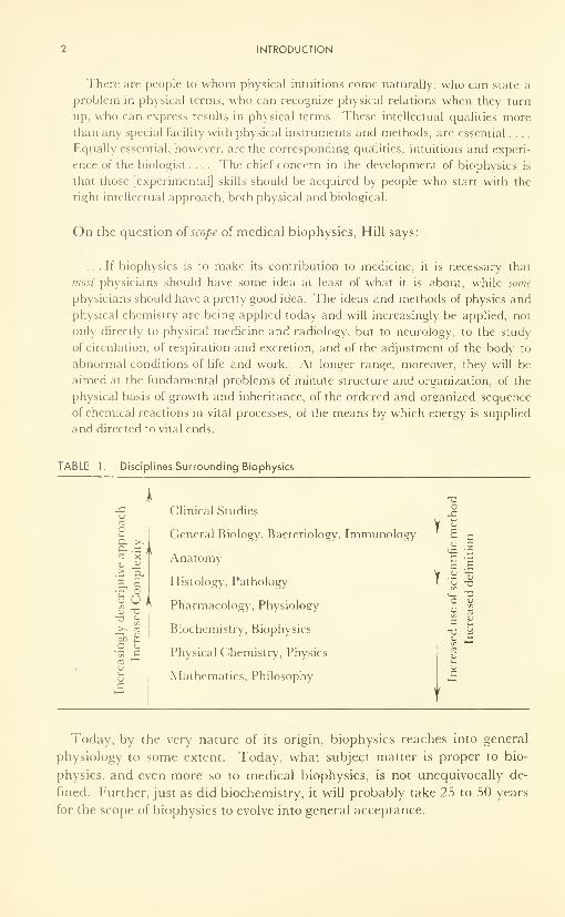
Consider some of the pertinent disciplines in terms of Table 1. Biochem-
istry and biophysics attempt to describe and interpret the chemical and
physical processes of biological materials in terms of the principles of or-
ganic chemistry, physical chemistry, and physics. Biophysics is concerned
with questions about the physics of biological systems. It has the advantages
of less complexity and more certainty than the biological subjects, but has
the disadvantage of being limited to only specific aspects of the whole living
system. For the human being, biophysics can be thought of as providing a
description of his whole physical system from the particular view of physics.
For medical research, for the highest forms of medical specialization, and
for the general medical practitioner of the years to come, the requirement
seems inevitably to be a strong background and experience in the medical
arts, coupled with a thorough grounding in the scientific knowledge of medi-
cine and the scientific approach to it. The same is true of the biosciences.
The scope of biophysics today is rather broad, if judged by the attitudes
of authors of papers in several of the current journals, and in various essays.
Yet the master, A. V. Hill, a Nobel prize winner who published his first
paper in 1910 and is still active in research and physiology, has cautioned
that the use of physical techniques or ideas alone for investigation of bio-
logical problems does not of itself make biophysics. He defines the subject
as: "the study of biological function, organization, and structure by physical
and physiochemical ideas and methods," and then hastens to emphasize
that he has put ideas first. He further expands* and drives home the key
point as follows:
*From "Lectures on the Scientific Basis of Medicine," Vol. 4, Athlone Press, London,
1954-1955; reprinted inSdence, 124, 1233 (1956).
1
INTRODUCTION
There are people to whom physical intuitions come naturally, who can state a
problem in physical terms, who can recognize physical relations when they turn
up, who can express results in physical terms. These intellectual qualities morethan any special facility with physical instruments and methods, are essential ....
Equally essential, however, are the corresponding qualities, intuitions and experi-
ence of the biologist .... The chief concern in the development of biophysics is
that those [experimental] skills should be acquired by people who start with the
right intellectual approach, both physical and biological.
On the question of scope of medical biophysics, Hill says:
... If biophysics is to make its contribution to medicine, it is necessary that
most physicians should have some idea at least of what it is about, while some
physicians should have a pretty good idea. The ideas and methods of physics and
physical chemistry are being applied today and will increasingly be applied, not
only directly to physical medicine and radiology, but to neurology, to the study
of circulation, of respiration and excretion, and of the adjustment of the body to
abnormal conditions of life and work. At longer range, moreover, they will be
aimed at the fundamental problems of minute structure and organization, of the
physical basis of growth and inheritance, of the ordered and organized sequence
of chemical reactions in vital processes, of the means by which energy is supplied
and directed to vital ends.
TABLE 1. Disciplines Surrounding Biophysics
ouCL >.
> a.
.a.|uUl
V TJ-a «->
en>> m"3d aC O
'35 £re
l— '
vs_<j
c
Clinical Studies
General Biology, Bacteriology, Immunology
Anatomy
Histology, Pathology
Pharmacology, Physiology
Biochemistry, Biophysics
Physical Chemistry, Physics
Mathematics, Philosophy
-a
SUBJECT MATTER
SUBJECT MATTER
From recent and current literature, and within the scope discussed, it has
been possible to arrive at a fair idea of the topics which are termed "Bio-
physics."
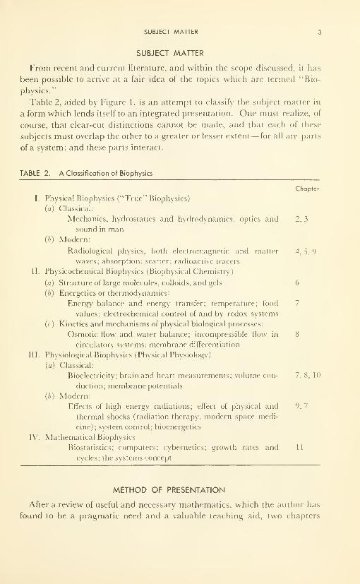
Table 2, aided by Figure 1, is an attempt to classify the subject matter in
a form which lends itself to an integrated presentation. One must realize, of
course, that clear-cut distinctions cannot be made, and that each of these
subjects must overlap the other to a greater or lesser extent— for all are parts
of a system; and these parts interact.
TABLE 2. A Classification of Biophysics
Chapter
I. Physical Biophysics ("True" Biophysics)
(a) Classical:
Mechanics, hydrostatics and hydrodynamics, optics and 2, 3
sound in man(b) Modern:
Radiological physics, both electromagnetic and matter 4, 5, 9
waves; absorption; scatter; radioactive tracers
II. Physicochemical Biophysics (Biophysical Chemistry)
(a) Structure of large molecules, colloids, and gels 6
(b) Energetics or thermodynamics:
Energy balance and energy transfer; temperature; food
values; electrochemical control of and by redox systems
(c) Kinetics and mechanisms of physical biological processes:
Osmotic flow and water balance; incompressible flow in 8
circulatory systems; membrane differentiation
III. Physiological Biophysics (Physical Physiology)
(a) Classical:
Bioelectricity; brain and heart measurements; volume con- 7, 8, 10
duction; membrane potentials
(b) Modern:
Effects of high energy radiations; effect of physical and 9, 7
thermal shocks (radiation therapy, modern space medi-
cine); system control; bioenergetics
IV. Mathematical Biophysics
Biostatistics; computers; cybernetics; growth rates and 1 1
cycles; the systems concept
METHOD OF PRESENTATION
After a review of useful and necessary mathematics, which the author has
found to be a pragmatic need and a valuable teaching aid, two chapters
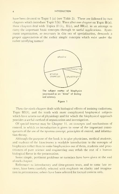
4 INTRODUCTION
have been devoted to Topic I (a) (see Table 2). These are followed by two
chapters which introduce Topic I (b). Then after one chapter on Topic II (a),
three chapters deal with Topics II (b), 11(c), and III (a), in an attempt to
carry the important basic concepts through to useful applications. Syste-
matic organization, so necessary in this era of specialization, demands a
proper appreciation of the rather simple concepts which exist under the
rather terrifying names!
The subject matter of biophysics
(expressed as an "Area" of biolog-
ical science).
Figure 1
Then the ninth chapter deals with biological effects of ionizing radiations,
Topic 111(6), and the tenth with more complicated biophysical subjects
which have arisen out of physiology and for which the biophysical approach
provides a useful method of organization and investigation.
Of special interest may be Chapter 11, on concepts and mechanisms of
control, in which an introduction is given to some of the important conse-
quences of the use of the systems concept, principles of control, and informa-
tion theory.
Although the purpose of the book is to give physicians, medical students,
and students of the biosciences a readable introduction to the concepts of
biophysics rather than to make biophysicists out of them, students and prac-
titioners of pure science and engineering may relish the zest of a humanbiological flavor in the presentation.
Some simple, pertinent problems or exercises have been given at the end
of each chapter.
References to introductory and time-proven texts, and to some late re-
views, have been carefully selected with emphasis on clarity and imagina-
tion in presentation; others have been selected for factual content only.

METHOD OF PRESENTATION 5
If the principles to follow are pondered at length, and reillustrated by the
reader in other examples of his choice, the clarity of thought, and the true
power and scope of the basic principle will become evident.
Conversely, it seems axiomatic, but it is often forgotten, that the serious
reader should seek and expect to find in a book such as this a continuous
thread of purpose in all the material contained between its covers.
CHAPTER 1
The Systems Concept, and
Ten Useful Pillars of
Mathematical Expression
In scientific thought we adopt the simplest theory which will explain all
the facts under consideration and enable us to explain new facts of the
same kind.
The catch in this criterion lies in the word "simplest. " It is really an
aesthetic canon such as we find implicit in our criticisms of poetry or
painting.
The layman finds such a law as
dx/dt = kd 2x/dy 2
much less simple than "It oozes," (or "It diffuses," or "It flows"), of
which it is the mathematical statement.
The physicist reverses this judgement, and his statement is certainly the
more fruitful of the two so far as prediction is concerned.
(J. B.S. Haldane.)
THE "SYSTEMS" CONCEPT
In modern science and engineering an almost unbelievably broad and
comprehensive use is made of the term "systems" and its various connota-
tions. Chemists have long used the term to indicate the collection of chemi-
cals—the chemical system—on which an experimenter was working. Biolo-
gists have long used the term to indicate the group of materials and events
THE SYSTEMS CONCEPT 7
within the containing walls of the living thing: the biological system, or the
living system. It was in the military campaign of ancient times that the idea
or concept of control, within the military system, began to creep in. In mod-
ern military systems, in educational, government, and business systems, the
idea of organization and control by the central authority of the system has
been developed. The concept has reached its highest state of definition and
description in military defense systems—based principally on the extension
of the use of electronic circuitry to other tasks than those performed by the
simple oscillators of thirty years ago. Nevertheless, in those days a one-tube
affair had all the elements of a modern system : a detector or source of in-
formation fed a voltage signal into the grid of the vacuum tube; the signal
modified the plate current by exercising a control over the direction of flow
of electrons in the tube; the modified plate current passed through an ex-
ternal load of resistors, the voltage drop across one of which was fed back
into the input grid and exerted instantaneous control of the plate current;
while the voltage drop across the rest of the load was used to perform the
task assigned— in this case to feed the stable oscillating voltage into further
circuitry.
The elements of this system are simple enough: a detector or source of in-
formation (grid input), the transmission to a central authority (the grid), the
control by the authority of expenditure of energy (in the plate circuit), and feed-
back of part of the expended energy into the central authority so that the
latter can know whether or not the energy is being expended in the desired
manner and make corrections if necessary. One other element which the
simple tube circuit does not have is the facility of being able to store informa-
tion for use when required. A modern computer has this facility.
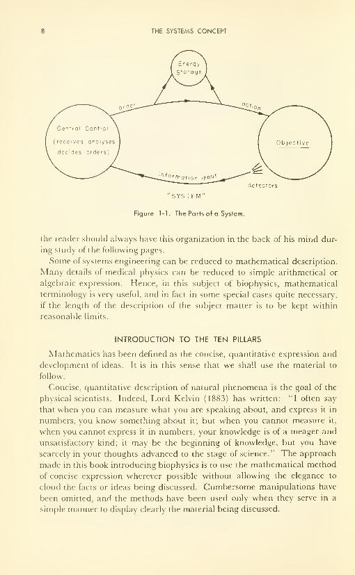
The living thing, and man especially, if a self-contained system (Figure 1-1)
in this sense, having all the essential elements, with versatility and adaptability
as well. The sensory organs (which enable one to see, touch, taste, smell,
and hear) are the detectors of relevant information. Nerve is the transmis-
sion line to the central authority, the brain, which stores information, ana-
lyzes and abstracts the relevant part, decides what to do, and then dis-
patches the necessary commands (electrochemical signals) to the nerve for
transmission to the muscles (say) which expend energy in response to the
command. Both a part of the muscle's expenditure and a continuous ob-
servation by the sensory organs feed back information to the brain so that
the central authority can know if the commands are being carried out. If
not, corrective commands can be dispatched.
Each of the ten chapters to follow is concerned with some aspect of man's
operation as a system. He is the most complex system we know, to be sure,
and it is not always immediately obvious what is the relation between the
detail which we must describe and the over-all systems concept. However,
THE SYSTEMS CONCEPT
detectors
"SYS TEM"
Figure 1-1. The Parts of a System.
the reader should always have this organization in the back of his mind dur-
ing study of the following pages.
Some of systems engineering can be reduced to mathematical description.
Many details of medical physics can be reduced to simple arithmetical or
algebraic expression. Hence, in this subject of biophysics, mathematical
terminology is very useful, and in fact in some special cases quite necessary,
if the length of the description of the subject matter is to be kept within
reasonable limits.
INTRODUCTION TO THE TEN PILLARS
Mathematics has been defined as the concise, quantitative expression and
development of ideas. It is in this sense that we shall use the material to
follow.
Concise, quantitative description of natural phenomena is the goal of the
physical scientists. Indeed, Lord Kelvin (1883) has written." "I often say
that when you can measure what you are speaking about, and express it in
numbers, you know something about it; but when you cannot measure it,
when you cannot express it in numbers, your knowledge is of a meager and
unsatisfactory kind; it may be the beginning of knowledge, but you have
scarcely in your thoughts advanced to the stage of science." The approach
made in this book introducing biophysics is to use the mathematical method
of concise expression wherever possible without allowing the elegance to
cloud the facts or ideas being discussed. Cumbersome manipulations have
been omitted, and the methods have been used only when they serve in a
simple manner to display clearly the material being discussed.
THE TEN PILLARS 9
For subsequent use in the introductory phases of biophysics we now de-
fine ten conveniently grouped concepts. Since most of this is review, the
presentation is cryptic. Since only the language and the logic, and not the
operations, are necessary for future use in this book, we follow the principle
so aptly stated by Lord Dunsany: "Logic, like whiskey, loses its beneficial
effect when taken in too large quantities."
THE TEN PILLARS
1. The Variable
If so'me entity— it may be a physical property or some other combination
of length, mass and time—changes under the influence of a force, that entity
is called a variable. There are dependent and independent variables in nature.
The value of the independent can be chosen at random, but any variable de-
pendent upon that choice is thereby fixed in value.
The ideal gas law, PV = nRT, illustrates this. In a closed vessel of vol-
ume V, containing n moles of gas, the independent variable (on the right-
hand side of the equation by convention) is the temperature, T. The tem-
perature can be chosen at will. However, once T has been fixed, the pres-
sure, P, dependent upon T in this case for its value, has also been fixed.
2. The Function
Further, it can be said that P is proportional to T, or varies directly as 7", or
P & T; that P vanes inversely as V, or is proportional to 1/F, or P <* \/V.
The constant number, R, which serves to equalize the dimensions or units on
the two sides, never varies with experimental conditions, contains all our
further ignorance of this relationship expressing the equivalence of thermal
and mechanical energy, and is one of the universal constants of nature, (7r, the
value of the quotient of the circumference of a circle and its diameter is
another example). There are constants other than the universal ones— they
are simply variables held constant over the course of a particular changing
situation. V in the preceding paragraph is an example. They are called
"constants of the system."
A relationship between two variables, such that a choice of a value for one
fixes the value of the other, is called a functional relationship. In general terms,
if we do not know the exact relationship between two variables, y and x say,
but we know that one exists, we can say y varies with x, or y is a junction of x,
or in shorthand (ormy = f(x).
Nowjy = f(x) is so general that it could describe any functional relation-
ship between y and x. In nature we find both rational and transcendental
functions. Rationals can be expressed as a sum of simple terms, transcen-
dental cannot. Three examples of the former functions are: (a) linear,
10 THE SYSTEMS CONCEPT
(b) parabolic, and (c) exponential. The periodic functions are transcenden-
tal (see Figure 1-2).
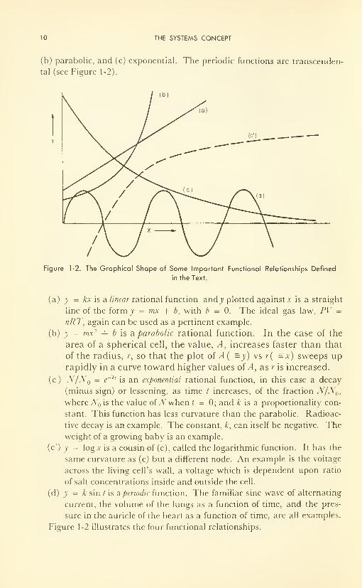
Figure 1-2. The Graphical Shape of Some Important Functional Relationships Defined
in the Text.
(a) y = kx is a linear rational function and j> plotted against x is a straight
line of the form y = mx + b, with b = 0. The ideal gas law, PV =
nRT, again can be used as a pertinent example.
(b) y = mx 2-f b is a parabolic rational function. In the case of the
area of a spherical cell, the value, A, increases faster than that
of the radius, r, so that the plot of A( =y) vs r( =x) sweeps up
rapidly in a curve toward higher values of A, as r is increased.
(c) N/N = e~ klis an exponential rational function, in this case a decay
(minus sign) or lessening, as time / increases, of the fraction N/N0i
where JV is the value of JV when t = 0; and A; is a proportionality con-
stant. This function has less curvature than the parabolic. Radioac-
tive decay is an example. The constant, k, can itself be negative. Theweight of a growing baby is an example.
(c') y = log x is a cousin of (c), called the logarithmic function. It has the
same curvature as (c) but a different node. An example is the voltage
across the living cell's wall, a voltage which is dependent upon ratio
of salt concentrations inside and outside the cell.
(d) y = k sin / is aperiodic function. The familiar sine wave of alternating
current, the volume of the lungs as a function of time, and the pres-
sure in the auricle of the heart as a function of time, are all examples.
Figure 1-2 illustrates the four functional relationships.
THE TEN PILLARS 11
These functions are all continuous; that is, at no point does the slope change
suddenly from one value to another. It is probable that there are no discon-
tinuous functions in nature, although the change in slope may be so sharp as
to seem discontinuous in the first and cursory observation. Thus, phe-
nomena involving the interface or juncture of two phases, as for example at
the cell wall, are examples of rapidly changing continuous functions which
at first sight appear to be discontinuous.
3. Limits
If a variable, changing in accordance with some assigned law, can be
made to approach a fixed constant value as nearly as we wish without ever
actually becoming equal to it, the constant is called the limiting value or limit
of the variable under these circumstances.
A circus abounds with examples in which exceeding a limit in either dis-
tance or time would mean a severe penalty. Consider the "hell drivers" whoride motorcycles inside a 40-ft cylinder, approaching the top— the limiting
height— as closely as they dare, yet never suffering the disaster of actually
reaching it. In other words, if y = /(*), and if, as x approaches a, y ap-
proaches some value, b, then b is said to be the limit of/(x) when x equals a.
In shorthand, for the functional relationship y = f(x), if x —* a as y—
* b,
then
Lim f(x) = b
x—'0
It is often useful to approach a limiting value and study its properties
without having to suffer the embarassment sometimes associated with the
limit itself. This concept was introduced by Leibnitz 300 years ago.
4. Increments
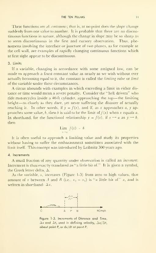
A small fraction of any quantity under observation is called an increment.
Increment is thus exactly translated as "a little bit of." It is given a symbol,
the Greek letter delta, A.
As the variable, x, increases (Figure 1-3) from zero to high values, that
amount of x between A and B (i.e., x2— x
x )is "a little bit of" x, and is
written in shorthand: \x.
!—*fi
i
i r
A P B 40mph
Figure 1-3. Increments of Distance and Time,
Ax and Af, used in defining velocity, Ax/Af,
abouf point P, or dx/dr ai point P.
12 THE SYSTEMS CONCEPT
Increments may be as large or as small as we like. If we reduce the dis-
tance between A and B, the value of Ax is reduced; this can continue until
Ax is infinitesimally small (so small that we cannot think of anything
smaller). Infinitely small increments are called infinitesimals, and are written
in shorthand with the Arabic letter "d", i.e., d.v.
Combining the ideas of Sections 3 and 4, it is seen that as A and B ap-
proach P, Ax gets smaller and smaller until, at the limit, Ax — dx, and it can
be made infinitely small. This means that if we view the point, P, from B, we
can move B in on P as closely as we please— in fact to an infinitely small
distance away—and observe Pfrom as closely as we please. At the limit we
observe Pfrom an infinitely small distance away, i.e., as A.v —> 0.
With the concepts of increments and limits we have implicitly intro-
duced the concept of continuous number, as opposed to the discrete number
which is familiar to us in our unitary, decimal, and fraction systems. Con-
tinuous number admits of the possibility of continuous variation of x be-
tween A and B; the number of steps can be infinite. Continuous number
is involved when a car accelerates from to 40 mph: the car passes through
every conceivable velocity between and 40, and not in the discrete jumps
which our decimal and/or fraction systems would describe. At best, these
latter are but very useful approximations, and can be considered as con-
venient, regular stop-off points, or stations, along the path of continuously
increasing number.
5. Instantaneous Rate of Change
Any living being is a complex system of interrelated physical and chemi-
cal processes. Each of these processes in the "well" being is characterized
by a particularly critical rate (speed or velocity) which enables it to fit into
the complex system without either being too slow and holding all the other
subsequent processes back, or too fast and allowing a runaway of certain
subsequent processes. The study of the factors affecting the rates of
processes is called "kinetics," and is discussed in detail for some biological
processes, in Chapter 8.
Average rate or speed, over some time interval, is often useful; but it is the
instantaneous rate, or the speed at any instant, that is most useful for an
understanding of these complex, interrelated reactions.
If j; = f(x) and the function is continuous, we may be interested in how-
fast y changes at any value of .v. In a diffusion process, for example, y would
be a concentration and v the time. The question is: How much is the concen-
tration in some particular volume changing per second at some particular
second in time? The following three examples, one experimental, one graph-
ical, and one analytical, illustrate the use of limits and increments to de-
scribe this situation.
THE TEN PILLARS 13
(1) Experimental : To measure the instantaneous velocity of an automobile
(refer again to Figure 1-3) requires measurements of distance and time be-
tween two stations, A and B. Two observers with stop watches and a tape
measure can easily do this. They measure a value of Ax/At, which is the in-
crement of distance covered in an increment of time. But the car is acceler-
ating between A and B, and hence Ax/At is only an average value between
A and B, and may be quite different from the velocity as the car passes P.
Better values can be obtained the closer the observers are to P, but of course
no value can be obtained if both observers are at P because Ax = and
At = 0, and 0/0 is indeterminate, or can have any value from — « to + °°.
The best value is obtained by taking observations at several values of A
and B, at smaller and smaller values of Ax, until a good extrapolation to
Ax = can be made. Hence the limit of Ax/At as At approaches zero is
the instantaneous velocity at the point, P. In shorthand notation, the instan-
taneous velocity at PisLim Ax/At.A/—
This symbolic description is further simplified by use of the infinitesimal
symbols: Lim Ax/At = dx/dt. Conversely the previous statement is actu-
ally the definition of dx/dt. In other words, dx/dt is the instantaneous rate
of change of x as t changes. A very simple experimental check on the method
is to ride in a car and note the speedometer reading at point P.
Both of these methods of determining instantaneous rate are exemplified
in biological processes.
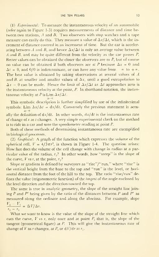
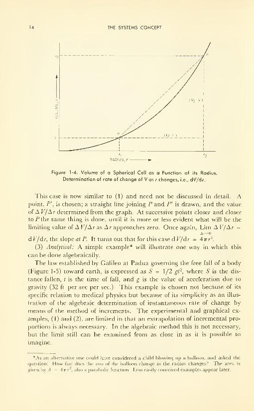
(2) Graphical: A graph of the function which expresses the volume of the
spherical cell, V = 4/37rr 3, is shown in Figure 1-4. The question arises:
How fast does the volume of the cell change with change in radius at a par-
ticular value of the radius, r,? In other words, how "steep" is the slope of
the curve, V vs r, at the point, r,?
Slope or gradient is defined by surveyors as "rise"/"run," where "rise" is
the vertical height from the base to the top and "run" is the level, or hori-
zontal distance from the foot of the hill to the top. The ratio "rise/run" de-
fines the value (trigonometric function) of the tangent of the angle enclosed by
the level direction and the direction toward the top.
The same is true in analytic geometry, the slope of the straight line join-
ing P and P' being given by the ratio of the distances between P and P' as
measured along the ordinate and along the abscissa. For example, slope
V2 - F,= AV/Ar.
r~,—
What we want to know is the value of the slope of the straight line which
cuts the curve, V vs r, only once and at point P, that is, the slope of the
tangent (geometrical figure) at P. This will give the instantaneous rate of
change of Fas r changes, at P, or d V/dr at r,.
14 THE SYSTEMS CONCEPT
RADIUS, r
Figure 1-4. Volume of a Spherical Cell as a Function of its Radius.
Determination of rate of change of V as r changes, i.e., dV/dr.
This case is now similar to (1) and need not be discussed in detail. Apoint, P', is chosen; a straight line joining P and P' is drawn, and the value
of A V/Ar determined from the graph. At successive points closer and closer
to .Pthe same thing is done, until it is more or less evident what will be the
limiting value of A V/A r as A r approaches zero. Once again, Lim A V/Ar =
d V/dr, the slope at P. It turns out that for this case d V/dr = Airr2.
(3) Analytical: A simple example* will illustrate one way in which this
can be done algebraically.
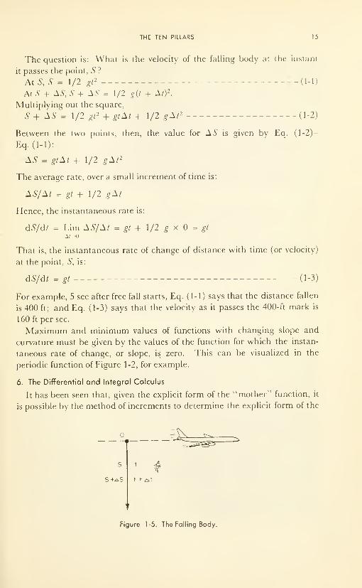
The law established by Galileo at Padua governing the free fall of a body
(Figure 1-5) toward earth, is expressed as S = 1/2 gt 2, where S is the dis-
tance fallen, t is the time of fall, and g is the value of acceleration due to
gravity (32 ft per sec per sec.) This example is chosen not because of its
specific relation to medical physics but because of its simplicity as an illus-
tration of the algebraic determination of instantaneous rate of change by
means of the method of increments. The experimental and graphical ex-
amples, (1) and (2), are limited in that an extrapolation of incremental pro-
portions is always necessary. In the algebraic method this is not necessary,
but the limit still can be examined from as close in as it is possible to
imagine.
*As an alternative one could have considered a child blowing up a balloon, and asked the
question: How fast does the area of the balloon change as the radius changes? The area is
given by A --= 4irr , also a parabolic function. Less easily conceived examples appear later.
THE TEN PILLARS 15
The question is: What is the velocity of the falling body at the instant
it passes the point, S ?
At S, S = 1/2 gt2 -d-1)
At 5 + AS, S + AS = 1/2 g(t + A/) 2.
Multiplying out the square,
S + AS = 1/2 gt 2 + gtAt + 1/2 gAt 2 - -(1-2)
Between the two points, then, the value for AS is given by Eq. (1-2)-
Eq. (1-1):
AS = gtAt + 1/2 gAt 2
The average rate, over a small increment of time is:
AS/At = gt + 1/2 gAt
Hence, the instantaneous rate is:
dS/dt = Lim AS/At = gt + 1/2 g x = gt
A/—
That is, the instantaneous rate of change of distance with time (or velocity)
at the point, S, is:
dS/dt = gt (1-3)
For example, 5 sec after free fall starts, Eq. (1-1) says that the distance fallen
is 400 ft; and Eq. (1-3) says that the velocity as it passes the 400- ft mark is
160 ft per sec.
Maximum and minimum values of functions with changing slope and
curvature must be given by the values of the function for which the instan-
taneous rate of change, or slope, is zero. This can be visualized in the
periodic function of Figure 1-2, for example.
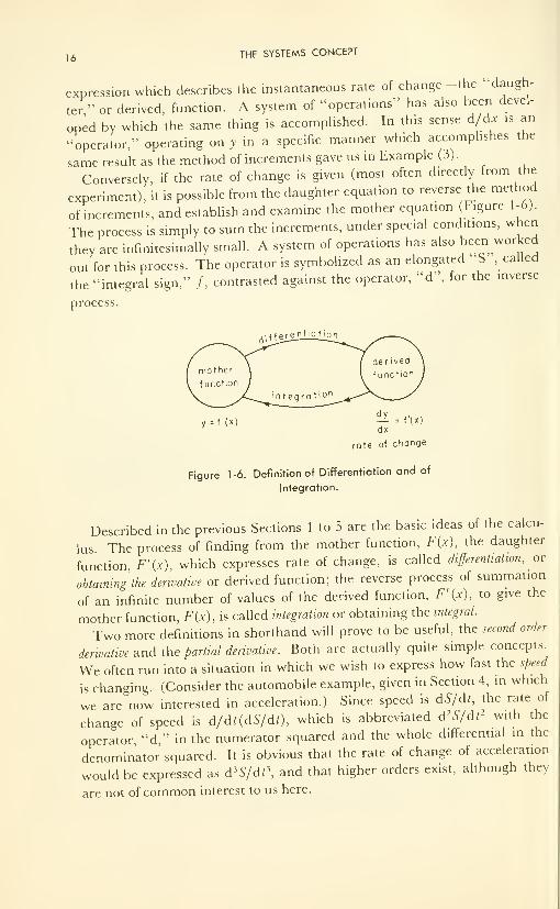
6. The Differential and Integral Calculus
It has been seen that, given the explicit form of the "mother" function, it
is possible by the method of increments to determine the explicit form of the
s t
^t + ^t
Figure 1-5. The Falling Body.
16 THE SYSTEMS CONCEPT
expression which describes the instantaneous rate of change-the "daugh-
ter" or derived, function. A system of "operations" has also been devel-
oped by which the same thing is accomplished. In this sense d/dx is an
"operator," operating on y in a specific manner which accomplishes the
same result as the method of increments gave us in Example (3)
.
Conversely, if the rate of change is given (most often directly from the
experiment), it is possible from the daughter equation to reverse the method
of increments, and establish and examine the mother equation (Figure 1-6).
The process is simply to sum the increments, under special conditions, when
they are infinitesimally small. A system of operations has also been worked
out for this process. The operator is symbolized as an elongated S,called
the "integral sign," f, contrasted against the operator, "d", for the inverse
process.
.-. ^Pj^e_ntio t_i£n
rate of change
Figure 1-6. Definition of Differentiation and of
Integration.
Described in the previous Sections 1 to 5 are the basic ideas of the calcu-
lus The process of finding from the mother function, F{x), the daughter
function, F'(x), which expresses rate of change, is called differentiation, or
obtaining the derivative or derived function; the reverse process of summation
of an infinite number of values of the derived function, F'(x), to give the
mother function, F(x), is called integration or obtaining the integral.
Two more definitions in shorthand will prove to be useful, the second order
derivative and the partial derivative. Both are actually quite simple concepts.
We often run into a situation in which we wish to express how fast the speed
is changing. (Consider the automobile example, given in Section 4, in which
we are now interested in acceleration.) Since speed is dS/dt, the rate of
change of speed is d/d«dS/dt), which is abbreviated d 2S/dt 2 with the
operator "d," in the numerator squared and the whole differential in the
denominator squared. It is obvious that the rate of change of acceleration
would be expressed as d'S/dt\ and that higher orders exist, although they
are not of common interest to us here.
THE TEN PILLARS 17
Sometimes one or more independent variables (y, z) are kept as constants
of the system while another (x) is varied. The rate of change of the dependent
variable, 0, as x changes, is expressed as an incomplete or partial derivative.
To emphasize the partial character, a rounded operator, d, is used; and the
constants of the system are stated as subscripts outside parentheses which
enclose the partial derivative. Thus:
(d(f>/dx)y>i
expresses the rate at which changes as x is changed, when y and z are
kept constant.
The second-order partial derivative, the "acceleration," is expressed as
before:
(d2<i>/dx
2)M
This notation is used in all heat and mass transfer- considerations. For
instance, note the Haldane quotation which introduced this chapter.
At this stage of development of biophysics (1962), the terminology of the
calculus is being used in published work, hence the need for introduction to
the bases and terminology of the subject. But explicit descriptions of most
biophysical phenomena are very rare; hence there seems to be no need to in-
troduce the operational calculus into an introductory book on biophysics at
this time. Therefore no attempt has been made to display the actual opera-
tions by which either differentiation or integration is accomplished. Opera-
tional calculus is treated in detail in many standard textbooks.
7. Distribution of Observations
A great many biological phenomena lend themselves to statistical meth-
ods of expression, i.e., age, height, weight, bloodcount, sugar analysis, etc.
This is so true that the "average value" over a large number is considered
the "normal" value, describing the "normal man." Hence it is instructive
to examine some of the methods of statistical expression, and to discuss their
reliability.
Statistics has come a long way since the publication in 1662 of JohnGraunt's "Natural and Political Observations Made upon the Bills of
Mortality," a study based on the records kept during the Black Plague in
London; and since Sir Edmund Halley (of "Comet" fame) wrote his basic
paper on life insurance, which appeared 30 years later. In the 20th century
statistical methods have penetrated nearly every field of learning in which
numerical measurement is possible. Moroney's book 4 gives a delightful in-
troduction to the subject.
First of all, there are two factors which will result in a distribution in a
number of observations. One is errors in measurement; the other is a real
18 THE SYSTEMS CONCEPT
distribution in what is being measured. Measuring the length of a room
with a 12-in. ruler will result in a fairly wide error, and although the meanvalue of a number of observations should be close to fact, there may be a
large uncertainty in an individual measurement. Besides such random errors,
there may exist also constant errors which are sometimes very important but
too rarely recognized. Suppose the ruler has been made 1/16 in. too short at
the factory. If the room were 32 ft long, in addition to the random errors,
every measurement would have been 2 in. short: even the mean value cannot
be trusted in the presence of a constant error! It is revealing to read the
temperatures on several of the thermometers in the laboratory thermometer
drawer! Constant errors and the need for calibration become quite obvious.
Even under the most carefully controlled experimental conditions, unknownconstant errors creep in. In addition, personal bias is always with us, in
reality if not in principle.
The variation in the quantity being measured is often called "biological
variance." Consider the height of 80 people at a lecture— it usually has a
distribution from about 5 ft, in. to 6 ft, 3 in., with the average approxi-
mately 5 ft, 7 in. Deviations from 5 ft, 7 in., however, could hardly be con-
sidered as errors or abnormalities!
Constant errors are deadly and can result in gross misinterpretations.
Analytical chemistry done without proper calibrations is an example. It
has been shown to be prevalent even in routine analyses done day in and
day out in the hospitals, with large variations in mean values being reported
between them—each hospital apparently having its own constant errors!
This is embarrassing, but it is a fact. Under these conditions, diagnoses
made with reference to some published work from another hospital could
easily be wrong. It is necessary continually to be on the alert against con-
stant errors, or "biased [not personal] observations," as they are sometimes
called.
Random errors and natural distribution in the variable measured can
both be treated with statistical methods. The most reliable methods, and in
fact the only reliable method in constant use, presuppose that the observa-
tions distribute themselves about a mean or average value such that the
density of points is greatest at the mean and progressively less and less as the
deviation from the mean becomes larger. That is, it presumes a "normal"
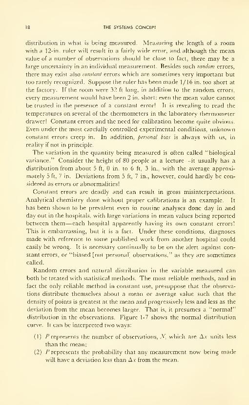
distribution in the observations. Figure 1-7 shows the normal distribution
curve. It can be interpreted two ways:
(1) P represents the number of observations, N, which are Ax units less
than the mean;
(2) P represents the probability that any measurement now being made
will have a deviation less than Ax from the mean.
THE TEN PILLARS 19
It is axiomatic that any expression of confidence made in terms of normal
distribution, presupposes normal distribution; and that any such expression
concerning a distribution which is not normal is not only unwarranted, but
also useless, and may be quite misleading. There are statistical methods for
handling non-normal data, but they are not simple and are seldom used
correctly. Mainland's book 3 goes into some of these, using examples of
medical interest.
-^x + ^y.
DEVIATION FROM MEAN VALUE
Figure 1-7. Normal Distribution of Observations. Solid Curve: Area under curve be-
tween -a and +a includes 68 per cent of observations; between —2a and +2o, 94 per
cent; and between -3a and +3o, greater than 99 per cent. Blocks: Typical Observa-
tions of Heights of Thirty People at a Lecture.
8. Expressions of Deviations
The most common method of expressing a number of observations, x, of
the same phenomenon is by the common average, or arithmetic mean, x. There
are others, such as the median and the mode, which have some use in nearly
normal distributions, but only the mean will be considered. Deviations Ax
from the mean can easily be computed by subtraction, and then averaged,
the result being expressed as the mean deviation Ax from the mean x.
A very common method of expressing the distribution is by the standard
deviation, a, defined as the square root of the average of the deviations
squared:
a = y/Ax 2, or a = y^Ax 2/n
20 THE SYSTEMS CONCEPT
Bessel's correction is introduced if the number, n, of samples is small
(< 30); then
a = j/£ Ax 2/(n - 1)
The most probable deviation, r, is that value of the deviation such that one-
half the observations lies between the limits ±r.
The relative deviation, usually expressed as a per cent, is the fraction which
the deviation is of the observed mean value, i.e., Ax/x.
Each of these has several names. In the case of random errors, "devia-
tion" should read "error," of course; Ax is often called the absolute error of
the measurement. Relative error is sometimes called per cent error or proportional
error. These are discussed in detail, and examples are given, in Mainland's
book.
Superposition of Errors. In the determination of a quantity, A, af(x, y, z)
which requires measurement of x, y, and z, each with an absolute error, the
errors must be superimposed one upon the other, or added; the reliability of the
value obtained for A is no better than the sum of the errors in x, y and z-
That is, the relative error in A is the sum of the relative errors in the meas-
urements oix,y, andz-
9. Indices and Logarithms
In arithmetic the ancient Greeks devised and used a notation, now called
that of indices, to express in shorthand the number of times a number is to be
multiplied by itself. Thus, "2 multiplied by itself 5 times" (i.e., 2 x 2 x
2x2x2) = 32. This is written in shorthand as 2 5 = 32. The index, 5, is
placed as a superscript to the base number 2.
A number of laws of indices can be shown to exist for the manipulation of
such numbers. These laws were observed for cases in which the indices are
whole numbers.
Now there is no reason to suppose that the rules would be different for
fractional indices, although to multiply 2 by itself 5 1/2 times would really
be tricky! Nevertheless, the rules are assumed to apply to fractional indices,
as well as to whole-number ones, and further also to algebraic, unknown
indices. In general, the laws of indices are as follows:
(\)a m = axaxaxaxa m times
(2) a m a" = a m+n
(3) a m/a n = am -"if m > n
1 ..
or am/a" = it n > m
THE TEN PILLARS 21
(4) (am
)
n = a mn
(5) (ab) m = am bm
(6) {a/b) m = a m/bm
Fractional indices are called roots. Thus, a i = y/a, the square root of a;
and in general a'/"' = m\/~a, the m lh root of a.
(7) a" = 1
(8) a~" = 1/a"
(9) a" = °o
(10) a~' = 1/a" =
Logarithms
Let .4 = a". The index x, which tells how many times the base number a
must be multiplied by itself to give A, is defined as the logarithm ofA to the base a.
In shorthand this statement is given by x = lbgaA, where "to the base a"
appears as a subscript to the abbreviated "logarithm."
Logarithms are indices and must obey the ten Laws of Indices, just as any
other. For example:
log AB = log A + log B
log A/B = log A - log£
log A m = m log ,4
A change of base from base a to base b turns out to be analogous simply
to a change of variable. In other words the logarithm to the base, a, is re-
lated to the logarithm to the base, b, by a constant, \o%ba. One is a linear
function of the other.
This can be shown as follows. Suppose A = a" and A = by , so that
a x = by . Then logaA = log
ab>, or x = y log
ab.
There are two systems of logarithms in daily use in biophysics, as in all
other science and technology:
(a) Common logarithms, to the base \0(y = \0" for example), used to
simplify the manipulations of multiplication and division, based on rules (2)
and (3). The abbreviation is log, or logl0
.
(b) Natural logarithms, to the base e (y = ex for example), where
e = 2.71828. . . . The base, e, and the functional relationship,}' = e", occur
over and over again in man's description of nature, and therefore will be
illustrated further. The abbreviation is In, or logf
.
22 THE SYSTEMS CONCEPT
Conversion, as described above, is accomplished as follows:
log A = In A2.303
where 2.303 = log, 10.
10. Infinite Series,- y = y eox
A series is any group of numbers, arithmetically related, which differ from
each other in some regular and explicit manner. Thus
1+2 + 3 + 4 + 5 + n
is a series. This particular series is divergent, since the larger the n chosen,
the greater the sum becomes. There are other series which are convergent,
whose value approaches a limit as the number of terms is increased toward
infinity. One such convergent series is
x x 2 x 3 x 4
1 + — + + ' + — +1 2x1 3x2x1 4x3x2x1
This series, for a value of x = 1, simplifies to
1 l2
l3
l4
1 + — + + + +1 2x1 3x2x1 4x3x2x1
which converges to the numerical value 2.71828 .... as more and more
higher index terms are added. In shorthand ex
is written for the first, and ex
or e for the second series. Thus
X X L X 3 X*
and
ex = 1 + — + — + + +
1 2x1 3x2x1 4x3x2x1
1 l2
l3
e = 1 + — + — + + = 2.718281 2x1 3x2x1
More generally, when x is preceded by a constant, k, kx is substituted for x
:
, , kx (kx) 2 (kxy (kxye** = 1 + h
-—— + — + — +1 2x1 3x2x1 4x3x2x1
The constant, k, simply tells how slowly the series converges for any particular
value of x : the greater the value of k the greater the number of terms which
will be necessary to define ekx
to a chosen number of significant figures.
Now, when x is the variable, and k constant, we can call its evaluation
proportional to jv and write
or y a ekx (1-4)
THE TEN PILLARS 23
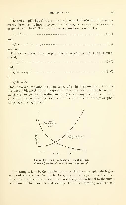
The series typified by ekx
is the only functional relationship in all of mathe-
matics for which its instantaneous rate of change at a value of x is exactly
proportional to itself. That is, it is the only function for which both
y a ekx (1-4)
and
dy/d.v « ekx (or « y) (1-5)
are true.
For completeness, if the proportionality constant in Eq. (1-4) is intro-
duced,
y =y^ ---(1-4')
and
dy/dx = ky ekx ,__(l-5')
or
dy/dx = ky
This, however, explains the importance of ex
in mathematics. The im-
portance in biophysics is that a great many naturally occurring phenomena
are observed to behave according to Eq. (1-5'): many chemical reactions,
growth, diffusion processes, radioactive decay, radiation absorption phe-
nomena, etc. (Figure 1-8).
TIME
Figure 1-8. Two Exponential Relationships:
Growth (positive k), and Decay (negative k).
For example, let y be the number of atoms of a given sample which give
out a radioactive emanation (alpha, beta, or gamma ray), and x be the time.
Eq. (1-4') says that the rate of emanation is always proportional to the num-
ber of atoms which are left and are capable of disintegrating, a statement
24 THE SYSTEMS CONCEPT
which, if reflected upon, will become quite obvious because it is not only
a "natural" law, an observed law of Nature, but also a logical deduction.
In our examples, most commonly a decay is involved, in this case the
decay of a concentration. Thus k is a negative number. If the minus sign
is taken out of the k and k replaced by — X, the expression becomes N =
N e~Xl
, sometimes written N = N exp(-Xt), for radioactive decay, where
JV is the number of particles present when t = 0.
Figure 1-8 shows the shape of the exponential curve for positive k values
(growth), and for negative k values (decay). Note that the former increases
to infinity, unless checked by the onset of some other law; and that the latter
decays toward zero, reaching zero only after an infinitely long time, although
it may be below the lowest measureable value within a very short time. Thelarger the value of k, the faster the growth curve sweeps upwards, and the
sooner the decay curve approaches zero.
PROBLEMS
1-1: (a) If a student must pass biochemistry, andJohn is a student, then . . . ?
(b) If y = 2x and Z = y, then what functional relationship exists between
Z and*?
(c) Uy =/,(*) and £ = f2(x); and f2
(x) = /, (x) -f3 (x), then what is the rela-
tionship between x andy?
(d) If A °c B, and B °c C, what is the relationship between A and C?
(e) If the weight of a given volume of gas is proportional to density, and if the
density is proportional to its pressure, then what is the relationship between
weight of a given volume and its pressure?
1-2: Choose at random, alphabetically for example, the heights in inches of
25 students.
(a) Is the distribution normal? Was the sample biased?
(b) What are the average deviation, Ax, and the standard deviation, a?
(c ) What fraction of the sample falls within the mean deviation from the mean?
(d) What fraction of the sample falls within one standard deviation from the
mean? If the distribution had been normal, what would have been the
fraction?
(e) What fractions of the sample fall with ±2 a and ±3 a? If the distribution
had been normal, what would have been the fractions?
1-3: Make a table showing how the distance fallen, the speed, and the acceleration
of a parachutist change in the first 5 sec before the chute opens. (Make the
calculations for each second.)
Suppose he hits the earth at a velocity of 120 ft per sec without the chute
opening. From what height did he jump?
1-4: The decay of Sr 90 follows the exponential law N = JV e~Xl
, where N is the
concentration of radiating material at any time, t; NQis the concentration at
some arbitrary zero of time; and X is the decay constant of Sr 90, namely 0.028
years" 1
(i.e., 0.028 is the fraction lost per year).
REFERENCES 25
(a) Make a table showing values of -Xl, e~ Xl, and N e~Xt
for various
values of / (years), assuming that N = 100% at/ = 0.
(b) From the results, make a plot of JV vs t, and estimate the half-life (the time,
r, in years, when N = 50% of A ).
(c) Sketch decay curves for P 32 (t = 14.3 days), I'31
(8 days), C' 4 (5100 years),
Co60(5.3 years), Po210 (138 days), and Ra 226 (1620 years), all on the same
graph. Compare them.
REFERENCES
1. Petrie, P. A., et al., "Algebra— a Senior Course (for High Schools)," The CoppClark Publishing Co. Ltd., Toronto, 1960. (See p. 314jffor discussion on incre-
ments.)
2. Thompson, Silvanus P., "Calculus Made Easy (Being a Very Simplest Introduc-
tion to Those Beautiful Methods of Reconing which are Generally called by
the Terrifying Names of the Differential and Integral Calculus)," 3rd ed.,
MacMillan& Co. Ltd., London, 1948.
3. Mainland, D., "Elementary Medical Statistics," W. B. Saunders Co., Philadel-
phia, Pa., 1952.
4. Moroney, M. J., "Facts from Figures," 3rd ed., Penguin Books Ltd., Toronto,
1956.
CHAPTER 2
Some Physical Forces Exemplified
in Man(Mechanical; Osmotic; Electrical)
All physical reality is a manifestation of what force does. On the ques-
tion of what force is, science can do no better than to call it by other names.
(Truth is a virtue, however inconvenient.)
INTRODUCTION
Force and energy, along with optics and acoustics, are the concerns of
classical medical physics, and some of the principles have been understood
for well over a hundred years. In this chapter the nature and the units of
force are reviewed, and the relationship between force and energy discussed.
The transfer of energy is reserved for Chapter 7.
The living system is in a state of continual exchange of force and energy
with the environment. What is force? According to Newton (1687), it is vis
impressa, an influence, measurable in both intensity and direction, operating
on a body in such a manner as to produce an alteration of its state of rest or
motion. Generically, force is the cause of a physical phenomenon. It is
measured by its effect. Further penetration of the nature of force seems
destined to remain a philosophical question, because the range of experi-
ment stops at measurement of the effects.
By experiment it is possible to measure the effect of different forces on the
same object, and devise a system of interconversion factors by which one
kind of force is related to another (for example, mechanical to osmotic). Ef-
26
MECHANICAL FORCES 27
forts to penetrate the generic nature of the "force field"— to develop a uni-
fied theory— received much impetus, without much success, during the life
of Albert Einstein, but one notices now that efforts at unification are falling
off as theorists drift into other problems. Hence the question most funda-
mental to all science, biophysics included, viz: "What is force?", seems
destined to remain unanswered for a long time yet. It is a more fundamental
question even than "What is life?", for life is only one manifestation of force!
MECHANICAL FORCES
Newton's Three Laws of Motion
These three laws are the basic description of mechanical systems. Fromthe simple statements can be inferred many properties of mass and inertia.
First Law: A body at rest tends to stay at rest, and a body in motion tends
to continue moving in a straight line unless the body is acted upon by someunbalanced force (F). The property of the body by virtue of which this is
true is given the name inertia. The measure of amount of inertia is called
the mass (m).
Second Law: A body acted on by an unbalanced force will accelerate in the
direction of the force; the acceleration (a) is directly proportional to the un-
balanced force and inversely proportional to the mass of the body.
This second law describes the familiar experimentally derived relationship
F a ma, or F = kma. If the dimensions of F are suitably defined, this be-
comes F = ma. The need to choose the dimensions in this manner results
from the fact, discussed earlier, that we really do not know what the nature
of force is, but rather do we know only its effects. This is certainly true of
the common forces of gravitation, electrostatics, and magnetism. Yet fric-
tional force we are able to relate to physical interference of microrough-
nesses and physical attraction of two surfaces—and thus have some idea of
what this force is. The force exerted by the finger to push the pencil, or the
force exerted by the thumb on a hypodermic needle drive home to us a
meaning of mechanical force based on its effects.
Third Law: For every physical action there exists an equal and opposite
reaction. The recoil of a rifle as the bullet is ejected, and the swinging arms
which help man to maintain his balance while walking briskly, are examples.
Careful consideration of the statements themselves will enable the reader
to appreciate the far-reaching consequences of these laws, consequences
which range from suspension bridges to the molecular interactions of bio-
chemistry, from the effects of high centrifugal forces on the pilot of a high-
speed aircraft to the simple levers of which the human body in motion is a
remarkably complex, though well coordinated, example.
28 SOME PHYSICAL FORCES EXEMPLIFIED IN MAN
Units and Dimensions
It is useful now to introduce definitions of certain quantities in mechanics.
By the first law, a force is defined as anything which changes the state of rest
or of motion in matter. The basic unit of force, in the centimeter-gram-
second system, is called the dyne. This is the force which will produce an ac-
celeration of 1 cm per sec each sec (1 cm sec-2
) on a mass of 1 gram (1 g).
All other forces (electrical, etc.) can be related by suitable experiments to
this fundamental quantity of motion.
Force gives to mass an energy, a capability of doing work. In the system
of mechanics, the amount of energy acquired by a mass under the influence
of a force depends upon how long or over what distance the force acts. Theenergy imparted to 1 g of mass by a force sufficient to give the mass an ac-
celeration of 1 cm sec-2
within the distance 1 cm, is called 1 erg. Oneerg = 1 dyne cm. This is an inconveniently small unit of energy, and a
quantity often million (107) ergs has been defined as 1 joule (1 jou).
By contrast with this definition of energy units in the mechanical system,
the unit of heat energy, the small calorie, has been defined as the amount of
energy which it takes to raise the temperature of 1 g of water 1° C, between
4.5 and 5.5° C, where water is the most dense.* (As the temperature is
lowered, water molecules begin to line up in "anticipation" of freezing, and
the volume increases; as the temperature is raised, increased thermal energy
tends to drive the molecules apart, and the volume also increases). Experi-
mentally, by transformation of mechanical motion into heat in a water
calorimeter, 1 cal has been found to equal 4.18 jou. One thousand cal, or
1 kilocalorie (1 kcal), has been defined 1 Cal, or large calorie. This is the
unit used to describe the energy available from different foods.
Power is the rate at which energy is expended; that is, energy expended per
unit time. The basic unit of power is the joule per second, called the watt
(w). One-thousand watts is 1 kilowatt (1 kw). One horsepower (1 hp) is
equivalent to 746 w or 3/4 kw.
Entergy exists in two general forms, kinetic and potential. Kinetic energy
is that possessed by mass in motion. In mechanics potential energy is that
possessed by a mass because of its position. In other disciplines potential
energy assumes different forms: the energy stored in chemicals, or that
stored in extended muscle, or in an electrostatic charge separation across a
cell membrane, could be released to do useful work or provide heat.
Heat energy is all kinetic energy. It is the total energy of motion of all
the molecules in the body under consideration. Temperature is an indicator
of the amount of heat in a body, and can be considered to be the "force-like"
*The amount of heat required to raise 1 g of a substance 1°C is called the specific heat, c. It
can be measured under constant pressure (cp ) or under constant volume (cy ).
MECHANICAL FORCES 29
factor of heat energy. The accompanying capacitive factor in effect sums up
the energies which can go into all the vibrations, rotations, and translations
of each molecule. This capacitive factor is called entropy, S. Heat energy is
therefore given as the product TS, and 5* must have the units calories per
degree, since the product must be simply calories.
Heat energy was chosen over electrical, mechanical, or other forms for no
other reason than that it is so common. All forms of energy can be factored
into two parts, a potential part and a capacitative part: thus in addition to
heat energy, we have force times distance for mechanical energy; voltage
times charge for electrical energy; pressure times volume for the mechanical
energy contained by a compressed gas; chemical potential times number of
moles for chemical energy. Energy and its factors will be considered more
fully in Chapter 7.
Kinetic energy of mass in motion is given by force x distance, which has
the dimensions (g cm/sec2) cm, or g cm 2/sec2
. Kinetic energy of motion is
also given by the familiar 1/2 mv 2, with the same dimensions. Another
familiar property of mass in motion is the momentum, M, defined as mv.
Hence KE = 1/2 Mv.
Some of these quantities can be illustrated by the example of a 200-lb**
football player running at full speed with the ball. His potential energy in
the form of food has been reprocessed into glycogen, etc., and stored as po-
tential energy. That part ready for rapid conversion is available in the form
of the mobile chemical adenosine triphosphate (ATP), whose role as a mo-
bile power supply is wondrously general throughout the living system. Dur-
ing the motion this chemical energy is being transformed, at least in part, to
the mechanical kinetic energy of motion. His KE amounts (speed 100 yds in
12 sec; 1 lb = 454 g) to about 26,000,000,000 (or 26 x 10") ergs, or 2600
jou, about 550 small calories. If he is stopped completely within 1 sec by
collision, he will have transferred energy at an average rate during that
second of 2600 jou per sec, 2600 w, or just over 3 hp. If that energy all went
into heat, it could vaporize about 1 g of water. On the other hand this
energy could have been transformed into electricity, and the power delivered
could have lighted twenty-four 100-w light bulbs to full brilliance for a sec-
ond! A further insight into the power expended in such collisions can be
gained if it is remembered that the bulk of the energy is transferred in
about 1/10 sec of contact, during which time the power is about 30 hp! It is
obvious that, in spite of the delights attached to such athletic pursuits, from
the point of view of pure physics alone, they are sheer waste of energy and
power which could be used more efficiently to do other tasks. In fact even
** Weight, a force. Since F = ma: 1 lb force = 1 lb mass x 32 ft/ sec2
, and 980 dynes
force = 1 g force = 1 g mass x 980 cm/ sec2
. (1 lb force is the force of attraction between
the earth and 454 g mass.)
30 SOME PHYSICAL FORCES EXEMPLIFIED IN MAN
at its slowest, when no work is being done, basal metabolism amounts to
about 0.1 hp. The human machine needs a minimum of 0.1 hp to keep it
alive, and can put out continuously a maximum of about 0.01 hp of useful
mechanical work, with occasional surges to several horsepower.
The football player's momentum just before collision was (200/32) x
(300/12) = 154 lb sec. If this were transferred in 0.1 sec during collision,
the impressed force, defined as rate of change of momentum, dM/dt, was
154/0.1 = 1540 lbs. This can be expressed as a "shock" (force per unit
mass) of about 7.7 g, where g is the acceleration of all bodies due to gravita-
tional attraction to the earth (32 ft/sec2, or 980 cm/sec 2
). The value 7.7 gis obtained directly from the second law, viz
J7I154° 77a = t m = = 7.7 g200/g
By contrast, and as further illustration, the passengers on a modern com-
mercial jet line experience about 2 g during take-off. The jet pilots for
fighter aircraft and the astronauts have been tested up to 18 g. The famous
right hand of boxer Joe Louis was said to impart up to 40 g to a stationary
and nonelastic target. A laboratory centrifuge will provide a centrifugal
acceleration of some thousands ofg; and the ultracentrifuge used in sedi-
mentation experiments in which molecular weights of large molecules are
obtained, develops up to 100,000 g. Centrifugal motion is convenient for
varying at will the inertial mass of a body: e.g., in the human centrifuges in
space-research laboratories.
As a machine, man is very versatile. However, he is quite inefficient be-
cause of the continuous power being expended to keep him alive when he is
not "in use." His highest purely physical role is that of a computer.
Two forces will now be considered: a mechanical force as applied to a
lever, and the mechanical force of a compressed gas.
The Lever
A lever is one of a great number of machines—devices for doing work. This
particular device permits mechanical energy to be factored into such values
of force and distance that some desired mechanical result can be ac-
complished. The lever does not create energy, of course, but simply makes
the energy more available to do the particular job at hand. The familiar
example of the crowbar to dislodge a large stone, using a log as a pry, is an
example. In this case a relatively small force applied over a relatively large
distance at the hands is transformed into a relatively large force applied over
a relatively small distance at the stone. The mechanical advantage is the
ratio of the two forces; it is inversely proportional to the ratio of the two dis-
tances since Fid
]
must equal F2d
2.
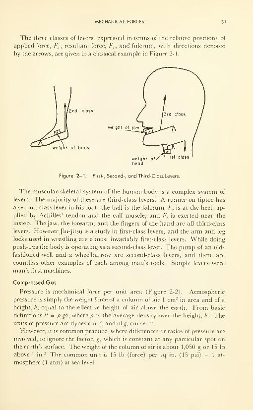
MECHANICAL FORCES 31
The three classes of levers, expressed in terms of the relative positions of
applied force, Fa , resultant force, F
r , and fulcrum, with directions denoted
by the arrows, are given in a classical example in Figure 2-1.
2nd class
weight of jaw
weight of body
weight of
head
Figure 2-1. First-, Second-, and Third-Class Levers.
The muscular-skeletal system of the human body is a complex system of
levers. The majority of these are third-class levers. A runner on tiptoe has
a second-class lever in his foot: the ball is the fulcrum, Fa
is at the heel, ap-
plied by Achilles' tendon and the calf muscle, and FT
is exerted near the
instep. The jaw, the forearm, and the fingers of the hand are all third-class
levers. However Jiu-jitsu is a study in first-class levers, and the arm and leg
locks used in wrestling are almost invariably first-class levers. While doing
push-ups the body is operating as a second-class lever. The pump of an old-
fashioned well and a wheelbarrow are second-class levers, and there are
countless other examples of each among man's tools. Simple levers were
man's first machines.
Compressed Gas
Pressure is mechanical force per unit area (Figure 2-2). Atmospheric
pressure is simply the weight force of a column of air 1 cm 2 in area and of a
height, h, equal to the effective height of air above the earth. From basic
definitions P = p gh, where p is the average density over the height, h. Theunits of pressure are dynes cm -2
, and of g, cm sec-2
.
However, it is common practice, where differences or ratios of pressure are
involved, to ignore the factor, g, which is constant at any particular spot on
the earth's surface. The weight of the column of air is about 1 ,050 g or 1 5 lb
above 1 in.2 The common unit is 15 lb (force) per sq in. (15 psi) = 1 at-
mosphere (1 atm) at sea level.
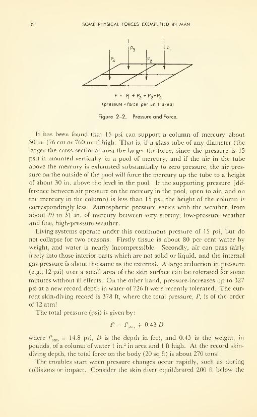
32 SOME PHYSICAL FORCES EXEMPLIFIED IN MAN
f .. pt
+ p2+ p3+ p4
(pressure = force per unit area)
Figure 2-2. Pressure and Force.
It has been found that 15 psi can support a column of mercury about
30 in. (76 cm or 760 mm) high. That is, if a glass tube of any diameter (the
larger the cross-sectional area the larger the force, since the pressure is 15
psi) is mounted vertically in a pool of mercury, and if the air in the tube
above the mercury is exhausted substantially to zero pressure, the air pres-
sure on the outside of the pool will force the mercury up the tube to a height
of about 30 in. above the level in the pool. If the supporting pressure (dif-
ference between air pressure on the mercury in the pool, open to air, and on
the mercury in the column) is less than 15 psi, the height of the column is
correspondingly less. Atmospheric pressure varies with the weather, from
about 29 to 31 in. of mercury between very stormy, low-pressure weather
and fine, high-pressure weather.
Living systems operate under this continuous pressure of 15 psi, but do
not collapse for two reasons. Firstly tissue is about 80 per cent water by
weight, and water is nearly incompressible. Secondly, air can pass fairly
freely into those interior parts which are not solid or liquid, and the internal
gas pressure is about the same as the external. A large reduction in pressure
(e.g., 12 psi) over a small area of the skin surface can be tolerated for some
minutes without ill effects. On the other hand, pressure-increases up to 327
psi at a new record depth in water of 726 ft were recently tolerated. The cur-
rent skin-diving record is 378 ft, where the total pressure, P, is of the order
of 12atm!
The total pressure (psi) is given by:
P = Patm + 0.43 D
where Palm = 14.8 psi, D is the depth in feet, and 0.43 is the weight, in
pounds, of a column of water 1 in.2 in area and 1 ft high. At the record skin-
diving depth, the total force on the body (20 sq ft) is about 270 tons!
The troubles start when pressure changes occur rapidly, such as during
collisions or impact. Consider the skin diver equilibrated 200 ft below the
MECHANICAL FORCES 33
surface of the water. An extra amount of nitrogen will be dissolved in all the
body fluids, including the blood stream. Henry's law describes how the
amount of gas dissolved, w, increases linearly as pressure increases: i.e.,
w = HP, where H is the proportionality (Henry's) constant. This expresses
the condition of the diver at equilibrium with his environment. If now, sud-
denly, he rises to the surface, the nitrogen which has diffused into the blood
stream is not able to diffuse out fast enough, and will come out of solution
in the form of small gas bubbles, which rapidly coalesce to form larger ones.
Under the conditions described, the bubbles so formed would be easily large
enough to form "air locks" and prevent the flow through the blood capil-
laries. This illustration simply shows the physical facts of the condition
known as "bends": circulation ceases, waste products of muscle activity ac-
cumulate, muscles cannot be reactivated; excruciating pain, paralysis, and
death can result. The only treatment is to increase the pressure in a pressure
tank in the hope that the nitrogen bubbles will redissolve.
A second problem, and often a more important one, illustrates another
physical point. It is a fact that sometimes during fear the individual will
hold his breath tightly as he pops to the surface from a considerable depth:
since the opening at the epiglottis is small, only a small force by the muscles
is necessary to apply the considerable pressure needed to keep this valve
closed. Up from even 25 ft, for instance, the external pressure has dropped
from 30 psi to 15, and if the extra gas is not exhaled, the excess pressure
is a full atmosphere on the delicate walls of the lungs. Punctures, called air
embolism, can occur, and cause a condition not unlike pneumonia, in which
air-CO, exchange on the lung walls is retarded.
The results are similar in the case of a high-flying airman if he is ejected
from the aircraft and is unprotected by a pressurized flying suit; or in the
case of a space traveller whose pressurizing equipment fails. In these cases,
in which the pressure is suddenly reduced from about 1 atm to (say) 0.01
atm, a second, more serious factor is introduced in addition to the first: the
body fluids boil at pressures below about 25 mm Hg at 37°C.
Facts which the anesthetist should know about gases are expounded and
illustrated beautifully by Macintosh et al.5
; and aside from the ideal gas law,
Henry's law, and recollections about thermal conductivity and resistance to
flow through tubes— properties which are discussed briefly later— no further
discussions on gases are presented in this book. The reader will have erred
if he fails to consult Macintosh at this level of study.
Some Important Mechanical Properties
If a mechanical pressure (dynes cm -2) produces deformation, the pressure
is called a stress. The amount of deformation, e.g., deformed length divided
by the original, unstressed length, is called the strain.
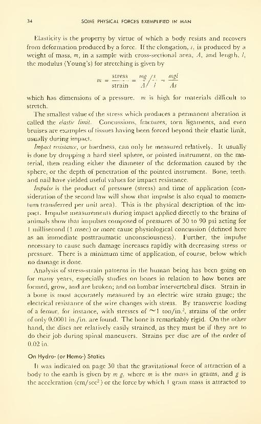
34 SOME PHYSICAL FORCES EXEMPLIFIED IN MAN
Elasticity is the property by virtue of which a body resists and recovers
from deformation produced by a force. If the elongation, s, is produced by a
weight of mass, m, in a sample with cross-sectional area, A, and length, /,
the modulus (Young's) for stretching is given by
stress mg /s mgl
strain A/ I As
which has dimensions of a pressure, m is high for materials difficult to
stretch.
The smallest value of the stress which produces a permanent alteration is
called the elastic limit. Concussions, fractures, torn ligaments, and even
bruises are examples of tissues having been forced beyond their elastic limit,
usually during impact.
Impact resistance, or hardness, can only be measured relatively. It usually
is done by dropping a hard steel sphere, or pointed instrument, on the ma-
terial, then reading either the diameter of the deformation caused by the
sphere, or the depth of penetration of the pointed instrument. Bone, teeth,
and nail have yielded useful values for impact resistance.
Impulse is the product of pressure (stress) and time of application (con-
sideration of the second law will show that impulse is also equal to momen-
tum transferred per unit area). This is the physical description of the im-
pact. Impulse measurements during impact applied directly to the brains of
animals show that impulses composed of pressures of 30 to 90 psi acting for
1 millisecond (1 msec) or more cause physiological concussion (defined here
as an immediate posttraumatic unconsciousness). Further, the impulse
necessary to cause such damage increases rapidly with decreasing stress or
pressure. There is a minimum time of application, of course, below which
no damage is done.
Analysis of stress-strain patterns in the human being has been going on
for many years, especially studies on bones in relation to how bones are
formed, grow, and are broken; and on lumbar intervertebral discs. Strain in
a bone is most accurately measured by an electric wire strain gauge; the
electrical resistance of the wire changes with stress. By transverse loading
of a femur, for instance, with stresses of ~1 ton/in. 2, strains of the order
of only 0.0001 in./in. are found. The bone is remarkably rigid. On the other
hand, the discs are relatively easily strained, as they must be if they are to
do their job during spinal maneuvers. Strains per disc are of the order of
0.02 in.
On Hydro- (or Hemo-) Statics
It was indicated on page 30 that the gravitational force of attraction of a
body to the earth is given by m g, where m is the mass in grams, and g is
the acceleration (cm/sec 2) or the force by which 1 gram mass is attracted to
THE OSMOTIC FORCE 35
the earth at sea level (980 dynes/g). Our goal now will be to show what
problem is introduced by the simple facts that man's head is 6 ft away from
his feet and he walks upright.
Two fluids circulate independently through the body: blood and lymph.
Both move via a canal system. The former is a closed system driven by a
pump; the latter is driven by muscle movement along the canals.
Because a column of air 6 ft high, of 1 in.2 cross-section, has negligible
weight, there is no difference in the weight force of air at the head and feet.
However, the weight of a column of water (or blood) of the same dimen-
sions is 2.8 lb, quite an appreciable fraction (12 per cent) of 14.8 psi of at-
mospheric pressure. In terms of the mercury manometer (1 atm, 14.8 psi,
supports a column of mercury 30 in., or 760 mm high, remember?) this
extra pressure at the feet due to the weight of the blood is 120 mm over 760.
Hence the pump must force blood along against a 120-mm back-pressure.
Add to this a small resistance to flow, mostly in the large arteries and veins
in which the total area of flow is relatively small and the flow rate high.
The heart is a pulse pump. It distends, collecting a volume of blood
freshly oxygenated in the lungs, closes its inlet valves, and contracts, forcing
the blood out through the aorta. The aorta, like the rest of the circulating
system, has elastic walls, which, in turn, distend under the hydraulic force
impressed by the contracting heart muscles. The pressure-rise in the aorta,
for a rather typical stroke-volume of 30 cc, may vary from 30 to 150 mm Hgpressure depending upon the reaction of the walls of the arterial system to
the pulse and the physical position of the person. In the highly elastic walls
of the young and healthy the value will be small; as the tissues becomeharder with age, or disease, it will rise.
The maximum value is called the systolic pressure, and is due directly to
the factors outlined. It is usually of the order of 120 mm. The minimumvalue—reached after the walls of the aorta, distended by the stroke from the
heart, have relaxed to the original diameter, having forced the blood along
the artery-capillary system— is called the diastolic pressure. Typically in a
healthy, adult male it is ~80 mm Hg. The mean value is about 100. Thepulse period is about 1 sec. Because the veins in the legs are more easily
distended than the arteries, most of the venous blood is stored there and re-
called when needed. The center of gravity is thus lowered, and storage re-
quires less work.
THE OSMOTIC FORCE
What Is It?
One of the most important forces at work in the living system is the os-
motic (literally, Greek: "impulse") one. It is the force which drives the dif-
fusion of water, nothing more, and is a property of a solution just as are
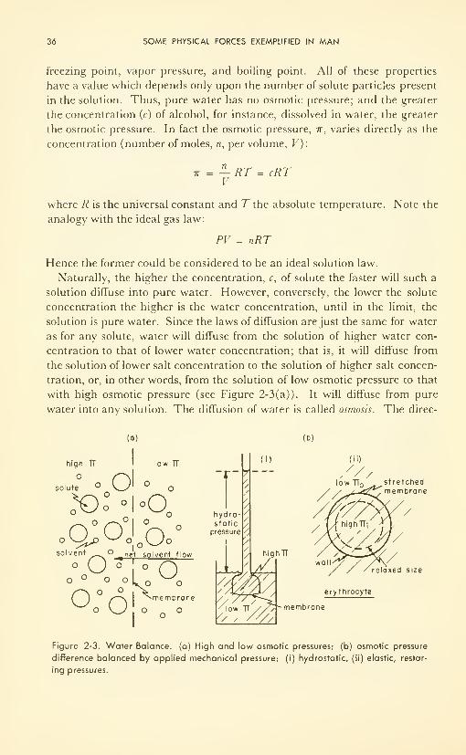
36 SOME PHYSICAL FORCES EXEMPLIFIED IN MAN
freezing point, vapor pressure, and boiling point. All of these properties
have a value which depends only upon the number of solute particles present
in the solution. Thus, pure water has no osmotic pressure; and the greater
the concentration (c) of alcohol, for instance, dissolved in water, the greater
the osmotic pressure. In fact the osmotic pressure, ir, varies directly as the
concentration (number of moles, n, per volume, V):
7T = —RT = cRTV
where R is the universal constant and Tthe absolute temperature. Note the
analogy with the ideal gas law:
PV = nRT
Hence the former could be considered to be an ideal solution law.
Naturally, the higher the concentration, c, of solute the faster will such a
solution diffuse into pure water. However, conversely, the lower the solute
concentration the higher is the water concentration, until in the limit, the
solution is pure water. Since the laws of diffusion are just the same for water
as for any solute, water will diffuse from the solution of higher water con-
centration to that of lower water concentration; that is, it will diffuse from
the solution of lower salt concentration to the solution of higher salt concen-
tration, or, in other words, from the solution of low osmotic pressure to that
with high osmotic pressure (see Figure 2-3(a)). It will diffuse from pure
water into any solution. The diffusion of water is called osmosis. The direc-
ts (b)
high TT
Osolute
'
low TT
Olo,
X>o °I°Qo
oU olasolvent
O
1°° .net solven t flow
O>L o
° r\̂membrane
_ O0:6! o
hydro-static
pressure
highTT
(ii)
lowTI ^^stretchedibrane
size
erythrocyte
membrane
Figure 2-3. Water Balance, (a) High and low osmotic pressures; (b) osmotic pressure
difference balanced by applied mechanical pressure; (i) hydrostatic, (ii) elastic, restor-
ing pressures.
THE OSMOTIC FORCE 37
tion of osmosis is determined by the osmotic pressure difference between the
two solutions in contact, but otherwise there is no relationship between
osmosis and osmotic pressure.
The osmotic pressure can be measured by determining the mechanical
pressure which must be applied to the solution of high osmotic pressure so
that osmosis ceases. The mechanical pressure might be a hydrostatic one
(Figure 2-3 (b) i), an elastic restoring force per unit area (Figure 2-3 (b) ii),
or some other.
Water Balance
In the body (mostly water) the balance among tissues is maintained by a
curious assortment of mechanical and osmotic forces, dictated in large part
by the physical characteristics of membranes which separate the fluids. All
living membranes pass water with ease. It is the solute content which deter-
mines the osmotic pressure difference between the two solutions separated
by the membrane, and this is determined in part by the membrane itself.
Some membranes pass everything— water, salts, molecules—excluding col-
loids and larger particles; the large intestine is an example. Membranes in
the kidney pass water, salts, and many small molecules readily and rapidly.
The membrane which forms the cell wall of the red blood cell passes water
and salts, and some small molecules readily. Nerve cell membrane passes
water and Cl~ readily, but balks at most molecules (its metabolic rate is
low), and lets K + and Na + through only with difficulty.
Since those species which can pass freely equalize their concentrations onopposite sides, only those which are restricted from passage can give rise to
a difference in osmotic pressure. In the erythrocytes, water balance is thus
controlled by the difference in soluble protein content between the cellular
fluid and the plasma. Since the concentration is slightly greater inside than
outside the cell, water runs in. As the cell walls become stretched, the re-
storing pressure (the wall is elastic, like a balloon) applies a mechanical
pressure on the liquid. An equilibrium is reached at which
7T, = 7T + PR
where the 7r's are osmotic pressures inside and outside the cell, and PR is
the restoring pressure of the walls of the distended cell. Table 2-1 gives a
quantitative illustration of this important point.
When membranes are ill-formed and cannot discriminate as they should,
or when metabolic processes produce impenetrable species such as a protein
whose concentration is different from the normal, the osmotic pressure dif-
ference, 7r, — 7r , is not the same, and the powerful osmotic force differs from
what it should be. The small mechanical compensation mechanisms (such
as the restoring force in the erythrocyte wall) become strained, and edema
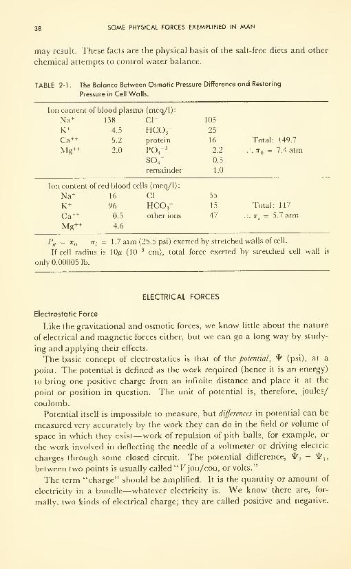
38 SOME PHYSICAL FORCES EXEMPLIFIED IN MAN
may result. These facts are the physical basis of the salt-free diets and other
chemical attempts to control water balance.
TABLE 2-1. The Be

ELECTRICAL FORCES 39
Positives repel; negatives repel; but positive attracts negative. Coulomb ob-
served that the force of repulsion of like charges increases as the size of each,
and decreases as the square of the distance. Thus
F =ed 2
where Fis the force in dynes, <7, and q 2are the charges in coulombs, d is the
distance in centimeters, and e is the proportionality constant, called the di-
electric constant (Figure 2-4). Unit charge is formally defined through
Coulomb's facts: when two like charges are 1 cm apart and repel each other
with a force of 1 dyne, each carries unit charge.
DISTANCE x
Figure 2-4. Interaction of Electrical Charges: (a) Coulomb's case; (b) field strength.
Bioelectric Potentials
At the microscopic level the most important potential differences in the
living system arise from concentration differences (why they do will be seen
later), and these occur almost without exception across living membranes.
For example, in heart muscle cell the potential difference or voltage between
the inside and outside of the cell, across the cell membrane, is about 85 mv,
on the average, and cycles above and below this, as the heart beats.
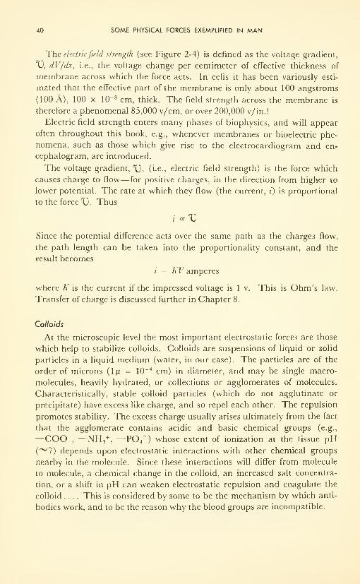
40 SOME PHYSICAL FORCES EXEMPLIFIED IN MAN
The electricfield strength (see Figure 2-4) is denned as the voltage gradient,
X), dV/dx, i.e., the voltage change per centimeter of effective thickness of
membrane across which the force acts. In cells it has been variously esti-
mated that the effective part of the membrane is only about 100 angstroms
(100 A), 100 x 10-8 cm, thick. The field strength across the membrane is
therefore a phenomenal 85,000 v/cm, or over 200,000 v/in.!
Electric field strength enters many phases of biophysics, and will appear
often throughout this book, e.g., whenever membranes or bioelectric phe-
nomena, such as those which give rise to the electrocardiogram and en-
cephalogram, are introduced.
The voltage gradient, TJ, (i.e., electric field strength) is the force which
causes charge to flow— for positive charges, in the direction from higher to
lower potential. The rate at which they flow (the current, i) is proportional
to the forced. Thus
l oc VSince the potential difference acts over the same path as the charges flow,
the path length can be taken into the proportionality constant, and the
result becomes
i = AT amperes
where K is the current if the impressed voltage is 1 v. This is Ohm's law.
Transfer of charge is discussed further in Chapter 8.
Colloids
At the microscopic level the most important electrostatic forces are those
which help to stabilize colloids. Colloids are suspensions of liquid or solid
particles in a liquid medium (water, in our case). The particles are of the
order of microns (1/i = 10-4 cm) in diameter, and may be single macro-
molecules, heavily hydrated, or collections or agglomerates of molecules.
Characteristically, stable colloid particles (which do not agglutinate or
precipitate) have excess like charge, and so repel each other. The repulsion
promotes stability. The excess charge usually arises ultimately from the fact
that the agglomerate contains acidic and basic chemical groups (e.g.,
—COO-, —NH 3
+, —P04
=) whose extent of ionization at the tissue pH
(~7) depends upon electrostatic interactions with other chemical groups
nearby in the molecule. Since these interactions will differ from molecule
to molecule, a chemical change in the colloid, an increased salt concentra-
tion, or a shift in pH can weaken electrostatic repulsion and coagulate the
colloid .... This is considered by some to be the mechanism by which anti-
bodies work, and to be the reason why the blood groups are incompatible.
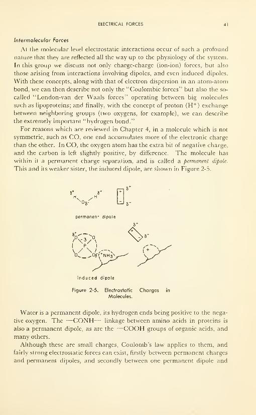
ELECTRICAL FORCES 41
Iniermolecular Forces
At the molecular level electrostatic interactions occur of such a profound
nature that they are reflected all the way up to the physiology of the system.
In this group we discuss not only charge-charge (ion-ion) forces, but also
those arising from interactions involving dipoles, and even induced dipoles.
With these concepts, along with that of electron dispersion in an atom-atom
bond, we can then describe not only the "Coulombic forces" but also the so-
called "London-van der Waals forces" operating between big molecules
such as lipoproteins; and finally, with the concept of proton (H +) exchange
between neighboring groups (two oxygens, for example), we can describe
the extremely important "hydrogen bond."
For reasons which are reviewed in Chapter 4, in a molecule which is not
symmetric, such as CO, one end accumulates more of the electronic charge
than the other. In CO, the oxygen atom has the extra bit of negative charge,
and the carbon is left slightly positive, by difference. The molecule has
within it a permanent charge separation, and is called a permanent dipole.
This and its weaker sister, the induced dipole, are shown in Figure 2-5.
8*
OS-
S'
permanent dipole
i pv
i
0^ ^Osi.*NH 3\
8*
-^B-
induced dipole
Figure 2-5. Electrostatic Charges in
Molecules.
Water is a permanent dipole, its hydrogen ends being positive to the nega-
tive oxygen. The —CONH— linkage between amino acids in proteins is
also a permanent dipole, as are the —COOH groups of organic acids, and
many others.
Although these are small charges, Coulomb's law applies to them, and
fairly strong electrostatic forces can exist, firstly between permanent charges
and permanent dipoles, and secondly between one permanent dipole and
42 SOME PHYSICAL FORCES EXEMPLIFIED IN MAN
another. Water molecules attract each other, dipole to dipole, and give to
bulk water a structure of oriented dipoles. Ions attract one end of the dipole
and repel the other, and the result is an array of water dipoles oriented
radially outwards from a central ion. The dipoles on large molecules can be
hydrated by attraction to water molecules. Big molecules can be attracted
to each other, or indeed have one part folded back and attracted to another
part where two dipoles fall in close proximity, or where one dipole falls close
to a charged group. Thus the dipolar character helps to determine not only
composition but also structure.
Still weaker forces exist between induced dipoles. Even if the molecule is
symmetrical about an atom, a strong positive or negative charge can some-
times induce the molecule's electrons to move a bit, so that the charge dis-
tribution becomes distorted. Such induced charge separation is called an
induced dipole. Interactions between the mutually induced dipoles of two
molecules in close proximity are called the van der Waals forces. Further, it
is postulated that the electron cloud of a molecule is in continuous motion,
continually varying both the size and direction of its dipole. It induces a
further dipole in its neighbor, and the new "dynamic" dipole interacts with
the old static one in a manner which seems to confer an extra stability on
the intermolecular "bond." The extra force of attraction is called the "dis-
persion force," first postulated by London in 1930. Since one occurs when-
ever the other does, today the mutually induced dipole and dispersion forces
of attraction are referred to as the London-van der Waals forces. They are
very weak by comparison with Coulombic forces, principally because the
charges are not only small but deformable. However, in the absence of
charged groups and when two molecules can come into close proximity
(< 5 A) at a great many places over a fairly long distance (~15 carbon
atoms in each molecular chain), considerable binding between the two has
been shown to be accountable on the basis of London-van der Waals forces.
Such is the case in lipoproteins in which a long hydrocarbon (and therefore
with no polar groups and no permanent dipoles) chain becomes and remains
intimately bonded to a polyamino acid or protein molecule. The strength
and the sensitivity of this bond to interatomic spacings have been very
evident in recent studies of lipoproteins in nerve cell membranes of the cen-
tral nervous system. For example, one form of encephalitis is currently
thought to be due to a change in binding which occurs as a result of inac-
curate protein synthesis and poor binding to its lipid.
Whereas Coulombic forces are fairly long-range forces (al/a' 2) the
London-van der Waals forces are very short-range ( « \/d 7) but become im-
portant when the particles approach very close to one another (see Table
2-2).
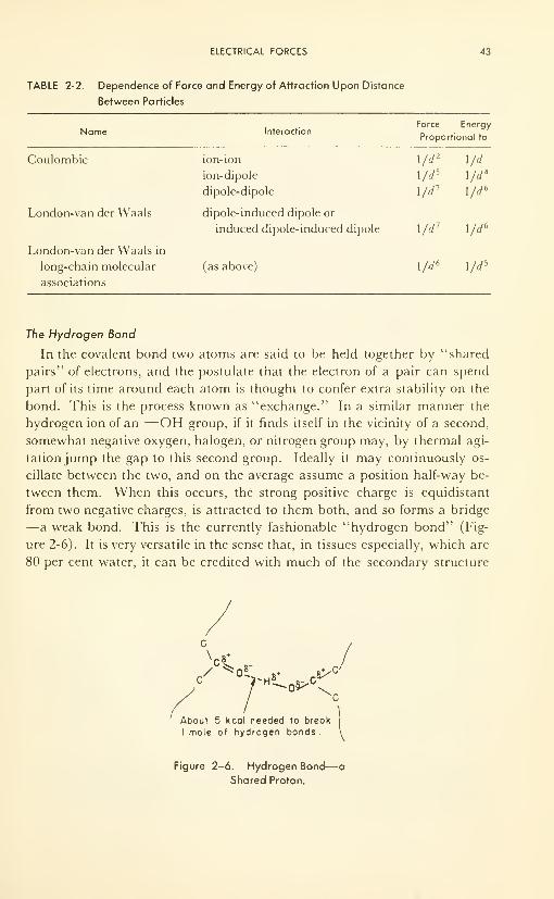
ELECTRICAL FORCES 43
TABLE 2-2. Dependence of Force and Energy of Attraction Upon Distance
Between Particles
Name InteractionForce Energy
Proportional to
Coulombic
London-van der Waals
London-van der Waals in
long-chain molecular
associations
ion-ion
44 SOME PHYSICAL FORCES EXEMPLIFIED IN MAN
of big molecules— for instance, for the paracrystallinity of the regular molec-
ular arrays so common in tissue, such as in muscle fiber and in the aqueous
humor of the lens of the eye.
Electromagnetic Force
Although we live in the magnetic field of the earth, no information exists
on the response of a man to large changes in magnetic-field strength. Tosmall changes there is no response, as far as is known. Many molecular ef-
fects are known, however, of which the recent exploitation of the so-called
nuclear magnetic resonance phenomena, in which the location of a hydrogen
atom in a molecule and the arrangement of atoms in molecular complexes
can be learned, are exciting examples.
However, on biological systems the effects of magnetic fields are yet poorly
understood. Small animals placed in fairly strong magnetic fields of ~4000gauss (at $2/gauss, ' 1 lb> of electromagnet/ gauss) show inability to repro-
duce. Cell division and growth are inhibited. Interference with the collec-
tion of the mitotic apparatus in preparation for cell division is implicated.
In this respect the effect of a magnetic field is similar to the effects of X or
gamma rays.
The effects of electromagnetic forces— oscillating forces of unknown na-
ture, which interact with both electric charges and magnetic poles, and with
other electromagnetic forces— are better understood and are most important
in the living system. In fact, the more the question is studied, the more it
is realized in how many aspects of inanimate as well as animate subjects,
electromagnetic forces play an important part.
Usually electromagnetic phenomena are described by their interaction
energy, rather than force; this expedient enables us to by-pass their nature,
and concentrate upon their effects. An "oscillating potential" permeates
electromagnetic energy. It is a periodic function of time (see Figure 1-2).
The amount of energy in a packet depends only upon its number of cycles
per second.
Because of their importance, Chapter 4 is devoted almost completely to
electromagnetic matters.
Yet will all this preoccupation with force, the physicist still is unable to
cope with some really big ones, such as political "forces," and economic
"pressures." In "The Razor's Edge" (1944), W. Somerset Maugham con-
cludes: "Goodness is the greatest 'force' in the world!". . . . Unfortunately,
we cannot measure it.
GENERALIZED FORCE
Although temperature is not usually thought of as a force, it is the driving
force for heat-energy flow. Discussion on driving forces for several processes
which occur in the living system is contained in Chapter 7.
PROBLEMS 45
All forces are, quite literally, ''factors of energy." Thus, a generalized
driving force times a quantity yields energy. Some examples are:
Mechanical force x distance = mechanical energy or work
Gas pressure x volume of gas = mechanical energy or work
Osmotic pressure x molar volume = osmotic energy or work
Electrical potential x charge = electrical energy or work
Temperature x entropy = heat energy or work
Chemical potential x concentration = chemical energy or work
The inherent difficulties of considering both temperature in "degrees"
(fractions of a length of a liquid metal along a tube!) and chemical poten-
tial (actually an energy per unit concentration) as "forces," are expounded
further in Chapter 7.
What happens to a biological system when the force responsible for the
acceleration due to gravity (g) is removed— that is, becomes weightless— is
critically important to future space travel. The meager information on the
few human beings who have so far orbited the earth is reviewed in Chapter 8.
PROBLEMS
2-1 : A 200-lb football player is running full speed at a rate of 100 yd in 12 sec. Cal-
culate his kinetic energy in ergs; in joules; in calories; in Calories or kilocalories.
If he were stopped completely in 1 sec, what power would he deliver during
that 1 sec (in watts; in horsepower; in Cal/hr)? Compare this with the basal
metabolic rate of 0.1 hp, or 60 Cal/hr (1 lb = 454 g; 1 cal = 4.18 jou; 1 hp =
746 w; 1 jou/sec = 1 w).
2-2: Values of the solubility of nitrogen and oxygen in water are 0.001 50 and 0.00332
g of gas at 1 atm/100 g water, respectively. Approximately how many cubic
centimeters of each gas are contained dissolved in the body fluids (200 lb, 80 per
cent water) under 1 atm of air (20 per cent oxygen, 80 per cent nitrogen)?
Neglect the fact that the solubility of gases is less in salt solutions than in pure
water.
An anethetist may use a mixture up to 90 per cent oxygen, but he always re-
tains about 5 per cent C02in the inhaled gas. Why?
2-3: Assuming the total area of the adult human body to be 1 sq yd, calculate the
total force due to the atmosphere (pressure 14.7 lb/in.2
) on the body. In dynes;
in tons force.
Calculate the total force on a skin diver at a depth of 450 ft. Why is he not
crushed? What precautions must he take while coming up to the surface? Why?2-4: Make two tables showing forces of repulsion— in dynes, of two like unit charges,
each with 3 x 10" 10 electrostatic units of charge— at distances 0.1, 1, 2, 5,
and 25 A apart; one table for a medium of air or a vacuum (dielectric con-
stant = 1), and the other for an aqueous solution (dielectric constant = 72).
Plot the numbers, force vs distance, for each case.

46 SOME PHYSICAL FORCES EXEMPLIFIED IN MAN
REFERENCES
1. Harrington, E. L., "General College Physics, " D. Van Nostrand Co., Inc., NewYork, N. Y., 1952.
2. Randall, J. T., "Elements of Biophysics," the Year Book Publ., Inc., Chicago,
111., 1958.
3. Glasser, O., Ed., "Medical Physics," Vol. Ill, Year Book Publ., Inc., Chicago,
111., 1960; papers by Carter, Featherstone, Lipson, et al.
4. Moore, W.J., "Physical Chemistry," Prentice-Hall, Inc., New York, N. Y., 1950.
5. Macintosh, Sir R., Mushin, W. W., and Epstein, H. G., "Physics for the
Anesthetist," 2nd ed., Chas. C Thomas Publ. Co., Springfield, 111., 1960.
6. Robbins, S. L., "Textbook of Pathology With Clinical Applications," W. B.
Saunders Co., Philadelphia, Pa., 1957.
7. Wolf, A. V., "Body Water, " Sci. Amer., 199, 125 (1958).
CHAPTER 3
Matter Waves: Sound
and Ultrasound(On Music and Noise "from
CtoC,"On Speech and Some Therapy)
According to Sir Richard Paget, human speech began by the performance
of sequences of simple pantomimic gestures of the tongue, lips, etc. . . .
Consider the word "hither. " The tongue makes the same beckoning gesture,
while [one is] speaking this word, as is made with the hand.
(H. Fletcher. 3)
INTRODUCTION
Our senses of touch and hearing reveal an environment which contains a
bewildering array of matter waves: the breeze; falling raindrops; noise,
speech, and music; earth tremors, shock, or blast waves; the vibrations en-
countered when riding a horse, or when operating a jack-hammer. Bees
and some other insects, and bats too, send and receive, and are guided in
flight by very high-frequency matter waves.
Thus waves in matter have a great spectrum of manifestations, uses, and
effects. It is the purpose of this chapter to illustrate them, for matter waves
and electromagnetic radiations together comprise the most important
method of man's continuous exchange of force and energy with his environ-
ment. The latter are introduced in Chapter 4. They are fundamentally
very different from matter waves, although often confused with them. In
47
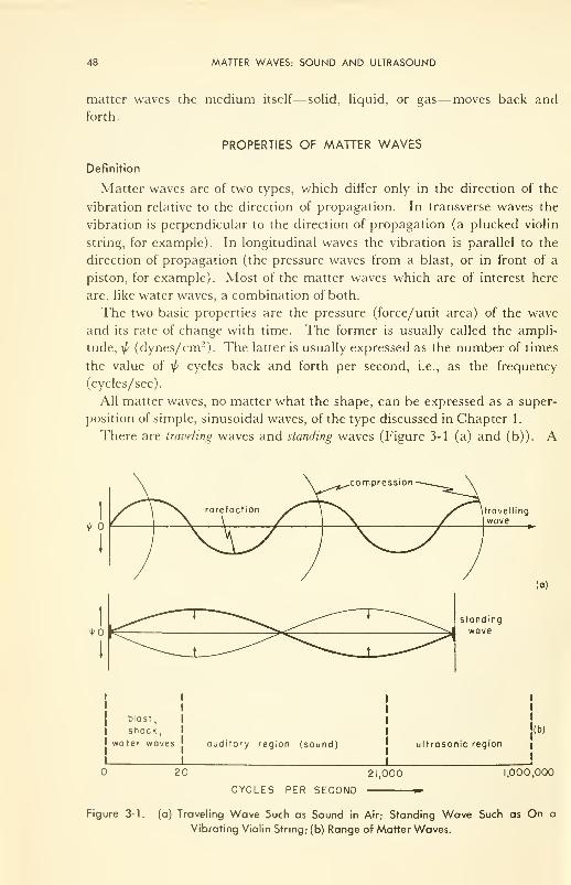
48 MATTER WAVES: SOUND AND ULTRASOUND
matter waves the medium itself— solid, liquid, or gas—moves back and
forth.
PROPERTIES OF MATTER WAVES
Definition
Matter waves are of two types, which differ only in the direction of the
vibration relative to the direction of propagation. In transverse waves the
vibration is perpendicular to the direction of propagation (a plucked violin
string, for example). In longitudinal waves the vibration is parallel to the
direction of propagation (the pressure waves from a blast, or in front of a
piston, for example). Most of the matter waves which are of interest here
are, like water waves, a combination of both.
The two basic properties are the pressure (force/unit area) of the wave
and its rate of change with time. The former is usually called the ampli-
tude, \p (dynes/cm 2). The latter is usually expressed as the number of times
the value of \p cycles back and forth per second, i.e., as the frequency
(cycles/sec).
All matter waves, no matter what the shape, can be expressed as a super-
position of simple, sinusoidal waves, of the type discussed in Chapter 1.
There are traveling waves and standing waves (Figure 3-1 (a) and (b)). A
<!>
biost,
shock,
water waves auditory region (sound) ultrasonic region
I
I
I
'(b)
I
20 21,000 .000,000
CYCLES PER SECOND
Figure 3-1. (a) Traveling Wave Such as Sound in Air; Standing Wave Such as On a
Vibrating Violin String; (b) Range of Matter Waves.
PROPERTIES OF MATTER WAVES 49
sound wave moving through air travels from its source and imparts an
energy to the receiver. This energy is primarily in the direction of propaga-
tion, but with scattering some of it becomes transverse.
By contrast, the standing wave can impart no longitudinal energy— it has
none. But it can impart transverse energy to the medium. The generation
of the sound by the vibrating violin string is an example.
The intensity, /, of the matter wave is the power delivered by it per unit
area. In. other words, / is the rate at which the wave expends energy. All
traveling waves move at a certain velocity, v (cm/sec). Hence the product of
amplitude (a pressure) times distance is the energy expended per unit area:
w = \p d (dynes/cm 2 x cm = ergs/cm 2)
The product of amplitude and velocity is the power expended per unit area:
I = \p v (dynes/cm 2 x cm/ sec = ergs/cm 2 sec)
The intensity or power expended per unit area by the traveling wave, is
highest for those media having molecules with the greatest number of de-
grees of freedom in which energy can be stored— gases for example. Both
the range and speed of sound are highest in solids, somewhat less in liquids,
far less in gases. However, for any medium of constant density, p, the ve-
locity has a fixed value. This fact results in another useful relationship, that
between amplitude (pressure) and intensity (power):
/ = Vlvp
which says simply that power delivered per unit area to any medium is pro-
portional to the pressure squared, if velocity and density are held constant.*
This (/ cc \^2
) is a very useful rule-of-thumb, applicable, it turns out, to all
field phenomena.
Useful also is the fact that, although low-frequency waves are easily re-
flected and diffracted by air and hence are nondirectional (or will go around
corners), high-frequency waves are only slightly scattered by air. Therefore,
the latter can be beamed in a preferred direction from a source, and even
focused on a particular spot by proper (saucer-like) design of the vibrating
source.
*Dimensions:
3sec cm
= ergs/cm sec
(Work it through.)
50 MATTER WAVES: SOUND AND ULTRASOUND
Illustrations
Frequency
Matter waves have a broad range of frequency, from zero up to the current
practical upper limit of about 1,000,000 cycles per sec (cps) in use in some
ultrasonic-therapy and submarine-detection studies (Figure 3-1 (c)). The
human ear is most sensitive from ^50 to ^10,000 cps; the range of man's
ear, however, may be from 20 to 21,000 cps. This, then, is the auditory or
sound range. Speech requires 60 to 500 cps. The piano ranges from 27.2 to
4138.4 cps. The great basso profondo, Italo Tajo, could reach a minimum of
~60 cps; the diminutive coloratura soprano, Lili Pons, could hit 1300 cps on
a good day. Of course, these are the basic frequencies, and it is understood
that a basic frequency generated by any physical vibrator will contain over-
tones, or harmonics, which are multiples (2x, 4x, even 8x) of the basic
frequency. The quality of the tone is determined by the sum of all the com-
ponents: the basic frequency plus its harmonics.
Training and youth combine to produce a receiver which can hear low-
power sound up to 12,000 cps. Some musicians can detect overtones from
their instruments up to 14,000 cps, but these are few. Most of us can detect
frequencies up to 18,000 from a signal generator, if the signal is intense
enough, and the odd person can detect up to 21,000 cps. Dogs do it with
ease. Porpoises have a phenomenal sonic system in their heads which can
sweep frequencies repetitively from a few cycles to many thousands of cycles
—both send and receive.
Below and overlapping the auditory range for man is the range (0 to 50
cps) of blast and shock waves, earth tremors, water waves, and the like. Themasseur will use vibrations 1 to 50 cps; a ship will roll at 0.1 cps. An air
hammer operates at ~ 15 cps, and we hear the overtones.
Above the range of sound, from 20,000 up to > 1,000,000, lies the im-
portant range of ultrasound, and the science and technology known as
ultrasonics.
Velocity
The speed of matter waves depends sharply upon the medium, and in the
case of a gas, its temperature and pressure. For instance, in air at 0°C and
1 atm pressure the speed is 331 meters/sec (mps) (730 miles/hr). In water
and soft tissue it is 4 12 times higher than in air, and in solids it goes up to
5000 mps. The velocity of sound through fat is 1440, through muscle 1570,
and through bone 3360 mps.
Velocity is independent of frequency; and it is probably just as well, other-
wise the low tones of the organ might reach our ears later than the high
tones of the same chord!
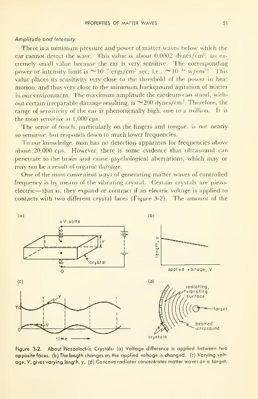
PROPERTIES OF MATTER WAVES 51
Amplitude and Intensity
There is a minimum pressure and power of matter waves below which the
ear cannot detect the wave. This value is about 0.0002 dynes/cm 2, an ex-
tremely small value because the ear is very sensitive. The corresponding
power or intensity limit is ~10 9 ergs/cm 2 sec, i.e., ^lO" 16 w/cm 2! This
value places its sensitivity very close to the threshold of the power in heat
motion, and thus very close to the minimum background agitation of matter
in our environment. The maximum amplitude the eardrum can stand, with-
out certain irreparable damage resulting, is ~200 dynes/cm 2. Therefore, the
range of sensitivity of the ear is phenomenally high, one to a million. It is
the most sensitive at 1,000 cps.
The sense of touch, particularly on the fingers and tongue, is not nearly
so sensitive, but responds down to much lower frequencies.
To our knowledge, man has no detection apparatus for frequencies above
about 20,000 cps. However, there is some evidence that ultrasound can
penetrate to the brain and cause psychological aberrations, which may or
may not be a result of organic damage.
One of the most convenient ways of generating matter waves of controlled
frequency is by means of the vibrating crystal. Certain crystals are piezo-
electric— that is, they expand or contract if an electric voltage is applied to
contacts with two different crystal faces (Figure 3-2). The amount of the
(o)
£l_
+ V volts
crystol
(b)
applied voltage, V
(c)
time
radiating,vibrati ngsurfoce
-target
beamedultrasound
crystals
Figure 3-2. About Piezoelectric Crystals: (a) Voltage difference is applied between two
opposite faces, (b) The length changes as the applied voltage is changed, (c) Varying volt-
age, V, gives varying length, y. (d) Concave radiator concentrates matter waves on a target.
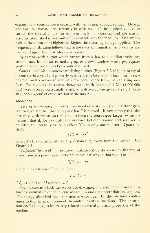
52 MATTER WAVES: SOUND AND ULTRASOUND
expansion or contraction increases with increasing applied voltage. Quartz
and barium titanate are currently in wide use. If the applied voltage is
varied, the crystal shape varies accordingly, or vibrates, and the matter
wave so established is transmitted by contact with the medium. The ampli-
tude of the vibration is higher the higher the vibrating voltage applied. The
frequency of vibration follows that of the electrical signal, if the crystal is not
too big. Figure 3-2 illustrates these points.
Apparatus with output which ranges from a few to a million cycles per
second, and from next to nothing up to a few hundred watts per square
centimeter of crystal, has been built and used.
Constructed with a concave radiating surface (Figure 3-2 (d)), an array of
piezoelectric crystals, if properly oriented, can be made to focus an intense
beam of matter waves at a point a few centimeters from the radiating sur-
face. For example, in recent therapeutic work beams of 1 Mc (1,000,000
cps) were focused on a small target, and delivered energy at a rate (inten-
sity) of 8 kw/cm2 of cross-section of the target
!
Absorption
If waves are diverging, or being dissipated or scattered, the important gen-
eral rule, called the "inverse square law," is obeyed. It says simply that the
intensity, /, decreases as the distance from the source gets larger, in such a
manner that if, for example, the distance between source and receiver is
doubled, the intensity at the receiver falls to only one quarter. Quantita-
tively,
I(x) oc \/x 2
where I(x) is the intensity at any distance, x, away from the source. See
Figure 3-3.
If a parallel beam of matter waves is absorbed by the medium, the rate of
absorption at a point is proportional to the intensity at that point; or
dl/dx = -kl
which integrates (see Chapter 1) to
/ = I e-*
if / is the value of / where x = 0.
For the case in which the waves are diverging and also being absorbed, a
linear combination of the inverse square law and the absorption law applies.
The energy absorbed from the matter-wave beam by the medium contri-
butes to the thermal motion of the molecules of the medium. The absorp-
tion coefficient, k, is intimately related to several physical properties of the
medium.
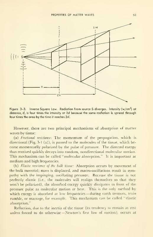
PROPERTIES OF MATTER WAVES 53
Figure 3-3. Inverse Square Law. Radiation from source S diverges. Intensity (w/cm2) at
distance, d, is four times the intensity at 2d because the same radiation is spread through
four times the area by the time it reaches 2d.
However, there are two principal mechanisms of absorption of matter
waves by tissue:
(a) Fnctional resistance: The momentum of the propagation, which is
directional (Fig. 3-1 (a)), is passed to the molecules of the tissue, which be-
come momentarily polarized by the pulse of pressure. The directed energy
thus received quickly decays into random, nondirectional molecular motion.
This mechanism can be called "molecular absorption." It is important at
medium and high frequencies.
(b) Elastic reactance of the bulk tissue: Absorption occurs by movement of
the bulk material; mass is displaced, and macro-oscillations result in sym-
pathy with the impinging, oscillating pressure. Because the tissue is not
perfectly elastic (i.e., the molecules will realign themselves so that they
won't be polarized), the absorbed energy quickly dissipates in front of the
pressure pulse as molecular motion or heat. This is the only method by
which energy is absorbed at low frequencies— during earth tremors, train
rumble, or massage, for example. This mechanism can be called "elastic
absorption."
Reflection, due to the inertia of the tissue (its tendency to remain at rest
unless forced to do otherwise—Newton's first law of motion), occurs at
54 MATTER WAVES: SOUND AND ULTRASOUND
high frequencies for soft tissue and even at low frequencies for dense tissue
such as bone. Truly elastic tissues simply reflect incident matter waves.
The absorption coefficient for molecular absorption (k) is well known for
air and water:
3vp
-> c_p °v jc
^P ^ V
where /is the frequency (cps) of the impinging wave, v the velocity (cm/sec),
p the density (g/cm 3), rj the viscosity (dyne sec/cm2
), A"7 the heat conductiv-
ity (cal/sec deg cm), and the c's are the specific heats (cal/deg g) at constant
pressure, P, and constant volume, V. Hence the energy absorbed per centi-
meter of penetration of the impinging wave increases linearly with the vis-
cosity or "stickiness" of the medium and with its thermal conductivity; in-
creases very rapidly with increasing frequency; but decreases with increas-
ing density.
For water, which is a sufficiently good approximation to soft tissue for
present purposes, k/f2 = 8.5 x 10" 17 sec 2/cm. For air the value is 1000
times higher, because although rj is 50 times smaller for air than for water,
v is 4^2 times smaller and p is 1000 times smaller. For liquids only the first
term (the frictional or viscous one) is important; for gases both are im-
portant. Therefore it is useful to aerate a tissue before sonic therapy is ap-
plied, because absorption is higher.
Since reflection increases with increasing frequency, the method of appli-
cation is important. In the absence of reflection, the above expressions
describe the situation well. Direct application of the vibrator to the tissue
assures this. However, if the sound is beamed through air, the situation is
quite different: reflection occurs.
Quantitative studies on tissues are only recent. The general rule which
has emerged is as follows: Beamed through air, sound of high frequency suf-
fers little absorption, and little damage results. The depth of penetration
increases with increasing frequency. Most (>95 per cent) of the incident
energy passes right through, or is reflected. Some of Von Gierke's figures
(1950) are: 5 to 6 per cent absorbed at 100 cps; 0.2 to 4 per cent absorbed
at 1000 cps; and <0.4 per cent absorbed at 10 kc. Beamed through liquid
or solid, ultrasonic radiation is easily controlled and its absorption pre-
dicted. More will be said about this later, in the section on therapy.
SENSITIVITY OF A DETECTOR, AND THE WEBER-FECHNER LAW
It is a fact that whether or not a receiver will detect a signal depends upon
how much the signal differs from the background noise. The dependence is
SENSITIVITY OF A DETECTOR, AND THE WEBER-FECHNER LAW 55
not a simple proportionality, but rather a logarithmic one. Thus, the sensa-
tion, or loudness, L, is given by
L oc log///°
where 1° is background intensity, and / is the intensity, over background, of
the signal to be detected. This is the basic form of the Weber-Fechner law.
It has many manifestations. For instance, if there are two signals equally
strong, with different backgrounds, the resolution of (difference in loudness),
L2- L, , is related to the ratio of the intensities of the two backgrounds,
1° and I2°, as follows:
L2— L
{
oc log I°/I°
This is a law which has rather wide application, not only in the psycho-
logical sensations but in detection of electromagnetic waves of many fre-
quency ranges, from the radio to the infrared. Therefore its implications
should be very thoroughly contemplated.
Because of this logarithmic law, it is convenient to express power ratios
by a logarithmic unit, so that sensation becomes approximately linearly pro-
portional to this unit. The unit is called the ^bel," (b) and is equal to the
logarithm of the ratio of two sound intensities if they are in a ratio of 10 : 1.
The number of bels then is given by
b = log 1/1°
For sound, the value 1° is arbitrarily chosen to be the lowest one which a
human ear can detect (10-16 w/cm 2
; or, in pressure units, 0.0002 dynes/cm 2,
since the same conversion factor applies to numerator and denominator).
The bel unit is too large for convenience, and the decibel, one tenth of a bel,
has received wider use. Therefore, the number of decibels is:
db = io log i/r
Another form of the Weber-Fechner law, then, is
L « db
It holds true for all sensory receptors.
Some minimum discernible relative changes,** (/, - 7°)//° (where I, is
threshold intensity), which man can detect are:
Brightness of light: 1 per cent
Lengths of lines: 2 per cent
Feeling of weight: 10 per cent
Loudness of sound: 30 per cent
** Remember relative error, defined in Chapter 1 ?
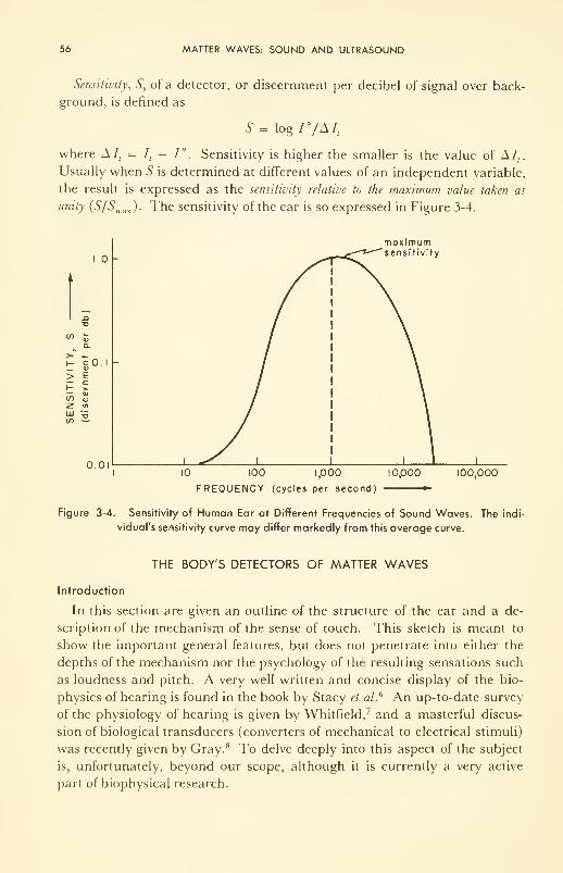
56 MATTER WAVES: SOUND AND ULTRASOUND
Sensitivity, S, of a detector, or discernment per decibel of signal over back-
ground, is defined as
s = log r/Mt
where A I, = I, - 1°. Sensitivity is higher the smaller is the value of A/,.
Usually when 6" is determined at different values of an independent variable,
the result is expressed as the sensitivity relative to the maximum value taken as
unity (S/Smax ). The sensitivity of the ear is so expressed in Figure 3-4.
moximumsensitivity
0.0110 100 1,000
FREQUENCY (cycles per second;
10,000 100,000
Figure 3-4. Sensitivity of Human Ear at Different Frequencies of Sound Waves. The indi
vidual's sensitivity curve may differ markedly from this average curve.
THE BODY'S DETECTORS OF MATTER WAVES
Introduction
In this section are given an outline of the structure of the ear and a de-
scription of the mechanism of the sense of touch. This sketch is meant to
show the important general features, but does not penetrate into either the
depths of the mechanism nor the psychology of the resulting sensations such
as loudness and pitch. A very well written and concise display of the bio-
physics of hearing is found in the book by Stacy et a/.6 An up-to-date survey
of the physiology of hearing is given by Whitfield, 7 and a masterful discus-
sion of biological transducers (converters of mechanical to electrical stimuli)
was recently given by Gray. 8 To delve deeply into this aspect of the subject
is, unfortunately, beyond our scope, although it is currently a very active
part of biophysical research.
THE BODY'S DETECTORS OF MATTER WAVES 57
Notes on the Ear
The structure of the ear can be pictured, in simplest terms, as consisting
of three main parts: the pinna (lobe) and external canal, the middle ear,
and the cochlea. The canal and the middle ear are separated by the tym-
panic membrane (ear drum) which covers and protects the latter. The
middle-ear cavity contains a system of three bony levers, the ossicles (the
malleus, incus, and stapes) whose main job seems to be to act as a matching
device transmitting matter vibrations between the two fluids: the air outside
in the external canal, and the perilymph inside the cochlea. The cochlea
is a spiral canal within the bone of the skull. It is divided axially into three
channels by membranous partitions. Into one of these, the scala vestibuli,
is inserted the end of the stapes; this chamber, then, receives directly the
transmitted vibrations. Through the membranes, vibrations are passed
laterally into the other two canals, the scala media and the scala tympani.
These two are separated by the basilar membrane, which receives the end-
ings of the auditory nerve, and the cells of which are the transducers that
convert the mechanical energy of vibration into the electrical energy trans-
mitted along the nerve. Most recent work has been aimed at the mechanism
of action of the region of the basilar membrane, the transducer. Some of the
cells on the membrane have hair-like processes projecting from their upper
ends and attached to the overhanging, tectorial membrane. Relative move-
ment between the tectorial and basilar membranes distorts the cells of both.
Note Figure 3-5.
The analogy with piezoelectric crystals is usefully drawn at this point:
distortion of the shape of the transducer in both cases leads to change in the
potential difference between two points on the surface of the transducer— in
one case the surface potential of the crystal, in the other case the membrane
potential of the cell.
An accumulation of evidence now exists—Von Bekesy 13 received the 1961
Nobel Prize in Physiology and Medicine for this work, done at Harvard
—
that a traveling wave passes along the basilar membrane during excitation.
The position at which the wave achieves its highest amplitude (think of
the whip) is dependent upon the frequency of the wave being detected.
Therefore, nerve signals from different tones arise at different spots, each
spot associated with specific nerve endings. At low frequencies the whole
basilar membrane vibrates in sympathy with the incoming matter wave.
The question of membrane potential change will be considered in Chap-
ters 7 and 10, in reference to erythrocytes and nerve cells, upon which
voltages have been directly measured in vivo.
Deformations in the structure, or failure of the ear to respond to matter
waves, is the subject matter of the otologist. Corrections are applied some-
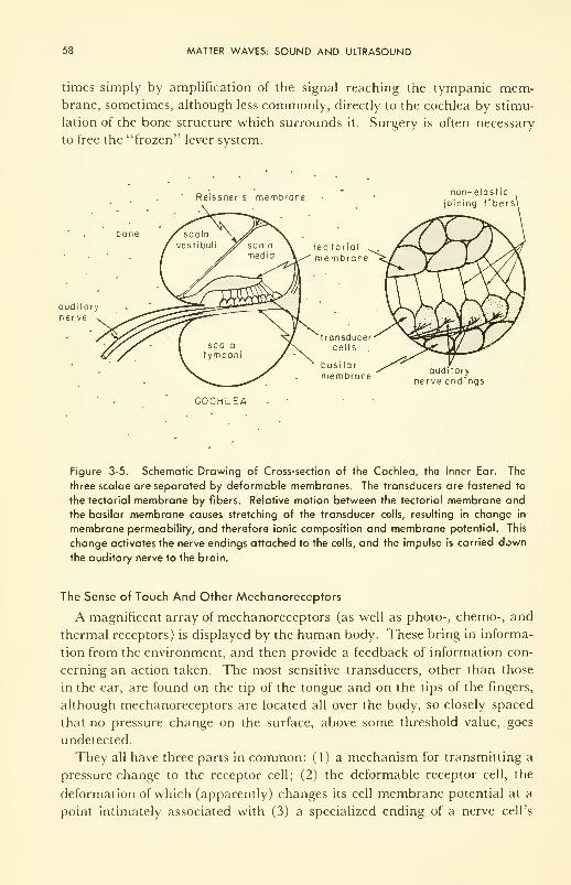
58 MATTER WAVES: SOUND AND ULTRASOUND
times simply by amplification of the signal reaching the tympanic mem-brane, sometimes, although less commonly, directly to the cochlea by stimu-
lation of the bone structure which surrounds it. Surgery is often necessary
to free the "frozen" lever system.
Reissner s membrane
bone
auditory
ner ve
tec torial
membrane
transducercells
basilarmembrane
COCHLEA
non-elastic
oining f i ber s\
auditoi y
nerve end ings
Figure 3-5. Schematic Drawing of Cross-section of the Cochlea, the Inner Ear. The
three scalae are separated by deformable membranes. The transducers are fastened to
the tectorial membrane by fibers. Relative motion between the tectorial membrane and
the basilar membrane causes stretching of the transducer cells, resulting in change in
membrane permeability, and therefore ionic composition and membrane potential. This
change activates the nerve endings attached to the cells, and the impulse is carried down
the auditory nerve to the brain.
The Sense of Touch And Other Mechanoreceptors
A magnificent array of mechanoreceptors (as well as photo-, chemo-, and
thermal receptors) is displayed by the human body. These bring in informa-
tion from the environment, and then provide a feedback of information con-
cerning an action taken. The most sensitive transducers, other than those
in the ear, are found on the tip of the tongue and on the tips of the fingers,
although mechanoreceptors are located all over the body, so closely spaced
that no pressure change on the surface, above some threshold value, goes
undetected.
They all have three parts in common: (1) a mechanism for transmitting a
pressure change to the receptor cell; (2) the deformable receptor cell, the
deformation of which (apparently) changes its cell membrane potential at a
point intimately associated with (3) a specialized ending of a nerve cell's
SPEECH 59
axon. Speculations are rampant on the mechanism of this transposition.
Transduction through changing electrical potentials across the receptor cell
wall is currently a very popular generalization; but reliable details of mech-
anism, unfortunately, are too few.
SPEECH
Three resonators, or vibrating cavities, are responsible for the organized
noise which we call speech. They are (1) the vocal chords, which close the
exit used by air exhaled from the lungs; (2) the throat and the mouth; and
(3) the nasal cavity. The vocal chords, the tongue, and the lips control the
changes in vibration which are induced in the exhaling air stream and which
are the sounds of speech. The combination of these three moving parts, each
of which can take several different shapes, gives remarkable versatility in the
production of sound.
The fundamental sounds of speech are divided into six classes: pure
vowels, diphthongs, transitionals, semivowels, fricative consonants, and stop
consonants. The subject of phonetics is well known, is heavily illustrated in
any good dictionary, and needs no review here.
Amplitude and intensity are controlled mainly by the rate of expulsion of
air, although secondary resonators such as the head and the chest play a
small role.
Speech sounds have been analyzed on many people by the Bell Telephone
Laboratories, for obvious reasons. Some of the results are contained in the
classic book by Fletcher. 3 For instance "oo" as in "pool" spoken by men(by women) has a mean fundamental frequency of 140 cps (270 cps), a meanlow frequency of 411 (581 for women), scattered high frequency of 3700
(4412 for women). All speech sounds have been carefully recorded and ana-
lyzed, and the sounds of the "average man" used for microphone design.
The fundamental speech sounds have a power. When one talks as loudly
as possible without shouting, the average speech power is about 1000 micro-
watts (1 nw = 0.000001 w) at the source. When one talks in as weak a voice
as possible, without whispering, it drops to 0.1 fiw. A very soft whisper has
a power of about 0.001 ^w. Very loud speech is ~20 db over average speech
power; a soft whisper is ~40 db under average.
NOISE
High-intensity noise has become one of the most disturbing problems of
the modern way of life. Noise is usually defined as any unwanted sound,
and hence the classification is highly subjective. High-intensity noise is
usually defined as any unwanted sound greater than 85 db (see Table 3-1).
60 MATTER WAVES: SOUND AND ULTRASOUND
Noise has many components— matter waves of many frequencies. The"buzz" from speech in a crowded room will center in the range 300 to 6000
cps. The noise generated by a wood planer has most of its energy between
200 and 2000 cps, while a power saw will emit noise from 50 to 6000 cps.
Only low-pitched or high-pitched voices can be clearly understood. This
is the crux of the problem facing communication engineers and otologists
alike: to provide a sufficient sound intensity level (over background noise)
to the middle ear. This question is considered in more general terms in
Chapter 11.
TABLE 3-1. Some Sources of Noise*
Location
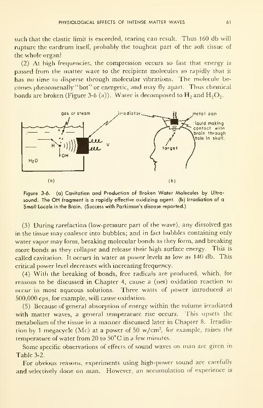
PHYSIOLOGICAL EFFECTS OF INTENSE MATTER WAVES 61
such that the elastic limit is exceeded, tearing can result. Thus 160 db will
rupture the eardrum itself, probably the toughest part of the soft tissue of
the whole organ!
(2) At high frequencies, the compression occurs so fast that energy is
passed from the matter wave to the recipient molecules so rapidly that it
has no time to disperse through molecular vibrations. The molecule be-
comes phenomenally "hot" or energetic, and may fly apart. Thus chemical
bonds are broken (Figure 3-6 (a)). Water is decomposed to H 2and H
2 2.
gas or steam irradiator metal pan
liquid makingcontact withbrain throughhole in skull.
(a) (b)
Figure 3-6. (a) Cavitation and Production of Broken Water Molecules by Ultra-
sound. The OH fragment is a rapidly effective oxidizing agent, (b) Irradiation of a
Small Locale in the Brain. (Success with Parkinson's disease reported.)
(3) During rarefaction (low-pressure part of the wave), any dissolved gas
in the tissue may coalesce into bubbles; and in fact bubbles containing only
water vapor may form, breaking molecular bonds as they form, and breaking
more bonds as they collapse and release their high surface energy. This is
called cavitation. It occurs in water at power levels as low as 140 db. This
critical power level decreases with increasing frequency.
(4) With the breaking of bonds, free radicals are produced, which, for
reasons to be discussed in Chapter 4, cause a (net) oxidation reaction to
occur in most aqueous solutions. Three watts of power introduced at
500,000 cps, for example, will cause oxidation.
(5) Because of general absorption of energy within the volume irradiated
with matter waves, a general temperature rise occurs. This upsets the
metabolism of the tissue in a manner discussed later in Chapter 8. Irradia-
tion by 1 megacycle (Mc) at a power of 50 w/cm2, for example, raises the
temperature of water from 20 to 50°C in a few minutes.
Some specific observations of effects of sound waves on man are given in
Table 3-2.
For obvious reasons, experiments using high-power sound are carefully
and selectively done on man. However, an accumulation of experience is
62 MATTER WAVES: SOUND AND ULTRASOUND
being gained on animals, principally guinea pigs, rats, and mice. The in-
vestigations have not been extensive enough to denote anything other than
generalities. However, at 165 db, 500 to 400,000 cps, on guinea pigs,
pathological changes occur in both the inner and middle ear; lesions appear
in the organ of Corti, and it is ruptured from the basilar membrane. Hemor-
rhages start where the malleus meets with the eardrum. Convulsions often
result. The skin becomes blistered and reddened. Death is hastened by the
damage.
TABLE 3-2. Effects of High-Intensity Sound on Man*
Frequency (cps) Level (db) Effect
stimulation of receptors in skin
mild warming of body surfaces
nausea, vomiting, dizziness; interference with touch
and muscle sense
significant changes in pulse rate
pain in middle ear
changes in muscle tone; increase in tendon reflexes;
incoordination
minor permanent damage if prolonged
major permanent damage in short time
vibration of muscles in arms and legs
resonance in mouth, nasal cavities, and sinuses
Collected by Neeley, K. K., "Noise—Some Implications for Aviation," Can. Aeronaut. J., 3, 312 (1957).
SONIC AND ULTRASONIC THERAPY
Certain uses have already been demonstrated; others await discovery, for
the technique is very new to medicine. The following applications are al-
ready well known in principle, and are now being introduced in practice very
cautiously— for the early 1950's saw the period of novelty wax strong, and
then wane into a hard reappraisal in the mid-50's; and one now observes the
gradual emergence of the place of vibrations in the medical arsenal. Details
can be found in the reviews of two masters of the subject, R. F. Herrick 10 and
W.J. Fry 1 and in the book edited by E. Kelly. 2
Present Applications
(1) Subcutaneous lesions can be located by ultra high-frequency matter
waves. They focus well at 1 Mc, and penetrate to a useful depth. The depth
of penetration is a function of the power of the source. Since reflection of
matter waves is greater the higher the density of the medium, tumors can be
distinguished from normal tissue at a location deep below the surface.
100

SONIC AND ULTRASONIC THERAPY 63
(2) Based on the same principle, the rate of blood flow through the ar-
terial system can now be measured by reflected ultrasound, in a nondestruc-
tive experiment in which all instrumentation is external to the body.
(3) Dentists have begun to apply sound to the ears of patients during
drilling, because it has been found that the brain cannot perceive pain from
the teeth and sound from the ear at the same time. The sound in this case
acts as a local anesthetic.
(4) "Rapid massage" heat therapy is now quite common, with an assort-
ment of low-frequency vibrator pads and belts available, and experimental
models operating in the 12,000 to 50,000 cps region. For deep "massage"
higher frequency ultrasound is used; it has the added advantage of comfort
from noise.
(5) Certain skin diseases can be treated with beamed and focused ultra-
sound. Thus viruses are destroyed (literally shaken into little bits!) by
ultrasound, and a future in sterilization seems assured. In this application
its competitor is soft X rays.
(6) "Neurosonic surgery" is now well advanced on animals, and has re-
ceived some experimental evaluation on humans. The most spectacular suc-
cess so far has been achieved in treatment of Parkinson's disease, the shaking
palsy. Because of its future importance,*** some details will now be given.
"Neurosonic Surgery"
The ultrasonic radiation reaches the brain through a hole cut in the skull,
and the matter waves are beamed and focused on that part, deep in the
brain, in which involuntary movements are controlled (Figure 3-6 (b)). The
energy dissipated by the beam is concentrated at the focus of the beam, and
gently destroys the metabolic activity at the site (the substantia nigra). The
method, when used carefully, has the advantage over all others that it pro-
duces lesions at the focus of the ultrasonic energy without interfering with
the normal blood flow from one part of the brain to another through the
region irradiated. Of course this is a great advantage from the medical point
of view. The techniques were worked out first on hundreds of cats and
monkeys, and are now very cautiously being applied to man. Functional
disruption of nervous conduction occurs within a few seconds of exposure to
ultrasound of sufficient dosage to produce lesions: 980,000 cps, 1.8- to 3-sec
duration, and particle velocity amplitude of 350 cm/sec, from a generator
with the capability of 20 to 1000 w/cm 2. From the therapeutic viewpoint it
has been found possible to irradiate simultaneously the four small parts of
the brain which are active with respect to Parkinsonism in the four limbs.
***In spite of the fact that Parkinsonism may be dying out. Thus the average age of these
patients is steadily increasing, in North America, a trend which, if it continues, would indicate
that the disease may have died out naturally by 1985.
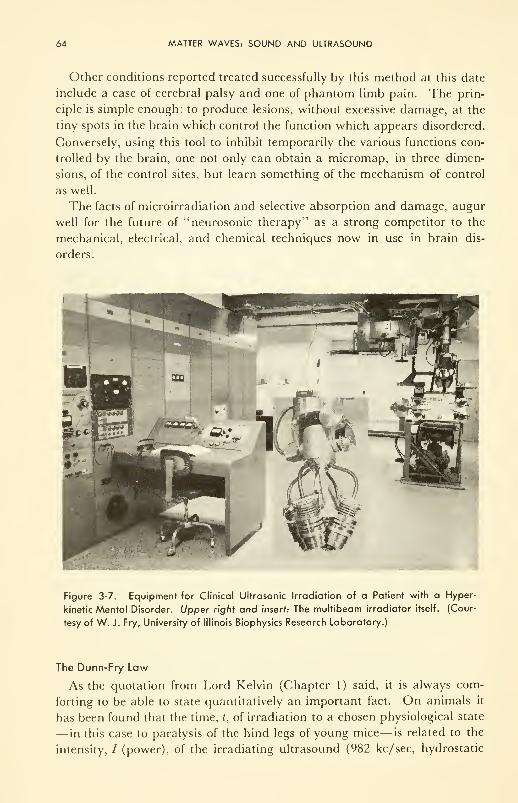
64 MATTER WAVES: SOUND AND ULTRASOUND
Other conditions reported treated successfully by this method at this date
include a case of cerebral palsy and one of phantom limb pain. The prin-
ciple is simple enough: to produce lesions, without excessive damage, at the
tiny spots in the brain which control the function which appears disordered.
Conversely, using this tool to inhibit temporarily the various functions con-
trolled by the brain, one not only can obtain a micromap, in three dimen-
sions, of the control sites, but learn something of the mechanism of control
as well.
The facts of microirradiation and selective absorption and damage, augur
well for the future of "neurosonic therapy" as a strong competitor to the
mechanical, electrical, and chemical techniques now in use in brain dis-
orders.
Figure 3-7. Equipment for Clinical Ultrasonic Irradiation of a Patient with a Hyper-
kinetic Mental Disorder. Upper right and insert: The multibeam irradiator itself. (Cour-
tesy of W. J. Fry, University of Illinois Biophysics Research Laboratory.)
The Dunn-Fry Law
As the quotation from Lord Kelvin (Chapter 1) said, it is always com-
forting to be able to state quantitatively an important fact. On animals it
has been found that the time, t, of irradiation to a chosen physiological state
— in this case to paralysis of the hind legs of young mice— is related to the
intensity, / (power), of the irradiating ultrasound (982 kc/sec, hydrostatic
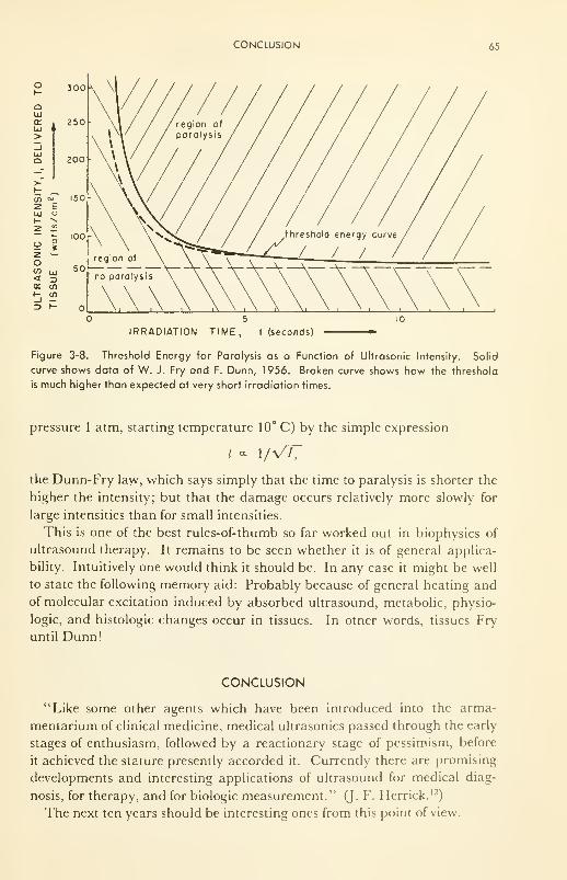
CONCLUSION 65
QUJ
or
300
250
200
co ~-?-
66 MATTER WAVES: SOUND AND ULTRASOUND
PROBLEMS
3- 1 : Express in decibels the sound which delivers 1 50 times the power of background
noise.
3-2: (a) Calculate the value of the absorption coefficient of sound in tissue at 50;
1000; 10,000; and 500,000 cycles per second (cps).
(b) Make a plot of intensity vs depth in tissue for each frequency.
3-3: How would you employ the inverse square law to "protect" yourself from an
intense source of noise? Suppose you wanted to reduce the noise level by
a factor often.
What could you learn about this problem from a = f(rj) as these terms are
defined in the text?
3-4: Two signals enter your ear: one at 500 cps, with intensities / and 7° equal to
10~ I2 and 10" l5 w/cm2, respectively; and the other at 6000 cps with intensities
/and 1° equal to 10 14 and 10~ 16 w/cm2. Which will seem the louder?
REFERENCES
1. Fry, W. J., Adv. in Biol, and Med. Phys., 6,281 (1959): a review, illustrated.
2. Kelly, E., Ed., "Ultrasound in Biology and Medicine," Amer. Inst, of Biol. Sciences,
Washington, D. C, 1957.
3. Fletcher, H., "Speech and Hearing," D. Van Nostrand Co., Inc., New York,
N.Y., 1946.
4. Ruch, T. C. and Fulton, J. F., Eds., "Medical Physiology and Biophysics,"
W. B. Saunders Co., Philadelphia, Pa., 1960.
5. Herzfeld, K. F. and Litovitz, T. A., "Absorption and Dispersion of Ultrasonic
Waves," Academic Press, New York, N. Y., 1959.
6. Stacy, R. W., Williams, D. T., Worden, R. E., and McMorris, R. O., "Essen-
tials of Biological and Medical Physics," McGraw-Hill Book Co., Inc., NewYork, N. Y., 1955.
7. Whitfield, I. C, "The Physiology of Hearing," in Progr. in Biophysics, 8, 1 (1957);
a review.
8. Gray, J. A. B., "Mechanical into Electrical Energy in Certain Mechano-
Receptors," Progr. in Biophysics, 9, 285 (1959); a review.
9. Neely, K. K., "Noise—Some Implications for Aviation," Can. Aeronaut. J., 3,
312(1957).
10. Herrick,J. F. and Anderson, J. A., "Circulatory System: Methods—Ultrasonic
Flow Meter," in "Medical Physics," Vol. Ill, O. Glasser, Ed., Yearbook
Publ., Inc., Chicago, 111., 1960, p. 181.
11. Gardner, W. H., "Speech Pathology," ibid., p. 637.
12. Herrick, J. F., Proc. Inst. Radio Engineers, Nov., 1959, p. 1957.
13. Von Bekesy, G., "The Ear,"5W. Amer., Aug., 1957; a review.
CHAPTER 4
Electromagnetic Radiations
and Matter
The next thing is striking: through the black carton container, which lets
through no visible or ultraviolet rays of the sun, nor the electric arc light, an
agent (X) goes through which has the property that it can produce a vivid
fluorescence ....
We soon found that the agent penetrates all bodies, but to a very different
degree. (W. C. Roentgen, Annalen der Physik und Chemie, 64, 1
(1898).)
INTRODUCTION
Within fifteen years, just before the turn of the century, complacent classi-
cal physics received three rude shocks. The first was Julius Plucker's de-
scription (circa 1890) of the electrical discharges which take place in gases
under low pressure and high voltage (the embryo of the "neon" sign). Thesecond was Henri Becquerel's discovery of natural radioactivity in 1895; andthe third was Wilhelm Roentgen's discovery ofX rays, reported in 1898. In
the years since then, the three discoveries have collectively engendered in-
tense investigation of: (1) the structure of molecules, atoms and nuclei;
(2) arrangements of molecules in crystals and other, less well-defined molec-
ular arrays; (3) the electromagnetic spectrum, from X rays through visible
to infrared radiation; and (4) the interactions—and in fact interconversion!
—of electromagnetic energy and matter. In this chapter a review is given of
those facts and theories which are useful to an understanding of the bio-
physics of the interactions of electromagnetic radiation and living matter.
67
68 ELECTROMAGNETIC RADIATIONS AND MATTER
THE STRUCTURE OF MATTER
The Elementary Particles and Atomic Architecture
Some of the key experimental facts accumulated within a few years of 1900
illustrate the bases upon which our knowledge of structure depends.
Roentgen found that his unknown, or "X," rays would cause fluorescence
in zinc sulfide and barium platinocyanide; and further that they would
ionize gases and darken a photographic plate. They were therefore easily
detected by an electroscope, or by an increase in current through a gaseous
discharge tube, or by photographic techniques. He studied penetration
through paper, wood, and metals, and showed that difference in penetration
is one of degree rather than of kind (cf. the quotation which opened this
Chapter.)
A fluorescent screen on each end of a cylindrical gaseous discharge tube
showed that particles, presumably charged, pass between the electrodes in
each direction. By placing metal shields between positive and negative elec-
trodes, and by impressing a voltage between horizontal plates placed with
their plane parallel to the direction of flow, it was shown that the rays com-
ing from the positive electrode bend toward the negative horizontal plate,
and are therefore positively charged; and likewise the rays from the negative
plate bend toward the positive plate, and are therefore negative. The nega-
tive particles were called cathode rays, and positives canal rays.
In 1897, J. J. Thomson (not William Thomson, Lord Kelvin) measured
the deviation of the (negative) cathode rays in an electric and magnetic field,
and obtained a value for the quotient of the charge to mass, i.e., e/m. This
value was found to be the same (1.757 x 10H cou/g) no matter what ma-
terials were used. Cathode rays were therefore recognized as elementary
particles of matter, and were called electrons. The (positive) canal rays, how-
ever, were found to be different for different materials.
By an ingenious experiment in late 1897, Milliken was able to obtain an
independent measure of e, the charge on the electron. One or two electrons
were trapped on atomized oil particles, and the electrical force necessary to
prevent each oil particle from falling under the influence of gravity was
measured. Since the size of the particle could be determined from the rate
of free fall, the charge absorbed by the particle could be evaluated. Thesmallest value obtained, 4.78 x 10~ 10 electrostatic units (1.600 x 10~ 19 cou),
corresponded to one electron absorbed.
From Thomson's value of e/m, the mass could then be determined as
9 x 10" 28g. This was an astounding achievement, the fact that exact meas-
urement of this mass was possible by these means, whereas the most sensi-
tive chemical balance weighs to only approximately 10" 6g!
For the canal rays, e/m for H + was found to be 1820 times smaller than
for the electron. Faraday in 1830 had shown by electrolysis that the charge
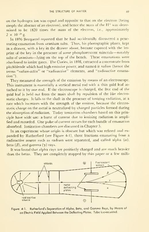
THE STRUCTURE OF MATTER 69
on the hydrogen ion was equal and opposite to that on the electron (being
simply the absence of an electron), and hence the mass of the H + was deter-
mined to be 1820 times the mass of the electron, i.e., approximately
2 x 10" 24g.
In 1896 Becquerel reported that he had accidentally discovered a pene-
trating emanation from uranium salts. Thus, his photographic plates, kept
in a drawer, with a key in the drawer above, became exposed with the im-
print of the key in the presence of some phosphorescent minerals— notably
salts of uranium— lying on the top of the bench. These emanations were
also found to ionize gases. The Curies, in 1898, extracted a concentrate from
pitchblende which had high emissive power, and named it radium (hence the
terms "radium-active" or "radioactive" elements, and "radioactive emana-
tion").
They measured the strength of the emission by means of an electroscope.
This instrument is essentially a vertical metal rod with a thin gold leaf at-
tached to it by one end. If the electroscope is charged, the free end of the
gold leaf is held out from the main shaft by repulsion of the like electro-
static charges. It falls to the shaft in the presence of ionizing radiation, at a
rate which increases with the strength of the emitter, because the electro-
static charge on the metal is neutralized by charged particles formed during
the absorption of radiation. Today ionization chambers based on this prin-
ciple have wide use: a burst of current due to ionizing radiation is ampli-
fied and recorded. One pulse of current occurs for each bundle of emanation
absorbed. Ionization chambers are discussed in Chapter 5.
In an experiment whose origin is obscure but which was refined and ex-
panded by Rutherford (see Figure 4-1), three fractions emanating from a
radioactive source such as radium were separated, and called alpha (a),
beta (/3), and gamma (7) rays.
It was found that alpha rays are positively charged and are much heavier
than the betas. They are completely stopped by thin paper or a few milli-
shields
rodioactivesource
Figure 4-1 . Rutherford's Separation of Alpha, Beta, and Gamma Rays, by Means of
an Electric Field Applied Between the Deflecting Plates. Tube is evacuated.
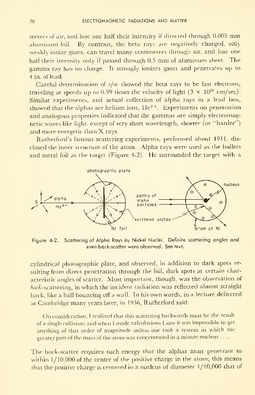
70 ELECTROMAGNETIC RADIATIONS AND MATTER
meters of air, and lose one half their intensity if directed through 0.005 mmaluminum foil. By contrast, the beta rays are negatively charged, only
weakly ionize gases, can travel many centimeters through air, and lose one
half their intensity only if passed through 0.5 mm of aluminum sheet. The
gamma ray has no charge. It strongly ionizes gases and penetrates up to
4 in. of lead.
Careful determination of e/m showed the beta rays to be fast electrons,
traveling at speeds up to 0.99 times the velocity of light (3 x 10 10 cm/sec).
Similar experiments, and actual collection of alpha rays in a lead box,
showed that the alphas are helium ions, He++. Experiments on penetration
and analogous properties indicated that the gammas are simply electromag-
netic waves like light, except of very short wavelength, shorter (or "harder")
and more energetic than X rays.
Rutherford's famous scattering experiments, performed about 1911, dis-
closed the inner structure of the atom. Alpha rays were used as the bullets
and metal foil as the target (Figure 4-2). He surrounded the target with a
photographic plate
nucleus
paths of
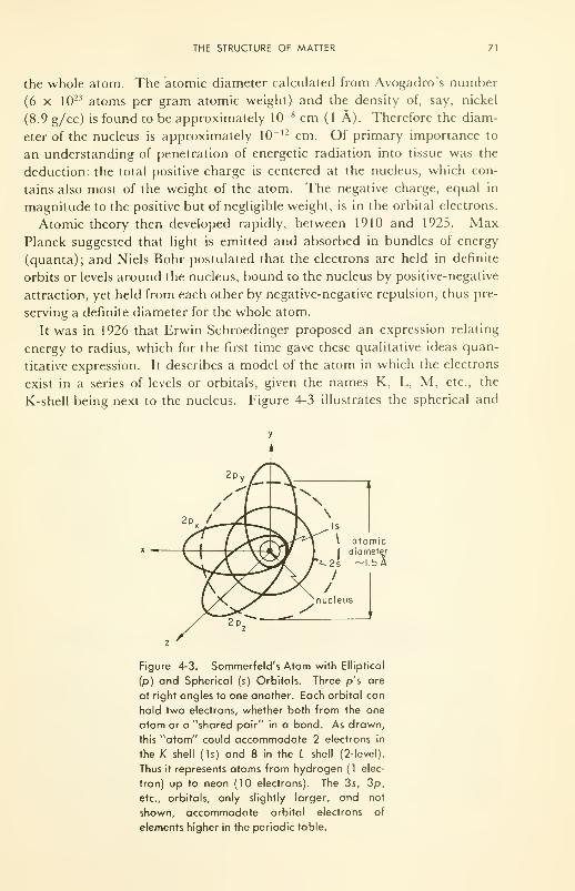
THE STRUCTURE OF MATTER 71
the whole atom. The atomic diameter calculated from Avogadro's number
(6 x 10²³ atoms per gram atomic weight) and the density of, say, nickel
(8.9 g/cc) is found to be approximately 10~ 8 cm (1 A). Therefore the diam-
eter of the nucleus is approximately 10 l2 cm. Of primary importance to
an understanding of penetration of energetic radiation into tissue was the
deduction: the total positive charge is centered at the nucleus, which con-
tains also most of the weight of the atom. The negative charge, equal in
magnitude to the positive but of negligible weight, is in the orbital electrons.
Atomic theory then developed rapidly, between 1910 and 1925. MaxPlanck suggested that light is emitted and absorbed in bundles of energy
(quanta); and Niels Bohr postulated that the electrons are held in definite
orbits or levels around the nucleus, bound to the nucleus by positive-negative
attraction, yet held from each other by negative-negative repulsion, thus pre-
serving a definite diameter for the whole atom.
It was in 1926 that Erwin Schroedinger proposed an expression relating
energy to radius, which for the first time gave these qualitative ideas quan-
titative expression. It describes a model of the atom in which the electrons
exist in a series of levels or orbitals, given the names K, L, M, etc., the
K-shell being next to the nucleus. Figure 4-3 illustrates the spherical and
Figure 4-3. Sommerfeld's Atom with Elliptical
(p) and Spherical (s) Orbitals. Three p's are
at right angles to one another. Each orbital can
hold two electrons, whether both from the one
atom or a "shared pair" in a bond. As drawn,
this "atom" could accommodate 2 electrons in
the K shell (Is) and 8 in the L shell (2-level).
Thus it represents atoms from hydrogen (1 elec-
tron) up to neon (10 electrons). The 3s, 3p,
etc., orbitals, only slightly larger, and not
shown, accommodate orbital electrons of
elements higher in the periodic table.
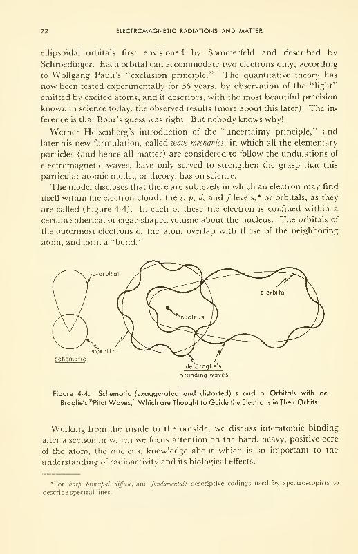
72 ELECTROMAGNETIC RADIATIONS AND MATTER
ellipsoidal orbitals first envisioned by Sommerfeld and described by
Schroedinger. Each orbital can accommodate two electrons only, according
to Wolfgang Pauli's "exclusion principle." The quantitative theory has
now been tested experimentally for 36 years, by observation of the "light"
emitted by excited atoms, and it describes, with the most beautiful precision
known in science today, the observed results (more about this later). The in-
ference is that Bohr's guess was right. But nobody knows why!
Werner Heisenberg's introduction of the "uncertainty principle," and
later his new formulation, called wave mechanics, in which all the elementary
particles (and hence all matter) are considered to follow the undulations of
electromagnetic waves, have only served to strengthen the grasp that this
particular atomic model, or theory, has on science.
The model discloses that there are sublevels in which an electron may find
itself within the electron cloud: the s, p, d, and / levels,* or orbitals, as they
are called (Figure 4-4). In each of these the electron is confined within a
certain spherical or cigar-shaped volume about the nucleus. The orbitals of
the outermost electrons of the atom overlap with those of the neighboring
atom, and form a "bond."
p-orbitol
s-orbito
schematic
de Broglie s
standing waves
Figure 4-4. Schematic (exaggerated and distorted) s and p Orbitals with de
Broglie's "Pilot Waves," Which are Thought to Guide the Electrons in Their Orbits.
Working from the inside to the outside, we discuss interatomic binding
after a section in which we focus attention on the hard, heavy, positive core
of the atom, the nucleus, knowledge about which is so important to the
understanding of radioactivity and its biological effects.
*For sharp, principal, diffuse, and fundamental: descriptive codings used by spectroscopists to
describe spectral lines.
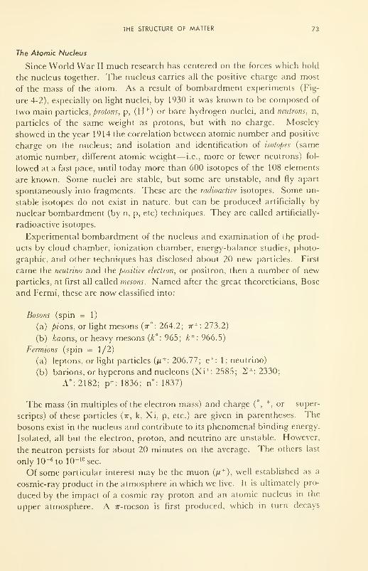
THE STRUCTURE OF MATTER 73
The Atomic Nucleus
Since World War II much research has centered on the forces which hold
the nucleus together. The nucleus carries all the positive charge and most
of the mass of the atom. As a result of bombardment experiments (Fig-
ure 4-2), especially on light nuclei, by 1930 it was known to be composed of
two main particles, protons, p, (H +) or bare hydrogen nuclei, and neutrons, n,
particles of the same weight as protons, but with no charge. Moseley
showed in the year 1914 the correlation between atomic number and positive
charge on the nucleus; and isolation and identification of isotopes (same
atomic number, different atomic weight— i.e., more or fewer neutrons) fol-
lowed at a fast pace, until today more than 600 isotopes of the 108 elements
are known. Some nuclei are stable, but some are unstable, and fly apart
spontaneously into fragments. These are the radioactive isotopes. Some un-
stable isotopes do not exist in nature, but can be produced artificially by
nuclear bombardment (by n, p, etc) techniques. They are called artificially-
radioactive isotopes.
Experimental bombardment of the nucleus and examination of the prod-
ucts by cloud chamber, ionization chamber, energy-balance studies, photo-
graphic, and other techniques has disclosed about 20 new particles. First
came the neutrino and the positive electron, or positron, then a number of new
particles, at first all called mesons. Named after the great theoreticians, Bose
and Fermi, these are now classified into:
Bosons (spin = 1)
(a) pions, or light mesons (t° : 264.2; tt±
: 273.2)
(b) A;aons, or heavy mesons (k°: 965; k ±: 966.5)
Fermions (spin = 1/2)
(a) leptons, or light particles {n*: 206.77; e*: 1; neutrino)
(b) barions, or hyperons and nucleons (Xi*: 2585; 2*: 2330;
A°:2182; p*: 1836; n°: 1837)
The mass (in multiples of the electron mass) and charge (°,+
,or " super-
scripts) of these particles (ir, k, Xi, p, etc.) are given in parentheses. The
bosons exist in the nucleus and contribute to its phenomenal binding energy.
Isolated, all but the electron, proton, and neutrino are unstable. However,
the neutron persists for about 20 minutes on the average. The others last
only 10- 6 tol0- 10 sec.
Of some particular interest may be the muon (p*), well established as a
cosmic-ray product in the atmosphere in which we live. It is ultimately pro-
duced by the impact of a cosmic ray proton and an atomic nucleus in the
upper atmosphere. A 7r-meson is first produced, which in turn decays
74 ELECTROMAGNETIC RADIATIONS AND MATTER
rapidly into the muon plus a gamma ray. The muon disintegrates into a
fast, ionizing electron and two more gamma rays, at sea level.
The atom and its nucleus were recently detailed in delightful form by
Gamov 10, in a little book highly recommended for its simple, colorful de-
scriptions of very complex phenomena.
Molecular Structure and Binding
It is the outer, or valence, electrons of the electron cloud which are evi-
dently involved in binding atom to atom (Figure 4-4). Two distinct cases,
and one intermediate case, have been studied thoroughly. First, the valence
electron in "atom 1" can jump into an empty orbital of "atom 2," leaving
atom 1 positive and making atom 2 negative. Strong electrostatic binding
exists (Coulomb's law) because the charge separation is small. This is the
case in all salts, both inorganic and organic. The bond is called ionic.
Secondly, the electron from atom 1 can simply exchange, or be "shared"
with that of atom 2. For instance if each of the two valence electrons is in
an s (spherical) orbital, and the orbitals can overlap so that exchange or
sharing takes place, a "sigma" bond is formed. If both are in p ("probing")
orbitals (cigar-shaped), and if they overlap, a so-called pi (7r) bond is formed
(Figure 4-5). Indeed combinations of s and p, called "hybrids," are pos-
sible. For example each of the four bonds made by a carbon atom is a hy-
brid of one s and three/? valence electrons— imagine, in Figure 4-4, the 2s
and 2p electron orbitals as distorted; it is a mixture, called an sp^ hybrid.
The four are directed tetrahedrally from each other, like four long noses,
each to form a bond (i.e., to share a pair of electrons) with a neighboring
atom. In the case of water, each of the p orbitals of oxygen overlaps with s
of hydrogen to form a bent (109°) molecule. The bond is called covalent.
TT-bondelectrons
closedloop
(b)
TT bond electrons, open path,\ m obi le
carbon atoms
Figure 4-5. Diagrams of Overlapping it Bonds: (a) A closed loop to form a dough-
nut of negative charge above the plane of a benzene ring; (b) on a protein with open
and ringed molecular structures, in which 7r-bond electrons are somewhat mobile and
can transfer charge from one end of the molecule to the other, if forced.
THE STRUCTURE OF MATTER 75
In between the ionic and covalent bond is the dative bond, in which the
electron of atom 1 is partially given over to atom 2, although exchange and
overlap still occur. Organic-phosphorus molecules are an important ex-
ample (ATP, for instance, the "mobile power supply" in the living system).
The oxygens of the phosphate assume a definite negative charge because of
dative bonding.
Of special importance is the w bond, formed by the overlap of two p orbi-
tals ("probosci"). It often forms the second bond in the "double bond" of
conjugated organic molecules, and restricts the relative rotation of atoms 1
and 2 if joined by the it. But the most important property of the it bond is
its position, directed parallel to, but not coaxial with, the atom—atom axis
(Figure 5 (b)). Although it helps to bind atom 1 to atom 2, it is an ac-
cumulation of negative charge outside the volume containing the two atoms.
It therefore can form weak bonds (complexes) with positive ends of other
molecules in the vicinity; but, most important, it can exchange electrons
with other it bonds close by, and hence provide a pathway by which elec-
trons can run along a molecule from a point of excess negative charge to a
point of deficiency of charge. Hence some organic molecules in tissues are
electronic conductors, a fact which only recently has been appreciated with
respect to nerve conduction and photosynthesis. (This very important topic
is pursued in Chapter 6.) Further, the possibility of different electronic
states in molecules, with different types of bonds, has profound ramifications
in interactions of the molecule (and the tissue of which it forms a part) with
electromagnetic radiations. These very important topics are also discussed
in Chapter 6.
It is obvious that the elementary particles are the building blocks of the
living stuff. From the molecular point of view, however, it is not at all clear
where the line is to be drawn between the living and nonliving. Usually the
attributes of growth and reproduction are used to classify the living. Yet, in
a supersaturated solution, copper sulfate crystals will "grow," layer upon
layer; and if the temperature is allowed to fluctuate up and down with a
frequency of one or two cycles per day, they will "reproduce" themselves, by
"seeding," in the form of many crystallites on the walls of the container. In-
deed, Teilhard de Chardin, in 1945, proposed that all the elementary par-
ticles of matter are living, that they have the potency to do the things which
living things can do, but that this potency is, to us, masked behind the
gross behavior of large numbers. The gross behavior— statistical behavior
—
is all that our experimental techniques can today perceive in inanimate na-
ture. Our techniques can examine the highlv organized individual man in
which ~1028 particles are organized and controlled from within, although
this inner FORCE is not amenable to physical examination as we know it
today. From the point of view of elementary particles, the only difference be-
tween living and nonliving matter is one of organization.

76 ELECTROMAGNETIC RADIATIONS AND MATTER
ELECTROMAGNETIC RADIATION; NATURE AND SPECTRUM
The electron clouds of atoms and molecules can be excited by various
methods—by heat, bombardment by some charged particle, and by absorp-
tion of incoming radiations. A simple example is the flame test for sodium:
if a sodium salt is heated in a flame, it glows with a characteristic yellow
glow. It is not burning (i.e., being oxidized by oxygen). Rather, the valence
(outermost) electron gets excited (accepts energy) and "jumps" to a higher-
energy orbital, from a 3s to a 3/?. Imagine the next set of orbitals around
the nucleus in Figure 4-3. Its lifetime there is short, however, and it falls
back to the original state ("ground state"), and emits the extra energy as
electromagnetic radiation {light in this case) of such a wavelength (5893 A)
that it excites the cone cells on the retina of the eye.
Biology is entering its electromagnetic age. Many parts of the electromag-
netic spectrum are beginning to be used for diagnosis and therapy, as well
as for studies which are leading to a better understanding of the roles of
each of the parts in the systematized whole.
Nature of Electromagnetic Radiation
The exact nature of electromagnetic (em) radiation is unknown. What is
known is that the wave has two component parts, an electric part and a mag-
netic part, moving in phase, but in direction 90° from each other—much like
two vibrating strings, one going up and down while the other goes back and
forth—superimposed on each other. Each oscillates about an average value
(zero) at a frequency which depends upon electronic vibrations in the
source. The em waves travel in a straight line, and have energies inversely
proportional to the wavelength, or directly proportional to the frequency
(number of cycles per second). The wave carries no net electrical charge,
and no net magnetic moment, but because of the components which can in-
terfere or react with electric or magnetic fields, it can lose or gain energy
(i.e., change frequency). All em waves travel at the velocity of "light."
They have both wave properties (such as the capability of being reflected or
diffracted) and particle properties (such as delivering their energy in
bundles or quanta.). The unit bundle of electromagnetic energy is called
the photon. Undulations in the electromagnetic field are described by the
celebrated Maxwell equations (1873).
Electromagnetic radiations vary only in frequency, and through this, in
energy. Therefore their use requires handling the energy contained in the
radiation. For example, we know how to handle light with mirrors, lenses,
microscopes, and prisms, and to detect it by photographic plates, photo-
electric cells, the eye, etc. Handling, or making it serve a useful purpose, is
simply a question of using equipment which does not absorb the light. Detec-
ELECTROMAGNETIC RADIATION; NATURE AND SPECTRUM 77
tion is simply a question of providing a medium which can absorb the light,
or a medium with which the light can interact and be partially absorbed, to
appear as another, more familiar form of energy.
Electromagnetic radiation propagates with undiminished energy through
a vacuum, always at the speed of light no matter what the frequency.
The Electromagnetic Spectrum— A Survey
Table 4-1 gives some properties of interest for the whole spectrum of elec-
tromagnetic radiations. Since the em radiation has both wave and particle
properties, the wavelength range of the different sections is given, and the
energy associated with an excitation in each section is given in electron volts
(1 electron volt/molecule = 22,000 cal/mole). Common means of detecting
and of handling the radiations are noted; and what happens during absorp-
tion is indicated.
If one expects to gain insight into the interactions of electromagnetic
radiations and matter, one must study the two Tables, 4-1 and 4-2, ex-
haustively. There is no easier way. One will find, for example, from in-
spection of the dimensions of the wavelength, A, and frequency, v, that
they are related through the velocity, c, which for all electromagnetic radia-
tions in vacuum, no matter what the wave length, is 3 x 10 10 cm/sec
(186,000 miles/sec). Thus
v = 3 x 10 !0/A cycles/sec
Table 4-2 indicates some of the effects of the interaction of various "cuts"
of the spectrum with matter. It is certainly true that radiation of short wave
length (high frequency) carries more energy, is more penetrating, and can
do more damage than that of long wave length. Thus, at wavelengths from
20,000 to 500,000 A, the radiation simply tickles the molecules into a rota-
tional and vibrational frenzy (high heat energy;. Radiation of 4000 to
7800 A excites electrons in the pigment molecules of the retina of the eye,
and is visible. (Maximum sensitivity of the eye is at about 6000 A.) Radia-
tion of wavelength 2000 to 4000 A (ultraviolet) excites even the bonding elec-
trons in a molecule, and so loosens up a bond that chemical reactions may
take place which otherwise could not. Wavelengths below 2000 A, in the
hard or vacuum ultraviolet, actually drive electrons out of a molecule, or
ionize it; and as the wavelength gets shorter, and the radiation "harder,"
more and more ions are formed in the wake of the incoming radiation. In
the X-ray region (X = 1 A) the electrons of even the K shell of the atom,
the most tightly bound ones, can be excited or ejected; and in the gammaregion (~0.01 A), even the nucleus can be penetrated by the radiation, al-
though electrons in the atomic cloud are a more probable target.
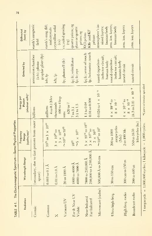
78
E ctt cu
cu
Q
CU
Cboco
a:
-a "o53 J2 —
co obe «u ^CO " IU
E £\SCJ —
<" -rl
-5 cu CO
COU
bo_c
tot-
bc
T3<U
bD
— _ JS ^ N2 CO 3 bD
CO
3cruO
be
-ace -5
c/i pi
CO co
3 J2cr bo
&
uucu
a £ c £
"O cu "3u o >• o>, _Q CO j3* C • C
CO 3 CO
£.2E3 _: 3
JZ
-
CU
JO
c
e£0
CU
JO
3_o
6to
cu
0"> O
S a- -t
cu
cO
-Ccu
C_o
toN'5
o
i u
x 9- CLCO_bD
cucu
oou33-
uo
cu
§ 2
cu
3
CU
CU
JZCU
-3cu .3-.
o S3
- ?c E«-> oci —.- o
u rl ^ u° " -£r °« .S T3 «
cu O '
cu
CO s-
S g-o -acu cC •-
_Q
CCO
cuCO
3
3. "3CU O
CU
3b, "o -a
cCO
E3
3cuu
'cu
"3CU
-3 c •- -C C
3cu
-acu
c3
3
r- T
>cu
3,
o
T33COen
3OJZ
Cu>
O
Oo
r-o
oCM
^1-
LO o
^ o o
2 - un
r- m
oooodo<*o
^- d
IO
o
o
O r-~I
<o Oo'-' XX 00
XO o>
i- o ^°"^- x 2X 00 <^
cp
cu>-cu
OOo
cu
cu
o
JU"cu>^ooooo"oo
cu
cu
CO
boCU
0)
tcu
Q.O
ou
Eo
E3i_^«(JCUato
ca>oEo
iu(U
CU
£ 0) 4)
cu <3> 4?3 C «cr o i)
« cc -- 2r
cu
o>co
ccu
D
i-cu
3O
3OO[_
Ou
in
JO
O
CU
3
CO"33OCUcu
COo
O =o O
oX ro —
OO O OJ
O )
* 2 2 o© o <-
X X (V) ,-
c^ <^> I
©O©
oo
tocu
cu^
CO ^-v ©M U OCU ^ r—E6o
CU
oun
©LT)
CU>-CU
3
•<
CUuCO
a.
©o
©©
••< »<
•<o
od
o©oo
©
o©©o©ooo
o©00
o
©©©
©©
8°8 N
-
Ecu
©
©LTl
O©O00 LT)
r» cm
'-"" 22 •<oo©
©©©©~oen
£cu
o
o
o
Ecu
©o
©©o
©oCO
co
o^5o
-a
CO
-oCO
CO<
CU
ECO
OU
CO
EECO
O X
CO
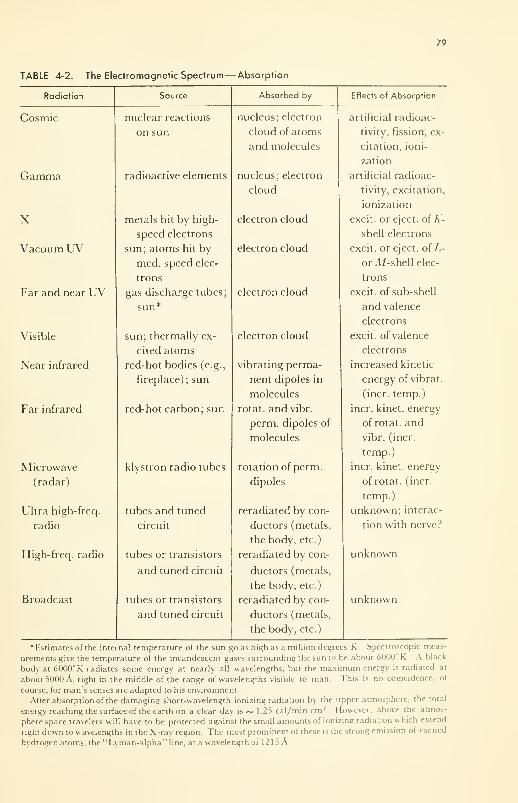
79
TABLE 4-2. The Electromagnetic Spectrum— Absorption
Radiation
80 ELECTROMAGNETIC RADIATIONS AND MATTER
Quantitative expression of these ideas followed Planck, who, in 1901, pro-
posed that the energy, e, contained per photon in incoming electromagnetic
radiation is proportional to the frequency, v, of the radiation. Thus
e = hv
where h is the proportionality (Planck's) constant, equal to 6.62 x 10" 27 erg
sec/photon (1 electron volt, ev, = 1.6 x 10 12 ergs).
Let w},w
2 , and w 3be the energies of binding of different atomic or molec-
ular orbital states of the electron to the nucleus, and accept Bohr's as-
sumption. If e = w}
, w 2 , or w3 , absorption of the incoming radiation will
easily occur,accompanied by excitation of the electron from its "ground
state," or orbital of lowest energy, to an excited state. If e ^ wy
, w 2 , or w3 ,
then absorption does not readily occur, although in favorable cases wxcan be
taken from a larger e, the electron excited to state 1, and the radiation pass
on with reduced energy (e - w, = hv2 ) and lower frequency (longer wave-
length). This is one aspect of the famous "Compton scattering."
If f is greater than some critical value, w, the ionization energy, the elec-
tron can be ejected completely from the atom or molecule, and may have any
kinetic energy up to and including e — w. Since the electron has a mass of
9 x 10 _28g, the kinetic energy (1/2 mv2
) is less than, or equal to, e — w.
Now a negative particle of velocity v, just like any other member of the elec-
tron cloud about a molecule, but moving with high velocity, is a very good
ionizer itself. Hence the ionization process continues along a track through
the tissue until all the incoming energy, e, has been dissipated either as heat
or in producing ions.
The Laws of Absorption
In the tables of properties of em radiations, the bases of the techniques for
handling them were implied. What happens when absorption takes place
was also indicated. We consider now the extent of absorption, and its con-
verse, the depth of penetration.
In brief and in summary, absorption of electromagnetic radiations is
governed only by the laws of chance. The chance that a photon will be ab-
sorbed depends only upon the number of target electrons and nuclei in its
path. From the fact that the higher energy (shorter wave length) radiations
penetrate deeper into any given material, it is inferred that they are more
difficult to capture— have a "smaller capture cross-sectional area." Con-
versely, the denser the target material the greater is the number of potential
targets per centimeter of the photon's path, and hence the greater is the ab-
sorption per unit length of path.
These ideas are expressed quantitatively in Lambert's law. The rate of
ELECTROMAGNETIC RADIATION; NATURE AND SPECTRUM 81
absorption is directly proportional to the amount to be absorbed; or
-dl/dx = k'l
where x is thickness and / is intensity, or number of photons passing 1 cm 2
per sec. This is one of the natural functions (Chapter 1) for which / is ex-
pressed explicitly as
/ = /,-k'x
where I is the intensity when x = 0, just as the radiation enters the ab-
sorbent; k' is a constant, characteristic of the absorbent (larger, the better
the absorption capacity of the medium), called the absorption coefficient. Theplot of / vs x is shown in Figure 1-2 (c).
Since In I /I = k'x, conversion to common logarithms by dividing by
2.303 gives log I /I = kx, where k' = 2.303 k, and k is called the "extinction
coefficient."
Lambert's law is applicable over the whole electromagnetic spectrum,
and, you will remember from Chapter 3, is useful also to describe the ab-
sorption of matter waves. It is an obvious but very important point that the
extinction coefficient of a substance will be different at different wave-
lengths. From the far infrared, through to the near ultraviolet, the extinc-
tion coefficient is large only for particular wavelengths. Such specificity is a
property of molecular absorption. If these molecules are suspended or dis-
solved in a medium, k will be directly proportional to the concentration, c
(Beer's law). Thus k can now be factored into ac, where a is called the molec-
ular extinction coefficient. Formally then:
log I /I = acx (Beer-Lambert law)
The specificity for absorption of selected wavelengths disappears from the
far ultraviolet through to gamma radiation—continuous absorption occurs
accompanied by ionization—and the extinction coefficient decreases more
or less linearly with decreasing wavelength (i.e., with increasing energy
/photon). Thus ultraviolet light penetrates only a small fraction of an inch
of tissue; and the k for tissue for near ultraviolet is very large. By contrast,
soft X rays penetrate tissue with only a small amount of absorption per cm;
and k is smaller. However, each photon of X rays absorbed carries roughly
1000 times more energy than each photon of near ultraviolet, and therefore
only 1/1000 as much absorption is required to do the same damage. It is
seen then that the important quantity is the energy absorbed per unit volume,
because this determines the subsequent effect: warming of tissue, triggering
of the optic nerve fiber, providing the energy for photochemical synthetic
processes, or ionization and rupture of molecular bonds.
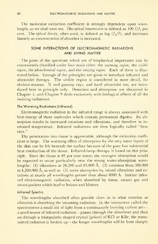
82 ELECTROMAGNETIC RADIATIONS AND MATTER
The molecular extinction coefficient is strongly dependent upon wave-
length, as we shall soon see. The optical transmission is defined as 100 7// per
cent. The optical density, often used, is defined as log (I /I), and increases
linearly as concentration of absorber is increased.
SOME INTERACTIONS OF ELECTROMAGNETIC RADIATIONS
AND LIVING MATTER
The parts of the spectrum which are of biophysical importance can be
conveniently classified under four main titles: the warming region, the visible
region, the photochemical region, and the ionizing region. Each of these is illus-
trated below. Enough of the principles are given to introduce infrared and
ultraviolet therapy. The visible region is considered in more detail, for
obvious reasons. X and gamma rays, and hard ultraviolet too, are intro-
duced here in principle only. Detection and absorption are discussed in
Chapter 5, and Chapter 9 deals exclusively with biological effects of all the
ionizing radiations.
The Warming Radiations (Infrared)
Electromagnetic radiation in the infrared range is always associated with
heat energy of those molecules which contain permanent dipoles. Its ab-
sorption results in increased rotations and vibrations, and therefore in in-
creased temperature. Infrared radiations are then logically called "heat
rays.
The penetration into tissue is appreciable, although the extinction coeffi-
cient is large. The warming effect of absorption by the very outer layers of
the skin can be felt beneath the surface because of the poor but substantial
heat conduction of the tissue. Infrared-lamp therapy is based on this prin-
ciple. Since the tissue is 85 per cent water, the strongest absorption would
be expected to occur particularly near the strong water-absorption wave-
lengths: (1) vibrations at 28,200 and 63,000 A, (2) rotations from 500,000
to 1,200,000 A, as well as (3) some absorption by mixed vibrations and ro-
tations at nearly all wavelengths greater than about 8000 A. Intense infra-
red electromagnetic radiation, when absorbed by tissue, causes gas and
steam pockets which lead to lesions and blisters.
Infrared Spectra
The wavelengths absorbed often provide clues as to what rotation or
vibration is absorbing the incoming radiation. In the instrument called the
spectrometer a small slit of light from a continuously burning carbon arc
—
a good source of infrared radiation— passes through the absorbent and then
on through a triangularly shaped crystal (prism) of KC1 or KBr; the trans-
mitted radiation is broken up—the longer wavelengths will be bent sharply
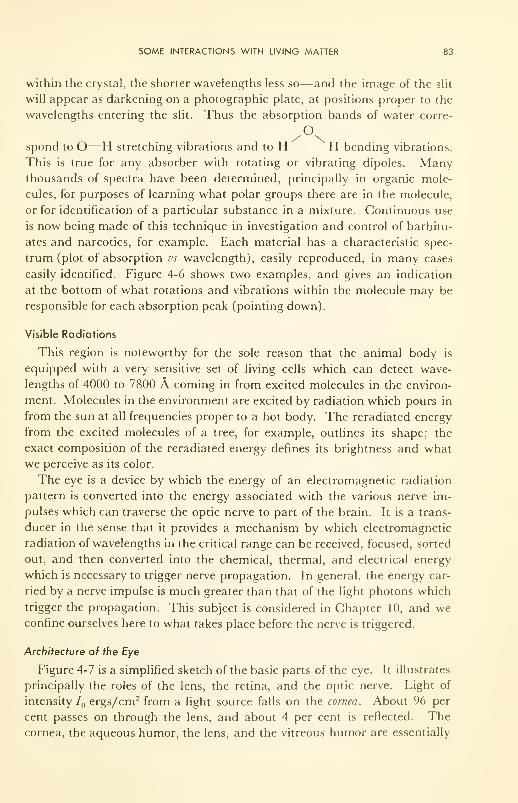
SOME INTERACTIONS WITH LIVING MATTER 83
within the crystal, the shorter wavelengths less so—and the image of the slit
will appear as darkening on a photographic plate, at positions proper to the
wavelengths entering the slit. Thus the absorption bands of water corre-
Ospond to O—H stretching vibrations and to H H bending vibrations.
This is true for any absorber with rotating or vibrating dipoles. Manythousands of spectra have been determined, principally in organic mole-
cules, for purposes of learning what polar groups there are in the molecule,
or for identification of a particular substance in a mixture. Continuous use
is now being made of this technique in investigation and control of barbitu-
ates and narcotics, for example. Each material has a characteristic spec-
trum (plot of absorption vs wavelength), easily reproduced, in many cases
easily identified. Figure 4-6 shows two examples, and gives an indication
at the bottom of what rotations and vibrations within the molecule may be
responsible for each absorption peak (pointing down).
Visible Radiations
This region is noteworthy for the sole reason that the animal body is
equipped with a very sensitive set of living cells which can detect wave-
lengths of 4000 to 7800 A coming in from excited molecules in the environ-
ment. Molecules in the environment are excited by radiation which pours in
from the sun at all frequencies proper to a hot body. The reradiated energy
from the excited molecules of a tree, for example, outlines its shape; the
exact composition of the reradiated energy defines its brightness and what
we perceive as its color.
The eye is a device by which the energy of an electromagnetic radiation
pattern is converted into the energy associated with the various nerve im-
pulses which can traverse the optic nerve to part of the brain. It is a trans-
ducer in the sense that it provides a mechanism by which electromagnetic
radiation of wavelengths in the critical range can be received, focused, sorted
out, and then converted into the chemical, thermal, and electrical energy
which is necessary to trigger nerve propagation. In general, the energy car-
ried by a nerve impulse is much greater than that of the light photons which
trigger the propagation. This subject is considered in Chapter 10, and weconfine ourselves here to what takes place before the nerve is triggered.
Architecture of the Eye
Figure 4-7 is a simplified sketch of the basic parts of the eye. It illustrates
principally the roles of the lens, the retina, and the optic nerve. Light of
intensity IQ ergs/cm2 from a light source falls on the cornea. About 96 per
cent passes on through the lens, and about 4 per cent is reflected. Thecornea, the aqueous humor, the lens, and the vitreous humor are essentially
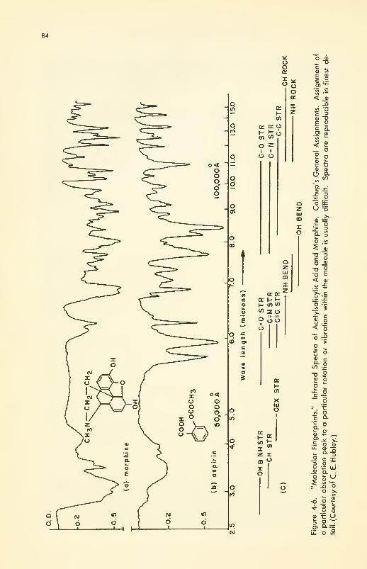
84
-• \-
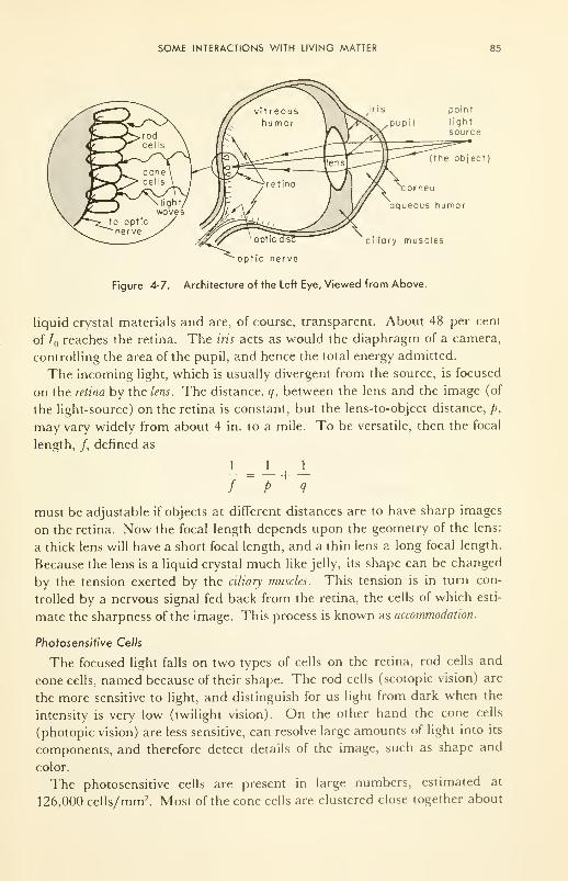
SOME INTERACTIONS WITH LIVING MATTER 85
point
light
source
(the object)
ornea
aqueous humor
ciliary muscles
optic nerve
Figure 4-7. Architecture of the Left Eye, Viewed from Above.
liquid crystal materials and are, of course, transparent. About 48 per cent
of 7 reaches the retina. The iris acts as would the diaphragm of a camera,
controlling the area of the pupil, and hence the total energy admitted.
The incoming light, which is usually divergent from the source, is focused
on the retina by the lens. The distance, q, between the lens and the image (of
the light-source) on the retina is constant, but the lens-to-object distance, p,
may vary widely from about 4 in. to a mile. To be versatile, then the focal
length, /, defined as
_L _L J_
/~
P+
q
must be adjustable if objects at different distances are to have sharp images
on the retina. Now the focal length depends upon the geometry of the lens:
a thick lens will have a short focal length, and a thin lens a long focal length.
Because the lens is a liquid crystal much like jelly, its shape can be changed
by the tension exerted by the ciliary muscles. This tension is in turn con-
trolled by a nervous signal fed back from the retina, the cells of which esti-
mate the sharpness of the image. This process is known as accommodation.
Photosensitive Cells
The focused light falls on two types of cells on the retina, rod cells and
cone cells, named because of their shape. The rod cells (scotopic vision) are
the more sensitive to light, and distinguish for us light from dark when the
intensity is very low (twilight vision). On the other hand the cone cells
(photopic vision) are less sensitive, can resolve large amounts of light into its
components, and therefore detect details of the image, such as shape and
color.
The photosensitive cells are present in large numbers, estimated at
126,000 cells/mm 2. Most of the cone cells are clustered close together about
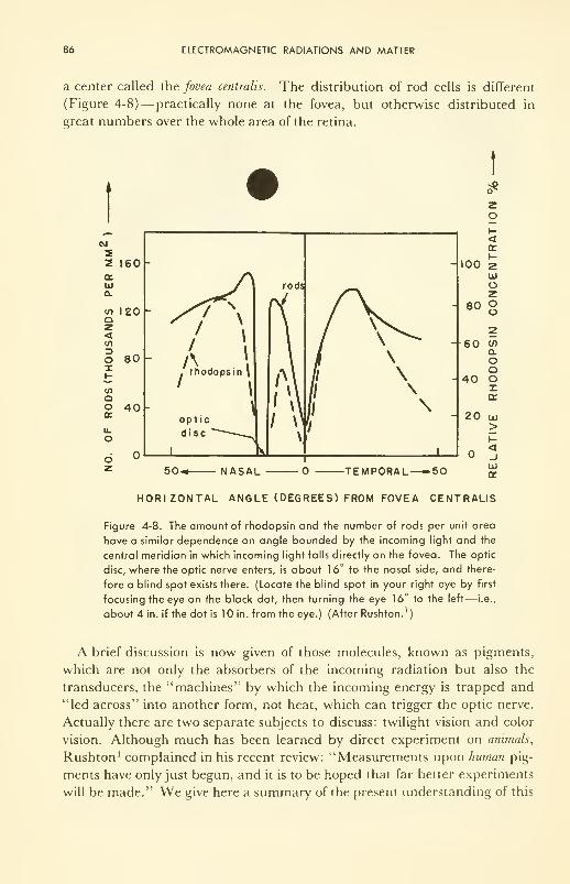
86 ELECTROMAGNETIC RADIATIONS AND MATTER
a center called the fovea centralis. The distribution of rod cells is different
(Figure 4-8)— practically none at the fovea, but otherwise distributed in
great numbers over the whole area of the retina.
*
eg
22E
a.UJCl
tf)
oz<
OI
COooce
u.
O
160 -
120 -
80 -
40
HORIZONTAL ANGLE (DEGREES) FROM FOVEA CENTRALIS
Figure 4-8. The amount of rhodopsin and the number of rods per unit area
have a similar dependence on angle bounded by the incoming light and the
central meridian in which incoming light falls directly on the fovea. The optic
disc, where the optic nerve enters, is about 16° to the nasal side, and there-
fore a blind spot exists there. (Locate the blind spot in your right eye by first
focusing the eye on the black dot, then turning the eye 16° to the left— i.e.,
about 4 in. if the dot is 10 in. from the eye.) (After Rushton.')
A brief discussion is now given of those molecules, known as pigments,
which are not only the absorbers of the incoming radiation but also the
transducers, the "machines" by which the incoming energy is trapped and
"led across" into another form, not heat, which can trigger the optic nerve.
Actually there are two separate subjects to discuss: twilight vision and color
vision. Although much has been learned by direct experiment on animals,
Rushton 1 complained in his recent review: "Measurements upon human pig-
ments have only just begun, and it is to be hoped that far better experiments
will be made." We give here a summary of the present understanding of this
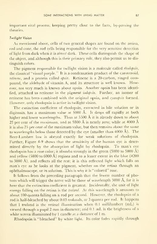
SOME INTERACTIONS WITH LIVING MATTER 87
important vital process, keeping pretty close to the facts, by-passing the
theories.
Twilight Vision
As mentioned above, cells of two general shapes are found on the retina,
rod and cone, the rod cells being responsible for the very sensitive detection
of light from dark when it is almost dark. These cells distinguish the shape of
the object, and although this is their primary role, they also permit us to dis-
tinguish colors.
The pigment responsible for twilight vision is a molecule called rhodopsin,
the classical "visual purple." It is a condensation product of the carotenoid,
retinene, and a protein called opsin. Retinene is a 20-carbon, ringed com-
pound, the aldehyde of vitamin A, and its structure is well known. How-
ever, not very much is known about opsin. Another opsin has been identi-
fied, attached to retinene in the pigment todopsin. Further, an isomer of
retinene has been combined with the original opsin, and cyanopsin formed.
However, only rhodopsin is active in twilight vision.
The extinction coefficient of rhodopsin, extracted in bile solution or in
digitonin, has a maximum value at 5000 A. It drops off rapidly at both
higher and lower wavelengths. Thus at 5500 A it is already down to about
25 per cent of the maximum, and at 5800 A is nearly zero; while at 4000 Ait is also 25 per cent of the maximum value, but then remains about the same
to wavelengths below those detected by the eye (smaller than 4000 A). The
Beer-Lambert law is obeyed exactly for weak solutions of rhodopsin.
Further, Figure 4-9 shows that the sensitivity of the human eye is deter-
mined directly by the absorption of light by rhodopsin. To man's eye
rhodopsin has a rose color; it absorbs strongly in the green (5000 to 5800 A)
and yellow (5800 to 6000 A) regions and to a lesser extent in the blue (4200
to 5000 A), and reflects all the rest; it is this reflected light which falls on
man's eye as he looks at the pigment, whether on the retina through an
ophthalmoscope, or in solution. This is why it is "colored" rose.
It follows from the preceding paragraph that the fewest number of pho-
tons which will trigger the nerve will be those of wavelength 5000 A, for it is
here that the extinction coefficient is greatest. Incidentally, the unit of light
energy falling on the retina is the troland. At this wavelength it amounts to
about 100 quanta falling on a rod per second. However, the rhodopsin of a
rod is half-bleached by about 0.03 trolands, or 3 quanta per rod. It happens
that 1 troland is the retinal illumination when 0.1 millilambert (mL) is
viewed through a pupil 2 mm in diameter; and 0.1 mL is the brightness of a
white screen illuminated by 1 candle at a distance of 1 m.
Rhodopsin is "bleached" by white light. Its color fades rapidly through
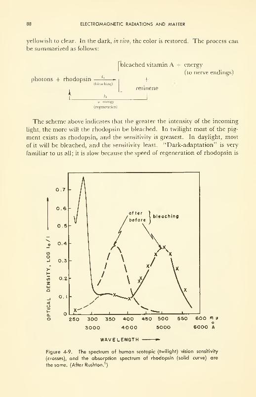
88 ELECTROMAGNETIC RADIATIONS AND MATTER
yellowish to clear. In the dark, in vivo, the color is restored. The process can
be summarized as follows:
photons + rhodopsin
k
A,
(bleaching)
k2
bleached vitamin A -f energy
(to nerve endings)
+retinene
+ energy
(regeneration)
The scheme above indicates that the greater the intensity of the incoming
light, the more will the rhodopsin be bleached. In twilight most of the pig-
ment exists as rhodopsin, and the sensitivity is greatest. In daylight, most
of it will be bleached, and the sensitivity least. "Dark-adaptation" is very
familiar to us all; it is slow because the speed of regeneration of rhodopsin is
o
in
zUJQ
<
a.
o
0.7 -
0.6 -
0.5 -
0.4 -
0.3 -
0.2 -
0. I-
250 300 350 400 450 500 550
3000 4000 5000
600 m Mo
6000 A
WAVELENGTH
Figure 4-9. The spectrum of human scotopic (twilight) vision sensitivity
(crosses), and the absorption spectrum of rhodopsin (solid curve) are
the same. (After Rushton. 1
)
SOME INTERACTIONS WITH LIVING MATTER 89
slow. The reader is invited to contemplate the expression of the Weber-Fechner law in this organ:
5" oc log r/M,
It says that the sensitivity, S, increases as the difference between the
threshold intensity and that of the background decreases.
This photochemical description of twilight vision, although satisfactory in
general, apparently needs revision, for serious troubles arise when quantita-
tive description is attempted. It now seems likely that individual pigment
molecules are attached to individual nerve endings, and the excitation of just
one pigment molecule by incoming radiation is sufficient to trigger the nerve.
Thus, although it takes upwards of half an hour for dark adaptation to oc-
cur—that is, for the bulk rhodopsin to be regenerated in man after a bleach-
ing—the minimum time during which the eye can recover enough from a
flash to see another flash is about 0.01 sec.
Color Vision
The cone cells somehow distinguish between wavelengths, and thus dis-
tinguish colors. The Young-Helmholtz theory, usually accepted, and nownearly 100 years old, suggested that three color-sensitive pigments exist,
each one sensitive to one of the basic colors: red (6200 to 7800 A), green
(5000 to 5800 A) and blue (4200 to 5000 A); and that various intensities mix
to give the colors and qualities commonly referred to as hue, brightness, etc.
The Young-Helmholtz theory is based on the experimental fact that by a
proper mixture of red, blue, and green light in an object, any shade of color
can be matched. The theory is that the three pigments absorb definite frac-
tions of the visible spectrum and overlap one another, and that the optic
nerve can receive and transmit signals which correspond to any and all
wavelengths of the spectrum. Apparently this theory now requires major
modification as a result of the very recent (1959) work of E. H. Land. 7 In
some remarkable experiments he has shown in effect that the full range of
colors can be recorded by the brain provided only that the proper mixtures of
intensities of two wavelengths (one greater than, and one less than, 5880 A(yellow)), fall on the retina! It seems that the information about colors other
than the two incoming wavelengths is developed in the retina. The possibility
that the pigment molecules are in intimate contact in the cone cells, and dis-
tribute the excitation energy among themselves in a manner controlled by
the intensity pattern of the incoming light, immediately suggests itself. But
more work is clearly needed following this surprising turn. Another recent
surprise is that some evidence has been turned up that other molecules in the
neurones, in the nerve pathway itself, contribute to the color perceived in
human vision.
90 ELECTROMAGNETIC RADIATIONS AND MATTER
In spite of the credence placed in the Young-Helmholtz three-pigment
theory of color vision, there is no direct evidence that three pigments exist
in the cones. There is direct experimental evidence for two, however; this
will now be recalled. Protanopes (color-blind people) cannot distinguish
green from red. By measurement of the intensity of the light reflected from
the retina as a function of incident wavelength on protanopes, it has been
shown that a definite absorption by a pigment, given the name "chlorolabe,"
takes place with maximum at about 5400 A.
Now the protanope can see green, but not red. This fact means that a sec-
ond pigment, given the name "erythrolabe," is missing in the protanope.
Difference spectra (unreliable) of two pigments in the normal fovea (collec-
tion of cone cells) show that the maximum absorption of the second, or miss-
ing, pigment is about 6000 A. Thus there is good knowledge of one pig-
ment, the chlorolabe, and knowledge of the existence of a second, erythro-
labe. There is no experimental knowledge of a third in cones. But, of course,
Land's new work indicates that only two are really necessary, one sensitive
above and one sensitive below 5800 A. The two pigments discussed have
these qualifications. Recall that the optical density maximum for rhodopsin
is at 5000 A.
What the relation is between the excited pigment molecule and the color
perceived is poorly known. Experimental approaches include that of meas-
uring the electrical signals in the optic nerve (the electroretinogram, ERG)during stimulation by light, the reflection densitometry experiments men-
tioned just above, studies of the rates of bleaching and recovery (adapta-
tion), visual acuity, color perception, and Land's new work. However, since
the excitation energy for electrons in large molecules is so dependent upon
structure, it would not be surprising if rhodopsin, chlorolabe, and erythro-
labe turn out to be very similar in composition. The answer will lie in
knowledge of the structure of these molecules.
Incidentally, an important new fact, bearing upon acuity especially, is
that the eyeball is never still, but rather is in a state of small, almost im-
perceptible oscillations, such that the incoming light falls on a spot on the
retina for only a few microseconds before it is deflected away. If the eyeball
is fixed relative to the light source, color vision disappears.
Physical Defects of the Eye
If the lens is too thick or the eyeball elongated (myopia), the ciliary
muscles are not able to make sufficient adjustment of the focal length to
permit distant objects to be focused on the retina. The phenomenon is
known as nearsightedness, and can be corrected with the aid of glasses with a
concave lens of the proper focal length. If the length of the eyeball is too

SOME INTERACTIONS WITH LIVING MATTER 91
small, the condition is called hypermetropia, and can be corrected with a con-
vex lens of proper focal length.
The lens of the eye often does not have the same curvature over all its sur-
face, and light passing through the area of improper curvature will not be
properly focused on the retina. The lens of such an eye is said to be astig-
matic. A properly ground astigmatic glass lens can compensate.
Sometimes translucent or opaque tissue grows in or on the liquid crystal
material of the lens and absorbs the incoming light before it reaches the
retina. Such tissues are generally termed cataracts. Some can be removed by
surgery; some are too extensive.
Depth Perception
Two detectors in different locations can inherently provide more informa-
tion than one; and if relative information is recorded and interpreted from
the two signals, more information is available from the two detectors than if
each were interpreted separately. This is the reason sensory organs come in
pairs. Typical of the relative information obtainable from two stations, in
general, are direction and distance, or depth. Sound can be reflected, and
hence the directional information provided by two stations is important.
Light travels in a straight line to the eye, and therefore directional informa-
tion is not important. However, the information derivable about distance or
depth is important when we attempt to compare distances or develop a per-
spective view. Ideally the eyes may each be rotated about 50° from a central
line of vision. The two have to be in focus at the same time, on a near or a
far object, and this requires a facility of minor individual adjustment. If the
eyes cannot be made to focus (crossed eyes), sufficient correction can some-
times be made with a suitable set of glass lenses, but often the cross must be
corrected by shortening the lateral muscles or by suitable exercises designed
to strengthen them.
Photochemical Radiations (Ultraviolet)
Photosynthesis
Subshell electrons are excited by the ultraviolet. The absorbed energy
may be passed off to the vibrations or rotations of nearby molecules and ap-
pear as heat energy; it may be re-emitted as ultraviolet; or it may excite
the molecule and make it more susceptible to chemical attack by neighbor-
ing molecules. Thus in the last case the ultraviolet may provide some or all
of the activation energy needed for reaction to occur, and thereby increase
the rate of reaction (treated later in Chapter 8). In fact, the photochemical
mechanism is sometimes the only mechanism by which certain reactions can
take place at a reasonable speed at biological temperature.
92 ELECTROMAGNETIC RADIATIONS AND MATTER
Because they carry more energy than photons in the visible region, the
photons in the ultraviolet region are less likely to be absorbed. They pene-
trate deeper into the absorbent and excite molecules at the point at which
they are finally caught.
Of all the synthetic biological reactions whose rate is sensitive to ultra-
violet light, probably the photosynthesis of simple organic sugars from C02
and2in plant leaves is the best understood; and yet the understanding of
this basic process is not completely satisfactory. Of course if it were, weshould be able to reproduce the syntheses in a test tube; but we cannot.
More important to present considerations is our knowledge of photo-
catalyzed syntheses of the vitamins from basic components. Some of the
vitamins have been purified, crystallized, and synthesized, and hence their
chemical composition and structure are known. Consider the antirickets
vitamin D 2 (calciferol) for instance. Its structure is well known: two six-
membered rings and a five-membered ring attached to an unsaturated
aliphatic side chain of six carbon atoms, with a molecular weight of 393.
This molecule is formed through the absorption of ultraviolet radiation of
2500 to 3000 A by ergosterol, a sterol molecule whose structure also is well
known. The synthesis occurs in at least two steps. The absorption is con-
sidered to take place at a carbon-carbon double bond, and the absorbed
energy to go into excitation of the t electrons which form the bond. Theopening of a benzene-like ring follows, and further rearrangements of the
atoms and bonds give the biochemically active vitamin B2structure. The re-
action will not occur at all unless photolyzed.
This synthesis takes place in the human body at a location to which both
the molecular components and ultraviolet radiation are accessible: that is,
just beneath the surface of the skin in the living tissue serviced by the blood
capillaries. Thus the principle upon which ultraviolet therapy is based, and
the advantages of moderate exposure to sunlight, both become apparent.
Phototherapy
Prolonged sun bathing can damage skin pigments and can cause ery-
thema. For instance, on the average it takes only 20 microwatts (/xw) of
ultraviolet of wavelength 2537 A (from a mercury vapor lamp) falling upon
the skin for 15 min to produce erythema. It is fortunate that the very in-
tense ultraviolet radiation from the sun is attenuated (scattered, absorbed,
converted into radiation of longer wavelength) by the ozone and nitrogen
compounds in the upper atmosphere. Ultraviolet radiation would be a prob-
lem in space travel if it were not so readily reflected by metallic surfaces.
The effects on the eye are well known and have been implied in the discus-
sion of the chemistry of the eye: the higher-energy photons of the ultraviolet
in falling on the retina can keep the rod and cone cells devoid of rhodopsin
SOME INTERACTIONS WITH LIVING MATTER 93
and damage the color pigment molecules. Snow-blindness and "whiteouts"
are the result. Further, ultraviolet has been attributed in some cases to
promoting the growth of cataracts and photothalamia, or inflammation of
the cornea. However, ordinary window glass absorbs all the dangerous
ultraviolet, and colored inorganic materials can be added to filter out (or
absorb) any undesired range of wave lengths. Therefore, protection is no
problem, if properly sought.
Ultraviolet light has a lethal effect on primitive animal and plant life.
This fact is used to good advantage in destroying the bacteria, eschenchia coli
and bacteria coli, in foods or in our water supply. Each of these is killed by
about 14 x 10" 6 ergs per bacterium. Among the abnormalities successfully
treated with ultraviolet light are conjunctivitis, fibrosis, acne, and surface in-
fections of various kinds. Certain heavy metals (calcium, gold, silver, etc.)
and certain highly absorptive molecules (methylene blue, quinine, etc.)
sometimes increase the therapeutic value of the ultraviolet irradiation.
The shortest-wave, vacuum-ultraviolet radiation overlaps the X-ray re-
gion. The principle difference between the two regions in the present classi-
fication is whether ionization and bond rupture is the exception (ultraviolet)
or the rule (X and gamma). The vacuum-ultraviolet will be discussed im-
plicitly in the next section, for the differences between it and the X ray are
of degree rather than of kind.
Ionizing Radiations (Mainly X and Gamma)
Principles
The only distinction between the radiations more and less energetic than
that with a wavelength about 2000 A is one of excitation vs ionization. That
is, at wavelength X greater than about 2000 A, excitation of electrons of the
electron cloud takes place as the rule, and ionization takes place only in
special circumstances; while at X less than about 2000 A the electrons can be
knocked right out of the atom by the absorbed photon. As X decreases, the
loosely held orbital electrons are the first to go, followed by the subshell elec-
trons, and as X —» 1 A (X-ray region) the tightly bound K-shell electrons
can be ejected.
A simple calculation will make this important point clear. It takes an in-
put, w, of ~230 kcal to make 1 mole of ions out of 1 mole of atoms, i.e.,
10 ev to make an ion out of an atom. (This is the energy carried by each
photon of em radiation of wavelength 1200 A.) Now the gamma radiation
of the radioactive isotope of cobalt of atomic weight 60 (referred to the hy-
drogen atom as 1), Co60, used in deep radiation therapy for cancer, has an
energy of about one million electron volts (1 mev/photon). Therefore, each
photon would leave a wake of about 16/ 1 = 10 5 pairs of ions (or molecules
which have been ionized) before it loses all its energy.
94 ELECTROMAGNETIC RADIATIONS AND MATTER
The electrons lost may have been valence, or bonding, electrons— active
in holding the molecule together. In covalent bonding two paired electrons
form the bond between carbon atoms, as in a sugar molecule for example.
Ionization weakens the bond and perhaps breaks it; in any case the unpaired
electron left is chemically very reactive and will make a new bond at any
time or place. Cross-bonding of molecules, the synthesis of new molecules,
polymerization of old ones, etc., all can occur. It is not hard to envisage
how such reactions could adversely affect the tightly geared steady-state of
normal living tissue.
It is convenient to reserve further discussion of the effects of ionizing radia-
tions until the principles of radioactivity have been outlined. The radioac-
tive emanations, alpha, beta, and the nucleons, are ionizing radiations, as
are gamma and X, and the effects of all are conveniently discussed together.
Diagnosis by X Rays
The absorption of electromagnetic radiation increases with increasing
density of the absorbent. Differentiation of diseased tissue from normal is
based on this fact. The higher the speed of the electrons which impinge
on the target metal, the harder the X rays so produced. Machines avail-
able today produce X rays from electrons which have been accelerated by
thousands to millions of volts. In general, the greater the voltage, the greater
the energy of the X-ray photons, and the greater their penetrating power.
For example, at 40,000 v (i.e., 40 kilovolt potential (kvp), in radiation
terminology) almost any tissue will stop some of the X radiation and cast
a shadow on the fluorescent screen or photographic plate behind it. At 80 to
100 kvp, commonly used in medical diagnosis, the radiograph displays
shadows which differentiate fat and other soft tissues from air space and
from bone.
Whenever it is possible to insert molecules containing heavy metal atoms
into a region of interest, differentiation of tissues in the region is enhanced
(Figure 4-10). Thus barium sulfate solution is commonly administered as
an enema so that the lower part of the intestines may be examined (by Xradiation). Iodine in a variety of compounds is also widely used to increase
differentiation. For instance, in iodophthalien it is preferentially taken up
by the liver and stored in the gall bladder; thus gallstones, if present, are
easily seen. Similarly, the kidneys, uterus, blood vessels, and even the heart
can be made visible to X-radiography (see Figure 4-10 (b), for example).
Location of broken bones, of swallowed pins, of stomach ulcers and of
tumors is routine.
The use of X rays for diagnosis introduces the serious question of the ex-
tent of the damage done by the rays absorbed. A complete fluoroscopic
gastrointestinal examination with barium sulfate can be done by a competent
physician with the dose to the region irradiated not exceeding 20 rads (the
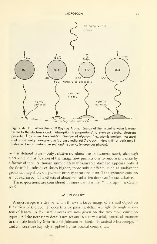
MICROSCOPY 95
impinging x-rays
80 kvp
1.32 1.35
+ +Co 20
5.0
0.99
Four targets or absorbers
1.54
transmittedx- rays
photographic plates
Figure 4- 10a. Absorption of X Rays by Atoms. Energy of the incoming wave is trans-
ferred to the electron cloud. Absorption is proportional to electron density, electrons
per cubic A (bold numbers inside). Number of electrons (i.e., atomic number - valence)
and atomic weight are given, as is atomic radius (at 7 o'clock). Note shift of both ampli-
tude (number of photons per sec) and frequency (energy per photon).
unit is defined later—only relative numbers are of interest now), although
electronic intensification of the image now permits one to reduce this dose by
a factor of ten. Although immediately measurable damage appears only if
the dose is hundreds of times higher, more subtle effects, such as malignant
growths, may show up years or even generations later if the greatest caution
is not exercised. The effects of absorbed radiation dose can be cumulative.
These questions are considered in more detail under "Therapy" in Chap-
ter 9.
MICROSCOPY
A microscope is a device which throws a large image of a small object on
the retina of the eye. It does this by passing definitive light through a sys-
tem of lenses. A few useful notes are now given on the two most commontypes. All the necessary details are set out in a very useful, practical manner
in the little book by Martin and Johnson entitled: "Practical Microscopy,"8
and in literature happily supplied by the optical companies.
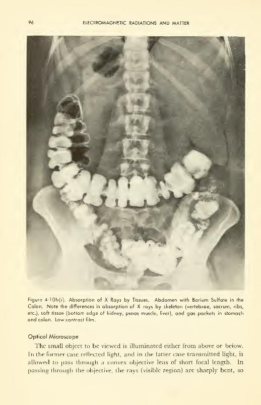
96 ELECTROMAGNETIC RADIATIONS AND MATTER
Figure 4-10b(i). Absorption of X Rays by Tissues. Abdomen with Barium Sulfate in the
Colon. Note the differences in absorption of X rays by skeleton (vertebrae, sacrum, ribs,
etc.), soft tissue (bottom edge of kidney, psoas muscle, liver), and gas pockets in stomach
and colon. Low contrast film.
Optical Microscope
The small object to be viewed is illuminated either from above or below.
In the former case reflected light, and in the latter case transmitted light, is
allowed to pass through a convex objective lens of short focal length. In
passing through the objective, the rays (visible region) are sharply bent, so
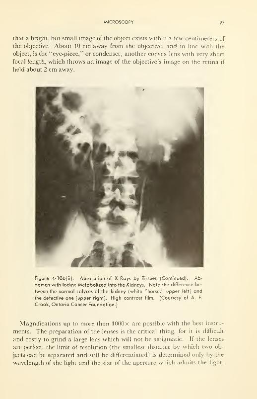
MICROSCOPY 97
that a bright, but small image of the object exists within a few centimeters of
the objective. About 10 cm away from the objective, and in line with the
object, is the "eye-piece," or condenser, another convex lens with very short
focal length, which throws an image of the objective's image on the retina if
held about 2 cm away.
Figure 4-10b(ii). Absorption of X Rays by Tissues (Continued). Ab-
domen with Iodine Metabolized into the Kidneys. Note the difference be-
tween the normal calyces of the kidney (white "horse," upper left) and
the defective one (upper right). High contrast film. (Courtesy of A. F.
Crook, Ontario Cancer Foundation.)
Magnifications up to more than lOOOx are possible with the best instru-
ments. The preparation of the lenses is the critical thing, for it is difficult
and costly to grind a large lens which will not be astigmatic. If the lenses
are perfect, the limit of resolution (the smallest distance by which two ob-
jects can be separated and still be differentiated) is determined only by the
wavelength of the light and the size of the aperture which admits the light.
98 ELECTROMAGNETIC RADIATIONS AND MATTER
For white light, with an average wavelength about 5000 A and a numerical
aperture of unity, the resolving power is 10,000 A, or 10" 4 cm, or 1 n. Onecan use monochromatic blue light to improve this somewhat; and the re-
search use of ultraviolet (A = 2537 A from a mercury arc, for example) with
fluorescent screens, is an attempt to push the resolution down to 0.1 ;u. In
common practice, however, "good" microscopes used in schools and routine
examination have a resolving power 5 to 20 /j..
The binocular microscope uses two microscopes in parallel, one for each
eye. From this double input, one obtains depth perception.
Phase-contrast and interference features have been superimposed on the
simple microscope, broadening its versatility by improving the contrast be-
tween different parts of the object under study. Contrast occurs in the
normal microscope because of differences in density. In phase and interfer-
ence microscopes, used when the density is about the same throughout (soft
tissue is ~90 per cent water), advantage is taken of the facts that the speed
of light through materials, which determines their refractive index, and the
amount to which the plane of polarized light can be rotated, often differ if
the molecular composition of the materials is different, even though their
density is the same. To take advantage of these facts, two methods are
available. Both present a highly contrasted image to the eye, one in inten-
sity, one in color.
The principles are really quite straightforward. The reader is referred to
the trade literature for operating detail. Both are extensions of the normal
bright-field transmission microscope; only the extensions will be noted here.
In the phase microscope, an annular diaphragm is inserted in front of the con-
denser lens and therefore before the light falls on the specimen, together
with a phase plate composed of a thinly evaporated ring of dielectric on a
background of thinly evaporated metal. Thus light passes at different speeds
through different parts of the object to be viewed, and the emerging light
waves are out of phase. At one point of emergence from the object the phase
difference will be such that the waves cancel each other; at another they
reinforce each other. The phase plate "fixes" these differences by retard-
ing those which pass through the dielectric, and absorbing some of those
which pass through the metal. Thus identification and analysis of the struc-
ture of (unstained) living cells and tissues, the components of which are so
similar in density that discrimination is impossible with the light microscope
without killing and staining, is made possible. This instrument, invented by
Zernicki in the Netherlands in 1932, is now an indispensible tool in clinical
analyses— in bacteriological, histological, and, in particular, pathological
studies of tumors and cancerous tissues. Note the contrast in Figure 4-11.
The interference microscope is a polarizing microscope, adapted so that part
of the light passes through the object and part around it, the two then being
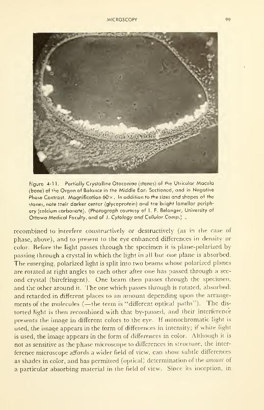
MICROSCOPY 99
Figure 4-11. Partially Crystalline Otoconiae (stones) of the Utricular Macula
(bone) of the Organ of Balance in the Middle Ear: Sectioned, and in Negative
Phase Contrast. Magnification 60 x . In addition to the sizes and shapes of the
stones, note their darker center (glycoprotein) and the bright lamellar periph-
ery (calcium carbonate). (Photograph courtesy of L F. Belanger, University of
Ottawa Medical Faculty, and of J. Cytology and Cellular Comp.) .
recombined to interfere constructively or destructively (as in the case of
phase, above), and to present to the eye enhanced differences in density or
color. Before the light passes through the specimen it is plane-polarized by
passing through a crystal in which the light in all but one plane is absorbed.
The emerging, polarized light is split into two beams whose polarized planes
are rotated at right angles to each other after one has passed through a sec-
ond crystal (birefringent). One beam then passes through the specimen,
and the other around it. The one which passes through is rotated, absorbed,
and retarded in different places to an amount depending upon the arrange-
ments of the molecules (—the term is "different optical paths"). The dis-
torted light is then recombined with that by-passed, and their interference
presents the image in different colors to the eye. If monochromatic light is
used, the image appears in the form of differences in intensity; if white light
is used, the image appears in the form of differences in color. Although it is
not as sensitive as the phase microscope to differences in structure, the inter-
ference microscope affords a wider field of view, can show subtle differences
as shades in color, and has permitted (optical) determination of the amount of
a particular absorbing material in the field of view. Since its inception, in
100 ELECTROMAGNETIC RADIATIONS AND MATTER
the early 1950's, it has been used for quantitative studies of proteins in living
muscle, growth rates of cells and parts of cells, and similar problems on
living tissue which can be studied only with a nondestructive tool.
Electron Microscope
This development of the last twenty years has added a new dimension to
the depth to which tissues can be viewed. After fixing and staining (e.g.,
permanganate, phosphotungstic acid, osmium oxide), a very thin cut to be
examined is placed in high vacuum, and bombarded from below by electrons
(from a hot filament) which have been accelerated through a small aperture.
Some of the electrons hit dense parts of the object and are scattered and
absorbed—the principle is the same as for X rays (Figure 4-10 (a)); others
pass on through less dense parts and fall upon a fluorescent screen or
photographic plate. Proper alignment permits, in today's machines, ampli-
fications of 500 to 100,000 x , with resolution of a few angstroms.
One instrument, which can be considered typical for biological work,**
gives a 15-A resolving power; 600 to 120,000x magnification; and accelera-
tion voltages of 100, 75, or 50 kv, to give electron beams of equivalent wave-
lengths of 0.037, 0.043, and 0.054 A. The "lenses" are electric voltages be-
tween charged plates. The amplification can be increased to over 1 ,000,000 x
by photographing the screen, and enlarging the photograph.
Others
The ultraviolet microscope and fluorescence microscope have been used
and improved since the early 1900's. They have some specialized uses in
biological research. X-ray microscopy is useful when the sections to be
studied are opaque to visible and ultraviolet light. For example, in histo-
logical sections on bone, soft (~5 kvp) X rays are absorbed by the mineral
component, passed by the organic component.
Reflection microscopy, especially the slowly developing infrared reflection
techniques, may find limited use in future studies on biological material.
PROBLEMS
4-1 : Draw the shapes of sigma and pi bonds.
4-2: If all 1028 atoms in a human being were lined up side by side, how long would
be the line, in miles?
4-3: It costs an input of about 105 kcal/mole to pull the first hydrogen off a water
molecule. "Light" of what wavelength will blast it off? (calculate it).
4-4: Sketch intensity vs distance for the penetration of electromagnetic radiation
into tissue, presuming concentration of absorbent of 0. 1 moles/1 and molecular
extinction coefficients of 0.1, 1.0, and 10.0.
**The limitations should be realized: the tissue sample is dead, dry, and thin while being
viewed in the electron microscope.

REFERENCES 101
REFERENCES
1. Rushton, W. A. H., "Visual Pigments in Man and Animals and Their Relation
to Seeing," Prog, in Bwphys., 9, 239 (1959).
2. Stacy, R. W., et al., "Essentials of Biological and Medical Physics," McGraw-
Hill Book Co., Inc., New York, N. Y., 1955, p. 262.
3. Brindley, G. A., "Human Color Vision," in Prog, in Bwphys., 8,49 (1959).
4. Evans, R. M., "An Introduction to Color," John Wiley & Sons, Inc., New York,
N.Y., 1948.
5. Ruch, T. C. and Fulton, J. F., Eds., "Medical Physiology and Biophysics,"
W. B. Saunders Co., Philadelphia, Pa., 1960.
6. The Physics Staff, University of Pittsburgh, "Atomic Physics," 2nd ed., John
Wiley & Sons, Inc., New York, N. Y., 1944.
7. Land, E. H., "Color Vision and the Natural Image," Proc. Nat. Acad. Set., 45,
115 (1959); Sri. Amer., 200,84 (1959).
8. Martin, L. C. and Johnson, B. K., "Practical Microscopy," 3rd ed., Blackie &Son, Ltd., London, 1958.
9. Shamos, M. H. and Murphy, G. M., "Recent Advances in Science," New York
Univ. Press and Interscience Pubis., Inc., New York, N. Y., 1956.
10. Gamov, G., "The Atom and its Nucleus," Prentice Hall, Inc., New York, N. Y.,
1961.
11. Richards, O. W., "Pioneer Phase and Interference Microscopes," N. T. State
J. Med., 61,430 (1961).
12. Bennett, A. H., Jupnik, H., Osterberg, H., and Richards, O. W., "Phase Micro-
scopy,"John Wiley & Sons, Inc., New York, N. Y., 1951.
13. Hale, A. J., "The Interference Microscope in Biological Research," Williams &WilkinsCo., Baltimore, Md., 1958.
14. Pritchard, R. M., "Stabilized Images on the Retina," Sri. Amer., 204, 72 (1961).
15. Hall, C. E., "Introduction to Electron Microscopy," McGraw-Hill Book Co.,
New York, N. Y., 1953.
16. Szent-Gyorgyi, A., "Introduction to a Sub-Molecular Biology," Academic Press,
Inc., New York, N. Y., 1960.
CHAPTER 5
Radioactivity; Biological Tracers
Our sensory data, even with complex equipment, consists of flashes of
light, of the rates of discharge of an electroscope, of audible clicks or totals
from an automatic counter, of tracks of liquid particles in a small chamber,
of the deposit of silver grains on a photographic film, of heat evolved, of
certain color changes. From these simple observations scientists have al-
ready created a complex and exciting description of particles far too small
to be seen directly (Miner, Shackelton, and Watson. 3)
INTRODUCTION
Properties of the Emanations
In 1897, we entered the golden age of nuclear physics. It was then that
Becquerel, experimenting with pitchblende, which is fluorescent, acciden-
tally discovered a new and exciting emanation from the material. The
emanation was rather penetrating (through his desk-top), and darkened
some photographic plates kept in a drawer below. The Curies extracted the
element which gave rise to the activity— radium—and called the emanation
"radium-activity/' from whdch we derive the modern name, radioactivity.
Chapter 4 has already described how three components were isolated from
one another by Rutherford, and named alpha, beta, and gamma rays. The
relevant properties of each as determined from scattering experiments, etc.,
are gathered in Table 5- 1
.
It is the penetrating properties of these radiations with which we are now
primarily concerned. However, to understand penetrating properties of
radiations from any radioactive source, we must first understand their origin
(i.e., in the atomic nucleus) and their absorption, as well as the methods
used to detect them, to identify them, and to measure their energy.
102
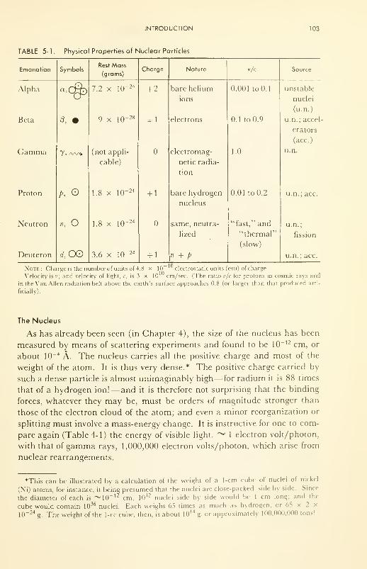
INTRODUCTION 103
TABLE 5-1.
104 RADIOACTIVITY; BIOLOGICAL TRACERS
It is exactly this huge energy carried by the alpha or beta particle, or by
the gamma photon (packet of light), which is responsible for its detection
as well as the damage it does to the molecules of a tissue. Thus, as the
emanation is absorbed by molecules of a gas, say, its energy is gradually dis-
sipated by being passed over to the gas molecules; these in turn are at least
excited, and many are ionized, a process which requires only a few electron-
volts per molecule.
The number of protons in the nucleus determines its positive charge, and
hence its position in the periodic table. Protons plus neutrons determine the
weight of the nucleus. There may be several numbers of neutrons which can
combine with a given number of protons, and thus there can be several
weights of the same element. These different weights of the same elements
are called "isotopes" (iso topos— in the same place in the periodic table).
Some isotopes are quite stable, some spontaneously disintegrate. For ex-
ample, carbon with 6 protons in the nucleus, may have 4 to 9 neutrons in
the nucleus, to form C6
!0, C 6
n, C 6
12, C 6
13, C 6
14, C 6
15. The isotope C 6
12is
the basic carbon in nature, and is quite stable, whereas C 6H
is a long-lived
beta emitter also found in nature. The others are short-lived, and are madeartificially by bombardment of nuclei by the "bullets" listed in Table 5-1
.
IONIZATION AND DETECTION
Ionization
Positive Ions
The mechanism by which ionization takes place in the path of each
emanation is important to considerations of penetration. Each mechanism
is different from the others because the emanations differ so remarkably.
The alpha (He2
4)++
, the broton (H, 1
)+
,and the deuteron (H, 2
)+ are very
small, but dense; the alpha carries the positive charge of two protons. Uponcollision with electron clouds of a target material, it easily ionizes the atoms
by pulling the negative electrons after it, wasting a small fraction of its
kinetic energy in the process. Since it is likely to tear at least one electron
out of every atom through which it passes, it leaves a very dense wake of
ionization (Figure 5-1). The alpha of radium (Ra) has a kinetic energy of
4.8 x 10⁶ electron volts, which means that it leaves a wake of about 140,000
ionized atoms. Thus in air it can travel a few inches; in metal it can pene-
trate only about 0.0001 cm; and in fact can be stopped by a piece of paper!
Although its path is short, the radiation damage or ionization along the path
is intense. Actually, theory shows that the energy transferred per centimeter
of path (called the linear energy transfer, LET) increases with increasing
charge, q, and decreases with increasing velocity, v, as follows:
LET cc q 2/v 2
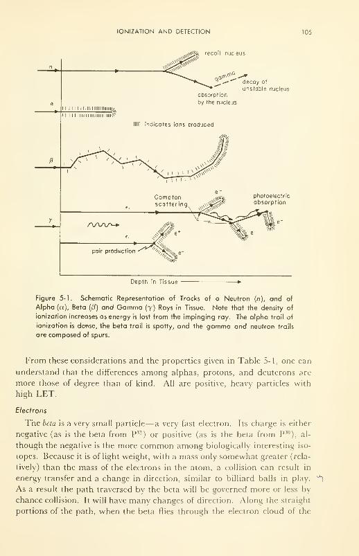
IONIZATION AND DETECTION 105
absorption
by the nucleus
decay of
unstable nucleus
photoelectric
absorption
Depth in Tissue
Figure 5-1. Schematic Representation of Tracks of a Neutron (n), and of
Alpha (a), Beta (/?) and Gamma (7) Rays in Tissue. Note that the density of
ionization increases as energy is lost from the impinging ray. The alpha trail of
ionization is dense, the beta trail is spotty, and the gamma and neutron trails
are composed of spurs.
From these considerations and the properties given in Table 5-1, one can
understand that the differences among alphas, protons, and deuterons art-
more those of degree than of kind. All are positive, heavy particles with
high LET.
Electrons
The beta is a very small particle—a very fast electron. Its charge is either
negative (as is the beta from P32) or positive (as is the beta from P30
), al-
though the negative is the more common among biologically interesting iso-
topes. Because it is of light weight, with a mass only somewhat greater (rela-
tively) than the mass of the electrons in the atom, a collision can result in
energy transfer and a change in direction, similar to billiard balls in play. **]
As a result the path traversed by the beta will be governed more or less by
chance collision. It will have many changes of direction. Along the straight
portions of the path, when the beta flies through the electron cloud of the
106 RADIOACTIVITY; BIOLOGICAL TRACERS
atom, excitation can occur, accompanied by loss of speed, and hence loss of
energy ( = 1/2 mv 2). The definite changes in direction result from collisions,
and the energy and momentum transferred can cause ejection of the electron
hit; i.e., ionization. When collisions are ''favorable," the trail of ionization,
although sparse, may penetrate quite deeply into a tissue; but when unfavor-
able, it will be very intense but very short. (See Figure 5-1.)
Very fast electrons may penetrate the atom as far as the nucleus, and by
interaction with the field of force about the nucleus lose energy, with the
production of secondary X rays. These X rays are called bremstrahlung.
Hence a hard beta source may produce a secondary radiation which is muchmore penetrating than the impinging betas.
The initial velocities of betas from a source vary widely because the
small neutral particle, the neutrino, is ejected from the nucleus along with
the beta, and the energy of the disintegration is split between the two. It is
the maximum energy of the betas which is usually given in tables of data. As
a result of the energy distribution and the deflection of betas as they enter
and lose energy in a target, the betas follow a nearly exponential law of
penetration.
Gamma Rays
The gamma ray is electromagnetic radiation, like light, but of very short
wavelength. Since it carries no charge, it is captured only by direct collision
or wave-like interaction with a target: with the nucleus or the electrons of an
atom. Some energy is transferred to the target electron and the gamma con-
tinues on, usually in a modified direction, at reduced energy (e, = hvt )
where v, is frequency and h is Planck's constant. The recoil electrons are
relatively slow, and are therefore good ionizers (see Figure 5-1). Just as in
the case of X rays (see Figure 4-10 (a)), then, absorption of gammas arises
from essentially two processes: (1) "pair production": strong interaction
with the nucleus and production of a pair of electrons (e + and e~)—impor-
tant in water only if energy of the y is above 3 Mev; and (2) "Compton ab-
sorption": ejection of an electron at an angle, some of the energy of the
gamma being lost, and the remainder ("Compton scattering") proceeding,
usually in a changed direction, and always at lower frequency. The process
(2) is repeated until, finally, the energy left from a succession of collisions is
absorbed by the electron clouds of atoms (photoelectric absorption) and is
ultimately dissipated as heat. At energies below about 0.2 Mev, elastic
(Rayleigh) scattering reduces the absorption and increases the range of the
gamma in water and soft tissues.
Neutrons
The neutron is as heavy as the proton, but carries no charge. Energy is
lost only by collision with light nuclei, and hence it can penetrate as deeply
IONIZATION AND DETECTION 107
as X rays. The nuclei set in motion by bombardment by fast neutrons (0.1
to 15 Mev) have a high LET and leave a wake of intense ionization. Slow
(thermal) neutrons are ultimately captured by nuclei; the product is nor-
mally unstable, and, for light atoms, usually emits a gamma ray. A good
billiard player will attest that maximum energy transfer can take place be-
tween two neutral "particles" if they have the same weight. Therefore,
neutrons are slowed down, or "moderated" best by materials containing
much hydrogen— water, paraffin, etc. Thus, penetration into these ma-terials is slight, or in other words, the absorption coefficient is high.
Neutrons are by-products of nuclear fission, or of proton- or deuteron-
bombardment of light nuclei; they have a half-life of the order of 20 min, can
be quite destructive of living tissue, and are difficult to detect. The damageis caused by charged nuclei set in motion by the impact of the neutron, or
from artificial radioactivity induced by capture of the neutron by the nucleus
(Figure 5-1).
Defection
Ionizing radiation is detected by any one of four basic methods:
(1) Exposure of a Photographic Plate: i.e., reduction of silver halides to silver
along the path of the photon or particle. If the plate is placed in contact with
a section of tissue containing a radioactive tracer, the plate will be exposed
where the activity is. This method of mapping is now known as "autoradi-
ography."
Microradiography is another interesting technique in which a large
shadow of a small object is allowed to fall on a photographic plate. This
technique has been used for years with X rays as the source, and recently it
has been demonstrated to be feasible and useful using alpha rays as the
source. Figure 5-2 shows a micro X radiograph of a section of bone—the
mineral content is clearly visible—and an alpha radiograph taken of the
organic part after the mineral had been removed.
(2) Ionization of a Gas Contained Between Two Electrodes: As the photon or
particle passes through the gas it leaves a wake of ion-pairs. If there is no
potential difference between the electrodes, the ions will recombine. If a
potential difference is applied (Figure 5-3 (a)), each ion will migrate toward
an oppositely charged plate. Those which reach the plate before recombin-
ing will be discharged and produce pulses of current in the external circuit.
The higher the potential difference, the less is the recombination. Thus at
an electric field strength of about 10^/cm almost all the ions produced are
"collected" at the electrodes. This is called the "saturation" condition, and
most ionization chamber systems operate in this region.
If the electric field strength is increased still further, the primary ions are
given sufficient energy to produce secondary ionization of the s*as molecules,
resulting in a multiplication of the original ionization. This is known as an
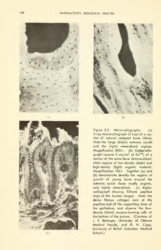
108 RADIOACTIVITY; BIOLOGICAL TRACERS
i*•
**M- f
(a) (b)
Figure 5-2. Microradiography. (a)
X-ray microradiograph (5 kvp) of a sec-
tion of natural compact bone (tibia).
Note the large (black) osteonic canals
and the (light) mineralized regions.
Magnification 500 x. (b) Alpharadio-
graph (source 2 mc/cm2of Po210
) of a
section of the same bone demineralized.
Note regions of low-density (dark) and
high-density (light) organic material.
Magnification 150x. Together (a) and
(b) demonstrate directly the regions of
growth of young bone around the
osteonic canal: tissue mostly organic,
only lightly mineralized. (c) Alpha-
radiograph showing filiform papillae
(top) of the human tongue. Note the
dense fibrous collagen core of the
papillae and of the supporting base of
the epithelium, and observe the low-
density (black) mucous-forming cells at
the bottom of the picture. (Courtesy of
L. F. Belanger, University of Ottawa
Medical Faculty, and D. H. Copp,
University of British Columbia Medical
School.)
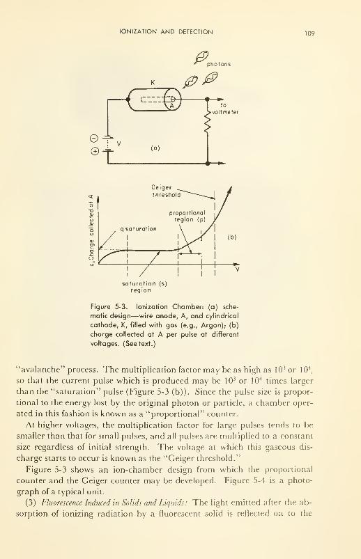
IONIZATION AND DETECTION 109
KZ3>©-v©t
(a)
0>' phot
00photons
to
• voltmeter
Geiger
threshold
saturation (s)
region
Figure 5-3. Ionization Chamber: (a) sche-
matic design—wire anode, A, and cylindrical
cathode, K, filled with gas (e.g., Argon); (b)
charge collected at A per pulse at different
voltages. (See text.)
"avalanche" process. The multiplication factor may be as high as 10 3 or 104,
so that the current pulse which is produced may be 103 or 104 times larger
than the "saturation" pulse (Figure 5-3 (b)). Since the pulse size is propor-
tional to the energy lost by the original photon or particle, a chamber oper-
ated in this fashion is known as a "proportional" counter.
At higher voltages, the multiplication factor for large pulses tends to be
smaller than that for small pulses, and all pulses are multiplied to a constant
size regardless of initial strength. The voltage at which this gaseous dis-
charge starts to occur is known as the "Geiger threshold."
Figure 5-3 shows an ion-chamber design from which the proportional
counter and the Geiger counter may be developed. Figure 5-4 is a photo-
graph of a typical unit.
(3) Fluorescence Induced in Solids and Liquids: The light emitted after the ab-
sorption of ionizing radiation by a fluorescent solid is reflected on to the
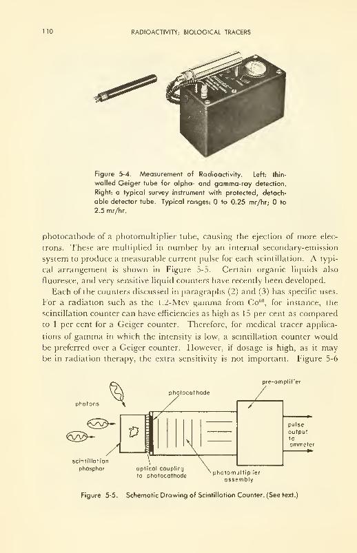
110 RADIOACTIVITY; BIOLOGICAL TRACERS
Figure 5-4. Measurement of Radioactivity. Left: thin-
walled Geiger tube for alpha- and gamma-ray detection.
Right: a typical survey instrument with protected, detach-
able detector tube. Typical ranges: to 0.25 mr/hr; to
2.5 mr/hr.
photocathode of a photomultiplier tube, causing the ejection of more elec-
trons. These are multiplied in number by an internal secondary-emission
system to produce a measurable current pulse for each scintillation. A typi-
cal arrangement is shown in Figure 5-5. Certain organic liquids also
fluoresce, and very sensitive liquid counters have recently been developed.
Each of the counters discussed in paragraphs (2) and (3) has specific uses,
tor a radiation such as the l.Z-Mev gamma from Co60, for instance, the
scintillation counter can have efficiencies as high as 15 per cent as compared
to 1 per cent for a Geiger counter. Therefore, for medical tracer applica-
tions of gamma in which the intensity is low, a scintillation counter would
be preferred over a Geiger counter. However, if dosage is high, as it maybe in radiation therapy, the extra sensitivity is not important. Figure 5-6
photons
scintillation
phosphor
photocathode
JT
\*fl X
optical coupling
to photocathode
pre-amplifier
T
photomultiplier
assembly
pulse
outputtoammeter
Figure 5-5. Schematic Drawing of Scintillation Counter. (See text.)
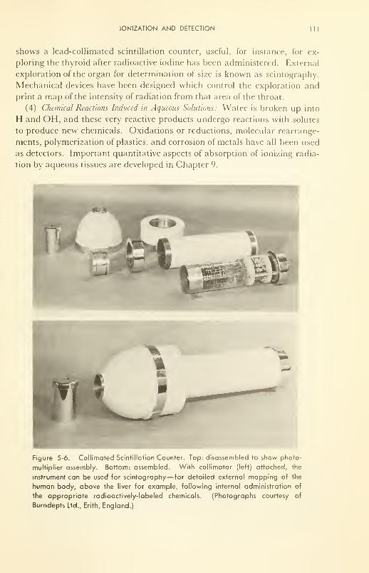
IONIZATION AND DETECTION 111
shows a lead-collimated scintillation counter, useful, for instance, for ex-
ploring the thyroid after radioactive iodine has been administered. External
exploration of the organ for determination of size is known as scintography.
Mechanical devices have been designed which control the exploration and
print a map of the intensity of radiation from that area of the throat.
(4) Chemical Reactions Induced in Aqueous Solutions: Water is broken up into
H and OH, and these very reactive products undergo reactions with solutes
to produce new chemicals. Oxidations or reductions, molecular rearrange-
ments, polymerization of plastics, and corrosion of metals have all been used
as detectors. Important quantitative aspects of absorption of ionizing radia-
tion by aqueous tissues are developed in Chapter 9.
Figure 5-6. Collimated Scintillation Counter. Top: disassembled to show photo-
multiplier assembly. Bottom: assembled. With collimator (left) attached, the
instrument can be used for scintography—tor detailed external mapping of the
human body, above the liver for example, following internal administration of
the appropriate radioactively-labeled chemicals. (Photographs courtesy of
Burndepts Ltd., Erith, England.)
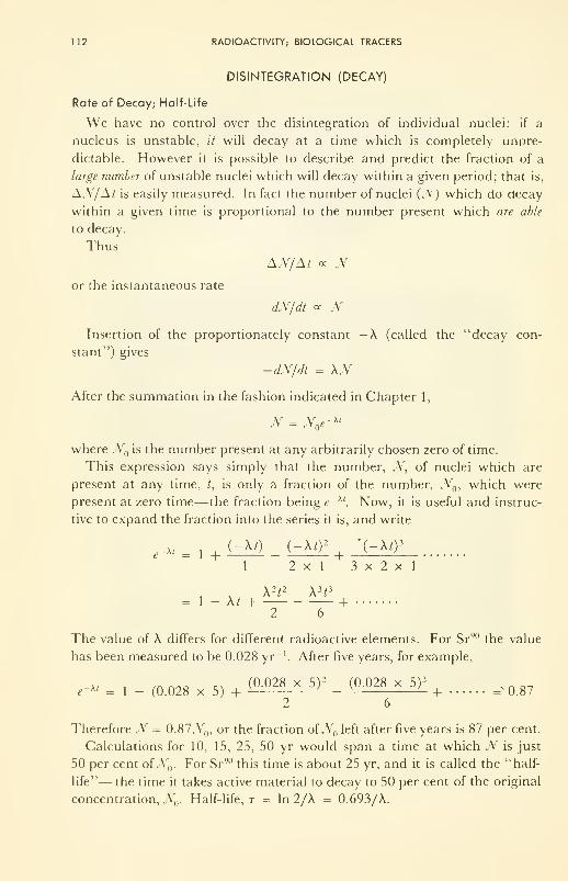
112 RADIOACTIVITY; BIOLOGICAL TRACERS
DISINTEGRATION (DECAY)
Rate of Decay; Half-Life
We have no control over the disintegration of individual nuclei: if a
nucleus is unstable, it will decay at a time which is completely unpre-
dictable. However it is possible to describe and predict the fraction of a
large number of unstable nuclei which will decay within a given period; that is,
AN/At is easily measured. In fact the number of nuclei (J\ ) which do decay
within a given time is proportional to the number present which are able
to decay.
ThusAN/At oc N
or the instantaneous rate
dN/dt ex TV-
Insertion of the proportionately constant — A (called the "decay con-
stant") gives
-dN/dt = \N
After the summation in the fashion indicated in Chapter 1,
N = N e~ Xl
where N is the number present at any arbitrarily chosen zero of time.
This expression says simply that the number, N, of nuclei which are
present at any time, t, is only a fraction of the number, N , which were
present at zero time—the fraction being e~ Xt. Now, it is useful and instruc-
tive to expand the fraction into the series it is, and write
e~ Xi = l + + +l 2x1 3x2x1
A 2/2 A 3
;3
1 _ \ t + +
The value of A differs for different radioactive elements. For Sr 90 the value
has been measured to be 0.028 yr '. After five years, for example,
.-* = 1 - (0.028 x 5) +(0 -°28 X 5)2
- (°-°28 X 5)3+ ^0.87
2 6
Therefore N = 0.87N , or the fraction ofN()left after five years is 87 per cent.
Calculations for 10, 15, 25, 50 yr would span a time at which N is just
50 per cent ofN . For Sr90this time is about 25 yr, and it is called the "half-
life"— the time it takes active material to decay to 50 per cent of the original
concentration, N . Half-life, r - In 2/A = 0.693/A.
DISINTEGRATION (DECAY) 113
If two radioactive elements have been concentrated chemically to the same
value of JV , the one with the shorter half-life decays faster, has greater "ac-
tivity" (higher dN/dt) at time zero, or delivers more emanations per second
to the tissue being irradiated.
The unit of activity is the curie (c), that amount of radioactive material
which provides 37 billion (i.e., 3.7 x 10 10) disintegrations each second.
Thus 1 g of pure Ra 226 which gives off 4.8 Mev (average of 3) alphas, has
a total activity of about 1 c. Sr 3890
, which gives off only a 0.6 Mev
beta, decays faster and is less dense than radium; 1 g of pure Sr90 provides
an activity of 147 c. However, since a pure radioactive substance is always
contaminated by its daughter products, the activity per unit weight is deter-
mined by the concentration of radioactive substance. Clearly 1 millicurie (mc)
per gram might be usable in a medical application, whereas 1 mc per ton
should be quite impractical. Specific activity is defined as the number of mc/g.
Figure 5-7 shows decay schemes for several radioactive isotopes of use as
tracers in diagnosis and as irradiation sources in therapy.
Energy Distribution of the Emitted Rays
Before we come to the question of depth of penetration and extent of
ionization of the rays from a radioactive source, we must consider two more
factors: the energy distribution (spectrum) of the rays from any given pure
source, and the number and kind of products of disintegration.
Both alphas and gammas are the result of a particular kind of fracture
or rearrangement of unstable nuclei. One could consider the nuclei to be in
excited states (think of an undulating water droplet), existing as such from
the time of their formation (in the sun?) millions of years ago, and disinte-
grating at a rate which we can measure but which we are not able to vary.
Thus, although half the atoms of Ra 226 in a sample will undergo alpha decay
in a definite and reproducible time, we do not understand why the disinte-
gration of Ra226is always by loss of one alpha particle, a package of 2 pro-
tons + 2 neutrons; and the most striking fact of all is that these alphas al-
ways come off with the same velocity. The similarity of this quantum-like be-
havior to the quantized absorption and radiation of light by the electron
cloud of the atom, suggested to theoreticians that a Bohr-like model for the
nucleus should be useful. Development of theory has proceeded along these
lines, and has led at least to a quantitative description, if not an answer to
the question "why?".
The alpha or the gamma radiation from a single elemental source occurs
at discrete energies—alphas of single velocity, gammas of single frequency
(Figure 5-8). However, with the beta is expelled a neutrino, a tiny neutral
particle of variable velocity; and therefore the beta radiation from a single
elemental source has a distribution of energies— low, corresponding to a
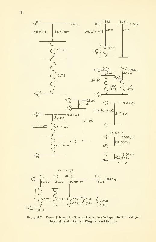
114
No,24
sodium-24 I. 39mev
Mg24
12
Co'60
J27~
cobalt-60
Ni60_'28
15 hrs K42 (18%) (82%)
19
potossium-42
y 1.37 Co42
20
/92.0
,yl.53
12.5 hrs
£3.6
y 2.76
. 59 (46%) (54%)Fe„ 1 1 4 5 days
/30.46
yl 29 ^ rl.10
(43%) ^ (57%)
Sr
y
90_38
90
28yrs 320.54 15"
0.306
39
5.25yrs
7 I. I7mev
Zr
yl. 33mev
9o_
40
phosphorus-32
j8 2.26.32
14 2 days
0l.7lmev
carbon-14.146~
4 1
C
N
H
He
— 5568yrs
0O.I55mev
3 i
12.26 y rs
00.0 1 8 me v
tritium
lod ine-131
(3%) (9%) (87%)
00.33 /30.6lmev
(1%)
y0.64 <wy0.36 <-yO 28
ri(80%)r^-(5%)
00.878 04 days
70.08
70.08
54 vstable
Figure 5-7. Decay Schemes for Several Radioactive Isotopes Used in Biological
Research, and in Medical Diagnosis and Therapy.
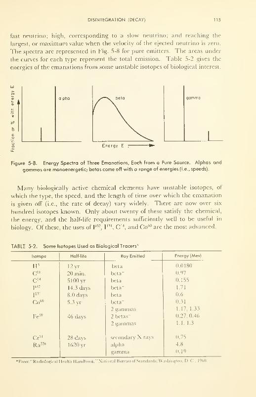
DISINTEGRATION (DECAY) 115
fast neutrino; high, corresponding to a slow neutrino; and reaching the
largest, or maximum value when the velocity of the ejected neutrino is zero.
The spectra are represented in Fig. 5-8 for pure emitters. The areas under
the curves for each type represent the total emission. Table 5-2 gives the
energies of the emanations from some unstable isotopes of biological interest.
en
01
i
co
alpha gamma
Energy E
Figure 5-8. Energy Spectra of Three Emanations, Each from a Pure Source. Alphas and
gammas are monoenergetic; betas come off with a range of energies (i.e., speeds).
Many biologically active chemical elements have unstable isotopes, of
which the type, the speed, and the length of time over which the emanation
is given off (i.e., the rate of decay) vary widely. There are now over six
hundred isotopes known. Only about twenty of these satisfy the chemical,
the energy, and the half-life requirements sufficiently well to be useful in
biology. Of these, the uses of P32, I
131, C 14
, and Co60 are the most advanced.
TABLE 5-2. Some Isotopes Used as Biological Tracers"
Isotope
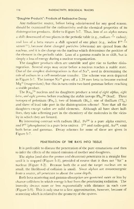
116 RADIOACTIVITY; BIOLOGICAL TRACERS
"Daughter Products": Products of Radioactive Decay
Any radioactive source, before being administered for any good reason,
should be examined for the radioactivity and the chemical properties of its
disintegration products. Refer to Figure 5-7. Thus, loss of an alpha means
a shift downward of two places in the periodic table (e.g., radium —* radon);
and loss of a beta means a shift upward of one place (e.g., iodine I131 —
>
xenon 131), because these charged particles (electrons) are ejected from the
nucleus, and it is the charge on the nucleus which determines the position of
the element in the periodic table. Loss of a gamma results in no shift, but is
simply a loss of energy during a nuclear reorganization.
The daughter products often are unstable and give rise to further disin-
tegration. Several steps may occur before a nucleus reaches a stable state.
One of the simplest disintegrations is that of Na24, used in determining the
role of sodium in a cell-membrane transfer. The scheme was seen depicted
in Figure 5-7. The isotope Na24 gives off a 1.39 mev beta to become excited
Mg 24 (magnesium); but this in turn emits two hard gammas before reaching
a stable product.
The Ra 88226 nucleus and its daughters produce a total of eight alphas, eight
betas, and eight gammas before reaching the stable isotope Pb82
206 (lead). Three
isotopes of polonium (Po 84 ), two of bismuth (Bi 83 ), one of thallium (Tl81 )
and three of lead take part in the disintegration scheme! Note that all the
daughters except radon are solid elements. Although all have short half-
lives, they take a fleeting part in the chemistry of the molecules in the vicin-
ity in which they are formed.
By interesting contrast with radium (Ra), Po210is a pure alpha emitter,
and P 32 (phosphorus) is a pure beta emitter. I131 and radio-gold, Au 198
, emit
both betas and gammas. Decay schemes for some of these are given in
Figure 5-7.
PENETRATION OF THE RAYS INTO TISSUE
It is preferable to discuss the penetration of the pure emanations and then
to infer the effects of the mixed emission of mother and daughters.
The alpha (and also the proton and deuteron) penetrates in a straight line
until it is stopped (Figure 5-1), provided of course that it does not "hit" a
nucleus (Figure 4-2). Because both the a and the target nucleus are so
small, the likelihood of collision is small. Since alphas are monoenergetic
from a source, all penetrate to about the same depth.
Both beta-scattering and gamma-absorption are governed more or less by
chance collisions in which energy is lost from the penetrating radiation. The
intensity decays more or less exponentially with distance in each case
(Figure 5-9). This is only true to a first approximation, however, because of
scattering which is related to the geometry of the system.
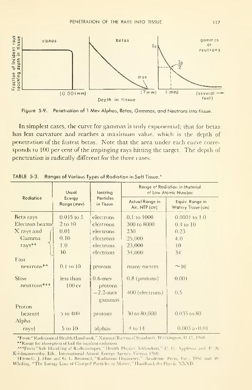
PENETRATION OF THE RAYS INTO TISSUE 117
2 2
c<u co —'o .c
^_ 0)
O T3
c a>o c"*- !cO oo
alphas
(0 001 mm) (7 mm (I mm)
gam masor
neu t rons
(severalfeet)Depth in tissue
Figure 5-9. Penetration of 1 Mev Alphas, Betas, Gammas, and Neutrons into tissue
In simplest cases, the curve for gammas is truly exponential; that for betas
has less curvature and reaches a maximum value, which is the depth of
penetration of the fastest betas. Note that the area under each curve corre-
sponds to 100 per cent of the impinging rays hitting the target. The depth of
penetration is radically different for the three cases.
TABLE 5-3. Ranges of Various Types of Radiation in Soft Tissue.*
118 RADIOACTIVITY; BIOLOGICAL TRACERS
In review, the nature and properties of the four main types of emanation
have been considered. Positive ions and electrons lose kinetic energy by
charge interaction with the electron cloud of atoms in the path: the greater
the electron density the greater the absorption. Gamma rays lose energy
to the electron cloud principally by pair production or Compton scattering.
A neutron must hit a nucleus to lose energy. When it does, either the
nucleus (charged) recoils through the medium and ionizes as a positive ion,
or the neutron is absorbed by the nucleus, usually to form an unstable iso-
tope which decays with the expulsion of beta or gamma, proton or neutrons.
The data of Table 5-3 illustrate these important principles. Note par-
ticularly the variation of the range in tissue for radiations of different type
and energy. Protons are the ionizing particles in tissue which is under fast-
neutron irradiation because hydrogen of water is the most plentiful target in
the tissue. . . . This table should be thoroughly studied and understood.
USES AS BIOLOGICAL TRACERS
One of the simplest, and yet one of the most intriguing applications of the
properties of radioactive substances has been in their use as tracers. The age
of the earth, the authenticity of oil paintings, the courses of water and wind
currents, have been probed simply by analyzing for the pertinent radioactive
isotope in the proper place in the proper manner.
Three uses as tracers concern us here: (1) as an aid in determining the
steps and paths by which molecular reactions occur, whether simple hydra-
tions of ions, or the more complex syntheses and degradations of large bio-
chemicals; (2) in plotting the course of fluid flow, through the blood capil-
laries, across cell walls, etc.; (3) in plotting the time and space distribution
of biologically active chemicals. Examples of each are now given to illustrate
the principles. The book by Kamen, s now a classic in the subject of tracers,
is highly recommended for further study.
Tracers of Molecular Reactions
The first use of isotopic tracers on a biological problem was reported by
Hevesy in 1923; this was a study of lead metabolism in plants. When heavy
water (D^O) became available in Urey's laboratory after the discovery of
deuterium there in 1932, many biochemical problems were attacked: hydro-
genations and dehydrogenations, cholesterol synthesis from smaller frag-
ments, conversion of phenylalanine to tyrosine, etc. Then, by 1942, am-
monium sulfate containing N 7
15, instead of the more common N 7
14,became
available, and compounded the possibilities for biochemical investigations.
Thanks to the nitrogen tracer, the fate of amino acids in protein synthesis
could be followed. Probably the most important of all these investigations,
from the point of view of biology, was the demonstration that protein mole-
USES AS BIOLOGICAL TRACERS 119
cules are in a dynamic equilibrium with their environment: they are not
fixed end-items, but rather they are continually breaking apart here and
there, accepting new amino acids and rejecting old. The same thing has
now been found in lipid and carbohydrate metabolic reactions. Thus a dy-
namic steady-state must now be considered well established in the biochem-
istry of life, even at the molecular level, a fact which could be established
only by this unique tool, the isotopic tracer.
To be useful as a tracer, the only requirement is that the isotope be
present in an amount different from that occurring in nature. If the isotope
is radioactive, its presence is easily detected by the ionization caused by its
disintegration product. If it is not radioactive (deuterium, H, 2, and nitro-
gen-15, N 7'\ are examples), it can be detected by two methods: (1) In the
highly evacuated mass spectrograph, the atom is ionized by bombardment
by electrons, and then, after the ion has been accelerated in an electric field
to a prechosen velocity, it is allowed to enter the space between the poles of
a strong magnet. It is deflected there by the magnetic field, by an amount
determined by the weight of the flying particle: the heavier the particle the
less the deflection. (2) By neutron activation: In some cases—
N
71S
is an ex-
ample—the nonradioactive isotopic tracer can be made active by bombard-
ment with thermal neutrons, and then its quantity measured as the radio-
activity of the product, N 716 in this case, a hard beta and gamma emitter
with a half-life of only a few seconds.
Tracers of Fluid Flow
The classical method of determining the flow pattern in the circulation
system is to inject nitrous oxide, N20, at one point and then sample at vari-
ous times and places after the injection.
The isotopic dilution technique, described under (1) and (2) above, has
been used to map blood flow in the brain, advantage being taken of the fact
that no new chemical reactions are introduced into the system in the ma-
terials injected.
During the past five years, the radioactive isotope method has also been
applied to the very difficult problem of measuring the rate of flow of blood
through various parts of the brain, and although these experiments have not
been done as yet on man, the work (mainly on cats) is interesting and in-
structive, and illustrates the power of the method. The chemically inactive,
freely diffusible gas, CF3I131
, has (5 and y emanations well-suited to the de-
tection techniques already described. For example, ~300 microcuries (fie)
are administered, either by injection into the blood stream in about 10 cc of
salt solution, or inhaled from a prepared air mixture. The blood can be
shunted through a glass tube from one part of an artery to another, and the
activity of the shunted blood determined with a counter attached to the
glass.
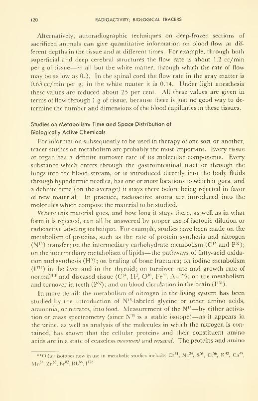
120 RADIOACTIVITY; BIOLOGICAL TRACERS
Alternatively, autoradiographic techniques on deep-frozen sections of
sacrificed animals can give quantitative information on blood flow at dif-
ferent depths in the tissue and at different times. For example, through both
superficial and deep cerebral structures the flow rate is about 1.2 cc/min
per g of tissue— in all but the white matter, through which the rate of flow
may be as low as 0.2. In the spinal cord the flow rate in the gray matter is
0.63 cc/min per g; in the white matter it is 0.14. Under light anesthesia
these values are reduced about 25 per cent. All these values are given in
terms of flow through 1 g of tissue, because there is just no good way to de-
termine the number and dimensions of the blood capillaries in these tissues.
Studies on Metabolism: Time and Space Distribution of
Biologically Active Chemicals
For information subsequently to be used in therapy of one sort or another,
tracer studies on metabolism are probably the most important. Every tissue
or organ has a definite turnover rate of its molecular components. Every
substance which enters through the gastrointestinal tract or through the
lungs into the blood stream, or is introduced directly into the body fluids
through hypodermic needles, has one or more locations to which it goes, and
a definite time (on the average) it stays there before being rejected in favor
of new material. In practice, radioactive atoms are introduced into the
molecules which compose the material to be studied.
Where this material goes, and how long it stays there, as well as in what
form it is rejected, can all be answered by proper use of isotopic dilution or
radioactive labeling technique. For example, studies have been made on the
metabolism of proteins, such as the rate of protein synthesis and nitrogen
(N 15) transfer; on the intermediary carbohydrate metabolism (C 14 and P32
);
on the intermediary metabolism of lipids—the pathways of fatty-acid oxida-
tion and synthesis (H 3); on healing of bone fractures; on iodine metabolism
(I131
) in the liver and in the thyroid; on turnover rate and growth rate of
normal** and diseased tissue (C 14, H 2
, O 18, Fe59
, Au 198); on the metabolism
and turnover in teeth (P32); and on blood circulation in the brain (I
131)-
In more detail: the metabolism of nitrogen in the living system has been
studied by the introduction of N'Mabeled glycine or other amino acids,
ammonia, or nitrates, into food. Measurement of the N 15—by either activa-
tion or mass spectrometry (since N 15is a stable isotope)— as it appears in
the urine, as well as analysis of the molecules in which the nitrogen is con-
tained, has shown that the cellular proteins and their constituent amino
acids are in a state of ceaseless movement and renewal. The proteins and amino
**Other isotopes now in use in metabolic studies include: Cr , Na , S , CI , K , Ca ,
Mn 54, Zn65
, Br82
, Rb86, I
128.
USES AS BIOLOGICAL TRACERS 121
acids are continually being degraded, and being replaced by syntheses.
That the rates of breakdown and resynthesis are the same is attested by the
fact that the concentrations are maintained constant during life. About 60
per cent of N ,5-containing protein has been shown to appear as glycine in the
urine within 24 hr after a high-protein diet has been eaten; about 80 per cent
appears within 60 hr. Liver, plasma, and intestinal-wall proteins are re-
generated much faster than those of muscle and connective tissue.
The nitrogen that goes into ringed structures such as the porphyrins,
which enter complexes with Fe+2 and Fe+3 to form the hemin of red-blood
cells, turns over quite slowly: it takes 10 days for the hemin to be synthesized
from isotopically tagged glycine, and then nearly 140 days before the deg-
radation process (cell replacement in this case) reduces the concentration of
tagged nitrogen to half the peak concentration (see Figure 5-10).
indirect
Time after oral administration
Figure 5-10. Radioactivity in a Particular Vol-
ume of Tissue as a Function of Time After
Administration. Time and height of the maxi-
mum depend upon location of the volume, upon
what chemical compound is given, its normal
biochemistry, where it was introduced (direct
or indirect), and the half-life of the isotope.
Other uses of radioactive tracers include the investigation of the effects of
drugs and hormones on the turnover rate in particular tissues or organs. Asubject of particular interest in recent years has been the role of insulin in
the control of diabetes. In a diabetic, sugars are transported across the
membrane and into the cell abnormally slowly, and they accumulate in the
plasma, useless for supplying energy, via oxidation, inside the cell. Insulin,
a medium-sized protein molecule whose structure has been well known since
it was first synthesized in 1956, has been tagged with I131 and introduced
into the blood stream. Within minutes, more than a third accumulates in
the liver and the kidneys. However, a fraction adsorbs in a nonspecific man-
ner on all membranes accessible to blood plasma and intracellular fluids.
Cell walls are no exception; and the adsorption of insulin has been associ-
122 RADIOACTIVITY; BIOLOGICAL TRACERS
ated with an increase of the rate of sugar penetration (a process which itself
has been followed by C 14-tagged sugars). Whether the control exercised by
insulin is simply by opening the access to pores, or whether it controls in a
more subtle manner by increasing the activity of the enzyme (hexokinase)
also thought to be adsorbed on the membrane, has not yet been settled.
However, it can be seen that the use of radioactive tracers in such a phar-
macological problem can make a valuable contribution to our knowledge of
the processes involved.
The pioneering work of Huff and Judd on the quantitative analysis of the
time and space distributions of Fe 59in blood plasma, will be discussed in
Chapter 11 as a concrete example of how possible methods of action can be
analyzed with a computer if it is fed reliable experimental measurements of
where the Fe 59 goes and how long it stays there. We learn a little about what
the iron does, and also something about just what processes are interfered
with during blood diseases.
Radioactive Mapping
Administration of compounds of I131
, followed bv external measurements
of beta-ray intensity in the thyroid region of the neck, has been introduced
in some centers as a replacement test for determining whether the thyroid is
normal, over-, or under-active. A hyperactive thyroid may absorb up to 80
per cent of the tagged iodine; a hypoactive gland may absorb as little as
15 per cent before normal biochemical turnover elsewhere in the body re-
duces the concentration via excretion. Mapping of the thyroid by I131
scintography is common practice. Both the outline of the organ, and its
turnover rate can be obtained from maps made at different time intervals
after administration. The maximum activity of the emission is a direct
measure of the uptake of iodine by the thyroid.
The flow of fluids through various critical parts of the system can also be
mapped satisfactorily by dissolving in the fluid a small amount of gas which
contains a radioactive emitter, and mapping from the outside with a col-
limated scintillation counter (Figure 5-6).
Conclusion
A great many elementary biochemical reactions are being studied via the
tracer technique, and a few physical processes also. Some of these will be
found mentioned as examples in different parts of this book. The techniques
are reliable and extremely sensitive, and have the unique advantage that the
introduction of the radioactive element can be done in such a manner as not
to upset the chemistry or the physics of the process in vivo. Already in ex-
tensive use in biological research— in his review Kuzin 12 was able to collect
358 references to new work published in 1959 alone!— now, led by successes
REFERENCES 123
with I131 and P32
, radioactive tracer techniques have a wonderful future in
medical diagnosis.
As it does in so many subjects, the National Bureau of Standards, in
Washington, periodically publishes reliable definitions of terms, values of
universal and experimental constants, and tables and graphs of collated data
on radiologically important parameters. The ''Radiological Health HandBook" is indispensible to further study of this subject, as a quantitative sup-
plement to the classic work of Kamen. 5
PROBLEMS
5- 1 : (a) What element is formed by the radioactive disintegration of:
P~ £- 0'
P32 ^ Co60 ^ p30 1+
0- 8-
Na24 ^ Ra 226 -^
a 8*
po210 _^ Na 22 %(b) Is the product radioactive too?
5-2: (a) Make a graph showing activity (counts per minute) against time, for up-
take, utilization, and elimination of I!3! by the thyroid,
(b) List five important reasons why I131
is used in irradiation-therapy of goiter.
5-3: The 1.70 mev /3-ray of P32 penetrates about 7 mm into tissue. The half-life is
14.3 days. A 1-millicurie (mc) source will deliver about 1 rad (radiation ab-
sorbed dose) per minute.
How long would it take for a 1 mc of NaHP04 , composed of P32
, taken orally
as a solution in water, to administer 6000 rads to an organ in which it concen-
trates?
REFERENCES
1. The Staff, Physics Dept., Univ. of Pittsburgh: "Atomic Physics," 2nd ed., John
Wiley & Sons, Inc., New York, N. Y., 1944.
2. "Atomic Radiation (Theory, Biological Hazards, Safety Measures, Treatment
of Injury)," RCA Service Co., Camden, N. J ., 1 959.
3. "Teaching with Radioisotopes," H. A. Miner, el al., Eds., U. S. Atomic Energy
Commission, Washington, D. C, 1959.
4. Scientific American, issue on "Ionizing Radiations," Vol. 201, September, 1959:
papers by S. Warren, p. 164, and R. L. Platzman, p. 74.
5. Kamen, M. D., "Tracer Techniques in Biology and Medicine," Academic Press,
New York, N. Y., 1960.
124 RADIOACTIVITY; BIOLOGICAL TRACERS
6. Glasser, O., Ed., "Medical Physics, Vol. Ill," Year Book Publishing Co.,
Chicago, 111., 1960: several short articles, p. 302-364. See especially: "Locali-
zation of Brain Tumors with /^-Emitting Isotopes," by Silverstone and
Robertson.
7. Kity, S. S., Methods in Med. Res., 1, 204 (1948).
8. Munck, O. and Lassen, N. A., Circulation Research, 5, 163 (1951).
9. Clarke, H. T., Urey, H. C, and 16 others, "The Use of Isotopes in Biology and
Medicine," in the Proceedings of a Symposium on the subject, The Univ. of
Wisconsin Press, Madison, Wis., 1948.
10. Huff, R. L. and Judd, O. J., "Kinetics of Iron Metabolism," in Adv. in Biol, and
Med. Phys., 4,223 (1956).
11. Freygang, W. H. and Sokoloff, L., "Quantitative Measurement of Regional Cir-
culation in the Central Nervous System by the Use of Radioactive Inert Gas,"
Adv. in Biol, and Med. Phys., 6,263 (1958).
12. Kuzin, A. M., "The Application of Radioisotopes in Biology," Review Series,
No. 7, International Atomic Energy Agency, Vienna, 1960.
13. "Scintography—A collection of Scintigrams Illustrating the Modern Medical
Technique of in vivo Visualization of Radioisotope Distribution," R-C Scien-
tific Co., Inc., Pasadena, Calif., 1955.
14. Cork, J. M., "Radioactivity and Nuclear Physics," 3rd ed., D. Van Nostrand,
Inc., New York, N. Y., 1957.
CHAPTER 6
Big Molecules
(Structure of Macromolecules and Living Membranes;
Isomers and Multiplets;
Codes and Molecular Diseases)
A score of diseases (including sickle cell anaemia and phenylketonuria)
have so far been recognized as enzyme diseases, presumably resulting from
the manufacture of abnormal molecules in place of active enzyme molecules.
I think that it is not unlikely that there are hundreds or thousands of such
diseases.
Iforesee the day when many of these diseases will be treated by the use of
artificial enzymes .... When our understanding of enzyme activity becomes
great enough, it will be possible to synthesize a catalyst, etc
Thus did Linus Pauling emphasize to an international sym-
posium of enzymologists in Chicago, in 1956, the relationship
between the structure of the macromolecule and its chemical and
physical roles in the living system.
INTRODUCTION
The structure of macromolecules and of arrays of them in living mem-
branes and other tissues has occupied the attention of an important class of
biophysicists for the past ten years. Using modern rapid-flow, quick-freeze-
drying, and micromanipulation techniques, and armed with the phase and
125
126 BIG MOLECULES
interference microscopes, the X-ray diffraction camera, and the elertron
microscope—the last now in such an advanced stage of development that, in
proper hands, it can resolve or "see" small particles just a few atomic
diameters apart— researchers have been able to gain new insight into the
actual shape of the molecule in the tissue, and even into the positions of
atoms and groups of atoms within the molecule.
Running concurrently with these physical researchers have been chemical
studies which have finally solved the puzzle of the complete chemical
composition of a few large, biologically important molecules. For example,
although the hormone, insulin, has been known and used widely in the treat-
ment of diabetes for nearly forty years, it was only in 1955 that Sanger and
his colleagues at Cambridge were finally able to write down the complete
structural formula. It contains 777 atoms! Since then, ribonuclease
(RNAse), an enzyme containing 1876 atoms and which catalyzes the
cleavage of ribonucleic acid, has also yielded the secret of its composition to
the attack of persistent chemists. This completes the first big step toward
knowing how this molecule works as a catalyst, although details of the struc-
ture at and around the active site(s) are not yet known. This is the next big
task, for if more than one of the chemical groups must exert their chemical
effects on a specific part of the molecule whose hydrolysis is to be promoted,
then their spatial arrangement must be very important. Not only must they
be present, but they must be present at the proper positions in space if the
catalytic activity of the site is to exist. In other words, if one of the players
is out of position, the game is lost.
Table 6-1 gives a spectrum of biologically important organic molecules,
small and large—some containing a metallic oxidizable and reducible ion
which enters the chemical reactions of the molecule. Although some details
are given in the following sections, the discussion is just an indication of the
scope of the subject. There are excellent reference sources: for example,
the recent book of Tanford. 16
STRUCTURE
Our purpose, first, will be to outline the structure of two big molecules of
critical biological importance, myoglobin and hemoglobin, learned in
the recent work of the schools of Kendrew and Perutz, respectively. Themethod used was X-ray crystallography, and although the chemical com-
position has not yet been fully worked out for these two molecules, X-ray
crystallographic studies have completely outlined the form of the molecule
in the dry crystalline state.
The second part of this section on structure is concerned with the cross-
linked structure of liquid crystals, such as in the aqueous humor of the lens
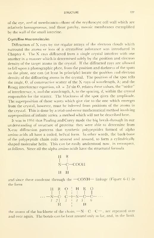
STRUCTURE 127
of the eye, anH of membranes—those of the erythrocyte cell wall which are
relatively homogeneous, and those patchy, mosaic membranes exemplified
by the wall of the small intestine.
Crystalline Macromolecules
Diffraction of X rays by the regular arrays of the electron clouds which
surround the atoms or ions of a crystalline substance was introduced in
Chapter 4. The X rays diffracted from a single crystal interfere with one
another in a manner which is determined solely by the position and electron
density of the target atoms in the crystal. If the diffracted rays are allowed
to fall upon a photographic plate, from the position and darkness of the spots
on the plate, one can (at least in principle) locate the position and electron
density of the diffracting atoms in the crystal. The position of the spot tells
the angle, 9, of constructive scatter of the X rays of wavelength, A; and the
Bragg interference equation, nX = 2d sin 0, relates these values, the "order"
of interference, n, and the wavelength, A, to the spacing, d, within the crystal
responsible for the scatter. The blackness of the spot gives the amplitude.
The superposition of those waves which give rise to the one which emerges
from the crystal, however, must be inferred from positions of the atoms in
the crystal. This is done by a trial-and-error mathematical method involving
superposition of infinite series, a method which will not be described here.
It was in 1951 that Pauling and Corey made the big break-through in our
understanding of structure of proteins: they were able to determine from
X-ray diffraction patterns that synthetic polypeptides formed of alpha
amino acids all have a coiled, helical form. In other words, the back-bone
of the polypeptide chain coils around and around, to form a cylindrically
shaped molecular helix. This can be easily understood now, in retrospect,
as follows. Since all the alpha amino acids have the structural formula
H RI I
N—C—COOH
H H
and since these condense through the —CONH-- linkage (Figure 6-1) in
the form
H R O ! H R OI
llil I II
• • • -N-C-C-T-N-C-C- • ••
1 2\ 3 ' 4 5 16
h;
h
the atoms of the backbone of the chain, —N—C—C— , are repeated over
and over again. The bonds can be bent around only so far, and, in the limit.
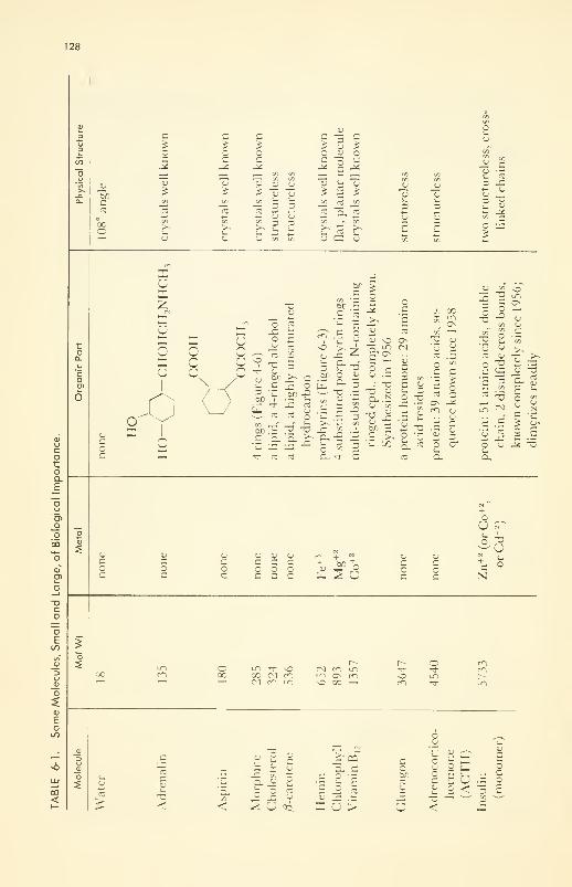
128
00
b/D
c
Ooco
c
oc
co>~t_U
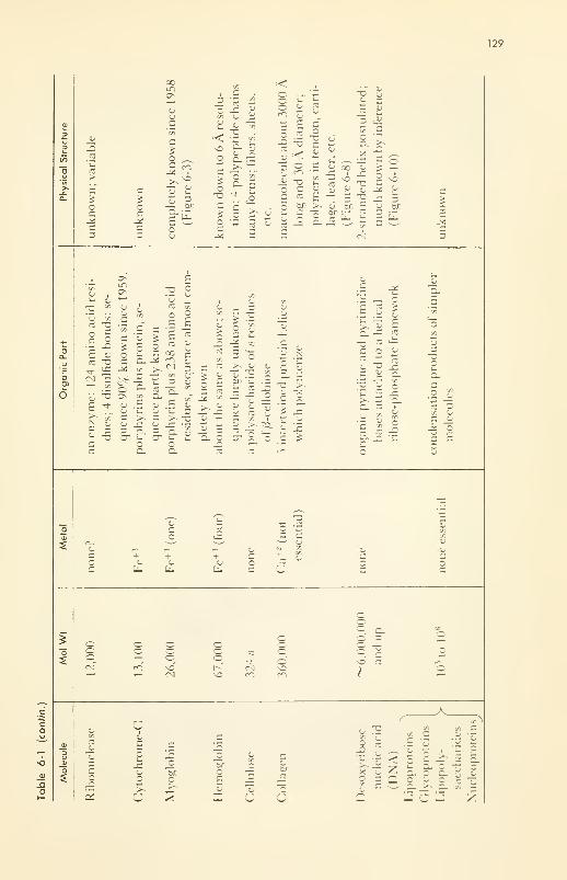
129
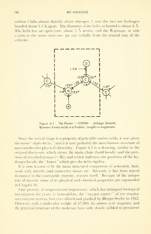
130 BIG MOLECULES
carbon 6 falls almost directly above nitrogen /, and the two are hydrogen
bonded about 1.5 A apart. The diameter of the helix so formed is about 8 A.
The helix has an open core, about 2 A across,. and the R-groups, or side
chains to the main structure, jut out radially from the central axis of the
cylinder.
Figure 6-1. The Planar -CONH— Linkage (boxed)
Between Amino Acids in a Protein. Lengths in angstroms.
Since the helical shape is a property of polyalpha amino acids, it was given
the name "alpha-helix, " and it is now probably the most famous structure of
macromolecular physical chemistry. Figure 6-2 is a drawing, similar to the
original disclosure, which shows the main chain (bold bonds) and the posi-
tions of attached groups (
—
R); and which indicates the positions of the hy-
drogen bonds, the "bones" which give the helix rigidity.
It is now known to be the main structural component of «-keratin, hair,
wool, nail, muscle, and connective tissue, etc. Recently it has been traced
in muscle to the contractile enzyme, myosin itself. Because of the unique
role of myosin, some of its physical and chemical properties are expounded
in Chapter 10.
One protein, of unquestioned importance, which has intrigued biological
investigators for years, is hemoglobin, the "oxygen carrier" of the respira-
tory enzyme system, first crystallized and purified by Hoppe-Seyler in 1862.
However, with a molecular weight of 67,000, its amino acid sequence and
the physical structure of the molecule have only slowly yielded to persistent
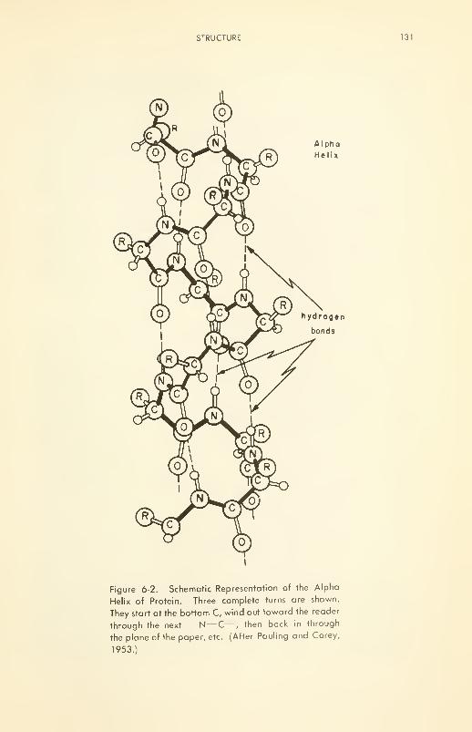
STRUCTURE 131
Figure 6-2. Schematic Representation of the Alpha
Helix of Protein. Three complete turns are shown.
They start at the bottom C, wind out toward the reader
through the next —N—C— , then back in through
the plane of the paper, etc. (After Pauling and Corey,
1953.)
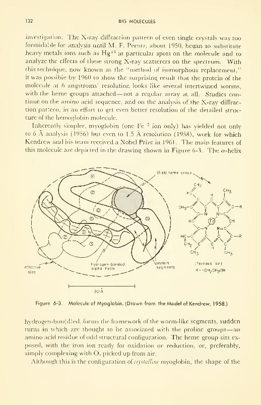
132 BIG MOLECULES
investigation. The X-ray diffraction pattern of even single crystals was too
formidable for analysis until M. F. Perutz, about 1950, began to substitute
heavy metals ions such as Hg+2 at particular spots on the molecule and to
analyze the effects of these strong X-ray scatterers on the spectrum. Withthis technique, now known as the "method of isomorphous replacement,"
it was possible by 1960 to show the surprising result that the protein of the
molecule at 6 angstroms' resolution looks like several intertwined worms,with the heme groups attached— not a regular array at all. Studies con-
tinue on the amino acid sequence, and on the analysis of the X-ray diffrac-
tion pattern, in an effort to get even better resolution of the detailed struc-
ture of the hemoglobin molecule.
Inherently simpler, myoglobin (one Fe +2 ion only) has yielded not only
to 6 A analysis (1956) but even to 1.5 A resolution (1958), work for which
Kendrew and his team received a Nobel Prize in 1961. The main features of
this molecule are depicted in the drawing shown in Figure 6-3. The a-helix
(flat) heme group
CH 2
< „ pCH,-/VV\
C-N N-C
HC (Fe) CHXC-N N=C/ I I \
HC-C C C C-
CH?XC C C/ H
|
CH 3 CH 3
protein (ferrous ion)seqments
R= -CH 2CH20H
I
11
30 A
Figure 6-3. Molecule of Myoglobin. (Drawn from the Model of Kendrew, 1958.)
hydrogen-bon(d)ed, forms the framework of the worm-like segments, sudden
turns in which are thought to be associated with the proline groups—an
amino acid residue of odd structural configuration. The heme group sits ex-
posed, with the iron ion ready for oxidation or reduction, or, preferably,
simply complexing with 2 picked up from air.
Although this is the configuration of crystalline myoglobin, the shape of the
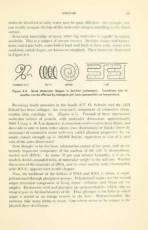
STRUCTURE 133
molecule dissolved in salty water may be quite different— for example, one
can readily imagine the legs of this molecular octopus unfolding in the blood
stream.
Structural knowledge of many other big molecules is rapidly becoming
available. This is a subject of intense interest. Straight chains and helices,
some coiled into balls, some folded back and forth to form rods, others with
randomly coiled shapes, are known or imagined. These forms are illustrated
in Figure 6-4.
1 1 1 1.)-
134 BIG MOLECULES
€
I
':••-.
'<ft8*
m -
'*
Figure 6-5. Electron Micrograph of Collagen Fibers Carefully Lifted from Human
Skin. Note how they are individually cross-segmented and collectively fused (Courtesy
of J. Gross, Massachusetts General Hospital, and of Scientific American.)
Lipid molecules themselves are generally small, by comparison with the
macromolecules discussed in this section (see Table 6-1). However, they
condense with proteins to form macromolecular lipoproteins, and with
cellulose to form lipocelluloses, and thus also play a primary role in the
structure of tissue.
Metal-organic molecules are varied and important in living tissue (Table
6-1). The bright light from the point of view of our knowledge of structure
STRUCTURE 135
is vitamin B P , a substituted cyano-cobalt amide of molecular weight 1357,
used in treating pernicious anemia, growth failure in children, etc. The
complete chemical composition was disclosed in 1955, and culminated with
X-ray diffraction analysis of structure three years later.
Dissolved Macromolecules
Unfortunately, when crystals such as those described in the previous sec-
tion are dissolved in water, the molecules are subjected to a number of new
and powerful forces of hydration and of polarization by ions, and the con-
figurations of many molecules change. Since water has a diffuse diffraction
pattern of its own, the X-ray technique used in crystals cannot be employed
to advantage on solutions of these molecules, and other methods which indi-
cate structure must be sought. All those to be described are useful, but each
has its limitations.
The problem in solution is complicated by three other facts: (1) The
molecule will not usually, unless it is globular-shaped, have a unique molec-
ular weight; but rather will its molecular weight vary, some molecules in the
solution having weights above, and some below an average value. The dis-
cs
oo
ooo
ooorO
o o oo o oo o o»fr If) tD
I I I I 1
1
I I 1 1 I I 1 1 I
1 1 1 1 1 1
1
L ( Angstroms )
(b)
Number average 2450
Weight average 2820
Light scattering 3000
Flow birefringence
2600 — 2950
Intrinsic viscosity plus
molecular weight 2970
Figure 6-6. Ichthyocol (a Protein Food Supplement from Fish): Direct Measure-
ments of Molecular Size by Electron Micrographs, Compared with Results from
Indirect Methods. Number (N) with length (L) multiplied by L gives total amount of
protein with particles of length L Molecular weights, M nand Mw ,
are derived from
data plotted. (Data of Hall and Doty, J. Amer. Chem. Soc, 80, 1 269 ( 1 958).)
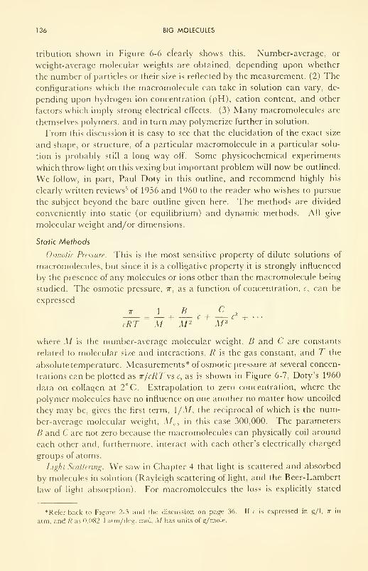
136 BIG MOLECULES
tribution shown in Figure 6-6 clearly shows this. Number-average, or
weight-average molecular weights are obtained, depending upon whether
the number of particles or their size is reflected by the measurement. (2) The
configurations which the macromolecule can take in solution can vary, de-
pending upon hydrogen ion concentration (pH), cation content, and other
factors which imply strong electrical effects. (3) Many macromolecules are
themselves polymers, and in turn may polymerize further in solution.
From this discussion it is easy to see that the elucidation of the exact size
and shape, or structure, of a particular macromolecule in a particular solu-
tion is probably still a long way off. Some physicochemical experiments
which throw light on this vexing but important problem will now be outlined.
We follow, in part, Paul Doty in this outline, and recommend highly his
clearly written reviews 5 of 1956 and 1960 to the reader who wishes to pursue
the subject beyond the bare outline given here. The methods are divided
conveniently into static (or equilibrium) and dynamic methods. All give
molecular weight and/or dimensions.
Static Methods
Osmotic Pressure. This is the most sensitive property of dilute solutions of
macromolecules, but since it is a colligative property it is strongly influenced
by the presence of any molecules or ions other than the macromolecule being
studied. The osmotic pressure, ir, as a function of concentration, c, can be
expressed
7T \ B C= — h c + c
2 +cRT Ad M 2 M 3
where M is the number-average molecular weight, B and C are constants
related to molecular size and interactions, R is the gas constant, and T the
absolute temperature. Measurements* of osmotic pressure at several concen-
trations can be plotted as tt/cRTvs c, as is shown in Figure 6-7, Doty's 1960
data on collagen at 2°C. Extrapolation to zero concentration, where the
polymer molecules have no influence on one another no matter how uncoiled
they may be, gives the first term, \/M, the reciprocal of which is the num-
ber-average molecular weight, M„, in this case 300,000. The parameters
B and Care not zero because the macromolecules can physically coil around
each other and, furthermore, interact with each other's electrically charged
groups of atoms.
Light Scattering. We saw in Chapter 4 that light is scattered and absorbed
by molecules in solution (Rayleigh scattering of light, and the Beer-Lambert
law of light absorption). For macromolecules the loss is explicitly stated
*Referback to Figure 2-3 and the discussion on page 36. If c is expressed in g/1, it in
atm, and R as 0.082 1 atm/deg. mol, M has units of g/mole.
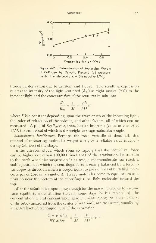
STRUCTURE 137
6.0
(0
4.0 -
' 1
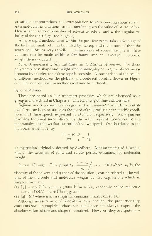
138 BIG MOLECULES
at various concentrations and extrapolation to zero concentrations so that
intermolecular interactions cannot interfere, gives the value of M, as before.
Here p is the ratio of densities of solvent to solute, and a> the angular ve-
locity of the centrifuge (radians/sec).
A more rapid method, used within the past few years, takes advantage of
the fact that small volumes bounded by the top and the bottom of the tube
reach equilibrium very rapidly; measurements of concentrations in these
volumes can be made within a few hours, and an "average" molecular
weight then evaluated.
Direct Measurement of Size and Shape via the Electron Microscope. For those
polymers whose shape and weight are the same, dry or wet, the direct meas-
urement by the electron microscope is possible. A comparison of the results
of different methods on the globular molecule icthyocol is shown in Figure
6-6. The nonequilibrium methods will now be outlined.
Dynamic Methods
These are based on four transport processes which are discussed as a
group in more detail in Chapter 8. The following outline sumcies here:
Diffusion under a concentration gradient and sedimentation under a centrif-
ugal force can both be stated as the speed of the process under specific condi-
tions, and these speeds expressed as D and s, respectively. An argument
involving factional force offered by the water against movement of the
macromolecules shows that the ratio of the two speeds, D/s, is related to the
molecular weight, M, by
(1 ~ P) D_ J_
RT s M '
an expression originally derived by Svedberg. Measurements of D and s,
and of the densities of solid and solute permit evaluation of molecular
weight.
Intrinsic Viscosity. This property, /c as c —» (where rj is the
Vo /viscosity of the solvent and r\ that of the solution), can be related to the vol-
ume of the molecule and molecular weight by two expressions which in
simplest form are:
(1) \r\\ = 2.5 V for spheres (7000 V for a big, randomly coiled molecule
such as DNA)— here Fis cc/g; and
(2) [77] « M a where a is an empirical constant, usually 0.5 to 1 .0.
Although measurement of viscosity is easy enough, the proportionality
constants have an empirical character, and hence one always suspects the
absolute values of size and shape so obtained. However, they are quite reli-
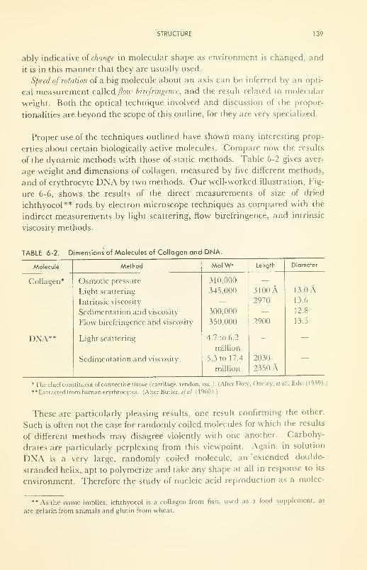
STRUCTURE 139
ably indicative of change in molecular shape as environment is changed, and
it is in this manner that they are usually used.
Speed of rotation of a big molecule about an axis can be inferred by an opti-
cal measurement calledy/oie» birefringence, and the result related to molecular
weight. Both the optical technique involved and discussion of the propor-
tionalities are beyond the scope of this outline, for they are very specialized.
Proper use of the techniques outlined have shown many interesting prop-
erties about certain biologically active molecules. Compare now the results
of the dynamic methods with those of. static methods. Table 6-2 gives aver-
age weight and dimensions of collagen, measured by five different methods,
and of erythrocyte DNA by two methods. Our well-worked illustration, Fig-
ure 6-6, shows the results of the direct measurements of size of dried
ichthyocol** rods by electron microscope techniques as compared with the
indirect measurements by light scattering, flow birefringence, and intrinsic
viscosity methods.
TABLE 6-2. Dimensions of Molecules of Collagen and DNA.
Molecule
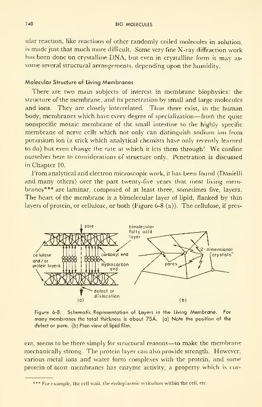
140 BIG MOLECULES
ular reaction, like reactions of other randomly coiled molecules in solution,
is made just that much more difficult. Some very fine X-ray diffraction workhas been done On crystalline DNA, but even in crystalline form it may as-
sume several structural arrangements, depending upon the humidity.
Molecular Structure of Living Membranes
There are two main subjects of interest in membrane biophysics: the
structure of the membrane, and its penetration by small and large molecules
and ions. They are closely interrelated. Thus there exist, in the humanbody, membranes which have every degree of specialization— frorn the quite
nonspecific mosaic membrane of the small intestine to the highly specific
membrane of nerve cells which not only can distinguish sodium ion from
potassium ion (a trick which analytical chemists have only recently learned
to do) but even change the rate at which it lets them through! We confine
ourselves here to considerations of structure only. Penetration is discussed
in Chapter 10.
From analytical and electron microscopic work, it has been found (Danielli
and many others) over the past twenty-five years that most living mem-branes*** are laminar, composed of at least three, sometimes five, layers.
The heart of the membrane is a bimolecular layer of lipid, flanked by thin
layers of protein, or cellulose, or both (Figure 6-8 (a)). The cellulose, if pres-
cellulose
ond/orprotein layers
bimolecularfatty acidlayer
Figure 6-8. Schematic Representation of Layers in the Living Membrane. For
many membranes the total thickness is about 75A. (a) Note the position of the
defect or pore, (b) Plan view of lipid film.
ent, seems to be there simply for structural reasons—to make the membrane
mechanically strong. The protein layer can also provide strength. However,
various metal ions and water form complexes with the protein, and some
protein of most membranes has enzyme activity, a property which is cur-
*** For example, the cell wall, the endoplasmic reticulum within the cell, etc.
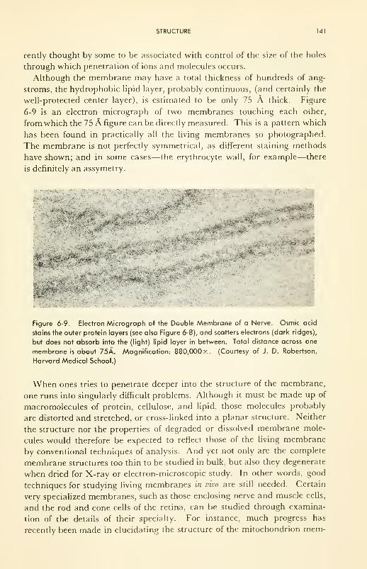
STRUCTURE 141
rently thought by some to be associated with control of the size of the holes
through which penetration of ions and molecules occurs.
Although the membrane may have a total thickness of hundreds of ang-
stroms, the hydrophobic lipid layer, probably continuous, (and certainly the
well-protected center layer), is estimated to be only 75 A thick. Figure
6-9 is an electron micrograph of two membranes touching each other,
from which the 75 A figure can be directly measured. This is a pattern which
has been found in practically all the living membranes so photographed.
The membrane is not perfectly symmetrical, as different staining methods
have shown; and in some cases—the erythrocyte wall, for example—there
is definitely an assymetry.
'-•>-,,* --.--
.
-">'
-
^
Figure 6-9. Electron Micrograph of the Double Membrane of a Nerve. Osmic acid
stains the outer protein layers (see also Figure 6-8), and scatters electrons (dark ridges),
but does not absorb into the (light) lipid layer in between. Total distance across one
membrane is about 75A. Magnification: 880,000 x. (Courtesy of J. D. Robertson,
Harvard Medical School.)
When ones tries to penetrate deeper into the structure of the membrane,
one runs into singularly difficult problems. Although it must be made up of
macromolecules of protein, cellulose, and lipid, those molecules probably
are distorted and stretched, or cross-linked into a planar structure. Neither
the structure nor the properties of degraded or dissolved membrane mole-
cules would therefore be expected to reflect those of the living membrane
by conventional techniques of analysis. And yet not only are the complete
membrane structures too thin to be studied in bulk, but also they degenerate
when dried for X-ray or electron-microscopic study. In other words, good
techniques for studying living membranes in vivo are still needed. Certain
very specialized membranes, such as those enclosing nerve and muscle cells,
and the rod and cone cells of the retina, can be studied through examina-
tion of the details of their specialty. For instance, much progress has
recently been made in elucidating the structure of the mitochondrion mem-
142 BIG MOLECULES
brane because of its unique function in electron transport in the step-wise
oxidation of foods. But the general problem of direct knowledge of the struc-
ture of living membranes probably awaits more knowledge of the structure of
macromolecules in solution.
Indirect methods— i.e., studies of penetration of the living membrane by
water, ions, and molecules— are proving to be very helpful to studies of
structure, because from such studies one can infer some properties of the
membrane in vivo: pore size, for example. An estimate of pore size (length
and area) requires at least two independent experimental measurements,
because there are two-dimensional parameters, area and length, to be evalu-
ated. Both the rate of diffusion of a substance down a concentration gradi-
ent and the rate of flow of a fluid under a mechanical pressure, should be
larger the larger the area of the hole in the membrane and the shorter its
length.
Although the rate of transport of water through the cell membrane of
erythrocytes is very rapid, both rate of diffusion and rate of flow have re-
cently been measured accurately enough to determine a value for average
pore diameter in the erythrocyte wall in vivo. Diffusion rate of water was
found by measuring the rate at which radioactively labeled water is picked
up by the cells within a few milliseconds of being bathed in the labeled
water. A fast-flow apparatus had to be used, the ingenious details of which
are best described in the original papers. 8 Then the rate of flow into the cell
was measured by suddenly changing the osmotic pressure (salt concentra-
tion) outside the cell, and following the change in cell diameter by means of
a light-scattering technique.
From the results, an analysis gives about 7 A as the diameter of the pores
in the erythrocyte wall. The beauty of this kind of experiment is that it is a
measure of a physical property of the membrane while it is living and func-
tioning normally. The limitation is that the analysis involves certain as-
sumptions which may or may not turn out to be absolutely correct. In the
next few years it will be supplemented by the so-called "differential osmotic
pressure" approach of Staverman, in which pore size can be inferred by the
"ieakiness" of the membrane to certain ions; and by the molecular- or ionic-
sieve approach, in which a large number of ions of various sizes are tested for
their penetration. The diameter of the largest one which can penetrate the
membrane is the effective diameter of the pore.
Further support for the pore theory comes from examination of mono-
molecular layers of large fatty acids and lipids. The lipid is spread out on
water in a pan with a moveable boom (the so-called "Langmuir trough").
The boom is then made to reduce the area which the spread lipid must
cover, and the force required to move the boom is measured on a sensitive
torsion balance. When the layer has closed in completely, the resistance to
ISOMERS AND MULTIPLETS 143
movement of the boom increases sharply, and thus the continuous mono-
molecular layer is formed. By means of electron microscopic examination
it has been found that the molecules assume a two-dimensional crystal struc-
ture, with many crystallites. Where these meet there is indication of defects
or dislocations which could be the precursor of pores in the membrane-
see Figure 6-8, (a) and (b).
All these approaches presume that pores really exist, and ignore Beutner's
old (1911) idea that the membrane's lipid layer is a continuous barrier
through which ions and molecules penetrate by either chemical reaction or
solution in the lipid layer. This idea still has much appeal, especially in
view of what is now known about the changes in transport mechanisms
through a film across which a large electrical voltage exists. Thus a typical
membrane potential of 100 mv across a membrane whose thickness is 100 A,
would exert an electrical field of 100,000 v per cm across the membrane, and
nobody knows yet what that would do to a continuous lipid layer. Perhaps
acidic and basic organic molecules are formed by electrical discharge, simi-
lar to the reactions known in organic transformer oils, to give the layer more
of an ionic character so that water and ions can more easily dissolve.
Structure within the living membrane is a treacherous problem for study;
but no problem is more intriguing, and none in biophysics more important.
ISOMERS AND MULTIPLETS
This section is concerned with (a) the stereoisomerism which is expected to
occur in macro-organic molecules as well as in classical organic molecules;
and with (b) excited states which one supposes to exist in macromolecules, by
analogy with the properties of smaller ones. These subjects have a bearing
on the physical structure of the molecules and their chemical reactivity; but
the current practical interest is in their relationship to inherited characteris-
tics, to disease, and to benign (passive) and malignant (invasive) tumors.
Unfortunately this subject is, experimentally, still in its infancy, although
the general principles had been discussed at some length by Delbriick and
Schroedinger6 by 1944. Since the principles are fairly straightforward, and
the experimental work by contrast very complicated and as yet not too
definitive, we outline first the principles, and relate them to a model, or
working hypothesis.
Isomers
Stereoisomerism—the existence of two or more chemicals with the same
composition and differing only in the arrangement of the atoms— has been
known in organic chemistry for a hundred years. Such isomers are truly
different compounds, having differing physical and chemical properties.
The propyl alcohols will illustrate this basic point. Thus normal propyl
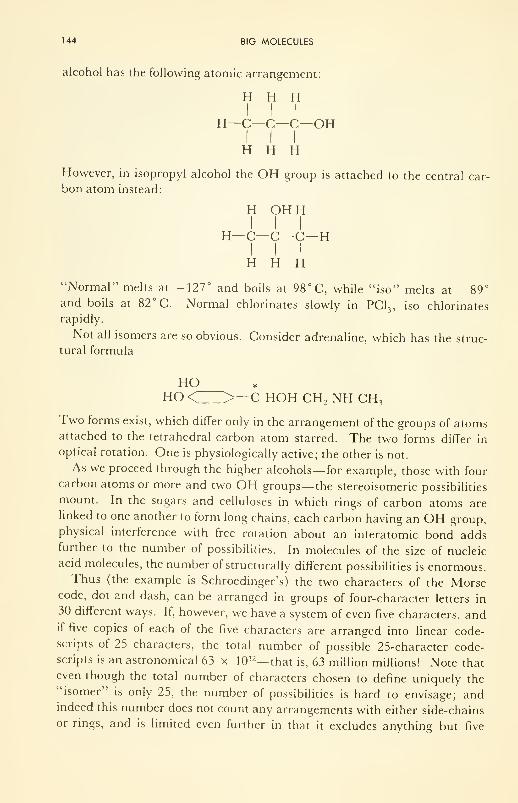
144 BIG MOLECULES
alcohol has the following atomic arrangement:
H H HI I I
H—C—C—C—OHI I I
H H H
However, in isopropyl alcohol the OH group is attached to the central car-
bon atom instead:
H OHHI I I
H—C—C—C—
H
I I I
H H H
"Normal" melts at -127° and boils at 98° C, while "iso" melts at -89°and boils at 82° C. Normal chlorinates slowly in PC1
3 , iso chlorinates
rapidly.
Not all isomers are so obvious. Consider adrenaline, which has the struc-
tural formula
HOHO < >—C HOH CH 2 NH CH
3
Two forms exist, which differ only in the arrangement of the groups of atomsattached to the tetrahedral carbon atom starred. The two forms differ in
optical rotation. One is physiologically active; the other is not.
As we proceed through the higher alcohols— for example, those with four
carbon atoms or more and two OH groups— the stereoisomeric possibilities
mount. In the sugars and celluloses in which rings of carbon atoms are
linked to one another to form long chains, each carbon having an OH group,physical interference with free rotation about an interatomic bond addsfurther to the number of possibilities. In molecules of the size of nucleic
acid molecules, the number of structurally different possibilities is enormous.Thus (the example is Schroedinger's) the two characters of the Morse
code, dot and dash, can be arranged in groups of four-character letters in
30 different ways. If, however, we have a system of even five characters, andif five copies of each of the five characters are arranged into linear code-scripts of 25 characters, the total number of possible 25-character code-scripts is an astronomical 63 x 10 12— that is, 63 million millions! Note that
even though the total number of characters chosen to define uniquely the"isomer" is only 25, the number of possibilities is hard to envisage; andindeed this number does not count any arrangements with either side-chains
or rings, and is limited even further in that it excludes anything but five

ISOMERS AND MULTIPLETS 145
copies of each character to make up the 25! Of course, not just any arrange-
ment of atoms gives a stable molecule; but on the other hand the number of
chemical groups of which a macromolecule is composed (—CH 2 , —NH,—CO, —C—S— , etc.) is certainly far more than five! .... One concludes
that the number of stable isomers of a macromolecule must be huge, but at
this stage of knowledge one really has no idea how many there are. Eachmust have a unique set of physical and chemical properties. Just as in the
case of the simple alcohols, each must be a stable molecular entity.
Excited States
No molecule, even if anchored at some point, must be completely quiet
if T > 0°K. Indeed, in an environment at 98°F (37°C) such a molecule,
even if initially at rest, or quiet— i.e., in its vibrational and rotational
"ground state" as it is called— will soon be buffeted into motion by neigh-
boring molecules of gas, liquid, or solid, until its energy level or tempera-
ture is, on the average, that of the environment. Heat energy enters the
molecule as the energy of rotation or vibration if the molecule is anchored,
and enters also as the kinetic energy of translation (linear motion) if the
molecule is free. The vibrations and rotations may be thought of as standing
or traveling matter waves moving across the molecule. Parts of the mole-
cule can be fixed and immobile; other parts can be free. The distribution of
energy within the molecule will be continuously changing.
Macromolecules accept and give up energy to the surroundings in discrete
bursts or bunches or quanta, if the quantum theory applies here as it is
known to apply to 2- and 3-atom molecules. However, the energy differ-
ences between mechanically excited states must be very small— so small that
almost a continuous exchange of energy must be possible.
The important point is that all of the configurations which result from
heat exchange are configurations proper to one isomer; in principle the
isomer may assume many shapes. Consider the random coil configuration
of protein as an example. The one chemical entity may assume many shapes
simply as a result of thermal exchange.
Electronically excited states also exist but these are different. It was seen
in Chapter 4 that electrons which make the bonds of molecules can absorb
and re-emit electromagnetic radiation, and that some excited states can be
reached by the absorption of such small amounts of energy that even local
heat energy sometimes will do the trick. It is a general rule-of-thumb that
whenever a bonding electron accepts energy of any kind and becomes itself
"excited," the bond is weakened. Once weakened, it is more susceptible to
thermal buffeting and to chemical attack. Its "defense" is to rid itself of the
extra energy and get back into the bond; this it does by reradiation, or by
transfer of energy into the mechanical motion of the molecule.
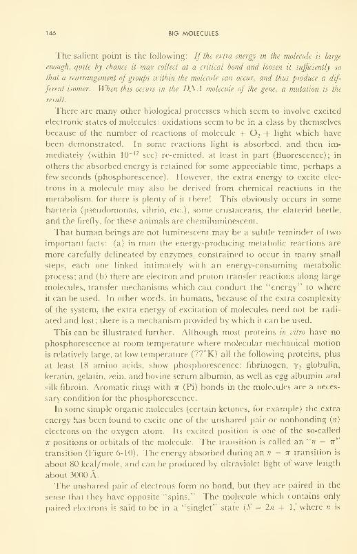
146 BIG MOLECULES
The salient point is the following: If the extra energy in the molecule is large
enough, quite by chance it may collect at a critical bond and loosen it sufficiently so
that a rearrangement of groups within the molecule can occur, and thus produce a dif-
ferent isomer. When this occurs in the DNA molecule of the gene, a mutation is the
result.
There are many other biological processes which seem to involve excited
electronic states of molecules: oxidations seem to be in a class by themselves
because of the number of reactions of molecule + 2 + hght which have
been demonstrated. In some reactions light is absorbed, and then im-
mediately (within 10~ 12 sec) re-emitted, at least in part (fluorescence); in
others the absorbed energy is retained for some appreciable time, perhaps a
few seconds (phosphorescence). However, the extra energy to excite elec-
trons in a molecule may also be derived from chemical reactions in the
metabolism, for there is plenty of it there! This obviously occurs in some
bacteria (pseudomonas, vibrio, etc.), some crustaceans, the elaterid beetle,
and the firefly, for these animals are chemiluminescent.
That human beings are not luminescent may be a subtle reminder of two
important facts: (a) in man the energy-producing metabolic reactions are
more carefully delineated by enzymes, constrained to occur in many small
steps, each one linked intimately with an energy-consuming metabolic
process; and (b) there are electron and proton transfer reactions along large
molecules, transfer mechanisms which can conduct the "energy" to where
it can be used. In other words, in humans, because of the extra complexity
of the system, the extra energy of excitation of molecules need not be radi-
ated and lost; there is a mechanism provided by which it can be used.
This can be illustrated further. Although most proteins in vitro have no
phosphorescence at room temperature where molecular mechanical motion
is relatively large, at low temperature (77° K) all the following proteins, plus
at least 18 amino acids, show phosphorescence: fibrinogen, y 2globulin,
keratin, gelatin, zein, and bovine serum albumin, as well as egg albumin and
silk fibroin. Aromatic rings with it (Pi) bonds in the molecules are a neces-
sary condition for the phosphorescence.
In some simple organic molecules (certain ketones, for example) the extra
energy has been found to excite one of the unshared pair or nonbonding (n)
electrons on the oxygen atom. Its excited position is one of the so-called
7r positions or orbitals of the molecule. The transition is called an "/? — ir"
transition (Figure 6-10). The energy absorbed during an n - w transition is
about 80 kcal/mole, and can be produced by ultraviolet light of wave length
about 3000 A.
The unshared pair of electrons form no bond, but they are paired in the
sense that they have opposite "spins." The molecule which contains only
paired electrons is said to be in a "singlet"1
state (S = In + 1,' where n is
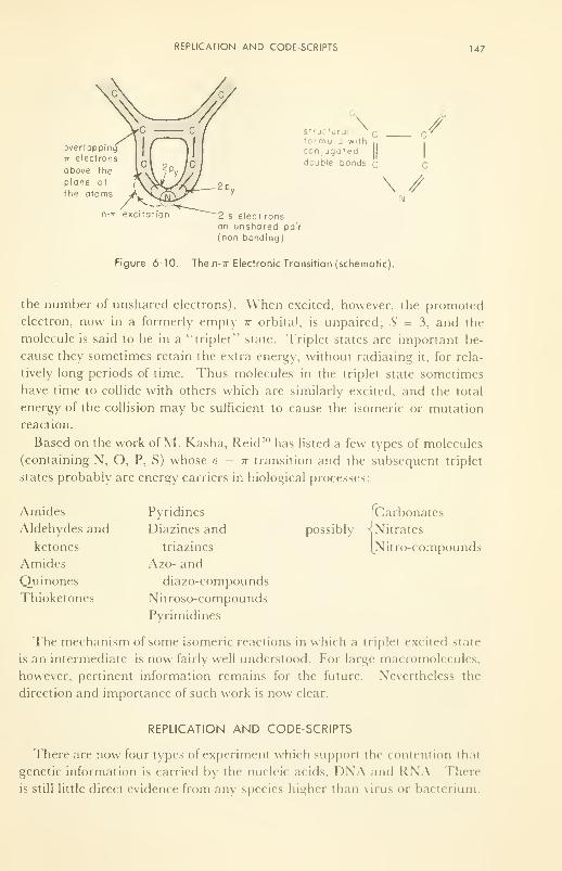
REPLICATION AND CODE-SCRIPTS 147
jverlappin
it electron
above the
plane of
the atoms
Structural r,
formu la with
conjugateddouble bonds q
//
\ //
n-T excitation 2 s electronsan unshared pair
(non bonding)
Figure 6-10. The n-7r Electronic Transition (schematic).
the number of unshared electrons). When excited, however, the promoted
electron, now in a formerly empty ir orbital, is unpaired; S = 3, and the
molecule is said to be in a "triplet1
' state. Triplet states are important be-
cause they sometimes retain the extra energy, without radiating it, for rela-
tively long periods of time. Thus molecules in the triplet state sometimes
have time to collide with others which are similarly excited, and the total
energy of the collision may be sufficient to cause the isomeric or mutation
reaction.
Based on the work of M. Kasha, Reid 10 has listed a few types of molecules
(containing N, O, P, S) whose n — it transition and the subsequent triplet
states probably are energy carriers in biological processes:
Amides
Aldehydes and
ketones
Amides
Quinones
Thioketones
Pyridines
Diazines and
triazines
Azo- and
diazo-compounds
Nitroso-compounds
Pyrimidines
possibly
Carbonates
Nitrates
Nitro-compounds
The mechanism of some isomeric reactions in which a triplet excited state
is an intermediate is now fairly well understood. For large macromolecules,
however, pertinent information remains for the future. Nevertheless the
direction and importance of such work is now clear.
REPLICATION AND CODE-SCRIPTS
There are now four types of experiment which support the contention thai
genetic information is carried by the nucleic acids, DNA and RNA. There
is still little direct evidence from any species higher than virus or bacterium.
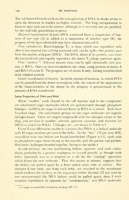
148 BIG MOLECULES
The celebrated French work on the transplanting of DNA in ducks seems to
open the doorway to studies on higher animals. The long extrapolation to
humans may turn out to be correct, although it is certainly not yet justified,
for this will take generations to prove.
Bacterial transformation: If pure DNA, extracted from a suspension of bac-
teria of one type (A) is added to a suspension of another type (B), the
progeny of the thus-infected B type have characteristics of A.
Virus reproduction: Bacteriophage T2 , a virus, which can reproduce only
after it has entered into a living bacterial cell, can be split—the protein part
from the nucleic acid part (DNA). The DNA, shorn of its protein, can enter
the bacterial cell and rapidly reproduce the intact T 2phage particles again.
Virus "synthesis'''': Tobacco mosaic virus can be split chemically into pro-
tein + RNA. One can then reconstitute the virus, using protein of strain Aand RNA of strain B. The progeny are of strain B only, having resnythesized
their original protein.
Genetic recombination of bacteria: In fertile strains of bacteria, in which DNAcan be passed from the donor to recipient cells, the extent of the appearance
of the characteristics of the donor in the progeny is proportional to the
amount ofDNA transferred.
Some Properties of DNA and RNA
These "nucleic" acids (found in the cell nucleus and in the cytoplasm)
are substituted sugar molecules which are polymerized through phosphate
linkages. In DNA the sugar is desoxyribose; in RNA it is ribose. Both have
5-carbon rings. The substituent groups on the sugar molecules are organic
nitrogen bases. These are ringed compounds with two nitrogen atoms in the
ring, and are four in number: adenine, guanine, cytosine, and thymine (in
DNA) or uracil (in RNA). Linkages, etc., are shown in Table 6-3.
From X-ray diffraction studies it is known that DNA is a helical molecule
with 10 sugar residues per turn of the helix. In the "dry" (70 per cent RH)crystalline state two helices are found interlocked (Figure 6-11), each with
its phosphate-sugar chain facing to the outside, and the purine and pyrimi-
dine bases, hydrogen-bonded together, facing to the inside.
f
At cell division, the two interlocking helices separate, and each repro-
duces, probably by a process analogous to crystal growth, as though each
helix, separated, acts as a template or a die for the "casting" operation
which forms the new molecule. That this occurs at mitosis, suggests that
the helices are pulled apart by a force which exists only at mitosis. For
instance if two ends, one from each helix, are attached to the membrane
which encloses the nucleus, in the expansion before division (25 per cent by
one measurement) the DNA helices could be pulled apart; then if each
template reproduces its opposite by "condensation," two DNA molecules
| A single-stranded DNA is known, in phage <pX 174.

149
TABLE 6-3: Components of the Nucleic Acids (Linkage at *]
"Bases" (B)
Purines
NH 2
A: adenine y NH
G: guanine
OH
N K
Pyrimidines
NH,C: cytosine N
o n;h
o
U: uracil HN
°VO
T: thymine H*f
oA<H*
CH
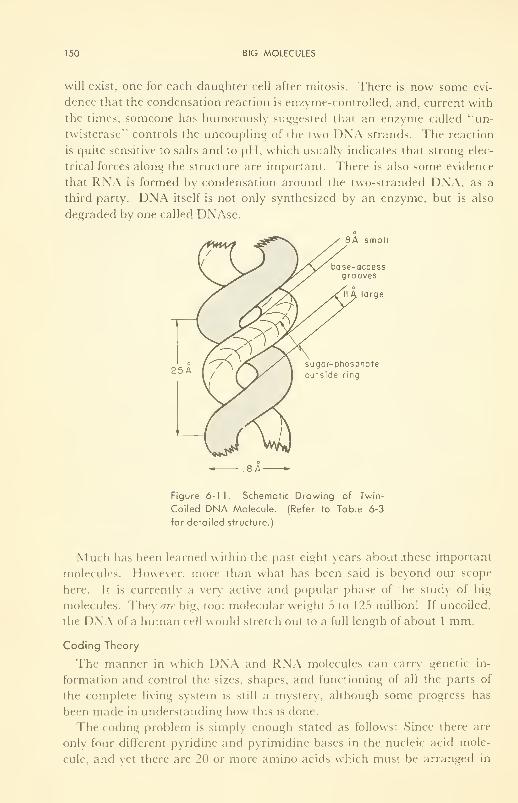
150 BIG MOLECULES
will exist, one for each daughter cell after mitosis. There is now some evi-
dence that the condensation reaction is enzyme-controlled, and, current with
the times, someone has humorously suggested that an enzyme called "un-
twisterase^ controls the uncoupling of the two DNA strands. The reaction
is quite sensitive to salts and to pH, which usually indicates that strong elec-
trical forces along the structure are important. There is also some evidence
that RNA is formed by condensation around the two-stranded DNA, as a
third party. DNA itself is not only synthesized by an enzyme, but is also
degraded by one called DNAse.
8A small
base-accessgrooves
large
sugar-phosphate
outside ring
ISA-
Figure 6-11. Schematic Drawing of Twin-
Coiled DNA Molecule. (Refer to Table 6-3
for detailed structure.)
Much has been learned within the past eight years about .these important
molecules. However, more than what has been said is beyond our scope
here. It is currently a very active and popular phase of the study of big
molecules. They are big, too: molecular weight 5 to 125 million! If uncoiled,
the DNA of a human cell would stretch out to a full length of about 1 mm.
Coding Theory
The manner in which DNA and RNA molecules can carry genetic in-
formation and control the sizes, shapes, and functioning of all the parts of
the complete living system is still a mystery, although some progress has
been made in understanding how this is done.
The coding problem is simply enough stated as follows: Since there are
only four different pyridine and pyrimidine bases in the nucleic acid mole-
cule, and vet there are 20 or more amino acids which must be arranged in
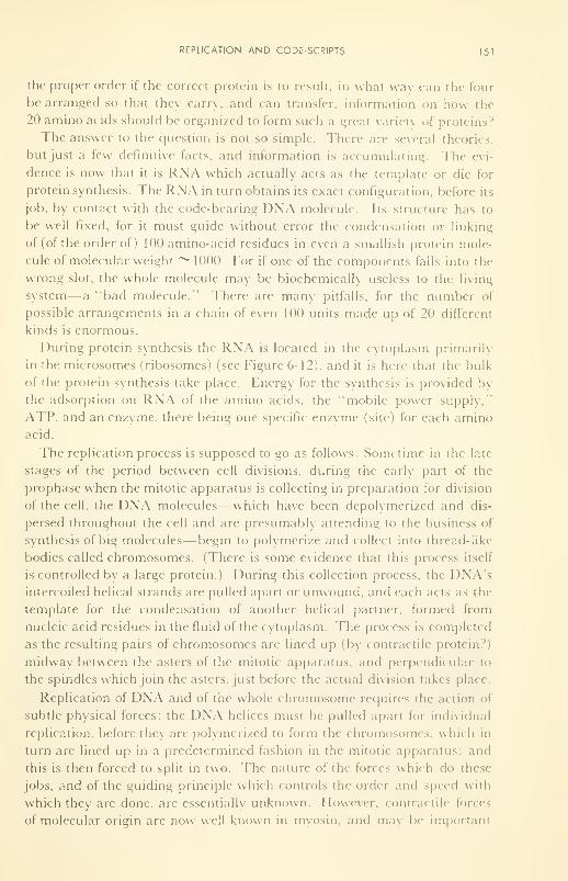
REPLICATION AND CODC-SCRIPTS 151
the proper order if the correct protein is to result, in what way can the four
be arranged so that they carry, and can transfer, information on how the
20 amino acids should be organized to form such a great variety of proteins?
The answer to the question is not so simple. There are several theori< s,
but just a tew definitive facts, and information is accumulating. The evi-
dence is now that it is RNA which actually acts as the template or die lor
protein synthesis. The RNA in turn obtains its exact configuration, before its
job, by contact with the code-bearing DNA molecule. Its structure lias to
be well fixed, for it must guide without error the condensation or linking
of (of the order of) 100 amino-acid residues in even a smallish protein mole-
cule of molecular weight ~1000. For if one of the components falls into the
wrong slot, the whole molecule may be biochemically useless to the living
system— a "bad molecule." There are many pitfalls, for the number of
possible arrangements in a chain of even 100 units made up of 20 different
kinds is enormous.
During protein synthesis the RNA is located in the cytoplasm primarily
in the microsomes (ribosomes) (see Figure 6-12), and it is here that the bulk
of the protein synthesis take place. Energy for the synthesis is provided by
the adsorption on RNA of the amino acids, the "mobile power supply,"
ATP, and an enzyme, there being one specific enzyme (site) for each amino
acid.
The replication process is supposed to go as follows: Sometime in the late
stages of the period between cell divisions, during the early part of the
prophase when the mitotic apparatus is collecting in preparation for division
of the cell, the DNA molecules—which have been depolymerized and dis-
persed throughout the cell and are presumably attending to the business of
synthesis of big molecules— begin to polymerize and collect into thread-like
bodies called chromosomes. (There is some evidence that this process itself
is controlled by a large protein.) During this collection process, the DNA'sintercoiled helical strands are pulled apart or unwound, and each acts as the
template for the condensation of another helical partner, formed from
nucleic acid residues in the fluid of the cytoplasm. The process is completed
as the resulting pairs of chromosomes are lined up (by contractile protein?)
midway between the asters of the mitotic apparatus, and perpendicular to
the spindles which join the asters, just before the actual division takes place.
Replication of DNA and of the whole chromosome requires the action ol
subtle physical forces: the DNA helices must be pulled apart for individual
replication, before they are polymerized to form the chromosomes, which in
turn are lined up in a predetermined fashion in the mitotic apparatus; and
this is then forced to split in two. The nature of the forces which do these
jobs, and of the guiding principle which controls the order and speed with
which they are done, are essentially unknown. However, contractile forces
of molecular origin are now well known in myosin, and m.i\ be important
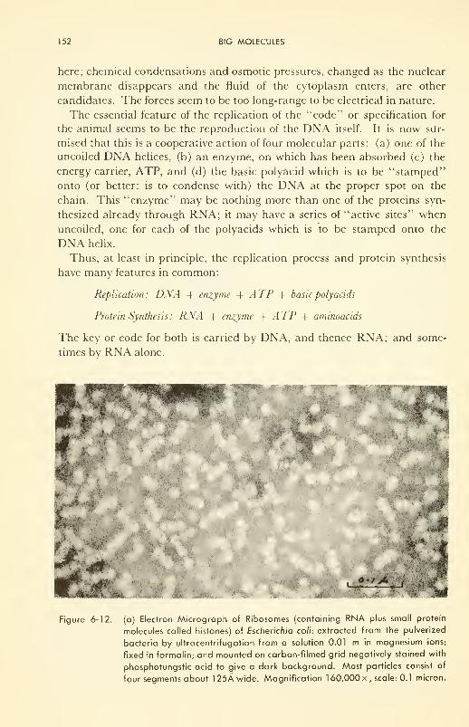
152 BIG MOLECULES
here; chemical condensations and osmotic pressures, changed as the nuclear
membrane disappears and the fluid of the cytoplasm enters, are other
candidates. The forces seem to be too long-range to be electrical in nature.
The essential feature of the replication of the "code" or specification for
the animal seems to be the reproduction of the DNA itself. It is now siir-
mised that this is a cooperative action of four molecular parts: (a) one of the
uncoiled DNA helices, (b) an enzyme, on which has been absorbed (c) the
energy carrier, ATP, and (d) the basic polyacid which is to be "stamped"
onto (or better: is to condense with) the DNA at the proper spot on the
chain. This "enzyme" may be nothing more than one of the proteins syn-
thesized already through RNA; it may have a series of "active sites" when
uncoiled, one for each of the polyacids which is to be stamped onto the
DNA helix.
Thus, at least in principle, the replication process and protein synthesis
have many features in common:
Replication: DNA + enzyme + ATP -f basic polyacids
Protein Synthesis: RNA + enzyme + ATP + aminoacids
The key or code for both is carried by DNA, and thence RNA; and some-
times by RNA alone.
•;*-% '.::- .'' \.$»
#*
...
~±
Figure 6-12. (a) Electron Micrograph of Ribosomes (containing RNA plus small protein
molecules called histones) of Escherichia coli: extracted from the pulverized
bacteria by ultracentrifugation from a solution 0.01 m in magnesium ions;
fixed in formalin; and mounted on carbon-filmed grid negatively stained with
phosphotungstic acid to give a dark background. Most particles consist of
four segments about 125A wide. Magnification 1 60,000 x , scale: 0.1 micron.
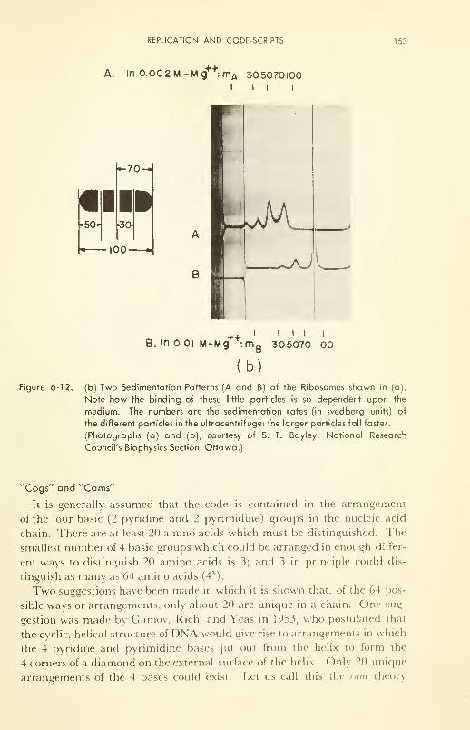
REPLICATION AND CODE-SCRIPTS 153
A. In 0.002 M-Mg++:mA 305070100I I I I I
B
'
D
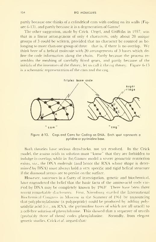
154 BIG MOLECULES
partly because one thinks of a cylindrical cam with coding on its walls (Fig-
ure 6-13), and partly because it is a degeneration of Gamovl
The other suggestion, made by Crick, Orgel, and Griffiths in 1957, was
that in a linear arrangement of only 4 characters, only about 20 unique
groups of 3 could be written, provided that no character be counted as be-
longing to more than one group of three— that is, if there is no overlap. Wethink here of a helical molecule with 20 arrangements of 3 bases which de-
fine the code information along the chain. Partly because the process re-
sembles the meshing of carefully fitted gears, and partly because of the
initials of the inventors of the theory, let us call it the cog theory. Figure 6-13
is a schematic representation of the cam and the cog.
triplet base code
sugarridge
cam cog
Figure 6-13. Cogs and Cams for Coding on DNA. Each spot represents a
pyridine or pyrimidine base.
Both theories have serious drawbacks, not yet resolved. In the Crick
model, the amino acids in solution must "know" that they are forbidden to
indulge in overlap; while in the, Gamov model a severe geometric restriction
exists, viz., the DNA molecule (and hence the RNA whose shape is deter-
mined by DNA) must always hold a very specific and rigid helical structure
if the diamond arrays are to persist on the surface.
However, successes in a flurry of investigation, genetic and biochemical,
have engendered the belief that the basic facts of the amino-acid code car-
ped by DNA may be completely known by 1963! There have been three
recent remarkable disclosures. First, Nirenburg startled the International
Biochemical Congress in Moscow in the Summer of 1961 by announcing
that polyphenylalanine (a polypeptide) could be produced by adding poly-
uridylic acid (i.e., an RNA, the pyrimidine bases of which are all uracil) to
a cell-free solution of phenylalanine. This showed that a sequence of uracils
(probably three of them) codes phenylalanine. Secondly, from elegant
genetic studies, Crick et al. argued that:
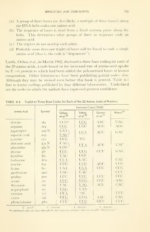
REPLICATION AND CODE-SCRIPTS 155
(a) A group of three bases (or, less likely, a multiple ol three l>ases) along
the DNA helix codes one amino acid.
(b) The sequence of bases is read from a fixed starting point along the
helix. This determines what groups of three in sequence code an
amino acid.
(c) The triplets do not overlap each other.
(d) Probably more than one triplet of bases will be found to code a single
amino acid (that is, the code is "degenerate"!.
Lastly, Ochoa et al., in March 1962, disclosed a three-base coding for each of
the 20 amino acids, a code based on the increased rate of amino-acid uptake
by E. coli protein to which had been added the polymerized bases of known
composition. Other laboratories have been publishing partial codes also.
Although they may be revised even before this book is printed. Table 6-4
lists tentative codings published by four different laboratories. Underlined
are the codes in which the authors have expressed greatest confidence.
TABLE 6-4. Triplet or Three-Base Codes for Each of the 20 Amino Acids of Proteins
156BIG MOLECULES
There are extensions and modifications of the cog and cam theories, and
even other theories of the physical arrangements on DNA and RNA. Theexperimental problem is not made simpler by the fact that there are
20 x 19 x 18- •• = 2.3 x 10 17 different ways in which 20 different amino
acids can be hooked together! Some "selection rules" must therefore follow
from a code, for, as Gamov says "if one could spend only one second to
check each assignment, one would have to work continuously for about five
billion years, which is [estimated to be] the present age of our Universe!"
Other experimental work has brightened the picture still further. For in-
stance, only with a specific enzyme does an amino acid form a complex with
ATP; polymerization and depolymerization occur in DNA and RNA; com-
plex formation occurs between the low-molecular-weight, soluble (or "trans-
fer") RNA and the DNA molecule; the helical shape of DNA is well estab-
lished in moist air; and chemical analyses have been made of certain mole-
cules. All these are experimental facts. There are many, many variables,
better knowledge of which will clarify the theory.
MUTATIONS AND MOLECULAR DISEASES
The idea of "sick people from bad molecules" is not really new, although
it certainly has been experimentally demonstrated in very convincing
fashion and exploited heavily since 1948. While Washington was busy on
the Delaware, Scheele in Germany showed that there is a good and bad form
of adrenalin. By 1913, F. G. Hopkins was able to state with some bio-
chemical authority: "Metabolic processes on which life depends consist in
toto of a vast number of well-organized and interlocking enzymic reactions,
interference with any one of which can product deleterious effects . . .." The
quotation from Pauling, with which this Chapter began, concerning the
need for better understanding of macromolecules and catalysts, is the mod-
ern approach to this question.
We have seen that, because of structural and/or compositional changes
in macromolecules, the following results may accrue:
( 1 ) Change in rate of chemical processes
(2) Change in rate of physical processes
(3) Introduction of new side reactions
A simple example of (3), introduced before recorded history and persisting
faithfully to our day, is offered in the different blood types in man: O, A, B,
AB. These differ from each other only in that the colloidal-stabilizing
mechanism of the macromolecules of the blood plasma is different: for if two
of the types are mixed, they agglutinate or gel; the mixture becomes thick
and refuses to flow. The physical nature of this subtle difference which
makes them incompatible still escapes us. The production, by each indi-

MUTATIONS AND MOLECULAR DISEASES 157
vidual, of antibodies (big molecules?) which are specific to that individual,
and incompatible with those built by any other individual for the same pur-
pose, is a well known phenomenon. Thus each individual has a specific bio-
chemistry and a biophysics of his own, which becomes manifested in manyways. It is not surprising, then, that even small changes in structure or
composition of certain large molecules can sometimes have disastrous
results.
A few examples will illustrate the point. No attempt is made to be ex-
haustive. Lathe's thesis 1 reviews several other molecular diseases.
Molecular Diseases
There is both a broad, generic connotation and a rather restricted, spe-
cialized one associated with the term "molecular diseases. " In the sense
that all diseases involve molecules which are incompatible with the chem-
istry or the physics of the system, all diseases are "molecular. " However,
in the more restricted sense, the term has evolved to mean diseases caused
by apparently small modifications of the chemical composition or the physi-
cal structure of a particular molecule. The hemoglobin diseases, recognized
only within the last decade, are now the classic example.
Hemoglobins : There are at least ten known modifications of the hemo-
globin molecule, each of which is associated with a pathologic condition.
The normal molecule is characterized by certain values for sedimentation
and diffusion constant (thence molecular wt.), electrophoretic mobility, elec-
tric charge as a function of pH (determined by titration), solubility, ultra-
violet absorption spectrum, etc. The most celebrated variant, S, which is
found in erythrocytes from people with sickle-cell anemia, differs from the
normal, A, principally in the manner in which it moves under the influence
of an electric field: it moves faster, and at pH = 7, toward the cathode,
whereas .4 is negatively charged at pH = 7 and moves toward the anode.
Some of the pertinent characteristics of ten different forms of the hemo-
globin molecule have been collected in Table 6-5. Although the differences
were first observed clinically, and then correlated with differences in physi-
cal properties, recent work has established that the differences arise because
of different composition or arrangement in the amino-acid sequences of the
protein. There are about 600 amino acids in the molecule. X-ray diffraction
studies have shown that type A (normal adult human hemoglobin) mole-
cules consist of four intertwined polypeptide chains. Two of these have a
valine, then a leucine residue just prior to attachment to the nitrogen of the
porphyrin (heme) group; two others have a valine, histidine, leucine sequence
before attachment to the (iron-containing) porphyrin group. It is nowknown that modifications occur right at that point: a different sequence, or
even different amino acids in the sequence, can occur.
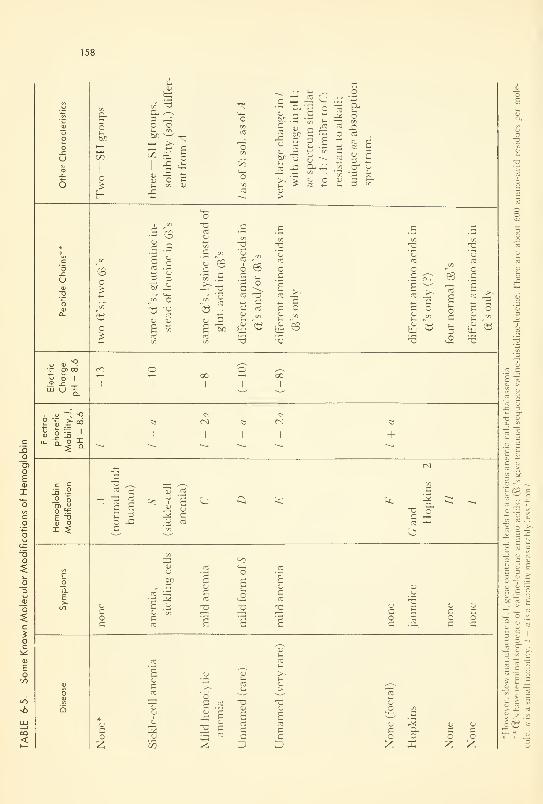
158
PROBLEMS 159
There may be other modifications out farther in the protein, but this is
not yet known. Likewise there may be many more modifications of hemo-
globin than those listed. The work is really quite new. Unfortunately, prac-
tically nothing is known of the shapes of these molecules—and won't be
until more is known of their structure. Sufficient familiarity with the physio-
logical reactions has been estimated to be about ten years away.
The sickling of erythrocytes occurs when the hemoglobin-.^ is in an at-
mosphere low in oxygen, and is a remarkable example of what "bad" mole-
cules can do. It is now fairly well established that these bad molecules are
so shaped that they can fit into each other and be piled up like a stack of
saucers. In so piling, their strength is sufficient to push out the sides of the
erythrocyte and cause it to buckle in the middle, i.e., to become sickle-
shaped. On oxygenation, the stack collapses, presumably because the mo-
lecular shapes are no longer so nicely complementary. Apparently the
process resembles the growth of a crystal. The reader is asked to meditate
on the known structure of myoglobin (Figure 6-3), and to study the pictures
of Perutz et al.24 on hemoglobin, before pressing further into this subject via
Reference 25.
Others. There are well over 20 diseases for which a "bad" molecule has
been postulated as the cause. One other which is receiving considerable at-
tention now is phenylketonuria. This is associated with mental deficiencies,
and has been traced to the fact that one of the enzymes which catalyze the
oxidation of phenylalanine through various steps toward pyruvic acid is not
doing its job fast enough. Whether the offending enzyme molecule is not
being synthesized, or has some physical deformity which renders it only
partially active; or whether it or the substrate is not being transported fast
enough to the place of catalysis, is not yet known. However, the result is ac-
cumulation of phenylalanine in the blood stream, and interference with syn-
thesis of nerve tissue.
PROBLEMS
6-1: Erythrocyte DNA has a molecular weight of above five million. Calculate the
diameter of the smallest sphere into which one molecule could be compressed.
(Assume an average atomic weight of 12: it has Cs, N's, O's, H's, and a few
P's and S's. Assume also that each atom occupies a cube 1 .2 A on each side.)
If it were stretched out, the atoms end to end, what would he the total length
of the chain?
6-2: Have you figured out how the two helical strands of DNA can unwind: for repli-
cation, or for coding transfer- 1<\ A.'
6-3: Describe the four possibilities open to a big molecule in an electronically ex< ited
state.

160 BIG MOLECULES
REFERENCES
1
.
Lathe, G. H., "Defective Molecules as a Cause of Diseases," Thesis, Leeds Univ.
Press, Leeds, England, 1960.
2. Dixon, M. and Webb, E. C, "Enzymes," Academic Press, New York, N. Y.,
1958.
3. Pauling, L., in "Enzymes: Units of Biological Structure and Function," edited
by Gaebler, O. H., Academic Press, 1956.
4. Putman, F. W., Ed., "The Plasma Proteins, I: Isolation, Characterization and
Function," Academic Press, 1960.
5. Oncley, J. L., et al., Eds., "Biophysical Science—A Study Program," John
Wiley & Sons, Inc., New York, N. Y., 1959; papers by Kendrew, Doty, Rich,
and many others.
6. Schroedinger, E., "What is Life?", Doubleday Anchor printing, 1956, of Cam-bridge Univ. Press book, 1944.
7. Butler, J. A. V., "Inside the Living Cell," Basic Books, Inc., New York, N. Y.,
1959; Butler, J. A. V., etal., Proc. Royal Soc, A, 250, 1 (1960).
8. Solomon, A. K., Scientific American, 203, 146 (1960), and references.
9. Hoagland, M. B., Scientific American, 201, 55 (1959).
10. Reid, G, "Excited States in Chemistry and Biology," Butterworths Sci. Publ.,
1957.
11. Gamov, G., Rich, A., and Yeas, M., Adv. Biol. Med. Pkys., 4,23 (1956).
12. Davson, H. and Danielli, J. F., "The Permeability of Natural Membranes," 2nd
ed., Cambridge Univ. Press, 1952.
13. Shooter, K. V., "The Physical Chemistry of Desoxyribosenucleic Acid," Prog, in
Biophysics and Biophysical Chem., 8,309 (1957).
14. Scientific Amer., Issue on "Giant Molecules," 197, No. 3, 1957; articles by Doty,
Crick, Schmitt, Debye, and others.
15. St. Whitelock, O., Ed., "Cellular Biology, Nucleic Acids and Viruses," N. Y.
Acad. Sci., 1957.
16. Tanford, C, "Physical Chemistry of Macromolecules," John Wiley & Sons,
Inc., New York, N. Y., 1961.
17. "The Merck Index," 7th ed„ Merck & Co., Inc., Rahway, N. J., I960.
18. Bonnar, R. V., Dimbat, M., and Stross, F. FL, "Number Average Molecular
Weights," Interscience Publishers Inc., New York, N. Y., 1958.
19. Crick, F. H. C, Barnett, L., Brenner, S., and Watts-Tobin, R. J., Nature, 192,
1227(1961).
20. Speyer, J. F., Lengyel, P., Basilio, C, and Ochoa, S., Proc. Nat. Acad. Sci., 48,
441 (1962).
21. Nirenberg, M. W., and Matthei, J. B., ibid., 47, 1588 (1961).
22. Zubay, G., and Quastler, H., ibid., 48,461 (1962).
23. Woese, C. G., Biophys. and Bwchem. Res. Com., 5,88 (1961).
24. Perutz, M. F., Rossman, M. G., Cullis, A. F., Muirhead, H., Will, G., and
North, A. C. T, Nature, 185, 416 (1960).
25. Itano, H. A., Singer, S. J. and Robinson, E., in "Biochemistry of Human
Genetics," G. E. W. Wolstenholme and C. M. O'Connor, Eds., Churchill
Ltd., London, 1959; p. 96 ff.
CHAPTER 7
A Conceptual Introduction
to Bioenergetics
Thermodynamics is a queer science. It is a system of logic based on three
postulates which have never been proved or disproved. By clever juggling
with symbols and ideas, it establishes relations between different forms of
energy .... These are most interesting relations which allow us to peep
behind the scenes of Nature's workshop .... Thermodynamics may yet tell
us how Nature molds such complex phenomena as muscular contraction out
of simpler reactions . (A. Szent-Gyorgyi. 7)
INTRODUCTION
Scope
The manipulation of the energy available from many natural sources has
been a problem of deep concern to man since the realization of the facts of
motion. Then came the mastery of fire; the kinematic machine; the use of
chemicals for ballistic purposes; and the water wheel for milling, and later
for producing the most versatile energy form of them all: electricity. Our
age is witnessing the development of the peaceful uses of atomic energy, the
energy of nuclear reactions; and a slower but perhaps more far-reaching de-
velopment of methods of trapping and storing the sun's radiation as heat,
and chemical and electrical energy.
Thermodynamics is the study of general principles which relate to trans-
fer of energy from one form to another (Figure 7-1). By contrast with some
of the more clearly understood systems, bioenergetics is still in its infancy,
although biochemists have done much toward describing the energetics of
161

162 A CONCEPTUAL INTRODUCTION TO BIOENERGETICS
(a)
heat
100 % conversion
(b)
Figure 7-1. Energy Interconversion, (a); (b) Degradation of Different Forms of
Energy into Heat Energy (the "Heat Death").
some pertinent chemical transformations, and physiologists have done some-
thing toward relating chemical energy and work. The many relationships
which must exist in living systems among mechanical, electrochemical,
chemical, and heat energies are as yet poorly known. This chapter attempts
to summarize the conclusions which arise from a generalized approach to
energy transfer, and to indicate how far they can be carried into a descrip-
tion of the living system.
In this account, use will be made of three different types of symbols,
small-case letters, capital letters, and script capital letters, which usually
refer to 1 gram, to 1 mole, and to the whole system, respectively. The capi-
tals and script capitals have the further property of being "variables of
state"—being variables the value of which help to define the state or condi-
tion of the system or subject, irrespective of past history. This will become
more clearly understood as the subject is developed.
Some Useful Definitions
Energy (from the Greek word meaning "active in work")— usually defined
as the potency for doing work. Remember the difficulties with definition
raised in Chapter 2?
kinetic Energy (KE)— energy of motion; energy contained within a bound-
ary by virute of the motion of the parts contained therein.
Potential Energy (PE)— literally "energy of position'1
; more generally
energy stored in any metastable but convertible form.
Heat Energy (HE or q)— in terms of the kinetic theory, identically equal to
the kinetic energy of motion (rotations, vibrations, translations) of the com-
ponent molecules.

LAWS OF THERMODYNAMICS 163
Specify Heat (c)— the heat energy required to raise 1 g of a substance one
degree in temperature. A particularly important specific heat is that of
water, by which the unit of heat energy is defined: One calorie is the amount
of heat energy required to raise 1 g of pure water 1°C, from 3.5 to 4.5°C
(where it is the most dense) at 1 atm pressure.
Heat Capacity {(.')— the heat energy required to raise 1 molecular vvt of
substance 1 deg in temperature. The units of specific heat, c, are cal/deg
Cent, g; and of heat capacity, C, are cal/deg Cent. mole.
Other forms of energy to be discussed are mechanical, electrical, gravita-
tional, chemical, nuclear, etc. Energetics or thermodynamics is the study
of interconversion of these. In biological systems the subject is usefully-
called bioenergetics. That part of the subject dealing with electromagnetic
and matter waves was considered in Chapters 3 and 4, and is expanded in
Chapter 9.
LAWS OF THERMODYNAMICS
Statements of the Three Laws
There are three general principles which summarize human experience
with energy interconversion. They are negative laws in the sense that they
cannot be proved always to hold, but nevertheless never have been known
to be violated.
The First Law: The first law states simply that energy can be transformed
from one form to another but cannot be created or destroyed. After the
equivalence of matter and energy were recognized (and proved in nuclear
reactions), the law was generalized still further to read: "mass-energy" in-
stead of "energy."
The Law stands as written, needing no extension, for all cases in which
any form of energy is converted into heat: 100 per cent conversion can al-
ways be realized. In Figure 7-1 (b) each of the arrows originates in a form
of energy other than heat.
The Second Law: For any machine which converts heat into mechanical
work, chemical into electrical energy, or the like, it is a universal experience
that only a fraction can be converted; the rest remains unavailable and un-
converted. There is thus an amount of unavailable energy as well as available
energy from the conversion. The unavailable, it would be logical to assume, is
the heat energy which must remain in the molecules of which the final state
(i.e., the product) is composed.
The Third Law: At 0°K (-273.16°C), the absolute zero of temperature,
at which all molecular motion has ceased, matter should be in a state of
perfect order, the molecules being perfectly aligned or oriented, and per-
fectly quiet. This law is concerned with the absolute heat energy contained
in molecules at any temperature. Although our present interest is in changes
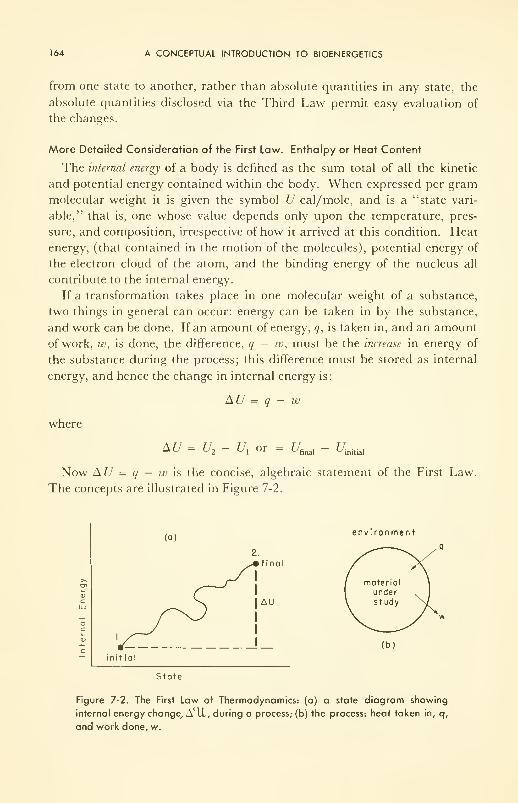
164 A CONCEPTUAL INTRODUCTION TO BIOENERGETICS
from one state to another, rather than absolute quantities in any state, the
absolute quantities disclosed via the Third Law permit easy evaluation of
the changes.
More Detailed Consideration of the First Law. Enthalpy or Heat Content
The internal energy of a body is defined as the sum total of all the kinetic
and potential energy contained within the body. When expressed per gram
molecular weight it is given the symbol U cal/mole, and is a "state vari-
able," that is, one whose value depends only upon the temperature, pres-
sure, and composition, irrespective of how it arrived at this condition. Heat
energy, (that contained in the motion of the molecules), potential energy of
the electron cloud of the atom, and the binding energy of the nucleus all
contribute to the internal energy.
If a transformation takes place in one molecular weight of a substance,
two things in general can occur: energy can be taken in by the substance,
and work can be done. If an amount of energy, q, is taken in, and an amount
of work, w, is done, the difference, q — w, must be the increase in energy of
the substance during the process; this difference must be stored as internal
energy, and hence the change in internal energy is:
AU = q - w
where
AU U2- U
xor
^final ' ' ^initial
Now AU = q — w is the concise, algebraic statement of the First Law.
The concepts are illustrated in Figure 7-2.
>>
aUJ
(a)environment
final
AU
(b)
initial
State
Figure 7-2. The First Law of Thermodynamics: (a) a state diagram showing
internal energy change, A il, during a process; (b) the process: heat taken in, q,
and work done, w.
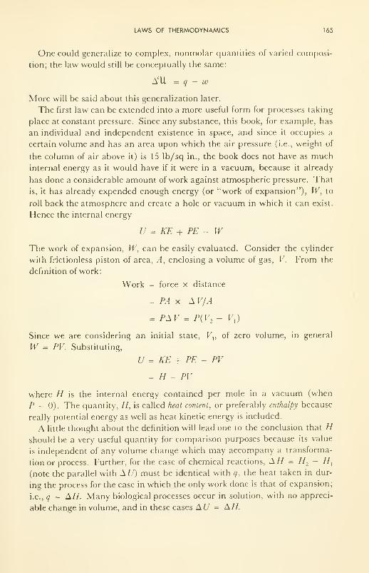
LAWS OF THERMODYNAMICS 165
One could generalize to complex, nonmolar quantities of varied composi-
tion; the law would still be conceptually the same:
AU = q - w
More will be said about this generalization later.
The first law can be extended into a more useful form for processes taking
place at constant pressure. Since any substance, this book, for example, has
an individual and independent existence in space, and since it occupies a
certain volume and has an area upon which the air pressure (i.e., weight of
the column of air above it) is 15 lb/sq in., the book does not have as much
internal energy as it would have if it were in a vacuum, because it already
has done a considerable amount of work against atmospheric pressure. That
is, it has already expended enough energy (or "work of expansion"), W, to
roll back the atmosphere and create a hole or vacuum in which it can exist.
Hence the internal energy
U = KE + PE - WThe work of expansion, W, can be easily evaluated. Consider the cylinder
with frictionless piston of area, A, enclosing a volume of gas, V. From the
definition of work:
Work = force x distance
= PA x AV/A
= PA V = P( V2- V,
)
Since we are considering an initial state, Vv of zero volume, in general
W = PV. Substituting,
U = KE + PE - PV
= H - PV
where H is the internal energy contained per mole in a vacuum (when
P = 0). The quantity, H, is called heat content, or preferably enthalpy because
really potential energy as well as heat kinetic energy is included.
A little thought about the definition will lead one to the conclusion that Hshould be a very useful quantity for comparison purposes because its value
is independent of any volume change which may accompany a transforma-
tion or process. Further, for the case of chemical reactions, AH = H2- //,
(note the parallel with A U) must be identical with q, the heat taken in dur-
ing the process for the case in which the only work done is that of expansion;
i.e., q = AH. Many biological processes occur in solution, with no appreci-
able change in volume, and in these cases AU = AH.
166 A CONCEPTUAL INTRODUCTION TO BIOENERGETICS
Now AH = q may be positive or negative depending upon which is larger,
the enthalpy of the final or of the initial state. The former characterizes an
endothermic reaction; the later an exothermic reaction. As a general rule
anabolic reactions are endothermic; catabolic reactions are exothermic.
More specifically, the synthesis of proteins in the metabolism of the living
svstem is endothermic; the combustion of glycogen and other food stores is
exothermic.
For chemical reactions the value of q or AH, the "heat of reaction," can be
measured calorimetrically. and quite accurate values obtained. For in-
stance, for the simplest reaction
H2 + 1/2 O, = H
2
the heat of reaction
&H = #final - ^initial
= H( 1 mole FTO) - H( 1 mole H2 + 1/2 mole
2 )
and although the absolute value of the enthalpy (or internal energy) for
neither reactants nor product is known (Who knows how to determine the
sum of all the potential energies in the nucleus, for example?), the difference,
AH, can be obtained with great precision: —57,798 cal/mole at 25°C, the
minus sign indicating that the reaction is exothermic.
An especially useful heat reaction is the heat of formation, AH., the
enthalpy change which occurs during the reaction by which the molecule of
interest is formed from its elements. Actually the example above was a
formation reaction. Another now follows:
6CW + 6H2 (£) + 3 2 (£) = C 6H,,06 (glucose)
AHf= -279,800 cal/mole
From a table of heats of formation, heats of reaction can be computed as
AH = (A//,)producls
- (AHf ) reactants
The heat of combustion or burning of glucose could be computed, from
heats of formation, from the following reaction:
C 6H 12 6 + 6<J2 (g) = 6H
20(1) + 6C0 2 (g) AH = -669.580 cal/mole
The fuel value of foods is usually expressed in units of thousands of calories:
i.e., kilocalories (kcal). kilogram calories (kg cal), or Calories (Cal). Hence
the fuel value of glucose is 669.58 kcal/mole. Other examples are given in
Table 7-1 (A), from which is readily apparent the origin of the very useful
"4-9-4 rule": the fuel values of carbohydrate, fat, and protein, are respec-
tively, about 4, 9, and 4 Cal/g.
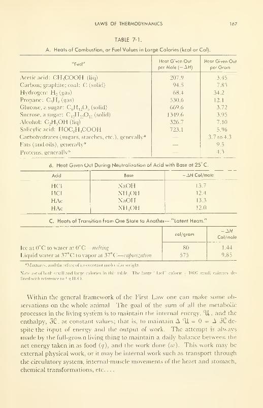
LAWS OF THERMODYNAMICS 167
TABLE 7-1.
A. Heats of Combustion, or Fuel Values in Large Calories (kcal or Cal).
"Fuel"
168 A CONCEPTUAL INTRODUCTION TO BIOENERGETICS
The quantity of heat given off by living animals can be measured either
calorimetrically or by the C02produced, (The two measurements agree!),
and when measured under conditions of a carefully defined rest, give a value
related to the internal work required to keep the living system alive. This
basal metabolic rate is about 70 kcal/hr, (about 1400 kcal/day) for a normal
man. In other units, the basal metabolic rate amounts to about 0.1 horse-
power (hp) continuously.
It is readily apparent that if an animal is ill, certain processes are running
at too high a rate; heat energy accumulates, and the temperature rises. Therate of energy loss is increased. By contrast with the normal animal in which
A 11 = 0, and q = w, in the ill animal w is much larger than q, the quantity
(q — w) is negative, and A lU is negative. Thus the animal lives at the ex-
pense of its internal energy, with resulting loss of weight— about 2 lb/day
for a human, assuming complete breakdown of assimilative processes and
food stored as glycogen and ignoring water loss. The quantities Tl and JC
decrease with time before the "turn" or "crisis, " then increase more or less
slowly back to normal because the animal begins to assimilate again during
the recovery period.
The ideas outlined in the preceding paragraphs show the versatility and
the usefulness of the First Law, that energy must be conserved, but of course
do not illustrate all its facets. Note parts B and C of Table 7-1 for other
examples.
More Detailed Consideration of the Second Law. Free Energy and Entropy
The Second Law of Thermo, does not violate the first, but rather extends
it. It says: Whenever energy is transformed from one kind into another, only
a fraction of the internal energy (enthalpy, if pressure is constant) change is
available for doing useful work; the rest remains as heat energy of the mole-
cules left at the completion of the reaction. Corollaries, although seemingly
unrelated, are the following: heat energy always passes from the hot to the
cold body; water always runs downhill; if energy available for doing work
can decrease during the course of a process, the process will proceed spon-
taneously, although not necessarily at a fast rate. (That last phrase is a very
important one!)
In algebraic terms, the Second Law can be expressed as:
AH = AF + Q
Here AF is the maximum available work, the "free" energy, which can be
extracted from AH, and Q is the unavailable energy. Note that both AFand Q as does AH, have units kcal/mole (i.e., Cal/mole).
The word "maximum" needs amplification. It is a fact of common ex-
perience that any mechanical job can be done in several ways, some ways
LAWS OF THERMODYNAMICS 169
more efficient than others. If the job is done by the hypothetical frictionless
machine, with minimum loss of energy, it is then done the most efficiently.
By analogy, work can be extracted from a process in many ways, some moreefficient than others. The hypothetical conditions of no waste are given the
special name, reversible conditions; AF is therefore the maximum work avail-
able under reversible conditions. One practical system from which nearly
maximum work can be extracted is the electrochemical one, a battery for
example; or, more pertinent here, the concentration cells which exist and
deliver energy at living membranes.
Very common are the processes which occur under nonreversible condi-
tions. The expression then becomes
A// = AF' + q' + Q_
for the reaction of 1 mole, or
A3"C = A3' + q' +Qfor the living system as a whole. Here AF' (or AJF') and q' refer to the ex-
ternally available work and "frictional" loss, respectively. The latter of
course shows up as heat energy, which must be dissipated to the environ-
ment by any of the well-recognized methods of perspiration, excretion,
respiration, etc., which will be discussed later.
A useful efficiency can be defined as:
8= AF'/AF, or £= AJF/Afr
This ratio is the fraction of the reversible Tree energy change which is re-
alized as useful work in the process. The value can easily be demonstrated
with a flashlight dry-cell; it ranges from per cent if the dry-cell is short-
circuited by a screwdriver across the terminals; through any value up to
about 70 per cent when operating in a flashlight; to close to 100 per cent
when used only as a source of voltage with almost no current being drawn.
Corresponding values for man cannot be given numerically, but must range
from nearly zero for a football team which expends an unimaginable amount
of energy to move a 2-lb football a few feet, to very high values for the nerve
transmission and mental activity which occur during computation. Other
examples will be given later.
The thermodynamic ratio AF/A/7, defined as T, is fixed by the value of
the unavailable energy, Q_. It is a more fundamental quantity than 8, in the
sense that it does not depend upon the frictional losses in the engine, or upon
the inefficiencies of the living machine. All processes of energy conversion
are producers or consumers of heat energy, and the conversion can take
place only as long as heat can be transferred from one part of the system to
another. When finally no further transfer is possible, the process ceases. It
170 A CONCEPTUAL INTRODUCTION TO BIOENERGETICS
is evident that the heat capacities of reactants and products help to deter-
mine the position of equilibrium. Thus if a product is formed which has
more degrees of freedom (i.e., modes of vibrations, translations, rotations)
than the reactant, the product can store more energy as kinetic energy (as
energy unavailable for work); then AFis less than H, and 7 is less than 1. In
other words the products, once formed, have to be heated up to the same
temperature as the environment, and are heated by an energy which could
have performed useful work were this not necessary. On the other hand if
the products can store less heat energy at 37° than the reactants, then A b
is greater than A// and Y is greater than 1. The unavailable energy in a
process depends upon the temperature and upon the heat capacities of reac-
tants and products. This special heat capacity, S cal/deg C. mole, is called
entropy. A list of different types of energy and their factors is given in Table
7-2. Note that heat energy is the only one listed for which the intensive
factor does not have the dimensions of a force. Perhaps it should be listed
as d(TS)/d.x.
TABLE 7-2. Factors of Several Kinds of Energy.
Type of Energy Intensive (Force) FactorExtensive (Capacity)
Factor (s)
Electrical (joules) dvjdx (volts/cm)
Mechanical (ergs) F' (dynes)
P (dynes/cm 2)
Chemical (cal) dF/d£ (cal/mole • cm)
Thermal (cal) T(deg)
q coulombs x .v(cm)
<7(cm)
!'(cm 3)
£(cm) x rz(moles)
S'(cal/deg. mole) x n
Explanation:
£ = reaction path length. F is free energy, Mechanical force, above, is given the symbol !' (in this Table
< »i 1 1 s ).
For the reaction or process under consideration,
a = ts2
TS,
where 2 and 1 refer to final and initial states. Then
d = TAS
Substitution in A// = AF+ Q., gives
AH = AF + TAS
which is the algebraic statement of the Second Law.
Table 7-3 lists values obtained experimentally for AH, AF and AS. An
example of particular biochemical and physiological importance is the hy-
drolysis of adenosine triphosphate, ATP. At pH = 7 and 37°C:
LAWS OF THERMODYNAMICS 171
AF = -7.73 kcal/mole; AH = -4.8 kcal/mole. and AS = 0.45 cal/deg
mole. If the reaction occurs in a test tube, no energy is converted into useful
work, and the heat produced is 4.8 kcal/mole. If, however, it is carried out
in the presence of an activated actomyosin filament (the contractile unit in
muscle), mechanical work (lifting a weight, for example) can be made to
occur, and the amount of work done can be anything up to 7.73 kcal/mole,
depending upon how it is done. If done reversibly (infinitely slowly), 7.73
kcal/mole is done, and S = 100 per cent; if done more and more rapidly,
8 becomes less and less.
The Production of Entropic Heat
Note that S is a state variable, like F, H, and U, and note that AS maybe positive or negative depending on whether the heat capacity of the prod-
ucts is greater or less than that of the reactants. Note further that if AS is
negative, and it often is, AF will be greater than AH. This is really not
surprising if one remembers that the extra energy for work was bound up as
extra heat energy of the reactants. Note also that the greater the numberof rotations, vibrations, and translations of which a system is capable, the
greater the heat capacity and hence the greater the entropy. Therefore
entropy (a heat capacity) is often used as a measure of disorder: the greater the
entropy, the greater the disorder.
For the living system, we write
A3C = AJ + TAS
under reversible conditions, and
AJC = A37' + q' + TAS
for practical conditions, in which not the maximum work, A{F, but rather a
lesser amount, Aj', is realized. An amount of energy, q', shows up as heat
energy and adds to the reversible, unavailable heat energy, TA S kcal. Ofcourse q' itself will factor into 7~A&', since it is a heat energy. Then if A S is
the reversible entropy increase, AS' is the extra entropy increase because
of the irreversibility of the process. Although q' is, strictly speaking, a waste,
it is the heat energy which maintains the temperature of a man some 10 or
more degrees C above his environment in spite of a steady heat energy loss
to the environment. Now, the work done may be internal work, A$'inV
or
external work, A57'- The internal work, however, is degraded into heat
internally, and forms part of q'. (Consider the pumping work of the heart,
for example: blood is forced along the circulatory system against a frictional
resistance, and the energy is finally expended as heat in the vessel walls.) II
we exclude growth and mental work for the moment (these hopelessly
complicate the argument), the contribution made by internal work to the
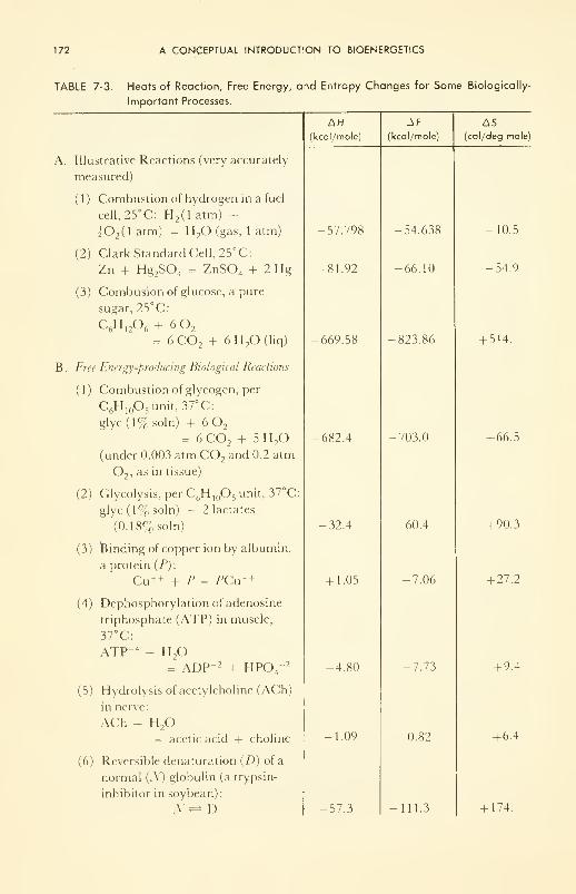
172 A CONCEPTUAL INTRODUCTION TO BIOENERGETICS
TABLE 7-3. Heats of Reaction, Free Energy, and Entropy Changes for Some Biologically-
Important Processes.
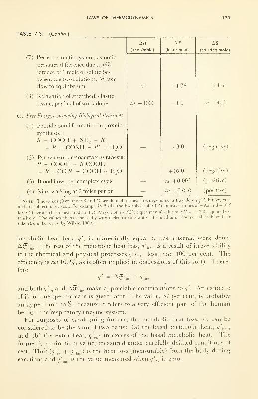
LAWS OF THERMODYNAMICS 173
TABLE 7-3. (Con/in.)
(7) Perfect osmotic system, osmotic
pressure difference due to dif-
ference of 1 mole of solute be-
tween the two solutions. Water
flow to equilibrium
(8) Relaxation of stretched, elastic
tissue, per kcal of work done
C. Free Energy-consuming Biological Reactions
{ 1 ) Peptide bond formation in protein
synthesis:
R - COOH + NH, - /?'
= R - CONH -- R' + H2
(2) Pyruvate or acetoacetate synthesis:
R - COOH + tf'COOH= R -COR' - COOH + H
2
(3) Blood flow, per complete cycle
(4) Man walking at 2 miles per hr
AH(kcol/mole)
ca -1000
Af(kcdl/mole)
1.38
1.0
+ 3.0
+ 16.0
ca +0.002
ca +0.010
AS
(col/deg mole)
+ 4.6
ca +400
(negative)
(negative)
(positive)
(positive)
Note: The values given under B and C are difficult to measure, depending as they do on pH, buffer. et<
and are subject to revision. For example in B (4), the hydrolysis of ATP in muscle, values of -9.2 and - 10.5
for A /-'have also been measured, and O. Meyerhof's (1927) experimental value of ±11 = - 12.0 is quoted ex-
tensively. The values change markedly with dielectric constant of the medium. (Some values have been
taken from the review by Wilkie, 1960.)
metabolic heat loss, q', is numerically equal to the internal work done,
A.JF'in, . The rest of the metabolic heat loss, q'irr , is a result of irreversibility
in the chemical and physical processes (i.e., less than 100 per cent. The
efficiency is not 100%, as is often implied in disucssions of this sort). There-
fore
and both q'irrand A9"'
inlmake appreciable contributions to q'
. An estimate
of 8 for one specific case is given later. The value, 37 per cent, is probably
an upper limit to 8 , because it refers to a very efficient part of the human
being— the respiratory enzyme system.
For purposes of cataloguing further, the metabolic heat loss, q', can be
considered to be the sum of two parts: (a) the basal metabolic heat, q'bm ,
and (b) the extra heat, q'rx ; in excess of the basal metabolic heat. The
former is a minimum value, measured under carefully defined conditions of
rest. Thus (q'cx + q'
hm ) is the heat loss (measurable) from the body during
exertion; and q'hm is the value measured when q'
exis zero.
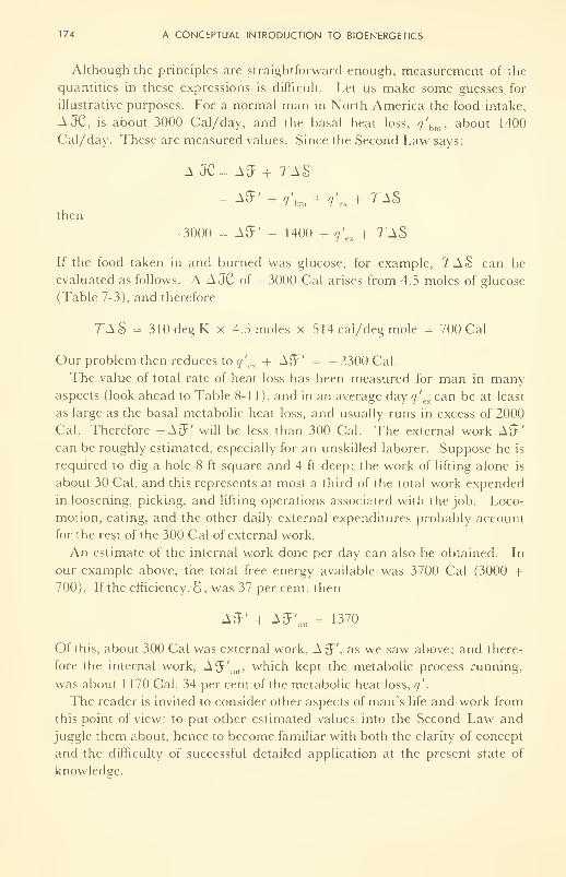
174 A CONCEPTUAL INTRODUCTION TO BIOENERGETICS
Although the principles are straightforward enough, measurement of the
quantities in these expressions is difficult. Let us make some guesses for
illustrative purposes. For a normal man in North America the food intake,
AJC, is about 3000 Cal/day, and the basal heat loss, q'bm , about 1400
Cal/day. These are measured values. Since the Second Law says:
A JC= A£F + TA§>
= A5' + q'hm + q'a + 7-AS
then
-3000 = AS' - 1400 + q\. x+ 7~AS
If the food taken in and burned was glucose, for example, XAS can be
evaluated as follows. A A JC of -3000 Cal arises from 4.5 moles of glucose
(Table 7-3), and therefore
TA$ = 310degK x 4.5 moles x 514 cal/deg mole = 700 Cal
Our problem then reduces to q'
cx -f AS' = -2300 Cal.
The value of total rate of heat loss has been measured for man in manyaspects (look ahead to Table 8-1 1), and in an average day q'
excan be at least
as large as the basal metabolic heat loss, and usually runs in excess of 2000
Cal. Therefore -AS' will be less than 300 Cal. The external work AS'can be roughly estimated, especially for an unskilled laborer. Suppose he is
required to dig a hole 8 ft square and 4 ft deep; the work of lifting alone is
about 30 Cal, and this represents at most a third of the total work expended
in loosening, picking, and lifting operations associated with the job. Loco-
motion, eating, and the other daily external expenditures probably account
for the rest of the 300 Cal of external work.
An estimate of the internal work done per day can also be obtained. In
our example above, the total free energy available was 3700 Cal (3000 +700). If the efficiency, S , was 37 per cent, then
AS' + AS'int
= 1370
Of this, about 300 Cal was external work, A^', as we saw above; and there-
fore the internal work, A L
J' int , which kept the metabolic process running,
was about 1170 Cal, 34 per cent of the metabolic heat loss, q'
.
The reader is invited to consider other aspects of man's life and work from
this point of view: to put other estimated values into the Second Law and
juggle them about, hence to become familiar with both the clarity of concept
and the difficulty of successful detailed application at the present state of
knowledge.
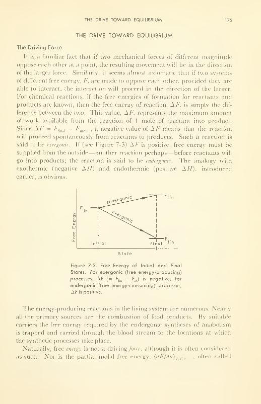
THE DRIVE TOWARD EQUILIBRIUM 175
THE DRIVE TOWARD EQUILIBRIUM
The Driving Force
It is a familiar fact that if two mechanical forces of difFerent magnitude
oppose each other at a point, the resulting movement will be in the direction
of the larger force. Similarly, it seems almost axiomatic that if two systems
of different free energy. F, are made to oppose each other, provided they are
able to interact, the interaction will proceed in the direction of the larger.
For chemical reactions, if the free energies of formation for reactants and
products are known, then the free energy of reaction. AF, is simply the dif-
ference between the two. This value, AF, represents the maximum amountof work available from the reaction of 1 mole of reactant into product.
Since AF = Ffinaj
- /'„„,,,,, a negative value of AF means that the reaction
will proceed spontaneously from reactants to products. Such a reaction is
said to be exergonic. If (see Figure 7-3) AF is positive, free energy must be
supplied from the outside— another reaction perhaps—before reactants will
go into products; the reaction is said to be endergonic. The analogy with
exothermic (negative AH) and endothermic (positive AH), introduced
earlier, is obvious.
State
Figure 7-3. Free Energy of Initial and Final
States. For exergonic (free energy-producing)
processes, AF (= Ffin
— Fin ) is negative; for
endergonic (free energy-consuming) processes,
AF is positive.
The energy-producing reactions in the living system are numerous. Nearly
all the primary sources are the combustion of food products. By suitable
carriers the free energy required by the endergonic syntheses of anabolism
is trapped and carried through the blood stream to the locations at which
the synthetic processes take place.
Naturally, free energy is not a driving force, although it is often considered
as such. Nor is the partial molal free energy, (dF/dn) T<P , often called
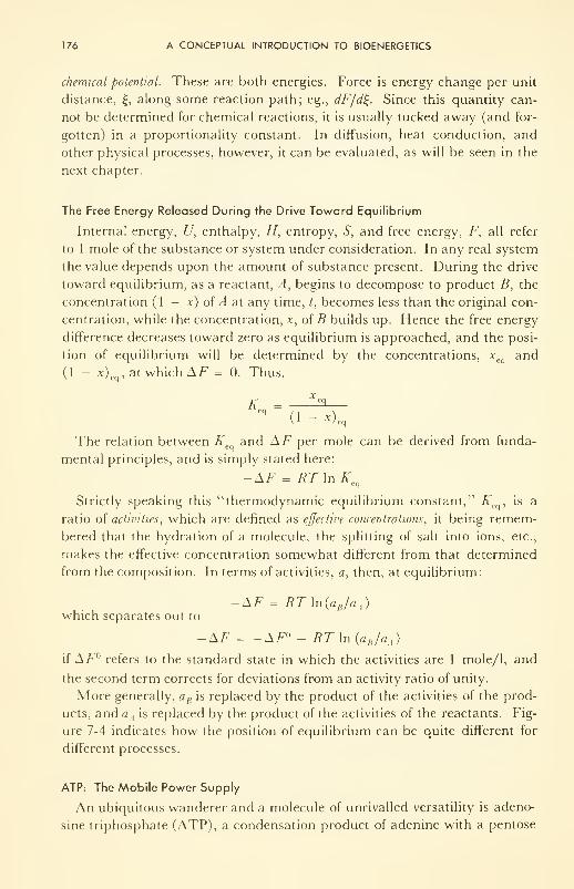
176 A CONCEPTUAL INTRODUCTION TO BIOENERGETICS
chemical potential. These are both energies. Force is energy change per unit
distance, £, along some reaction path; eg., dF/di~. Since this quantity can-
not be determined for chemical reactions, it is usually tucked away (and for-
gotten) in a proportionality constant. In diffusion, heat conduction, and
other physical processes, however, it can be evaluated, as will be seen in the
next chapter.
The Free Energy Released During the Drive Toward Equilibrium
Internal energy, U, enthalpy, H, entropy, S, and free energy, F, all refer
to 1 mole of the substance or system under consideration. In any real system
the value depends upon the amount of substance present. During the drive
toward equilibrium, as a reactant, A, begins to decompose to product B, the
concentration (1 — x) of A at any time, t, becomes less than the original con-
centration, while the concentration, x, of B builds up. Hence the free energy
difference decreases toward zero as equilibrium is approached, and the posi-
tion of equilibrium will be determined by the concentrations, x and
(1 - x)eq , at which AF = 0. Thus,
K = —^3" (1 - *) eq
The relation between K and AF per mole can be derived from funda-
mental principles, and is simply stated here:
-AF = RT\nKeq
Strictly speaking this "thermodynamic equilibrium constant," K is a
ratio of activities, which are defined as effective concentrations, it being remem-
bered that the hydration of a molecule, the splitting of salt into ions, etc.,
makes the effective concentration somewhat different from that determined
from the composition. In terms of activities, a, then, at equilibrium:
-AF = RT\n(aB/aA )
which separates out to
-AF = -AF° + RT In (a B /a A )
if AF° refers to the standard state in which the activities are 1 mole/1, and
the second term corrects for deviations from an activity ratio of unity.
More generally, aB is replaced by the product of the activities of the prod-
ucts, and aA is replaced by the product of the activities of the reactants. Fig-
ure 7-4 indicates how the position of equilibrium can be quite different for
different processes.
ATP: The Mobile Power Supply
An ubiquitous wanderer and a molecule of unrivalled versatility is adeno-
sine triphosphate (ATP), a condensation product of adenine with a pentose
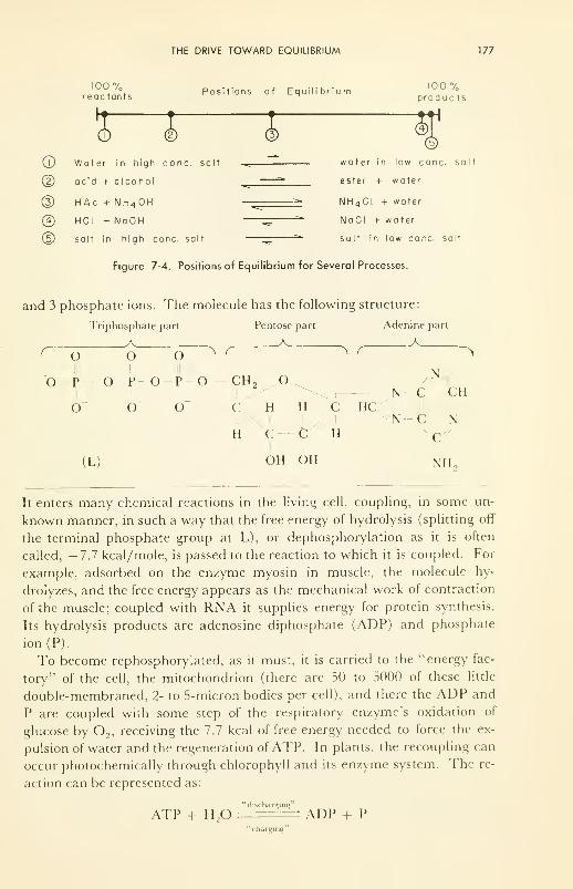
THE DRIVE TOWARD EQUILIBRIUM 177
100%reac tants
Positions of Equilibrium
© ©© Water in high cone, salt
(D acid + alcohol
(3) HAc + NH4OH
@ HCI + NaOH
(5) salt in high cone, salt
1 %produc ts
water in low cone, salt
ester + water
NH4CI + water
NaCI + water
salt in low cone, salt
Figure 7-4. Positions of Equilibrium for Several Processes.
and 3 phosphate ions. The molecule has the following structure:
Triphosphate part Pentose part Adenine part
A A . A.r o o o> r ^ r
0—p--o-p-o-P'-o--ch. on- :h
/N w
O" o o c
H
(L)
H H CJ .'/IC--C H
I I
OH OH
HC
I
NH
It enters many chemical reactions in the living cell, coupling, in some un-
known manner, in such a way that the free energy of hydrolysis (splitting off
the terminal phosphate group at L), or dephosphorylation as it is often
called, —7.7 kcal/mole, is passed to the reaction to which it is coupled. For
example, adsorbed on the enzyme myosin in muscle, the molecule hy-
drolyzes, and the free energy appears as the mechanical work of contraction
of the muscle; coupled with RNA it supplies energy for protein synthesis.
Its hydrolysis products are adenosine diphosphate (ADP) and phosphate
ion(P).
To become rephosphorylated, as it must, it is carried to the "energy fac-
tory" of the cell, the mitochondrion (there are 50 to 5000 of these little
double-membraned, 2- to 5-micron bodies per cell), and there the ADP and
P are coupled with some step of the respiratory enzyme's oxidation of
glucose by2 , receiving the 7.7 kcal of free energy needed to force the ex-
pulsion of water and the regeneration of ATP. In plants, the recoupling can
occur photochemically through chlorophyll and its enzyme system. The re-
action can be represented as:
"discharging"
ATP + H2
, ADP + P"charging"
178 A CONCEPTUAL INTRODUCTION TO BIOENERGETICS
and it is reversible. Left to right, it couples in wherever free energy is needed
throughout the living system. Right to left it becomes "charged back up,"
ready to supply energy at another site.
Now the living system is not wasteful of free energy without a good pur-
pose, such as to keep the system warm in a cold environment. Thus most
endergonic processes occur in steps of about 8 kcal/mole, or slightly less,
making full use of the free energy of the hydrolysis reaction. Likewise the
oxidation of foods also goes in steps of slightly more than 8 kcal/mole each,
so that the charging reaction is also not wasteful. Indeed, the very complex
sets of steps in the oxidation of carbohydrates, fats, and proteins seem de-
signed so that at several stages of each the ADP + P can couple in and be
condensed into ATP. This is the principle of the Krebs (citric acid) cycle,
for instance, in which it is estimated that 38 ATPs are reformed per mole-
cule of glucose oxidized to C0 2and H
20. This number permits an estimate
of the efficiency of the recharge process to be made:
8 kcal/mole ofATP x 38 ATP's inn „'- x 100 = 37 per cent
824 kcal/mole of glucose
This efficiency is very respectable, especially since the reactions are going
very fast. By contrast, a steam or diesel engine could probably do 20 to
30 per cent on glucose (for a short while!), and up to about 35 per cent on
gasoline or oil; solar batteries can convert only about 10 per cent; and
thermoelectric converters about 5 per cent from the fuel (including nuclear,
or radioactive fuels). Other (like ATP/ADP) electrochemical devices—eg.
batteries and fuel cells—are able to give very high efficiences (>80 per
cent) if operated slowly, much less if required to operate very fast.
A simple calculation (note the approximations) will emphasize the im-
portant point of how efficient the human machine really is. Man's basal
metabolic rate is about 70 kcal/hr. If this is all expended through ATP, the
turnover (charge-recharge) rate is 70/8 ~ 9 moles ATP/hr. If we assume
that a 150-lb man of density about 1 g/cc contains on the average 10~ 4 moles
ATP per liter, the turnover time for ATP is:
150 1b x454g/lb x 1 1/1000 g x 10~ 4 moles/1 „-— 2 — x 3600 sec/hr ~ 30 sec9 moles/hr
That is, each ATP molecule in the body is hydrolyzed and reformed about
once every 30 sec! At this speed of discharge and charge, a man-made bat-
tery would have an efficiency well below 1 per cent. Indeed, it would burn
up in the attempt! Hence 37 per cent in the living system is truly remark-
^ , , , ,• , / c 70 kcal/hr
able. To supply the basal energy, it burns the equivalent ot ~ 1 / g~4 kcal/g
glucose each hr, 24 hr a day.
REDOX SYSTEMS; ELECTRON TRANSFER PROCESSES 179
The ATP-ADP system is one of a class of oxidation-reduction (redox) or
electron-transfer systems operating in the living being. There are manyothers.
REDOX SYSTEMS; ELECTRON TRANSFER PROCESSES
Equivalence of Electrical and Chemical Energy
Oxidation-reduction reactions have very wide exemplification in living
systems: They bring about energy-producing oxidations of food; electro-
chemical reactions in the brain and nerve; hydrogenation of oils and dehy-
drogenation of fats and sugars, etc. Some are simple electron-transfer re-
actions, the reaction
Fe+ 2 __» pe+3 + g-
for example. The free energy of this /W/-reaction (There must be a place
for the electron to go!) can be trapped as un-neutralized electrons— i.e., as
electrical energy. In fact if a metallic or molecular electron-acceptor is
present at the site, such as
H + + e~ — 1/2 H 2
the chemical free energy of the total reaction
1/2 FT + Fe +3 — H + + Fe +2
can be drained off as electrical energy. This transformation is almost re-
versible (and therefore highly efficient), even at fairly high speed. The free
energy of oxidation of foodstuffs is guided by a series of redox enzymes
through a particular reaction scheme, in which each step of the process is a
fairly efficient redox process. Most of the free energy of each step is trapped
as an electron per molecule, and then passed on at the site where it can
be used.
Equivalence of electrical and chemical energy is a requirement of the First
Law. Thus AF calories/mole of reaction must be equal to the electrical
energy derived per mole of reaction. Now Faraday showed about 1830 that
96,500 coulombs (amperes x seconds) are required to oxidize or reduce one
equivalent weight of redox substance; and one equivalent weight is defined
as the weight which will transfer one electron per molecule. Hence if the
number of electrons transferred per mole, or the number of equivalents per
mole, is n, and if 96,500 cou/equiv is abbreviated to F, then the product nF
is the number of coulombs required to oxidize or reduce 1 mole. But elec-
trical energy in joules is volts x coulombs. Therefore
-AF = nF E
What voltage is E? It is the voltage measured between the hydrogen end
180 A CONCEPTUAL INTRODUCTION TO BIOENERGETICS
and the ferrous-ferric end of the reaction cell. To make this measurement,
and thereby to measure AF, one might simply bubble hydrogen over a piece
of platinum (the metallic contact) in \N-ac\d soution; and attach the plati-
num through a voltmeter to another platinum piece sitting in equimolar fer-
rous and ferric salt solution. The two solutions must be connected if the
circuit is to be complete. The value measured in this case is 0.77 v, con-
sistent with a free energy of reaction of about 40 kcal per mole of hydrogen
consumed. The ferric end is positive to the voltmeter, the hydrogen negative.
The concentrations may not be as stated, however, and we would expect,
and indeed find, that the voltage measured would then differ from 0.77. Theconditions specified in our example are arbitrarily chosen "standard state
conditions": unit (1) activities of reactants and products, 1 atm pressure,
25° C; and reversibility. We have already seen what a deviation from unit
activity ratio will do to AF.
Purely as a matter of convenience and of convention, since the absolute
value of no redox system is known, the normal hydrogen electrode (NHE)
(1 atm pressure, normal acid, and H2on platinum) has been chosen as the
standard reference, and defined as zero volts. All other redox systems are
referred to this standard. In fact a table has been drawn up of known
standard redox potentials, F°'s, and is called the electromotive series.
However, a special table has been drawn up for biological redox systems. It
differs from the standard F°'s, referred to the NHE, in two ways: all the
redox reactions are measured against hydrogen at pH = 7, not zero; and
since the effective concentrations or activities are not usually known for bio-
logical molecules, measured concentrations are used instead; and the tabu-
lated values, Eml , refer to equal concentrations (midpoint, m) of oxidized and
reduced form (i.e., material 50 per cent oxidized). Table 7-4 lists some of
these. A very complete discussion of biological redox systems is given in the
remarkable book of W. Mansfield Clark, 2 who has spent a lifetime making a
systematic study of, and attempting to organize our knowledge of this
subject.
Free Energy and Concentration. The Nernst Equation
The free energy of reaction, and hence the emf, F, of reaction, varies with
the concentrations, as is evident from the relation between AF and K given
above. Insertion of nFE° for -AF°, and nFE for -AF, and rearrange-
ment gives the famous expression of the emf as a function of concentrations,
introduced just before the turn of the century by Walther Nernst:
DTE = F°- —\n (am/a TJ
nb
REDOX SYSTEMS; ELECTRON TRANSFER PROCESSES 181
TABLE 7-4. Redox Potentials fm7 of Some Important Biochemical Reactions.
Steady-state Redox Process
182 A CONCEPTUAL INTRODUCTION TO BIOENERGETICS
Plots of E vs aH + and of E vs pH are shown in Figure 7-5. It can be seen that
at the physiological pH of 7, Eml on the \HE scale is —0.42 v.
0.0
0,4 2
-0.82
-m 7
(all reduced)
100%(all oxidized)
% oxidized
(b)
Figure 7-5. Reversible Potential of an Oxidation-Reduction Reaction: (a) as a function of
pH,onthe normal hydrogen electrode (NHE) scale; (b) as a function of per cent oxidation.
Definition of Em7 : potential (on the NHE scale) when pH = 7 and when the redox system
is 50 per cent oxidized.
As a further clarification and as a summary, Figure 7-6 shows schema-
tically the relation between the NHE scale of £"°'s (pH = 0), to which AEvalues have been traditionally related through —AE = nEE, and the physio-
logical scale, Em7 (pH = 7). The latter is now commonly used as a relative
measure of free energy changes in biological reactions. The values in Table
7-5 have been measured simply by putting a platinum wire into a mixture of
equal concentrations of sodium succinate and sodium fumarate at pH 7,
containing an enzyme and a mediator (discussed later), and measuring its
voltage against a hydrogen electrode in the same solution. Such measured
values can be used to predict the direction of reaction, or as a basis for com-
parison, but not for the determination of AE, because the effective con-
centrations (activities) are not known. It is well to be clear on this limitation
of the £"-, listing;.
Difficulty often arises in this subject because of notation. Different
authors use different subscripts and superscripts. In this book we have de-
fined, and use, only E, E°, and Em7 . One should be aware of the variations
which one may find. Further, one should understand clearly that the values
given in the table for intermediary processes of oxidation are midpoint
values; that although these redox systems are generally poised at their most
stable point (Figure 7-5), a tight control must be kept by the living system at
all times on the concentration of oxidized and reduced states of each system;
REDOX SYSTEMS; ELECTRON TRANSFER PROCESSES 183
that too much variation could cause a normally proceeding reaction actually
to go backwards !
A special application of the Nernst Equation is discussed under concen-
tration cells.
+ 1.22
+ 0.80
-0.42
pH = pH=7
Figure 7-6. £m7 's (center vertical line), and Their Relation to the Corresponding
P's. (See text and Table 7-4.)
Balky Redox Reactions
There are three tricks provided by nature to promote electron exchange in
oxidation-reduction reactions. The first is catalysis : providing a surface or a
site on which the exchange can rapidly take place. For example, electrons
exchange immeasurably slowly between H2and H + in solution, but if a sur-
face such as finely divided platinum metal is added, electron exchange is
rapid, and the potential readily manifested.
The second trick is the use of an indicator redox system. If one wishes to
know the redox potential of a solution in which the electron transfer is slow
or sluggish, one can add a very small amount of an entirely foreign redox
system, which exchanges electrons rapidly with the system of interest, and
which is either itself highly colored or exchanges rapidly at a metal elec-
trode. In the first case the depth of color of the resulting solution can be
related to the redox potential; and in the second case the potential can be
read directly against a reference electrode. Methylene blue, a colored redox
dye, is one of a class of dyes commonly used for this purpose, while the addi-
tion of a small amount of potassium iodide often will permit direct measure-
ment of the redox potential of the solution against some suitable reference
184 A CONCEPTUAL INTRODUCTION TO BIOENERGETICS
electrode. If the redox indicator (KI, for example) is present to an amount
much less than the redox systems in the solution to which it is added, it can
exchange electrons (KI —* I2 ) until its potential (determined by a
x/a KX ) is
the same as that of the solution.
The third trick is really a combination of the first two. If a solution con-
tains two reactants, such as glucose and oxygen, which can react together
spontaneously (negative AF), the reaction will be extremely slow unless the
solution contains mediators. Consider one step in the over-all process, for
example succinate added to pyruvate in a test tube. Although these two ions
can exchange electrons (and hydrogen atoms), with the liberation of free
energy, they don V unless a redox system such as cytochrome-C is present as
a mediator. Its job is to couple with succinate and reduce it to fumarate,
then (itself now oxidized) to oxidize pyruvate. In other words it provides a
path by which the over-all reaction can go in two steps, via the mediator,
whereas it could not go at all in one. The whole respiratory enzyme sys-
tem is a system of mediators, permitting the complete, controlled oxidation
of glucose by oxygen to go in discrete-
steps, the free energy of each step
being thus made readily available to recharge ATP, for example, and there-
fore to be usable elsewhere in the system.
There seem to be no generic differences among electrochemical catalysts,
redox indicators, and mediators. The name used depends upon one's point
of view. Indeed, in his classical work on the succinate-fumarate system,
Lehman (1930) called succinic dehydrogenase the catalyst and methylene
blue the mediator.
MEASUREMENT OF AH, AF, AND TAS
The simplest way to measure all three energies is in an electrochemical
redox cell, described in the previous section, if indeed the reaction is an
oxidation-reduction reaction. Thus AF is directly related to the voltage on
the NHE scale by -AF = nFE, and A S is directly related to the rate of
change of AF with temperature through the relationships'
^1=-A<>; and AS = nFd-^-
dT dT
Since AFand AS can be so determined, AH can be obtained from the Sec-
ond Law:
AH = AF + TAS
However, AH, the heat of reaction, is itself hard not to measure! If no
work at all is extracted in a calorimeter experiment, as a process is allowed
to go spontaneously to equilibrium, all the free energy is wasted away into
heat, and A His the quantity of heat measured in the experiment.
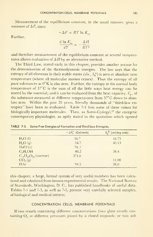
CONCENTRATION CELLS; MEMBRANE POTENTIALS 185
Measurement of the equilibrium constant, in the usual manner, gives a
measure of AF, since
-AF = RT\n Keq
Further,
d In KeqAH
dT " ~RT~2
and therefore measurement of the equilibrium constant at several tempera-
tures allows evaluation of A //by an alternative method.
The Third Law, stated early in this chapter, provides another avenue for
the determination of the thermodynamic energies. The law says that the
entropy of all elements in their stable states (viz., S °) is zero at absolute zero
temperature (where all molecular motion ceases). Thus the entropy of all
pure substances at 0°K is also zero. Further, the entropy at the normal body
temperature of 37°C is the sum of all the little ways heat energy can be
stored by the material; and it can be evaluated from the heat capacity, C. , of
the substance measured at different temperatures from 37°C down to abso-
lute zero. Within the past 25 years, literally thousands of "third-law en-
tropies" have been so evaluated. Table 7-5 lists some of these values for
biologically important molecules. Then, as Szent-Gyorgyi, 13 the energetic
contemporary physiologist, so aptly stated in the quotation which opened
TABLE 7-5. Some Free Energies of Formation and Third Law Entropies.
-Aff
° (Cal/mole) SQ
(cal/deg mole)
H20(1) 56.7 16.75
H20(g) 54.7 45.13
NaCl(s) 91.7
C2H
5OH 40.2 38.4
C12H 22O n (sucrose) 371.6
C02 (g) 51.08
HAc 94.5 38.0
this chapter, a large, formal system of very useful numbers has been calcu-
lated and tabulated from known experimental results. The National Bureau
of Standards, Washington, D. C, has published handbooks of useful data.
Tables 7-1 and 7-3, as well as 7-5, present very carefully selected samples,
of biological and medical interest.
CONCENTRATION CELLS; MEMBRANE POTENTIALS
If two vessels containing different concentrations (two glass vessels con-
taining2
at different pressures joined by a closed stopcock; or two salt
186 A CONCEPTUAL INTRODUCTION TO BIOENERGETICS
solutions of different concentrations separated by a suitable membrane. Fig-
ure 7-7) are allowed to interact, the difference in free enegery, AF, can be
manifested by transport or movement of molecules or ions. By a rather neat
argument involving the dependence of electrical potential upon concentra-
tion of ions, it can be shown that the A/7 can also be manifested as a poten-
tial difference in such a system. With suitable electrodes the value can be
measured. A form of the Nernst equation relates the emf of this concentra-
tion cell to the ratio of the salt activities. Thus
0.060log (a, /a,;
This equation shows the relationship between the potential and the activity
ratio for condition of no transport across the interface. For example, for a
cell composed of IN - NaCl:0.1Ar - NaCl, in which the activity ratio is
about 10, the value of £conc
= 0.060 v ( = 60 mv).
I
I
salt inJwater)
membrane
dif f useinterface
ai
greater than o 2
Figure 7-7. Concentration Cell (left); with Transport (right).
If flow or transport of ions or water occurs, and it usually does to someextent across living membranes, the value observed, E, differs from E by
a "diffusion potential," Em , which can be approximated by either the Hen-
derson (1911) or Planck (1915) equations, and measured, approximately,
under certain rigorous experimental conditions. Thus,
E = £'diff
Values 50 to 100 mv are found routinely in living systems, across the mem-branes of nerve cells and red blood cells, for example (see Table 7-6). These
values are due principally to potassium chloride concentration differences
across the membranes. It is interesting to note that in the electric eel, simi-
lar cells are arranged in series, and potential differences of 200 to 1000 v are
usually observed! In nerve, the stationary values of about 80 mv are modi-
fied rapidly with passage of a stimulus, due to a change in permeability.
NEGATIVE ENTROPY CHANGE IN LIVING SYSTEMS 187
TABLE 7-6. Membrane Potentials, E, Observed, and Calculated from MeasuredConcentration Ratios Across Cell Walls.
188 A CONCEPTUAL INTRODUCTION TO BIOENERGETICS
method takes the following form: The properties of a quantity, In 12, are
considered in some detail, and it is shown that In 12 has the two fundamental
characteristics of thermodynamic entropy: (1) that In 12 for two or more in-
dependent systems is the sum of the In 12's for all the individual systems
—
that is, that In 12 is an extensive property dependent upon quantity; and (2)
that In 12 increases for all spontaneous changes occurring in a system for
which the quantity of material and the energy are held constant. Both these
properties have been introduced earlier, although not in just this form. The
proportionality constant, R (cal/deg mole), then is introduced to relate S
and In 12:
S = R In 12
In this development 12 is a pure number, the number of ways in which the
particles or parts of the system can be arranged (organized or disorganized).
For one of a pair of playing dice the number is 6 (six sides). For a mole,
which contains 6 x 10²³ molecules, this number, 12, could be counted out,
if we were clever and patient enough! However, approximations can be
made through the methods of statistics which give closely enough the num-
ber of ways the particles can be arranged. Hence the expression above
means that the entropy, S, of a system increases as the number of ways in
which the system can be arranged increases. The greater the chaos or dis-
order, the greater the number of ways; and the greater the entropy of the
system.
It has already been shown that all naturally occurring processes, which
occur irreversibly, make a positive contribution to the entropy and hence the
heat energy of the universe. If there are no violations of the Second Law
elsewhere in the universe, the available energy is decreasing all the time,
and the universe is approaching the ominous "heat death" or "entropic
death," in which the free energy will have reached zero and the entropy a
maximum or upper limit. We have then the two interesting possiblities:
a one-step creation during which the whole was wound up, from which condi-
tion it has been slowly running down ever since; or the continuous violation
of the Second Law is occurring somewhere in the universe. An interesting
question, then, is: Is continuous creation occurring within the living thing?
Hence, one of the more important aspects of this study of entropy changes
centers on the fact that, although the net result of any physical process must
be (Second Law) a positive entropy contribution to the universe, there are
some processes in which the entropy definitely decreases within a limited
space; and it is not very obvious where the overriding increase, if any, occurs
to the universe. The process referred to is the creation of the living thing
(Figure 7-8), which, although very complex, is certainly not disordered. In
fact it is much more highly ordered than the components from which it is
NEGATIVE ENTROPY CHANGE IN LIVING SYSTEMS 189
made. Growth of the living system, controlled from the outset by a molecule
such as DNA (desoxyribonucleic acid), must be one of the great "consumersof entropy" or "producers of negative entropy" Is it in the growth of anever-increasing number of living individuals that we find our continuous
creation? .... Although during death and decay the order of life is gradually
replaced by disorder, the quantity of physical order existing at any one time
seems to be increasing each generation, and higher social and economicorder runs parallel with the higher physical order of a larger population.
Expanding Universe
(entropy increasing)
r ii i i

190 A CONCEPTUAL INTRODUCTION TO BIOENERGETICS
living systems. It is recommended that he take the appetizers, References 13
and 14, before he starts the full courses offered by References 2, 6, 10, or 15.
PROBLEMS
7-1 : If a man submits to a diet of 2500 Cal/day, and expends energy in all forms to a
total of 3000 Cal/day, what is the change in internal energy per day?
If the energy lost was stored as sucrose (390 Cal/100 g), how many days
should it take to lose 1 lb? (Ignore water loss for this problem.)
7-2: (a) From the following heats of formation at 25° C, compute the heat of com-
bustion (i.e., the "fuel value") of d-glucose. Give the answer in Cal/mole
and Cal/gram.
\Hf
All elements (Na,0,, etc.)
CO, -94.4 Cal/mole
HX> -64.4
C6H
12 6(d-glucose) -279.8
(b) Given the heat of combustion of sucrose to CO, and H2
to be 1349
Cal/mole, compute the heat of formation from the elements.
7-3: (a) From the values given for AH and AFfor any two reactants tabulated in
the text, calculate the entropy change per mole,
(b) For each of these two cases, calculate the standard emf of the reaction. Are
these values for pH = 7?
7-4: Given the fact that the standard emf 's for the redox systems methylene blue and
maleate-succinate are respectively 0.05v and 0. 1 v, at the physiological pH of 7,
calculate the standard free energy of reaction (at pH = 0). {Note how important
it is to define the pH, or alternatively that the living system keep its pH con-
stant.)
7-5: (a) Using the Nernst equation, plot E as afn of pH for:
(i) 1/2 H 2—H + + e" -0.42 v
(ii) succinate —* fumarate 4- 2H + 2e~ -0.00 v
(iii) 4 OH" — O, + 4e" + 2H2
+0.80 v
(iv) Cu — Cu ++ + 2e"atpH = 7. +0.36 v
(b) If EQ= 0.50 v and n = 2, plot E as fn of per cent oxidation from to
100 per cent.
REFERENCES
1. Clark, VV. M., "Topics in Physical Chemistry," 2nd ed., The Williams and
WilkinsCo., Baltimore, Md., 1952.
2. Clark, W. M., "Oxidation-Reduction Potentials of Organic Systems," The
Williams and Wilkins Co., Baltimore, Md., 1960.
3. Fruton,J. S., and Simmonds, S., "General Biochemistry," John Wiley & Sons,
Inc., New York, N. Y., 1953.
4. Glasstone, S., "Thermodynamics for Chemists," D. Van Nostrand Co., Inc.,
New York, N.Y., 1947.
REFERENCES 191
5. Kaplan, N. O., in "The Enzymes—Chemistry and Mechanism of Action.''
J. A. B. Sumner and K. Myrback, Eds., Vol. II. Pan 1, Acad. Press Inc..
New York, N. Y., 1951.
6. Sodeman, VV. A., Ed., "Pathologic Physiology: Mechanisms of Disease," 2nd
ed., W. B. Saunders Co., Philadelphia, Pa., 1956.
7. Szent-Gyorgyi, A., "Thermodynamics and Muscle," in "Modern Trends in
Physiology and Biochemistry," E. S. G. Barron, Ed.. Acad. Press Inc., NewYork, N.Y., 1952, p. 377.
8. Teilhand de Chardin, P., "The Phenomenon of Man." Harper & Bros. London,
1955.
9. Wilkie, D. R., "Thermodynamics and the Interpretation of Biological Heat
Measurements," Prog, in Biophys., 10, 259 (1960).
10. Augenstine, L. C, Ed., "Bioenergetics," Acad. Press, New York, N. Y., 1960:
dealing mainly with energy absorbed from radiations.
11. West, E. S., "Textbook of Biophysical Chemistry," The Macmillan Co.. NewYork, N. Y., 1960: good discussion on energy of metabolism, with worked
examples, p. 386, eg.
12. George, P. and Rutman, R. J., "The 'High Energy Phosphate Bond' Concept."
Prog, in Biophys., 10, 1, 1960.
13. Szent-Gybrgyi, A., "Bioenergetics," Academic Press, New York, N. Y., 1958.
14. Lehninger, A., "How Cells* Transform Energy," Scientific American. 205, 62
(1961).
15. Oncley,J. L.,eial., Eds., "Biophysical Science—A Study Program," John Wiley
& Sons, Inc., New York, N. Y., 1959: papers by Lehninger, Calvin, and
others.
16. Lewis, G. N., and Randall, M., "Thermodynamics," revised by K. S. Pitzer and
L. Brewer, McGraw-Hill Book Co., Inc., New York, N. Y., 1961.
CHAPTER 8
Speeds of Some Processes in
Biological Systems
The ultimate goal of biophysical kinetics is the understanding of that
remarkable integration of heat, mass, and work transfer by chemicals which
maintains so reliably the steady-state condition in every spot in the living
system.
INTRODUCTION
Biophysical kinetics is the study of the rate or speed at which chemical
reactions or physical processes take place. Factors which influence the speed
are elucidated in detail, when possible, by experimental methods, and are
then analyzed in terms of the actions of the molecules which give the over-
all result. It is the study of mechanism of reaction, and of molecular mech-
anism in particular.
Kinetics is formally defined as "that branch of dynamics which treats
changes in motion produced by forces." It is the purpose of the subject to
define and interpret these forces, which may be functions of temperature,
pressure, molecular interactions, concentration gradients, electrical poten-
tial, etc.
Within the broad field of kinetics there are two main subjects which are of
interest in biology:
( 1 ) Kinetics of chemical reactions in solution.
(2) Kinetics of physical process such as diffusion, fluid flow, transport of
electrical charge, and heat conduction.
The basic principles of the main subject are sketched, and then each
of the subjects of particular interest is considered. Since chemical re-
192
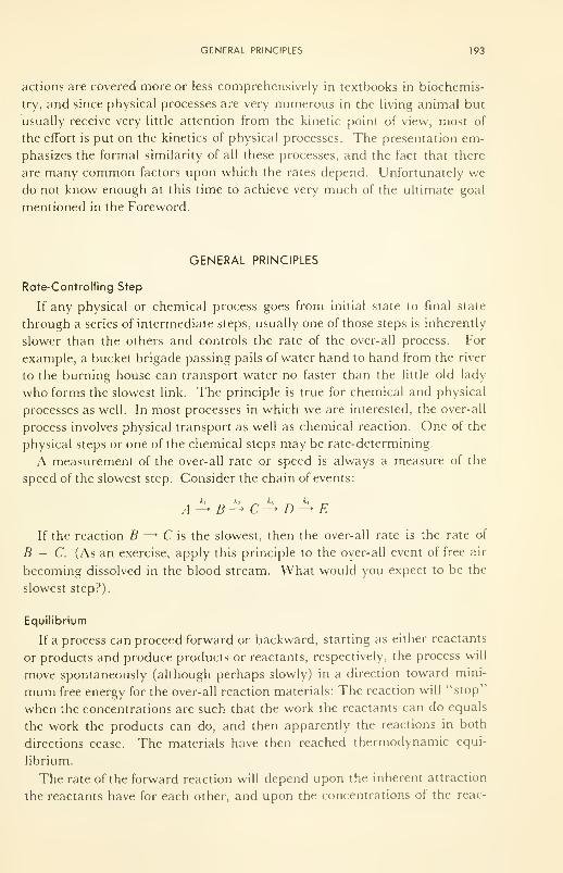
GENERAL PRINCIPLES 193
actions are covered more or less comprehensively in textbooks in biochemis-
try, and since physical processes are very numerous in the living animal but
usually receive very little attention from the kinetic point of view, most of
the effort is put on the kinetics of physical processes. The presentation em-
phasizes the formal similarity of all these processes, and the fact that there
are many common factors upon which the rates depend. Unfortunately wedo not know enough at this time to achieve very much of the ultimate goal
mentioned in the Foreword.
GENERAL PRINCIPLES
Rate-Controlling Step
If any physical or chemical process goes from initial state to final state
through a series of intermediate steps, usually one of those steps is inherently
slower than the others and controls the rate of the over-all process. For
example, a bucket brigade passing pails of water hand to hand from the river
to the burning house can transport water no faster than the little old lady
who forms the slowest link. The principle is true for chemical and physical
processes as well. In most processes in which we are interested, the over-all
process involves physical transport as well as chemical reaction. One of the
physical steps or one of the chemical steps may be rate-determining.
A measurement of the over-all rate or speed is always a measure of the
speed of the slowest step. Consider the chain of events:
If the reaction B —* C is the slowest, then the over-all rate is the rate of
B — C. (As an exercise, apply this principle to the over-all event of free air
becoming dissolved in the blood stream. What would you expect to be the
slowest step?).
Equilibrium
If a process can proceed forward or backward, starting as either reactants
or products and produce products or reactants, respectively, the process will
move spontaneously (although perhaps slowly) in a direction toward mini-
mum free energy for the over-all reaction materials: The reaction will "stop"
when the concentrations are such that the work the reactants can do equals
the work the products can do, and then apparently the reactions in both
directions cease. The materials have then reached thermodynamic equi-
librium.
The rate of the forward reaction will depend upon the inherent attraction
the reactants have for each other, and upon the concentrations of the reac-
194 SPEEDS OF SOME PROCESSES IN BIOLOGICAL SYSTEMS
tants. The same is true of the reverse reaction. Thus, if
aA + bB^ cC + dD
where k]
is the measure of inherent attraction A and B have for each other,
the over-all rate of reaction in the forward direction of a moles of A with
b moles of B (i.e., i\ = -d[A]/dt, or -d[B]/dt, where[ ] denotes concen-
tration), is:
,, = k,[A][A] ••• x [B][B] ••• = k,[A]"[Bf
Similarly
v2= k
2 [C]'[DY
This first principle, that of mass action in reaction kinetics, was demon-
strated quantitatively by Wilhelmy in 1850.
At equilibrium the over-all reaction ceases. Therefore i 1
,= v
2at equi-
librium:
k.imBY = k2[C\[DY
[CYiDY =kL = K
[A]"[BY " k2
'
eq
where K is the equilibrium constant. This form of the Law of Mass Action
was stated thus by Guldberg and Waage in 1863. For any reaction
-AF° = RT\n Keq , which states that the free energy change per mole (eg.,
refer to sucrose oxidation) is a measure of the position of equilibrium.
Steady State
Consider again the consecutive process discussed above and consider
specifically the case in which the supply of A is unlimited, so that the con-
centration of A, [A], never changes. If£, > k2 , A will be converted into B
faster than B will be removed into C, and B will accumulate. Since the rate
of the reaction B — C is
v2= k
2[B]
as we saw above, as B accumulates, v2increases until it reaches the value
of v{
. At this point B will have reached its steady-state concentration be-
cause the concentration B neither increases nor decreases further. The same
is true of the other steps.
In the steady-state then
»1 = V2 = V
i = V4
or
kt[A] = k
2[B) = k,[C] = k
4[D]
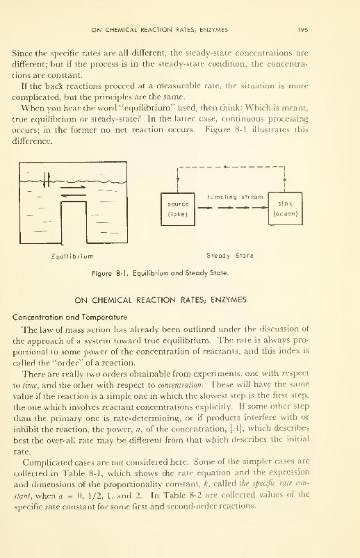
ON CHEMICAL REACTION RATES; ENZYMES 195
Since the specific rates are all different, the steady-state concentrations are
different; but if the process is in the steady-state condition, the concentra-
tions are constant.
If the back reactions proceed at a measurable rate, the situation is more
complicated, but the principles are the same.
When you hear the word "equilibrium" used, then think: Which is meant,
true equilibrium or steady-state? In the latter case, continuous processing
occurs; in the former no net reaction occurs. Figure 8-1 illustrates this
difference.
source
(lake)
tumbling stream
Equilibrium Steady State
Figure 8-1. Equilibrium and Steady State.
ON CHEMICAL REACTION RATES; ENZYMES
Concentration and Temperature
The law of mass action has already been outlined under the discussion of
the approach of a system toward true equilibrium. The rate is always pro-
portional to some power of the concentration of reactants, and this index is
called the "order" of a reaction.
There are really two orders obtainable from experiments, one with respect
to time, and the other with respect to concentration. These will have the same
value if the reaction is a simple one in which the slowest step is the first step,
the one which involves reactant concentrations explicitly. If some other step
than the primary one is rate-determining, or if products interfere with or
inhibit the reaction, the power, a, of the concentration, [A], which describes
best the over-all rate may be different from that which describes the initial
rate.
Complicated cases are not considered here. Some of the simpler cases are
collected in Table 8-1, which shows the rate equation and the expression
and dimensions of the proportionality constant, k, called the specific rate con-
stant, when a = 0, 1/2, 1, and 2. In Table 8-2 are collected values of the
specific rate constant for some first and second-order reactions.
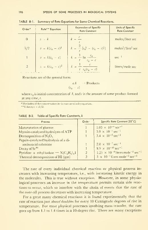
196 SPEEDS OF SOME PROCESSES IN BIOLOGICAL SYSTEMS
TABLE 8- 1 . Summary of Rate Equations for Some Chemical Reactions.
Order*
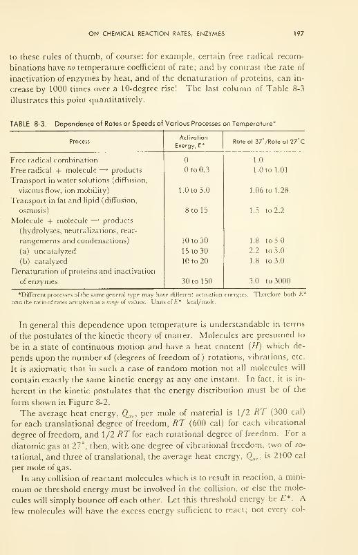
ON CHEMICAL REACTION RATES; ENZYMES 197
to these rules of thumb, of course: for example, certain free radical recom-
binations have no temperature coefficient of rate; and by contrast the rate of
inactivation of enzymes by heat, and of the denaturation of proteins, can in-
crease by 1000 times over a 10-degree rise! The last column of Table 8-3
illustrates this point quantitatively.
TABLE 8-3. Dependence of Rates or Speeds of Various Processes
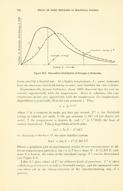
198 SPEEDS OF SOME PROCESSES IN BIOLOGICAL SYSTEMS
threshold energy E
Energy E
Figure 8-2. Maxwellian Distribution of Energies in Molecules.
lision need be a fruitful one. At a higher temperature, T2 , more molecules
have the necessary threshold energy to react, and therefore the rate is faster.
Experimentally, Svante Arrhenius, about 1889, observed that the rate in-
creased exponentially with the temperature. Since in solutions, the con-
centrations do not vary appreciably with the temperature, the temperature-
dependence is practically all in the rate constant, k. Thus
k = Ae-E*'RT
where A is a constant in moles per liter per second, E* is the threshold
energy in calories per mole, R the gas constant (1.987 cal per degree per
mole), T the temperature in degrees K, and "e" is 2.71828, the base of
natural logarithms. Taking logarithms of both sides
In A = In A - E*/RT
or, changing to the base 10, the more familiar system:
log k = log A - E*/2.303RT
Hence a graphical plot of experimental results of rate measurements at dif-
ferent temperatures plotted as log v vs \/T has a slope of -E*/2303 R; and,
since R is known, the value of the threshold energy, E*, can be determined
(see Figure 8-3).
Table 8-3 gives values of E* for different kinds of processes. E* is often
called energy of activation as well as threshold energy, and the measured value
can often aid in the characterization of the rate-determining step of a
process.
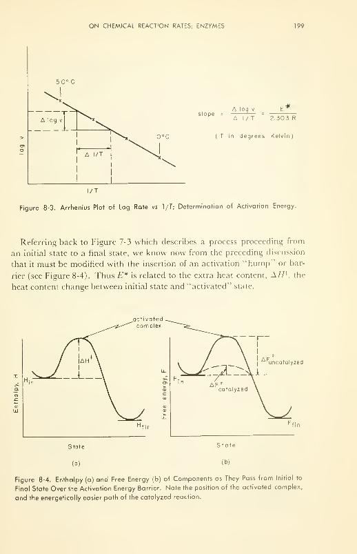
ON CHEMICAL REACTION RATES, ENZYMES 199
slopeA log v
A l/T 2.303 R
( T in degrees Kelvin )
Figure 8-3. Arrhenius Plot of Log Rate vs l/T; Determination of Activation Energy.
Referring back to Figure 7-3 which describes a process proceeding from
an initial state to a final state, we know now from the preceding discussion
that it must be modified with the insertion of an activation "hump" or bar-
rier (see Figure 8-4). Thus E* is related to the extra heat content, A// }, the
heat content change between initial state and "activated" state.
activatedcomplex
cUJ
State
(a)
|uncatalyzed
State
(b)
Figure 8-4. Enthalpy (a) and Free Energy (b) of Components as They Pass from Initial to
Final State Over the Activation Energy Barrier. Note the position of the activated complex,
and the energetically easier path of the catalyzed reaction.
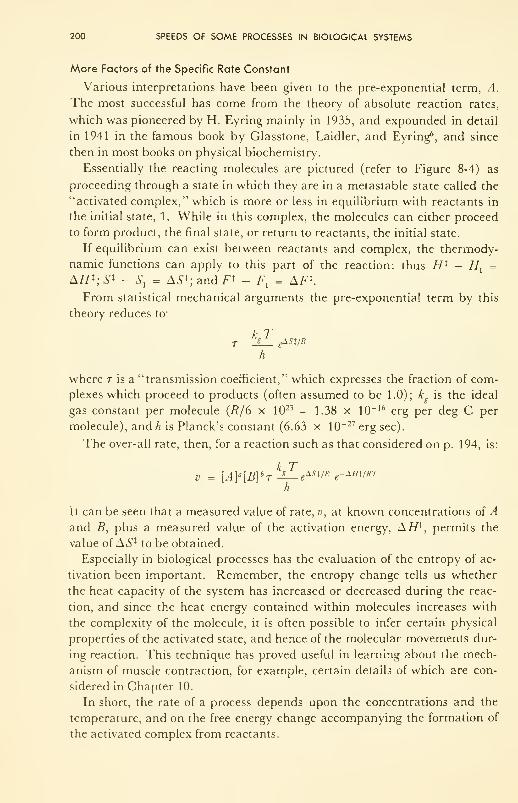
200 SPEEDS OF SOME PROCESSES IN BIOLOGICAL SYSTEMS
More Factors of the Specific Rate Constant
Various interpretations have been given to the pre-exponential term, A.
The most successful has come from the theory of absolute reaction rates,
which was pioneered by H. Eyring mainly in 1935, and expounded in detail
in 1941 in the famous book by Glasstone, Laidler, and Eyring6 , and since
then in most books on physical biochemistry.
Essentially the reacting molecules are pictured (refer to Figure 8-4) as
proceeding through a state in which they are in a metastable state called the
"activated complex," which is more or less in equilibrium with reactants in
the initial state, 1. While in this complex, the molecules can either proceed
to form product, the final state, or return to reactants, the initial state.
If equilibrium can exist between reactants and complex, the thermody-
namic functions can apply to this part of the reaction: thus H i - //, =
A//*; S x - Sx= AS*; and F* - F, = AF*.
From statistical mechanical arguments the pre-exponential term by this
theory reduces to"
k T
h
where t is a "transmission coefficient," which expresses the fraction of com-
plexes which proceed to products (often assumed to be 1.0); kg
is the ideal
gas constant per molecule (R/6 x 1023 = 1.38 x 10" 16 erg per deg C per
molecule), and h is Planck's constant (6.63 x 10~ 27 erg sec).
The over-all rate, then, for a reaction such as that considered on p. 194, is:
v = [A)a [B] bT^— g*sv* e-W/xrh
It can be seen that a measured value of rate, v, at known concentrations of A
and B, plus a measured value of the activation energy, AH*, permits the
value of AS X to be obtained.
Especially in biological processes has the evaluation of the entropy of ac-
tivation been important. Remember, the entropy change tells us whether
the heat capacity of the system has increased or decreased during the reac-
tion, and since the heat energy contained within molecules increases with
the complexity of the molecule, it is often possible to infer certain physical
properties of the activated state, and hence of the molecular movements dur-
ing reaction. This technique has proved useful in learning about the mech-
anism of muscle contraction, for example, certain details of which are con-
sidered in Chapter 10.
In short, the rate of a process depends upon the concentrations and the
temperature, and on the free energy change accompanying the formation of
the activated complex from reactants.
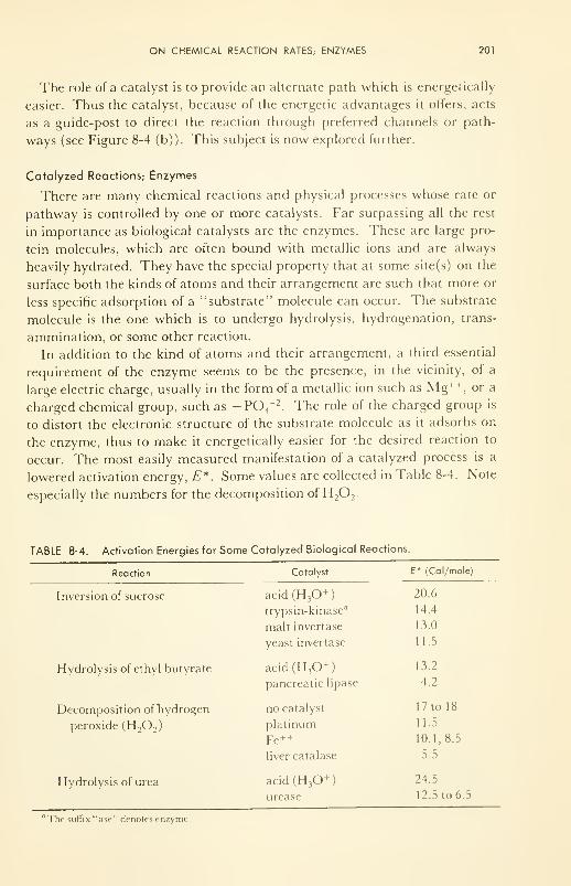
ON CHEMICAL REACTION RATES; ENZYMES 201
The role of a catalyst is to provide an alternate path which is energetically
easier. Thus the catalyst, because of the energetic advantages it offers, acts
as a guide-post to direct the reaction through preferred channels or path-
ways (see Figure 8-4 (b)). This subject is now explored further.
Catalyzed Reactions; Enzymes
There are many chemical reactions and physical processes whose rate or
pathway is controlled by one or more catalysts. Far surpassing all the rest
in importance as biological catalysts are the enzymes. These are large pro-
tein molecules, which are often bound with metallic ions and are always
heavily hydrated. They have the special property that at some site(s) on the
surface both the kinds of atoms and their arrangement are such that more or
less specific adsorption of a "substrate" molecule can occur. The substrate
molecule is the one which is to undergo hydrolysis, hydrogenation, trans-
ammination, or some other reaction.
In addition to the kind of atoms and their arrangement, a third essential
requirement of the enzyme seems to be the presence, in the vicinity, of a
large electric charge, usually in the form of a metallic ion such as Mg++,or a
charged chemical group, such as -PCV 2. The role of the charged group is
to distort the electronic structure of the substrate molecule as it adsorbs on
the enzyme, thus to make it energetically easier for the desired reaction to
occur. The most easily measured manifestation of a catalyzed process is a
lowered activation energy, E*. Some values are collected in Table 8-4. Note
especially the numbers for the decomposition of H2 2
.
TABLE 8-4. Activation Enerc
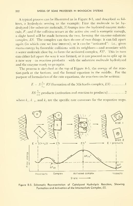
202 SPEEDS OF SOME PROCESSES IN BIOLOGICAL SYSTEMS
A typical process can be illustrated as in Figure 8-5, and described as fol-
lows, a hydrolysis serving as the example: First the molecule to be hy-
drolyzed (the substrate molecule, S) bumps into the hydrated enzyme mole-
cule, E; and if the collision occurs at the active site and is energetic enough,
a slight bond will be made between the two, forming the enzyme-substrate
complex, ES. The complex can then do one of two things: it can fall apart
again (in which case we lose interest); or it can be "activated"— i.e., given
excess energy by favorable collisions with its neighbors—and associate with
a water molecule close by, to form the activated complex, ESK This in turn
can either fall apart the way it was formed, or it can proceed on to split up in
a new way— as reaction products— with the substrate molecule hydrolyzed
and the enzyme ready to go again.
The process is sketched at the top of Figure 8-5, the energy of the reac-
tion path at the bottom, and the formal equation in the middle. For the
purpose of formulation of the rate equations, the reaction can be written:
E + S^ ES (formation of the M jchaelis complex, ES) 1
*-i
ES ~^> products (activation and reaction to products) 2
where kt
, k_ x , and k2are the specific rate constants for the respective steps.
prod. 2
*• E + products
Reoctonts Complex Activated complex
State —
Products
Figure 8-5. Schematic Representation of Catalyzed Hydrolysis Reaction, Showing
Formation and Activation of the Intermediate Complex, ES.
ON CHEMICAL REACTION RATES; ENZYMES 203
Now this reaction can be very complicated, but for our purpose it will suf-
fice to consider one (the simplest) set of conditions, and examine how the
rate varies with changes in either enzyme or substrate concentration. First
we assume that v_}
is much faster than v2 , and therefore that reaction 1 is
essentially at equilibrium, or that K = kjk,
.
The rate is then given by
v2 = k2[ES]
where the square bracket again denotes concentration. Now the only prob-
lem remaining is to compute [ES] from the equilibrium constant. If the
initial concentrations of enzyme and substrate, respectively, are [£"]n and
[S] , the concentrations of free E and S are given by
[E] = [E] - [ES]
and
[S] = [S] - [ES]
and therefore the equilibrium constant is given by
K = \mCq
([£]„ - [ES]) ([S] - [ES])
This becomes simpler if only the usual case is considered, namely that in
which the substrate concentration is much higher than the enzyme concen-
tration; for under this condition only a small fraction of the substrate mole-
cules will ever be tied up as complexes ES because there are so few enzyme
molecules with which the substrate can form a complex. Hence
[S] - [ES] - [S}
Rearrangement gives
[ES] =Keq [E] [S]
()[E] Q [S]
i + /ysio Km + [s]
if Km is defined as 1/A~,f]
. This holds for any value of [S] at any time.
Therefore the rate, v2 , of the enzyme-catalyzed reaction (proportional to
the concentration of complexes) is:
k2[E] [S]v2= -d[S]/dt =
Km + [S]
This is the rather celebrated Michaelis-Menten Equation, and describes the
rate as a function of initial substrate concentration under the particular con-
ditions we assumed. A plot of v2vs [5"] is shown in Figure 8-6 for both high
and low enzyme concentrations. The expression says that: (1) the rate is
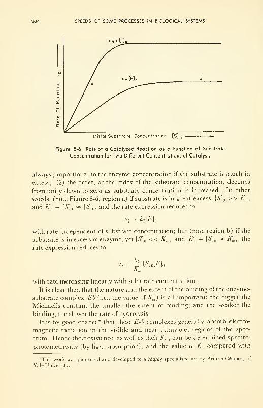
204 SPEEDS OF SOME PROCESSES IN BIOLOGICAL SYSTEMS
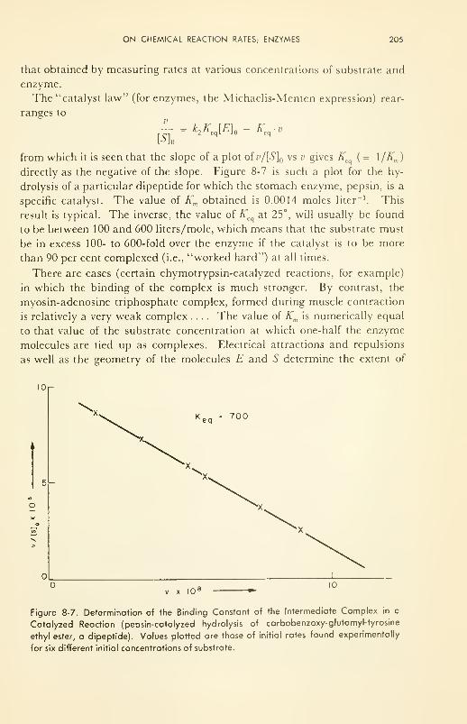
ON CHEMICAL REACTION RATES; ENZYMES 205
that obtained by measuring rates at various concentrations of substrate and
enzyme.
The "catalyst law" (for enzymes, the Michaelis-Menten expression) rear-
ranges to
[S\W£]„ - Keq
from which it is seen that the slope of a plot ofv/[S] vs v gives K (= 1/A"m )
directly as the negative of the slope. Figure 8-7 is such a plot for the hy-
drolysis of a particular dipeptide for which the stomach enzyme, pepsin, is a
specific catalyst. The value of Km obtained is 0.0014 moles liter-1
. This
result is typical. The inverse, the value of K at 25°, will usually be found
to be between 100 and 600 liters/mole, which means that the substrate must
be in excess 100- to 600-fold over the enzyme if the catalyst is to be more
than 90 per cent complexed (i.e., "worked hard") at all times.
There are cases (certain chymotrypsin-catalyzed reactions, for example)
in which the binding of the complex is much stronger. By contrast, the
myosin-adenosine triphosphate complex, formed during muscle contraction
is relatively a very weak complex .... The value of Km is numerically equal
to that value of the substrate concentration at which one-half the enzyme
molecules are tied up as complexes. Electrical attractions and repulsions
as well as the geometry of the molecules E and S determine the extent of
K eq = 700
Figure 8-7. Determination of the Binding Constant of the Intermediate Complex in a
Catalyzed Reaction (pepsin-catalyzed hydrolysis of carbobenzoxy-glutamyl-tyrosine
ethyl ester, a dipeptide). Values plotted are those of initial rates found experimentally
for six different initial concentrations of substrate.
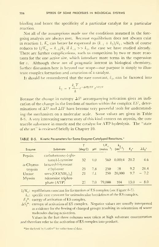
206 SPEEDS OF SOME PROCESSES IN BIOLOGICAL SYSTEMS
binding and hence the specificity of a particular catalyst for a particular
reaction.
Not all of the assumptions made nor the conditions assumed in the fore-
going analysis are always met. Because equilibrium does not always exist
in reaction 1, Km can better be expressed as (£_, + k2)/k
], which of course
reduces to l/K = k_]
/k]
if k_x>> k
2 , the case we have studied already.
There are further complications, such as competition by two or more reac-
tants for the one active site, which introduce more terms in the expression
for v. Although these are of pragmatic interest in biological chemistry,
further discussion here is beyond our scope—our purpose is simply to illus-
trate complex formation and saturation of a catalyst.
It should be remembered that the rate constant, k2 , can be factored into
k T
h
Because the change in entropy AS 1 accompanying activation gives an indi-
cation of the change in the freedom of motion within the complex ES 1,deter-
minations of AS 1 and A//1 have become very powerful tools for understand-
ing the mechanism on a molecular scale. Some values are given in Table
8-5. A very interesting success story of this kind centers on myosin, the con-
tractile substance in muscle and the catalyst for ATP hydrolysis. The "state
of the art1
' is reviewed briefly in Chapter 10.
TABLE 8-5. Kinetic Parameters for Some Enzyme-Catalyzed Reactions.*
Enzyme Substrate (deg C) pH (moles ') (sec]
) E2* ±S
2*
Pepsin carbobenzoxy-1-glu-
tamyl-1-tyrosine 32 4.0 560 0.0014 20.2 4.6
a-Chymo- benzoyl-1-tyrosine
trypsin ethyl ester 25 7.8 250 78 9.2 -21.4
Urease urea [CO(NH2 ) 2 ]
21 7.1 250 20,000 9.7 - 7.2
Myosin adenosine triphos-
phate (ATP) 25 7.0 79,000 104 13.0 - 8.0
\/Km . equilibrium constant for formation of ES complex (see Figure 8-5).
k2
: specific rate constant for unimolecular breakdown of the ES complex.
£,* : energy of activation of ES complex.
AS2*: entropy of activation of ES complex. Negative values are usually interpreted
as evidence for the freeing of charged groups resulting in orientation of water
molecules during activation.
Values in the last three columns were taken at high substrate concentration
and therefore refer to the activation of ES complex into product.
*See the book bv Laidler15 for collections of data.
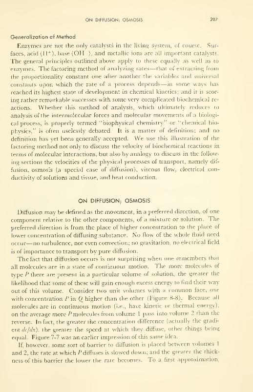
ON DIFFUSION; OSMOSIS 207
Generalization of Method
Enzymes are not the only catalysts in the living system, of course. Sur-
faces, acid (H +), base (OH ), and metallic ions are all important catalysts.
The general principles outlined above apply to these equally as well as to
enzymes. The factoring method of analyzing rates— that of extracting from
the proportionality constant one after another the variables and universal
constants upon which the rate of a process depends— in some ways has
reached its highest state of development in chemical kinetics; and it is scor-
ing rather remarkable successes with some very complicated biochemical re-
actions. Whether this method of analysis, which ultimately reduces to
analysis of the intermolecular forces and molecular movements of a biologi-
cal process, is properly termed "biophysical chemistry'1
or "chemical bio-
physics," is often uselessly debated. It is a matter of definition; and no
definition has yet been generally accepted. We use this illustration of the
factoring method not only to discuss the velocity of biochemical reactions in
terms of molecular interactions, but also by analogy to discuss in the follow-
ing sections the velocities of the physical processes of transport, namely dif-
fusion, osmosis (a special case of diffusion), viscous flow, electrical con-
ductivity of solutions and tissue, and heat conduction.
ON DIFFUSION; OSMOSIS
Diffusion may be defined as the movement, in a preferred direction, of one
component relative to the other components, of a mixture or solution. The
preferred direction is from the place of higher concentration to the place of
lower concentration of diffusing substance. No flow of the whole fluid need
occur—no turbulence, nor even convection; no gravitation, no electrical field
is of importance to transport by pure diffusion.
The fact that diffusion occurs is not surprising when one remembers that
all molecules are in a state of continuous motion. The more molecules of
type P there are present in a particular volume of solution, the greater the
likelihood that some of these will gain enough excess energy to find their way
out of this volume. Consider two unit volumes with a common face, one
with concentration P in Q higher than the other (Figure 8-8). Because all
molecules are in continuous motion (i.e., have kinetic or thermal energy),
on the average more P molecules from volume 1 pass into volume 2 than the
reverse. In fact, the greater the concentration difference (actually the gradi-
ent dc/dx), the greater the speed at which they diffuse, other things being
equal. Figure 7-7 was an earlier impression of this same idea.
If, however, some sort of barrier to diffusion is placed between volumes 1
and 2, the rate at which P diffuses is slowed down; and the greater the thick-
ness of this barrier the lower the rate becomes. To a first approximation,
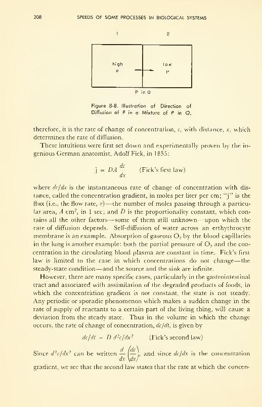
208 SPEEDS OF SOME PROCESSES IN BIOLOGICAL SYSTEMS
high
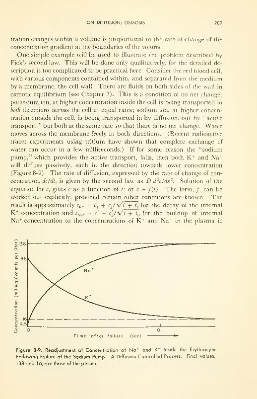
ON DIFFUSION; OSMOSIS 209
tration changes within a volume is proportional to the rate of change of the
concentration gradient at the boundaries of the volume.
One simple example will be used to illustrate the problem described by
Fick's second law. This will be done only qualitatively, for the detailed de-
scription is too complicated to be practical here. Consider the red blood cell,
with various components contained within, and separated from the mediumby a membrane, the cell wall. There are fluids on both sides of the wall in
osmotic equilibrium (see Chapter 2). This is a condition of no net change:
potassium ion, at higher concentration inside the cell is being transported in
both directions across the cell at equal rates; sodium ion, at higher concen-
tration outside the cell, is being transported in by diffusion, out by "active
transport," but both at the same rate so that there is no net change. Water
moves across the membrane freely in both directions. (Recent radioactive
tracer experiments using tritium have shown that complete exchange of
water can occur in a few milliseconds.) If for some reason the "sodium
pump," which provides the active transport, fails, then both K + and Na +
will diffuse passively, each in the direction towards lower concentration
(Figure 8-9). The rate of diffusion, expressed by the rate of change of con-
centration, dc/dt, is given by the second law as D d 2c/dx 2. Solution of the
equation for c, gives c as a function of t; or c = /(/). The form, /, can be
worked out explicitly, provided certain other conditions are known. Theresult is approximately rK+ = c
{+ c
2/y/T+t for the decay of the internal
K + concentration and rNa+ = c[ — c'2/y/t + t for the buildup of internal
Na + concentration to the concentrations of K + and Na + in the plasma in
Time after failure (sec)
Figure 8-9. Readjustment of Concentration of Na + and K + Inside the Erythrocyte
Following Failure of the Sodium Pump—A Diffusion-Controlled Process. Final values,
1 38 and 1 6, are those of the plasma.
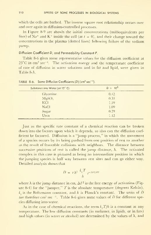
210 SPEEDS OF SOME PROCESSES IN BIOLOGICAL SYSTEMS
which the cells are bathed. The inverse square root relationship occurs over
and over again in diffusion-controlled processes.
In Figure 8-9 are shown the initial concentrations (milliequivalents per
liter) of Na + and K+ inside the cell (at / = 0), and their change toward the
concentrations in the plasma (dotted lines) following failure of the sodiumpump.
Diffusion Coefficient D, and Permeability Constant P.
Table 8-6 gives some representative values for the diffusion coefficient at
25°C in cm 2 sec-1
. The activation energy and the temperature coefficient
of rate of diffusion in water solutions and in fat and lipid, were given in
Table 8-3.
TABLE 8-6. Some Diffusion Coefficients (D) (cm2sec
]
).
Substance into Water (at 12° C) D x 105
Glycerine 0.42
MgS04 0.35
KC1 1.59
NaCl 1.09
Sugar 0.29
Urea 1.12
Just as the specific rate constant of a chemical reaction can be broken
down into the factors upon which it depends, so also can the diffusion coef-
ficient be factored. Diffusion is a "jump process," in which the movement
of a species occurs by its being pushed from one position of rest to another
as the result of favorable collisions with neighbors. The distance between
successive positions of rest is called the jump distance, A. The activated
complex in this case is pictured as being an intermediate position in which
the jumping species is half way between rest sites and can go either way.
Detailed analysis shows that
D = t\ 2 JiL e-*Ft/RT
h
where A is the jump distance in cm, AF* is the free energy of activation (Fig-
ure 8-4) for the "jumper," 7" is the absolute temperature (degrees Kelvin),
k is the Boltzmann constant, and h is Planck's constant. The units of Dare therefore cm2 sec "'. Table 8-6 gives some values of D for different spe-
cies diffusing into water.
As in the case of chemical reactions, the term kgT/h is a constant at any
temperature. The low diffusion constants (in molasses, in lipids, or in fats)
and high values (in water or alcohol) are determined by the values of X, and
ON DIFFUSION; OSMOSIS 211
by AF\ the energy of binding within the shroud of neighboring molecules
through which the jumping species must penetrate if it is to move success-
fully to the next position of rest.
Two innovations have been introduced into discussions of diffusion in
recent years, one for theoretical reasons and the other for practical reasons.
Firstly, it is more proper to consider activities (effective concentrations) than
measured concentrations, and more proper still to consider as the "force,"
the gradient of the chemical potential which drives the diffusion process; and
therefore dc/dx is replaced by dn/dx, in the more esoteric discussions, if not
in practice.
Secondly, the thickness of the interface, at a cell wall for instance, is really
a matter of definition rather than of position of chemicals. Who can say
where the water phase stops and the heavily hydrated protein of the wall
begins? Therefore dc/dx is hard to measure for living membranes, and re-
course is made to a phenomenological trick: dx is taken into the diffusion
constant, and the rate of flow is expressed as the difference between the flows
in the two directions through the membrane. Thus
j =(P,ACl -(P 2Ac
2
where 1 and 2 represent diffusions in the forward and back reactions, and
c, and c2represent concentrations on the two sides; the (P's then have units
cm sec-1
(velocity) and are called permeability constants. A few of these are
collected in Table 8-7 for monovalent cations penetrating through living
membranes. These permeability constants can be compared with values de-
termined for synthetic interfaces also given in the table.
TABLE 8-7. Some Permeability Constants ( § ) for Synthetic and Biological Membranes.*
, _._ . Permeability Constant x 10Intertace Diffusion ' _i.
(cm sec )
K+ into erythrocyte of: man 5.0
dog 1 .0
rat 10
KC1, KBr, KI into nitrobenzene 0.007, 0.075, 1 .4
Na + into erythrocyte of: rabbit 3.0
dog 0.5
Na + through frog skin 5.0
Na + (as iodide) into nitrobenzene 0.2
Alcohols into erythrocyte 10,000 to 100,000
Water into erythrocyte ~ 1 0,000
Collected by J. T. Davies, J. Rhys. Coll. Chem., 54,185(1950). See also Ref. 17.
For ionic flow the values in the table can be transformed very easily into
electrical resistance units. Thus if the concentration of the salt at the mem-
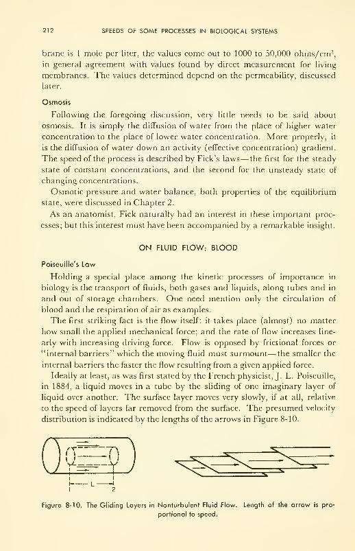
212 SPEEDS OF SOME PROCESSES IN BIOLOGICAL SYSTEMS
brane is 1 mole per liter, the values come out to 1000 to 50,000 ohms/cm2,
in general agreement with values found by direct measurement for living
membranes. The values determined depend on the permeability, discussed
later.
Osmosis
Following the foregoing discussion, very little needs to be said about
osmosis. It is simply the diffusion of water from the place of higher water
concentration to the place of lower water concentration. More properly, it
is the diffusion of water down an activity (effective concentration) gradient.
The speed of the process is described by Fick's laws—the first for the steady
state of constant concentrations, and the second for the unsteady state of
changing concentrations.
Osmotic pressure and water balance, both properties of the equilibrium
state, were discussed in Chapter 2.
As an anatomist, Fick naturally had an interest in these important proc-
esses; but this interest must have been accompanied by a remarkable insight.
ON FLUID FLOW; BLOOD
Poiseuille's Law
Holding a special place among the kinetic processes of importance in
biology is the transport of fluids, both gases and liquids, along tubes and in
and out of storage chambers. One need mention only the circulation of
blood and the respiration of air as examples.
The first striking fact is the flow itself: it takes place (almost) no matter
how small the applied mechanical force; and the rate of flow increases line-
arly with increasing driving force. Flow is opposed by frictional forces or
"internal barriers" which the moving fluid must surmount— the smaller the
internal barriers the faster the flow resulting from a given applied force.
Ideally at least, as was first stated by the French physicist, J. L. Poiseuille,
in 1884, a liquid moves in a tube by the sliding of one imaginary layer of
liquid over another. The surface layer moves very slowly, if at all, relative
to the speed of layers far removed from the surface. The presumed velocity
distribution is indicated by the lengths of the arrows in Figure 8-10.
Figure 8-10. The Gliding Layers in Nonturbulent Fluid Flow. Length of the arrow is pro-
portional to speed.
ON FLUID FLOW; BLOOD 213
If P, and P2 are the pressures measured at the points 1 and 2 in the tube,
and R is the distance from the center bore of the tube, the driving force is
given by
ttR 2 (Px
- P2 )
The frictional force on the layer at distance R from the center is propor-
tional to the area of the layer (2ttRI), and to the velocity difference between
the layer we are considering and its nearest neighbors; in the limit this is
dv/dR.
After the two forces have been equated, integration (or summing all veloc-
ities from that at the center of the tube to zero at the wall) gives
P — Pv = £j £2( r
2 _ R 2)
4/
where r is the radius of the tube. This expression gives the linear speed of
the layer which is R cm from the center. is the proportionality constant,
and is called thefluidity (the higher its value the higher the velocity).
The total volume of fluid flowing per second through the tube is calculated
by summing all the elemental volumes, 2irRdR, for which v is expressed. The
result is the celebrated Poiseuille equation which' expresses rate of flow
(cc/sec) of liquid through a tube of radius r and length / under an applied
pressure difference of AP = P]
— P2
:
irr4
dVldt = AP cc/sec8/
If A P is given in dynes per cm 2, r and / in cm, and the speed of flow in cc per
sec, the fluidity, 0, must be cm per sec for a force gradient of 1 dyne per cm;
i.e., has the dimensions: — /—— . It is the velocity of flow of a fluid
sec/ cmunder a unit force gradient.
The case for gases is slightly more complicated because of the added fact
that the volume depends strongly upon the pressure and the temperature.
With the proper modifications the expression for rate of flow of gases
approximates:
Trr4
P. 2 - P 2
dV/dt = !
16/ - Pn
if P is the pressure at which the volume is measured.
Fluidity, 0, and Viscosity, rj
Table 8-8 gives values of the fluidities of various substances at different
temperatures. Of the liquids, ether is the most fluid one listed; glycerine at
0°C is the least fluid— indeed at 0°C it is almost a glass! The fluidity of
214 SPEEDS OF SOME PROCESSES IN BIOLOGICAL SYSTEMS
TABLE 8-8. Fluidities (0) poise 1
,or —cm /dyne
sec/ cm
Temp(°Q
ON FLUID FLOW; BLOOD 215
fluidity. Viscosity can be considered as the frictional force opposing the
a x j- • dynes /cm . . , . . . . „, ,How. Its dimensions are — /
, or dyne sec/cm , this unit is called thecm / sec
poise, after Poiseuille.
A very simple way to measure fluidity or viscosity is in the Ostwald vis-
cometer. The capillary pipette is filled to a mark with fluid, and measure-
ment made of the time it takes the fluid to run out of the pipette. This time
is divided into the time taken by water, or some other fluid, to drain at the
same temperature. The quotient is called the relative viscosity. A density cor-
rection is necessary if the driving force (gravitational) is to be equal in the
two cases.
Solutions or suspensions (of molecules or particles respectively) in water
usually increase the viscosity (decrease the fluidity). The fractional increase
is (r;, —ri )/rj , where the subscripts s and refer to solution and pure
water, respectively. But this value, often called 77', varies with the concentra-
tion. It is convenient, then, to measure the 77' at several concentrations, and
express each measurement in terms of unit concentration by dividing by the
concentration at which the measurement was made. This number is called
the specific viscosity. It is also concentration-dependent, because intermolec-
ular interactions are higher at higher concentrations. It is useful, then, to
extrapolate measurements of specific viscosity to infinite dilution (zero con-
centration), for this value is the value of that part of the viscosity due to the
suspension only, and unaffected by interactions which solute particles could
have on each other. This value is called intrinsic viscosity, usually symbolized
[77]. Values range from .02 for small- molecular-weight solutes to 20 for
macromolecules, and to much higher values for suspensions of living cells.
Turbulent Flow
Laminar flow will exist in most fluids at low rates of flow. When the flow
rate becomes high, the glide planes get off-track, and turbulence sets in.
Small whirlpools and eddy currents are initiated, and the fluidity drops
abruptly; therefore, if the rate of flow is to be maintained, higher driving
force must be applied and more energy must be expended. Unless some
result of particular value is derived from the turbulence (more rapid mixing
of chemical reactants at a reaction site, for example), it is obviously waste-
ful of energy. The circulatory system in man has certain features, such as
flexible walls lined with hydrated protein "hairs," which help direct the fluid
flow and damp out trends toward turbulence.
The Reynolds number, Re, a dimensionless parameter of fluid flow, is
defined as
Re = 20p vr
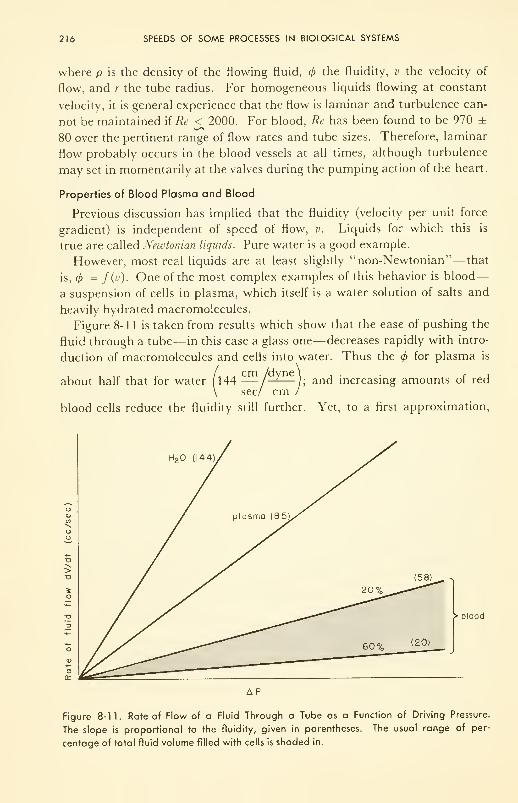
216 SPEEDS OF SOME PROCESSES IN BIOLOGICAL SYSTEMS
where p is the density of the flowing fluid, the fluidity, v the velocity of
flow, and r the tube radius. For homogeneous liquids flowing at constant
velocity, it is general experience that the flow is laminar and turbulence can-
not be maintained if Re < 2000. For blood, Re has been found to be 970 ±
80 over the pertinent range of flow rates and tube sizes. Therefore, laminar
flow probably occurs in the blood vessels at all times, although turbulence
may set in momentarily at the valves during the pumping action of the heart.
Properties of Blood Plasma and Blood
Previous discussion has implied that the fluidity (velocity per unit force
gradient) is independent of speed of flow, v. Liquids for which this is
true are called Newtonian liquids. Pure water is a good example.
However, most real liquids are at least slightly "non-Newtonian"— that
is, = f(v). One of the most complex examples of this behavior is blood
—
a suspension of cells in plasma, which itself is a water solution of salts and
heavily hydrated macromolecules.
Figure 8-11 is taken from results which show that the ease of pushing the
fluid through a tube— in this case a glass one— decreases rapidly with intro-
duction of macromolecules and cells into water. Thus the for plasma is
about half that for water [144 — /—
—
-); and increasing amounts of red
\ sec/ cm /
blood cells reduce the fluidity still further. Yet, to a first approximation,
> blood
AP
Figure 8-11. Rate of Flow of a Fluid Through a Tube as a Function of Driving Pressure.
The slope is proportional to the fluidity, given in parentheses. The usual range of per-
centage of total fluid volume filled with cells is shaded in.
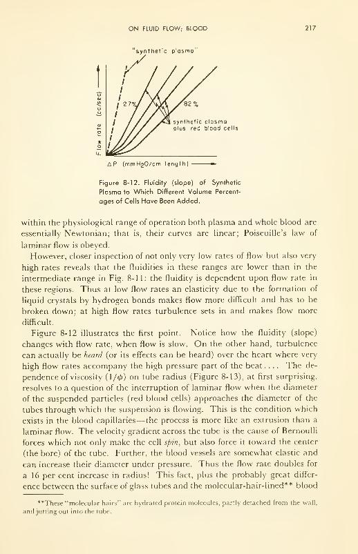
ON FLUID FLOW; BLOOD 217
'synthetic plasmo"
o
synthetic plasmoplus red blood cells
AP (mmHgO/cm length)
Figure 8-12. Fluidity (slope) of Synthetic
Plasma to Which Different Volume Percent-
ages of Cells Have Been Added.
within the physiological range of operation both plasma and whole blood are
essentially Newtonian; that is, their curves are linear; Poiseuille's law of
laminar flow is obeyed.
However, closer inspection of not only very low rates of flow but also very
high rates reveals that the fluidities in these ranges are lower than in the
intermediate range in Fig. 8-11: the fluidity is dependent upon flow rate in
these regions. Thus at low flow rates an elasticity due to the formation of
liquid crystals by hydrogen bonds makes flow more difficult and has to be
broken down; at high flow rates turbulence sets in and makes flow more
difficult.
Figure 8-12 illustrates the first point. Notice how the fluidity (slope)
changes with flow rate, when flow is slow. On the other hand, turbulence
can actually be heard (or its effects can be heard) over the heart where very
high flow rates accompany the high pressure part of the beat .... The de-
pendence of viscosity (1/0) on tube radius (Figure 8-13), at first surprising,
resolves to a question of the interruption of laminar flow when the diameter
of the suspended particles (red blood cells) approaches the diameter of the
tubes through which the suspension is flowing. This is the condition which
exists in the blood capillaries— the process is more like an extrusion than a
laminar flow. The velocity gradient across the tube is the cause of Bernoulli
forces which not only make the cell spin, but also force it toward the center
(the bore) of the tube. Further, the blood vessels are somewhat elastic and
can increase their diameter under pressure. Thus the flow rate doubles for
a 16 per cent increase in radius! This fact, plus the probably great differ-
ence between the surface of glass tubes and the molecular-hair-lined** blood
**These "molecular hairs" arc hydrated protein molecules, partly detached from the wall,
and jutting out into the tube.
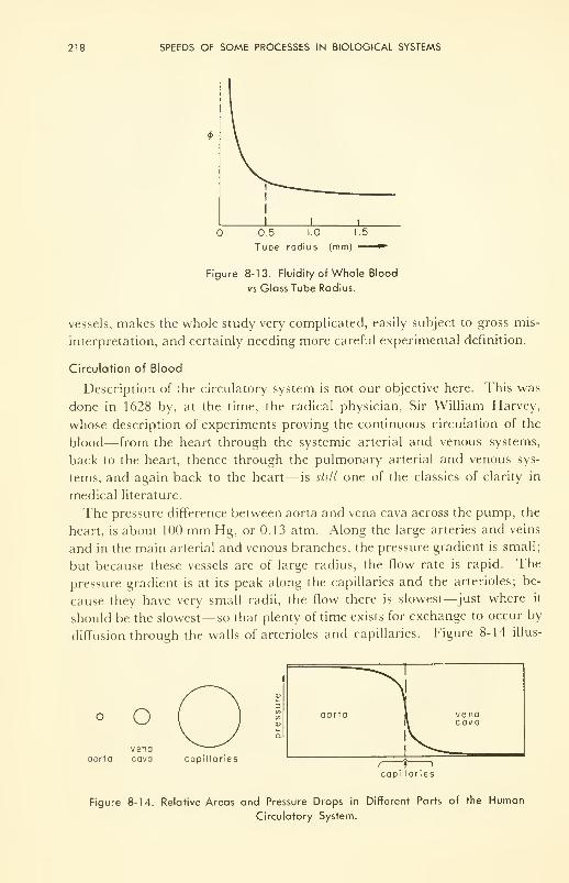
218 SPEEDS OF SOME PROCESSES IN BIOLOGICAL SYSTEMS
0.5 1.0 1.5
Tube radius (mm) »"
Figure 8-13. Fluidity of Whole Blood
vs Glass Tube Radius.
vessels, makes the whole study very complicated, easily subject to gross mis-
interpretation, and certainly needing more careful experimental definition.
Circulation of Blood
Description of the circulatory system is not our objective here. This was
done in 1628 by, at the time, the radical physician, Sir William Harvey,
whose description of experiments proving the continuous circulation of the
blood— from the heart through the systemic arterial and venous systems,
back to the heart, thence through the pulmonary arterial and venous sys-
tems, and again back to the heart— is still one of the classics of clarity in
medical literature.
The pressure difference between aorta and vena cava across the pump, the
heart, is about 100 mm Hg, or 0.13 atm. Along the large arteries and veins
and in the main arterial and venous branches, the pressure gradient is small;
but because these vessels are of large radius, the flow rate is rapid. The
pressure gradient is at its peak along the capillaries and the arterioles; be-
cause they have very small radii, the flow there is slowest—just where it
should be the slowest— so that plenty of time exists for exchange to occur by
diffusion through the walls of arterioles and capillaries. Figure 8-14 illus-
° Ovena
aorta cava capillaries
aorta 1 venaR cava
, 1 »
capillaries
Figure 8-14. Relative Areas and Pressure Drops in Different Parts of the Human
Circulatory System.
ON ELECTRICAL CONDUCTANCE; EEG AND EKG 219
trates this point, showing the pressure changes and the relative total cross-
sectional areas.
Two quantities can be measured, the flow rate (cc/sec), and the speed of
flow (cm/sec). Measurements in the aorta show that enough blood flows
past a flow-meter detector per second for one complete cycle to require 45
min. Insertion into the aorta of a bit of radioactive argon as an inert tracer,
and measurement of how long it takes for the tracer to complete the ciruit,
confirms this.
Speed is less easily measured. One method is by tracer. The ultrasonic
method (see Chapter 3) introduces no pathological changes, but needs
calibration.
ON ELECTRICAL CONDUCTANCE; EEG AND EKG
The next rate process to be considered in this chapter is the movement of
ions under the influence of an electrical field— in other words, the con-
ductance of solutions of salts in water. This subject is basic to an under-
standing of the gross current paths through the human body upon which are
based the techniques of electrocardiography (EKG) and electroencepalog-
raphy (EEG), and also basic to some of the transport processes driven by
membrane potentials which are of importance in nerve conduction and elec-
trical shock treatment.
Towards the latter part of the last century the big-three "solution" pio-
neers, Kohlrausch, Arrhenius, and Van't Hoff, showed that salts dissolve in
water as ions. These are electrically charged and free to move about at
random because of thermal energy, but subject to movement in a preferred
direction under the force of an electrical voltage gradient. Positive ions are
forced to the negative electrode, and negative ions to the positive electrode
by the electrical field. The speed of movement, or mobility (centimeters per
second under a voltage gradient of 1 v per cm) was understood quantitatively
by 1923 (the work of Debye and Hiickel, Onsager, and later others) as being
determined by the ease with which a charged ion, complete with "hangers-
on" such as electrically charged ions and water molecules, can slip from
hole to hole in the liquid. The process is very similar to diffusion, which
was described earlier. The difference is that ions are charged and move under a
voltage gradient, whereas the diffusing particle may or may not be charged and moves
under a concentration gradient . If a potential difference exists for any reason be-
tween two parts of an electrolyte, or is applied from the outside, ions move
and current flows— in other words, charge is transferred. Hence this is just
another transport process.
Ohm's Law Concerning Current
\{ n is the number of charge carriers per cc, w their average velocity under
the impressed voltage, and q the electrical charge carried by each, then the
220 SPEEDS OF SOME PROCESSES IN BIOLOGICAL SYSTEMS
amount of electrical charge passing per second through a plane of 1 cm2
area, called the current 'density , I, is
/ = nwq
If A' is the number of molecules per mole: n/N is the concentration, c, in
moles/cc; and qN is the charge per mole. The charge required to oxidize or
reduce 1 mole of anything is zF, where F is the charge (96,500 coulombs per
equivalent weight) required to oxidize or reduce 1 g equivalent weight, and
Z is the number of equivalent weights per mole (i.e., the number of electrons
transferred in the redox reaction). This is Faraday's law.
Summed (2) for all different ions, s, then
I = FZcs WsZ s
Since csis moles/cc and w
sis cm/sec, the current density has the dimensions:
coulombs per cm2 per sec, or amperes per cm 2.
Note that the current increases linearly with the concentration of charged
particles, with their speed, and with the charge they carry.
Specific Conductivity of a Solution, k
This is defined as the current which passes for an impressed voltage gradi-
ent,13 , of 1 v/cm. That is, k = //I). This is a form of Ohm's law. Nowalthough the dissociation of ions of a salt is usually complete, sometimes
there is association and always there is hydration, and hence often the ef-
fective "degree of dissociation," a, is less than 1. Introducing this concept
gives
FjLcsWl z s amp S
k = a or ohm ' cmTJ volt/cm
One more concept completes the picture. If the mobility, i± s , which is the
speed under an impressed voltage gradient of 1 v/cm, is defined as ws /\),
then
* = Fj^cs n s z s
a
Note that this expression describes the rate of the electrical transport
process. Thus k is the rate in amperes at which charge is transferred across 1-cm 2
area of electrolyte if the voltage gradient along the path is I v per cm. The value is
proportional to the concentration. The proportionality constant factors into
three constants (a, z, F) and the mobility, fi; and n is really the specific rate
constant for the process. Therefore n plays the same role for conductance as
does k for chemical reactions, D for diffusion, and 4> for fluid flow, respec-
cm / vtively. The units of /x are / . Values of the mobilities of small ions
sec/ cm
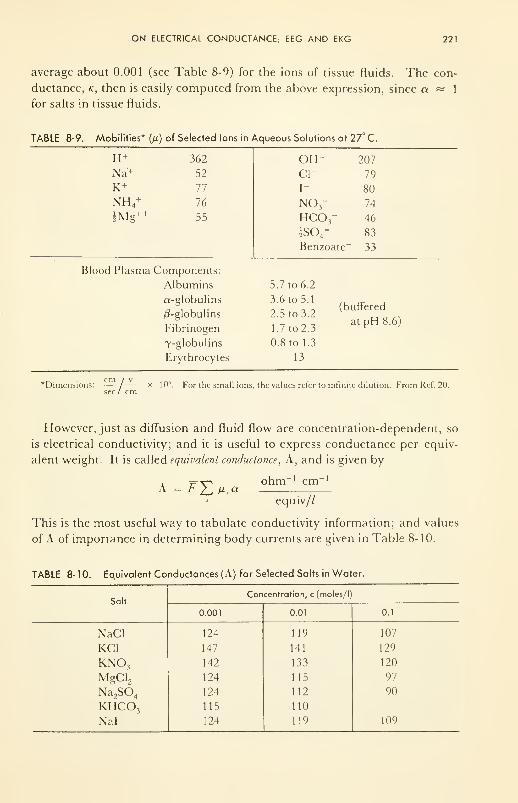
ON ELECTRICAL CONDUCTANCE; EEG AND EKG 221
average about 0.001 (see Table 8-9) for the ions of tissue fluids. The con-
ductance, k, then is easily computed from the above expression, since a « 1
for salts in tissue fluids.
TABLE 8-9. Mobilities* (n) of Selected Ions in Aqueous Solutions at 27° C.
IT
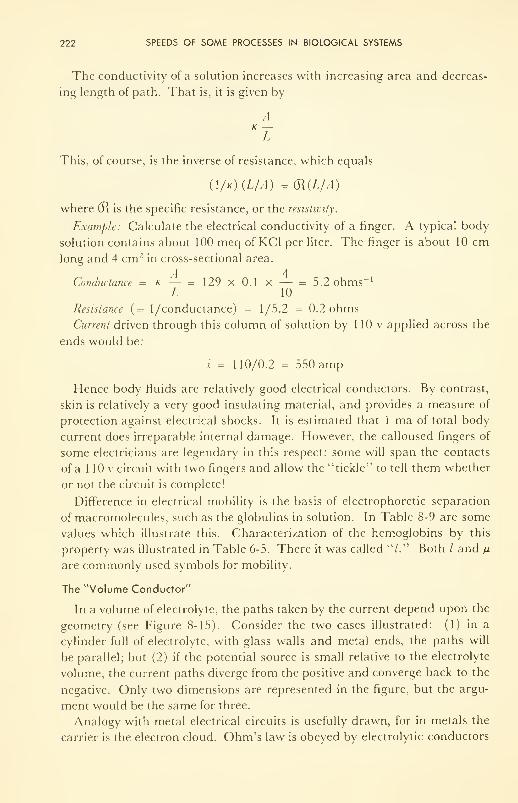
222 SPEEDS OF SOME PROCESSES IN BIOLOGICAL SYSTEMS
The conductivity of a solution increases with increasing area and decreas-
ing length of path. That is, it is given by
AK —L
This, of course, is the inverse of resistance, which equals
(\/k)(L/A) =(R(L/A)
where (R is the specific resistance, or the resistivity.
Example: Calculate the electrical conductivity of a finger. A typical body
solution contains about 100 meq of KC1 per liter. The finger is about 10 cmlong and 4 cm 2 in cross-sectional area.
A 4Conductance = k — = 129 x 0.1 x — = 5.2 ohms '
L 10
Resistance (= 1 /conductance) = 1/5.2 = 0.2 ohms
Current driven through this column of solution by 1 10 v applied across the
ends would be:
i = 110/0.2 = 550 amp
Hence body fluids are relatively good electrical conductors. By contrast,
skin is relatively a very good insulating material, and provides a measure of
protection against electrical shocks. It is estimated that 1 ma of total body
current does irreparable internal damage. However, the calloused fingers of
some electricians are legendary in this respect: some will span the contacts
of a 1 10 v circuit with two fingers and allow the ''tickle" to tell them whether
or not the circuit is complete!
Difference in electrical mobility is the basis of electrophoretic separation
of macromolecules, such as the globulins in solution. In Table 8-9 are some
values which illustrate this. Characterization of the hemoglobins by this
property was illustrated in Table 6-5. There it was called "/." Both / and \i
are commonly used symbols for mobility.
The "Volume Conductor"
In a volume of electrolyte, the paths taken by the current depend upon the
geometry (see Figure 8-15). Consider the two cases illustrated: (1) in a
cylinder full of electrolyte, with glass walls and metal ends, the paths will
be parallel; but (2) if the potential source is small relative to the electrolyte
volume, the current paths diverge from the positive and converge back to the
negative. Only two dimensions are represented in the figure, but the argu-
ment would be the same for three.
Analogy with metal electrical circuits is usefully drawn, for in metals the
carrier is the electron cloud. Ohm's law is obeyed by electrolytic conductors
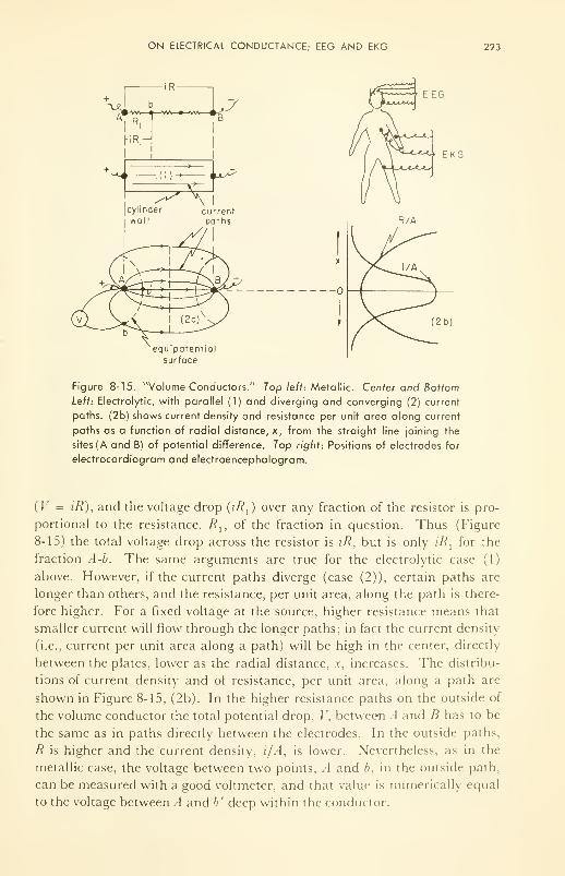
ON ELECTRICAL CONDUCTANCE; EEG AND EKG 223
EKG
equipotential
surface
Figure 8-15. "Volume Conductors." Top left: Metallic. Center and Bottom
Left: Electrolytic, with parallel (1) and diverging and converging (2) current
paths. (2b) shows current density and resistance per unit area along current
paths as a function of radial distance, x, from the straight line joining the
sites (A and B) of potential difference. Top right: Positions of electrodes for
electrocardiogram and electroencephalogram.
(V = z7?), and the voltage drop (z'/?,) over any fraction of the resistor is pro-
portional to the resistance, /?,, of the fraction in question. Thus (Figure
8-15) the total voltage drop across the resistor is iR, but is only z7?, for the
fraction A-b. The same arguments are true for the electrolytic case (1)
above. However, if the current paths diverge (case (2)), certain paths are
longer than others, and the resistance, per unit area, along the path is there-
fore higher. For a fixed voltage at the source, higher resistance means that
smaller current will flow through the longer paths; in fact the current density
(i.e., current per unit area along a path) will be high in the center, directly
between the plates, lower as the radial distance, x, increases. The distribu-
tions of current density and of resistance, per unit area, along a path are
shown in Figure 8-15, (2b). In the higher resistance paths on the outside of
the volume conductor the total potential drop, V, between A and B has to be
the same as in paths directly between the electrodes. In the outside paths,
R is higher and the current density, i/A, is lower. Nevertheless, as in the
metallic case, the voltage between two points, A and b, in the outside path,
can be measured with a good voltmeter, and that value is numerically equal
to the voltage between A and b' deep within the conductor.
224 SPEEDS OF SOME PROCESSES IN BIOLOGICAL SYSTEMS
The electrodes of the electrocardiograph (EKG) and electroencephalo-
graph (EEG) are placed on the outside of such a volume conductor, the
body, and measure potential differences between points in outside paths. If
the concentrations of salts remain constant throughout the body, as they
should in the steady-state, then any variations in the voltage measured
should reflect variations in the internal currents resulting from variations in
source voltage, E.
In biological systems the source of the potential difference between differ-
ent places or spots is invariably a concentration difference, whether uni-
ionic or bi-ionic. Concentration differences occur for two reasons: (1) ion
selectivity of membranes, and (2) continuous exchange with the mediumthrough which the distribution systems (blood and lymph systems) pass.
Membrane potentials cancel out over the whole system, because the im-
portant ion selectors are cell walls, which completely enclose and isolate a
volume. In the absence of disturbances then, concentration difference is
the source of the bioelectric potentials.
However, two major disturbances exist, both of which "irritate" the mem-branes of the cell wall, cause them to become permeable, and thereby reduce
the selectivity and permit mixing of otherwise separated salts. One is the
mechanical pressure variations transmitted through the blood stream by the
heart; the other is the electrical polarizing action of nerve. The former causes
a concentration change by the application of a mechanical force, the latter
by electrical interference with the membrane potentials of cells. Potential
variations with time, between electrodes on the skull, above different lobes
of the brain, give a precise record of the electrical action within the meas-
ured region; and electrodes placed on the torso and leg at spots where a
major artery runs close to the surface, give a reliable record of the pumping
action of the heart. Since any mechanical stimulus will cause momentaryirritation (and therefore potential variations), the measurements are always
made under controlled conditions when the electrical "noise" generated by
the involuntary muscles of the organs (and always present) is at a minimum.
ON HEAT CONDUCTION; 98.6° F: A CONSTANT?
Heat Production
The human body has a heat capacity, as does any other, measured as the
heat in calories required to raise 1 g 1°C. Also, the ambient (surrounding
temperature may vary widely— for example, from 95° F (35° C) down to
— 20° F (— 30° C). This lower value is 67 Centigrade degrees, or 1 19 Fahren-
heit degrees, below body temperature, and yet the body is able to maintain
within a small fraction of a degree the normal value of 37° C. Admittedly,
insulation-aids such as skin, clothing, and hair play a large part; and the
ON HEAT CONDUCTION; 98.6° F: A CONSTANT? 225
temperature in different parts of the body may vary. Especially on the outer
part of the skin and in the extremities (fingers, toes), the temperature is
lower than 37° C— i.e., at points farthest from the glycogen storehouse, the
liver, and where the area to volume quotient is high.
As we saw in Chapter 7, heat is produced by oxidation of glycogens and
by hydrolysis of fats and proteins. Under the reversible conditions of a
perfect energy-converting machine, no heat energy would be given off as
heat because AF is used for work and TAS is needed to establish the state
conditions of the products of reaction. However, the body "machine" is not
perfect, and in it conversions take place at efficiencies somewhat less than
the maximum thermodynamic efficiency. Thus,
AH = AF' + q' + Q
where AF' is the work extracted, Q. is the reversible, unavailable heat used
to bring the products to the reaction temperature, and q' is that part of
AF which could have been used to do work but which appears as heat be-
cause the "engine" could not extract the work reversibly. The degradation
reactions of fats and proteins are especially inefficient from this point of
view, and are thus good producers of "wasted" heat energy, q', which in
fact is not wasted but serves to maintain body heat-content or temperature
during cold weather. (Eskimos, for example, by design eat unprocessed
animal fat for its heat-producing effects.)
Heat Loss; Fourier's Law
Heat energy is lost from the body by several mechanisms, all of which
are simple physical transport processes or change-of-state processes. The
basic method is by conduction, for which the rate of loss, i\, is given by
K 4dT
vx
= KTA —ax
where A is the area exposed, T is temperature, and x is thickhess of the in-
sulation. If T is in degrees Fahrenheit, A in square feet, and thickness in
inches, the rate of heat loss is given in BTU per hour; and the proportional-
ity constant, KT , is given in BTU per hr per sq ft of area per ° F per in. of
thickness. Common values of KT for good insulating materials are: cork,
0.28; wood, 0.35; wool, 0.30; plaster, 0.48; fat, 0.33; skin, 0.30. Since
BTU Cal
hr ft2 °F/in. hr m 2 °C/cm
the conversion, if useful, is easy. Approximately 4 BTU = 1 Cal (or kcal).
Usually engineers use the units on the left side of the conversion equality,
and physiologists thpse on the right side.
226 SPEEDS OF SOME PROCESSES IN BIOLOGICAL SYSTEMS
The important effective-thickness term which determines dT/d.v, depends
upon the nature of the contact, whether skin-air, skin-water, skin-metal, etc.,
and also depends critically on the heat capacity and heat conductivity of the
materials of the contact. Thus the rate of heat loss into cold water is greater
than into cold air at the same temperature because of the higher heat capac-
ity of the water; while the rate of heat loss to steel at the same temperature
is greater because of the rate at which steel can conduct heat away.
Clothing increases the effective thickness and hence decreases the- tem-
perature gradient: so do hair, thickness of skin, and subcutaneous fat. Oneof the best insulators in the body is the dermis-epidermis combination,
whose effective thickness changes with the ambient temperature by virtue of
involuntary, lateral muscle movements which govern the depth of blood
capillaries carrying the heat energy to be thrown away: in the cold these
capillaries retract, thus increasing the effective thickness of the insulation.
Aides to Conduction
Conduction is aided—often exceeded—by convection, radiation and
vaporization. A very brief account of these allied processes is now given,
and then a comparison drawn among the relative methods of heat loss for
man in different aspects.
For convection the rate is given by:
v2= K
2dT/dx /(») Cal/hr
where f(v) is related to "wind chill" and increases with the velocity, v, of
the air flowing over the surface. Convection losses are those of air circula-
tion, and act primarily by removing the layers of semiwarmed air from above
the surface of the skin, thus reducing the effective thickness of insulation.
The form of/ is beyond the scope of this book, for it involves complex
principles of eddy currents in the subject of aerodynamics. We shall con-
tent ourselves with the general observation that the stronger the breeze pass-
ing over the body, the greater the rate of cooling. In extreme cases this could
be several hundred Cal/hr.
For radiation the rate, v 3 , is given by
v3
aA'(Tb
4- T 4
) (the Stefan-Boltzmann law)
where Th
is skin temperature, Ta
is ambient temperature, A' is the body's
effective radiating surface area (70 to 85 per cent of real area (~20 ft2), de-
pending upon posture and position, and correspondingly less if the area is
clothed), and a is the Stefan-Boltzmann constant. For the so-called black-
body, which the human body approximates in the sense that it absorbs and
emits all wave lengths in the infrared (that is, those important at 37°C), the
value of a is about 0.045 Cal ft"2 deg~ 4 hr" 1
. Thus if the surroundings are
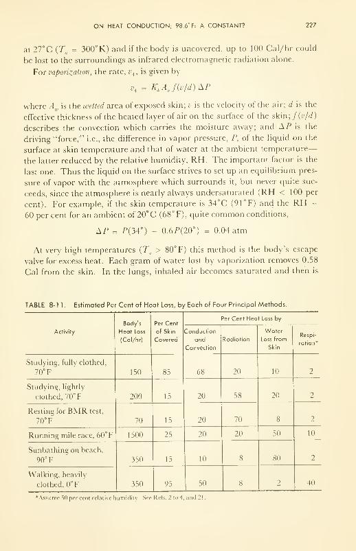
ON HEAT CONDUCTION; 98.6° F: A CONSTANT? 227
at 27°C {Ta= 300°K) and if the body is uncovered, up to 100 Cal/hr could
be lost to the surroundings as infrared electromagnetic radiation alone.
For vaporization, the rate, vA , is given by
v< = KAAJ(v/d)SP
where Aw is the wetted area of exposed skin; v is the velocity of the air; d is the
effective thickness of the heated layer of air on the surface of the skin;/(z>/rf)
describes the convection which carries the moisture away; and AP is the
driving "force/" i.e., the difference in vapor pressure, P, of the liquid on the
surface at skin temperature and that of water at the ambient temperature-
the latter reduced by the relative humidity, RH. The important factor is the
last one. Thus the liquid on the surface strives to set up an equilibrium pres-
sure of vapor with the atmosphere which surrounds it, but never quite suc-
ceeds, since the atmosphere is nearly always undersaturated (RH < 100 per
cent). For example, if the skin temperature is 34°C (91°F) and the RH =
60 percent for an ambient of 20° C (68° F), quite common conditions,
IP = P(34°) - 0.6P(20°) = 0.04 atm
At very high temperatures {Ta> 80°F) this method is the body's escape
valve for excess heat. Each gram of water lost by vaporization removes 0.58
Cal from the skin. In the lungs, inhaled air becomes saturated and then is
TABLE 8-11. Estimated Per Cent of Heat Loss, by Each of Four Principal Methods.
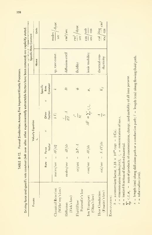
228
CD
O-xCD
229
CU
X.2°uCO>-4—
•
COX£
COO-3
C03
T3CJen
Q_XCJ
'0
_ocu>cuxxcj
X
C
C3CJ
COcxD
ccj
-accj
a.CUacco
coo<u
Cco
3 .
CO CO o ^
g^ 5 £ I^
e u a. -o o3 -^ C u33 en fc .5
o £
oE
(Ux
-° -5
—" .£D- 'to
"S cC .2CO
"O "5cj U-n 'CO cj
D. CE 12
COcu cO
^ TJC Uz
cucu
ocuX
~ X 3 <«0 CO X- cj en >^ cu cu
O * ~ .2 «J •
3 C C ^S« « s o
I £5
CO "O
t: co
E X33
£oCJ
cu
3 cX « 3 .3 co -3
m 2 <* -E ^ —
cu4—
»
CO
E
co —s-gen -Id
a o
c_o
CO
3O"cu
'Go"cu
>cu
1- CO
cj CJ
3 °_Q CU —•r -c cCJen en
cu 2 oen i-i .
-*-1
w —CU U5 TJCJ cu cu
O en cu
a. b ccu O> u "O —cu cu ^ cu
cu t- ~
CO CUCJ CJ
c/) C
O•accO
6l0
s> X
cj CO
C uC t«
a. uO CO
Nt: ex: «j —
•=. c 3Cj en
h>, a_c
cu—CJ
e« «J
.« ° "i CJw w s *^
Z C ^ ">
-* O •- tT-. o- - x; tt:
>- ej •
S cj 3
rr» CO Oe0 .2 > CJ y
cj > i~ CCU CJ 3
CJ
en
Cen CJ
CJ
3 X
cjoO(-
a.cu>usCU
z
er.
S o3 "C
"S X ^
CJjr.
CJ
CO
E 5
aXCJ
-ccCO
3o-aCJ
o £
a cj
u
oCJ
as
0-
£ 23 ^3 «J
c C T3c •- c£ C 3^— O Ti
C/3 C^
° o
!o £Q. .XCJ
3 uO <-
bfl^> cj
co
—t/1
c/i
c
CO
cj —CJ
CJCOl_
(0 CO I
'-3
J3
:^ K s « o
^d to
oJS
ccjCJ
X>CJ>CO
j3
Oan
^ 3
X -O CO;r — *-- 3 en
< - " - -C -O "O, 5 O OC 3 3? 'Z: ^ In CO CO 3
C «- — UZ q_ CJ en
O e > *J
en
u
xcj
Xcj
Xen
cj
cu
a.o
u
230 SPEEDS OF SOME PROCESSES IN BIOLOGICAL SYSTEMS
exhaled, with the same loss of heat per gram of water. Respiration then be-
comes important, especially when the air is dry and/or cold.
Urine and feces contribute a small fraction to daily heat loss.
Heat Loss from the Body Under Various Conditions
Table 8-1 1 illustrates that the escape valve for excess heat may be any one
of the several methods of heat loss and will vary for different activities. Thevery important role of the skin as a heat insulator and as a water supplier
to the surface, and the role of cover and clothing now become clear.
To sum up: the maintenance of constant body temperature is a very re-
markable example of the "steady state." In Chapter 7 we illustrated heat-
producing reactions—chemical, physical, mechanical, etc. In this chapter
we have discussed the rates of heat-producing reactions and the rate of heat
loss. In the steady state there is continuous flow—and the rate of "waste"
heat production is exactly balanced by the rate of heat loss, no matter what
the ambient conditions. So it is with literally hundreds of processes in the
living thing.
FORMAL SIMILARITY AND INTEGRATION OF THE FIVE PROCESSES
The method of presentation used in this chapter permits us to summarize
in a table the factors upon which the rates of the five processes depend, and
to note their similarities and differences. Since each of the processes was dis-
cussed individually, no comment on Table 8-r12 and its extension, Table
8-13, will be made now. other than to ask the reader to note that the classi-
cal driving force and the role of the activated complex are both stated ex-
plicity. The reader should consider these tables to be a memory aide, which,
if understood, will give him a powerful grasp of the nature of each of these
important processes occurring within the living system.
In the living thing, these processes are not separate and distinct, isolated
from one another. On the contrary, at every spot in the body probably three
or more are simultaneously operative. For instance at some point each
moment, a chemical reaction, requiring the transport in of reactants and the
transport out of products, produces heat which must be removed if the
steady state is to be maintained. As is the trend now in engineering kinetics'9
,
the future of biophysical kinetics lies in the study of the integration and con-
trol of rates of all the relevant processes proceeding in so orderly a manner
within the framework of the steady state. Motivation for the ultimate mas-
tery of biophysical kinetics is clear enough: deviations from the steady state
are diseases, the most vicious of which today has the popular generic name
"cancer." Some aspects of the all-important subject of control are discussed
in Chapters 10 and 1 1, following (next) an important chapter on the biologi-
cal effects of the ever-increasing ionizing radiation of our environment.
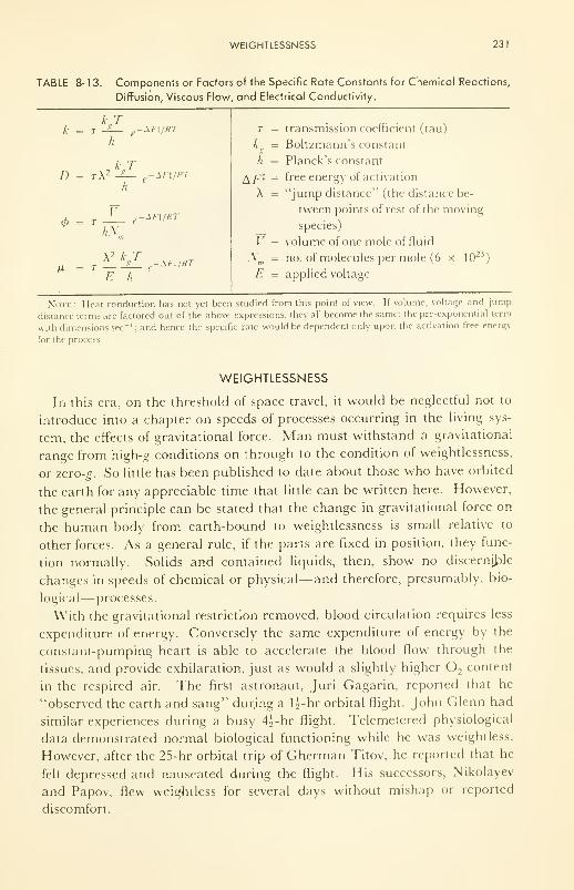
WEIGHTLESSNESS 231
TABLE 8- 1 3. Components or Factors of the Specific Rate Constants for Chemical Reactions,
Diffusion, Viscous Flow, and Electrical Conductivity.
kjh
I) = t\ 2kj
= TV
SI-t/RT
-AFt/RT
E h
r = transmission coefficient (tau)
k = Boltzmann's constant
h = Planck's constant
free energy of activation
"jump distance" (the distance be-
tween points of rest of the moving
species)
volume of one mole of fluid
,V;i
= no. of molecules per mole (6 x 10²³
E = applied voltage
A/-'*
A
I
Note Heat conduction has not yet been studied from this point of view. If volume, voltage and jump
distance terms are factored out of the above expressions, they all become the same: the pre-exponential term
with dimensions sec-1
; and hence the specific rate would be dependent only upon the activation free energy
for the process.
WEIGHTLESSNESS
In this era, on the threshold of space travel, it would be neglectful not to
introduce into a chapter on speeds of processes occurring in the living sys-
tem, the effects of gravitational force. Man must withstand a gravitational
range from high-g conditions on through to the condition of weightlessness,
or zero-g. So little has been published to date about those who have orbited
the earth for any appreciable time that little can be written here. However,
the general principle can be stated that the change in gravitational force on
the human body from earth-bound to weightlessness is small relative to
other forces. As a general rule, if the parts are fixed in position, they func-
tion normally. Solids and contained liquids, then, show no discernible
changes in speeds of chemical or physical—and therefore, presumably, bio-
logical— processes.
With the gravitational restriction removed, blood circulation requires less
expenditure of energy. Conversely the same expenditure of energy by the
constant-pumping heart is able to accelerate the blood flow through the
tissues, and provide exhilaration, just as would a slightly higher O, content
in the respired air. The first astronaut, Juri Gagarin, reported that he
"observed the earth and sang" dur.ing a li-hr orbital flight. John Glenn had
similar experiences during a busy 4^-hr flight. Telemetered physiological
data demonstrated normal biological functioning while he was weightless.
However, after the 25-hr orbital trip of Gherman Titov, he reported that he
felt depressed and nauseated during the flight. His successors, Nikolayev
and Papov, flew weightless for several days without mishap or reported
discomfort.
232 SPEEDS OF SOME PROCESSES IN BIOLOGICAL SYSTEMS
Perhaps the psychological effects of isolation, uncertainty, and frustration
will prove to be far more important than the effects of weightlessness on the
biophysics of the space traveler. The effects of ionizing radiation in free
space, unfiltered by the atmosphere, are discussed in the next chapter.
PROBLEMS
8- 1 : The rate of denaturation of a protein or of inactivation of an enzyme by heat is
dependent upon the concentration of the enzyme in a rather peculiar way,
which can be represented as v cc [E] n, where [E] is enzyme concentration and
n is the order of the reaction, interpreted as the number of molecules of enzyme
which come together to form a cluster in the inactivation process.
The temperature dependence is normal in that v cc e~E*l
, where E* is the
energy of activation, R is the gas constant (2 cal per degree per mole), and T is
the temperature in degrees Kelvin. For one case at low concentration, n was found
to be independent of temperature, and E* equal to 150,000 cal/mole.
(a) Calculate the ratio of velocity at 104°F to that at 98.6°F.
(b) Calculate the ratio for a 10°C rise in temperature.
(c) Calculate the ratio for a 10°C rise in temperature for a hydrolysis reac-
tion for which E* is 20,000 cal/mole.
(d) Calculate the ratio for a 10°C rise in temperature for a transport process
for which E* is 4000 cal/mole.
8-2: The basal metabolic rate of the "normal" man is about 0.1 hp. Express this in
Cal/hr; in watts; in cal/sec.
8-3: Using Poiseuille's Equation, calculate the pressure which would have to be ap-
plied to a No. 17 hypodermic needle (2 cm long, 0.05 cm radius), if a water solu-
tion of viscosity 0.01 poises (dyne/cm. sec2
) is to be forced, at a rate of 1 cc/sec,
into an artery which is already 100 mm Hg average pressure above atmospheric.
8-4: Under low rates of flow, blood has a viscosity (~0.02 poise) about twice that
of water; but under high rates, such as in the capillaries, it flows more easily
(~0.012 poise). Calculate the flow rate through two parallel tubes 1 mm long,
of radii 0.001 and 0.005 cm, if the pressure drop is 100 cm Hg.
8-5: One milliampere of total body current may be fatal. Estimate the path length
and average cross-section from hand to hand; and given the fact that the specific
conductivity (i.e., of a volume of soln. 1 cm2in area and 1 cm long) of a solu-
tion of 100 milliequivalents of KC1 per liter (approx concentration of body
fluids) is 0.015 ohm" 1 cm" 1
at 98°F, calculate the applied potential sufficient
to force 1 ma of current from hand to hand.
REFERENCES
1. Newburg, L. H., "Physiology of Heat Regulation (and the Science of Clothing),"
W. B. Saunders Co., Philadelphia, Pa., 1.949.
2. Kuno, Y., "Human Perspiration," Charles C. Thomas Publ., Springfield, 111.,
1956.
REFERENCES 233
3. Burton, A. C. and Edholm, O. G., "Man in a Cold Environment," Edw. Arnold
Publ. Ltd., London, 1955.
4. Ruch, T. C. and Fulton, J. F., "Medical Physiology and Biophysics," 18th ed.,
W. B. Saunders Co., Philadelphia, Pa., I960. (See the chapter by A. C.
Burton on Hematolysis.)
5. Greisheimer, E. M., "Physiology and Anatomy," J. B. Lippincott Co., Phila-
delphia, Pa., 1955.
6. Glasstone, S., Laidler, K. F., and Eyring, H., "The Theory of Rate Processes,"
McGraw-Hill Book Co., New York, N. Y., 1941.
7. Gaebler, O. FL, "Enzymes: Units of Biological Structure and Function," Aca-
demic Press, Inc., New York, N. Y., 1 956.
8. Tyrrell, H.J. V., "Diffusion and Heat Flow in Liquids," Butterworths, London,1961.
9. Baldwin, E., "Dynamic Aspects of Biochemistry," Cambridge Univ. Press,
Cambridge, England, 1953.
10. West, E. S., "Textbook of Biophysical Chemistry," The Macmillan Co., NewYork, N. Y., 1956.
1 1
.
Szent-Gybrgyi, A., "Introduction to a Submolecular Biology," Academic Press,
Inc., New York, N. Y., 1960.
12. Clarke, H. T., Ed., "Ion Transport Across Membranes," Academic Press, Inc.,
New York, N. Y., 1954.
13. Wintrobe, M. M., "Clinical Hematology," 4th ed., Lea and Febiger, Philadel-
phia, Pa., 1956.
14. Nikolaev, L. A., "Problems in Modelling of Biocatalysts," Vestnik Akademn nauk
SSSR, 13 (1960) ;LLU Translations Bulletin, London, 1960.
15. Laidler, K. J., "The Chemistry of Enzyme Action," McGraw-Hill Book Co.,
New York, N. Y., 1958.
16. Dixon, M. and Webb, E. C, "Enzymes," Academic Press, New York, N. Y.,
1958.
17. Davson, H. and Danielli, J. F., "The Permeability of Natural Membranes,"Cambridge Univ. Press, Cambridge, England. 1952.
18. Hober, R., etal, "Physical Chemistry of Cells and Tissues," The Blakiston Co.,
Philadelphia, Pa., 1945.
19. Bird, R. B., Stewart, W. E., and Lightfoot, E. N., "Transport Phenomena,"
John Wiley & Sons, Inc., New York, N. Y., 1960.
20. Conway, B. E., "Electrochemical Data," Elsevier Publ. Co., Amsterdam, Hol-
land, 1952; Parsons, R., "Handbook of Electrochemical Constants." Butter-
worths Scientific Pubis., London, 1959.
21. Kleiber, \1., "The Fire of Life," John Wiley & Sons, Inc., New York, X. V..
1961.
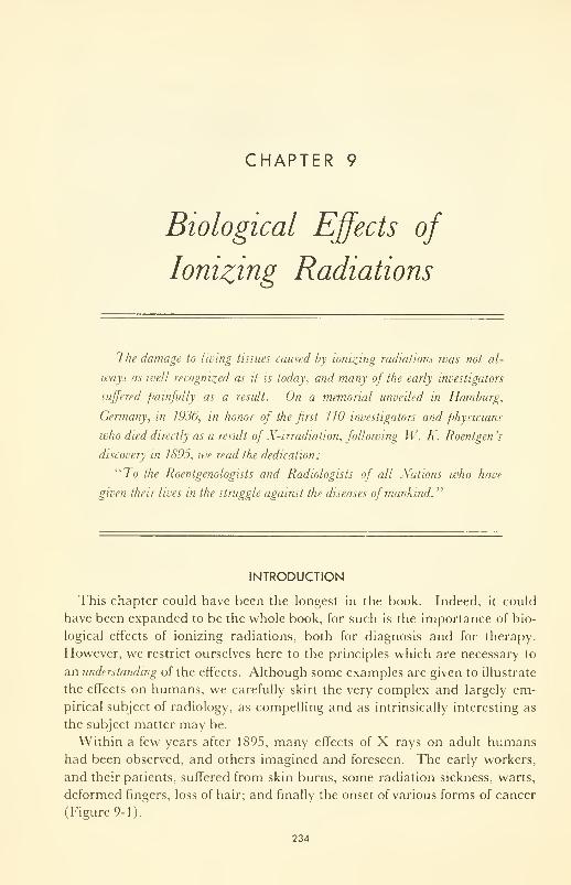
CHAPTER 9
Biological Effects of
Ionizing Radiations
The damage to living tissues caused by ionizing radiations was not al-
ways as well recognized as it is today, and many of the early investigators
suffered painfully as a result. On a memorial unveiled in Hamburg,
Germany, in 1936, in honor of the first 1 10 investigators and physicians
who died directly as a result of X-irradiatwn, following W. K. Roentgen 's
discovery in 7895, we read the dedication :
"To the Roentgenologists and Radiologists of all Nations who have
given their lives in the struggle against the diseases of mankind."
INTRODUCTION
This chapter could have been the longest in the book. Indeed, it could
have been expanded to be the whole book, for such is the importance of bio-
logical effects of ionizing radiations, both for diagnosis and for therapy.
However, we restrict ourselves here to the principles which are necessary to
an understanding of the effects. Although some examples are given to illustrate
the effects on humans, we carefully skirt the very complex and largely em-
pirical subject of radiology, as compelling and as intrinsically interesting as
the subject matter may be.
Within a few years after 1895, many effects of X rays on adult humanshad been observed, and others imagined and foreseen. The early workers,
and their patients, suffered from skin burns, some radiation sickness, warts,
deformed fingers, loss of hair; and finally the onset of various forms of cancer
(Figure 9-1).
234
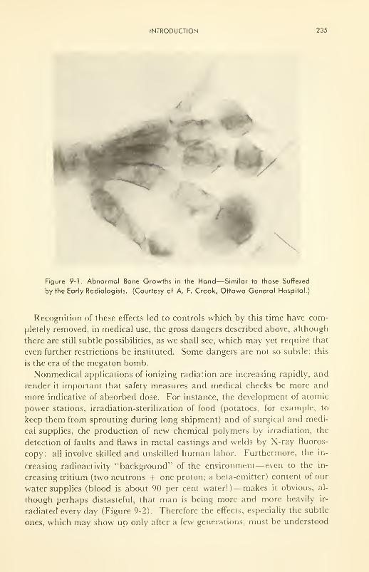
INTRODUCTION 235
' *
N
^
Figure 9-1. Abnormal Bone Growths in the Hand—Similar to those Suffered
by the Early Radiologists. (Courtesy of A. F. Crook, Ottawa General Hospital.)
Recognition of these effects led to controls which by this time have com-
pletely removed, in medical use, the gross dangers described above, although
there are still subtle possibilities, as we shall see, which may yet require that
even further restrictions be instituted. Some dangers are not so subtle: this
is the era of the megaton bomb.
Nonmedical applications of ionizing radiation are increasing rapidly, and
render it important that safety measures and medical checks be more and
more indicative of absorbed dose. For instance, the development of atomic
power stations, irradiation-sterilization of food (potatoes, for example, to
keep them from sprouting during long shipment) and of surgical and medi-
cal supplies, the production of new chemical polymers by irradiation, the
detection of faults and flaws in metal castings and welds by X-ray fluoros-
copy: all involve skilled and unskilled human labor. Furthermore, the in-
creasing radioactivity "background" of the environment— even to the in-
creasing tritium (two neutrons + one proton; a beta-emitter) content of our
water supplies (blood is about 90 per cent water!)—makes it obvious, al-
though perhaps distasteful, that man is being more and more heavily ir-
radiated every day (Figure 9-2). Therefore the effects, especially the subtle
ones, which may show up only after a few generations, must be understood
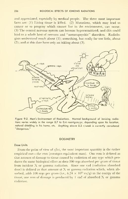
236 BIOLOGICAL EFFECTS OF IONIZING RADIATIONS
and appreciated, especially by medical people. The three most important
facts are: (1) Living tissue is killed. (2) Mutations, which may lead to
cancer or to progeny which cannot live in the environment, can occur.
(3) The central nervous system can become hypersensitized; and this could
lead to a whole host of nervous and "somatopsychic" disorders. Radiolo-
gists understand much about (1); something, but really far too little, about
(2); and at this date have only an inkling about (3).
Figure 9-2. Man's Environment of Radiations. Normal background of ionizing radia-
tions varies widely in the range 0.7 to 0.4 roentgens/yr, depending upon his location,
natural shielding in his home, etc. Anything above 0.3 r/week is currently considered
"dangerous."
DOSIMETRY
Dose Units
From the point of view of effect, the most important quantity is the rather
empirical one—the rem (roentgen equivalent man). One rem is defined as
that amount of damage to tissue caused by radiation of any type which pro-
duces the same biological effect as does 100 ergs absorbed per gram of tissue
from incident X or gamma radiation. Since one rad (radiation absorbed
dose) is defined as that amount of X or gamma radiation which, when ab-
sorbed, adds 100 ergs per gram (i.e., 6.24 x 10 13 ev/g) to the energy of the
tissue, one rem of damage is produced by 1 rad of absorbed X or gammaradiation.
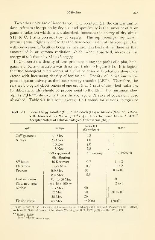
DOSIMETRY 237
Two other units are of importance. The roentgen (r), the earliest unit of
dose, refers to absorption by dry air, and specifically is that amount of X or
gamma radiation which, when absorbed, increases the energy of dry air at
STP (0°C, 1 atm pressure) by 83 ergs/g. The rep (roentgen equivalent
physical) was originally defined as the tissue-equivalent of the roentgen, but
with conversion difficulties being as they are, it is best defined here as that
amount of X or gamma radiation which, when absorbed, increases the
energy of soft tissue by 83 to 93 ergs/g.
In Chapter 5 the density of ions produced along the paths of alpha, beta,
gamma or X, and neutrons was described (refer to Figure 5-1). It is logical
that the biological effectiveness of a unit of absorbed radiation should in-
crease with increasing density of ionization. Density of ionization is ex-
pressed quantitatively as the linear energy transfer (LET). Therefore, the
relative biological effectiveness of one unit (i.e., 1 rad) of absorbed radiation
(of different kinds) should be proportional to the LET. For instance, slow
alphas (
4
2He ++
) do twenty times the damage of X rays of equivalent dose
absorbed. Table 9-1 lists some average LET values for various energies of
TABLE 9-1. Linear Energy Transfer (LET) in Thousands (Kev) or Millions (Mev) of Electron-
Volts Absorbed per Micron (10~ 4 cm) of Track for Some Atomic "Bullets."
Accepted Values of Relative Biological Effectiveness (rbe).*
Type

238 BIOLOGICAL EFFECTS OF IONIZING RADIATIONS
different radiations. One should bear in mind that the LET is not constant
while the radiation energy is being absorbed by tissue because with every bit
of energy lost there remains less to lose.
For several types of irradiation, approximate values of relative biological
effectiveness (rbe)— i.e., damage per unit dose, relative to 250 kvp X rays
—
can be written down, and can be used if the reservation be kept in mind that
they are rules-of-thumb, only approximate. Column 4 in Table 9-1 lists
such values. In general, the higher the energy of the impinging radiation,
the less energy it loses per unit length in tissue, and hence the longer it will
take a source to deliver a unit of absorbed radiation. In summary, the bio-
logical damage is given as:
rads x rbe = rems ^ reps x rbe
Several ways of receiving 1 rem of damage are depicted in Figure 9-3. For
workers, such as radiologists, who must necessarily be exposed, it is recom-
mended (by international agreement) that the whole-body dose be kept to
less than 0.3 rem per week in the blood-forming organs, the gonads, and the
eyes; less than 0.6 rem per week for surface irradiation. Relaxation to 1.5
rem per week is permitted if the radiation is of low penetrating power or if
only limited parts of the body are irradiated. Table 9-2 gives the number
of rems received under different conditions.
100 ergs of I Mev beta
I rad)
5 ergs of I Mev alpha
(0.05 rad)
10 ergs of I Mev neutron
(0.1 rad)I rem of
damage
100 ergs of X or
gamma (I rad)
10 ergs of 0.1 Mev
protons (0.1 rad)
Figure 9-3. Some Ways of Receiving 1 rem of Damage.
Dose Measurement
In air the dose received is measured accurately by the ionization chamber
and scintillation counters described in Chapter 5. However, in aqueous
solutions or in tissue, the situation is quite different. Here the primary tar-
get is water, and it breaks up not into ions but into free radicals (H + OH);
these react and produce new chemicals.
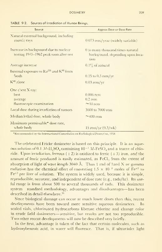
DOSIMETRY 239
TABLE 9-2. Sources of Irradiation of Human Beings.
Source Approx Dose or Dose Rate
Natural external background, including
cosmic rays
Increase in background due to nuclear
testing 1945-1962 peak soon after test
Average increase
Internal exposure to Ra226 and K40 from
foods
K40 alone
One chest X ray:
best
average
fluoroscopic examination
Local dose during irradiation of tumors
Median lethal dose, whole body
Maximum permissable* dose rate,
whole body
0.073 rem/year (widely variable)
to many thousand times natural
background, depending upon loca-
tion
0.1% of natural
0.15 to 0.5 rem/yr
0.03 rem/yr
0.006 rem
0.2 rem~
1 rem
3000 to 7000 rem
~400 rem
15 rem/yr (0.3/wk)
*Recommended by the International Commission on Radiological Protection. 1 958.
The celebrated Fricke dosimeter is based on this principle. It is an aque-
ous solution of 0.1 .\/-H2S04 containing 10
~ 3 A/-FeS04and a tracce of chlo-
ride. Upon irradiation, ferrous ( + 2) is oxidized to ferric ( + 3) iron, and the
amount of ferric produced is easily estimated, as FeCl, from the extent of
absorption of light of wave length 3040 A. Thus 1 rad of hard X or gammaradiation has the chemical effect of converting 1.5 x 10~ 8 moles of Fe +2 to
Fe+3 per liter of solution. The system is widely used, because it is simple,
reproducible, accurate, and independent of dose rate (e.g., rads/hr). Its use-
ful range is from about 500 to several thousands of rads. This dosimeter
system— standard methodology, advantages and disadvantages— has been
described in detail elsewhere. 18
Since biological damage can occur at much lower doses than this, recent
developments have been toward more sensitive aqueous dosimeters. In
sealed vials, chlorinated hydrocarbons liberate chlorine and change color
in crude field dosimeters— sensitive, but results are not too reproducible.
Two other recent developments will now be described very briefly.
In the first, advantage is taken of the fact that certain molecules, such as
hydroxybenzoic acid, in water will fluoresce. That is, if ultraviolet light
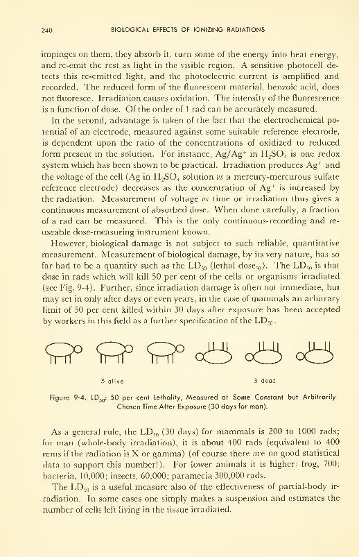
240 BIOLOGICAL EFFECTS OF IONIZING RADIATIONS
impinges on them, they absorb it, turn some of the energy into heat energy,
and re-emit the rest as light in the visible region. A sensitive photocell de-
tects this re-emitted light, and the photoelectric current is amplified and
recorded. The reduced form of the fluorescent material, benzoic acid, does
not fluoresce. Irradiation causes oxidation. The intensity of the fluorescence
is a function of dose. Of the order of 1 rad can be accurately measured.
In the second, advantage is taken of the fact that the electrochemical po-
tential of an electrode, measured against some suitable reference electrode,
is dependent upon the ratio of the concentrations of oxidized to reduced
form present in the solution. For instance, Ag/Ag+ in H 2S0 4is one redox
system which has been shown to be practical. Irradiation produces Ag+ and
the voltage of the cell (Ag in H2S04
solution vs a mercury-mercurous sulfate
reference electrode) decreases as the concentration of Ag+is increased by
the radiation. Measurement of voltage vs time or irradiation thus gives a
continuous measurement of absorbed dose. When done carefully, a fraction
of a rad can be measured. This is the only continuous-recording and re-
useable dose-measuring instrument known.
However, biological damage is not subject to such reliable, quantitative
measurement. Measurement of biological damage, by its very nature, has so
far had to be a quantity such as the LD 50 (lethal dose 50 ). The LD 50 is that
dose in rads which will kill 50 per cent of the cells or organisms irradiated
(see Fig. 9-4). Further, since irradiation damage is often not immediate, but
may set in only after days or even years, in the case of mammals an arbitrary
limit of 50 per cent killed within 30 days after exposure has been accepted
by workers in this field as a further specification of the LD 50 .
3 alive 3 dead
Figure 9-4. LD50 : 50 per cent Lethality, Measured at Some Constant but Arbitrarily
Chosen Time After Exposure (30 days for man).
As a general rule, the LD 50 (30 days) for mammals is 200 to 1000 rads;
for man (whole-body irradiation), it is about 400 rads (equivalent to 400
rems if the radiation is X or gamma) (of course there are no good statistical
data to support this number!). For lower animals it is higher: frog, 700;
bacteria, 10,000; insects, 60,000; paramecia 300,000 rads.
The LD 50 is a useful measure also of the effectiveness of partial-body ir-
radiation. In some cases one simply makes a suspension and estimates the
number of cells left living in the tissue irradiated.
PRIMARY EFFECTS (ON CHEMICAL COMPOSITION) 241
As more is learned about effects of ionizing radiation on metabolic proc-
esses, physiological measurements of effects on rates of specific processes
within cells and tissues will probably add much-needed refinements to the
useful LD 50 number.
Incidentally, one should realize that only a small amount of energy need be
absorbed to cause damage. It is the form in which this energy enters the tis-
sue that is critical. Thus the LD 50 for man, 400 rads, is only 400 x 100
ergs/g. This is 0.001 cal/g, roughly enough energy, if in the form of heat, to
raise the body temperature only 0.001 deg! Because the energy is concen-
trated in packets, so that when it is absorbed it tears apart the molecules
of important biological structures, localized damage occurs at sensitive sites,
enabling a small quantity of energy to promote death. Table 9-3 gives some
useful irradiation data and conversion factors.
TABLE 9-3. Some Useful Numbers.
1 rad = 100 ergs/gram = 6.24 x 10 13 electron volts/gram.
1 roentgen of hard X or 7 delivers 0.98 rad to water.
1 curie of radioactive substance delivers 3.7 x 10'° disintegrations/second.
1 curie of Co60 gives a dose rate of 1 .35 roentgen/hour at 1 meter from the source.
1 curie of radium gives a dose rate of 0.83 roentgen/hour at 1 meter.
1 curie of cesium 137gives a dose rate of 0.33 roentgen/hour at 1 meter.
PRIMARY EFFECTS (on Chemical Composition)
Direct and Indirect Action
Two schools of thought have arisen on the question of how the primary
effects occur. However, there are so many variables involved that it is un-
likely that either will ever be proved to be unequivocally wrong.
The fact is that the solution after irradiation contains molecules (chemi-
cals) which were not there before irradiation. One school maintains that
this is because the solute dissolved in the water acted as a target, was blown
apart by the incoming "bullets," and the fragments rearranged into a new
molecule. The other school remembers that the whole target (tissue, for
example) can be at least 80 per cent water, that eight out of ten potential
targets are water molecules, and maintains that the primary act is the ex-
citation of water, followed by its decomposition into the active chemicals
hydrogen atom and hydroxyl radical. Enough energy is left over so that
these are thrown violently apart. Hydrogen is a reducing agent, which can
donate an electron to become H + in solution; OH is an oxidizing agent
which can accept an electron to become OH in solution. From this view,
then, these molecular fragments, H and OH, cause the formation of new
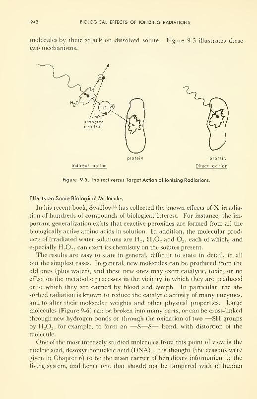
242 BIOLOGICAL EFFECTS OF IONIZING RADIATIONS
molecules by their attack on dissolved solute. Figure 9-5 illustrates these
two mechanisms.
unsharedelectron
protein protein
Indirect action Direct action
Figure 9-5. Indirect versus Target Action of Ionizing Radiations.
Effects on Some Biological Molecules
In his recent book, Swallow" has collected the known effects of X irradia-
tion of hundreds of compounds of biological interest. For instance, the im-
portant generalization exists that reactive peroxides are formed from all the
biologically active amino acids in solution. In addition, the molecular prod-
ucts of irradiated water solutions are H2 , H 2 2
and2 , each of which, and
especially H2 2 , can exert its chemistry on the solutes present.
The results are easy to state in general, difficult to state in detail, in all
but the simplest cases. In general, new molecules can be produced from the
old ones (plus water), and these new ones may exert catalytic, toxic, or no
effect on the metabolic processes in the vicinity in which they are produced
or to which they are carried by blood and lymph. In particular, the ab-
sorbed radiation is known to reduce the catalytic activity of many enzymes,
and to alter their molecular weights and other physical properties. Large
molecules (Figure 9-6) can be broken into many parts, or can be cross-linked
through new hydrogen bonds or through the oxidation of two —SH groups
by H2 2 , for example, to form an —S—S— bond, with distortion of the
molecule.
One of the most intensely studied molecules from this point of view is the
nucleic acid, desoxyribonucleic acid (DNA). It is thought (the reasons were
given in Chapter 6) to be the main carrier of hereditary information in the
living system, and hence one that should not be tampered with in human
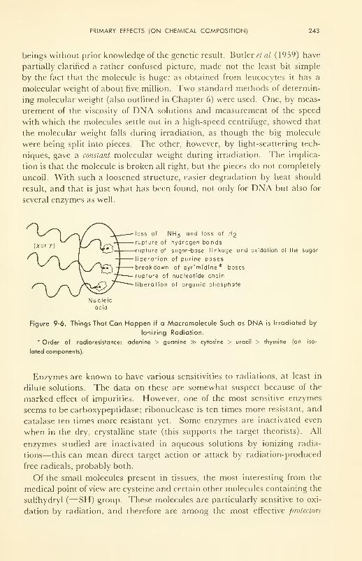
PRIMARY EFFECTS (ON CHEMICAL COMPOSITION) 243
beings without prior knowledge of the genetic result. Butler et al (1 959) have
partially clarified a rather confused picture, made not the least bit simple
by the fact that the molecule is huge: as obtained from leucocytes it has a
molecular weight of about five million. Two standard methods of determin-
ing molecular weight (also outlined in Chapter 6) were used. One, by meas-
urement of the viscosity of DNA solutions and measurement of the speed
with which the molecules settle out in a high-speed centrifuge, showed that
the molecular weight falls during irradiation, as though the big molecule
were being split into pieces. The other, however, by light-scattering tech-
niques, gave a constant molecular weight during irradiation. The implica-
tion is that the molecule is broken all right, but the pieces do not completely
uncoil. With such a loosened structure, easier degradation by heat should
result, and that is just what has been found, not only for DNA but also for
several enzymes as well.
loss of NH3 and loss of H2
rupture of hydrogen bondsrupture of sugar-base linkage and oxidation of the sugar
iberation of purine bases
breakdown of pyrimidine* bases
rupture of nucleotide chain
liberation of organic phosphate
Nucleicacid
Figure 9-6. Things That Can Happen if a Macromolecule Such as DNA is Irradiated by
Ionizing Radiation.
* Order of radioresistance: adenine > guanine » cytosine > uracil > thymine (on iso-
lated components).
Enzymes are known to have various sensitivities to radiations, at least in
dilute solutions. The data on these are somewhat suspect because of the
marked effect of impurities. However, one of the most sensitive enzymes
seems to be carboxypeptidase; ribonuclease is ten times more resistant, and
catalase ten times more resistant yet. Some enzymes are inactivated even
when in the dry, crystalline state (this supports the target theorists). All
enzymes studied are inactivated in aqueous solutions by ionizing radia-
tions—this can mean direct target action or attack by radiation-produced
free radicals, probably both.
Of the small molecules present in tissues, the most interesting from the
medical point of view are cysteine and certain other molecules containing the
sulfhydryl (—SH) group. These molecules are particularly sensitive to oxi-
dation by radiation, and therefore are among the most effective protectors
244 BIOLOGICAL EFFECTS OF IONIZING RADIATIONS
known. By one view they scavenge free radicals H, OH, H02 , etc., pro-
duced in the radiolysis. By another view they attach themselves to enzymes
or nucleic acids at just the spots most sensitive to radical attack (—SHgroups) and thereby reduce the effects of irradiation on the big molecules.
For example, even impure acetylcholinesterase, in a solution with muchother protein, is only half as sensitive to irradiation damage in the presence
of 10" 3 M-cysteine as in its absence. In living cells the enzymes are well
protected, and seem to be resistant to much larger doses of radiation than
the same molecule in vitro.
The "Oxygen Effect"
The radiation sensitivity of most molecules is greater the higher the oxy-
gen content of the solution. Thus, the rate of oxidation of Fe +2 to Fe+3 by
X rays is twice as high in the presence, than in the absence, of oxygen. For
small molecules like phenol and the amino acids the rate is often even more
enhanced by 2. This increased radiation sensitivity in the presence of oxy-
gen is observed right on up the hierarchy of structures— viruses, bacteria,
cells, tissues, to whole animals.
A striking practical demonstration of this effect has been shown with
carcinoma tissue. Due to necrosis, many parts of a tumor can become
anoxic. By increasing the pressure of the respired air this anoxia can be re-
duced, with a consequent increase in the radiosensitivity of the carcinoma
cells.
Now, oxygen itself is known to accelerate many metabolic reactions, and
the effect of oxygen in increasing radiation damage is thought by some to
result from this fact. However, in other quarters the effect is thought to
occur through the radical, H02
. This radical is produced from the reaction
H + 2— H02
after the radiolysis reaction has produced the hydrogen atoms as follows:
H2
-^ H 20* — H + OH
The radical HO, is a strong oxidizing agent, since it readily accepts an elec-
tron from any source to become the peroxide ion, H0 2~. Hence, one can
consider that the H atom simply puts 2into a form in which it can react
faster. Since 2is used up in the reaction, it must be supplied continuously
if advantage is to be continuously taken of enhanced rate of destruction.
Conversely, of course, oxygen scavenger molecules increase the protection of
macromolecules against ionizing radiations.
The mode of action of oxygen is one of the most intriguing practical prob-
lems of radiology. Once it is understood, it can be controlled and utilized
BIOPHYSICAL EFFECTS 245
more fully. Other species, such as NO and Co++ also enhance radiation
effects.
BIOPHYSICAL EFFECTS
These can be considered as effects on molecular structure and type, with
the resulting effects on the physical properties of agglutination and trans-
port, and on the speeds of vital chemical processes.
Agglutination or Coagulation
Colloids— small particles, large molecules— are stabilized by electric
charges on their surface. At any particular pH, the acidic and basic chemi-
cal groups on the surface are in equilibrium with the electrolyte, and the sur-
face carries a net positive or negative charge. Repulsion between like
charges stabilizes the colloid. Further stabilization comes from water mole-
cules adsorbed on the polar groups of the surface, so that, from the outside,
the big colloid particle looks, to a particle in solution, just like a wall of
ordinary water molecules.
Irradiation causes, first of all, chemical polymerization or cross-linking to
occur between particles. It causes changes in the polar groups, and hence in
the "water front" which the colloid presents to the solution. Finally it
causes rearrangement in acidic and basic groups such that the net surface
charge changes. The colloid then precipitates, or agglutinates, and becomes
semisolid.
On the other hand, the colloid may be split within by radiant energy, and
the structure then rearranged to a form which is unstable, and it precipitates.
Modification of Transport Properties
Thermal Conductivity. This property is difficult to measure even under the
most advantageous of circumstances, and nothing is known yet about how it
is affected by radiation. Structural changes induced by radiation may turn
out to be of importance to the structural lipoproteins and collagen of the
skin, for example.
Diffusion. As it was shown in Chapter 8, the diffusion coefficient depends
critically upon the molecular structure of the medium, with particular refer-
ence to the "jump distance" between rest sites in the medium and to the size
and shape of the diffusing species. Naturally, if the diffusing molecule is
broken up into small and free parts by the action of ionizing radiation, it
will diffuse faster. Conversely, if it or the medium becomes cross-polymer-
ized, diffusion will occur more slowly.
It is expected that, as more is learned about the diffusion of water, ions,
and molecules through living membranes, the effects of irradiation on dif-
fusion will become more evident. In the absence of definitive work on this
246 BIOLOGICAL EFFECTS OF IONIZING RADIATIONS
subject, one can only say that the possibilities exist, and should be remem-
bered during discussions of the physiological effects, which are currently
receiving more attention.
Fluidity (Inverse of Viscosity). Most of the useful information on the ef-
fects of ionized radiations on fluidity (ease of flow in response to a physical
force) has been done either on plastics or on aqueous solutions of big
molecules.
From the former it has been learned that cross-linking of polythene by ir-
radiation increases markedly its melting point and increases its elasticity.
By contrast, irradiation of teflon (a fluorinated and inert organic) leads to
hardening and embrittlement, and loss of elasticity. This might lead one to
anticipate similar effects in elastomeric tissue in the walls of blood vessels,
were it not for the fact that the effects are exhibited only after the absorption
of a few million rads!
On the other hand, the fluidity of aqueous solutions of biologically active
molecules has been intensively studied, especially as a technique of measur-
ing the change in molecular weight effected by radiations. Like diffusion,
many examples are known in which cross-polymerization is important, and
many in which molecular rupture is to be inferred.
Electrical Conductivity. In body fluids the conductivity is high. Irradiation
makes no detectable change.
It is in the inner, fatty-acid or lipid part of the living membrane (Figure
6-7) that we expect a change in conductivity. The lipid, an oil, has very low
conductivity. Analogy with polythene or lucite may be useful as a guide.
These materials break down internally under irradiation, such that electrons
are knocked off one part of the molecule and caught or trapped elsewhere,
leaving a positive site behind. The conductivity increases, because the
charges are somewhat mobile, and a steady-state concentration, higher the
higher the dose rate, is set up and maintained. Upon cessation of the radia-
tion, the charges recombine slowly, and the conductivity drops to its original
value. Although the k for these substances is very, very small (~10~ 21
ohm-1 cm '), it is raised as much as fifty thousand times by an X-ray dose
of only 8 roentgens (r) per min. By comparison, the conductivity of a resting
nerve membrane is of the order of 10~ 12 ohm "' cm ', due almost entirely
to the lipid inner layer.
The "activation" of nerves by radiations, and some effects on the central
nervous system, to be discussed in the next section, indicate that enhanced
electrical conductivity may be one of the most important biophysical effects
of ionizing radiations.
Chemical Reactivity
The effects of ionizing radiations on the rate of chemical reactions could
be inferred from knowledge of the factors upon which rate depends. In gen-
PHYSIOLOGICAL EFFECTS 247
eral terms there are two methods by which the rate can be increased:
through increase in local temperature (thermal energy of vibrations, etc.) in
the vicinity of the ionized track, and through excited electronic states of re-
actant molecules (photochemical processes). The mechanisms have been
discussed in Chapters 4 to 8. The synthesis of new isomers and of entirely
new molecules was considered also in Chapter 6, as well as the nature of
toxins, catalysts, and useful and destructive mutants.
PHYSIOLOGICAL EFFECTS
Outlined in this section are the effects of ionizing radiation on cells,
organs and tissues.
Sensitivity of Cells
The sensitivity, a (sigma), is the rate at which cells die because of irre-
versible damage suffered during irradiation. Since the unit of absorbed
dose, D, is the rad, the fraction of cells lost per rad is the sensitivity. Thus
dNIdDa =
N
cells killed per unit dose per unit number of cells irradiated. If the dose rate,
dD/dt rads/sec, is a constant, p, then the sensitivity can be expressed
dN/dta =
p-N
cells killed per sec per unit number of cells irradiated. Based on what is now
known about factors affecting the radiosensitivity of cells, the early (1905)
"law" of Bergonie and Tribondeau can be extended and rewritten:
l [dN/dt]g
eW,a,dD/dt \
where / denotes a functional relationship between a and the quantities in
parentheses; [dN/dt]g
, the rate at which the cells reproduce themselves (i.e.,
the growth rate, or number produced per unit time); 'W , the metabolic rate
—energy used up per unit time; a, a number less than 1 which varies with
the state of cell division— unity at the prophase of mitosis, much less at any
other time; m, the degree of maturity— unity for old, well-developed, spe-
cialized cells, less for those newly formed; and dD/dt, the dose rate. In sum-
mary, the sensitivity increases with increasing rate of cell division, metabolic
rate, and dose rate; increases sharply at prophase; and decreases as the cell
becomes more mature. The exact functional relationships are not known.
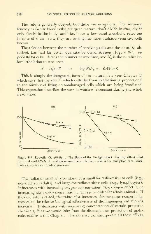
248 BIOLOGICAL EFFECTS OF IONIZING RADIATIONS
The rule is generally obeyed, but there are exceptions. For instance,
leucocytes (white blood cells) are quite mature, don't divide in vitro, divide
only slowly in the body, and they have a low basal metabolic rate; but
in spite of these facts, they are among the most radiation-sensitive cells
known.
The relation between the number of surviving cells and the dose, Z), ab-
sorbed, has had far better quantitative demonstration (Figure 9-7), es-
pecially for cells. If N is the number at any time, and N is the number be-
fore irradiation started, then
N N e-aD or log JV/jV = -0.434 a D
This is simply the integrated form of the natural law (see Chapter 1)
which says that the rate at which cells die from irradiation is proportional
to the number of living or nondamaged cells which are being irradiated.
This expression describes the case in which o is constant during the whole
irradiation.
Dose (rods) Dose (rods)
Figure 9-7. Radiation-Sensitivity, a-. The Slope of the Straight Line in the Logarithmic Plot
(b) for Haploid Cells. Low slope means low a. Broken curve is for multiploid cells: sensi-
tivity increases as irradiation proceeds.
The radiation sensitivity constant, a, is small for radio-resistant cells (e.g.,
nerve cells in adults), and large for radiosensitive cells (e.g., lymphocytes).
It increases with increasing oxygen concentration ("the oxygen effect"), or
increasing nitric oxide concentration. This is true also for whole animals. If
the dose rate is raised, the value of a increases, for the same reason it in-
creases as the relative biological effectiveness of the impinging radiation is
increased. It decreases with increasing concentration of certain protector
chemicals, P, as we would infer from the discussion on protection of mole-
cules earlier in this Chapter. Therefore we can incorporate all these effects
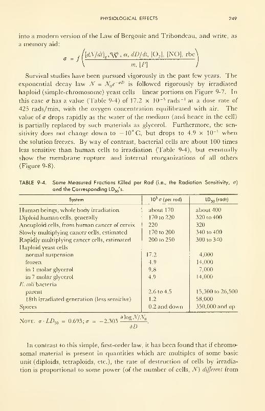
PHYSIOLOGICAL EFFECTS 249
into a modern version of the Law of Bergonie and Tribondeau, and write, as
a memory aid:
\dN/dt]e ,<W > oc, dD/dt, [02 ], [NO], rbe'/
m, [P]
Survival studies have been pursued vigorously in the past few years. The
exponential decay law N = N e~ aD is followed rigorously by irradiated
haploid (simple-chromosome) yeast cells— linear portions on Figure 9-7. In
this case a has a value (Table 9-4) of 17.2 x 10 -5 rads -1at a dose rate of
425 rads/min, with the oxygen concentration equilibrated with air. The
value of a drops rapidly as the water of the medium (and hence in the cell)
is partially replaced by such materials as glycerol. Furthermore, the sen-
sitivity does not change down to -10°C, but drops to 4.9 x 10~ 5 when
the solution freezes. By way of contrast, bacterial cells are about 100 times
less sensitive than human cells to irradiation (Table 9-4), but eventually
show the membrane rupture and internal reorganizations of all others
(Figure 9-8).
TABLE 9-4. Some Measured Fractions Killed per
and the Corresponding LD50's.
Rad (i.e., the Radiation Sensitivity, a)
System
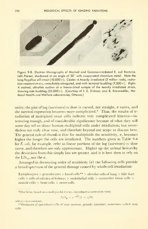
250 BIOLOGICAL EFFECTS OF IONIZING RADIATIONS
Figure 9-8. Electron Micrographs of Normal and Gamma-irradiated E. coli Bacteria.
Left: Parent, shadowed at an angle of 30° with evaporated chromium metal. Note the
long flagellae still intact (10,000 x). Center-. A heavily irradiated (2 million rads), radia-
tion-resistant strain, remarkably elongated, and with terminal budding (7,000 x). Right:
A stained, ultrathin section of a freeze-dried sample of the heavily irradiated strain,
showing side budding (25,000 x). (Courtesy of I. E. Erdman and B. Kronmueller, Na-
tional Health and Welfare Laboratories, Ottawa.)
unity; the plot of log (survivors) vs dose is curved, not straight, a varies, and
the survival expression becomes more complicated.* Thus, the results of ir-
radiation of multiploid yeast cells indicate very complicated kinetics— in-
teresting enough, and of considerable significance because of what they will
some day tell us about human multiploid cells under irradiation; but never-
theless not truly clear now, and therefore beyond our scope to discuss here.
The general rule-of-thumb is that for multiploids the sensitivity, a, becomes
higher the longer the cells are irradiated. The numbers given in Table 9-4
for E. coli, for example, refer to linear portions of the log (survivors) vs dose
curve, and therefore are only approximate. Higher up the animal heirarchy
the deviations from this simply law are greater, and it is best then to rely on
the LD 50 , not the a.
Arranged in decreasing order of sensitivity (<x) the following cells provide
a broad spectrum of the general damage caused by whole-cell irradiation:
Lymphocytes > granulocytes > basal cells** > alveolar cells of lung > bile duct
cells > cells of tubules of kidneys > endothelial cells > connective tissue cells >
muscle cells > bone cells > nerve cells.
"One form, based on a multiple-hit theory, introduces a correction term:
yvyA-oD
(1 - c/D)
where c is a constant.
**Producers of specialized cells of bone marrow, gonads, intestines, sometimes called stem
cells.
PHYSIOLOGICAL EFFECTS 251
Microirradiation of Cells
So far, the discussion has been on whole-cell irradiation. However, by
microirradiation techniques, in which just a small volume within a single cell
receives radiation, it has been found that not all parts of the cell are equally
sensitive. In fact, a is much higher if the nucleus (in particular the cnromo-
somes within the nucleus), rather than any other part of the cytoplasm or
cell membrane, is irradiated.
Microirradiation is not easy experimentally, but it has now been done
with proton and alpha particles, and with X and far ultraviolet electro-
magnetic rays. Production of the micro beam is done by a colinear series of
apertures in a number of absorbents (e.g., lead bricks). Sometimes it is done
by passing the radiation through a glass or platinum capillary mounted in a
lead shield. Thus any X rays falling on the wall of a Pt capillary at an angle
of 0.6 deg or less to the axis of the capillary are completely reflected, and are
propagated unchanged to the exit and thence to the target. The position of
the target cells can be set by means of apparatus which is not essentially
different from the traveling stage of a microscope: by means of a micro-
manipulator with worm gears the target can be moved into any desired posi-
tion within a limited space.
Results with protons, alphas, X, and ultraviolet have all shown that the
nucleus, and specifically the nucleolus which begins to become more prom-
inent as mitosis begins, is far more radiation-sensitive than the rest of the
cell. For example, in a specific case, irradiation through an area 2.5/i in
diameter on a chromosome (~5ju x 30yu) with 36,000 rads of proton energy
(60 protons, ~1.5 Mev) caused the chromosome to become sticky (to cross-
link?) and the cell to die in the attempt to divide, while irradiation else-
where in the cell with up to 1.7 million rads caused no change in speed or
reliability of division, nor did it have any effect on the several observed suc-
ceeding generations.
However, indirect effects on the chromosomes by irradiation elsewhere in
the cell have been demonstrated. Nor should one infer that irradiation else-
where does no permanent damage to the cell or its progeny. For such spec-
tacular things as blistering of the cell wall, and coagulation of cytoplasm and
of the mitochondria, as well as death to all the progeny of cells irradiated
generally elsewhere than the chromosomes, have been observed. Considera-
tion of the cell as "a bag of enzymes," each subject to irradiation isomeriza-
tion, gives one an idea of how complex this question can be.
Unfortunately the important microirradiation studies have not, yet yielded
any case in which irradiation of a certain part of the cell has caused an in-
creased rate of reproduction of modified or cancerous cells. Hence, just howabsorbed radiation induces cancer at the cell level remains unanswered. It
is now generally assumed to be irradiation of the DNA of the chromosomes,
252 BIOLOGICAL EFFECTS OF IONIZING RADIATIONS
but it could just as well be modification of one of the catalysts of the syn-
thesis of DNA, or the membrane which contains them.
There is some direct information on DNA in solution, however. By vis-
cosity and titration methods it has been found that the molecule is shattered
by X and a rays, to an amount of about 1.5 x 10 11 chain-breaks per gram
of DNA per rad absorbed. The analogy with the effect of ultrasound on
viruses is usefully drawn at this point, for ultrasound quite literally shakes
the molecule to pieces.
There is also some semidirect information on DNA in vivo. Thus, T. T.
Puck 4 and others have allowed irradiated human cells to culture, and have
measured, not the LD 50 , but the "reproductive death"—the irradiation dose
which is just sufficient to cause the cells not to reproduce. These cells are
not killed by the radiation, but often show abnormalities, such as growing
to a huge size or showing a change in metabolic rates. Reproductive
"death" is relatively very sensitive, its L"D" 50 being 25 to 40 rads in humancells. The corresponding sensitivity, a, is about 2000 (compare with the
values in Table 9-4).
Irradiation of Organs and Tissues
The histologic and pathologic changes in tissues resulting from irradiation
are properly part of the subject matter of radiology, and will not be dis-
cussed here. However, as illustration, some of the results of whole- and
partial-body irradiation are listed below, with no explanation, as simple
statements.
Just as some parts of the cell are more radiation-sensitive than others, also
some tissues and organs are more sensitive than others. The analogy goes
further. Some parts of the human body can be irradiated relatively heavily
without severe general damage; others are very radiation-sensitive. The fol-
lowing list includes the most sensitive.
(1) Red blood cell manufacture slows down in the bone marrow.
(2) Manufacture of lymphocytes in the spleen is drastically reduced and
cannot replace fast enough those killed by irradiation of the general lym-
phatic circulation system.
(3) The skin shows reddening or blistering, after only 140 rem; larger
doses can precipitate skin cancer.
(4) Impairment of secretion or of assimilation occurs in the alimentary
canal, mostly as a result of membrane destruction. Sloughing off of the
mucous lining of the canal is an early symptom of damage and often results
in death due to infection.
(5) The critically important steady-states in the adrenal glands are upset.
Because these are the source of certain rate-controlling molecules, the hor-
PHYSIOLOGICAL EFFECTS 253
mones, greater body susceptibility to heat, cold, injury, and infection results
from the damage.
(6) Decreased activity of the thyroid can result, causing lower basal
metabolic rate.
(7) In the lungs, the membranes across which2and C02
exchange be-
tween blood capillaries and air takes place are broken, and persistent oxy-
gen deficiency and excess carbon dioxide in the blood result.
(8) Enough radiation can ruin the very selective membranes in the
kidney.
(9) Similar damage in the liver results in hemorraging.
(10) Cataracts develop in the lens of the eye from coagulation of liquid
crystals. The effects may be delayed, however.
(11) Large local doses (~400 rem) to the gonads can cause sterility by
killing off the sensitive spermatogenic cells. The sperm themselves are rela-
tively resistant. Much lower doses could cause mutations in the DNA-gene-chromosome structure of the germ cells, while large doses could simply
break the chromosomes into pieces. Gonadal doses from various sources are
collected in Table 9-5.
(12) Even low doses to some tissues can produce enough variation in the
cell reproduction system so that the tissue becomes carcinogenic. (This is
probably the most important, and still the least understood, physiological
effect of irradiation. Unfortunately, the susceptibility may not become
manifested for several generations of cells.)
(13) The rate of production of antibodies is lowered markedly, and the
tissue is more subject to infection and disease. This effect is related to the
rapid destruction of the lymphatic tissues.
TABLE 9-5. Gonadal Doses from Various Sources*
Source Dose or Dose Rate
Background radiation 0.095-0.180 rem/yr
Maximum dose permitted to X ray workers 1 5 rem/yr (0.3 rem/wk)
Pelvic examinations, fluoroscopic ^1 rem
Salpingogram ~1.7 rem
Photographic X ray of kidney and ureters 0.9 rem
Photographic X ray of pelvis 0.7 rem
Photographic X ray of hip 0.5 rem
Collected by C Don."
The following general principles are important to remember:
(1) The physiological effects are direct results of changes in the rates of
chemical or transport processes.
254 BIOLOGICAL EFFECTS OF IONIZING RADIATIONS
(2) The long-term damage may prove to be greatest in the chromosomes,
at mitosis, but such genetic effects may not appear for several gen-
erations.
(3) Damage to the fine network of molecular membranes and canals in
the cell's substructure, where the enzyme-controlled protein and nu-
cleic acid syntheses take place, can result in immediate physiological
changes. Damage to cell walls and structural tissue is important at
high dose or after some time at low dose.
EFFECTS OF WHOLE-BODY IRRADIATION
The Facts and the Complexity of the Problem
Three events, each horrible in its own way, provide the foundation of our
knowledge about whole-body irradiation of normal humans. The first was
the bombs at Hiroshima and Nagasaki; the second was an accident at OakRidge, and others, less publicized, later; and the third, unpredicted winds
over Bikini and the Marshall Islands during H-bomb trials.
Three months after the publication by Roentgen of his experiments with
X rays, puzzling radiation burns on the skin were observed. Within a few
years, premature loss of hair and early ageing befell the early workers. Fromten to forty years after intense exposure, some gruesome cancers appeared,
and case histories showed they could be attributed to the exposures long
before.
Careful analyses, now sixteen years in progress, of the results of the atom
bombs over Japan, have yielded much modern clinical experience with
radiation-induced epilation, premature ageing, and cancer. The effects re-
sulted principally from gamma rays and neutrons given off by fission prod-
ucts. In the Marshalls it was principally betas from heavy hydrogen (tri-
tium). At Oak Ridge two scientists died slowly from a 600-rad accidental
exposure during a demonstration of thermonuclear fusion, and half a dozen
more received severe, but sublethal, doses. All these cases were very care-
fully documented.
Studies on animals have mushroomed in the past decade. The guinea-
pig, pig, mouse, dog, goat, monkey, rat, hamster, and rabbit: all have con-
tributed their bit to the phenomenology. Various interesting things have
been learned. For example, if any tissue is selectively protected by shield-
ing, usually a substantial increase in the animal's LD 50occurs. In mice, pro-
tection of a hind leg, or the intestine, or the head, or the liver, but particu-
larly the spleen, causes significant increase in the LD 50 . In the larger ani-
mals, the results of protection (shielding) of the long bones, the site of red
blood cell synthesis, have been spectacular. As a corollary, irradiation of
specific tissues and organs in the larger animals has shown (1) the great
EFFECTS OF WHOLE-BODY IRRADIATION 255
sensitivity of erythrocyte synthesis (perhaps aided by reflection and strong
absorptions of X rays within the long bones; and (2) the rather subtle, and
perhaps more serious, sensitivity of the central nervous system itself. In the
first case, changes in blood count have been measured. In the second, the
appearance of new and changed peaks in the electroencephalogram have
been observed. The meanings of these peaks in terms of effects on memory,
judgement, irritability, etc., are only vaguely understood so far.
Accumulation of all this information—effects on both human beings andanimals—has provided rough rules-of-thumb which are very useful. Onecan be sure, however, that they are by no means final. For instance, it is
known from studies of persons connected with radiation therapy in hospi-
tals that doses of less than 1 rem/wk produce definite symptoms of irradia-
tion damage over several years. Yet a complete diagnostic X-ray examina-
tion of thorax and intestines, even when done under the responsibility of a
very competent radiologist, delivers about 1 rem to the tissues being studied.
Since long-term genetic effects are indicated by what information is avail-
able, and since the genetic results really are not yet known for humans,
maximum permissable dose and dose rate have been arbitrarily chosen for
radiologists, patients, and workers with ionizing radiations in industry and
government. For X and gamma radiation, the current value is 0.3 rem/wk(or 0.3 rem individually to the blood-forming organs, to the gonads, to the
lens of the eye, to other organs and tissues), and it may soon be revised
downward. If the rate is 0.02 rem/hr the work is considered very hazardous.
However, these tolerances, as well as the minimum shielding requirements,
are now very carefully controlled by the governments of most countries, and
the symptoms and necessary precautions are continuously being revised and
published as new information bearing on these questions accumulates.
However, background irradiation from rocks, cosmic rays, tritium in the
water, etc., amounts to 0.15 to 0.4 r/yr, and because of long-term genetic
effects which may result from even small doses to humans, physicians, es-
pecially, should be aware of the potential harm of needless and incompetent
clinical exposure to diagnostic X rays, and aware of the possible effects
which may result from an ever-increasing background. In these terms the
probable effects of all-out or even limited nuclear war are distasteful to dis-
cuss. One could mention especially those effects from radioactive gases
which could enter the lungs; and those from dust-carried "fallout" contain-
ing such isotopes as Sr90 which can enter the bones and teeth, and, having a
low turnover rate there and being a hard beta-emitter (0.54 and 2.26 Mevs)
with a long half-life (25 yrs), could irradiate the human body continuously
from within—and nothing could be done about it, except to try to chelate it
out by some chemical process .... However, one can provide for himself
some protection (see Fig. 9-9).
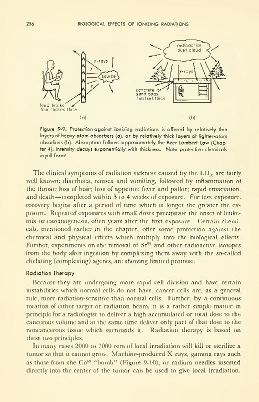
256 BIOLOGICAL EFFECTS OF IONIZING RADIATIONS
X
r>
ft
lead bricks" /four inches thick
(a)
y-rays
(_sourceS
concrete or
sand bags,two feet thick
(b)
Figure 9-9. Protection against ionizing radiations is offered by relatively thin
layers of heavy-atom absorbers (a), or by relatively thick layers of lighter-atom
absorbers (b). Absorption follows approximately the Beer-Lambert Law (Chap-
ter 4): intensity decays exponentially with thickness. Note protective chemicals
in pill form!
The clinical symptoms of radiation sickness caused by the LD 50 are fairly
well known: diarrhoea, nausea and vomiting, followed by inflammation of
the throat; loss of hair; loss of appetite; fever and pallor; rapid emaciation,
and death—completed within 3 to 4 weeks of exposure. For less exposure,
recovery begins after a period of time which is longer the greater the ex-
posure. Repeated exposures with small doses precipitate the onset of leuke-
mia or carcinogenesis, often years after the first exposure. Certain chemi-
cals, mentioned earlier in the chapter, offer some protection against the
chemical and physical effects which multiply into the biological effects.
Further, experiments on the removal of Sr90 and other radioactive isotopes
from the body after ingestion by complexing them away with the so-called
chelating (complexing) agents, are showing limited promise.
Radiation Therapy
Because they are undergoing more rapid cell division and have certain
instabilities which normal cells do not have, cancer cells are, as a general
rule, more radiation-sensitive than normal cells. Further, by a continuous
rotation of either target or radiation beam, it is a rather simple matter in
principle for a radiologist to deliver a high accumulated or total dose to the
cancerous volume and at the same time deliver only part of that dose to the
noncancerous tissue which surrounds it. Radiation therapy is based on
these two principles.
In many cases 2000 to 7000 rem of local irradiation will kill or sterilize a
tumor so that it cannot grow. Machine-produced X rays, gamma rays such
as those from the Co60 "bomb" (Figure 9-10), or radium needles inserted
directly into the center of the tumor can be used to give local irradiation.
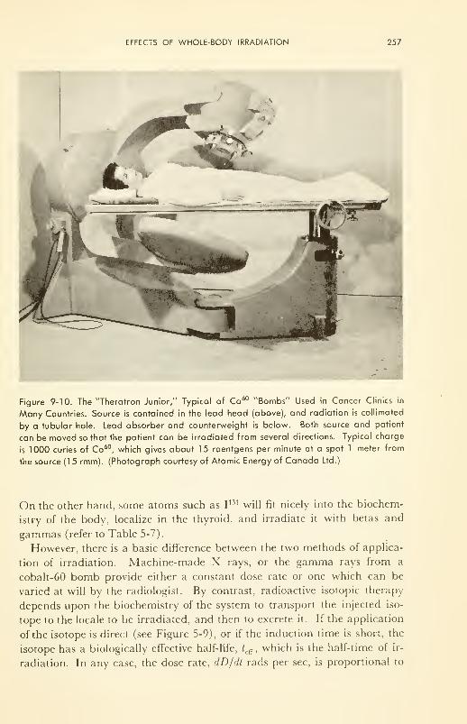
EFFECTS OF WHOLE-BODY IRRADIATION 257
Figure 9-10. The "Theratron Junior," Typical of Co60 "Bombs" Used in Cancer Clinics in
Many Countries. Source is contained in the lead head (above), and radiation is collimated
by a tubular hole. Lead absorber and counterweight is below. Both source and patient
can be moved so that the patient can be irradiated from several directions. Typical charge
is 1000 curies of Co60, which gives about 15 roentgens per minute at a spot 1 meter from
the source (15 rmm). (Photograph courtesy of Atomic Energy of Canada Ltd.)
On the other hand, some atoms such as I131 will fit nicely into the biochem-
istry of the body, localize in the thyroid, and irradiate it with betas and
gammas (refer to Table 5-7).
However, there is a basic difference between the two methods of applica-
tion of irradiation. Machine-made X rays, or the gamma rays from a
cobalt-60 bomb provide either a constant dose rate or one which can be
varied at will by the radiologist. By contrast, radioactive isotopic therapy
depends upon the biochemistry of the system to transport the injected iso-
tope to the locale to be irradiated, and then to excrete it. If the application
of the isotope is direct (see Figure 5-9), or if the induction time is short, the
isotope has a biologically effective half-life, /cff , which is the half-time of ir-
radiation. In any case, the dose rate, dD/dt rads per sec, is proportional to
258 BIOLOGICAL EFFECTS OF IONIZING RADIATIONS
the average energy*** of the emission and to the strength of the source, c
microcuries. Thus
dD/dt = 5.92 x lO- 4^where the constant arises from the definitions of the curie (3.7 x 10 10 disin-
tegrations per sec) and the rad (100 ergs absorbed per gram), and the fact
that 1 Mev = 1.6 x 10~ 6 ergs. In the case in which /eff
is shorter than the
physical half-life of the isotope, the dose received integrates to
DB {t) = 74V ;eff
(l - r»«W)for any time t; or
D& {*) = 74 E co t
efr
for the total dose administered (by an initial concentration, c microcuries,
of a beta emitter with an average energy Eg Mevs and a biological half-life
oHeff
days) up to the time the isotope has been practically completely ex-
creted. Table 9-6 gives pertinent data for different isotopes and organs. In
some cases ^eff
is limited by rapid chemical turnover, in others by the decay
half-life. Note that only a fraction of an isotope accumulates at a particular
locale in the system. Therapy depends upon preferential uptake by an organ.
The rest of the system gets irradiated too, but less.
P32 has been used successfully for the irradiation of excess white (leuke-
mia) and red (polycythaemia) blood cells. Other isotopes are being used in
ever-increasing numbers and amounts as new techniques (e.g., the insertion
of radioactive colloidal material (Au 198, for example) into the tumor: it
"floats," but it cannot get into the blood stream and be washed away), and
as new methods of preparation and purification become known.
The technique of bone-marrow therapy is now in an advanced state, al-
though its application is limited. The principle is the complete replacement
of irradiation-damaged marrow with that from a donor. Transplants are
normally limited to inbred strains or to isologous animals. However, if the
natural immunity reactions of human beings are completely destroyed by
large radiation doses first, then complete blood transplant can be successful.
Even so, further complications often arise later, in terms of a secondary
disease. Rare cases of transplant from one identical twin to another have
been more successful.
An advanced technique, which may keep radiologists in business even
***Genera!ly the average energy for gammas is about the same as the listed values, for gam-
mas are monoenergetic; but for betas the average energy, Eg, is approximately 1/3 the maxi-
mum (nominal) energy usually listed. For X rays the average energy is always well below the
peak value listed—about 0.3 of the nominal kvp if the soft end has not been filtered out (by,
say, 0.5 mm Al), and about 0.6 of the nominal kvp if it has.
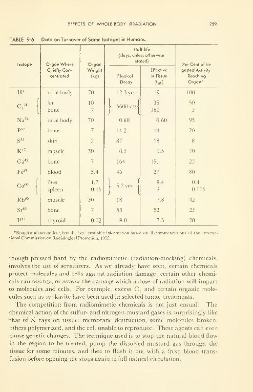
EFFECTS OF WHOLE-BODY IRRADIATION 259
TABLE 9-6.
260 BIOLOGICAL EFFECTS OF IONIZING RADIATIONS
This is the period of enthusiasm for the use of mustards in this new role
(they originally saw service as war gases). As their limitations for therapy
become better known, and if history repeats, both the new chemical therapy
and ionizing radiation therapy will oscillate through periods of enthusiasm
and reappraisal before ultimately finding their proper place in the medical
arsenal.
The reader may now wish to pursue the subject matter of this chapter in
more detail. The author suggests perusal of References 28 and 23, then of
References 10, 1, and 2.
PROBLEMS
9- 1 : (a) From tabulated values of the sensitivity constant, a, estimate the dose which
would be expected to kill 20, 50 and 80 per cent of a tumor.
(b) Suppose this tumor were just under the skin. Discuss three different ways
— ultrasonic, machine-made X-rays, and cobalt-60 gamma rays— in which
you could apply the irradiation.
(c) How would you monitor the air dose? The tissue dose?
9-2: How do you rationalize the facts that X rays induce cancer, and that X rays are
used in the treatment of cancer?
REFERENCES
1. Alexander, P., "Atomic Radiation and Life," Penguin Books, Inc., Baltimore,
1959: a "popular" introduction to the text, Ref. 2.
2. Bacq, Z. M. and Alexander, P., "Fundamentals of Radiobiology," Butter-
worths, London, 1955; 2nd ed., 1961.
3. Hollaender, A., Ed., "Radiation Biology," McGraw-Hill Book Co., Inc., NewYork, N. Y., 1954.
4. Oncley, J. L., et al., "Biophysical Science—A Study Program," John Wiley &Sons, Inc., New York, N. Y., 1959; especially the contributions by R. E.
Zirkle, W. Bloom, E. Pollard, T. H. Wood, C. A. Tobias and T. T. Puck on
radiation effects.
5. Livshits, N. N., "Physiological Effects of Nuclear Radiations on the Central
Nervous System," in Adv. in Biol, and Med. Pkys., 7, 174-241 (1960): a review
of the extensive Russian work, and that of others, on this important question.
6. Law, L. W., "Radiation Carcinogenesis," ibid., 7,295-337 (1960): a penetrating
survey of recent work, and a lucid account of the present position of knowledge
on radiation-induced neoplasms.
7. Howard-Flanders, P., "Physical and Chemical Mechanisms in the Injury of
Cells by Ionizing Radiations," ibid., 6, 554-596 (1958).
8. Kinsman, S., Ed., "Radiological Health Handbook," U. S. Dept. of Health,
Education and Welfare, 1954.
REFERENCES 261
9. International Conference, Geneva: "Peaceful Uses of Atomic Energy. II. Bio-
logical Effects of Radiation, "United Nations, New York, N. Y., 1955.
10. Butrer, J. A. V., "Inside the Living Cell," Methuen, London, 1960.
11. Swallow, A. J., "Radiation Chemistry of Organic Compounds," Pergamon
Press, London, 1960.
12. Appleton, G.J. and Krishnamoorthy, P. N., "Safe Handling of Radioisotopes:
Health Physics Addendum," Internat. Atomic Energy Agency, Vienna, 1960.
13. Hercik, F. and Jammet, H., "Safe Handling of Radioisotopes: Medical Adden-
dum," Internat. Atomic Energy Agency, Vienna, 1960.
14. Glasser, O., "Medical Physics," Vol. Ill, Year Book Publ. Inc., Chicago, 111.,
1960: many contributed articles on radiation effects on living tissue.
15. Cronkite, E. P., Bond, V. P., and Dunham, C. L., "Some Effects of Ionizing
Radiation on Human Beings," a Report by the U. S. Atomic Energy Com-mission, July, 1956.
16. Buchanan, A. R., Heim, H. C, Stilson, D. W., "Biomedical Effects of Exposure
to Electromagnetic Radiation," a Report to Life Support Systems Lab.,
Wright Air Development Div., USAF, 1960.
17. Shchepot'yeva, E. S., et al., "Effect of Oxygen in Ionizing Radiation." publ. by
State Publ. House for Medical Literature, Moscow, 1959 (U.S. A.E.C. Trans-
lation 4265, 1960).
18. "Report of the International Commission on Radiological Units and Measure-
ments," U. S. National Bureau of Standards Handbook 78, 1959.
19. Burton, M., Kirby-Smith, J. S., and Magee, J. L., "Comparative Effects of
Radiation,"John Wiley & Sons, Inc., New York, N. Y., 1960.
20. Kuzin, A. M., Shapiro, N. I., Livshits, N. N., and Breslavets, L. P., "Reviews
on Radiobiology," Inst. Biol. Physics, Publ. House Acad. Sci., SSSR, Mos-
cow, 1956 (U. S. Atomic Energy Commission Translation 3353).
21. Peacocke, A. R., "The Structure and Physical Chemistry of Nucleic Acids and
Nucleoproteins," Prog, in Biophys., 10, 55 (1960).
22. Don, C, "Radiation Hazards of Mass Miniature Radiography," Can. Med. Assn.
Jour., 84, 5-7 (1961).
23. Lea, D. E., "Actions of Radiations on Living Cells," 2nd ed., Cambridge Univ.
Press, 1955.
24. Henderson, I. H. S., "Electrochemical Radiation Dosimetry," Defence Research
Chemical Laboratories, Canada, Report No. 352, 1961.
25. Hine, G.J. and Brownell, G. L., "Radiation Dosimetry," Academic Press, Inc.,
New York, N. Y., 1956.
26. Smith, D. E., Ed., "Proc. Internat. Cong, of Radiation Research," Radiation
Research, Suppl. 1, Academic Press, Inc., New York, N. Y., 1959.
27. Augenstine, L. G., Ed., "Bioenergetics," Radiation Research, Suppl. 2, Academic
Press, Inc., New York, N. Y., 1960.
28. Allen, A. O., "The Radiation Chemistry of Water and Aqueous Solutions,"
D. Van Nostrand Co., Inc., Princeton, N. J., 1961.
29. Haissinsky, M., Ed., "The Chemical and Biological Actions of Radiations,"
Vols. 1 to 5, Academic Press, Inc., New York, N. Y.; Vol. 5, 1961.
CHAPTER 10
Biophysical Studies on
Nerve and Muscle
I had dissected a frog . . . and had placed it upon a table on which there
was an electric machine . . . . I took up the scalpel and moved its point close
to one or the other of the crural nerves of the frog, while at the same time
one of my assistants elicited sparks from the electric machine .... Strong
contractions took place in every muscle of the limb, and at the very moment
when sparks appeared the animal was seized as it were with tetanus ....
(Luigi Galvani, anatomist, surgeon, and obstetrician; 1781.)
This chapter presents an outline of some recent studies on nerve, and
shows how these are related to motion effected beyond the nerve endings by
excitable tissue in muscle. In the next chapter these facts are interpreted as
part of the enveloping concept of the human physical system. Then some
generalizations about this system are made which develop the framework in-
troduced in Chapter 1 and upon which the various parts of this book are
strung.
First, however: What is the nature of the physical apparatus— nerve and
muscle?
TRANSIENT BIOELECTRICS IN NERVE
In Chapter 7 the rest-condition of tissue was shown to exhibit voltage dif-
ferences in living membranes between the points at which solute activities
differ—and even in normal bulk tissue (Chapter 8) if bioelectric currents are
driven through it. Transient, or sudden, changes in voltages or currents are
262
TRANSIENT BIOELECTRICS IN NERVE 263
common, however, throughout living tissue, and play a uniquely important
role in nerve conduction. Here an electrical transient— the change in voltage
across the nerve cell membrane— is propagated with great speed along the
surface of the cell and along the nerve fiber formed by many axons in paral-
lel. The voltage change is the unit of information. First we describe how
this transmission takes place.
From Volta* to Hodgkin
It was in the late 1770's in Bologna that the Italian physician, Luigi
Galvani, and his wife Lucia observed quite by accident that the leg of the
frog with which they were experimenting could be made to twitch if certain
parts of the animal were touched simultaneously with the ends of two differ-
ent pieces of metal (iron and zinc, for example) joined together. Actually
they had discovered two things: the electrical voltage of a Zn-Fe couple, and
the electrochemical exictation of living tissue. In the succeeding two hun-
dred years a great body of facts has accumulated; these have demonstrated
quite conclusively the electrochemical nature of nerve conduction and the
resulting stimulation of excitable tissue. The afferent and efferent nerve
systems have been well tracked and catalogued—the job of the former being
to conduct commands, despatched by the brain, out to muscles and other
effector tissues. The so-called "all-or-none law," which says simply that the
excitable tissue will not fire (act) unless the stimulus has some minimum
power, and that the impulse moves down the nerve with constant amplitude
and velocity,** is now an accepted working principle for the physiologist.
Various chemical and physical methods have been developed to modify the
sensitivity of the nerve to stimulating agents—chemical catalysts in the form
of drugs; electrical pacemakers, etc.
However, even with all this great accumulation of useful knowledge on
how to modify the operation of the nervous system, it has only been since the
early 1930's that definitive examination could be made of several of the many
theories of operation of the nerve fiber. About that time it was realized
that the main nerve axon of the squid— in this respect unique among all
others— is a tube large enough (~1 mm od and several centimeters long
(see Figure 10-1)) to be examined both electrically and chemically, inside
and out. The fact that its physical structure could be examined by both
*In Phil. Trans., 1800, Alcssandro Volta, Professor Natural Philosophy, University of
Pavia, published a paper in which he not only described his new "artificial electric organ"
(i.e., the first storage battery), but also discussed the effects which electric current from this
invention "exercises on the different parts of our body," effects "which will open a very wide
field for reflection, . . . particularly interesting to Medicine."
**In certain unnatural media (sodium-deficient, for example) decremental propagation
occurs: both amplitude and velocity decrease as the impulse moves along the nerve.
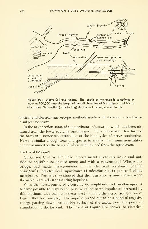
264 BIOPHYSICAL STUDIES ON NERVE AND MUSCLE
string
Figure 10-1. Nerve Cell and Axons. The length of the axon is sometimes as
much as 1 00,000 times the length of the cell. Insertion of Micropipets and Micro-
electrodes. Stimulating (or detecting) electrodes touching myelin sheath.
optical and electron-microscopic methods made it all the more attractive as
a subject for study.
In the next section some of the pertinent information which has been ob-
tained from the lowly squid is summarized. This information has formed
the basis of a better understanding of the biophysics of nerve conduction.
Nerve is similar enough from one species to another that some generalities
can be assumed on the basis of information gained from the squid axon.
The Era of the Squid
Curtis and Cole by 1936 had placed metal electrodes inside and out-
side the squid's tube-shaped axon; and with a conventional Wheatstone
bridge, had made measurements of the electrical resistance (20,000
ohms/cm2) and electrical capacitance (1 microfarad (^f) per cm 2
) of the
membrane. Further, they showed that the resistance is much lower when
the nerve is actively transmitting impulses.
With the development of electronic dc amplifiers and oscilloscopes, it
became possible to display the passage of the nerve impulse as detected by
thin platinum-wire contacts (electrodes) touching the nerve (see bottom of
Figure 10-1, for example). The impulse turned out to be a band of negative
charge passing down the outside surface of the axon, from the point of
stimulation to the far end. The insert in Figure 10-2 shows the electrical
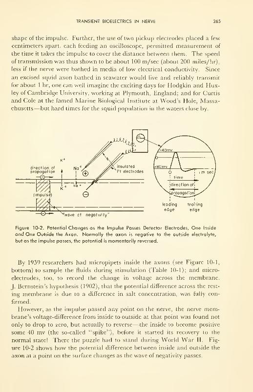
TRANSIENT BIOELECTRICS IN NERVE 265
shape of the impulse. Further, the use of two pickup electrodes placed a few
centimeters apart, each feeding an oscilloscope, permitted measurement of
the time it takes the impulse to cover the distance between them. The speed
of transmission was thus shown to be about 100 m/sec (about 200 miles/hr),
less if the nerve were bathed in media of low electrical conductivity. Since
an excised squid axon bathed in seawater would live and reliably transmit
for about 1 hr, one can well imagine the exciting days for Hodgkin and Hux-ley of Cambridge University, working at Plymouth, England; and for Curtis
and Cole at the famed Marine Biological Institute at Wood's Hole, Massa-
chusetts—but hard times for the squid population in the waters close by.
direction of
propagation f
(impulse)
*g£̂ ^wave of negativity'
leading
edgetrailing
edge
Figure 10-2. Potential Changes as the Impulse Passes Detector Electrodes, One Inside
and One Outside the Axon. Normally the axon is negative to the outside electrolyte,
but as the impulse passes, the potential is momentarily reversed.
By 1939 researchers had micropipets inside the axons (see Figure 10-1,
bottom) to sample the fluids during stimulation (Table 10-1); and micro-
electrodes, too, to record the change in voltage across the membrane.
J. Bernstein's hypothesis (1902), that the potential difference across the rest-
ing membrane is due to a difference in salt concentration, was fully con-
firmed.
However, as the impulse passed any point on the nerve, the nerve mem-brane's voltage-difference from inside to outside at that point was found not
only to drop to zero, but actually to reverse— the inside to become positive
some 40 mv (the so-called "spike"), before it started its recovery to the
normal state! There the puzzle had to stand during World War II. Fig-
ure 10-2 shows how the potential difference between inside and outside the
axon at a point on the surface changes as the wave of negativity passes.
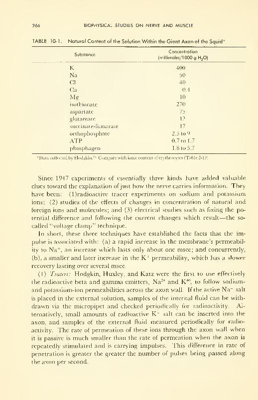
266 BIOPHYSICAL STUDIES ON NERVE AND MUSCLE
TABLE 10-1. Natural Content of the Solution Within the Giant Axon of the Squid"
SubstanceConcentration
(millimoles/lOOOg H20)
K 400
Na 50
CI 40
Ca 0.4
Mg 10
isothionate 270
aspartate 75
glutamate 12
succinate-fumarate 17
orthophosphate 2.5 to 9
ATP 0.7 to 1.7
phosphagen 1 .8 to 5.7
*Data collected by Hodgkin. 14 Compare with ionic content of erythrocytes (Table 2-1)'.
Since 1947 experiments of essentially three kinds have added valuable
clues toward the explanation ofjust how the nerve carries information. They
have been: (l)radioactive tracer experiments on sodium and potassium
ions; (2) studies of the effects of changes in concentration of natural and
foreign ions and molecules; and (3) electrical studies such as fixing the po-
tential difference and following the current changes which result—the so-
called "voltage clamp'1
technique.
In short, these three techniques have established the facts that the im-
pulse is associated with: (a) a rapid increase in the membrane's permeabil-
ity to Na +, an increase which lasts only about one msec; and concurrently,
(b), a smaller and later increase in the K+ permeability, which has a slower
recovery lasting over several msec.
(1) Tracers: Hodgkin, Huxley, and Katz were the first to use effectively
the radioactive beta and gamma emitters, Na24 and K40, to follow sodium-
and potassium-ion permeabilities across the axon wall. If the active Na + salt
is placed in the external solution, samples of the internal fluid can be with-
drawn via the micropipet and checked periodically for radioactivity. Al-
ternatively, small amounts of radioactive K + salt can be inserted into the
axon, and samples of the external fluid measured periodically for radio-
activity. The rate of permeation of these ions through the axon wall when
it is passive is much smaller than the rate of permeation when the axon is
repeatedly stimulated and is carrying impulses. This difference in rate of
penetration is greater the greater the number of pulses being passed along
the axon per second.
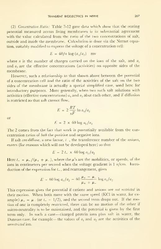
TRANSIENT BIOELECTRICS IN NERVE 267
(2) Concentration Ratio: Table 7-12 gave data which show that the resting
potential measured across living membranes is in substantial agreement
with the value calculated from the ratio of the two concentrations of salt,
outside and inside the membrane. Calculation is done via the Nernst equa-
tion, suitably modified to express the voltage of a concentration cell:
E = 60/n log (a,/<32 ) mv
where n is the number of charges carried on the ions of the salt, and a,
and a-, are the effective concentrations (activities) on opposite sides of the
membrane.
However, such a relationship as that shown above between the potential
of a concentration cell and the ratio of the activities of the salt on the two
sides of the membrane is actually a special simplified case, used here for
introductory purposes. More generally, when two such salt solutions with
activities (effective concentrations) a, and a2abut each other, and if diffusion
is restricted so that salt cannot flow,
E = 2 —=- In ax/a
2
nFor
E = 2 x 60 log a}/a
2
The 2 comes from the fact that work is potentially available from the con-
centration ratios of both the positive and negative ions.
If salt can diffuse, a new factor, /_ , the transference number of the anions,
enters (for reasons which will not be developed here) so that
£ = 2 L x 60 log a, /a2
Here /_ = /u_/(/*+ + M- ), where the yu's are the mobilities, or speeds, of the
ions in centimeters per second when the voltage gradient is 1 v/cm. Intro-
duction of the expression for <_ , and rearrangement, gives
E = 60 log a, /a2- 60
M+ ~ M~log aja
2
M+ + M-
This expression gives the potential if cations and anions are not restricted in
their motion. When both move with the same speed (KC1 in water, for ex-
ample) n+ = M- (or t_ = 1/2), and the second term drops out. If the mo-
tion of one is completely restricted, there can be no motion of the other if
micro-neutrality is to be maintained, and the potential is given by the first
term only. In such a case—charged protein ions plus salt in water, the
Donnan case, for example—the values of a, and a2are the activities of the
unrestricted ion.
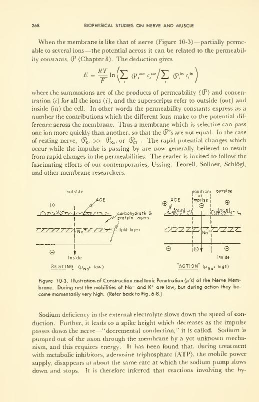
268 BIOPHYSICAL STUDIES ON NERVE AND MUSCLE
When the membrane is like that of nerve (Figure 10-3)—partially perme-
able to several ions—the potential across it can be related to the permeabil-
ity constants, (P (Chapter 8). The deduction gives
RTE = -= ln (Z ®r cr/H (?r c
J
where the summations are of the products of permeability ((P) and concen-
tration (c) for all the ions (z), and the superscripts refer to outside (out) and
inside (in) the cell. In other words the permeability constants express as a
number the contributions which the different ions make to the potential dif-
ference across the membrane. Thus a membrane which is selective can pass
one ion more quickly than another, so that the (P's are not equal. In the case
of resting nerve, (?K+ » (PNa+ or (PCI _ . The rapid potential changes which
occur while the impulse is passing by are now generally believed to result
from rapid changes in the permeabilities. The reader is invited to follow the
fascinating efforts of our contemporaries, Ussing, Teorell, Sollner, Schldgl,
and other membrane researchers.
outside position! outside
!
ofACE _ ACE impulse ^
© , // ®J I _®_
r\r^^S^r^'\r^^-iy-\ /v. carbohydrate 8 ^*-^rPh$\protein layers.
ZZZ^ZZ^^DV^aTZZCZy^f \^d layer ZL^ZT^-T^f^
I
1
© tinside inside
I©*
loJ^r^
Na +| -
l
I
+
RESTING (p NQ+ low) ACTION (/J Na+ high)
Figure 10-3. Illustration of Construction and Ionic Penetration (;u's) of the Nerve Mem-
brane. During rest the mobilities of Na + and K+ are low, but during action they be-
come momentarily very high. (Refer back to Fig. 6-8.)
Sodium deficiency in the external electrolyte slows down the speed of con-
duction. Further, it leads to a spike height which decreases as the impulse
passes down the nerve—"decremental conduction," it is called. Sodium is
pumped out of the axon through the membrane by a yet unknown mecha-
nism, and this requires energy. It has been found that, during treatment
with metabolic inhibitors, adenosine triphosphate (ATP), the mobile power
supply, disappears at about the same rate at which the sodium pump slows
down and stops. It is therefore inferred that reactions involving the hy-
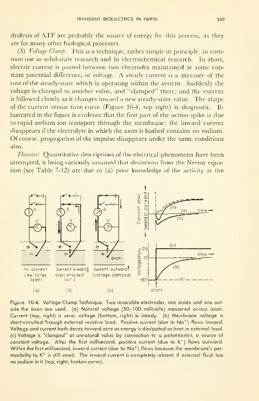
TRANSIENT BIOELECTRICS IN NERVE 269
drolysis of ATP are probably the source of energy for this process, as they
are for many other biological processes.
(3) Voltage Clamp: This is a technique, rather simple in principle, in com-
mon use in solid-state research and in electrochemical research. In short,
electric current is passed between two electrodes maintained at some con-
stant potential difference, or voltage. A steady current is a measure of the
rate of the steady-state which is operating within the system. Suddenly the
voltage is changed to another value, and "clamped" there; and the current
is followed closely as it changes toward a new steady-state value. The shape
of the current versus time curve (Figure 10-4; top right) is diagnostic. Il-
lustrated in the figure is evidence that the first part of the action spike is due
to rapid sodium ion transport through the membrane: the inward current
disappears if the electrolyte in which the axon is bathed contains no sodium.
Of course, propagation of the impulse disappears under the same conditions
also.
Theories: Quantitative descriptions of the electrical phenomena have been
attempted, it being variously assumed that deviations from the Nernst equa-
tion (see Table 7-12) are due to (a) poor knowledge of the activity at the
6^y -®-
no current
( switchesopen)
<h
\-®~
e
axon
current inwordl current outwardf
(cell shorted (voltage clamped)out )
(a) (b) (c) start
Figure 10-4. Voltage Clamp Technique. Two reversible electrodes, one inside and one out-
side the axon are used, (a) Natural voltage (50-100 millivolts) measured across axon.
Current (top, right) is zero; voltage (bottom, right) is steady, (b) Membrane voltage is
short-circuited through external resistive load. Positive current (due to Na'
) flows inward.
Voltage and current both decay toward zero as energy is dissipated as heat in external load,
(c) Voltage is "clamped" at unnatural value by connection to a potentiostat, a source of
constant voltage. After the first millisecond, positive current (due to K +) flows outward.
Within the first millisecond, inward current (due to Na +) flows because, the membrane's per-
meability to K + is still small. The inward current is completely absent if external fluid has
no sodium in it (top, right; broken curve).
270 BIOPHYSICAL STUDIES ON NERVE AND MUSCLE
given concentrations; (b) leaky membranes, through which the Na+ and K +
permeate, or diffuse down their respective gradients; (c) electrical charges
permanently fixed within the hundred angstroms or so of effective thickness
of membrane; or (d) changes in shape of acetylcholinesterase (ACE), an
enzyme located on the surface of nerve (Figure 10-3 illustrates) and thought
by some to be the cover whose shape determines whether or not Na+, or K h
,
or both, can enter the slip through the pores in the membrane.
Two quantitative theories permeate the literature on nerve transmission:
the use of the electrical cable theory to describe the spread of a localized
electrical disturbance; and the description of ionic currents through the
membrane as a function of permeability.
Early in the century electrical engineers had worked out the effect of a
break, or a series of breaks, in the insulation of an electric cable having a
metallic conductor inside and salt water outside. By 1938 Curtis and Cole
had used this application of Ohm's law to describe how a localized dis-
turbance in a nerve membrane can spread on down the nerve. The key
expression is:
d Vd2 V
=r
x+ r
2
dx 2r
E- V - rCmdt
where /: is the concentration-cell voltage across the membrane in the ab-
sence of a disturbance, i.e., when the membrane is resting; V is the "action"
voltage at any time, /, at a distance, x, along the surface from the site of the
disturbance, 0; r, and r2are the electrolytic resistances (ohms), between
and x, in the outside conductor and the inside conductor, respectively;
r is the resistivity of the membrane (fixed, unknown thickness) in ohm cm 2;
and Cm is the capacitance of the membrane, which is being depolarized (dis-
charged) at a rate dV / dt. The expression teaches that the depolarization oc-
curs at a rate which increases as the divergence (spread) of voltage along the
surface increases, and decreases as the resistances to ion flow (r„ r2 , and
r) increase.
By 1952 Hodgkin and Huxley had described measured changes in mem-
brane conductance of the giant axon of the squid in terms of change in the
permeabilities of the simple ions of the external and internal media. The
principle ideas of this theory will now be given.
Currents through the membrane are considered to charge (or discharge)
the membrane capacitance and to leak Na +, K+
, and other ions as well.
Thus:/ = Cm dV/dt + /Na+ + /K+ + /
where /is total current, and the 7,'s are the currents due to the different ions.
Then each /, is expressed as being the product of the membrane conduct-
ance (g t) and the driving voltage for that ion. Thus: I
t
= g tA V
TRANSIENT BIOELECTRICS IN NERVE 271
Each g t
was then related by a phenomenological trick to time and voltage in
such a way as to fit the experimental results. Thus, for potassium ion,
n . fj max„4.6K+ ~ &K + "
and
dn= a„n{\ — n) — B„n
at
where n is a dimensionless parameter which has a value between and 1;
it is time-dependent and is related to voltage-dependent penetration con-
stants, anand /?n
. The first, an , expresses the rate of K+ movement into the
cell, and /?nexpresses its rate out. Similar expressions have been devised for
Na+ and the other ions of the system. From these expressions the total cur-
rent (/) can be expressed in terms of time-dependent and voltage-dependent
parameters related to permeability. With proper choice of the values of the
different parameters, the experimental values of conductance as a function
of time and voltage can be completely described.
These two theories have been bright lights in the quantitative descrip-
tion of nerve propagation. The interested reader is referred to the analyti-
cal and summary papers 21,22 for the detailed arguments. The papers are
difficult, but rewarding.
The charged-pore theory of membrane potential differences has been suc-
cessful with synthetic membranes of collodion, ion-exchanger resins, and
other synthetic polymers. It will not be developed here, although it has been
put into elegant quantitative form by Meyer and Siever and, more recently,
by Teorell.
This is a very active and important part of biophysics today, and, as was
stated in Chapter 6, probably there is no part of the research in the subject
which will be more rewarding. Hodgkin's Croonian Lecture 14is an excellent
statement of the state of the art, and Nachmansohn's recent, short review, 18
more from the biochemical viewpoint, will nicely balance the further devel-
opment of the reader's concepts.
Is Semiconductivity Important?
It may be. We saw in Chapter 4 that the tt electrons of many organic
compounds have a certain freedom and can move under the influence of an
electric field. Most vertebrate nerve is sheathed in myelin, the protein-and-
fat wrapping formed by the doubled membrane of the Schwann cells. This
is illustrated schematically in Figure 10-1, top right, and shown very dra-
matically by the electron micrograph, Figure 10-5. The myelin sheath offers
physical protection to the fine nerve fibers of vertebrates. But it has further
roles. For instance, since it completely covers the nerve fiber except at
certain interruptions about 1 mm apart, called the nodes of Ranvier (Fig-
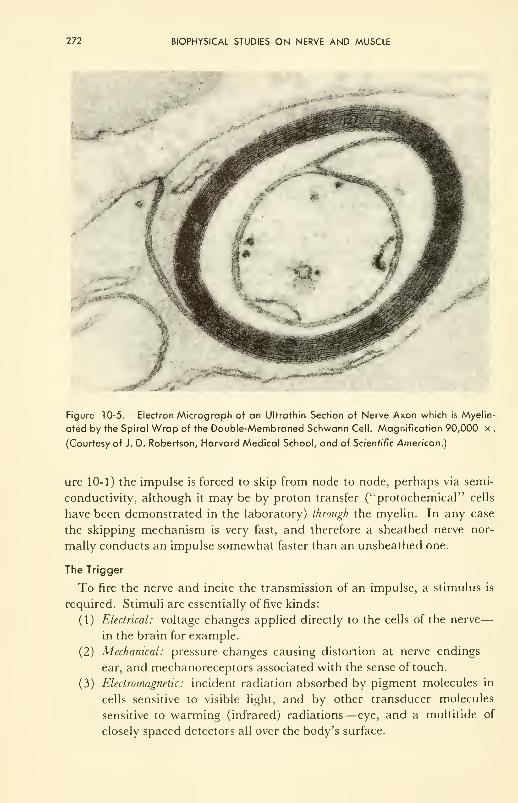
272 BIOPHYSICAL STUDIES ON NERVE AND MUSCLE
Figure 10-5. Electron Micrograph of an Ultrathin Section of Nerve Axon which is Myelin-
ated by the Spiral Wrap of the Double-Membraned Schwann Cell. Magnification 90,000 x .
(Courtesy of J. D. Robertson, Harvard Medical School, and of Scientific American.)
ure 10-1) the impulse is forced to skip from node to node, perhaps via semi-
conductivity, although it may be by proton transfer ("protochemical" cells
have been demonstrated in the laboratory) through the myelin. In any case
the skipping mechanism is very fast, and therefore a sheathed nerve nor-
mally conducts an impulse somewhat faster than an unsheathed one.
The Trigger
To fire the nerve and incite the transmission of an impulse, a stimulus is
required. Stimuli are essentially of five kinds:
(1) Electrical: voltage changes applied directly to the cells of the nerve
—
in the brain for example.
(2) Mechanical: pressure changes causing distortion at nerve endings
—
ear, and mechanoreceptors associated with the sense of touch.
(3) Electromagnetic: incident radiation absorbed by pigment molecules in
cells sensitive to visible light, and by other transducer molecules
sensitive to warming (infrared) radiations— eye, and a multitide of
closely spaced detectors all over the body's surface.
TRANSIENT BIOELECTRICS IN NERVE 273
(4) Chemical: foreign chemicals applied, or changes in concentration of
natural chemicals— taste buds, dehydrated tissue cells, etc.
(5) Gravitational: continuous attraction to earth, occasionally varied by
superposition of various accelerations—balance-detectors in middle
ear, for example. (These are essentially of type (2).)
Deserving special mention as a trigger is trie "pacemaker" of the heart,
which in man repetitively stimulates the pump to compress and relax once
about every 1.3 sec 24 hr a day for life. Recordings from microelectrodes
inserted into pacemaker cells show that they are self-contained oscillators.
Very recently D. Noble has shown 19 that if certain limiting conditions are
imposed on the cable-and-changing-permeability theory described above,
the theory can describe the condition of oscillating permeability and oscil-
lating potential of the membrane of the pacemaker cell.
When and if the pacemaker fails, it has been shown to be possible to
stimulate the heart artificially. With small transistor circuitry and small
zinc-mercuric oxide batteries, it has been demonstrated recently that an
artificial pacemaker can be buried, by surgery, in the abdominal cavity
under the skin and stimulate a weak heart regularly for at least a year be-
fore the battery has to be changed (again by surgery). This device has
brought a normal life to many people.
Recent advances in microelectrode preparation have permitted glass tubes
to be drawn down to an outer diameter of 0.0005 cm, filled with electrolyte,
and the ends inserted carefully right into the individual muscle cells in the
animal's beating heart. Thus the electrical measurements on cells working in
situ are now being made. Great care has to be taken that the electrical meas-
urements are not affected by the huge electrical resistance of these micrelec-
trodes (try Problem 10-5). For steady potentials an electrometer with a high
impedance is usually used; but for rapidly-varying potentials, such an in-
strument is too slow to follow the potential changes without inducing dis-
tortion. There this problem of measurement presently rests. Once it is
solved, although the cross-correlation of electrical and chemical information
may still not be possible in these small cells because of the size of the object
under study, pharmacological problems should receive much attention with
this technique. Indeed the neuromuscular junction is already being so ex-
plored.
Studies on the Central Nervous System
By contrast with the normally resting peripheral nerve tissue, which is
activated upon demand, the brain is a mass of spontaneously pulsating neu-
ral networks, seemingly continuously energized and active. It is usually as-
sumed that the basic processes are electrochemical, like those just outlined
as being proper to nerve conduction. However, biophysical knowledge of
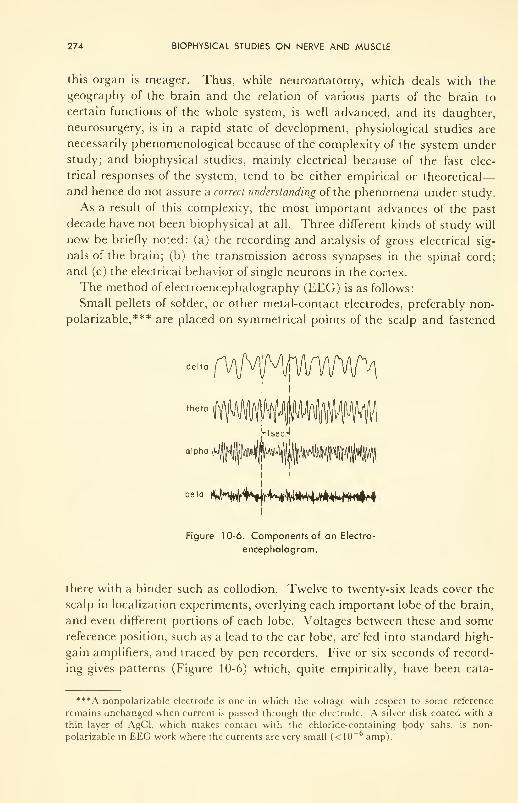
274 BIOPHYSICAL STUDIES ON NERVE AND MUSCLE
this organ is meager. Thus, while neuroanatomy, which deals with the
geography of the brain and the relation of various parts of the brain to
certain functions of the whole system, is well advanced, and its daughter,
neurosurgery, is in a rapid state of development, physiological studies are
necessarily phenomenological because of the complexity of the system under
study; and biophysical studies, mainly electrical because of the fast elec-
trical responses of the system, tend to be either empirical or theoretical
—
and hence do not assure a correct understanding of the phenomena under study.
As a result of this complexity, the most important advances of the past
decade have not been biophysical at all. Three different kinds of study will
now be briefly noted: (a) the recording and analysis of gross electrical sig-
nals of the brain; (b) the transmission across synapses in the spinal cord;
and (c) the electrical behavior of single neurons in the cortex.
The method of electroencephalography (EEG) is as follows:
Small pellets of solder, or other metal-contact electrodes, preferably non-
polarizable,*** are placed on symmetrical points of the scalp and fastened
delta vI
theta
alpha
Hsed
beta 4h4H|rWpvHM^
Figure 10-6. Components of an Electro-
encephalogram.
there with a binder such as collodion. Twelve to twenty-six leads cover the
scalp in localization experiments, overlying each important lobe of the brain,
and even different portions of each lobe. Voltages between these and some
reference position, such as a lead to the ear lobe, are" fed into standard high-
gain amplifiers, and traced by pen recorders. Five or six seconds of record-
ing gives patterns (Figure 10-6) which, quite empirically, have been cata-
***A nonpolarizable electrode is one in which the voltage with respect to some reference
remains unchanged when current is passed through the electrode. A silver disk coated with a
thin layer of AgCl, which makes contact with the chloride-containing body salts, is non-
polarizable in EEG work where the currents are very small (< 10 amp).
TRANSIENT BIOELECTRICS IN NERVE 275
logued as coming from normal or diseased tissue. Patterns taken on an indi-
vidual vary with the emotional state. A creative man is said to have pat-
terns quite different from one who lacks new ideas. However, the fine struc-
ture of these waves is not well understood. Recorded spikes are only about
150 nv high. Characteristic spikes of different shapes and frequencies have
been named alpha, theta, delta, etc. These are depicted in Figure 10-6.
Location of tumors, via predominance of the delta waves (see Table 10-2),
has been particularly successful, with 73 to 90 per cent accuracy claimed.
Bagchi has reported 84 per cent in 333 tries. Other abnormalities, such as
epilepsy, have been studied by this technique.
TABLE 10-2. Classification of Electroencephalograph Waves.
Names of
WavesFrequency (cps) Association
delta 0.5 to 3.5 "disease, degeneration,
death; defence"*
theta 6 to 7
alpha 8 to 13 a scanning mechanism?
beta 14 to 30 alertness; active response
Walter, W. Gray, "The Living Brain," Penguin Books, Baltimore, 1960, p. 81.
While the all-encompassing phenomenological techniques of EEG have
been making useful contributions to life, studies of individual neurons, via
microelectrodes in the cortex, and studies of the properties of synapses and
ganglia in the spinal cord have demonstrated interesting phenomena such
as: inhibition of transmission across nerve endings (strong signals passed
through one nerve ending reduce the effectiveness of one close by); post-
tetanic potentiation (faster and more energetic transmission through a par-
ticular nerve path following a rapid succession of pulses through that path);
and the promotion of epileptic-like seizures and peculiar mental images in
man by electrical stimulation of particular spots in the cortex via micro-
electrodes.
Transfer of an impulse across a synapse (Figure 10-7) is currently thought
to be by means of "chemical" transfer rather than by "electrical," for two
reasons: the observed salt concentration changes associated with a single im-
pulse are very small; and there is fairly good evidence that acetylcholine
(ACh) accumulates in the gap during transmission across the gap. A theory
is that ACh is contained in the many little vesicles in the pre-synaptic end-
ing: that, during "activity," ACh is expelled through the membrane and
diffuses to the post-synaptic membrane and locally depolarizes it. The de-
tails of this mechanism are still unknown.
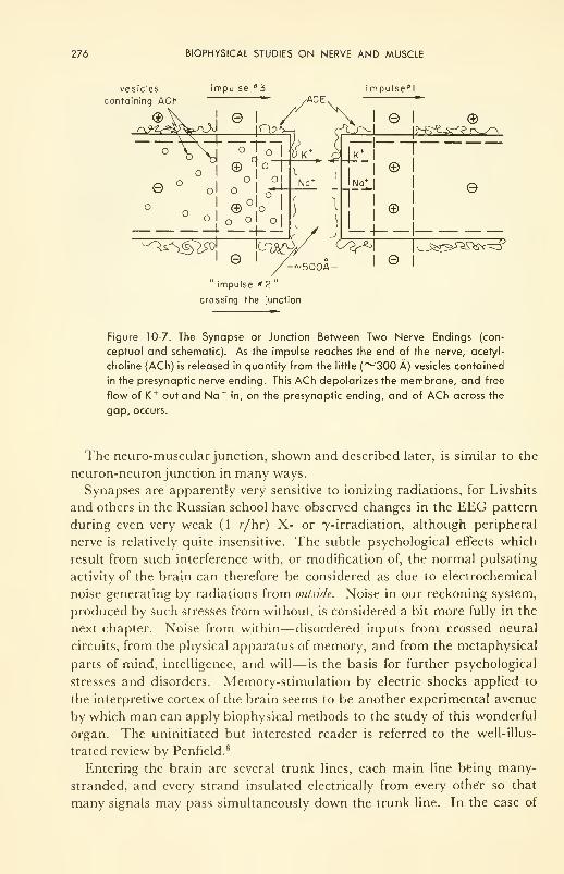
276 BIOPHYSICAL STUDIES ON NERVE AND MUSCLE
vesicles
containing ACh
e
impulse "3 i mpulse*!/ACE/\l8l ©
C i-K
e
impulse #2
crossing the junction
|^?^-^?r^^
No* I
fl I
©
©
;7v5^1 ^wy &>?^ io^^^sv^y500A- ©
Figure 10-7. The Synapse or Junction Between Two Nerve Endings (con-
ceptual and schematic). As the impulse reaches the end of the nerve, acetyl-
choline (ACh) is released in quantity from the little (~300 A) vesicles contained
in the presynaptic nerve ending. This ACh depolarizes the membrane, and free
flow of K + out and Na +in, on the presynaptic ending, and of ACh across the
gap, occurs.
The neuromuscular junction, shown and described later, is similar to the
neuron-neuron junction in many ways.
Synapses are apparently very sensitive to ionizing radiations, for Livshits
and others in the Russian school have observed changes in the EEG pattern
during even very weak (1 r/hr) X- or 7-irradiation, although peripheral
nerve is relatively quite insensitive. The subtle psychological effects which
result from such interference with, or modification of, the normal pulsating
activity of the brain can therefore be considered as due to electrochemical
noise generating by radiations from outside. Noise in our reckoning system,
produced by such stresses from without, is considered a bit more fully in the
next chapter. Noise from within— disordered inputs from crossed neural
circuits, from the physical apparatus of memory, and from the metaphysical
parts of mind, intelligence, and will— is the basis for further psychological
stresses and disorders. Memory-stimulation by electric shocks applied to
the interpretive cortex of the brain seems to be another experimental avenue
by which man can apply biophysical methods to the study of this wonderful
organ. The uninitiated but interested reader is referred to the well-illus-
trated review by Penfield. 8
Entering the brain are several trunk lines, each main line being many-
stranded, and every strand insulated electrically from every other so that
many signals may pass simultaneously down the trunk line. In the case of
THE MOLECULAR BASIS OF MUSCLE CONTRACTION 277
the two optic nerve trunks, a nerve-ending from each fiber carries an im-
pulse from a rod or cone to a bipolar cell, thence to the brain. There is evi-
dence now that insulation among these strands is not complete, and that
parallel signals from two may trigger a third, and so on. This is a mecha-
nism which seems to be operative in color vision, as was inferred in the dis-
cussion on that subject in Chapter 4. Cross-stimulation seems to be very
generally operable, for there is a great deal of psychological evidence that
saturation of one sensing organ will have a marked effect on the sensitivity
of another. Mentioned earlier was the dentist's new trick of flooding the ear
with noise of a suitable frequency so that the pain of drilling cannot be felt!
The physical network which accommodates, sorts, and retains certain im-
pulses and rejects others is a topic for future study. Furthermore, memoryis still a very mysterious phenomenon. One recent proposal about the phy-
sical mechanism of memory deserves mention: the "training" of the neural
network to store information is done by means of the synthesis of certain
("different") protein molecules. These result from a change in shape of the
ribonucleic acid (RNA) effected by a passing stimulus— i.e., the RNA within
nerve and neighboring glial (Schwann) cells. Although this does not sound
very convincing at first glance, it seems to be the best model yet put forward
in the baffling question of what is the physical apparatus of memory; and
it certainly is consistent with the known fact that the rate of protein synthesis
is very high in active nerve cells. One cannot help thinking that these "dif-
ferent" proteins may be imbedded right in the membrane, and exert their
effect as "permanent" changes in its permeability. In conclusion, one could
say that, from the biophysical point of view, the study of the central nervous
system is becoming more and more a study in applied electrochemistry, a
study of membrane biophysics.
THE MOLECULAR BASIS OF MUSCLE CONTRACTION
By means of nerve, the brain exercises control over both chemical and
physical processes in the body. There are good examples of each: for the
former, the endocrine gland system; and for the latter, muscle. Of the two,
the latter is in many ways inherently less complicated, and only it will be
discussed in this attempt to illustrate how control is achieved in a particular
case of a physical action. For this we need to know some relevant physical
properties of muscle tissue; and, more important still from the biophysical
point of view, we need to know the molecular behavior which is at the root
of this physical behavior. Fortunately, both electron microscopic examina-
tion of muscle-tissue slices, and kinetic methods of analysis of rate data seem
to be succeeding with this problem of providing an understanding of mus-
cular contraction. On the other hand, a review of muscular contraction from
the molecular viewpoint has the added advantage of illustrating the powerful
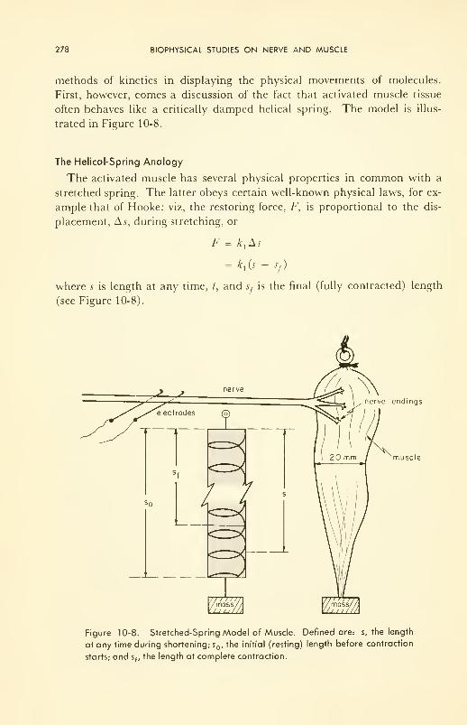
278 BIOPHYSICAL STUDIES ON NERVE AND MUSCLE
methods of kinetics in displaying the physical movements of molecules.
First, however, comes a discussion of the fact that activated muscle tissue
often behaves like a critically damped helical spring. The model is illus-
trated in Figure 10-8.
The Helical-Spring Analogy
The activated muscle has several physical properties in common with a
stretched spring. The latter obeys certain well-known physical laws, for ex-
ample that of Hooke: viz, the restoring force, F, is proportional to the dis-
placement, As, during stretching, or
F k,As
= kt(s - s
f )
where s is length at any time, t, and Sj is the final (fully contracted) length
(see Figure 10-8).
nerve endings
muscle
Figure 10-8. Stretched-Spring Model of Muscle. Defined are: s, the length
at any time during shortening; s , the initial (resting) length before contraction
starts; and s,, the length at complete contraction.
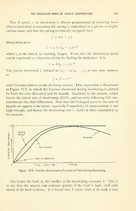
THE MOLECULAR BASIS OF MUSCLE CONTRACTION 279
Now if speed, v, of shortening is always proportional to restoring force
(this is equivalent to assuming the spring is embedded in a plastic or highly
viscous mass, and that the spring is critically damped) then:
Integration gives
k(s - sf )
s, + (s()- s
f)e-•7 1 v J 7.
where s is the initial, or starting, length. From this the shortening speed
can be expressed as a function of time by finding the derivative. It is
v = k(s — s,)e~ kt
The fraction shortened,/, defined as (sQ— ^)/(.r — Sj), at any time reduces
to
/= 1- -*/
and k becomes known as the shortening constant. This expression is illustrated
in Figure 10-9, in which the fraction shortened during shortening is plotted
for both the case discussed and for muscle. Elasticity in the muscle, which
lowers the initial rate of shortening (df/dt), and recovery following full con-
traction are the chief differences. Note that the ^-shaped curve in the case of
muscle can appear to be linear, especially if sensitivity of measurement is not
high enough; and hence the shortening rate (— ds/dt) is often considered to
be constant.
T ime 0,5 sec
Figure 10-9. Fraction Shortened as Function of Time During Shortening.
The larger the load, m, the smaller is the shortening constant, k. This is
to say that the muscle can contract quickly if the load is light, and only
slowly if the load is heavy. It is found that k varies with m in such a way
280 BIOPHYSICAL STUDIES ON NERVE AND MUSCLE
that momentum, mv, is conserved (remains constant):
£) I —1 = constant
\ max / \ max /
where v max is the maximum speed of shortening (no load), and ra max is the
largest weight which can be lifted. Here v and m are constants. This result
is often written as a product of velocity and force, when the acceleration is
that due to gravity— a constant; it then becomes the "force-velocity rela-
tionship." Thus for two masses, m, and m2 , momentum conservation is
expressed
<mx
— m \ lv~, — vAfm 2— m^
max / \ max / \ max / \ max
The denominators cancel out. Then if mass 2 is chosen to be just big enough
that the muscle can sustain it but not lift it, v2= 0, and m
2= mmax . Mul-
tiplication through by g, the acceleration due to gravity, converts masses to
forces (F = mg), and then rearrangement gives
(F+a)(v + b) = (F. + aji,
the force-velocity relationship, first stated in 1938 by A. V. Hill (a and b are
his constants, equal to -gm and -v respectively). Figure 10-10 illustrates
this equation, and says simply that the greater the force to be overcome by
the contracting muscle the less the speed at which it can contract. Rear-
ranged in the form
v = (Fmax -nb/(F+a)
it says that the velocity of shortening depends upon the difference between
the maximum force it can develop and the actual force on the muscle. This
hyperbolic relationship is obeyed by a wide variety of muscle and muscle
systems, including the human arm. 16
On Energetics of Muscle
The work done by the muscle in lifting a weight is given by the product
mgh, where g is the acceleration due to gravity (and therefore mg is force, since
F = ma) and h is height to which the weight is lifted. We saw in Chapter 7
that part (ALT') of hydrolysis of ATP— a reaction catalyzed by the con-
tractile enzyme, myosin—could appear as work of contraction. Thus:
A5' = mgh
and the rest of the total free energy of reaction (A?F) is wasted because of ir-
reversibility or inefficiency in the process, and thrown away as heat, TAS '.
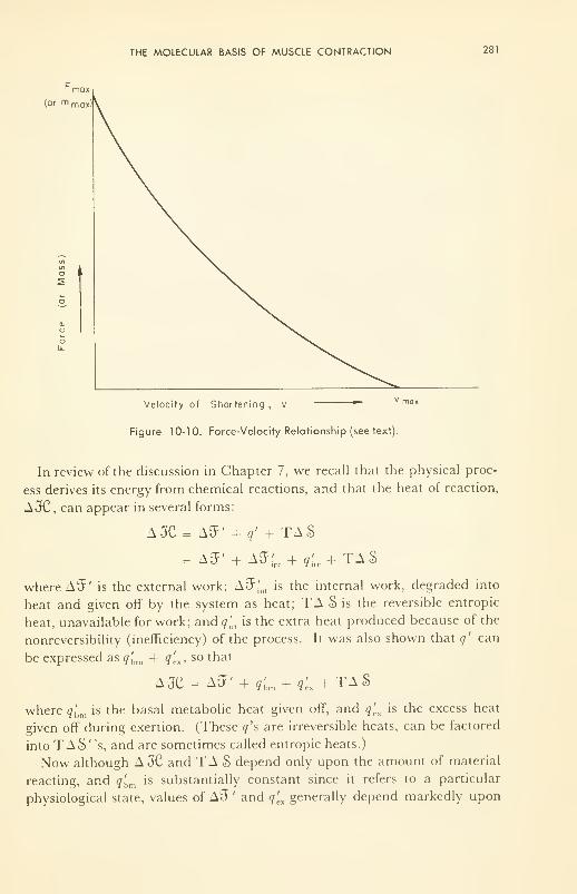
THE MOLECULAR BASIS OF MUSCLE CONTRACTION 281
r max
(or rn max )
Velocity of Shortening , v ""
Figure 10-10. Force-Velocity Relationship (see text).
In review of the discussion in Chapter 7, we recall that the physical proc-
ess derives its energy from chemical reactions, and that the heat of reaction,
A3C, can appear in several forms:
A3C = AS' + q' + TAS
= a^' + a^ + ^ + taS
where A31' is the external work; AO1
^, is the internal work, degraded into
heat and given off by the system as heat; TA S is the reversible entropic
heat, unavailable for work; and q'm is the extra heat produced because of the
nonreversibility (inefficiency) of the process. It was also shown that q' can
be expressed as q'bm + <7ex> so tnat
A3C = AJF' + ^ + ^ + TAS
where q'hm is the basal metabolic heat given off, and q'
(.x
is the excess heat
given off during exertion. (These r/'s are irreversible heats, can be factored
into TA S "s, and are sometimes called entropic heats.)
Now although A JC and TA S depend only upon the amount of material
reacting, and q'bm is substantially constant since it refers to a particular
physiological state, values of A^ ' and q[.xgenerally depend markedly upon
282 BIOPHYSICAL STUDIES ON NERVE AND MUSCLE
the rate at which the physical process occurs. Thus the faster the process the
less efficient it is: i.e., the greater the fraction q'cx/(A^' + q'
ex ) which is
lost as heat, and the less is the fraction A$7'/(A;F' + q'ex ) which is realized
as external work. However, whether the work is done fast or slowly, nu-
merically the same amount of work is done; and therefore, because Ao' is
independent of speed, so must q'tx be, provided the same amount of fuel is
consumed. It is a well-established experimental finding that the total heat,
£•)', given out during a shortening (the "contraction heat
,,
)
is constant, independent of speed of shortening. However, ~/ ', is propor-
tional to the distance (a) shortened; i.e., °c ax; and the constant a has the
same dimensions (energy/distance, or force) and numerical value (~400 g
wt/cm 2 area of cross section) as the a in the force-velocity relationship. The
significance of this coincidence is not yet clear.
Careful measurements, with small thermocouples imbedded in the muscle
and fast galvanometers to record small electrical currents, have shown that
the contraction heat is composed of two parts: a rapid initial surge follow-
ing stimulation, and completed before contraction starts; and then the con-
traction heat proper. The first has been called the heat of activation, .4, by
analogy with the terminology of the threshold in chemical kinetics. There-
fore q'excan be written as
q'ex
= A + ax
in Hill's terminology, the first term being the activation heat and the second
the contraction heat proper. +
Discussion of the production of enthalpy, JC , by biochemical reactions in
muscle is beyond our scope in this book. A few notes suffice. Muscle glyco-
gen is the primary fuel, being oxidized to lactic acid with — A// = 16.2
Cal/mole of lactic acid produced. This energy is used in the synthesis of
creatine phosphate (CP) which acts as a secondary fuel. Both glycogen and
creatine phosphate supply free energy for the synthesis of adenosine tri-
phosphate (ATP), the hydrolysis of which is the immediate source of free
energy for the physical work of contraction. Regeneration of the hydrolysis
product, the diphosphate (ADP) is effected by reaction of ADP with CP
—
the famous Lohmann reaction. The enzyme myosin, which has the con-
tractile property, adsorbs ATP and catalyses its hydrolysis.
+ In Hill's terms (Ref. 23, for example) the extra metabolic energy involved in contraction is
composed of three parts: the work done (AJP), the activation heat (.-J), and the heat of short-
ening (ax). The total energy will include q'bm and T A S
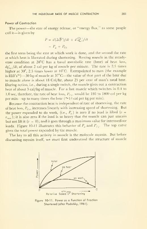
THE MOLECULAR BASIS OF MUSCLE CONTRACTION 283
Power of Contraction
The power— the rate of energy release, or "energy flux," as some people
call it— is given by
P = d(A$')/dt + dSll/dt
= PW + tin
the first term being the rate at which work is done, and the second the rate
at which heat is liberated during shortening. Resting muscle in the steady-
state condition at 20°C has a basal metabolic rate (bmr) of heat loss,
dq'bm /dt, of about 2 cal per kg of muscle per minute. The rate is 2.5 times
higher at 30°, 2.5 times lower at 10°C. Extrapolated to man (the example
is Hill's 16)— 30 kg of muscle at 37°C— the value of that part of the bmr due
to muscle alone is about 18 Cal/hr, about 25 per cent of man's total bmr.
During action, i.e., during a single twitch, the muscle gives out a contraction
heat of about 3 cal/kg of muscle. For a fast muscle which twitches in 0.1 to
1.0 sec, therefore, the rate of heat loss, P%.n would be 180 to 1800 cal per kg
per min—up to many times the bmr (~14 cal per kg per min).
Because the contraction heat is independent of rate of shortening, the rate
of heat loss, P^n increases linearly with increasing speed of shortening. But
the power expended to do work, (i.e., Pw ) is zero if no load is lifted (v =
v ): it is also zero if the load is so heavy that the muscle can just sustainmax /
*' **
but not lift it {v = 0); and it goes through a maximum value for intermediate
loads. Figure 10-11 illustrates this behavior of Pw and P^r The top curve
gives the total power expended by the muscle.
The key to all this activity in muscle is the molecule myosin. But before
discussing myosin itself, we must first understand the structure of muscle
v/v moxRelative Speed of Shortening
Figure 10-1 1. Power as a Function of Fraction
Shortened (after Podolsky, 1961).
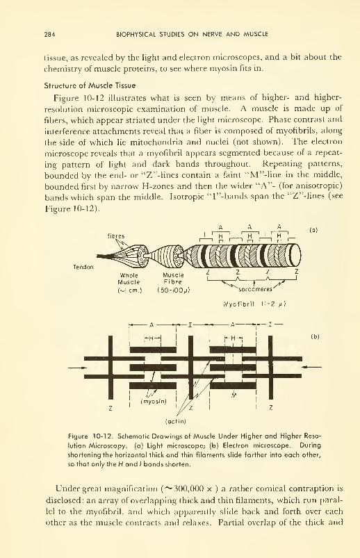
284 BIOPHYSICAL STUDIES ON NERVE AND MUSCLE
tissue, as revealed by the light and electron microscopes, and a bit about the
chemistry of muscle proteins, to see where myosin fits in.
Structure of Muscle Tissue
Figure 10-12 illustrates what is seen by means of higher- and higher-
resolution microscopic examination of muscle. A muscle is made up of
fibers, which appear striated under the light microscope. Phase contrast and
interference attachments reveal that a fiber is composed of myofibrils, along
the side of which lie mitochondria and nuclei (not shown). The electron
microscope reveals that a myofibril appears segmented because of a repeat-
ing pattern of light and dark bands throughout. Repeating patterns,
bounded by the end- or "Z"-lines contain a faint "M"-line in the middle,
bounded first by narrow H-zones and then the wider "A"- (for anisotropic)
bands which span the middle. Isotropic 'T'-bands span the "Z"-lines (see
Figure 10-12).
fibres
Tendon
Uh ii
r (a)
WholeMuscle
(vl cm.)
MuscleFibre
i50-100/j)
mmm )
z z z z1
<..,.*> '
sarcomeres
Myofibril (1-2 u)
— I
—
(actin)
Figure 10-12. Schematic Drawings of Muscle Under Higher and Higher Reso-
lution Microscopy, (a) Light microscope; (b) Electron microscope. During
shortening the horizontal thick and thin filaments slide farther into each other,
so that only the H and / bands shorten.
Under great magnification (~ 300,000 x ) a rather comical contraption is
disclosed: an array of overlapping thick and thin filaments, which run paral-
lel to the myofibril, and which apparently slide back and forth over each
other as the muscle contracts and relaxes. Partial overlap of the thick and
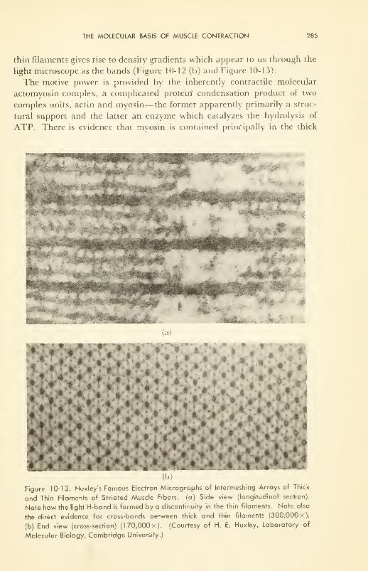
THE MOLECULAR BASIS OF MUSCLE CONTRACTION 285
thin filaments gives rise to density gradients which appear to us through the
light microscope as the bands (Figure 10-12 (b) and Figure 10-13).
The motive power is provided by the inherently contractile molecular
actomyosin complex, a complicated protein condensation product of two
complex units, actin and myosin— the former apparently primarily a struc-
tural support and the latter an enzyme which catalyzes the hydrolysis of
ATP. There is evidence that myosin is contained principally in the thick
* :%V
(a)
Figure 10-13. Huxley's Famous Electron Micrographs of Intermeshing Arrays of Thick
and Thin Filaments of Striated Muscle Fibers, (a) Side view (longitudinal section).
Note how the light H-band is formed by a discontinuity in the thin filaments. Note also
the direct evidence for cross-bonds between thick and thin filaments (300,000 x).
(b) End view (cross-section) (170,000x). (Courtesy of H. E. Huxley, Laboratory of
Molecular Biology, Cambridge University.)
286 BIOPHYSICAL STUDIES ON NERVE AND MUSCLE
filaments, actin in the thin ones. The Z-lines are the outer edges of areas
which bisect the myofibril, and have been shown to be the medium through
which the stimulus, or order to contract, is carried from the surface mem-brane of the fiber (the sarcolemma) into the myofibril. The sarcolemma car-
ries it electrochemically (like nerve) along the fiber.
Muscle consists of 18 to 20 per cent protein, by weight. About 60 per cent
of this protein is a condensation product of several "myosins" with actin, a
very complex molecule whose complete physical structure is very sensitive to
the ionic content and pH of the medium. It interchanges between a globu-
lar, almost spherical, hard G-actin, to a fibrous, stiff F-actin. Only myosin
has the ATPase activity and can accept the free energy of hydrolysis of ATP.But the myosin of muscle is itself made up of smaller parts:
Rapidly extractable from minced muscle in salt solutions is myosin-A
(called "myosin" or "/-myosin" in some books). Electrophoresis causes
separation of myosin-/! into three fractions: one heavy (//) meromyosin,
and two light (L) meromyosins. Only the //-meromyosin retains the ATP-ase activity, Extractable only slowly, or in other media, are myosin-/?
("natural actomyosin" or "^-myosin") and tropomyosin, which differ in
physical properties from myosin-^4. Rejected by the extraction procedures
is the globular G-actin, which, in the presence of ATP and dilute salts,
slowly converts to the much more viscous, fibrous F-actin. The chemical
composition is not simple. Thus there is some evidence that tropomyosin +G-actin + another protein constitute myosin-A. Some physical characteris-
tics of myosin and actin are gathered in Table 10-3.
The muscle proteins are rich in polar residues such as —P03
" 3, —OH,
—CONH— , and —COOH. These polar residues seem to be intimately
connected with the process of contraction. Myosin's partner in the con-
tractile reaction is ATP. To ATP, the fact that the catalytic enzyme, myo-
sin, contracts during the hydrolysis, or splitting of ATP into ADP + P, is
quite incidental. To the living system, however, the fact is vital! Dephos-
phorylation occurs during or immediately after the contraction process.
Hydrolysis of ATP as a free energy-producing reaction is not confined to
myosin as a catalyst, as we saw in Chapter 7. It provides the energy which
drives many living processes. The following scheme represents the splitting
reaction and its auxiliary reactions:
H2
+ ATP 4 ^ ADP 2 + HP04
" 2
+ K2 }[
+//+ //+ //+
K,11 + K
3 11
ATP- 3 ADP 3 H2P0
4
i #iI
THE MOLECULAR BASIS OF MUSCLE CONTRACTION 287
The L-step is the splitting reaction proper. In the vicinity of pH = 7, the
values of the equilibrium constants, K\, K2and AT
3 , are such that most of the
adenosine is in the form of either ATP" 4 or ADP 2; and hence the meas-
ured values of AH and AF refer mainly to the hydrolysis itself—the hori-
zontal reaction. The reaction is both exothermic and exergonic, a source
of heat and a source of free energy for work. Respectable values (see com-
ments in Table 7-3) are:
AF = -10.5kcal/mole
AH = -9.2kcal/mole
However, as is obvious from the reaction scheme, a shift in pH can shift the
position of equilibrium of reactions 1, 2, and 3, and therefore shift the free
energy of the splitting reaction. In a similar manner to the effect of hydro-
gen ions, metallic cations— principally Mg++ and Ca + +—can and do form
complexes with the highly charged phosphate groups; each complex with its
own equilibrium to affect the reaction scheme, and thereby to affect the
values of AF and A H.
TABLE 10-3. Sedimentation Constant (s), Diffusion Coefficient (D), Molecular Weight (M),
Intrinsic Viscosity ([r/ ]), Length (/) and Thickness (d) of the Muscle Proteins.
Protein
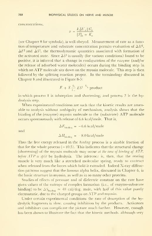
288 BIOPHYSICAL STUDIES ON NERVE AND MUSCLE
concentrations,
vk
2[E] n [S] a
[S] + Km
(see Chapter 8 for symbols), is well obeyed. Measurement of rate as a func-
tion of temperature and substrate concentration permits evaluation of AH t,
AF l and AS 1, the thermodynamic quantities associated with formation of
the activated state. Since AS* is usually (for various conditions) found to be
positive, it is inferred that a change in configuration of the enzyme (and/or
the release of adsorbed water molecules) occurs during the binding step in
which an ATP molecule sits down on the myosin molecule. This step is then
followed by the splitting reaction proper. In the terminology discussed in
Chapter 8 and illustrated in Figure 8-5:
E + S y^ ES l —^ product
in which process 1 is adsorption and shortening; and process 2 is the hy-
drolysis step.
When experimental conditions are such that the kinetic results are amen-
able to analysis without ambiguity of mechanism, analysis shows that the
binding of the (enzyme) myosin molecule to the (substrate) ATP molecule
occurs spontaneously with release of 6.6 kcal/mole. That is,
A/7bind,ng
= -6.6 kcal/mole
and
A//binding = -8.0 kcal/mole
Thus the free energy released in the binding process is a sizable fraction of
that for the whole process (— 10.5). This indicates that the structural change
(shortening) of the myosin molecule may occur at the time of binding of ATP,
before ATP is split by hydrolysis. The inference is, then, that the resting
muscle is very much like a stretched molecular spring, ready to contract
when released from the forces which hold it extended. Indeed X-ray diffrac-
tion patterns suggest that the famous alpha helix, discussed in Chapter 6, is
the basic structure in myosin, as well as in so many other proteins.
Studies of effects of pressure and of dielectric constant on the rate have
given values of the entropy of complex formation (i.e., of enzyme-substrate
binding) to be A.S'bindj
~ 48 cal/deg. mole, with half of this value purely
electrostatic, due to the charged groups on ATP and myosin.
Under certain experimental conditions the rate of desorption of the hy-
drolytic fragments is slow, causing inhibition by the products. Activators
and inhibitors can complicate the picture much further. However, enough
has been shown to illustrate the fact that the kinetic methods, although very
THE MOLECULAR BASIS OF MUSCLE CONTRACTION 289
specialized in detail, provide a general mechanistic description of the physi-
cal actions of the key molecules which play the vital roles.
A Theory of Contraction
One simplified working hypothesis about the physical activity of the con-
tractile molecule will now be outlined. It is as though the myosin were a
coiled molecule (like other proteins whose structures are known from X-ray
diffraction) which, at rest, is held in a stretched condition by virtue of a se-
ries of mutually repelling, charged ionic groups along its length, —COOMg +
or —NH3+
, for example. Adsorption of ATP -4to form the Michaelis-
Menten complex, discharges the myosin network, permitting the interatomic
restoring forces, which exist because of bent bonds, to relax the molecule to
its neutralized (contracted) length. After hydrolysis, ADP" 2 and P" 2 desorb,
because they are bound less tightly than ATP -4 and are perhaps aided by
other molecular species in the vicinity. After the products have desorbed,
the positive charges along the molecule lengthen the coil again, and the
molecule is ready to repeat the cycle.
What is the nature of the trigger which starts ATP -4 adsorbing? The
answer is not known, but the hypothesis, based on indirect (but nevertheless
substantial) evidence, is that a covering molecule, the "blanket," weakly ad-
sorbs on and protects the charged network of the stretched myosin. Its
shape is thought to be determined partly by Mg++ ions, without which the
contractile power of myosin ceases. Distortion of the shape of the blanket
by the more strongly chelating (complexing) Ca++is supposed to bare the
myosin to attack by ATP -4: thus injection of Ca++ causes contraction. Nerve
endings, which run almost to the membranous sheath (sarcolemma) which
covers the muscle fibers, are thought by some to be capable of releasing Ca +
at the myosin sites via electrochemical stimuli propagated down nerve axons
to the nerve ending, and thence down the sheath and in the Z-bands to the
myosin sites.
The connectors between filaments, shown so beautifully in the electron
microscope pictures of sliced muscle tissue (Figure 10-13), in this theory take
on a very positive character, composition, and role: viz., the ends and par-
ticular side groups of stretched myosin molecules, attached at one end to a
thin actin filament, but lying within and forming part of an adjacent thick
one so that shortening of the myosin molecule itself causes filaments to slide
over each other, and the whole tissue to contract. The concept is illustrated
in Figure 10-14. Approximate obedience of the whole muscle to Hooke's
Law would qualitatively result from behavior on the molecular level. Both
the chemistry of the contraction process and the physical sliding of the fibers
complement the model.
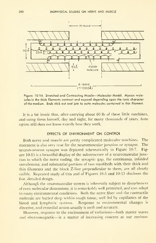
290 BIOPHYSICAL STUDIES ON NERVE AND MUSCLE
110 A
A- band
"(~I000A)
Figure 10-14. Stretched and Contracting Muscle—Molecular Model. Myosin mole-
cules in the thick filaments contract and expand depending upon the ionic character
of the medium. Ends stick out and join to actin molecules contained in thin filament.
It is a bit ironic that, after carrying about 60 lb of these little machines,
and using them himself, day and night, for many thousands of years, homo
sapiens still does not know exactly how they work.
EFFECTS OF ENVIRONMENT ON CONTROL
Both nerve and muscle are pretty complicated molecular machines. The
statement is also very true for the neuromuscular junction or synapse. The
neuron-neuron synapse was depicted schematically in Figure 10-7. Fig-
ure 10-15 is a beautiful display of the substructure of a neuromuscular junc-
tion in which the nerve ending, the synaptic gap, the continuous, infolded
sarcolemma, and substantial portions of two myofibrils with their thick and
thin filaments and the black Z-line perpendicular to them, are all clearly
visible. Repeated study of this and of Figures 10-5 and 10-13 discloses the
fine, detailed design.
Although the neuromuscular system is inherently subject to disturbances
of even molecular dimensions, it is remarkably well protected, and can adapt
to many environmental conditions. Both the nerve fiber and the contractile
molecule are buried deep within tough tissue, well fed by capillaries of the
blood and lymphatic systems. Response to environmental changes is
directive, and remedial action usually is swift and accurate.
However, response to the environment of radiations— both matter waves
and electromagnetic— is a matter of increasing concern as our environ-
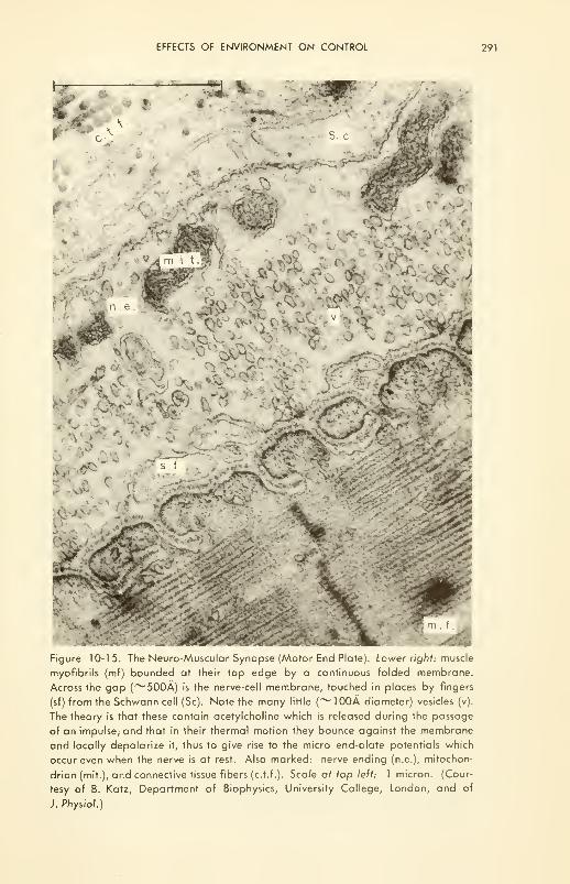
EFFECTS OF ENVIRONMENT ON CONTROL 291
^v
I-
a4 S' J
m
Figure 10-15. The Neuro-Muscular Synapse (Motor End Plate). Lower right: muscle
myofibrils (mf) bounded at their top edge by a continuous folded membrane.
Across the gap (~500A) is the nerve-cell membrane, touched in places by fingers
(sf) from the Schwann cell (Sc). Note the many little (~100A diameter) vesicles (v).
The theory is that these contain acetylcholine which is released during the passage
of an impulse; and that in their thermal motion they bounce against the membrane
and locally depolarize it, thus to give rise to the micro end-plate potentials which
occur even when the nerve is at rest. Also marked: nerve ending (n.e.), mitochon-
drion (mit.), and connective tissue fibers (c.t.f.). Scale af top left: 1 micron. (Cour-
tesy of B. Katz, Department of Biophysics, University College, London, and of
J. Physiol.)
292 BIOPHYSICAL STUDIES ON NERVE AND MUSCLE
ment becomes "noisier." In Chapter 2, the effects of shock, blast, sound,
and ultrasound were reviewed; and in Chapter 4 the effects of the warming,
visible, ultraviolet, and ionizing regions of the electromagnetic spectrum
were discussed. In Chapter 7 heat production, and in Chapter 8 heat loss
were discussed, as also were changes in our chemical environment (poisons
and catalysts—competitors) as they affect the metabolism of the system and
its control. Although the details of the complicated processes of control are
beyond our means in this book, enough has been introduced to illustrate the
mechanisms and the A-B-C's of environmental effects—atomic, biological,
and chemical, at least in general terms.
One further point will be made on the effects of ionizing radiations on
the physical apparatus of control— of increasing importance, especially to
medical people, in this atomic age. Nerve itself is relatively insensitive to
X rays (Chapter 9). Muscle shows good resistance too: it takes thousands
of rads to cause detectable damage. The neuromuscular junction, however,
is much more sensitive. For instance, consider a nerve-muscle system such
as the sciatic nerve-gastrocnemius muscle freshly dissected from a frog,
mounted in such a fashion that the nerve can be stimulated electrically by
short, square pulses of voltage applied by the platinum wire contacts refer
to (Figure 10-8). If the stimulus repetition rate is chosen at about 1 pulse
per sec, the muscle will respond faithfully. If now the whole is irradiated,
the muscle soon stops, although the nerve continues to transmit, and the
muscle will respond to a stimulus given directly to it.
Further, the neural network in the brain is now known to be affected by
only a few rads; and although this radiation does not affect the motor ability
of a man, there is reason to believe that short-circuiting in the network and
psychological effects accrue. Since it is not likely to be the nerve cells them-
selves, it is probably the synapse, or "junction box" which is implicated as
radiation-sensitive.
The parallelism is clear. The neuromuscular junction and the synapse are
the most sensitive parts of man's physical control system. Both of these
junctions involve production of a chemical or chemicals at one spot in the
junction, transport across the junction, and utilization of the chemical(s) at
the other end of the junction. With the background of knowledge of the
pertinent chemical and physical effects of ionizing radiations discussed in
the previous Chapter, and that of the physical apparatus of control given in
this Chapter, what do you think is likely to be the first molecular process to
fail during irradiation of the control apparatus?
PROBLEMS
10-1: If one side of a concentration cell has KC1 at 0.002 equivalents per liter, what
must be the opposing concentration so that the "membrane'' potential reaches
90 millivolts? Assume restricted diffusion.
REFERENCES 293
10-2: Two platinum electrodes placed 3.0 cm apart on a nerve fiber detect the "waveof negativity"of a transmitted impulse 0.37 milliseconds apart. Calculate the
speed of transmission in meters/sec, yards/sec, and miles per hour. Comparethis with the speed of sound in air (1090 feet/sec); of light through a vacuum
(186,000 miles/sec); of a signal along a telephone cable (1000 miles/ sec); of
the fastest thrown baseball (88 miles/hr); of the fastest sprinter (100
yds/ 10 sec).
10-3: During the testing of a reflex at the sole of the foot, the signal must travel up
the leg to the spinal column and an order be transmitted back before the re-
sponse can occur. If the distance is 3 ft each way, how long should the interval
between stimulus and response be?
10-4: Good rules-of-thumb to remember are: (a) the speed of shortening of a striated
muscle can reach a maximum value ymax of about ten times its length per sec-
ond; and (b) it can exert a force which can reach a maximum Fmax of about
42 lb per sq in. of cross-sectional area of the muscle.
Assuming the model of Figure 10-5, the force-velocity curve of Figure 10-10,
and the above data, calculate values of velocity with which three different
weight forces can be lifted, at v/vmax equal to 0.1, 0.5, and 0.9.
REFERENCES
1. Keynes, R. D., "The Nerve Impulse and the Squid," Scientific American, 199,
No. 6, p. 83(1958).
2. Podolsky. R. D., "The Mechanism of Muscular Contraction," Amer. J. Medicine,
30,708(1961).
3. Huxley, H. E., "The Contraction of Muscle," Scientific Amer., 199, No. 5. p. 66
(1958).
4. Szent-Gyorgyi, A., "Mechanochemical Contraction in Muscle," in "Enzymes:
Units of Biological Structure and Function," O. H. Gaebler, Ed., Academic
Press, New York, N. Y., 1956.
5. Morales, M. F., et ai, "The Mechanism of Muscle Contraction," Physiol. Rev.,
35,475(1955).
6. Hodgkin, A. L. and Keynes, R. D., "Active Transport of Cations in Giant Axons
from Sepia and Loligo," J. Physiol., 128,28 (1955).
7. Nachmansohn, D., "Chemical Factors Controlling Movements during Nerve Ac-
tivity, from The Method of Isotopic Tracers Applied to the Study of Active Ion
Transport," Pergamon Press, New York, N. Y., 1959.
8. Penfield, W., "The Interpretive Cortex," Science, 129, 1719 (1959).
9. Walter, W. G., "The Living Brain," Penguin Books, Harmondsworth, England,
1961.
10. Shedlovsky, T., Ed., "Electrochemistry in Biology and Medicine,"John Wiley &Sons, Inc., New York, N, Y., 1955: review papers by B. K. Bagchi, H. H.
Jasper, K. S. Cole, and others.
11. Hodgkin, A. L., "Ionic Movements and Electrical Activity in Giant Nerve
Fibers," Proc. Roy. Soc, B., 148, 1 (1958); a fine review lecture.
12. Szent-Gyorgyi, A., "Chemistry of Muscular Contraction," 3rd ed., Academic
Press, Inc„ New York, N. Y., 1960.
294 BIOPHYSICAL STUDIES ON NERVE AND MUSCLE
13. Wilkie, D. R., "Facts and Theories about Muscle," Prog, in Biophysics and Bio-
physical Chem., 4,288 (1954).
14. Hodgkin, A. L., "Ionic Movements and Electrical Activity in Giant Nerve
Fibers," Proc. Roy. Soc, B., 148, 1 (1958).
15. Hill, A. V., "Chemical Change and Mechanical Response in Stimulated Mus-
cle," Proc. Roy. Soc, B, 314 (1953).
16. Paton, W. D. M., Ed. of special issue: "Physiology of Voluntary Muscle,"
British Med. Bull., 12, 161-236 (1956); see especially the papers by A. V. Hill,
A. F. Huxley, D. R. Wilkie, and R. G. Bannister.
17. Bourne, G. H., Ed., "Structure and Function of Muscle," Vol. I, Academic Press
Inc., New York, N. Y., 1960; contributions by H. E. Huxley, J. Hanson, and
A. Csapo.
18. Nachmansohn, D., "Basic Aspects of Nerve Activity Explained by Biochemical
Analysis," J. Amer. Med. Assoc, 179, 145 (1962).
19. Hodgkin, A. L., and Huxley, A. F., "A Quantitative Description of Membrane
Current and its Application to Conduction and Excitation in Nerves," J.
Physiol., 117, 500 (1952).
20. Cole, K. S., and Curtis, H. J., "Electric Impedance of the Squid Giant Axon
during Activity, "J. Gen. Physiol., 22,649 (1939).
21. Suckling, E. E., "Bioelectricity," McGraw-Hill Book Company, Inc., New York,
N. Y., 1961.
22. Noble, D., "A Modification of the Hodgkin-Huxley Equations Applicable to
Purkinje Fibre Action and Pace-Maker Potentials," J. Physiol., 160, 317
(1962).
23. Three short papers on contraction of muscle, Nature, 167 (1-951): by A. V. Hill,
p. 377; by A. Szent-Gydrgyi, p. 380; and by H. H. Weber, p. 381.
24. Stacy, R. W., Williams, D. T, Worden, R. E., and McMorris, R. O., "Essen-
tials of Biological and Medical Physics," McGraw-Hill Book Company, Inc.,
New York, N. Y., 1955: Chapters 32 to 34.
25. Brazier, M. A. B., "The Analysis of Brain Waves," Scientific American, 206, 142
(1962).
CHAPTER 11
The Language and Concepts
of Control
The natural systems are of enormous complexity, and it is clearly neces-
sary to subdivide the problem ....
The first part of the problem [is] the structure and functioning of such
elementary units individually. The second part of the problem consists of
understanding how these elements are organized into a whole, and how the
functioning of the whole is expressed in terms of these elements ....
The number of cells in the human body is somewhere in the general order
of W15or 10m . The number of neurons in the central nervous system is
somewhere in the order of 10w . . . . All artificial automata ["thinking
machines''''] made by man have numbers of parts which, by any comparably
schematic count, are of the order of W3to W 6
. . . . The prototypes for these
[living] systems are the modern computing machines ....
[However], whereas I can conceive of a machine which could reproduce
itself, I cannot imagine a machine which could create itself/ (John von
Neumann, Vanuxem Lectures, Princeton, 1952.)
INTRODUCTION
In the very first chapter of this book we introduced rather superficially
the concept of man as an integrated system operating in continuous ex-
change with his environment. During the next few chapters we dwelt on the
forces, momenta, and energy which comprise this exchange, and showed
what these are, their properties, and their effects on the living system.
Through the middle of the book we dwelt on the workings of individual parts
295
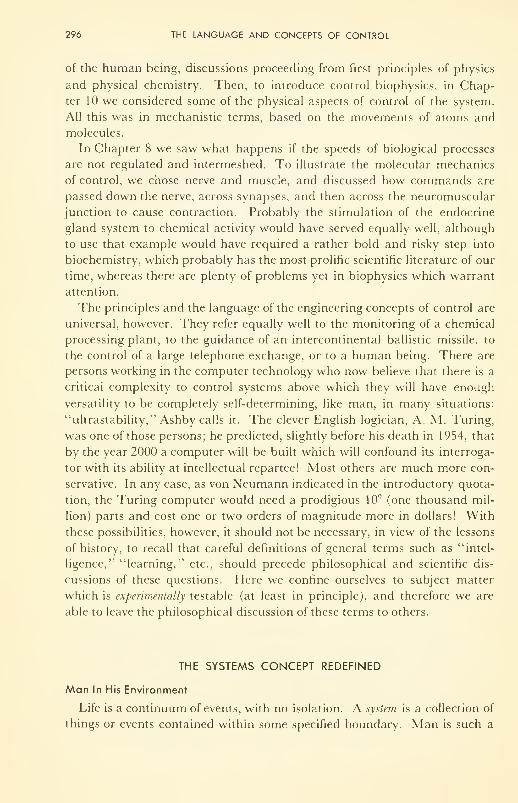
296 THE LANGUAGE AND CONCEPTS OF CONTROL
of the human being, discussions proceeding from first principles of physics
and physical chemistry. Then, to introduce control biophysics, in Chap-
ter 10 we considered some of the physical aspects of control of the system.
All this was in mechanistic terms, based on the movements of atoms and
molecules.
In Chapter 8 we saw what happens if the speeds of biological processes
are not regulated and intermeshed. To illustrate the molecular mechanics
of control, we chose nerve and muscle, and discussed how commands are
passed down the nerve, across synapses, and then across the neuromuscular
junction to cause contraction. Probably the stimulation of the endocrine
gland system to chemical activity would have served equally well, although
to use that example would have required a rather bold and risky step into
biochemistry, which probably has the most prolific scientific literature of our
time, whereas there are plenty of problems yet in biophysics which warrant
attention.
The principles and the language of the engineering concepts of control are
universal, however. They refer equally well to the monitoring of a chemical
processing plant, to the guidance of an intercontinental ballistic missile, to
the control of a large telephone exchange, or to a human being. There are
persons working in the computer technology who now believe that there is a
critical complexity to control systems above which they will have enough
versatility to be completely self-determining, like man, in many situations:
"ultrastability," Ashby calls it. The clever English logician, A. M. Turing,
was one of those persons; he predicted, slightly before his death in 1954, that
by the year 2000 a computer will be built which will confound its interroga-
tor with its ability at intellectual repartee! Most others are much more con-
servative. In any case, as von Neumann indicated in the introductory quota-
tion, the Turing computer would need a prodigious 109 (one thousand mil-
lion) parts and cost one or two orders of magnitude more in dollars! With
these possibilities, however, it should not be necessary, in view of the lessons
of history, to recall that careful definitions of general terms such as "intel-
ligence," "learning," etc., should precede philosophical and scientific dis-
cussions of these questions. Here we confine ourselves to subject matter
which is experimentally testable (at least in principle), and therefore we are
able to leave the philosophical discussion of these terms to others.
THE SYSTEMS CONCEPT REDEFINED
Man In His Environment
Life is a continuum of events, with no isolation. A system is a collection of
things or events contained within some specified boundary. Man is such a
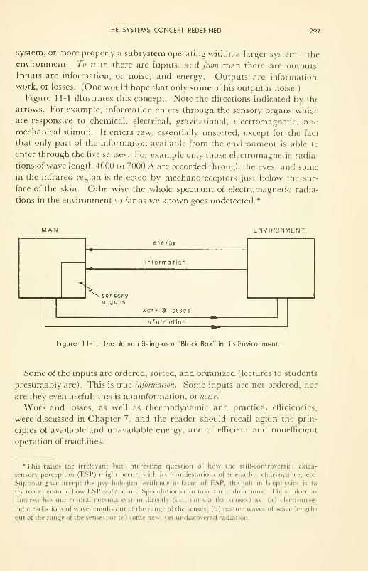
THE SYSTEMS CONCEPT REDEFINED 297
system, or more properly a subsystem operating within a larger system— the
environment. To man there are inputs, and from man there are outputs.
Inputs are information, or noise, and energy. Outputs are information,
work, or losses. (One would hope that only some of his output is noise.)
Figure 11-1 illustrates this concept. Note the directions indicated by the
arrows. For example, information enters through the sensory organs whichare responsive to chemical, electrical, gravitational, electromagnetic, andmechanical stimuli. It enters raw, essentially unsorted, except for the fact
that only part of the information available from the environment is able to
enter through the five senses. For example only those electromagnetic radia-
tions of wave length 4000 to 7000 A are recorded through the eyes, and somein the infrared region is detected by mechanoreceptors just below the sur-
face of the skin. Otherwise the whole spectrum of electromagnetic radia-
tions in the environment so far as we known goes undetected.*
MAN ENVIRONMENT
298 THE LANGUAGE AND CONCEPTS OF CONTROL
Information and Entropy
The broad use of the term "entropy" as a quantitative measure of the
amount of disorder in a system, or subsystem, was introduced in Chapter 7.
Now we carry the concept one step further. For communication, which re-
quires a description of a system in words or codings, the simpler the system
the simpler the information needed to describe it. Four sticks standing fixed
in a row (||||) is a very simple system, A, easily described; but the same four
sticks comprise an infinitely complex system, B, if the four sticks are thrown
off a roof-top and each stick allowed to assume any position and degree of
rotation during fall. The information required to describe A unambiguously
is small; likewise its entropy or disorder is low. By contrast the information
required to describe B unambiguously is relatively very large; its entropy or
disorder is high.
Therefore, a measure of the quantity of information needed to describe some-
thing is the entropy of the system being described.
It follows that if the information, S, put into a computational system such
as man becomes distorted for one reason or another, the changed informa-
tion is now 5" + AS, where AS is the distortion. It is always positive, in-
creasing the entropy.
However, if two inputs, Sl
and S2 , are faithfully recorded and analyzed,
and if from the two informations a third piece of information, a synthesis of
the two, occurs, then the total information needed to describe S}
and S2
is less
than the sum 5\ + S2 , and the total entropy has thereby been decreased ....
One's information is now better organized. One remembers now a simple
principle which describes both systems 1 and 2.
Measurement
Measurement implies a reference. What is measured is a difference be-
tween two quantities, one of which is taken as the reference, against which
many similar quantities are measured. The fact that no two physical beings
are in all respects identical implies variation. Variation in turn introduces
uncertainty.
There is an inherent uncertainty in all measurement, a principle first
propounded by Heisenberg. The formal statement of this is known as his
"uncertainty principle." It takes various forms, a simple statement of which
is the following: To make a physical measurement, energy must be trans-
ferred between the object and the measuring device; otherwise there is noth-
ing to detect; this transfer introduces uncertainty, because the object is not
now the same as it was before the energy was transferred: the smaller the
object the more difficult it is to measure its properties.
However, in the macroscopic physical world, objects are big enough so
that this uncertainty is far smaller than are gross errors in measurement,
THE SYSTEMS CONCEPT REDEFINED 299
be they random or constant errors. No measurement is likely to be perfect.
We are always faced with this probability of error, and of (biological) varia-
tion in the thing being measured.
The human machine is subject to error in measurement, just as is any
other machine. It is no accident that athletic competitions, especially by
professional athletes, are described as "games of inches," the differentiating
factor being the ability to estimate distance under great psychological stress.
In summary, it is a measurement which is fed back into a computer to
guide it in making corrections to its actions. This measurement is ot the dif-
ference, A, between where one is and where one wants to be— that is, of the
error. The error is increased by noise.
Noise
The subject of the detection of a signal of information (energy) over back-
ground was discussed in Chapter 3 in the discussion of sensitivity of a detec-
tor and the VVeber-Fechner Law. The principles introduced there apply also
to the detection of information to be fed to a computer. If the source provides
a strong signal over background, the detector will feed a correspondingly
strong signal to the computer. If the background noise is high (i.e., the
signal-to-noise ratio is low) the signal sent to the computer may not be in-
telligible (discernible from the background). Strength of the signal, back-
ground noise, and degradation of the information by noise introduced in the
detector determine what the computer receives as input.
Unfortunately there usually are many strong signals entering a detector,
only some of which are useful. Those which are not useful are also noise,
like the background. The machine must be able to classify signals: to accept
the information and by-pass the rest. One of the most useful systems yet
built to separate information from noise is the EEG analyzer, a machine
which scans the information and sorts the rather complex total waves into
their three or four main components.
Continuously confusing the control circuits of a human being is an un-
remitting input of noise— disordered, and perhaps not even useful informa-
tion. Noise can take several forms. First of all it may be of either external
or internal origin. External noise comes in from the environment through
the senses. It is probably better to call it incomplete rather than disordered,
for there is order and regularity reaching our senses from everywhere about
us in nature. The trouble arises because we have only a limited eapa< it\
or interest— subject to, or determined by our freely-chosen goals in life. In
other words, what is useful, interesting information to one man is noise to
another; and for one man, what is noise at () P. M. may not be so at 9 A. M.
(traffic information, for example). This is unfortunate, but nevertheless true.
It is unfortunate because it means that two men with a common interest in
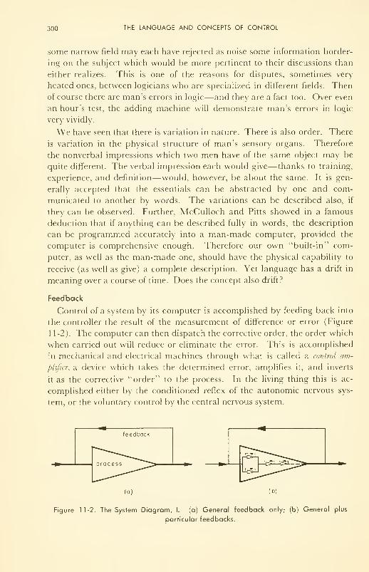
300 THE LANGUAGE AND CONCEPTS OF CONTROL
some narrow field may each have rejected as noise some information border-
ing on the subject which would be more pertinent to their discussions than
either realizes. This is one of the reasons for disputes, sometimes very
heated ones, between logicians who are specialized in different fields. Thenof course there are man's errors in logic—and they are a fact too. Over even
an hour's test, the adding machine will demonstrate man's errors in logic
very vividly.
We have seen that there is variation in nature. There is also order. There
is variation in the physical structure of man's sensory organs. Therefore
the nonverbal impressions which two men have of the same object may be
quite different. The verbal impression each would give—thanks to training,
experience, and definition—would, however, be about the same. It is gen-
erally accepted that the essentials can be abstracted by one and com-
municated to another by words. The variations can be described also, if
they can be observed. Further, McCulloch and Pitts showed in a famous
deduction that if anything can be described fully in words, the description
can be programmed accurately into a man-made computer, provided the
computer is comprehensive enough. Therefore our own "built-in" com-
puter, as well as the man-made one, should have the physical capability to
receive (as well as give) a complete description. Yet language has a drift in
meaning over a course of time. Does the concept also drift?
Feedback
Control of a system by its computer is accomplished by feeding back into
the controller the result of the measurement of difference or error (Figure
11-2). The computer can then dispatch the corrective order, the order which
when carried out will reduce or eliminate the error. This is accomplished
in mechanical and electrical machines through what is called a control am-
plifier, a device which takes the determined error, amplifies it, and inverts
it as the corrective "order" to the process. In the living thing this is ac-
complished either by the conditioned reflex of the autonomic nervous sys-
tem, or the voluntary control by the central nervous system.
(a) (b)
Figure 11-2. The System Diagram, I. (a) General feedback only; (b) General plus
particular feedbacks.
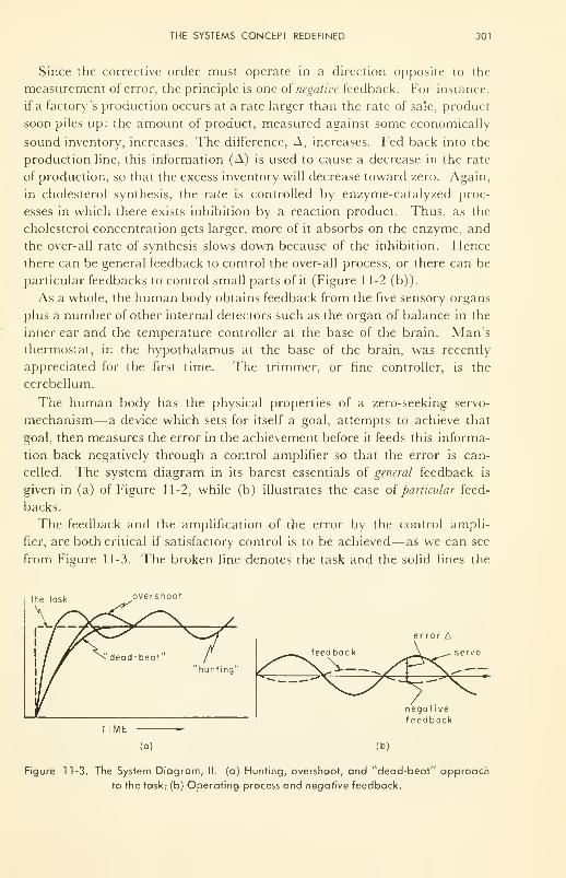
THE SYSTEMS CONCEPT REDEFINED 301
Since the corrective order must operate in a direction opposite to the
measurement of error, the principle is one of negative feedback. For instance,
if a factory's production occurs at a rate larger than the rate of sale, product
soon piles up: the amount of product, measured against some economically
sound inventory, increases. The difference, A, increases. Fed back into the
production line, this information (A) is used to cause a decrease in the rate
of production, so that the excess inventory will decrease toward zero. Again,
in cholesterol synthesis, the rate is controlled by enzyme-catalyzed proc-
esses in which there exists inhibition by a reaction product. Thus, as the
cholesterol concentration gets larger, more of it absorbs on the enzyme, and
the over-all rate of synthesis slows down because of the inhibition. Hence
there can be general feedback to control the over-all process, or there can be
particular feedbacks to control small parts of it (Figure 11-2 (b) )
.
As a whole, the human body obtains feedback from the five sensory organs
plus a number of other internal detectors such as the organ of balance in the
inner ear and the temperature controller at the base of the brain. Man's
thermostat, in the hypothalamus at the base of the brain, was recently
appreciated for the first time. The trimmer, or fine controller, is the
cerebellum.
The human body has the physical properties of a zero-seeking servo-
mechanism— a device which sets for itself a goal, attempts to achieve that
goal, then measures the error in the achievement before it feeds this informa-
tion back negatively through a control amplifier so that the error is can-
celled. The system diagram in its barest essentials of general feedback is
given in (a) of Figure 11-2, while (b) illustrates the case oi particular feed-
backs.
The feedback and the amplification of the error by the control ampli-
fier, are both critical if satisfactory control is to be achieved—as we can see
from Figure 11-3. The broken line denotes the task and the solid lines the
the task overshoot
302 THE LANGUAGE AND CONCEPTS OF CONTROL
achievement for an elementary process such as heating a house. More sensi-
tive detectors provide more accurate feedback and reduce the oscillation
about the task. The loss of fine control in a man's attempt to walk along a
straight line under the effects of drugs, disease, or alcohol is well known.
A recent innovation into the heating systems which the human body has
had for thousands of years is the facility for anticipation. This takes two phy-
sical forms in the human being, only one form in the heating system. Theone which is common to both, is the early-warning system: the external
thermostat in the heating system, which predicts a change inside as soon as
the weather changes; the kinematic (or kinesthetic) sense, for example, in
the human which tells him where his hands are even when his eyes are
closed. In addition, the human has a memory, which helps his anticipation
by extrapolating from the present situation into the future along a path sug-
gested by previous experience. Modern computers have the memory circuits
and the extrapolation circuits too.** Whether man will eventually be able
to make computers which can abstract and then extrapolate with abstrac-
tions, as man can do, remains for the future to answer.
The sensory detectors are so sensitive in the human, and the cerebellum
such an effective trimmer on the control apparatus, that man is the ideal ex-
ample of a "dead-beat servo," with no cycling at all about the task .... This
is true only as a first approximation, however. Thus the physical trim of a
trained athlete or of a practiced surgeon is far more precise than that of
his neighbor. Similarly, those who are afflicted with Parkinsonism or al-
coholism are less precise in their physical and chemical process control.
Precise control of the biological chemistry and physics is at the very root of the
prevention and cure of disease, and of life itself.
Memory, Concept and Implementation
The mind stores information. Physical machines can be made to do this
by (a) magnetic tapes or magnetic cores, (b) on-or-off relays, (c) slow pene-
tration processes in which electric or sonic signals bounce around inside
crystals for a time before escaping, and (d) electrochemical devices such as
capacitors. In fact the machine can be programmed to collect information
while it is operating and use it thereafter, thus closely simulating man's
memory. A recent postulate about the physical nature of neural memoryapparatus is that the repeated, passing electrical signal distorts the RNA
** Perhaps the earliest popularly recognized and amusing example of machine out-anticipat-
ing man came during the counting of the U. S. Presidential election returns in 1 948. The com-
puter, UNIVAC, on the job seriously for the first time, started predicting a Truman victory
at about 8:15 P.M., much to the derision of the human political pundits. By 11 P.M. the pun-
dits were beginning to waver, remarking that the pollsters could possibly be wrong. Mean-
while UNIVAC was pounding out a 99 per cent certainty for Truman. Dewey finally con-
ceded to Truman at 2 A.M !
THE SYSTEMS CONCEPT REDEFINED 303
molecule for a time sufficiently long to give the oddly-shaped protein mole-
cules an opportunity to synthesize. These then slip into the chemistry of the
cell and perhaps later affect the rate of a reaction which guides the neural
switching pattern which is characteristic of the fact so "memorized." Themachine can be taught a rudimentary classification, and can thereafter
classify appropriate inputs. That a machine could be made which can take
random information and develop a classification, as Farley says, 17"is not
impossible; it is just excruciatingly difficult/'
However, the question of whether a machine can be made which will be
able to develop a concept or abstract idea is destined to remain unanswered
for the foreseeable future, for it is subject to only one experimental test: a
machine must be built capable of developing a concept, and then it must be
able to tell us about it! As a first step a machine must be developed which
can do abstract mathematics. Already the groundwork is being laid. In the
meantime, concepts as such are probably better analyzed from within the
framework of epistemology, in which, like mathematics, logical self-consis-
tency is the final criterion of certainty.
Physically very real, however, is the implementation of a concept through the
action of physical things. An artisan produces with his hands, in real ma-
terials, a structure in conformity with the concept in his mind. Having madeone, he can make others. Having been told of an object in great enough de-
tail (i.e., having been given a concept), he can make the object. Thus the
surgeon fashions a heart valve in conformity with a concept in his mind; but
he modifies in detail as he goes along if he finds odd shapes or formations
which need correction.
Control Biophysics
The discussion in the proceeding sections has defined terms for com-
parison of the modern computer with man's brain as units of control. Both
can accept, store, and redeliver information, and in this sense can learn.
Both can do logical arguments, (i.e., decide on the basis of premises), do
arithmetic and solve equations. Both can issue commands which result from
logical arguments, and can receive feedback which tells whether or not the
commands are being successfully carried out.
There are major differences. The brain is usually able to find another
route to accomplish a task if the direct route is physically damaged. Gen-
erally, malfunction of one component of a machine will stop its operation, al-
though Ashby's machine was said to have sufficient parallel circuitry that he
could rip out a wire at random and the machine still function. Machines are
generally much more accurate and much faster than humans at computa-
tion. Machines have not yet been made which can do abstract mathe-
matics, or do pattern recognition other than rudimentary classification, al-
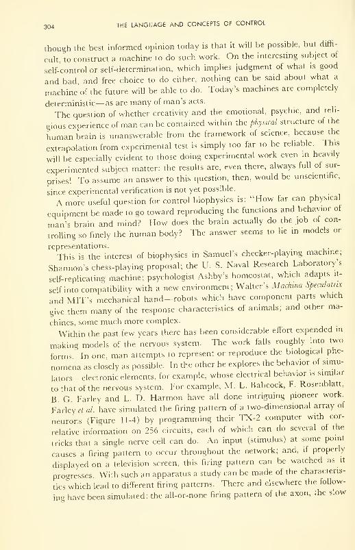
304 THE LANGUAGE AND CONCEPTS OF CONTROL
though the best informed opinion today is that it will be possible, but diffi-
cult, to construct a machine to do such work. On the interesting subject of
self-control or self-determination, which implies judgment of what is good
and bad, and free choice to do either, nothing can be said about what a
machine' of the future will be able to do. Today's machines are completely
deterministic— as are many of man's acts.
The question of whether creativity and the emotional, psychic, and reli-
gious experience of man can be contained within the physical structure of the
human brain is unanswerable from the framework of science, because the
extrapolation from experimental test is simply too far to be reliable. This
will be especially evident to those doing experimental work even in heavily
experimented subject matter: the results are, even there, always full of sur-
prises! To assume an answer to this question, then, would be unscientific,
since experimental verification is not yet possible.
A more useful question for control biophysics is: "How far can physical
equipment be made to go toward reproducing the functions and behavior of
man's brain and mind? How does the brain actually do the job of con-
trolling so finely the human body? The answer seems to he in models or
representations.
This is the interest of biophysics in Samuel's checker-playing machine;
Shannon's chess-playing proposal; the U. S. Naval Research Laboratory's
self-replicating machine; psychologist Ashby's homeostat, which adapts it-
self into compatibility with a new environment; Walter's Machina Speculative
and MIT's mechanical hand— robots which have component parts which
give them many of the response characteristics of animals; and other ma-
chines, some much more complex.
Within the past few years there has been considerable effort expended in
making models of the nervous system. The work falls roughly into two
forms In one, man attempts to represent or reproduce the biological phe-
nomena as closely as possible. In the other he explores the behavior of simu-
lators-electronic elements, for example, whose electrical behavior is similar
to that of the nervous system. For example, M. L. Babcock, F. Rosenblatt,
B. G. Farley and L. D. Harmon have all done intriguing pioneer work.
Farley et al. have simulated the firing pattern of a two-dimensional array of
neurons (Figure 11-4) by programming their TX-2 computer with cor-
relative information on 256 circuits, each of which can do several of the
tricks that a single nerve cell can do. An input (stimulus) at some point
causes a firing pattern to occur throughout the network; and, if properly
displayed on a television screen, this firing pattern can be watched as it
progresses. With such an apparatus a study can be made of the characteris-
tics which lead to different firing patterns. There and elsewhere the follow-
ing have been simulated: the all-or-none firing pattern of the axon, the slow
ANALOGIES 305
axon
nucleus
neuron
Figure 11-4. A Neural Network.
chemical step of the crossing of the synapse, and the smaller, graded, at-
tenuating potential induced at the far side of the synapse. Because other
properties such as a slow wave of electrical activity on the neuron itself, vari-
able spike amplitude, varying wave form and overshoot of the spike, and
shifting baseline potentials are ignored, the simulations are still approxi-
mate. Replicated by such simulation have been: (a) intensity of electrical
activity as a function of time; (b) burst firing; (c) repetitive firing; (d) ac-
commodation, and change in excitability. Further, the simulated circuits
have disclosed certain conditions under which the firing frequency of the net-
work will shift. This is a clue from the machine about a phenomenon which
has not yet been observed experimentally by neurophysiologists. Thus
workers in the field hopefully look forward to advances in man's understand-
ing of his brain through its simulation by machines. The reader is en-
couraged to study the papers by Bullock, 18 and of Harmon, 19 and to treat
himself to the optimism of Reiss, 17 and the careful analysis of Farley, 17
thereby to prepare for himself a proper perspective of this exciting new
aspect of biophysics.
We turn now to an outline of the principles upon which are based the
two great classes of computers, digital and analog.
ANALOGIES
The Digital Nature of Nervous Propagation
The electrochemical burst arising at the point of stimulation and moving
rapidly along the nerve, and called the impulse, was discussed in Chap-
ter 10. To a first approximation, the nerve is either stimulated into action
or it is not. This is the "all-or-none" property. The stimulation must be
306 THE LANGUAGE AND CONCEPTS OF CONTROL
above some critical minimum strength,*** otherwise the nerve will not fire.
That the threshold is not really as critical as is often claimed, and that the
spike, or "wave of negativity," modifies its shape under certain circum-
stances, are useful facts to know and are thought by some physiologists to
be more important that the spike itself. The main point for the moment,
however, is that the passage of a stimulus is a binary process, to a first ap-
proximation always the same. Only the pulse-repetition frequency (pulses
per second) can change; this is frequency modulation.
For example, in the case of transmission of a signal from the pressure-
sensing device which reports blood pressure, the nerve encodes the informa-
tion as a frequency: the higher the pressure the greater the number of pulses
per second (e.g., 125 pulses per sec for high pressure, 70 for low). There is
an inherent accuracy in the counting, or digital, method of transmitting in-
formation— more so than in the decimal-expansion method. The accuracy
comes from repetition, or redundancy.
The Digital Computer
Information can be fed into a machine in either of two ways: intermit-
tently or continuously. If done intermittently, it takes the form of pulses of
energy. The number of pulses then becomes the important thing, for in the
number is contained the information in question. Thus five pulses means
one thing, three another, and so on. (The Morse code was an early example
of this idea.) Since number is important, counting and recording of number
are necessary. Therefore, the performing of operations on the information
becomes simply a matter of arithmetic, nothing more. Since it is numbers,
or digits with which the arithmetic is done, a machine which processes in-
formation in the form of numbers is known as a digital computer. An adding
machine is a primitive example; IBM's "650" has intermediate complexity;
and IBM's 7090 (see Figure 11-5) is a 20,000-component, complicated ex-
ample. It has 32,000 words of high-speed memory and can add two 10-digit
decimal numbers in 4.5 microseconds— facts to be compared with 2000
words for the 650 and an addition time of 800 microseconds.
In computation with digits we normally use the decimal system, with
units often. This system was chosen quite arbitrarily by our ancestors dur-
ing a process of arithmetical evolution in which they counted in twos
(hands), tens (fingers), twenties (fingers and toes), etc. Other systems
could have been chosen equally well. For instance the binary system (units
of two), it is now realized, more closely represents many naturally occurring
phenomena than does the decimal system. Thus only two digits are needed
***That is, a minimum energy must pass through the nerve membrane—most simply stated:
a current, at some voltage, for some length of time (amps x volts x sec = joules).
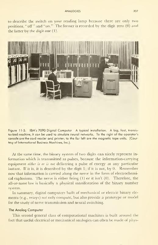
ANALOGIES 307
to describe the switch on your reading lamp because there are only two
positions, "off" and "on." The former is recorded by the digit zero (0) and
the latter by the digit one (1).
Figure 11-5. IBM's 7090 Digital Computer—A typical installation. A big, fast, transis-
torized machine, it can be used to simulate neural networks. To the right of the operator's
console are the card reader and printer; to the far left are the magnetic tape units. (Cour-
tesy of International Business Machines, Inc.).
At the same time, the binary system of two digits can nicely represent in-
formation which is transmitted as pulses, because the information-carrying
equipment either is or is not delivering a pulse of energy at any particular
instant. If it is, it is described by the digit 1; if it is not, by 0. Remember
now that information is carried along the nerve in the form of electrochemi-
cal explosions. The nerve is either firing (1) or it isn't (0). Therefore, the
all-or-none law is basically a physical manifestation of the binary number
system.
In summary, digital computers built of mechanical or electric binary ele-
ments (e.g., relays) not only compute, but also provide a prototype or model
for the study of nerve transmission and neural switching.
The Analog Computer
This second general class of computational machines is built around the
fact that useful electrical or mechanical analogies can often be made of phys-
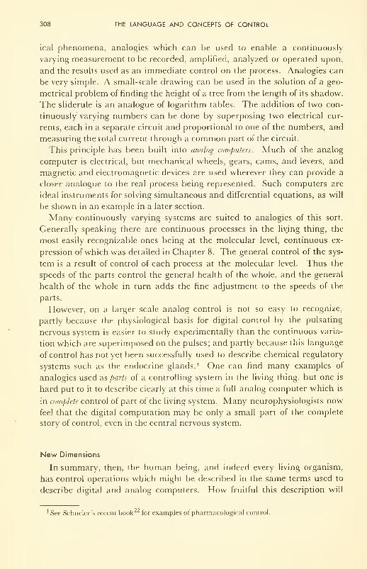
308 THE LANGUAGE AND CONCEPTS OF CONTROL
ical phenomena, analogies which can be used to enable a continuously
varying measurement to be recorded, amplified, analyzed or operated upon,
and the results used as an immediate control on the process. Analogies can
be very simple. A small-scale drawing can be used in the solution of a geo-
metrical problem of finding the height of a tree from the length of its shadow.
The sliderule is an analogue of logarithm tables. The addition of two con-
tinuously varying numbers can be done by superposing two electrical cur-
rents, each in a separate circuit and proportional to one of the numbers, and
measuring the total current through a common part of the circuit.
This principle has been built into analog computers. Much of the analog
computer is electrical, but mechanical wheels, gears, cams, and levers, and
magnetic and electromagnetic devices are used wherever they can provide a
closer analogue to the real process being represented. Such computers are
ideal instruments for solving simultaneous and differential equations, as will
be shown in an example in a later section.
Many continuously varying systems are suited to analogies of this sort.
Generally speaking there are continuous processes in the living thing, the
most easily recognizable ones being at the molecular level, continuous ex-
pression of which was detailed in Chapter 8. The general control of the sys-
tem is a result of control of each process at the molecular level. Thus the
speeds of the parts control the general health of the whole, and the general
health of the whole in turn adds the fine adjustment to the speeds of the
parts.
However, on a larger scale analog control is not so easy to recognize,
partly because the physiological basis for digital control by the pulsating
nervous system is easier to study experimentally than the continuous varia-
tion which are superimposed on the pulses; and partly because this language
of control has not yet been successfully used to describe chemical regulatory
systems such as the endocrine glands. * One can find many examples of
analogies used disparts of a controlling system in the living thing, but one .is
hard put to it to describe clearly at this time a full analog computer which is
in complete control of part of the living system. Many neurophysiologists now
feel that the digital computation may be only a small part of the complete
story of control, even in the central nervous system.
New Dimensions
In summary, then, the human being, and indeed every living organism,
has control operations which might be described in the same terms used to
describe digital and analog computers. How fruitful this description will
+ See Schueler's recent book for examples of pharmacological control.
THE COMPUTER IN BIOLOGICAL RESEARCH 309
be in man's understanding of his control biophysics is hard to predict; but
today it is an exciting avenue by which people are approaching the subject.
Quantitative description of these ideas is developing rapidly, as an inte-
gral part of missile and space technology, in which man has control of the
characteristics of the components, through design. The neuronal circuit,
with switches (synapses) (Figure 1 1-4), is about a billion times smaller than
the vacuum tube circuit, and perhaps about a million times smaller than the
transistor circuit, and a thousand times smaller than thin-film, solid-state
circuits now in the research stage. The neuron operates on the movements of
ions rather than electrons, and much has yet to be learned about its opera-
tion. Further, the number of "components" in the brain is about a million
times the number in the largest of today's computers. Therefore it is certain
that quantitative description of the control circuitry of the central nervous
system is a long way off !
Inherent in all these systems is an error, or noise, or background, above
which the information, the signal, must be distinguished. It is easy to build
an analog computer with a precision of about 1/1000; harder to build one
with 1/10,000: and impossible to build one with 1/100,000 or less because
machining of parts and electrical measurements cannot be made with
greater precision. By contrast, simply increasing the number of components
can increase the precision of the digital machine to 1/10,000,000,000, if it is
desirable and practicable.
Since the central nervous system operates with about 10,000,000,000 com-
ponents, or neurons, and since it has both digital and analog facility, the
problem of understanding this system is obviously not an easy one. Al-
though the normal operation of this system is wondrous enough, errors in
"switching" can give rise to a whole host of disorders— problems not only
for the neurologist but also some that are likely to keep the psychologist and
psychiatrist in business for a long time to come.
THE COMPUTER IN BIOLOGICAL RESEARCH
As a tool in medical research, the computer can do many useful things.
The day may not be too far off, for instance, when medical clinics will be
equipped with general diagnostic machines which, when properly fed with
factual information on symptoms, will not only punch out a statement of
what the possible diseases are but also arrange them in order (with the most
probable one at the top) and state what further examinations can most
profitably be done to save the time of the physician and the money of the
patient. The machine-processing of records and accounts in clinics and hos-
pitals is closer still. With us now is the use of computational machinery to
help the researcher in studies of those biophysical problems in which rea-
sonably precise quantitative measurement is possible. Rapidly maturing as
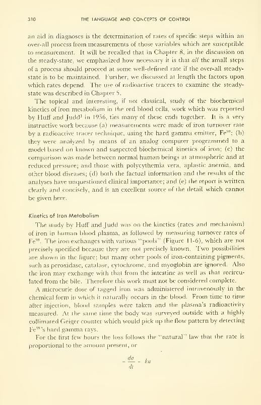
310 THE LANGUAGE AND CONCEPTS OF CONTROL
an aid in diagnoses is the determination of rates of specific steps within an
over-all process from measurements of those variables which are susceptible
to measurement. It will be recalled that in Chapter 8, in the discussion on
the steady-state, we emphasized how necessary it is that all the small steps
of a process should proceed at some well-defined rate if the over-all steady-
state is to be maintained. Further, we discussed at length the factors upon
which rates depend. The use of radioactive tracers to examine the steady-
state was described in Chapter 5.
The topical and interesting, if not classical, study of the biochemical
kinetics of iron metabolism in the red blood cells, work which was reported
by Huff and Judd 1 in 1956, ties many of these ends together. It is a very
instructive work because (a) measurements were made of iron turnover rate
by a radioactive tracer technique, using the hard gamma emitter, Fe59;(b)
they were analyzed by means of an analog computer programmed to a
model based on known and suspected biochemical kinetics of iron; (c) the
comparison was made between normal human beings at atmospheric and at
reduced pressure; and those with polycythemia vera, aplastic anemia, and
other blood diseases; (d) both the factual information and the results of the
analyses have unquestioned clinical importance; and (e) the report is written
clearly and concisely, and is an excellent source of the detail which cannot
be given here.
Kinetics of Iron Metabolism
The study by Huff and Judd was on the kinetics (rates and mechanism)
of iron in human blood plasma, as followed by measuring turnover rates of
Fe59. The iron exchanges with various "pools" (Figure 1 1-6), which are not
precisely specified because they are not precisely known. Two possibilities
are shown in the figure; but many other pools of iron-containing pigments,
such as peroxidase, catalase, cytochrome, and myoglobin are ignored. Also
the iron may exchange with that from the intestine as well as that recircu-
lated from the bile. Therefore this work must not be considered complete.
A microcurie dose of tagged iron was administered intravenously in the
chemical form in which it naturally occurs in the blood. From time to time
after injection, blood samples were taken and the plasma's radioactivity
measured. At the same time the body was surveyed outside with a highly
collimated Geiger counter which would pick up the flow pattern by detecting
Fe59's hard gamma rays.
For the first few hours the loss follows the "natural" law that the rate is
proportional to the amount present, or
da ,= kadt
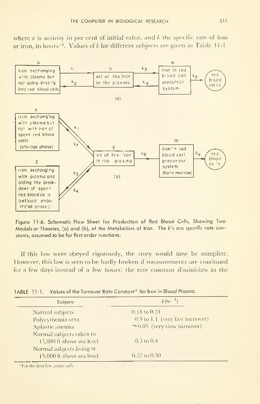
THE COMPUTER IN BIOLOGICAL RESEARCH 311
where a is activity in per cent of initial value, and k the specific rate of loss
or iron, in hours -1. Values of A; for different subjects are given in Table 11-1.
u
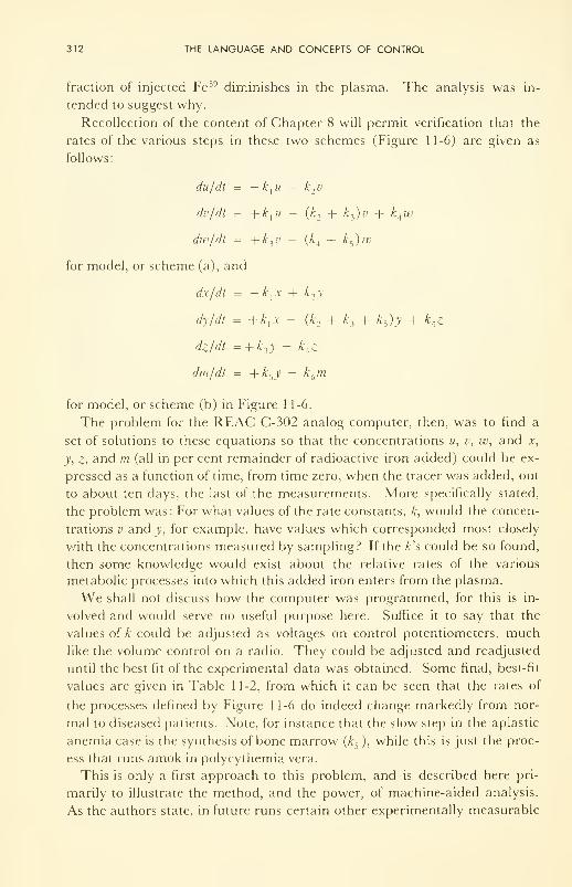
312 THE LANGUAGE AND CONCEPTS OF CONTROL
fraction of injected Fe 59 diminishes in the plasma. The analysis was in-
tended to suggest why.
Recollection of the content of Chapter 8 will permit verification that the
rates of the various steps in these two schemes (Figure 11-6) are given as
follows:
du/dt = — k\u + k2v
dv/dt = +k-[u — (k n + k
3)v + k
Aw
dw/dt = +k3v - (k
A + k5)w
for model, or scheme (a), and
dx/dt = -kxx + k2y
dy/dt = +A,.v - (k2 + £
3 + k5 )y + k
4 z
dz/di = + k-iy - k
4 z
dm/dt = + k5y
— kbm
for model, or scheme (b) in Figure 1 1-6.
The problem for the REAC C-302 analog computer, then, was to find a
set of solutions to these equations so that the concentrations u, v, w, and x,
y, z, and m (all in per cent remainder of radioactive iron added) could be ex-
pressed as a function of time, from time zero, when the tracer was added, out
to about ten days, the last of the measurements. More specifically stated,
the problem was: For what values of the rate constants, k, would the concen-
trations v and y, for example, have values which corresponded most closely
with the concentrations measured by sampling? If the k's could be so found,
then some knowledge would exist about the relative rates of the various
metabolic processes into which this added iron enters from the plasma.
We shall not discuss how the computer was programmed, for this is in-
volved and would serve no useful purpose here. Suffice it to say that the
values of A could be adjusted as voltages on control potentiometers, much
like the volume control on a radio. They could be adjusted and readjusted
until the best fit of the experimental data was obtained. Some final, best-fit
values are given in Table 11-2, from which it can be seen that the rates of
the processes defined by Figure 11-6 do indeed change markedly from nor-
mal to diseased patients. Note, for instance that the slow step in the aplastic
anemia case is the synthesis of bone marrow (A5 ), while this is just the proc-
ess that runs amok in polycythemia vera.
This is only a first approach to this problem, and is described here pri-
marily to illustrate the method, and the power, of machine-aided analysis.
As the authors state, in future runs certain other experimentally measurable
REFERENCES 313
TABLE 1 1-2. Table of Rate Constants and Steady-State Concentrations Evaluated by Ana-
log Computer and Giving Best Fit to Experimental Results.
314 THE LANGUAGE AND CONCEPTS OF CONTROL
12. von Neumann, J., "The Computer and the Brain," Yale University Press, NewHaven, Conn., 1958.
13. Adrian, E. D., Bremer, F., and Jasper, H. H., Eds., "Brain Mechanisms and
Consciousness," Blackwell Scientific Publications, Oxford, 1954.
14. Shannon, C. E., "Mathematical Theory of Communication," University of Illi-
nois Press, Urbana, 111., 1949.
15. Thomson, Sir C, "The Two Aspects of Science," Science, 132, 996 (1960).
16. Teilhard de Chardin, P., "The Phenomenon of Man," Harper & Bros., London,
1955.
17. Barnard, G. A., Chairman, "Proc. 1962 Spring Joint Computer Conference,"
The National Press, Palo Alto, California, 1962: see papers by Ernst, Reiss,
Farley, Harmon and Tiffany.
18. Bullock, T. H., "Neuron Doctrine and Electrophysiology," Science, 129, 997
(1959).
19. Harmon, L. D., " Artificial Neuron, "Science, 129,962 (1959); see ref. 17 for sum-
mary of more recent work.
20. Rosenblatt, F., "Perceptron Simulation Elements," Proc. Institute of Radio Engi-
neers, 48,301 (1960).
21. Minsky, M, "Steps toward Artificial Intelligence," Proc. IRE, 49, 8 (1961); see
also his bibliography on artificial intelligence, IRE Trans, on Human Factors in
Electronics, March 1961.
21. Davis, M., "Computability and Unsolvability," McGraw-Hill Book Co., Inc.,
New York, N. Y., 1958 (interpreting Godel's incompleteness theorem as ap-
plied to computers).
22. Scheuler, F. W., "Chemobiodynamics and Drug Design," McGraw-Hill Book
Co., Inc., New York, N. Y., 1961
.
23. Donaldson, P. E. K., "Electronic Apparatus for Biological Research," Butter-
worth's Scientific Publications, Ltd., London, 1958.
24. Proceedings of the Institute of Radio Engineers, 50, Issue No. 5, May, 1962: a review
of the progress of the last 50 years, and prognostications for the next—with
special emphasis on informational science and control. Many contributors.
Epilogue—A Perspective
It is useful to have a perspective of a subject such as biophysics. In the
Introduction we located the subject nestled in among other pure and bio
sciences. However, the questions raised about information and control in
the last chapter—about man's brain and the computers which he is fashion-
ing—make us wonder where biophysics fits in among those disciplines
which are not physical sciences. In other words, Where does the biophysics
of man fit into the framework of all knowledge about man?
F. O. Schmitt has introduced the thought very nicely:*
"Biophysics, like biochemistry, has to reckon with hierarchies of organiza-
tion and with the properties that are characteristic of systems no less com-
plex than those provided by living organisms at each particular level of
organizational complexity: viz., molecular, macromolecular, subcellular,
cellular, supercellular, organismic, and superorganismic . . . theoretical
biology must deal not only with the properties of cellular constituents but
also with the properties of the organism as a whole."
Interpreting man as an organism, complete with his esthetic, emotional,
and religious experiences, and as part of a superorganism complete with
social, cultural, and religious activities, we can view man's knowledge of him-
self, his history, and his destiny, in a very broad and intriguing perspective.
However, within the framework of the logical disciplines as they now exist,
we know: that logic and experiment are the tools of the scientist; that logical
^//-consistency is the final test for philosophers and mathematicians; and
that the theologion has logic, the results of natural science, and revelation in
his workshop.
Man's intellectual destiny is to know the truth—about the Creator, about
Man, and about Nature—even though "man's body is but a fleeting
thing."** He has the right to know, the ability to find out, and the responsi-
bility to try. Ultimately there is no substitute for the truth in any intellectual
disciplines.
Classification of inputs into "information" and "noise" (in the sense in
which these terms are used in the last chapter) is man's greatest obstacle to
knowing all about man, for such classification is highly subjective.
'Biophysical Science—A Study Program," J. L. Oncley, el ai, Eds., John Wiley & Sons,
Inc., New York, N.Y., 1959, pp. 5 and 6.
**Ecclesiasticus, 41, 11.
315

316 EPILOGUE—A PERSPECTIVE
Man's problem is to find the truth, in spite of the noise which plagues himfrom without and within. There are many pitfalls. Will he find truth by
rejecting a prion, or subjectively, part of the input? Or by rejecting logic's
prime tenet of the excluded middle, as some now suggest? .... I think not.
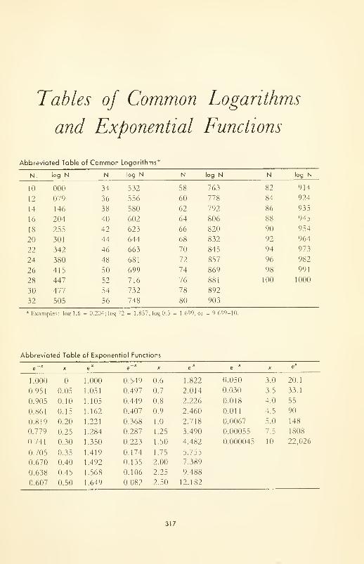
Tables of Common Logarithms
and Exponential Functions
Abbreviated Table of Common Logarithms*
N.

List of Symbols
GREEK LETTERS USED AS SYMBOLS
a—alpha—a radiated particle (Ch. 4, 5, 9); degree of ionization (Ch. 8);
state of cell division (Ch. 9); membrane penetration rate in (Ch. 10).
/?—beta—a radiated particle (Ch. 4, 5, and 9); membrane penetration
rate out (Ch. 10).
7—gamma—ratio of specific heats measured under constant pressure and
constant volume (Ch. 3); radiated electromagnetic radiation (Ch. 4, 5,
and 9).
<5—small delta—a small, measureable length (Ch. 1).
A—capital delta— 'a little bit of" (Ax, Ay, AS, AH, AF, etc.).
e—epsilon—dielectric constant (Ch. 2); incremental energy (Ch. 4).
7]—eta—viscosity (Ch. 8); the neutrino (Ch. 5).
r]Q—eta subscript zero—viscosity of solvent (Ch. 8).
[77]—eta in square brackets— intrinsic viscosity (Ch. 8).
6—small theta—scattering angle (Ch. 4).
K—capital kappa—specific conductivity of a solution (Ch. 8).
A—small lambda— usually a decay constant (Ch. 5); a wavelength (Ch.
3); jump distance (Ch. 8).
A—capital lambda—equivalent conductance (Ch. 8).
A -—equivalent conductance at infinite dilution (Ch. 8); a nuclear particle
(Ch.4).
p.— small mu—mesons (Ch. 4); free energy per mole ("chemical po-
tential") (Ch. 7).
v—small nu—frequency (Ch. 4).
7r—small pi—the constant circumference/diameter of a circle; osmotic
pressure (Ch. 2 and Ch. 6); pion (Ch. 4).
p—small rho—density (Ch. 2).
p—small rho overscored— ratio of densities of solvent to solute (Ch. 6).
a—small sigma—standard deviation (Ch. 1); Stefan's constant (Ch. 8);
specific radiation sensitivity (Ch. 9).
2—capital sigma—a fermion (Ch. 4); a type of bond (Ch. 4); see also
below,
r— small tau—transmission coefficient.
—small phi— fluidity (Ch. 8); a dependent variable.
319
320 LIST OF SYMBOLS
\p—small psi—an independent variable; pressure or amplitude (Ch. 3).
^—capital psi—potential.
co—small omega— unit of resistance, ohms; angular velocity of centrifuge
(Ch. 6).
£—small xi—reaction path length (Ch. 7).
Q—capital omega—the number of ways a system can be arranged (Ch. 7).
MATHEMATICAL SYMBOLS
/—elongated S to represent elongated sum— the sum of an infinite num-
ber of infinitely small parts : the integral sign.
a —the ''infinity1
' sign.
<x —the proportionality sign.
V^the root sign; if no number appears in the hook, a square root sign.
y^—capital sigma—to denote the summation of a finite number of small
but finite parts.
d—rounded "dee11— the partial differential symbol.
=—identically equal to.
X—as superscript—refers to activated complex.
Index
a— a number or constant; acceleration; mo-
lecular extinction coefficient; activity or
effective concentration
.-1— a reactant; area; activation heal of
muscle
A—angstrom unit (10~ cm)
Absorption, of electromagnetic radiation,
80-82
of matter (acoustic) waves, 53
Acceleration, definition, 27
due to gravity, 30
in a centrifuge, 30, 137
Acetylcholine, ACh, as substrate for acetyl-
cholinesterase, ACE, 276
in nerve, 275 ff
thermodynamics of hydrolysis of, 172
Acetylsalicylic acid, formula, 84
infrared absorption spectrum of, 84
Acoustic transducers, 51
Acoustic waves, nature of, 48-49
absorption of, 52, 54
cavitation by, 61
clinical applications of, 62-65
decibel scale for, 55
detection by ear, 56 ff
penetration of, 54
physiological effects of, 60-62
reflection of, 53
therapy by, 62-65
velocity, 50
Actin, 286
Action potential (See Nerve propagation)
Activity, specific, 1 13
of strontium and radium, 113
thermodynamic (effective concentration),
176
Activation energy, and rates of physical and
chemical processes, 198-200
table of values for catalyzed and un-
catalyzed reactions, 201
enthalpy, defined, 200
entropy, defined, 200
Acuity, visual, 87-90
and scanning by the eyeball, 90
Adaptation, dark-, 88
Adenine, 149
Adenosine triphosphate, 29, 177
as mobile power supply, 177, 178
in Krebs cycle, 178
in muscle, 282
in nerve, 268
in protein and DNA synthesis, 151, 156
hydrolysis by myosin, 177, 286, 288
thermodynamics of binding to myosin, 288
Adrenalin, isomers of, 144, 156
All-or-none law, 263, 305
and binary number system, 307
Alpha amino acids, 127
polymerization of, into helices, 130
Alpha helix of proteins, 131
in myoglobin, 132
Alpha keratin, 130
Alpha radiograph, principle, 107
of demineralized bone, 108
of filiform papillae of tongue, 108
Alpha rays or particles, 102
absorption of, 105
energy distribution of, from a source, 1 13
ionization by, 104, 105
penetration of, 116-118
physical properties of, 103, 104-105
Alpha waves (in EEC), 274
Amplitude of matter (acoustic) waves, 55
Anemias, hemolytic and unnamed, 158
sickle cell, 157-159
Anticipation, by brain and computers, 302
Aperture, numerical, of microscope, 98
Arrhenius' equation, 198
Arterial pressure, 218
Assimilation, impaired by ionizing radia-
tions, 252
Astigmatism, 90
Ashby's computer, 296, 304
Astronauts, acceleration of, 30
weightlessness of, 45, 231
Atom, structure of, 71
Atomic nucleus, structure of, 73, 103
Atomic orbitals, or electron shells, 71
321
322 INDEX
ATP (See Adenosine triphosphate)
Autoradiography, principle, 107-108
on blood flow in brain, 120
Avogadro's number, 71
Axon of squid, ion content of, 266
b—number of bels
b—a number or constant
bmr—basal metabolic rate
B—a reactant
Background of ionizing radiations, 235, 239
Bacteria, genetic recombination of, and cod-
ing, 148
effects of ionizing radiations on, 249, 250ultrasonic radiations on, 63
ultraviolet light on, 93
Bacteriophage, and DNA synthesis, 148
Bagchi, on EEG, 275
Balance, kinematic, 99
water, 40
Basal metabolic rate, defined, 168
Bases, purine and pyrimidine, in DNA andRNA, 149
Basilar membrane, 58
Becquerel, and radioactivity, 69
Beer-Lambert law, 81, 136
Bel, definition, 55
Bends, the, 33
Bergonie and Tribondeau, law of, 247, 249
Bernstein, hypothesis of, 265
Beta rays, absorption of, 105-106
energy of emission, and neutrino, 1 15
integrated dose by, 258
ionization by, 105
physical properties of, 103, 105-106
range in air and soft tissue, 1 18
Binding, interatomic, 74
intermolecular, 41-43
Bioenergetics, 161 ff
chemical sources of energy, 166, 167,
173-174
acetylcholine, 172, 275
adenosine triphosphate, 170, 172, 176,
178, 282
caloric value of foods, 166
creatine phosphate, 282
fats, carbohydrates, and proteins, 166
glycogen, 172
table of values, 172
electrical energy transfer and, 179 ff
n — it transition, 147
via redox systems, 179 ff
via mobile 7r-electrons, 146, 147
via electronically excited states, 145
of interaction of tissues with:
infrared radiations, 82-83
ionizing radiations, 93-95, 234 ff
matter (acoustic) waves, 52, 54
ultraviolet radiations. 91-93
visible light, 83-91, 95-99
of mechanical systems:
by matter (acoustic) waves, 52-53, 56-59
by molecular vibrations, etc., 145
by muscle contraction, 280-284
of metabolic processes (See Heat loss,
170, 174)
specific references to, 189-190
Birefringence, flow, 139
Black body radiation and the sun, 79
Blood, circulation of, 218
effects of gravity on, 34-35
fluidity and composition of, 216, 219laminar and turbulent flow in, 212, 215,
217
pressure drops in circulation of, 218
Blood flow in brain, tracers, experiments,
119
Boltzmann's constant, 200
Boltzmann distribution of energies, 197
Bonds, covalent, ionic and dative, 74-75
cross-, induced by X rays, 94
hydrogen, 41, 43, 131-133
interatomic, 74
intermolecular, 42
Bone, impact resistance of, 34
effects of ionizing radiations on, 252
iron turnover rate constants, normal anddiseased, 31
1
marrow therapy, 258Bosons, 73
Bragg's law for scattering of X rays, 127
Brain, as computer, 295-296
as controller of human system, 303-305,
309
electroencephalograms of, 273-277
(See also Central nervous system andComputers)
Bremstrahlung, 106
Burns, on central nervous system, 273-275
Butler, on irradiation of DNA, 243
reviews on biophysics, 66, 160, 294
c—a constant; concentration; specific heat
C— a reaction product
Cable theory, introduction to, 270-271
cal,—small calorie, 28
Cal, or kcal— large calorie (= 1000 cal)
Calculus, definitions of terms of the, 15-17
Cancer, chemical therapy, 259
localization, 62, 94, 97
radiation therapy, 62-65, 256-258
of skin, induced by irradiation, 252
Candle, a photometric unit, 87
INDEX 323
Capacitance, electrical, of double-layer of
membranes, 264
Carbohydrates, 125 ff
structure and properties of, 129
polysaccharides, 133
with lipids, 134
in living membranes, 140
fuel value of, 166
Cataracts, nature of, 91
induced by ionizing radiations, 253
Cavitation, 61
Catalysts, principle, 199
enzymes as, 201 ff
for balky redox reactions, 183
Cells, division of, effect of ionizing radia-
tions on, 247 ff
distortion of, by bad molecules, 157-159
leucocytes, DNA content of, 243
membrane of:
electron micrograph and schematic dia-
grams of, 141, 272
structure and properties of, 140, 264
water flow through, 37, 142
nucleus of, illustration, 264
radiation sensitivity of, 247 fT, 250, 251
Central nervous system, studies on, 273-277
behavior of neurons, 274
effect of ionizing radiations on, 276
electroencephalography and the, 274
transmission across synapses in spinal
cord, 274, 275
Charge, electrical, definition of, 38
and Coulomb's law, 39
on colloids, 40
Chemical potential, n< 176
Chemical reactions, factors of rate expres-
sions, 228
Chemiluminescence, 146
Chromosomes, effects of irradiation during
mitosis, 251, 252
Clark, book on redox systems, 180
Classification of inputs of information, 299,
315
of subject matter of biophysics, 3
problems of, by a computer, 303
subjective character of, in man, 299, 315
Cochlea, 58
Codes, molecular, 125, 148 ff
biochemical information on, 154, 156
cogs and cams in, 154
in bases of nucleic acids, 1 53-1 56
genetic information on, 154-155
table of three-base, 155
Cole, on nerve propagation, 264
Collagen, molecular weight and dimensions,
139
of skin, 134
on tongue, 108
properties of, 129, 137
tensile strength of, in tendon, 133
Colloids, agglutination of, by ionizing radia-
tions, 245
blood groups and, 40, 156
electrostatic forces and, 40
stability of, 40, 156
Color, complementary pairs, 89
sources of, 83
Color blindness, 90
Color vision, theories of, 89-90, 27^
Compton absorption and scattering of X and
7 rays, 80, 105-106
Complex, activated, 200, 202
enzyme— substrate (Michaelis). 202
Computers, analog, 305, 307-308
problem for, 312
and the brain, 295, 296, 303-305, 309
and representation of nerve phenomena,
304-305
calculations by, on iron metabolism, 309-
313
data processing, storage and retrieval by,
302
determinism in, 296, 304
digital, 306-307
number of components in, vs brain, 295,
296, 309
Turing's, 296
with animal-like behavior, 296, 304
Concentration cells, potentials of, 267
(See also Electrical potentials, and Mem-branes)
Concepts in the mind, 302, 303, 315
Conductivity, Electrical, 219 ff (See Electrical
conductivity)
heat, 244 ff (See also Heat conductivity)
Conductor, "volume"— principle of, 223
and EKG and EEG, 224
Cones, distribution and properties of, 85-86
Convection in heat transfer, 226
Cooling, Fourier's law of, 225
Newton's law of, in terms of vapor pres-
sure, 227
Control, concepts and language of, 295 ff, 315
effects of environment on, 290 ff
endocrine, over biochemistry of body, 277,
308
nervous, over muscle, 277
Control biophysics, 296, 303-305
brain vs computer, 303 ff
Corey, on alpha helix. 12"
Cornea, 85
inflammation of due to ultraviolet, 93
Coulomb's law, 74
cps—cycles per second
324 INDEX
Crick, on coding theory, 154
Cross-stimulation of neural networks, 277
Curie, as unit of radioactivity, 1 13
Mme. Marie and Pierre, 102
Current, electronic, in certain organics,
74, 147
factors of, 219-222
in active membranes, 270-271
ionic, in salt solutions and tissues, 219
Curtis, on nerve propagation, 264
Cybernetics, 295 ff
Cytosine, 149
d—infinitesimal amount of, or full differen-
tial symbol; distance; lattice spacing;
deuteron
D—depth (ft) in water; diffusion coefficient;
radiation dose; reaction product
Do—integrated beta dose to tissue
db—number of decibels (10 db = 1 b);
definition, 55
de Broglie's pilot waves, 72
Debye-Hiickel theory of electrolytes, 219
Decay {See Radioactive decay)
Delta, a symbol: A, meaning "a change in"
Delta waves in EEG, 275
Depth perception, 91
Derivative, full and partial, 16
Desoxyribosenucleic acid (DNA)chemical composition of, 133, 148, 149
coding in {See-Codes)
helical structure of, 133, 148, 150
molecular weight of, 139, 150
physical properties of, 129, 148
pyrimidine and purine bases in, 149
Detection of ionizing radiations, 107 ff
by fluorescence, 109
by induced chemical reactions, 1 1
1
by ionization of a gas, 107
by photography, 107, 108
Deuteron, ionization by, 104
penetration of, 116-118
physical properties of, 103, 104-105
Deviations, standard, and others, 19-20
Diastolic pressure, 35
Diathermy by infrared, 82-83
Diffusion, 6, 207 ff
as a rate process, 210
driving force for, 211, 228-229
effect of ionizing radiations on, 245
Fick's laws of, 208-209, 228
of water: osmosis, 142, 212
w fluid flow, 214
Diffusion coefficient, definition of, 208
factors of, 210, 231
table of values, 210
Diffusion potential, 186
Dipoles, induced and permanent, 41
in chemical bonds, 41-42
interaction of, 42-43
Disc, intervertebral, strain on, 34
Disintegration, radioactive (See Radioactive
decay)
Donnan, on membrane selectivity, 187, 267
Dosimetry, radiation, 107-1 11, 238-241
electrochemical, 240
fluorescence, 240
Fricke ferrous sulfate, 239
in gases, 107-1 1
1
Doty, on macromolecules, 135, 136, 139
Dunn-Fry law, 64
e—base of natural logarithms, 2.71828
e+
, e~—positive and negative electrons
E—enzyme ([E] = concentration of enzyme);
reversible electromotive force
Eq—emf of standard state
E*—energy of activation
^difl— diffusion potential
Econc—concentration potential
Em7—midpoint potential
£— efficiency, AF'/AFEar, bones in, 57
detection by inner, 56-58
external, 57
membranes of cochlea, 58
middle, amplifying action of, 57
sensitivity, absolute of, 50, 51, 56
theory of hearing, and, 57
Edema and water balance, 37-38
Efficiency, £ = AF'/A/-', 169
in muscular contraction, 171
of metabolic processes, 171-173
Elastic modulus, definition, 34
of bone, 34
Elastic reactance, and sound absorption, 53
Elasticity of bone, 34
of capillary walls, 35
of cell membranes, 37-38
of muscle tissue, 278-279
Electric potential, definition of, 38
and free energy of reaction, 180
electrodes for the measurement of, 264
electromotive series of, for biological
systems, 180
measured against the normal hydrogen
electrode (NHE), 180
membrane, values of, 187
midpoint, Em7 , 180, 181, 183
of concentration cells, 186, 187,267-268
theory, 180-182,270-271
Electric shock, damage from, 222
INDEX 325
Electric field strength, definition, (voltage
gradient), 38, 40
across living membranes, 40, 143
in ionization chambers, 107
Electrical conductivity, 219 ff
and dimensions of the conductor, 222
effect of ionizing radiations on, 246
Ohm's law of; an analysis, 219-222
of electrolytes, in body, 219
of organic materials, 74, 147
specific, defined, 220
Electrocardiography (EKG), recording,
223Electrochemical nature of nerve propaga-
tion. 266
Electroencephalography (EEG), analysis,
275, 299
effect of ionizing radiations on, 276
Electrolytes, ionic transport in, 219 ff, 228
conductivity of, 219, 222
in giant axon of squid, 266
in red blood cells, 38
Electrolytic cells (See Redox systems)
Electromagnetic spectrum, 77 ff
detailed table of properties, 78-79
(See Infrared radiations, Ionizing radia-
tions, Ultraviolet radiations, and
Visible radiations)
Electromagnetic waves, 76
absorption of, 80-82
interaction with matter, 77-80, 82-95
Electromotive force (See Electrical potentials)
Electron, charge and mass, 68
properties of, 68, 103
and current flow, 219 ff
Electron microscope, principles and limita-
tions of, 100
Electron microscopy, illustrations:
of bacteria, 250
of collagen fibers, 134
of muscle, 284, 285
of neuromuscular junction, 291
of ribosomes of cell, 152
of Schwann cell and nerve axon, 272
Electron volt (ev), unit, 77
Electroretinogram, 90
Elementary particles, 73, 82
Endergonic (synthetic, anabolic) processes,
175
Endothermic processes, 166
Energy, kinetic and potential, 28, 161 ff
chemical, 166, 167
electrical, 45
factors of, 29,45, 175
heat, 45, 162
interconversion of various kinds, 162, 163-
171
internal, definition, 164
in thermodynamic systems, 161 ff
in visual processes, 87-90
mechanical, 45
of matter waves, sound and ultrasound,
49
of muscle contraction, 280
transfer processes, 179 ff
references, 189-190
(See also Bioenergetics)
Enthalpy of reaction (heat content change)
definition, 165
fuel value of foods, 166
of combustion, mixing, and of transition,
166, 167
table of values, 172
Entropy, as a specific heat, 29, 170
as a factor of heat energy, 29, 170
and information, in communications the-
ory, 298
and order vs disorder, 171, 187,298
and probability, 188
negative change in, in living systems, 187
of activation in enzyme catalysis, 200, 206
of reaction in chemical and physical proc-
esses, 171
table of values, 172
Enzymes, as biological catalysts, 201-204,
286, 289
table of rate parameters of reactions cata-
lyzed by, 206
Equilibrium, as a dynamic process, 193
as a thermodynamic process, 176, 185, 194
the drive toward, 175
Equivalent conductance, defined, and ex-
emplified, 221
Errors, kinds, analysis of, and means of
expressing, 17-20
in measurement, and effects on biophysi-
cal control, 298
Erythema, energy of ultraviolet to produce,
92
Erythrocytes (See Red Blood Cells)
ev— electron volt
Excitation, all-or-none property of nerve,
262-263, 305
and electrical conductivity, 74, 147
and photosynthesis, 91
and stimulation of eye, 87
of molecules, electronic, 145
thermal, of rotations and vibrations, 145
Excited states of molecules, 143-146
Extinction coefficients, molar and molecular,
81
326 INDEX
Exergonic (degradation, catabolic) proc-
esses, 175
Exothermic processes, 166
Exponential relationship, defined, 10, 23
Exponents (See Indices)
Extrasensory perception (ESP), 297 (foot-
note)
Eye, architecture and parts of the, 83-85
cataracts on the, 91
color vision of the, 89-90, 277
depth perception, 91
optic nerve endings in, 89, 277
electroretinogram ol, 90
rod and cone cells; pigments in, 88
sensitivity, 89
twilight vision, 87
/—a function (form unspecified): /(*), f(v),
f(v/d), etc.; focal length of lens; frequency
f—
f
ree energy per mole (Gibbs' free energy);
force
F—Faraday's constant, 96,500 cou/equiv.
AF—reversible change in free energy per
mole of reaction; maximum work
AF'—external work per mole of reaction
5J—free energy of whole system
Air— reversible change in free energy of
whole system; maximum work
AC? '—external work done
A3' -internal work done
Falling body, description of, 15
Faraday, on electrolysis, 68, 220
Farley, on neural simulation by com-
puters, 303, 304
Fats, fuel value of, 166
lipids and, properties of some, 129
Feedback in biological systems, 300
general and particular, 301
negative, 301
Fermions. 73
Fick, laws of diffusion of, 208-209, 228
Field strength, defined, 38, 40
across living membranes, 143
in ionization chambers, 107
Fletcher, on speech, 47, 59
Fluid flow, 212 ff
as a molecular process, 214
factors of rate expression for, 228
laminar and turbulent, 212, 215
Newtonian and non-Newtonian, 213
Poiseuille's law of, 213
Reynolds number, 215
Fluidity or specific rate of viscous flow
and molecular weight of dissolved macro-
molecules, 138
definition of fluidity constant, 213
effect of ionizing radiations on, 246
effect of tube length and radius, for
blood, 217
inverse of viscosity, 138, 215
of plasma and blood, 216
table of values, 214
temperature coefficient of, 214
Fluorescence, definition of, 146
and discovery of radioactivity, 102
and discovery of X rays, 68
in amino acids, nitrogen compounds, and
proteins, 147
in solids and liquid detectors of ionizing
radiations, 110, 239-240
Force, basic definition, and dimensions, 26,
28
and energy, 45, 175, 228-229
driving, in chemical processes, 175
electrical, electromagnetic and magnetic,
42, 44
generalized, 26-27, 44, 45, 228, 229
intermolecular, 41-43
osmotic, 35-38
Force-velocity relationship for muscle, 280
Fourier, law of heat conduction, 225
analysis of EEG, method of,
analysis of diffracted X-ray spectrum
Free energy, defined, 168
and equilibrium, 175-177
as maximum work, 168-169
of formation, 185
of reaction; table of values, 172
Functions, definition and illustrations, 9-16
g—grams
g—acceleration due to gravity, 30
g t— ionic conductances through nerve mem-
branes, 270-271
o-forces, and atmospheric pressure, 31
on man in several aspects, and upon im-
pact, 30
Galileo, on falling bodies, 14
Galvani, on electrochemistry and nerve, 263
Gamma rays, absorption, mechanisms of, 106
effect of, as ionizing radiations, 105
energy distribution of, 113
integrated dose of, 258
penetration of, 116-118
physical properties of, 103, 106
Gamov, on the atomic nucleus, 74
on coding theory, 154
Gases, effects of pressure on, 31
solubility (Henry's law), 33
Geiger counter, 109
Genetic effects of ionizing radiations, 253
INDEX 327
Gibbs, throus5ho1.1t thermodynamics, 161 ff
Glasser, source books on biophysics, 261
Gonads, radiation dose on, from various
sources, 253
Gray, on mechanoreceptors, 56
Ground state, definition of, 76, 145
Guanine, in DNA, 149
Guldberg and Waage, on mass action, 194
in alpha helix of proteins, 130-131
Hydrogen peroxide, formed by electromag-
netic radiations, 111, 241
by intense ultrasound, 61
Hydrostatics, 34-35
Hypermetropia, 91
Hypothalamus, as man's temperature con-
troller, 301
h— Planck's constant
//—enthalpy, or heat content, per mole;
Henry's constant
A//—change in enthalpy, per mole; heat of
reaction per mole
JC —enthalpy of whole system
A JC —heat of reaction
Haldane, on scientific terminology, 6
Half-life, biological, 121; table of values, 259
physical, 1 12; table of values, 115
Halley, first on statistics, 17
Hearing (See Ear)
Heart, pacemaker of, 273
pumping action of, 35
Heat, activation, in muscle, 282
basal metabolic, q'bm , 30, 174
extra metabolic, q'ex , 174
total metabolic, q\ 174
energy from absorbed radiations, 241
of shortening of muscle, 282
production and loss in body, 167, 224 ff
Heat capacity, definition, 163
Heat conduction, Fourier's law, 225, 228
Heat content, or enthalpy, 164
Heat death. 188
Heat loss, 173-174,224-227
irreversible, q'irr , 173
Heat of combustion, neutralization, reaction,
and transition, 167
Heisenberg, on wave mechanics, 72
on uncertainty principle, 298
Helmholtz and Young, three-pigment theory
of color vision of, 89-90
Hemoglobin, structure and properties of,
126, 129
Hemostatics, 34-35
Herrick, on ultrasonics in medicine, 65
Hill, about biophysics. 1. 2
on muscle, 280
Hodgkin. on nerve, 263, 266, 270-271
Hodgkin-Huxlev theory of nerve propaga-
tion, 270-271
Hooke's law of elasticity, 34, 278
Hopkins, on bad molecules, 156
hp— horsepower, 28
Huxley, on muscle, 285
Hydrogen bond, nature and strength, 43, 47
1—current
/—current density; intensity; power; one of a
number./ — ion currents, 270
/ or / —intensity or power at a chosen ref-
erence point
/ —threshold intensity
A/, /, /w * 1
Ichthyoccl, molecular weight of, 135, 139
Illumination, 87
Impact resistance, 34
Impulse, and injury, 34
of nerve (See Nerve impulse)
Index of refraction, and microscopy, 96-98
Indicator redox systems, 183
Indices, laws of, and logarithms, 20-21
Information, theory, and control, bio-
physics, 303
and entropy, 298
definitions and terminology, 295 ff
storage and retrieval, 302
Inertia, definition of, 27
and reflection of acoustic waves, 53
Infinitesimals, 12
Infrared radiations, absorption and effects of,
77, 82-83
spectra, assignments, 84
Insulation, thermal; skin, hair, and clothing
as, 227
Insulin, structure of, 126, 128
tracer studies with, 121
Integration, nature of mathematical, 16
Intellectual destiny of man, 31 5
Intensity or power, of matter waves. 55
Intermolecular forces, kinds, 41-42
Internal energy, 164
Inverse Square law. illustration, 52-53
Ionic mobility, ^ or /, 220
Ionic transport in electrolytes. 2 1 9 ff, 228
Ionization chambers, 69, 108-110
Ionization, degree of, in electrolytes, 220
Ionizing radiations, 77, 93-95
absorption of, 81, 105-106, 242
action, direct and indirect, 241-242
Compton scattering of, 105
detection. 1(14 111
328 INDEX
Ionizing radiations (contin.)
dose measurement of, 236-237
effects of, biological, 93, 234 ff
biophysical, 245-247
on molecules, 243, 252
on neuromuscular junctions, 292
whole body, 254-256
energy loss of (LET), 104
excitation by, 93
pair production by, 105-106
production of foreign molecules by,
242-244
therapy by, 93, 256
Iron metabolism, kinetics of, 310
Irradiation, natural sources of, 239
Isomers, definition, 143
Isotopes: decay schemes, 114, 116
stable and unstable, 73, 104
tables of biologically useful, 114, 115,259
j— flow rate; flux during diffusion; current
Joule, unit, 28
Junction potential, 186
k—constant, usually a specific rate constant,
such as k,, k 7 , etc.
kw—kilowatt
KE—kinetic energy, 28
k —Boltzmanns constant (ideal gas constant
per molecule)
K—constant in light scattering equation
A" —equilibrium constant
A" — Michaelis constant
At— thermal conductivity
Kamen, on tracer isotopes, 118
Katz, on neuromuscular junction, 291
K-shell electrons, 71
Kelvin, on measurement, 8
Kendrew, on structure of myoglobin, 126,
132
Kinetics, definitions, 12, 92
biophysical, 192 ff
in analysis of iron metabolism, 310-313
in analysis of muscle contraction, 287-289
Kinetic processes, five: similarities and in-
tegration of, 228-229, 230
Kuzin, on tracers, 122
kvp— kilovolt potential
/— distance; mobility
L— length of an electrical conductor; sym-
bol of a splitting reaction
Lambert, unit of illumination, 87
Lambert's law, 80-81
Land, on color vision, 89
LD 50lethal dose for 50 per cent of sample,
values, 240, 241
Lehmann, on mediators, 184
Leibnitz, on limits, 1
1
Lenses, 85
aberrations and astigmatism, 90-91
aperture of, 98
in microscope, 96-100
LET— linear energy transfer (-dE/dx), 104,
237 (Table)
Levers, bones in ear as, 58
classes and illustration of, 31
mechanical advantage of, 30
Life, 75
Light, nature and absorption of, 76, 80, 83 ff
energy quanta of, 76, 80
extinction coefficient of, 81
sources, 79
wave and particle nature, 177
Limits, 11-12
Linear energy transfer (LET), 104, 237
(Table)
Lipocellulose, 134
Lipoproteins, binding forces in, 42
encephalitis, and binding in, 42
structure of, 129, 134
Liquid crystals, 124
In— natural logarithm
Load-velocity relationship for muscle, 280
Logarithms, definition and laws of, 21
table of, 317
Lohmann reaction, 282
London-van der Waals' forces described, 42
Loudness of sound, 55
relation of, to sensitivity of detector, 56
m—meter
m—constant; mass; degree of maturity
M—momentum; molecular weight
Mev—million electron volts
c
JTl —Young's modulus
Machines, computing {See Computers)
Macintosh, an anesthesia, 33, 56
Macromolecules, crystalline, 127-134
dissolved, 135-140
excited states of, 145
in living membranes, 140-143
molecular weights, methods and table,
135-140
mutations in DNA and RNA, 154-156
physical properties of typical, 128-129
Magnetic field, effect on cell division, 44
Mainland, on medical statistics, 19, 25
Martin and Johnson, on microscopy, 95
Mass and inertia, 27
Mass action, law of, 194
Mathematics, in biophysics, 8
review, 9-25
INDEX 329
Matter (acoustic) waves, 47-48
absorption coefficient of. 52-54
and noise, 59
divergence of, 52
frequencv of different sounds, 50
penetration. 54
physiological effects of intense. 60
power of certain sources (Table). 51, 60
speech, 59
ultrasound and ultrasonics, 47 ff
velocity of, in air, water and solids, 50
Maximum permissible doses of ionizing
radiations. 252, 255
Maxwell, on distribution of thermal energies,
198
on electromagnetic theory, 76
Measurement, and control, 298
errors in, 17-20
Mechanical advantage of a system, 30
Mechanoreceptors, 58
Mediators, 84
Membranes, living:
chemical composition and defects of, 140
effects of ionizing radiations on, 253
in the inner ear, 58
permeability of, 37, 142, 268, 270
phenomenological studies on, 142, 268
physical properties of, 140, 264
selectivity, 271, 268
synthetic, 268-271
thickness, by electron microscopy, 141
Memory, of computers, 302
suggested physicochemical nature of, 27"
Michaelis constant, Km , 203
and binding of enzyme to substrate, 204,
205
Michaelis-Menten equation, 203
in muscle, 288
Microelectrodes, 186, 264 ff (See also Elec-
trical Potentials)
Microendplate potentials, 291
Microirradiation, principle and techniques,
251
of cells and chromosomes, 251
Microradiography, 107-108
Microscopes, electron, 100
fluorescence, 100
optical 95, 96-98
interference, phase, polarizing, 98-100
resolving power of, 98
X-ray, 100
Miner, Shackelton, and Watson, on sensory
data, 102
Mitochondria, as energy factories of cell,
177
structure of membrane of, 141
Mobility, ionic, defined, 220
and conductivity, 220
of ions in nerve membranes, and potential,
267
table of values, 221
Molecular absorption of matter waves, 53
Molecular diseases, definition, 157
anemias, etc., 157-159
Molecular weight, determination of, for
macromolecules, 136-139
Momentum, definition of, 29
conservation of, in shortening of muscle,
280
transfer of, in fluid flow, 214
Moroney, on statistics, 17, 25
Morphine, infrared spectrum of, 84
Motor end plate, micropotentials at, and ir-
radiation of, 291-292
ms— millisecond
Muscle, bands in, 284
biophysics of, 161, 277 ff
chemical composition and structure of, 284,
286, 287
electron microscopy of, 285
energetics of contraction of. 161, 280-282
fibers of, 284, 285
force-velocity relationship, 280, 281
helical spring analogy, 278
molecular mechanism of contraction
(theory) of, 286-290
speed of shortening, and load, 280, 281
strength of (in problem), 293
work done by, 280
Musical sounds, composition of. 50
Mustard gases, as radiomimetic chemicals,
259
Mutations:
and molecular diseases, 156 ff
induced by chance, through excited states,
146
induced by radiations, 253
induced by radiomimetic chemicals, 259
Myelin, electrical conduction in, 271
formation by Schwann cells, 272
Myoglobin, physical properties of, 129
structure of, by X-ray diffraction, 126, 132
Myopia, 90
Myosin, as ATPase, 282 ff, 287
as contractile molecule in muscle, 286
molecular weight of, by several methods.
287
n—usually a constant; order of a reaction;
time-dependent phenomenological pa-
rameter in nerve transmission theory;
valence change, or number of equivalents
330 INDEX
per mole; nonbonding orbital; neutron;
order of diffracted radiation; number of
moles
_/V—a number, used generally as a depend-
ent variable; number of radioactive
atoms; number of impinging particles
yV — a reference state for .\
Nachmansohn, on biochemistry of nerve
conduction, 271
on ACE, 276
n— 7r transition, electronic transition from
nonbonding orbital to a 7r-orbital, il-
lustration and energy of, 147
molecules which can undergo the, 147
mutations resulting from the, 146
Nernst equation, 180
and hydrogen electrode, 181-182
in concentration cells, 267
method of derivation from thermody-
namic principles, 180
Neutrino, 73, 106
Nerve, action potentials of, 262 ff
axons, 264 ff
change in structure in phenylketonuria,
159
electrolytic conduction of, 265, 306
propagation velocity of, 265
Nerve impulse, as a transient bioelectric,
262, 265
digital nature of, 263
shape of spike, 265, 306
Nerve propagation, concentration changes
during, 267
experiments on, 266 ff
permeability changes during, 271
theory of, 269 ff
tracers in the study of, 266
Neural networks, simulation of properties
by computers, 304-305
Neuron, illustrations of, 264, 291
in central nervous system, 274
propagation along axon of, 262-265, 269-
271
Neurosonic surgery and therapy, 72-73
Dunn-Fry law, time to paralysis vs intens-
ity, 64
Neutron activation, for analysis, 1 19
Neutrons:
in nucleus, 82
ionization by, 105
nature and physical properties, 103,
106-107
penetration by, 1 18
Newton, law of cooling, 227
laws of motion, 27
Nirenburg, Ochoa, et al., on coding in
nucleic acids, 154 ff
Noise, and absolute threshold of hearing, 55
and control, 296
from several sources in man's environ-
ment, 60
in the computer, and control, 309
influence on information, 315
subjective character of, in man, 299, 31 5
Nonpolarizable reversible electrodes, 274
Nucleic acids (See DNA and RNA)structure of, 149, 150
Nucleus of an atom, 73-74, 103-104
properties and stability, 103
Nucleus of a cell, 264
— zero"— degree sign (° C, ° F and ° K: degrees
Centigrade, Fahrenheit, and Kelvin,
respectively)
Ohm's law, 40, 219
Optics (See Eye, Color vision. Light, etc.)
Orbitals, atomic and molecular, 74-75
electron migration and energy transfer
along 7T-, 74, 145-147,271
Order, of differential equation, 16
of reaction, 195-196, 311
Organization, and definition of life, 75
from subatomic to superorganismic, 315
Osmosis, as a special case of diffusion,
36, 212
driving force for, 36
in living systems; water balance, 37
through red blood cell wall, 142
Osmotic pressure, as a thermodynamic
property, 36
and water balance, 37
and restoring pressure in cell walls, 37-38
and molecular weight of macromolecules,
136
cell plasma composition and, 38
Otoconiae, 99
Otology, 57
Oxidation-reduction reactions (See Redox
systems)
Oxygen effect in radiation sensitivity and
radiology, 244
p—distance from image to lens; proton
P—pressure; number of points, or even a
single point; power or energy flux, 283
/'M—power expended as work, 283
I\yt—power expended as heat, 283
(P—permeability constant
Pacemaker of heart, 273

INDEX 331
Parkinson's disease, ultrasonic therapy and.
61, 63
Paralysis, acoustic irradiation to, 64-65
Partic Irs. elementary, 73
Pauli, exclusion principle, 72
Pauling, on molecular diseases, 125, 157
on alpha helix in proteins, 127, 131
Penetration of radiations into tissue, 11"
I Set also the specific radiation)
Penfield, on stimulation of cerebral cortex,
276
Permeability constant, of synthetic and
natural membranes, 21 1, 267
of potassium and sodium, 268
changes, in nerve, during action, 270-271
Perspective in biophysics:
among biological sciences, 2
within all knowledge of man, 315
Perutz, on hemoglobin, 159
on isomorphous replacement in X-ray dif-
fraction, 126
Phlebostatic axis, 35
Phosphorescence, and 7r-bonding, 146
in amino acids and proteins, 146
Pholot atalyzed synthesis of vitamin D2 ,
92
Photoelectric absorption of electromagnetic
radiations, 105, 106
Photon, 76
Photophthalamis, induced by ultraviolet. 93
Photopic vision, 86
Photosensitive cells, 85 (See also Color vi-
sion; Pigments)
Photosensitized reactions, 91-92
Phototherapy, 92-93
Physii s, state of, about 1890, 67
Pi(7T) bonds. ^4. 75
and phosphores< en( e in amino acids, 146
in photosynthesis, 92
Piezoelectric i rystals, 51
analogy with transdui er cells in ear, 57
Pigments, visual, 86 90
absorption spectrum of rhodopsin, 88
chlorolabe and erythrolabe, 90
Helmholtz theory ol three, in color
vision, 89
Land theory of two, 89
Pitch of sound. r^ I
Planck's constant, "1. 80
Plasma, blood, ion content of and osmotic
pressure, 38
fluidity of, 216. 217
proteins in, 22 1
Pliicker. on electrical discharges in
Podolskv. on muscle power, 283
Poiseuille's law. 212 213
and measurement of fluidity, 213, 214
obedience of blood to, 216
Polarized light (See Mn roscopy)
Pores in living membranes, 140
effective diameter of, in red blood cells,
142
Potential, defined as an energy, 38
bioelectric, differences of, 38, 39
chemical, n, 176
(See also Electric potentials)
Power, units of, 28
of matter (acoustic) waves, 49
of muscle contraction, as heat, l'^, and
work, Pw , 283
Pressure, basic definitions, 32-33
as a stress, 33
on a skin-diver, 37
osmotic, 35-38
Prisms, 78-79, 82
Propagation of excitation along nerve, 265,
306
frequency of, 306
redundancy, and reliability of informa-
tion, 306
velocity of, 265
( See also Nerve impulse)
Proportional counter, 109
Protanope (color-blind), 90
Protectors against ionizing radiations, 243
Proteins, 125 ff
alpha-helical structure of crystalline, 131,
132
binding to cellulose and lipid, 134
chemical composition and structure, 128-
129
cross-bonding induced by X rays, 94, 242
hydrogen bonding in, 1 $0
mechanism of synthesis of, guided by
DNA, 151-152
tables of molecular weights, 128-129, 287
Protons, exc hange in hydrogen bond, 43
ionization by, 106
penetration of. 116 118
physical properties of, 10.3
psi— pounds per square in< h
q— quantity ol electrii charge; distance of
object from lens, flux, or amount ol ma-
in i.il being transpoi ted a. ross an area in
unit tune, energ) taken in 1>\ a swcmq . (Set Rates, temperature coefficient of)
q'. —basal metabolic heat given ofl by a
nonreversible process in living system,
281
332 INDEX
q'. —total heat given off by an irreversible
process
q'ex—heat, over and above q'bm ,
given off
during work
q' = ?'bm + ?'ex = A $' + ?'irr
Q, — number of cells which die in a given
time; reversible heat per mole
Q — whole population of cells
Q 10— sameas(? ]0
Q — reversible, unavailable heat of whole
process
Q't
— contraction heat of muscle
Quantum, 76, 145
Quantum theory of Planck and Bohr, 76
r—radius of a tube; radius of a sphere; roent-
gen
R— universal gas constant per mole; electri-
cal resistance; distance
/? 90 — light scattered by 90° by macromole-
cules of a layer from the center of a
tube
r«, r2— resistances
(R — electrical resistivity, or specific re-
sistance
rad, radiation absorbed dose, 236
and rem, 236
Radiation of electromagnetic waves:
infrared (See Infrared radiation)
ionizing (See Ionizing radiations)
ultraviolet (See Ultraviolet radiation)
visible (See Visible radiation)
Radiation of matter (acoustic) waves (See
Matter waves)
Radiation of heat energy, 226 (See also
Infrared radiation)
Radioactive decay, law of, 24, 112
and biological half-life, 121
and physical half-life, 112
schemes for certain isotopes, 1 14
Radioactive mapping, 122
Radioactivity, 69, 102
energy distribution, 113, 115
natural background of, 235, 239
separation of emanations, 69
source strength, 1 1
3
Radioactive isotopes as tracers, 115, 118 ff
autoradiography, 107-108, 120
biological half-life, 121,259
distribution and localization, 120-122
in absorption and secretion studies, 120
in radioactive mapping for medical diag
nosis, 122, 130
in studies of fluid flow, 119, 122
in studies on nerve, 266
penetration of, 102, 116-118, 118 (Table)
Radiomimetic chemicals, 259
Radiosensitivity of cells, 250
under microirradiation, 251
Randall, textbook in biophysics, 46
Rate or speed, defined, 13
of chemical reactions, 195 ff
catalyzed by enzymes, 199, 201-206
temperature coefficient of (? 10 ), 197
Rate constant, specific, 195
in kinetics of iron metabolism, normal
and diseased, 31
1
factors of, 198-200
table of values, 196
rbe (See Relative biological effectiveness)
Red blood cells, axial accumulation and
spin of, 217
cell wall structure of, 141
effect on blood fluidity of, 217
effect of ionizing radiations on, 252
ion content of, 38
kinetics of iron metabolism and, 31
1
sickling of, 158-159
size of pores in wall, 142
water balance in, 37
water penetration of, 142
Redox systems, as electron transfer proc-
esses, 179 ff
equivalence of electrical and chemical en-
ergy in, 179
hydrogen, and pH, 181-182
indicators for, 183
iron in heme as, 132
mediators for, 184
midpoint reference potential, Em7 ,182
Nernst equation for, 180
normal hydrogen electrode (NHE) as ref-
erence, 181-182
potentials of, 180, 181, 186, 187,267-268
Redundancy, and precision, 305, 306
rem (See Roentgen equivalent man)
Replication processes, theory of, 151
Reproductive death, induced by ionizing
radiations, 252
rep (See Roentgen equivalent physical)
Resistivity of body fluids, 221, 222
Resolving power of microscope, 98
Relative biological effectiveness (rbe) of
ionizing radiations, 237-238
Relative humidity and heat loss, 227
Reversible conditions, defined, 169
Reynolds number of blood, 215, 216
Rhodopsin, a pigment, 86-88
Ribosomes, 151-152
Ribosenucleic acid (RNA), 129, 148, 151
Robertson, on membrane structure, 272
Roentgen, on X rays, 67, 234
INDEX 333
Roentgen, unit, 237
Roentgen equivalent man (rem), 236
and LET, 237
and rbe, 238, 239
Roentgen equivalent physical, 237
Rushton, on color vision, 88
Rutherford, on atomic structure, 69-72
s—elongation; length or distance; sedimenta-
tion coefficient
S—space or distance between points; sensi-
tivity of a detector; entropy per mole;
substrate for enzyme-catalyzed reactions;
[S], concentration of substrate; state;
sickle cell variant of hemoglobin.
A.Y—change in entropy per mole of reaction
£ — entropy of whole system
A§ — change in entropy in a process; dis-
tortion.
Saltatory conduction (See Electrolytes)
Samuel, on computers, 304
Sanger, on insulin, 126
Scalae: media, tympani, and vestibuli, 58
Scattering experiments, 70
Schmitt, on collagen, 133
on theoretical biology, 315
Schroedinger, on wave mechanics, 71
on mutations, 145
Schwann cells, and myelin sheath, 272, 27"
Scintillation counters, 110, 111
Scintography and radioactive mapping, 111,
122
Scope of biophysics, 1-2
Scotopic (twilight) vision, 88
Second law of thermodynamics, 168, 188
Sedimentation, 137, 138, 153
Self-consistency, logical, in mathematics
and philosophy, 315-316
Semiconductivity, of organic materials, 74,
145-147
in nerve propagation, 271
Sensation of loudness, 55
Sensitivity, and background noise, 55-56
of ear, 51, 56
of eye, 89
to ionizing radiations, 247
Sensitivity constant, ff, for ionizing radia-
tions, 247-249
Sensory data, about, 47, 102
Series, infinite, in biophysics, 22-24
Servomechanism, man as a, 301
properties of a, 300
Shannon, on computers, 304
Shock, 30
Shortening, rate and rate constant for, in
muscle, 279
Sickle-cell anemia, physical basis of, 159
Signal-to-noise ratio, 299
Sommerfeld's atom, 71
Sonic and ultrasonic therapy, methods andeffects, 62-65
in dentistry, 63
physicochemical basis for, 61, 64
paralysis induced by, 64-65
sterilization by, 63
Sound (See Matter waves)
Specific heat, 28, 163
Speech, range of frequency, and power in, 59
Spike, in nervous propagation, 265, 306
Spring, muscle as a, 278
Statistical methods of expressing deviations,
19
Stacy, textbook on biophysics, 56, etc.
Steady-state, and equilibrium, 195
effects of ionizing radiations on, 253
kinetic processes and, 229
Stereoisomers, 143
Sterilization, by beta and gamma rays,
250, 252
by ultrasound, 63, 252
by ultraviolet light, 93
Stimuli, kinds of, for nerve, 272
Strain and stress on bone; elasticity, 34
Sugars, (See Polysaccharides)
Substrate of enzyme-catalyzed reaction, 201
Sun, as earth's primary source of energy, 79
Swallow, on irradiation effects on mole-
cules, 242
Symbols, list of, 319
Synapse, and central nervous system. 276,
304-305
effect of ionizing radiations on, 292
neuromuscular, 291
neuron-neuron, simulation of, 304
transmission across, 276
Systems, concept, 6 ff, 296-297
diagram illustrating feedbacks, 300, 301
pans, 7, 8, 297
properties and theory, 296-301, 315
Systolic pressure, 35
Szent Gyorgyi, on energetics, 161, 185, 191,
293
t—time; time to paralysis
T—temperature (in deg. Kelvin unless other-
wise stated)
Tanford, on macromolecules, 126
Target theory of radiation damage, 241, 242
Teilhard de Chardin, on life, 75, 1 89
334 INDEX
Temperature, and rates of processes of body,
control and regulation of, 225 ff
Tensile strength, denned, of collagen, e.g.,
133
Therapy, 93, 256 {See also Ionizing radia-
tions, Sonic and ultrasonic therapy,
etc.)
Thermal conductivity, 225
effect of radiation on, 245
Thermodynamics {See Energy, Bioenergetics)
Threshold energy and reaction rates, 197-
199, 201 (Table)
of nerve propagation, 306
Thymine, 149
Thyroid, radioactive mapping of, 122
Tissues {See properties of interest)
Touch, 58
Transducers, 51, 58
Tracers {See Radioactive isotopes)
Transmission {See Nerve)
Transmission coefficient, 200, 231
Transport processes {See individual listings)
Troland, unit of retinal illumination, 87
Tumors, irradiation of, 62-65, 239, 256-258
Turbulent flow, in blood, 215, 216
Turnover of radioactive isotopes in humans,
259
Turning, idealized computer, 296
Twilight vision, 87
u—mobility of an ion in an electric field;
;/+ for cation; u~ for anion
V—internal energy per mole
A U—change in internal energy per mole of
reactionlU — internal energy of system
A 'U —change in internal energy in a process
Ultracentrifuge, 137
Ultrasound, ultrasonics. 47 ff
medical applications of, 62, 63
physiological effects of, 60-62
{See Matter waves; Sonic and ultrasonic
therapy)
Ultrastability, in a computer, 296
Ultraviolet light, absorption of, 81
microscopy, 100
photosynthesis by, 91-92
sterilization by, 93
wavelengths and sources of, 79
Uncertainty in measurement and control,
72, 298
Uracil, in DNA, 149
Urey, and amino acid tracing, 1 1
8
-rate or speed; voltage
-volts
V—volume; voltage or electrical potential
Y— specific volume (volume per mole or per
gram)
U— voltage gradient
van der Waals, forces between molecules, 41
van't Hoff, electrolyte theory of, 219-220
reaction isotherm (formula) of, 185
Variables and variation, definitions, 9
Viscosity (inverse of fluidity; See Fluidity)
and measurement of molecular weight of
macromolecules, 215
as a rate process, 214
intrinsic, relative, and specific, defined, 215
Visible light {See Light)
Vision, color, 89-90, 277
twilight, 87
Visual purple {See Rhodopsin)
Volta, on artificial electric organs, 263
Voltage clamp technique, on nerve, 266, 269
von Bekesy, on hearing. 57
von Neumann, on computers and the brain,
295
w speed of an ion under an impressed
voltage; weight; energy transferred by
electromagnetic radiation; angular ve-
locity of a centrifuge; physical work done
U'—work of expansion
.ruy energy used per unit time in metabolic
processes
Walking, heat lost while, 227
Walter, on EEG, 275, 304
Water balance, 36, 37-38
Waves, brain (EEG), 275, 304
in arteries, 35
induced in tissue by intense ultrasound,
53, 60
matter (acoustic), 48
Maxwell's electromagnetic, 76
"of negativity," in nerve, 265, 306
pilot, de Broglie's, for orbital electrons, 72
Weber-Fechner law, in ear, 55
in eye, 89
in general, 299
Weightlessness, effect on rates of biological
processes, 231
Wilhelmy, on mass action, 194
Work, chemical, of synthesis, 167
done by muscle. 167. 280-283
expansion, in a process, 165
external, A 5', 169, 171
internal, A iT'lnt
, of body, 167, 169, 171
physical, 167, 281
{See also Energy, Bioenergetics)

INDEX 335
v— usually an independent variable; distance
X— electromagnetic radiation (X rays)
X rays, 77, 78-79, 93-96, 243, 252
absorption of, 94
characteristic (Moseley; atomic number),
77
discovery of, 234
effects of, 243, 252
in medical diagnosis, 94-97
nonmedical applications of, 235
penetration by, 68
properties of, 78-79
therapy by, 93, 234 ff
wavelength, 77
X-ray diffraction, principle, 127
method of isomorphous replacement in,
132
X-ray burns, 234
V— usually a dependent variable
y —a constant; initial value of y
Y—a thermodynamic fraction, A/*'/A//, or
A3/A.TCYoung-Helmholtz theory of color vision, 89
Young's modulus of elasticity, 34
Z—usually an independent variable; number
of changes carried by an ion
Z-line, in muscle, 290
Zero-seeking servomechanisms, 301, 302


'CO /•« ^LIBRARY