Embed Size (px)
Citation preview

lable at ScienceDirect
Journal of Human Evolution 56 (2009) 405–416
Contents lists avai
Journal of Human Evolution
journal homepage: www.elsevier .com/locate/ jhevol
Dental microwear texture analysis of two families of subfossil lemurs fromMadagascar
J.R. Scott a, L.R. Godfrey b, W.L. Jungers c, R.S. Scott d, E.L. Simons e, M.F. Teaford f, P.S. Ungar g,*,A. Walker h
a Environmental Dynamics Doctoral Program, University of Arkansas, 113 Ozark Hall, Fayetteville, AR 72701, USAb Department of Anthropology, University of Massachusetts, 240 Hicks Way, Amherst, MA 01003, USAc Department of Anatomical Sciences, Health Sciences Center, School of Medicine, Stony Brook University, Stony Brook, NY 11794, USAd Department of Anthropology, Rutgers, State University of New Jersey, New Brunswick, NJ 08901, USAe Division of Fossil Primates, Duke Primate Center, 1013 Broad Street, Durham, NC 27705, USAf Center for Functional Anatomy and Evolution, Johns Hopkins University School of Medicine, 1830 E. Monument St., Baltimore, MD 21205, USAg Department of Anthropology, University of Arkansas, Old Main 330, Fayetteville, AR 72701, USAh Department of Anthropology and Biology, Pennsylvania State University, 409 Carpenter Building, University Park, PA 16802, USA
a r t i c l e i n f o
Article history:Received 18 July 2008Accepted 16 November 2008
Keywords:DietTeethMegaladapisArchaeolemurHadropithecus
* Corresponding author.E-mail address: [email protected] (P.S. Ungar).
0047-2484/$ – see front matter � 2008 Elsevier Ltd.doi:10.1016/j.jhevol.2008.11.003
a b s t r a c t
This study employs dental microwear texture analysis to reconstruct the diets of two families of subfossillemurs from Madagascar, the archaeolemurids and megaladapids. This technique is based on three-dimensional surface measurements utilizing a white-light confocal profiler and scale-sensitive fractalanalysis. Data were recorded for six texture variables previously used successfully to distinguish betweenliving primates with known dietary differences. Statistical analyses revealed that the archaeolemuridsand megaladapids have overlapping microwear texture signatures, suggesting that the two familiesoccasionally depended on resources with similar mechanical properties. Even so, moderate variation inmost attributes is evident, and results suggest potential differences in the foods consumed by the twofamilies. The microwear pattern for the megaladapids indicates a preference for tougher foods, such asmany leaves, while that of the archaeolemurids is consistent with the consumption of harder foods. Theresults also indicate some intraspecific differences among taxa within each family. This evidence suggeststhat the archaeolemurids and megaladapids, like many living primates, likely consumed a variety of foodtypes.
� 2008 Elsevier Ltd. All rights reserved.
Introduction
Diet is widely hypothesized to be a principal factor contributingto the differences between living primate species. For instance, ithas been directly correlated with critical ecological factors such asgroup size and composition, habitat range, and even locomotion(Fleagle, 1999). With so many important elements of the primateniche tied to subsistence, it is no wonder that paleoprimatologistsseek to learn all they can about the diets of past species. In fact, inaddition to revealing information about critical aspects of theecology of fossil taxa, dietary reconstructions help us understandthe processes of evolution and extinction.
The adaptive radiation of the lemurs of Madagascar providesa classic example of the potential usefulness of dietary
All rights reserved.
reconstruction. Geographic isolation and limited competition havemade these forms the primate equivalent of Darwin’s finches.Extant lemurs are represented by more than 100 species within fivefamilies, with a substantive range of adaptations (see summary byWright, 1999). But the subfossil record shows that this radiationwas even more remarkable just a few hundred years ago. In fact,during the past two millennia, the range of their sizes, shapes, andadaptations was greater than that of the rest of the primate order(summarized in Godfrey and Jungers, 2002; Jungers et al., 2002).What were they like? What ecological niches on Madagascar didthey occupy? These are questions that can best be addressed withcareful reconstruction of their paleoecology, especially of theirdiets.
Dental microwear is the study of microscopic scratches and pitsformed on the surfaces of teeth as the result of use. Because of thedirect relationship between the microwear and the materialproperties of foods and abrasives, analysis of dental microwearreveals important clues regarding the diet and ecology of fossil
J.R. Scott et al. / Journal of Human Evolution 56 (2009) 405–416406
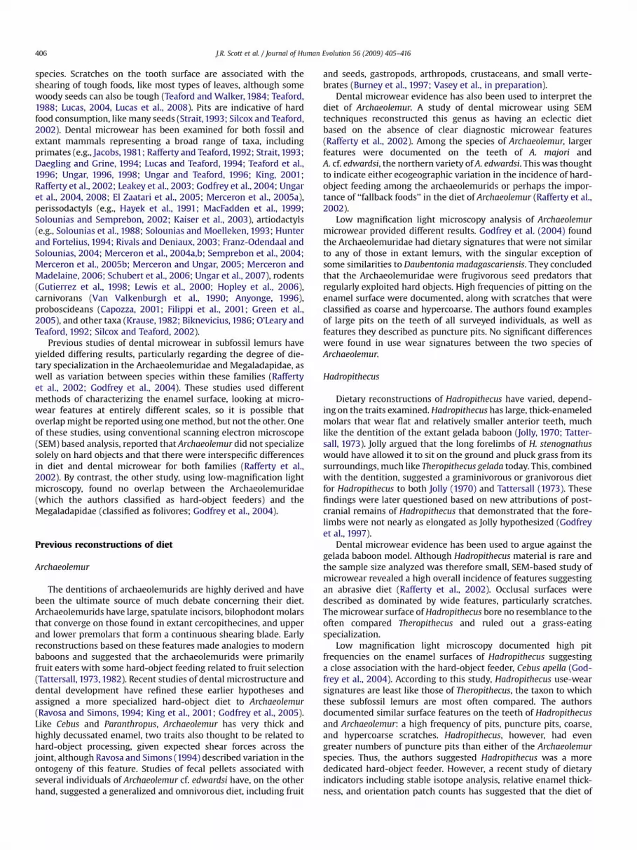
species. Scratches on the tooth surface are associated with theshearing of tough foods, like most types of leaves, although somewoody seeds can also be tough (Teaford and Walker, 1984; Teaford,1988; Lucas, 2004, Lucas et al., 2008). Pits are indicative of hardfood consumption, like many seeds (Strait,1993; Silcox and Teaford,2002). Dental microwear has been examined for both fossil andextant mammals representing a broad range of taxa, includingprimates (e.g., Jacobs, 1981; Rafferty and Teaford, 1992; Strait, 1993;Daegling and Grine, 1994; Lucas and Teaford, 1994; Teaford et al.,1996; Ungar, 1996, 1998; Ungar and Teaford, 1996; King, 2001;Rafferty et al., 2002; Leakey et al., 2003; Godfrey et al., 2004; Ungaret al., 2004, 2008; El Zaatari et al., 2005; Merceron et al., 2005a),perissodactyls (e.g., Hayek et al., 1991; MacFadden et al., 1999;Solounias and Semprebon, 2002; Kaiser et al., 2003), artiodactyls(e.g., Solounias et al., 1988; Solounias and Moelleken, 1993; Hunterand Fortelius, 1994; Rivals and Deniaux, 2003; Franz-Odendaal andSolounias, 2004; Merceron et al., 2004a,b; Semprebon et al., 2004;Merceron et al., 2005b; Merceron and Ungar, 2005; Merceron andMadelaine, 2006; Schubert et al., 2006; Ungar et al., 2007), rodents(Gutierrez et al., 1998; Lewis et al., 2000; Hopley et al., 2006),carnivorans (Van Valkenburgh et al., 1990; Anyonge, 1996),proboscideans (Capozza, 2001; Filippi et al., 2001; Green et al.,2005), and other taxa (Krause, 1982; Biknevicius, 1986; O’Leary andTeaford, 1992; Silcox and Teaford, 2002).
Previous studies of dental microwear in subfossil lemurs haveyielded differing results, particularly regarding the degree of die-tary specialization in the Archaeolemuridae and Megaladapidae, aswell as variation between species within these families (Raffertyet al., 2002; Godfrey et al., 2004). These studies used differentmethods of characterizing the enamel surface, looking at micro-wear features at entirely different scales, so it is possible thatoverlap might be reported using one method, but not the other. Oneof these studies, using conventional scanning electron microscope(SEM) based analysis, reported that Archaeolemur did not specializesolely on hard objects and that there were interspecific differencesin diet and dental microwear for both families (Rafferty et al.,2002). By contrast, the other study, using low-magnification lightmicroscopy, found no overlap between the Archaeolemuridae(which the authors classified as hard-object feeders) and theMegaladapidae (classified as folivores; Godfrey et al., 2004).
Previous reconstructions of diet
Archaeolemur
The dentitions of archaeolemurids are highly derived and havebeen the ultimate source of much debate concerning their diet.Archaeolemurids have large, spatulate incisors, bilophodont molarsthat converge on those found in extant cercopithecines, and upperand lower premolars that form a continuous shearing blade. Earlyreconstructions based on these features made analogies to modernbaboons and suggested that the archaeolemurids were primarilyfruit eaters with some hard-object feeding related to fruit selection(Tattersall, 1973, 1982). Recent studies of dental microstructure anddental development have refined these earlier hypotheses andassigned a more specialized hard-object diet to Archaeolemur(Ravosa and Simons, 1994; King et al., 2001; Godfrey et al., 2005).Like Cebus and Paranthropus, Archaeolemur has very thick andhighly decussated enamel, two traits also thought to be related tohard-object processing, given expected shear forces across thejoint, although Ravosa and Simons (1994) described variation in theontogeny of this feature. Studies of fecal pellets associated withseveral individuals of Archaeolemur cf. edwardsi have, on the otherhand, suggested a generalized and omnivorous diet, including fruit
and seeds, gastropods, arthropods, crustaceans, and small verte-brates (Burney et al., 1997; Vasey et al., in preparation).
Dental microwear evidence has also been used to interpret thediet of Archaeolemur. A study of dental microwear using SEMtechniques reconstructed this genus as having an eclectic dietbased on the absence of clear diagnostic microwear features(Rafferty et al., 2002). Among the species of Archaeolemur, largerfeatures were documented on the teeth of A. majori andA. cf. edwardsi, the northern variety of A. edwardsi. This was thoughtto indicate either ecogeographic variation in the incidence of hard-object feeding among the archaeolemurids or perhaps the impor-tance of ‘‘fallback foods’’ in the diet of Archaeolemur (Rafferty et al.,2002).
Low magnification light microscopy analysis of Archaeolemurmicrowear provided different results. Godfrey et al. (2004) foundthe Archaeolemuridae had dietary signatures that were not similarto any of those in extant lemurs, with the singular exception ofsome similarities to Daubentonia madagascariensis. They concludedthat the Archaeolemuridae were frugivorous seed predators thatregularly exploited hard objects. High frequencies of pitting on theenamel surface were documented, along with scratches that wereclassified as coarse and hypercoarse. The authors found examplesof large pits on the teeth of all surveyed individuals, as well asfeatures they described as puncture pits. No significant differenceswere found in use wear signatures between the two species ofArchaeolemur.
Hadropithecus
Dietary reconstructions of Hadropithecus have varied, depend-ing on the traits examined. Hadropithecus has large, thick-enameledmolars that wear flat and relatively smaller anterior teeth, muchlike the dentition of the extant gelada baboon (Jolly, 1970; Tatter-sall, 1973). Jolly argued that the long forelimbs of H. stenognathuswould have allowed it to sit on the ground and pluck grass from itssurroundings, much like Theropithecus gelada today. This, combinedwith the dentition, suggested a graminivorous or granivorous dietfor Hadropithecus to both Jolly (1970) and Tattersall (1973). Thesefindings were later questioned based on new attributions of post-cranial remains of Hadropithecus that demonstrated that the fore-limbs were not nearly as elongated as Jolly hypothesized (Godfreyet al., 1997).
Dental microwear evidence has been used to argue against thegelada baboon model. Although Hadropithecus material is rare andthe sample size analyzed was therefore small, SEM-based study ofmicrowear revealed a high overall incidence of features suggestingan abrasive diet (Rafferty et al., 2002). Occlusal surfaces weredescribed as dominated by wide features, particularly scratches.The microwear surface of Hadropithecus bore no resemblance to theoften compared Theropithecus and ruled out a grass-eatingspecialization.
Low magnification light microscopy documented high pitfrequencies on the enamel surfaces of Hadropithecus suggestinga close association with the hard-object feeder, Cebus apella (God-frey et al., 2004). According to this study, Hadropithecus use-wearsignatures are least like those of Theropithecus, the taxon to whichthese subfossil lemurs are most often compared. The authorsdocumented similar surface features on the teeth of Hadropithecusand Archaeolemur: a high frequency of pits, puncture pits, coarse,and hypercoarse scratches. Hadropithecus, however, had evengreater numbers of puncture pits than either of the Archaeolemurspecies. Thus, the authors suggested Hadropithecus was a morededicated hard-object feeder. However, a recent study of dietaryindicators including stable isotope analysis, relative enamel thick-ness, and orientation patch counts has suggested that the diet of

Table 1Subfossil lemur specimens used in this analysis.a
Archaeolemur cf. edwardsi: DUPC10850, DUPC10895, DUPC10903, DUPC11729,DUPC11744, DUPC11807, DUPC11819, DUPC11828, DUPC11829, DUPC11830,DUPC11883, DUPC6803, DUPC7849, DUPC7900, DUPC7927, DUPC7928,DUPC7943, DUPC7970, DUPC9104, DUPC9106, DUPC9890, DUPC9899, DUPC9907
Archaeolemur edwardsi: BMNH9909, BMNH9965, BMNH9966, BMNH9968,BMNH9969, BMNH9970, BMNH9972, OXUM5098, UA2769, UA2850, UA5135,UA6773
Archaeolemur majori: AMNH30007, DULCBB2, BMNH13923, BMNH7374,OXUM5099, UA2808, UA5377
Hadropithecus stenognathus: AM Display, AM6382, UA5170, Vienna 1934.IV.2,Vienna1934. IV.1/2
Megaladapis edwardsi: AM6031, AM6071, AM6143, AM6174, AMNH30024,AMNH30025, AMNH30027, AMNH30028, DUPC13663, BMNH13912,BMNH13916, BMNH13917, BMNH7370, BMNH7438, MMV
Megaladapis grandidieri: AM6173, BMNH9917, BMNH9918, BMNH9920,BMNH9921A, BMNH9921B, BMNH9921C, BMNH9922A, BMNH9922B,BMNH9922E, BMNH9975, BMNH9976, BMNH9977, OXUM5101, OXUM5103
Megaladapis madagascariensis: BMNH4848, BMNH4849, DUPC11787, DUPC17218,DUPC18827, DUPC18935, DUPC18938, OXUM5105, UA5484
a AM¼Academie Malgache, AMNH¼American Museum of Natural History,BMNH¼ British Museum, Natural History, DUPC¼Duke University Lemur Center,UA¼University of Antananarivo, OXUM¼Oxford University Natural HistoryMuseum, UA¼University of Antananarivo, Vienna¼Vienna NaturhistorischesMuseum.
J.R. Scott et al. / Journal of Human Evolution 56 (2009) 405–416 407
Hadropithecus might have been more like that of Theropithecus thanpreviously thought (Godfrey et al., 2008).
Megaladapis
The megaladapids lack maxillary incisors and have molars thatincrease in size from anterior to posterior (Godfrey and Jungers,2002). Members of the genus, along with other subfossil lemurs,also have a fused mandibular symphysis, a rarity in strepsirrhines.Their enormous teeth exhibit large shearing crests on the upperand lower molars, which led early researchers to conclude thatMegaladapis was likely a folivore (Thenius, 1953). Tattersall (1975)also invoked a folivory model by comparing the Megaladapis jawapparatus to that of extant Australian koalas. Based on dentalmorphology, Godfrey et al. (1997) posited up to 20% frugivory in allspecies of Megaladapis, with the majority of the remaining dietbeing accounted for by folivory. More recently, Jungers et al. (2002)calculated the shearing quotients of several species of Megaladapisand other extinct and extant lemurs, corroborating the inference ofstrong folivory in the former.
Dietary reconstruction inferred using an SEM-based study ofdental microwear confirmed the folivore label. The documentedmicrowear features included narrow pits, a high incidence ofnarrow scratching, and a low overall frequency of pitting (Raffertyet al., 2002). The study suggested differences within the genus indegree of folivory, with M. grandidieri and M. madagascariensis, thesmaller species, trending towards more varied diets. M. edwardsiwas classified as a ‘‘hyper-folivore,’’ with narrow scratches and thelowest frequency of pitting recorded for any primate.
Godfrey et al. (2004), using low magnification light microscopy,agreed that all Megaladapis species were leaf dominated browsersand compared them most closely to the extant genera Avahi, Lep-ilemur, and Alouatta. Their study found no evidence of hard-objectfeeding or seed predation, based on a lack of puncture pitting anda low incidence of pitting overall. They also found no significantdifferences between the species of Megaladapis, althoughM. edwardsi had slightly larger features and more pitting than thesmaller species. Godfrey and co-authors suggested that thisdifference may be related to habitat, as M. edwardsi shared thespiny scrub forests of southern Madagascar currently occupied byLepilemur leucopus, a species with similar use wear patterns. Theyalso hypothesized that this variation, although not statisticallysignificant, could be evidence for niche differentiation in thesouthern part of Madagascar, where M. edwardsi and M. mada-gascariensis overlap in range.
This paper brings a new technique, dental microwear textureanalysis, to bear on the study of subfossil lemur microwear, toprovide a new set of results that may illuminate the issue of diet.This technique is automated and the results are repeatable whenthe same area of the facet is scanned. The technique also allowscharacterization of surfaces in three-dimensions over a continuousrange of scales and operates at high resolution to facilitate theidentification and exclusion of taphonomically damaged specimens(Scott et al., 2005, 2006; Ungar et al., 2008). Results should there-fore allow an independent assessment of interspecific variationamong the Archaeolemuridae and Megaladapidae, as well asinterspecific differences within each genus.
Methods
We examined all identified species of archaeolemurids andmegaladapids for this study. These included both species within thegenus Archaeolemur: A. edwardsi (n¼ 35), and A. majori (n¼ 7). Totest for the possibility of dietary differences between A. edwardsifrom northern and central Madagascar, specimens from the two
geographic regions were analyzed separately. Although the samplesize for A. majori is small, this taxon was included in the statisticalanalyses. Specimens for the only species within the genus Hadro-pithecus, H. stenognathus, were also examined, although onlya small number (n¼ 5) were without postmortem damage andsuitable for microwear analysis. All species within the genus Meg-aladapis were also included: M. edwardsi (n¼ 15), M. grandidieri(n¼ 15), and M. madagascariensis (n¼ 9).
The specimens used in this study are housed at the AmericanMuseum of Natural History (AMNH), the British Museum of NaturalHistory (BMNH), the Duke University Lemur Center (DUPC), theAcademie Malgache (AM), Vienna Naturhistorisches Museum(NHNW), Oxford University Natural History Museum (OXUM), andthe University of Antananarivo (UA). Table 1 lists the included taxaand specimens used in the analysis.
Data collection
As in previous studies of dental microwear (see Teaford andRobinson, 1989; Teaford and Runestad, 1992), the maxillary secondmolar was used in the analysis whenever possible. When themaxillary tooth was unavailable or exhibited postmortem damagethat obscured microwear, the second mandibular molar or firstmaxillary molar was substituted to maximize sample sizes, espe-cially for rare subfossil material. Conventional SEM-based micro-wear studies have shown no consistent pattern of differences inupper and lower microwear pattern or between tooth types (Gor-don, 1982, 1984; Teaford and Walker, 1984; Teaford, 1985, 1986;Grine, 1986, 1987; Bullington, 1991). Additionally, this studyfocused on the so-called ‘‘Phase II’’ facets (facets 9, 10 n, and x), as isusual for dental microwear analysis (Kay, 1977; Gordon, 1982;Teaford and Walker, 1984; Krueger et al., 2008). The criteria used fordetermining suitability for microwear analysis were those of Tea-ford (1988) and based on examination of the sides and occlusalsurfaces of the teeth, and a ‘‘clean’’ enamel use-wear surface free ofpost mortem damage, coating, adhesive, or casting defects.
The specimens used in this study came from two differentcollections and replicas were prepared separately by three of us(LG, WJ, and MT). High resolution replicas were prepared usingconventional procedures (Ungar, 1996). Teeth were cleaned usingeither acetone or alcohol, and the crown surfaces were molded
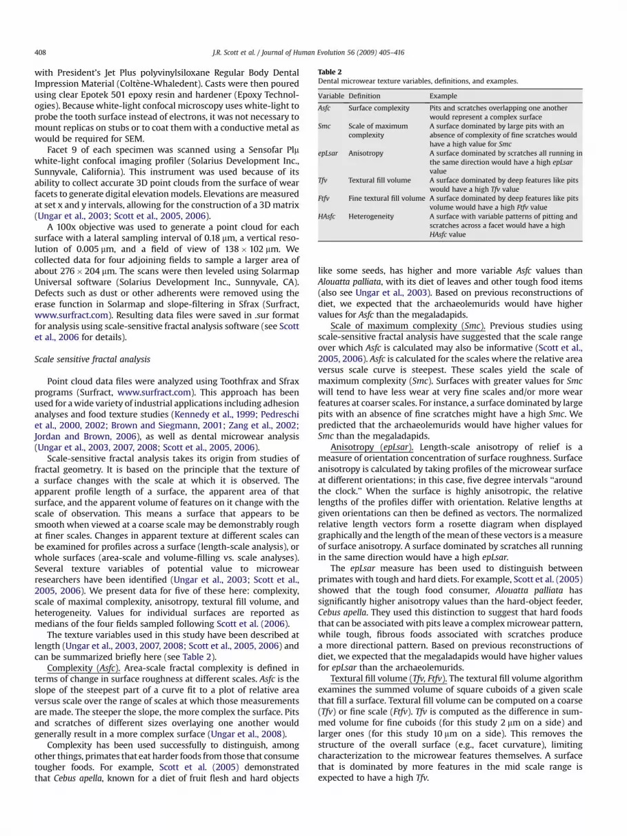
Table 2Dental microwear texture variables, definitions, and examples.
Variable Definition Example
Asfc Surface complexity Pits and scratches overlapping one anotherwould represent a complex surface
Smc Scale of maximumcomplexity
A surface dominated by large pits with anabsence of complexity of fine scratches wouldhave a high value for Smc
epLsar Anisotropy A surface dominated by scratches all running inthe same direction would have a high epLsarvalue
Tfv Textural fill volume A surface dominated by deep features like pitswould have a high Tfv value
Ftfv Fine textural fill volume A surface dominated by deep features like pitsvolume would have a high Ftfv value
HAsfc Heterogeneity A surface with variable patterns of pitting andscratches across a facet would have a highHAsfc value
J.R. Scott et al. / Journal of Human Evolution 56 (2009) 405–416408
with President’s Jet Plus polyvinylsiloxane Regular Body DentalImpression Material (Coltene-Whaledent). Casts were then pouredusing clear Epotek 501 epoxy resin and hardener (Epoxy Technol-ogies). Because white-light confocal microscopy uses white-light toprobe the tooth surface instead of electrons, it was not necessary tomount replicas on stubs or to coat them with a conductive metal aswould be required for SEM.
Facet 9 of each specimen was scanned using a Sensofar Plmwhite-light confocal imaging profiler (Solarius Development Inc.,Sunnyvale, California). This instrument was used because of itsability to collect accurate 3D point clouds from the surface of wearfacets to generate digital elevation models. Elevations are measuredat set x and y intervals, allowing for the construction of a 3D matrix(Ungar et al., 2003; Scott et al., 2005, 2006).
A 100x objective was used to generate a point cloud for eachsurface with a lateral sampling interval of 0.18 mm, a vertical reso-lution of 0.005 mm, and a field of view of 138� 102 mm. Wecollected data for four adjoining fields to sample a larger area ofabout 276� 204 mm. The scans were then leveled using SolarmapUniversal software (Solarius Development Inc., Sunnyvale, CA).Defects such as dust or other adherents were removed using theerase function in Solarmap and slope-filtering in Sfrax (Surfract,www.surfract.com). Resulting data files were saved in .sur formatfor analysis using scale-sensitive fractal analysis software (see Scottet al., 2006 for details).
Scale sensitive fractal analysis
Point cloud data files were analyzed using Toothfrax and Sfraxprograms (Surfract, www.surfract.com). This approach has beenused for a wide variety of industrial applications including adhesionanalyses and food texture studies (Kennedy et al., 1999; Pedreschiet al., 2000, 2002; Brown and Siegmann, 2001; Zang et al., 2002;Jordan and Brown, 2006), as well as dental microwear analysis(Ungar et al., 2003, 2007, 2008; Scott et al., 2005, 2006).
Scale-sensitive fractal analysis takes its origin from studies offractal geometry. It is based on the principle that the texture ofa surface changes with the scale at which it is observed. Theapparent profile length of a surface, the apparent area of thatsurface, and the apparent volume of features on it change with thescale of observation. This means a surface that appears to besmooth when viewed at a coarse scale may be demonstrably roughat finer scales. Changes in apparent texture at different scales canbe examined for profiles across a surface (length-scale analysis), orwhole surfaces (area-scale and volume-filling vs. scale analyses).Several texture variables of potential value to microwearresearchers have been identified (Ungar et al., 2003; Scott et al.,2005, 2006). We present data for five of these here: complexity,scale of maximal complexity, anisotropy, textural fill volume, andheterogeneity. Values for individual surfaces are reported asmedians of the four fields sampled following Scott et al. (2006).
The texture variables used in this study have been described atlength (Ungar et al., 2003, 2007, 2008; Scott et al., 2005, 2006) andcan be summarized briefly here (see Table 2).
Complexity (Asfc). Area-scale fractal complexity is defined interms of change in surface roughness at different scales. Asfc is theslope of the steepest part of a curve fit to a plot of relative areaversus scale over the range of scales at which those measurementsare made. The steeper the slope, the more complex the surface. Pitsand scratches of different sizes overlaying one another wouldgenerally result in a more complex surface (Ungar et al., 2008).
Complexity has been used successfully to distinguish, amongother things, primates that eat harder foods from those that consumetougher foods. For example, Scott et al. (2005) demonstratedthat Cebus apella, known for a diet of fruit flesh and hard objects
like some seeds, has higher and more variable Asfc values thanAlouatta palliata, with its diet of leaves and other tough food items(also see Ungar et al., 2003). Based on previous reconstructions ofdiet, we expected that the archaeolemurids would have highervalues for Asfc than the megaladapids.
Scale of maximum complexity (Smc). Previous studies usingscale-sensitive fractal analysis have suggested that the scale rangeover which Asfc is calculated may also be informative (Scott et al.,2005, 2006). Asfc is calculated for the scales where the relative areaversus scale curve is steepest. These scales yield the scale ofmaximum complexity (Smc). Surfaces with greater values for Smcwill tend to have less wear at very fine scales and/or more wearfeatures at coarser scales. For instance, a surface dominated by largepits with an absence of fine scratches might have a high Smc. Wepredicted that the archaeolemurids would have higher values forSmc than the megaladapids.
Anisotropy (epLsar). Length-scale anisotropy of relief is ameasure of orientation concentration of surface roughness. Surfaceanisotropy is calculated by taking profiles of the microwear surfaceat different orientations; in this case, five degree intervals ‘‘aroundthe clock.’’ When the surface is highly anisotropic, the relativelengths of the profiles differ with orientation. Relative lengths atgiven orientations can then be defined as vectors. The normalizedrelative length vectors form a rosette diagram when displayedgraphically and the length of the mean of these vectors is a measureof surface anisotropy. A surface dominated by scratches all runningin the same direction would have a high epLsar.
The epLsar measure has been used to distinguish betweenprimates with tough and hard diets. For example, Scott et al. (2005)showed that the tough food consumer, Alouatta palliata hassignificantly higher anisotropy values than the hard-object feeder,Cebus apella. They used this distinction to suggest that hard foodsthat can be associated with pits leave a complex microwear pattern,while tough, fibrous foods associated with scratches producea more directional pattern. Based on previous reconstructions ofdiet, we expected that the megaladapids would have higher valuesfor epLsar than the archaeolemurids.
Textural fill volume (Tfv, Ftfv). The textural fill volume algorithmexamines the summed volume of square cuboids of a given scalethat fill a surface. Textural fill volume can be computed on a coarse(Tfv) or fine scale (Ftfv). Tfv is computed as the difference in sum-med volume for fine cuboids (for this study 2 mm on a side) andlarger ones (for this study 10 mm on a side). This removes thestructure of the overall surface (e.g., facet curvature), limitingcharacterization to the microwear features themselves. A surfacethat is dominated by more features in the mid scale range isexpected to have a high Tfv.

J.R. Scott et al. / Journal of Human Evolution 56 (2009) 405–416 409
Tfv has also been suggested to have the potential to distinguishbetween primates that consume foods with different fractureproperties. Cebus apella, for example, has high values for Tfv,a reflection of the higher pit count on the surface. Tough foodconsumers that rely on leaves may have lower Tfv values thatcorrespond to the fine, scratched microwear surface these foodsproduce. We expected that the archaeolemurids would have highervalues for Tfv than the megaladapids.
Heterogeneity (HAsfc). As discussed in the above sections, vari-ables including surface complexity, roughness, and anisotropy canprovide accurate descriptions of microwear surfaces. Howeveruseful these might be, adjoining scans of the same surface can varyin their values for each variable. In fact, the amount of variation incomplexity, roughness, and anisotropy across a facet or evena single scan may be important in characterizing the microwearsurface.
Heterogeneity of area-scale fractal complexity (HAsfc) is calcu-lated by splitting individual scanned areas into successively smallersubregions given equal numbers of rows and columns. This algo-rithm is performed by using the Auto-Split function in the Tooth-frax software. HAsfc is calculated by splitting each individual scaninto smaller sections with equal numbers of rows and columns. Thescans are divided first into 2� 2 and into increasingly smallersections up to 11�11. Resulting distributions are typically skewed,so the relative variations in complexity for each set of subregionswere calculated as the median absolute deviation of Asfc divided bythe median of Asfc. Based on previous reconstructions of diet, weexpected that the archaeolemurids would have higher values forHAsfc(9) and HAsfc(81) than the megaladapids.
Statistical analyses were performed to determine the extent ofvariation in microwear texture among taxa following Ungar et al.(2006, 2008). All data were rank-transformed before analysis(Conover and Iman, 1981) because unranked microwear datatypically violate assumptions associated with parametric statisticaltests. Data for the variables were compared among species usinga multivariate analysis of variance model, with taxon as the factor,Asfc, Smc, epLsar, Tfv, HAsfc(9), and HAsfc(81) as the dependent vari-ables, and values for each individual as the replicates. This testassesses significance of variation among the taxa in overallmicrowear surface texture. Single classification ANOVAs for eachvariable and multiple comparisons tests were used to determinethe sources of significant variation (but see Enders, 2003; Keselmanet al., 1998 for alternate approaches to analysis). Because thesegroups were chosen for their dietary (and expected microwear)differences, Fisher’s LSD a priori tests were used to comparespecies. Tukey’s HSD post hoc tests were also run to balance risks ofType I and Type II errors (Cook and Farewell, 1996).
Results
Examples of microwear surfaces for each taxon are illustrated inFigure 1. Figure 2 shows three-dimensional renderings of surfacesfor both families. Results suggest differences between the twofamilies and between the species within the families for at leastsome of the tested texture variables. Both Wilks’ l and Pillai Traceresults indicated significant variation in the model.
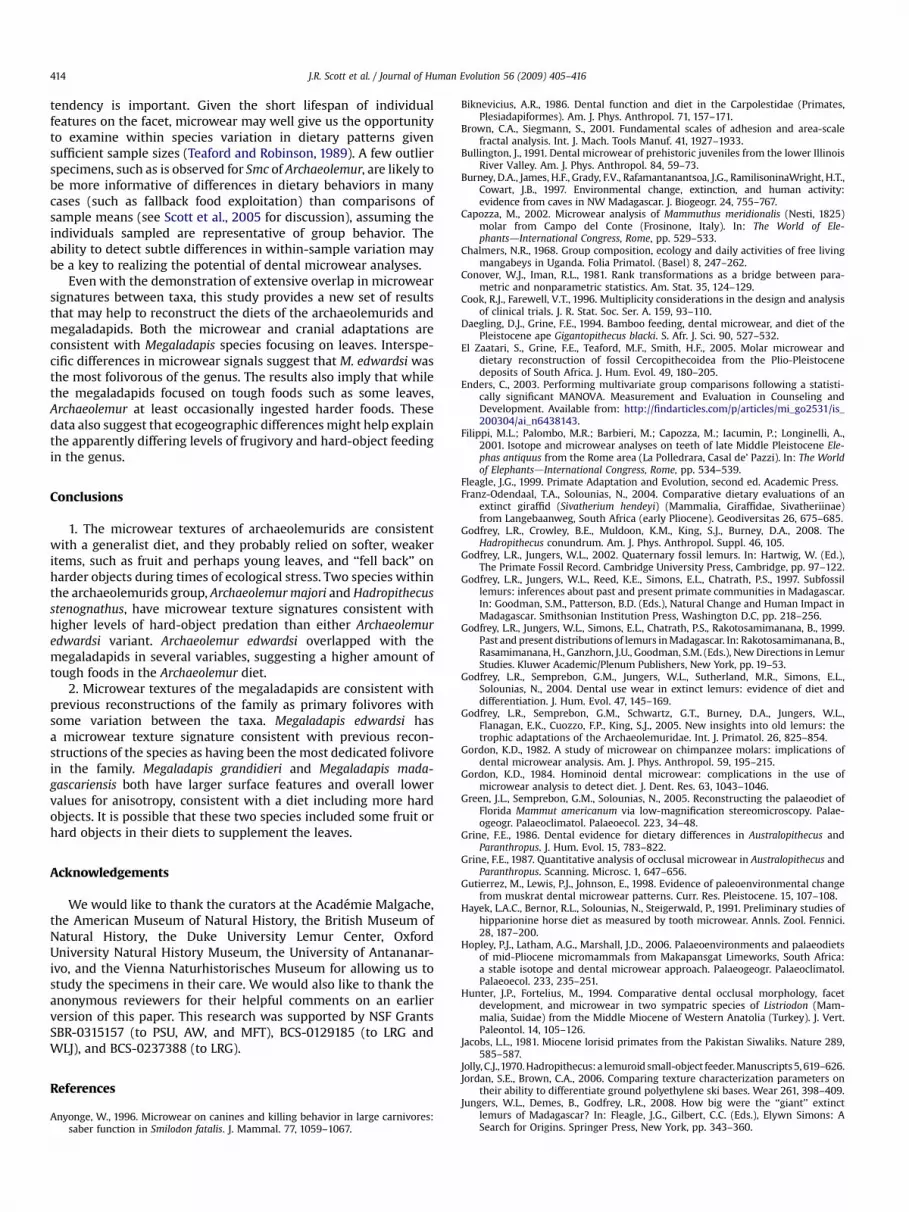
The archaeolemurids tended to have surfaces dominated bylarger features, usually pits, with low anisotropy and moderateheterogeneity. The megaladapids, in contrast, more often hadsurfaces dominated by scratches of varying sizes and depth, withhigh anisotropy and moderate heterogeneity. Thus, the two familiesseem to fall into two clusters (Fig. 3), with some overlap betweenthem. Individual ANOVA results indicate that the archaeolemuridshave significantly higher Asfc, Tfv, and Ftfv than do the mega-ladapids (Table 3).
Some differences are also evident within each of the families.For example, both the central and northern variants of A. edwardsitend to have smaller, shallower features than A. majori and H.stenognathus. Further, M. edwardsi tends to have smaller, shallowerfeatures than the other megaladapids. These visually apparentdifferences are confirmed by statistical analyses (Table 4). Tukey’stest results indicate significant differences between A. cf. edwardsiand A. majori in Tfv, Smc, and Ftfv, with A. majori having highervalues for all variables. A. cf. edwardsi also had significantly lowerSmc values than H. stenognathus. No significant differences inmicrowear texture were found between the geographic variants ofA. edwardsi or between A. majori and H. stenognathus. Within themegaladapids, M. grandidieri has higher values for Tfv, Ftfv, andHAsfc(81) than M. edwardsi. M. edwardsi also has higher Smc valuesthan M. madagascariensis. Results from Fisher’s LSD Test generallycorroborate those from Tukey’s, as well as suggesting additionaldifferences between the archaeolemurids in epLsar and HAsfc(81).
In summary, within the archaeolemurids, the two geographicvariants of A. edwardsi were not consistently different, althoughonly A. cf. edwardsi sensu stricto is significantly different from A.majori and H. stenognathus. Overall, A. majori and H. stenognathushave higher values for fill volume and the scale of maximalcomplexity. Within the megaladapids, M. edwardsi has lower valuesfor fill volume than M. grandidieri and higher scales of maximalcomplexity than M. madagascariensis. The data suggest that, amongthe archaeolemurids, A. majori and H. stenognathus have larger,deeper surface features and that among the megaladapids, M.edwardsi has smaller, shallower surface features. These results areconsistent with dietary differences both between and withinArchaeolemuridae and Megaladapidae.
Discussion
This study presents new data on the dental microwear of theArchaeolemuridae and Megaladapidae, based on the application ofa new technique, dental microwear texture analysis. These datacorroborate many of the previous dietary inferences made aboutthe Archaeolemuridae and Megaladapidae using other methods ofmicrowear analysis. In essence, they affirm the dietary separationbetween the two families and some variation within each family.This study suggests that these subfossil lemurs, like the majorityof living primates, did not focus on specific food types andtightly controlled dietary categories. The question then becomesone of degree. How dedicated a folivore was Megaladapis or howspecialized a hard-object feeder was Archaeolemur or Hadr-opithecus?
The characterization of Megaladapis as a folivore is reaffirmed bythe low epLsar and Tfv values revealed here. An interesting differ-ence between these results and those of previous studies concernsthe inference of differences in dietary signals between species ofMegaladapis. Significant differences between the species were notfound using low magnification light microscopy. However, simi-larities between M. edwardsi and the extant spiny forest/openwoodland-adapted lemur, Lepilemur leucopus and between M.grandidieri and M. madagascariensis and closed forest lepilemurswere noted (Godfrey et al., 2004). SEM analysis suggested that M.edwardsi was more folivorous than other members of the genus(Rafferty et al., 2002). Results of the present study confirm thevariability within Megaladapis with significant differences from theother taxa in almost every variable, with a more homogenoussurface and smaller features. When compared to previously pub-lished data by Scott et al. (2006) for the folivore Alouatta palliata, M.edwardsi has higher mean values for Asfc, suggesting the inclusionof harder objects in the diet; however, the lower values for Tfv areconsistent with a highly folivorous diet.
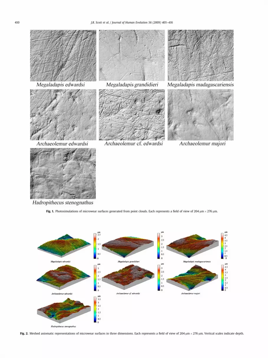
Fig. 2. Meshed axiomatic representations of microwear surfaces in three dimensions. Each represents a field of view of 204 mm� 276 mm. Vertical scales indicate depth.
Fig. 1. Photosimulations of microwear surfaces generated from point clouds. Each represents a field of view of 204 mm� 276 mm.
J.R. Scott et al. / Journal of Human Evolution 56 (2009) 405–416410
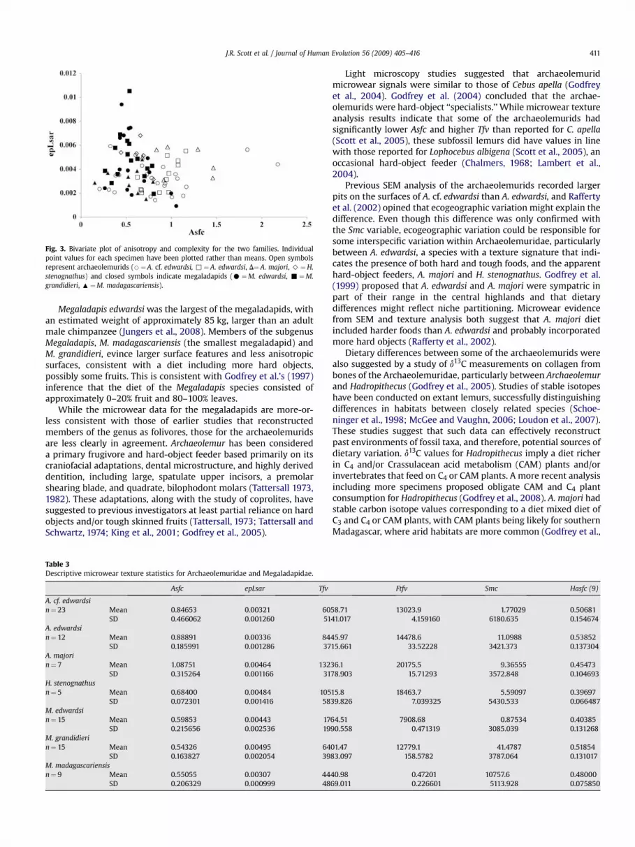
Fig. 3. Bivariate plot of anisotropy and complexity for the two families. Individualpoint values for each specimen have been plotted rather than means. Open symbolsrepresent archaeolemurids (B¼ A. cf. edwardsi, ,¼ A. edwardsi, D¼ A. majori, >¼H.stenognathus) and closed symbols indicate megaladapids (C¼M. edwardsi, -¼M.grandidieri, :¼M. madagascariensis).
J.R. Scott et al. / Journal of Human Evolution 56 (2009) 405–416 411
Megaladapis edwardsi was the largest of the megaladapids, withan estimated weight of approximately 85 kg, larger than an adultmale chimpanzee (Jungers et al., 2008). Members of the subgenusMegaladapis, M. madagascariensis (the smallest megaladapid) andM. grandidieri, evince larger surface features and less anisotropicsurfaces, consistent with a diet including more hard objects,possibly some fruits. This is consistent with Godfrey et al.’s (1997)inference that the diet of the Megaladapis species consisted ofapproximately 0–20% fruit and 80–100% leaves.
While the microwear data for the megaladapids are more-or-less consistent with those of earlier studies that reconstructedmembers of the genus as folivores, those for the archaeolemuridsare less clearly in agreement. Archaeolemur has been considereda primary frugivore and hard-object feeder based primarily on itscraniofacial adaptations, dental microstructure, and highly deriveddentition, including large, spatulate upper incisors, a premolarshearing blade, and quadrate, bilophodont molars (Tattersall 1973,1982). These adaptations, along with the study of coprolites, havesuggested to previous investigators at least partial reliance on hardobjects and/or tough skinned fruits (Tattersall, 1973; Tattersall andSchwartz, 1974; King et al., 2001; Godfrey et al., 2005).
Table 3Descriptive microwear texture statistics for Archaeolemuridae and Megaladapidae.
Asfc epLsar Tfv
A. cf. edwardsin¼ 23 Mean 0.84653 0.00321 60
SD 0.466062 0.001260 51A. edwardsin¼ 12 Mean 0.88891 0.00336 84
SD 0.185991 0.001286 37A. majorin¼ 7 Mean 1.08751 0.00464 132
SD 0.315264 0.001166 31H. stenognathusn¼ 5 Mean 0.68400 0.00484 105
SD 0.072301 0.001416 58M. edwardsin¼ 15 Mean 0.59853 0.00443 17
SD 0.215656 0.002536 19M. grandidierin¼ 15 Mean 0.54326 0.00495 64
SD 0.163827 0.002054 39M. madagascariensisn¼ 9 Mean 0.55055 0.00307 44
SD 0.206329 0.000999 48
Light microscopy studies suggested that archaeolemuridmicrowear signals were similar to those of Cebus apella (Godfreyet al., 2004). Godfrey et al. (2004) concluded that the archae-olemurids were hard-object ‘‘specialists.’’ While microwear textureanalysis results indicate that some of the archaeolemurids hadsignificantly lower Asfc and higher Tfv than reported for C. apella(Scott et al., 2005), these subfossil lemurs did have values in linewith those reported for Lophocebus albigena (Scott et al., 2005), anoccasional hard-object feeder (Chalmers, 1968; Lambert et al.,2004).
Previous SEM analysis of the archaeolemurids recorded largerpits on the surfaces of A. cf. edwardsi than A. edwardsi, and Raffertyet al. (2002) opined that ecogeographic variation might explain thedifference. Even though this difference was only confirmed withthe Smc variable, ecogeographic variation could be responsible forsome interspecific variation within Archaeolemuridae, particularlybetween A. edwardsi, a species with a texture signature that indi-cates the presence of both hard and tough foods, and the apparenthard-object feeders, A. majori and H. stenognathus. Godfrey et al.(1999) proposed that A. edwardsi and A. majori were sympatric inpart of their range in the central highlands and that dietarydifferences might reflect niche partitioning. Microwear evidencefrom SEM and texture analysis both suggest that A. majori dietincluded harder foods than A. edwardsi and probably incorporatedmore hard objects (Rafferty et al., 2002).
Dietary differences between some of the archaeolemurids werealso suggested by a study of d13C measurements on collagen frombones of the Archaeolemuridae, particularly between Archaeolemurand Hadropithecus (Godfrey et al., 2005). Studies of stable isotopeshave been conducted on extant lemurs, successfully distinguishingdifferences in habitats between closely related species (Schoe-ninger et al., 1998; McGee and Vaughn, 2006; Loudon et al., 2007).These studies suggest that such data can effectively reconstructpast environments of fossil taxa, and therefore, potential sources ofdietary variation. d13C values for Hadropithecus imply a diet richerin C4 and/or Crassulacean acid metabolism (CAM) plants and/orinvertebrates that feed on C4 or CAM plants. A more recent analysisincluding more specimens proposed obligate CAM and C4 plantconsumption for Hadropithecus (Godfrey et al., 2008). A. majori hadstable carbon isotope values corresponding to a diet mixed diet ofC3 and C4 or CAM plants, with CAM plants being likely for southernMadagascar, where arid habitats are more common (Godfrey et al.,
Ftfv Smc Hasfc (9)
58.71 13023.9 1.77029 0.5068141.017 4.159160 6180.635 0.154674
45.97 14478.6 11.0988 0.5385215.661 33.52228 3421.373 0.137304
36.1 20175.5 9.36555 0.4547378.903 15.71293 3572.848 0.104693
15.8 18463.7 5.59097 0.3969739.826 7.039325 5430.533 0.066487
64.51 7908.68 0.87534 0.4038590.558 0.471319 3085.039 0.131268
01.47 12779.1 41.4787 0.5185483.097 158.5782 3787.064 0.131017
40.98 0.47201 10757.6 0.4800069.011 0.226601 5113.928 0.075850

Table 4Statistical Analyses.a
A. Multivariate Nested Analysis of VarianceBetween families
Test statistic F df pb
Wilks’ Lambda 0.554 8.516 7, 74 0.000Pillai Trace 0.446 8.516 7, 74 0.000Hotelling-Lawley 0.806 8.516 7, 74 0.000
Between species nested within familiesTest statistic F df pb
Wilks’ Lambda 0.321 2.778 35, 313 0.000Pillai Trace 0.958 2.641 35, 390 0.000Hotelling-Lawley 1.366 2.825 35, 362 0.000
B. Nested ANOVAsBetween familiesVariable F df pb
Asfc 23.00 1, 80 0.000epLsar 0.087 1, 80 0.769Tfv 28.32 1, 80 0.000Smc 8.778 1, 80 0.004Ftfv 35.42 1, 80 0.000HAsfc9 0.046 1, 80 0.831HAsfc81 1.636 1, 80 0.205
Between species within familiesVariable F df pb
Asfc 1.865 5, 80 0.110epLsar 3.487 5, 80 0.007Tfv 5.661 5, 80 0.000Smc 4.542 5, 80 0.001Ftfv 4.943 5, 80 0.001HAsfc9 2.765 5, 80 0.024HAsfc81 1.383 5, 80 0.240
C. Matrices of pairwise differences (within family comparisons)c
Archaeolemurids Megaladapids1. epLsarA.edwardsi A. cf. edwardsi A. edwardsi A. majori M. edwardsi M. grandidieri
0.607 M. grandidieri 3.667A. majori 13.85* 13.24* M. madagascariensis �7.267 �10.93*H. stenognathus 15.14* 14.54* 1.2982. Tfv
A. cf. edwardsi A. edwardsi A. majori M. edwardsi M. grandidieriA. edwardsi 6.678 M. grandidieri 14.00**A. majori 18.83** 12.15* M. madagascariensis 8.011 �5.989H. stenognathus 10.76 4.083 �8.0713. Sm
A. cf. edwardsi A. edwardsi A. majori M. edwardsi M. grandidieriA. edwardsi 8.437* M. grandidieri �7.700A. majori 14.83** 6.399 M. madagascariensis �12.58** �4.889H. stenognathus 17.64** 9.208 2.810
4. FtfvA. cf. edwardsi A. edwardsi A. majori M. edwardsi M. grandidieri
A. edwardsi 2.304 M. grandidieri 13.93**A. majori 17.44** 15.14* M. madagascariensis 6.822 �7.111H. stenognathus 13.47* 11.16 �3.9765. HASFC
A. cf. edwardsi A. edwardsi A. majori M. edwardsi M. grandidieriA. edwardsi 4.051 M. grandidieri 9.733**A. majori �4.497 �8.548 M. madagascariensis 8.044 �1.689H. stenognathus �13.11* �17.16* �8.619
a All analyses on ranked data.b P values reported as 0.000, while effectively zero probabilites, represent actual values of less than 0.001.c * significant with Fisher’s Least Significance Test; ** significant with Tukeys HSD Multiple Comparisons Test.
J.R. Scott et al. / Journal of Human Evolution 56 (2009) 405–416412
2005). The values for A. edwardsi were consistent with a dietdominated by C3 plants, which are associated with mosaic orforested habitats. Coprolite analysis for A. edwardsi suggests a dietof frugivory and omnivory for A. edwardsi (Vasey et al., inpreparation).
The sample size of Hadropithecus stenognathus is small (n¼ 5)and any differences between it and the larger samples of other taxa
should be taken as suggestive at best. The microwear of H. sten-ognathus was similar to that of A. majori, with high values for Tfv,Ftfv, and Smc. Fisher’s test results indicate that Hadropithecus hadsignificantly higher values for anisotropy and heterogeneity thandid A. edwardsi and A. cf. edwardsi. Along with the other archae-olemurids, the microwear signature of Hadropithecus is consistentwith that of a mixed feeder, although with the highest values for

J.R. Scott et al. / Journal of Human Evolution 56 (2009) 405–416 413
volume among the studied taxa, it is likely that hard objects werea more frequent part of the diet for Hadropithecus than for the otherincluded subfossils. If this is the case, the hard object feedingadaptations reflected in Hadropithecus could have evolved to allowthem to fall back on harder foods when preferred resources werescarce. However, it is also possible that the higher anisotropy valuesreported for H. stenognathus and A. majori could imply a reliance ontough foods that require more repetitive tooth-food-tooth move-ments. Isotope analysis also suggests that Hadropithecus ate moreleaves and other plant material, while Archaeolemur ate more fruitand animal products (Godfrey et al., 2008; Ryan et al., 2008).
The results of the present study support a reconstruction of thearchaeolemurids as at least occasional or facultative hard-objectfeeders, as has previously been suggested by both Rafferty et al.(2002) and Godfrey et al. (2004). The microwear texture analysisresults also appear to reflect ecogeographic differences within thegenus, as earlier hypothesized by both Godfrey et al. (1997) andRafferty et al. (2002). As for the specialized morphological adap-tations in Archaeolemur, it is important to note that adaptations fortough or hard-object processing may not have been necessary toprocess all its foods. In other words, Archaeolemur may have hadthe capability to process such hard or tough foods, but not alwaysdone so.
Rafferty et al. (2002) argued that the dental microwear ofArchaeolemur indicated an eclectic diet with no distinctive signa-tures. Some individuals were documented to have larger featuresthan others, suggesting to the authors ecogeographic variation indiet or the use of hard objects as fallback resources. Godfrey et al.(2004) found that the dental microwear of Archaeolemur mostclosely resembled that of Cebus apella, a primate that, whileutilizing a variety of resources, has anatomical specializations thatit regularly uses for hard-object processing. It was suggested that,as in Cebus, the morphological specializations of Archaeolemur suchas extremely thick and heavily decussated enamel, might have alsobeen used to supplement a variable diet. The results of the currentstudy support both conclusions and suggest that the archae-olemurids probably had a variable diet that included hard-objectsin addition to other resources. The microwear texture data suggestcomplex surfaces with a mosaic of layered features of various sizes.The textural variable data for the archaeolemurids overlap with thelower ranges of data reported for C. apella but are concentrated nearvalues more similar to those reported for Lophocebus albigena,a generalist primate that consumes hard objects as fallback items.This suggests that while the archaeolemurids did utilize somehard-objects, they may have done so less frequently than did Cebus.
Studies of primate dentition and diet have suggested correla-tions between morphological features, including tooth shape andenamel thickness, with the ability of a species to utilize certainresources (Lucas, 1979; Lucas and Peters, 2000). The application ofdental morphological analysis to the study of dietary preferences inthe subfossil lemurs has suggested that taxa had specializedadaptations, such as the fast-wearing premolar shearing bladefound in Archaeolemur (Tattersall 1973, 1982) and the high molarshearing crests and loss of upper incisors in Megaladapis (Thenius,1953), allowing them to take advantage of hard or tough resources,respectively.
However, these seemingly specialized adaptations do not tell uswhether selective pressures resulted from preferred foods or criticalones taken only on occasion. As described by Liem (1980), speciesthat seem to be very specialized in terms of morphological orbehavioral adaptations can, in reality, be ecological generalists. Insome cases, the resources that organisms seem specialized toexploit may comprise only a small percentage of their total diet. Thisphenomenon has become known as Liem’s Paradox (Robinson andWilson, 1998). For example, a species may have specialized
adaptations to allow the consumption of hard or tough fallbackfoods when less mechanically challenging preferred foods areunavailable. The preferred items may not require any specializedprocessing (e.g., young leaves or fruit flesh), and therefore may notselect for specialized morphology. Thus, while functionalmorphology can tell us something about what an animal is capableof eating, it does not necessarily give us insights into its food pref-erences, or how often it ate foods with given fracture properties.
Primate diets are highly variable and they depend on preferredresource availability and the occasional use of fallback resources.Differences between taxa are often obscured by the fact thatprimates assigned to different diet categories in the popular liter-ature often eat similar foodsde.g., gorillas have traditionally beendescribed as folivores, while chimpanzees are known as frugivores.However, Ungar et al. (2008) found overlap in dental microwearsignatures between the two species. In fact, gorillas have beenreported to consume 73% of the same species eaten by sympatricchimpanzees at Lope, Gabon (Tutin and Fernandez, 1985). Thus, thelack of clear differences described by Ungar et al. (2008) in centraldietary tendencies between chimpanzees and gorillas is notunexpected.
Primates tend to prefer high energy foods, such as ripe fruits,that do not require specialization to process (Remis, 1997; Remiset al., 2001; Marshall and Wrangham, 2007). Fallback foods such ashard seeds or tough leaves may require morphological specializa-tions to process efficiently, and so it seems that dental morphologyshould actually be more reflective of fallback resource use thanpreferred diet (Robinson and Wilson, 1998). A study of the dentalmorphology and diet of five lemur species by Yamashita (1998b)revealed that the hardest and toughest resources consumed werefallback foods and that these were more tightly correlated with thedental adaptations found in the lemurs than were the mostcommonly consumed resources.
Reports on the amount of dietary overlap between living lemurshave yielded different results. Many examples of dietary variationand fallback resource use have been reported for lemurs (e.g., Straitand Overdorff, 1996; Yamashita,1996, 1998a,b; Simmen et al., 2003)and several species of generalist lemurs are known, includingLemur catta. A recent study of lemur seed dispersal at Ranomafanasuggests that there is a lack of dietary overlap between species(Wright, 2008). Varecia variegata, Eulemur fulvus, Eulemur rubri-venter, and Propithecus edwardsi were reported to not overlap inmany of the fruits consumed. Yamashita (2002) also found littleoverlap in resources selected between Lemur catta and Propithecusverreauxi verreauxi at Beza Mahafaly Special Reserve but did findthat these species utilized foods with similar mechanical proper-ties. This suggests that in some habitats lemurs may not overlap inspecific foods, but does not mean that they limit themselves to onlya few food types or that they always choose foods with the samemechanical properties
Individual microwear features on a tooth surface are themselvesultimately worn away and replaced by others. The ‘‘lifespan’’ ofa feature depends on its depth. This is known as the ‘‘Last SupperEffect’’ and indicates that microwear features on the enamel surfaceonly reflect feeding activity in the days or weeks prior to death(Grine, 1986). Most primate species exploit a variety of resourceswith varying fracture properties and abrasives. As a result, it is likelythat even species with different central dietary tendencies willoverlap in their microwear patterning. But then again, some mayalso vary dramatically at one period and not the next. Thus, it is onlywhen larger samples sizes are used, preferably taken from speci-mens that were collected at different times of the year, that thebigger picture of specialization and dietary overlap begins to emerge.
This suggests that to understand dietary adaptations andbehaviors of primates, a measure other than the central microwear
J.R. Scott et al. / Journal of Human Evolution 56 (2009) 405–416414
tendency is important. Given the short lifespan of individualfeatures on the facet, microwear may well give us the opportunityto examine within species variation in dietary patterns givensufficient sample sizes (Teaford and Robinson, 1989). A few outlierspecimens, such as is observed for Smc of Archaeolemur, are likely tobe more informative of differences in dietary behaviors in manycases (such as fallback food exploitation) than comparisons ofsample means (see Scott et al., 2005 for discussion), assuming theindividuals sampled are representative of group behavior. Theability to detect subtle differences in within-sample variation maybe a key to realizing the potential of dental microwear analyses.
Even with the demonstration of extensive overlap in microwearsignatures between taxa, this study provides a new set of resultsthat may help to reconstruct the diets of the archaeolemurids andmegaladapids. Both the microwear and cranial adaptations areconsistent with Megaladapis species focusing on leaves. Interspe-cific differences in microwear signals suggest that M. edwardsi wasthe most folivorous of the genus. The results also imply that whilethe megaladapids focused on tough foods such as some leaves,Archaeolemur at least occasionally ingested harder foods. Thesedata also suggest that ecogeographic differences might help explainthe apparently differing levels of frugivory and hard-object feedingin the genus.
Conclusions
1. The microwear textures of archaeolemurids are consistentwith a generalist diet, and they probably relied on softer, weakeritems, such as fruit and perhaps young leaves, and ‘‘fell back’’ onharder objects during times of ecological stress. Two species withinthe archaeolemurids group, Archaeolemur majori and Hadropithecusstenognathus, have microwear texture signatures consistent withhigher levels of hard-object predation than either Archaeolemuredwardsi variant. Archaeolemur edwardsi overlapped with themegaladapids in several variables, suggesting a higher amount oftough foods in the Archaeolemur diet.
2. Microwear textures of the megaladapids are consistent withprevious reconstructions of the family as primary folivores withsome variation between the taxa. Megaladapis edwardsi hasa microwear texture signature consistent with previous recon-structions of the species as having been the most dedicated folivorein the family. Megaladapis grandidieri and Megaladapis mada-gascariensis both have larger surface features and overall lowervalues for anisotropy, consistent with a diet including more hardobjects. It is possible that these two species included some fruit orhard objects in their diets to supplement the leaves.
Acknowledgements
We would like to thank the curators at the Academie Malgache,the American Museum of Natural History, the British Museum ofNatural History, the Duke University Lemur Center, OxfordUniversity Natural History Museum, the University of Antananar-ivo, and the Vienna Naturhistorisches Museum for allowing us tostudy the specimens in their care. We would also like to thank theanonymous reviewers for their helpful comments on an earlierversion of this paper. This research was supported by NSF GrantsSBR-0315157 (to PSU, AW, and MFT), BCS-0129185 (to LRG andWLJ), and BCS-0237388 (to LRG).
References
Anyonge, W., 1996. Microwear on canines and killing behavior in large carnivores:saber function in Smilodon fatalis. J. Mammal. 77, 1059–1067.
Biknevicius, A.R., 1986. Dental function and diet in the Carpolestidae (Primates,Plesiadapiformes). Am. J. Phys. Anthropol. 71, 157–171.
Brown, C.A., Siegmann, S., 2001. Fundamental scales of adhesion and area-scalefractal analysis. Int. J. Mach. Tools Manuf. 41, 1927–1933.
Bullington, J., 1991. Dental microwear of prehistoric juveniles from the lower IllinoisRiver Valley. Am. J. Phys. Anthropol. 84, 59–73.
Burney, D.A., James, H.F., Grady, F.V., Rafamantanantsoa, J.G., RamilisoninaWright, H.T.,Cowart, J.B., 1997. Environmental change, extinction, and human activity:evidence from caves in NW Madagascar. J. Biogeogr. 24, 755–767.
Capozza, M., 2002. Microwear analysis of Mammuthus meridionalis (Nesti, 1825)molar from Campo del Conte (Frosinone, Italy). In: The World of Ele-phantsdInternational Congress, Rome, pp. 529–533.
Chalmers, N.R., 1968. Group composition, ecology and daily activities of free livingmangabeys in Uganda. Folia Primatol. (Basel) 8, 247–262.
Conover, W.J., Iman, R.L., 1981. Rank transformations as a bridge between para-metric and nonparametric statistics. Am. Stat. 35, 124–129.
Cook, R.J., Farewell, V.T., 1996. Multiplicity considerations in the design and analysisof clinical trials. J. R. Stat. Soc. Ser. A. 159, 93–110.
Daegling, D.J., Grine, F.E., 1994. Bamboo feeding, dental microwear, and diet of thePleistocene ape Gigantopithecus blacki. S. Afr. J. Sci. 90, 527–532.
El Zaatari, S., Grine, F.E., Teaford, M.F., Smith, H.F., 2005. Molar microwear anddietary reconstruction of fossil Cercopithecoidea from the Plio-Pleistocenedeposits of South Africa. J. Hum. Evol. 49, 180–205.
Enders, C., 2003. Performing multivariate group comparisons following a statisti-cally significant MANOVA. Measurement and Evaluation in Counseling andDevelopment. Available from: http://findarticles.com/p/articles/mi_go2531/is_200304/ai_n6438143.
Filippi, M.L.; Palombo, M.R.; Barbieri, M.; Capozza, M.; Iacumin, P.; Longinelli, A.,2001. Isotope and microwear analyses on teeth of late Middle Pleistocene Ele-phas antiquus from the Rome area (La Polledrara, Casal de’ Pazzi). In: The Worldof ElephantsdInternational Congress, Rome, pp. 534–539.
Fleagle, J.G., 1999. Primate Adaptation and Evolution, second ed. Academic Press.Franz-Odendaal, T.A., Solounias, N., 2004. Comparative dietary evaluations of an
extinct giraffid (Sivatherium hendeyi) (Mammalia, Giraffidae, Sivatheriinae)from Langebaanweg, South Africa (early Pliocene). Geodiversitas 26, 675–685.
Godfrey, L.R., Crowley, B.E., Muldoon, K.M., King, S.J., Burney, D.A., 2008. TheHadropithecus conundrum. Am. J. Phys. Anthropol. Suppl. 46, 105.
Godfrey, L.R., Jungers, W.L., 2002. Quaternary fossil lemurs. In: Hartwig, W. (Ed.),The Primate Fossil Record. Cambridge University Press, Cambridge, pp. 97–122.
Godfrey, L.R., Jungers, W.L., Reed, K.E., Simons, E.L., Chatrath, P.S., 1997. Subfossillemurs: inferences about past and present primate communities in Madagascar.In: Goodman, S.M., Patterson, B.D. (Eds.), Natural Change and Human Impact inMadagascar. Smithsonian Institution Press, Washington D.C, pp. 218–256.
Godfrey, L.R., Jungers, W.L., Simons, E.L., Chatrath, P.S., Rakotosamimanana, B., 1999.Past and present distributions of lemurs in Madagascar. In: Rakotosamimanana, B.,Rasamimanana, H., Ganzhorn, J.U., Goodman, S.M. (Eds.), New Directions in LemurStudies. Kluwer Academic/Plenum Publishers, New York, pp. 19–53.
Godfrey, L.R., Semprebon, G.M., Jungers, W.L., Sutherland, M.R., Simons, E.L.,Solounias, N., 2004. Dental use wear in extinct lemurs: evidence of diet anddifferentiation. J. Hum. Evol. 47, 145–169.
Godfrey, L.R., Semprebon, G.M., Schwartz, G.T., Burney, D.A., Jungers, W.L.,Flanagan, E.K., Cuozzo, F.P., King, S.J., 2005. New insights into old lemurs: thetrophic adaptations of the Archaeolemuridae. Int. J. Primatol. 26, 825–854.
Gordon, K.D., 1982. A study of microwear on chimpanzee molars: implications ofdental microwear analysis. Am. J. Phys. Anthropol. 59, 195–215.
Gordon, K.D., 1984. Hominoid dental microwear: complications in the use ofmicrowear analysis to detect diet. J. Dent. Res. 63, 1043–1046.
Green, J.L., Semprebon, G.M., Solounias, N., 2005. Reconstructing the palaeodiet ofFlorida Mammut americanum via low-magnification stereomicroscopy. Palae-ogeogr. Palaeoclimatol. Palaeoecol. 223, 34–48.
Grine, F.E., 1986. Dental evidence for dietary differences in Australopithecus andParanthropus. J. Hum. Evol. 15, 783–822.
Grine, F.E., 1987. Quantitative analysis of occlusal microwear in Australopithecus andParanthropus. Scanning. Microsc. 1, 647–656.
Gutierrez, M., Lewis, P.J., Johnson, E., 1998. Evidence of paleoenvironmental changefrom muskrat dental microwear patterns. Curr. Res. Pleistocene. 15, 107–108.
Hayek, L.A.C., Bernor, R.L., Solounias, N., Steigerwald, P., 1991. Preliminary studies ofhipparionine horse diet as measured by tooth microwear. Annls. Zool. Fennici.28, 187–200.
Hopley, P.J., Latham, A.G., Marshall, J.D., 2006. Palaeoenvironments and palaeodietsof mid-Pliocene micromammals from Makapansgat Limeworks, South Africa:a stable isotope and dental microwear approach. Palaeogeogr. Palaeoclimatol.Palaeoecol. 233, 235–251.
Hunter, J.P., Fortelius, M., 1994. Comparative dental occlusal morphology, facetdevelopment, and microwear in two sympatric species of Listriodon (Mam-malia, Suidae) from the Middle Miocene of Western Anatolia (Turkey). J. Vert.Paleontol. 14, 105–126.
Jacobs, L.L., 1981. Miocene lorisid primates from the Pakistan Siwaliks. Nature 289,585–587.
Jolly, C.J.,1970. Hadropithecus: a lemuroid small-object feeder. Manuscripts 5, 619–626.Jordan, S.E., Brown, C.A., 2006. Comparing texture characterization parameters on
their ability to differentiate ground polyethylene ski bases. Wear 261, 398–409.Jungers, W.L., Demes, B., Godfrey, L.R., 2008. How big were the ‘‘giant’’ extinct
lemurs of Madagascar? In: Fleagle, J.G., Gilbert, C.C. (Eds.), Elywn Simons: ASearch for Origins. Springer Press, New York, pp. 343–360.

J.R. Scott et al. / Journal of Human Evolution 56 (2009) 405–416 415
Jungers, W.L., Godfrey, L.R., Simons, E.L., Wunderlich, R.E., Richmond, B.G.,Chatrath, P.S., 2002. Ecomorphology and behavior of giant extinct lemurs fromMadagascar. In: Plavcan, J.M., Kay, R.F., Jungers, W.L., van Schaik, C.P. (Eds.),Reconstructing Behavior in the Primate Fossil Record. Kluwer Academic/PlenumPublishers, New York, pp. 371–411.
Kaiser, T.M., Bernor, R.L., Franzen, J., Scott, R.S., Solounias, N., 2003. New interpre-tations of the systematics and palaeoecology of the Dorn-Durkheim 1 hippa-rions (Late Miocene, Turolian Age [MN11]), Rheinhessen, Germany.Senckenbergiana Lethaea. 83, 103–133.
Kay, R.F., 1977. Evolution of molar occlusion in Cercopithecidae and early catar-rhines. Am. J. Phys. Anthropol. 46, 327–352.
Kennedy, F.E., Brown, C.A., Kolodny, J., Sheldon, B.M., 1999. Fractal analysis of harddisk surface roughness and correlation with static and lowspeed friction. ASMEJ. Tribol. 121 (4), 968–974.
Keselman, H.J., Huberty, C.J., Lix, L.M., Olejnik, S., Cribbie, R.A., Donahue, B.,Kowalchuk, R.K., Lowman, L.L., Petoskey, M.D., Keselman, J.C., Levin, J.R., 1998.Statistical practices of educational researchers: an analysis of their ANOVA,MANOVA, and ANCOVA. Rev. Ed. Res. 68, 350–386.
King, S.J., Godfrey, L.R., Simons, E.L., 2001. Adaptive and phylogenetic significance ofontogenetic sequences in Archaeolemur, subfossil lemur from Madagascar. J.Hum. Evol. 41, 545–576.
King, T., 2001. Dental microwear and diet in Eurasian Miocene catarrhines. In: deBonis, L., Koufos, G.D., Andrews, P. (Eds.), Phylogeny of the Neogene HominoidPrimates in Europe. Cambridge University Press, Cambridge, pp. 102–117.
Krause, D.W., 1982. Jaw movement, dental function, and diet in the Paleocenemultituberculate Ptilodus. Paleobiology 8, 265–281.
Krueger, K.L., Scott, J.R., Kay, R.F., Ungar, P.S., 2008. Dental microwear textures of‘‘Phase I’’ and ‘‘Phase II’’ facets. Am. J. Phys. Anthropol. 137, 485–490.
Lambert, J.E., Chapman, C.A., Wrangham, R.W., Conklin-Brittain, N.L., 2004. Thehardness of Cercopithecine foods: implications for the critical function of enamelthickness in exploiting fallback foods. Am. J. Phys. Anthropol. 214, 363–368.
Leakey, M.G., Teaford, M.F., Ward, C.V., 2003. Cercopithecidae from Lothagam. In:Leakey, M.G., Harris, J. (Eds.). Columbia University Press, New York, pp. 130–177.
Lewis, P.J., Gutierrez, M., Johnson, E., 2000. Ondatra zibethicus (Arvicolinae,Rodentia) dental microwear patterns as a potential tool for palae-oenvironmental reconstruction. J. Archaeol. Sci. 27, 789–798.
Liem, K.F., 1980. Adaptive significance of intra- and interspecific differences in thefeeding repertoires of cichlid fishes. Am. Zool. 2, 295–314.
Loudon, J.E., Sponheimer, M., Sauther, M.L., Cuozzo, F.P., 2007. Intraspecific variation inhair d13C and d15N values of ring-tailed lemurs (Lemur catta) with known individualhistories, behavior, and feeding ecology. Am. J. Phys. Anthropol. 133, 978–985.
Lucas, P.W., 1979. The dental-dietary adaptations of mammals. Neues Jahrbuch FurGeologies und Palaontologie 8, 486–512.
Lucas, P.W., 2004. Dental Functional Morphology: How Teeth Work. CambridgeUniversity Press, New York.
Lucas, P.W., Constantino, P., Wood, B., Lawn, B., 2008. Dental enamel as a dietaryindicator in mammals. Bioessays 30, 374–385.
Lucas, P.W., Peters, C.R., 2000. Function of postcanine tooth crown shape inmammals. In: Teaford, M.F., Smith, M.M., Ferguson, M.W.J. (Eds.), Development,Function, and Evolution of Teeth. Cambridge University Press, Cambridge, pp.282–289.
Lucas, P.W., Teaford, M.F., 1994. Functional morphology of colobine teeth. In:Davies, A.G., Oates, J.F. (Eds.), Colobine Monkeys: Their Ecology, Behaviour andEvolution. Cambridge University Press, Cambridge, pp. 173–203.
MacFadden, B.J., Solounias, N., Cerling, T.E., 1999. Ancient diets, ecology, andextinction of 5-million-year-old horses from Florida. Science 283, 824–827.
Marshall, A.J., Wrangham, R.W., 2007. Evolutionary consequences of fallback foods.Int. J. Primatol. 28, 1219–1235.
McGee, E., Vaughn, S., 2006. Stable isotope analysis: a technique for evaluatingecological change in disturbed habitats. Int. J. Primatol. 27 (Suppl. 1), 499.
Merceron, G., Blondel, C., de Bonis, L., Koufos, G.D., Viriot, L., 2005a. A new methodof dental microwear analysis: Application to extant primates and Our-anopithecus macedoniensis (Late Miocene of Greece). Palaios 20, 551–561.
Merceron, G., Blondel, C., Brunet, M., Sen, S., Solounias, N., Viriot, L., Heintz, E., 2004a.The Late Miocene paleoenvironment of Afghanistan as inferred from dentalmicrowear in artiodactyls. Palaeogeogr. Palaeoclimatol. Palaeoecol. 207,143–163.
Merceron, G., de Bonis, L., Viriot, L., Blondel, C., 2005b. Dental microwear of fossil bovidsfrom northern Greece: paleoenvironmental conditions in the eastern Mediterra-nean during the Messinian. Palaeogeogr. Palaeoclimatol. Palaeoecol. 217, 173–185.
Merceron, G., Madelaine, S., 2006. Molar microwear pattern and palaeoecology ofungulates from La Berbie (Dordogne, France): environment of Neanderthals andmodern human populations of the Middle/Upper Palaeolithic. Boreas 35,272–278.
Merceron, G., Ungar, P., 2005. Dental microwear and palaeoecology of bovids fromthe Early Pliocene of Langebaanweg, Western Cape province, South Africa. S.Afr. J. Sci. 101, 365–370.
Merceron, G., Viriot, L., Blondel, C., 2004b. Tooth microwear pattern in roe deer(Capreolus capreolus, L.) from Chize (Western France) and relation to foodcomposition. Small Ruminant Res. 53, 125–132.
O’Leary, M., Teaford, M.F., 1992. Dental microwear and diet of Mesonychids. J. Vert.Paleontol. 12, 45A.
Pedreschi, F., Aguilera, J.M., Brown, C.A., 2000. Quantitative characterization of foodsurfaces using scale-sensitive fractal analysis. J. Food. Process. Eng. 23, 127–143.
Pedreschi, F., Aguilera, J.M., Brown, C.A., 2002. Characterization of the surface propertiesof chocolate using scale-sensitive fractal analysis. Int. J. Food Prop. 5, 523–535.
Rafferty, K., Teaford, M.F., 1992. Diet and dental microwear in Malagasy subfossillemurs. Am. J. Phys. Anthropol. Suppl. 14, 134.
Rafferty, K.L., Teaford, M.F., Jungers, W.L., 2002. Molar microwear of subfossil lemurs:improving the resolution of dietary inferences. J. Hum. Evol. 43, 645–657.
Ravosa, M.J., Simons, E.L., 1994. Mandibular growth and function in Archaeolemur.Am. J. Phys. Anthropol. 95, 63–76.
Remis, M.J., 1997. Western lowland gorillas (Gorilla gorilla gorilla) as seasonalfrugivores: use of variable resources. Am. J. Phys. Anthropol. 43, 87–109.
Remis, M.J., Dierenfeld, E.S., Mowry, C.B., Carroll, R.W., 2001. Nutritional aspects ofwestern lowland gorilla diet during seasons of fruit scarcity at Bai Hokou,Central African Republic. Int. J. Primatol. 22, 807–836.
Rivals, F., Deniaux, B., 2003. Dental microwear analysis for investigating the diet ofan argali population (Ovis ammon antiqua) of mid-Pleistocene age, Caune deI’Arago cave, eastern Pyrenees, France. Palaeogeogr. Palaeoclimatol. Palaeoecol.193, 443–455.
Robinson, B.W., Wilson, D.S., 1998. Optimum foraging, specialization, and a solutionto Liem’s Paradox. Am. Nat. 151, 223–235.
Ryan, T.M., Burney, D.A., Godfrey, L.R., Gohlich, U., Jungers, W.L., Vasey, N.,RamilisoninaWalker, A., Weber, G.W., 2008. A reconstruction of the Vienna skullof Hadropithecus stenognathus. Proc. Natl. Acad. Sci. 105 (31), 10698–10701.
Schoeninger, M.J., Iwaniec, U.T., Nash, L.T., 1998. Ecological attributes recorded instable isotope ratios of arboreal prosimian hair. Oecologia. 113, 222–230.
Schubert, B., Ungar, P.S., Sponheimer, M., Reed, K.E., 2006. Microwear evidence forPlio-Pleistocene bovid diets from Makapansgat Limeworks Cave, South Africa.Palaeogeogr. Palaeoclimatol. Palaeoecol. 241, 301–319.
Scott, R.S., Ungar, P.S., Bergstrom, T.S., Brown, C.A., Childs, B.E., Teaford, M.F.,Walker, A., 2006. Dental microwear texture analysis: technical considerations. J.Hum. Evol. 51, 339–349.
Scott, R.S., Ungar, P.S., Bergstrom, T.S., Brown, C.A., Grine, F.E., Teaford, M.F.,Walker, A., 2005. Dental microwear texture analysis shows within speciesdietary variability in fossil hominins. Nature 436, 693–695.
Semprebon, G.M., Godfrey, L.R., Solounias, N., Sutherland, M.R., Jungers, W.L., 2004.Can low-magnification stereomicroscopy reveal diet? J. Hum. Evol. 47, 115–144.
Silcox, M.T., Teaford, M.F., 2002. The diet of worms: an analysis of mole dentalmicrowear. J. Mammal. 83, 804–814.
Simmen, B., Hladik, A., Ramasiarisoa, P., 2003. Food intake and dietary overlap innative Lemur catta and Propithecus verreauxi and introduced Eulemur fulvus atBerenty, southern Madagascar. Int. J. Primatol. 24, 948–967.
Solounias, N., Moelleken, S.M.C., 1993. Tooth microwear and premaxillary shape ofan archaic antelope. Lethaia 26, 261–268.
Solounias, N., Semprebon, G., 2002. Advances in the reconstruction of ungulateecomorphology with application to early fossil equids. Am. Mus. Novit. 3366(1), 1–49.
Solounias, N., Teaford, M., Walker, A., 1988. Interpreting the diet of extinct rumi-nants: the case of a non-browsing giraffid. Paleobiology 14, 287–300.
Strait, S.G., 1993. Molar microwear in extant small-bodied faunivorous mammals:an analysis of feature density and pit frequency. Am. J. Phys. Anthropol. 92,63–79.
Strait, S., Overdorff, D., 1996. Food properties of fruit eaten by four species ofMalagasy prosimian primate. Am. J. Phys. Anthropol. Suppl. 22, 224.
Tattersall, I., 1973. Cranial anatomy of the Archaeolemurinae (Lemuroidea,Primates). Anthropol. Pap. Am. Mus. Nat. Hist. 52, 1–110.
Tattersall, I., 1975. Notes on the cranial anatomy of the subfossil Malagasy lemurs.In: Tattersall, I., Sussman, R.W. (Eds.), Lemur Biology. Plenum Press, New York,pp. 111–124.
Tattersall, I., 1982. The Primates of Madagascar. Columbia University Press, New York.Tattersall, I., Schwartz, J.H., 1974. Craniodental morphology and the systematics of
the Malagasy lemurs (Primates, Prosimii). Anthropol. Pap. Am. Mus. Nat. Hist.52, 139–192.
Teaford, M.F., 1985. Molar microwear and diet in the genus Cebus. Am. J. Phys.Anthropol. 66, 363–370.
Teaford, M.F., 1986. Dental microwear and diet in two species of Colobus. In:Proceedings of the 10th Annual International Primatology Conference. PrimateEcology and Conservation. Cambridge University Press, Cambridge, pp. 63–66.
Teaford, M.F., 1988. Scanning electron microscope diagnosis of wear patterns versusartifacts on fossil teeth. Scanning. Microsc. 2, 1167–1175.
Teaford, M.F., Maas, M.C., Simons, E.L., 1996. Dental microwear and microstructurein early Oligocene primates from the Fayum, Egypt: implications for diet. Am. J.Phys. Anthropol. 101, 527–543.
Teaford, M.F., Robinson, J.G., 1989. Seasonal or ecological differences in diet andmolar microwear in Cebus nigrivittatus. Am. J. Phys. Anthropol. 80, 391–401.
Teaford, M.F., Runestad, J.A., 1992. Dental microwear and diet in Venezuelanprimates. Am. J. Phys. Anthropol. 88, 347–364.
Teaford, M.F., Walker, A., 1984. Quantitative differences in dental microwearbetween primate species with different diets and a comment on the presumeddiet of Sivapithecus. Am. J. Phys. Anthropol. 64, 191–200.
Thenius, E., 1953. Zur Gebiss-Analyse von Megaladapis edwardsi (Lemur, Mammal).Zool. Anz. 150, 251–260.
Tutin, C.E.G., Fernandez, M., 1985. Foods consumed by sympatric populations ofGorilla g. gorilla and Pan t. troglodytes in Gabon: some preliminary data. Int. J.Primatol. 6, 27–43.
Ungar, P.S., 1996. Dental microwear of European Miocene catarrhines: evidence fordiets and tooth use. J. Hum. Evol. 31, 335–366.
Ungar, P.S., 1998. Dental allometry, morphology, and wear as evidence for diet infossil primates. Evol. Anthropol. 6, 205–217.
J.R. Scott et al. / Journal of Human Evolution 56 (2009) 405–416416
Ungar, P.S., Brown, C.A., Bergstrom, T.S., Walker, A., 2003. A quantification of dentalmicrowear by tandem scanning confocal microscopy and scale-sensitive fractalanalysis. Scanning 25, 189–193.
Ungar, P.S., Grine, F.E., Teaford, M.F., El Zaatari, S., 2006. Dental microwear and dietsof African early Homo. J. Hum. Evol. 50, 78–95.
Ungar, P.S., Merceron, G., Scott, R.S., 2007. Dental microwear texture analysis ofVarswater bovids and early Pliocene paleoecology of Langebaanweg, WesternCape Province, South Africa. J. Mammal. Evol. 14, 163–181.
Ungar, P.S., Scott, R.S., Scott, J.R., Teaford, M.F., 2008. Dental microwear analysis: historicalperspectives and new approaches. In: Irish, J.D. (Ed.), Technique and Application inDental Anthropology. Cambridge University Press, Cambridge, pp. 389–425.
Ungar, P.S., Teaford, M.F., 1996. Preliminary examination of non-occlusal dentalmicrowear in anthropoids: implications for the study of fossil primates. Am. J.Phys. Anthropol. 100, 101–113.
Ungar, P.S., Teaford, M.F., Kay, R.F., 2004. Molar microwear and shearing crestdevelopment in Miocene catarrhines. Anthropology 42, 21–35.
Van Valkenburgh, B., Teaford, M.F., Walker, A., 1990. Molar microwear and diet inlarge carnivores: inferences concerning diet in the sabretooth cat, Smilodonfatalis. J. Zool. 222, 319–340.
Vasey, N.; Burney, D.A.; Godfrey, L.R., Archaeolemur coprolites from Anjohikely Cavein Northwestern Madagascar reveal dietary diversity and cave use in a subfossilprosimian. In: (Masters, J.; Gamba, M.; Genin, F. (Eds.)), Leaping Ahead: Advancesin Prosimian Biology. Springer Verlag, in preparation
Wright, P.C., 1999. Lemur traits and Madagascar ecology: coping with an islandenvironment. Yearb. Phys. Anthropol. 42, 31–72.
Wright, P.C. 2008. What is the role of lemurs in maintaining ecosystem health inMadagascar forests? Association for Tropical Biology and ConservationConference Oral Presentation, 2008.
Yamashita, N., 1996. Seasonality and site-specificity of mechanical dietary patterns intwo Malagasy lemur families (Lemur and Indriidae). Int. J. Primatol. 17, 355–387.
Yamashita, N., 1998b. Functional dental correlates of food properties in five Mala-gasy lemur species. Am. J. Phys. Anthropol. 106, 169–188.
Yamashita, N., 1998a. Molar morphology and variation in two Malagasy lemurfamilies (Lemuridae and Indriidae). J. Hum. Evol. 35, 137–162.
Yamashita, N., 2002. Diets of two lemur species in different microhabitats in BezaMahafaly Special Reserve, Madagascar. Int. J. Primatol. 23, 1025–1051.
Zang, B., Brown, C.A., Bergstrom, T.S., 2002. Microgrinding of nanostructuredmaterial coatings. Ann. CIRP 51, 251–254.