Embed Size (px)
Citation preview
Hindawi Publishing CorporationDiagnostic and Therapeutic EndoscopyVolume 2010, Article ID 598165, 6 pagesdoi:10.1155/2010/598165
Clinical Study
Cost Reductive Laparoendoscopic Single Site SurgeryEndotrainer and Animal Lab Training—Our Methodology
Manickam Ramalingam,1 Kallappan Senthil,2 Anandan Murugesan,1 and M. G. Pai2
1 Department of Urology, PSG Institute of Medical Sciences & Research, Peelamedu, Coimbatore 641004, India2 Urology Clinic, 3 Gowtham Annexe, 1054 Avinashi Road, Coimbatore 641 018, India
Correspondence should be addressed to Manickam Ramalingam, [email protected]
Received 30 October 2009; Accepted 2 December 2009
Academic Editor: Pedro F. Escobar
Copyright © 2010 Manickam Ramalingam et al. This is an open access article distributed under the Creative CommonsAttribution License, which permits unrestricted use, distribution, and reproduction in any medium, provided the original work isproperly cited.
Laparoendoscopic single site surgery (LESS) is a new avenue in laparoscopic urology. The main advantage is the enhanced cosmeticbenefits of single hidden scar. Lately many papers are being published on various procedures done by LESS. Like conventionallaparoscopy, this approach is likely to be used more widely and hence exposure to this field is essential. However, formal training inthis technique is not widely available. Expensive ports and nonavailability of endotrainer may be the factors deterring the training.We have modified the standard laparoscopic endotrainer with improvised ports, to make it suitable for single port laparoscopictraining. For the animal lab training improvised ports and low cost instruments were used. Thus the overall cost of the training inLESS was reduced, and better confidence levels were achieved prior to human applications.
1. Introduction
Laparoendoscopic single site surgery (LESS) is a new avenuein laparoscopic urology. The main advantage is the enhancedcosmetic benefits of single hidden scar [1]. Lately manypapers are being published on various procedures done byLESS [2]. Like conventional laparoscopy, this approach islikely to be used widely in future and hence exposure to thisfield is essential. However, formal training in this techniqueis not available. Expensive ports and nonavailability ofendotrainer may be the factors deterring the training. Wehave modified the standard laparoscopic endotrainer. Portwas improvised, to make it suitable for this procedure.For the animal lab training improvised ports and low costinstruments were used. Thus the overall cost of the trainingin LESS was reduced and better confidence levels wereachieved prior to human applications.
2. Materials and Methods
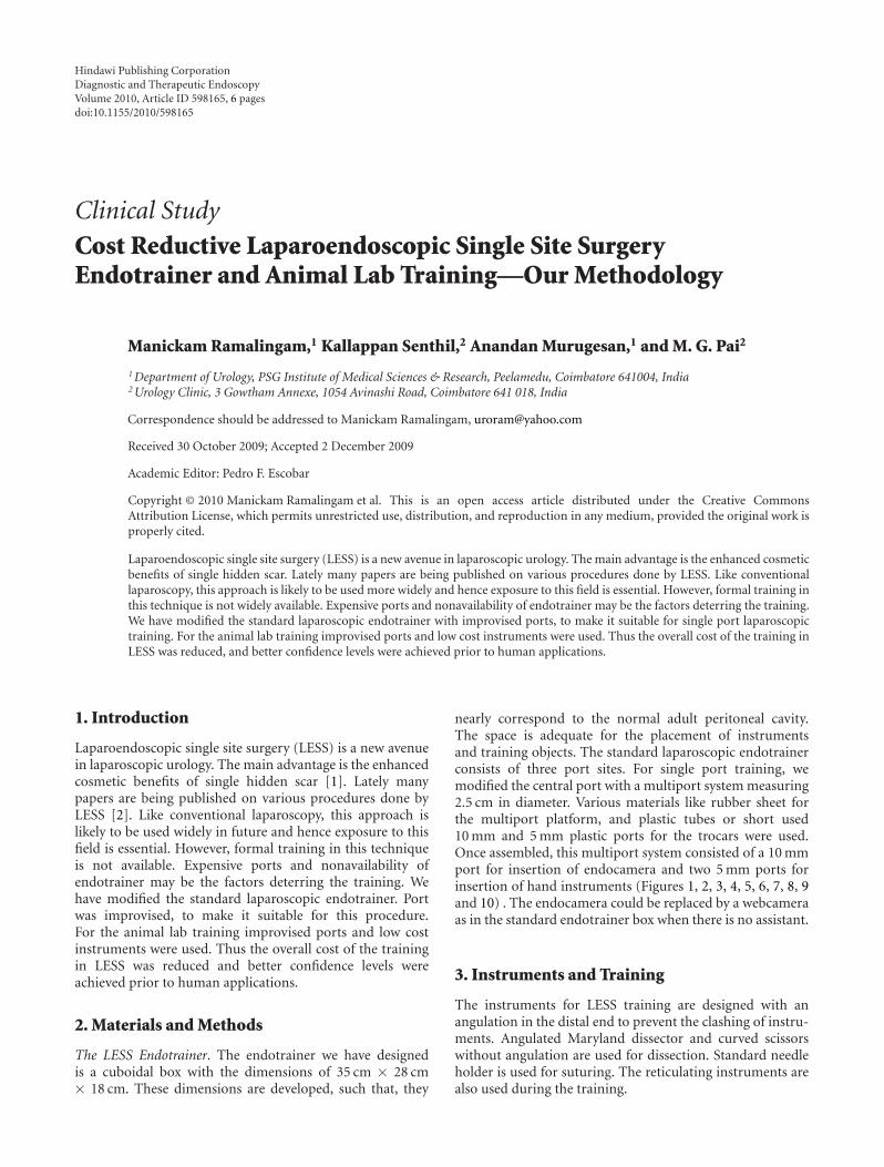
The LESS Endotrainer. The endotrainer we have designedis a cuboidal box with the dimensions of 35 cm × 28 cm× 18 cm. These dimensions are developed, such that, they
nearly correspond to the normal adult peritoneal cavity.The space is adequate for the placement of instrumentsand training objects. The standard laparoscopic endotrainerconsists of three port sites. For single port training, wemodified the central port with a multiport system measuring2.5 cm in diameter. Various materials like rubber sheet forthe multiport platform, and plastic tubes or short used10 mm and 5 mm plastic ports for the trocars were used.Once assembled, this multiport system consisted of a 10 mmport for insertion of endocamera and two 5 mm ports forinsertion of hand instruments (Figures 1, 2, 3, 4, 5, 6, 7, 8, 9and 10) . The endocamera could be replaced by a webcameraas in the standard endotrainer box when there is no assistant.
3. Instruments and Training
The instruments for LESS training are designed with anangulation in the distal end to prevent the clashing of instru-ments. Angulated Maryland dissector and curved scissorswithout angulation are used for dissection. Standard needleholder is used for suturing. The reticulating instruments arealso used during the training.
2 Diagnostic and Therapeutic Endoscopy
Figure 1: LESS endotrainer with modified port (1).
Figure 2: Modified LESS endotrainer port (2) being used fortraining.
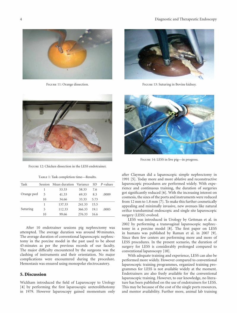
Training pattern for single port laparoscopy was similarto conventional laparoscopic training. Hand eye coordina-tion, dissection, and suturing exercises were carried out.The hand eye co-ordination exercises included transfer ofobjects between the instruments and transfer between bowlsplaced within the trainer. The technique of dissection wasby peeling orange skin (Figure 11) and dissecting a chickenleg model (Figure 12). Suturing was practiced on chickenleg and cadaver bovine kidney ureteropelvic junction (UPJ)(Figure 13).
Evaluation of training. We conducted training sessionsamong 3 of our faculty, who are experienced laparoscopicsurgeons, using our LESS endotrainer. A total of 10 sessionswere carried out by each of the consultants over a periodof 10 days. Objective assessment was done using the taskcompletion time (TCT) [3] as the unit of measurement.
Figure 3: Modified LESS endotrainer port (2).
Figure 4: Endotrainer in our center with modified single port (3).
Figure 5: Standard endotrainer with modified port (3).
Diagnostic and Therapeutic Endoscopy 3
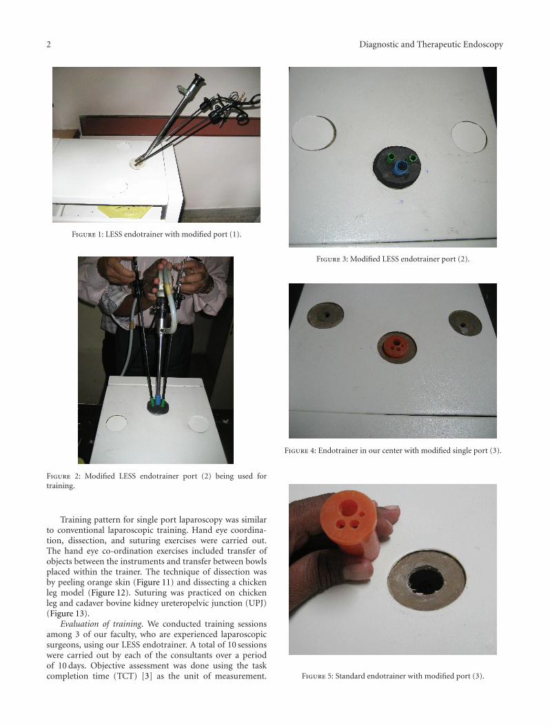
Figure 6: LESS Endotrainer with modified port (3). Note thecrowding of instruments.
Figure 7: LESS Endotrainer setup.
Figure 8: Single port endotrainer with angulated instruments.
Figure 9: Modified LESS port (4) lateral view.
Figure 10: Modified LESS port (4).
Complete peeling of orange and suturing of UPJ in cadaverbovine kidney (six interrupted sutures) were the two tasksassessed at the first, sixth, and tenth sessions. After com-pletion of the endotrainer sessions, animal lab training wascarried out on a live anesthetised pig using single port(Figures 14 and 15).
4. Results
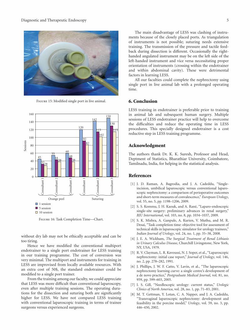
The results are summarized in the form of a chart(Figure 16). The results showed that with each passingsession the subjective difficulty of the procedure decreased.Statistical analysis was done using SPSS software. The averageTCT decreased as the sessions progressed from the firstattempt to fifth attempt and tenth attempt. The correlationcoefficient of the TCT was −0.8 and −0.9 for orange peeland bovine model suturing, respectively. This shows that,with the progression of sessions, the task completion timesignificantly decreases. The difference in the TCT of bothorange peeling and suturing between sessions 1, 5, and 10was highly significant (P < .0001) (Table 1).
4 Diagnostic and Therapeutic Endoscopy
Figure 11: Orange dissection.
Figure 12: Chicken dissection in the LESS endotrainer.
Table 1: Task completion time—Results.
Task Session Mean duration Variance SD P-values
Orange peel1 53.33 58.33 7.6
5 41.33 69.33 8.3 .0009
10 34.66 33.33 5.73
Suturing1 137.33 241.33 15.5
5 112.33 366.33 19.1 .0005
10 99.66 276.33 16.6
After 10 endotrainer sessions pig nephrectomy wasattempted. The average duration was around 90 minutes.The average duration of conventional laparoscopic nephrec-tomy in the porcine model in the past used to be about45 minutes as per the previous records of our faculty.The major difficulty encountered by the surgeons was theclashing of instruments and their orientation. No majorcomplications were encountered during the procedure.Hemostasis was ensured using monopolar electrocautery.
5. Discussion
Wickham introduced the field of Laparoscopy to Urology[4] by performing the first laparoscopic ureterolithotomyin 1979. However laparoscopy gained momentum only
Figure 13: Suturing in Bovine kidney.
Figure 14: LESS in live pig—in progress.
after Clayman did a laparoscopic simple nephrectomy in1991 [5]. Today more and more ablative and reconstructivelaparoscopic procedures are performed widely. With expe-rience and continuous training, the duration of surgeriesgot significantly reduced [6]. With the increasing interest oncosmesis, the sizes of the ports and instruments were reducedfrom 12 mm to 1.8 mm [7]. To make this further cosmeticallyappealing and minimally invasive, new avenues like naturalorifice transluminal endoscopic and single site laparoscopicsurgery (LESS) evolved.
LESS was introduced in Urology by Gettman et al. in2002 by performing a transvaginal laparoscopic nephrec-tomy in a porcine model [8]. The first paper on LESSin humans was published by Raman et al. in 2007 [9].Since then few centers are performing more and more ofLESS procedures. In the present scenario, the duration ofsurgery for LESS is considerably prolonged compared toconventional laparoscopy [10].
With adequate training and experience, LESS can also beperformed more widely. However compared to conventionallaparoscopic training programmes, organized training pro-grammes for LESS is not available widely at the moment.Endotrainers are also freely available for the conventionallaparoscopic training. However, to our knowledge, no litera-ture has been published on the use of endotrainers for LESS.This may be because of the cost of the single ports resources,and mentor availability. Further more, animal lab training
Diagnostic and Therapeutic Endoscopy 5
Figure 15: Modified single port in live animal.
0
20
40
60
80
100
120
140
160
1 session5 session10 session
45
322838
4455
60
4838
120
9282
142
115
102
150
130
115
Con
sult
ant
1
Con
sult
ant
2
Con
sult
ant
3
Con
sult
ant
1
Con
sult
ant
2
Con
sult
ant
3
Orange peel Suturing
Sess
ion
s
Figure 16: Task Completion Time—Chart.
without dry lab may not be ethically acceptable and can betoo tiring.
Hence we have modified the conventional multiportendotrainer to a single port endotrainer for LESS trainingin our training programme. The cost of conversion wasvery minimal. The multiport and instruments for training inLESS are improvised from locally available resources. Withan extra cost of 50$, the standard endotrainer could bemodified to a single port trainer.
From the training among our faculty, we could appreciatethat LESS was more difficult than conventional laparoscopy,even after multiple training sessions. The operating dura-tions for the dissection and suturing both are significantlyhigher for LESS. We have not compared LESS trainingwith conventional laparoscopic training in terms of traineesurgeons versus experienced surgeons.
The main disadvantage of LESS was clashing of instru-ments because of the closely placed ports. As triangulationof instruments is not possible; suturing needs extensivetraining. The transmission of the pressure and tactile feed-back during dissection is different. Occasionally the right-handed angulated instrument may be on the left side of theleft-handed instrument and vice versa necessitating properorientation of instruments (crossing within the endotrainerand within abdominal cavity). These were detrimentalfactors in learning LESS.
All our faculties could complete the nephrectomy usingsingle port in live animal lab with a prolonged operatingtime.
6. Conclusion
LESS training in endotrainer is preferable prior to trainingin animal lab and subsequent human surgery. Multiplesessions of LESS endotrainer practice will help to overcomethe difficulties and reduce the operating time in LESSprocedures. This specially designed endotrainer is a costreductive step in LESS training programme.
Acknowledgment
The authors thank Dr. K. K. Suresh, Professor and Head,Deptment of Statistics, Bharathiar University, Coimbatore,Tamilnadu, India, for helping in the statistical analysis.
References
[1] J. D. Raman, A. Bagrodia, and J. A. Cadeddu, “Single-incision, umbilical laparoscopic versus conventional laparo-scopic nephrectomy: a comparison of perioperative outcomesand short-term measures of convalescence,” European Urology,vol. 55, no. 5, pp. 1198–1206, 2009.
[2] S. S. Kommu, J. H. Kaouk, and A. Rane, “Laparo-endoscopicsingle-site surgery: preliminary advances in renal surgery,”BJU International, vol. 103, no. 8, pp. 1034–1037, 2009.
[3] S. K. Mishra, A. Ganpule, A. Kurien, V. Muthu, and M. R.Desai, “Task completion time: objective tool for assessment oftechnical skills in laparoscopic simulator for urology trainees,”Indian Journal of Urology, vol. 24, no. 1, pp. 35–38, 2008.
[4] J. E. A. Wickham, The Surgical Treatment of Renal Lithiasisin Urinary Calculus Disease, Churchill Livingstone, New York,NY, USA, 1979.
[5] R. V. Clayman, L. R. Kavoussi, N. J. Soper, et al., “Laparoscopicnephrectomy: initial case report,” Journal of Urology, vol. 146,no. 2, pp. 278–282, 1991.
[6] J. Phillips, J. W. F. Catto, V. Lavin, et al., “The laparoscopicnephrectomy learning curve: a single centre’s development ofa de novo practice,” Postgraduate Medical Journal, vol. 81, no.959, pp. 599–603, 2005.
[7] I. S. Gill, “Needlescopic urology: current status,” UrologicClinics of North America, vol. 28, no. 1, pp. 71–83, 2001.
[8] M. T. Gettman, Y. Lotan, C. A. Napper, and J. A. Cadeddu,“Transvaginal laparoscopic nephrectomy: development andfeasibility in the porcine model,” Urology, vol. 59, no. 3, pp.446–450, 2002.

6 Diagnostic and Therapeutic Endoscopy
[9] J. D. Raman, K. Bensalah, A. Bagrodia, J. M. Stern, and J.A. Cadeddu, “Laboratory and clinical development of singlekeyhole umbilical nephrectomy,” Urology, vol. 70, no. 6, pp.1039–1042, 2007.
[10] M. M. Desai, A. K. Berger, R. Brandina, et al., “Laparoendo-scopic single-site surgery: initial hundred patients,” Urology,vol. 74, no. 4, pp. 805–812, 2009.

Submit your manuscripts athttp://www.hindawi.com
Stem CellsInternational
Hindawi Publishing Corporationhttp://www.hindawi.com Volume 2014
Hindawi Publishing Corporationhttp://www.hindawi.com Volume 2014
MEDIATORSINFLAMMATION
of
Hindawi Publishing Corporationhttp://www.hindawi.com Volume 2014
Behavioural Neurology
EndocrinologyInternational Journal of
Hindawi Publishing Corporationhttp://www.hindawi.com Volume 2014
Hindawi Publishing Corporationhttp://www.hindawi.com Volume 2014
Disease Markers
Hindawi Publishing Corporationhttp://www.hindawi.com Volume 2014
BioMed Research International
OncologyJournal of
Hindawi Publishing Corporationhttp://www.hindawi.com Volume 2014
Hindawi Publishing Corporationhttp://www.hindawi.com Volume 2014
Oxidative Medicine and Cellular Longevity
Hindawi Publishing Corporationhttp://www.hindawi.com Volume 2014
PPAR Research
The Scientific World JournalHindawi Publishing Corporation http://www.hindawi.com Volume 2014
Immunology ResearchHindawi Publishing Corporationhttp://www.hindawi.com Volume 2014
Journal of
ObesityJournal of
Hindawi Publishing Corporationhttp://www.hindawi.com Volume 2014
Hindawi Publishing Corporationhttp://www.hindawi.com Volume 2014
Computational and Mathematical Methods in Medicine
OphthalmologyJournal of
Hindawi Publishing Corporationhttp://www.hindawi.com Volume 2014
Diabetes ResearchJournal of
Hindawi Publishing Corporationhttp://www.hindawi.com Volume 2014
Hindawi Publishing Corporationhttp://www.hindawi.com Volume 2014
Research and TreatmentAIDS
Hindawi Publishing Corporationhttp://www.hindawi.com Volume 2014
Gastroenterology Research and Practice
Hindawi Publishing Corporationhttp://www.hindawi.com Volume 2014
Parkinson’s Disease
Evidence-Based Complementary and Alternative Medicine
Volume 2014Hindawi Publishing Corporationhttp://www.hindawi.com