Embed Size (px)
Citation preview
Anatomical Labeling of the Anterior Circulationof the Circle of Willis using Maximum
A Posteriori Classification
Hrvoje Bogunovic, Jose Marıa Pozo, Ruben Cardenes, and Alejandro F. Frangi
Center for Computational Imaging & Simulation Technologies in Biomedicine(CISTIB) – Universitat Pompeu Fabra (UPF) and CIBER-BBN, Barcelona, Spain
Abstract. Automated anatomical labeling of the arteries forming theCircle of Willis is of great interest as facilitates inter-subject compar-ison required to discover geometric risk factors for the development ofvascular pathologies. In this paper, we present a method for anatomicallabeling of vessels forming anterior part of the Circle of Willis by detect-ing the five main vessel bifurcations. The method is first trained on aset of pre-labeled examples, where it learns local bifurcation features aswell as global variation in the anatomy of the extracted vascular trees.Then the labeling of the target vascular tree is formulated as maximuma posteriori solution where the classifications of individual bifurcationsare regularized by the prior learned knowledge of the tree they span. Themethod was evaluated by cross-validation on 30 subjects, which showedthe vascular trees were correctly anatomically labeled in 90% of cases.The proposed method can naturally handle anatomical variations and isshown to be suitable for labeling arterial segments of Circle of Willis.
1 Introduction
The Circle of Willis is a ring of cerebral vessels that connects the two halves ofthe anterior circulation with the posterior one and its bifurcations are a commonsite of aneurysm formation (pathological dilations of vessels) [1]. Analyzing thestatistical variation of characteristics of these vessels and bifurcations is of greatinterest as it can lead to the identification of geometric risk factors for theonset of vascular pathologies. Two important operations required for comparingand registering the vasculature between subjects are landmark matching andanatomical labeling. These are tedious and time consuming tasks to be performedmanually. Thus, automating them becomes crucial for processing large numberof cases.
In this work, we present a method for robust classification of bifurcations in avascular tree, which we applied to the task of anatomically labeling the anteriorpart of the Circle of Willis. The anterior part is formed by the three main ves-sels: internal carotid artery (ICA), middle cerebral artery (MCA) and anteriorcerebral artery (ACA). Statistically, 80% of the aneurysms occur along thesevessels [1]. In particular, we are interested in anatomical labeling of the follow-ing five bifurcations and the vessels connecting them. The bifurcations of ICA

2 H. Bogunovic et al.
ICA terminal
bifurcationAchA
bifurcation
PcoA
bifurcation
OA
bifurcation
ICA
ACA MCA
(a)
MCA
bifurcation
MCA-M1
MCA-M2
ICA
terminal
bifurcation
(b)
Fig. 1: The arteries of the Circle of Willis and the bifurcations of interest on: (a)internal carotid artery (ICA), (b) middle cerebral artery (MCA).
with ophthalmic artery (OA), posterior communicating artery (PcoA), anteriorchoroidal artery (AchA) and the terminal ICA bifurcation, plus the principal bi-furcation of MCA, which divides the MCA-M1 and MCA-M2 segments (Fig. 1).To that set of bifurcations, throughout the text, we will refer to as bifurcationsof interest (BoI).
The state of the art for anatomical labeling of vasculature is, in general,sparse. The seminal work was done by Tschirren et al. [2] for anatomical label-ing of airway trees that relies on branchpoint matching to a prelabeled tree thatrepresents population average. Mori et al. in a series of works [3, 4] presenteda knowledge-based framework for anatomical labeling of bronchial branchesbased on machine learning and combination optimization. However, their methoduses a fixed set of topological constraints and does not seem to be robust tolarge anatomical variations of the reference tree topology. Airway and bronchialtrees are characterized by many similar bifurcations connected by short straightbranches. This makes the methods design for them difficult to apply to the taskof labeling cerebral vessels where the vessels are in general longer and morecurved and the bifurcations more complex. Recently, Mori et al. [5] tuned theirapproach to a specific task of labeling abdominal arteries but even the authorsthemselves stated that the application to other organs would be challenging. Bo-gunovic et al. [6] showed that the ICA terminal bifurcation can be successfullyclassified using support vector machine (SVM) but only a single bifurcation isdetected and no tree properties are considered.
The rest of the paper is organized as follows. In section 2, we present theworkflow of the method, starting from angiographic 3D image, and formulate theclassification of bifurcations in a maximum a posteriori framework. Evaluationof the method on a set of 30 images is presented in section 3. Finally, section 4discusses the advantages and limitations of the proposed method and concludesthe paper.

Anatomical Labeling of the CoW using MAP classification 3
(a)
Parent
branch
Bigger
branch
Smaller
branch
(b) (c)
Fig. 2: Elements of the method’s workflow: (a) Centerline extraction. (b) Bifur-cation features. (c) Example of a labeling L (dotted arrows) of a target tree T t
(right) based on a reference tree T r with the bifurcations of interest (left).
2 Methods
2.1 Vascular Tree Extraction and Characterization
Segmentations of the cerebral vasculature from 3D images were performed in afully automated way with a geometric deformable model called Geodesic ActiveRegions (GAR) [7]. From the segmented vasculature, fast topological thinningbased on collapsing fronts using [8] was applied to obtain a rough estimateof the underlying topology of the vessel tree. Due to image acquisition noiseand inaccuracies in vessel segmentation, touching vessels effect can appear, thuscausing the extracted topology to form a connected graph containing cycles andnot a tree. However, the graph’s end-points do correspond to the root and theterminal leafs of the vascular tree. Root was taken to be the end-point withthe maximal associated radius at the lowest axial plane which, in our images,corresponded to the ICA entering the field of view. Then, the set of accuratecenterlines is obtained by backtracking along the minimal cost path from theend-points toward the root using [9], which is available within the open-sourcelibrary VMTK [10]. These centerlines now form a rooted tree with the edgesdirected away from the root in accordance with the blood flow (Fig. 2 (a)).
Bifurcation Feature Vector Each bifurcation of the tree is characterized usingthe method of Antiga [11], available in VMTK, which relies on objective crite-ria for defining the origin of a bifurcation and bifurcation vectors of the parentbranch and the two daughter branches. The two daughter branches are differ-entiated by their radius: Larger daughter branch and smaller daughter branch.We chose the following collection of features to quantify the geometry of a bi-furcation (Fig. 2 (b)), which form a 21 dimensional, scale invariant, bifurcationfeature vector:
– Sagittal, axial and coronal-components of the normal vector of the bifurca-tion plane (3).
4 H. Bogunovic et al.
– Sagittal, axial and coronal-components of the three bifurcation vectors (9).– Angles between each pair of bifurcation vectors (3).– Ratios of mean vessel radii between each pair of bifurcation branches (3).– Ratios of mean vessel radii between each bifurcation branch and the root
branch (ICA) of the tree (3).
Tree Features To characterize the extracted tree, to each pair of bifurcationswe associate an inheritance relationship: ancestor -offspring or sibling. In addi-tion, a probability is assigned that a particular bifurcation is present in the tree.In general, one could easily also include features of the vessels connecting thepair of bifurcations, like their mean length, curvature, torsion or tortuosity.
2.2 Bifurcation Classification
Extracted vascular tree can be considered as Attributed Relational Tree (ART)which has attributes in a form of feature vectors associated to its vertexes andedges. ART is defined as
Definition 1. Rooted Attributed Relational Tree is a quadruple T = (V,E,A, r),where V is the vertex set, E is the edge set, r is root and A is the attribute setthat contains unary attribute ai attaching to each vertex vi ∈ V , and a binaryattribute ai,j attaching to each edge ek = (ni, nj) ∈ E.
The classification of bifurcations is based on the availability of a referenceART T r, created from a representative sample of a population, having BoI as itsvertices V r = {vri : 1 ≤ i ≤ M} together with an entire set of sample bifurcationfeature vectors as {ari}, while edge features {ari,j} are currently not used. Such areference ART is normally created from a training set of trees. The target ARTT t corresponds to the extracted vascular tree, having its bifurcations as verticesV t = {vtj : 1 ≤ j ≤ N} and the bifurcation feature vectors as {atj}. Then, onthe target tree we define a labeling process L : V t → V r ∪ {NI}, where the labelNI represents a bifurcation which is not of interest and is not present in thereference ART (Fig. 2(c)).
We are interested in estimating the probability P (L|T t, T r) of L being cor-rect, and finding the mode of this posterior distribution. Thus, the problem canbe formulated as finding a labeling L∗ that satisfies the maximum a posteriori(MAP) solution
P (L∗|T t, T r) > P (L|T t, T r) ∀L 6= L∗, (1)
P (L|T t, T r) ∝ P (T t|L, T r)P (L|T r), (2)
where the constant denominator term in Eq. 2 has been left out for brevity.The prior term P (L|T r) presents our knowledge-based expectations about
the topology and tree properties of the labeled target tree and regularizes theclassification based on bifurcation features. The likelihood term, assuming that
Anatomical Labeling of the CoW using MAP classification 5
the feature vectors of each bifurcation {ai} are statistically independent fromeach other, can be written as
P (T t|L, T r) =
N∏
i=1
P (ati|L(vti), T r). (3)
Likelihood Estimation. To estimate P (ati|L(vti)) i.e. a likelihood that a bi-furcation with known label L(vti) has the feature vector ati, we employed theSVM with probability estimates available in LIBSVM open-source library [12].A non-linear model based on radial basis function (RBF) kernel was used. Forthe multi-class problem, several binary SVM were created in a one-against-onestrategy where binary SVMs, for all pairs of classes, were trained on the data andthe results for each pair accumulate to yield the final probability distribution.
MAP Estimation. Unfortunately MAP estimation is extremely computation-ally expensive. Testing for all possible labelings L is not feasible. However, theprior term P (L|T r) is positive only for a small subset of all combinations i.e.only for those for which the topology corresponds to the one of the referencetree T r. This happens when L is an isomorphism from a subgraph of T t to asubdivision of T r.
A standard algorithm to find all isomorphic mappings between two graphs(trees) is based on building their association graph and then finding all maximalcliques of such undirected graph. Association graph G = (V a, Ea) is built fromT r and T t, where nodes are denoted with a pair of indices V a = {va ≡ (vri , v
tj)} ≡
V r×V t, with the following rule. Edge (vai,j , vak,l) is created only if vri and vrk have
the same relationship (ancestor -offspring or sibling) as do vtj and vtl , for i 6= kand j 6= l. We have now extended the above rules by adding the label NI to V t,hence V a = V r × (V t ∪{NI}) with the special rule that NI is in all relationshipswith any other vertex of V a including itself.
Finding maximal cliques (in our case they will always be of size N) in anundirected graph is in general NP-complete problem and we used Bron-Kerboschalgorithm [13], that tries to reduce the size of search space, to find the maximalcliques.
3 Results
We evaluated the proposed methodology on a dataset consisting of images from30 subjects, acquired with 3D rotational angiography (3DRA), where contrastinjection was carried out to enhance the vessels comprising the anterior cerebralcirculation of either left or right hemisphere. Images were reconstructed with a2563 matrix having a voxel size of 0.29× 0.29× 0.29 (mm).
All images were successfully segmented and had their centerlines and treetopology extracted. The obtained trees were containing bifurcations and trifur-cations. Since two close bifurcations are sometimes reconstructed as a trifurca-

6 H. Bogunovic et al.
Table 1: Performance of the three method variants. Scores for successfully label-ing the complete vessel tree and detecting the individual bifurcations are given.
MethodVessel treelabeling rate
Bifurcation detection rate
OA PcoA AchA ICA MCA NI
SVM ∼[6] 0.7 1.00 1.00 1.00 0.96 0.89 0.85
SVM+TP ∼[5] 0.8 1.00 1.00 1.00 0.96 0.82 0.94
SVM+TP+AP 0.9 1.00 1.00 1.00 1.00 0.96 0.91
tion, we decided to always treat trifurcations as two bifurcations sharing thesame parent branch and the larger daughter branch.
The success of bifurcation classification and hence the anatomical labelingwas evaluated using cross-validation. One observer manually labeled the fiveBoI on each extracted tree. All the other bifurcations were assigned the labelNI. Leave-one-out cross-validation was then run, where iteratively K = 30 times,one case is used for testing while K − 1 cases were selected to form the trainingset from which the bifurcation vectors, reference tree topology and its probabilityof appearance were learned.
To evaluate the contribution of the regularizing prior term, we performed theabove validation for three method variants: the first variant is a natural extensionof [6] and is relying just on the SVM classification based on the bifurcationfeature vectors with no regularization. The second variant has the regularizationby topology added (SVM+TP), and can be considered as an adaptation of [5].Finally, the third variant is the proposed one, which includes the prior term(SVM+TP+AP), where the probability of appearance of a particular anatomicalvariability coming from the missing BoI is learned from the training set. Thecross-validation results are presented in Table 1.
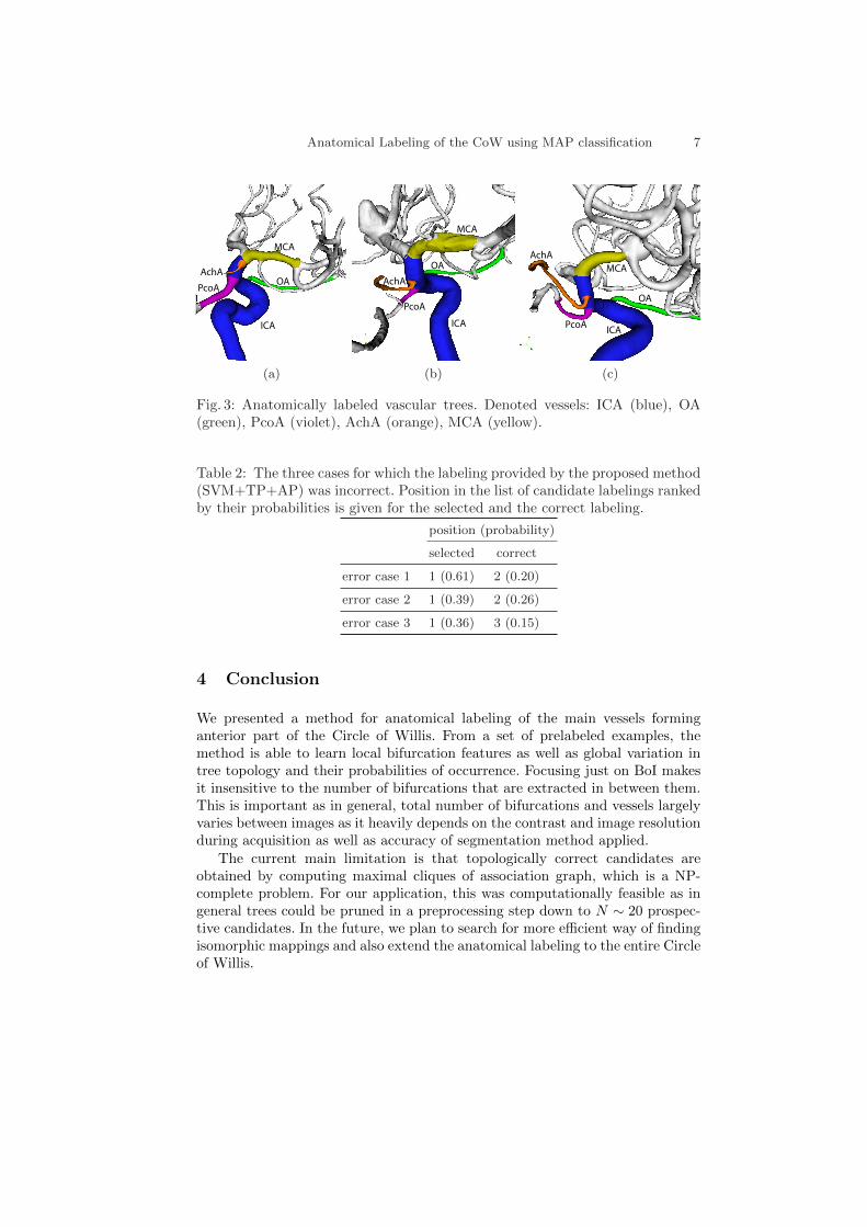
As seen from the first row, PcoA, AchA and OA bifurcations were well clas-sified just based on their feature vectors. Gradually adding the regularizationterms improved the tree labeling accuracy each time by 10%. The proposedmethod (SVM+TP+AP) outperformed similar state of the art methods andhad success rate of 90% for correctly labeling the complete vessel tree. The erroroccurred in three cases, when MCA bifurcation was detected as a NI and viceversa. A sample of correct anatomical labeling results is shown in Fig. 3 wherethe surface area of each corresponding vessel segment is labeled.
As the method is formulated in a probabilistic framework the candidate label-ings can be ranked by their estimated probabilities. The method always selectsthe most probable one (the highest ranked one). However, in the case that se-lected labeling is visually observed to be incorrect, this enables us to select thenext most probable candidate in the ranked list. For the three cases where themethod assigned the incorrect labels, we searched for the correct labeling in theranked list (Table 2). We can observe that the correct labeling appears as thesecond or at most the third most probable candidate.
Anatomical Labeling of the CoW using MAP classification 7
MCA
OA
ICA
PcoA
AchA
(a)
MCA
ICA
OA
AchA
PcoA
(b)
OA
ICA
MCA
AchA
PcoA
(c)
Fig. 3: Anatomically labeled vascular trees. Denoted vessels: ICA (blue), OA(green), PcoA (violet), AchA (orange), MCA (yellow).
Table 2: The three cases for which the labeling provided by the proposed method(SVM+TP+AP) was incorrect. Position in the list of candidate labelings rankedby their probabilities is given for the selected and the correct labeling.
position (probability)
selected correct
error case 1 1 (0.61) 2 (0.20)
error case 2 1 (0.39) 2 (0.26)
error case 3 1 (0.36) 3 (0.15)
4 Conclusion
We presented a method for anatomical labeling of the main vessels forminganterior part of the Circle of Willis. From a set of prelabeled examples, themethod is able to learn local bifurcation features as well as global variation intree topology and their probabilities of occurrence. Focusing just on BoI makesit insensitive to the number of bifurcations that are extracted in between them.This is important as in general, total number of bifurcations and vessels largelyvaries between images as it heavily depends on the contrast and image resolutionduring acquisition as well as accuracy of segmentation method applied.
The current main limitation is that topologically correct candidates areobtained by computing maximal cliques of association graph, which is a NP-complete problem. For our application, this was computationally feasible as ingeneral trees could be pruned in a preprocessing step down to N ∼ 20 prospec-tive candidates. In the future, we plan to search for more efficient way of findingisomorphic mappings and also extend the anatomical labeling to the entire Circleof Willis.

8 H. Bogunovic et al.
Acknowledgments. This work has been partially funded by the CENIT pro-gramme of CDTI (cvREMOD CEN-20091044) and VPH-NoE project from FP7of the European Union (nr 223920). The work of H.B. is supported by the FIprogramme of AGAUR. The work of R.C. is supported by the Beatriu de Pinos,programme of AGAUR. A.F.F. is partially funded by the ICREA-Academiaprogramme.
References
1. Brisman, J.L., Song, J.K., Newell, D.W.: Cerebral aneurysms. N. Engl. J. Med.355(9) (August 2006) 928–939
2. Tschirren, J., McLennan, G., Palagyi, K., Hoffman, E.A., Sonka, M.: Matchingand anatomical labeling of human airway tree. IEEE Trans. Med. Imag. 24(12)(December 2005) 1540–1547
3. Mori, K., Hasegawa, J., Suenaga, Y., Toriwaki, J.: Automated anatomical labelingof the bronchial branch and its application to the virtual bronchoscopy system.IEEE Trans. Med. Imag. 19(2) (February 2000) 103–114
4. Mori, K., Ota, S., Deguchi, D., Kitasaka, T., Suenaga, Y., Iwano, S., Hasegawa, Y.,Takabatake, H., Mori, M., Natori, H.: Automated anatomical labeling of bronchialbranches extracted from CT datasets based on machine learning and combinationoptimization and its application to bronchoscope guidance. In Yang, G.Z., Hawkes,D., Rueckert, D., Noble, A., Taylor, C., eds.: Proc. MICCAI. Volume 5762 ofLNCS., Springer (2009) 707–714
5. Mori, K., Oda, M., Egusa, T., Jiang, Z., Kitasaka, T., Fujiwara, M., Misawa, K.:Automated nomenclature of upper abdominal arteries for displaying anatomicalnames on virtual laparoscopic images. In Liao, H., Edwards, E., Pan, X., Fan, Y.,Yang, G.Z., eds.: Proc. MIAR. Volume 6326 of LNCS., Springer (2010) 353–362
6. Bogunovic, H., Pozo, J.M., Cardenes, R., Frangi, A.F.: Automatic identificationof internal carotid artery from 3DRA images. In: Proc. IEEE EMBC, IEEE Press(2010) 5343–5346
7. Bogunovic, H., Pozo, J.M., Villa-Uriol, M.C., Majoie, C.B.L.M., van den Berg,R., Gratama van Andel, H.A.F., Macho, J.M., Blasco, J., San Roman, L., Frangi,A.F.: Automated segmentation of cerebral vasculature with aneurysms in 3DRAand TOF-MRA using geodesic active regions: An evaluation study. Med. Phys. 38(January 2011) 210–222
8. Cardenes, R., Bogunovic, H., Frangi, A.F.: Fast 3D centerline computation fortubular structures by front collapsing and fast marching. In: Proc. IEEE ICIP,IEEE Press (2010) 4109–4112
9. Antiga, L., Ene-Iordache, B., Remuzzi, A.: Computational geometry for patient-specific reconstruction and meshing of blood vessels from MR and CT angiography.IEEE Trans. Med. Imag. 22(5) (May 2003) 674–684
10. Antiga, L., Steinman, D.A.: VMTK: the Vascular Modeling Toolkit. http://www.vmtk.org (2011)
11. Antiga, L., Steinman, D.A.: Robust and objective decomposition and mapping ofbifurcating vessels. IEEE Trans. Med. Imag. 23(6) (June 2004) 704–713
12. Chang, C.C., Lin, C.J.: LIBSVM: a library for support vector machines. http:
//www.csie.ntu.edu.tw/~cjlin/libsvm (2011)13. Bron, C., Kerbosch, J.: Algorithm 457: finding all cliques of an undirected graph.
Comm. ACM 16(9) (September 1973) 575–577


