Embed Size (px)
Citation preview

CODED-APERTURE I M A G I N G I N NUCLEAR M E D I C I N E
Warren E . Smi th The I n s t i t u t e of Optics University of Rochester
Harrison H . Bar re t t and John N . Aarsvold Radiology Research Laboratory and
Optical Sciences Center University of Arizona
SUMMARY
Coded-aperture imaging i s a technique f o r imaging sources t h a t emit high-energy r ad ia t ion . T h i s type of imaging involves shadow cas t ing and not r e f l e c t i o n or r e f r a c t i o n . High-energy sources e x i s t i n x-ray and gamma-ray astronomy, nuclear reac tor fuel-rod imaging, and nuclear medicine. O f these th ree a reas nuclear medicine i s perhaps the most challenging because of t h e l i m i t e d amount of r ad ia t ion ava i lab le and because a three-dimensional source d i s t r i b u t i o n i s t o be determined. I n nuclear medicine a radioact ive pharmaceutical i s administered t o a p a t i e n t . T h e pharmaceutical i s designed t o be taken up by a p a r t i c u l a r organ of i n t e r e s t , and i t s d i s t r i b u t i o n provides c l i n i c a l information about t he function of t he organ, o r t he presence of l e s ions w i t h i n t h e organ. T h i s d i s t r i b u t i o n i s determined from s p a t i a l measurements of t h e rad ia t ion emitted by t h e radiopharmaceutical.
The p r inc ip l e s of imaging radiopharmaceutical d i s t r i b u t i o n s w i t h coded aper tures w i l l be reviewed. Included w i l l be a discussion of l i n e a r sh i f t -va r i an t pro jec t ion operators and t h e associated inverse problem. A system developed a t t he University of Arizona i n Tucson cons is t ing of small modular gamma-ray cameras f i t t e d w i t h coded aper tures w i l l be described.
INTRODUCTION
I n nuclear medicine a radiopharmaceutical i s given t o a p a t i e n t . The pharmaceutical i s designed t o go t o a p a r t i c u l a r organ of i n t e r e s t , such a s t he bra in , t h e hear t , bone, o r the l i v e r , t o name a few. The three-dimensional d i s t r i b u t i o n of t he pharmaceutical provides c l i n i c a l information about how well t he organ i s functioninq. T h i s i s qu i t e d i f f e r e n t than the type of information provided by x-ray imaging (e lec t ron d e n s i t y ) , magnetic resonance imaging ( M R I ) (proton densi ty and magnetization re laxa t ion r a t e s ) and ultrasound (acous t ic impedance of t i s s u e ) . The d i s t r i b u t i o n of t h e pharmaceutical i s determined by imaging t h e r ad ia t ion given off by t h e isotope t h a t t ags i t . There i s always a concern t o l i m i t t he t o t a l amount of r ad ia t ion t h a t t h e pa t i en t i s exposed t o , so t h a t i n nuclear medicine w e have a photon- l imi ted s i t u a t i o n .
33

The isotopes used i n nuclear medicine f a l l i n t o two broad ca tegor ies : those t h a t emit s ing le gamma rays d i r e c t l y from the nucleus, and those t h a t emit posi t rons from the nucleus. Three- dimensional imaging associated w i t h t h e f i r s t category i s ca l l ed s ingle photon emission computed tomography (SPECT) , and i s t h e subject of t h i s paper. I n t h i s method t h e photons mus t be blocked by a t tenuat ing ape r tu re s . Imaging associated w i t h t he second category i s ca l l ed posi t ron emission tomography ( P E T ) . I n PET t he posi t ron t h a t i s emitted by a source nucleus ann ih i l a t e s an e lec t ron w i t h i n 1 t o 2 mm of t h e source po in t . T h i s annih i la t ion r e s u l t s i n two photons, each of approximately 511 K e V , t r a v e l i n g i n almost opposi te d i r e c t i o n s . By coincidental ly de tec t ing these two photons w i t h s p a t i a l l y separate de tec tors , t he l i n e along t h e i r path which contains t h e source point can be found. T h i s technique removes the need t o physical ly block the photons w i t h aper tures t o determine t h e i r d i r ec t ion of o r i g i n . W i t h PET one can obtain extremely good resolut ion by nuclear-medicine standards, on the order of 5 mm. The disadvantage of PET i s t h a t an on-si te cyclotron i s needed t o c rea te the short- l ived positron-emitt ing i so topes . The expense associated w i t h t h i s requirement has l imi ted the number of PET f a c i l i t i e s . SPECT imaging, on the other hand, i s r e l a t i v e l y l e s s expensive and well es tab l i shed throughout t h e world. Thus t he motivation e x i s t s t o continue t o improve SPECT imaging techniques t o approach t h e qua l i t y already a t t a i n a b l e w i t h PET.
Two-dimensional pro jec t ions of source d i s t r i b u t i o n s a r e obtained i n nuclear medicine by e i t h e r scanning the source i n two dimensions w i t h a s ing le , coll imated gamma-ray point de tec tor o r by forming a t w o - dimensional image w i t h a camera t h a t i s capable of measuring the x and y pos i t ions of t he incident gamma rays and s t o r i n g them i n an image histogram. ( r e f . 1 ) . T h i s camera can a l so estimate the gamma-ray energy. Energy est imat ion i s important f o r r e j ec t ion of Compton-scattered rad ia t ion from t h e nuclear-medicine image. A photon t h a t i s Compton sca t t e red by t h e a t t enua t ing t i s s u e s between the source and t h e de t ec to r w i l l s u f f e r an energy s h i f t , dependent upon the angle of s c a t t e r . nuclear medicine the energy spectrum of the usefu l isotopes i s r e l a t i v e l y narrow, so t h a t t he Compton-scattered photons can be i d e n t i f i e d and removed i f t h e i r energy i s outs ide of t h e peak assoc ia ted w i t h t h e source isotope.
Such a camera i s the Anger camera, named a f t e r i t s inventor
Fortunately i n
Gamma rays have such a h i g h energy t h a t they cannot be conveniently r e f l e c t e d or r e f r ac t ed . I n f ront of t he gamma-ray camera i s t h u s placed a shadow-casting aperture , usual ly made of lead or some o ther high atomic-number element. There a r e two bas ic types of aper tures , t he col l imator and t h e pinhole. The col l imator cons i s t s of a la rge number of usual ly p a r a l l e l holes d r i l l e d through a th i ck lead p l a t e . Each hole causes the s e n s i t i v i t y of a given de tec to r element t o be confined t o a narrow penci l t h a t i n t e r s e c t s t h e source d i s t r i b u t i o n . T h i s narrow penci l i s an approximation t o a l i n e i n t e g r a l through the source. A l l of t h e holes together form a p a r a l l e l - l i n e 2-D project ion of t he source onto t h e 2 - D de t ec to r . The pinhole i s a s ing le hole punched i n a r e l a t i v e l y t h i n lead p l a t e . T h i s aper ture produces a pinhole image of t he source d i s t r i b u t i o n on the 2-D d e t e c t o r . The
34

pinhole image represents a s e r i e s of l i n e i n t e g r a l s through the object t h a t converge on t h e pinhole. Conventional systems i n nuclear medicine of ten employ para l le l -hole col l imators . T h e coded-aperture systems t o be discussed employ ar rays of pinholes.
Tomography i n nuclear medicine i s achieved by taking mult iple views of t h e source d i s t r i b u t i o n , and reconstruct ing a 3 - D es t imate of t h e source from these views. Conventionally these views a r e obtained by r o t a t i n g a la rge gamma-ray camera f i t t e d w i t h a para l le l -hole col l imator around t h e p a t i e n t . The camera stops every few degrees and takes a two-dimensional snapshot of t h e pa t i en t l a s t i n g about a minute. Each snapshot of t h e pa t i en t approximates a s e t of p a r a l l e l l i n e i n t e g r a l s , defined by the coll imator, through t h e source volume a t the p a r t i c u l a r angle . Neglecting at tenuat ion of t he source by t h e body, t h e s e t of a l l of these snapshots over 180 or 3 6 0 degrees cons t i t u t e s an approximation t o the Radon transform of the source d i s t r i b u t i o n . The inverse Radon transform ( r e f . 2 ) i s then appl ied t o these pro jec t ions t o form an estimate of t h e three-dimensional source d i s t r i b u t i o n . T h i s inverse involves f i l t e r i n g and then back-projecting each pro jec t ion i n t o the reconstruction space, and can be done rapidly w i t h modern equipment. Without modification of t he inverse Radon transform t o include a t tenuat ion of t h e photons by the body, reconstruct ions appear darker f o r p ixe l s deeper w i t h i n t he tomographic s l i c e . T h i s a t tenuat ion problem can be corrected a n a l y t i c a l l y by the a t tenuated Radon transform ( r e f s . 3 , 4 ) , assuming constant a t tenuat ion and a known convex a t t enua t ion boundary. Typical scan t i m e s f o r t h e rotating-camera approach a r e 3 0 t o 4 5 minutes. Dynamic s tud ie s of pharmaceutical uptake a r e ruled out because of t h e required motion of t h e camera about t he p a t i e n t .
I n t h i s paper we discuss tomography i n nuclear medicine w i t h non- moving coded ape r tu re s . The reconstruction of both 2 - D and 3 - D source d i s t r i b u t i o n s w i l l be addressed. A coded-aperture system f o r nuclear medicine being developed a t the University of Arizona w i l l be descr ibed.
CODED-APERTURE TOMOGRAPHY IN NUCLEAR MEDICINE
I n nuclear medicine we a r e able t o observe only a small number of photons because the rad ia t ion dose t o the pa t i en t i s kept a s low a s possible and because the f r a c t i o n a l s o l i d angles of t h e col l imator or pinhole openings a re small, on the order of These openings must be small because i n shadow-casting t h e a b i l i t y of t he aper ture t o resolve two c lose ly spaced poin ts i n t he source i s d i r e c t l y proport ional t o t h e s i z e of t h e openings. Thus we have a fundamental trade-off between t h e signal-to-noise r a t i o ( S N R ) i n t h e nuclear- medicine image, which goes a s t h e square root of t h e number of detected photons, and t h e reso lu t ion of t he s y s t e m . T h i s t rade-off i s qu i t e d i f f e r e n t i n focusing systems, such a s lenses focusing v i s i b l e l i g h t , where t h e d i f f rac t ion- l imi ted spot s i z e decreases ( t h u s improving reso lu t ion) a s t he aperture i s opened up, allowing more photons i n t o t h e system.
35
There i s t h u s s t rong motivation f o r increasing t h e number of photons i n a nuclear-medicine image without degrading t h e r e so lu t ion . To t h i s end coded aper tures have been developed. Figure 1 shows a single-view coded-aperture system. Here a planar source d i s t r i b u t i o n i s projected through an aperture cons is t ing of several pinholes t o form a coded image. T h e pos i t ion of the pinholes represent t h e code. We have t h u s increased the number of photons detected by t h e system, a t t h e p r i ce of overlap i n t h e pinhole views of t he ob jec t . T h i s overlap i s r e fe r r ed t o a s s p a t i a l "multiplexing", and i s more ser ious f o r l a rge r ob jec ts and denser spacing of t he pinholes . Thus we suspect immediately t h a t t he code should be optimized w i t h respect t o t h e type of object t h a t we w i s h t o view.
I n t h i s planar case, neglecting radiometry and ob l iqu i ty f ac to r s , we can wr i te t h e coded image a s a convolution of t he source w i t h t h e aper ture :
where t h e double prime indica tes de tec tor coordinates . The quant i ty g(x" ,yr l ) i s t h e coded image, h(x"/M,yn/M) i s the scaled aper ture function, and f (x"/m,y"/m) i s the scaled source d i s t r i b u t i o n . The source sca l ing m = ( z - d ) / z and the aperture sca l ing M = d / z , where z i s t h e source-aperture dis tance, and d i s the source-detector d i s t ance . The two-dimensional convolution operator is represented by **. A s we see, both t h e source and the aperture functions a r e scaled d i f f e r e n t l y i n forming the coded image.
T o form a reconstruct ion of the o r i g i n a l source d i s t r i b u t i o n , we use the concept of matched f i l t e r i n g . A matched f i l t e r i s a version of t h e ac tua l s igna l t h a t we a re looking f o r . I t can be shown t h a t a matched f i l t e r i s t h e optimum f i l t e r t o be used t o de tec t a s igna l i n t h e presence of noise ( r e f . 5 ) . I n t h e coded-imaging case, t h e matched f i l t e r i s a properly scaled, inverted, complex-conjugated version of t h e o r i g i n a l code, so t h a t t he reconstruction ? ( x " , y " ) can be wr i t ten a s
? (x"/m, y"/m) = g (x", y") ** h* (-x"/M, -y"/M) . ( 2 )
Equation ( 2 ) can be wri t ten, using Eq. (l), a s :
where t h e bracketed term represents t h e ove ra l l point-spread function (PSF) of t h e data-taking and reconstruction process, and i s ca l l ed the au tocorre la t ion of t h e code. We mus t design the code t o make i t s au tocorre la t ion function a s c lose t o a d e l t a function a s possible , simultaneously allowing a s many openings a s poss ib le . Unfortunately, these two requirements work against each o the r . Generally t h e au tocorre la t ion has a la rge cen t r a l peak surrounded by a background w i t h s t r u c t u r e t h a t depends upon the number of openings. T h i s background tends t o both smear out t h e reconstruct ion a s well a s increase t h e noise i n t he reconstruct ion.
36

Note t h a t we a r e not r e s t r i c t e d t o r e a l and pos i t i ve functions i n our search f o r optimum codes. Any physical ly r e a l i z a b l e code w i l l be r e a l and pos i t i ve because of t h e shadow-casting nature of t h e image formation from the incoherent source. Bipolar complex codes can be simulated, however, by c rea t ing 4 separate codes and forming 4 separate coded images and su i t ab ly adding them i n t h e computer with proper pos i t i ve , negative, and imaginary weights. We pay the p r i ce f o r t h i s f l e x i b i l i t y by increasing t h e amount of time needed t o form an image, however.
There has been considerable research i n t o def ining codes t o optimize t h e SNR of t he f i n a l reconstruct ion. Some of t h e more well- known codes a r e random pinhole arrays ( r e f . 61, t h e F r e s n e l zone p l a t e ( r e f . 7 ) , t he annulus ( r e f . 8 ) , and time-modulated aper tures ( r e f . 9 ) .
Much of t h i s code optimization has been i n t he context of single-view imaging of a planar ob jec t , however, a s i n Fig. 1. I f we were t o image a three-dimensional volume object w i t h t h i s approach, our reconstruct ion of E q . ( 3 ) would be f o r a p a r t i c u l a r plane of t he source, depending upon the sca l e f ac to r used f o r t he matched f i l t e r . The o ther planes of t h e source would present a strong background superimposed on t h i s reconstruction, degrading both reso lu t ion and SNR of t h e plane of i n t e r e s t . Thus there i s a fundamental l imi t a t ion of t h e planar c o r r e l a t i o n decoding method described above because our bas i c da ta set cons is t ing of a s ing le view i s not complete enough. We must i n f a c t t ake mult iple views of a volume object so t h a t we a re sampling i t s three-dimensional Fourier components s u f f i c i e n t l y . Combining mult iple views of t he object t o form a s ing le volume reconstruct ion i s not obvious w i t h t h e planar decorre la t ion method descr ibed. I n f a c t , we mus t general ize our e n t i r e approach t o the problem and move away from the sh i f t - inva r i an t formulations of E q s . (1- 3 ) .
W i t h a multiple-view system, shown schematically i n F i g . 2 , we m u s t g ive up t h e convenience of s h i f t invariance. Thus t he convolution operation can no longer be used t o connect t he object t o the da ta . Instead the mapping from object t o data takes on t h e more general form:
g (x" , y", 2 " ) = I f ( x , y , z ) h ( x " , y " , z " ; x , y , z ) d3V, (4) source
where g ( ~ " , y ~ ~ , z ' ~ ) represents a l l of t h e coded images (spread out i n three-dimensions) , f (x ,y , z ) i s the three-dimensional source d i s t r i b u t i o n , h ( x " , y V 1 , z " ;x ,y , z ) i s t h e sh i f t -va r i an t mapping from the source t o t h e coded images, and d3V i s a volume element of t h e source. A l l of t h e radiometry and apertEre geometry i s contained w i t h i n h (x" , y", z";x, y, z) . The d i s t r i b u t i o n g (x", y", z " ) forms the da ta s e t from which t o reconstruct t he est imate of t h e object ? (x , y, z ) . Numerically, it i s necessary t o map t h e continuous problem i n t o a d i s c r e t e formulation by choosing a s u i t a b l e bas i s s e t . W e can see how t h i s i s done by a demonstration w i t h a one-dimensional analog of E q . (4) :
37
g ( x " ) = f ( x ) h ( x " ; x ) dx. source
W e can approximate f ( x ) and g ( x " ) each i n t h e fo l lowing way:
n=l
where
+-
and
( 5 )
- W
The b a s i s se t s Wn(x) and Qm(x") span t h e i r r e s p e c t i v e spaces and a r e assumed or thonormal i n t h i s development. Thus w e have approximated t h e sou rce and t h e d a t a w i t h N and M d i s c r e t e c o e f f i c i e n t s , r e s p e c t i v e l y . An example of a p a r t i c u l a r source b a s i s set i s t h e " p i x e l " basis se t , where t h e vn (X) are N non-overlapping s h i f t e d and s c a l e d r e c t a n g l e f u n c t i o n s . Another example i s t h e F o u r i e r b a s i s se t , where t h e v n ( x ) r e p r e s e n t complex e x p o n e n t i a l s , t h e e i g e n f u n c t i o n s of s h i f t - i n v a r i a n t o p e r a t o r s . By t h e a p p r o p r i a t e s u b s t i t u t i o n s , w e can now approximate E q . ( 5 ) a s :
N
n = l gm C hmn f n
where
J -- - W
Thus t h e s h i f t - v a r i a n t problem of E q . ( 5 ) is r e p r e s e n t e d by a m a t r i x m u l t i p l i c a t i o n , where t h e nth column of t h e m a t r i x H, w i t h e l e m e n t s
38
hmn, r e p r e s e n t s t h e discrete, m-element s h i f t - v a r i a n t PSF due t o t h e nth expans ion t e r m of t h e s o u r c e . I n f a c t , i f w e choose t h e Wn(x) and @m(x") c o r r e c t l y , dependent upon h ( x " ; x ) , w e can have hmn = hm f o r m = n, and hmn = 0 o t h e r w i s e . I n o t h e r words, H i s d i a g o n a l o r pseudo- d i a g o n a l i f M i s n o t e q u a l t o N . T h i s c h o i c e o f bas i s leads t o w h a t i s cal led t h e s i n g u l a r v a l u e decomposi t ion ( S V D ) o f H .
G e n e r a l i z i n g t o th ree d imens ions , u t i l i z i n g basis f u n c t i o n s such as v n ( x , y , z ) a n d @m(x" ,y" ,z" ) , w e can w r i t e E q . ( 4 ) f o r a l l coded images i n a way i d e n t i c a l t o E q . ( 1 0 ) . T h i s can be done by s imply o r d e r i n g t h e N expans ion c o e f f i c i e n t s o f f ( x , y , z ) i n t o a one- d i m e n s i o n a l N x 1 vector f and t h e M expans ion c o e f f i c i e n t s of g ( x l l , y l l , z l ' ) i n t o a one-d imens iona l M x 1 v e c t o r g:
g = H f + n , (12)
where w e have i n t r o d u c e d t h e M x 1 zero-mean n o i s e vector n t o a l l o w f o r image d e g r a d a t i o n from e f f e c t s o u t s i d e of t h e d i rec t mapping due t o H . E q u a t i o n ( 1 2 ) i s t h e g e n e r a l f o r m of a s h i f t - v a r i a n t imaging system t h a t w e w i l l u s e i n t h e subsequen t d i s c u s s i o n o f f i n d i n g t h e s o u r c e estimate 2.
I n g e n e r a l , E q . ( 1 2 ) r e p r e s e n t s an i l l - p o s e d problem, i n t h a t one o r more of t h e f o l l o w i n g c o n d i t i o n s o c c u r : no ^f e x i s t s t h a t s a t i s f i e s g e x a c t l y ; f i s n o t un ique ; t h e s o l u t i o n 2 i s s e n s i t i v e t o s m a l l changes i n g or H . W e must u s u a l l y c o n t e n t o u r s e l v e s w i t h a s o l u t i o n 2 t h a t a g r e e s w i t h g t o w i t h i n some l i m i t s , and i f t h e s e approximate s o l u t i o n s are n o t un ique , choose one t h a t s a t i s f i e s some independen t p r i o r knowledge a b o u t f. s u c h as s i n g u l a r v a l u e decomposi t ion (SVD) a l l u d e d t o b r i e f l y above ( r e f . lo), Monte C a r l o methods ( r e f . ll), l i n e a r e s t i m a t i o n t h e o r y ( r e f s . 1 2 , 13), and i t e r a t ive methods ( r e f . 1 4 ) . W e w i l l f o c u s here on t h e Monte Carlo method, which w e have found t o be a p rac t i ca l t e c h n i q u e f o r h a n d l i n g t h e l a r g e - s c a l e p s e u d o i n v e r s i o n o f E q . ( 1 2 ) i n t h e coded- a p e r t u r e c o n t e x t . W e have s u c c e s s f u l l y s i m u l a t e d t h e r e c o n s t r u c t i o n of volume o b j e c t s f of up t o 32000 s o u r c e e l e m e n t s from data sets g c o n s i s t i n g of n e a r l y t h e same number o f d e t e c t o r e l e m e n t s u s i n g less t h a n 1 0 Mbytes o f computer memory, i n CPU t i m e s unde r 30 m i n u t e s on a VAX 8 6 0 0 . The r e a s o n f o r t h i s s p a c e and t i m e economy i s t h a t t h e H m a t r i x i s s p a r s e i n coded-ape r tu re imaging . O f c o u r s e t h i s s p a r s e n e s s i s reduced as t h e number of p i n h o l e open ings i n c r e a s e s , o r as t h e s i z e of t h e p i n h o l e s i n c r e a s e s , s i n c e more detectors are b e i n g i l l u m i n a t e d by e a c h s o u r c e e l e m e n t .
A
There a re several t e c h n i q u e s f o r f i n d i n g 2,
I n t h e Monte C a r l o r e c o n s t r u c t i o n p r o c e s s w e d e f i n e an ene rgy A
f u n c t i o n E t h a t i s minimized when t h e r e c o n s t r u c t i o n f a c h i e v e s a desired level o f agreement w i t h t h e data g and s i m u l t a n e o u s l y i s c o n s i s t e n t w i t h any p r i o r knowledge a b o u t t h e t y p e s of s o u r c e s p r e s e n t . Such p r io r knowledge i n t h e nuc lea r -med ic ine c o n t e x t c o n s i s t s o f s o u r c e p o s i t i v i t y , s o u r c e boundary, and p e r h a p s c o r r e l a t i o n s t a t i s t i c s between n e a r b y s o u r c e p i x e l s . One of t h e c o s t f u n c t i o n s t h a t w e have used i s :
39

where t h e double bar ind ica tes magnitude of t h e vector and t h e brackets i nd ica t e an averaging process over nearest-neighbor p i x e l s i n t h e given est imate f . The f i r s t term of t h i s expression measures agreement w i t h t h e da ta , and t h e second t e r m imposes a smoothing cons t r a in t on f , r e l a t i n g each p ixe l of t h e reconstruction t o i t s nearest neighbors. The ad jus tab le s c a l a r a weights the agreement-with-data t e r m against t h e smoothing term. We begin t h e reconstruction process w i t h an i n i t i a l guess a t f ( a zero object o r a uniform grey-level o b j e c t ) . W e then per turb each p ixe l of 3 and ca l cu la t e AE, the per turba t ion t o E. This ca lcu la t ion i s r e l a t i v e l y rapid, because only a few de tec to r s out of t he hundreds o r thousands of de tec tors ac tua l ly see the per turbat ion t o 2. required, so t h a t even a 32000 by 32000 matrix can be s tored i n a small f r a c t i o n of t h e space otherwise needed.
A
A
A
I t should be mentioned t h a t only non-zero elements of H a re
T h e per turba t ion i s always accepted i f AE <= 0 , and i f AE > 0 , it i s accepted according t o the Boltzmann probabi l i ty of s t a t i s t i c a l mechanics:
P(AE) = exp (-AE/kT) (14)
where k i s Boltzmann's constant (usual ly s e t t o 1 i n t h i s context) and T i s an e f f e c t i v e "temperature" of t h e est imate a t any given t ime. I f T i s la rge , w e f requent ly allow la rge pos i t i ve AES i n t o t h e reconstruc- t i o n . I f T i s small, t h e probabi l i ty of accepting l a rge p o s i t i v e AES i s much reduced. The concept of s t a r t i n g t h e reconstruct ion a t a large T and slowly reducing i t s value a s E i s decreased i s known a s "simulated annealing" ( r e f . 1 5 ) . Such annealing i s necessary i f t h e energy sur face E e x h i b i t s l oca l minima: t he occasional u p h i l l energy swings of t h e reconstruction reduce t h e p robab i l i t y of being trapped i n a l o c a l energy m i n i m u m . For quadrat ic energy funct ions a s shown i n Eq. (13 ) , annealing i s not required. However, i f E i s not quadrat ic , perhaps due t o the imposition of s t rongly non-linear p r i o r knowledge, annealing may become s ign i f i can t i n improving t h e recons t ruc t ion . We have observed t h e importance of annealing f o r cases of very powerful p r i o r knowledge, such a s binary-object reconstruct ion, when a p i x e l i s constrained t o be on or off and t h e r u l e s weighting i t s agreement w i t h neighboring p ixe l s a r e very nonl inear .
I n our experience w i t h t h e Monte Carlo algorithm, we f i n d t h a t we can t y p i c a l l y obtain est imates '1 t h a t agree very w e l l w i t h t h e da ta , w i t h i n a f r a c t i o n of a percent . The smoothing cons t r a in t i s a very important one; without it we get good data agreement, b u t t h e r e a r e la rge l o c a l f l uc tua t ions i n t he reconstruct ion t h a t reduce i t s v i sua l qua l i t y . T h e smoothing operation i s imposed continuously a s t he reconstruct ion evolves permit t ing an ongoing compromise between t h e smoothing cons t ra in t and the data cons t r a in t .
40

A n important aspect of coded-aperture imaging i s t h e determination of t h e system operator H . T h i s matrix contains a l l of t h e geometry and radiometry ( including at tenuat ion, assuming known source-volume a t tenuat ion parameters) mapping t h e d i s c r e t e object space t o t h e d i s c r e t e de t ec to r space. H can be modeled t h e o r e t i c a l l y a s i n Eq. (ll), b u t f o r an a c t u a l system it should be found experimentally by a ca l ib ra t ion procedure. Such a procedure cons i s t s of placing a point- source gamma-ray e m i t t e r i n a volume a t tenuator t h a t approximates the expected a t tenuat ing proper t ies of t h e source, and stepping the point source through t h i s volume one p ixe l locat ion a t a t ime. For each p i x e l locat ion, t he data s e t corresponds t o a column of t h e H matrix, including the e f f e c t s of a t tenuat ion , radiometry, aper ture vignet t ing, and de tec tor e f f i c i e n c i e s . I t i s important t o have a br ight enough source so t h a t t he SNR of t h e H-matrix e l e m e n t s i s high enough not t o degrade t h e SNR of t he reconstruct ion. Reconstructing the object u s i n g t h i s H matrix automatically includes the e f f e c t s of a t tenuat ion and de tec tor c h a r a c t e r i s t i c s .
There a r e severa l advantages t o pinhole coded-aperture imaging as compared t o t h e conventional ro t a t ing coll imated gamma-ray camera. The a b i l i t y of a col l imator t o resolve two source poin ts degrades f a s t e r w i t h source depth than w i t h a pinhole aper ture . Thus t he coded-images may contain higher spatial-frequency information than the col l imator images. Also, t he number of photons detected by a coded-aperture w i t h many openings i s g rea t e r than t h a t of a col l imator because t h e f r a c t i o n a l s o l i d angle of t he coded aperture i s g r e a t e r . Thus we expect t h e SNR of a coded image t o be superior t o t h a t of a coll imator image. F ina l ly , i n a coded-aperture system cons is t ing of multiple views, no de tec tor motion i s required so t h a t dynamic s tud ie s a re poss ib le .
There a r e a l s o disadvantages t o the coded-aperture approach. Even though w e de tec t more photons, t h i s advantage i s o f f s e t by t h e f a c t t h a t w e s u f f e r from the multiplexing problem i n t h e da ta s e t s . These two e f f e c t s a r e coupled and both together determine t h e f i n a l SNR of t h e reconstruct ion. An addi t iona l complication i s the need t o ca re fu l ly charac te r ize t h e H matrix through t h e c a l i b r a t i o n procedure described above. For a f ixed system of modules and a t tenuat ion boundaries, however, t h i s need be done only pe r iod ica l ly . The a t tenuat ion boundaries can be f ixed by placing t h e pa t i en t w i t h i n a water s leeve, whose outer dimensions remain f i x e d . The reconstruction of t he objec t from a coded-image data s e t i s a l s o more d i f f i c u l t i n general than applying the inverse Radon transform i n conventional tomography, b u t special-purpose hardware i s being developed t o optimize t h i s procedure.
A set of small, independent gamma-ray cameras a r e being developed a t t h e University of Arizona f o r appl ica t ions i n coded-aperture imaging ( r e f . 1 6 ) . These cameras use a 1 0 cm by 1 0 c m NaI c r y s t a l coupled o p t i c a l l y t o 4 photomultiplier t u b e s ( P M T s ) . The outputs of t h e 4 PMTs form a 20-bit address t h a t e x t r a c t s from a previously defined lookup t a b l e t h e s t a t i s t i c a l l y most l i k e l y x and y loca t ion of t h e gamma-ray impact point on t h e c r y s t a l f ace . Each camera, o r a bank of cameras,
41

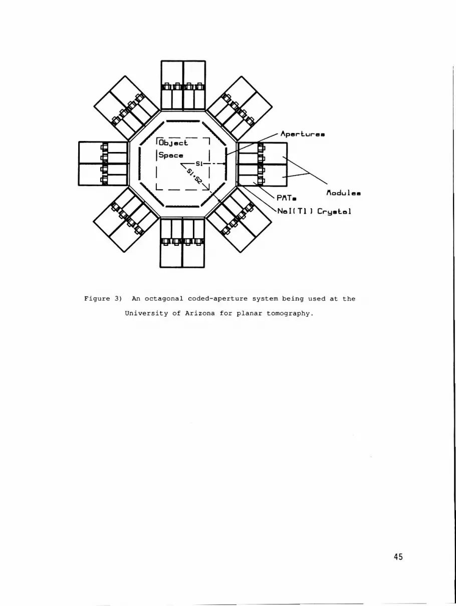
has i t s own coded aperture , t h u s forming a camera module. These modules can then be posit ioned about the pa t i en t i n a configuration t h a t w i l l optimally u t i l i z e the detector a rea . Figure 3 i s an example of an 8-view system f o r planar tomography t h a t i s cur ren t ly being constructed i n Arizona t o be used fo r hear t and brain imaging.
Preliminary simulations w i t h systems s imi la r t o t h a t of Fig. 3 demonstrate t h a t s ta te-of- the-ar t reconstruct ions a re obtainable w i t h data-acquis i t ion times of t he order of a t h i r d or l e s s than t h a t of t he conventional r o t a t i n g gamma-ray camera, which a re t y p i c a l l y 30 t o 4 0 minutes. T h i s p o t e n t i a l data-acquis i t ion time reduction, a s well t he s t a t i c nature of t he s y s t e m allowing dynamic s tudies , may cont r ibu te t o improving the s ta te-of- the-ar t i n nuclear-medicine imaging.
CONCLUSION
We have b r i e f l y described the p r inc ip l e s of imaging i n nuclear medicine, and have focused on a p a r t i c u l a r approach using coded ape r tu re s . The formulation of t h i s sh i f t -va r i an t problem was developed, and a p a r t i c u l a r reconstruction algorithm was presented. A coded-aperture system being developed a t t h e University of Arizona f o r tomographic imaging i n nuclear medicine was b r i e f l y described.
ACKNOWLEDGMENTS
T h i s work was supported by the National Cancer I n s t i t u t e through grant no. 2 PO1 CA 23417. We thank Bruce Moore f o r h i s technica l a s s i s t ance .
REFERENCES
Anger, H . O . , " S c i n t i l l a t i o n Camera, Rev. S c i . I n s t r u m . , 29, 27 (1958) .
Ba r re t t , H . H . , and W . Swindell, R a d i o l o g i c a l I m a g i n g : The Theory o f Image F o r m a t i o n , D e t e c t i o n , and P r o c e s s i n g , Vols. I and I1 (Academic, New York, 1 9 8 1 ) .
Tret iak, O . , and C . Metz, "The Exponential Radon Transform," SIAM J . Appl. Math. 39, 341 ( 1 9 8 0 ) .
Clough, A . V . , and H . H . Bar re t t , "Attenuated Radon and Abel Transforms," J . Opt. SOC. Am. A, 73, 1590-1595 (1985) .
Gaski l l , J . D . , L i n e a r Systems, F o u r i e r T r a n s f o r m s , & O p t i c s (John Wiley, New York, 1 9 7 8 ) .
D i c k e , R . H . , "Scatter-hole Cameras f o r X-rays and Gamma Rays," Astrophys. J . 153, L l O l ( 1 9 6 8 ) .
Ba r re t t , H . H . , "Fresnel Zone P la t e Imaging i n Nuclear Medicine," J . Nucl. Med. 2 3 , 382-385 ( 1 9 7 2 ) .

14)
1 5 )
16)
Simpson, R.G., "Annular Coded-Aperture System for Nuclear Medicine," doctoral dissertation (University of Arizona, Tucson, Ariz., 1978) .
Koral, K.F.,W.L. Rogers, and F.G. Knoll, "Digital Tomographic Imaging with a Time-Modulated Pseudorandom Coded Aperture and an Anger Camera," J. Nucl. Med. 16, 402 (1975).
Strang, G. Linear A l g e b r a and I t s Appl ica t ions , (Academic, New York, 1976) .
Smith, W.E., R.G. Paxman, and H.H. Barrett, "Image Reconstruction from Coded Data: I. Reconstruction Algorithms and Experimental Results," J. Opt. SOC. Am. A, 2, 491-500 (1985).
Melsa, J.L., and D.L. Cohn, Decision and Est imation Theory, (McGraw-Hill, New York, 1978).
Smith, W.E., and H.H. Barrett, "Linear Estimation Theory Applied to the Evaluation of A Pr ior i Information and Svstem Optimization in Coded-Aperture Imaging," J. Opt: SOC. Am. A, 5, 315-330 (1988).
Frieden, B.R., "Image Enhancement and Restoration," in T.-S. Huang, ed., Pic ture Processing and D i g i t a l F i l t e r i n g , Vol. 6 of Topics in Applied Physics, (Springer-Verlag, New York, 1975) .
Kirkpatrick, S., C.D. Gelatt, Jr., and M.P. Vecchi, "Optimization by Simulated Annealing, I' Science, 220, 671-680 (1983) .
Aarsvold, J.N., H.H. Barrett, J. Chen, A.L. Landesman, T.D. Milster, D.D. Patton, T.J. Roney, R.K. Rowe, R.H. Seacat, 111, and L.M. Strimbu, "Modular Scintillation Cameras: A Progress Report,"Medical Imaging 11: Image Formation, Detection, Processing, and Interpretation, SPIE 914, 319-325 (1988) .
43

Figure 1) A single-view coded-aperture system,
imaging a planar source.
Figure 2 ) A multiple-view coded-aperture system,
imaging a volume source.
44
-
-- odu I en
No1 T1 I Cryntal
Figure 3 ) An oc tagona l coded-aperture system be ing used a t t h e
U n i v e r s i t y of Arizona f o r p l a n a r tomography.
45


