Embed Size (px)
Citation preview
Characterizing the replication and stability regionsof Spiroplasma citri plasmids identifies a novelreplication protein and expands the genetic toolboxfor plant-pathogenic spiroplasmas
Marc Breton,1,2 Sybille Duret,1,2 Nathalie Arricau-Bouvery,1,2
Laure Beven1,2 and Joel Renaudin1,2
Correspondence
Joel Renaudin
1INRA, UMR 1090 Genomique Diversite et Pouvoir Pathogene, F-33883 Villenave d’Ornon, France
2Universite de Bordeaux 2, UMR 1090 Genomique Diversite et Pouvoir Pathogene, F-33883Villenave d’Ornon, France
Received 11 April 2008
Revised 12 June 2008
Accepted 16 June 2008
Spiroplasma citri strain GII3 contains seven plasmids, pSciA and pSci1–6, that share extensive
regions of sequence homology and display a mosaic gene organization. Plasmid pSci2 comprises
12 coding sequences (CDS), three of which encode polypeptides homologous to proteins Soj/
ParA, involved in chromosome partitioning, and TrsE and Mob/TraG, implicated in the type IV
secretion pathway. One CDS encodes the adhesin-like protein ScARP3d whereas the other eight
encode polypeptides with no homology to known proteins. The pSci2 CDS pE and soj have
counterparts in all seven plasmids. Through successive deletions, various pSci2 derivatives were
constructed and assessed for their ability to replicate by transformation of S. citri 44, a strain
which has no plasmid. The smallest functional replicon was found to contain a single CDS (pE)
and its flanking intergenic regions. Shuttle (S. citri/Escherichia coli) plasmids, in which CDS pE
was disrupted, failed to replicate in S. citri, suggesting that PE is the replication protein of the
S. citri plasmids. Successive propagations of pSci2-derived transformed spiroplasmas, in the
absence of selection pressure, revealed that only pSci2 derivatives having an intact soj gene were
stably maintained, indicating that the soj-encoded polypeptide is most likely involved in plasmid
partitioning. Upon transformation, pSci2 derivatives, including shuttle (S. citri/E. coli) plasmids,
were shown to replicate in all S. citri strains tested regardless of whether the strain possesses
endogenous plasmids, such as strain GII3, or not, such as strain R8A2. In addition, the pSci
replicons were introduced efficiently into the plant-pathogenic spiroplasmas Spiroplasma kunkelii
and Spiroplasma phoeniceum, the transformation of which had never, to our knowledge, been
described before. These studies show that, besides their implications for the biology of S. citri, the
pSci plasmids hold considerable promise as vectors of general use for genetic studies of plant-
pathogenic spiroplasmas. As an example, a HA-tagged S. citri protein was expressed in S.
kunkelii. Detection of pE-hybridizing sequences in various group I spiroplasma species indicated
that pE replicating plasmids were not restricted to the three plant-pathogenic spiroplasmas.
INTRODUCTION
Spiroplasmas are a group of helical, cell-wall-free bacteriabelonging to the class Mollicutes. Three spiroplasmaspecies, Spiroplasma citri (Saglio et al., 1973), Spiroplasmakunkelii (Whitcomb et al., 1986) and Spiroplasma phoeni-ceum (Saillard et al., 1987), have been associated with plantdiseases. S. citri is the aetiological agent of the citrusstubborn and horseradish brittle root diseases and was the
first plant mollicute to be cultured in vitro (Fletcher et al.,1981; Saglio et al., 1971). Since then, S. citri has beenstudied extensively (Bove et al., 1989, 2003). Theavailability of both an experimental transmission assayvia the leafhopper vector (Foissac et al., 1996) and genetransfer systems (Foissac et al., 1997; Renaudin, 2002;Renaudin & Lartigue, 2005) has made it a unique modelfor studying plant mollicute–host interactions (Andre et al.,2005; Berho et al., 2006a; Bove et al., 2003; Killiny et al.,2005).
In spiroplasmas, plasmids were first detected as extra-chromosomal, covalently-closed-circular DNAs (Ranhand
Abbreviation: CDS, coding sequence(s).
Five supplementary figures and a supplementary table are available withthe online version of this paper.
Microbiology (2008), 154, 3232–3244 DOI 10.1099/mic.0.2008/019562-0
3232 2008/019562 G 2008 SGM Printed in Great Britain

et al., 1980) and some of them were mapped and/or clonedin Escherichia coli (Archer et al., 1981; Mouches et al.,1983a). Extrachromosomal DNAs, including plasmids andviral DNAs, were most frequently detected in group Ispiroplasmas (Gasparich et al., 1993), especially in S. citristrains. However, these plasmids have for a long time beendesignated cryptic since no phenotypic character had beenassociated with their presence. In addition, due to the lackof sequence and functional data, the engineering of naturalplasmids to produce cloning vectors has had very limitedsuccess (Salvado et al., 1989). More recently sequencing ofplasmids pBJS-O from S. citri strain BR3-3X (Joshi et al.,2005), and pSciA and pSci1–6 from S. citri GII3 (Saillardet al., 2008), revealed that they encode proteins, includingadhesin-like molecules, which have been tentativelyassociated with insect transmission (Killiny et al., 2006;Yu et al., 2000). Furthermore, it was shown that plasmidspSci1–6 from the insect-transmissible strain GII3 wereabsent in the non-insect-transmissible strains of S. citri(Berho et al., 2006b). However, the first clear-cut evidenceof a phenotypic trait associated with spiroplasmal plasmidswas the finding that pSci6 from S. citri GII3 conferredinsect-transmissibility to the plasmid-free, non-insect-transmissible strain 44 (Berho et al., 2006a). Besidesassessing their role in insect transmission, the successfultransfer of pSci plasmids between strains of S. citri raisedtheir potential as genetic tools. In addition to adhesin-likeproteins, pSci1–6 encode proteins with similarities tocomponents of bacterial type IV secretion systems,suggesting the possibility of conjugative transfer of theseplasmids. In turn, none of the plasmid-encoded proteinsshared similarity with known replication proteins (Saillardet al., 2008).
Previously, we showed that sequences essential forreplication and maintenance of pSci2 in S. citri werelocated within an 8 kbp restriction fragment (Berho et al.,2006a). In the present study, we describe the functionalcharacterization of the replication and stability regions ofS. citri pSci-derived plasmids, with the aim of furtherinvestigating their role in spiroplasmal biology. We alsodemonstrate the usefulness of pSci-derived plasmids asvectors for gene transfer and expression in plant-patho-genic spiroplasmas other than S. citri.
METHODS
Spiroplasma strains, growth conditions and transformation. S.
citri GII3 wild-type strain was originally isolated from its leafhopper
vector Circulifer haematoceps, captured in Morocco (Vignault et al.,
1980). S. citri 44 was isolated from a stubborn-diseased sweet orange
tree in Iran (Hosseini Pour, 2000). Other spiroplasma strains used in
this study are listed in Supplementary Table S1, available with the
online version of this paper. Spiroplasmas were grown at 32 uC in SP4
medium (Whitcomb, 1983) from which fresh yeast extract was
omitted. Spiroplasma cells were transformed by electroporation
(Stamburski et al., 1991) using 2–10 mg purified plasmid or various
ligation mixtures. Spiroplasmal transformants were selected in the
presence of 2 mg tetracycline ml21, and further propagated in broth
medium containing 5 mg tetracycline ml21. The Spiroplasma speciesof tetracycline-resistant transformants were confirmed by PCR-RFLPanalysis of the spiralin genes, using amplification with primer pairSR28/SR29 (Table 1) and digestion with MboI and/or RsaI.
Plasmid constructs. Plasmids pSRT1 and pSRT2 are pBS+
(Stratagene)-derived, ColE1 replicons carrying the tetracycline resist-ance gene tetM under the control of the spiralin gene promoter(Lartigue et al., 2002). Plasmid pSRT1 differs from pSRT2 in that thetetM cassette is in the opposite orientation. Plasmids pSci1NT andpSci6PT were obtained by inserting the tetM cassette (as a 2.3 kbp PstIfragment) into the NsiI and PstI sites of S. citri pSci1 and pSci6,respectively, whereas pSci21NT resulted from the combination of thetetM cassette with the 8034 bp NsiI fragment of pSci2 (Berho et al.,2006a). In pSci1NTDS, the scarp3d gene was disrupted by inserting the20 bp oligonucleotide duplex SBamF1/SBamR1 (Table 1) into the NheIsite of pSci1NT. Similarly, in pSci21NTDsoj, the soj gene was disruptedby insertion of the 19 bp SojBamF1/SojBamR1 duplex into the SmlIsite of pSci21NT. As a result, the Soj coding sequence (CDS) encodes atruncated polypeptide of 67 amino acids as compared with 260 in thewild-type. Plasmid pFL3 was obtained by self-ligation of the 6675 bpEcoRI–BglII fragment of pSci21NT. Plasmid pFL4 was constructed bydeleting the 891 bp HincII–HaeIII fragment within the mob gene ofpFL3, and pFL5 by deletion of the 504 bp AclI–EcoRV fragment ofpFL4. Plasmid pBS1 was derived from pBS+ by inserting the EPBg1/EPBg2 duplex at the EcoRI site to introduce a BglII site. Plasmid pBST1was then obtained by combining the BglII-linearized pBS1 with thetetM cassette (as a 2311 bp BamHI–BglII fragment) of pSRT2. PlasmidpFL6, containing the pE gene and the flanking, intergenic regions,resulted from the ligation of the 2265 bp PacI fragment from pFL4 tothe 2329 bp PacI fragment of pBST1 containing the tetM cassette.Plasmid pFL7 (4259 bp) was obtained by deleting the 341 bp AclI–BglIIfragment of pFL6. The shuttle (S. citri/E. coli) plasmid pBSFL6 resultedfrom the combination of PstI-linearized pBS+ and pFL6. To constructpSDE1, the amplification product of pSci2 with primer pair PEF1/IEFR2 was inserted into pSRT1 after they were both digested withBamHI+EcoRI. Similarly, the Rep1-F1_BglII/Rep1-R1_BglII amp-lification product, comprising the pE gene and the flanking intergenicregions of pSci1, was digested with BglII, and the 1963 bp fragment wasinserted into BglII-linearized pSRT1 to yield pMBRE. While pBSFL6comprised the pE region of pSci2, pMBRE contained the comparablepE region of pSci1 having a MluI restriction site within the pE gene. Toconstruct pMBRDE1, pMBRE was linearized with MluI, and recircu-larized by ligation after the 4 bp cohesive ends were made blunt-ended.In this plasmid, disruption of pE led to a truncated polypeptide of 97amino acids instead of 217 in the wild-type. Similarly, in pMBRDE2inserting the 19 bp oligonucleotide duplex EBamF1/EBamR1 into theMluI site led to a truncated PE of 95 amino acids. Plasmid pNAB32Hresulted from insertion of the p32-HA tagged gene inserted intopBSFL6. Each plasmid construct was confirmed by restriction mappingand sequencing the appropriate regions.
DNA isolation, Southern blot hybridization and Western
immunoblotting. Spiroplasma genomic DNA was prepared from10 ml cultures using the Wizard genomic DNA purification kit(Promega), whereas plasmid DNA was purified from 25 ml cultureswith the Wizard SV minipreps DNA purification kit (Promega).Southern blot hybridization of spiroplasmal DNA with appropriatedigoxigenin-dUTP-labelled probes has been described elsewhere(Andre et al., 2003). Hybridization signals were detected with anti-digoxigenin–alkaline phosphatase conjugate and 2-hydroxy-3-naphthoic acid-29-phenylanilide phosphate (HNPP) as the substrate,following the supplier’s instructions (Roche Diagnostics). Fluorescentsignals were detected using a Fluor-S Multimager Phosphoimager(Bio-Rad). Probes specific to ScARPs, P32 and PE were generated byPCR amplification with primer pairs S235F/S235R, P32F/P32R andPEF1/PER1, respectively (Table 1).
Replication and stability of S. citri plasmids
http://mic.sgmjournals.org 3233

Spiroplasmal proteins were separated by SDS-PAGE and further
analysed by immunoblotting as described previously (Duret et al.,
2003) except that the proteins reacting with the primary antibodies
were visualized by using a goat anti-rabbit immunoglobulin G-
peroxidase conjugate and the SuperSignal West Pico chemilumin-
escent substrate (Pierce).
In silico analyses. The BLAST program (Altschul et al., 1997) was used
to search for homologies in general databases (http://www.ncbi.nlm.-
nih.gov/blast/), the mollicute dedicated database MolliGen (Barre et al.,
2004), and the S. kunkelii partially sequenced genome (http://
www.genome.ou.edu/blast/spiro_blastall.html). Conserved domains
were detected by CD-Search against the NCBI’s conserved domain
database (Marchler-Bauer & Bryant, 2004). Global sequence align-
ments were accomplished using a Needleman–Wunsch algorithm
implemented in the Needle program (Needleman & Wunsch, 1970).
Multiple alignments were done with T-Coffee (Notredame et al., 2000)
and/or CLUSTAL W (Thompson et al., 1994). For subsequent
phylogenetic analyses, poorly aligned positions and divergent regions
were eliminated using the alignment curation program Gblocks
(Castresana, 2000). Neighbour-joining trees based on these alignments
were constructed using the MEGA 3.1 program (Kumar et al., 2004) and
maximum-likelihood trees were computed with the WAG substitution
model using the PhyML program (Guindon & Gascuel, 2003). All trees
were bootstrapped 100 times.
RESULTS
Plasmids from S. citri GII3 all contain CDS pE and soj
S. citri strain GII3 carries seven plasmids, pSciA (7.8 kbp)and pSci1–6 (13–35 kbp) (Saillard et al., 2008), whichshare a large number of highly conserved regions and
display a mosaic gene organization (Fig. 1). Most of theCDS are present on several plasmids and, reciprocally, veryfew of them are specific to one given plasmid. Such anexception is p32, encoding a hydrophilic protein specific topSci6 (Killiny et al., 2006). Surprisingly, most of theplasmid CDS have no orthologues in other bacterialgenera, with the exceptions of soj, trsE, traG and mob,the naming of which indicates that they encode proteinsclosely related to proteins involved in partitioning (Soj/ParA family COG1192) and transfer (TrsE, TraG and Mob)of DNA molecules. The scarp genes of pSci1–5 encodeproteins sharing strong similarities with the adhesin-likeprotein SARP1 from S. citri BR3 (Berg et al., 2001).Plasmid pSci2 (14.4 kbp) seems to represent the ‘coreplasmid’ as it displays a gene organization (pE, pF, pA, soj,pB, scarp, pC, pD, trsE, p4, mob) which is conserved inpSci1 (13 kbp) and larger plasmids pSci3 (19.3 kbp) andpSci4 (20.2 kbp) (Fig. 1). Plasmid pSci3 possesses anadditional CDS (pG), whereas pSci4 carries two scarpgenes, two copies of soj, and the additional CDS pH, whichis absent in the other three plasmids pSci1–3. In pSci1,pSci5 and pSci6, the mob-like gene is disrupted. The largeplasmids pSci5 and pSci6 probably result from recomb-ination between plasmids and/or from gene duplication.Whereas pSci1–3 contain a single copy of pE and soj,pSci4–6 have two copies of soj, and multiple copies of pEare present in pSci5 (2 copies) and pSci6 (4 copies).Interestingly, pE and soj are the only two CDS that areconserved, uninterrupted, in all seven plasmids, pSciA andpSci1–6, suggesting that these genes may be essential forreplication and maintenance of the plasmids.
Table 1. Oligonucleotides and primers used in this study
Name Nucleotide sequence (5§–3§) Position Accession no.
S235F TAAACATTGATATTGCCAACCCC 3617–3639 AJ969069
S235R GGTTAAAGTTGCAGAATTATTATC 3973–3996 AJ969069
SR28 ACTTTTATCGATTTTAGCAG 114268–114287 AM285305
SR29 ATCTTTTGCTTTAACTGTTACT 114886–114908 AM285305
P32F TAACGAATTAAATCATTCTAATAGC 24852–24876 AJ969074
P32R TAGTTCCGGCTTGCTCACCA 24332–24351 AJ969074
PEF1 CCCACGgaATTCTTCTATACACCTATTAAG 11022–11051 AJ969070
PER1 AGTATTggATcCATTGTCTTGCTACGCTGT 12046–12075 AJ969070
Rep1-F1_BglII ACAGTTAgAtcTGCTTGCTCTGAATAAC 10208–10235 AJ969069
Rep1-R1_BglII TTAATCaGaTCTGTCATAATTTCAACTCCT 12159–12188 AJ969069
IEFR2 CTAAATTTAAGgATCCTCATTTATTAATCATATC 12620–12653 AJ969070
EPBg1 AATTCCCTTAATTAACCAGATCTCCTTAATTAACCG – –
EPBg2 AATTCGGTTAATTAAGGAGATCTGGTTAATTAAGGG – –
EBamF1 CGCGATAGGATCCAACTAT – –
EBamR1 CGCGATAGTTGGATCCTAT – –
SBamF1 CTAGATTAGGATCCACTAAT – –
SBamR1 CTAGATTAGTGGATCCTAAT – –
SojBamF1 TCAATGGATCCATAGCTAC – –
SojBamR1 TTGAGTAGCTATGGATCCA – –
*Bold type indicates restriction enzyme sites. Lower-case type indicates mismatched nucleotides.
M. Breton and others
3234 Microbiology 154

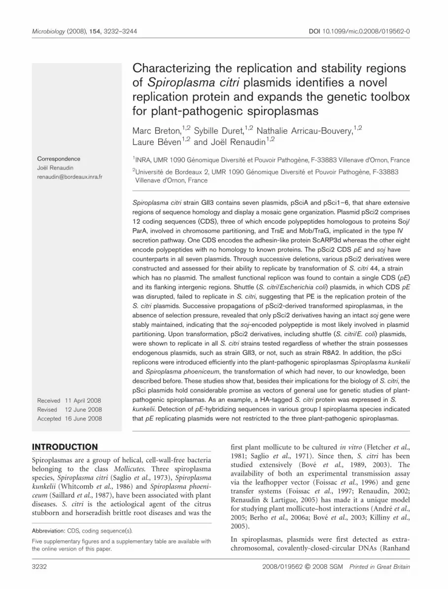
Replication region of pSci2
Preliminary data strongly suggested that the replicationregion of pSci2 was located within the NsiI fragment of8034 bp (Berho et al., 2006a). This fragment encodes sevenputative polypeptides, including a Mob-like ATPase and aSoj-like protein (Fig. 2). Polypeptides PA and PE, as well asthe truncated PB and PF, are hypothetical proteins thathave counterparts in other pSci plasmids, whereas thehypothetical polypeptide HP shares no homology withother proteins. None of these polypeptides share homologywith known replication proteins. To identify the replica-tion region of pSci2, various pSci2 derivatives wereconstructed by successive deletions of pSci21NT, andtested for their ability to replicate through transformationof S. citri 44, a strain which contains no plasmid. Plasmidconstructs are described in Methods. The finding thatpFL3, pFL4, pFL5, pFL6 and pFL7 all replicated in S. citrishowed that the replication region was located within the1876 bp HaeIII–AclI fragment of pFL3 (Fig. 2).Interestingly, this DNA fragment comprised a singleCDS, suggesting that the pE-encoded polypeptide mightbe the replication protein and that the flanking intergenicregions contained the cis-acting binding sites. In silicoanalyses revealed the region upstream of pE to containspecific features including the imperfect repeatsATTTAATCCCCC and ATTTAACCCCC (at positions10963 and 11084 of pSci2) and four sequences(GTTTCCATA, ATTTCCACA, TCTACCACA andTTTTCCAAA, at positions 10833, 10899, 10977 and11268, respectively) resembling the DnaA boxes
(TTTTCCACA) of the S. citri chromosomal replicationorigin (oriC) (Ye et al., 1994). However, in contrast to thesituation at oriC, these sequences did not cluster within ashort region. Immediately downstream of pE, two tandemrepeated sequences of 11 nucleotides (TAACTCCCCTA),as well as an A-T rich region, were detected (Fig. 2).However, none of the six inverted repeat sequencesidentified in the intergenic region downstream of pE(Saillard et al., 2008) was located within the 1876 bpminimal replicon, indicating that these sequences, whichputatively lead to hairpin structures, are not essential forplasmid replication. To further delineate the replicationregion, DNA fragments were retrieved from pFL6 bydigestion or PCR amplification and inserted into ColE1replicons by cloning in E. coli. Whereas the recombinantplasmid pBSFL6, which contained the pE gene and theflanking intergenic regions, readily replicated in S. citri,plasmid pSDE1 did not, indicating that replicationprobably requires sequences upstream of primer PEF1.
CDS E encodes the replication protein
To further demonstrate the role of PE in plasmidreplication, shuttle plasmid pMBRE and its pE-disruptedderivatives pMBRDE1 and pMBRDE2 were used totransform S. citri 44. As expected, transformation bypMBRE yielded tetracycline-resistant transformantsat relatively high frequency (~1024 transformantsc.f.u.21mg21), indicating that the plasmid replicated inthese cells. The presence of the relevant plasmid in the
Fig. 1. Gene organization of plasmids pSciA and pSci1–6 from S. citri GII3. CDS pE and soj are indicated by black and whitearrows, respectively. Numbers indicate the percentages of identical amino acids shared by the pE and soj protein products withtheir counterparts from pSci2. Conserved CDS are named (A to N, corresponding to pA–N in the databases) whereas orphanCDS are not. Positions of BstBI restriction sites (B) of pSci6 are indicated. scarp, genes encoding S. citri adhesion-relatedproteins. * indicates truncated CDS. For alignment, pSciA and pSci1–6 were linearized arbitrarily regardless of the sequencenumbering (GenBank accession numbers AJ966734 and AJ969069–74).
Replication and stability of S. citri plasmids
http://mic.sgmjournals.org 3235
spiroplasmal transformants was confirmed by PCR andSouthern blot hybridization (data not shown). In contrast,no transformants were obtained through transformationwith pMBRDE1 and pMBRDE2, in which the pE gene wasdisrupted (see Methods and Fig. 2). As a control,disruption of the scarp gene in pSci1NTDS had no effecton the ability of the plasmid to replicate.
Considering PE as the replication protein, one wouldexpect pMBRDE1 and pMBRDE2 to replicate in S. citriGII3, a strain with plasmids, from which wild-type PE wasknown to be expressed, as indicated by proteome analyses(unpublished data). However, transformation of S. citriGII3 by these plasmids failed repeatedly, indicating thatreplication of pMBRDE1 and pMBRDE2 cannot be drivenby the PE protein when provided in trans from endogenousplasmids.
Alignments of the PE proteins from pSci1–6 showed thatthey share strong similarities to each other, with theconserved regions being distributed all along the proteinsequence. Identities ranged from 76.1 % between pSci1-PEand pSci6-PE4 to 97.7 % between pSci3-PE and pSci5-PE1
(Fig. 1 and Supplementary Fig. S1). The pSciA-PE wasmuch less conserved with identities ranging from 25.2 %with pSci1-PE to 28.4 % with pSci6-PE1. Unexpectedly,despite the PE protein being essential for plasmidreplication, no putative DNA-binding motif such ashelix-turn-helix, coiled coil, zinc finger, or leucine zipperwas predicted in the PE sequence. It is noteworthy thatmost of the PE polypeptides end with an identical C-terminus (ELDNLD), except that from pSciA and three ofthe four PE from pSci6, which have distinct C-termini(Supplementary Fig. S1). The autonomous replication ofthe 16195 bp circularized BstBI fragment of pSci6PT(unpublished data) indicated that at least one PE ofpSci6 (pSci6 E2 in Fig. 1) functioned as the replicationprotein. Whether the other three are functional or not hasnot yet been investigated. Studying the genetic relatednessof the S. citri GII3 proteins PEs showed that they clusteredwithin three distinct groups (Fig. 3). The largest groupconsisted of PEs from pSci1–5 and the PE counterparts ofpBJS-O from S. citri BR3 (Joshi et al., 2005) and pSKU146from S. kunkelii CR2-3X (Davis et al., 2005). Unexpectedly,whereas one PE (PE2) from pSci6 also fell in this group, the
Fig. 2. Structural organization of pSci2 deletion derivatives, and ColE1 replicons carrying the pSci1 or pSci2 replication region.Plasmid constructs are described in Methods. Putative DnaA boxes (h), repeated sequences (c), iteron-like sequences (smallwhite arrows), and an AT-rich region (|||||) are indicated in the minimum replicon pFL7 only. A, AclI; B, BglII; E, EcoRI; EV,EcoRV; H, HincII; Ha, HaeIII; M, MluI; N, NsiI; P, PacI; Ps, PstI; S, SmlI; nd, not done. + and ” indicate the ability or inability toreplicate or to be stably maintained in the absence of selection pressure. * indicates truncated CDS of pSci2.
M. Breton and others
3236 Microbiology 154

other three (PE1, 3 and 4) formed a distinct, heterogeneousgroup. Such variability is consistent with the hypothesisthat, in contrast to PE2, PE1, 3 and 4 might not befunctional. Whether the occurrence of multiple copies ofthe pE gene in pSci6 resulted from gene duplication orrecombination between plasmids is unclear. Intriguingly,the pSciA PE did not cluster with any other PE proteins,suggesting that pSciA had a distinct origin. This is inagreement with the fact that, except for PE and Soj, thepSciA CDS have no counterparts in any other S. citriplasmids.
Stability of pSci plasmids
S. citri strain GII3, originally isolated in 1980, has beentriply cloned and further propagated for hundreds ofgenerations. Plasmids pSci1–6 from this strain, GII3-3X,displayed characteristic hybridization patterns whenprobed with a mixture of probes scarp and p32 (Berho etal., 2006b). To further confirm the presence of the sixplasmids in every cell, the spiroplasma strain was furthersubcloned twice, and the plasmid contents of 10 isolatedcolonies were characterized by Southern blot hybridizationof total DNA. The hybridization patterns of all 10subclones tested were identical to each other and to thatof the original culture (Supplementary Fig. S2), indicatingthat each spiroplasma cell contained the whole set ofplasmids, pSci1–6. Identical hybridization patterns wereobtained with spiroplasma cultures isolated from theleafhopper vector and from the host plant after experi-mental transmission of GII3-3X, by injection, to itsleafhopper vector (data not shown).
As indicated above, each S. citri plasmid contains at leastone soj-like gene, encoding a polypeptide with significant
similarities to proteins of the Soj/ParA, ATPase familyinvolved in chromosome partitioning. Similarities to thechromosomal ParA of S. citri and the Soj protein of Bacillussubtilis ranged from 41.3 to 46.3 % and from 41.6 to50.8 %, respectively. In the plasmid-encoded Soj, COG1192(Soj) and COG0455 (ATPases involved in chromosomepartitioning) were predicted with significant probabilities(e-values ranging from 3610210 to 1610228). Like thechromosomal ParA, the pSci-encoded Soj all possessed thedeviant Walker-type ATPase consensus motif with thetypical A, A9, and B boxes (Koonin, 1993) (SupplementaryFig. S3).
To investigate the role of Soj in S. citri plasmidpartitioning, we assessed the stability of various pSci2derivatives in S. citri 44 transformants. In the experimentshown in Fig. 4, S. citri 44 cells transformed with pSci21NT(containing soj) or pFL3 (lacking soj) were propagated inthe absence of selection pressure. After 5, 15 and 30passages (30 passages correspond to 100 generations), theplasmid contents of 10 individual clones were analysed bySouthern blot hybridization. The results showed thatpSci21NT was detected in each of the 10 clones tested,regardless of the passage number (5, 15 or 30). In contrast,pFL3 was lost in three clones out of ten, as early as the fifthpassage. After 15 passages, pFL3 was detected in only twoclones, and after 30 passages, none of the ten clones carriedpFL3. The finding that, in the absence of selection pressure,pSci21NT was stably maintained for many generationswhereas pFL3 was rapidly lost strongly suggested that thesoj-encoded protein was indeed involved in partitioning ofpSci2. In accordance with these results, plasmid loss wasalso noticed in spiroplasmas transformed with pSci2derivatives pFL4, pFL5 and pFL6, all of which lack soj.However, when compared with pSci21NT these plasmids
Fig. 3. Phylogenetic tree of the PE proteins from S. citri (pSciA,pSci1–6, and pBJS-O) and S. kunkelii (pSKU146) plasmids.Sequences were aligned with the T-Coffee program and analysedas described in Methods.
Fig. 4. Segregational stability of pSci21NT and pFL3. Southernblot hybridization of HincII-restricted DNAs from S. citri GII3 (laneG) and S. citri 44 transformants (lanes 1–10) with probe pE. TotalDNAs were extracted after 5, 15 and 30 passages in the absenceof selection pressure. (a) pSci21NT; (b) pFL3.
Replication and stability of S. citri plasmids
http://mic.sgmjournals.org 3237

lack not only soj but also three additional CDS, the twoconserved hypothetical proteins PA and PB as well as theorphan CDS HP (Fig. 2). The role of Soj was furtherdemonstrated by evaluating the stability of pSci21NTDsoj,a plasmid in which the soj gene was disrupted, in S. citri 44transformants. After propagation for up to 30 passages inthe absence of selection pressure, the presence of theplasmid was checked by monitoring the percentage ofspiroplasma cells growing on selective (tetracycline) agarmedium. As expected from the above experiment, nearly100 % (285 /294) of cells from pSci21NT transformantswere still tetracycline-resistant after 30 passages. Incontrast, in pSci21NTDsoj transformants, the ratio oftetracycline-resistant to total cells progressively decreasedto 81 % (515/638) at the fifth passage, 27 % (132/496) atthe 15th passage, and less than 0.2 % (less than 1/417) after30 passages. These results clearly indicated that the soj geneproduct is involved in the stability of pSci plasmids.
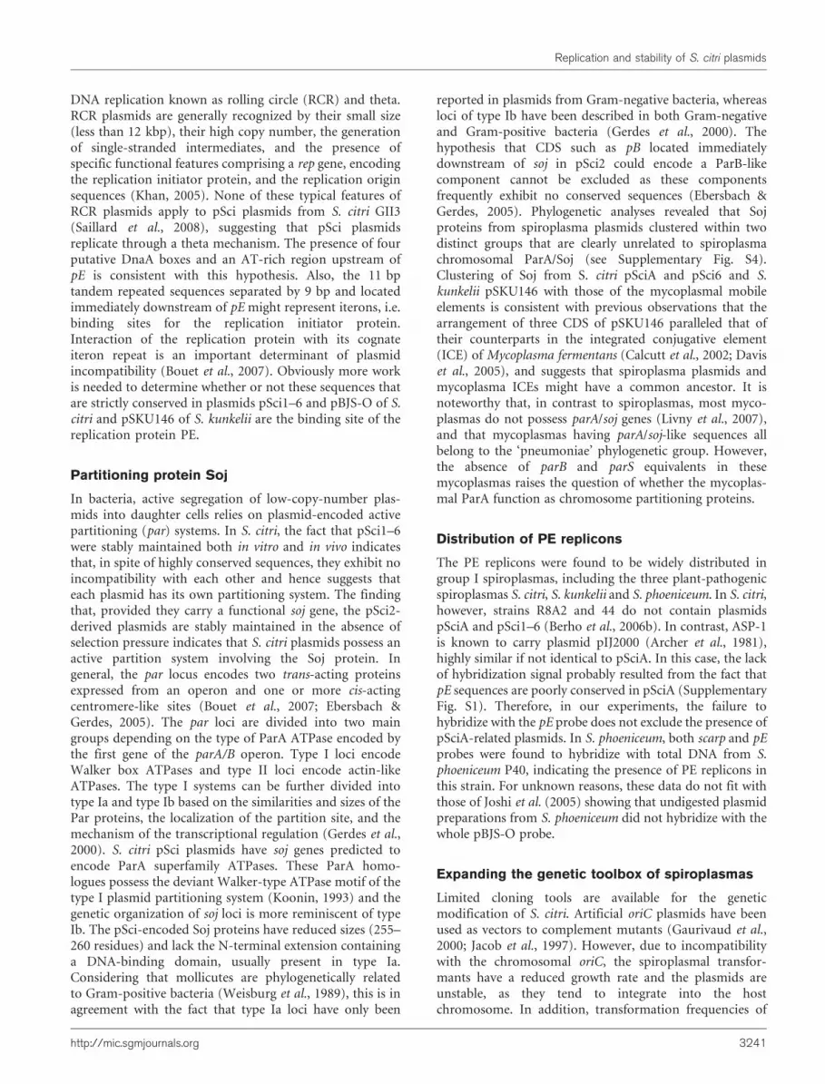
Phylogeny of plasmid Soj proteins
Sequence alignments of predicted Soj proteins(Supplementary Fig. S3) revealed that those from plasmidspSci1–5 were more closely related to each other (identitiesranging from 50.4 to 82.9 %) than to Soj from pSci6 (22.4to 28.3 % identity), and that those from pSci1–6 shared alow percentage (24 to 29.2 %) of identical amino acids withpSciA. The bootstrapped phylogenetic tree (Fig. 5)indicates that plasmid-encoded Soj were distinct from thechromosomal Soj/ParA and clustered in two separategroups: Soj from pSci1–5 (S. citri GII3) and pBJS-O (S.citri BR3) in one group, and Soj from pSciA and pSci6 (S.citri GII3), and pSKU146 (S. kunkelii E275) in the other,suggesting that these groups had evolved separately and
disjointedly from the chromosomal ParA. In an effort tounderstand the origin of these plasmids, phylogeneticanalyses of spiroplasma Soj proteins were extended to a setof representative ParA homologues (Gerdes et al., 2000)including the 23 annotated ParA homologues frommollicute genomes (http://cbi.labri.fr/outils/molligen/).The phylogenetic tree showed that, as in many bacteria(McLeod et al., 2006), the chromosomally encoded ParAproteins of spiroplasmas had phylogenies congruent withthose of their hosts. In contrast, the plasmid-encoded Soj/ParA proteins were more diverse and their clustering didnot follow the organismal phylogeny (Supplementary Fig.S4). When restricted to the mollicutes and several otherfirmicutes, these analyses identified four major clusterssupported by strong bootstrap values (Supplementary Fig.S5). One coherent cluster was made from the S. citrichromosomal ParA and other chromosomal ParA proteinsof firmicutes (except those of mycoplasmas). A secondcluster consisted of the mycoplasmal chromosome-encoded ParA/Soj proteins, whose genes are located inthe vicinity of the chromosome replication origin. Theseproteins are clearly distinct from ParA/Soj proteins of otherfirmicutes and the spiroplasmas in particular. The thirdcluster comprised plasmid-encoded Soj proteins from S.citri pSci1–5 and pBJS-O; the fourth cluster consisted ofSoj proteins from S. citri pSciA and pSci6, S. kunkeliipSKU146, and chromosomal Soj proteins that are encodedby putative or demonstrated mobile elements of myco-plasmas (Marenda et al., 2006; Sirand-Pugnet et al., 2007).
Distribution and host-range of S. citri pSciplasmids
Previous studies showed that, in contrast to GII3, variousS. citri strains did not contain plasmids pSci1–6, as revealedby the failure of genomic DNAs to hybridize with scarp andp32 probes (Berho et al., 2006b). To investigate theoccurrence of PE replicating plasmids in spiroplasmasfurther, genomic DNAs from various strains of S. citri andother spiroplasma species were hybridized with probesscarp and pE (Fig. 6, Table 2). Both scarp and pEhybridization signals were detected in strains GII3, Corse,Palmyre, Hinckley, IP85 and Israel. In contrast, nohybridization signal was detected in ASP-1, R8A2 and 44.Detection of pE but not scarp sequences in strain Alcanar254 is consistent with the fact that this strain carries asingle plasmid nearly identical to pSci6 (unpublished data),which contains no scarp genes. Interestingly, pE-hybri-dizing signals were also detected in the plant-pathogenicspiroplasmas S. kunkelii E275 (subgroup I-3) and S.phoeniceum P40 (subgroup I-8), indicating that PEreplicons were not restricted to the species S. citri, i.e.subgroup I-1 of the spiroplasmal classification (Williamsonet al., 1998). In addition, the pE probe was found tohybridize with genomic DNAs from the bee pathogenSpiroplasma melliferum (subgroup I-2) strains BC3, G1 andAS576, the rabbit tick spiroplasma 277F (I-4), the plantbug spiroplasma sp. LB-12 (I-5), and the plant surface
Fig. 5. Phylogenetic relationships of plasmid Soj proteins from S.
citri (pSciA, pSci1–6 and pBJS-O) and S. kunkelii (pSKU146),and chromosomal Soj proteins from S. citri and B. subtilis.Sequences were aligned with the T-Coffee program and analysedas described inMethods.
M. Breton and others
3238 Microbiology 154

Spiroplasma sp. N525 (I-7) but not with DNAs from strainsB13 and B31 of the bee pathogen Spiroplasma apis (groupIV) and strain 23.6 of the plant surface Spiroplasmafloricola (group IV). From these data, the PE repliconsappear to be restricted to species of group I spiroplasmas.Unlike pSci1–5 from S. citri GII3, these replicons do notnecessarily contain conserved scarp genes. Similarly to S.citri Alcanar 254, PE replicons from S. melliferum did nothybridize with the scarp probe.
The occurrence of PE replicons in many spiroplasmaspecies suggested that these plasmids might have a broadhost-range. Therefore we explored the possibility oftransforming various strains of S. citri as well as otherspiroplasma species with pSci21NT and pFL6, twoderivatives of pSci2 from S. citri GII3. Both plasmids wereintroduced successfully and replicated in all S. citri strainstested (GII3, ASP-1, Alcanar 254, R8A2 and 44), regardlessof the strains’ natural plasmid contents. Transformationfrequencies ranged from 1025 to 1023 transformantsc.f.u.21 (mg plasmid)21. In particular, it is noteworthy thatstrain R8A2, which could only be transformed at extremelylow efficiency (less than 1028 transformants c.f.u.21 mg21)by oriC plasmids (Renaudin et al., 1995) was repeatedlytransformed by pSci21NT and pFL6 at frequencies close to1025 transformants c.f.u.21 mg21. Interestingly, trans-formation of S. kunkelii E275 and S. phoeniceum P40 bypSci21NT, pFL6, and its shuttle derivative pBSFL6 alsoyielded tetracycline-resistant transformants at frequenciesof 1025–1024 transformants c.f.u.21 mg21. The possibilitythat these tetracycline-resistant cells could be spontaneousmutants or contaminants was excluded by showing thatthey did contain the relevant plasmid, by PCR and Southernblot hybridization, and by confirming the spiroplasma
species through PCR-RFLP analysis of the spiralin gene(Methods).
pSci-derived plasmids as gene vectors
To demonstrate the usefulness of pSci-derived plasmids asgene vectors, the plasmid construct pNAB32H, in whichthe p32 gene fused to a HA tag was inserted into the shuttleplasmid pBSFL6, was introduced into S. kunkelii E275 byelectrotransformation. The presence of pNAB32H in thespiroplasmal transformants was confirmed by PCR andSouthern blot hybridizations (data not shown). In theexperiment shown in Fig. 7, total proteins were probed byWestern immunoblotting with P32- and HA-specific,polyclonal antibodies (Fig. 7a and b, respectively). Cellsfrom S. citri GII3 and S. kunkelii E275 transformed bypSci1NT were used as controls. As expected, P32 wasdetected in S. citri GII3 (Fig. 7a, lane 1) but not inuntransformed S. kunkelii (lane 2) or in S. kunkeliitransformed with pSci1NT (lane 6). In contrast, P32-related proteins were detected in S. kunkelii cells trans-formed by pNAB32H, carrying the p32–HA fusion gene(lanes 3 and 4), or pSci6PT containing the wild-type p32gene (lane 5). As expected, the HA-tagged protein wasclearly detected in pNAB32H-transformed cells, indicatingthat the fusion protein was expressed from the recombin-ant plasmid in S. kunkelii E275 (Fig. 7b, lanes 3 and 4). Inaddition, the relative intensities of the P32 detection signalswere consistent with an expression level of the P32–HAfusion protein from pNAB32H similar to that of the wild-type P32 encoded by pSci6 in S. citri GII3.
DISCUSSION
Replication protein PE
The occurrence of highly conserved regions in the plasmidsfrom S. citri GII3 suggests that they have a common origin.Except for soj, traG, trsE and mob, most of the plasmidgenes encode hypothetical proteins with no counterparts inbacteria other than spiroplasmas. Moreover, homologysearches failed to identify a putative replication protein,indicating that these plasmids probably represent a newplasmid family. In particular, they do not share anyhomology with the phytoplasma plasmids that encodeputative replication proteins (Rep) having the conservedmotifs associated with the rolling circle mechanism ofreplication (Oshima et al., 2001; Tran-Nguyen & Gibb,2006). S. citri plasmids pSciA and pSci1–6 all possess genessoj and pE, which are also conserved in plasmids pBJS-Ofrom S. citri BR3 (Joshi et al., 2005) and pSKU146 from S.kunkelii CR2-3X (Davis et al., 2005). In this study, weshowed that pE is essential and the only plasmid-encodedprotein required for plasmid replication. The finding thatinactivation of pE by a single 4 bp insertion preventedreplication clearly indicates that the polypeptide, ratherthan the nucleotide sequence, is required. The fact that the
Fig. 6. Southern blot hybridization of HincII-restricted DNAs fromvarious spiroplasmas with the pE probe. DNAs were from S. citri
GII3, Palmyre, Israel, Hinckley, IP85 and R8A2 (lanes 1–6), ASP-1and 44 (lanes 13 and 14), M4 and MHcl2 (lanes 15 and 16),Corse (lane 21), S. melliferum BC3 and G1 (lanes 19 and 20), S.
kunkelii E275 (lane 7), Spiroplasma sp. 277F and LB12 (lanes 8and 12), S. phoeniceum P40 (lane 9), S. floricola 23.6 (lane 10),S. apis B31 (lane 11), S. mirum SMCA (lane 17) and S. ixodetis
Y32 (lane 18).
Replication and stability of S. citri plasmids
http://mic.sgmjournals.org 3239

pE-disrupted plasmids failed to replicate in S. citri GII3 (astrain that produces PE from endogenous plasmids)suggests that PE, unlike other replication initiator proteins(Emond et al., 2001) might be a cis-acting protein, unableto support replication when provided in trans. In thisexperiment, however, the hypotheses of a retro-controlsystem specific for S. citri GII3, or incompatibility betweenplasmids having identical replication regions, cannot beexcluded (Bouet et al., 2007). Therefore, complementationof the mutation carried by the pE-disrupted plasmids inS. citri 44 will be required to definitely establish whether ornot PE is a cis-acting protein.
Although the PE protein was found to be essential forreplication, it lacks the leucine zipper and helix-turn-helixmotifs found in many Rep proteins (Del Solar et al., 1998).The lack of sequence and/or structural similarity betweenPE and known replication proteins suggests that replicationof spiroplasma plasmids pSci involves a distinctivemechanism. Circular bacterial plasmids use two modes of
Table 2. Distribution of scarp and pE sequences among various spiroplasma strains
Group Species Strains* Habitat ProbeD
scarp pE
I-1 S. citri ASP-1 Phloem/leafhopper 2 2
C189 ND 2
Corse + +
Alcanar 254 2 +
GII3 + +
Hinckley + +
Israel + +
IP85 + +
MHcl2 ND +
M4 ND +
Palmyre + +
R8A2 2 2
44 2 2
I-2 S. melliferum AS576 Honey bee 2 +
BC3 2 +
G1 2 +
I-3 S. kunkelii E275 Phloem/leafhopper + +
I-4 Spiroplasma sp. 277F Rabbit tick + +
I-5 Spiroplasma sp. LB-12 Plant bug 2 +
I-6 S. insolitum M55 Flower surface 2 2
I-7 Spiroplasma sp. N525 Plant surface 2 +
I-8 S. phoeniceum P40 Phloem/leafhopper + +
III S. floricola 23.6 Plant surface 2 2
IV S. apis B13 Honey bee 2 2
B31 2 2
V S. mirum SMCA Rabbit tick ND 2
VI S. ixodetis Y32 Ixodid tick ND 2
XV Spiroplasma sp. I-25 Leafhopper 2 2
XXIII S. gladiatoris TG-1 Horse fly 2 2
*The references for spiroplasma strains are given in Supplementary Table S1.
D+ and 2 indicate positive and negative hybridization under standard stringent conditions. ND, Not done.
Fig. 7. Western immunoblotting of total proteins from S. citri, S.
kunkelii and S. kunkelii transformants with P32- (a) and HA- (b)specific polyclonal antibodies. Lanes: 1, S. citri GII3; 2–6, S.
kunkelii E275 wild-type strain (lane 2), transformed withpNAB32H (lanes 3 and 4), pSci6PT (lane 5) and pSci1NT (lane6).
M. Breton and others
3240 Microbiology 154
DNA replication known as rolling circle (RCR) and theta.RCR plasmids are generally recognized by their small size(less than 12 kbp), their high copy number, the generationof single-stranded intermediates, and the presence ofspecific functional features comprising a rep gene, encodingthe replication initiator protein, and the replication originsequences (Khan, 2005). None of these typical features ofRCR plasmids apply to pSci plasmids from S. citri GII3(Saillard et al., 2008), suggesting that pSci plasmidsreplicate through a theta mechanism. The presence of fourputative DnaA boxes and an AT-rich region upstream ofpE is consistent with this hypothesis. Also, the 11 bptandem repeated sequences separated by 9 bp and locatedimmediately downstream of pE might represent iterons, i.e.binding sites for the replication initiator protein.Interaction of the replication protein with its cognateiteron repeat is an important determinant of plasmidincompatibility (Bouet et al., 2007). Obviously more workis needed to determine whether or not these sequences thatare strictly conserved in plasmids pSci1–6 and pBJS-O of S.citri and pSKU146 of S. kunkelii are the binding site of thereplication protein PE.
Partitioning protein Soj
In bacteria, active segregation of low-copy-number plas-mids into daughter cells relies on plasmid-encoded activepartitioning (par) systems. In S. citri, the fact that pSci1–6were stably maintained both in vitro and in vivo indicatesthat, in spite of highly conserved sequences, they exhibit noincompatibility with each other and hence suggests thateach plasmid has its own partitioning system. The findingthat, provided they carry a functional soj gene, the pSci2-derived plasmids are stably maintained in the absence ofselection pressure indicates that S. citri plasmids possess anactive partition system involving the Soj protein. Ingeneral, the par locus encodes two trans-acting proteinsexpressed from an operon and one or more cis-actingcentromere-like sites (Bouet et al., 2007; Ebersbach &Gerdes, 2005). The par loci are divided into two maingroups depending on the type of ParA ATPase encoded bythe first gene of the parA/B operon. Type I loci encodeWalker box ATPases and type II loci encode actin-likeATPases. The type I systems can be further divided intotype Ia and type Ib based on the similarities and sizes of thePar proteins, the localization of the partition site, and themechanism of the transcriptional regulation (Gerdes et al.,2000). S. citri pSci plasmids have soj genes predicted toencode ParA superfamily ATPases. These ParA homo-logues possess the deviant Walker-type ATPase motif of thetype I plasmid partitioning system (Koonin, 1993) and thegenetic organization of soj loci is more reminiscent of typeIb. The pSci-encoded Soj proteins have reduced sizes (255–260 residues) and lack the N-terminal extension containinga DNA-binding domain, usually present in type Ia.Considering that mollicutes are phylogenetically relatedto Gram-positive bacteria (Weisburg et al., 1989), this is inagreement with the fact that type Ia loci have only been
reported in plasmids from Gram-negative bacteria, whereasloci of type Ib have been described in both Gram-negativeand Gram-positive bacteria (Gerdes et al., 2000). Thehypothesis that CDS such as pB located immediatelydownstream of soj in pSci2 could encode a ParB-likecomponent cannot be excluded as these componentsfrequently exhibit no conserved sequences (Ebersbach &Gerdes, 2005). Phylogenetic analyses revealed that Sojproteins from spiroplasma plasmids clustered within twodistinct groups that are clearly unrelated to spiroplasmachromosomal ParA/Soj (see Supplementary Fig. S4).Clustering of Soj from S. citri pSciA and pSci6 and S.kunkelii pSKU146 with those of the mycoplasmal mobileelements is consistent with previous observations that thearrangement of three CDS of pSKU146 paralleled that oftheir counterparts in the integrated conjugative element(ICE) of Mycoplasma fermentans (Calcutt et al., 2002; Daviset al., 2005), and suggests that spiroplasma plasmids andmycoplasma ICEs might have a common ancestor. It isnoteworthy that, in contrast to spiroplasmas, most myco-plasmas do not possess parA/soj genes (Livny et al., 2007),and that mycoplasmas having parA/soj-like sequences allbelong to the ‘pneumoniae’ phylogenetic group. However,the absence of parB and parS equivalents in thesemycoplasmas raises the question of whether the mycoplas-mal ParA function as chromosome partitioning proteins.
Distribution of PE replicons
The PE replicons were found to be widely distributed ingroup I spiroplasmas, including the three plant-pathogenicspiroplasmas S. citri, S. kunkelii and S. phoeniceum. In S. citri,however, strains R8A2 and 44 do not contain plasmidspSciA and pSci1–6 (Berho et al., 2006b). In contrast, ASP-1is known to carry plasmid pIJ2000 (Archer et al., 1981),highly similar if not identical to pSciA. In this case, the lackof hybridization signal probably resulted from the fact thatpE sequences are poorly conserved in pSciA (SupplementaryFig. S1). Therefore, in our experiments, the failure tohybridize with the pE probe does not exclude the presence ofpSciA-related plasmids. In S. phoeniceum, both scarp and pEprobes were found to hybridize with total DNA from S.phoeniceum P40, indicating the presence of PE replicons inthis strain. For unknown reasons, these data do not fit withthose of Joshi et al. (2005) showing that undigested plasmidpreparations from S. phoeniceum did not hybridize with thewhole pBJS-O probe.
Expanding the genetic toolbox of spiroplasmas
Limited cloning tools are available for the geneticmodification of S. citri. Artificial oriC plasmids have beenused as vectors to complement mutants (Gaurivaud et al.,2000; Jacob et al., 1997). However, due to incompatibilitywith the chromosomal oriC, the spiroplasmal transfor-mants have a reduced growth rate and the plasmids areunstable, as they tend to integrate into the hostchromosome. In addition, transformation frequencies of
Replication and stability of S. citri plasmids
http://mic.sgmjournals.org 3241

S. citri by oriC plasmids are relatively low, and some S. citristrains cannot be transformed consistently (Renaudin,2002; Renaudin & Lartigue, 2005). Characterizing thereplication and stability regions of pSci2 proved the S. citriplasmids and their shuttle derivatives to hold considerableadvantages for the development of new vectors: (i) theytransform the various S. citri strains tested at relatively highfrequencies (1024–1023 transformants c.f.u.21 mg21), (ii)the doubling times of the transformants are not signifi-cantly affected as compared with the wild-type, (iii)according to the demand they are either stably maintainedor not, depending on the presence of soj, and neverintegrate into the chromosome, and (iv) their host range isnot restricted to S. citri. Indeed, they were shown toreplicate in S. phoeniceum P40 and S. kunkelii E275, inwhich expression of the foreign protein P32-HA wasdemonstrated. To our knowledge this is the first report oftransformation and expression of foreign genes in plant-pathogenic mollicutes other than S. citri.
In pathogenic bacteria, plasmids are usually considered asbeing non-essential genetic elements that confer a selectiveadvantage under specific conditions (Stewart et al., 2005;Vivian et al., 2001). Whether plasmids are required for S.citri survival in nature is unknown. Nevertheless, plasmid-encoded determinants have been tentatively associated withinsect transmission (Berg et al., 2001; Berho et al., 2006a;Killiny et al., 2006). In S. citri GII3, ~10 copies of eachplasmid co-exist within the same cell. Transformation of S.citri 44 by extrachromosomal DNA from S. citri GII3 alsorevealed that many transformants contained more than oneplasmid (Berho et al., 2006a). Therefore, the compatibilityof the S. citri plasmids would permit the design ofexperiments in which two distinct plasmids must be stablymaintained in the same cell. Conversely, from thepreliminary observation that transformation of S. citriGII3 by selectable pSci2 derivatives led to rapid loss of thewild-type pSci2, incompatibility of identical replicationregions would provide a means to displace naturalplasmids by their mutated/deleted derivatives and henceto investigate the biological function of plasmid-encodeddeterminants.
ACKNOWLEDGEMENTS
We are grateful to P. Sirand-Pugnet for helpful discussions onphylogenetic studies. We thank F. Labroussaa for his participation inconstructing pSci derivatives and S. Richard for characterizing theplasmid content of S. citri Alcanar 254. This work was funded byINRA and the Universite Victor Segalen Bordeaux2. Support for M. B.was provided by the Ministere de l’Enseignement Superieur et de laRecherche.
REFERENCES
Altschul, S. F., Madden, T. L., Schaffer, A. A., Zhang, J. H., Zhang, Z.,Miller, W. & Lipman, D. J. (1997). Gapped BLAST and PSI-BLAST: a newgeneration of protein database search programs. Nucleic Acids Res 25,3389–3402.
Andre, A., Maccheroni, W., Doignon, F., Garnier, M. & Renaudin, J.(2003). Glucose and trehalose PTS permeases of Spiroplasma citriprobably share a single IIA domain, enabling the spiroplasma to adaptquickly to carbohydrate changes in its environment. Microbiology 149,2687–2696.
Andre, A., Maucourt, M., Moing, A., Rolin, D. & Renaudin, J. (2005).Sugar import and phytopathogenicity of Spiroplasma citri: glucoseand fructose play distinct roles. Mol Plant Microbe Interact 18, 33–42.
Archer, D. B., Best, J. & Barber, C. (1981). Isolation and restrictionmapping of a spiroplasma plasmid. J Gen Microbiol 126, 511–514.
Barre, A., de Daruvar, A. & Blanchard, A. (2004). MolliGen, adatabase dedicated to the comparative genomics of Mollicutes.Nucleic Acids Res 32, D307–D310.
Berg, M., Melcher, U. & Fletcher, J. (2001). Characterization ofSpiroplasma citri adhesion related protein SARP1, which contains adomain of a novel family designated sarpin. Gene 275, 57–64.
Berho, N., Duret, S., Danet, J. L. & Renaudin, J. (2006a). PlasmidpSci6 from Spiroplasma citri GII-3 confers insect transmissibility tothe non-transmissible S. citri strain 44. Microbiology 152, 2703–2716.
Berho, N., Duret, S. & Renaudin, J. (2006b). Absence of plasmidsencoding adhesion-related proteins in non-insect-transmissiblestrains of Spiroplasma citri. Microbiology 152, 873–886.
Bouet, J. Y., Nordstrom, K. & Lane, D. (2007). Plasmid partition andincompatibility – the focus shifts. Mol Microbiol 65, 1405–1414.
Bove, J. M., Carle, P., Garnier, M., Laigret, F., Renaudin, J. &Saillard, C. (1989). Molecular and cellular biology of spiroplasmas. InThe Mycoplasma, vol. 5, pp. 243–364. Edited by R. F. Whitcomb &J. G. Tully. New York: Academic Press.
Bove, J. M., Renaudin, J., Saillard, C., Foissac, X. & Garnier, M.(2003). Spiroplasma citri, a plant pathogenic mollicute: relationshipswith its two hosts, the plant and the leafhopper vector. Annu RevPhytopathol 41, 483–500.
Calcutt, M. J., Lewis, M. S. & Wise, K. S. (2002). Molecular and geneticanalysis of IECF, an integrative conjugative element that is present inthe chromosome of Mycoplasma fermentans PG18. J Bacteriol 184,6929–6941.
Castresana, J. (2000). Selection of conserved blocks from multiplealignments for their use in phylogenetic analysis. Mol Biol Evol 17,540–552.
Davis, R. E., Dally, E. L., Jomantiene, R., Zhao, Y., Roe, B., Lin, S. & Shao, J.(2005). Cryptic plasmid pSKU146 from the wall-less plant pathogenSpiroplasma kunkelii encodes an adhesin and components of a type IVtranslocation-related conjugation system. Plasmid 53, 179–190.
Del Solar, G., Giraldo, R., Ruiz-Echevarria, M. J., Espinoza, M. &Diaz-Orejas, R. (1998). Replication and control of circular bacterialplasmids. Microbiol Mol Biol Rev 62, 434–464.
Duret, S., Berho, N., Danet, J. L., Garnier, M. & Renaudin, J. (2003).Spiralin is not essential for helicity, motility, or pathogenicity but isrequired for efficient transmission of Spiroplasma citri by its leafhoppervector Circulifer haematoceps. Appl Environ Microbiol 69, 6225–6234.
Ebersbach, G. & Gerdes, K. (2005). Plasmid segregation mechanisms.Annu Rev Genet 39, 453–479.
Emond, E., Lavallee, R., Drolet, G., Moineau, S. & Lapointe, G.(2001). Molecular characterization of a theta replication plasmid andits use for development of a two-component food-grade cloningsystem for Lactococcus lactis. Appl Environ Microbiol 67, 1700–1709.
Fletcher, J., Schultz, G. A., Davis, R. E., Eastman, C. E. & Goodman,R. M. (1981). Brittle root disease of horseradish: evidence for anetiological role of Spiroplasma citri. Phytopathology 71, 1073–1080.
Foissac, X., Danet, J. L., Saillard, C., Whitcomb, R. F. & Bove, J. M.(1996). Experimental infections of plants by spiroplasmas. In
M. Breton and others
3242 Microbiology 154

Molecular and Diagnostic Procedures in Mycoplasmology, vol. 2, pp.
385–389. Edited by S. Razin & J. G. Tully. New York: Academic Press.
Foissac, X., Saillard, C. & Bove, J. M. (1997). Random insertion of
transposon Tn4001 in the genome of Spiroplasma citri strain GII3.
Plasmid 37, 80–86.
Gasparich, G. E., Hackett, K. J., Clark, E. A., Renaudin, J. &Whitcomb, R. F. (1993). Occurrence of extrachromosomal deoxy-
ribonucleic acids in spiroplasmas associated with plants, insects, and
ticks. Plasmid 29, 81–93.
Gaurivaud, P., Danet, J. L., Laigret, F., Garnier, M. & Bove, J. M.(2000). Fructose utilization and phytopathogenicity of Spiroplasma
citri. Mol Plant Microbe Interact 13, 1145–1155.
Gerdes, K., Moller-Jensen, J. & Jensen, R. B. (2000). Plasmid and
chromosome partitioning: surprises from phylogeny. Mol Microbiol
37, 455–466.
Guindon, S. & Gascuel, O. (2003). A simple, fast, and accurate
algorithm to estimate large phylogenies by maximum likelihood. Syst
Biol 52, 696–704.
Hosseini Pour, A. (2000). Determination of some molecular and
cellular characteristics of Spiroplasma citri, the causal agent of citrus
stubborn disease in Kerman, Fars and Mazadaran provinces. PhD
thesis, Tarbiat Modares University, Tehran.
Jacob, C., Nouzieres, F., Duret, S., Bove, J. M. & Renaudin, J. (1997).Isolation, characterization, and complementation of a motility
mutant of Spiroplasma citri. J Bacteriol 179, 4802–4810.
Joshi, B. D., Berg, M., Rogers, J., Fletcher, J. & Melcher, U. (2005).Sequence comparisons of plasmids pBJS-O of Spiroplasma citri and
pSKU146 of S. kunkelii: implications for plasmid evolution. BMC
Genomics 6, 175–185.
Khan, S. A. (2005). Plasmid rolling-circle replication: highlights of
two decades of research. Plasmid 53, 126–136.
Killiny, N., Castroviejo, M. & Saillard, C. (2005). Spiroplasma citri
spiralin acts in vitro as a lectin binding to glycoproteins from its
insect vector Circulifer haematoceps. Phytopathology 95, 541–548.
Killiny, N., Batailler, B., Foissac, X. & Saillard, C. (2006).Identification of a Spiroplasma citri hydrophilic protein associated
with insect transmissibility. Microbiology 152, 1221–1230.
Koonin, E. V. (1993). A superfamily of ATPases with diverse functions
containing either classical or deviant ATP-binding motif. J Mol Biol
229, 1165–1174.
Kumar, S., Tamura, K. & Nei, M. (2004). MEGA3: integrated software
for Molecular Evolutionary Genetics Analysis and sequence align-
ment. Brief Bioinform 5, 150–163.
Lartigue, C., Duret, S., Garnier, M. & Renaudin, J. (2002). New
plasmid vectors for specific gene targeting in Spiroplasma citri.
Plasmid 48, 149–159.
Livny, J., Yamaichi, Y. & Waldor, M. K. (2007). Distribution of
centromere-like parS sites in bacteria: insights from comparative
genomics. J Bacteriol 189, 8693–8703.
Marchler-Bauer, A. & Bryant, S. H. (2004). CD-Search: protein
domain annotations on the fly. Nucleic Acids Res 32, W327–W331.
Marenda, M., Barbe, V., Gourgues, G., Mangenot, S., Sagne, E. &
Citti, C. (2006). A new integrative conjugative element occurs in
Mycoplasma agalactiae as chromosomal and free circular forms.
J Bacteriol 188, 4137–4141.
McLeod, M. P., Warren, R. L., Hsiao, W. W., Araki, N., Myhre, M.,Fernandes, C., Miyazawa, D., Wong, W., Lillquist, A. L. & otherauthors (2006). The complete genome of Rhodococcus sp. RHA1
provides insights into a catabolic powerhouse. Proc Natl Acad Sci U S
A 103, 15582–15587.
Mouches, C., Barroso, G. & Bove, J. M. (1983a). Characterization andmolecular cloning in Escherichia coli of a plasmid from the mollicuteSpiroplasma citri. J Bacteriol 156, 952–955.
Needleman, S. B. & Wunsch, C. D. (1970). A general methodapplicable to the search for similarities in the amino acid sequence oftwo proteins. J Mol Biol 48, 443–453.
Notredame, C., Higgins, D. G. & Heringa, J. (2000). T-Coffee: a novelmethod for fast and accurate multiple sequence alignment. J Mol Biol302, 205–217.
Oshima, K., Kakizawa, S., Nishigawa, H., Kuboyama, T., Miyata, S.,Ugaki, M. & Namba, S. (2001). A plasmid of phytoplasma encodes aunique replication protein having both plasmid- and virus-likedomains: clue to viral ancestry or result of virus/plasmid recombina-tion? Virology 285, 270–277.
Ranhand, J. M., Mitchell, W. O., Popkin, T. J. & Cole, R. M. (1980).Covalently closed circular deoxyribonucleic acids in spiroplasmas. JBacteriol 143, 1194–1199.
Renaudin, J. (2002). Extrachromosomal elements and gene transfer. InMolecularBiology and Pathogenicity of Mycoplasmas, pp. 347–370. Editedby S. Razin & R. Herrmann. New York: Kluwer Academic/Plenum.
Renaudin, J. & Lartigue, C. (2005). OriC plasmids as gene vectors formollicutes. In Mycoplasmas: Pathogenesis, Molecular Biology, andEmerging Strategies for Control, pp. 3–30. Edited by A. Blanchard & G.Browning. Norwich, UK: Horizon Scientific Press.
Renaudin, J., Marais, A., Verdin, E., Duret, S., Foissac, X., Laigret, F. &Bove, J. M. (1995). Integrative and free Spiroplasma citri oriCplasmids: expression of the Spiroplasma phoeniceum spiralin inSpiroplasma citri. J Bacteriol 177, 2870–2877.
Saglio, P., Lafleche, D., Bonisol, C. & Bove, J. M. (1971). Culture in vitrodes mycoplasmes associes au stubborn des agrumes et leur observationau microscope electronique. C R Acad Sci Paris 272, 1387–1390.
Saglio, P., Lhospital, M., Lafleche, D., Dupont, G., Bove, J. M., Tully,J. G. & Freundt, E. A. (1973). Spiroplasma citri gen. and sp. nov.: amycoplasma-like organism associated with stubborn disease of citrus.Int J Syst Bacteriol 23, 191–204.
Saillard, C., Vignault, J. C., Bove, J. M., Raie, A., Tully, J. G,Williamson, D. L., Fos, A., Garnier, M., Gadeau, A., Carle, P. &Whitcomb, R. F. (1987). Spiroplasma phoeniceum sp. nov., a new plantpathogenic species from Syria. Int J Syst Bacteriol 37, 106–115.
Saillard, C., Carle, P., Duret-Nurbel, S., Henri, R., Killiny, N., Carrere, S.,Gouzy, J., Bove, J. M., Renaudin, J. & Foissac, X. (2008). The abundantextrachromosomal content of Spiroplasma citri strain GII3–3X. BMCGenomics 9, 195–207.
Salvado, J. C., Barroso, G. & Labarere, J. (1989). Involvement of aSpiroplasma citri plasmid in the erythromycin-resistance transfer.Plasmid 22, 151–159.
Sirand-Pugnet, P., Lartigue, C., Marenda, M., Jacob, D., Barre, A.,Barbe, V., Schenowitz, C., Mangenot, S., Couloux, A. & otherauthors (2007). Being pathogenic, plastic, and sexual while livingwith a nearly minimal bacterial genome. PLoS Genet 3, e75.
Stamburski, C., Renaudin, J. & Bove, J. M. (1991). First step toward avirus-derived vector for gene cloning and expression in spiroplasmas,organisms which read UGA as a tryptophan codon – synthesis ofchloramphenicol acetyltransferase in Spiroplasma citri. J Bacteriol 173,2225–2230.
Stewart, P. E., Byram, R., Grimm, D., Tilly, K. & Rosa, P. A. (2005).The plasmids of Borrelia burgdorferi: essential genetic elements of apathogen. Plasmid 53, 1–13.
Thompson, J. D., Higgins, D. G. & Gibson, T. J. (1994). CLUSTAL W:improving the sensitivity of progressive multiple sequence alignmentthrough sequence weighting, position-specific gap penalties andweight matrix choice. Nucleic Acids Res 22, 4673–4680.
Replication and stability of S. citri plasmids
http://mic.sgmjournals.org 3243
Tran-Nguyen, L. T. & Gibb, K. S. (2006). Extrachromosomal DNAisolated from tomato big bud and Candidatus Phytoplasmaaustraliense phytoplasma strains. Plasmid 56, 153–166.
Vignault, J. C., Bove, J. M., Saillard, C., Vogel, R., Faro, A., Venegas, L.,Stemmer, W., Aoki, S., McCoy, R. E. & other authors (1980). Mise enculture de spiroplasmes a partir de materiel vegetal et d’insectesprovenant de pays circum mediterraneens et du Proche Orient. C RAcad Sci Paris 290, 775–780.
Vivian, A., Murillo, J. & Jackson, R. W. (2001). The roles of plasmids inphytopathogenic bacteria: mobile arsenals? Microbiology 147, 763–780.
Weisburg, W. G., Tully, J. G., Rose, D. L., Petzel, J. P., Oyaizu, H.,Yang, D., Mandelco, L., Sechrest, J., Lawrence, T. G. & other authors(1989). A phylogenetic analysis of the mycoplasmas: basis for theirclassification. J Bacteriol 171, 6455–6467.
Whitcomb, R. F. (1983). Culture media for spiroplasma. In Methods inMycoplasmology, vol. I, pp. 147–158. Edited by S. Razin & J. G. Tully.New York: Academic Press.
Whitcomb, R. F., Chen, T. A., Williamson, D. L., Liao, C., Tully, J. G.,Bove, J. M., Mouches,, C., Rose, D. L., Coan, M. E. & Clark, T. B.(1986). Spiroplasma kunkelii sp. nov.: characterization of the etiologic
agent of corn stunt disease. Int J Syst Bacteriol 36, 170–178.
Williamson, D. L., Whitcomb, R. F., Tully, J. G., Gasparich, G. E., Rose,D. L., Carle, P., Bove, J. M., Hackett, K. J., Adams, J. R. & other
authors (1998). Revised group classification of the genus Spiroplasma.
Int J Syst Bacteriol 48, 1–12.
Ye, F., Renaudin, J., Bove, J. M. & Laigret, F. (1994). Cloning and
sequencing of the replication origin (oriC) of the Spiroplasma citri
chromosome and construction of autonomously replicating artificial
plasmids. Curr Microbiol 29, 23–29.
Yu, J., Wayadande, A. C. & Fletcher, J. (2000). Spiroplasma citri
surface protein P89 implicated in adhesion to cells of the vector
Circulifer tenellus. Phytopathology 90, 716–722.
Edited by: I. K. Toth
M. Breton and others
3244 Microbiology 154