Embed Size (px)
Citation preview

Depletion of Cellular Pre-Replication Complex FactorsResults in Increased Human Cytomegalovirus DNAReplicationTamara Evans Braun., Emma Poole., John Sinclair*
Department of Medicine, University of Cambridge, Addenbrooke’s Hospital, Cambridge, United Kingdom
Abstract
Although HCMV encodes many genes required for the replication of its DNA genome, no HCMV-encoded orthologue of theorigin binding protein, which has been identified in other herpesviruses, has been identified. This has led to speculation thatHCMV may use other viral proteins or possibly cellular factors for the initiation of DNA synthesis. It is also unclear whethercellular replication factors are required for efficient replication of viral DNA during or after viral replication originrecognition. Consequently, we have asked whether cellular pre-replication (pre-RC) factors that are either initially associatedwith cellular origin of replication (e.g. ORC2), those which recruit other replication factors (e.g. Cdt1 or Cdc6) or those whichare subsequently recruited (e.g. MCMs) play any role in the HCMV DNA replication. We show that whilst RNAi-mediatedknock-down of these factors in the cell affects cellular DNA replication, as predicted, it results in concomitant increases inviral DNA replication. These data show that cellular factors which initiate cellular DNA synthesis are not required for theinitiation of replication of viral DNA and suggest that inhibition of cellular DNA synthesis, in itself, fosters conditions whichare conducive to viral DNA replication.
Citation: Evans Braun T, Poole E, Sinclair J (2012) Depletion of Cellular Pre-Replication Complex Factors Results in Increased Human Cytomegalovirus DNAReplication. PLoS ONE 7(5): e36057. doi:10.1371/journal.pone.0036057
Editor: Maria G. Masucci, Karolinska Institutet, Sweden
Received October 5, 2011; Accepted March 29, 2012; Published May 7, 2012
Copyright: � 2012 Poole et al. This is an open-access article distributed under the terms of the Creative Commons Attribution License, which permitsunrestricted use, distribution, and reproduction in any medium, provided the original author and source are credited.
Funding: This work was funded by the British Medical Research Council. The funders had no role in study design, data collection and analysis, decision topublish, or preparation of the manuscript.
Competing Interests: The authors have declared that no competing interests exist.
* E-mail: [email protected]
. These authors contributed equally to this work.
Introduction
Replication from the single origin of lytic replication (oriLyt) of
HCMV requires the proteins from 11 loci of the HCMV genome,
6 core replication proteins and 5 transacting or cell survival factors
in oriLyt amplification assays [1]. The core viral replication
proteins are a single stranded binding protein (UL57); the helicase-
primase complex (UL70, UL102 and UL105) [2]; the viral DNA
polymerase (UL54) and the polymerase accessory factor (UL44)
[3][4]. UL44, essential for virus DNA replication [5,6], localises to
viral replication compartments in infected cells [7] and been
shown to interact with a potential replication initiator UL84 [8]
HCMV does not encode a functional origin binding protein (OBP)
and, whilst it has been suggested that this role is carried out by
UL84 and IE86 [9], whether this process involves cellular DNA
replication proteins is not known.
The control of cellular and viral DNA replication during
infection is intimately linked. Upon infection, HCMV pushes cells
through the G1/S check point into early S phase and it appears
that one mechanism for this is via IE86 activation of E2F
[10][11][12][13]. Once in S phase, cells arrest and do not replicate
their DNA. Both advance into S phase and then cell cycle arrest
have been attributed to IE86 [14][15][16][17].
It has been suggested that the inhibition of cellular DNA
replication by HCMV is due to disruption of the assembly of the
cellular pre-replication complex (pre-RC) [18] [13] which
constitutes the cellular origin binding complex. The key compo-
nents of the pre-RC are the origin recognition complex (ORC)
[19,20] [21] [22,23], [24][25] Cdc6 [24,25], Cdt1 [26] [24] [27]
and the minichromosome maintenance proteins (MCMs)
[28][29][30].
Several studies have addressed the expression and loading of
pre-RC proteins onto cellular DNA during HCMV infection [18]
[13] [31]. Except for a decrease in Cdt1 and an increase in Cdc6,
HCMV infection of quiescent fibroblasts results in little change in
the levels of expression of pre-RC proteins [18]. Similarly, analysis
of loading of pre-RCs onto cellular DNA also showed that HCMV
infection had little effect on chromatin loading of Cdt1 and the
ORCs [13]. In contrast, loading of MCMs onto cellular chromatin
appears to be prevented during HCMV infection [13] and this has
been shown to be due, at least in part, to the viral protein UL117
[31].
In this study, we have used siRNA knockdowns of Cdc6, Cdt1,
ORC2 and MCM7 to address their role, if any, in HCMV DNA
replication and show that infection of cells depleted for these
proteins results in enhanced viral lytic DNA replication. These
data formally rule out a requirement for cellular pre-RC proteins
for HCMV DNA replication and underscores the view that that
inhibition of cellular DNA synthesis, per se, is important for
efficient HCMV DNA replication.
PLoS ONE | www.plosone.org 1 May 2012 | Volume 7 | Issue 5 | e36057

Methods
Cell Culture and Virus InfectionHuman foetal foreskin fibroblasts (HFFFs) were obtained from
the American Type Culture Collection. Cells were grown in
Eagle’s minimal essential medium (Invitrogen) supplemented with
2 mM L-glutamine, 10% fetal calf serum (FCS), 100 U/ml
penicillin and 100 U/ml streptomycin (EMEM-10). Cells were
grown at 37uC in 5% CO2. For synchronisation of cells in G0
phase of cell cycle, cells were serum starved for 24 hours using
EMEM without added FCS (EMEM Wash). To induce the cells to
enter S phase, EMEM washed was replaced with EMEM-10.
HCMV Toledo strain [32] was used for all experiments. Virus
was cultured in HFFFs and the titre was determined by TCID50.
For replication assays, cells were infected at an MOI of 1.
siRNA TransfectionAll siRNAs were provided by Dharmacon and were made up in
1x Dharmacon siRNA buffer to a concentration of 20 mM.
siRNAs were stored as aliquots at -80uC to avoid multiple freeze
thaw cycles. Sequences were as follows:
Scramble control (Sc) (custom) GCGCGCUUUGUAG-
GAUUCG. Cdc6 (sigenome) GAGCAGAGAUGUCCACU-
GAUU. Cdc6 (custom) ACUAGAACCAACAAAUGUC. Cdt1
(sigenome) CCAAGGAGGCACAGAAGCAUU. Cdt1 (custom)
GAUAAAGAAAUCCACCCCG. Cdt1 (Higa et al., 2003)
GUACCCCCGAGGCCCCAGA. ORC2 (sigenome) GAA-
GAAACCUCCUAUGAGAUU. ORC2 (custom) GGAGGAG-
CUAAAUUGAAGA. MCM7 (sigenome) GGAAAUAUCCCUC-
GUAGUAUU. UL44 1 (custom) GCCGUACAAGACGG-
CUAU. UL44 2 (custom) UUACUUCAAGACGCGAAA.
Transfection of siRNAs was carried out as previously described
[33]. In detail, where two or three different siRNAs were available
for one target, equivalent amounts of siRNAs were pooled. For
transfection of HFFF cells on a six well plate, cells were grown to
60% confluency and EMEM-10 was replaced with Opti-MEM
(Invitrogen) four hours prior to transfection. 192 pmol siRNA was
added to 470.4 ml Opti-MEM and incubated for 5 minutes at
room temperature, this was then added to a mix of 4.8 ml
Lipofectamine-2000 (Invitrogen) and 475.2 ml Opti-MEM, which
had been freshly made and incubated for 5 minutes at room
temperature. The siRNA-Lipofectamine solutions were incubated
for 20 minutes at room temp. 960 ml of the each solution was then
added to each well of plated cells and mixed gently. EMEM-10 or
serum-free EMEM where indicated, was replaced 3–6 hours post-
transfection. The protocol was scaled down for HFFFs on 8 well
Table 1. Primer sequences and PCR conditions.
Primer Target Sense Primer Sequence Anti-sense Primer Sequence
Annealing Temp.
(6C)Mg Conc.(mM)
Product size(Kb)
IE CGTCCTTGACACGATGGAGT ATTCTTCGGCCAACTCTGGA 55 2.5 194
pp28 GAGGATGACGATAACGAGGA TCAAACAGCACATTAGACACACGG 55 2 548
MIEP TGGGACTTTCCTACTTGG CCAGGCGATCTGACGGTT 50 1.5 285
GAPDH GAGTCAACGGATTTGGTCGT TTGATTTTGGAGGGATCTCG 60 2.5 236
MIEP (QPCR) CCAAGTCTCCACCCCATTGAC GACATTTTGGAAAGTCCCGTTG 60 4.5 71
GAPDH (QPCR) CGGCTACTAGCGGTTTTACG AAGAAGATGCGGCTGACTGT 60 5.5 189
doi:10.1371/journal.pone.0036057.t001
Figure 1. Knockdown of UL44 results in a decrease in viral DNA replication. HFFFs transfected with siUL44 or scramble siRNA were infectedat 48 hours post-transfection with HCMV Toledo strain at an M.O.I. of 1. 96 hours post infection, knockdown of UL44 by siRNA was confirmed bywestern blot analysis (A). Levels of viral DNA were also assessed by PCR (B). PP28 (at 96 h post-infection) and IE (at 24 h post-infection) was assayedby western blot (C). Similarly, pp28 and IE RNA was also assessed at 96 h post-infection by RT-PCR (D).doi:10.1371/journal.pone.0036057.g001
Cellular ORC Depletion Increases HCMV Replication
PLoS ONE | www.plosone.org 2 May 2012 | Volume 7 | Issue 5 | e36057

slides so that 20 pmol siRNA was used per well. ‘Mock
transfection’ refers to cells treated with Lipofectamine without
the addition of siRNA. Specifically in Figure S2, siRNA-treated
cells were maintained in EMEM wash for 24 h prior to infection.
Western Blot AnalysisCells and media were harvested 24 and 96 hours post-infection
using a cell scraper. Cell pellets were re-suspended in Laemmli
sample buffer (0.125 M Tris-HCl pH 6.8, 25% v/v glycerol,
1.25% v/v 2-Mercaptoethanol, and 1.25% SDS in distilled water),
boiled for 5 minutes and centrifuged at low speed for 2 minutes.
Proteins were separated by SDS PAGE and transferred onto
Hybond C nitrocellulose filters (Amersham). Following transfer,
the membrane was incubated in blocking solution (5% dried
skimmed milk, 0.1% Tween20 in PBS) for 30 minutes at room
temperature. The membrane was then incubated with the primary
antibody (diluted in blocking solution) for 1 hour at room
temperature or overnight at 4uC. Following this, the filter was
washed (0.1% Tween20 in PBS). The filters were then incubated
for 20 minutes at RT with the secondary antibody (diluted in
blocking solution). Antibody binding was detected using the ECL
or ECL+ system from Amersham (as described by the manufac-
turers’ instructions) and X-ray film. Exposure times varied from 1
second to 1 hour.
Antibodies: Anti-pp28 (mouse, Virusys) used 1 in 500, Anti-beta
actin (goat, Abcam) used 1 in 1000, Anti-UL44/p52/ICP36
(mouse, Autogenbioclear) used 1 in 1000, Anti-gapdh HRP
(rabbit, Abcam) used 1 in 1000, Anti-MCM7 (goat, Abcam) used
1 in 1000, Anti-ORC2 (mouse, Stressgen) used 1 in 1000, Anti-
mouse HRP (donkey, Dako) used 1 in 2000, Anti-goat HRP
(donkey, Dako) used 1 in 2000.
RNA Extraction16106 Cells were lysed in 1 ml TRI REAGENT and RNA was
extracted according to manufacturers instructions. RNA was
treated with DNase (Promega) prior to reverse transcription
according to manufactueres instructions. RNA was reverse
transcribed using the Promega reverse transcription kit according
to the manufactures instruction. cDNA was stored at 220uC and
analysed by PCR.
Preparation of DNA from CellsCells and media were harvested using a cell scraper and pellets
were washed twice in 50 mM EDTA. The pellets were then re-
suspended in 0.25 ml TE. 0.25 ml PK buffer (300 mM NaCl,
200 mM tris pH 7.5, 25 mM EDTA) was added followed by 50 ml
10% SDS to lyse cells. Samples were then incubated at 37uC for
1–1.5 hours with 50 ml RNase A (2 mg/ml) to remove the RNA.
Protein was degraded by the addition of 10 ml proteinase K
(20 mg/ml) and incubation for a further 2 hours. DNA was then
extracted by phenol chloroform extraction and ethanol precipita-
tion in the presence of sodium acetate. Finally, the pellets were re-
suspended in 100 ml TE.
Standard Polymerase Chain Reaction (PCR)PCR reactions for replication analysis were carried out using
Bioline Red PCR mix in accordance with the manufacturer’s
protocol. The PCR conditions used were denaturation at 94uC for
40 seconds, annealing for 40 seconds (temperature was Tm
dependent) and extension at 72uC for 90 seconds. PCR primer
sequences are shown in table 1. PCR for Cdc6 was carried out as
previously published [34]. DNA products were analysed by
separation on a 1–2% agarose gel at 100V (containing ethidium
bromide) and visualised under UV.
Quantitative PCR (QPCR)QPCR was carried out using the taqman system (primers shown
in table 1). Mixes were made with Buffer, MgCl2 and Taq
polymerase (Qiagen), dNTPs (Bioline), primers (Sigma) and probes
(TIB MolBiol). GAPDH probe: Cy5-CACGTAGCTCAGGCCT-
CAAGACCT-BBQ. MIEP probe: AFAM-TGGGAGTTTG-
TTTTGGCACCAAA-TMR.
For analysis of levels of viral DNA, QPCR to the MIEP region
of the HCMV genome was performed. QPCR reactions were set
up as follows: 2 ml 10x Hifidelity Polymerase buffer, 2.4 ml MgCl2(25 mM), 1.6 ml dNTP mix (25 mM each DNTP) 0.6 ml AD169
forward primer (10 mM), 0.1 ml AD169 reverse primer (10 mM),
0.05 ml AD169 probe (10 mM), 0.16 ml HotStar HiFidelity DNA
polymerase, 5.09 ml water and 8 ml DNA sample.
Figure 2. ORC2 knock-down decreases cellular DNA synthesis. Cells were mock transfected, transfected with control siRNA or transfectedwith a siRNA to ORC2 in the presence of serum. 48 hours post-transfection, cells and media were harvested by trypsinisation and centrifugation andanalysed by western blot for ORC2 (A). Additionally, mock treated cells or cells transfected with siRNAs were maintained for 72 hours post-transfection in EMEM-wash and then BrdU was added to the cell media in the presence of serum for 4 hours after which cells were fixed and stainedfor BrdU incorporation and samples were analysed by immunofluorescence. 5 independent fields of view were counted for BrdU staining and thepercentage of and the percentage of BrdU positive cells was calculated. Error bars are mean plus standard error margin (B). Numbers of BrdU positivecells are also shown for cells continually deprived of serum (minus serum) (B).doi:10.1371/journal.pone.0036057.g002
Cellular ORC Depletion Increases HCMV Replication
PLoS ONE | www.plosone.org 3 May 2012 | Volume 7 | Issue 5 | e36057

Results were normalized against cellular DNA content mea-
sured using a QPCR of the GAPDH gene. Reactions were set up
as follows: 2 ml 10x HiFidelity Polymerase buffer, 3.2 ml MgCl2(25 mM), 1.6 ml dNTP mix (Bioline, 25 mM each DNTP) 0.6 ml
huGAP forward primer (10 mM), 1.8 ml huGAP reverse primer
(10 mM), 0.1 ml huGAP probe (10 mM), 0.16 ml HotStar HiFidelity
DNA polymerse, 2.54 ml water and 8 ml DNA sample.
Samples were run in a RotorGene 3000 thermocycler alongside
reactions containing known concentration of target DNA. These
were used to produce a standard curve and to allow quantitative
analysis of the sample DNA. The reaction conditions were as
follows: an initial denaturation and activation of enzyme at 95uCfor 15 min, then 45 cycles of annealing/extension at 60uC for
60 sec then denaturation at 95uC for 15 sec.
BrdU StainingAfter treatment with siRNAs, HFFFs on 8 well slides were
cultured in EMEM-wash for 72 h. At this time, 10 nM
Bromodeoxyuridine (BrdU) in EMEM-10 was added for 4 hours
at 37uC. After this period, slides were fixed in 1% paraformalde-
hyde for 10 minutes then washed in PBS 3 times for 5 minutes
each. Slides were then permeabilized by incubation with 1% triton
X and washed 3 times for 5 minutes each. To allow detection of
BrdU by the antibody, the DNA was then denatured by
incubation with 4 M HCL for 10 minutes after which the slides
were washed 4 times for 5 minutes. 50 ml neat Rat anti-BrdU
supernatant (Abd Serotec, OBT00305) was added to each well and
incubated for 1 hour at room temperature. Following this, the
slides were washed 3 times for 5 minutes with PBS. Secondary
anti-rat alexaflour 594 (Molecular Probes), 1 in 100 dilution, was
added to each well and left for 1 hour. Slides were washed as
before and were then left to dry, mounted and viewed by
fluorescence microscopy. Images were analysed using Image pro
software.
Propidium Iodide StainingAfter treatment with siRNAs, cells were cultured in EMEM-10
for 48 h. 16106 cells were washed twice in PBS then harvested
using cell dissociation buffer before being centrifuged at 1800 rpm
for 5 min in a MSE Falcon 6/300 centrifuge. Cell pellets were re-
suspended in 1 ml 70% ethanol and left for a minimum of 5
minutes at -20uC. The fixed cells were then washed in PBS and re-
suspended in 1 ml PBS plus 10 ml RNAse (2 mg/ml). Samples
were left for 30 minutes at room temperature and then spun at
2500 rpm as before. The pellet was then re-suspended in 1 ml
propidium iodide (50 mg/ml) and incubated for 1 hour at room
temperature. Finally, cells were washed twice in PBS, re-
suspended in 0.5 ml PBS and analysed by flow cytometry. Single
Figure 3. Knockdown of ORC2 increases viral DNA replication. HFFFs were transfected with siRNAs ORC2 or a scramble control in thepresence of serum. 48 hours post-transfection cells were infected at an MOI of 1. Cells and media were harvested at 24 (A and B) and 96 (A and C)hours post-infection and DNA or protein isolated. Levels of viral DNA were quantified by qPCR and normalized against cellular DNA (A). Error barsshown are mean plus standard error margin. Alternatively, cells were analysed at 24 h post-infection for the viral gene product IE (B) or at 96 h post-infection for the viral gene product pp28 (C) by western blot.doi:10.1371/journal.pone.0036057.g003
Cellular ORC Depletion Increases HCMV Replication
PLoS ONE | www.plosone.org 4 May 2012 | Volume 7 | Issue 5 | e36057

cells were gated and percentages of cells in the G0/G1 phases of
the cell cycle were calculated using winMDi.
Results
Removal of Cellular ORC2 Results in Increased HCMVDNA Replication
In order to determine the effect of removal of cellular DNA
replication factors on HCMV infection, we used RNAi technology
to deplete expression of the cellular proteins using transient
delivery of specific siRNAs; a protocol which has been used
effectively on a number of HCMV genes to specifically inhibit viral
gene expression [33] [35]. Initially, as a proof of principle, we
tested the effect of siRNA knock down of HCMV UL44, an
essential viral DNA polymerase accessory protein [5], on viral
DNA replication. Figure 1A shows that UL44 siRNAs specific for
UL44 effectively prevent the expression of UL44 protein by
western blot as late as 96 h post-infection. Consistent with the
known essential function of UL44 as a viral DNA polymerase
accessory protein [36], analysis of accumulation of viral DNA at
96 h post-infection showed that levels of viral DNA were also
substantially reduced in UL44-specific siRNA treated cells
(figure 1B). We also noted that siRNAs to UL44 prevented
accumulation of the late protein pp28 RNA or protein (figure 1C
and D), whose expression is dependent on viral DNA replication
[37], but did not prevent expression of the major IE72 protein at
24 h post-infection (Figure 1C and statistical analysis shown in
Figure S1A). Again, this was consistent with the view that siRNAs
to UL44 specifically inhibited viral replication but not virus entry
or immediate early events. Similar results have observed by others
using anti-sense RNAs to UL44 in immortalised glioblastoma cells
[36].
On the basis that siRNA delivery, targeting a factor known to be
essential for viral DNA synthesis did, indeed, specifically inhibit
Figure 4. Cdt1 and Cdc6 knock-down reduces cellular DNA synthesis, as expected. Cells were transfected with control siRNA or transfectedwith a siRNA to Cdt1 or Cdc6 in the presence of serum. 48 hours post-transfection cells and media were harvested by trypsinisation andcentrifugation. Cells were analysed by RT-PCR for Cdt1 or Cdc6 mRNA levels (A). Additionally, mock treated cells or cells transfected with siRNAs cellswere maintained for 72 hours post-transfection in EMEM-wash and then BrdU was added to the cell media for 4 hours after which cells were fixed andstained for BrdU incorporation and samples were analysed by immunofluorescence. 5 independent fields of view were counted for BrdU staining andthe percentage of BrdU positive cells was calculated (B). Error bars are mean plus standard error margin. Numbers of BrdU positive cells are alsoshown for cells deprived of serum (minus serum) (B).doi:10.1371/journal.pone.0036057.g004
Cellular ORC Depletion Increases HCMV Replication
PLoS ONE | www.plosone.org 5 May 2012 | Volume 7 | Issue 5 | e36057
viral DNA replication, we next tested the effect of knock-down of a
cellular factor normally constitutively associated with cellular
origins of replication. We initially chose to target cellular ORC2
which is a key component of the origin recognition complex and
acts as a platform for the assembly of the pre-RC [19,20]
[21][22][23]. Delivery of siRNAs specific for ORC2 resulted in
good levels of ORC2 depletion (figure 2A) which also resulted in a
decrease in cellular DNA synthesis (figure 2B). The effect of ORC2
depletion on HCMV infection was profound. We observed a
substantial increase in the level of viral DNA replication at 96 h
post-infection in siORC2 treated cells compared to cells treated
with control siRNA (figure 3A). This could not be attributed to
increased uptake of viral DNA in siORC2 treated cells as
evidenced from the equivalent levels of viral DNA in control
siRNA treated cells and siORC2 treated cells at 24 h post-
infection (figure 3A). Removal of ORC2 also led to increased levels
of IE72 expression at 24 h post-infection (figure 3B and statistical
analysis shown in figure S1A) and, consistent with the increase in
viral DNA replication, an increase in expression of the true late
protein pp28 (figure 3C).
Taken together, these data show in the absence of ORC2, an
initiating factor for the cellular pre-RC, viral IE gene expression
and subsequent viral DNA replication is substantially enhanced.
Removal of Cellular Cdt1 or Cdc6 Causes an Upregulationof HCMV DNA Replication
Clearly, removal of the cellular factor ORC2 during HCMV
infection did not impede viral DNA synthesis and actually led to
an increase in viral replication. Consequently, we next asked
whether removal of cellular proteins such as Cdt1 and Cdc6,
which are responsible for licensing cellular replication origins by
recruiting MCM proteins [30] [19] [24][25][26][27], had any
effect on infection. It is interesting to note that at IE/early times of
HCMV infection these factors are differentially regulated: Cdt1 is
downregulated whereas Cdc6 is upregulated [18]. Such an
increase in Cdc6, could reflect a requirement for this cellular
factor during infection. Similarly, in our laboratory, we have
identified an interaction between the viral gene IE86 and Cdt1
(Bain et al., manuscript in preparation). The role for this
interaction between Cdt1 and IE86 is not known. However, one
possibility is that the interaction between Cdt1 and IE86, which
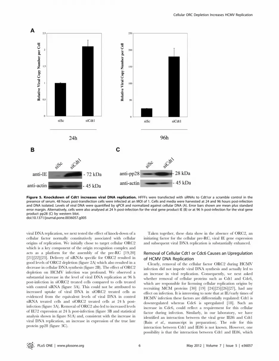
Figure 5. Knockdown of Cdt1 increases viral DNA replication. HFFFs were transfected with siRNAs to Cdt1or a scramble control in thepresence of serum. 48 hours post-transfection cells were infected at an MOI of 1. Cells and media were harvested at 24 and 96 hours post-infectionand DNA isolated. Levels of viral DNA were quantified by qPCR and normalized against cellular DNA (A). Error bars shown are mean plus standarderror margin. Alternatively, cells were also analysed at 24 h post-infection for the viral gene product IE (B) or at 96 h post-infection for the viral geneproduct pp28 (C) by western blot.doi:10.1371/journal.pone.0036057.g005
Cellular ORC Depletion Increases HCMV Replication
PLoS ONE | www.plosone.org 6 May 2012 | Volume 7 | Issue 5 | e36057

itself binds close to the viral origin of replication, results in
recruitment of Cdt1 to viral origins of replication, perhaps
facilitating viral DNA replication. Alternatively, given that steady
state levels of Cdt1 are reduced during HCMV infection [18],
IE86 may sequester Cdt1 to prevent functions of Cdt1 which are
perhaps detrimental to viral replication.
To address this, we first ensured that we were able to remove
Cdt1 or Cdc6 by RNAi and confirmed the effects of their removal
on cellular DNA synthesis. Figure 4A clearly shows that Cdt1 and
Cdc6 mRNA levels were decreased following specific siRNA
treatment. Figure 4B also shows that removal of either Cdt1 or
Cdc6 led to a considerable decrease in the ability of cells to
synthesis cellular DNA.
In contrast, removal of Cdt1 during the course of a virus
infection had profound effects on viral DNA replication. Figure 5A
shows that treatment of cells with siRNAs specific for Cdt1
resulted in a major increase in virus genomes at 96 post-infection
compared to cells treated with scrambled siRNA, although we
note that knock-down of Cdt1 resulted in a small increase in
delivery of viral genomes (about 2 fold, see figure 5A). Interest-
ingly, removal of Cdt1 during infection also led to a profound
increase in levels of IE72 expression at 24 h post-infection
(figure 5B and statistical analysis shown in figure S1A) and,
consistent with increased levels of viral DNA replication, also
resulted in increased levels of the true late pp28 protein at 96 h
post-infection (figure 5C).
Similar to removal of Cdt1, removal of Cdc6 during infection
(figure 6) also resulted in increased viral DNA replication
(figure 6A) with a concomitant increase in IE72 expression at
24 h post-infection (figure 6B and statistical analysis shown in
figure S1A) and, consistent with an increase in viral DNA
replication, increased pp28 expression at 96 h post-infection
(figure 6C). However, the level of increase in IE gene expression,
viral DNA replication and pp28 expression was not as high as that
induced by removal of Cdt1.
Removal of Cellular MCM7 Causes an upregulation ofHCMV DNA Replication
MCM7 is an integral component of the MCM complex which is
essential for both initiation and elongation of DNA replication and
is recruited as part of the pre-RC during cellular DNA replication
[29][30] [28]. An integral function of the MCM complex is
helicase activity and once MCM complex has been recruited to
the cellular origin of replication a fully licensed replication origin
exists. Although HCMV encodes its own replicative helicase-
primase complex in the viral UL70, UL102, and UL105 gene
products [38][39], it is not known if cellular MCMs are required
for viral DNA replication. Consequently, we also analysed HCMV
DNA replication in cells in which MCM7 had been depleted by
Figure 6. Knockdown of Cdc6 increases viral DNA replication. HFFFs were transfected with siRNA Cdc6 or a scramble control in the presenceof serum. 48 hours post-transfection cells were infected at an MOI of 1. Cells and media were harvested at 24 and 96 hours post-infection and DNAisolated. Levels of viral DNA were quantified by qPCR and normalized against cellular DNA. Error bars shown are mean plus standard error margin (A).Alternatively, cells were also analysed at 24 h post-infection for the viral gene product IE (B) or at 96 h post-infection for the viral gene product pp28(C) by western blot.doi:10.1371/journal.pone.0036057.g006
Cellular ORC Depletion Increases HCMV Replication
PLoS ONE | www.plosone.org 7 May 2012 | Volume 7 | Issue 5 | e36057

RNAi. To test whether MCM7 is required for the replication of
viral DNA, siRNA molecules were designed to target MCM7.
Figure 7A shows that treatment of cells with MCM7 specific
siRNAs led to a good reduction in steady state levels of MCM7
protein by western blot analysis. Consistent with the known effects
of MCM7 depletion [40], this also led to a profound inhibition of
cellular DNA synthesis (figure 7B).
Nevertheless, this RNAi-mediated reduction in MCM7 during
infection with HCMV once again led to a robust increase in
cellular DNA synthesis at 96 h post-infection (figure 8A) which was
not due to differences in input genome and also led to concomitant
increases in IE72 expression at 24 h post-infection (figure 8B and
statistical analysis shown in figure S1A) and pp28 expression at
96 h post-infection (figure 8C).
In the experiments so far, knock-downs of specific pre-RC
components were carried out in a background of serum which
would otherwise provide the necessary cellular conditions required
for cellular DNA replication. We, therefore, asked whether the
effects of knock-down of pre-RC components in serum-depleted
conditions would also result in similar effects on viral IE gene
expression. Intriguingly, knock-down of pre-RC components in
serum-free medium showed only marginal effects on viral IE gene
expression (see figure S2). One interpretation of this is that under
conditions where there is no competition between the virus and
the cell for cellular factors required for DNA replication, because
cellular DNA replication is already potently inhibited by serum-
deprivation, additional knock-down of specific pre-RC compo-
nents have little additional effects.
Taken together, the data suggest that knock-down of ORC2,
Cdt1 and Cdc6 or MCM7 in a background of conditions
otherwise conducive to cell proliferation, leads to an enhancement
of viral DNA replication.
Discussion
The pre-RC represents a key component of the cellular
replication machinery. The complex assembles from its compo-
nents in a stepwise manner onto cellular DNA replication origin.
This culminates in phosphorylation of the MCMs which leads to
origin melting and recruitment of the DNA polymerase [41].
Cellular pre-RC proteins have been shown to be involved in
herpesvirus latent replication. Both MCM and ORC proteins have
been shown by ChIP assays to bind EBV oriP [42][43] where they
appear to be recruited to the origin by EBNA1 [44]. ORC and
MCMs have also been shown to play a role at the latent origin of
KSHV [45][46][47]. However, to date, it is not known if such
cellular factors are required for herpesvirus lytic replication.
In, for example, herpes simplex virus (HSV) lytic replication,
viral DNA synthesis is initiated by an origin binding protein, UL9.
HCMV does not encode a de facto origin binding protein, so the
exact mechanism by which it initiates viral lytic DNA replication is
unclear. Although, recently, it has been shown that HCMV UL84
can interact with the viral origin of replication and may function as
a replication initiator via interactions with C/EBP [38,48] [49],
other work has shown that UL84 is dispensible for HCMV lytic
replication [50]. Consequently, we have analysed whether
initiation of HCMV DNA replication involves host cell pre-RC
factors.
We tested whether viral DNA replication required factors
involved in three defined stages of cellular pre-RC formation;
these were an initiation factor (ORC2), two recruiting factors
(Cdt1 and Cdc6) and a recruited factor (MCM7).
Our data clearly showed that none of the components of the
cellular pre-RC were required for viral DNA replication. On the
contrary, removal of cellular components required for any stage of
cellular pre-RC formation, resulting in inhibition of cellular DNA
replication, actually led to robust increases in viral DNA
replication. It is well established that HCMV infection results in
cell cycle arrest, suggesting that cellular DNA replication may
compete against viral DNA replication [10][11][12] [51] [17]
[52][53].
Thus, compromising cellular DNA replication by removing
cellular components required for cellular DNA synthesis likely
results in a cellular environment which is more conducive to viral
DNA replication.
Figure 7. MCM7 knock-down reduces cellular DNA synthesis, as expected. Cells were transfected with control siRNA, or transfected with asiRNA to MCM7. 48 hours post-transfection cells and media were harvested by trypsinisation and centrifugation. Cells were analysed by western blotfor MCM7 content (A). Additionally, mock treated cells or cells transfected with siRNAs were maintained for 72 hours post-transfection in EMEM-washand then BrdU was added to the cell media for 4 hours after which cells were fixed and stained for BrdU incorporation and samples were analysed byimmunofluorescence. 5 independent fields of view were counted for BrdU staining and the percentage of BrdU positive cells was calculated. Errorbars are mean plus standard error margin (B). Numbers of BrdU positive cells are also shown for cells deprived of serum (minus serum) (B).doi:10.1371/journal.pone.0036057.g007
Cellular ORC Depletion Increases HCMV Replication
PLoS ONE | www.plosone.org 8 May 2012 | Volume 7 | Issue 5 | e36057

We also noted that knock-down of ORC2, Cdt1, Cdc6 or
MCM7 as well as resulting in increased viral DNA replication
resulted in robust increases in IE72 expression at IE times of
infection - this could not be explained by, for instance, an
increase in delivery of viral genome to cells lacking these cellular
replication proteins. It is known that HCMV only initiates IE
gene expression in cells in G0/G1 or very early S phase
[54,55,56] and it is well established that depletion of ORC2,
Cdt1, Cdc6 or MCM7 can result in cell cycle arrest at G0/G1
[40] [6,57,58,59,60,61,62]. Consequently, we tested whether cells
treated with specific siRNAs to pre-RC factors under the
conditions we used for our infection assays, resulted in major
differences in the number of cells in G0/G1. Delivery of siRNAs
to ORC2, Cdt1, Cdc6 or MCM7 did result in low but consistent
increases in cells in G0/G1 (see figure S3) which may explain, at
least in part, the increased levels of IE expression in these cells.
However, knock-down of Cdt1 routinely resulted in the highest
increase in IE72 expression (see Figure 5B and figure S1A) and,
while this may have been partly due a minor increase in genome
delivery, Cdt1 knockdown did not routinely result in the highest
increase in cells in G0/G1 - the highest increase in cells in G0/
G1 actually resulted from treatment with siMCM7 (see figure 7
and figure S3) yet siMCM7 treated cells did not result in the
highest increase in IE gene expression.
Consequently, we believe other effects besides increases in
numbers of cells in G0/G1, per se, are responsible for increased
efficiency of IE gene expression and viral DNA replication. These
analyses are presently ongoing.
In conclusion, we have shown that the independent depletion
of a number of cellular pre-RC components, which leads to arrest
of cellular DNA synthesis, does not prevent HCMV DNA
synthesis. On the contrary, removal of ORC2, Cdt1,Cdc6 or
MCM7 led to increased viral DNA synthesis. The reasons for this
are unclear, however, it is possible that arrest of cellular DNA
synthesis enhances the availability of other, as yet unidentified
components important for viral IE expression and viral DNA
synthesis which are normally abundant during G0/G1 phase of
the cell cycle.
Supporting Information
Figure S1 Quantification of the effects of knockdown ofUL44, Cdt1, Cdc6, MCM7 or ORC2 on IE72 expression.A) HFFFs were transfected with siRNAs as indicated or a scramble
control in the presence of serum. 24 hours post-infection (at an
M.O.I. of one), cells were analysed by western blot for IE
expression. Data represent triplicate experiments averaged to
GAPDH levels and analysed using Image J freeware.
(TIF)
Figure S2 Cells halted at G0 by serum-starvation in theabsence of Cdc6, Cdt1 or MCM7 show marginalincreases in IE and pp28 expression during HCMVinfection. A) Cells transfected with siRNAs were maintained in
Figure 8. Knockdown of MCM7 increases viral DNA replication. HFFFs were transfected with siRNAs to MCM7 or a scramble control in thepresence of serum. 48 hours post-transfection cells were infected at an MOI of 1. Cells and media were harvested at 24 and 96 hours post-infectionand DNA isolated. Levels of viral DNA were quantified by qPCR and normalized against cellular DNA. Error bars shown are mean plus standard errormargin (A). Alternatively, at 24 h post-infection cells were analysed for the viral gene product IE (B) or at 96 h post-infection for the viral gene productpp28 (C) by western blot.doi:10.1371/journal.pone.0036057.g008
Cellular ORC Depletion Increases HCMV Replication
PLoS ONE | www.plosone.org 9 May 2012 | Volume 7 | Issue 5 | e36057

EMEM-wash for 24 h then infected with HCMV. 24 h or 96 h
post-infection, IE and pp28 protein was analysed by western blot,
respectively. B) Relative levels of IE proteins in (A) were quantified
using Image J freeware after normalisation to actin levels.
(TIF)
Figure S3 Effects of knockdown of ORC2, MCM7, Cdt1,and Cdc6 on cell cycle. HFFFs were transfected with siRNAs as
labelled. After this, cells were cultured in EMEM-10 for 48 h and
then stained with PI and cell cycle analysis was assayed by flow
cytometry after gating for single cells. Percentages of cells in the
G0/G1 phases of the cell cycle were calculated using winMDi
software. Untreated cells in the absence of serum (minus serum)
are also shown for comparison.
(TIF)
Acknowledgments
We thank Linda Teague and Joan Baillie for technical assistance.
Author Contributions
Conceived and designed the experiments: TEB JS. Performed the
experiments: TEB. Analyzed the data: EP TEB JS. Contributed
reagents/materials/analysis tools: JS. Wrote the paper: EP TEB JS.
References
1. Iskenderian AC, Huang L, Reilly A, Stenberg RM, Anders DG (1996) Four of
eleven loci required for transient complementation of human cytomegalovirusDNA replication cooperate to activate expression of replication genes. J Virol
70(1): 383–392.
2. Chee MS, Satchwell SC, Preddie E, Weston KM, Barrell BG (1990) Human
cytomegalovirus encodes three G protein-coupled receptor homologues. Nature344(6268): 774–777.
3. Ertl PF, Powell KL (1992) Physical and functional interaction of human
cytomegalovirus DNA polymerase and its accessory protein (ICP36) expressed in
insect cells. J Virol 66(7): 4126–4133.
4. Ertl PF, Thomas MS, Powell KL (1991) High level expression of DNApolymerases from herpesviruses. J Gen Virol 72 ( Pt 7): 1729–1734.
5. Appleton BA, Brooks J, Loregian A, Filman DJ, Coen DM, et al. (2006) Crystalstructure of the cytomegalovirus DNA polymerase subunit UL44 in complex
with the C terminus from the catalytic subunit. Differences in structure andfunction relative to unliganded UL44. J Biol Chem 281(8): 5224–5232.
6. Da-Silva LF, Duncker BP (2007) ORC function in late G1: maintaining thelicense for DNA replication. Cell Cycle 6(2): 128–130.
7. Park MY, Kim YE, Seo MR, Lee JR, Lee CH, et al. (2006) Interactions amongfour proteins encoded by the human cytomegalovirus UL112–113 region
regulate their intranuclear targeting and the recruitment of UL44 toprereplication foci. J Virol 80(6): 2718–2727.
8. Gao Y, Colletti K, Pari GS (2008) Identification of human cytomegalovirusUL84 virus- and cell-encoded binding partners by using proteomics analysis.
J Virol 82(1): 96–104.
9. Colletti KS, Smallenburg KE, Xu Y, Pari GS (2007) Human cytomegalovirus
UL84 interacts with an RNA stem-loop sequence found within the RNA/DNAhybrid region of oriLyt. J Virol 81(13): 7077–7085.
10. Bresnahan WA, Albrecht T, Thompson EA (1998) The cyclin E promoter is
activated by human cytomegalovirus 86-kDa immediate early protein. J Biol
Chem 273(34): 22075–22082.
11. Hagemeier C, Caswell R, Hayhurst G, Sinclair J, Kouzarides T (1994)Functional interaction between the HCMV IE2 transactivator and the
retinoblastoma protein. Embo J 13(12): 2897–2903.
12. Song YJ, Stinski MF (2002) Effect of the human cytomegalovirus IE86 protein
on expression of E2F-responsive genes: a DNA microarray analysis. Proc NatlAcad Sci U S A 99(5): 2836–2841.
13. Wiebusch L, Uecker R, Hagemeier C (2003) Human cytomegalovirus preventsreplication licensing by inhibiting MCM loading onto chromatin. EMBO Rep
4(1): 42–46.
14. Castillo JP, Yurochko AD, Kowalik TF (2000) Role of human cytomegalovirus
immediate-early proteins in cell growth control. J Virol 74(17): 8028–8037.
15. Murphy EA, Streblow DN, Nelson JA, Stinski MF (2000) The humancytomegalovirus IE86 protein can block cell cycle progression after inducing
transition into the S phase of permissive cells. J Virol 74(15): 7108–7118.
16. Sinclair J, Baillie J, Bryant L, Caswell R (2000) Human cytomegalovirus
mediates cell cycle progression through G(1) into early S phase in terminally
differentiated cells. J Gen Virol 81(Pt 6): 1553–1565.
17. Wiebusch L, Hagemeier C (2001) The human cytomegalovirus immediate early2 protein dissociates cellular DNA synthesis from cyclin-dependent kinase
activation. Embo J 20(5): 1086–1098.
18. Biswas N, Sanchez V, Spector DH (2003) Human cytomegalovirus infection
leads to accumulation of geminin and inhibition of the licensing of cellular DNAreplication. J Virol 77(4): 2369–2376.
19. DePamphilis ML (2003) The ‘ORC cycle’: a novel pathway for regulatingeukaryotic DNA replication. Gene 310: 1–15.
20. Bell SP, Kobayashi R, Stillman B (1993) Yeast origin recognition complex
functions in transcription silencing and DNA replication. Science 262(5141):
1844–1849.
21. Foss M, McNally FJ, Laurenson P, Rine J (1993) Origin recognition complex(ORC) in transcriptional silencing and DNA replication in S. cerevisiae. Science
262(5141): 1838–1844.
22. Micklem G, Rowley A, Harwood J, Nasmyth K, Diffley JF (1993) Yeast origin
recognition complex is involved in DNA replication and transcriptionalsilencing. Nature 366(6450): 87–89.
23. Radichev I, Kwon SW, Zhao Y, DePamphilis ML, Vassilev A (2006) Genetic
analysis of human Orc2 reveals specific domains that are required in vivo forassembly and nuclear localization of the origin recognition complex. J Biol
Chem 281(32): 23264–23273.
24. Randell JC, Bowers JL, Rodriguez HK, Bell SP (2006) Sequential ATP
hydrolysis by Cdc6 and ORC directs loading of the Mcm2–7 helicase. Mol Cell21(1): 29–39.
25. Speck C, Chen Z, Li H, Stillman B (2005) ATPase-dependent cooperative
binding of ORC and Cdc6 to origin DNA. Nat Struct Mol Biol 12(11): 965–971.
26. Nishitani H, Taraviras S, Lygerou Z, Nishimoto T (2001) The human licensing
factor for DNA replication Cdt1 accumulates in G1 and is destabilized afterinitiation of S-phase. J Biol Chem 276(48): 44905–44911.
27. Wohlschlegel JA, Dwyer BT, Dhar SK, Cvetic C, Walter JC, et al. (2000)Inhibition of eukaryotic DNA replication by geminin binding to Cdt1. Science
290(5500): 2309–2312.
28. Ishimi Y, Okayasu I, Kato C, Kwon HJ, Kimura H, et al. (2003) Enhanced
expression of Mcm proteins in cancer cells derived from uterine cervix.Eur J Biochem 270(6): 1089–1101.
29. Ying CY, Gautier J (2005) The ATPase activity of MCM2–7 is dispensable forpre-RC assembly but is required for DNA unwinding. Embo J 24(24):
4334–4344.
30. You Z, Masai H (2008) Cdt1 forms a complex with the minichromosome
maintenance protein (MCM) and activates its helicase activity. J Biol Chem283(36): 24469–24477.
31. Qian Z, Leung-Pineda V, Xuan B, Piwnica-Worms H, Yu D (2008) Human
cytomegalovirus protein pUL117 targets the mini-chromosome maintenance
complex and suppresses cellular DNA synthesis. PLoS Pathog 6(3): e1000814.
32. Griffin C, Wang EC, McSharry BP, Rickards C, Browne H, et al. (2005)Characterization of a highly glycosylated form of the human cytomegalovirus
HLA class I homologue gpUL18. J Gen Virol 86(Pt 11): 2999–3008.
33. Poole E, King CA, Sinclair JH, Alcami A (2006) The UL144 gene product of
human cytomegalovirus activates NFkappaB via a TRAF6-dependent mecha-nism. Embo J 25(18): 4390–4399.
34. Lau E, Zhu C, Abraham RT, Jiang W (2006) The functional role of Cdc6 in S-G2/M in mammalian cells. EMBO Rep 7(4): 425–430.
35. Bennett NJ, Ashiru O, Morgan FJ, Pang Y, Okecha G, et al. (2010) Intracellular
sequestration of the NKG2D ligand ULBP3 by human cytomegalovirus.
J Immunol 185(2): 1093–1102.
36. Ripalti A, Boccuni MC, Campanini F, Landini MP (1995) Cytomegalovirus-mediated induction of antisense mRNA expression to UL44 inhibits virus
replication in an astrocytoma cell line: identification of an essential gene. J Virol
69(4): 2047–2057.
37. Depto AS, Stenberg RM (1992) Functional analysis of the true late humancytomegalovirus pp28 upstream promoter: cis-acting elements and viral trans-
acting proteins necessary for promoter activation. J Virol 66(5): 3241–3246.
38. Pari GS (2008) Nuts and bolts of human cytomegalovirus lytic DNA replication.
Curr Top Microbiol Immunol 325: 153–166.
39. Woon HG, Scott GM, Yiu KL, Miles DH, Rawlinson WD (2008) Identification
of putative functional motifs in viral proteins essential for human cytomegalo-virus DNA replication. Virus Genes 37(2): 193–202.
40. Crevel G, Hashimoto R, Vass S, Sherkow J, Yamaguchi M, et al. (2007)Differential requirements for MCM proteins in DNA replication in Drosophila
S2 cells. PLoS One 2(9): e833.
41. Sun J, Kong D (2010) DNA replication origins, ORC/DNA interaction, and
assembly of pre-replication complex in eukaryotes. Acta Biochim Biophys Sin(Shanghai) 42(7): 433–439.
42. Chaudhuri B, Xu H, Todorov I, Dutta A, Yates JL (2001) Human DNA
replication initiation factors, ORC and MCM, associate with oriP of Epstein-
Barr virus. Proc Natl Acad Sci U S A 98(18): 10085–10089.
43. Dhar SK, Yoshida K, Machida Y, Khaira P, Chaudhuri B, et al. (2001)Replication from oriP of Epstein-Barr virus requires human ORC and is
inhibited by geminin. Cell 106(3): 287–296.
44. Schepers A, Ritzi M, Bousset K, Kremmer E, Yates JL, et al. (2001) Human
origin recognition complex binds to the region of the latent origin of DNAreplication of Epstein-Barr virus. Embo J 20(16): 4588–4602.
Cellular ORC Depletion Increases HCMV Replication
PLoS ONE | www.plosone.org 10 May 2012 | Volume 7 | Issue 5 | e36057

45. Lim C, Sohn H, Lee D, Gwack Y, Choe J (2002) Functional dissection of
latency-associated nuclear antigen 1 of Kaposi’s sarcoma-associated herpesvirusinvolved in latent DNA replication and transcription of terminal repeats of the
viral genome. J Virol 76(20): 10320–10331.
46. Stedman W, Deng Z, Lu F, Lieberman PM (2004) ORC, MCM, and histonehyperacetylation at the Kaposi’s sarcoma-associated herpesvirus latent replica-
tion origin. J Virol 78(22): 12566–12575.47. Verma SC, Bajaj BG, Cai Q, Si H, Seelhammer T, et al. (2006) Latency-
associated nuclear antigen of Kaposi’s sarcoma-associated herpesvirus recruits
uracil DNA glycosylase 2 at the terminal repeats and is important for latentpersistence of the virus. J Virol 80(22): 11178–11190.
48. Gao Y, Pari GS (2009) Interaction of human cytomegalovirus pUL84 withcasein kinase 2 is required for oriLyt-dependent DNA replication. J Virol 83(5):
2393–2396.49. Kagele D, Gao Y, Smallenburg K, Pari GS (2009) Interaction of HCMV UL84
with C/EBPalpha transcription factor binding sites within oriLyt is essential for
lytic DNA replication. Virology 392(1): 16–23.50. Spector DJ, Yetming K (2010) UL84-independent replication of human
cytomegalovirus strain TB40/E. Virology 407(2): 171–177.51. Petrik DT, Schmitt KP, Stinski MF (2006) Inhibition of cellular DNA synthesis
by the human cytomegalovirus IE86 protein is necessary for efficient virus
replication. J Virol 80(8): 3872–3883.52. Noris E, Zannetti C, Demurtas A, Sinclair J, De Andrea M, et al. (2002) Cell
cycle arrest by human cytomegalovirus 86-kDa IE2 protein resembles prematuresenescence. J Virol 76(23): 12135–12148.
53. Jault FM, Jault JM, Ruchti F, Fortunato EA, Clark C, et al. (1995)Cytomegalovirus infection induces high levels of cyclins, phosphorylated Rb,
and p53, leading to cell cycle arrest. J Virol 69(11): 6697–6704.
54. Fortunato EA, Sanchez V, Yen JY, Spector DH (2002) Infection of cells with
human cytomegalovirus during S phase results in a blockade to immediate-early
gene expression that can be overcome by inhibition of the proteasome. J Virol
76(11): 5369–5379.
55. White EA, Spector DH (2007) Early viral gene expression and function. In:
Arvin A, Campadelli-Fiume G, Mocarski E, Moore PS, Roizman B, et al.,
editors. Human Herpesviruses: Biology, Therapy, and Immunoprophylaxis.
Cambridge: Cambridge University Press; Chapter 18.
56. Salvant BS, Fortunato EA, Spector DH (1998) Cell cycle dysregulation by
human cytomegalovirus: influence of the cell cycle phase at the time of infection
and effects on cyclin transcription. J Virol 72(5): 3729–3741.
57. Christensen TW, Tye BK (2003) Drosophila MCM10 interacts with members of
the prereplication complex and is required for proper chromosome condensa-
tion. Mol Biol Cell 14(6): 2206–2215.
58. Shen Z, Sathyan KM, Geng Y, Zheng R, Chakraborty A, et al. (2010) A WD-
repeat protein stabilizes ORC binding to chromatin. Mol Cell 40(1): 99–111.
59. Nevis KR, Cordeiro-Stone M, Cook JG (2009) Origin licensing and p53 status
regulate Cdk2 activity during G(1). Cell Cycle 8(12): 1952–1963.
60. Prasanth SG, Mendez J, Prasanth KV, Stillman B (2004) Dynamics of pre-
replication complex proteins during the cell division cycle. Philos Trans R Soc
Lond B Biol Sci 359(1441): 7–16.
61. Machida YJ, Teer JK, Dutta A (2005) Acute reduction of an origin recognition
complex (ORC) subunit in human cells reveals a requirement of ORC for Cdk2
activation. J Biol Chem 280(30): 27624–27630.
62. Rialland M, Sola F, Santocanale C (2002) Essential role of human CDT1 in
DNA replication and chromatin licensing. J Cell Sci 115(Pt 7): 1435–1440.
Cellular ORC Depletion Increases HCMV Replication
PLoS ONE | www.plosone.org 11 May 2012 | Volume 7 | Issue 5 | e36057


