Embed Size (px)
Citation preview

Hoatlin Fall 2009!
DNA Replication!
Instructor: Maureen Hoatlin!• Biochemistry and Molecular Biology!• Email: [email protected] !

Public Service Announcement!
Hoatlin Fall 2009!

Hoatlin Fall 2009!
Lecture Overview!• DNA Review!• Essential components of the replisome!
– Genetic analysis identifies components!• Polymerases, SSB proteins, clamps and
clamp loaders, helicases!• How DNA replication proceeds !• How DNA replication starts!• What is known and what remains to be
discovered!

Hoatlin Fall 2009!
Significance of DNA Replication!
• At least 38 diseases are caused by defects in DNA replication, 40 by mutations in genes required for DNA replication or repair!
• Many drugs used to treat diseases caused by viruses are targeted to DNA replication!
• Many chemotherapy agents are targeted to DNA replication!
• Deconstructing DNA replication is central to efforts aimed at developing new diagnostic tools and new treatments for cancer!

Hoatlin Fall 2009!
From Alberts text!
• Long strands of polymerized nucleotides!
• Base, sugar and a phosphate!
General Structure of DNA!

Hoatlin Fall 2009!
DNA Bases are Paired!• A pairs with T, C pairs with G!• Purine/pyrimidine pairing results in a consistent overall
dimension of the DNA duplex!• Base pairing is stabilized by hydrogen bonds; G:C rich
helices are more stable than A:T rich ones!• Helix dimension has implications for DNA replication
fidelity and for detection of certain types of DNA damage!
2 H bonds! 3 H bonds!

Hoatlin Fall 2009!

Hoatlin Fall 2009!
Two DNA Strands are twisted together in a helix, called a double helix!
Sugar phosphates are on outside of helix, bases on the inside!
A bulky two-ring base (purine; A&G) is always paired with a single-ring base (pyrimidine; T &C). Heterocyclic rings are flat.!
Helix is stabilized by hydrogen bonding between the bases and stacking forces!
Nucleotides are linked by covalent phosphodiester bonds (3’-OH to 5’-PO4)!
DNA ends have chemically- defined polarity!
C#3’!
C#5’!
2’!3’!
DNA Strands form an anti-parallel helix!
1’!
5’!o!
4’!
N!base!
N-glycosidic bond!sugar!
Note: review chemical bonds of helix!

Hoatlin Fall 2009!
Base-Pairing Underlies DNA Replication and Repair!
Each strand can act as a template for duplication-nucleotide A pairs with T, and G with C!
Each template nucleotide is recognized by a free complementary nucleotide!

Hoatlin Fall 2009!
Overview of Replication!
• Initiation (covered in lecture #2)!– Recognition of an origin by a complex of proteins.
Parental strand separation and stabilization. Synthesis can start at the replication fork.!
• Elongation (covered in lecture #1)!– the parental strands unwind, the replisome (complex
of proteins) moves along DNA, daughter strands synthesized!
• Termination (covered in lecture #2)!– Termination reactions preceed separation of duplicated
chromosomes!

Hoatlin Fall 2009!
Our Attention!is here!

Hoatlin Fall 2009!
Mutation Rates Are Very Low!
Bacteria-estimates from rapidly growing cultures are 1 nucleotide change in 109 per cell generation!
Mammals-similar rates per round of replication. Ex: human genome (3 x 10⁹) could have 3 mutations/cell division !
Some nucleotide changes are silent, while others can have major consequences for the species or the individual. !
Maintaining genomic integrity is dependent on accuracy of duplication and distribution of DNA to daughter cells, and on response to and repair of DNA damage.!
Hoatlin Fall 2009!
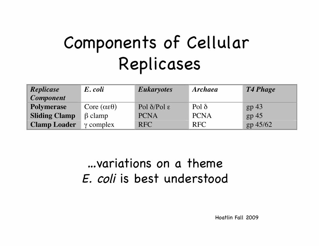
Components of Cellular Replicases!
ReplicaseComponent
E. coli Eukaryotes Archaea T4 Phage
Polymerase Core (αεθ) Pol δ/Pol ε Pol δ gp 43Sliding Clamp β clamp PCNA PCNA gp 45Clamp Loader γ complex RFC RFC gp 45/62
…variations on a theme!E. coli is best understood!

Hoatlin Fall 2009!
Dissecting Replication!
• dna mutants: large set of E. coli mutants that distinguish two stages of replication!
• Inability to replicate is lethal. Mutants in replication are obtained as conditional lethals. These mutants are able to replicate under permissive conditions (e.g., 25°) but are defective under non-permissive conditions (e.g., 39°).!

Hoatlin Fall 2009!
dna Mutants!• Quick stop mutants (majority)!
– Cease replication immediately under non-permissive temperatures!
– Typically defective in components of the replication apparatus (enzymes needed for elongation, or essential precursors)!
• Slow stop mutants (minority)!– Complete current round of replication but
cannot start another!– Defective in events leading to initiation of
replication at the origin!Examples to follow!

Hoatlin Fall 2009!
• In vitro complementation!
• Bacterial replication proteins purified, eukaryotic replication systems analogous but incomplete and unsatisfying.!
Identifying the Components of the Replication Apparatus!
Replicating extract from wild type cells!
Replicating extract from mutants (defective) non-
permissive conditions!
Assay for restored activity!Protein coded for by dna locus can be purified by
fractionation to isolate active component in the extract !

Hoatlin Fall 2009!
DNA Polymerases make DNA!
• Two basic types of DNA synthesis!– Semiconservative replication!– DNA synthesis in a DNA repair reaction!
Damaged base is excised and new DNA
is synthesized!

Hoatlin Fall 2009!
DNA Polymerases Control the Fidelity of Replication!
DNA is synthesized in both semi-conservative replication and in repair reactions !
A bacterium or eukaryotic cell has several different DNA polymerase enzymes!
One bacterial DNA polymerase undertakes semi-conservative replication; the others are involved in repair reactions!
Eukaryotic nuclei, mitochondria, and chloroplasts each have a single unique DNA polymerase required for replication, and other DNA polymerases involved in ancillary or repair activities!

Hoatlin Fall 2009!
Pol III is the DNA replicase responsible for new strands of DNA in E. coli. Pol I also contributes, while others participate in specific DNA damage repair!
Organisms Contain Many Types of Polymerases!
Multi-subunit:!dnaE-DNA synthetic activity!dnaQ- 3’-5’ exo!

Hoatlin Fall 2009!
E.coli pol III part of a large assembly: Replicase!
Functional Mass component Subunit (kDa) Gene Activity Core polymerase α 130 polC (dnaE) 5' to 3' polymerase (aka, Pol III) ε 27.5 dnaQ (mutD) 3'-5' exonuclease
θ 10 holE Stimulates ε exonuclease Gamma complex τ 71 dnaX Dimerizes cores (Clamp loader/ γ 45.5 dnaX Binds ATP ATPase) δ 35 holA Binds to β δ' 33 holB Binds to γ and β
χ 15 holC Binds to SSB ψ 12 holD Binds to χ and γ
Sliding clamp β 40.6 dnaN Processivity factor
Consider the polymerase…!

Hoatlin Fall 2009!
The addition of a deoxyribonucleotide to the 3’ end of a primer strand !
Polymerases Perform the Fundamental Reaction in DNA Synthesis!
4! 1!2!3!
5!
1!2!3!

Hoatlin Fall 2009!
DNA Synthesis!
Release of pyrophosphate followed by its hydrolysis to inorganic phosphate!

Hoatlin Fall 2009!
Features of a DNA Polymerase!Many DNA polymerases have a large cleft composed of three domains that resemble a hand !(if you use your imagination)!
DNA lies across the "palm" in a groove created by the "fingers" and "thumb”-this is a good conceptual figure!
DNA pols fall into 5 families based on sequence homologies. Palm is well conserved, thumb and fingers are analogous!

Hoatlin Fall 2009!
Replication Fork: Region of Active DNA Synthesis !
DNA polymerases are in multi-enzyme complexes which synthesize the DNA of both daughter strands!
How are daughter stands synthesized?!

Hoatlin Fall 2009!
Possibilities for Replication Mechanism !

Hoatlin Fall 2009!
Meselson-Stahl Experiment!
• Parental DNA carries a “heavy” density label because organism grown in suitable isotope!
• Switch to medium containing “light” isotope for replication!
• Analyze replicated DNA in CsCl density gradients !

Hoatlin Fall 2009!
• Parental DNA consists of a duplex of two heavy strands!• Generation 1= one heavy parental strand and one light daughter strand!• Generation 2= half hybrid and half light!

Hoatlin Fall 2009!This is what happens--but what is the mechanism?!

Hoatlin Fall 2009!
The Simple Model Fails!Exactly how are nucleotides added to the anti-parallel strands?!
Requires one strand to grow in 5’-to-3’ direction, while the other strand grows in the 3’-to-5’ direction!
But DNA polymerase requires an incoming nucleotide in a 5’-to-3’ direction!

Hoatlin Fall 2009!
The Replication Fork is Asymmetric !
Leading strand: synthesized *almost* continuously !
Lagging strand: discontinuous synthesis. Okazaki fragments are polymerized in 5’-to-3’ direction and stitched together after synthesis. Lagging, or delayed, because it must wait for leading strand to expose template to synthesize Okazaki fragment (more on this later)!
nb!
Parental DNA!
Single strand!
Fork movement!
1-3 kb!

Hoatlin Fall 2009!
Hi-Fi Replication Facts!• During synthesis mispairing is inevitable!• Correct nuc has a higher affinity for moving
polymerase compared to incorrect--e.g., correct base-pairing, correct shape!
• Incorrectly bound nucleotide more likely to dissociate as polymerase undergoes conformational change after binding but before covalent addition of nucleotide to growing chain!
Hoatlin Fall 2009!
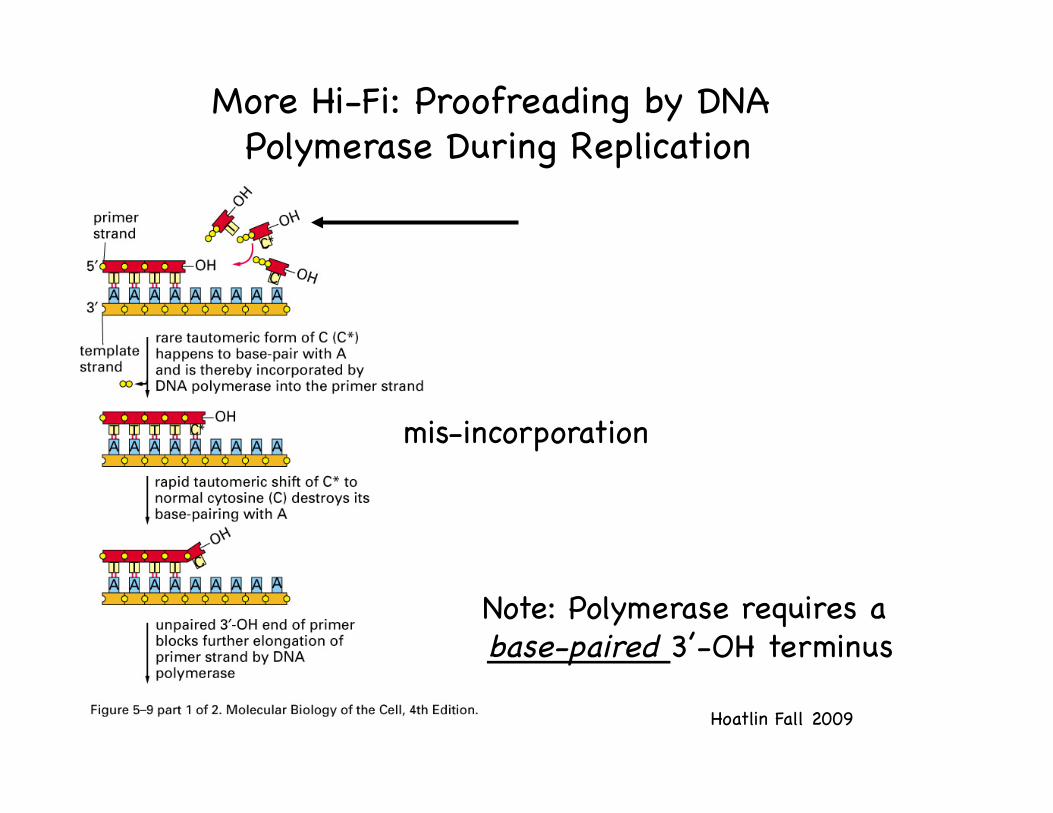
More Hi-Fi: Proofreading by DNA Polymerase During Replication!
mis-incorporation!
Note: Polymerase requires a base-paired 3’-OH terminus!

Hoatlin Fall 2009!
A separate catalytic site (3’-5’ exonuclease) in the polymerase-- or in a sub-unit-- clips off unpaired residues until a base-paired terminus is regenerated!
Fidelity of replication is improved by proofreading by a factor of approx. 100!
All bacterial Pols have 3’-5’ exo activity!
Cont’d---The Hi-Fi DNA Polymerase!

Hoatlin Fall 2009!
DNA Polymerase: Two Modes!
P: polymerization site!E: exonucleolytic site!
“thumb” has moved!

Hoatlin Fall 2009!
Inward rotation of the O helix grasps incoming nuc and creates a catalytic site by rotation of fingers and thumb. !Cyclic: reversed when nuc is incorporated into growing DNA strand--and then nuc moves out of site!When mismatched bp is in catalytic site, fingers cannot rotate toward the palm, leaving 3’ end free to bind active site in exonuclease domain!
Example: Polymerase from T7 Bacteriophage!
Template strand takes a sharp turn so it can be exposed to the incoming nucleotide!

Hoatlin Fall 2009!
DNA Pol I Nick Translation*!• DNA pol I, 103 kD!• Cleavage into two domains: !
– 68 kD (Klenow fragment) with pol and 3’-5’ exonuclease activities !
– 35 kD fragment with 5’-3’ exonucleolytic activity. !
• These 3 functions give DNA pol I unique ability to start replication in vitro at a nick in DNA!
• The nick just moves along (is translated..kind of an unfortunate description in this case)!*very useful at the bench! !

Hoatlin Fall 2009!
DNA Pol’s Needs are Rigid!
• All DNA polymerases require a 3´-OH priming end to initiate DNA synthesis!
• The priming end can be provided by an RNA primer, a nick in DNA, or a priming protein!
• Template strand must be single stranded!

Hoatlin Fall 2009!
Rational Strategy for an anti-HIV Drug !
• Azidodeoxythymidine (AZT) is an analog of thymidine!
• AZT is a reverse transcriptase inhibitor !
• Reverse transcriptase enzymes are polymerases required for retroviral replication !
• The N3 group does not allow DNA synthesis to continue (why?)!
• 100x to 300x affinity for rt vs DNA pol. Gamma DNA pol (mitochondrial pol) is affected with associated side effects!

Hoatlin Fall 2009!
RNA primer: commonly used in cellular DNA replication and some viruses (pre-formed RNA to prime reverse transcription of RNA)--details on next slide!
Nicking: primer terminus generated within duplex DNA. Pre-existing strand displaced by new strand (not degraded, as in “nick translation” --only pol I can do that)!
Protein presents a nucleotide to DNA pol (adenoviral strategy)!
3 Ways To Satisfying Pol’s Need for a 3’ End!

Hoatlin Fall 2009!
DNA Primase: RNA Primer Synthesis!
Synthesizes a short polynucleotide (approx 10) in the 5’-to-3’ direction and then stops!
Uses DNA as a template and builds primer from ribonucleoside triphosphates!
Unlike DNA pol, which requires a primer, DNA primase can start up a new template-guided polynucleotide strand!
Product of the dnaG gene!
Eu [10 nucs long! 100-200 intervals]!
RNA-DNA hybrid!

Hoatlin Fall 2009!
Lagging Strand Synthesis-!Many RNA Primers !
RNA primers have a free 3’-OH, substrate for DNA pol!
Nicks between Okazaki fragments sealed by DNA ligase!
RNA removed, DNA filled in!

Hoatlin Fall 2009!
Helicases: Opening up DNA Double Helix in Front of the Replication Fork!
Hydrolysis of ATP changes shape of helicase in cyclical manner that allows helicase to propel rapidly (1000 nucleotide pairs/sec) along a DNA single strand, prying apart the helix!
12 different helicases in E. coli!
DnaB encircles the lagging strand !
Helical view:!

Hoatlin Fall 2009!
Structure of a DNA Helicase!
Hexamer in a ring form!
Replication fork and helicase to scale!
Bacteriophage T7 replicative helicase (red is ATP). Six identical s.u. bind single strand and duplex DNA alternately and hydrolyze ATP in an ordered fashion to propel molecule along DNA-- a single strand that passes through the central hole!

Motif 4 within the central core of the hexameric gene 4 helicase
Lee S et al. PNAS 2008;105:8908-8913
©2008 by National Academy of Sciences
DnaB-like
helicases!

• Crystal structure of hexameric helicase (viral)!• Showed the path of DNA; Each subunit of the hexamer
contributes a nucleotide binding loop that binds single-stranded DNA on one side of the ring and escorts it through the channel like an escalator!
• Five adjacent subunits escort five adjacent nucleotides and the loop of the sixth subunit is recycled from the exit side to the entry side to pick-up a new nucleotide. !
• the hexamer encircles single-, and not double-, stranded DNA. !
Hoatlin Fall 2009!

Escort Model!
Hoatlin Fall 2009!

Hoatlin Fall 2009!
Helicase Helpers: Single strand DNA-binding (SSB) Proteins!
Bind cooperatively to exposed single strands of DNA !
SSBs do not interfere with base-pairing!
Unable to open helix directly!
Aid helicases by stabilizing unwound DNA-reduce “hairpins”!
ssb mutants have a quick-stop phenotype-defective in repair and recombination as well as in replication!

Hoatlin Fall 2009!
Structure of a Single-Strand Binding Protein (human RPA)!
DNA strand!

Hoatlin Fall 2009!
Coordinating Synthesis of Leading and Lagging Strands!
• Different enzyme units are required to synthesize the leading and lagging strands!
• In E. coli both these units contain the same catalytic subunit (DnaE), supported by other proteins that differ between leading and lagging strands!
• In other organisms, different catalytic subunits may be required for each strand!

Hoatlin Fall 2009!
Replication at the Fork!• "hybrid" of prokaryote and
eukaryote replication proteins and dynamics!
http://www.wehi.edu.au/education/wehi-tv/dna/replication.html!

Hoatlin Fall 2009!
E.coli pol III part of a large assembly: Replicase!
Functional Mass component Subunit (kDa) Gene Activity Core polymerase α 130 polC (dnaE) 5' to 3' polymerase (aka, Pol III) ε 27.5 dnaQ (mutD) 3'-5' exonuclease
θ 10 holE Stimulates ε exonuclease Gamma complex τ 71 dnaX Dimerizes cores (Clamp loader/ γ 45.5 dnaX Binds ATP ATPase) δ 35 holA Binds to β δ' 33 holB Binds to γ and β
χ 15 holC Binds to SSB ψ 12 holD Binds to χ and γ
Sliding clamp β 40.6 dnaN Processivity factor
Consider the polymerase…!

Hoatlin Fall 2009!
DNA polymerase III is a holoenzyme with 3 subcomplexes!
The E. coli replicase DNA polymerase III is a 900 kD complex with a dimeric structure!
Each monomeric unit has a catalytic core, a dimerization subunit, and a processivity (stays on DNA) component!
A clamp loader places the processivity subunits on DNA, and they form a circular clamp around the nucleic acid!
One catalytic core is associated with each template strand!

Hoatlin Fall 2009!
E. Coli Replicase (Pol III)!
10 proteins: ! 2 copies of the catalytic core, each containing
the α subunit (pol activity), ε su (3’-5’ proofreading exonuclease), and θ su (stimulates exonuclease)!
Two copies of dimerizing s.u., τ, which link the catalytic cores!
Two copies of the processivity su, β, clamps which are responsible for holding the catalytic cores onto the template strands!
The γ complex (the clamp loader), five proteins that place the processivity su on DNA !

Hoatlin Fall 2009!
DNA pol III Assembles in Stages!• The β dimer (clamp) plus a γ complex
(clamp loader) recognizes the primer-template and forms pre-initiation complex. Transfer of β su to primed template, Clamp formed by β su !
• Binding to DNA changes conformation of site on β that binds to γ. Now β has high affinity for core pol, and core is brought in to DNA!
• The τ dimer binds to core pol and provides a dimerization that allows second core (and β clamp) to bind!

Hoatlin Fall 2009!
The Sliding Clamp!Water slide!
The core polymerase on the leading strand is processive because the sliding clamp keeps it on the DNA!-ring shaped!

Hoatlin Fall 2009!
Structure of Clamp Proteins!
Clamp protein from E. coli, helix shown through central hole!
(E. coli has four different clamp loader proteins)!
Clamp Protein (β clamp) from E. coli (left) and human clamp
protein, PCNA (right)--very similar!-!
Clamps interact with recombination, repair, and cell cycle proteins!
-!

Hoatlin Fall 2009!
"Clamp Loader Cycle (E. coli)!P
age
1159
Pol Loads!
β clamp!

Hoatlin Fall 2009!
The clamp associated with the core polymerase on the lagging strand dissociates at the end of each Okazaki fragment and reassembles for the next fragment.! While one pol synthesizes leading strand the other cycles within the single-stranded template loop (generated by spooling of leading strand) to synthesize the next fragment.!
γ
α,ε,θ
DnaB!
DnaG!α,ε,θ
τ,τ
A Replication Machine!

Hoatlin Fall 2009!
E. Coli Replicase!
The helicase DnaB is responsible for interacting with the primase DnaG to initiate each Okazaki fragment

Hoatlin Fall 2009!
Meets next Okazaki fragment, falls off!
Helicase DnaB interacts with primase DnaG to signal initiation of Okazaki fragment !
Pol III stays associated with
replicating complex!
Lagging Strand Synthesis: Cycles of !Loading and Unloading DNA Pol and Clamp Proteins!
DNA Pol is released and associates with a new clamp assembled on the RNA primer of the next Okazaki fragment!

General architecture of the helicase–primase complex; coordination!
Hoatlin Fall 2009!K. Marians. Nature Structural & Molecular Biology (2008)!
The lagging strand of the unwound DNA threads through the helicase and is captured by the RNA Pol Domain of one of the associated DnaG molecules; the Zinc binding domain from a different DnaG might participate
in this process. The primer is extruded towards the outside of
the complex.!HBD= helicase binding domain!

Hoatlin Fall 2009!
Replication at the Fork!• This animation was built as a "hybrid" of
prokaryote and eukaryote replication proteins and dynamics. !
• eukaryote system speed is 20-50 bases/second with Okazaki fragments 100-150 bases long. The bacterial (prokaryotic) replisome works about 10 times faster, with Okazaki fragments 700-1000 bases long and the incoming DNA rotating at 10,000RPM!
• two tau subunits (light blue) connect the DNA helicase (dark blue) to the clamp loader (grey fingers), and each tau connects to one of the two polymerase (purple) enzymes. !
• sliding clamps are green and the DNA primase, which is yellow-green, adds RNA primers (yellow) to the ssDNA as it emerges from the helicase. !
http://www.wehi.edu.au/education/wehi-tv/dna/replication.html!
Note single strand DNA (ssDNA) binding proteins that cap the exposed bases were
left out for clarity!

Hoatlin Fall 2009!

Hoatlin Fall 2009!
Rewrite the Textbooks:!Triple Polymerase Replisome!
McInerney at al. Mol Cell 2007. E. coli pol III assembles into a particle that contains THREE DNA polymerases, capable of simultaneous activity. 3 pol is the dominant form.!
Two polymerases on the lagging strand, consistent with T4 replication forks (Nossal et al., 2007) !
Propose: The third polymerase can act as a reserve enzyme to overcome certain types of replication obstacles or to coordinate the slower lagging strand synthesis!

Hoatlin Fall 2009!
Emerging Concepts!
• Textbook view: single DNA polymerases are bound continuously to their substrates!
• Revised view: Polymerases and repair factors are cycled and switched as needed during replication!
• The replisome is not dismantled in this process, transient dissociations occur!
• Some repair processes previously thought to be “postreplictive” may occur during replication when the replisome is paused!
Lovett, Mol Cell 2007!

Hoatlin Fall 2009!

Hoatlin Fall 2009!
Topoisomerase Classes!• Topoisomerase I: relaxes DNA !
-Transient break in one strand of duplex DNA!E. coli: nicking-closing enzyme!
• Topoisomerase II: introduces negative superhelical turns !-Breaks both strands of the DNA and passes
another part of the duplex DNA through the break; then reseals the break.!
-Uses energy of ATP hydrolysis!-Acts as the SWIVEL for DNA replication.!

Hoatlin Fall 2009!
Topoisomerases!• Topoisomerases relieve winding/
unwinding tension of DNA during replication fork movement!
• Topos wrap around DNA, make a cut which permits the helix to spin and relax, then reconnects strands!
• Several drugs interfere with the topoisomerase function. Examples:!– Fluoroquinolone antibiotic interferes
with bacterial type II topoisomerases !– Topoisomerase II (TOP2) is the target
of several important classes of anticancer drugs, including the epipodophyllotoxin etoposide and the anthracycline doxorubicin. !

Roles of topoisomerase II (TOP2) in replication!
Helicase creates positive superhelical stress on the DNA!
As the replication forks converge, there is a limited ability to generate positive supercoiling, and complete unlinking absolutely requires TOP2!
At early steps in replication, when forks are widely separated either TOP1 or TOP2 can function as a replication swivel. TOP1 acts by relaxing positive supercoils whereas TOP2 unlinks precatenanes.!
Nitiss, Nature Reviews Cancer, May 2009!
Precatenane!A structure related to a catenane (Circles linked as in a chain) that results from the interwinding of DNA strands behind a replication fork. Precatenanes interconvert with positive supercoils that arise in front of a replication fork.!

The problem of replicating linear genomes!
Hoatlin Fall 2009!
The 3’ end… !

Winners: Carol Greider, Elizabeth Blackburn and Jack Szostak.!
“Blackburn and Szostak discovered that telomeres include a specific DNA sequence. Szostak took the telomere sequences that Blackburn had identified in the protozoan Tetrahymena thermophila and coupled it with mini-chromosomes that he inserted into yeast. !The sequence was able to protect the chromosomes in this different species. The protection conferred by telomeres is a fundamental biological mechanism present in nearly all animals and plants. Szostak and Blackburn suspected that an unknown enzyme must be involved. On Christmas Day in 1984, Greider — then Blackburn's graduate student — saw the first evidence that this enzyme, which Greider and Blackburn named telomerase, was responsible for constructing telomere DNA. “!“!

Hoatlin Fall 2009!
Telomeres!• Telomeres; series of short,
tandemly repeated sequences!• G-T-rich strand that extends
beyond a C-A-rich strand!• A loop of DNA forms at the
telomere, sealing the chromosome ends and stabilizing the chromosome!
• Extension of the ends of the replicating linear chromosome is performed by telomerase, a large ribonucleoprotein enzyme that provides the template for extension of the C-A-rich strand!
See animation at http://spine.rutgers.edu/cellbio/assets/flash/tel.htm!

• “…there is still a lot of basic biology to discover — such as how telomerase activity is regulated at individual telomeres, and how telomeres manage to avoid the attentions of DNA repair enzymes which seek out breaks in DNA and restitch the torn ends. “!
Hoatlin Fall 2009!

Hoatlin Fall 2009!
Overview of Replication Themes!• Of the 165 genes that are involved in replicating
the human genome only 3 are not found in yeast!• The sequence of events that initiate, elongate and
terminate DNA replication is essentially the same throughout the eukaryotic world!
• >15 proteins are required to assemble eukaryotic pre-replication complexes!
• >20 additional proteins are required to assemble replication forks and initiate DNA synthesis!
• Many more proteins are required to maintain forks, assemble newly replicated DNA into chromatin, methylate DNA, respond to DNA damage and maintain the ends of chromosomes!
Adapted from DePamphilis 2006!

Hoatlin Fall 2009!

Hoatlin Fall 2009!
DNA Replication II!• Eukaryotic versions of polymerases, clamps and
clamp loaders!• The prokaryotic system !
– Initiation: begins at a specific site, e.g. oriC for E. coli.!– Elongation: movement of the replication fork!– Termination: at ter sites for E. coli!
• Eukaryotic replication initiation!• Helicases: replication and other functions!• What next?!

Eukaryotic Replication!

Hoatlin Fall 2009!
γ
α,ε,θ
DnaB!
DnaG!α,ε,θ
τ,τ
The Replication Machine!
Review: !DnaG: primase!
[α,ε,θ] DNA Pol III catalytic subunits!
β- sliding clamp!γ-clamp loader!DnaB: helicase !

Hoatlin Fall 2009!
alpha!delta!
epsilon!gamma!
beta!
zeta!eta!iota!
kappa!
Eukaryotic cells have many DNA polymerases!
Replicative enzymes operate with high fidelity. !Except for the β enzyme, the repair enzymes all have low fidelity. !
Replicative enzymes have large structures, with separate subunits for different activities. Repair enzymes have much simpler structures.!
replication!
repair!

Hoatlin Fall 2009!
Primase, Pol Delta and Pol Epsilon!

Hoatlin Fall 2009!
Eukaryotic Replicative Pols!• DNA pol α- initiates new strands leading and
lagging (unusual! called pol α/primase)!– limited processivity, lacks intrinsic 3′ exonuclease
activity for proofreading errors, it is not well suited to efficiently and accurately copy long templates. !
– initiates replication at origins and during lagging-strand synthesis of Okazaki fragments!
• DNA pols ε , δ - Elongation!
Endonucleases are enzymes that cleave the phosphodiester bond within a polynucleotide chain, in contrast to exonucleases, which cleave
phosphodiester bonds at the end of a polynucleotide chain.!

Pol ε and pol δ • Intrinsic 3’ exonucleolytic proofreading activities!• DNA pol ε primarily involved in leading strand
synthesis (N.B. most textbooks and many ref. papers may be incorrect on this point) !– Kunkel Lab: Yeast DNA polymerase epsilon participates in
leading-strand DNA replication. Science 2007. Created defective polymerase epsilon with a signature reduced fidelity to mark leading strand. Landmark advance. !
• DNA pol δ- primarily involved in lagging strand synthesis !
Hoatlin Fall 2009!

Models for eukaryotic DNA replication forks!
Hoatlin Fall 2009!
*the flap endonuclease FEN1 degrades the initiator RNA during Okazaki-fragment maturation. !
Kunkel & Burgers. Trends in Cell Biology, v 18, Nov 2008!
Helicase, yellow!

Hoatlin Fall 2009!
Eukaryotic Replication!• DNA polymerase α/primase binds an initiation
complex at the origin and synthesizes 10 bases of RNA followed by 20-30 bases of DNA (iDNA)!
• RFC clamp loader binds to 3’ end of iDNA and and uses ATP hydrolysis to open the ring of PCNA (PCNA is like the β sliding clamp in structure but no sequence similarity)--see next slide!
• Pol Switch- ε on leading interacts with RFC (clamp loader) and PCNA (sliding clamp)!
• Lagging strand-elongation by pol δ • There are many pols…!

Hoatlin Fall 2009!
Human DNA Polymerases!
Shcherbakova, et al. 2003!

Hoatlin Fall 2009!
Special Function Eukaryotic Polymerases!
• Replicative bypass of DNA damage or in specialized repair that would block normal replication!
• Signal that the cell is not ready to progress in the cell cycle towards mitosis!

Hoatlin Fall 2009!
Pols with Special Powers!• Additional enzymatic activities !• Pol I ι (iota) misinserts a G opposite a template T
much better than the correct A. !• Implicated in repair of interstrand crosslinks: Pol ζ
(REV3), Pol θ (mus308)!• Some Pols have “hand” with a snug fit and cannot
accommodate bulky lesions➜ replication block, while Pol ζ, and the Y family can bypass (TLS or translesion synthesis)!

Hoatlin Fall 2009!
The Y Family (η, ι, κ, Rev1)!
• Extra DNA binding domain, and more open “hand”!• Very low fidelity reflecting lower selectivity !• Factory model: many pols are present at the
replication complex, called on to function when replicative pols are stalled, and then allow main pols to resume!
• Note: defective Pol η (eta) in Xeroderma Pigmentosum, defect in removing cyclobutane pyrimidine dimers ➜➜ skin cancer!

Hoatlin Fall 2009!
Eukaryotic Clamps and Clamp Loaders: !similar theme in the three domains of
life!
ReplicaseComponent
E. coli Eukaryotes Archaea T4 Phage
Polymerase Core (αεθ) Pol δ/Pol ε Pol δ gp 43Sliding Clamp β clamp PCNA PCNA gp 45Clamp Loader γ complex RFC RFC gp 45/62

Hoatlin Fall 2009!
The Sliding Clamp--Directing Traffic for the Pol Switch in TLS?!
PCNA (clamp) becomes modified by monoubiquitination after DNA damage!
PCNA-Ub, but not unmodified PCNA, interacts with DNA polymerase eta!
monoubiquitination of Pol η (eta) may be central to polymerase switching during TLS (Kannouche PL, Wing J, Lehmann AR. Mol Cell 2004 14(4):491-500)!

Examples of protein trafficking on sliding clamps (E.coli)!
Hoatlin Fall 2009!
The γ complex clamp loader associates tightly with β when bound to ATP. DNA triggers ATP hydrolysis, resulting in low
affinity for β and DNA!
When Pol III encounters a lesion in the DNA template, it stalls, unable to overcome its inherent fidelity to incorporate opposite a damaged base. Stalling allows an error-prone polymerase, such as Pol IV (red)
passively traveling on β, an opportunity to trade places with Pol III on β to replicate
past the lesion.!
Pol III has a tight grip on βuntil complete replication of its substrate DNA, when the polymerase releases from β to recycle to the next primed site. The τ s.u. modulates
this interaction, binding the polymerase C tail only when no more single-stranded template
is present. This severs the connection between the polymerase and the clamp.!

Hoatlin Fall 2009!
Recall the Clamp Loader Cycle (E. coli)!P
age
1159
Pol Loads!
β clamp!

Hoatlin Fall 2009!
Eukaryotic Clamp Loading!• New structural studies: Miyata et al, Nature Structural
Biology (archaeal), 11:632. 2004 and Bowman et al, Nature, 429:724, 2004 (yeast)!
• 5 subunit assembly, (γ equivalent clamp loader) Replication Factor C (RFC) with PCNA (clamp) and DNA helix using a hydrolytically inert ATPγS!
• Structure suggests a”screw cap” assembly, with the pitch of the RFC subunits matching the pitch of the B form of helical DNA--replacement of the clamp loader with the polymerase on PCNA would position the pol for DNA synthesis!
PCNA! RFC! PCNA! pol!

Structures of the eukaryotic clamp and clamp loader from S. cerevisiae!
Hoatlin Fall 2009!
C-terminal face of PCNA!
Replication factor C (RFC) bound to PCNA reveals the structural similarity
between RFC and γ complex!
Two conserved helices in each RFC subunit (yellow) are in position to
interact with DNA (orange/green) that passes through the central channel of
PCNA (gray) with the 5' terminus (green spheres) exiting between RFC1
and RFC5!
5' terminus of a recessed primer template is positioned to exit the
central channel of the clamp and clamp loader through the gap between RFC1
and RFC5!
The RFC subunits are arranged in
a helix that tracks the minor groove of B-form
DNA modeled through the PCNA ring!
*new structural studies!
Bowman GD, O'Donnell M, Kuriyan J. 2004. Nature !

Alternative Clamp Loaders!
• In response to DNA damage, RFC1 is replaced by a Rad protein (Rad24 in S. cerevisiae, Rad17 in humans); DNA Repair. 2004.!
• The alternative clamp loader Rad–RFC loads an alternative clamp (the 911 clamp, which is different from PCNA) onto the 5’ end of a primed site and thus recognizes the opposite side of a single-stranded/double-stranded junction compared to RFC–PCNA.!
Hoatlin Fall 2009!

Alternative clamp–clamp loader systems!
Hoatlin Fall 2009!
All these clamp loaders have the Rfc2-5 core in common with RFC. However, Rfc1 is replaced by a pathway-specific Rfc1-like protein, Rad24 for the DNA damage
checkpoint, Ctf18 for the establishment of chromatid cohesion, and Elg1 for an ill-defined pathway that functions in the maintenance of chromosome stability. !
From Majka & Burgers. Progress in Nucleic Acid Research and Molecular Biology!2004.!

Hoatlin Fall 2009!
Structure of the Rad9-Rad1-Hus1 (911) Complex!
Dore, et al., Mol Cell 2009!
Very similar to PCNA!

Hoatlin Fall 2009!
Clamp Loader Interactions with 9-1-1!
PCNA similar to Rad 9 and Hus1!Interchangeable interaction with common RFC su 2-5!
Rad1 interacts with Rad17 su that replaces rfc1 in the alternative !rad-RFC clamp loader!
Rad-RFC-9-1-1 complex!

Questions!• The nature of the ATP-induced conformational change in
the clamp loader needed for clamp and DNA binding remains uncertain. Is the clamp also required for this conformational change? !
• How does the clamp loader read the polarity of a primer–template junction? Does it recognize the 3′ or 5′ terminus at a primed site? Perhaps instead it reads the polarity of the template strand. !
• How are alternative RFC complexes coordinated? !• Both prokaryotic and eukaryotic clamps interact with a
wide variety of DNA polymerases, repair factors and cell cycle regulators. How is this coordinated? !
Hoatlin Fall 2009!From O’Donnell & Kuriyan, Current Opinion in Structural Biology. 2006!

Hoatlin Fall 2009!
Recall Telomeres!• Telomeres; series of short,
tandemly repeated sequences!• G-T-rich strand that extends
beyond a C-A-rich strand!• A loop of DNA forms at the
telomere, sealing the chromosome ends and stabilizing the chromosome!
• Extension of the ends of the replicating linear chromosome is performed by telomerase, a large ribonucleoprotein enzyme that provides the template for extension of the C-A-rich strand!
• See figs 7.25 & 26 pgs 288-9 in text!

The structure of mammalian telomeres!
Hoatlin Fall 2009!M.Blasco, Nat Rev Genet!

Hoatlin Fall 2009!
Clinical Significance: Replication!
Topo II inhibitor; treatment for lung, ovarian, testicular cancers!

Initiation!

Hoatlin Fall 2009!
Replicon: a unit that controls replication!
Replicator: cis-acting DNA sequence required for initiation; defined genetically!
Origin: site at which DNA replication initiates; defined biochemically!
Initiator: protein needed for initiation, acts in trans !
Initiator Replicator Replicator Initiator
+ E. coli oriC DnaA Yeast ARS
an autonomously replicating sequence
ORC (the origin recognition complex) + ABF1 (ARS binding
factor 1)
Complex of Replicator +
initiator allows replication to
begin
duplex DNA

Hoatlin Fall 2009!
E. Coli chromosome

Hoatlin Fall 2009!
Common events in priming replication at the origin !
• The general principle of bacterial initiation is that the origin is initially recognized by a protein that forms a large complex with DNA!
• A short region of A·T-rich DNA is melted!• DnaB is bound to the complex and creates the
replication fork!

Hoatlin Fall 2009!
Priming in E. coli!
• E. coli contains the oriC origin of replication!• oriC was identified by its ability to confer
autonomous replication on a DNA molecule, thus it is a replicator!
• Replication from oriC is bidirectional!• Structure of oriC:!

Hoatlin Fall 2009!
DnaA binds to short repeated sequences
and forms an oligomeric complex
that melts DNA!
6 DnaC monomers bind each hexamer of DnaB and this complex binds to
the origin!
DnaA is uniquely involved in initiation!
Creating replication forks at an origin! Better cartoon of
strand opening!

Hoatlin Fall 2009!
Initiation!
• DnaB initiates the two replication forks!• Displaces DnaA from the 13 bp repeats and
extends the unwound region!• Each DnaB activates a DnaG primase!• DnaG primase initiates the leading strand
and the first Okazaki fragment of the lagging strand!

Hoatlin Fall 2009!
Initiation, cont’d!
• Gyrase-provides a swivel that allows one DNA strand to rotate around the other to reduce torsional strain of unwinding!
• SSB- stabilizes the single strand DNA as it is formed!
• About 60 bp needed to initiate replication!

Hoatlin Fall 2009!
Prepriming complex forms at origin!
Pre-priming by sequential association of proteins.!
The complex at oriC can be detected by electron microscopy
with anti- DnaB!

Hoatlin Fall 2009!
Once started, replication progresses for elongation bidirectionally!
Forks meet!Control point only at initiation!

Hoatlin Fall 2009!
• DnaB !– ATP-dependent helicase!
– Displaces DnaA and unwinds DNA further to form replication forks!– “Activates” primase, apparently by stabilizing a secondary structure in single-stranded DNA!
• DnaC !– Is in complex with DnaB before loading onto template!
• Others: DnaG primase, Gyrase, SSB !
• DnaA !– Only used at initiation, ATP bound form is active!
– Mutations cause a slow-stop phenotype!– Binds to set of 9 bp repeats!
– Further cooperative binding brings in 20 to 40 DnaA monomers!– Melts the DNA at the 3- 13 bp repeats!

Hoatlin Fall 2009!
DNA damage can halt replication!
When there is damage to a base in the DNA or a nick in one
strand, DNA synthesis is halted, and the replication fork is either
stalled or disrupted. !
Stalling is very common.!
It is not clear whether the components of the fork remain
associated with the DNA or disassemble!

Hoatlin Fall 2009!
The primosome is needed to restart replication !
• The primosome describes the complex of proteins involved in the priming action that is involved in restarting stalled replication forks!
• Primosome: PriA (3' to 5’ helicase, site recog), PriB, PriC (adds DnaB-DnaC complex ), DnaT, DnaB (5' to 3' helicase, hexamer, DNA dependent ATPase), and DnaC (complexes with DnaB) !
• PriA displaces SSB from ssDNA !New! Rewrite the textbooks! Heller& Marians. Mol Cell, 2005. PriC loads
DnaB at a gap structure formed when a leading strand block occurs and the lagging strand continues, causing a gapped fork. Repriming the leading strand skips lesions, leaving gaps to be repaired later by recombination or trans-lesion polymerases!
PriA loads replication proteins at another irregular structure to establish a new fork.!
(lecture question….so what??) !

Hoatlin Fall 2009!
A stalled replication fork can restart!
The primosome is required to restart a stalled
replication fork after the DNA has been repaired.!
The primosome essentially reloads helicase DnaB in the absence of an origin so that helicase action can continue!

Hoatlin Fall 2009!
Termination of replication: DNA sites and proteins needed!
• DNA sites: ter sequences, 23 bp!– terD and terA block progress of counter-clockwise
fork, allow clockwise fork to pass!– terC and terB block progress of clockwise fork, allow
counter- clockwise fork to pass!• Protein: Tus!
– “ter utilization substance”!– Binds to ter!
– Prevents helicase action from a specific replication fork!

Hoatlin Fall 2009!
Tus binds to ter asymmetrically and blocks replication in only one direction!
Ter and tus mutations are not lethal--not essential to terminate!

Hoatlin Fall 2009!
Replicon: a unit that controls replication!
Replicator: cis-acting DNA sequence required for initiation; defined genetically!
Origin: site at which DNA replication initiates; defined biochemically!
Initiator: protein needed for initiation, acts in trans !
Initiator Replicator Replicator Initiator
+ E. coli oriC DnaA Yeast ARS
an autonomously replicating sequence
ORC (the origin recognition complex) + ABF1 (ARS binding
factor 1)
Complex of Replicator +
initiator allows replication to
begin
duplex DNA

Hoatlin Fall 2009!
Multiple replicons per chromosome!
• Many replicons per chromosome, with many origins!
• Replicons initiate at different times of S phase. !
• Replicons containing actively transcribed genes replicate early, those with non-expressed genes replicate late.!

Hoatlin Fall 2009!
ARS Assay!
From, D. Gilbert, Nature Reviews Molecular Cell Biology, v5, 2004
“It is impossible to say how many clever permutations of this approach were tried and failed or found to be non-reproducible…the dirty ‘random’ word had reared its ugly head again, placing a
nasty stigma on the ARS assay.”!

Hoatlin Fall 2009!
Replicators
* There are many more replicators (that is, potential replication origins that are occupied by the origin-recognition complex (ORC)) than are used during S phase. * More replicators are used than are needed to complete S phase. * Replicators fire asynchronously throughout S phase. * Replicator usage can be regulated during development and under different metabolic conditions. * Once-per-cell-cycle genome duplication does not require specific replicator sequences. * Different replicators have different DNA-sequence requirements; some have no DNA-sequence requirements. * Many different features of chromatin structure and function can influence the activity of replicators.
Initiators
* ORC homologues are found in all eukaryotes. * ORC occupies replicators in vivo. * The factors that influence the DNA-binding activity of ORC are highly divergent.
Acceptable Statements About Eukaryotic Replicons!
From, D. Gilbert, Nature Reviews Molecular Cell Biology, v5, 2004

Hoatlin Fall 2009!
Relaxed Replicon Model!
From, D. Gilbert, Nature Reviews Molecular Cell Biology, v5, 2004
Needs loading co-factor(s)
Geometry has to be OK
Methylation may block
Transcription proteins may hinder access

Hoatlin Fall 2009!
Licensing Factor Controls Eukaryotic Rereplication!
• The origins in the multiple replicons fire once and only once in a single cell division!
• Licensing factor is necessary for initiation of replication at each origins!
• It is present in the nucleus prior to replication, but is inactivated or destroyed by replication!
• Initiation of another replication cycle becomes possible only after licensing factor reenters the nucleus after mitosis!

Hoatlin Fall 2009!
Licensing factor in the nucleus is inactivated
after replication. !A new supply of
licensing factor can enter only when the nuclear membrane
breaks down at mitosis!

Hoatlin Fall 2009!
Licensing Replication: The Players!
• The ORC (origin recognition complex) is a 400 kD protein complex that is associated with yeast origins (ARS) throughout the cell cycle!
• Changes occur in the pattern of DNA protected by ORC as a result protein-protein interactions with loading factors (e.g., Cdc6, Cdt1) and MCM!
• The major role of ORC is to identify the origin to Cdc6 and MCM proteins that control initiation and licensing!
• MCM 2-7 proteins form a 6 member ring-shaped complex MCM 4,6,7 have helicase activity, Mcm 2/3/7 are regulatory su.!

Hoatlin Fall 2009!
Proteins at the origin control susceptibility to initiation, Big Picture!
Cdc6 is unstable, has ATPase activity, synthesized only in G1!
Loading factors Cdc6 & Cdt1 assembly on ORC, ORC position changes and allows
MCM2-7 proteins to bind!
When replication is initiated, loading factors are displaced, Cdc45 loads at each fork followed by phos Mcm➔ S
phase!
No re-initiation because loading factors are gone: degraded in yeast,
phosphorylated and exported in mammalian cells!
Cdt1!

Proteins at the origin control susceptibility to initiation!
Hoatlin Fall 2009!
1. ATP-bound ORC first binds origin DNA. !
2. Cdc6 then binds ORC and ATP. !
3. Cdt1 and Mcm2-7 (possibly as a complex) associate with ORC and Cdc6 at the origin. !4. ATP hydrolysis by Cdc6 leads to the loading of
Mcm2-7 complexes on DNA and the release of Cdt1 from
the origin. !5.Cdc6 association is
destabilized by Cdc6 ATP hydrolysis. !
6. ATP hydrolysis by ORC completes the Mcm2-7
loading reaction allowing further rounds of Mcm2-7
loading. !
Randell at al 2006!

Hoatlin Fall 2009!
Introducing the MCM helicases!
• MCM helicases forms double hexamer, modeled by SV40 large T antigen (Ltag)!
• MCM s.u. zinc domain followed by AAA+ domain!• MCM looks like LTag in EM, but seq not similar!• Compare to the T7 hexamer …NTP binding and
hydrolysis “ripple” around ring leading to DNA translocation!

Hoatlin Fall 2009!
Large T Ag Helicase!
• LTag orchestrates!– Recognition of SV40 viral origin of replication!– assembles into a double hexamer!– distorts origin locally!– unwinds strands bidirectionally!– recruits human initiation proteins!

Hoatlin Fall 2009!
Bidirectional Unwinding!• Duplex DNA pumped into double hexamer from both
ends !• Two single stranded loops are extruded from the
center!• Single stranded loops serve as templates for
replication!• Mutations that prevent double hexamer prevent
bidirectional unwinding and replication but not helicase activity of individual hexamers!

Hoatlin Fall 2009!
LTag Model!
Gai, et al. Cell. 2004 Oct 1;119(1):47-60.

Hoatlin Fall 2009!
The LTag (~MCM) Motor!• All 6 su bind ATP in concert!• Binding leads to conformational changes!• Iris-like motion narrows the central channel!• ATP hydrolysis causes rotational reversal!• Hypothesis: power stroke is iris opening and closing,
driving translocation on DNA !• Basic β-hairpin loop in central channel binds origin
DNA…loop moves 17 Å along channel when ATP binds!

Hoatlin Fall 2009!
Helicases!
• Helicases are translocases and DNA-dependent ATPases !
• Molecular motors using ATP hydrolysis to catalyze the unwinding of DNA duplex!
• Roles in replication, repair, recombination, transcription!
• Move unidirectionally along DNA!

Hoatlin Fall 2009!
Tuteja, Narendra & Tuteja, Renu (2004) European Journal of Biochemistry 271 (10), 1835-1848.
Unwound
Substrate
General Unwinding Assay

Hoatlin Fall 2009!Tuteja, Narendra & Tuteja, Renu (2004)
European Journal of Biochemistry 271 (10), 1835-1848.
Forms of DNA Helicases at Forked Substrates
Bind both ss and ds DNA (PriA, UvrD)
1 su binds to ss DNA and motors
1 domain binds ds DNA and anchors other binds to ss and motors
Encircles DNA, binds ss at ss/ds junction (ex: BLM,
SV40 LTAg, DnaB)

Hoatlin Fall 2009!
Helicase Needs!• Usually require ssDNA loading zone-binding seq
independent!• Exceptions are RecBCD and LTag, RuvB: all like ds
DNA!• Many need a replication fork like structure!• Some (RecBCD,UvrD, Rep, RecQ, LTag) can unwind
from blunt ended duplex DNA!• E. coli RecG, RuvA, and RuvB recognize Holliday
junctions!

Human DNA Helicases!
Tuteja, et al. FEBS 2004!
Aka Brip1/FANCJ!Hoatlin Fall 2009!

Hoatlin Fall 2009!
Stepwise loading and activation of the replicative helicase in Escherichia coli and Saccharomyces cerevisiae!
Remus & Diffley 2009 !

Eukaryotic and Bacterial Comparisons!
• DnaA and ORC (subunits 1–5) belong to the same clade of AAA+ ATPases !– ORC subunits 1–5 and Cdc6 are structurally homologous to DnaA !
• replicative helicases DnaB and Mcm2-7 are not orthologous !• DnaB and Mcm2-7 differ in their mode of DNA unwinding!• DnaB forms hexameric rings around the single-stranded
lagging strand template of a replication fork and translocates in the 5’–3’ direction, sterically displacing the leading strand!
• Mcm2-7: 3’–5’. similar to SV40 Ltag or bacterial recombination motor RuvB?!– Requires Cdc45 and GINS!– No DNA melting, ssDNA might not be required!
Hoatlin Fall 2009!Remus & Diffley 2009 !

• Loading onto single-stranded origin DNA is sufficient to activate E. coli DnaB !
• Mcm2-7 is inactive after loading in M/G1 phase and requires activation in subsequent S phase!
• Specific origin DNA sequences and origin melting do not appear to be required for helicase loading in eukaryotes!
• Mechanism of CDK-dependent and DDK dependent activation of the Mcm2-7 helicase!– induce the phosphorylation-dependent binding of Sld2 and
Sld3 to Dpb11 (required for initiation but function unknown)!
• DDK preferentially phosphorylates Mcm2-7 complexes that have been loaded onto DNA…then Cdc45 binds!
Hoatlin Fall 2009!Remus & Diffley 2009 !
Eukaryotic and Bacterial Comparisons!

Hoatlin Fall 2009!
What remains to be discovered: prokaryotic!
• how are 2 pols oriented, does helicase encircle one or 2 strands, is it a double hexamer, what happens when there is a pause/stall or a lesion, how is replication coordinated with repair and recombination? How are specialized pols delivered and unloaded?!
Hoatlin Fall 2009!
What remains to be discovered: eukaryotic!
• how many more replisome factors are yet to be found? All replisomes the same or are there different types? How do DNA damage checkpoint mechanisms influence ongoing S-phase events? Are there orthologs of PriA and PriC that enable replication restart?….!

Eukaryotic:Multiple Pathways Regulate Replication!
• At least five protein pathways ensure that the genome is replicated once and only once during cell division!
• Several additional pathways ensure that DNA replication is complete before mitosis begins!
Adapted from DePamphilis 2006!

Bonus Slides!

Hoatlin CSF 2009!
Mutants: On Genetic Variety and the Human Body by Armond Leroi, Available on Amazon, etc. !
Great Book!

Hoatlin Fall 2009!
Does methylation at the origin regulate initiation? !
• oriC contains 11 [GATC] repeats that are methylated on adenine on both strands!• Replication generates
hemimethylated DNA, which cannot initiate replication!
• There is short delay before the repeats are
remethylated!
CTAG!

Hoatlin Fall 2009!
Replication of methylated DNA gives hemimethylated DNA, which maintains its state at GATC sites until the Dam
methylase restores the fully methylated condition!
Methylation Circuit!
Target sequence for
Dam Methylase in OriC repeats

Hoatlin Fall 2009!
A membrane-bound inhibitor binds to
hemimethylated DNA at the origin, and may
function by preventing the binding of DnaA. It is released when the DNA is remethylated!

Hoatlin Fall 2009!
Origins may be sequestered after replication !
• While the origins are hemimethylated, they bind to the cell membrane, and may be unavailable to methylases!
• The nature of the connection between the origin and the membrane is still unclear!
• The only point at which E. coli can control DNA replication is at initiation--still not well understood!


