Embed Size (px)
Citation preview

International Journal of
Environmental Research
and Public Health
Article
Characterization of the Skin Cultivable MicrobiotaComposition of the Frog Pelophylax perezi InhabitingDifferent Environments
Diogo Neves Proença 1,† , Emanuele Fasola 2,† , Isabel Lopes 2 and Paula V. Morais 1,*
�����������������
Citation: Proença, D.N.; Fasola, E.;
Lopes, I.; Morais, P.V.
Characterization of the Skin
Cultivable Microbiota Composition of
the Frog Pelophylax perezi Inhabiting
Different Environments. Int. J.
Environ. Res. Public Health 2021, 18,
2585. https://doi.org/10.3390/
ijerph18052585
Academic Editor: Paul Tchounwou
Received: 9 February 2021
Accepted: 28 February 2021
Published: 5 March 2021
Publisher’s Note: MDPI stays neutral
with regard to jurisdictional claims in
published maps and institutional affil-
iations.
Copyright: © 2021 by the authors.
Licensee MDPI, Basel, Switzerland.
This article is an open access article
distributed under the terms and
conditions of the Creative Commons
Attribution (CC BY) license (https://
creativecommons.org/licenses/by/
4.0/).
1 Department of Life Sciences and Centre for Mechanical Engineering, Materials and Processes,University of Coimbra, Calçada Martim de Freitas, 3000-456 Coimbra, Portugal; [email protected]
2 CESAM and Department of Biology, University of Aveiro, 3810-005 Aveiro, Portugal;[email protected] (E.F.); [email protected] (I.L.)
* Correspondence: [email protected]; Tel.: +35-1239240700† Diogo Neves Proença and Emanuele Fasola contributed equally to this work.
Abstract: Microorganisms that live in association with amphibian skin can play important rolesin protecting their host. Within the scenarios of global change, it is important to understand howenvironmental disturbances, namely, metal pollution, can affect this microbiota. The aim of thisstudy is to recognize core bacteria in the skin cultivable microbiota of the Perez frog (Pelophylaxperezi) that are preserved regardless of the environmental conditions in which the frogs live. Thecharacterization of these isolates revealed characteristics that can support their contributions tothe ability of frogs to use metal impacted environments. Frog’s skin swabs were collected fromP. perezi populations that inhabit a metal-polluted site and three reference (non-metal polluted)sites. Bacterial strains were isolated, identified, and subjected to an acid mine drainage tolerance(AMD) test, collected upstream from a site heavily contaminated with metals, and tested to produceextracellular polymeric substances (exopolysaccharide, EPS). All frog populations had Acinetobacterin their cutaneous cultivable microbiota. Significant growth inhibition was observed in all bacterialisolates exposed to 75% of AMD. EPS production was considered a characteristic of several isolates.The data obtained is a preliminary step but crucial to sustain that the cultivable microbiota is amechanism for protecting frogs against environmental contamination.
Keywords: cutaneous cultivable microbiota; acid mine drainage; amphibians; Perez’s frog; Acineto-bacter; exopolysaccharide
1. Introduction
Amphibians are a group of vertebrates that harbor a rich and diverse microbiome ontheir skin [1–3]. It has been shown that this cutaneous microbiota may play an importantrole as a complement to the innate immune system of amphibians, by acting as a physicalbarrier, through the production of antimicrobial substances or even by stimulating thehost immune system [4,5]. Amphibians colonized all continents (except Antarctica) withmore than 7000 species, known nowadays [6,7]. Conservation practices must recognizemenaces to amphibians’ populations. Almost all amphibian species live both in aquaticand terrestrial habitats; changing its physiology during their life [8]. Therefore, amphibiansare susceptible to many stressors and environmental perturbations [9]. For some decadesamphibians are the most threatened group of vertebrates [6,9,10], indeed 40% of all theirspecies are in decline and about 30% are “critically endangered”, “endangered”, or “vul-nerable” in the International Union for Conservation of Nature (IUCN) red list [7]. Habitatdestruction, climate change, diseases, allochthonous species invasion, and pollution, arethe most important stressors that accounted for the amphibians’ decline [9,11–13]. In fact,similar to what occurs in other vertebrate classes, some of the bacteria found in frog’sskin act as symbionts, enhancing hosts tolerance to pathogens [3,14–21]. In the context
Int. J. Environ. Res. Public Health 2021, 18, 2585. https://doi.org/10.3390/ijerph18052585 https://www.mdpi.com/journal/ijerph
Int. J. Environ. Res. Public Health 2021, 18, 2585 2 of 13
of the global amphibians’ crisis, pathogens are one of the major threats to their naturalpopulations [22–26]. The effectiveness of skin bacteria in protecting against pathogens hasalready been proved by both in vitro and in vivo studies [15,16,27,28]. As some examples,in vitro assays by Kruger et al. [27] reported that bacteria isolated from the skin of Lithobatesclamitans could inhibit the growth of Batrachochytrium dendrobatidis (Bd). In addition to Jan-thinobacterium lividum [19,21,28–30], anti-Bd bacteria from genera Pseudomonas, Serratia, andAcinetobacter were identified and found in several individuals in natural populations [21,31].Therefore, it is important to study amphibians’ skin cultivable microbiota composition,understand its variations (among species, life stage, environment, and year’s season), andcharacterize the organisms that compose it.
Several authors have proven that the microbiome of amphibians is species-specific [1,3,32].These microbiomes are additionally influenced by seasonal shifts and the health statusof the sampled individual [21,33]. The establishment of the microbial skin community inamphibians is mainly obtained on the vertical transmission of parental microbes, horizontaltransmission (e.g., during mating), pool of microbes present in the external environment(both aquatic and terrestrial), and on the interaction between the host’s immune sys-tem/mucous composition with bacteria [21,29,34,35]. Beyond this, the skin microbialcommunity can be influenced by several biotic and abiotic environmental factors likechemical contamination [4,33,35–38]. However, the research about the influence of pol-lution on the skin microbiome composition is starting to understand how these changesare achieved. As a consequence, this dysbiosis could lead to an increased sensitivity to-wards infections with pathogens. Hernández-Gómez and colleagues [39] observed thatexposure to environmental stressors led to higher relative abundances of Flavobacteriumand Acinetobacter.
Bacterial biofilms have been shown to sequester metals from the water through anextracellular polymer matrix, composed of exopolysaccharides (EPS), proteins, and nucleicacids [40,41]. In a previous work, authors tried to relate the ability of bacteria to chelatemetals in their EPS with positive effects in anurans. The biofilm assays performed revealedthat bacteria isolated associated with frogs exposed to arsenic waters (Boana faber, Ololygonluizotavioi, and Rhinella crucifer) had higher biofilm production [42].
In such a context, it becomes crucial to understand the factors structuring the am-phibians’ skin microbial communities. This study focused on the recognition of the corecultivable bacterial organisms in the skin cultivable microbiota of the Perez’s frog (Pelo-phylax perezi) that are preserved independently of the environmental conditions and thatmay support frogs’ ability to inhabit metal impacted environments. In the frame of thisstudy, it was analyzed the sex-specific influence of environmental metal contamination onthe diversity of the skin cultivable microbiota of Perez’s frog. To our knowledge, only onestudy addressed the skin microbiome of P. perezi natural populations [36]. In that study,Costa et al. [36], compared the microbiome of P. perezi populations inhabiting reference andchemically impacted sites, from different climatic regions of Portugal: Mediterranean tem-perate with marine influence (reference and salinized sites) and Mediterranean temperate(metal impacted site). These authors found that frogs sampled at the metal impacted siteresulted in lower number of cultivable strains and lower bacterial diversity, comparativelyto the other sites. However, whether those differences could also be due to metal impactedpopulation originated from a different climatic region was not taken into consideration.In the present study, it is intended to bring new insights, by sampling all the P. perezipopulations at the same climatic region. The identification of the core microbiota in theskin of this species, may in the future promote the use of these core bacteria as pro-bioticsto protect vulnerable populations against metal contamination.
2. Materials and Methods2.1. Sampling Sites
Four populations of the anuran species Pelophylax perezi (López-Seoane, 1885) weresampled at four sites in Portugal, one metal contaminated and three reference sites. This
Int. J. Environ. Res. Public Health 2021, 18, 2585 3 of 13
species has a conservation status of least concern. It is endemic and very common inthe Iberian Peninsula, being capable of colonizing a wide diversity of habitats, includinganthropogenically-modified ones, which enables obtaining individuals from a broad type ofreference and impacted sites. The samples from the contaminated site were labeled with theletter C while letter R samples from reference sites; in the same way, M indicates male frogsand F females. Three sampling sites were considered as reference (Barragem de Reguengos1 (BR1): 37◦51′38 N/ 8◦13′56 W (samples 17M and 20M); Barragem de Reguengos 2 (BR2):37◦51′38 N/ 8◦14′11 W (samples 22F and 23M); Lagoa do Cao (LC): 37◦55′20 N/ 8◦06′44 W)(samples 9F, 10M, 11M, and 12F). The metal-polluted site (Ribeira da Água Forte (AF):37◦57′31 N/ 8◦14′02 W) (samples 1F, 2M, 3F, 4M, 5F, 6M, 7M, and 8F) is a stream located inthe Aljustrel mining area, in the Iberian Pyrite Belt. The stream drains from the area of theold mining dumps and has for decades been receiving acid mine drainage (AMD) [43–45].This site was considered metal-contaminated according to the water quality Portugueselegislation, which follows the European guidelines (Decree-Law 236/98).
A sample of surface water was collected at the AMD to perform toxicity assays withthe bacterium species, collected from the skin of P. perezi, and assess their tolerance tometal contamination. The total metal concentrations in the AMD, obtained by ICP-MSaccording to [36], were 2300 µg/L Al, 24 µg/L As, 95,000 µg/L Ca, 5.2 µg/L Cd, 0.7 µg/LCr, 226 µg/L Cu, 18,000 µg/L Fe, 16,000 µg/L K, 43,000 µg/L Mg, 174,000 µg/L Na,36 µg/L Ni, 10 µg/L Pb, and 2100 µg/L Zn. Conductivity (µS/cm), pH, and dissolvedoxygen (DO, mg/L) values were 4240 µS/cm, pH 2.1, and DO 11.5 mg/L, respectively.
2.2. Collection of Skin Cultivable Microbiota
Sixteen frogs were captured with a hand net, eight frogs from the polluted site (fivemales, three females), and eight frogs from the reference sites (four males, four females). Theanimals were handled with nitrile gloves, previously disinfected with ethanol (70%). Imme-diately before skin cultivable microbiota collection, each frog was rinsed abundantly (dorsaland ventral side) with sterile distilled water to ensure the collection of skin-associatedmicrobes rather than water-associated transient microbes [46]. The skin cultivable micro-biota was collected using sterile swabs that were scrubbed along the animal, followingthe protocol described by Brem et al. [47]. To standardize the procedure, each frog wasswabbed five times along the length of the ventral and dorsal region, head, lateral region,the surface of thigh and legs. All the frogs were maintained closed in a bucket for a limitedamount of time to prevent double sampling and then released into their habitat. Swabswere placed in a sterile 1.5 mL tube with 500 µL of Nutrient Broth medium (Merck Milli-pore, Germany), 10x diluted, and immediately placed on ice until lab arrival, where theywere further processed according to Costa et al. [36] for bacteria isolation.
2.3. Bacterial Isolates and Phylogenetic Analysis
Isolates from all sixteen-frog skin cultivable microbiota samples were obtained. There-fore, swabs were washed with 500 µL of 1/10 NB medium followed by plating the suspen-sion, induplicate, on non-selective R2A agar medium (Difco Laboratories, Detroit, USA)and incubation at 22 ◦C for 8 days. Colonies with different morphotypes, based on color,border, size, brightness, and texture [48], were randomly isolated. In total 166 isolates wereobtained, which were sub-cultured and preserved in NB-15% glycerol (v/v) at −80 ◦C.The number of CFU obtained for male and female samples was then compared by using aStudent t test.
The phylogeny of the isolates was analyzed via 16S rRNA gene analysis of extractedDNA from the isolates according to Nielsen et al. [49]. Briefly, the bacterial strains grownon R2A plates and its cell mass was obtained by sterile loop. Bacterial cells were resus-pended in TES buffer (50 mM Tris, 5 mM EDTA, 2.5% sucrose, pH 8) containing 20 mglysozyme mL−1 and incubated at 37 ◦C for 1 h. Lysis buffer followed by 7.5 M ammoniumacetate were added, mixed, and incubated on ice for 10 min. Microtubes were centrifuged15 min at 16,000× g and supernatant was recovered to a new tube. The extraction was
Int. J. Environ. Res. Public Health 2021, 18, 2585 4 of 13
then performed with chloroform/isoamyl alcohol (24:1 v/v), followed by precipitationwith 2-propanol. It was washed with ethanol 70%, dried and resuspended in ddH2O. ThePCR of 16S rRNA gene was performed according to Proença et al. [50] and the ampliconswere sent for sequencing by Macrogen company. To describe diversity indices from theobtained isolates, PAST 2.08 software (Reference manual: Øyvind Hammer, Natural His-tory Museum University of Oslo, Sweden) [51] was used to calculate the Shannon–Weaverindex (H′) and Simpson index. The Shannon–Weaver diversity indexes were comparedbetween sites and sex by using the Hutcheson’s t-test. Additionally, similarities amongbacterial strains were analyzed using Primer 6.1.6 (PRIMER-E Ltd., 2006) by performing thefollowing analyses: non-metrical multidimensional scaling (NMDS, resemblance matrixconstructed with the Jaccard’s method), and a two-way similarity percentage analysis(SIMPER, using Bray–Curtis similarity method). Redundancy analysis (RDA) was per-formed to reveal relationships between frog’s sex, bacterial genera, and the presence ofpollution using the software package CANOCO (version 4.5.1; Microcomputer Power,Ithaca, NY, USA). A Monte Carlo permutation test was performed (p < 0.001) to evalu-ate the statistical significance of the effects of the explanatory variables on the speciescomposition of the samples [52]. Venn diagrams were constructed using the VENN DIA-GRAMS (http://bioinformatics.psb.ugent.be/beg/tools/venn-diagrams). (assessed on 13October 2020)
2.4. Tolerance of Bacterial Isolates to AMD
To test the tolerance against AMD of all isolated species, representative strains wereused to determine growth rates in the presence of three AMD dilutions (25%, 50%, and 75%)compared to a control (distilled autoclaved water). Therefore, a colony from each strainwas suspended in LB medium to an OD600nm of 0.4 and incubated at 22 ◦C for 48 h togetherwith an autoclaved solution of AMD in the three different dilutions. Optical densities weremeasured at 600 nm using a Jenway 6405 UV Spectrophotometer (Keison International Ltd.,Essex, UK). The toxicity of pure AMD (100%) was not included into analysis for two reasons:(i) natural populations of Perez’ frogs never live in waters presenting such a high metaland hydrogen ion concentrations, making the test with pure AMD an unrealistic scenario;(ii) pure AMD concentration could not be achieved because the inoculum preparation ofbacterial cells was necessarily prepared in non-contaminated liquid medium, thereforeadding the inoculum to AMD it could never result in a 100% AMD concentration. Thebacterial growth at the AMD dilution was compared with bacterial growth in the controlby mean of a one-way analysis of variance followed by the multicomparison Dunnett’s test.All parameters’ distributions were previously checked for normality and homoscedasticityof variance by using the Kolmogorov–Smirnov and the Levene tests, respectively. Allthe statistical analysis was performed by suing the software Statistica for Windows 8.0(StatSoft, Aurora, CO, USA).
2.5. Exopolysaccharide (EPS) Production by Core Cultivable Microbiota Bacteria
Strains from all isolated species belonging to the core cultivable microbiota genusAcinetobacter were screened for EPS production as previously reported [53] with somemodifications. Briefly, bacterial active cultures were streaked on Tryptic Soy Agar, supple-mented with 5% of sucrose as carbon source, and incubated at 30 ◦C for 3 days. Bacterialcolonies presenting slimy surfaces were reported as EPS-producing [53]. To confirm it,the bacterial isolates were transferred to Tryptic Soy broth medium with 5% of sucroseas carbon source and incubated at 30 ◦C for 24 h at 160 rpm. Cells were removed bycentrifugation (11,500 rpm, 15 min, 4 ◦C) and supernatant was recovered. EPS presented inthe supernatant was precipitated by cold ethanol (95%) in proportion 1:3 (v/v). EPS wasrecovered by centrifugation, after discard the supernatant, and dried to determinate theyield by weight.

Int. J. Environ. Res. Public Health 2021, 18, 2585 5 of 13
2.6. Nucleotide Sequence Accession Numbers
The 16S rRNA gene sequences of the bacterial isolates reported in this study were de-posited at Genbank database, under the accession numbers according KY611613–KY611778.
2.7. Ethic Statement
This work was carried out under the approval for research ethics of Direcção Geral deVeterinária-DGAV (the Portuguese institution responsible for authorizing animal experi-mentation research). The certificate of approval is provided within this submission.
3. Results3.1. Bacterial Isolates of the Skin Cultivable Microbiota of P. perezi
The number of cultivable skin bacteria did not differ for samples obtained from thecontaminated and reference sites and ranged between 5.22 ± 4.11 and 5.21 ± 4.18 log CFUsper frog sampled, respectively. Overall, CFUs obtained from male were slightly lower(p = 0.3481), compared to female (from all sites), with an average (±standard deviation)of 4.66 ± 2.88 and 5.94 ± 4.84, respectively. In this study, a total of 166 bacterial isolateswere obtained to determine the frogs’ skin cultivable microbiota composition and furtherunderstanding their tolerance to AMD and ability to produce polymers. All the strainsbelonging to different species (104) were tested for toxicity analysis, representing 18 strainsfrom CM, 18 strains from CF, 33 strains from RM, and 35 strains from RF. Nine Acinetobacterstrains from all the different Acinetobacter species detected were tested for EPS production.
The bacterial diversity was estimated by the Shannon–Weaver diversity index (H′)and Margalef index (D). The H′ indices were 4.29 for RM, 4.21 for RF; 3.46 for CM, and 3.11for CF, while D indexes were 2.48, 2.51, 2.24, and 2.09, respectively. Both indices showed ahigher diversity of bacteria on the skin of frogs from reference sites. However significantdifferences were only observed between CF/CM (p = 0.020) and CF/RF (p = 0.028).
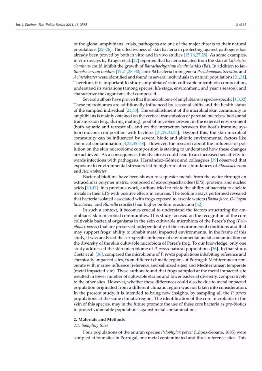
Phylogenetic analysis of the 16S rRNA gene sequences of the bacterial isolates, fromall the sites, revealed that three phyla: Proteobacteria, Actinobacteria, and Firmicutes weredominating. Isolates belonging to Proteobacteria were dominant at all the sites, indepen-dently from contamination or frog’s sex (>60%), mostly belonging to Alphaproteobacteria,Betaproteobacteria, and Gammaproteobacteria. The most abundant families at the non-contaminated site were Enterobacteriaceae (20.3%), and Pseudomonadaceae (14.5%); whileat the contaminated site, Enterobacteriaceae (31.3%) was most abundant followed byStaphylococcaceae (12.5%) was observed (Figure 1).
For the organisms sampled at the contaminated site, some differences were observedbetween sex; mainly for Enterobacteriaceae (CF—58%, CM—15.8%), Moraxellaceae (RF—20.6% and RM—21.2%), and Staphylococcaceae (CF—0%, CM—21.0%) (Figure 1).
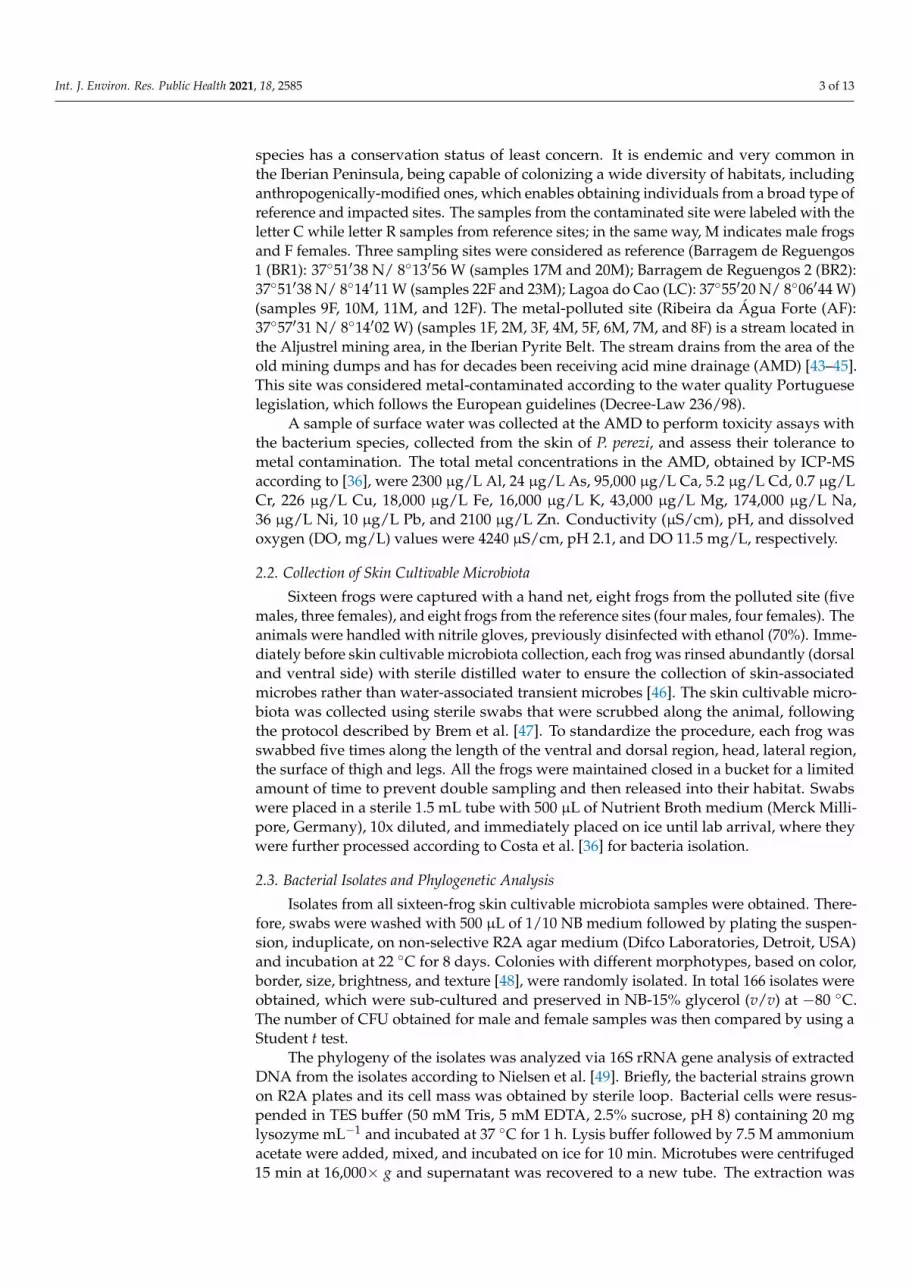
The frogs’ skin cultivable microbiota core group, here defined at genus level by bac-teria present in all frogs’ samples, was composed of strains of the genus Acinetobacter(Figure 2). Five different bacterial genera were found only in samples from the contam-inated site (Paenibacillus, Erwinia, Herbaspirillum, Cupriavidus, and Bordetella). Seventeendifferent genera were exclusively sampled in reference sites. Strains from Staphylococcus,Cellulomonas, and Serratia genera were present only on male frogs. On the other hand,female frogs shared exclusively strains from Citrobacter and Brevundimonas genera. ThePseudomonas strains cultivated in the present study were found only at reference sites, fouron female and one on male frogs. Among the Serratia strains, four were found at the metalcontaminated site and one at the reference site; all of them on male frogs.

Int. J. Environ. Res. Public Health 2021, 18, 2585 6 of 13Int. J. Environ. Res. Public Health 2021, 18, x 6 of 13
Figure 1. Cultivable microbiota composition of Pelophylax perezi skin at family level. The chart is divided into rows for (top to bottom): total samples from the metal-contaminated site (CON), total samples from the reference sites (REF), females (CF) and males (CM) frogs sampled at the metal-contaminated sites, and for female and male frogs sampled at the reference sites (RF and RM, re-spectively).
The frogs’ skin cultivable microbiota core group, here defined at genus level by bac-teria present in all frogs’ samples, was composed of strains of the genus Acinetobacter (Fig-ure 2). Five different bacterial genera were found only in samples from the contaminated site (Paenibacillus, Erwinia, Herbaspirillum, Cupriavidus, and Bordetella). Seventeen different genera were exclusively sampled in reference sites. Strains from Staphylococcus, Cellulomo-nas, and Serratia genera were present only on male frogs. On the other hand, female frogs shared exclusively strains from Citrobacter and Brevundimonas genera. The Pseudomonas strains cultivated in the present study were found only at reference sites, four on female and one on male frogs. Among the Serratia strains, four were found at the metal contami-nated site and one at the reference site; all of them on male frogs.
Figure 2. Venn diagram of the whole Pelophylax perezi skin microbial community at genus level. The central part consists in a core group, composed by bacteria belonging to the genera Acinetobac-ter; seventeen genera are present only in the reference sites while just five genera are restricted to
Figure 1. Cultivable microbiota composition of Pelophylax perezi skin at family level. The chart is divided into rows for (topto bottom): total samples from the metal-contaminated site (CON), total samples from the reference sites (REF), females (CF)and males (CM) frogs sampled at the metal-contaminated sites, and for female and male frogs sampled at the reference sites(RF and RM, respectively).
Int. J. Environ. Res. Public Health 2021, 18, x 6 of 13
Figure 1. Cultivable microbiota composition of Pelophylax perezi skin at family level. The chart is divided into rows for (top to bottom): total samples from the metal-contaminated site (CON), total samples from the reference sites (REF), females (CF) and males (CM) frogs sampled at the metal-contaminated sites, and for female and male frogs sampled at the reference sites (RF and RM, re-spectively).
The frogs’ skin cultivable microbiota core group, here defined at genus level by bac-teria present in all frogs’ samples, was composed of strains of the genus Acinetobacter (Fig-ure 2). Five different bacterial genera were found only in samples from the contaminated site (Paenibacillus, Erwinia, Herbaspirillum, Cupriavidus, and Bordetella). Seventeen different genera were exclusively sampled in reference sites. Strains from Staphylococcus, Cellulomo-nas, and Serratia genera were present only on male frogs. On the other hand, female frogs shared exclusively strains from Citrobacter and Brevundimonas genera. The Pseudomonas strains cultivated in the present study were found only at reference sites, four on female and one on male frogs. Among the Serratia strains, four were found at the metal contami-nated site and one at the reference site; all of them on male frogs.
Figure 2. Venn diagram of the whole Pelophylax perezi skin microbial community at genus level. The central part consists in a core group, composed by bacteria belonging to the genera Acinetobac-ter; seventeen genera are present only in the reference sites while just five genera are restricted to
Figure 2. Venn diagram of the whole Pelophylax perezi skin microbial community at genus level. Thecentral part consists in a core group, composed by bacteria belonging to the genera Acinetobacter;seventeen genera are present only in the reference sites while just five genera are restricted tothe contaminated sites. CM, samples from contaminated sites of male frogs; CF, samples fromcontaminated sites of female frogs; RM, samples from reference sites of male frogs; RF, samples fromreference sites of female frogs.
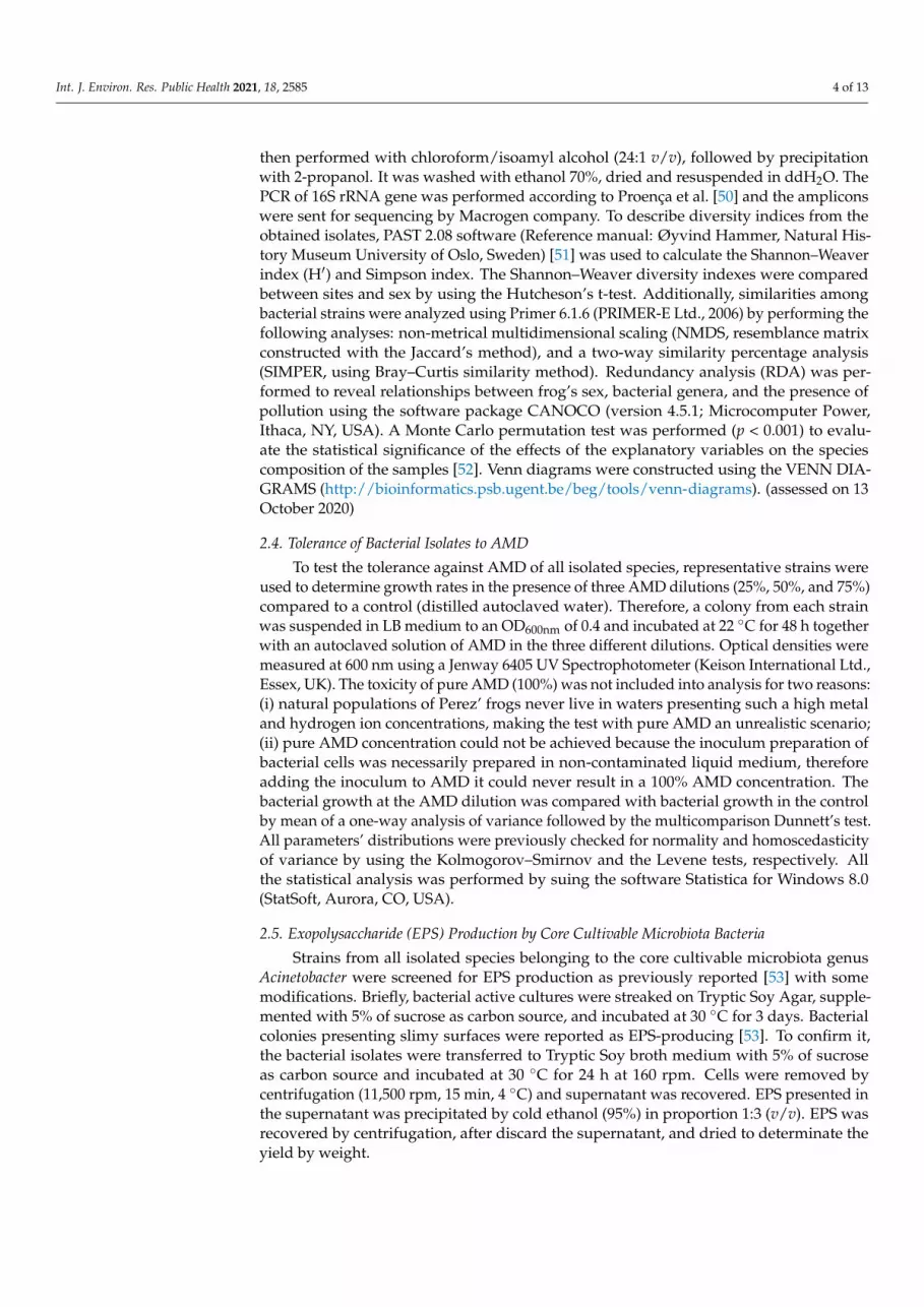
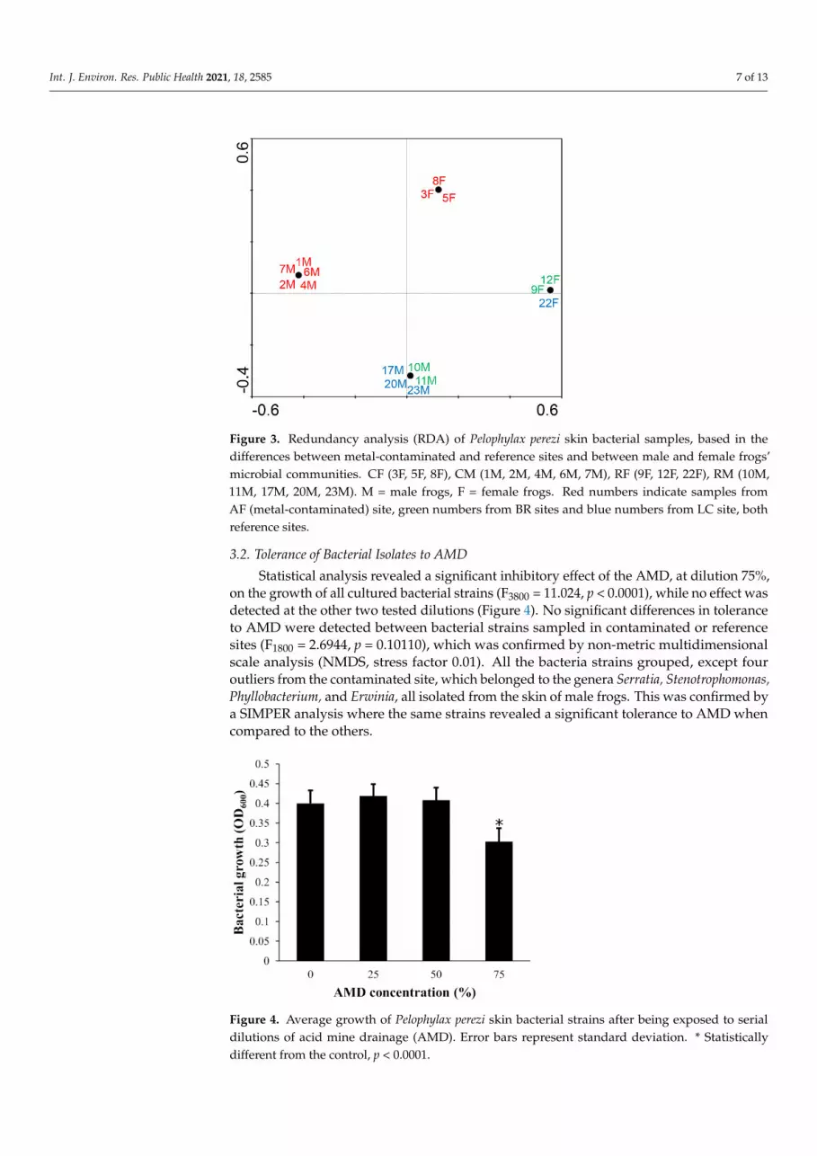
The RDA analysis, for the cultivable microbiota fraction, supports a strong and cleardistinction of the microbial community based on two factors: contamination of the samplingsite and the sex of the frog (p = 0.014, Figure 3), but does not show a distinction betweenthe microbial communities of the different reference sites.
Int. J. Environ. Res. Public Health 2021, 18, 2585 7 of 13
Int. J. Environ. Res. Public Health 2021, 18, x 7 of 13
the contaminated sites. CM, samples from contaminated sites of male frogs; CF, samples from con-taminated sites of female frogs; RM, samples from reference sites of male frogs; RF, samples from reference sites of female frogs.
The RDA analysis, for the cultivable microbiota fraction, supports a strong and clear distinction of the microbial community based on two factors: contamination of the sam-pling site and the sex of the frog (p = 0.014, Figure 3), but does not show a distinction between the microbial communities of the different reference sites.
Figure 3. Redundancy analysis (RDA) of Pelophylax perezi skin bacterial samples, based in the dif-ferences between metal-contaminated and reference sites and between male and female frogs’ microbial communities. CF (3F, 5F, 8F), CM (1M, 2M, 4M, 6M, 7M), RF (9F, 12F, 22F), RM (10M, 11M, 17M, 20M, 23M). M = male frogs, F = female frogs. Red numbers indicate samples from AF (metal-contaminated) site, green numbers from BR sites and blue numbers from LC site, both ref-erence sites.
3.2. Tolerance of Bacterial Isolates to AMD Statistical analysis revealed a significant inhibitory effect of the AMD, at dilution 75
%, on the growth of all cultured bacterial strains (F3800 = 11.024, p < 0.0001), while no effect was detected at the other two tested dilutions (Figure 4). No significant differences in tol-erance to AMD were detected between bacterial strains sampled in contaminated or ref-erence sites (F1800 = 2.6944, p = 0.10110), which was confirmed by non-metric multidimen-sional scale analysis (NMDS, stress factor 0.01). All the bacteria strains grouped, except four outliers from the contaminated site, which belonged to the genera Serratia, Stenotroph-omonas, Phyllobacterium, and Erwinia, all isolated from the skin of male frogs. This was confirmed by a SIMPER analysis where the same strains revealed a significant tolerance to AMD when compared to the others.
Figure 3. Redundancy analysis (RDA) of Pelophylax perezi skin bacterial samples, based in thedifferences between metal-contaminated and reference sites and between male and female frogs’microbial communities. CF (3F, 5F, 8F), CM (1M, 2M, 4M, 6M, 7M), RF (9F, 12F, 22F), RM (10M,11M, 17M, 20M, 23M). M = male frogs, F = female frogs. Red numbers indicate samples fromAF (metal-contaminated) site, green numbers from BR sites and blue numbers from LC site, bothreference sites.
3.2. Tolerance of Bacterial Isolates to AMD
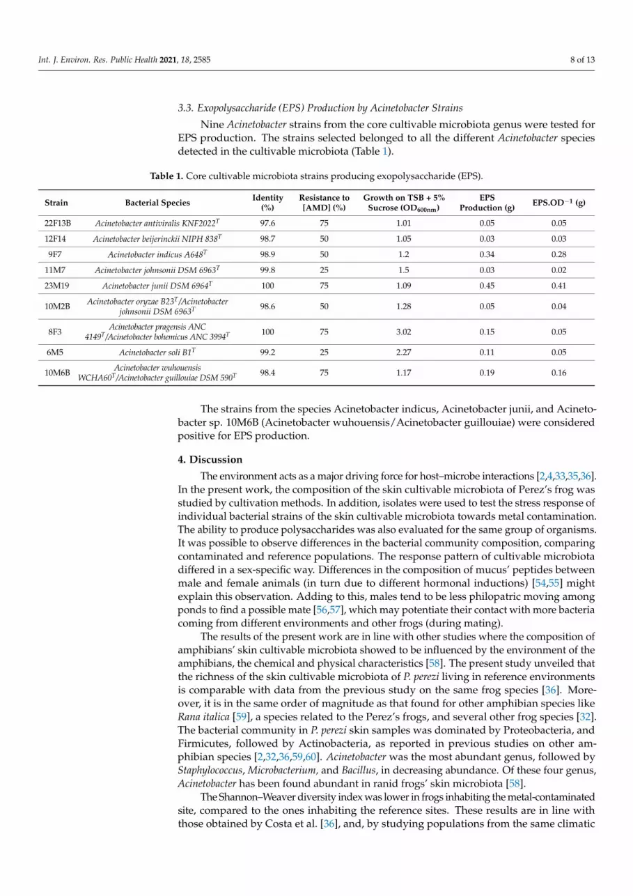
Statistical analysis revealed a significant inhibitory effect of the AMD, at dilution 75%,on the growth of all cultured bacterial strains (F3800 = 11.024, p < 0.0001), while no effect wasdetected at the other two tested dilutions (Figure 4). No significant differences in toleranceto AMD were detected between bacterial strains sampled in contaminated or referencesites (F1800 = 2.6944, p = 0.10110), which was confirmed by non-metric multidimensionalscale analysis (NMDS, stress factor 0.01). All the bacteria strains grouped, except fouroutliers from the contaminated site, which belonged to the genera Serratia, Stenotrophomonas,Phyllobacterium, and Erwinia, all isolated from the skin of male frogs. This was confirmed bya SIMPER analysis where the same strains revealed a significant tolerance to AMD whencompared to the others.
Int. J. Environ. Res. Public Health 2021, 18, x 8 of 13
Figure 4. Average growth of Pelophylax perezi skin bacterial strains after being exposed to serial dilutions of acid mine drainage (AMD). Error bars represent standard deviation. * Statistically different from the control, p < 0.0001.
3.3. Exopolysaccharide (EPS) Production by Acinetobacter Strains Nine Acinetobacter strains from the core cultivable microbiota genus were tested for
EPS production. The strains selected belonged to all the different Acinetobacter species de-tected in the cultivable microbiota (Table 1).
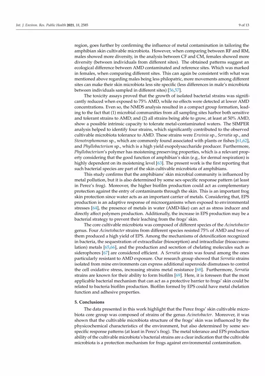
The strains from the species Acinetobacter indicus, Acinetobacter junii, and Acinetobacter sp. 10M6B (Acinetobacter wuhouensis/Acinetobacter guillouiae) were considered positive for EPS production.
Table 1. Core cultivable microbiota strains producing exopolysaccharide (EPS).
Strain
Bacterial Species Identity (%)
Resistance to [AMD] (%)
Growth on TSB + 5% Sucrose (OD600nm)
EPS Production
(g)
EPS.OD−1 (g)
22F13B Acinetobacter antiviralis KNF2022T 97.6 75 1.01 0.05 0.05
12F14 Acinetobacter beijerinckii NIPH 838T 98.7 50 1.05 0.03 0.03
9F7 Acinetobacter indicus A648T 98.9 50 1.2 0.34 0.28 11M
7 Acinetobacter johnsonii DSM 6963T 99.8 25 1.5 0.03 0.02
23M19
Acinetobacter junii DSM 6964T 100 75 1.09 0.45 0.41
10M2B
Acinetobacter oryzae B23T/Acinetobacter johnsonii DSM 6963T
98.6 50 1.28 0.05 0.04
8F3 Acinetobacter pragensis ANC 4149T/Acinetobacter bohemicus ANC 3994T 100 75 3.02 0.15 0.05
6M5 Acinetobacter soli B1T 99.2 25 2.27 0.11 0.05 10M6B
Acinetobacter wuhouensis WCHA60T/Acinetobacter guillouiae DSM 590T 98.4 75 1.17 0.19 0.16
4. Discussion The environment acts as a major driving force for host–microbe interactions
[2,4,33,35,36]. In the present work, the composition of the skin cultivable microbiota of Perez’s frog was studied by cultivation methods. In addition, isolates were used to test the stress response of individual bacterial strains of the skin cultivable microbiota towards
Figure 4. Average growth of Pelophylax perezi skin bacterial strains after being exposed to serialdilutions of acid mine drainage (AMD). Error bars represent standard deviation. * Statisticallydifferent from the control, p < 0.0001.
Int. J. Environ. Res. Public Health 2021, 18, 2585 8 of 13
3.3. Exopolysaccharide (EPS) Production by Acinetobacter Strains
Nine Acinetobacter strains from the core cultivable microbiota genus were tested forEPS production. The strains selected belonged to all the different Acinetobacter speciesdetected in the cultivable microbiota (Table 1).
Table 1. Core cultivable microbiota strains producing exopolysaccharide (EPS).
Strain Bacterial Species Identity(%)
Resistance to[AMD] (%)
Growth on TSB + 5%Sucrose (OD600nm)
EPSProduction (g) EPS.OD−1 (g)
22F13B Acinetobacter antiviralis KNF2022T 97.6 75 1.01 0.05 0.05
12F14 Acinetobacter beijerinckii NIPH 838T 98.7 50 1.05 0.03 0.03
9F7 Acinetobacter indicus A648T 98.9 50 1.2 0.34 0.28
11M7 Acinetobacter johnsonii DSM 6963T 99.8 25 1.5 0.03 0.02
23M19 Acinetobacter junii DSM 6964T 100 75 1.09 0.45 0.41
10M2B Acinetobacter oryzae B23T/Acinetobacterjohnsonii DSM 6963T 98.6 50 1.28 0.05 0.04
8F3 Acinetobacter pragensis ANC4149T/Acinetobacter bohemicus ANC 3994T 100 75 3.02 0.15 0.05
6M5 Acinetobacter soli B1T 99.2 25 2.27 0.11 0.05
10M6B Acinetobacter wuhouensisWCHA60T/Acinetobacter guillouiae DSM 590T 98.4 75 1.17 0.19 0.16
The strains from the species Acinetobacter indicus, Acinetobacter junii, and Acineto-bacter sp. 10M6B (Acinetobacter wuhouensis/Acinetobacter guillouiae) were consideredpositive for EPS production.
4. Discussion
The environment acts as a major driving force for host–microbe interactions [2,4,33,35,36].In the present work, the composition of the skin cultivable microbiota of Perez’s frog wasstudied by cultivation methods. In addition, isolates were used to test the stress response ofindividual bacterial strains of the skin cultivable microbiota towards metal contamination.The ability to produce polysaccharides was also evaluated for the same group of organisms.It was possible to observe differences in the bacterial community composition, comparingcontaminated and reference populations. The response pattern of cultivable microbiotadiffered in a sex-specific way. Differences in the composition of mucus’ peptides betweenmale and female animals (in turn due to different hormonal inductions) [54,55] mightexplain this observation. Adding to this, males tend to be less philopatric moving amongponds to find a possible mate [56,57], which may potentiate their contact with more bacteriacoming from different environments and other frogs (during mating).
The results of the present work are in line with other studies where the composition ofamphibians’ skin cultivable microbiota showed to be influenced by the environment of theamphibians, the chemical and physical characteristics [58]. The present study unveiled thatthe richness of the skin cultivable microbiota of P. perezi living in reference environmentsis comparable with data from the previous study on the same frog species [36]. More-over, it is in the same order of magnitude as that found for other amphibian species likeRana italica [59], a species related to the Perez’s frogs, and several other frog species [32].The bacterial community in P. perezi skin samples was dominated by Proteobacteria, andFirmicutes, followed by Actinobacteria, as reported in previous studies on other am-phibian species [2,32,36,59,60]. Acinetobacter was the most abundant genus, followed byStaphylococcus, Microbacterium, and Bacillus, in decreasing abundance. Of these four genus,Acinetobacter has been found abundant in ranid frogs’ skin microbiota [58].
The Shannon–Weaver diversity index was lower in frogs inhabiting the metal-contaminatedsite, compared to the ones inhabiting the reference sites. These results are in line withthose obtained by Costa et al. [36], and, by studying populations from the same climatic
Int. J. Environ. Res. Public Health 2021, 18, 2585 9 of 13
region, goes further by confirming the influence of metal contamination in tailoring theamphibian skin cultivable microbiota. However, when comparing between RF and RM,males showed more diversity, in the analysis between CF and CM, females showed morediversity (between individuals from different sites). The obtained patterns suggest anecological difference between AMD contaminated and reference sites. Which was markedin females, when comparing different sites. This can again be consistent with what wasmentioned above regarding males being less philopatric, more movements among differentsites can make their skin microbiota less site specific (less differences in male’s microbiotabetween individuals sampled in different sites) [56,57].
The toxicity assays proved that the growth of isolated bacterial strains was signifi-cantly reduced when exposed to 75% AMD, while no effects were detected at lower AMDconcentrations. Even so, the NMDS analysis resulted in a compact group formation, lead-ing to the fact that (1) microbial communities from all sampling sites harbor both sensitiveand tolerant strains to AMD; and (2) all strains being able to grow, at least at 50% AMD,show a possible intrinsic capacity to tolerate metal-contaminated waters. The SIMPERanalysis helped to identify four strains, which significantly contributed to the observedcultivable microbiota tolerance to AMD. These strains were Erwinia sp., Serratia sp., andStenotrophomonas sp., which are commonly found associated with plants or fishes [61,62],and Phyllobacterium sp., which is a high yield exopolysaccharide producer. Furthermore,Phyllobacterium’s polymer has moistening preserving properties, which is a relevant prop-erty considering that the good function of amphibian’s skin (e.g., for dermal respiration) ishighly dependent on its moistening level [63]. The present work is the first reporting thatsuch bacterial species are part of the skin cultivable microbiota of amphibians.
This study confirms that the amphibians’ skin microbial community is influenced bymetal pollution, but it is also determined by some sex-specific response pattern (at leastin Perez’s frog). Moreover, the higher biofilm production could act as complementaryprotection against the entry of contaminants through the skin. This is an important frogskin protection since water acts as an important carrier of metals. Considering that, EPSproduction is an adaptive response of microorganisms when exposed to environmentalstresses [64], the presence of metals in water (AMD-like) can act as stress inducer anddirectly affect polymers production. Additionally, the increase in EPS production may be abacterial strategy to prevent their leaching from the frogs’ skin.
The core cultivable microbiota was composed of different species of the Acinetobactergenus. Four Acinetobacter strains from different species resisted 75% of AMD and two ofthem produced a high yield of EPS. Among the mechanisms of detoxification recognizedin bacteria, the sequestration of extracellular (biosorption) and intracellular (bioaccumu-lation) metals [65,66], and the production and secretion of chelating molecules such assiderophores [67] are considered efficient. A Serratia strain was found among the onesparticularly resistant to AMD exposure. Our research group showed that Serratia strainsisolated from mine environments can express additional superoxide dismutases to controlthe cell oxidative stress, increasing strains metal resistance [68]. Furthermore, Serratiastrains are known for their ability to form biofilm [69]. Here, it is foreseen that the mostapplicable bacterial mechanism that can act as a protective barrier to frogs’ skin could berelated to bacteria biofilm production. Biofilm formed by EPS could have metal chelationfunction and adhesive properties.
5. Conclusions
The data presented in this work highlight that the Perez frogs’ skin cultivable micro-biota core group was composed of strains of the genus Acinetobacter. Moreover, it wasshown that the cultivable microbiota structure of the frogs’ skin was influenced by thephysicochemical characteristics of the environment, but also determined by some sex-specific response patterns (at least in Perez’s frog). The metal tolerance and EPS productionability of the cultivable microbiota’s bacterial strains are a clear indication that the cultivablemicrobiota is a protection mechanism for frogs against environmental contamination.
Int. J. Environ. Res. Public Health 2021, 18, 2585 10 of 13
The obtained data could be a preliminary but crucial step in understanding howskin cultivable microbiota works in amphibians and how it can be related to the frogs’protection capabilities. The impact of changes in the cultivable microbiota and its functionalcharacteristics on endangered species, whether threatened by a different kind of pollutionor impacted by pathogens, is still to be discovered.
Author Contributions: Conceptualization, I.L. and P.V.M.; methodology, D.N.P. and E.F.; formalanalysis, D.N.P., E.F., and P.V.M.; investigation, D.N.P. and E.F.; resources, I.L. and P.V.M.; datacuration, D.N.P. and P.V.M.; writing—original draft preparation, D.N.P., E.F., and P.V.M.; writing—review and editing, all authors; supervision, I.L. and P.V.M.; project administration, I.L. and P.V.M.;funding acquisition, I.L. and P.V.M. All authors have read and agreed to the published version ofthe manuscript.
Funding: This work was supported by FEDER funds within the PT2020 Partnership Agreementand Compete 2020—Programa Operacional Factores de Competitividade, by the Portuguese Foun-dation for Science and Technology (FCT), by the CESAM and CEMMPRE strategic programme(UID/AMB/50017/2013, and UID/EMS/00285/2013 and UID/EMS/00285/2020, respectively) andthe research project GENEROSI project number PTDC/BIA-BIC/3488/2012. This work was alsofunded by national funds via FCT/MEC (PIDDAC) by the doctoral fellowship of Emanuele FasolaSFRH/BD/88955/2012, and by investigator FCT grant of Isabel Lopes IF/00475/2013.
Informed Consent Statement: Not applicable.
Data Availability Statement: The 16S rRNA gene sequences of the bacterial isolates reported in thisstudy were deposited at Genbank database, under the accession numbers according KY611613–KY611778.
Acknowledgments: We are extremely grateful to all the researchers and institutions involved in thisresearch. The authors would also like to thank Tania Duarte for helping in the laboratory.
Conflicts of Interest: The authors declare no conflict of interest.
References1. Belden, L.K.; Hughey, M.C.; Rebollar, E.A.; Umile, T.P.; Loftus, S.C.; Burzynski, E.A.; Minbiole, K.P.C.; House, L.L.; Jensen, R.V.;
Becker, M.H.; et al. Panamanian frog species host unique skin bacterial communities. Front. Microbiol. 2015, 6, 1–21. [CrossRef][PubMed]
2. Kueneman, J.C.; Parfrey, L.W.; Woodhams, D.C.; Archer, H.M.; McKenzie, R.; Knight, V.J. The amphibian skin-associatedmicrobiome across species, space and life history stages. Mol. Ecol. 2014, 23, 1238–1250. [CrossRef]
3. Rebollar, E.A.; Hughey, M.C.; Medina, D.; Harris, R.N.; Ibáñez, R.; Belden, L.K. Skin bacterial diversity of Panamanian frogs isassociated with host susceptibility and presence of Batrachochytrium dendrobatidis. ISME J. 2016, 1–14. [CrossRef]
4. Assis, A.B.; Bevier, C.R.; Chaves Barreto, C.; Arturo Navas, C. Environmental influences on and antimicrobial activity of the skinmicrobiota of Proceratophrys boiei (Amphibia, Anura) across forest fragments. Ecol. Evol. 2020, 10, 901–913. [CrossRef] [PubMed]
5. Mangoni, M.L.; Miele, R.; Renda, T.G.; Barra, D.; Simmaco, M. The synthesis of antimicrobial peptides in the skin of Rana esculentais stimulated by microorganisms. FASEB J. 2001, 15, 1431–1432. [CrossRef] [PubMed]
6. Alexander Pyron, R.; Wiens, J.J. A large-scale phylogeny of Amphibia including over 2800 species, and a revised classification ofextant frogs, salamanders, and caecilians. Mol. Phylogenet. Evol. 2011, 61, 543–583. [CrossRef] [PubMed]
7. IUCN. Available online: http://www.iucn.org/ (accessed on 2 February 2021).8. Sparling, D.W.; Linder, G.; Bishop, C.A.; Krest, S.K. Ecotoxicology of Amphibians and Reptiles, 2nd ed.; Sparling, D.W., Linder, G.,
Bishop, C.A., Krest, S.K., Eds.; CRC Press: Boca Raton, FL, USA, 2010; ISBN 978-1-4200-6416-2.9. Blaustein, A.R.; Bancroft, B.A. Amphibian population declines: Evolutionary considerations. Bioscience 2007, 57, 437–444.
[CrossRef]10. Hoffmann, M.; Hilton-Taylor, C.; Angulo, A.; Böhm, M.; Brooks, T.M.; Butchart, S.H.M.; Carpenter, K.E.; Chanson, J.; Collen, B.;
Cox, N.A.; et al. The impact of conservation on the status of the world’s vertebrates. Science 2010, 330, 1503–1509. [CrossRef]11. Fedorenkova, A.; Vonk, J.A.; Lenders, H.J.R.; Creemers, R.C.M.; Breure, A.M.; Hendriks, A.J. Ranking ecological risks of multiple
chemical stressors on amphibians. Environ. Toxicol. Chem. 2012, 31, 1416–1421. [CrossRef] [PubMed]12. Maiorano, L.; Amori, G.; Capula, M.; Falcucci, A.; Masi, M.; Montemaggiori, A.; Pottier, J.; Psomas, A.; Rondinini, C.; Russo,
D.; et al. Threats from climate change to terrestrial vertebrate hotspots in Europe. PLoS ONE 2013, 8, e74989. [CrossRef]13. Ortiz, M.E.; Marco, A.; Saiz, N.; Lizana, M. Impact of ammonium nitrate on growth and survival of six European amphibians.
Arch. Environ. Contam. Toxicol. 2004, 47. [CrossRef]14. Pask, J. The ebb and flow of antimicrobial skin peptides defends northern leopard frogs (Rana pipiens) against chytridiomycosis.
Glob. Chang. 2012, 1231–1238. [CrossRef]
Int. J. Environ. Res. Public Health 2021, 18, 2585 11 of 13
15. Harris, R.; Brucker, R.; Walke, J. Skin microbes on frogs prevent morbidity and mortality caused by a lethal skin fungus. ISME2009, 3, 818–824. [CrossRef]
16. Becker, M.H.; Brucker, R.M.; Christian, R.; Harris, R.N.; Minbiole, K.P.C.; Schwantes, C.R. The bacterially produced metaboliteviolacein is associated with survival of amphibians infected with a lethal fungus. Appl. Environ. Microbiol. 2009, 75, 6635–6638.[CrossRef]
17. Woodhams, D.C.; Brandt, H.; Baumgartner, S.; Kielgast, J.; Küpfer, E.; Tobler, U.; Davis, L.R.; Schmidt, B.R.; Bel, C.; Hodel, S.; et al.Interacting symbionts and immunity in the amphibian skin mucosome predict disease risk and probiotic effectiveness. PLoS ONE2014, 9, e96375. [CrossRef] [PubMed]
18. McKenzie, V.; Peterson, A. Pathogen pollution and the emergence of a deadly amphibian pathogen. Mol. Ecol. 2012, 5151–5154.[CrossRef]
19. Woodhams, D.C.; Bletz, M.; Kueneman, J.; McKenzie, V. Managing amphibian disease with skin microbiota. Trends Microbiol.2016, 24, 161–164. [CrossRef]
20. Laugen, A.T.; Laurila, A.; Merilä, J. Maternal and genetic contributions to geographical variation in Rana temporaria larvallife-history traits. Biol. J. Linn. Soc. 2002, 76, 61–70. [CrossRef]
21. Bresciano, J.; Salvador, C.; Paz-Y-Miño, C.; Parody-Merino, A.; Bosch, J.; Woodhams, D. Variation in the presence of anti-Batrachochytrium dendrobatidis bacteria of amphibians across life stages and elevations in Ecuador. Ecohealth 2015, 12, 310–319.[CrossRef] [PubMed]
22. Blaustein, A.R.; Hokit, D.G.; O’Hara, R.K. Pathogenic fungus contributes to amphibian losses in the Pacific Northwest. Biol.Conserv. 1994, 67, 251–254. [CrossRef]
23. Fernández-Benéitez, M.J.; Ortiz-Santaliestra, M.E.; Lizana, M.; Diéguez-Uribeondo, J. Differences in susceptibility to Saprolegniainfections among embryonic stages of two anuran species. Oecologia 2011, 165, 819–826. [CrossRef] [PubMed]
24. Baláž, V.; Vörös, J.; Civiš, P.; Vojar, J.; Hettyey, A.; Sós, E.; Dankovics, R.; Jehle, R.; Christiansen, D.G.; Clare, F.; et al. Assessingrisk and guidance on monitoring of Batrachochytrium dendrobatidis in Europe through identification of taxonomic selectivity ofinfection. Conserv. Biol. 2013, 28, 213–223. [CrossRef]
25. Pounds, J.A.; Bustamante, M.R.; Coloma, L.A.; Consuegra, J.A.; Fogden, M.P.L.; Foster, P.N.; La Marca, E.; Masters, K.L.;Merino-Viteri, A.; Puschendorf, R.; et al. Widespread amphibian extinctions from epidemic disease driven by global warming.Nature 2006, 439, 161–167. [CrossRef] [PubMed]
26. Pearman, P.B.; Garner, T.W.J. Susceptibility of Italian agile frog populations to an emerging Ranavirus parallels population geneticdiversity. Ecol. Lett. 2005, 8, 401–408. [CrossRef]
27. Kruger, A. Functional redundancy of Batrachochytrium dendrobatidis inhibition in bacterial communities isolated from Lithobatesclamitans skin. Microb. Ecol. 2020, 79, 231–240. [CrossRef]
28. Muletz, C.; Myers, J.; Domangue, R.; Herrick, J.; Harris, R. Soil bioaugmentation with amphibian cutaneous bacteria protectsamphibian hosts from infection by Batrachochytrium dendrobatidis. Biol. Conserv. 2012, 152, 119–126. [CrossRef]
29. Rebollar, E.A.; Simonetti, S.J.; Shoemaker, W.R.; Harris, R.N. Direct and indirect horizontal transmission of the antifungal probioticbacterium Janthinobacterium lividum on green frog (Lithobates clamitans) tadpoles. Appl. Environ. Microbiol. 2016, 82, 2457–2466.[CrossRef] [PubMed]
30. Becker, M.H.; Harris, R.N.; Minbiole, K.P.C.; Schwantes, C.R.; Rollins-Smith, L.A.; Reinert, L.K.; Brucker, R.M.; Domangue, R.J.;Gratwicke, B. Towards a better understanding of the use of probiotics for preventing chytridiomycosis in Panamanian goldenfrogs. Ecohealth 2011, 8, 501–506. [CrossRef] [PubMed]
31. Bates, K.A.; Clare, F.C.; O’Hanlon, S.; Bosch, J.; Brookes, L.; Hopkins, K.; McLaughlin, E.J.; Daniel, O.; Garner, T.W.J.; Fisher,M.C.; et al. Amphibian chytridiomycosis outbreak dynamics are linked with host skin bacterial community structure. Nat.Commun. 2018, 9, 693. [CrossRef]
32. McKenzie, V.J.; Bowers, R.M.; Fierer, N.; Knight, R.; Lauber, C.L. Co-habiting amphibian species harbor unique skin bacterialcommunities in wild populations. ISME 2012, 588–596. [CrossRef] [PubMed]
33. Longo, A.V.; Zamudio, K.R. Environmental fluctuations and host skin bacteria shift survival advantage between frogs and theirfungal pathogen. ISME 2016, 1–13. [CrossRef]
34. Fitzpatrick, B.; Amanda, A. Similarity and differentiation between bacteria associated with skin of salamanders (Plethodon jordani)and free-living assemblages. FEMS 2014. [CrossRef] [PubMed]
35. Muletz Wolz, C.R.; Yarwood, S.A.; Campbell Grant, E.H.; Fleischer, R.C.; Lips, K.R. Effects of host species and environment onthe skin microbiome of Plethodontid salamanders. J. Anim. Ecol. 2018, 87, 341–353. [CrossRef] [PubMed]
36. Costa, S.; Lopes, I.; Proença, D.N.; Ribeiro, R.; Vasconcelo Morais, P. Diversity of cutaneous bacterial community of Pelophylaxperezi populations inhabiting different environments. Sci. Total Environ. 2016, 572, 995–1004. [CrossRef] [PubMed]
37. Jimenez, R.; Sommers, S. The amphibian microbiome: Natural range of variation, pathogenic dysbiosis, and role in conservation.Biodivers. Conserv. 2017, 26, 763–786. [CrossRef]
38. Ellison, S.; Rovito, S. The influence of habitat and phylogeny on the skin microbiome of the influence of habitat and phylogeny onthe skin microbiome of amphibians in Guatemala and Mexico. Microb. Ecol. 2018. [CrossRef]
39. Hernández-Gómez, O.; Wuerthner, V.; Hua, J. Amphibian host and skin microbiota response to a common agricultural antimicro-bial and internal parasite. Microb. Ecol. 2020, 79, 175–191. [CrossRef]
Int. J. Environ. Res. Public Health 2021, 18, 2585 12 of 13
40. Huang, Y.; Wang, W.; Peng, A. Accumulation of Cu (II) and Pb (II) by biofilms grown on particulate in aquatic systems. J. Environ.Sci. Health Part A 2000, 35, 575–592. [CrossRef]
41. Kazy, S.K.; Sar, P.; Singh, S.P.; Sen, A.K.; D’Souza, S.F. Extracellular polysaccharides of a copper-sensitive and a copper-resistantPseudomonas aeruginosa strain: Synthesis, chemical nature and copper binding. World J. Microbiol. Biotechnol. 2002, 18, 583–588.[CrossRef]
42. Cordeiro, I.F.; Fonseca, N.P.; Felestrino, É.B.; Caneschi, W.L.; Silvério Pires, M.R.; Moreira, L.M. Arsenic resistance in culturedcutaneous microbiota is associated with anuran lifestyles in the Iron Quadrangle, Minas Gerais State, Brazil. Herpetol. Notes 2019,12, 1083–1093.
43. Maia, F.; Pinto, C.; Waeremborg, J.; Gonçalves, M.; Prazeres, C.; Carreira, O.; Serio, S. Metal partitioning in sediments andmineralogical controls on the acid mine drainage in Ribeira da Água Forte (Aljustrel, Iberian Pyrite Belt, Southern Portugal).Appl. Geochem. 2012, 27, 1063–1080. [CrossRef]
44. Luis, A.; Duraes, N.; Pinheiro De Almeida, S.; Ferreira Da Silva, E. Integrating geochemical (surface waters, stream sediments)and biological (diatoms) approaches to assess AMD environmental impact in a pyritic mining area: Aljustrel (Alentejo, Portugal).J. Environ. Sci. 2016, 42, 215–226. [CrossRef] [PubMed]
45. Luis, A.; Teixaira, P.; Almeida, S.; Ector, S.; Matos, J.; Ferreira Da Silva, E. Impact of acid mine drainage (AMD) on water quality,stream sediments and periphytic diatom communities in the surrounding streams of Aljustrel mining area (Portugal). Water AirSoil Pollut. 2009, 200, 147–167. [CrossRef]
46. Lauer, A.; Simon, M.A.; Banning, J.L.; Lam, B.A.; Harris, R.N. Diversity of cutaneous bacteria with antifungal activity isolatedfrom female four-toed salamanders. ISME J. 2008, 145–157. [CrossRef]
47. Brem, F.; Mendelson, J.R.; Lips, K.R. Field-Sampling Protocol for Batrachochytrium Dendrobatidis from Living Amphibians, Using AlcoholPreserved Swabs; Version 1.0; Conservation International: Arlington, VA, USA, 2007; Available online: http://www.amphibians.org(accessed on 2 February 2021).
48. Smibert, R.M.; Krieg, N.R. Phenotypic Characterization. Methods for General and Molecular Bacteriology; Gerhardt, P., Murray, R.G.E.,Wood, W.A., Krieg, N.R., Eds.; American Society for Microbiology: Washington, DC, USA, 1994.
49. Nielsen, P.; Fritze, D.; Priest, F.G. Phenetic diversity of alkaliphilic Bacillus strains: Proposal for nine new species. Microbiology1995, 141, 1745–1761. [CrossRef]
50. Proença, D.N.; Francisco, R.; Kublik, S.; Scholer, A.; Vestergaard, G.; Schloter, M.; Morais, P.V. The microbiome of endophytic,wood colonizing bacteria from pine trees as affected by Pine Wilt Disease. Sci. Rep. 2017, 7, 4205. [CrossRef] [PubMed]
51. Hammer, Ø.; Harper, D.A.T.; Ryan, P.D. Past: Paleontological statistics software package for education and data analysis.Palaeontol. Electron. 2001, 4, 1–9.
52. van den Brink, P.J.; Braak, C.J.F.T. Principal response curves: Analysis of time-dependent multivariate responses of biologicalcommunity to stress. Environ. Toxicol. Chem. 1999, 18, 138–148. [CrossRef]
53. Kumar, M.A.; Anandapandian, K.T.K.; Parthiban, K. Production and characterization of exopolysaccharides (EPS) from biofilmforming marine bacterium. Braz. Arch. Biol. Technol. 2011, 54, 259–265. [CrossRef]
54. Edwards, H.A. A novel mechanism for salt and fluid transport across epithelia. J. Exp. Biol. 1979, 83, 335–338.55. Boutilier, R.G.; Stiffler, D.F.; Toews, D. Exchange of respiratory gases, ions and water in amphibious and aquatic amphibians. In
Environmental Physiology of the Amphibians; Burggren, W.W., Ed.; University of Chicago Press: Chicago, IL, USA; London, UK,1992; pp. 81–124.
56. Smith, M.A.; Green, D.M. Dispersal and the metapopulation paradigm in amphibian ecology and conservation: Are all amphibianpopulations metapopulations? Ecography 2005, 28, 110–128. [CrossRef]
57. Bowne, D.; Bowers, M. Interpatch movements in spatially structured populations: A literature review. Landsc. Ecol. 2004, 19, 1–20.[CrossRef]
58. Bletz, M.C.; Perl, R.G.B.; Vences, M. Skin microbiota differs drastically between co-occurring frogs and newts. R. Soc. Open Sci.2017, 4, 170107. [CrossRef]
59. Federici, E.; Rossi, R.; Fidati, L.; Paracucchi, R.; Scargetta, S.; Montalbani, E.; Franzetti, A.; La Porta, G.; Fagotti, A.; Simoncelli, F.;et al. Characterization of the skin microbiota in Italian stream frogs (Rana italica) infected and uninfected by a cutaneous parasiticdisease. Microbes Environ. 2015, 30, 262–269. [CrossRef] [PubMed]
60. Walke, J.B.; Becker, M.H.; Loftus, S.C.; House, L.L.; Cormier, G.; Jensen, R.V.; Belden, L.K. Amphibian skin may select for rareenvironmental microbes. ISME J. 2014, 8, 2207–2217. [CrossRef]
61. Skrodenyte-Arbaciauskiene, V.; Sruoga, A.; Butkauskas, D. Assessment of microbial diversity in the river trout Salmo trutta farioL. intestinal tract identified by partial 16S rRNA gene sequence analysis. Fish. Sci. 2006, 72, 597–602. [CrossRef]
62. Wolf, A.; Fritze, A.; Hagemann, M.; Berg, G. Stenotrophomonas rhizophila sp. nov., a novel plant-associated bacterium withantifungal properties. Int. J. Syst. Evol. Microbiol. 2002, 52, 1937–1944. [CrossRef]
63. Li, Y.; Zhang, G.; Du, C.; Mou, H.; Cui, J.; Guan, H.; Hwang, H.; Wang, P. Characterization of high yield exopolysaccharideproduced by Phyllobacterium sp. 921F exhibiting moisture preserving properties. Int. J. Biol. Macromol. 2017, 101, 562–568.[CrossRef] [PubMed]
64. Singh, S.; Singh, S.K.; Chowdhury, I.; Singh, R. Understanding the mechanism of bacterial biofilms resistance to antimicrobialagents. Open Microbiol. J. 2017, 11, 53–62. [CrossRef] [PubMed]
Int. J. Environ. Res. Public Health 2021, 18, 2585 13 of 13
65. Coimbra, C.; Branco, R.; Morais, P.V. Efficient bioaccumulation of tungsten by Escherichia coli cells expressing the Sulfitobacterdubius TupBCA system. Syst. Appl. Microbiol. 2019, 42, 126001. [CrossRef] [PubMed]
66. Morais, P.V.; Branco, R.; Francisco, R. Chromium resistance strategies and toxicity: What makes Ochrobactrum tritici 5bvl1 a strainhighly resistant. BioMetals 2011, 24, 401–410. [CrossRef]
67. Proença, D.N.; Heine, T.; Senges, C.H.R.; Bandow, J.E.; Morais, P.V.; Tischler, D. Bacterial metabolites produced under ironlimitation kill pinewood nematode and attract Caenorhabditis elegans. Front. Microbiol. 2019, 10, 2166. [CrossRef] [PubMed]
68. Caldeira, J.B.; Morais, P.V.; Branco, R. Exploiting the biological response of two Serratia fonticola strains to the critical metals,gallium and indium. Sci. Rep. 2020, 10, 1–12. [CrossRef] [PubMed]
69. Ray, C.; Shenoy, A.T.; Orihuela, C.J.; González-Juarbe, N. Killing of Serratia marcescens biofilms with chloramphenicol. Ann. Clin.Microbiol. Antimicrob. 2017, 16, 19. [CrossRef]