Embed Size (px)
Citation preview

THE MICROBIOTA-GUT-BRAIN AXISJohn F. Cryan, Kenneth J. O’Riordan, Caitlin S. M. Cowan, Kiran V. Sandhu,Thomaz F. S. Bastiaanssen, Marcus Boehme, Martin G. Codagnone, Sofia Cussotto,Christine Fulling, Anna V. Golubeva, Katherine E. Guzzetta, Minal Jaggar,Caitriona M. Long-Smith, Joshua M. Lyte, Jason A. Martin, Alicia Molinero-Perez,Gerard Moloney, Emanuela Morelli, Enrique Morillas, Rory O’Connor, Joana S. Cruz-Pereira,Veronica L. Peterson, Kieran Rea, Nathaniel L. Ritz, Eoin Sherwin, Simon Spichak,Emily M. Teichman, Marcel van de Wouw, Ana Paula Ventura-Silva,Shauna E. Wallace-Fitzsimons, Niall Hyland, Gerard Clarke, and Timothy G. Dinan
APC Microbiome Ireland, University College Cork, Cork, Ireland; Department of Anatomy and Neuroscience,University College Cork, Cork, Ireland; Department of Psychiatry and Neurobehavioural Science, UniversityCollege Cork, Cork, Ireland; and Department of Physiology, University College Cork, Cork, Ireland
LCryan JF, O’Riordan KJ, Cowan CSM, Sandhu KV, Bastiaanssen TFS, Boehme M,Codagnone MG, Cussotto S, Fulling C, Golubeva AV, Guzzetta KE, Jaggar M,Long-Smith CM, Lyte JM, Martin JA, Molinero-Perez A, Moloney G, Morelli E,Morillas E, O’Connor R, Cruz-Pereira JS, Peterson VL, Rea K, Ritz NL, SherwinE, Spichak S, Teichman EM, van de Wouw M, Ventura-Silva AP, Wallace-Fitzsi-
mons SE, Hyland N, Clarke G, Dinan TG. The Microbiota-Gut-Brain Axis. Physiol Rev 99:1877–2013, 2019. Published August 28, 2019; doi:10.1152/physrev.00018.2018.—Theimportance of the gut-brain axis in maintaining homeostasis has long been appreciated. However,the past 15 yr have seen the emergence of the microbiota (the trillions of microorganisms withinand on our bodies) as one of the key regulators of gut-brain function and has led to the appreciationof the importance of a distinct microbiota-gut-brain axis. This axis is gaining ever more traction infields investigating the biological and physiological basis of psychiatric, neurodevelopmental, age-re-lated, and neurodegenerative disorders. The microbiota and the brain communicate with each other viavarious routes including the immune system, tryptophan metabolism, the vagus nerve and the entericnervous system, involving microbial metabolites such as short-chain fatty acids, branched chain aminoacids, and peptidoglycans. Many factors can influence microbiota composition in early life, includinginfection, mode of birth delivery, use of antibiotic medications, the nature of nutritional provision,environmental stressors, and host genetics. At the other extreme of life, microbial diversity diminisheswith aging. Stress, in particular, can significantly impact the microbiota-gut-brain axis at all stages of life.Much recent work has implicated the gut microbiota in many conditions including autism, anxiety,obesity, schizophrenia, Parkinson’s disease, and Alzheimer’s disease. Animal models have been para-mount in linking the regulation of fundamental neural processes, such as neurogenesis and myelination,to microbiome activation of microglia. Moreover, translational human studies are ongoing and willgreatly enhance the field. Future studies will focus on understanding the mechanisms underlying themicrobiota-gut-brain axis and attempt to elucidate microbial-based intervention and therapeutic strat-egies for neuropsychiatric disorders.
brain-gut; microbiome; neurogastroenterology; second brain; stress
I. INTRODUCTION 1877II. STUDYING THE... 1881III. MICROBIOTA-GUT-BRAIN AXIS... 1907IV. PATHWAYS OF COMMUNICATION 1911V. MICROBIOTA AND SYNAPTIC... 1929VI. FACTORS INFLUENCING THE... 1932VII. BEHAVIOR AND THE... 1941VIII. DISEASES AND DISEASE PROCESSES 1947IX. BEYOND THE “BACTERIOME” 1961X. CONCLUSIONS 1963
I. INTRODUCTION“All disease begins in the gut.”
–Hippocrates of Kos (Hippokráte�s ho Kṓos:c. 460–c. 370 BCE)
It was over 2,000 years ago when the Greek physicianHippocrates, oft-lauded as the father of modern medi-cine, is purported to have made this proclamation. Al-though the attribution to Hippocrates has been ques-tioned, its inherent wisdom continues to influence re-
Physiol Rev 99: 1877–2013, 2019Published August 28, 2019; doi:10.1152/physrev.00018.2018
18770031-9333/19 Copyright © 2019 the American Physiological SocietyDownloaded from journals.physiology.org/journal/physrev (093.039.178.031) on October 2, 2020.

searchers and practitioners in medicine (and beyond)regardless of its authenticity (443).
Although the links between rural Michigan and ancientGreece are not obvious, it was there in the 1800s that anunfortunate injury to a Canadian fur-trader Alexis St. Mar-tin created a serendipitous opportunity to advance the studyof the gut and digestion in line with the sentiments of Hip-pocrates and the other great Greek physician-philosopher,Galen of Pergamon (1001). St. Martin was accidentally shotat close range and, during his treatment by the United StatesArmy surgeon William Beaumont, became one of the mostfamous patients in gastroenterology (115). The surgery leftSt. Martin with a fistula in his gut, a window into the intes-tine, for Beaumont to study. The doctor took careful notesthroughout the recovery period and discovered the mannerin which many aspects of digestion occurred via experi-ments where he inserted food into St. Martin’s stomach,then later removing it to observe the extent of digestion. Hetook gastric secretion samples and sent them to chemistsof the day for analysis, a very uncommon medical processfor the mid-19th century. This was one of the first recordedobservations of human digestion taking place in real time.Even more fascinating were Beaumont’s notes of “pain anduneasiness” at corporeal sites far from the wound, linkingdigestion with disease, and emotionality. Moreover, whenSt. Martin became angry or irritable, it greatly affected therate of digestion, indicating that the subject’s emotionalstate affected digestion, i.e., there was a brain-gut axis.Notwithstanding the discomfort of his patient, Beaumont’swork moved the field beyond the 2nd-century teachings ofGalen (1001) to pioneer a new era of precise clinical datacollection, observation, and recording of conclusions forfuture reference. Other great historical scientists, includingCharles Darwin (74) and Claude Bernard (138), continuedthe effort to formally establish and standardize the use ofthe scientific method in medicine. While Darwin was fastid-iously investigating, collecting, and cataloging biologicalspecimens to build evidence for his famed theory of naturalselection (388), Bernard was practicing the scientificmethod at the Sorbonne and the Natural History Museumin Paris, France. Through his feeding experiments with rab-bits, Bernard determined the process for the emulsificationand saponification of fats by the pancreas and identifiedthat the process of digestion took place not in the stomachbut the small intestine. Further studies of glycogen stores inthe liver and blood sugar levels illustrated that digestion notonly breaks up complex molecules from food but also stores
them for future energy requirements. Encapsulating hisbody of research, Bernard developed the concept of milieuintérieur, stating that “The stability of the internal environ-ment is the condition for the free and independent life”(139). This would later become the foundation for our un-derstanding of corporeal homeostasis.
Bernard, as one of the earliest pioneers of animal experi-mentation, also paved the way for future scientific discov-ery. Among those following in this tradition was Ivan Pav-lov, whose defining studies of classical conditioning weredirectly inspired by William Beaumont’s observations ofdigestion. Under the tutelage of Carl Ludwig in Leipzig,Germany, Ivan Pavlov helped develop the Pavlov pouch(1173), a piece of exteriorized dog intestine used to studythe processes of digestion in dogs. He perfected the tech-nique by maintaining the innervation to the exteriorizedintestine section to allow more accurate measurement ofdigestive processes in real time over extended periods; it isbelieved that this is one of the first recorded uses of achronic model of animal experimentation in modern sci-ence. These studies set the basis for our understanding ofthe critical role that the gut-brain axis plays in homeostaticprocesses in health and disease. With the advent of brainimaging technology in the 1980s, the full appreciation ofthe bidirectionality of this axis emerged. Studies showedthat distension of the gut resulted in activation of key path-ways within the brain and that such pathways are exagger-ated in disorders such as irritable bowel syndrome (IBS), afunctional gastrointestinal (GI) disorder with dysregulatedmicrobiota-gut-brain axis (503, 784, 1009). Moreover, thegut-brain axis is seen as an important node in mammalianinteroception (351).
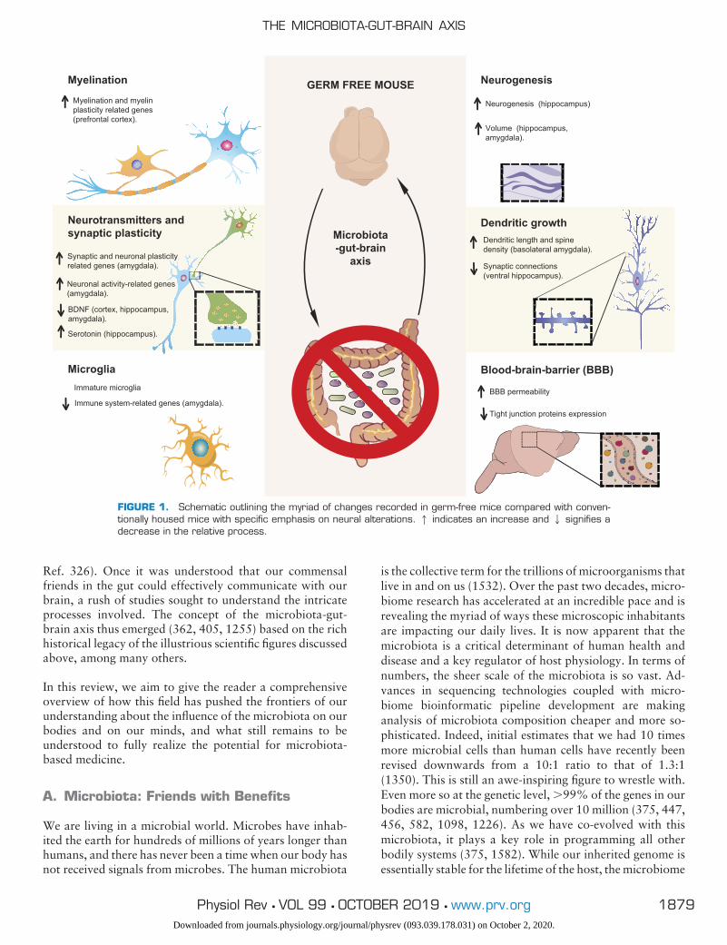
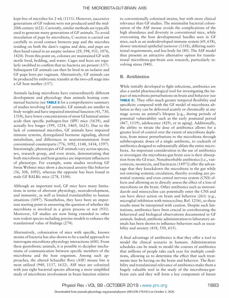
Finally, in the past decades, a new player has emerged as akey regulator of the gut-brain axis, the trillions of microbeswithin the gut, the microbiota. Five separate lines of evi-dence converged to establish this. First, studies in germ-free(GF) animals showed that the brain is affected in the ab-sence of microbiota (see FIGURE 1 and TABLE 2) (308, 437,569, 654, 1092, 1434). Second, animals given specificstrains of bacteria had alterations in behavior (132, 209,429, 1021, 1321, 1560), and human studies of such strainsconfirmed the potential translatability of such findings (38,1198, 1493). Third, population-based studies of people ex-posed to infection, most notably in Walkerton in Canada,demonstrated alterations in gut-brain symptoms (1477).These findings were also echoed in animal studies wherelow-level infections altered behavior even in the absence ofimmune activation (949). Fourth, preclinical studies withantibiotic administration, either in early life (1119) oradulthood (1560), have shown long-lasting effects on brain,spinal cord, and the enteric nervous system (ENS). Finally,these data synergized with the long-known clinical situationthat hepatic encephalopathy could be broadly treated bytargeting the microbiota with antibiotics in humans (see
This is a comprehensive review of current knowledge of theinfluence that the microbiota has on brain and behavior. Inparticular, we focus on the pathways involved and the modelsused in the field. Moreover, we highlight what still remains to beunderstood to fully realize the potential for the development ofmicrobiota-based therapeutic strategies for brain disorders.
CRYAN ET AL.
1878 Physiol Rev • VOL 99 • OCTOBER 2019 • www.prv.orgDownloaded from journals.physiology.org/journal/physrev (093.039.178.031) on October 2, 2020.
Ref. 326). Once it was understood that our commensalfriends in the gut could effectively communicate with ourbrain, a rush of studies sought to understand the intricateprocesses involved. The concept of the microbiota-gut-brain axis thus emerged (362, 405, 1255) based on the richhistorical legacy of the illustrious scientific figures discussedabove, among many others.
In this review, we aim to give the reader a comprehensiveoverview of how this field has pushed the frontiers of ourunderstanding about the influence of the microbiota on ourbodies and on our minds, and what still remains to beunderstood to fully realize the potential for microbiota-based medicine.
A. Microbiota: Friends with Benefits
We are living in a microbial world. Microbes have inhab-ited the earth for hundreds of millions of years longer thanhumans, and there has never been a time when our body hasnot received signals from microbes. The human microbiota
is the collective term for the trillions of microorganisms thatlive in and on us (1532). Over the past two decades, micro-biome research has accelerated at an incredible pace and isrevealing the myriad of ways these microscopic inhabitantsare impacting our daily lives. It is now apparent that themicrobiota is a critical determinant of human health anddisease and a key regulator of host physiology. In terms ofnumbers, the sheer scale of the microbiota is so vast. Ad-vances in sequencing technologies coupled with micro-biome bioinformatic pipeline development are makinganalysis of microbiota composition cheaper and more so-phisticated. Indeed, initial estimates that we had 10 timesmore microbial cells than human cells have recently beenrevised downwards from a 10:1 ratio to that of 1.3:1(1350). This is still an awe-inspiring figure to wrestle with.Even more so at the genetic level, �99% of the genes in ourbodies are microbial, numbering over 10 million (375, 447,456, 582, 1098, 1226). As we have co-evolved with thismicrobiota, it plays a key role in programming all otherbodily systems (375, 1582). While our inherited genome isessentially stable for the lifetime of the host, the microbiome
GERM FREE MOUSEMyelination
Dendritic growth
Neurogenesis
Dendritic length and spinedensity (basolateral amygdala).
Synaptic connections (ventral hippocampus).
Neurotransmitters andsynaptic plasticity
Synaptic and neuronal plasticityrelated genes (amygdala).
Neuronal activity-related genes (amygdala).
Microglia Blood-brain-barrier (BBB)Immature microglia
Immune system-related genes (amygdala).
BDNF (cortex, hippocampus, amygdala).
Neurogenesis (hippocampus)
Volume (hippocampus, amygdala).
Myelination and myelin plasticity related genes (prefrontal cortex).
Microbiota-gut-brain
axis
BBB permeability
Tight junction proteins expression
Serotonin (hippocampus).
FIGURE 1. Schematic outlining the myriad of changes recorded in germ-free mice compared with conven-tionally housed mice with specific emphasis on neural alterations. 1 indicates an increase and 2 signifies adecrease in the relative process.
THE MICROBIOTA-GUT-BRAIN AXIS
1879Physiol Rev • VOL 99 • OCTOBER 2019 • www.prv.orgDownloaded from journals.physiology.org/journal/physrev (093.039.178.031) on October 2, 2020.
is immensely diverse (1074, 1166), dynamic (915), and re-sponsive to external input, enhancing its potential as a tar-get for therapeutic intervention (see sect. IV).
There is a distinct microbiome in almost every niche of thehuman body. However, the main sites of human microbialcolonization are the skin, the airways, the urogenital tract,the eyes, and the GI tract. While it is appreciated that othersites such as the oral (795) and pulmonary microbiota (939)are important, the majority of our microbial inhabitantsreside in the gut. The intriguing complexity of this microbialcommunity, alongside the fact that certain gut microbestend to grow well in laboratory environments, has resultedin the gut microbiota being historically the most well stud-ied of our microbial biogeographical niches. The gut hosts adiverse population of microorganisms including yeasts, ar-chaea, parasites such as helminths, viruses, and protozoa,but the bacterial population is currently the most well char-acterized (468, 557, 853, 1326, 1612).
Current ongoing large collaborative efforts including theHuman Microbiome Project (695, 696), MetaHIT (888,1226), American Gut Project (1017), British Gut Project(714), as well as important gut microbiome cohort analyses(501, 1680) have been instrumental in surveying and de-scribing the gut microbiota at a population level. Currentcombined Human Microbiome Project and MetaHIT dataestimate that there are at least 2776 prokaryotic species thathave been isolated from human fecal matter. These havebeen classified into 11 different phyla with Proteobacteria,Firmicutes, Actinobacteria, and Bacteroidetes comprisingover 90% of the microbiome (163, 694, 888), while Fuso-bacteria and Verrucomicrobia phyla are present in lowabundance (468).
We are only at the beginning of understanding what relativeshifts in the microbiome correspond to functionally. Thus,in this review, although we endeavor to report broad cor-relations between large obvious compositional changes inthe microbiota, in many instances it is not yet possible todefine a causal role for these correlational observations.This endeavor is further complicated by the fact that the finestructure of the healthy microbiota seems to be unique toindividuals; intra-individual differences across time are typ-ically much smaller than differences between individuals(246, 339). Incredibly, recent findings have identified anelementary layer of variability in the microbiome identifiedas microbial genomic structural variants (the term pertain-ing to the existence of a few genes which are different be-tween otherwise identical bacterial strains) that are specifi-cally unique to the host microbiota and demonstrate astrong association with host metabolic health (1663). As aresult, throughout this review, we will state outcomes fromstudies on a case-by-case basis and will discuss where pos-sible when it is known if the changes seen are causative orcorrelative. What does appear to be important, however, is
maintenance of homeostasis for each balanced composi-tional signature with disruption of this homeostasis confer-ring disease susceptibility (901), such as that found withcolorectal cancer (1614). Despite the challenges posed bysuch wide inter-individual variation, some have attemptedto classify human gut microbiota colonies into differententerotypes (63). While this classification system remainssomewhat controversial (338) and over-simplistic (819),three distinct enterotypes have been proposed, each ofwhich is characterized by relatively high levels of a singlemicrobial genus: Bacteroides, Prevotella, or Ruminococcus(63, 1263). These enterotypes do seem to have some func-tional relevance with the Bacteroides enterotype being as-sociated with high-fat or high-protein diets, and the Pre-votella enterotype with high-carbohydrate diets (1630).
It is hoped that future studies in the field will capitalize onnewer technologies, such as whole-genome shotgun metag-enomics, which provide higher resolution and sensitivity inmicrobiome analysis. Currently, metagenome-wide associ-ation studies are being conducted (1587, 1588) (see sect.IIG). If lessons are learned from genome-wide associationstudies (GWAS) in human genetics, such studies will notonly allow more reliable estimates of the composition anddiversity of our microbiome, but also provide valuable in-sight into the functional potential of the microbiome as weseek to understand its influence on the host and the gut-brain axis in particular (1211, 1474). Moreover, the impor-tance of metabolomic analysis in going beyond describingwhat microbes are there to what they are doing has becomeincreasingly informative (1687). The most recent combina-tion analysis using GWAS of the microbiome and metag-enomic sequencing has discovered a causal effect of the gutmicrobiome on metabolic traits, suggesting increased gutbutyrate production associated with improved insulin re-sponse after an oral glucose-tolerance test, but errors inproduction or absorption of propionate causally related toenhanced risk of type II diabetes (1311). One can only hopethat studies like these propagate quickly in the field giventheir immense potential to inform alternative therapies forhuman diseases.
B. Gut-Brain Axis
As previously described, the GI tract exerts an influence onbrain function, and vice versa (see also sect. IV). Much ofthe earlier work regarding gut-brain communication con-centrated on digestive function and satiety (141, 831,1449), but recent research has taken an increasing focus onhigher-order cognitive and psychological effects of gut-to-brain and brain-to-gut communication (18, 248, 1255,1314). Through this research, we now understand some ofthe pathophysiological consequences of an aberrant recip-rocal gut-brain network, including exacerbated gut inflam-mation disorders (140, 211, 1013), altered responses toacute and chronic stress (442, 540, 567, 979, 986, 1164,
CRYAN ET AL.
1880 Physiol Rev • VOL 99 • OCTOBER 2019 • www.prv.orgDownloaded from journals.physiology.org/journal/physrev (093.039.178.031) on October 2, 2020.
1220, 1440), as well as altered behavioral states (56, 442,540, 656, 717, 935, 979). As a result, the gut-brain axispresents an attractive target for the development of noveltherapeutics for an ever-growing list of disorders related tomental health and cognitive function (305, 442, 734, 1498),obesity (1503), and GI disorders such as inflammatorybowel disease (IBD) (140, 188) and IBS (328, 1008). Im-proved targeting of the gut-brain axis, for example throughapplication of psychobiotics (targeted microbiota interven-tions that support good mental health) (38, 1314, 1537), isexpected to pave the way for the development of noveldisease therapies (1363) (see sect. VIII).
C. Microbiota-Gut-Brain Axis
Over recent decades, the fields of microbiology and neuro-science have become ever more entwined. Although theconcept of a microbiota-gut-brain axis is relatively new, it isbecoming increasingly accepted that the resident microbi-ota can exert considerable influence over host behavior(315, 316, 768, 1347, 1526), which we shall illustrate insection VI (Behavior and the Microbiota-Gut-Brain Axis)and section VIII (Diseases and Disease Processes). Bidirec-tional communication along the gut-brain axis is a funda-mental aspect of the synergy between microbiota and hostin accessing gut-brain signaling pathways to modulate hostbrain and behavior (see sect. VII) (360, 442, 605, 1013,1027, 1255). The studies conducted to identify and exam-ine the microbiota-gut-brain axis have used different, yetcomplementary, microbiota interventions, including GF ro-dents (see TABLE 2) (931, 935), antibiotic-induced depletion(see TABLE 3) (429, 615, 1410), prebiotic/probiotic supple-mentation (see TABLES 4 AND 5) (229, 552, 608, 763, 778,1448), GI infection (639, 1693), and fecal microbiota trans-plantation (FMT) (see sect. IIC) (361, 1364, 1381, 1682),all of which will be discussed in greater detail in section IV.
D. Evolution, Microbiota, and the Holobiont
It is important to contextualize the recent appreciation ofthe microbiome on host health in an evolutionary context.Over time the microbiota has co-evolved with host organ-isms, becoming mutually co-dependent for survival (193,573). Given that there has never been a time when mam-mals existed without microbes (apart from under highlyrestrictive laboratory conditions), there has also never beena time when the brain has been without signals from the gut,and it is important to consider the relationship between thehost and its microbiota from an evolutionary perspective(1424). The concept of the holobiont has been developed todescribe the ecological unit comprising both the host speciesand its symbiotic microbiota (193, 1372, 1689). This, inturn, has led to the hologenome theory of evolution, whichsuggests that the holobiont and its associated hologenomeact as a unit of evolutionary selection (1689). One key prin-
ciple of the hologenome theory is that genetic variation inthe holobiont is facilitated by both the host genome and itsassociated microbial genome.
Moreover, genetic variation of the hologenome can be en-hanced through transmission of different microbial symbi-ont populations that facilitate the optimum adaption todifferent environmental demands (e.g., changes in nutri-tion, stress, temperature). The hologenome theory mayeven account for complex biological phenomena such ascertain behaviors. For instance, behavior that facilitates so-cial interaction among holobionts might be considered evo-lutionarily adaptive/advantageous as it gives rise to greatertransmission of microbiota, thereby enhancing genetic vari-ation (1285, 1286, 1689). In light of these inextricable linksbetween the microbiota and the brain throughout evolu-tionary history, it is imperative for the study of our ownbiology (and that of the entire animal kingdom) to under-stand how microbial symbionts influence brain physiologyand behavior.
II. STUDYING THE MICROBIOTA-GUT-BRAIN AXIS
Although we do not yet fully understand the functionalsignificance of the symbiotic relationship between host andmicrobe especially in the context of brain health, a numberof tools and animal models have been invaluable in allow-ing the scientific research community to constantly narrowthe gaps in our understanding of the microbiota-gut-brainaxis (see TABLE 1).
A. Germ-Free Models
GF animals (166, 1611) have been invaluable tools for un-derstanding microbe-host relationships (see TABLE 2).Lacking exposure to microorganisms since birth, GF ani-mals provide insights into how the microbiota is integral inshaping the behavior, physiology, and neurobiology of itshost (1597).
In 1885, Louis Pasteur hypothesized that certain microbeswere essential for the survival of complex life due to theco-existence and co-evolution of micro- and macro-organ-isms (1167). Yet in the post-World War II era, coincidingwith the discovery of antibiotics, public distrust of bacteriaevolved to a point where the dream of living GF increasinglyappeared in fictional futuristic fantasies (809). The conceptof humans living in sterile worlds was even realized in 1971,when David Vetter was isolated in GF conditions as a new-born due to a severe combined immune deficiency and thusbecame known as the “Bubble Boy” (809).
Perhaps the first reported GF animals were guinea pigs pro-duced in 1897 via aseptic cesarean section (C-section) and
THE MICROBIOTA-GUT-BRAIN AXIS
1881Physiol Rev • VOL 99 • OCTOBER 2019 • www.prv.orgDownloaded from journals.physiology.org/journal/physrev (093.039.178.031) on October 2, 2020.
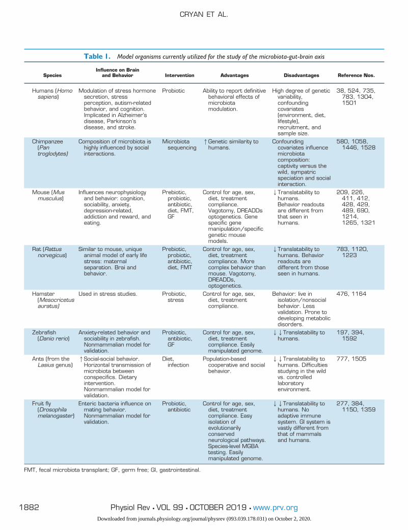
Table 1. Model organisms currently utilized for the study of the microbiota-gut-brain axis
SpeciesInfluence on Brain
and Behavior Intervention Advantages Disadvantages Reference Nos.
Humans (Homosapiens)
Modulation of stress hormonesecretion, stressperception, autism-relatedbehavior, and cognition.Implicated in Alzheimer’sdisease, Parkinson’sdisease, and stroke.
Probiotic Ability to report definitivebehavioral effects ofmicrobiotamodulation.
High degree of geneticvariability,confoundingcovariates(environment, diet,lifestyle),recruitment, andsample size.
38, 524, 735,783, 1304,1501
Chimpanzee(Pantroglodytes)
Composition of microbiota ishighly influenced by socialinteractions.
Microbiotasequencing
1Genetic similarity tohumans.
Confoundingcovariates influencemicrobiotacomposition:captivity versus thewild, sympatricspeciation and socialinteraction.
580, 1058,1446, 1528
Mouse (Musmusculus)
Influences neurophysiologyand behavior: cognition,sociability, anxiety,depression-related,addiction and reward, andeating.
Prebiotic,probiotic,antibiotic,diet, FMT,GF
Control for age, sex,diet, treatmentcompliance.Vagotomy, DREADDsoptogenetics. Genespecific genemanipulation/specificgenetic mousemodels.
2Translatability tohumans.Behavior readoutsare different fromthat seen inhumans.
209, 226,411, 412,428, 429,489, 690,1214,1265, 1321
Rat (Rattusnorvegicus)
Similar to mouse, uniqueanimal model of early lifestress: maternalseparation. Brai andbehavior.
Prebiotic,probiotic,antibiotic,diet, FMT
Control for age, sex,diet, treatmentcompliance. Morecomplex behavior thanmouse. Vagotomy,DREADDs,optogenetics.
2Translatability tohumans. Behaviorreadouts aredifferent from thoseseen in humans.
783, 1120,1223
Hamster(Mesocricetusauratus)
Used in stress studies. Probiotic,stress
Control for age, sex,diet, treatmentcompliance.
Behavior: live inisolation/nonsocialbehavior. Lessvalidation. Prone todeveloping metabolicdisorders.
476, 1164
Zebrafish(Danio rerio)
Anxiety-related behavior andsociability in zebrafish.Nonmammalian model forvalidation.
Probiotic,antibiotic,GF
Control for age, sex,diet, treatmentcompliance. Easilymanipulated genome.
22Translatability tohumans.
197, 394,1592
Ants (from theLasius genus)
1Social-social behavior.Horizontal transmission ofmicrobiota betweenconspecifics. Dietaryintervention.Nonmammalian model forvalidation.
Diet,infection
Population-basedcooperative and socialbehavior.
22Translatability tohumans. Difficultiesstudying in the wildvs. controlledlaboratoryenvironment.
777, 1505
Fruit fly(Drosophilamelanogaster)
Enteric bacteria influence onmating behavior.Nonmammalian model forvalidation.
Probiotic,antibiotic
Control for age, sex,diet, treatmentcompliance. Easyisolation ofevolutionarilyconservedneurological pathways.Species-level MGBAtesting. Easilymanipulated genome.
22Translatability tohumans. Noadaptive immunesystem. GI system isvastly different fromthat of mammalsand humans.
277, 384,1150, 1359
FMT, fecal microbiota transplant; GF, germ free; GI, gastrointestinal.
CRYAN ET AL.
1882 Physiol Rev • VOL 99 • OCTOBER 2019 • www.prv.orgDownloaded from journals.physiology.org/journal/physrev (093.039.178.031) on October 2, 2020.
kept free of microbes for 2 wk (1111). However, successivegenerations of GF rodents were not produced until the mid-20th century (622). Currently, similar methods are typicallyused to generate many generations of GF animals. To avoidinoculation of pups by microbiota, C-section is carried outcarefully to avoid contact between pup and the microbesresiding on both the dam’s vagina and skin, and pups arethen hand-raised in an aseptic isolator (29, 598, 931, 1076,1424). From this point on, colonies are maintained GF withsterile food, bedding, and water. Cages and feces are regu-larly swabbed to confirm that no bacteria are present (157).Subsequent GF animals can then be bred in an isolator andGF pups born per vaginum. Alternatively, GF animals canbe produced by embryonic transfer at the two-cell stage intoa GF host mother (157).
Animals lacking microbiota have extraordinarily differentdevelopment and physiology than animals hosting com-mensal bacteria (see TABLE 2 for a comprehensive summaryof studies involving GF animals). GF animals are smaller inbody weight and have impaired intestinal function (41, 730,1318), have lower concentrations of most GI luminal aminoacids than specific pathogen-free (SPF) mice (1639), andactually live longer (599, 1254, 1469, 1625). Due to thelack of commensal microbes, GF animals have impairedimmune systems, dysregulated hormone signaling, alteredmetabolism, and differences in neurotransmission fromconventional counterparts (776, 1092, 1148, 1434, 1597).Interestingly, phenotypes of GF animals vary across species,sex, research group, and even strain, demonstrating thatboth microbiota and host genetics are important influencersof phenotype. For example, some studies involving GFSwiss Webster mice show a decreased anxiety-like behavior(56, 308, 1092), whereas the opposite has been found inmale GF BALB/c mice (278, 1104).
Although an important tool, GF mice have many limita-tions in terms of aberrant physiology, neurodevelopment,and immunity, as well as limited translatability to humansituations (1097). Nonetheless, they have been an impor-tant starting point in answering the question of whether themicrobiota is involved in a given process or not (931).Moreover, GF studies are now being extended to othernon-rodent species including porcine models to enhance thetranslational value of findings (274).
Alternatively, colonization of mice with specific, knownstrains of bacteria has also shown to be a useful approach tointerrogate microbiota-physiology interactions (600). Fromthese gnotobiotic animals, it is possible to decipher mecha-nisms of communication between specific members of themicrobiota and the host organism. Among such ap-proaches, the altered Schaedler flora (ASF) mouse line ismost utilized (940, 1137, 1632). ASF mice are colonizedwith just eight bacterial species allowing a more simplifiedstudy of microbiota involvement in brain function relative
to conventionally colonized strains, but with more clinicalrelevance than GF studies. The minimalist bacterial coloni-zation of the ASF mouse avoids the complications of thehigh abundance and diversity in conventional mice, whileovercoming the host developmental hurdles seen in GFmice, such as an underdeveloped immune system (69, 658),slower intestinal epithelial turnover (1318), differing nutri-tional requirements, and less body fat (80). The ASF modelthus presents an attractive alternative option for transla-tional microbiota-gut-brain axis research, particularly in-volving stress (940).
B. Antibiotics
While initially developed to fight infections, antibiotics arealso a useful pharmacological tool for investigating the im-pact of microbiota perturbations on brain and behavior (seeTABLE 3). They offer much greater temporal flexibility andspecificity compared with the GF model of microbiota ab-lation as they can be delivered acutely or chronically at anystage across an animal’s lifespan [e.g., during periods ofpotential vulnerability such as the early postnatal period(867, 1119), adolescence (429), or in aging]. Additionally,the ability to titrate the dose of antibiotics allows for agreater level of control over the extent of microbiota deple-tion, from minor perturbations to the microbiota throughsubtherapeutic doses of a single antibiotic, to cocktails ofantibiotics designed to substantially ablate the entire micro-biota. An important consideration in the use of antibioticsto investigate the microbiota-gut-brain axis is their absorp-tion from the GI tract. Nonabsorbable antibiotics [i.e., van-comycin, neomycin, and bacitracin (1497)] offer the advan-tage that they knockdown the microbiota in the gut whilenot entering systemic circulation, thereby avoiding any po-tential systemic and even central nervous system (CNS) ef-fects and allowing us to directly assess the effect of a loss ofmicrobiota on the brain. Other antibiotics such as metroni-dazole and minocycline can potentially enter the CNS andcan have direct action on brain and behavior (261) (e.g.,microglial inhibition with minocycline; Ref. 1256), so theseresults must be interpreted with caution. Despite such lim-itations, antibiotics have been crucial in corroborating thebehavioral and biological observations documented in GFanimals. Indeed, antibiotic administration to laboratory an-imals has been shown to influence behaviors such as socia-bility and anxiety (418, 550, 615).
A final advantage of antibiotics is that they offer a tool tomodel the clinical scenario in humans. Administrationschedules can be made to model the courses of antibioticsthat millions of people take each year for multiple condi-tions, allowing us to determine the effect that such treat-ments may be having on the brain and behavior. The flexi-bility and translational relevance of antibiotics make them ahugely valuable tool in the study of the microbiota-gut-brain axis and they will form a key component of future
THE MICROBIOTA-GUT-BRAIN AXIS
1883Physiol Rev • VOL 99 • OCTOBER 2019 • www.prv.orgDownloaded from journals.physiology.org/journal/physrev (093.039.178.031) on October 2, 2020.
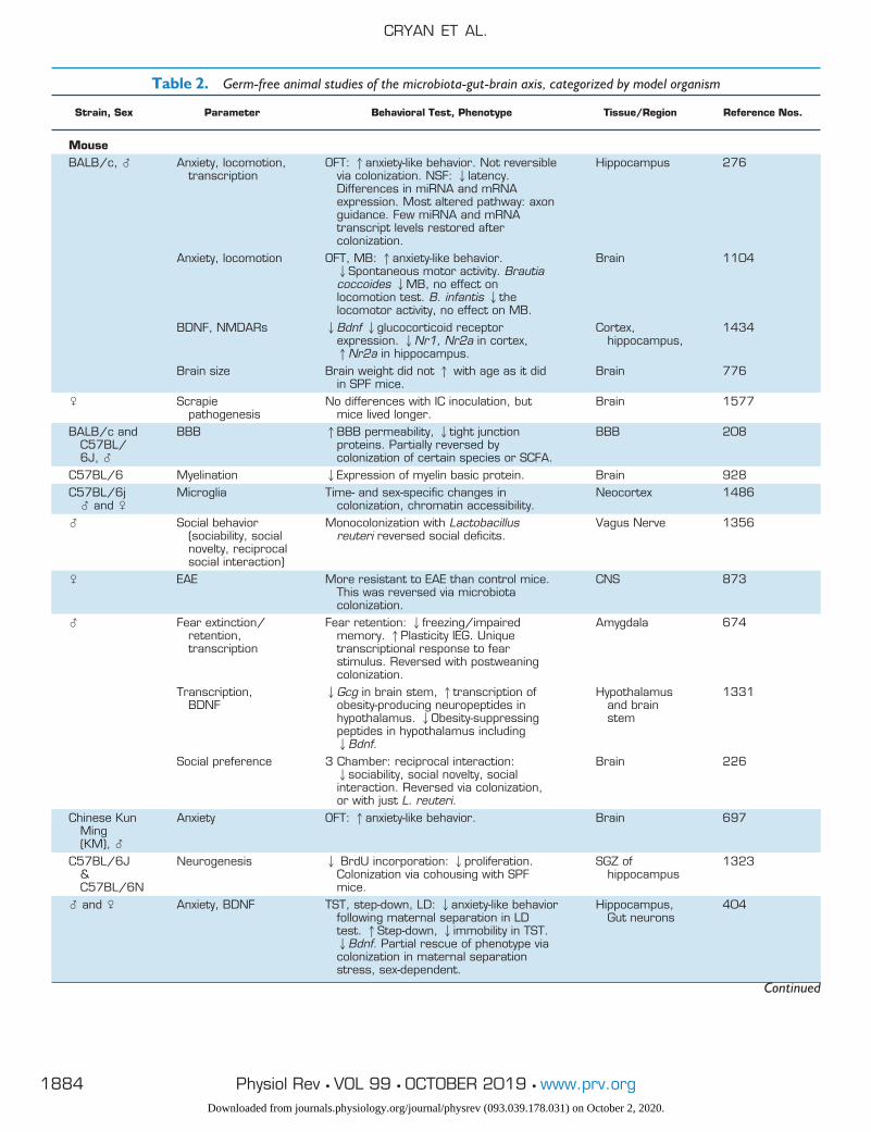
Table 2. Germ-free animal studies of the microbiota-gut-brain axis, categorized by model organism
Strain, Sex Parameter Behavioral Test, Phenotype Tissue/Region Reference Nos.
MouseBALB/c, � Anxiety, locomotion,
transcriptionOFT: 1anxiety-like behavior. Not reversible
via colonization. NSF: 2latency.Differences in miRNA and mRNAexpression. Most altered pathway: axonguidance. Few miRNA and mRNAtranscript levels restored aftercolonization.
Hippocampus 276
Anxiety, locomotion OFT, MB: 1anxiety-like behavior.2Spontaneous motor activity. Brautiacoccoides 2MB, no effect onlocomotion test. B. infantis 2thelocomotor activity, no effect on MB.
Brain 1104
BDNF, NMDARs 2Bdnf 2glucocorticoid receptorexpression. 2Nr1, Nr2a in cortex,1Nr2a in hippocampus.
Cortex,hippocampus,
1434
Brain size Brain weight did not 1 with age as it didin SPF mice.
Brain 776
� Scrapiepathogenesis
No differences with IC inoculation, butmice lived longer.
Brain 1577
BALB/c andC57BL/6J, �
BBB 1BBB permeability, 2tight junctionproteins. Partially reversed bycolonization of certain species or SCFA.
BBB 208
C57BL/6 Myelination 2Expression of myelin basic protein. Brain 928C57BL/6j
� and �Microglia Time- and sex-specific changes in
colonization, chromatin accessibility.Neocortex 1486
� Social behavior(sociability, socialnovelty, reciprocalsocial interaction)
Monocolonization with Lactobacillusreuteri reversed social deficits.
Vagus Nerve 1356
� EAE More resistant to EAE than control mice.This was reversed via microbiotacolonization.
CNS 873
� Fear extinction/retention,transcription
Fear retention: 2freezing/impairedmemory. 1Plasticity IEG. Uniquetranscriptional response to fearstimulus. Reversed with postweaningcolonization.
Amygdala 674
Transcription,BDNF
2Gcg in brain stem, 1transcription ofobesity-producing neuropeptides inhypothalamus. 2Obesity-suppressingpeptides in hypothalamus including2Bdnf.
Hypothalamusand brainstem
1331
Social preference 3 Chamber: reciprocal interaction:2sociability, social novelty, socialinteraction. Reversed via colonization,or with just L. reuteri.
Brain 226
Chinese KunMing(KM), �
Anxiety OFT: 1anxiety-like behavior. Brain 697
C57BL/6J&C57BL/6N
Neurogenesis 2 BrdU incorporation: 2proliferation.Colonization via cohousing with SPFmice.
SGZ ofhippocampus
1323
� and � Anxiety, BDNF TST, step-down, LD: 2anxiety-like behaviorfollowing maternal separation in LDtest. 1Step-down, 2immobility in TST.2Bdnf. Partial rescue of phenotype viacolonization in maternal separationstress, sex-dependent.
Hippocampus,Gut neurons
404
Continued
CRYAN ET AL.
1884 Physiol Rev • VOL 99 • OCTOBER 2019 • www.prv.orgDownloaded from journals.physiology.org/journal/physrev (093.039.178.031) on October 2, 2020.
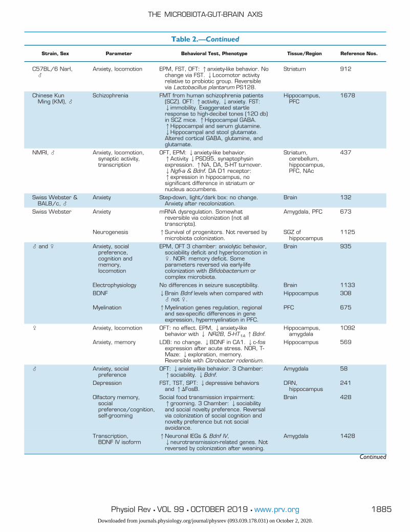
Table 2.—Continued
Strain, Sex Parameter Behavioral Test, Phenotype Tissue/Region Reference Nos.
C57BL/6 NarI,�
Anxiety, locomotion EPM, FST, OFT: 1anxiety-like behavior. Nochange via FST. 2Locomotor activityrelative to probiotic group. Reversiblevia Lactobacillus plantarum PS128.
Striatum 912
Chinese KunMing (KM), �
Schizophrenia FMT from human schizophrenia patients(SCZ). OFT: 1activity, 2anxiety. FST:2immobility. Exaggerated startleresponse to high-decibel tones (120 db)in SCZ mice. 1Hippocampal GABA.1Hippocampal and serum glutamine.2Hippocampal and stool glutamate.Altered cortical GABA, glutamine, andglutamate.
Hippocampus,PFC
1678
NMRI, � Anxiety, locomotion,synaptic activity,transcription
OFT, EPM: 2anxiety-like behavior.1Activity 2PSD95, synaptophysinexpression. 1NA, DA, 5-HT turnover.2Ngfi-a & Bdnf. DA D1 receptor:1expression in hippocampus, nosignificant difference in striatum ornucleus accumbens.
Striatum,cerebellum,hippocampus,PFC, NAc
437
Swiss Webster &BALB/c, �
Anxiety Step-down, light/dark box: no change.Anxiety after recolonization.
Brain 132
Swiss Webster Anxiety mRNA dysregulation. Somewhatreversible via colonization (not alltranscripts).
Amygdala, PFC 673
Neurogenesis 1Survival of progenitors. Not reversed bymicrobiota colonization.
SGZ ofhippocampus
1125
� and � Anxiety, socialpreference,cognition andmemory,locomotion
EPM, OFT 3 chamber: anxiolytic behavior,sociability deficit and hyperlocomotion in�. NOR: memory deficit. Someparameters reversed via early-lifecolonization with Bifidobacterium orcomplex microbiota.
Brain 935
Electrophysiology No differences in seizure susceptibility. Brain 1133BDNF 2Brain Bdnf levels when compared with
� not �.Hippocampus 308
Myelination 1Myelination genes regulation, regionaland sex-specific differences in geneexpression, hypermyelination in PFC.
PFC 675
� Anxiety, locomotion OFT: no effect. EPM, 2anxiety-likebehavior with 2 NR2B, 5-HT1A 1Bdnf.
Hippocampus,amygdala
1092
Anxiety, memory LDB: no change. 2BDNF in CA1. 2c-fosexpression after acute stress. NOR, T-Maze: 2exploration, memory.Reversible with Citrobacter rodentium.
Hippocampus 569
� Anxiety, socialpreference
OFT: 2anxiety-like behavior. 3 Chamber:1sociability. 2Bdnf.
Amygdala 58
Depression FST, TST, SPT: 2depressive behaviorsand 1�FosB.
DRN,hippocampus
241
Olfactory memory,socialpreference/cognition,self-grooming
Social food transmission impairment:1grooming. 3 Chamber: 2sociabilityand social novelty preference. Reversalvia colonization of social cognition andnovelty preference but not socialavoidance.
Brain 428
Transcription,BDNF IV isoform
1Neuronal IEGs & Bdnf IV,2neurotransmission-related genes. Notreversed by colonization after weaning.
Amygdala 1428
Continued
THE MICROBIOTA-GUT-BRAIN AXIS
1885Physiol Rev • VOL 99 • OCTOBER 2019 • www.prv.orgDownloaded from journals.physiology.org/journal/physrev (093.039.178.031) on October 2, 2020.
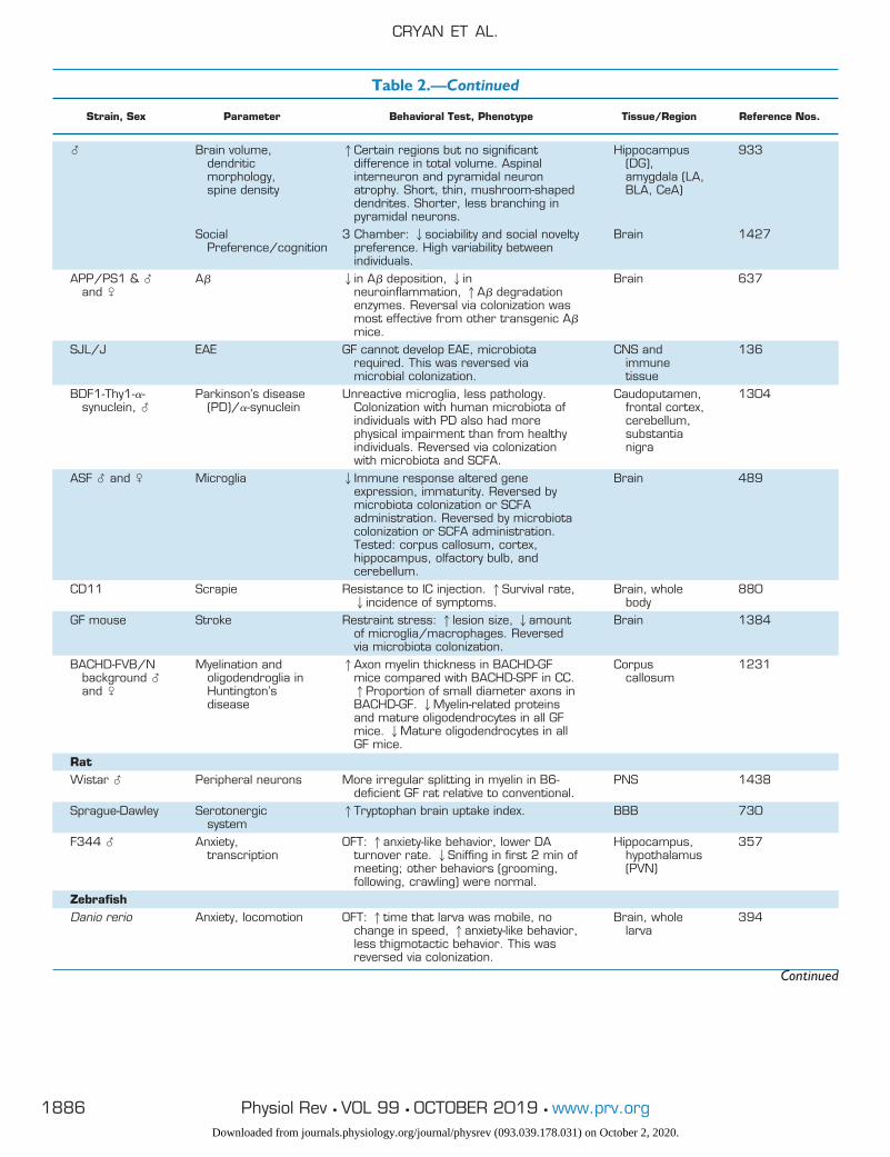
Table 2.—Continued
Strain, Sex Parameter Behavioral Test, Phenotype Tissue/Region Reference Nos.
� Brain volume,dendriticmorphology,spine density
1Certain regions but no significantdifference in total volume. Aspinalinterneuron and pyramidal neuronatrophy. Short, thin, mushroom-shapeddendrites. Shorter, less branching inpyramidal neurons.
Hippocampus(DG),amygdala (LA,BLA, CeA)
933
SocialPreference/cognition
3 Chamber: 2sociability and social noveltypreference. High variability betweenindividuals.
Brain 1427
APP/PS1 & �and �
A� 2in A� deposition, 2inneuroinflammation, 1A� degradationenzymes. Reversal via colonization wasmost effective from other transgenic A�mice.
Brain 637
SJL/J EAE GF cannot develop EAE, microbiotarequired. This was reversed viamicrobial colonization.
CNS andimmunetissue
136
BDF1-Thy1-�-synuclein, �
Parkinson’s disease(PD)/�-synuclein
Unreactive microglia, less pathology.Colonization with human microbiota ofindividuals with PD also had morephysical impairment than from healthyindividuals. Reversed via colonizationwith microbiota and SCFA.
Caudoputamen,frontal cortex,cerebellum,substantianigra
1304
ASF � and � Microglia 2Immune response altered geneexpression, immaturity. Reversed bymicrobiota colonization or SCFAadministration. Reversed by microbiotacolonization or SCFA administration.Tested: corpus callosum, cortex,hippocampus, olfactory bulb, andcerebellum.
Brain 489
CD11 Scrapie Resistance to IC injection. 1Survival rate,2incidence of symptoms.
Brain, wholebody
880
GF mouse Stroke Restraint stress: 1lesion size, 2amountof microglia/macrophages. Reversedvia microbiota colonization.
Brain 1384
BACHD-FVB/Nbackground �and �
Myelination andoligodendroglia inHuntington’sdisease
1Axon myelin thickness in BACHD-GFmice compared with BACHD-SPF in CC.1Proportion of small diameter axons inBACHD-GF. 2Myelin-related proteinsand mature oligodendrocytes in all GFmice. 2Mature oligodendrocytes in allGF mice.
Corpuscallosum
1231
RatWistar � Peripheral neurons More irregular splitting in myelin in B6-
deficient GF rat relative to conventional.PNS 1438
Sprague-Dawley Serotonergicsystem
1Tryptophan brain uptake index. BBB 730
F344 � Anxiety,transcription
OFT: 1anxiety-like behavior, lower DAturnover rate. 2Sniffing in first 2 min ofmeeting; other behaviors (grooming,following, crawling) were normal.
Hippocampus,hypothalamus(PVN)
357
ZebrafishDanio rerio Anxiety, locomotion OFT: 1time that larva was mobile, no
change in speed, 1anxiety-like behavior,less thigmotactic behavior. This wasreversed via colonization.
Brain, wholelarva
394
Continued
CRYAN ET AL.
1886 Physiol Rev • VOL 99 • OCTOBER 2019 • www.prv.orgDownloaded from journals.physiology.org/journal/physrev (093.039.178.031) on October 2, 2020.
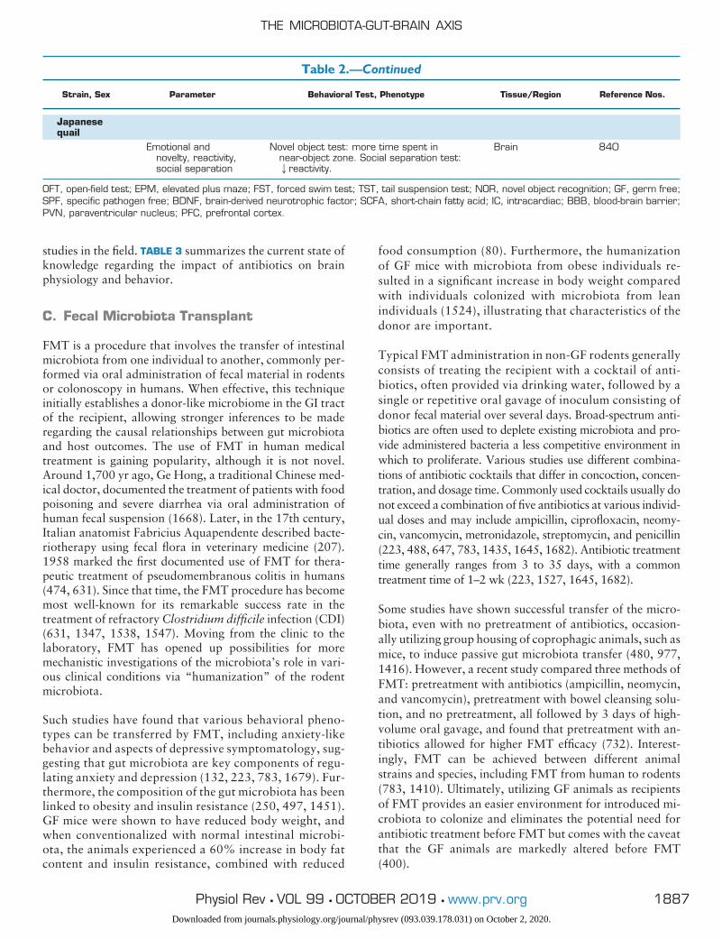
studies in the field. TABLE 3 summarizes the current state ofknowledge regarding the impact of antibiotics on brainphysiology and behavior.
C. Fecal Microbiota Transplant
FMT is a procedure that involves the transfer of intestinalmicrobiota from one individual to another, commonly per-formed via oral administration of fecal material in rodentsor colonoscopy in humans. When effective, this techniqueinitially establishes a donor-like microbiome in the GI tractof the recipient, allowing stronger inferences to be maderegarding the causal relationships between gut microbiotaand host outcomes. The use of FMT in human medicaltreatment is gaining popularity, although it is not novel.Around 1,700 yr ago, Ge Hong, a traditional Chinese med-ical doctor, documented the treatment of patients with foodpoisoning and severe diarrhea via oral administration ofhuman fecal suspension (1668). Later, in the 17th century,Italian anatomist Fabricius Aquapendente described bacte-riotherapy using fecal flora in veterinary medicine (207).1958 marked the first documented use of FMT for thera-peutic treatment of pseudomembranous colitis in humans(474, 631). Since that time, the FMT procedure has becomemost well-known for its remarkable success rate in thetreatment of refractory Clostridium difficile infection (CDI)(631, 1347, 1538, 1547). Moving from the clinic to thelaboratory, FMT has opened up possibilities for moremechanistic investigations of the microbiota’s role in vari-ous clinical conditions via “humanization” of the rodentmicrobiota.
Such studies have found that various behavioral pheno-types can be transferred by FMT, including anxiety-likebehavior and aspects of depressive symptomatology, sug-gesting that gut microbiota are key components of regu-lating anxiety and depression (132, 223, 783, 1679). Fur-thermore, the composition of the gut microbiota has beenlinked to obesity and insulin resistance (250, 497, 1451).GF mice were shown to have reduced body weight, andwhen conventionalized with normal intestinal microbi-ota, the animals experienced a 60% increase in body fatcontent and insulin resistance, combined with reduced
food consumption (80). Furthermore, the humanizationof GF mice with microbiota from obese individuals re-sulted in a significant increase in body weight comparedwith individuals colonized with microbiota from leanindividuals (1524), illustrating that characteristics of thedonor are important.
Typical FMT administration in non-GF rodents generallyconsists of treating the recipient with a cocktail of anti-biotics, often provided via drinking water, followed by asingle or repetitive oral gavage of inoculum consisting ofdonor fecal material over several days. Broad-spectrum anti-biotics are often used to deplete existing microbiota and pro-vide administered bacteria a less competitive environment inwhich to proliferate. Various studies use different combina-tions of antibiotic cocktails that differ in concoction, concen-tration, and dosage time. Commonly used cocktails usually donot exceed a combination of five antibiotics at various individ-ual doses and may include ampicillin, ciprofloxacin, neomy-cin, vancomycin, metronidazole, streptomycin, and penicillin(223, 488, 647, 783, 1435, 1645, 1682). Antibiotic treatmenttime generally ranges from 3 to 35 days, with a commontreatment time of 1–2 wk (223, 1527, 1645, 1682).
Some studies have shown successful transfer of the micro-biota, even with no pretreatment of antibiotics, occasion-ally utilizing group housing of coprophagic animals, such asmice, to induce passive gut microbiota transfer (480, 977,1416). However, a recent study compared three methods ofFMT: pretreatment with antibiotics (ampicillin, neomycin,and vancomycin), pretreatment with bowel cleansing solu-tion, and no pretreatment, all followed by 3 days of high-volume oral gavage, and found that pretreatment with an-tibiotics allowed for higher FMT efficacy (732). Interest-ingly, FMT can be achieved between different animalstrains and species, including FMT from human to rodents(783, 1410). Ultimately, utilizing GF animals as recipientsof FMT provides an easier environment for introduced mi-crobiota to colonize and eliminates the potential need forantibiotic treatment before FMT but comes with the caveatthat the GF animals are markedly altered before FMT(400).
Table 2.—Continued
Strain, Sex Parameter Behavioral Test, Phenotype Tissue/Region Reference Nos.
Japanesequail
Emotional andnovelty, reactivity,social separation
Novel object test: more time spent innear-object zone. Social separation test:2reactivity.
Brain 840
OFT, open-field test; EPM, elevated plus maze; FST, forced swim test; TST, tail suspension test; NOR, novel object recognition; GF, germ free;SPF, specific pathogen free; BDNF, brain-derived neurotrophic factor; SCFA, short-chain fatty acid; IC, intracardiac; BBB, blood-brain barrier;PVN, paraventricular nucleus; PFC, prefrontal cortex.
THE MICROBIOTA-GUT-BRAIN AXIS
1887Physiol Rev • VOL 99 • OCTOBER 2019 • www.prv.orgDownloaded from journals.physiology.org/journal/physrev (093.039.178.031) on October 2, 2020.
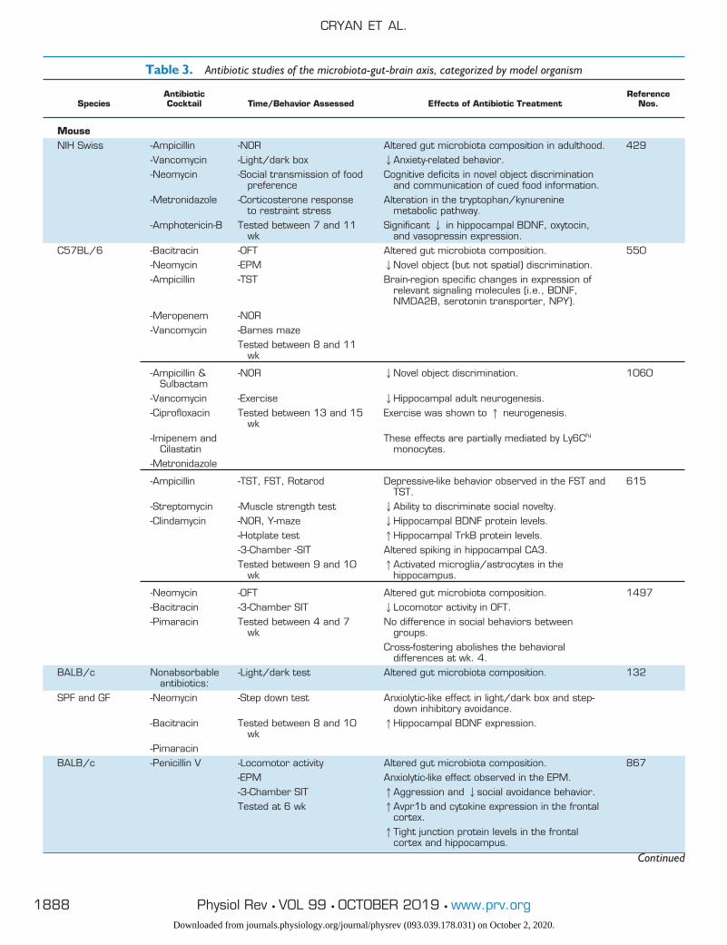
Table 3. Antibiotic studies of the microbiota-gut-brain axis, categorized by model organism
SpeciesAntibioticCocktail Time/Behavior Assessed Effects of Antibiotic Treatment
ReferenceNos.
MouseNIH Swiss -Ampicillin -NOR Altered gut microbiota composition in adulthood. 429
-Vancomycin -Light/dark box 2Anxiety-related behavior.-Neomycin -Social transmission of food
preferenceCognitive deficits in novel object discrimination
and communication of cued food information.-Metronidazole -Corticosterone response
to restraint stressAlteration in the tryptophan/kynurenine
metabolic pathway.-Amphotericin-B Tested between 7 and 11
wkSignificant 2 in hippocampal BDNF, oxytocin,
and vasopressin expression.C57BL/6 -Bacitracin -OFT Altered gut microbiota composition. 550
-Neomycin -EPM 2Novel object (but not spatial) discrimination.-Ampicillin -TST Brain-region specific changes in expression of
relevant signaling molecules (i.e., BDNF,NMDA2B, serotonin transporter, NPY).
-Meropenem -NOR-Vancomycin -Barnes maze
Tested between 8 and 11wk
-Ampicillin &Sulbactam
-NOR 2Novel object discrimination. 1060
-Vancomycin -Exercise 2Hippocampal adult neurogenesis.-Ciprofloxacin Tested between 13 and 15
wkExercise was shown to 1 neurogenesis.
-Imipenem andCilastatin
These effects are partially mediated by Ly6Chi
monocytes.-Metronidazole
-Ampicillin -TST, FST, Rotarod Depressive-like behavior observed in the FST andTST.
615
-Streptomycin -Muscle strength test 2Ability to discriminate social novelty.-Clindamycin -NOR, Y-maze 2Hippocampal BDNF protein levels.
-Hotplate test 1Hippocampal TrkB protein levels.-3-Chamber -SIT Altered spiking in hippocampal CA3.Tested between 9 and 10
wk1Activated microglia/astrocytes in the
hippocampus.
-Neomycin -OFT Altered gut microbiota composition. 1497-Bacitracin -3-Chamber SIT 2Locomotor activity in OFT.-Pimaracin Tested between 4 and 7
wkNo difference in social behaviors between
groups.Cross-fostering abolishes the behavioral
differences at wk. 4.BALB/c Nonabsorbable
antibiotics:-Light/dark test Altered gut microbiota composition. 132
SPF and GF -Neomycin -Step down test Anxiolytic-like effect in light/dark box and step-down inhibitory avoidance.
-Bacitracin Tested between 8 and 10wk
1Hippocampal BDNF expression.
-PimaracinBALB/c -Penicillin V -Locomotor activity Altered gut microbiota composition. 867
-EPM Anxiolytic-like effect observed in the EPM.-3-Chamber SIT 1Aggression and 2social avoidance behavior.Tested at 6 wk 1Avpr1b and cytokine expression in the frontal
cortex.1Tight junction protein levels in the frontal
cortex and hippocampus.
Continued
CRYAN ET AL.
1888 Physiol Rev • VOL 99 • OCTOBER 2019 • www.prv.orgDownloaded from journals.physiology.org/journal/physrev (093.039.178.031) on October 2, 2020.
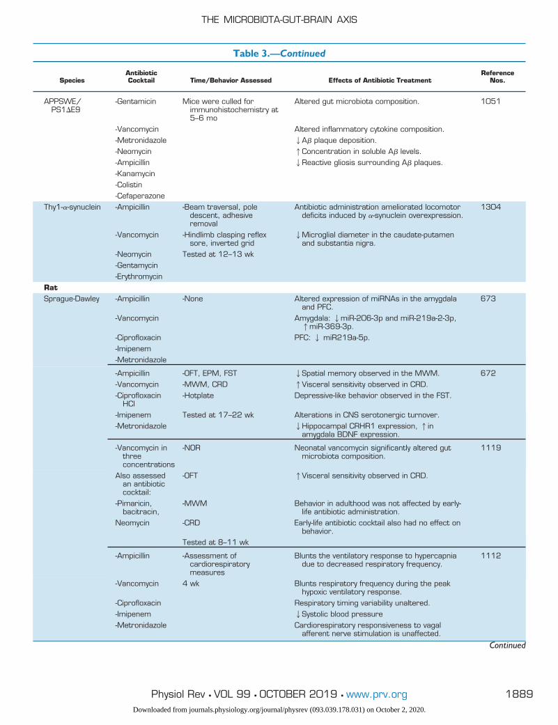
Table 3.—Continued
SpeciesAntibioticCocktail Time/Behavior Assessed Effects of Antibiotic Treatment
ReferenceNos.
APPSWE/PS1�E9
-Gentamicin Mice were culled forimmunohistochemistry at5–6 mo
Altered gut microbiota composition. 1051
-Vancomycin Altered inflammatory cytokine composition.-Metronidazole 2A� plaque deposition.-Neomycin 1Concentration in soluble A� levels.-Ampicillin 2Reactive gliosis surrounding A� plaques.-Kanamycin-Colistin-Cefaperazone
Thy1-�-synuclein -Ampicillin -Beam traversal, poledescent, adhesiveremoval
Antibiotic administration ameliorated locomotordeficits induced by �-synuclein overexpression.
1304
-Vancomycin -Hindlimb clasping reflexsore, inverted grid
2Microglial diameter in the caudate-putamenand substantia nigra.
-Neomycin Tested at 12–13 wk-Gentamycin-Erythromycin
RatSprague-Dawley -Ampicillin -None Altered expression of miRNAs in the amygdala
and PFC.673
-Vancomycin Amygdala: 2miR-206-3p and miR-219a-2-3p,1miR-369-3p.
-Ciprofloxacin PFC: 2 miR219a-5p.-Imipenem-Metronidazole
-Ampicillin -OFT, EPM, FST 2Spatial memory observed in the MWM. 672-Vancomycin -MWM, CRD 1Visceral sensitivity observed in CRD.-Ciprofloxacin
HCl-Hotplate Depressive-like behavior observed in the FST.
-Imipenem Tested at 17–22 wk Alterations in CNS serotonergic turnover.-Metronidazole 2Hippocampal CRHR1 expression, 1in
amygdala BDNF expression.
-Vancomycin inthreeconcentrations
-NOR Neonatal vancomycin significantly altered gutmicrobiota composition.
1119
Also assessedan antibioticcocktail:
-OFT 1Visceral sensitivity observed in CRD.
-Pimaricin,bacitracin,
-MWM Behavior in adulthood was not affected by early-life antibiotic administration.
Neomycin -CRD Early-life antibiotic cocktail also had no effect onbehavior.
Tested at 8–11 wk
-Ampicillin -Assessment ofcardiorespiratorymeasures
Blunts the ventilatory response to hypercapniadue to decreased respiratory frequency.
1112
-Vancomycin 4 wk Blunts respiratory frequency during the peakhypoxic ventilatory response.
-Ciprofloxacin Respiratory timing variability unaltered.-Imipenem 2Systolic blood pressure-Metronidazole Cardiorespiratory responsiveness to vagal
afferent nerve stimulation is unaffected.
Continued
THE MICROBIOTA-GUT-BRAIN AXIS
1889Physiol Rev • VOL 99 • OCTOBER 2019 • www.prv.orgDownloaded from journals.physiology.org/journal/physrev (093.039.178.031) on October 2, 2020.
FMT is increasingly being utilized in humans for the treat-ment of CDI in the clinic (240) and, in a research setting,FMT has also been tested for the treatment of IBD, IBS, andchronic constipation. In a double-blind, randomized trialtreating IBS with FMT, 65% of participants receiving FMTshowed a response to treatment at 3 mo, compared with43% receiving a placebo (739). CDI is generally treatedwith antibiotics, but in the case of recurrent CDI, treatmentwith FMT ultimately cured 98% (207). The potential ofFMT in research and as a medicinal therapy provides prom-ise for the treatment of GI-related diseases and conditions,including the practice of autologous FMT. Here, a patient isgiven an FMT of their own presurgery/“healthy” fecal mat-ter during the recovery phase, effectively reconstitutingtheir major commensal bacterial populations and reestab-lishing the patient’s gut microbiota diversity as well as com-position (1436, 1465). This may well result in an increase inthe practice of fecal matter banking for post-treatment re-colonization of a patient’s gut microbiota, a practice couldbecome commonplace in the very near future.
D. Prebiotics and Fermented Foods
The definition of prebiotics as determined by the Interna-tional Scientific Association for Probiotics and Prebiotics is“a substrate that is selectively utilized by host microorgan-
isms conferring a health benefit” (579). One of the mainclasses of prebiotics is dietary fiber, often defined as “car-bohydrates with a degree of polymerization greater than 2,which fail to be hydrolyzed or absorbed in the small intes-tine” (1419). These include inulin, fructooligosaccharides(FOS), galactooligosaccharides (GOS), resistant starch, andother soluble dietary fibers, among others (although not alldietary fibers are prebiotic). Typical dietary sources of pre-biotics include fruits and vegetables such as asparagus, leek,banana, chicory, and grains such as oats and wheat. AsWestern-style diet consumption increases, a drop in prebi-otic intake that correlates with a rise in the incidence ofinflammatory diseases, obesity, metabolic syndrome andanxiety, stress, and other “lifestyle” disorders have beenseen. Importantly, prebiotics do not always change thecomposition and activity of the gut microbiota in a selectiveand predictable manner (164). Nonetheless, prebiotic sup-plementation has been demonstrated to reduce stress re-sponsiveness, anxiety, and depressive-like behavior, as wellas facilitate changes in hippocampal synaptic efficacy, in-cluding increased hippocampal brain-derived neurotrophicfactor (BDNF) expression, general hypothalamic neuronalactivity, and enhanced cognition and learning (see TABLE 4).Most studies thus far have been descriptive and are limitedto demonstrating prebiotic influence on brain physiologyand behavior (see TABLE 4). Further studies should, there-
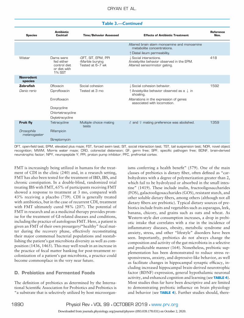
Table 3.—Continued
SpeciesAntibioticCocktail Time/Behavior Assessed Effects of Antibiotic Treatment
ReferenceNos.
Altered brain stem monoamine and monoaminemetabolite concentrations.1Distal ileum permeability.
Wistar Dams werefed eithercontrol dietor diet with1% SST
-OFT, SIT, EPM, PPI-Marble buryingTested at 6–7 wk
2Social interactions.Anxiety-like behavior observed in the EPM.Altered sensorimotor gating.
418
Nonrodentspecies
Zebrafish Ofloxacin Social cohesion 2Social cohesion behavior. 1592Danio rerio Ciprofloxacin Tested at 3 mo 1Anxiety-like behavior observed as a 2 in
shoaling.Enrofloxacin Alterations in the expression of genes
associated with locomotion.DoxycyclineChlortetracyclineOxytetracycline
Fruit fly Tetracycline Multiple choice matingtests
� and � mating preference was abolished. 1359
Drosophilamelanogaster
Rifampicin
Streptomycin
OFT, open-field test; EPM, elevated plus maze; FST, forced swim test; SIT, social interaction test; TST, tail suspension test; NOR, novel objectrecognition; MWM, Morris water maze; CRD, colorectal distension; GF, germ free; SPF, specific pathogen free; BDNF, brain-derivedneurotrophic factor; NPY, neuropeptide Y; PPI, proton pump inhibitor; PFC, prefrontal cortex.
CRYAN ET AL.
1890 Physiol Rev • VOL 99 • OCTOBER 2019 • www.prv.orgDownloaded from journals.physiology.org/journal/physrev (093.039.178.031) on October 2, 2020.
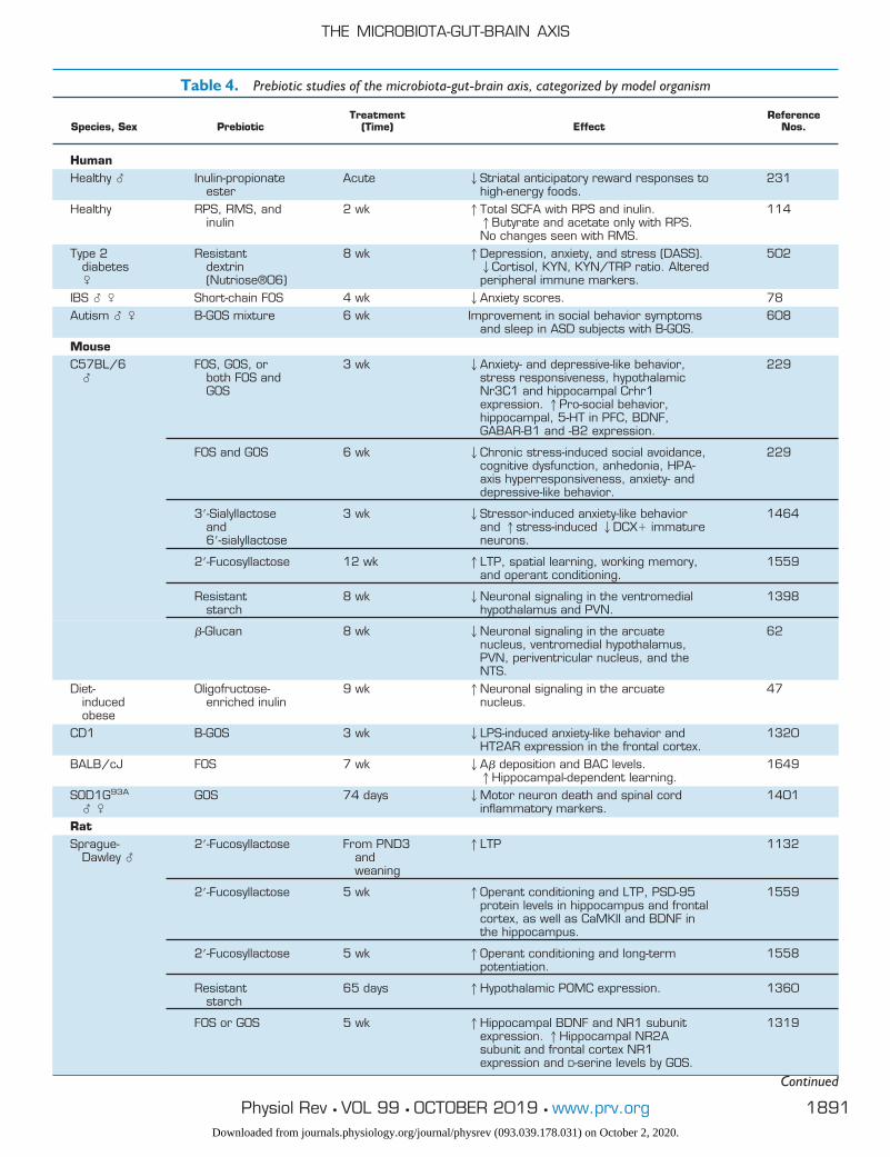
Table 4. Prebiotic studies of the microbiota-gut-brain axis, categorized by model organism
Species, Sex PrebioticTreatment
(Time) EffectReference
Nos.
HumanHealthy � Inulin-propionate
esterAcute 2Striatal anticipatory reward responses to
high-energy foods.231
Healthy RPS, RMS, andinulin
2 wk 1Total SCFA with RPS and inulin.1Butyrate and acetate only with RPS.No changes seen with RMS.
114
Type 2diabetes�
Resistantdextrin(Nutriose®06)
8 wk 1Depression, anxiety, and stress (DASS).2Cortisol, KYN, KYN/TRP ratio. Alteredperipheral immune markers.
502
IBS � � Short-chain FOS 4 wk 2Anxiety scores. 78Autism � � B-GOS mixture 6 wk Improvement in social behavior symptoms
and sleep in ASD subjects with B-GOS.608
MouseC57BL/6
�FOS, GOS, or
both FOS andGOS
3 wk 2Anxiety- and depressive-like behavior,stress responsiveness, hypothalamicNr3C1 and hippocampal Crhr1expression. 1Pro-social behavior,hippocampal, 5-HT in PFC, BDNF,GABAR-B1 and -B2 expression.
229
FOS and GOS 6 wk 2Chronic stress-induced social avoidance,cognitive dysfunction, anhedonia, HPA-axis hyperresponsiveness, anxiety- anddepressive-like behavior.
229
3=-Sialyllactoseand6=-sialyllactose
3 wk 2Stressor-induced anxiety-like behaviorand 1stress-induced 2DCX� immatureneurons.
1464
2=-Fucosyllactose 12 wk 1LTP, spatial learning, working memory,and operant conditioning.
1559
Resistantstarch
8 wk 2Neuronal signaling in the ventromedialhypothalamus and PVN.
1398
�-Glucan 8 wk 2Neuronal signaling in the arcuatenucleus, ventromedial hypothalamus,PVN, periventricular nucleus, and theNTS.
62
Diet-inducedobese
Oligofructose-enriched inulin
9 wk 1Neuronal signaling in the arcuatenucleus.
47
CD1 B-GOS 3 wk 2LPS-induced anxiety-like behavior andHT2AR expression in the frontal cortex.
1320
BALB/cJ FOS 7 wk 2A� deposition and BAC levels.1Hippocampal-dependent learning.
1649
SOD1G93A
� �GOS 74 days 2Motor neuron death and spinal cord
inflammatory markers.1401
RatSprague-
Dawley �2=-Fucosyllactose From PND3
andweaning
1LTP 1132
2=-Fucosyllactose 5 wk 1Operant conditioning and LTP, PSD-95protein levels in hippocampus and frontalcortex, as well as CaMKII and BDNF inthe hippocampus.
1559
2=-Fucosyllactose 5 wk 1Operant conditioning and long-termpotentiation.
1558
Resistantstarch
65 days 1Hypothalamic POMC expression. 1360
FOS or GOS 5 wk 1Hippocampal BDNF and NR1 subunitexpression. 1Hippocampal NR2Asubunit and frontal cortex NR1expression and D-serine levels by GOS.
1319
Continued
THE MICROBIOTA-GUT-BRAIN AXIS
1891Physiol Rev • VOL 99 • OCTOBER 2019 • www.prv.orgDownloaded from journals.physiology.org/journal/physrev (093.039.178.031) on October 2, 2020.
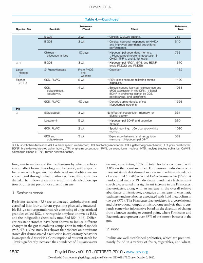
fore, aim to understand the mechanisms by which prebiot-ics can affect brain physiology and behavior, with a specificfocus on which gut microbial-derived metabolites are in-volved, and through which pathways these effects are me-diated. The following sections are a more detailed descrip-tion of different prebiotics currently in use.
1. Resistant starch
Resistant starches (RS) are undigested carbohydrates andclassified into four different types: the physically inaccessi-ble RS1, a native granular starch consisting of ungelatinizedgranules called RS2, a retrograde amylose known as RS3,and the indigestible chemically modified RS4 (646). Differ-ent resistant starches have been shown to induce differentchanges in the gut microbiota composition in animal models(945, 971). One study has shown that rodents on a resistantstarch diet demonstrated a reduction in exploratory behaviorsin an open-field test (945). Consumption of resistant starch for10 wk significantly increased the abundance of Ruminococcus
bromii, constituting 17% of total bacteria compared with3.8% on the non-starch diet. Furthermore, individuals on aresistant starch diet showed an increase in relative abundanceof uncultured Oscillibacter and Eubacterium rectale (1579). Arandomized study of 39 individuals found that a high resistantstarch diet resulted in a significant increase in the Firmicutes:Bacteroidetes, along with an increase in the overall relativeabundance of Firmicutes, alongside an increase in enzymaticpathways and metabolites associated with lipid metabolism inthe gut (971). The Firmicutes:Bacteroidetes is a correlationaland observational output of microbiome analysis that is cur-rently somewhat informative based on the direction of changefrom a known starting or control point, where Firmicutes andBacteroidetes represent over 99% of the known bacteria in thegut.
2. Inulin
Inulins are well-established prebiotics, which are predomi-nantly found in a variety of fruits, vegetables, and wheat.
Table 4.—Continued
Species, Sex PrebioticTreatment
(Time) EffectReference
Nos.
B-GOS 3 wk 1Cortical GluN2A subunit. 763
B-GOS 3 wk 1Cortical neuronal responses to NMDAand improved attentional set-shiftingperformance.
610
Chitosanoligosaccharides
10 days 1Hippocampal-dependent memory.2Hippocampal neuronal apoptosis, 8-OHdG, TNF-�, and IL-1� levels.
733
� � B-GOS 3 wk 1Hippocampal NR2A, SYN, and BDNFlevels PND22 and PND56
1610
ListerHooded�
2=-Fucosyllactose From PND3andweaning
1Cognition 1132
Fischer344 �
GOS, PLWC 9 wk 1REM sleep rebound following stressexposure.
1490
GOS,polydextrose,lactoferrin.
4 wk 2Stress-induced learned helplessness andcFOS expression in the DRN. 1BasalBDNF in prefrontal cortex by GOS,polydextrose, and lactoferrin.
1038
GOS, PLWC 40 days 1Dendritic spine density of rathippocampal neurons.
1596
Pig� Sialyllactose 3 wk No effect on recognition, memory, or
diurnal activity.531
Lactoferrin 5 wk 1Hippocampal BDNF and cognitivefunction.
280
GOS, PLWC 2 wk 1Spatial learning. 2Cortical grey/whitematter.
1080
GOS andpolydextrose
3 wk 1Exploratory behavior and recognitionmemory. 2Hippocampal 5-HT.
532
SCFA, short-chain fatty acid; ASD, autism spectrum disorder; FOS, fructooligosaccharide; GOS, galactooligosaccharide; PFC, prefrontal cortex;BDNF, brain-derived neurotrophic factor; LTP, long-term potentiation; PVN, paraventricular nucleus; NTS, nucleus tractus solitarius; CaMKII,calmodulin kinase II; TNF, tumor necrosis factor.
CRYAN ET AL.
1892 Physiol Rev • VOL 99 • OCTOBER 2019 • www.prv.orgDownloaded from journals.physiology.org/journal/physrev (093.039.178.031) on October 2, 2020.
Numerous studies in humans have shown that inulin canstimulate the growth of Bifidobacterium spp. and Faecali-bacterium prausnitzii, while increasing butyrate production(406, 825, 1235). Furthermore, administration of inulin toa dextran sulfate sodium-induced colitis rat model resultedin an attenuation of the colitis symptoms in addition to anincrease in Lactobacillus composition (1564). Moreover,exposure to inulin/GOS prebiotic supplementation duringpregnancy and lactation has been shown to bring aboutprotection against food allergies with a decrease in hista-mine levels and intestinal permeability in the offspring(200).
3. GOS and FOS
GOS are well-established prebiotics known to be present inhuman milk (104, 1552). Infants fed formula supplementedwith Bimuno-galactooligosaccharide (B-GOS; Bimuno,Clasado Biosciences, Buckinghamshire, UK), a proprietaryproduct containing at least 65% GOS, had increased abun-dance of Bifidobacterium and Lactobacillus compared withunsupplemented infants, similar to levels reported in breast-fed infants (571, 1553). Administration of B-GOS in anelderly population reported a significant increase in Bacte-roides and Bifidobacterium spp. with elevated levels of lac-tic acid in fecal water. Moreover, they also reported admin-istration of B-GOS resulted in a reduction in proinflamma-tory cytokines with an increase in both interleukin (IL)-10and IL-8, anti-inflammatory cytokines (1575). Studies havedemonstrated a significant increase in pro-inflammatory cy-tokine with stress (1243). However, administration of B-GOS in mice attenuated post-inflammatory anxiety (1320).In addition, B-GOS prevented a lipopolysaccharide (LPS)-mediated increase in cortical 1L-1� and 5-HT2A receptorlevels (1320). Administration of B-GOS to individuals in-duced suppression of the neuroendocrine stress responseand an increase in the processing of positive versus negativeattentional vigilance, thus resulting in an early anxiolytic-like phenotype (1337).
FOS are oligosaccharides known to be predominantly pres-ent in fruits. A double-blind intervention study in obesewomen with FOS showed an enhanced abundance in Bifi-dobacterium and Faecalibacterium prausnitzii (434). In arandomized, double-blind crossover study, administrationof FOS and GOS for 14 days showed significant increases inBifidobacterium along with a reduction in butyrate-produc-ing bacteria with adverse glycemic metabolism (908). Ad-ministration of FOS�GOS and GOS has been shown toreduce stress-induced corticosterone release, combinedwith a significant increase in cecal acetate and propionateconcentrations, with a reduction in isobutyrate levels.Moreover, mice fed FOS�GOS spent more time in the cen-ter of an open-field test, with an increase in the percentageof entries into the open area (229), indicating a reducedanxiety phenotype.
E. Probiotics and Psychobiotics
Probiotics refer to candidate species of live bacteria that,when ingested in adequate amounts, confer beneficialhealth effects upon the host (230). Through interacting withthe host microbiota and intestinal epithelium, probioticshave been shown to exert a wide range of effects upon hosthealth, with various strains improving metabolism, immu-nity, endocrine function, and slowing aging in preclinicalstudies (477, 1170). Although looking forward towardsutilizing candidate probiotics for host health, we must ac-knowledge the potential impact that the inherent host dietand microbiota complexity can have on the probiotic itself,such as that seen recently with cumulative genetic muta-tions occurring to Escherichia coli Nissle during passagethrough the murine gut (354). Perhaps the most intriguingeffect of probiotics on the host is their modulation of brainphysiology and behavior. Faecalibacterium prausnitzii(ATCC 27766) may function as a promising psychobioticwhere it recently demonstrated an anxiolytic and antide-pressant-like phenotype in rats, probably via increasingcecal SCFA and plasma IL-10 levels while reducing cor-ticosterone and IL-6 levels (636). Considerable researchover the last decade has documented how probiotics caninfluence various central neuronal processes such as neu-rotransmission, neurogenesis, expression of neuropep-tides, neuroinflammation, and even behavior (1365). In-deed, certain bacterial strains or cocktails of multiplebacteria have demonstrated efficacy in improving behav-ioral symptoms of various disorders from depression andanxiety to autism (see also sect. VIII) (38, 209, 226, 690,759, 1321). These findings (summarized in TABLE 5) haveled to the concept of psychobiotics for the treatment ofvarious neurological and psychiatric disorders throughtargeting of the gut microbiota (445). Psychobiotics arenow defined as microbiota-targeted interventions such as“beneficial bacteria (probiotics) or support for such bac-teria (e.g., prebiotics) that influence bacteria-brain rela-tionships” (1314). As the evidence to support the effectsof psychobiotics on brain and behavior grows (289), thefield is now turning to mechanistic studies to elucidatethe biological underpinnings of psychobiotic effects.
F. Brain Imaging
The advent of human brain imaging techniques such aspositron emission tomography in the 1980s allowed forconclusive demonstrations that alterations in the gut (e.g.,by distension) lead to activation of key brain networks(1010, 1548). Currently, studies that examine the interac-tion between gut microbes, brain, and behavior in humansare limited. Magnetic resonance imaging (MRI) as a brainimaging tool became widely available in the early 2000s,with the field of neuroimaging reaching a stage where the
THE MICROBIOTA-GUT-BRAIN AXIS
1893Physiol Rev • VOL 99 • OCTOBER 2019 • www.prv.orgDownloaded from journals.physiology.org/journal/physrev (093.039.178.031) on October 2, 2020.
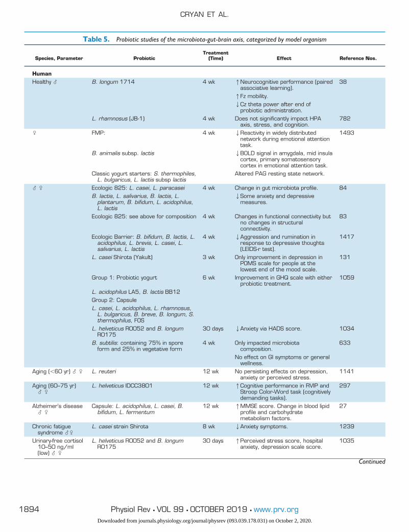
Table 5. Probiotic studies of the microbiota-gut-brain axis, categorized by model organism
Species, Parameter ProbioticTreatment
(Time) Effect Reference Nos.
HumanHealthy � B. longum 1714 4 wk 1Neurocognitive performance (paired
associative learning).38
1Fz mobility.2Cz theta power after end of
probiotic administration.L. rhamnosus (JB-1) 4 wk Does not significantly impact HPA
axis, stress, and cognition.782
� FMP: 4 wk 2Reactivity in widely distributednetwork during emotional attentiontask.
1493
B. animalis subsp. lactis 2BOLD signal in amygdala, mid insulacortex, primary somatosensorycortex in emotional attention task.
Classic yogurt starters: S. thermophiles,L. bulgaricus, L. lactis subsp lactis
Altered PAG resting state network.
� � Ecologic 825: L. casei, L. paracasei 4 wk Change in gut microbiota profile. 84B. lactis, L. salivarius, B. lactis, L.
plantarum, B. bifidum, L. acidophilus,L. lactis
2Some anxiety and depressivemeasures.
Ecologic 825: see above for composition 4 wk Changes in functional connectivity butno changes in structuralconnectivity.
83
Ecologic Barrier: B. bifidum, B. lactis, L.acidophilus, L. brevis, L. casei, L.salivarius, L. lactis
4 wk 2Aggression and rumination inresponse to depressive thoughts(LEIDS-r test).
1417
L. casei Shirota (Yakult) 3 wk Only improvement in depression inPOMS scale for people at thelowest end of the mood scale.
131
Group 1: Probiotic yogurt 6 wk Improvement in GHQ scale with eitherprobiotic treatment.
1059
L. acidophilus LA5, B. lactis BB12Group 2: CapsuleL. casei, L. acidophilus, L. rhamnosus,
L. bulgaricus, B. breve, B. longum, S.thermophilus, FOS
L. helveticus R0052 and B. longumR0175
30 days 2Anxiety via HADS score. 1034
B. subtilis: containing 75% in sporeform and 25% in vegetative form
4 wk Only impacted microbiotacomposition.
633
No effect on GI symptoms or generalwellness.
Aging (�60 yr) � � L. reuteri 12 wk No persisting effects on depression,anxiety or perceived stress.
1141
Aging (60–75 yr)� �
L. helveticus IDCC3801 12 wk 1Cognitive performance in RVIP andStroop Color-Word task (cognitivelydemanding tasks).
297
Alzheimer’s disease� �
Capsule: L. acidophilus, L. casei, B.bifidum, L. fermentum
12 wk 1MMSE score. Change in blood lipidprofile and carbohydratemetabolism factors.
27
Chronic fatiguesyndrome ��
L. casei strain Shirota 8 wk 2Anxiety symptoms. 1239
Urinary-free cortisol10–50 ng/ml(low) � �
L. helveticus R0052 and B. longumR0175
30 days 1Perceived stress score, hospitalanxiety, depression scale score.
1035
Continued
CRYAN ET AL.
1894 Physiol Rev • VOL 99 • OCTOBER 2019 • www.prv.orgDownloaded from journals.physiology.org/journal/physrev (093.039.178.031) on October 2, 2020.
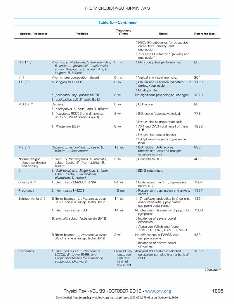
Table 5.—Continued
Species, Parameter ProbioticTreatment
(Time) Effect Reference Nos.
1HSCL-90 subscores for obsessive-compulsive, anxiety, anddepression.11HSCL-90 in factor 1 (anxiety and
depression).HIV-1� � Vivomixx: L. plantarum, S. thermophiles,
B. breve, L. paracasei, L.delbrueckiisubsp. Bulgaricus, L. acidophilus, B.longum, B. infantis
6 mo 1Neurocognitive performance. 263
� � Vivomix (see composition above) 6 mo 1Verbal and visual memory. 264IBS � � B. longum NCC3001 6 wk 2HAD-A and D scores indicating 2 in
anxiety/depression.1198
1Quality of life.L. paracasei, ssp. paracasei F19 8 wk No significant psychological changes. 1379L. acidophilus La5 B. lactis Bb12
MDD � � Capsule: 8 wk 2BDI score. 28L. acidophilus, L. casei, and B. bifidumL. helveticus R0052 and B. longum
R0175 (CNCM strain I-3470)8 wk 2BDI score (depressive index). 778
2Kynurenine-to-tryptophan ratio.L. Plantarum 299v 8 wk 1APT and CVLT total recall of trials
1–51293
2Kynurenine concentration13-Hydroxykynurenine: kynurenine
ratioMS � � Capsule: L. acidophilus, L. casei, B.
bifidum, L. fermentum12 wk 1BDI, EDSS, DHQ scores
(depression, diet and multiplesclerosis scores).
836
Normal weightobese syndromeand obesity
1 “bag”: S. thermophiles, B. animalissubsp. Lactis, S. thermophilus, B.bifidum
3 wk 2Positivity to BUT. 403
� L. delbrueckii spp. Bulgaricus, L. lactissubsp. Lactis, L. acidophilus, L.plantarum, L. reuteri
2EDI-2 responses.
Obesity � � L. rhamnosus CGMCC1.3724 24 wk 1Body esteem in �, 2depressionscore in �.
1307
Pregnancy L. rhamnosus HN001 �6 mo 2Postpartum depression and anxietyscores.
1387
Schizophrenia � � Bifiform balance: L. rhamnosus strainGG B. animalis subsp. lactis Bb12
14 wk 2 C. albicans antibodies in � serum,associated with 2psychiatricsymptom occurrence.
1354
L. rhamnosus strain GG 14 wk No changes in frequency of psychoticsymptoms.
1500
B. animalis subsp. lactis strain Bb12 2Incidence of severe boweldifficulties.2Acute von Willebrand factor,1MCP-1, BDNF, RANTES, MIP-1.
Bifiform balance: L. rhamnosus strainGG B. animalis subsp. lactis Bb12
2 wk No differences in PANSS totalsymptom score.
439
2Incidence of severe boweldifficulties.
Pregnancy L. rhamnosus GG, L. rhamnosusLC705, B. breve Bb99, andPropionibacterium freudenreichii-subspecies shermanii
From 36 wkgestationuntil thebirth ofthe infant
Analyzed 81 randomly selectedcolostrum samples from a bank of500.
1352
Continued
THE MICROBIOTA-GUT-BRAIN AXIS
1895Physiol Rev • VOL 99 • OCTOBER 2019 • www.prv.orgDownloaded from journals.physiology.org/journal/physrev (093.039.178.031) on October 2, 2020.
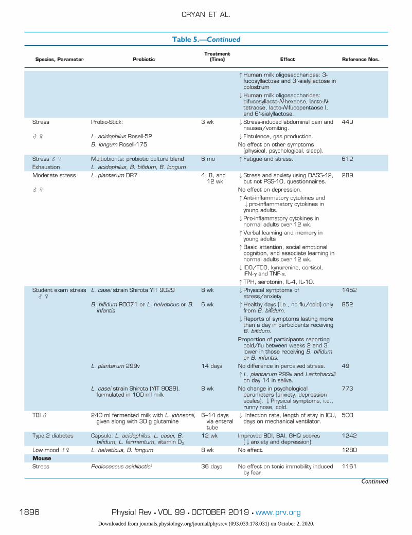
Table 5.—Continued
Species, Parameter ProbioticTreatment
(Time) Effect Reference Nos.
1Human milk oligosaccharides: 3-fucosyllactose and 3=-sialyllactose incolostrum2Human milk oligosaccharides:
difucosyllacto-N-hexaose, lacto-N-tetraose, lacto-N-fucopentaose I,and 6=-sialyllactose.
Stress Probio-Stick: 3 wk 2Stress-induced abdominal pain andnausea/vomiting.
449
� � L. acidophilus Rosell-52 2Flatulence, gas production.B. longum Rosell-175 No effect on other symptoms
(physical, psychological, sleep).Stress � � Multiobionta: probiotic culture blend 6 mo 1Fatigue and stress. 612Exhaustion L. acidophilus, B. bifidum, B. longumModerate stress L. plantarum DR7 4, 8, and
12 wk2Stress and anxiety using DASS-42,
but not PSS-10, questionnaires.289
� � No effect on depression.1Anti-inflammatory cytokines and2pro-inflammatory cytokines inyoung adults.2Pro-inflammatory cytokines in
normal adults over 12 wk.1Verbal learning and memory in
young adults1Basic attention, social emotional
cognition, and associate learning innormal adults over 12 wk.2IDO/TDO, kynurenine, cortisol,
IFN-� and TNF-�.1TPH, serotonin, IL-4, IL-10.
Student exam stress� �
L. casei strain Shirota YIT 9029 8 wk 2Physical symptoms ofstress/anxiety
1452
B. bifidum R0071 or L. helveticus or B.infantis
6 wk 1Healthy days (i.e., no flu/cold) onlyfrom B. bifidum.
852
2Reports of symptoms lasting morethan a day in participants receivingB. bifidum.
Proportion of participants reportingcold/flu between weeks 2 and 3lower in those receiving B. bifidumor B. infantis.
L. plantarum 299v 14 days No difference in perceived stress. 491L. plantarum 299v and Lactobaccili
on day 14 in saliva.L. casei strain Shirota (YIT 9029),
formulated in 100 ml milk8 wk No change in psychological
parameters (anxiety, depressionscales). 2Physical symptoms, i.e.,runny nose, cold.
773
TBI � 240 ml fermented milk with L. johnsonii,given along with 30 g glutamine
6–14 daysvia enteraltube
2 Infection rate, length of stay in ICU,days on mechanical ventilator.
500
Type 2 diabetes Capsule: L. acidophilus, L. casei, B.bifidum, L. fermentum, vitamin D3
12 wk Improved BDI, BAI, GHQ scores(2anxiety and depression).
1242
Low mood �� L. helveticus, B. longum 8 wk No effect. 1280MouseStress Pediococcus acidilactici 36 days No effect on tonic immobility induced
by fear.1161
Continued
CRYAN ET AL.
1896 Physiol Rev • VOL 99 • OCTOBER 2019 • www.prv.orgDownloaded from journals.physiology.org/journal/physrev (093.039.178.031) on October 2, 2020.
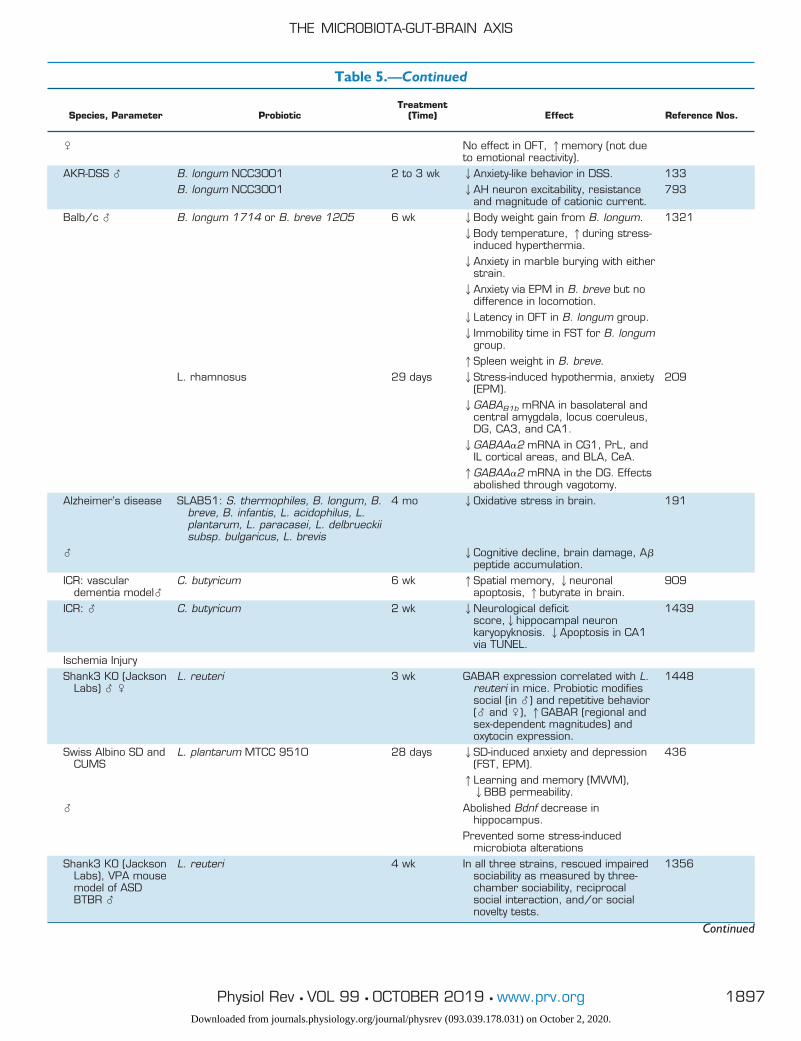
Table 5.—Continued
Species, Parameter ProbioticTreatment
(Time) Effect Reference Nos.
� No effect in OFT, 1memory (not dueto emotional reactivity).
AKR-DSS � B. longum NCC3001 2 to 3 wk 2Anxiety-like behavior in DSS. 133B. longum NCC3001 2AH neuron excitability, resistance
and magnitude of cationic current.793
Balb/c � B. longum 1714 or B. breve 1205 6 wk 2Body weight gain from B. longum. 13212Body temperature, 1during stress-
induced hyperthermia.2Anxiety in marble burying with either
strain.2Anxiety via EPM in B. breve but no
difference in locomotion.2Latency in OFT in B. longum group.2Immobility time in FST for B. longum
group.1Spleen weight in B. breve.
L. rhamnosus 29 days 2Stress-induced hypothermia, anxiety(EPM).
209
2GABAB1b mRNA in basolateral andcentral amygdala, locus coeruleus,DG, CA3, and CA1.2GABAA�2 mRNA in CG1, PrL, and
IL cortical areas, and BLA, CeA.1GABAA�2 mRNA in the DG. Effects
abolished through vagotomy.Alzheimer’s disease SLAB51: S. thermophiles, B. longum, B.
breve, B. infantis, L. acidophilus, L.plantarum, L. paracasei, L. delbrueckiisubsp. bulgaricus, L. brevis
4 mo 2Oxidative stress in brain. 191
� 2Cognitive decline, brain damage, A�peptide accumulation.
ICR: vasculardementia model�
C. butyricum 6 wk 1Spatial memory, 2neuronalapoptosis, 1butyrate in brain.
909
ICR: � C. butyricum 2 wk 2Neurological deficitscore,2hippocampal neuronkaryopyknosis. 2Apoptosis in CA1via TUNEL.
1439
Ischemia InjuryShank3 KO (Jackson
Labs) � �L. reuteri 3 wk GABAR expression correlated with L.
reuteri in mice. Probiotic modifiessocial (in �) and repetitive behavior(� and �), 1GABAR (regional andsex-dependent magnitudes) andoxytocin expression.
1448
Swiss Albino SD andCUMS
L. plantarum MTCC 9510 28 days 2SD-induced anxiety and depression(FST, EPM).
436
1Learning and memory (MWM),2BBB permeability.
� Abolished Bdnf decrease inhippocampus.
Prevented some stress-inducedmicrobiota alterations
Shank3 KO (JacksonLabs), VPA mousemodel of ASDBTBR �
L. reuteri 4 wk In all three strains, rescued impairedsociability as measured by three-chamber sociability, reciprocalsocial interaction, and/or socialnovelty tests.
1356
Continued
THE MICROBIOTA-GUT-BRAIN AXIS
1897Physiol Rev • VOL 99 • OCTOBER 2019 • www.prv.orgDownloaded from journals.physiology.org/journal/physrev (093.039.178.031) on October 2, 2020.
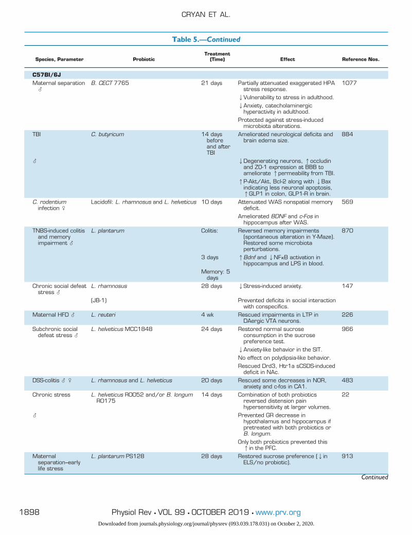
Table 5.—Continued
Species, Parameter ProbioticTreatment
(Time) Effect Reference Nos.
C57BI/6JMaternal separation
�B. CECT 7765 21 days Partially attenuated exaggerated HPA
stress response.1077
2Vulnerability to stress in adulthood.2Anxiety, catecholaminergic
hyperactivity in adulthood.Protected against stress-induced
microbiota alterations.TBI C. butyricum 14 days
beforeand afterTBI
Ameliorated neurological deficits andbrain edema size.
884
� 2Degenerating neurons, 1occludinand ZO-1 expression at BBB toameliorate 1permeability from TBI.1P-Akt/Akt, Bcl-2 along with 2Bax
indicating less neuronal apoptosis,1GLP1 in colon, GLP1-R in brain.
C. rodentiuminfection �
Lacidofil: L. rhamnosus and L. helveticus 10 days Attenuated WAS nonspatial memorydeficit.
569
Ameliorated BDNF and c-Fos inhippocampus after WAS.
TNBS-induced colitisand memoryimpairment �
L. plantarum Colitis: Reversed memory impairments(spontaneous alteration in Y-Maze).Restored some microbiotaperturbations.
870
3 days 1Bdnf and 2NF�B activation inhippocampus and LPS in blood.
Memory: 5days
Chronic social defeatstress �
L. rhamnosus 28 days 2Stress-induced anxiety. 147
(JB-1) Prevented deficits in social interactionwith conspecifics.
Maternal HFD � L. reuteri 4 wk Rescued impairments in LTP inDAergic VTA neurons.
226
Subchronic socialdefeat stress �
L. helveticus MCC1848 24 days Restored normal sucroseconsumption in the sucrosepreference test.
966
2Anxiety-like behavior in the SIT.No effect on polydipsia-like behavior.Rescued Drd3, Htr1a sCSDS-induced
deficit in NAc.DSS-colitis � � L. rhamnosus and L. helveticus 20 days Rescued some decreases in NOR,
anxiety and c-fos in CA1.483
Chronic stress L. helveticus R0052 and/or B. longumR0175
14 days Combination of both probioticsreversed distension painhypersensitivity at larger volumes.
22
� Prevented GR decrease inhypothalamus and hippocampus ifpretreated with both probiotics orB. longum.
Only both probiotics prevented this1in the PFC.
Maternalseparation–earlylife stress
L. plantarum PS128 28 days Restored sucrose preference (2inELS/no probiotic).
913
Continued
CRYAN ET AL.
1898 Physiol Rev • VOL 99 • OCTOBER 2019 • www.prv.orgDownloaded from journals.physiology.org/journal/physrev (093.039.178.031) on October 2, 2020.
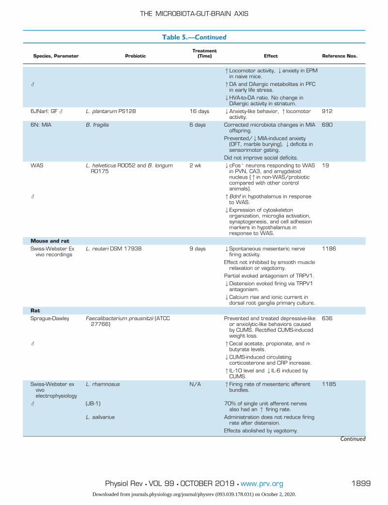
Table 5.—Continued
Species, Parameter ProbioticTreatment
(Time) Effect Reference Nos.
1Locomotor activity, 2anxiety in EPMin naive mice.
� 1DA and DAergic metabolites in PFCin early life stress.2HVA-to-DA ratio. No change in
DAergic activity in striatum.6JNarl: GF � L. plantarum PS128 16 days 2Anxiety-like behavior, 1locomotor
activity.912
6N: MIA B. fragilis 6 days Corrected microbiota changes in MIAoffspring.
690
Prevented/2MIA-induced anxiety(OFT, marble burying), 2deficits insensorimotor gating.
Did not improve social deficits.WAS L. helveticus R0052 and B. longum
R01752 wk 2cFos� neurons responding to WAS
in PVN, CA3, and amygdaloidnucleus (1in non-WAS/probioticcompared with other controlanimals).
19
� 1Bdnf in hypothalamus in responseto WAS.2Expression of cytoskeleton
organization, microglia activation,synaptogenesis, and cell adhesionmarkers in hypothalamus inresponse to WAS.
Mouse and ratSwiss-Webster Ex
vivo recordingsL. reuteri DSM 17938 9 days 2Spontaneous mesenteric nerve
firing activity.1186
Effect not inhibited by smooth musclerelaxation or vagotomy.
Partial evoked antagonism of TRPV1.2Distension evoked firing via TRPV1
antagonism.2Calcium rise and ionic current in
dorsal root ganglia primary culture.RatSprague-Dawley Faecalibacterium prausnitzii (ATCC
27766)Prevented and treated depressive-like
or anxiolytic-like behaviors causedby CUMS. Rectified CUMS-inducedweight loss.
636
� 1Cecal acetate, propionate, and n-butyrate levels.2CUMS-induced circulating
corticosterone and CRP increase.1IL-10 level and 2IL-6 induced by
CUMS.Swiss-Webster ex
vivoelectrophysiology
L. rhamnosus N/A 1Firing rate of mesenteric afferentbundles.
1185
� (JB-1) 70% of single unit afferent nervesalso had an 1 firing rate.
L. salivarius Administration does not reduce firingrate after distension.
Effects abolished by vagotomy.
Continued
THE MICROBIOTA-GUT-BRAIN AXIS
1899Physiol Rev • VOL 99 • OCTOBER 2019 • www.prv.orgDownloaded from journals.physiology.org/journal/physrev (093.039.178.031) on October 2, 2020.
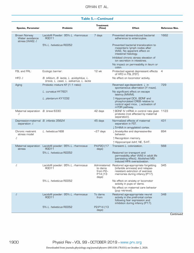
Table 5.—Continued
Species, Parameter ProbioticTreatment
(Time) Effect Reference Nos.
Brown NorwayWater avoidancestress (WAS) �
Lacidofil Powder: 95% L. rhamnosusR0011
7 days Prevented stress-induced bacterialadherence to enterocytes.
1662
5% L. helveticus R0052 Prevented bacterial translocation tomesenteric lymph nodes afterWAS. No apparent effect onintestinal histology.
Inhibited chronic stress elevation ofion secretion in intestines.
No impact on permeability in ileum orcolon.
FSL and FRL: Ecologic barrier: 12 wk Protected against depressant effectsof HFD in FSL (FST).
4
HFD � B. bifidum, B. lactis, L. acidophilus, L.brevis, L. casei, L. salivarius, L. lactis
No effect on locomotor activity.
Aging Probiotic mixture KF (1:1 ratio): Reversed age-dependent 2 inspontaneous alternation (Y maze).
729
L. curvatus HY7601 No significant effect on escapelatency (MWM).
L. plantarum KY1032 1Hippocampal DCX, BDNF andphosphorylated CREB relative tocontrol aged mice, 2activation ofmTOR pathway.
Maternal separation�
B. breve 6330 42 days 1BDNF IV mRNA in control rats givenprobiotic (not affected by maternalseparation).
1123
Depression-maternalseparation �
B. infantis 35624 45 days Normalized effects of maternalseparation in FST.
431
25-HIAA in amygdaloid cortex.Chronic restraint
stress modelL. helveticus NS8 ~27 days 2Anxiety-like and depressive-like
behavior.894
� 1Recognition memory.1Hippocampal bdnf, NE, 5-HT.
Maternal separationstress
Lacidofil powder: 95% L. rhamnosusR0011
P4-P20 (17days)
Transient L. colonization. 568
5% L. helveticus R0052 Restored ion transport andpermeability after WAS in adult life(persisting effect). Abolished MS-induced HPA overactivation.
� Lacidofil powder: 95% L. rhamnosusR0011
Administeredto damsfrom P2–P14 (13days)
Restored age-appropriate forgetting(infantile amnesia) and relapse-resistant extinction of aversivememories during infancy (P17).
345
5% L. helveticus R0052 No effect on anxiety or locomotoractivity in pups or dams.
No effect on maternal care behavior(pup retrieval).
� Lacidofil powder: 95% L. rhamnosusR0011
To damsfrom
Restored age-appropriate neuralactivity in the prefrontal cortexfollowing fear expression andinhibition during infancy (P17).
348
5% L. helveticus R0052 P2-P14 (13days)
Continued
CRYAN ET AL.
1900 Physiol Rev • VOL 99 • OCTOBER 2019 • www.prv.orgDownloaded from journals.physiology.org/journal/physrev (093.039.178.031) on October 2, 2020.
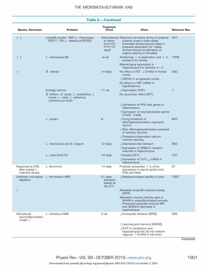
Table 5.—Continued
Species, Parameter ProbioticTreatment
(Time) Effect Reference Nos.
� � Lacidofil powder: 95% L. rhamnosusR0011, 5% L. helveticus R0052
Administeredto damsfrom P2–P14 (13days)
Restored normative timing of physicalpuberty onset in both sexes(reversed stress-induced delay inpreputial separation for males,stress-induced acceleration ofvaginal opening in females).
347
� � L. rhamnosus GG ~4 wk Ameliorate 2 in exploration and 1 inanxiety from stress.
1026
Altered gene expression inhippocampus (no behavior in �).
� B. infantis 14 days No effect in FST. 25-HIAA in frontalcortex.
430
2DOPAC in amygdaloid cortex.No effect on CRF mRNA in
hypothalamus.Ecologic barrier: 11 wk 2Depression (FST). 7B. bifidum, B. lactis, L. acidophilus, L.
brevis, L. casei, L. salivarius,Lactococcus lactis
No locomotor effect (OFT).
2Expression of HPA axis genes inhippocampus.1Expression of neuroprotective genes
(Trek2, Traak).L. reuteri 9 2Firing threshold of
afterhyperpolarization myentericneuron.
845
2Slow afterhyperpolarization potentialof sensory neurons.2Potassium-dependent calcium
channel opening.L. rhamnosus and B. longum 12 days 2Depression-like behavior. 893
1Expression of GABA-A receptorsubunits in hippocampus.
L. casei 54-2-33 14 days 1Anxiety (OFT). 1072Expression of 5-HT1A mRNA in
hippocampus.Response to CRD
after partial �restraint stress
L. farciminis 14 days Probiotic prevented 1 in c-Fosexpression in sacral spinal cord,PVN and MeA.
21
Antibiotic microbiotadepletion
L. fermentum NS9 41 days(behaviortested atday 31)
2Myeloperoxidase activity in colon. 1591
� Alleviated ampicillin-induced anxiety(EPM).
Alleviated memory deficits seen inMWM in ampicillin-treated animals.Prevented ampicillin-induced MRand NMDA-R decrease inhippocampus.
HA-inducedneuroinflammationmodel �
L. helveticus NS8 2 wk 2Anxiety-like behavior (EPM). 936
1Learning and memory (MWM).25-HT in cerebellum and
hippocampus but did not restoreregional. 15-HIAA in HA brain.
Continued
THE MICROBIOTA-GUT-BRAIN AXIS
1901Physiol Rev • VOL 99 • OCTOBER 2019 • www.prv.orgDownloaded from journals.physiology.org/journal/physrev (093.039.178.031) on October 2, 2020.
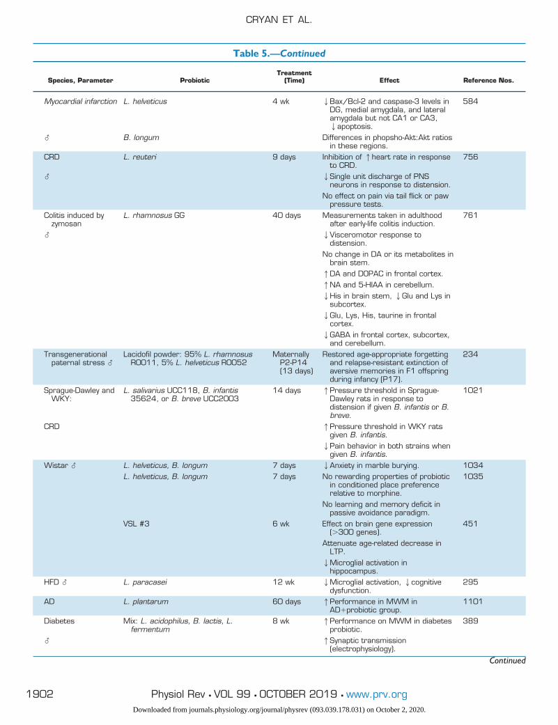
Table 5.—Continued
Species, Parameter ProbioticTreatment
(Time) Effect Reference Nos.
Myocardial infarction L. helveticus 4 wk 2Bax/Bcl-2 and caspase-3 levels inDG, medial amygdala, and lateralamygdala but not CA1 or CA3,2apoptosis.
584
� B. longum Differences in phopsho-Akt:Akt ratiosin these regions.
CRD L. reuteri 9 days Inhibition of 1heart rate in responseto CRD.
756
� 2Single unit discharge of PNSneurons in response to distension.
No effect on pain via tail flick or pawpressure tests.
Colitis induced byzymosan
L. rhamnosus GG 40 days Measurements taken in adulthoodafter early-life colitis induction.
761
� 2Visceromotor response todistension.
No change in DA or its metabolites inbrain stem.1DA and DOPAC in frontal cortex.1NA and 5-HIAA in cerebellum.2His in brain stem, 2Glu and Lys in
subcortex.2Glu, Lys, His, taurine in frontal
cortex.2GABA in frontal cortex, subcortex,
and cerebellum.Transgenerational
paternal stress �Lacidofil powder: 95% L. rhamnosus
R0011, 5% L. helveticus R0052Maternally
P2-P14(13 days)
Restored age-appropriate forgettingand relapse-resistant extinction ofaversive memories in F1 offspringduring infancy (P17).
234
Sprague-Dawley andWKY:
L. salivarius UCC118, B. infantis35624, or B. breve UCC2003
14 days 1Pressure threshold in Sprague-Dawley rats in response todistension if given B. infantis or B.breve.
1021
CRD 1Pressure threshold in WKY ratsgiven B. infantis.2Pain behavior in both strains when
given B. infantis.Wistar � L. helveticus, B. longum 7 days 2Anxiety in marble burying. 1034
L. helveticus, B. longum 7 days No rewarding properties of probioticin conditioned place preferencerelative to morphine.
1035
No learning and memory deficit inpassive avoidance paradigm.
VSL #3 6 wk Effect on brain gene expression(�300 genes).
451
Attenuate age-related decrease inLTP.2Microglial activation in
hippocampus.HFD � L. paracasei 12 wk 2Microglial activation, 2cognitive
dysfunction.295
AD L. plantarum 60 days 1Performance in MWM inAD�probiotic group.
1101
Diabetes Mix: L. acidophilus, B. lactis, L.fermentum
8 wk 1Performance on MWM in diabetesprobiotic.
389
� 1Synaptic transmission(electrophysiology).
Continued
CRYAN ET AL.
1902 Physiol Rev • VOL 99 • OCTOBER 2019 • www.prv.orgDownloaded from journals.physiology.org/journal/physrev (093.039.178.031) on October 2, 2020.
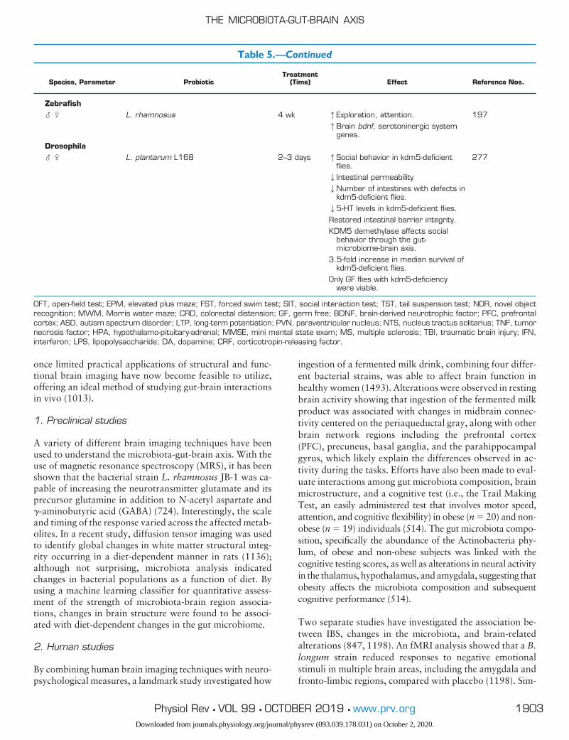
once limited practical applications of structural and func-tional brain imaging have now become feasible to utilize,offering an ideal method of studying gut-brain interactionsin vivo (1013).
1. Preclinical studies
A variety of different brain imaging techniques have beenused to understand the microbiota-gut-brain axis. With theuse of magnetic resonance spectroscopy (MRS), it has beenshown that the bacterial strain L. rhamnosus JB-1 was ca-pable of increasing the neurotransmitter glutamate and itsprecursor glutamine in addition to N-acetyl aspartate and�-aminobutyric acid (GABA) (724). Interestingly, the scaleand timing of the response varied across the affected metab-olites. In a recent study, diffusion tensor imaging was usedto identify global changes in white matter structural integ-rity occurring in a diet-dependent manner in rats (1136);although not surprising, microbiota analysis indicatedchanges in bacterial populations as a function of diet. Byusing a machine learning classifier for quantitative assess-ment of the strength of microbiota-brain region associa-tions, changes in brain structure were found to be associ-ated with diet-dependent changes in the gut microbiome.
2. Human studies
By combining human brain imaging techniques with neuro-psychological measures, a landmark study investigated how
ingestion of a fermented milk drink, combining four differ-ent bacterial strains, was able to affect brain function inhealthy women (1493). Alterations were observed in restingbrain activity showing that ingestion of the fermented milkproduct was associated with changes in midbrain connec-tivity centered on the periaqueductal gray, along with otherbrain network regions including the prefrontal cortex(PFC), precuneus, basal ganglia, and the parahippocampalgyrus, which likely explain the differences observed in ac-tivity during the tasks. Efforts have also been made to eval-uate interactions among gut microbiota composition, brainmicrostructure, and a cognitive test (i.e., the Trail MakingTest, an easily administered test that involves motor speed,attention, and cognitive flexibility) in obese (n � 20) and non-obese (n � 19) individuals (514). The gut microbiota compo-sition, specifically the abundance of the Actinobacteria phy-lum, of obese and non-obese subjects was linked with thecognitive testing scores, as well as alterations in neural activityin the thalamus, hypothalamus, and amygdala, suggesting thatobesity affects the microbiota composition and subsequentcognitive performance (514).
Two separate studies have investigated the association be-tween IBS, changes in the microbiota, and brain-relatedalterations (847, 1198). An fMRI analysis showed that a B.longum strain reduced responses to negative emotionalstimuli in multiple brain areas, including the amygdala andfronto-limbic regions, compared with placebo (1198). Sim-
Table 5.—Continued
Species, Parameter ProbioticTreatment
(Time) Effect Reference Nos.
Zebrafish� � L. rhamnosus 4 wk 1Exploration, attention. 197
1Brain bdnf, serotoninergic systemgenes.
Drosophila� � L. plantarum L168 2–3 days 1Social behavior in kdm5-deficient
flies.277
2Intestinal permeability2Number of intestines with defects in
kdm5-deficient flies.25-HT levels in kdm5-deficient flies.Restored intestinal barrier integrity.KDM5 demethylase affects social
behavior through the gut-microbiome-brain axis.
3.5-fold increase in median survival ofkdm5-deficient flies.
Only GF flies with kdm5-deficiencywere viable.
OFT, open-field test; EPM, elevated plus maze; FST, forced swim test; SIT, social interaction test; TST, tail suspension test; NOR, novel objectrecognition; MWM, Morris water maze; CRD, colorectal distension; GF, germ free; BDNF, brain-derived neurotrophic factor; PFC, prefrontalcortex; ASD, autism spectrum disorder; LTP, long-term potentiation; PVN, paraventricular nucleus; NTS, nucleus tractus solitarius; TNF, tumornecrosis factor; HPA, hypothalamo-pituitary-adrenal; MMSE, mini mental state exam; MS, multiple sclerosis; TBI, traumatic brain injury; IFN,interferon; LPS, lipopolysaccharide; DA, dopamine; CRF, corticotropin-releasing factor.
THE MICROBIOTA-GUT-BRAIN AXIS
1903Physiol Rev • VOL 99 • OCTOBER 2019 • www.prv.orgDownloaded from journals.physiology.org/journal/physrev (093.039.178.031) on October 2, 2020.
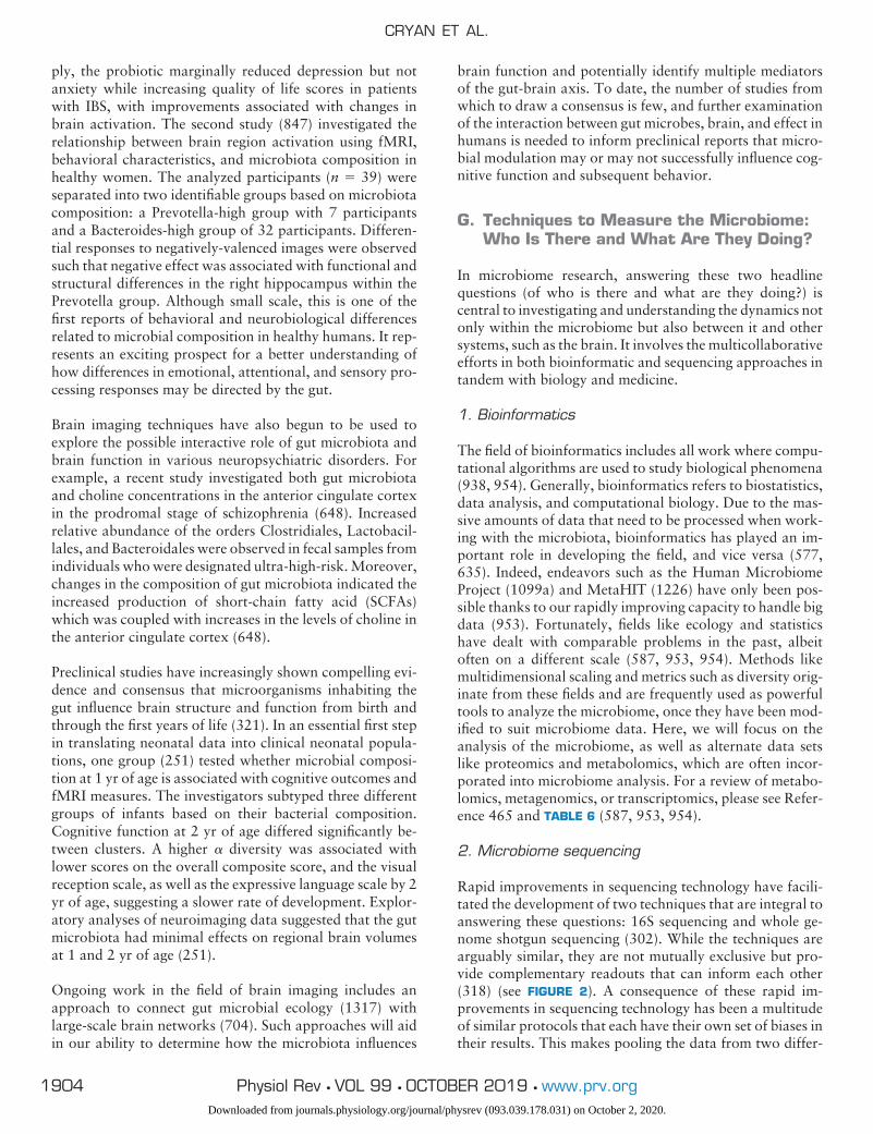
ply, the probiotic marginally reduced depression but notanxiety while increasing quality of life scores in patientswith IBS, with improvements associated with changes inbrain activation. The second study (847) investigated therelationship between brain region activation using fMRI,behavioral characteristics, and microbiota composition inhealthy women. The analyzed participants (n � 39) wereseparated into two identifiable groups based on microbiotacomposition: a Prevotella-high group with 7 participantsand a Bacteroides-high group of 32 participants. Differen-tial responses to negatively-valenced images were observedsuch that negative effect was associated with functional andstructural differences in the right hippocampus within thePrevotella group. Although small scale, this is one of thefirst reports of behavioral and neurobiological differencesrelated to microbial composition in healthy humans. It rep-resents an exciting prospect for a better understanding ofhow differences in emotional, attentional, and sensory pro-cessing responses may be directed by the gut.
Brain imaging techniques have also begun to be used toexplore the possible interactive role of gut microbiota andbrain function in various neuropsychiatric disorders. Forexample, a recent study investigated both gut microbiotaand choline concentrations in the anterior cingulate cortexin the prodromal stage of schizophrenia (648). Increasedrelative abundance of the orders Clostridiales, Lactobacil-lales, and Bacteroidales were observed in fecal samples fromindividuals who were designated ultra-high-risk. Moreover,changes in the composition of gut microbiota indicated theincreased production of short-chain fatty acid (SCFAs)which was coupled with increases in the levels of choline inthe anterior cingulate cortex (648).
Preclinical studies have increasingly shown compelling evi-dence and consensus that microorganisms inhabiting thegut influence brain structure and function from birth andthrough the first years of life (321). In an essential first stepin translating neonatal data into clinical neonatal popula-tions, one group (251) tested whether microbial composi-tion at 1 yr of age is associated with cognitive outcomes andfMRI measures. The investigators subtyped three differentgroups of infants based on their bacterial composition.Cognitive function at 2 yr of age differed significantly be-tween clusters. A higher � diversity was associated withlower scores on the overall composite score, and the visualreception scale, as well as the expressive language scale by 2yr of age, suggesting a slower rate of development. Explor-atory analyses of neuroimaging data suggested that the gutmicrobiota had minimal effects on regional brain volumesat 1 and 2 yr of age (251).
Ongoing work in the field of brain imaging includes anapproach to connect gut microbial ecology (1317) withlarge-scale brain networks (704). Such approaches will aidin our ability to determine how the microbiota influences
brain function and potentially identify multiple mediatorsof the gut-brain axis. To date, the number of studies fromwhich to draw a consensus is few, and further examinationof the interaction between gut microbes, brain, and effect inhumans is needed to inform preclinical reports that micro-bial modulation may or may not successfully influence cog-nitive function and subsequent behavior.
G. Techniques to Measure the Microbiome:Who Is There and What Are They Doing?
In microbiome research, answering these two headlinequestions (of who is there and what are they doing?) iscentral to investigating and understanding the dynamics notonly within the microbiome but also between it and othersystems, such as the brain. It involves the multicollaborativeefforts in both bioinformatic and sequencing approaches intandem with biology and medicine.
1. Bioinformatics
The field of bioinformatics includes all work where compu-tational algorithms are used to study biological phenomena(938, 954). Generally, bioinformatics refers to biostatistics,data analysis, and computational biology. Due to the mas-sive amounts of data that need to be processed when work-ing with the microbiota, bioinformatics has played an im-portant role in developing the field, and vice versa (577,635). Indeed, endeavors such as the Human MicrobiomeProject (1099a) and MetaHIT (1226) have only been pos-sible thanks to our rapidly improving capacity to handle bigdata (953). Fortunately, fields like ecology and statisticshave dealt with comparable problems in the past, albeitoften on a different scale (587, 953, 954). Methods likemultidimensional scaling and metrics such as diversity orig-inate from these fields and are frequently used as powerfultools to analyze the microbiome, once they have been mod-ified to suit microbiome data. Here, we will focus on theanalysis of the microbiome, as well as alternate data setslike proteomics and metabolomics, which are often incor-porated into microbiome analysis. For a review of metabo-lomics, metagenomics, or transcriptomics, please see Refer-ence 465 and TABLE 6 (587, 953, 954).
2. Microbiome sequencing
Rapid improvements in sequencing technology have facili-tated the development of two techniques that are integral toanswering these questions: 16S sequencing and whole ge-nome shotgun sequencing (302). While the techniques arearguably similar, they are not mutually exclusive but pro-vide complementary readouts that can inform each other(318) (see FIGURE 2). A consequence of these rapid im-provements in sequencing technology has been a multitudeof similar protocols that each have their own set of biases intheir results. This makes pooling the data from two differ-
CRYAN ET AL.
1904 Physiol Rev • VOL 99 • OCTOBER 2019 • www.prv.orgDownloaded from journals.physiology.org/journal/physrev (093.039.178.031) on October 2, 2020.
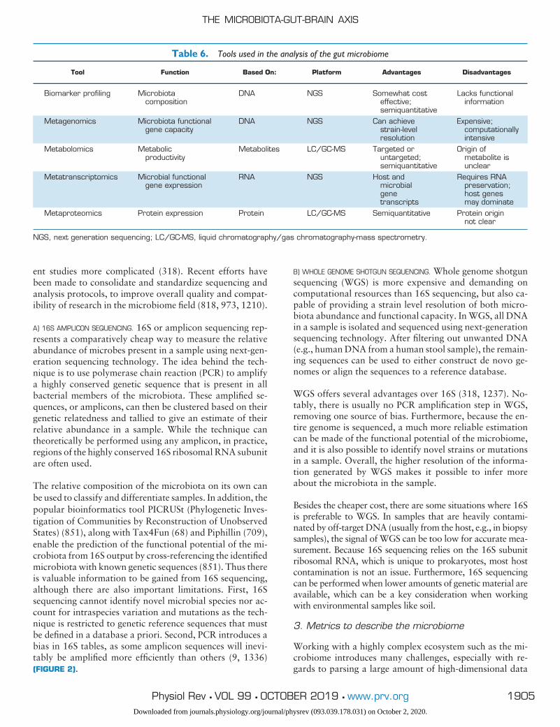
ent studies more complicated (318). Recent efforts havebeen made to consolidate and standardize sequencing andanalysis protocols, to improve overall quality and compat-ibility of research in the microbiome field (818, 973, 1210).
A) 16S AMPLICON SEQUENCING. 16S or amplicon sequencing rep-resents a comparatively cheap way to measure the relativeabundance of microbes present in a sample using next-gen-eration sequencing technology. The idea behind the tech-nique is to use polymerase chain reaction (PCR) to amplifya highly conserved genetic sequence that is present in allbacterial members of the microbiota. These amplified se-quences, or amplicons, can then be clustered based on theirgenetic relatedness and tallied to give an estimate of theirrelative abundance in a sample. While the technique cantheoretically be performed using any amplicon, in practice,regions of the highly conserved 16S ribosomal RNA subunitare often used.
The relative composition of the microbiota on its own canbe used to classify and differentiate samples. In addition, thepopular bioinformatics tool PICRUSt (Phylogenetic Inves-tigation of Communities by Reconstruction of UnobservedStates) (851), along with Tax4Fun (68) and Piphillin (709),enable the prediction of the functional potential of the mi-crobiota from 16S output by cross-referencing the identifiedmicrobiota with known genetic sequences (851). Thus thereis valuable information to be gained from 16S sequencing,although there are also important limitations. First, 16Ssequencing cannot identify novel microbial species nor ac-count for intraspecies variation and mutations as the tech-nique is restricted to genetic reference sequences that mustbe defined in a database a priori. Second, PCR introduces abias in 16S tables, as some amplicon sequences will inevi-tably be amplified more efficiently than others (9, 1336)(FIGURE 2).
B) WHOLE GENOME SHOTGUN SEQUENCING. Whole genome shotgunsequencing (WGS) is more expensive and demanding oncomputational resources than 16S sequencing, but also ca-pable of providing a strain level resolution of both micro-biota abundance and functional capacity. In WGS, all DNAin a sample is isolated and sequenced using next-generationsequencing technology. After filtering out unwanted DNA(e.g., human DNA from a human stool sample), the remain-ing sequences can be used to either construct de novo ge-nomes or align the sequences to a reference database.
WGS offers several advantages over 16S (318, 1237). No-tably, there is usually no PCR amplification step in WGS,removing one source of bias. Furthermore, because the en-tire genome is sequenced, a much more reliable estimationcan be made of the functional potential of the microbiome,and it is also possible to identify novel strains or mutationsin a sample. Overall, the higher resolution of the informa-tion generated by WGS makes it possible to infer moreabout the microbiota in the sample.
Besides the cheaper cost, there are some situations where 16Sis preferable to WGS. In samples that are heavily contami-nated by off-target DNA (usually from the host, e.g., in biopsysamples), the signal of WGS can be too low for accurate mea-surement. Because 16S sequencing relies on the 16S subunitribosomal RNA, which is unique to prokaryotes, most hostcontamination is not an issue. Furthermore, 16S sequencingcan be performed when lower amounts of genetic material areavailable, which can be a key consideration when workingwith environmental samples like soil.
3. Metrics to describe the microbiome
Working with a highly complex ecosystem such as the mi-crobiome introduces many challenges, especially with re-gards to parsing a large amount of high-dimensional data
Table 6. Tools used in the analysis of the gut microbiome
Tool Function Based On: Platform Advantages Disadvantages
Biomarker profiling Microbiotacomposition
DNA NGS Somewhat costeffective;semiquantitative
Lacks functionalinformation
Metagenomics Microbiota functionalgene capacity
DNA NGS Can achievestrain-levelresolution
Expensive;computationallyintensive
Metabolomics Metabolicproductivity
Metabolites LC/GC-MS Targeted oruntargeted;semiquantitative
Origin ofmetabolite isunclear
Metatranscriptomics Microbial functionalgene expression
RNA NGS Host andmicrobialgenetranscripts
Requires RNApreservation;host genesmay dominate
Metaproteomics Protein expression Protein LC/GC-MS Semiquantitative Protein originnot clear
NGS, next generation sequencing; LC/GC-MS, liquid chromatography/gas chromatography-mass spectrometry.
THE MICROBIOTA-GUT-BRAIN AXIS
1905Physiol Rev • VOL 99 • OCTOBER 2019 • www.prv.orgDownloaded from journals.physiology.org/journal/physrev (093.039.178.031) on October 2, 2020.
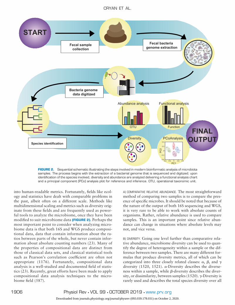
into human-readable metrics. Fortunately, fields like ecol-ogy and statistics have dealt with comparable problems inthe past, albeit often on a different scale. Methods likemultidimensional scaling and metrics such as diversity orig-inate from these fields and are frequently used as power-ful tools to analyze the microbiome, once they have beenmodified to suit microbiome data (FIGURE 2). Perhaps themost important point to consider when analyzing micro-biome data is that both 16S and WGS produce composi-tional data, data that contain information about the ra-tios between parts of the whole, but never contain infor-mation about absolute counting numbers (23). Many ofthe properties of compositional data are distinct fromthose of classical data sets, and classical statistical toolssuch as Pearson’s correlation coefficient are often notappropriate (1176). Fortunately, compositional dataanalysis is a well-studied and documented field of statis-tics (23). Recently, great efforts have been made to applycompositional data analysis techniques to the micro-biome field (587).
A) COMPARATIVE RELATIVE ABUNDANCE. The most straightforwardmethod of comparing two samples is to compare the pres-ence of specific microbes. It should be noted that because ofthe nature of the output of both 16S sequencing and WGS,it is very rare to be able to work with absolute counts oforganisms. Rather, relative abundance is used to comparesamples. This is an important point since relative abun-dance can change in situations where absolute levels maynot, and vice versa.
B) DIVERSITY. Going one level further than comparative rela-tive abundance, microbiome diversity can be used to quan-tify the degree of heterogeneity within a sample or the dif-ference between two samples. There are many different for-mulas that produce diversity metrics, all of which can becategorized into three closely related classes: �, �, and �diversity (1520, 1521). �-Diversity describes the diverse-ness within a sample, while �-diversity describes the diver-sity, or dissimilarity, between samples (1520). �-Diversity israrely used and describes the total species diversity over all
START
FINALOUTPUT0.4
0.2
0
-0.2
-0.40.40.2-0.2-0.4 0
PC2
PC1
Fecal bacteria genome extraction
Bacteria genome data digitized
Species identification
OTU Function
Abundance analysis Functional analysis
Diversity PCoAnalysis
Fecal samplecollection
FIGURE 2. Sequential schematic illustrating the steps involved in modern bioinformatic analysis of microbiotasamples. The process begins with the extraction of a bacterial genome that is sequenced and digitized; uponidentification of the species involved, diversity and abundance are analyzed delivering a functional analysis chartand a principal component (PCo) analysis plot for reference and inference. OTU, operational taxonomic unit.
CRYAN ET AL.
1906 Physiol Rev • VOL 99 • OCTOBER 2019 • www.prv.orgDownloaded from journals.physiology.org/journal/physrev (093.039.178.031) on October 2, 2020.

samples, comparable to �-diversity in a single sample. Dif-ferent formulas for diversity lay different weights on factorslike the richness of a sample in terms of the number ofdifferent species represented, or the evenness of the distri-bution of the species in a sample. It can often be valuable tocalculate diversity using several different metrics, depend-ing on the research question.
C) PRINCIPAL COMPONENT ANALYSIS. A common way to visualizehigh-dimensional data, like relative abundance, is by apply-ing a multidimensional scaling (MDS) algorithm. Principalcomponent analysis (PCA) is both common and appropri-ate in microbiome research, but other algorithms are avail-able and sometimes useful. In PCA, the distances betweenevery combination of two points in a set are taken as aninput, and a set of coordinates is generated as an output.These coordinates can be used to plot each sample as a pointin two-dimensional or three-dimensional space, using theprincipal coordinates as the axis. On the plot, points thatare closer together represent samples that are more similarin composition.
D) FUNCTIONAL METAGENOMICS. The analysis of function repre-sents a high-level readout of the microbiome that can giveinsight into the effect or consequence that a change in themicrobial community can have on its host. Functional met-agenomics are often a product of either WGS or the appli-cation of functional prediction tools on 16S data sets. Thereliability of functional analysis depends on the availabilityof high-quality curated data on the metagenome in question(657). Promisingly, the classical ecology concept of func-tional “guilds,” groups of taxonomically distinct but func-tionally related organisms, has been proposed to apply tothe microbiome (1674).
E) ROBUSTNESS. Recently, robustness has been explored in thecontext of the microbiome (485). Robustness as a metricexpresses information about the degree of change a com-munity will undergo after a perturbation and how fast andto what degree it can recover, if at all. Robustness highlightsan interesting aspect of complex ecosystems. For example, aspecies, or clade (a branch of a phylogenetic/ contextualtree), can have little or no effect on its environment by itselfbut plays an important role in the maintenance of a steadystate. This phenomenon is known in ecology as a keystonespecies, whereby the disappearance of that species willcause the ecosystem to drastically change or even collapse(1046).
4. In silico models of the microbiome
Although human and animal models have many advan-tages, there are numerous situations where the specific con-straints of an in vivo model can overcomplicate an experi-ment. In cases like this, in silico models of the microbiomecan be utilized. Metabolic models of the microbiome, oftenutilizing our growing capacity to produce, process, and
handle big data, have yielded invaluable understanding inmicrobe-microbe interactions and microbe-environment in-teractions (968, 1443). For instance, Flux Balance Analysis(FBA) models have been used to predict and understand thebehavior of microbes given their environment (1139, 1484,1664). The generation and refinement of genome-scale met-abolic reconstructions (GENREs) is essential for realisticmodeling of these types of interactions, which will be facil-itated by the recent publication of a large database contain-ing 773 high-quality human gut GENREs (967). In silicomodels of the microbiome can be used not only to answerexperimental questions but also to verify the reliability ofthe results. One of the current limitations of the field is thedifficulty in finding a gold standard data set with which tovalidate statistical models for microbiome data. Often,there is no sufficiently large data set of the required typeavailable. However, synthetic data sets, generated by pro-grams like SparseDOSSA (696), can be used to tackle thisproblem by generating realistic samples from a completelyknown and controllable source. Hopefully, with the devel-opment of evermore powerful computing technologies, andintricately designed programming suites, in silico modelingwill increasingly complement the necessary use of animalmodels in fundamental microbiome research.
III. MICROBIOTA-GUT-BRAIN AXIS ACROSSTHE LIFESPAN
As stated earlier, the microbiota has been our constant com-panion in life throughout evolutionary history. This richecosystem is not static, but rather is in a constant state offlux across the lifespan. Within individuals, small day-to-day variations in microbiota composition are generally ob-served (339), but these fluctuations become most obviouswhen we take a wider view across the lifespan (see FIGURE3). At both extremes of life, the microbiota is characteristi-cally different from the typical adult gut microbiota in bothlevels of diversity and representation of specific taxa (304,1301, 1648). It has been hypothesized that these periods oftransformation in the microbiota may be likened to sensi-tive periods, during which the microbiota is not only re-sponsive to external influences (and therefore amenable totreatment) but also highly influential with regards to theoverall health of the host (196, 575, 621). In the neurosci-ence literature, a sensitive period is defined as a develop-mental time window during which the brain is more sensi-tive/vulnerable to environmental inputs (660). Originallyapplied to the development of different sensory systems,sensitive periods are now being investigated for higher or-der behaviors as well, such as language development andcognition (236, 661, 1602). It is likely to be biologicallyrelevant that sensitive periods in the microbiota align withsensitive periods in the development or decline of otherbodily systems; this includes, but is not limited to, the innateimmune system, the hypothalamic-pituitary-adrenal (HPA)axis, and brain development in general (196, 575, 602). In
THE MICROBIOTA-GUT-BRAIN AXIS
1907Physiol Rev • VOL 99 • OCTOBER 2019 • www.prv.orgDownloaded from journals.physiology.org/journal/physrev (093.039.178.031) on October 2, 2020.
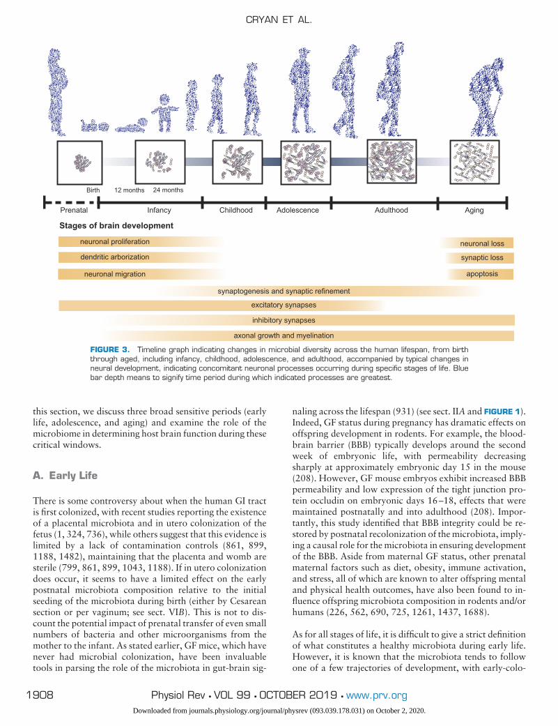
this section, we discuss three broad sensitive periods (earlylife, adolescence, and aging) and examine the role of themicrobiome in determining host brain function during thesecritical windows.
A. Early Life
There is some controversy about when the human GI tractis first colonized, with recent studies reporting the existenceof a placental microbiota and in utero colonization of thefetus (1, 324, 736), while others suggest that this evidence islimited by a lack of contamination controls (861, 899,1188, 1482), maintaining that the placenta and womb aresterile (799, 861, 899, 1043, 1188). If in utero colonizationdoes occur, it seems to have a limited effect on the earlypostnatal microbiota composition relative to the initialseeding of the microbiota during birth (either by Cesareansection or per vaginum; see sect. VIB). This is not to dis-count the potential impact of prenatal transfer of even smallnumbers of bacteria and other microorganisms from themother to the infant. As stated earlier, GF mice, which havenever had microbial colonization, have been invaluabletools in parsing the role of the microbiota in gut-brain sig-
naling across the lifespan (931) (see sect. IIA and FIGURE 1).Indeed, GF status during pregnancy has dramatic effects onoffspring development in rodents. For example, the blood-brain barrier (BBB) typically develops around the secondweek of embryonic life, with permeability decreasingsharply at approximately embryonic day 15 in the mouse(208). However, GF mouse embryos exhibit increased BBBpermeability and low expression of the tight junction pro-tein occludin on embryonic days 16–18, effects that weremaintained postnatally and into adulthood (208). Impor-tantly, this study identified that BBB integrity could be re-stored by postnatal recolonization of the microbiota, imply-ing a causal role for the microbiota in ensuring developmentof the BBB. Aside from maternal GF status, other prenatalmaternal factors such as diet, obesity, immune activation,and stress, all of which are known to alter offspring mentaland physical health outcomes, have also been found to in-fluence offspring microbiota composition in rodents and/orhumans (226, 562, 690, 725, 1261, 1437, 1688).
As for all stages of life, it is difficult to give a strict definitionof what constitutes a healthy microbiota during early life.However, it is known that the microbiota tends to followone of a few trajectories of development, with early-colo-
Prenatal Infancy Childhood Adolescence Adulthood Aging
Birth
Stages of brain development
neuronal loss
synaptic loss
apoptosis
synaptogenesis and synaptic refinement
excitatory synapses
inhibitory synapses
12 months 24 months
neuronal proliferation
dendritic arborization
neuronal migration
axonal growth and myelination
FIGURE 3. Timeline graph indicating changes in microbial diversity across the human lifespan, from birththrough aged, including infancy, childhood, adolescence, and adulthood, accompanied by typical changes inneural development, indicating concomitant neuronal processes occurring during specific stages of life. Bluebar depth means to signify time period during which indicated processes are greatest.
CRYAN ET AL.
1908 Physiol Rev • VOL 99 • OCTOBER 2019 • www.prv.orgDownloaded from journals.physiology.org/journal/physrev (093.039.178.031) on October 2, 2020.
nizing species shaping the long-term composition (992).Soon after birth, the microbiota is typically characterized byrelatively high abundances of Enterobacteriaceae, Bifido-bacteriaceae, and Clostridiaceae, but low levels of Lachno-spiraceae and Ruminococcaceae at the family level (185,293, 1647). As the infant matures, strict anaerobes gradu-ally take over as the dominant taxa, and there is an increasein overall diversity to adult-like levels by around 1–3 yr ofage, coinciding with weaning and a shift to solid food intake(823, 1147, 1648). This rapid development at weaning isobserved across species and was recently shown to be crit-ical for protection against later development of immunopa-thology in mice. However, even after weaning, the micro-biota continues to change. The composition and function ofthe microbiota in healthy children (measured at 7–12 yr ofage) remains significantly different from the healthy adultmicrobiota (676). In particular, the gut microbiota of chil-dren is functionally enriched in pathways supporting ongo-ing development (e.g., genes involved in vitamin synthesisand de novo folate synthesis, anti-inflammatory pathways;Ref. 676).
The infant and child microbiota is susceptible to a range ofenvironmental influences, from birth mode (vaginal vs. C-section) (156, 454), prematurity (540a), and birth location(home vs. hospital) (331) to diet (including breastfeeding vs.formula feeding) (77, 671, 1681), maternal gestational dietand weight gain (1352), pet ownership (77a, 1519), physi-cal illness (757), antibiotic use (835, 1647), and stress (235,621, 1688). Although some of these factors exert a dimin-ishing influence on the microbiota over time (e.g., the effectsof birth mode on microbiota composition are no longerapparent by 6 wk of age; Refs. 293, 667), these early lifefactors may still have long-lasting implications for the phys-ical and mental health of the individual (235, 575, 1043),consistent with the idea that there are sensitive periods formicrobiota-gut-brain interactions. Longitudinal studiestesting this hypothesis are currently limited, but emergingresearch in both humans and rodents suggests that early-lifemicrobiota manipulations can alter trajectories of physicaland mental health or cognitive performance.
In humans, the currently available evidence for enduringeffects of early-life microbiota changes on host physiologyand brain health, although moving more towards mecha-nism and causation, is largely correlational. The evidencesupports a link between early-life microbiota composition,or antibiotic use, and later metabolic and immune function(specifically overweight/obesity and asthma/allergy; Refs. 8,76, 1180, 1386, 1412, 1437). Correlations have also beenobserved between childhood microbiota composition andbehavioral temperament (291), functional activity/connec-tivity in the brain (235, 566), and cognitive function (251).Beyond correlation, preliminary clinical trials of probioticinterventions for at-risk children have yielded promisingresults with regards to reducing risk for GI problems (703,
1601), sepsis (1152), and even autism spectrum disorder(ASD) (1163, 1165) and attention deficit hyperactivity dis-order (1165), although a recent study of postnatal probiot-ics (L. rhamnosus HN001 or B. animalis HN019) found noeffect of either strain on later neurocognitive outcomes(1388). Such investigations are becoming more widespreadbased on strong preclinical evidence that early-life disrup-tion of the microbiota alters a wide range of behavioral andneural outcomes both during development and later in life(308, 437, 867, 1050, 1119, 1428). Preclinical research alsoprovides cause to focus on early-life microbiota interven-tions. Several groups have now shown that early probioticinterventions mitigate the effects of antibiotics, C-sectiondelivery, early-life stress, maternal high-fat diet, and mater-nal immune activation on infant outcomes (see TABLE 5)(226, 234, 345, 348, 552, 568, 690, 834).
B. Adolescence
Adolescence has been labeled a time of “storm and stress,”reflecting the unique challenges associated with this stage oflife (627). In addition to the well-known hormonal fluctu-ations that occur during adolescence, the brain undergoesvast reshaping, including pruning, myelination, volumetricchanges in various regions, and changes in functional con-nectivity (170, 171, 255, 1406) (FIGURE 3). Rapid physicaldevelopment of the body and brain coincides with dramaticshifts in social networks (notably increased independencefrom caregivers), diet, sleep patterns, and exposure to alco-hol and drugs. Faced with this cocktail of stressful newexperiences on a background of unstable hormones andaltered brain function, it is perhaps not surprising that ad-olescents are vulnerable to mental health problems (789,869, 1172). Given that all of these factors have been linkedto alterations in the microbiota-gut-brain axis in adults,exploration of the microbiota during the sensitive period ofadolescence has the potential to provide both insights andinterventions to improve adolescent well-being (529, 1023,1024).
There have been very few studies directly comparing theadolescent and adult microbiota in humans. In a small co-hort (n � 22) of healthy 11- to 18-yr-old adolescents, acorrelation between age and microbiota composition wasobserved such that older adolescents were more similar to(but generally still separate from) the adult microbiota pro-file (12). At the genus level, adolescents had higher relativeabundances of Bifidobacterium and Clostridium, but lowerrelative abundances of Prevotella and Sutterella. These dif-ferences are similar to those reported in other studies of thechild microbiota (682), indicating that there is a gradualtransition from childhood to adulthood rather than a dis-tinct adolescent profile, although further work is needed toconfirm this result. Initial reports show that certain featuresof the microbiota correlate with both diet and metabolicoutcomes in adolescent populations (419, 721, 1413), sug-
THE MICROBIOTA-GUT-BRAIN AXIS
1909Physiol Rev • VOL 99 • OCTOBER 2019 • www.prv.orgDownloaded from journals.physiology.org/journal/physrev (093.039.178.031) on October 2, 2020.

gesting that more in-depth analyses of the functional rele-vance of the microbiota in adolescence are warranted. Thispursuit would be aided by more preclinical studies in thisarea as there is a paucity of research addressing the questionof age specificity. In rodents, it has been shown that sexdifferences in the microbiota emerge only after puberty on-set (987) and that probiotic treatment during a period ofearly-life stress reverses stress-induced changes in the timingof puberty onset (347). Furthermore, long-term antibiotic-induced depletion of the microbiota from adolescence altersadult cognition, social behavior, and anxiety (429). Thischronic treatment also reduced central levels of BDNF, oxy-tocin, and vasopressin and altered tryptophan metabolismin adulthood. While this particular study did not provideclarity with regards to the idea that adolescence is a sensi-tive period (because the treatment continued into adult-hood and no adulthood-only treatment was included), thereare now a number of reports that early-life microbiota in-terventions are more efficacious when directly comparedwith the same interventions administered in adulthood(226, 437, 1134, 1434). Overall, this work provides furthersupport for the existence of early-life sensitive periods ofmicrobiota-gut-brain interaction for at least some behav-ioral and neural outcomes.
C. Aging
Aging is a slow process of deterioration of various homeo-static functions accompanied by an increased prevalence ofdisease (920). The United Nations defines “older persons”as those over 60 yr of age, which roughly translates to 20mo in a rodent model (1217). Distinct hallmarks of agingare apparent from the genetic level (genomic instability,epigenetic alterations, telomere attrition) to the cellularlevel (mitochondrial dysfunction, cellular senescence, stemcell exhaustion, oxidative stress, altered proteasomal activ-ity, and autophagy), including an imbalance of key messen-gers (decline in growth factors, neurotransmitter imbal-ance, dysregulated immunity) and altered receptor sensing(altered stress axis activity, deregulated nutrient sensing),ultimately disrupting the homeostasis of the aging organism(920) (FIGURE 3). Aging is also associated with changes ingut physiology, including hypochlorhydria, gastric motilitydisorders, and degenerative changes in the ENS, yieldingdramatic effects on the composition and function of the gutmicrobiome (830).
While the composition of the adult human gut microbiota isgenerally stable if unperturbed, its stability deteriorates inold age (303). Diet and physical activity, two factors thatgenerally decline in later life, can dramatically affect well-being, cognitive performance, and the microbiota at anystage of life, but their effects seem to be exaggerated in olderindividuals (1556). Aside from this loss of stability, or per-haps because of it, characterization of the aging gut micro-biome has proven difficult. First, the timing of the transition
to an “elder-type” microbiome is not as clearly demarcatedas the shift from an infant-type to an adult-type micro-biome. Furthermore, while comparisons of adult and el-derly individuals have identified clear differences in the mi-crobiota composition between these two groups, the differ-ences between studies have been sizable, perhaps due tocultural, geographical, or methodological variances (152,154, 304, 1081, 1124, 1302, 1362). Decreasing diversity ofthe gut microbiota, generally associated with adverse out-comes in adults, has been linked to aging (153) and age-related impairments like frailty in humans (304, 715). Incontrast, aged (24 mo old) mice have been shown to exhibitincreased diversity compared with younger adult mice(1345). In terms of specific taxa, some studies have ob-served a decrease in beneficial Lactobacillus and Bifido-bacterium in aging (651). A reduced Firmicutes-to-Bac-teroidetes ratio has been reported in Irish and Frenchelderly compared with young adults (303, 985), althoughthis effect was not observed in a comparison of Italiancentenarians, elderly, and young adults (153). Studies insemi-supercentenarians (people between 105 and 109 yrof age) have found specific taxa such as Akkermansia tobe more abundant, suggesting distinct gut microbiotachanges at this extreme of life might be promotinghealthy aging and longevity (152, 1544), as well as re-store intestinal permeability and subsequent immuno-modulation in aged mice (175).
Despite difficulties in identifying consistent aging-relatedcompositional changes in the microbiota, further evidencesupports the hypothesis that the microbiota plays a func-tional role in (un)healthy aging. A recent study demon-strated that suppression of the gut microbiome using broad-spectrum antibiotics restored arterial function in old mice(20–24 mo old) to levels observed in young animals (8–10wk old), which was coupled with normalization of bothoxidative stress and inflammation (225). Earlier studies inrodents demonstrated that GF mice live longer than con-ventional controls, linking gut microbiota to the decline ofimmune system function, or senescence (586, 622). Morerecently in a human population, the ELDERMET studyreported breakthrough research in which age-related shiftsin gut microbiota composition were linked to various func-tional health measures, including frailty, cognition, depres-sion, and inflammatory markers (303, 304). For example,one study found that the more diverse the diet, the morediverse the microbiota, which was linked to improvedhealth and reduced frailty (304). Specific microbial taxa arealso associated with reduced frailty in elderly populations,including Bacteroidetes (109, 682, 1551), Clostridium clus-ter XIVa, and Faecalibacterium prausnitzii, one of manybutyrate-producing bacteria with anti-inflammatory prop-erties (109, 1551, 1694). In contrast, the bacterial familyPorphyromonadaceae has been linked to declines in cogni-tion and affective disorders (92, 328). This nicely parallelsresults in a preclinical study showing that aged mice have a
CRYAN ET AL.
1910 Physiol Rev • VOL 99 • OCTOBER 2019 • www.prv.orgDownloaded from journals.physiology.org/journal/physrev (093.039.178.031) on October 2, 2020.
higher relative abundance of Porphyromonadaceae andthat the levels of this family correlate with increased anxi-ety-like behavior (1345).
Altered aged gut microbiota compositions have also beenproposed to contribute to “inflammaging” (1483), theheightened proinflammatory status and decline in adaptiveimmunity progressively expressed in older age (542). In-flammaging contributes to the speed of the aging processand may progress the development of age-related diseases(1634), from neurological disorders like Alzheimer’s dis-ease (585) to metabolic and other physical disorders likeheart disease, osteoporosis, and type II diabetes (194, 543,875). Both aging and stress weaken the integrity and func-tion of the GI barrier (783, 1483) and negatively affect BBBpermeability (491, 1066) potentially accelerating inflam-maging. By utilizing various models targeting the gut micro-biome including pre-, pro-, and postbiotics, recent studieshave shown a role for the gut microbiome in regulatingneuroimmunity from middle to old age which have impli-cations for therapeutic interventions combatting age-re-lated neurodegeneration and cognitive decline (177, 535,1000). One prebiotic study examining the effect of dietsdiffering in sugar, fat, and fiber content on the gut micro-biota of mice humanized with microbiota from healthy orfrail older people, reported that the frailty-associated gutmicrobiota did not reciprocally switch to a youngerhealthy-subject like state (1506). Furthermore, supplemen-tation with prebiotics was associated with fewer detectedeffects in humans than diet adjustment in animal models(1506).
Further evidence of a link between the microbiota and in-flammaging comes from studies showing compositionalchanges in the microbiota that occur with age induce sub-clinical intestinal inflammation in elderly individuals with ahigh incidence of chronic disease (616), while reduced levelsof Akkermansia following FMT from old mice into youngGF mice was associated with an inflammaging phenotype inthe recipient young mice (546). An important component ofthe immune system and potential contributor to inflam-maging are microglia, which are the brain’s resident im-mune cells. As key players in the brain’s immune orchestra,microglia shape neuronal wiring and activity, synaptic plas-ticity, and phagocytosis and support the survival of neuronsand neuronal progenitors via the secretion of growth fac-tors (919, 1466) (see sects. IV, C and E, and V). However,during aging, microglia develop into a highly reactive andunbalanced state promoting cognitive dysfunction includ-ing altered brain plasticity and neurodegeneration (176,934, 1466, 1574). Recent studies have shown that GF micedisplay deficits in microglia maturation and function, whilerecolonization or administration of key gut microbiota me-tabolites such as SCFAs restore microglial function (489).Such studies suggest that targeting microglia could present
an interventional approach to ameliorate neurodegenera-tive disease (177, 1000).
Indeed, administration of microbiota-targeted diets to pre-vent the age-associated decline of beneficial Bifidobacte-rium has been found to have positive effects on gut micro-biota composition and associated health. A recent studyfound that a 14-wk-long dietary intervention with prebiot-ics increased both Bifidobacterium and Akkermansia inmiddle-aged mice (177). Moreover, the abundance of Bifi-dobacterium strongly correlated with genes involved in co-lonic health in the early phase of aging (1544). Similarly,Bifidobacterium species have been shown to be negativelycorrelated with pro- and anti-inflammatory cytokine levelsin humans, indicating that modulation of Bifidobacteriamay represent a target for reducing the inflammatory re-sponse (1144).
Thus far, dietary restriction has been the most effectivestrategy to demonstrate an increase in lifespan across awhole range of investigated species, including nonhumanprimates (329, 536, 547, 903, 1002, 1294, 1665). This,once again, highlights diet as a key determinant ofhealthy aging, possibly via modulation of the gut micro-biota. Studies in rodents support this interpretation, find-ing that short-term to life-long caloric restriction leads tostructural changes of the gut microbiota (499, 547, 1665)with the genus Lactobacillus, among others, positivelycorrelated with lifespan (1665) and accompanied withchanges in microbial metabolite production such as SC-FAs (1460). Moreover, an intriguing FMT study in theshort-living killifish showed that exchange of microbialcommunities from young to middle-aged fish increasedlongevity in the older group (1392). This illustrates thepotential for gut microbiota modulation as a therapeuticstrategy to benefit an aging host.
IV. PATHWAYS OF COMMUNICATION
There are many pathways of potential communicationbetween the gut microbiota and the brain, from intri-cately innervated and highly modifiable neuronal path-ways to incredibly subtle and difficult to measure smallmolecule messaging systems, both locally in the gut anddistally in the brain. In the following section, we willintroduce many of these communication methods (FIG-URE 4). However, much work is needed to fully resolvethe exact mechanisms as to how bacteria in the lumen ofthe gastrointestinal tract can exert such marked effectson brain and behavior.
A. Autonomic Nervous System
The autonomic nervous system (ANS) is a neural relay net-work, with neurons located within the central and periph-
THE MICROBIOTA-GUT-BRAIN AXIS
1911Physiol Rev • VOL 99 • OCTOBER 2019 • www.prv.orgDownloaded from journals.physiology.org/journal/physrev (093.039.178.031) on October 2, 2020.
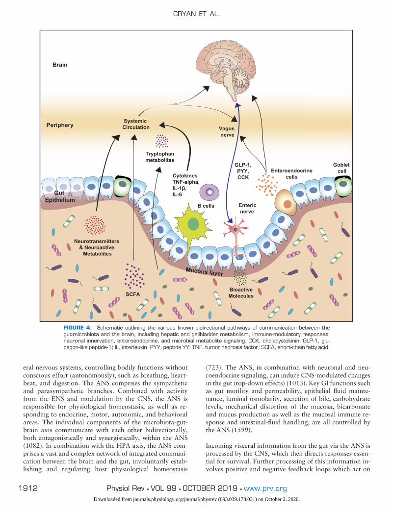
eral nervous systems, controlling bodily functions withoutconscious effort (autonomously), such as breathing, heart-beat, and digestion. The ANS comprises the sympatheticand parasympathetic branches. Combined with activityfrom the ENS and modulation by the CNS, the ANS isresponsible for physiological homeostasis, as well as re-sponding to endocrine, motor, autonomic, and behavioralareas. The individual components of the microbiota-gut-brain axis communicate with each other bidirectionally,both antagonistically and synergistically, within the ANS(1082). In combination with the HPA axis, the ANS com-prises a vast and complex network of integrated communi-cation between the brain and the gut, involuntarily estab-lishing and regulating host physiological homeostasis
(723). The ANS, in combination with neuronal and neu-roendocrine signaling, can induce CNS-modulated changesin the gut (top-down effects) (1013). Key GI functions suchas gut motility and permeability, epithelial fluid mainte-nance, luminal osmolarity, secretion of bile, carbohydratelevels, mechanical distortion of the mucosa, bicarbonateand mucus production as well as the mucosal immune re-sponse and intestinal-fluid handling, are all controlled bythe ANS (1599).
Incoming visceral information from the gut via the ANS isprocessed by the CNS, which then directs responses essen-tial for survival. Further processing of this information in-volves positive and negative feedback loops which act on
SCFA
Vagus nerve
EnteroendocrinecellsCytokines
TNF-alpha,IL-1β,IL-6
B cells
GLP-1,PYY,CCK
Neurotransmitters & Neuroactive
Metabolites
Enteric nerve
Goblet cell
Mucous layer
Tryptophan metabolites
BioactiveMolecules
Periphery
Brain
Gut Epithelium
Systemic Circulation
FIGURE 4. Schematic outlining the various known bidirectional pathways of communication between thegut-microbiota and the brain, including hepatic and gallbladder metabolism, immune-modulatory responses,neuronal innervation, enteroendocrine, and microbial metabolite signaling. CCK, cholecystokinin; GLP-1, glu-cagon-like peptide-1; IL, interleukin; PYY, peptide YY; TNF, tumor necrosis factor; SCFA, short-chain fatty acid.
CRYAN ET AL.
1912 Physiol Rev • VOL 99 • OCTOBER 2019 • www.prv.orgDownloaded from journals.physiology.org/journal/physrev (093.039.178.031) on October 2, 2020.

peripheral organs (188). The ANS provides the gut with themost direct neurological response available, leading torapid changes in gut physiology, through innervation of thetarget organ, in both health and disease (160), such as withthe pain response (1012) and stress (1530).
Direct or indirect ENS-microbiota interactions can occur asa result of ANS activity. The sympathetic and parasympa-thetic systems can influence ENS neurocircuitry, resulting inchanges in motility that can affect the rate of delivery of pre-and probiotics to the small intestine and colon, includingresistant starches and dietary fibers, and other critical mi-crobial nutrients (971). Local GI autonomic activation canbe triggered by interoceptive afferent feedback from the gutas well as CNS cognitive and emotional efferent modulation(1012).
Microbes can communicate with each other via metabo-lites, similar to those recognized by host cells and canthereby interact with gut ANS synapses (1255). Microbi-ota-derived neuromodulatory metabolites include trypto-phan precursors and metabolites, serotonin (5-hydroxy-tryptamine, 5-HT), GABA, and catecholamines (FIGURE 4).Multiple research groups (283, 690, 941, 1399, 1580) havedemonstrated that the microbially modulated metabolite4-ethylphenylsulfate is sufficient to induce anxiety-like be-havior in mice. Indeed, the gut microbiota has been shownto modulate locomotor activity in Drosophila, probably viabacterial-derived metabolites (277, 1338). Furthermore,vagal sensory neuron activation via Campylobacter jejunigut inoculation of mice resulted in direct activation of theneuronal activation marker c-Fos in the vagal sensory gan-glia and the primary sensory relay nucleus for the vagusnerve, the nucleus tractus solitarius (NTS) in the medullaoblongata (588). These findings indicate that gut auto-nomic nerves carrying sensory information that can signaldirectly to the brain upon local GI microbial metabolitestimulation.
Sympathetic gut innervation involves subclasses of postgan-glionic vasoconstrictor as well as secretion and motility sup-pressing neurons. Altered neurophysiology of sympatheticinnervation results in altered GI transit, motility, and secre-tion, primarily via modification of cholinergic transmissionand sphincter contractions on smooth muscle (556, 723).The size and quality of the intestinal mucus layer are be-lieved to be mediated by sympathetic innervation wheremucosal immune system modulation (479) and microbialcomposition and behavior alterations (commensal andpathogenic) were seen (957, 1255).
1. The vagus nerve and beyond
The vagus nerve is the 10th cranial nerve and the fastest andmost direct route that connects the gut and the brain. Itsname is derived from the Latin for wandering, due to itsextensive innervation, which allows collection of informa-
tion from different visceral organs (144). It is composed of80% afferent and 20% efferent fibers (14, 1216), whichtonically transmit vital information from the GI, respira-tory, and cardiovascular systems (bottom-up), and providefeedback to the viscera (top-down).
The gut is innervated by the hepatic and celiac branches ofthe vagus nerve, with decreasing density moving caudallyfrom the proximal duodenum, ileum, and the ileocecal junc-tion as well as the remainder of the small and large intes-tines up to the level of the transverse colon (1584). Vagalafferents form three different types of connections in thegut: intraganglionic laminar endings and intramuscular ar-rays, which both end in muscle wall, as well as terminalaxonal endings in the mucosa, and a recently describedconnection with a subset of enteroendocrine cells, now re-ferred to as neuropods, which form synapses with vagalneurons (750). Depending on their location and type, vagalafferents are ideally suited to detect stretch, tension, or in-testinal molecules such as bacterial by-products, gut hor-mones, or neurotransmitters. Due to the vast variety ofreceptors expressed on vagal afferents, they are thought tobe polymodal, in that they respond to a variety of signalsthat are mechanical, chemical, or hormonal in nature (143).Nevertheless, evidence suggests that there are distinct pop-ulations and subpopulations of vagal afferents respondingto specific stimuli, including stretch (1609) or gut hormones(471).
The vago-vagal anti-inflammatory reflex loop comprisesvagal efferents originating mostly in the medullary dorsalmotor nucleus of the vagus, which can modulate circulatinglevels of proinflammatory cytokines. Stimulation of the re-flex can result in vagal modification of macrophage activa-tion (1174), which is an important and well-documentedmechanism involved in the pathophysiology of inflamma-tory bowel diseases (1082) (see sect. VIIIM). A reduction inparasympathetically controlled intestinal transit has beenassociated with an increase in small intestinal bacterialovergrowth and bacterial translocation (1545). Moreover,pain responses in functional bowel disorders of the gut-brain axis such as IBS are associated with altered parasym-pathetic activity (283). Furthermore, a biphasic vagal acti-vation consisting of gastro-vagal inhibition and sacral acti-vation has been correlated with the incidence of acute stress(993) which potentially could lead to altered regional reg-ulation of the microbiota.
Sensory vagal fiber cell bodies reside in the nodose gangliaand synapse on various nuclei of the brain stem. Vagalfibers from the GI tract mostly synapse bilaterally on theNTS; whereas afferents from the intestine synapse within thesubnucleus commisuralis and medialis in the intermediate tocaudal part of the NTS (40). From there information is relayedto other brain stem nuclei and forebrain structures (1260).Also, multisynaptic pathways ascending from the NTS link
THE MICROBIOTA-GUT-BRAIN AXIS
1913Physiol Rev • VOL 99 • OCTOBER 2019 • www.prv.orgDownloaded from journals.physiology.org/journal/physrev (093.039.178.031) on October 2, 2020.
information from the viscera with the entire brain. Most of thesensory vagal afferents are glutamatergic, where second-orderNTS neurons express �-amino-3-hydroxy-5-methylisoxazole-4-propionate (AMPA), N-methyl-D-aspartate (NMDA), andmetabotropic glutamate (mGlu) receptors (43). In addition,NTS neurons express a variety of neuropeptides includingcholecystokinin (CCK), prolactin-releasing peptide, or pro-opiomelanocortin (1277), and their receptors. Expressionpatterns of these neuropeptides and the target region of theaxons can be used to subcategorize NTS neurons, allowingthe identification of NTS neurons responsible for specificemotional and behavioral responses (979). For example,activation of projections of CCK and immunopositive neu-rons to the parabrachial nucleus results in decreased foodintake without influencing anxiety-related behavior (1277).In contrast, projections to the lateral hypothalamic areainduce feeding behavior upon activation and projectionsfrom the NTS to the bed nucleus of the stria terminalis andthe central amygdala are involved in anxiety as well as fearand avoidance behaviors, respectively (161). Furthermore,neuronal projections to the nucleus accumbens and the ba-solateral amygdala are capable of modulating memory fa-cilitation after physiological arousal (787, 1284). By eitherdirect or multisynaptic projections, the NTS is capable ofinfluencing major neurotransmitter systems such as norepi-nephrine (A2 cell group in the NTS and locus coeruleus),dorsal amygdala (nucleus accumbens, ventral tegmentalarea), and 5-HT (dorsal raphe). It appears that the NTS iswell suited to coordinate the integration of interoceptivefeedback transmitted via the vagus nerve from the gut to thebrain and from the brain to the periphery, thereby acting asan excellent hub for microbiota-gut-brain signaling.
Studies utilizing vagotomy have clearly demonstrated theimportance of constant bidirectional vagal signaling for ap-propriate brain function including host behavior. In hu-mans, there have been older reports that ablation of thevagus nerve, a facet of gastrectomy formerly used for thetreatment of peptic ulcer, resulted in an increase in the in-cidence of psychiatric-related disorders (222, 1604). More-over, neurogenic bowel dysfunction is quite common inpatients with chronic traumatic complete spinal cord injury(1667). In rodents, vagotomy results in decreased locomo-tor activity during the dark phase (706) and increased epi-nephrine concentrations in plasma at baseline and after im-mobilization stress (791, 1078). Furthermore, vagotomyreduced proliferation and survival of newborn cells, de-creased the number of immature neurons (1117) and acti-vation of microglia in the dentate gyrus (DG) of the hip-pocampus (1282), all symptoms that can also be found inpsychiatric disease (see sect. VIII). In addition, subdia-phragmatic vagal deafferentation (1558), a method thateliminates abdominal vagal afferents but keeps most effer-ents intact (1107), highlights the involvement of vagal af-ferents in anxiety-like (elevated plus maze, open-field test,food neophobia) and fear-related behavior (auditory-cued
fear conditioning) (810). Furthermore, it demonstrated anenhancement in reinforced left-right discrimination and re-versal learning (812), sensorimotor gating (pre-pulse inhi-bition), and attentional control of associative learning suchas conditioned taste aversion (811).
Consistent with the data collected on vagotomy, studies onvagus nerve stimulation (VNS) support the role of the vagusnerve in mood regulation. In humans, VNS is used for thetreatment of refractory depression (211) in addition tochronic pain (270, 1250), Crohn’s disease (187), and cer-tain epilepsies (839, 1183). VNS in rodent models hasshown to increase adult hippocampal neurogenesis (609)and modulate the release of norepinephrine, 5-HT, and do-pamine in brain regions related to anxiety and depression(211), as well as increase hippocampal BDNF expressionimproving depressive�like behaviors in chronic restraintstress animals (1367). Furthermore, a recent study in micehas shown that activation of GI vagal afferents influencereward behavior in mice (632), further reinforcing the con-cept that vagal signaling is involved in behavior.
Evidence that gut bacteria utilize vagal afferents to altertheir hosts’ emotional and behavioral responses comes fromstudies observing changes in c-Fos expression in the cellbodies of vagal afferents following oral administration ofCampylobacter jejuni (588). C. jejuni administration hadpreviously been shown to induce anxiety-related behaviorwithout initiating a systemic immune response (949).Moreover, vagotomy inhibited the effects of the prebiotic2=-fucosyllactose in associative learning-related paradigmsand hippocampal LTP (1558). Interestingly, animals sub-jected to subdiaphragmatic vagal deafferentation show al-tered gene expression in the nucleus accumbens (811). Fur-thermore, an increase in the concentration of indole in thegut, a microbial metabolite, has been linked to the activa-tion of the vagus nerve (717), similar to findings whichshow that administration of L. rhamnosus increases thefiring rate of the mesenteric nerve bundle, which containsvagal and spinal afferents (1185). Finally, recent studies in agenetic mouse model of autism (Shank3B�/� mouse) showsthat the effects of L. reuetri on social behavior are no longerpresent in vagotomized animals (1356). Despite plenty ofevidence implicating the vagus as a conduit for microbiometo brain signaling, it has thus far not been possible to mapthe neuronal networks underlying microbiota-gut-brainaxis in detail, and more work will be required to disentanglesuch circuits.
B. Enteric Nervous System
At the interface between the microbiota and the host lies anetwork of neurons known as the ENS, positioned to re-spond either directly, or indirectly, to the microbiota and itsmetabolites. Broadly, the ENS is structured into two gan-glionated plexi, the submucosal and myenteric plexus, and
CRYAN ET AL.
1914 Physiol Rev • VOL 99 • OCTOBER 2019 • www.prv.orgDownloaded from journals.physiology.org/journal/physrev (093.039.178.031) on October 2, 2020.
is largely responsible for the coordination of gut functionssuch as motility and control of fluid movement (555). In thecontext of gut-brain signaling, the ENS communicates withthe CNS via intestinofugal neurons to sympathetic gangliawith sensory information traveling via extrinsic primaryafferent neurons that follow spinal and vagal afferent routes(555). These intrinsic and afferent neural pathways provideopportunities for factors derived from the gut lumen, andtherefore potentially the microbiota, to influence not onlygut function but also the CNS.
ENS structure and neurochemistry resemble that of theCNS, and therefore any mechanisms implicated in CNSdysfunction may also lead to ENS dysfunction (1240) orvice versa. While the development of the ENS occurs pri-marily during embryogenesis, key events such as proliferationof progenitor cells, differentiation of mature neuronal pheno-types and formation of functional neural circuits continue dur-ing the postnatal period (590), concurrent with the develop-ment of the gut microbiota (1623). For example, the density ofganglion cells decreases significantly across the first 3–4 yr oflife (228). This provides a critical window for the microbiotato influence the ENS during critical neurodevelopmental peri-ods. We have recently reviewed the mechanisms by which themicrobiota influences ENS development and function whichincludes activation of pattern recognition receptors (PRRs)including Toll-like receptors (TLRs), in particular, TLR2 andTLR4, which are involved in recognition of microbial mole-cules (700). Inferences on the role of the microbiota on ENSdevelopment can also be drawn from studies in GF rodents(700, 749) (see TABLE 2).
Regarding the expression of PRRs, the ENS has the molec-ular machinery to respond to viral RNA and LPS (the mem-brane component of gram-negative bacteria), through ex-pression of TLRs 3, 7, and 4, respectively (98). Though aninteresting observation, whether microbial factors can di-rectly access the ENS, at least in a healthy state, remainsunclear. Nonetheless, functionally, the absence of TLRs im-pacts functions dependent on the ENS. For example, TLR4-deficient mice display a decrease in fecal pellet output andstool water content, perhaps reflective of alterations in my-enteric and submucosal plexus function, respectively (247),while TLR2-deficient mice display dysregulated small intes-tinal motility (224). A consistent finding across studies in-vestigating members of the TLR family and the ENS haveidentified ultrastructural and neurochemical, as well asfunctional alterations between mice lacking TLRs, and theirwild-type counterparts (700). Perhaps providing a moredefinitive role for the microbiota in this regard, GF micealso display significant ENS abnormalities in terms of ultra-structure and neurochemistry in the early postnatal period(325). These abnormalities observed in GF mice were notapparent in colonized animals, or following colonizationwith a defined bacterial consortia (ASF). This suggests thatthere is potential for particular bacterial species to play a
role in determining the fate of ENS development and hintsat the possible therapeutic application to offset detrimentalfactors which may negatively influence the trajectory ofnormal ENS development and function. In addition, GFanimals also appear to display deficits in intrinsic sensorysignaling, which one could infer might also consequentlyinfluence communication with the CNS. Moreover, thiscould be reversed upon conventionalization of the animalswith microbiota from specific pathogen-free (SPF) donors(1027).
A recent study has provided mechanistic insight into howthe microbiota might influence the ENS, implicating a rolefor 5-HT in this regard (414). Colonization of GF miceincreased enteric neural 5-HT and expression of 5-HT4 re-ceptors while colonization of GF mice lacking tryptophanhydroxylase 1 (TPH1) failed to restore enteric neuron num-bers to those observed in GF and conventional Tph1 trans-genic mice and decreased Nestin� neural precursors, indi-cating reduced levels of neurogenesis (414). Moreover, col-onization of GF mice in the presence of a 5-HT4 receptorantagonist negatively influenced the ENS. In contrast, stim-ulation of 5-HT4 receptors in GF animals had the oppositeeffect and restored normal gut physiology (414). This workprovides a body of evidence implicating a role for microbi-ota-induced effects on 5-HT and its impact on the ENS,although there are subtleties in the findings that would sug-gest this pathway may differentially influence neurogenesis,differentiation, cell turnover, and gut function dependingon the nature and timing of the interventions (FIGURE 4).
Antibiotic-induced disruption of the gut microbiota hasalso been applied as a method to determine the impact ofthis microbial community on the ENS and resulted in wide-ranging effects on ENS architecture (neuronal and glial),neurochemistry, and function (247) (see TABLE 3). Withinenteric ganglia reside glial cells, which in antibiotic-treatedanimals were decreased (247), and evidence from GF ani-mals suggests a more specific role for the microbiota inmodulating mucosal enteric glial cell migration which wasnot restricted to the early postnatal developmental period(748). Notably, the density and proportion of substanceP-containing neurons, a neurotransmitter involved in mo-tor and sensory neurotransmission in the gut, were in-creased in response to antibiotic-induced disruption of thegut microbiota as was the density of TLR2, which whenactivated partially recovered deficits in gut function (247).These changes occurred concomitantly with a rebalancingof fecal bile acid profile reflected as increased levels of fecaltaurocholic acid and a reduction of cholic acid in antibiotic-treated animals (247). However, whether such changes inbile acid profile are mechanistically linked to the structuraland functional alterations in the ENS warrants further in-vestigation. Notwithstanding this, in a mouse model of au-tism, not only was abnormal gut function observed, char-acterized by a constipation-like phenotype, ultrastructural
THE MICROBIOTA-GUT-BRAIN AXIS
1915Physiol Rev • VOL 99 • OCTOBER 2019 • www.prv.orgDownloaded from journals.physiology.org/journal/physrev (093.039.178.031) on October 2, 2020.
and neurochemical changes in the ENS but also changes infecal bile acid profile not dissimilar to those observed inantibiotic-treated mice, with elevated fecal taurocholic acidalso observed in this mouse model of autism (593). How-ever, shifts in the microbiota composition associated withstress and ENS abnormalities can occur independently ofeach other as observed in offspring of dams exposed toprenatal stress in which the most significant stress-inducedchanges in the gut microbiota failed to correlate with gutphysiological and ENS parameters (592). Therefore, onemust exert a degree of caution in interpreting concomitantchanges in the microbiota and ENS as being intrinsicallylinked.
Microbial status, however, is not the only determinant ofENS function/activity, which also displays sensitivity to ex-posure to early-life stress (404). In a recent study, stress-induced alterations in ENS activity, characterized by stim-ulated acetylcholine release, was influenced by both mater-nal separation and the microbiota (404), thus providingevidence that the microbiota may underpin ENS-related gutdysfunction associated with early-life stress. At a single-celllevel, GF status is accompanied by significant alterations inthe electrophysiology and characteristic profile of afterhy-perpolarization-type enteric neurons (intrinsic primary af-ferent neurons) reflected as decreased excitability and dis-charge (1025), which in the context of the microbiota-gut-brain axis may be especially important as these intrinsicafferent neurons may represent the first neural elements tosense changes in the microbiota with consequential effectson the gut and the brain. However, these single-cell changesmay not necessarily manifest as changes in gut function,where secretomotor responses, and those stimulated bycapsaicin, and presumably driven by the submucosalplexus, were comparable in colonic segments from GF ani-mals and colonized controls (917).
In addition to displaying sensitivity to colonization with com-plex or simplified microbiota, the ENS also responds to spe-cific bacterial strains, or components thereof, perhaps provid-ing some insight into the microbial mechanisms which mayaffect ENS function. While some of these studies have taken adirect approach by stimulating enteric neurons with bacterialsupernatants or conditioned media (793, 845), others haveconsidered the epithelial barrier which separates the gutlumen from its underlying nervous system (30, 845). Thelatter approach has demonstrated that not only do differentbacterial strains differentially influence enteric nerve activ-ity but that they may do so by different mechanisms (981).Bacteroides fragilis, for example, likely as a consequence ofits capsular exopolysaccharide, can influence ENS functionwhile an L. rhamnosus strain (JB-1) does so via a G protein-coupled receptor-mediated pathway (981). Of note in thislatter study, the authors show that the effects on the ENSare not dependent on diffusion of molecules from the epi-thelium to the myenteric neurons. Using a similar method-
ology, the same laboratory also demonstrated that the mi-crobial-derived SCFA butyrate and epithelial-applied 5-HTcould affect ENS activity providing some further insightinto additional mechanisms by which the microbiota mayinteract with the ENS (845). Functional studies support thatdifferent microbial strains can differentially affect neurally-driven secretomotor responses (916). Further characteriza-tion of the mechanism underlying the effect of the L. rham-nosus strain (JB-1) on ENS function confirmed that an in-tact epithelium was required to mediate the effect ofmicrovesicles derived from JB-1, which themselves recapit-ulated the effects of the strain per se on the ENS (30),indicative that epithelial elements may be an important in-termediary in transducing microbial signals from the gutlumen to the ENS. The ENS is also central to facilitatingchanges in motility consequential to diet-microbe interac-tions (435).
Evidence now also suggests that there may be a reciprocalrelationship between the ENS and the gut microbiota. Thusfar, we have considered the impact of the microbiota on theENS. However, the ENS appears to be able to exert controlof the microbiota (1274). In zebrafish lacking an ENS as aconsequence of a mutation in an SRY-related HMG-box(sox10 transcription factor gene), a “pro-inflammatory”microbiota profile developed, the effects of which could beameliorated by transplantation of wild-type ENS precur-sors into sox10 mutants or by the introduction of “anti-inflammatory” microbes (1274). These data might chal-lenge the dogma that alterations in the microbiota precedealterations in the ENS and thus lead to the proposal thatchanges in neural activity may precede alterations in the gutmicrobiota.
Abnormalities in the ENS are associated with life-threaten-ing GI disorders including Hirschsprung disease and neuro-pathic chronic intestinal pseudo-obstruction (570). Also,the ENS has now been implicated in disorders of the CNS,including ASD, Alzheimer’s disease, and Parkinson’s dis-ease, generally considered primary disorders of the CNS(1240). ASD is particularly noteworthy given the high levelof comorbid GI symptoms observed (1240) (see sect.VIIIA). Future studies are needed to fully appreciate therelative contribution of the microbiota in shaping the path-ological effects of ENS dysfunction.
C. Immune System and Neuroimmunity
The GI tract harbors the densest concentration of immunecells in the body and is in constant communication with thetrillions of microbes that inhabit our gut, either throughdirect physical contact or the release of secreted compounds(FIGURE 4). As such, one critical function of the single-celllayer of our gut is to limit the contact of intestinal microbi-ota with the visceral tissue, which it does by secreting aprotective viscous mucus layer from goblet cells of the epi-
CRYAN ET AL.
1916 Physiol Rev • VOL 99 • OCTOBER 2019 • www.prv.orgDownloaded from journals.physiology.org/journal/physrev (093.039.178.031) on October 2, 2020.
thelium. This luminal-mucosal interface is where the major-ity of host-microbe interaction occurs, and the exchange ofmolecules through the mucous layer and epithelium serve tofacilitate communication between the gut and the immunesystem through the recognition of self and non-self antigens(505), and thus to prime the immune system to identifypotentially harmful pathogens.
In addition to providing a physical barrier, the epitheliumcontains a number of different cell types including entero-cytes, secretory cells, chemosensory cells, and gut-associ-ated lymphoid tissue (1213). The enterocytes express innateimmune receptors and can release cytokines and chemo-kines, while the gut-associated lymphoid tissue utilizes lym-phocytes to mount a more specific immune response (im-munoglobulins). Chemosensory cells play a key role in de-fense against helminths (36), while secretory cells areinvolved in mucus release from goblet cells, the release ofantimicrobials from Paneth cells, and the release of neu-roendocrine compounds including ghrelin, somatostatin,CCK, peptide YY, and 5-HT among others from enteroen-docrine cells (243).
Peptidoglycans, polysaccharides, and other antigens onbacteria that confer beneficial roles for the bacteria (protec-tion against degradation) also allow the host immune cellsto identify the numerous and diverse bacteria to the host,and to identify a change in the homeostatic balance of thegut. Epithelial PRRs recognize molecular patterns unique tobacteria and other microorganisms (pathogen-associatedmolecular patterns) (461, 1533) of which the TLR familyare the most studied, and once activated can recruit inflam-matory mediators, cytokine production, and chemokine-mediated recruitment of acute inflammatory cells (1455).
Along with activating the innate immune system, manycommensal bacteria metabolites including neuromodula-tors, bacteriocins, bile acids, choline, and SCFAs are immu-nomodulatory (FIGURE 4). A growing body of evidence sug-gests that microbiota-host interactions at the level of the gutleads to the release of cytokines, chemokines, neurotrans-mitters, neuropeptides, endocrine messengers, and micro-bial by-products that can infiltrate the blood and lymphaticsystems, or influence neural messages carried by the vagaland spinal afferent neurons to constantly communicatewith the brain and update health status, and possibly regu-late brain and behavior. However, it seems unlikely thatany one of these classes of compounds is individually re-sponsible for mediating all microbiota-gut-brain interac-tions, and these will be discussed individually in subsequentsections.
1. Innate immune system
The immune system is subdivided into two separate subsys-tems: innate and adaptive/acquired. Innate immunity is re-garded as the body’s primary line of defense against poten-
tially infectious organisms and involves the activity of cellsderived from the myeloid lineage–monocytes and macro-phages, basophils, eosinophils, and neutrophils, in additionto mast cells, platelets, and natural killer cells (1423). Thegut microbiota influences the relative populations, migra-tion, and function of various subsets of immune cells includ-ing helper T cells, regulatory T cells, mononuclear phago-cytes, and innate lymphoid cells (458, 1283), and the mech-anisms as to how a microbial population of the gut canmodulate both the innate and adaptive immune responsesat mucosal surfaces during infection, inflammation, stress,and autoimmunity are being unraveled (256, 477, 754,1014, 1215). However, until recently, the interaction be-tween microbes confined to the gut and immune cells of thebrain had not been investigated.
As alluded to in earlier sections, microbiota-microglia inter-actions are receiving much attention of late as key mecha-nisms underlying microbiota-immune-brain interactions (3,1243, 1576). The CNS-restricted microglia are innate sen-tinel immune cells that can detect subtle changes in thesurrounding molecular milieu (162, 420, 663, 804, 1102,1509), and are responsible for the mounting of neuroin-flammatory responses (790, 1238, 1640). These residentcells compromising 5–12% of all cells in the brain arehighly ramified and extremely plastic, and once activatedcan release a number of cytokines and chemokines, expressnumerous antigenic markers, regulate neurotransmitters,and undergo extreme morphological changes (1206). Cyto-kines and chemokines are chemical messengers involved inrecruiting other immune cells to the vicinity, the repair ofdamaged tissue, and the restoration of homeostasis (275).Cytokines are generally classed as pro-inflammatory oranti-inflammatory, which facilitate or inhibit inflammatoryprocesses, respectively, and both play an equally importantrole in the mounting of a suitable physiological neuroin-flammatory response, and the return to homeostasis. In-deed, the balance between these anti- and pro-inflammatorycytokines and chemical messengers are key determinants inan appropriate defense of the host against infection or tissuedamage.
Along with their established immune role in the CNS, theconstitutively active microglia are critically involved in neu-ronal events at various stages in development and adult-hood, including synaptic remodeling to improve neuronalnetwork signaling (1327). Recently, it was determined thata diverse GI microbiota is necessary for the maintenance of,and maturation of, microglia in a healthy functional state(489). In contrast, the absence of a complex host microbiota(when using GF animals or antibiotic-treated mice) in-creased microglial populations, caused defects in microgliamaturation, activation state and differentiation, altered mi-croglia morphology (with longer processes and increasedbranching, terminal points and clubbing at synaptic bou-tons), and compromised immune response to bacterial or
THE MICROBIOTA-GUT-BRAIN AXIS
1917Physiol Rev • VOL 99 • OCTOBER 2019 • www.prv.orgDownloaded from journals.physiology.org/journal/physrev (093.039.178.031) on October 2, 2020.
viral infection (489). These alterations in microglial pheno-type were reversed with recolonization of gut microbiota,following 6 wk co-habitation of GF mice with control mice.These findings demonstrate that a healthy and diverse GImicrobiota is essential for the continuous preservation ofhealthy microglia and proper brain function throughoutour lifespans (359, 1486).
Aside from releasing cytokines and chemokines to recruitlocal immune cells, microglia also recruit monocytes fromthe periphery to the brain to aid in defense and cell debrisclearing. While the multimodal influence of microbiota onphysiological events at the level of the CNS has gainedcredence, more recently there has been a shift toward un-derstanding the role of the microbiota in regulating neuro-inflammation via intervention in the recruitment of localimmune regulators (trafficking monocytes) from the periph-ery to the brain (1615). Evidence suggests that the traffick-ing of these monocytes to the brain appeared to be governedby tumor necrosis factor (TNF)-�-mediated microglia acti-vation (376) and was reversible in preclinical studies withprobiotic intervention with VSL#3 (377). Indeed, the medi-ation of certain behavioral phenotypes as a consequence ofprolonged HPA axis activation involves the trafficking ofmonocyte from the spleen (1615), a tissue with a high den-sity of free fatty acid receptor type 2 (FFAR2) (489). TheseFFAR2 receptors are G protein-coupled receptors that havebeen localized to peripheral lymphocytes (1100). GI micro-biota are responsible for the production of SCFAs, the nat-ural ligands for FFARs, which were shown to reverse thedetrimental effects on microglia density, morphology, andmaturity in the absence of a complex microbiota (489).Taken together, these findings suggest that GI microbiotamay govern centrally mediated events indirectly throughregulation of monocyte trafficking to the brain and subse-quent microglia activation, possibly via SCFA-mediatedmechanisms.
2. Adaptive immune system
Adaptive immunity induces a specifically targeted responseto pathogens and comprises cells originating in the lym-phoid lineage (i.e., B and T lymphocytes). The adaptiveimmune system distinguishes itself by its specialization,specificity, memory, regulation, diversity, and tolerance(1554). Furthermore, these cells are also capable of secret-ing both pro- and anti-inflammatory cytokines (296). Theadaptive immune system develops postnatally and is, there-fore, critically shaped by exposure to microbes. Further-more, disease states such as allergy and asthma and/or theimmune hypothesis of depression/schizophrenia are linkedto the disruption of microbes and immune function.
Over the last decade, efforts have been made to increase ourunderstanding of the potential role the adaptive immunesystem plays in the brain and behavior (46, 522, 1068).Recent intriguing studies in recombination activation gene
2 knockout (Rag2) transgenic mice, which lack mature Band T lymphocytes, showed that adoptive transfer of lym-phocytes from stressed mice reduced anxiety, increased so-cial behavior, and increased hippocampal cell proliferationcompared with those receiving no cells or cells from un-stressed donors (206), and that transferred lymphoid cellsinfiltrate the choroid plexus and the meninges (1330). Re-cently, intraepithelial lymphocytes (CD4CD8��� double-positive T cells specifically) were shown to require the pres-ence of L. reuteri, in combination with a tryptophan-richdiet, to reprogram intraepithelial CD4� T cells into immu-noregulatory T cells (268), evidence that gut microbiomeinfluences/interacts with the adaptive immune system. Fur-thermore, Drosophila social behavior appears to be regu-lated through immunoregulation and microbiota mainte-nance (277). This finding reveals that adaptive immune cellsmight be mediating the behavioral response to stress, thusrepresenting an interesting novel pathway, which needs tobe further explored.
Interestingly, adaptive immunity and the intestinal micro-biota also seem to be acting synergistically towards modu-lating homeostasis. IgA plasma cells from the gut are able toaccess the CNS in the induction of experimental autoim-mune encephalomyelitis multiple sclerosis mouse model,and that these cells suppress inflammation in a pathway thatinvolves IL-10 (1272). In Rag2 transgenic mice, adaptiveimmunity influences microbiota composition (563). A linkbetween stress, the microbiota, and the adaptive immunesystem has been illustrated in a recent study examining theimpact of chronic stress on long-lasting altered levels ofIL-10� T regulatory cells (146). The expression of IL-10was associated with an increased abundance of Clostrid-ium, therefore reinforcing the hypothesis that microbes inthe gut, the adaptive immune system, and the CNS may beworking synergistically. Subsequently, it was shown thatthe impairment in the adaptive immune system in Rag1transgenic mice is linked to alterations in cognition andanxiety-like behavioral tasks, which were ameliorated bytreatment with a combination of L. rhamnosus and L. hel-veticus. This study, therefore, establishes that lymphocytedeficiency results in impairments in behavior, which can benormalized by probiotic treatment, implicating a specificrole of adaptive immunity in the microbiota-gut-brain axis(1390). Adaptive immunity has also been shown to respondto specific bacterial antigens that are modulated by dietarycomponents, suggesting that dietary modifications could bea therapeutic avenue for human disease such as IBD (1598).A link between the adaptive immune system, interferon(IFN)-� signaling in the meninges, and social behavior wasalso illustrated (523). This study showed how IFN-� acts asa link between meningeal immunity and neural circuits re-cruited for social behavior. Given the importance of themicrobiome in shaping social behavior (see sect. VIIB), fur-ther studies should focus on the possible role of the micro-biome in shaping such immune-brain communication.
CRYAN ET AL.
1918 Physiol Rev • VOL 99 • OCTOBER 2019 • www.prv.orgDownloaded from journals.physiology.org/journal/physrev (093.039.178.031) on October 2, 2020.
D. Enteroendocrine Signaling
Though enteroendocrine cells (EECs) represent only 1% ofepithelial cells in the GI tract, they are critically importantfor the maintenance of gut homeostasis due to the pleiotro-pic effects of their secreted signaling molecules. Ten differ-ent types of EECs have been described so far, all of whichare sensory cells, coordinating changes in the gut-nutrientluminal content with metabolic and behavioral responses,such as the regulation of insulin secretion or food intake(606). Two of the best understood EECs are enteroendo-crine L cells and enterochromaffin cells which are abundantin the distal small and large intestines, where the majority ofbacterial taxa reside. They can establish direct contact withthe luminal constituents via the apical surface, includingbacterial metabolites. They have a long lifespan (181,1518), potentially enabling them to integrate into the localsignaling network of the ENS, glia and immune cells of theGI submucosa.
1. Enteroendocrine L cells
Enteroendocrine L cells secrete glucagon-like peptide-1(GLP-1) and peptide YY (PYY) in the postprandial state.Both peptides are potent anorexigenic hormones involvedin the modulation of eating. The receptors to these peptidesare expressed locally in the gut enteric neurons and vagalafferents, as well as in the CNS, including the brain stemand hypothalamus (408, 1258). GLP-1 and PYY can stim-ulate satiation and inhibit eating, acting either directly onthe hypothalamic centers of appetite control, or indirectlyvia the vagal-brainstem-hypothalamic pathway (860,1418). Which of these pathways is more important for theeffects of peripherally released hormones on food intake isstill not fully understood. Notably, a new route of commu-nication between L cells and the ENS has recently beendiscovered (180, 181, 750). A basolateral cytoplasmic pro-cess of the L cells, the neuropod, was shown to establish afunctional synaptic contact with the enteric glia and vagalafferents. The existence of synaptic connections between Lcells and the ENS suggests that the EECs signaling from thegut to the brain can respond more precisely and much fasterthan we initially thought and that the CNS can have atuning effect on the L cells.
In the proximal gut, the release of GLP-1 and PYY is acti-vated mainly by luminal nutrients, providing a postprandialspike of peptide release. For instance, L cells can sense car-bohydrates via the sodium-coupled glucose transporter(SLC5A1) (597, 1249), long-chain fatty acids via FFAR1and FFAR4 receptors (470, 670), and monoacylglycerolsvia the GPR119 receptor (1249). Strikingly, in the distalgut, the activation of L cells is triggered almost exclusivelyby bacteria-derived metabolites. Moreover, SCFAs canstimulate the secretion of GLP-1 and PYY through theFFAR2 and, to a lesser extent, the FFAR3 receptor (900,1221, 1499). Bacteria-derived secondary bile acids (litho-
cholic acid, deoxycholic acid) can activate the G protein-coupled bile acid receptor Gpbar1 receptor (TGR5) in Lcells, promoting the peripheral release of GLP-1 and PYY(774, 1487, 1529). Indole was shown to induce GLP-1 se-cretion (285). Furthermore, bacterial LPS were recentlyshown to enhance GLP-1 secretion, suggesting the involve-ment of TLRs in L cell activity (855, 1095).
Because bacterial fermentation of nondigested nutrientscontinues between meals, bacterial metabolites can supportthe activity of colonic L cells and the secretion of anorexi-genic hormones for many hours after a meal (606). Thisbasal secretion of GLP-1 and PYY is thought to play animportant role in the control of food intake, body weight,and metabolism since obese individuals are generally char-acterized by decreased serum levels of both GLP-1 and PYY(408). Changing the fermentation capacity of the gut micro-biota through chronic dietary supplementation of prebiot-ics or probiotics was quite successful in decreasing foodintake and body weight and improving glucose tolerance inpreclinical models of obesity and diabetes (see TABLES 4AND 5; for further details, see review in Ref. 1203). Someprebiotics, a polysaccharide inulin, as well as FOS and GOSwere shown to increase the production of both GLP-1 andPYY (242, 498). Certain Lactobacillus strains were capableof stimulating GLP-1 production both in vitro and in vivo(94, 1154, 1377). Altogether, these findings clearly demon-strate the importance of bacterial metabolites and adequatediet in the maintenance of metabolic health, and that gutmicrobes can regulate the secretion of GLP-1 and PYY.
Although EEC L cells are perhaps the best-studied compo-nent of the host-microbe dialogue at the enteroendocrineinterface other protagonists have also emerged. Autoanti-bodies directed against the peptide �-melanocyte-stimulat-ing hormone (�-MSH), which is involved in appetite con-trol, have been discovered in both healthy individuals andin subjects with eating disorders (519, 520). Many of theseantibodies represented the IgA class, which points to anintestinal origin of such antigens. This was a genuinely in-triguing discovery because it raised the possibility thatacross evolution, in close interactions with the host, bacte-rial taxa could have acquired protein mimetics of mamma-lian appetite-regulating hormones. This endows gut bacte-ria with a powerful tool to control nutrient supply by ma-nipulating host food intake. A few years later, a caseinolyticprotease B (ClpB) heat shock protein was discovered in gutcommensal E. coli, an antigen mimetic of �-MSH whichconfirmed this hypothesis (1471). Moreover, it was shownthat ClpB plasma levels were elevated in a variety of eatingdisorders, and its concentration has been correlated withvarious psychopathologic traits (215). Moreover, chronicintragastric delivery of ClpB-expressing E. coli in mice stim-ulated the production of �-MSH-reactive antibodies anddecreased food intake (217, 1471). Interestingly, peripheral�-MSH was shown to trigger the release of PYY and GLP-1
THE MICROBIOTA-GUT-BRAIN AXIS
1919Physiol Rev • VOL 99 • OCTOBER 2019 • www.prv.orgDownloaded from journals.physiology.org/journal/physrev (093.039.178.031) on October 2, 2020.
from EEC L cells in the gut via activation of the melanocor-tin 4 receptor (1149). This suggests PYY and GLP-1 couldmediate the effects of bacterial ClpB on satiety. Theseground-breaking findings posit that evolution of molecularmimicry between gut bacteria and the host (1129) can affecthost eating behavior. Most intriguingly, humans seem tocarry autoantibodies to some appetite-regulating peptidesincluding PYY and ghrelin (520), and the correspondingbacterial antigens, as well as their physiological role, haveyet to be unraveled.
2. Enterochromaffin cells
Enterochromaffin cells (ECs) produce the majority of 5-HTin the body from dietary tryptophan (see also sect. IVE).5-HT activates a diverse family of receptors on intrinsic andextrinsic afferent nerve fibers in the GI tract and mediatesmany GI functions including intestinal peristalsis, electro-lyte secretion, pain perception, and inflammatory responses(1007). Unlike L cells, interactions between gut microbiotaand ECs are much less understood.
Taking advantage of GF mice, recent studies have shownthat specific gut bacterial strains, such as the spore-formingclostridia taxa, can upregulate the expression of colonicTPH1 (the rate-limiting enzyme in the production of 5-HT),boosting 5-HT biosynthesis in the gut, and increasing intes-tinal transit in the host (1248, 1645). It is unclear whetherthis was a direct interaction of bacteria with ECs or indirectstimulation of ECs with bacterial metabolites. However, asingle intrarectal injection of deoxycholic acid (a secondarybile acid produced by clostridia) could partially replicatethe effects of the bacteria (1645). Interestingly, earlier find-ings suggest that secondary bile acids can mediate the effectof gut bacteria on 5-HT synthesis in the host. ECs wereshown to express TGR5, and administration of deoxycholicacid increased 5-HT secretion in colonic tissue of the wild-type, but not Tgr5 transgenic mice (33). There is also evi-dence that bacterial LPS can increase 5-HT secretion viaTLR4 activation in ECs isolated from Crohn’s disease pa-tients, but not from healthy subjects (794).
Variations in intestinal 5-HT production are unlikely tohave a direct effect on the brain because 5-HT cannot crossthe BBB (457). The availability of tryptophan for the brain,as a source for 5-HT synthesis, is also partially determinedby the metabolism of tryptophan along the physiologicallydominant kynurenine pathway (785) (see sect. IVE). How-ever, by modulating vagal afferent activity (1007) and in-flammatory responses in the gut (1357), 5-HT releasedfrom EC cells can potentially have an impact on gut-brainsignaling. Perhaps the best-known example of such an in-teraction is during chemotherapy-induced nausea and em-esis, caused by a massive release of intestinal 5-HT andconsequent activation of vagal afferents in the gut (561).More recently, altered 5-HT signaling has been associatedwith IBS (1006) (see sect. VIIIM). Moreover, in a mouse
model of ASD (the BTBR T� Itpr3tf/J mouse) (see sect.VIIIA) (828, 1036, 1075), a strong association between thereduction in the relative abundance of Blautia species fromClostridiales order and a decrease in 5-HT production inthe gut was observed (593). Further studies utilizing specificbacterial strains with pro-5-HT activity (such as bile-me-tabolizing clostridia) are required to shed more light on therole of gut microbiota and intestinal 5-HT in gut-braincommunication.
Adding to the complexity of this task, it is worth notingthat EC-produced 5-HT is not only secreted towards theintestinal submucosa, but a substantial amount of 5-HTis continuously released in the apical direction, which canbe easily detected in the gut lumen (1168). Furthermore,gut bacteria were shown to deconjugate host-producedcatecholamines via the �-glucuronidase enzyme path-way, and thus generate free luminal 5-HT (64, 645). Thephysiological consequences of these processes, either forthe host or the microbiota, are not yet known or will bethe subject of future studies.
E. Neurotransmitters
Microbial endocrinology is becoming an important conceptin advancing our knowledge of the microbiota-gut-brainaxis, helping move our understanding from correlation tocausation (942). One of the main facets of the concept ofmicrobial endocrinology is based on the shared neurochem-ical language that exists between host and microbe. It hasbeen long understood that microbes are capable of produc-ing neurochemicals that we typically associate with mam-malian organisms. Landmark studies in the 1990s were thefirst and demonstrate that bacteria respond to host neu-roendocrine signaling molecules (947, 948), including nor-epinephrine and epinephrine, and that the microbiota canaffect host behavior via the gut-brain axis (949). Since thattime, the microbiota has been shown to synthesize and re-spond to several key neurochemicals (e.g., 5-HT, GABA,among others) that are involved in host mood, behavior,and cognition. Many of these host- and microbial-derivedneuroactive molecules are also important signaling mole-cules in host-microbiota interactions at the intestinal inter-face.
1. Catecholamines
Catecholamines play diverse roles in host physiology, rang-ing from the stress-induced fight-or-flight response (1415)to influencing gut integrity (1028) and affecting host moti-vational behavior as well as decision-making (1473). Like-wise, norepinephrine and epinephrine induce wide-rangingresponses in bacteria, including the promotion of pathogen-esis and growth (945). The catecholaminergic system wasone of the first demonstrated to mediate host-microbecrosstalk, and holds important clinical implications for
CRYAN ET AL.
1920 Physiol Rev • VOL 99 • OCTOBER 2019 • www.prv.orgDownloaded from journals.physiology.org/journal/physrev (093.039.178.031) on October 2, 2020.
both animals and humans (943). Although it was previouslyaccepted that susceptibility to illness following acute stresswas a consequence of a suppressed immune system, it wasshown that the host production of norepinephrine causedthe induction of bacterial virulence genes, thereby drivinginfection and mortality (942). Norepinephrine signals thesechanges in bacteria via several methods, including possiblereceptor-based mechanisms. At increasing levels within thehost, norepinephrine exerts a chemotactic effect on bacte-ria, which can increase bacterial migration toward the hostintestinal mucosa (950). Second, catecholamines can be-have as siderophores, which cause the release of iron fromhost-iron sequestering proteins, increasing the availabilityof iron for bacteria, thereby enhancing bacterial growth(548). Also, norepinephrine can act as a quorum sensingmolecule to increase the expression of bacterial virulencegenes through interaction with bacterial quorum sensinghistidine kinase (371, 1070). In GF rodents, circulating nor-epinephrine concentration is higher compared with conven-tional rodents (807), and within ileal, cecal, and coloniclumens, dopamine, norepinephrine, and epinephrine are de-tectable (64).
Enteric nerves contain the requisite genetic machinery tosynthesize dopamine and norepinephrine, but lack phenyle-thanolamine N-methyltransferase, the enzyme that con-verts norepinephrine into epinephrine (336). This is an im-portant consideration because the microbiota, and in par-ticular the bacterial enzyme �-glucuronidase, have beendemonstrated to play a critical role in converting host nor-epinephrine and dopamine from a biologically inactive to abiologically active form (64). It was recently demonstratedthat certain bacteria contain plasma membrane monoaminetransporter and organic cation transporter proteins whichenable the transfer of extracellular norepinephrine into thebacterial cell (944).
A variety of bacteria, many of which are found within thehuman GI tract, are recognized to produce catecholaminesthat are identical in chemical structure to those producedby the host. Evolutionary adaptations, including late hori-zontal gene transfer (711), have been proposed to explainbacterial metabolic pathways of neuroendocrine molecules.Briefly, members of the genus Escherichia, including humangut commensals, are known to produce catecholamines,such as norepinephrine (1369). Bacillus are likewise knownto produce norepinephrine in addition to dopamine (1514).Although several host cell types in the GI tract are respon-sive to catecholamines (124), it is largely unknown howcatecholamines of bacterial origin influence host physiol-ogy.
2. GABA
Both host and bacteria have the capacity to convert theamino acid glutamate to GABA (1391, 1432), the majorinhibitory neurotransmitter of the host nervous system. In-
deed, Escherichia spp. (1257) and Lactobacillus spp.(1385) have been demonstrated to synthesize GABA. Addi-tionally, several fermented foods used by Lactobacillus spp.also contain millimolar levels of GABA, such as the Chinesepaocai (885). Interestingly, certain Lactobacillus spp. iden-tified in fermented foods are also known to synthesize glu-tamic acid (1661). Although the importance of the bacterialproduction of GABA in the host intestinal lumen is unclear,GF mice exhibit significantly lower luminal GABA levelscompared with mice that have a microbiota (999). Interest-ingly, it has been shown that the probiotic E. coli strainNissle 1917 (EcN) can produce a GABA associated analge-sic lipopeptide, C12AsnGABA OH (1184). They were ableto demonstrate that the addition of the C12AsnGABA OHmoiety to GABA facilitated diffusion across the epithelialbarrier, allowing the activation of GABA receptors on sen-sory neurons. It is currently unknown if this is a commonfeature of other microorganisms in the GI tract. In a studyexamining Lactobacillus spp. and Bifidobacterium spp. iso-lated from the human GI tract, five strains were found toconvert GABA from monosodium glutamate (108). Re-cently, evidence from the Human Microbiome Project hassuggested the fecal microbiome contains the capacity toencode glutamate decarboxylase, the enzyme that convertsglutamic acid to GABA (1208). The oral administration ofB. dentium, a GABA-producing bacterial species isolatedfrom human feces, was shown to inhibit visceral hypersen-sitivity in rats, suggesting functional insight into the in vivofunction of bacterial-produced GABA (1208). Likewise,bacteria express receptors capable of sensing extracellularGABA (623). It is therefore likely that host production ofGABA can influence the microbiota. Indeed, the adminis-tration of GABA to Pseudomonas aeruginosa culture hasbeen demonstrated to increase the organism’s virulence(379). Further studies are required to reveal the functionaldynamics and importance of glutamate and GABA as me-diators of host-microbe crosstalk.
3. Histamine
Histamine is a biogenic amine that is synthesized from his-tidine via histidine decarboxylase. This enzymatic pathwayis conserved among mammals and certain species of bacte-ria, and therefore represents an important area of host-microbe communication (88). In mammals, histamine playsseveral roles in host physiology including modulating wake-fulness (1480) as well as a diverse array of immune func-tions. Several host immune cell types synthesize histamine,including mast cells and gastric enterochromaffin-like cells(50), which are found in the GI mucosa, are at the interfaceof host-microbe interaction. Mucosal mast cell degranula-tion within the gut can affect host intestinal integrity (1312)and has been related to visceral pain (100). The bacterialproduction of histamine has long been recognized as animportant food safety concern (330). Interestingly vagalafferents can respond to histamine (779), suggesting a routeby which microbial-derived histamine may interact with the
THE MICROBIOTA-GUT-BRAIN AXIS
1921Physiol Rev • VOL 99 • OCTOBER 2019 • www.prv.orgDownloaded from journals.physiology.org/journal/physrev (093.039.178.031) on October 2, 2020.
host nervous system. Although the microbiota containsbacterial species capable of synthesizing histamine, it is notyet clear how this influences host physiology. For example,commensals Morganella morganii and E. coli have beenshown to produce biogenic amines (1222), including hista-mine (121, 803). Indeed, the human fecal microbiota isenriched with bacterial species that express the histidinedecarboxylase gene, suggesting a capacity to synthesize his-tamine within the host gut (102). Although little is knownabout how the microbial production of histamine in thehost gut affects host physiology, in vitro evidence hasshown histamine produced by L. reuteri suppressed humanmonocytoid production of TNF-� via the TLR (1489). Per-haps most interestingly, L. reuteri was shown to modulatehost gut immune function (517), as well as intestinal inflam-mation via the histamine H2 receptor (565). It is clear thathistamine functions as a neuroendocrine-immune mediatorof host-microbe crosstalk; however, how host histaminemay impact microbial function remains to be uncovered.
4. Serotonin, tryptophan, and kynurenine
Serotonin (5-HT) is a neurotransmitter produced fromtryptophan via the enzyme TPH. The capacity to synthesize5-HT from tryptophan is widely conserved among mam-mals and several bacterial species (88). In mammals, 5-HTplays diverse roles ranging from modulation of host behav-ior to impacting GI motility as well as influencing boneremodeling and erythrocyte health (1409). Although ECcells of the GI tract are responsible for over 95% of thebody’s 5-HT production (540), the gut microbiota are un-derstood to impact the host GI serotonergic system (645,1118). Indeed, it has been long appreciated that the bacte-rial species Clostridium perfringens, a member of the hu-man and rodent microbiota, modulates gut production of5-HT (116). It was recently demonstrated that C. perfrin-gens modulates host colonic 5-HT synthesis via host TPH1(1645). Since microbial products such as SCFAs modulatehost TPH1 (1248), there may be microbial regulation ofhost GI motility via the gut serotonergic system (574). Like-wise, 5-HT concentrations in the cecal and colonic lumensare significantly reduced in GF mice (645). The convention-alization of GF mice with a microbiota resulted in increasedconcentrations of intestinal luminal 5-HT. Moreover, in thesame study, the conjugated, biologically inactive form of5-HT was found at a higher percentage in GF mice, whereasthe unconjugated, biologically active form of 5-HT wasfound in greater concentration in conventionalized GFmice. Indeed, transporter-based mechanisms for the updateof extracellular 5-HT were recently highlighted in a probi-otic strain of Lactobacillus. As a neuroendocrine signal ofhost-microbe crosstalk, 5-HT has been shown to modulatebacterial motility and induce the expression of virulencegenes in bacteria via a quorum-sensing mechanism (151,816). GF animals exhibit increased circulating tryptophanlevels, which are corrected following colonization with amicrobiota (308). This was associated with elevated hip-
pocampal 5-HT, which was not corrected by colonization.GF rats, on the other hand, have reduced circulating 5-HTlevels (1606), as well as reduced 5-HT in the hippocampus(357). As the serotonergic system is involved in several hu-man behavioral and physical maladies (see sect. VIII), it hasbecome ever more important to understand how the micro-biota influence 5-HT along the microbiota-gut-brain axis.
Tryptophan can be diverted away from 5-HT production,into the kynurenine pathway. Indeed, it has been estimatedthat ~90% of available tryptophan is funneled towards theproduction of kynurenine (1118, 1292). This activity con-sists of several enzymatic steps that can differ depending ontissue type. For example, indoleamine-2,3-dioxygenase(IDO-1) is found in most tissues including the intestine, andtryptophan-2,3-dioxygenase (TDO) is found in the liver(82). Kynurenine and its metabolites, including kynurenicacid and quinolinic acid, have been implicated in mentalhealth (269, 1340). In GF mice, there is reduced metabolismof tryptophan along the kynurenine pathway, which islikely partly responsible for the increased circulating avail-ability of tryptophan (308, 785). Importantly, hepatic TDOis strongly regulated by glucocorticoids (385, 1127), whileIDO is immune responsive. Since the microbiota modulateshost circulating glucocorticoids and their production in re-sponse to stress (1434), as well as educating the immunesystem, the microbiota may indirectly affect host kynure-nine production via glucocorticoid control of liver TDO(82, 1115) or immune-based IDO activation (1433). More-over, a correlation was seen between the reduction of L.reuteri in mice following chronic stress with an increase inserum kynurenine concentrations, mediated via a proposedmechanism based on bacterial-regulation of GI IDO viaH2O2 production (986). This is consistent with the fact thatkynurenine pathway enzymes such as IDO are sensitive tothe redox environment (594).
In addition to the metabolic pathways leading to the pro-duction of 5-HT and kynurenine, microbial-derived indolederivatives of tryptophan are increasingly recognized as vi-tal in the crosstalk between microbiota and host (17). Forexample, indole production from the bacterial metabolismof L-tryptophan can result in diverse changes in host phys-iology. It can be further metabolized by the host and/ormicrobial pathways into biologically active metabolites(871). Indeed, indole has been demonstrated to affect hostintestinal epithelial barrier integrity (97), modulate intesti-nal inflammation (849), and protect against mortality fol-lowing chemically induced colitis (1366), as well as posi-tively affect host longevity (1403). In human feces, indole isdetectable at micromolar concentrations (387), therebywarranting increased translational investigation of indoleeffects on host-microbe crosstalk in humans. Recently, mi-crobial metabolites derived from tryptophan have been im-plicated in microglial control of astrocytes in the CNS(453).
CRYAN ET AL.
1922 Physiol Rev • VOL 99 • OCTOBER 2019 • www.prv.orgDownloaded from journals.physiology.org/journal/physrev (093.039.178.031) on October 2, 2020.
Indole-3-propionic acid and other biologically active com-pounds are produced in the liver from microbiota-derivedindole, supplied via hepatic portal circulation (871). Indole-3-propionic acid has also been reported to be produced byClostridium sporogenes (453) and is usually undetectable inGF animals. GF mice were found to only have indole-3-propionic acid following colonization with C. sporogenes(1606). Indole-3-propionate has been shown to affect hostintestinal inflammation and to be reduced in patients withactive colitis (35). Also, indole-3-propionic acid may affectother aspects of host physiology, including glucose metab-olism (6).
Isatin, another bacterial and host metabolite of indole(583), has been demonstrated to affect anxiety-like behav-ior in rodents (717). Interestingly, isatin is detectable in arange of host tissues, including human cerebral spinal fluidand the hippocampus (1029), and has been shown to beelevated in patients diagnosed with bulimia nervosa (218).Although isatin has been reported in micromolar concen-trations in both blood and host tissues (1029), the precisedetails of the pathway control points (be they microbial,host, or at the host-microbe interface) leading from micro-bial-produced indole to the subsequent potential impact ofisatin on host health, brain, and behavior, such as anxiety,remain unclear. Indole metabolites have also been recog-nized to act on the aryl hydrocarbon receptor, a xenobioticreceptor distributed throughout the GI tract (871). Micro-bial-endocrinology-based media has recently been devel-oped to study the production mechanisms of microbial neu-roendocrine signals within different host-simulated envi-ronments (1566) and may help shed some light on thisimportant issue.
In addition to bacterial-derived indole and its byproducts,the gut microbiota is also associated with the production oftryptamine from tryptophan, a monoamine similar to5-HT, and at least 10% of the human population have thenecessary gut microbiota decarboxylases for this reaction(1611). Although there are data from neuropharmacologi-cal and electrophysiological studies to suggest that it mightactivate postsynaptic receptors for tryptamine independentof those for 5-HT, its CNS function remains unclear (743).Much attention has been directed towards the GI tract, inwhich tryptamine activates the 5-HT4 receptor expressed inthe colonic epithelium to control colonic transit (150, 358).
F. Branched Chain Amino Acids
Branched chain amino acids (BCAAs), such as valine, leu-cine, and isoleucine, are considered essential amino acidsbecause they cannot be synthesized de novo and must beobtained from the diet. They participate indirectly and di-rectly in a variety of biochemical functions in the peripheraland central nervous systems (220, 515, 1407). These in-clude protein synthesis, insulin secretion, energy produc-
tion, brain amino acid uptake, and immunity in humansand animals. In addition, BCAAs are considered key nitro-gen donors involved in inter-organ and intracellular nitro-gen shuttling. The liver and skeletal muscle play a role ininter-organ shuttling of BCAA nitrogen, whereas in thebrain intercellular shuttling is predominant (1407). Al-though vital for normal physiological function, excessiveamounts of BCAAs are considered toxic and can cause se-vere tissue damage, especially to the CNS, as evidencedfrom the neuropathology associated with maple syrup urinedisease, an autosomal recessive metabolic disorder affectingBCAA levels (1031). Furthermore, disruption of BCAA lev-els by branched chain ketoacid dehydrogenase kinase andSLC7A5 mutations has been shown to have extensive im-plications for survival and function of several neuronal cir-cuits (1109).
BCAAs and other large neutral amino acids are transportedquite readily across the BBB for transamination or deliveryto neurons (333, 1128). However, the influx of leucine ex-ceeds that of all others (1394, 1657). BCAAs also act as aregulator to promote intestinal development, nutrienttransporters, and immune-related function.
Gut bacteria produce a higher proportion of BCAAs rela-tive to the other amino acids, but whether this influenceshost BCAAs availability remains to be determined (381,1091), although a number of amino acids are altered in GFanimals (1606). Microbial-derived BCAAs include valerate,isobutyrate, and isovalerate. Notably, these bacterial me-tabolites have been shown to influence epithelial physiologyand the mucosal immune system of the host (169, 1329). Ithas been shown that the addition of BCAAs to trypticaseyeast extract increases the yield of BCAAs in Clostridia(482), suggesting that amino acids can be used for SCFAand BCAA production by gut bacteria. Interestingly, sup-plementation with a BCAA cocktail has been shown topromote well-being and extend the lifespan of mice (374)similar to the benefits conferred by caloric restriction (614,1535). A recent study showed that caloric restriction couldestablish a structurally balanced composition of the gutmicrobiota in mice indicating a potential association be-tween caloric restriction, BCAA supplementation, and thegut ecosystem (1665). Furthermore, BCAA mixture supple-mentation has been shown to influence gut microbiota andmetabolism (1644). At 15 mo of age, BCAA mixture-sup-plemented BALB/c mice compared with the control groupwere shown to display decreased Bacteroidetes and in-creased Firmicutes phyla (1644). The decrease of Bacte-roidetes phylum was related to reduced LPS-binding pro-tein leading to reduced inflammation in BCAA mixture-supplemented mice (1644). Other findings have reported astrong relationship between changes in Firmicutes-to-Bac-teroidetes ratio and obesity (257). Interestingly, previousstudies have found that BCAA supplementation modulatesthe expression of endogenous intestinal �-defensin, which
THE MICROBIOTA-GUT-BRAIN AXIS
1923Physiol Rev • VOL 99 • OCTOBER 2019 • www.prv.orgDownloaded from journals.physiology.org/journal/physrev (093.039.178.031) on October 2, 2020.

can regulate porcine LPS (1251). Consistent with the roleBCAAs play in inflammatory disorders, acute amino acidstarvation was reported to suppress intestinal inflammationvia a mechanism dependent on the serine/threonine-proteinkinase general-control-nonderepressible 2 (GCN2), anamino acid deficiency sensing enzyme (1241). More re-cently, the role of amino acids was associated with assessingseizure susceptibility. Specifically, it was demonstrated thatthe restriction of peripheral ketogenic amino acids is neces-sary for mediating microbiota- and ketogenic diet-depen-dent increases in seizure resistance (1133).
G. Bile Moieties
Bile acids are best known for facilitating the absorption ofdietary lipids and lipid-soluble vitamins from the gut lumen.Primary bile acids are synthesized in the liver from choles-terol:cholic acid and chenodeoxycholic acid in humans, andcholic acid and �/�-muricholic acid in mice (1295). Afterconjugation with either taurine or glycine, primary bile ac-ids are released into the intestine to assist with lipid diges-tion and are recycled back into the liver (1488). Bile acidshave been recognized as potent and versatile signaling mol-ecules in and around the GI tract. By activating nuclearfarnesoid X receptor (FXR) and plasma membrane TGR5receptor, bile acids were shown to regulate systemic lipid,cholesterol, and glucose metabolism, as well as energy andimmune homeostasis (238, 397, 1488).
Recent advances in deciphering genome and metabolic ca-pacities of the gut microbiota have shown bidirectionalcommunication between the host bile system and gut bac-teria, potentially leading to neural modulation. On the hostside, bile acids help to limit the expansion of the bacterialpopulation within the GI tract. Deficits in luminal bile acidlevels have been associated with small intestine bacterialovergrowth, activation of inflammation, and subsequentdamage to the epithelium (702, 922). Bile acids can exertantimicrobial effects directly, due to their membrane-solu-bilizing properties (119). Furthermore, by activating FXRsignaling, bile acids can induce the expression of antimicro-bial defense genes in the host [such as the inducible isoformof nitric oxide synthases (iNOS) and the antibacterial lectinRegIII�] protecting the gut from epithelial deteriorationand bacterial translocation (702, 745). These findings sug-gest that bile acids act as an important component of intes-tinal antimicrobial defense.
On the bacterial side, selective pressure of conjugated bileacids has favored the expansion of bile-metabolizing bacte-rial taxa in the human gut. Strikingly, all major gut-associ-ated bacterial divisions, including Lactobacillus, Bifidobac-terium, and Bacteroidetes taxa, were shown to express bilesalt hydrolase (BSH) enzymes which allow deconjugation ofbile acids from taurine and glycine (120, 741). Bacteria withBSH activity have benefited from enhanced tolerance
against bile and better survival in the gut environment (120,433). The expansion of microbial BSH activity has in-creased the diversity of bile acids in the host (744). Decon-jugated bile acids are less effectively reabsorbed from thesmall intestine and thus can leak into the large intestine, asrecently reviewed (655). Numerous studies have explicitlydemonstrated that bacteria-mediated transformation ofhost bile has a notable impact on host physiology, regulat-ing bile acid and cholesterol metabolism, host energy bal-ance, and weight gain (641, 1169, 1324, 1578).
From the perspective of microbiota-gut-brain signaling, therole of bile acids in the regulation of barrier function andimmune status of the GI tract is of particular interest. Acti-vation of TGR5 has generally been associated with anti-inflammatory effects and protective features towards theintestinal epithelium (1313, 1594). The beneficial effects ofTGR5 activation are at least in part mediated through directaction on the GI immune system (155, 298, 643, 701).Multiple reports regarding TGR5 function (559, 641, 702,1557) have indicated that a very fine balance between host-and microbiota-produced bile acids is required to supportepithelial barrier function and control the inflammatoryresponse in the intestine. Maintenance of epithelial integrityis critically important not only for the gut but also for brainhealth (see sect. VIII). To this end, alterations in gut micro-biota composition which result in changes to the repertoireof bile acids present can have an impact on the gut-brainaxis signaling via the inflammatory route of communica-tion.
In support of this, it was recently shown that deficient bileacid deconjugation was associated with impaired intestinalbarrier function and behavior in a mouse model of ASD (theBTBR T� Itpr3tf/J mouse) (593). Moreover, such changeswere coincident with a substantial reduction in the relativeabundance of bile-metabolizing bacterial taxa (Bifidobac-terium and Blautia). An understanding of the bile-metabo-lizing capacities of specific probiotic strains would be im-portant in parsing the relationships between bile and thebrain. However, even though the two of the most widelyused probiotic genera, Bifidobacterium and Lactobacillusspecies, demonstrate on average very high BSH activity,there are only scarce data available on their impact on hostbile metabolism. For example, the multispecies VSL#3 pro-biotic (carrying Lactobacillus and Bifidobacterium strains)was shown to enhance bile acid deconjugation and upregu-late hepatic bile acid synthesis via the intestinal FXR/FGF15signaling pathway (417). BSH-active L. reuteri NCIMB30242 reduced cholesterol levels and increased plasma bileacid levels in hypercholesterolemic subjects (994).
Intriguingly, some recent evidence indicated that bile acidscan affect the brain function directly. First, it has beenshown that bile-induced activation of FXR/FGF signaling inthe ileum can suppress hypothalamic agouti-related pep-
CRYAN ET AL.
1924 Physiol Rev • VOL 99 • OCTOBER 2019 • www.prv.orgDownloaded from journals.physiology.org/journal/physrev (093.039.178.031) on October 2, 2020.
tide/neuropeptide Y (AGRP/NPY) neurons in the brainthrough the centrally expressed fibroblast growth factor(FGF) receptors, and markedly improve glucose tolerancein obese mice (911). It was further demonstrated that themurine olfactory system is capable of discriminating be-tween male and female feces, based on sex-dependent dif-ferences in the fecal composition of bile acids (459); someolfactory neurons were firing exclusively in response to ei-ther primary or secondary bile acid application. This sug-gests that bile acids can act as natural murine pheromones.Further studies are required to examine the possible linkbetween gut microbiota, bile signaling, and neural function,potentially implicating bile acids directly in gut-brain axissignaling.
H. Short-Chain Fatty Acids
Perhaps the most examined gut microbial-derived metabo-lites are SCFAs, of which more than 95% consists of ace-tate, propionate, and butyrate. SCFAs have been implicatedin a variety of host processes including GI function (581),blood-pressure regulation (1204), circadian rhythm (1450),and (neuro)immune function (490), even though their rolein brain physiology and behavior has received less atten-tion. Nonetheless, decreased fecal SCFA levels have beenreported in various disorders where brain physiology andbehavior are changed, including anorexia nervosa (1073)and Parkinson’s disease (1531). In animal models, de-creased gastrointestinal SCFA levels have been associatedwith Alzheimer’s disease (1670) (see sect. VIIIO) and insome studies chronic stress (974). Conversely, in humans,increased SCFA levels have been associated with obesity(513, 1232, 1342), chronic psychosocial stress in children(1037), and ASD (1589). However, a more recent reportdemonstrated decreased fecal acetate and butyrate levels inchildren with ASD (910). Such data implicate the potentialof SCFAs to be a key player in microbiota-gut-brain axiscommunication, which we will explore in this section.
The primary source of SCFAs is the microbial fermentationof specific host-indigestible dietary fibers (114, 958), whichis supported by the fact that GF animals and antibiotictreatment results in drastically lower SCFA levels (81, 687,1146, 1675). Other sources of endogenous SCFAs includethe breakdown of proteins by the microbiota (1646), hostmetabolism of long-chain fatty acids and pyruvate into ac-etate (820), as well as the consumption of alcohol (1315).Finally, minor amounts of SCFAs can be attained by theconsumption of fermented foods (581). Levels of SCFAs inhuman feces are ~60 g/kg for acetate, 10–20 g/kg for pro-pionate, and 3.5–32.6 g/kg for butyrate (958, 1022), whichis in line with the typically cited 60:20:20 ratio (424). Gut-derived SCFAs are absorbed by the host epithelium, afterwhich butyrate, in particular, is used as an energy source forcolonocytes (314, 629). The remainder of butyrate, as wellas the majority of propionate, is subsequently metabolized
by hepatocytes, resulting in 1–15 �M concentrations ofpropionate and butyrate in circulation, whereas acetate isfound within a range of 100–200 �M (173, 366, 1192).This is supported by reports showing that exogenously ad-ministered SCFAs are metabolized in the same SCFA-spe-cific preferential manner (i.e., butyrate � propionate � ac-etate) (178). As such, only acetate has been detected incerebral spinal fluid (CSF) at approximate concentrationsof 35 �M (1088), whereas no data on human physiologicalconcentrations of propionate and butyrate in the brain orCSF have been published to our knowledge.
1. SCFA uptake and metabolism
SCFAs can diffuse through epithelia as nonionized SCFA(996), or through transporters such as the pH-dependenthydrogen-coupled monocarboxylate transporter 1 and 4(780, 1457), the sodium-coupled monocarboxylate trans-porter 1 (1055), and, specifically for butyrate, the liver or-ganic anion transporter 7 (1368). In the cell, SCFAs aremainly metabolized via the Krebs cycle as an energy source.This subsequently boosts mammalian target of rapamycin(mTOR), which is known to be an ATP and a homeostaticcellular energy sensor (426) that has been shown to be im-plicated in brain physiology (1122) and behavior (1638).
2. Epigenetic modulation of SCFAs
All SCFAs induce histone deacetylase (HDAC) inhibitoryeffects, with butyrate being the most potent inhibitor ofclass I and IIa HDACs (317, 393). In addition, acetate canbe converted to acetyl-CoA, which leads to increased his-tone acetylation (1400). Finally, SCFAs have recently beenimplicated in histone crotonylation, even though the rele-vance to brain physiology and behavior needs to be furtherinvestigated (509).
Butyrate has been used as a tool for investigating the role ofHDAC inhibition in brain physiology and behavior (for acomprehensive review covering this topic, see Ref. 1429).This is usually achieved by acute intraperitoneal or subcu-taneous administration of supraphysiological doses of bu-tyrate, resulting in an immediate burst of increased histoneacetylation. As such, studies using this methodology mightnot necessarily translate to gut microbiota-derived bu-tyrate. These studies revealed that such an acute dose ofbutyrate enhances (re-)learning and memory, decreases de-pressive-like and perseverative behavior, and increases so-ciability in a mouse model of ASD (210, 527, 841, 881,1339, 1513). It is also interesting to note that using thismethod of administration results in a stresslike effect andincreased anxiety-like behavior (560, 617). This might beexplained by the fact that FFAR3 is expressed in the periph-eral nervous system, activation of which results in increasedsympathetic nervous system activity (806).
THE MICROBIOTA-GUT-BRAIN AXIS
1925Physiol Rev • VOL 99 • OCTOBER 2019 • www.prv.orgDownloaded from journals.physiology.org/journal/physrev (093.039.178.031) on October 2, 2020.
3. SCFA receptors
SCFAs can activate several G protein-coupled receptors(GPCR), of which free fatty acid receptor 2 (FFAR2, alsoreferred to as GPR43) and 3 (FFAR3, also referred to asGPR41) are the most investigated (221, 864, 1100). Expres-sion of both receptors has been reported in the colon, vari-ous immune cells, and the heart. Only FFAR2 is expressedin adipocytes and skeletal muscle, whereas FFAR3 is ex-pressed in the peripheral nervous system and the BBB (689,824). No expression of FFAR2 has been reported in thebrain to our knowledge (1105).
A lesser-known colonic GPCR activated by butyrate is thereceptor for niacin: hydroxycarboxylic acid receptor 2(1481). Another receptor activated by butyrate is the olfac-tory receptor family 51 subfamily E member 1 (OR51E1 forhumans; olfactory receptor 558, Olfr558 for mice) (1219).Importantly, since both receptors are activated by butyratein higher micromolar to millimolar concentrations, it is un-likely that circulating butyrate could activate either of thesereceptors in non-gut or -liver tissue. The only one activatedby acetate and propionate is the GPCR olfactory receptorfamily 51 subfamily member 2 (OR51E2 for humans; ol-factory receptor 78, Olfr78 for mice) (1205), which hasbeen detected in the brain of rodents, but not humans(1656). It is clear that more work is needed to understandthe relative contribution of these receptors especially at keytime windows across the lifespan where their expressionmay vary, to microbiota-brain signaling.
4. SCFA-induced enteroendocrine signaling
FFAR2, FFAR3, OR51E1, and Olfr78 are expressed bycolonic enteroendocrine L cells (530, 765, 766, 1219,1468), of which FFAR2 and FFAR3 activation results in thesecretion of GLP-1 and PYY into the peripheral circulation(282, 463, 1221). Also, SCFA signaling stimulates the pro-duction of PYY in enteroendocrine cells, which is mostlymediated by HDAC inhibition (856). Importantly, acutedelivery of propionate to the human colon using an inulin-propionate ester resulted in increased PYY and GLP-1 levels(271), even though this was not replicated in a later study(231). Interestingly, an infusion of acetate into the humandistal, but not proximal, colon also results in increasedcirculating PYY, stressing the importance of spatial differ-ences across the colon in regard to SCFA-induced enteroen-docrine signaling (1543).
Another well-studied hormone affected by SCFA-inducedsignaling is leptin, of which the secretion from adipocytes isenhanced following SCFA stimulation (1637, 1660). In ad-dition, administration of butyrate to fasting mice results inelevated levels of GLP-1, GIP, PYY, insulin, and amylin.The same was reported for propionate regarding GIP, insu-lin, and amylin, whereas no effects were observed in ace-tate-treated mice (900). These data may implicate the diur-
nal variation of anorexigenic hormones (518), as SCFA lev-els also show a diurnal variation (876, 1450), potentiallyinvolving SCFA-driven changes in anorexigenic hormonesin daily host eating behavior. Interestingly, long-term sup-plementation of tributyrin, a butyrate prodrug, to high-fat diet (HFD)-induced obese mice, resulted in an ame-lioration of heightened leptin, resistin, and insulin levelsassociated with obesity (1568). In diet-induced obese,low-density-lipoprotein receptor transgenic mice, bu-tyrate decreased insulin levels (60). As such, the acuteadministration of propionate and butyrate led to an in-crease in various anorexigenic hormones, while long-termsupplementation decreased levels of such hormones in mod-els of diet-induced obesity. Importantly, it has been re-ported that acetate promotes glucose-stimulated insulinand ghrelin secretion as well as obesity (1191), which is inline with some reports indicating that obesity is associatedwith increased SCFA levels (513, 1232, 1342). As such,more research is needed regarding the mechanisms bywhich SCFAs affect host metabolism and endocrine sys-tems, to achieve a better understanding of the exact roleSCFAs play herein under conditions of increased endoge-nous SCFAs and SCFA supplementation.
Initial reports illustrated that SCFAs can stimulate the se-cretion of the neurotransmitter 5-HT into the lumen of thegut, as well as into the vasculature (553). Later it was shownthat the central, but not the orad or caudad compartment,of the colon secreted 5-HT in response to SCFAs, highlight-ing once again the importance of spatial differences acrossthe colon (607). In addition, butyrate induces the secretionof 5-HT in vitro, which was not the case for acetate (1645).Conversely, SCFAs failed to stimulate 5-HT secretion fromprimary mouse enterochromaffin cells (989). Furthermore,SCFAs stimulate colonic TPH1 gene expression indicatingincreased 5-HT production (1248, 1645). Overall, moreresearch is needed to be performed on the mechanisms as tohow SCFAs affect colonic, systemic, and CNS serotonergicsignaling, with an emphasis on spatial differences across thecolon, including the 5-HT receptors, seeing that acetate hasbeen shown to decrease 5-HT3B expression (149).
5. SCFA-induced vagus nerve activation
It is well established that SCFAs can activate the PNS,where FFAR3 receptor has been detected in the enteric neu-ral plexus, the portal nerve, and autonomic and sensoryganglia (415, 752, 1105, 1617). Also, Olfr78 has also beendetected in the PNS (1205). Locally, in the gut, FFAR3 isexpressed on nitrergic and cholinergic enteric neural plex-uses (751, 752). Interestingly, butyrate can increase the pro-portion of myenteric cholinergic neurons through HDACinhibition (1404), indicating that SCFAs might play a rolein the homeostasis of ENS physiology. Particularly impor-tant for gut-brain axis signaling, activation of FFAR3 onthe vagus nerve innervating the portal vein results in in-creased neuronal activity in the dorsal vagal complex, para-
CRYAN ET AL.
1926 Physiol Rev • VOL 99 • OCTOBER 2019 • www.prv.orgDownloaded from journals.physiology.org/journal/physrev (093.039.178.031) on October 2, 2020.
brachial nuclei, and hypothalamus (415). Oral administra-tion of butyrate to fasting mice results in decreased neuro-nal activity in the NTS and dorsal vagal complex, as well asdecreased activity of orexigenic NPY-positive neurons inthe hypothalamus, indicating a dynamic regulation ofSCFAs on hypothalamic neuronal circuitry (891). Overall,it is clear that SCFA-induced vagus nerve signaling results inthe activation of various neurons in the CNS. However,more research needs to be performed investigating whichspecific neuronal populations are activated by SCFA-in-duced vagus nerve signaling throughout the brain and howthis relates to behavior.
6. SCFAs and host systemic immunity
SCFAs have been implicated in the first line of defense be-tween the microbiota and host intestinal barrier permeabil-ity. First of all, SCFAs can enhance the mucosal barrierthrough the stimulation of mucus production (101, 526,747, 1608), which is likely mediated through FFAR3(1300). In addition, SCFAs have been demonstrated to al-leviate impairments in epithelial barrier integrity by stimu-lating tight junction assembly (1182, 1502, 1586). Further-more, SCFAs have been shown to modulate immune cellsthemselves (335, 798). These include the regulation of neu-trophil chemotaxis and inflammation (864, 1565, 1567),the suppression of cytokine production from myeloid cells(1090, 1159) and by regulating T regulatory, T helper 1,and T helper 17 cell differentiation (31, 65, 1158, 1393,1459). Overall, SCFAs influence the immune system in ananti-inflammatory manner.
7. SCFAs: direct effects on the brain?
Current research indicates that gut microbial-derivedSCFAs might be able to affect brain physiology throughdirect interactions. Specifically, acetate has been reported tobe present in the cerebrospinal fluid, and gut microbial-derived acetate has been shown to reach the brain and trig-ger PNS signaling (1088, 1191). It is therefore interesting tonote that acetate signaling in the hypothalamus promotesan expression profile of regulatory neuropeptides that favorappetite suppression (551).
Considering the relatively low levels of propionate and bu-tyrate in the circulation, whether these SCFAs can directlyimpact the brain is still very much uncertain. However, it isimportant to note that a propionate concentration as low as1 �M can affect the BBB, indicating that physiologicallyrelevant SCFAs could potentially impact the brain (689).Notably, a study tracing radiolabeled butyrate in primatesfound that only 20% remained after 5 min post-adminis-tration, with the brain only taking up less than 0.006%(805). As such, more research needs to be performed tovalidate whether gut microbial-derived propionate and bu-tyrate can directly affect brain physiology.
8. Gut-derived SCFAs, brain physiology, and behavior
As stated earlier, SCFAs have been shown to amelioratedeficits in microglia morphology and immaturity as seen inGF mice (489). In addition, FFAR2 transgenic mice haveseverely malformed microglia even though microglia do notexpress FFAR2, indicating that FFAR2 plays an indirectrole in microglia function and homeostasis (489). GF micealso have increased BBB permeability, which can be rescuedby SCFAs (208). Interestingly, the latter study also reportedthat butyrate rescued deficits in histone acetylation in thefrontal cortex of GF mice (208). This is in line with previousreports showing that butyrate supplementation in drinkingwater increased brain histone acetylation (192, 1049).
Pigs supplemented with butyrate have increased hippocam-pal granular cell layer volume, hippocampal neurogenesis,and altered hippocampal glucose metabolism (1534). Inaddition, 9-mo-old, but not 12-mo-old, diet-induced obese,low-density-lipoprotein receptor transgenic mice have de-creased hippocampal cerebral blood flow and functionalconnectivity, spatial memory, and increased hippocampalmicroglia activation, all of which were rescued by butyratesupplementation (60). Acute and targeted administration ofpropionate to the human colon using an inulin-propionateester results in a reduced anticipatory reward response tohigh-energy foods in the striatum (231). Furthermore, in-trarectal administration of propionate in rats undergoingchronic unpredictable mild stress ameliorates deficits in re-ward-seeking and anxiety-like behaviors (886). Also, pigssupplemented with butyrate have increased glucose metab-olism in the nucleus accumbens (1534).
Overall, data from GF mice indicate that SCFAs play a rolein the maintenance of homeostasis within the CNS. In ad-dition, particularly the hippocampus and striatum seem tobe affected by gut-derived SCFAs, indicating that SCFAsmay play a modulatory role in learning and cognition (60,1534), as well as reward-associated behaviors (231, 1540).A recent study has shown that supplementation of a mix ofthe principal SCFAs (i.e., acetate, propionate, and butyrate)in drinking water ameliorated long-lasting psychosocialstress-induced HPA axis hyperactivity, intestinal permea-bility, and alterations in anhedonia in mice (1540). Also,SCFAs decreased anxiety- and depressive-like behavior inselective behavioral tests, which was not present when micehad previously undergone psychosocial stress. This was ac-companied by decreased gene expression of the mineralo-corticoid receptor in the hypothalamus, hippocampus, andthe colon, as well as decreased CRF receptor 1 and 2 in thecolon. Such data further implicate SCFAs in stress-relateddisorders and reinforce the role SCFAs play in microbiota-gut-brain axis communication (1540).
It is worth noting that not all of the effects of SCFAs onbehavior have been positive. For instance, increased SCFAlevels have been implicated in the pathophysiology of Par-
THE MICROBIOTA-GUT-BRAIN AXIS
1927Physiol Rev • VOL 99 • OCTOBER 2019 • www.prv.orgDownloaded from journals.physiology.org/journal/physrev (093.039.178.031) on October 2, 2020.
kinson’s disease in mice (1304). In addition, intraventricu-lar infusions of propionate result in increased repetitive be-haviors, impaired sociability, as well as epileptic and con-vulsive responses and are therefore used as a reliable modelof ASD (955, 956). As such, the authors of these latterstudies hypothesized that one of the underlying mechanismsin ASD pathophysiology is an overproduction of propi-onate by the gut microbiota, resulting in elevated propi-onate levels in the brain and contributing to the ASD phe-notype (955). However, more research is needed to under-stand whether propionate can influence the brain directlyand whether this results in the same phenotype as thatfound in intraventricular propionate infusion-inducedASD.
I. Spinal Mechanisms
From an anatomical perspective, it is usually described thatthere are two distinct neuroanatomical routes for neuralsignaling from the intestine to the brain with the non-pain-ful (satiety, distension, motility, and other homeostaticfunctions) mediated predominantly by vagal/pelvic innerva-tion as described previously and the painful sensory stimuliconveyed by splanchnic innervation (1563). However, itmust be stated that the sensory tone of these signaling path-ways is not mutually exclusive and that physiological eventsrelating to the status of the gut including distension, motil-ity, inflammation, pain, pH change, cellular damage, ortemperature (219) can also be transmitted through spinalsplanchnic innervation via NTS of the brain stem (292,1250) to higher centers in the brain.
Stimuli associated with pain, inflammation, discomfort,and injury are carried through spinoreticular, spinomesen-cephalic, spinohypothalamic, and spinothalamic tracts tothe brain (1044), and the specific role of the GI microbiotain visceral pain response will be discussed in section VIIIL.The spinoreticular tract projects to the dorsal reticular nu-cleus in the brain stem to regulate the affective-motivationalcomponent of pain. The spinomesencephalic tract relaysinformation about the intensity and location of the painfulstimulus to the periaqueductal gray and other brain regions.In contrast, the spinohypothalamic tract communicateswith the hypothalamus, which together with other neuro-anatomical loci coordinates emotional, autonomic, and be-havioral responses to the nociceptive information, mediat-ing the sensation of pain, heat, cold, and touch (1044). Thethalamus functions as a relay station where multiple so-matic and visceral information converge. Once the informa-tion has been processed, further inputs are conveyed tocortical regions including the anterior cingulate cortex, theinsula, and somatosensory cortex to be interpreted as apainful stimulus. Once the nociceptive information hasbeen processed, the descending pathways (from brain tospinal cord) can exert an inhibitory or facilitatory effect onthe sensation of pain. However, under normal physiological
conditions, at the level of the spinal cord, the neurons in-volved in nociception are under “gated” control. Until acertain threshold of stimulation is surpassed, these neuronsare quiescent, and no nociceptive information is propagatedto supraspinal sites.
J. Hypothalamic-Pituitary-Adrenal Axis
The HPA axis is one of the main neuroendocrine systems inthe human body and one of the key non-neuronal routes ofcommunication within the microbiota-gut-brain axis. It isbest known as the principal neuroendocrine coordinator ofthe response to stress (1009). Upon disturbance of homeo-stasis, corticotrophin releasing factor (CRF) is releasedfrom the paraventricular nucleus (PVN) of the hypothala-mus, stimulating the release of adrenocorticotrophic hor-mone (ACTH) from the anterior pituitary (FIGURE 4). Thishormone is secreted into systemic circulation and targetsthe adrenal cortex, ultimately resulting in a release of glu-cocorticoids (416). Once in systemic circulation, they aredistributed throughout the body. In the brain, glucocorti-coids interact with high-affinity mineralocorticoid recep-tors and lower affinity glucocorticoid receptors (662, 1395,1516). The primary function of the HPA axis activation isto prime the body for the “fight or flight” response (1009).One of the main outputs is negative feedback in whichglucocorticoids act back on the hypothalamus and pituitarygland inhibiting adrenal secretion. At the same time, theactivity of the PVN is regulated by multiple afferent sym-pathetic, parasympathetic, and limbic circuits (1395).
1. Connecting the microbiota to the HPA axis
The seminal research linking the gut microbiota to theHPA axis has come from work with GF mice (see TABLE2). In response to restraint stress, male GF mice exhibiteda hyperresponsive HPA axis, characterized by elevatedlevels of plasma corticosterone (308, 1434), suggestingthat the exposure to microbiota has a specific timeframe offunctionality (1434). Alterations in hippocampal NMDAand 5-HT1A receptor mRNA expression have also been re-corded in GF mice (1092). Both of these receptors areknown to influence CRF release from the hypothalamus,and changes in their expression may contribute to alteredHPA function in GF animals. In humans, patients with ir-ritable bowel syndrome had exaggerated ACTH and corti-sol responses to CRF infusion (444) together with an al-tered microbiota (475, 605). It is clear that although themicrobiome regulates the HPA axis, the opposite is alsotrue. In animal models of stress with HPA axis alterations,there are marked changes in microbiota composition (seesect. IVJ and TABLE 2).
2. Dialogue between the HPA axis and otherpathways of microbe to brain communication
The HPA axis interacts with other non-neuronal as wellas neuronal pathways of communication between the gut
CRYAN ET AL.
1928 Physiol Rev • VOL 99 • OCTOBER 2019 • www.prv.orgDownloaded from journals.physiology.org/journal/physrev (093.039.178.031) on October 2, 2020.
and the brain. Interestingly, there is interplay betweenthe vagus nerve and the HPA axis. In rodents, vagal stim-ulation increased the expression of CRF mRNA in thehypothalamus (685). Furthermore, plasma levels ofACTH and corticosterone were strikingly elevated aftervagal stimulation.
Immunity-HPA axis interactions are implicated in a varietyof stress and inflammatory disorders; thus it is not surpris-ing that such cross-talk may also affect microbiota-gut-brain signaling. In animal models of psychological stress,gut permeability is increased, and translocation of nativebacteria into the host occurs (421). Activation of the muco-sal immune response via exposure to bacteria and bacterialantigens beyond the epithelial barrier induces pro-inflam-matory cytokine secretion and ultimately activates the HPAaxis. In the absence of the resident microbiota, members ofthe TLR family have low or absent expression profiles in thegut, thus compromising appropriate neuroendocrine re-sponses to pathogenic threats (1116). In TLR4 transgenicmice, activation of the HPA axis in response to gram-nega-tive bacteria is absent (601). Although much has beenlearned from these and other important studies linking themicrobiota to HPA activity, it is evident that more scrutinyneeds to be placed on this influential relationship.
K. Peptidoglycans
PRRs, as their name suggests, detect distinct microbial pat-terns or structures of microorganisms such as proteins, lip-ids, glycans, and nucleic acids (among others) known aspathogen-associated molecular patterns (1229), for exam-ple, LPS on the surface of gram-negative bacteria and pep-tidoglycan (PGLN) on the surface of gram-positive bacteria(1030). Consisting of sugars and amino acids, PGLN are acritical part of most bacterial cell walls. The thickness of thebacterial cell wall is governed by the thickness of the pepti-doglycan. In gram-positive bacteria, the cell wall can bebetween 20 and 80 nm thick, whereas in gram-negativebacteria it is much thinner, measuring 7–8 nm (340).Throughout evolution, multicellular organisms were re-quired to develop the capacity to differentiate self fromnon-self; given the large number of microorganisms in theenvironment, the host had to evolve a controlled and spe-cific structural recognition of these bacterial cell wall com-ponents (1271, 1573). Similarly, PGLN is a large polymericmolecule which structurally is similar between organisms,but critically the core sugar backbone is made of repeatingdisaccharides, which vary significantly between bacterialspecies (1335, 1573). This sugar backbone is also cross-linked with amino acid side chains, which can be cross-linked in different confirmations, adding an additional layerof complexity (1573).
The innate immune system has evolved to detect both com-plete and fragmented peptidoglycan using numerous PRRs
both intracellularly and at the cell surface (775, 1030).There are four peptidoglycan recognition proteins (PG-LYRP 1–4) which bind to peptidoglycan in bacterial cellwalls via recognition of the muramyl pentapeptide (or tet-rapeptide) structures (1291). Interestingly, PGLYRPs arealso capable of binding LPS using different binding sites tothose used for binding PGLN, meaning they can bind toboth gram-positive and gram-negative bacterial cell walls(1291). The exact method by which PGLYRPs induce celldeath through binding to PGLN is unclear, but it is thoughtto be facilitated by the activation of enzymes that induceoxidative and stress response cell suicide (770, 771). Inter-estingly, a recently discovered anti-inflammatory protein,Micro Integral Membrane Protein, found on the surface ofL. plantarum, has been shown to enhance gut barrier func-tion and can modulate microbiota and inflammatory cyto-kines (1651).
The gut microbiota is an important source of PGLNwhere it translocates from the mucosa to the systemiccirculation under physiological conditions (312). In thecirculation it has been found to interact with bone mar-row-derived neutrophils, a cell not normally observed inthe gut, indicating that PGLN may be present also atbody sites far removed from the gut (57). Interestingly,these studies revealed that PGLNs and their receptorsfrom commensal microbes resident in the gut were pres-ent in the brain of normal developing mice. The PRRnucleotide-binding oligomerization domain-containingprotein-1 (Nod1) which identifies meso-2,6-diamin-opimelic acid-containing PGLN was recognized as the keyregulator of the systemic effects of PGLN. Moreover, theseresults suggest that PGLN can cross the BBB under normalconditions and that viral or bacterial infection is not re-quired for this breach. PGLYRP-2 was highly expressed(along with Nod1) in neurons, implying that PGLN mayinfluence neurons directly (57). While PGLYRPs have pre-dominately been implicated in innate immunity, their rolein behavior has also recently been explored. In the bacterialpeptidoglycan sensing molecule Pglyrp2-transgenic mouse,major sex-dependent alterations of motor and anxiety-likebehaviors in aged mice were observed. Furthermore, therewere subtle alterations of synaptic-related gene expressionin brain regions associated with the processing of emotionalstimuli (56). These data suggest that bacteria are sensed bythe innate immune system and may affect brain functionand development.
V. MICROBIOTA AND SYNAPTICPLASTICITY
Neuroplasticity is an important aspect of neural function inboth the GI tract and the brain and, as a result, should be acrucial consideration when examining microbiota-gut tobrain communication processes. However, the extent thatthe microbiota can exact long-lasting neuroplastic changes
THE MICROBIOTA-GUT-BRAIN AXIS
1929Physiol Rev • VOL 99 • OCTOBER 2019 • www.prv.orgDownloaded from journals.physiology.org/journal/physrev (093.039.178.031) on October 2, 2020.

in the host’s gut or brain is understudied, and currentlyunresolved. In the following section, we examine what thecurrent state of the field is and identify important futurepotential avenues of study.
One important and well-characterized method of measur-ing any potential long-term effects of microbial influence onthe innervation of the gut, and neural network of the brain,is through the measurement of synaptic plasticity. Plasticityspecifically refers to a long-lasting change to a system, witha specific functional purpose. Plasticity of the synapse in-volves alteration of the synaptic strength, where increasedor decreased neuronal activity results in long-lastingchanges in synaptic weights distributed throughout a net-work (172). Much work has been performed studying syn-aptic plasticity of the CNS, but recently it has become evermore apparent that neuronal plastic changes also occur inthe ENS (422, 918). In the following section, we will exam-ine what is known and what future directions are mostcompelling.
A. Synaptic Plasticity in the CNS
From initial observations by Ramón y Cajal over a centuryago (742), to the development of Hebb’s rule in 1949 (649),with first experimental evidence emerging from seminalwork by Eric Kandel and colleagues soon after (232, 258),investigating synaptic plasticity has long been at the fore-front of our quest to understand the mechanisms of theworking brain. CNS synaptic plasticity specifically refers tothe cellular molecular and physiological processes underly-ing cognitive and emotional behavioral phenotypes such aslearning and memory (86). In the brain, activity-dependentmodification of the synapse is believed to be pivotally in-volved in the brain’s capacity to assimilate transient expe-riences to form perseverant memory engrams (299). Here,an activity-dependent persistent strengthening of synapticefficacy is described as long-term potentiation (LTP) (963).In the widely studied unidirectional trisynaptic excitatorypathway of the rodent hippocampus (817), any significantenhancement in synaptic efficacy lasting at least an hourafter a conditioning stimulus is believed to be LTP (1099).LTP is used as an indicator of healthy brain function and isimpaired in many rodent models of neurodegenerative dis-ease (1099). Furthermore, deficits in LTP usually accom-pany cognitive dysfunction (890, 902, 1605), and it is re-garded as a reliable, functional readout for neurophysiolog-ical processes.
A change in the state of plasticity can involve presynaptic,postsynaptic, or extrinsic factors modifying the synapse.These changes can be short-lived, resulting in a more dy-namic synaptic state, or long-term, resulting in a change insynaptic efficacy that can last for several months if notlonger (758). Synaptic plasticity in the brain is particularlyvulnerable to disruption by factors that alter cognition in
neurological and psychiatric disease. Examples of processesthat involve alteration of synaptic plasticity include synap-tic remodeling (1690), synaptogenesis (118), axonalsprouting/ pruning and dendritic remodeling (1348), as wellas (neurogenesis) and recruitment (564), many of whichhave been shown to be influenced by gut microbes (984).Most importantly, any sensory, motor, inter-, or CNS neu-ron-neuron connection is a synaptic connection. Therefore,any mechanism that can alter the plasticity of the synapsecan be considered as having a potential impact on the phe-notype associated with the synapses involved; in the CNSneuronal synapse, this includes cognitive and emotional phe-notypes. For example, the hippocampus is highly responsive toglucocorticoids (937), which can be metabolized by the micro-biota (1259) into the steroid and pro-hormone 11�-hydroxy-androstenedione (11�-OHAD) (183, 1259). Hormone bind-ing to receptors in the CA1 region of the hippocampus leads tomultiple cellular responses, including changes in synaptic func-tion and, in excess, neuronal injury (279). Many models ofmicrobiota perturbation have been used in the study of synap-tic plasticity and cognitive function. However, in totality, thisarea has lagged behind other physiological or behavioral read-outs in parsing the role of the microbiome in brain function.
1. Germ-free animal studies of CNS and theimplications for synaptic plasticity and behavior
GF animals have been utilized in examining the involve-ment of the microbiota in CNS synaptic plasticity (see TA-BLE 2). However, for perhaps logistical reasons for settingan electrophysiological rig within a germ-free setting, therehas been limited direct plasticity measures published. None-theless, GF animals have altered expression of genes impli-cated in neurophysiology in the amygdala (1428) and hip-pocampus (276). This ultimately leads to an altered tran-scriptional profile, combined with a marked increase inimmediate-early gene (Fos, Egr2, Fosb, Arc) expressionin the amygdala (674), as well as enhanced expression ofgene splicing factors and alternative exon usage (1427).A recent study, utilizing the GF mouse model, linkedmaternal diet, gut microbiota imbalance, and ventral teg-mental area plasticity with behavior modifications (226).
BDNF is an important plasticity-related protein. It has beenshown to be involved in synaptogenesis (368) and neuro-genesis (1456), as well as restoring LTP in aged rats whenoverexpressed (1252; but see Ref. 469). Moreover, BDNF isbelieved to play an important role in anxiety and depressionin humans and has been reported to be expressed in lowerthan normal levels in the GF mouse cortex and hippocam-pus (308, 437). The gut microbiota have been shown todirectly moderate brain stem and hypothalamic expressionof BDNF, where conventionally raised mice differed signif-icantly from GF mice (1331). Moreover, studies fromGareau et al. (569) showed BDNF expression as measuredby immunohistochemistry was decreased in the CA1 hip-pocampal brain area in GF stressed mice compared with
CRYAN ET AL.
1930 Physiol Rev • VOL 99 • OCTOBER 2019 • www.prv.orgDownloaded from journals.physiology.org/journal/physrev (093.039.178.031) on October 2, 2020.
stressed controls. GF studies also show that microbes canalso regulate NMDA receptors (NMDAR) (1092), andthese proteins have an important role in brain developmentand neural plasticity (134, 1123, 1434). Further studies intomicrobiota-driven modulation of neuronal NMDAR andBNDF expression are warranted.
2. Antibiotics in animal studies of the CNS andimplications for synaptic plasticity and behavior
As previously stated, antibiotic-induced ablation of the gutmicrobiota in rodents results in long-term effects on neuro-chemistry and behavior (272, 429, 615, 1092) (see TABLE3). In a recent long-term treatment regime, application of abroad-spectrum antibiotic for 7 wk (1060) resulted in be-havioral deficits in the hippocampus-reliant novel objectrecognition (NOR) task as well as decreased neurogenesis.These deficits were corrected with adoptive transfer of Ly-6chi monocytes into antibiotic-treated animals, coupledwith access to a running wheel (voluntary exercise), or pro-biotic treatment. In another study, 2 wk of antibiotic cock-tail administration resulted in marked changes in hip-pocampal BDNF tropomyosin receptor kinase B (TrkB) sig-naling, transient receptor potential vanilloid 1 (TRPV1)phosphorylation, and overall heavily reduced hippocampalCA3-CA1 synaptic activity. This was somewhat amelio-rated by treatment with the probiotic L. casei DG probablyvia a gut anti-inflammatory mechanism (615). Results suchas these demonstrate that neurogenesis (1125, 1323), apo-ptosis, and synaptic pruning, and hence synaptic plasticity,can be regulated by microbiota signaling.
3. Prebiotics, probiotics, and CNS implications forsynaptic plasticity and behavior
A number of studies have shown that administration of pre-or probiotics can alter components of neuroplasticity (seesect. II and TABLES 4 AND 5). Indeed, the prebiotic B-GOSelevated rat brain NMDAR levels (1319) and enhancedcortical responses to PFC-NMDAR activation. The electro-physiological phenotype induced by the prebiotic was consis-tent with improved behavioral attentional set-shifting and in-creased cortical glutamate [NMDA] receptor subunit epsilon-2(GRIN2) A/B-containing populations of NMDARs (610),rather than increased cortical glutamate levels per se (1319).Furthermore, prebiotics and probiotics were recently shownto act therapeutically in middle-aged rats, improving spatialmemory (1281).
The potential for coordinated hypothalamic synaptic plas-ticity modulation by a probiotic combination has also re-cently been shown. With the combination treatment of B.longum (R0175) and L. helveticus (R0052) on neural plas-ticity genes, hypothalamic BDNF was increased whereasgrowth-associated protein 43 (GAP43), glial fibrillary acidprotein (GFAP), vimentine (Vim), synaptotagmin (Syt4),
25-kDa synaptic�associated protein 25 kDa (Snap25), andcell adhesion markers [Reln, semaphorine (Sema)] were alldecreased relative to a stressed control cohort (19). Hip-pocampal NMDAR expression was also increased in theDG and CA1 following daily consumption of the probioticL. fermentum strain NS9, which also enhanced radial mazememory compared with antibiotic-treated rat (1591). In aseminal in vivo, hippocampal electrophysiology experi-ment, neurophysiological and behavioral impairments in adiabetic rat model of diabetes mellitus were rescued withthe probiotic treatment of L. acidophilus, B. lactis, and L.fermentum (389).
A novel probiotic formulation of bifidobacteria (SLAB51)partially restored deficits in autophagy and the ubiquitin-proteasome system, two neuronal proteolytic pathwaysthat influence neuroplasticity in a mouse model of AD(3xTg-AD) (190). Moreover, chronic administration of L.rhamnosus (JB-1) induced region-specific alterations inGABAB1b and GABAA�2 mRNA (209). Furthermore, L. rh-amnosus and B. longum reversed antibiotic-induced reduc-tions in GABAA receptor �5 and � subunits in the juvenilerat hippocampus (893). One further study found that L.reuteri could reverse observed social behavior deficits andrestore hypothalamus oxytocin immunoreactive neurons tocontrol levels as well as correct deficits in synaptic plasticity(1685).
Interestingly, memory dysfunction observed followingshort-term colitis, induced via infection with Citrobacterrodentium, was prevented following daily probiotic (mix-ture composed of an L. rhamnosus and L. helveticus) ad-ministration (569). These changes were coincident withchanges in BDNF and c-Fos modulation in the hippocam-pus. Synbiotic approaches (combining specific potentialprobiotics with prebiotics) have also been utilized to inves-tigate the effect of microbiota on synaptic plasticity. In acombined pre/probiotic study, L. casei and inulin fed ratsenhanced 5-HT1A mRNA levels in the DG and CA1 areas ofthe hippocampus in healthy juvenile rats, indicating anearly behavioral change as a direct result of synbiotic ad-ministration (107).
Multispecies approaches have also been used to examinebrain plasticity modulation via microbiota. A recent studyutilized a probiotic mixture comprising eight differentgram-positive bacterial strains, VSL#3, which is believed tohave an anti-inflammatory effect (800, 1048, 1247) andreduce pain in an animal model of visceral hypersensitivity(450). VSL#3, which contains Streptococcus thermophilusDSM24731, B. breve DSM24732, B. longum DSM24736,B. infantis DSM24737, L. acidophilus DSM24735, L. plan-tarum DSM24730, L. paracasei DSM24733, L. delbrueckiisubspecies Bulgaricus DSM24734, was shown to rescueage-related deficits in LTP (451). VSL#3 was also able tomodulate the expression of a cohort of genes in the cortex
THE MICROBIOTA-GUT-BRAIN AXIS
1931Physiol Rev • VOL 99 • OCTOBER 2019 • www.prv.orgDownloaded from journals.physiology.org/journal/physrev (093.039.178.031) on October 2, 2020.
associated with age-related decline. Here, they witnessed anincrease in hippocampal BDNF and synapsin expression,suggesting that VSL#3 exerted a neuro-synaptotrophic ef-fect. This is consistent with the fact that VSL#3 acted as ananti-inflammatory in the mouse gut, which was mirrored inthe brain, as noted by a reduction in CD11b and CD68mRNA expression (two markers of microglial activation) inthe hippocampus in VSL#3-treated aged rats (451). While itis obvious that gut microbiota can influence central neuro-plasticity, much has yet to be discovered, especially in termsof mechanistic insight and whether microbiota interven-tions have a clinically relevant therapeutic option for mod-ulating neuroplasticity in humans.
B. Synaptic Plasticity in the ENS
As previously mentioned (sect. IV), the ENS encompasses alarge part of the ANS. Due to the organization and structureof the number, morphology, and neurochemical nature ofthe ENS networks within the ANS, it is believed to be moreanalogous to the CNS than other networks within the pe-ripheral nervous system (576, 1496, 1618, 1619). Muchlike the CNS, the ENS maintains its capacity to adapt andreorganize its synapses throughout the host’s lifetime, sim-ilar to synaptic plasticity in the CNS (422, 918). It has beenshown that enteric ganglia share some morphological sim-ilarities to CNS astrocytes (731), which lie adjacent to en-teric neuronal cell bodies, which are very much like glia inthe CNS (1653).
One specific example of ENS plasticity includes increased sen-sitivity at sensory nerve terminals in the gut, either directly orindirectly, by both chemical and mechanical stimuli (613). It ispossible that bacterial infections can enhance pain signalingand induce a plastic state in the gut by directly activatingsensory neuron afferents and modulating inflammation (286).It is believed that plasticity of a heightened pain response,known as central sensitization, contributes to chronic pain.The processes are remarkably similar to LTP in the hippocam-pus where increased synaptic transmission, and hence sensoryneuronal plasticity, can be promoted via the application ofglutamate (858), substance P (1655), or calcitonin gene-re-lated peptide (CGRP) (710), onto ascending excitatory path-ways, leading to CNS sensitization on second-order spinalneurons. However, an unintended consequence of enhancedsignaling and sensitization promotes abnormal processing ofpain (see sect. VIIIM). Interestingly, L. reuteri (DSM 17938)has been shown to attenuate jejunal spinal nerve firing evokedby distension or capsaicin via TRPV1 channel recruitment(1186).
1. Germ-free animal studies of the ENS andimplications for synaptic plasticity
GF animals have been used extensively in the search for alter-ations in ENS plasticity (see TABLE 2). These animals appear to
have altered mesenteric nerve firing rates (1027), as well asdecreased basal neural excitability, as evidenced by an aug-mented post action potential, as well as altered intrinsic pri-mary afferent neuron excitability, as measured in the form ofan enhanced slow afterhyperpolarization, when comparedwith SPF and conventionally recolonized GF control animals(1025). It is now understood that the microbiota plays a cru-cial role in moderating normal expression of the intracellularcalcium-binding protein calbindin (1027). Furthermore, net-work activity of host physiology in myenteric sensory neuronsdiffered greatly in GF mice from conventional and SPF strainsin a study examining jejunum intracellular current-clamp re-cordings (1025), indicating a fundamental shift in synapticefficacy in the absence of microbiota. GF animals have beenand will continue to be instrumental in uncovering the funda-mentals of ENS mechanisms of plasticity.
2. Antibiotics and ENS physiology
Currently, to our knowledge, there are no known studiesdirectly examining ENS physiology utilizing antibiotics, butclearly such studies are warranted.
3. Prebiotics, probiotics, and ENS implications forsynaptic plasticity
Some of the best links with gut neuroplasticity and micro-biome come from studies focused on visceral pain. In a ratmodel of acute colorectal distension (CRD)-evoked pain, therewas an increase in the number of spikes discharged, and adecrease in the threshold for action potential generation, dur-ing a standard depolarizing test stimulus. Prior treatment withL. rhamnosus (JB-1) (formally misidentified as a L. reuteri)blocked the CRD-mediated gut electrophysiological changesusually associated with acute visceral pain (952), where usu-ally the myenteric intrinsic sensory neurons become hyperex-citable. From an earlier study (756), this strain was known todecrease CRD-evoked bradycardia and single unit dorsal rootfiber discharge in the anesthetized rat. Direct gastric adminis-tration of L. casei Shirota strain has been shown to potentiallyact on the afferent vagal nerve, decreasing sympathetic nerveoutflow and regulating tissue-specific efferent sympatheticnervous activity via the CNS (1461). It has become apparentthat commensal bacteria can signal to local sensory neurons toalter their excitability state, which itself can last long after thebacteria have been removed (1462), somewhat reminiscent ofhippocampal synaptic plasticity. Even though much advance-ment has been accomplished examining the effect microbiota-gut-brain perturbations have on neuronal plasticity, and viceversa, much more investigation is still necessary to elucidatethis integral and intricate communication pathway.
VI. FACTORS INFLUENCING THEMICROBIOTA-GUT-BRAIN AXIS
There are many factors that have been shown to have amodulating effect on both the brain and microbiota, includ-
CRYAN ET AL.
1932 Physiol Rev • VOL 99 • OCTOBER 2019 • www.prv.orgDownloaded from journals.physiology.org/journal/physrev (093.039.178.031) on October 2, 2020.
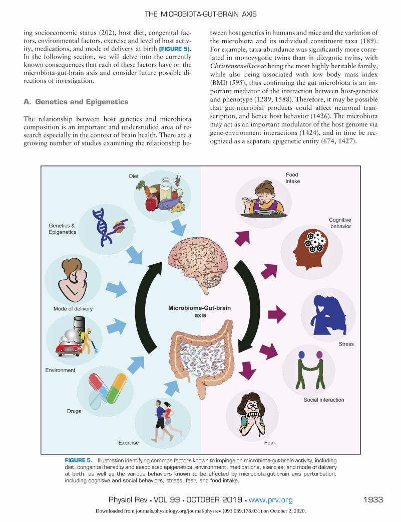
ing socioeconomic status (202), host diet, congenital fac-tors, environmental factors, exercise and level of host activ-ity, medications, and mode of delivery at birth (FIGURE 5).In the following section, we will delve into the currentlyknown consequences that each of these factors have on themicrobiota-gut-brain axis and consider future possible di-rections of investigation.
A. Genetics and Epigenetics
The relationship between host genetics and microbiotacomposition is an important and understudied area of re-search especially in the context of brain health. There are agrowing number of studies examining the relationship be-
tween host genetics in humans and mice and the variation ofthe microbiota and its individual constituent taxa (189).For example, taxa abundance was significantly more corre-lated in monozygotic twins than in dizygotic twins, withChristensenellaceae being the most highly heritable family,while also being associated with low body mass index(BMI) (595), thus confirming the gut microbiota is an im-portant mediator of the interaction between host-geneticsand phenotype (1289, 1588). Therefore, it may be possiblethat gut-microbial products could affect neuronal tran-scription, and hence host behavior (1426). The microbiotamay act as an important modulator of the host genome viagene-environment interactions (1424), and in time be rec-ognized as a separate epigenetic entity (674, 1427).
Microbiome-Gut-brain axis
Mode of delivery
Exercise
Drugs
Diet Food Intake
Fear
Social interaction
Stress
Cognitive behaviorGenetics &
Epigenetics
Environment
FIGURE 5. Illustration identifying common factors known to impinge on microbiota-gut-brain activity, includingdiet, congenital heredity and associated epigenetics, environment, medications, exercise, and mode of deliveryat birth, as well as the various behaviors known to be affected by microbiota-gut-brain axis perturbation,including cognitive and social behaviors, stress, fear, and food intake.
THE MICROBIOTA-GUT-BRAIN AXIS
1933Physiol Rev • VOL 99 • OCTOBER 2019 • www.prv.orgDownloaded from journals.physiology.org/journal/physrev (093.039.178.031) on October 2, 2020.
The microbiota is predominantly in direct contact with ep-ithelial cells, but can also indirectly interact with peripheralneurons and immune cells (1426). In gut epithelial cells,HDAC3 was shown to be critical in maintaining host-com-mensal signaling (34). Furthermore, pharmacological inhi-bition of HDAC activity reversed early-life stress-inducedvisceral hypersensitivity and anxiety (1064), behaviors de-pendent on appropriate microbiota-gut-brain axis signaling(405, 1120). Future studies should focus on whether bacte-rial-derived modulators of HDAC activity can modify suchphysiological and behavioral outcomes.
Another component of genetic-microbiome interactions isat the level of regulatory RNAs or noncoding RNAs whichrepresent a cornerstone of molecular regulation of tran-scription, gene expression, and protein abundance (44,1114). Animal models of microbial manipulation, includ-ing the GF mouse (see TABLE 2), have become invaluabletools in the study of the microbiota-gut-brain axis, and theyhave been key in studying one branch of the noncodingRNA network, microRNAs (miRNAs). miRNAs regulategene expression through translational repression and inhi-bition, and they are viable targets for intervention in neu-ropsychiatric disease (705, 1113, 1344). The gut microbi-ota has been shown to regulate the expression of miRNA inthe amygdala and PFC of GF mice (673). Similarly, deple-tion of the gut microbiota with an antibiotic cocktail alsoimpacted miRNA expression levels in these specific brainregions. For example, within the amygdala, miR-183-5pwas decreased in GF mice, but in recolonized mice, theexpression of this miRNA returned to control levels. Also,the hippocampus has demonstrated susceptibility towardsgut microbiota diversity and abundance modulation withexpression of seven miRNAs altered in GF mice which weresubsequently restored to control levels in recolonized mice(276).
B. Mode of Delivery at Birth
Mode of delivery has a tremendous impact on the establish-ment of the microbiota of infants, as the moment of birth isthe first opportunity for large-scale bacterial colonization.The initial seeding of microbiota during vaginal delivery(i.e., natural birth) occurs during parturition, as the infantpasses through the birth canal, where they are exposed tothe maternal vaginal microbiota. A seminal study hasshown that there is vertical transmission of bacteria fromthe mother to the newborn, with the microbiota of vaginal-ly-delivered babies closely resembling the vaginal microbi-ota of their mothers (454).
On the other hand, when a C-section is performed, ver-tical transmission of vaginal microbiota from the motherto the baby is circumvented, as the C-section deliverednewborn does not pass through the birth canal. Instead,the initial colonization of bacteria in C-section delivered
babies seems to be dominated by bacteria typically pres-ent on the skin and in the environment, with increasedlevels of Staphylococcus spp (454). Moreover, in thatstudy, there was no relation between the specific skinmicrobiota of the mother and the microbiota of theirC-section born infants, demonstrating that bacterial col-onization originates from nonmaternal sources. In com-parison to vaginal delivery, the C-section procedure isassociated with decreased colonization rates of Bifido-bacterium, Bacteroides, and Lactobacillus (156, 1181),and with a decrease in diversity and richness of the mi-crobiota (77, 718, 1421). Interestingly, a recent study hasshown that in addition to these differences in bacterialrepresentation, mode of delivery has an impact on thefecal virome of infants, with increased viral and bacterio-phages diversity in babies born per vaginum (1015).
Despite at least 14 different cohorts showing differences inthe microbiota of infants depending on the delivery mode,the full impact of the C-section procedure on the microbiotais still unclear; there are several confounding factors thatcan influence the results. Specifically, it has recently beenshown that differences in microbiota may be dependent onbody site (293). Another crucial factor to be taken intoconsideration is whether the C-section is elective or per-formed for a medical need, and if so, whether labor wasinitiated before the C-section procedure. In this case, themicrobiota of the C-section born babies seems to be similarto vaginally delivered newborns as these infants come intocontact with the birth canal during the early stages of labor(77, 293).
Longitudinal studies have shown that the differences be-tween microbiota attributed to mode of delivery are transi-tory. Some studies report that birth mode effects on themicrobiota can be absent as little as 6–8 wk after birth(293, 667) or as late as the first 2 yr of life (185, 718, 1147).However, despite this transient nature, early life differentialcolonization has a long-lasting effect, and several studiesshow a correlation between birth by C-section and in-creased risk of developing a variety of disorders includingobesity, type 1 diabetes, as well as immune disorders such asasthma or allergies (166, 174, 718, 1069, 1270). C-section-born babies also have a higher risk of developing neonatalinfections by methicillin-resistant Staphylococcus aureus(454), or C. difficile (1181). Consequently, there appears tobe a minor correlation between C-section delivery and poorschool performance in adolescents (370). In animal models,delivery by C-section leads to transient alterations in theneural development including decreased dendritic arboriza-tion (284, 746) and increased neuronal cell death at birth(259). Both neurodevelopmental alterations are associatedwith early-life changes in vocalization. Furthermore, C-sec-tion can also induce long-term changes to dopamine func-tion (184).
CRYAN ET AL.
1934 Physiol Rev • VOL 99 • OCTOBER 2019 • www.prv.orgDownloaded from journals.physiology.org/journal/physrev (093.039.178.031) on October 2, 2020.
Independent of the elective or emergency nature of the C-section procedure, more studies are needed to understandthe impact of delivery mode and how this impact can beminimized. It has been suggested that using vaginal seedingto transfer vaginal microbiota from the mother to the new-born may be an effective intervention (454). Although thistechnique seems to partially restore the microbiota in thesebabies, safety concerns have been raised (369, 1430) andfurther studies must be done to evaluate the effects. In ad-dition to this, supplementation with probiotics and prebi-otics can be used to decrease the impact of delivery mode onthe microbiota (294, 1076). Factors such as diet (breast-feeding vs. formula), environment, and antibiotic use alsoneed to be taken into consideration as possible modulatorsof the microbiome in early-life.
C. Diet
Diet has been shown to be one of the most critical factorsmodulating gut microbiota composition, and hence thebrain and behavior (1523). Clinical and preclinical datahave shown how different sources of diet significantly affectthe composition of the gut microbiota (1309), and mood innon-mood disorder diagnosed individuals (1467). More-over, the effects of diet can be dramatic in terms of bothdrastic compositional changes and the immediacy of sucheffects. For instance, individuals on either an animal- orplant-based diet, when switched to the other diet, experi-enced substantial changes in the gut microbiota composi-tion, within 24 h (392). A different dietary pattern has beenshown to influence � diversity of the gut microbiota com-position, but not � diversity (110), which was quite diver-gent in individuals on an animal-based diet (392). Changesin our lifestyle and food preferences have had a drasticimpact on our diet (67, 446, 1052, 1076, 1246). Indeed,different types of diet have been associated with differentimpacts on the composition of the gut microbiota. In fact,dietary iron has been shown to regulate insulin resistance toaffect susceptibility to infection, highlighting the impor-tance of evolved cooperative mechanisms (1306). However,much more research is needed to delineate if the effects ofdiets or dietary components on the microbiota are drivingchanges in overall brain function or happiness or if they areoccurring in a microbiota-independent fashion.
1. Western diet
The Western diet, a diet rich in sugar, salt, and/or fat, hasbeen widely regarded as a major contributing factor in theonset of metabolic disorders and associated pathologicalconditions. Individuals on a Western diet share a similar gutmicrobiota profile to that observed in obese individuals(392). Intake of a high-fat diet (HFD) consisting of onlyanimal-based products, including meat and cheese, pro-foundly shifted their gut microbiota community and altered� diversity, within 48 h of consumption (253). In another
study, five different inbred mouse strains and four differenttransgenic strains (Rag�/�, MyD88�/�, NOD2, and ob/ob)crucial for host-microbiota interaction, combined with over200 outbred mice, were administered a Western high-fatand high-sugar diet. They reported a dramatic shift in thegut microbiota composition in animal groups due to con-sumption of the Western diet (253).
Furthermore, animal models fed a HFD demonstrated analteration in their gut microbiota profile with a reduction inBacteroidetes levels and an increase in both Proteobacteriaand Firmicute levels (664). This was also reported in a sim-ilar study where the animals fed with a diet rich in animal-derived saturated fats demonstrated a significant increase inProteobacteria (Bilophila wadsworthia) abundance (433).It is worth noting that B. wadsworthia is a major member ofsulfidogenic bacteria found in the human gut and have beenshown to induce systemic inflammation (512). However,one must consider this increase could be attributed to thesource of fat being animal based which is rich in saturatedfats and salt. This further illustrates the critical role diet hason gut microbiota function (1607), and such strong shifts ingut microbiota profiles are subsequently poised to affectbrain and behavior.
2. Mediterranean diet
The Mediterranean diet has garnered a lot of media atten-tion for its numerous potential health benefits. A Mediter-ranean diet consists mostly of cereals (whole grains), nuts,legumes, vegetables, and fruits with moderate consumptionof poultry and fish (492), and results in distinctive andidentifiable gut microbiota characteristics (1053). Humanintervention studies have shown that the consumption of aMediterranean diet can dramatically reduce the incidenceof neurodegenerative disorders (769, 1276, 1325), psychi-atric conditions, cancer (1343), and cardiovascular disease(CVD) (186, 1517, 1629). Interestingly, the Mediterraneandiet has been shown in a number of studies to correlate witha reduced risk of depression (26, 492, 859, 1042, 1305),probably due to the rich source polyphenols present in dif-ferent components. The positive impacts of a Mediterra-nean diet are mediated by its anti-inflammatory potentialbut are also associated with marked changes at the level ofthe gut microbiota, resulting in an increase in the abun-dance of Bacteroides and Clostridium phyla, and a reduc-tion in Proteobacteria and Firmicutes phyla (988) and as-sociated metabolome (398). Furthermore, consumption ofa Mediterranean or Western diet resulted in distinct mam-mary gland microbiota and metabolite profiles, with Lac-tobacillus in greater abundance in Mediterranean diet-fedmonkeys, showing that diet can influence mammary glandmicrobiota, indicating a potential novel avenue for breastcancer prevention (1370).
A randomized controlled trial of dietary intervention inmajor depression (SMILES) confirmed a poor diet corre-
THE MICROBIOTA-GUT-BRAIN AXIS
1935Physiol Rev • VOL 99 • OCTOBER 2019 • www.prv.orgDownloaded from journals.physiology.org/journal/physrev (093.039.178.031) on October 2, 2020.

lated with a depressed cohort (n � 67) and furthermoreshowed that the Mediterranean diet intervention(ModiMedDiet) improved depression scores (713). An ear-lier study in a cohort of 119 depressed patients, which againfocused on providing dietary advice, provided some evi-dence of a beneficial effect of diet to the individual. How-ever, participants in this study were at a mild level of de-pression at baseline and may have had less scope for im-provement (537).
These studies are just the beginning of a new era of nutri-tional psychiatry (712). However, much more work isneeded to define what the contribution of the microbiome inmediating such effects is and to determine how diet and itscomponents imbue their effect on the microbiota-gut-brainaxis.
3. Ketogenic diet
The ketogenic diet is a high-fat, low-carbohydrate diet thatmimics the metabolic effects of starvation by forcing thebody to use its primary fat reserves (99). It was devisedbased on the observation that fasting had anti-seizure prop-erties. Administration of the ketogenic diet resulted in in-creased levels of the ketone bodies �-hydroxybutyrate, ace-toacetate, and acetone in the peripheral blood and urine.This elevation in the serum ketones has been shown toinhibit apoptotic proteins and improve mitochondrial ac-tivity, thus reducing apoptosis in neurodegenerative dis-eases (262). Moreover, a ketogenic diet mediates its neuro-protective function through the attenuation of oxidativestress and induction of protein expression of antioxidants(604), as well as the modulation of neurotransmitter levelssuch as GABA, monoamines, and glutamate (640, 1658).Therefore, a ketogenic diet can offer beneficial health effectstowards ameliorating symptoms of neurological conditionsincluding autism, depression, epilepsy, cancer, as well asAlzheimer’s and Parkinson’s diseases (198, 572, 850).However, more recently the microbiome has emerged as akey player in the mechanism of action of the ketogenic diet.
One study showed the ketogenic diet increasing the relativeabundance of Akkermansia, Parabacteroides, Sutterella,and Erysipelotrichaceae levels in the gut microbiota in micecompared with a control group, and that the gut microbiotais required for ketogenic diet-mediated protection againstacute epileptogenic seizures (1133). Furthermore, coloniza-tion of GF mice with ketogenic diet-associated strains, Ak-kermansia and Parabacteroides, restored seizure protec-tion. They also reported alterations in colonic luminal, se-rum, and hippocampal metabolomic profiles that alsocorrelate with seizure protection (1133). This evidencedemonstrates that different dietary components can inducedifferential effects on the brain via their actions on the gutmicrobiota. Further studies of the ketogenic diet in animalmodels of autism (1093) and schizophrenia (837) are alsoencouraging that such effects extend beyond epilepsy.
4. Carbohydrates
Carbohydrates come in two forms: digestible and indigest-ible. Digestible carbohydrates include starch and sugars,which are enzymatically degraded (glucose, fructose, su-crose, and lactose) and readily absorbed into the blood-stream and can stimulate insulin release into the blood-stream (1382). A diet rich in glucose, fructose, and sucrosecan significantly increase the abundance of Bifidobacteriumwith a significant reduction in Bacteroides (472). Mice fed afructose-rich diet demonstrated significant increases in Co-prococcus, Ruminococcus, and Clostridium and a reduc-tion in Clostridiaceae family (352). However, various Clos-tridium cluster species have been shown to be associatedwith inflammatory bowel syndrome (112), metabolic disor-ders (1626), and psychiatric conditions (889).
Undigested carbohydrates, i.e., resistant carbohydrates,which are often fermented by the microbiota residing in thedistal part of the colon (1309, 1397), are often a source ofmicrobiota accessible carbohydrates (744), which are a richcarbon source for the microbes and are known to influencethe intestinal environment (927, 1402). As a result, mostprebiotics are undigested carbohydrates known to inducebeneficial effects on the host GI system via stimulating thegrowth of healthy gut microbiota.
5. Protein
Protein is an important component in our diet as it is theprime source of amino acids, the building blocks of life.Amino acids are critical for the synthesis of neurotransmit-ters and brain health. There are different sources based ontheir origin, such as plant- or animal-based protein (1382).Numerous studies have shown a strong correlation betweenprotein intake and overall microbial diversity (964). Indi-viduals on a plant-based protein diet reported low levels ofFirmicutes-to-Bacteroidetes ratio with higher microbiotadiversity compared with individuals on a high-fat, high-sugar diet (399). Different sources of protein in a diet havebeen shown to influence different gut microbiota profiles(1382). Rats treated with fish and chicken meat reported asignificant increase in Firmicutes with a reduction in Bacte-roidetes abundance in the gut microbiota profile. In con-trast, intake of a soy-rich diet resulted in an increase inabundance of Bacteroidetes in the microbiota of rats. Fur-thermore, intake of chicken protein showed an increase inabundance in Actinobacteria, whereas administration ofbeef increased the abundance of Proteobacteria (1683).
An increase in a protein-based diet has been associated withincreases in SCFAs and BCAAs (1382). For example, con-sumption of pea protein increases intestinal SCFA levels,which have been associated with anti-inflammatory effectsand are critical for the maintenance of the mucosal barrier(798). On the contrary, consumption of a protein-rich dietis associated with increases in the abundance of Bacteroides
CRYAN ET AL.
1936 Physiol Rev • VOL 99 • OCTOBER 2019 • www.prv.orgDownloaded from journals.physiology.org/journal/physrev (093.039.178.031) on October 2, 2020.
because they are crucial for the initial proteolysis of proteininto amino acids in the gut. Intake of an animal-based dietshowed significant increases in bile-tolerant anaerobes in-cluding Alistipes, Bilophila, and Bacteroides (341, 392). Asimilar effect was observed in a culture-based study com-paring Italian children fed animal protein with childrenfrom rural Africa fed an agrarian diet; the Italian childrenshowed an increased abundance of Alistipes and Bacte-roides in their microbiota corresponding to protein intake(399). Clinical data have shown an increased abundance ofAlistipes in the gut microbiota of individuals with depres-sion (735). This further supports the role of diet in themodulation of the microbiota-gut-brain axis. Recent re-search has suggested long-term animal-based diets have adeleterious effect on the gut microbiota (1071). Therefore,more research is required to further dissect the underlyingmechanisms of the effect of various components of a pro-tein-based diet on the gut microbiota and its metabolites.
6. Fats
Diets rich in saturated and trans-fat are associated with ahigh incidence of CVD and an increase in total blood low-density lipoprotein cholesterol. For example, administra-tion of a HFD to healthy subjects caused a decrease inBacteroidetes levels with an increase in both Firmicutes andProteobacteria levels of gut microbiota (664, 1666). Similarchanges in the phylum were observed with mice fed a high-fat and high-sucrose diet (1160).
Healthy fats, also known as polyunsaturated fatty acids(PUFAs), such as omega-6 and omega-3, have been shownto induce beneficial effects by lowering the onset of CVDand can be protective against depression, some cancers,arthritis, and cognitive decline (337), as well as to supportvisual, cognitive, motor, and social development in mice(252). Studies show intake of PUFAs increase the abun-dance of healthy microbiota including Roseburia, Bifido-bacterium, and Lactobacillus spp. PUFAs have also beenshown to prevent alteration of the gut microbiota (432). Forinstance, early-life exposure to omega-3 prevented the onsetof metabolic disorders known to be mediated through aperturbed gut microbiota profile post-antibiotic treatment(753). Similarly, in adult mice, chronic exposure to ome-ga-3 showed a significant increase in Bifidobacterium andLactobacillus spp. with higher bifidobacteria-to-enterobac-teria ratio in adult mice exposed to omega-3 (1265). Arecent study comparing 876 middle-aged female subjectsshowed that the administration of omega-3 was able tomodulate gut microbiota by significantly increasing � diver-sity (1032). Docosahexaenoic acid levels from subjects onan omega-3 diet strongly correlated with 38 operationaltaxonomic units from the Lachnospiraceae family (1032),which have been reported to be in abundance in herbivo-rous animals (554). In humans, members belonging to theLachnospiraceae family have been shown to be protectiveagainst C. difficle (1195) and metabolic disorders (287).
The members of Lachnospiraceae are potent producers ofSCFAs (464). Overall, more work is needed to understandthe relative contribution of the effects of fats on the micro-biota to their impact on brain function.
D. Environment
The environment we live in has perhaps one of the greatestimpacts in shaping cross-cultural differences in health out-comes and microbiota composition. For example, differentmembers of the gut microbiota have been shown to be in-volved in metabolizing more than 40 different environmen-tal chemicals utilizing the enzymes azoreductase, nitrore-ductase, �-glucuronidases, sulfatases, and �-lyases (313).Conversely, these chemicals also modulate the gut microbi-ota composition (313), which may thus have effects onbrain and behavior through the microbiota-gut-brain axis.For instance, bisphenol A (BPA) is an endocrine-disruptingchemical widely used in the manufacture of plastic contain-ers. Exposure to BPA through diet results in a dramaticreduction in species diversity of gut microbiota in rodents,while increasing the abundance of Proteobacteria and He-licobacteraceae, and reducing Clostridia (848).
Heavy metals such as cadmium, mercury, arsenic, and leadare potent toxicants to a living organism (929); they arehighly carcinogenic and can disrupt the immune system,damage nucleic acid structures, and cause oxidative stress(91, 738, 1536). Exposure to heavy metals can also alter thegut microbiota composition in both animals and humans(313, 929, 1671). Administration of heavy metals to ani-mals dramatically perturbed their gut microbiota andmetabolomic profile, increasing relative levels of Lactoba-cillaceae and Erysipelotrichaceae while reducing relativelevels of Lachnospiraceae (216). Low levels of Lachno-spiraceae have been associated with human depression(1089), hinting at a potential microbiota-gut-brain path-way for the mental health impact of exposure to variousenvironmental pollutants.
Finally, extensive abuse of antibiotic usage by humans hascontributed to an increase in the concentration of variousantibiotics in our natural environment, including lakes, riv-ers, agriculture soil, waste, and surface water (455, 516,1225). In combination with the high levels of antibiotics inmany food sources, this will likely have widespread conse-quences not only for individual host health but also from abroader planetary health perspective (1218).
E. Exercise
Regular exercise is synonymous with good health includingbrain health. Moderate amounts of exercise can make ameaningful difference to brain structures and their func-tion; for example, in rodents, voluntary exercise impacts the
THE MICROBIOTA-GUT-BRAIN AXIS
1937Physiol Rev • VOL 99 • OCTOBER 2019 • www.prv.orgDownloaded from journals.physiology.org/journal/physrev (093.039.178.031) on October 2, 2020.
rate of neurogenesis (10, 1549). Exercise has also demon-strated a capacity to overcome age-dependent depletion ofhippocampal neurogenesis (1550), offering a prospectivepotential for the reversal of aging within brain structures.Despite the extent of research looking at the relationshipbetween exercise and brain structures/function, there is rel-atively little information currently available on the effect ofexercise on the gut, its microbiota, and the influence ofexercise on the gut-brain axis. Exercise along with a numberof different factors, such as diet, infection, disease, andantibiotics (605, 961), also modulates the health and �-di-versity of gut microbiota. Moderate levels of exercise areparticularly beneficial in reducing levels of stress and build-ing immunity along with positive changes in energy homeo-stasis and regulation (137, 1039). It is reasonable to thinkof exercise as being crucial to healthy interactions withinthe gut microbiota (288). The extent of beneficial effects ofexercise on the brain via the gut microbiota remains un-clear.
Research examining the reciprocal relationship between ex-ercise and gut microbiota composition is still in its infancy,as such little is known about how the gut microbiota maycontribute to an individual’s exercise performance (959).To date, the majority of studies undertaken have been pre-clinical animal studies that only indirectly address how ex-ercise positively influences gut microbiota composition.The potential role of exercise-induced microbial changes inpreventing HFD-induced obesity has been investigated inmice (495). The authors reported that voluntary exercisehad significant effects on the relative balance of the majorbacterial phyla (Bacteroidetes and Firmicutes) that wasconcurrent with prevention of dietary-induced obesity andnormalization in glucose tolerance and that the change inratio between Firmicutes:Bacteroidetes phyla was propor-tional to the distance ran (in the HFD-fed mice). In a case-control study, free access to exercise was associated with asignificant increase in Lactobacillus, Bifidobacterium, andBlautia coccoides – Eubacterium rectale species, in principle,improving the �-diversity of the gut microbiota (1227), withpotential subsequent brain and behavior effects. Exercisewas reported to cause massive shifts in mice gut microbiotaat nearly the same magnitude as an HFD, with exercisereducing the phyla Bacteroidetes, and increased Firmicutes,Proteobacteria, and Actinobacteria. Exercise increased cog-nitive abilities but was not able to prevent a significantincrease in anxiety associated with the HFD. Similarly, theeffects of controlled exercise training on gut microbial com-position in obese, nonobese, and hypertensive male ratshave been studied (1194).
Recent human clinical studies have attempted to elucidatehow exercise modifies gut microbiota composition usingdifferent approaches to stratify the level of physical activityor fitness. In the American Gut Project, 1,493 participants’fecal samples were categorized based on exercise frequency
[Never, Rarely, Occasionally (1–2 times per week), Regu-larly (3–5 times per week), and Daily], and analyses re-ported that groups who exercised more frequently had agreater � diversity, with an elevation in certain members ofthe Firmicutes phylum (1019). A finding along similar lineswas reported in a recent study of professional athletes fromthe Irish international rugby union squad which suggestedthat professional athletes in comparison to the sedentaryindividuals (311) had a significantly higher diversity of gutmicrobiota, that positively correlated with protein and cre-atine kinase consumption, than both control groupsmatched for physical size, age, and gender. Notably, boththe athletes and low BMI group had significantly higherproportions of the genus Akkermansia than the high BMIgroup, a factor generally associated with a healthier meta-bolic profile (863). Furthermore, indices of microbiota di-versity of the athletes positively correlated with protein in-take and levels of plasma creatine kinase with support forthe protein and microbiota diversity relationship providedby a positive correlation between urea levels, a by-productof diets that are rich in protein and microbiota diversity,suggesting that both diet and exercise are drivers of biodi-versity in the gut.
An increase of microbial diversity was once again reportedin individuals who performed a 4-day cross-country ski-march in Arctic conditions compared with controls (767),among other changes. To determine whether increasingphysical activity and/or increased protein intake modulatesgut microbial composition and function, a recent study(353) challenged healthy sedentary adults with an 8-wkcombined exercise regime, with and without concurrentdaily whey protein consumption. A combined aerobic andresistance training exercise regime led to modest alterationsin the composition and activity of the gut microbiota of thesedentary individuals. The link of such changes to brainfunction requires further analysis.
Perhaps the most compelling argument for the benefits ofexercise can be taken from studies that show the negativeconsequences during withdrawal of regular exercise, wheresymptoms of increased negative mood and fatigue wereseen in healthy individuals in as little as 14 days, comparedwith controls (833). It may be that changes in mood andfatigue are associated more with the ability of the gut mi-crobiota to control host tryptophan metabolism and levelsof plasma kynurenine, which are strongly correlated withdepression (301). A recent preclinical study investigated apotential peripheral mechanism through which exercisemay bring about its beneficial effects via the kynureninepathway of tryptophan metabolism and exercise-relatedPGC-1a1 expression (15, 638); increased expression ofmuscle-specific PGC-1a1 in transgenic mice was associatedwith greater resilience to chronic stress and stress-inducedCNS inflammation (15).
CRYAN ET AL.
1938 Physiol Rev • VOL 99 • OCTOBER 2019 • www.prv.orgDownloaded from journals.physiology.org/journal/physrev (093.039.178.031) on October 2, 2020.
Taken together, although exercise has many potential ben-eficial effects on the gut microbiota there is a need for lon-gitudinal studies to resolve the many gaps in current knowl-edge and to fully understand the mechanisms that regulatechanges in the composition and functions of microbiotaespecially in the context of brain health.
F. Medications and the Microbiome
Among the different therapeutic classes, antibiotics repre-sent the most direct and effective way of targeting the gutmicrobiota (53, 719) (see TABLE 3). However, a growingbody of evidence suggests that non-antibiotic drugs can alsoaffect the composition of the gut microbiota, with potentialimplications for behavior, as well as the involvement of themicrobiota in drug pharmacokinetics in general (310). In arecent large-scale observational study, the use of medicalinterventions was associated with a significant variation inthe microbiota. Of the 69 microbiota covariates from theBelgian Flemish Gut Flora Project, 13 were drugs belongingto the following classes: antibiotics, osmotic laxatives, IBDmedications, female hormones, benzodiazepines, antide-pressants, and antihistamines (501). In another population-based study, deep-sequencing of the gut microbiotas re-vealed a relationship between the microbiota and 44 cate-gories of drugs (1680). As expected, antibiotics weresignificantly associated with altered microbiota composi-tion; more surprising was the number of other drug catego-ries, including proton pump inhibitors (PPIs), metformin,statins, and laxatives that were also shown to have robusteffects on the gut microbiota.
Polypharmacy, the concurrent use of multiple medicationsby a patient, has also been associated with gut microbiotachanges. One study demonstrated that there was a signifi-cant negative correlation between the number of differentdrugs consumed and microbial diversity, although it is un-known if the lower diversity resulted in reduced cognitivefunction (1492). Specifically, the drug classes that had thestrongest association with single taxa abundance were PPIs,antidepressants, and antipsychotics. Gut microbiota sam-ples exposed to different drugs have also been analyzedthrough metatranscriptomic approaches. The impact ofshort-term exposure of human feces to various non-antibi-otic drugs including cardiac glycosides, a gastric acid sup-pressant (nizatidine), an anthelmintic (levamisole), an anal-gesic (phenacetin), and sulfasalazine significantly changedthe expression of microbial genes linked to drug import andmetabolism (1004). Several studies have been performed invitro to determine the antimicrobial activity of non-antibi-otic drugs (618, 619, 842, 843), all of which have beenshown to possess antimicrobial activity, with a strong po-tential for subsequent functional neural effects.
Given the rise of interest in the microbiota-gut-brain axis, itis not surprising that in recent years much effort has been
placed on understanding the role of psychotropic medica-tions. The antidepressant selective-serotonin reuptake in-hibitors (SSRIs), including sertraline, paroxetine, and flu-oxetine (737), have antimicrobial activity against gram-positive bacteria such as Staphylococcus and Enterococcus(75, 320). In addition, the antimicrobial activity of someantidepressants has been confirmed by the synergistic effectof some SSRIs in combination with antibiotics, as well astheir effects against some antibiotic-resistant bacteria (179,1083, 1084). In one recent study chronic fluoxetine admin-istration induced a depletion of cecal levels of Prevotellaand Succinivibrio (373), while another witnessed a reduc-tion in Lactobacillus johnsonii and Bacteroidales S24–7which belong to a phyla that has been associated with theregulation of body mass (946). Another class of antidepres-sants, tricyclic antidepressants (TCAs), have been shown toprevent the growth of gut pathogens such as E. coli, Yer-sinia enterocolitica (363, 1061), and the parasite Giardialamblia (1600).
Antipsychotics belonging to the non-antibiotic phenothia-zines class and their derivatives have been shown to protectmice from E. coli infection (826). This action has beenconfirmed in a clinical study where promethazine wasshown to exert a synergistic effect when combined withgentamycin in children with frequently recurring pyelone-phritis (1062). In a large cohort study in elderly hospitalizedpatients, antipsychotics were one of the three drug classesthat exhibited the strongest association with single taxaabundance, together with PPIs and antidepressants (1492).Another recent study of patients with bipolar disease foundthat atypical antipsychotics induced a decrease in microbialdiversity, with the effect being present in females but not inmales (533). At the microbiota genus level, individuals inthis bipolar cohort treated with atypical antipsychotics ex-hibited a significant increase in Lachnospiraceae abundanceand a significant decrease in Akkermansia. Finally, in thelarge-scale study of �1,000 drugs mentioned above, nearlyall subclasses of the chemically diverse antipsychotics ex-hibited anticommensal activity (970), raising the possibilitythat direct bacterial effects might be part of the mechanismof action of these drugs. This is in line with animal studieson antipsychotics whereby chronic administration of olan-zapine and risperidone affects the gut microbiota composi-tion (390, 391, 763, 1072). Overall, one cannot neglect thepressing need to examine any potential effect all orally con-sumed medications may have on our microbiota, and sub-sequent efficacy of the active compounds thereafter.
G. Stress
Stress is a state in which the normal homeostasis of anorganism is disturbed due to an actual or perceived threat(401, 1018). Acute stress activates the HPA axis (see sect.IVJ), resulting in an immediate release of cortisol (or corti-costerone in rodents; Refs. 440, 1395). This is an evolution-
THE MICROBIOTA-GUT-BRAIN AXIS
1939Physiol Rev • VOL 99 • OCTOBER 2019 • www.prv.orgDownloaded from journals.physiology.org/journal/physrev (093.039.178.031) on October 2, 2020.
arily conserved response that prepares the individual to de-fend against or escape from threat. After the threat subsides,normal homeostasis should return. However, when thatfails to occur, chronic activation of the stress response re-sults in dysregulation of the HPA axis (440) and an in-creased risk of subsequent maladies. In humans, stress-re-lated disorders such as anxiety and depression cost the Eu-ropean Union upward of €160 million in 2010 (1130). Assuch, chronic stress is rapidly becoming a global societalchallenge.
More than four decades ago, a link between stress and theabundance of lactobacilli in mice was discovered for thefirst time (1463). This finding was subsequently replicatedin the rhesus monkey (87), and rat models of early lifematernal separation stress (568, 1120, 1261). Many pre-clinical studies have since demonstrated that stress impactsgut microbial composition in a number of different hosts,including rodents (146, 592, 1164), pigs (1080), horses(959), and non-human primates (87, 89) (see TABLE 1). Inone instance, chronic psychosocial stress induced an in-crease of Helicobacter pylori in the gastric mucosa, concur-rent with an increase in serum corticosterone levels in mice(620). These effects appear to be replicated across differentstress models. Psychological stressors ranging from wateravoidance to maternal separation, heat, and acoustic stressand overcrowding have all been shown to change the com-position of the gut microbiota (88, 146, 404, 690, 1121,1442). In addition, maternal stress during pregnancy dis-plays a distinct fecal microbiota profile (634, 652), whichhas generational consequences. The maternal microbiotainfluences offspring microbiota and correlates with hyper-reactivity of the HPA axis, together with other perinatalfactors, as a key determinant of offspring outcomes (seesect. IIIA). Furthermore, the transfer of the maternal vagi-nal microbiota from stressed dams to nonstressed pups issufficient to alter their response to stress in adulthood (726).These findings also translated to humans in a population-based study whereby infants born to mothers with highcumulative stress during pregnancy exhibited an aberrantmicrobial composition (1688). It has become quite clearthat many factors one comes in contact with daily can havean impact on our microbiota-gut-brain axis, some as subtleas food cravings to as long-lasting and impacting as con-genital heredity and mode of delivery. In the following sec-tion (see sect. VII), we will delve further into behaviorsmodified directly and indirectly by the microbiota, afterwhich we will examine the role of the microbiota-gut-brainaxis in disease.
H. Circadian Rhythm
The circadian rhythm describes the 24-h cycle that regulatesbodily functions, from rest/wake timing to cellular levelmetabolic processes in the majority of organisms, includinghumans, mice, and bacteria (1570). Modern day lifestyles
contain many unavoidable and harmful disruptions to thecircadian rhythm including jet lag and shift work. Bothhave been linked to metabolic (54, 1005) and psychiatric(1153, 1631) illness. Recently, the interplay between circa-dian rhythm and the microbiome is being investigated in thecontext of obesity, CVD, diabetes, psychiatric disorders,and neurodegenerative disorders (467, 896, 1570), whichmay have key implications for our understanding of themicrobiota-gut-brain axis.
The mammalian circadian clock follows a feedback loop oftranscription and translation. The positive transcriptionfactors CLOCK and BMAL1 regulate expression of theinhibitory transcription factors Period (PER1/2) and cryp-tochrome (CRY1/2), which in turn repress CLOCK andBMAL1, restarting the cycle (1454). Interestingly, the av-erage human circadian rhythm is 24.2 h which is amenableto the endogenous biological clock zeitgebers, light, andfood (1570). Recent evidence has identified the microbiotaas a potential circadian clock modulator, effecting changeto the peripheral and central clocks (896, 1479). Further-more, it appears that circadian disruption can alter the in-testinal microbiota (1571). When there is a change in thelight, food (diet), or the microbiome, the peripheralclocks are affected. Furthermore, disruption or dysregu-lation of the peripheral or central clocks can lead toserious negative consequences, including microbiomedysregulation, as one recent study has demonstrated utiliz-ing transgenic mice containing deletions of circadian clockgenes (1571). The researchers witnessed significant changesto the microbiome, and a dampening or abolishment ofmicrobiota compositional oscillations (895, 1479, 1572).In one study in particular, the dysregulation of the micro-biota was rescued by specifically timed feeding (1479).From a human perspective, it has been reported that pa-tients with IBD have an overall reduction in circadian clockgene transcripts (666). Since both circadian disruption andmicrobiome dysregulation express complex bidirectionalregulation leading to immune activation and inflammation,it may be that inflammation acts as an intermediary be-tween the circadian rhythm and the microbiome (666,1570). The mammalian circadian clock may be modulatedby the microbiome by tuning the amplitude of the circadiangene NFIL3 with the microbial metabolite LPS, potentiallyresulting in microbially controlled energy storage and body-fat accumulation (1593).
Upon examination of circadian clock mRNA in GF mice, itwas reported that the microbiota was required for correctintegration of liver clock-oscillations, which in turn regu-late metabolic gene expression for optimal liver function(1067). Moreover, conventional mice treated with antibiot-ics demonstrated systemic disruption of microbiota diurnalrhythmicity (1478). Indeed, host homeostatic colonizationdisruption leads to a loss in microbiota compositional andbiogeographical rhythmicity, which in turn disrupts hosts
CRYAN ET AL.
1940 Physiol Rev • VOL 99 • OCTOBER 2019 • www.prv.orgDownloaded from journals.physiology.org/journal/physrev (093.039.178.031) on October 2, 2020.

rhythmicity (1478). Furthermore, a human study examin-ing jet lag and mouse models of shift work found an ar-rhythmic microbiome that had a significantly altered micro-biota composition that promoted metabolic imbalance,which was transferrable to GF mice via FMT (1479). Acontemporaneous study reported that mice undergoinga 12-h phase shift, mimicking human shift work when fed ahigh-fat and high-sugar diet, experienced a significant de-crease in microbial diversity and marked changes to theirmicrobiota (1571). The exacerbation of shift work and jetlag’s adverse metabolic effects by a HFD, or a high-fat andhigh-sugar diet, is especially poignant considering these life-styles are often accompanied by poor eating habits, and themicrobiota modulation presents as a potential therapeuticavenue for these conditions. More work is needed to under-
stand the relationship between circadian rhythms, microbi-ota, and brain health, but it is clear that interrelationshipwill be physiological and perhaps clinically relevant.
VII. BEHAVIOR AND THE MICROBIOTA-GUT-BRAIN AXIS
The microbiota-gut-brain axis is poised to affect and bereciprocally affected by many factors, including social andcognitive behavior, fear, stress, and food intake (FIGURE 6).We are slowly beginning to understand the relative contri-bution this axis has to such complex physiology and behav-ior. In this section, we will examine these behaviors andpropose possible therapeutic avenues.
Microbiome-Gut-brain axis
Anxiety & stress
Obesity
IBS
PainPsychiatric disorders
Neurodegenerative disorders
Stroke
Epilepsy
Addiction
FIGURE 6. An outline illustrating the variety of disease and disease processes the microbiota are currentlyimplicated in; examples include psychiatric and neurodegenerative disorders, pain, stress, irritable bowelsyndrome (IBS), stroke, addiction, and obesity.
THE MICROBIOTA-GUT-BRAIN AXIS
1941Physiol Rev • VOL 99 • OCTOBER 2019 • www.prv.orgDownloaded from journals.physiology.org/journal/physrev (093.039.178.031) on October 2, 2020.
A. Food Intake
What we eat and when we eat is likely affected by thecomposition and function of our microbiota, where an ar-ray of orexigenic (i.e., ghrelin, NPY) and anorexigenic hor-mones (i.e., GLP-1, PYY, CCK, and CRF; see sect. IV, Dand H) play a crucial role (518, 1541). This is supported bythe fact that many conditions wherein food intake behavioris dysregulated, like anorexia nervosa and obesity (see sect.VIIIK), are associated with an altered gut microbiota (813,863, 960, 1073, 1523–1525). In addition, amelioration ofobesity by bariatric or Roux-en-Y gastric bypass surgeryresults in a change in gut microbial composition (603, 1508,1669), even though this is not the case for weight gain inanorexia nervosa (960). Many conditions in which foodintake behavior is dysfunctional share comorbidities withother psychiatric disorders (814, 921). As such, it is impor-tant to note that levels of depression, anxiety, and eatingdisorder psychopathology correlate with various measuresof gut microbiota composition (813).
One of the most provocative propositions has been that gutmicrobes are under a selective and evolutionary pressure tomanipulate the eating behavior of the host to enhance theirown fitness, which can be done by inducing dysphoria untilwe consume the nutrient these microbes thrive on, or bygenerating cravings for food that specifically promote theirown growth and survival (32). Gut microbial diversity playsa key role in this theory, as decreased diversity inherentlyhas an increased prevalence of specific bacterial species,which might allow them to affect the host more efficiently.This is because these microbes have relatively less competi-tion and have to spend fewer resources on survival whilehaving more resources available for the manipulation ofhost eating behavior (32). Such theories are difficult toprove experimentally. However, an intriguing study in Dro-sophila points to a key role for the microbiota in foodchoice behavior, as when GF flies were given Acetobacterpomorum and Lactobacillus spp., it changed their overallfood preference (874).
Various mechanisms through which the microbiota can af-fect food intake behavior have been suggested. Gut mi-crobes can produce protein sequences of 5 amino acidsthat share an arrangement identical to various appetite-regulating peptides in the host (487, 520), which couldtrigger the production of immunoglobulins. These immu-noglobulins can inhibit the degradation of such hormones,which has been reported for the orexigenic hormone ghrelin(1453). As mentioned earlier (sect. IVD), E. coli has beenreported to produce caseinolytic protease B (ClpB), a smallprotein sequence and antigen-mimetic of �-MSH (215,1471, 1472). Increased ClpB levels have been reported inanorexia nervosa, bulimia nervosa, and binge-eating disor-der and were correlated with various psychopathologicaltraits (215). Administration of ClpB-producing E. coli de-creases short-term body weight and food intake compared
with ClpB-deficient E. coli (1471). As such, gut microbial-derived peptide sequences provide a pathway in which thegut microbiota can influence host-eating behaviors.
Another likely mechanism by which the microbiota caninfluence food intake behavior is by affecting the ability tosense and taste nutrients. Interestingly, obesity is associatedwith a decreased responsiveness to sweet and fatty tastes,which results in needing a higher intensity of such stimuli toattain the same level of taste perception (145). Anorexianervosa is also associated with an impaired taste perception(396, 1445), and weight gain has been shown to amelioratethese impairments (66, 1110). Signals conveying taste aremediated through taste receptors on the tongue, which canbe either transmitted directly through the solitary tract tothe thalamus (214, 1275), or by the local secretion of an-orexigenic hormones like GLP-1, PYY, and CCK (511, 990,1361). The role of these anorexigenic hormones is sup-ported by the fact that PYY transgenic mice have a de-creased behavioral response to fat- and bitter-tasting com-pounds, of which their response to fat taste is improvedafter reconstitution of salivary PYY (846). A continuoussupply of oral taste receptor cells plays a crucial role in thisprocess, and the disruption thereof can be detrimental fortaste signaling (510). In particular, the immune system isimplicated in taste receptor cell renewal, as its activationresults in the decrease of cell renewal and lifespan (322,797, 1585). Moreover, systemically administered LPS re-sults in decreased taste cell lifespan (322), as well as de-creased taste preference (72, 355, 857), indicating systemicimmunity additionally plays a role in taste perception.Overall, data indicate that the GI microbiota can affect tasteperception, although more studies on microbiota-depen-dent mechanisms need to be performed to further validatethis theory.
B. Social Behavior
Sociability may be classified as any form of interaction be-tween more than one animal. It is a fundamental behaviorfor all species as it facilitates many beneficial outcomes suchas learning, cooperation, protection, and mating. Interest-ingly, social behavior across the animal kingdom appears tobe strongly influenced by the microbiota. Studies in GF miceshowed they spend less time interacting with a novel con-specific compared with a conventionally colonized mouse(226, 428, 1356, 1427, but see Ref. 57). Moreover, while aconventionally colonized mouse will prefer to spend moretime interacting with a novel conspecific over a familiar one,GF mice are unable to distinguish between either animal,which represents a cognitive deficit in the identification ofsocial novelty (428, 1427). These findings of social deficitsin the absence of a microbiota have been corroborated withstudies of antibiotic administration in rodents. Several stud-ies have documented that antibiotic administration, result-ing in a marked reduction in gut microbial diversity, is
CRYAN ET AL.
1942 Physiol Rev • VOL 99 • OCTOBER 2019 • www.prv.orgDownloaded from journals.physiology.org/journal/physrev (093.039.178.031) on October 2, 2020.
associated with deficits in social behavior (418, 429). Theprecise mechanisms that underlie microbiota-mediated reg-ulation of social behavior are unknown and most likelyinvolve a multitude of biological pathways acting coopera-tively.
However, several studies have taken great strides in eluci-dating how gut bacteria may influence social behaviors. Forinstance, knockdown of Pglyrp2 in mice resulted in agreater level of social interaction with conspecifics, whichsuggests that bacterial components such as peptidoglycanare capable of crossing from the gut into the brain (see sect.IVK) and influencing social behavior circuitry via this sig-naling cascade (57). The amygdala appears to be a brainregion that is central to the influence of the microbiota onsocial behavior. GF mice display heightened expression oftranscription factors related to neuronal activation (e.g.,fos) in the amygdala relative to conventional controls(1428). Moreover, morphological analysis of the murineGF amygdala reveals extensive neuronal hypertrophy anddendritic arborization leading to an overall increase in thevolume of the various amygdalar subnuclei (931). In re-sponse to social interaction, there are profound alterationsin the transcriptome and spliceosome in the amygdala of GFmice, which may contribute to the social deficits observed(1427). These genetic and functional changes in theamygdala of GF mice demonstrate that this brain region isnot only a crucial node in the neuronal circuitry underlyingsocial behavior, but it is also the focal point through whichthe microbiota modulates this behavior.
As the microbiota appears to be an influential factor inshaping social behavior, it stands to reason that modulationof the microbiota through diet or probiotic administrationcan also affect sociability. Indeed, administration of L. reu-teri to mice resulted in an increase in the circulation levelsand expression of oxytocin, which was associated with anincrease in social behavior (226, 1214). In another experi-mental rodent model, bile duct ligation-induced social with-drawal behavior in mice was ameliorated following treat-ment with the probiotic mixture VSL#3, in addition to low-ering circulating levels of proinflammatory cytokines (i.e.,TNF-�) (377). Consequently, modulation of immune sig-naling to the brain may also be an additional means throughwhich the microbiota may influence social behavior. Di-etary-mediated alterations in the microbiome may also havean important bearing on neurocircuitry of social behaviors,especially during development. Experimental dietary defi-ciency in polyunsaturated omega-3 fatty acids resulted insocial behavior impairments in adult, but not adolescent,rats (1265). Moreover, supplementation of mice withomega-3 fatty acids prevented social behavior impairmentsfollowing induction of allergy (410). While a PUFA-richdiet may be exerting its prosocial effects via modulation offatty acid membrane levels in the brain, it has also beenshown to positively affect the composition of the microbi-
ota (1223, 1266), and thus a role for the microbiota is likely(1264). Future studies should focus on translating theseanimal studies to the human disorders of social behaviorsuch as ASD, schizophrenia, and social anxiety disorder.
C. Cognition
There is increasing evidence supporting the idea thatchanges in the composition of the gut microbiota can influ-ence cognitive function at multiple levels.
1. Rodent studies
The complete absence of microorganisms can induce nu-merous disruptions of host cognition. It has been shownthat GF mice exhibit an impaired ability to remember afamiliar object when presented with a novel object (569,935), as well as impaired working memory in rememberinga familiar environment in the spontaneous alternation task(569). In addition, these animals exhibit altered BDNF ex-pression in the hippocampus (132, 437, 569), which asstated earlier has an important role in synaptic plasticityand cognition (90, 929, 1092), suggesting a crucial involve-ment of microbes in regulating hippocampal-dependentmemory function.
Antibiotics are known to disrupt the intestinal microbialcommunity, which can result in detrimental effects on brainfunction and behavior (see TABLE 3). It has been shown thatantibiotic administration from weaning age and onwardscan induce gut microbiota changes, along with subsequentobject recognition memory impairments and altered BDNFexpression in the hippocampus when measured duringadulthood (429). More recent studies have found similarimpairments in object recognition memory after the admin-istration of antibiotics in adulthood. An 11-day exposure toan antibiotic cocktail disrupted object recognition memoryin adult male mice (550). This memory impairment wasassociated with changes in the expression of signaling mol-ecules relevant to cognition (i.e., BDNF, GRIN2B, 5-HTtransporter, and NPY) within the hippocampus amongother memory-related brain regions. Similarly, chroniclong-term antibiotic treatment was found to induce similarmemory deficits in adult female mice (1060), along withdecreased hippocampal neurogenesis.
Such findings highlight the importance of the gut microbi-ota in recognition memory performance and hippocampalfunction. Nonetheless, antibiotic-induced microbiota de-pletion has generated mixed results in other types of hip-pocampal-dependent memories. While chronic antibiotictreatment impaired spatial memory in the Morris watermaze in adult rats (672, 1591), acute treatments did notaffect this type of memory when administered in early life inrats (1119), nor when administered during adulthood inmice and tested in the Barnes maze (550). This suggests
THE MICROBIOTA-GUT-BRAIN AXIS
1943Physiol Rev • VOL 99 • OCTOBER 2019 • www.prv.orgDownloaded from journals.physiology.org/journal/physrev (093.039.178.031) on October 2, 2020.
differential effects based on rodent background, behavioraltask, and especially in the type and/or duration of antibiotictreatment. Hence, further research is needed to clarify theeffects of gut microbiota depletion on spatial memory.
A large number of studies have focused on exploring thebeneficial effects of probiotic and prebiotic treatments inmodulating health and preventing or restoring cognitivedeficits associated with changes in gut microbiota (see TA-BLES 4 AND 5). In this regard, Lactobacillus strains havebeen widely used for this purpose. Indeed, different Lacto-bacillus strains were capable of restoring deficits in objectrecognition memory induced by chronic restraint stress(894), as well as preventing these deficits in GF mice (569),in a mouse model of colitis (483), and in immunodeficientmice (1390). These strains also proved effective in restoringspatial memory impairments induced by diet in immunode-ficient mice (1126), age (729), hyperammonemia (936), andby gut microbiota depletion after chronic antibiotic treat-ment (1591). It has been found that probiotic treatmentwith selective Bifidobacterium strains in healthy mice canselectively improve object recognition memory, decreasethe number of errors in a spatial memory test, and inducebetter long-term learning in fear conditioning (1321), indi-cating the beneficial effects of a rich microbiota in cognitivebehavior. Furthermore, a recent study found a procognitiveeffect of the prebiotic B-GOS in healthy rats (610). B-GOS-fed rats showed an improved performance in the attentionalset-shifting task, thereby indicating greater cognitive flexi-bility, along with an increase in cortical NMDAR functionwithin the frontal cortex.
Other probiotic strains were able to induce similar benefi-cial effects. For example, probiotic supplementation with amixture of Lactobacillus and Bifidobacterium strains con-siderably improved spatial memory deficits in diabetic ani-mals (389). VSL#3, a commercially available probiotic mix-ture consisting of Streptococcus, Bifidobacterium, and Lac-tobacillus strains restored object recognition memoryimpairment induced by chronic antibiotic treatment (1060)and cafeteria-diet supplementation (122). More recently, amixture of “infant type” Bifidobacterium strains had thesame beneficial effects improving the cognitive deficits inGF mice (935), suggesting the importance of early-life mi-crobiota in adult cognitive behavior. Together, these datademonstrate the substantial impact of enriching the gut mi-crobiota in reverting cognitive deficits associated with di-verse factors.
2. Human studies
Although there is abundant evidence from animal researchsupporting the role of the gut microbiota in modulatingcognitive function, only a few studies have examined theinfluence of gut microbes on human cognition. Most ofthese studies are relatively small, but some are promisingnonetheless. One of these studies showed that the gut mi-
crobiota composition of obese and non-obese subjects waslinked with scores in speed, attention, and cognitive flexi-bility in a Trail Making Test coupled with alterations inneural activity in the thalamus, hypothalamus, andamygdala, suggesting that obesity affects the microbiotacomposition and subsequent cognitive performance (514).In a more recent study, the microbiota composition in 1-yr-old babies was associated with cognitive developmenttested with the Mullen Scales of Early Learning showingdifferences in brain volume (251). They identified threegroups depending on the microbial composition (i.e., highlevels of Faecalibacterium, Bacteroides, and Ruminococ-caceae) and found better performance in the group withhigher levels of Bacteroides. This group was also less likelyto be born via C-section, which fits with previous studieslinking mode of delivery with child cognitive development(1209), highlighting the importance of gut microbiota col-onization in cognitive development and function.
Probiotics have also been employed in humans demonstrat-ing beneficial effects on cognitive performance in bothhealthy and diseased individuals. Treatment with Lactoba-cillus strains in healthy elderly subjects induced an improve-ment in cognitive test performance compared with a pla-cebo group (297). In healthy women, consumption of afermented milk product supplemented with a probioticmodulated the activity in brain regions involved in cognitiveperformance during an emotional attention test (1493). Im-provements in memory tasks and subjective improvementsin mood were observed in a study on healthy individualsreceiving a prebiotic of oligofructose-enriched inulin(1389). B. longum 1714 which had shown procognitiveeffects in mice (1321) was shown to attenuate stress-in-duced cortisol increases reducing perceived and subjectiveanxiety, and moderately enhancing hippocampus-depen-dent visuospatial memory performance (38). Moreover, L.plantarum P8 was also found to alleviate stress and anxietywhile reducing levels of pro-inflammatory cytokines instressed adults, accompanied by enhanced social-emotionalcognition and verbal learning and memory (883). Recently,a probiotic mixture containing B. longum and differentLactobacillus strains positively affected cognitive functionand metabolic status in Alzheimer’s disease patients (27).Furthermore, cognitive improvements in impulsive choiceand decision-making were observed in patients diagnosedwith fibromyalgia after receiving a multispecies probioticintervention (1278), a group of patients that present with analtered microbiome as measured by disrupted microbiotametabolites (972). These results taken together indicate thepotential efficacy of probiotics for improving cognitivefunction in both healthy and Alzheimer’s disease clinicalpopulations. However, much more work is needed to un-derstand why specific strains/interventions have the poten-tial to modulate cognition and the constraints that exist onthis. Moreover, it is clear that the combination of brainimaging techniques with neuropsychological and cognitive
CRYAN ET AL.
1944 Physiol Rev • VOL 99 • OCTOBER 2019 • www.prv.orgDownloaded from journals.physiology.org/journal/physrev (093.039.178.031) on October 2, 2020.
measures will greatly enhance our understanding of the mi-crobiota-gut-brain axis in regulating cognition in healthyand vulnerable populations.
D. Fear
One aspect of cognition that warrants deeper examinationis that of fear regulation. Exaggerated fear is a core symp-tom of clinical anxiety and fear responding is closely linkedto stress (983, 1018, 1233), which (as described in sects.VIG and VIIE) is tightly intertwined with gut-brain axisfunction. The expression and inhibition of learned fear islargely regulated by the amygdala, hippocampus, and PFCin adult rodents and humans (815, 868, 984, 1504), andthese brain regions are all modulated by changes in themicrobiota (132, 308, 346, 558, 673, 675, 933, 1092,1125, 1428). Despite this, studies of the microbiota in fearregulation remain relatively scarce.
In humans, there are now a few functional MRI (fMRI)studies linking the gut microbiota to functional brain activ-ity during observation of threat stimuli (negatively valancedemotional images). The initial study in this area demon-strated, in a sample of healthy women, that intake of afermented milk product containing multiple probioticstrains could reduce the neural response to faces showingnegative affect (fear or anger) (1493). Specifically, brainreactivity was reduced across a distributed network of brainregions including the PFC and parahippocampal gyrus. An-other study from the same group later showed that naturaldifferences in the microbiota composition of healthywomen were associated with altered reactivity of the righthippocampus to negative emotional images (1494). Finally,in a recent study of adversity-exposed and control children,variation in Bacteroides and Lachnospiraceae were associ-ated with brain reactivity to fear faces, particularly in thePFC (235).
Using animal models, emerging research is beginning todelineate the links between the microbiota and learned fearresponses, or more specifically fear memory and extinction.With GF mice, short-term fear recall was shown to be im-paired in the absence of the microbiota (674). Adult con-ventionally colonized, GF, and ex-GF mice underwent clas-sical fear conditioning, whereby a previously innocuousconditioned stimulus was paired with an innately aversiveunconditioned stimulus. Six hours later, GF animals exhib-ited low levels of conditioned fear responding relative toconventionally colonized animals, but this deficit was res-cued in the ex-GF mice that were recolonized at weaning.These behavioral changes were accompanied by alteredamygdala gene expression, specifically indicating elevatedbaseline amygdala activity and reduced responsiveness tothe fear stimuli in the GF animals. Similar impairments offear recall and additional enhancements of fear extinctionare observed following acute antibiotic exposure in humans
and rodents (79, 395, 1268). Although the broad-spectrumantibiotics used in these cases (doxycycline and D-cycloser-ine) were chosen for their direct neuromodulatory proper-ties, the similarities to GF function suggest that it is worth-while considering the possibility of an alternate microbiota-mediated mechanism in these cases as well.
Providing further support for microbial modulation oflearned fear, fear responding is also altered by probiotictreatments, at least in rodents. These effects are strain-de-pendent, with different outcomes depending on the chosenprobiotic. For example, B. longum 1714 enhanced fearlearning and short-term memory in adult mice, withoutaffecting extinction, while B. breve 1205 had no effect oneither measure (1322). Another probiotic strain, L. rham-nosus (JB-1), slowed extinction learning in adult mice(209), whereas heat-killed Mycobacterium vaccae has theopposite effect, accelerating extinction learning in adult rats(541). Finally, administration of a probiotic formulationcontaining L. rhamnosus R0011 and L. helveticus R0052has been shown to prevent the effects of early-life stress onfear behavior and its supporting neural network during de-velopment (234, 345). Specifically, young rats exposed toearly-life stress exhibit a phenotype characterized by persis-tent fear memory and high rates of fear relapse followingextinction (237, 344), a profile that is passed down the maleline for at least two generations (i.e., the grand-offspring ofthe stressed males exhibit the same high-fear phenotypedespite never directly experiencing stress) (234). Probiotictreatment rescued the normal trajectory of fear memorydevelopment in stressed rats, restoring an age-appropriatenormal phenotype of rapid forgetting and low rates of fearrelapse, not only in probiotic-exposed neonatal rats, butalso their offspring (237, 344).
E. Stress-Related Behaviors
As described previously (see sect. VIG), an ever-growingbody of preclinical studies using a variety of animal modelshas shown that stress can alter the gut microbiota (see TA-BLES 2–5). This relationship, like the microbiota-gut-brainaxis itself, is bidirectional; the microbiota can modulatestress-induced alterations in anxiety, memory, cognition,and neuroinflammation (for reviews, see Refs. 372, 504,540, 1243, 1364). Many studies were conducted using GFmice (see also sect. IIA and TABLE 2) (308, 357, 437, 931,1092, 1434). Their collective findings were critical in shed-ding light on the existence of a link between the microbiotaand stress. Moreover, this work paved the way for furtherresearch on the stress-microbiota link to explore potentialtherapeutic benefits for microbiota manipulations in thecontext of stress.
1. Stress susceptibility
Given the link between the microbiota and the stress re-sponse, a relationship between resilience and microbiota
THE MICROBIOTA-GUT-BRAIN AXIS
1945Physiol Rev • VOL 99 • OCTOBER 2019 • www.prv.orgDownloaded from journals.physiology.org/journal/physrev (093.039.178.031) on October 2, 2020.
composition has also been proposed (786). Current studiesare exploring whether basal microbiota composition itselfcan predict stress susceptibility. For example, a single expo-sure to social stress in Syrian hamsters was sufficient toinduce changes in the microbiota, where multiple encoun-ters exacerbated the effect (1164). Microbiota was differ-entially affected depending on the outcome of the chronicsocial defeat encounter. Furthermore, with the use of achronic social defeat stress protocol, mice with higher socialavoidance scores had greater correlational changes in cecalmicrobiota taxa than controls (1447). When analyzing thefecal microbiome from rats more vulnerable to repeatedsocial defeat stress, compared with those classified as moreresilient, shotgun metagenome sequencing identified an in-crease in the expression of immune-modulating microbiota,including Clostridia, in the vulnerable rats (1175). The de-pressive-like behavioral phenotype was transferrable viaFMT from stress vulnerable to naive rats, where ventralhippocampal microglial density and IL-1� expression wereenhanced, when compared with naive rats receiving FMTfrom stress resilient donors (1175). In a different study,defeated mice displayed increased depressive-like behaviorsthat correlated with a reduction in the abundance of specificbacterial genera (1020). Future studies should focus onwhether the microbiota provides a mechanism for stresssusceptibility or resilience in both rodents and humans.
2. Probiotics and stress-related changes
There is growing evidence that specific manipulations of themicrobiota might modulate the negative effects of stress,including stress-related behavior and HPA axis activation(1314). Much of this work has focused on administration ofprobiotics, and particularly Bifidobacterium and Lactoba-cillus species, with promising effects observed on stress andrelated anxiety and depression in both preclinical and hu-man studies (see also sect. VIII, E and F, and TABLE 5) (230,1199, 1581).
In a randomized placebo-controlled fMRI study, a multi-species probiotic increased a buffer against stress-relatednegative effects on working memory, specifically in PFCrecruitment, identifying the use of probiotics as a supportfor cognition under stressful situations in healthy individu-als (1155). In a preclinical study, oral administration of L.rhamnosus (JB-1) reduced stress-induced corticosterone re-sponses as well as anxiety- and depression-related behaviorin mice, effects that were prevented by vagotomy (209).Furthermore, L. farciminis prevented hyperactivation ofthe HPA axis in response to acute stress, probably due to theprevention of excessive gut permeability (20). Recently, atwo-strain probiotic, L. helveticus and B. longum, signifi-cantly improved measures of HPA axis responsiveness toCRD, an acute stressor, rather than with either strain alone(22). Conversely, infection with pathogenic C. rodentiumenhanced vulnerability to stress-induced memory impair-ments in mice, which was ameliorated by pretreatment with
a combination of probiotics (L. rhamnosus and L. helveti-cus) (569).
Similar attenuation of stress responses has also been ob-served in chronic stress paradigms. For example, adminis-tration of L. helveticus NS8 to Sprague-Dawley rats im-proved behavior following chronic restraint stress (894). Adifferent strain of L. helveticus, MCC1848, was also shownto ameliorate social defeat stress-induced anxiety- or de-pressive-like behaviors in mice (966). L. rhamnosus abro-gated anxiety-like behaviors due to chronic social defeatstress. However, the gut microbiota alterations that mani-fested due to the stressor remained unchanged (147). An-other recent study has shown that supplementation with L.plantarum successfully abrogated heightened stress re-sponses due to chronic unpredictable stress and sleep depri-vation stress and resulting in increases in host gut Lactoba-cillus species (436). Likewise, administration of C. butyri-cum prevented depressive-like behaviors associated withchronic unpredictable stress (1440).
The studies discussed so far were conducted in adult ani-mals following acute or chronic stress, but probiotic treat-ments also ameliorate the effects of stress during vulnerableperiods, such as in early-life stress. Several different probi-otic strains have been tested in the maternal separationmodel with rats. A combination of L. rhamnosus and L.helveticus normalized fear behavior in stressed, maternally-separated pups and their later offspring (234, 345). Theprobiotic B. infantis normalized behavioral deficits in adultrats exposed to maternal separation (431). Interestingly, thesame strain did not affect depressive-like behavior in ani-mals reared in a stress-free early environment (430). Inter-vention with B. animalis and Propionibacterium jenseniirestored some gut microbial perturbations in adult mater-nally-separated animals exposed to a “second hit” of stressin adulthood (105). Finally, the administration of B. bifi-dum G9–1 concomitantly to the maternal separation stressprevented juvenile hypersensitivity to acute stress (552).
The evidence for translating psychobiotic therapies into theclinic is becoming hard to ignore; studies are beginning toaddress the question of translation to humans. A combina-tion probiotic L. helveticus R0052 and B. longum R0175given to both rats and humans had an anxiolytic-like effectin rats and reduced urinary cortisol levels 24 h followingadministration in humans, suggesting a normalization ofthe HPA axis response to stressors (1034, 1035). Addition-ally, a study using the probiotic L. plantarum 299v resultedin decreased cortisol levels in a group of healthy adult stu-dents undergoing supplementation of probiotic during aperiod of exam stress (49), and L. plantarum DR7 wasshown to alleviate stress and anxiety in a randomized, dou-ble-blind, placebo-controlled study with stressed adults(289). A B. longum species, already proven to reduce anx-iety and stress responses during acute stress in mice (1321),
CRYAN ET AL.
1946 Physiol Rev • VOL 99 • OCTOBER 2019 • www.prv.orgDownloaded from journals.physiology.org/journal/physrev (093.039.178.031) on October 2, 2020.
similarly reduced stress and anxiety measures in a popula-tion of healthy adults as well as improving cognition (38).Physical symptoms of exam stress, such as the onset ofstress-induced GI symptoms and head-colds, proved ame-nable to probiotic treatment with L. casei (773) and B.bifidum (852). Studies in related IBS cohorts have alsoshown an improvement in stress-related GI symptoms dueto probiotic treatments (449). While not all potential pro-biotic interventions showing promise in preclinical studieshave translated successfully to human studies (782, 1279),there is enough evidence to warrant further investigation ofpotential probiotics as a therapeutic strategy to alleviate thedetrimental effects of stress.
3. Prebiotics
Although it is accepted that certain prebiotics can alter thegut microbiota (164, 434), their effect on stress is less wellunderstood. Administration of sialyl lactoses (human milkoligosaccharides) to mice exposed to social disruption pre-vented stress-induced colonic microbial disruption andanxiety-like behavior (1464). The prebiotics FOS and GOShave been shown to have anxiolytic effects in naive animals,protecting mice from the impact of chronic stress on themicrobiota (229). In humans, one important small-scaleplacebo-controlled study in healthy individuals demon-strated that intervention with B-GOS decreased waking sal-ivary cortisol levels as well as increased positive processingof emotional information (1337). In light of these results, itcould be hypothesized that prebiotic intake could also mod-ulate the HPA axis and have beneficial effects on stress-related disorders.
4. Other microbiota interventions
In addition to prebiotics and probiotics, other strategies tomodify or deplete the microbiota have also been shown toalter stress responses. Although the mechanism is not clear,microbiota depletion using antibiotics has been shown toimpact stress-related behaviors. Administration of nonab-sorbable antibiotics during pregnancy alters the maternalmicrobiota and can influence the behavior of the offspring,including increased anxiety-like behavior (1497). However,prenatal and early postnatal administration of penicillindecreases anxiety-like behavior and sociability in a sex-spe-cific way (867). Moving to a later time window of admin-istration, exposure to antibiotics from early adolescencedecreases anxiety and impairs cognition in mice (429).However, antibiotics are not the only interventional strat-egy that can modulate stress-related behavior. An FMTfrom stress-prone BALB/c to GF Swiss Webster mice in-creased anxiety-related behavior, whereas performing thetransfer from the more stress-resilient Swiss Webster miceto GF BALB/c reduced the anxiety-like phenotype (132).Recently, FMT from mice exposed to chronic immobiliza-tion stress to conventional C57s induced anxiety-like be-
havior and suppressed hippocampal BDNF expression(722). From a more translational approach, FMT from de-pressed patients to microbiota-depleted rats was sufficientto increase anhedonia and anxiety-like behaviors in the ro-dent recipients (783). Although these studies did not includeany measurement of the neuroendocrine response to stress,they demonstrate that microbiota composition may play acausative role in stress-related behavioral changes. A clearerunderstanding is needed to elucidate the role neurohor-mones play in these behavioral outcomes. Intriguingly, ex-cessive stress combined with the use of stomach acid sup-pressants have also been shown to additively and indepen-dently affect the composition of stomach microbiota (962).Indeed, multiple neurocognitive processes in the hippocam-pus were coincident with such changes in the microbiotacomposition (962).
VIII. DISEASES AND DISEASE PROCESSES
As the field of microbiota-gut-brain research has progressedand matured, the microbiota has been implicated in a grow-ing list of psychological and neurological diseases and dis-ease processes (FIGURE 6). Of note, many disorders of brainand behavior are related to exogenous stressor exposure,dysregulation of the HPA axis stress response (see sect. IVJ),and individual coping mechanisms, or resilience to stress,all of which are components of stress responding, that arenow recognized as being modulated by the microbiota asdescribed previously (see sects. IVH, VIG, and VIID). As wewill outline below, the state of the evidence varies betweendisorders, with some in a preliminary stage of researchwhere studies have been limited to correlational observa-tions of altered microbiota composition in clinical popula-tions, while there is stronger support for a causal role of themicrobiota in other disease processes.
A. Autism Spectrum Disorder
Autism spectrum disorder is a heterogeneous group ofneurodevelopmental disorders characterized by pro-found deficits in sociability, stereotyped or repetitive be-havior in addition to anxiety and cognitive disturbances(997). With a worldwide prevalence of 1 in 68 children, itis more common in males, who are four times more likelyto develop the condition than females (976). The behav-ioral disturbances are accompanied by profound altera-tions in key central physiological processes such as neu-roinflammation, neurogenesis, neurotransmission and inthe production of pro-social hormones, oxytocin, andvasopressin (1365). Although ASD may be classicallythought of as primarily neurological in its pathology,there is growing evidence to demonstrate a role for the GIsystem and its resident microbiota in aspects of ASDsymptomatology also. Approximately 70% of childrenwith ASD report comorbid GI disturbances such as bloat-
THE MICROBIOTA-GUT-BRAIN AXIS
1947Physiol Rev • VOL 99 • OCTOBER 2019 • www.prv.orgDownloaded from journals.physiology.org/journal/physrev (093.039.178.031) on October 2, 2020.
ing, constipation, and diarrhea, indicating that gut phys-iology is indeed altered (1419). Interestingly, in a smallopen-label study in which children with ASD weretreated with the broad-spectrum antibiotic vancomycin,there was a marked improvement in behavioral symp-toms (759, 1310). Although antibiotics are not a viablelong-term intervention strategy for the management ofASD, this study provided the field with a critical insightthat the gut microbiota may contribute to the behavioraldisturbances in this neurodevelopmental disorder. Sev-eral additional studies have observed significant altera-tions to the composition of the microbiota and in theproduction of microbial metabolites in children withASD (524, 525, 760, 1162, 1590). Of note, there is areduced abundance of the beneficial bacteria genus, Bifi-dobacterium, along with an increased abundance of po-tentially pathogenic Desulfovibrio and Clostridia generain the gut microbiota of children with ASD, a result thatwas consistent across all studies. A more recent studyidentified that the gene cpb2, which encodes for the clos-tridial toxin b, is significantly more expressed in C. per-fringens isolated from the fecal microbiota of childrenwith ASD relative to controls (596). The �2 toxin hasbeen shown previously to cause GI-related illnesses (i.e.,diarrhea) and the increased presence of this gene in clos-tridial species in the ASD microbiota may thus help toexplain the GI-related comorbidities observed in thisneurodevelopmental condition. While several studies todate have characterized the alterations of the microbiotain individuals with autism, there is limited evidence todemonstrate whether targeting the microbiota throughprobiotic or dietary interventions can improve symptomsof ASD in humans. However, a small open-label clinicalintervention demonstrated that FMT of a standardizedmicrobiota cocktail to children with ASD was efficaciousin improving the GI and behavioral symptoms (759).Although the sample size was small and the experimentaldesign lacked a randomized, double-blind structure, thestudy provided promising preliminary evidence to dem-onstrate that the microbiota may indeed be a viable tar-get as a treatment strategy for ASD. A more recent ran-domized, double-blind, placebo-controlled study demon-strated that a combination of a casein/gluten-free dietalong with the prebiotic B-GOS led to an improvement inthe behavioral symptoms of autistic children (608).These behavioral changes were accompanied by an in-crease in the relative abundance of the beneficial strain,B. longum, in the microbiota of autistic children. Furtherclinical trials are required with larger sample sizes andmore rigorous study design.
Preclinical studies have been invaluable in providing thefield with an insight into how the microbiota-gut-brain axismay be involved in ASD. Knockout of the shank 3 (autismcandidate) gene results in profound autistic-like behavior inmice (1356, 1448). In addition to this, shank 3 transgenic
mice also display alterations to the composition of their GImicrobiota, with notable reductions in Lactobacillus, Pre-votella, and Veillonella. Interestingly, treatment with L.reuteri improved social deficits in male, but not female,shank 3 transgenic mice. This corresponded with an in-creased expression of oxytocin mRNA expression withinthe hypothalamus of male shank 3 transgenic mice. How-ever, the probiotic strain reduced expression of the neuro-peptide in the hypothalamus of female mice, which mayexplain the absence of any improvement in social behaviorin these animals (1448). Moreover, such results suggestthat, other than modulation of oxytocin expression, theremay be sexual dimorphic factors underlying L. reuteri’smechanism of action.
The BTBR mouse, which displays an inherent autistic-likephenotype across multiple behavioral domains, also exhib-its altered GI physiology similar to what is reported in theclinical setting (593). For instance, BTBR mice display pro-longed intestinal motility indicative of constipation. More-over, it has been reported that there is a breakdown in thepermeability of the small and large intestine of these ani-mals (334). This permeability deficit phenomenon may con-tribute towards bacterial or related components, translo-cated to systemic circulation, whereby they can elicit aninflammatory response (593). Profound alterations in themicrobiota composition characterized by deficits in the rel-ative abundance of Bifidobacterium and Blautia genera arealso observed compared with C57BL/6 mice. The absenceof these two bacterial genera was linked to additional defi-cits in bile acid signaling in these mice, which may contrib-ute towards its observed GI physiology.
While genetic models such as the Shank 3 and BTBR areimportant tools in elucidating any genetic component tothe alterations in the microbiota-gut-brain axis in ASD, itis also vital to model how environmental factors maycontribute towards the neurodevelopmental disorder andtheir impact upon the GI microbiota. For instance, inutero exposure to the viral component and TLR3 ago-nist, Poly I:C, facilitated the development of autistic-likebehavior in mice, while also increasing intestinal perme-ability and altering the composition of the gut microbiota(690). Moreover, exposure to the teratogen valproic acid(VPA) in utero has been shown to result in intestinalinflammation (1287), and dysregulation to the GI micro-biota observed as changes in the Firmicutes:Bacteroidetesratio (412) and an increase in the abundance of the spe-cies Desulfovibrio (524). Furthermore, VPA exposureleads to a deficit in social interaction in mice while alsoaffecting serotonergic turnover in the amygdala (411,412). Given the influence that the microbiota has oncentral processes such as serotonergic neurotransmission(1118), alterations to the gut microbiota through envi-ronmental insults such as in utero VPA or Poly I:C expo-sure may contribute towards the observed behavioral
CRYAN ET AL.
1948 Physiol Rev • VOL 99 • OCTOBER 2019 • www.prv.orgDownloaded from journals.physiology.org/journal/physrev (093.039.178.031) on October 2, 2020.
deficits in these models. Furthermore, considering theneuroactive properties of microbial metabolites, such asSCFAs (1429), their role in modulating autism-relatedbehavior should not be overlooked. Indeed, several pre-clinical studies have documented how neurotoxic dosesof the SCFA propionic acid induce autism-like behaviorin rodents (956, 1373). Whether elevated levels of SC-FAs, such as propionate, contribute towards what is ob-served clinically in autistic individuals is unknown butwarrants further attention. Furthermore, while studyingthe loss-of-function mutation in the histone demethylaseKDM5 Drosophila found in ASD, social behavior ap-pears to be modulated via immunoregulation and micro-biota maintenance (277).
Preclinical models of ASD have also provided considerableinformation into the potential efficacy of candidate probi-otic strains in improving autism-related behavior and theirunderlying mechanism of action (1356). Mice from moth-ers fed a HFD during pregnancy exhibit deficits in socialbehavior and social cognition (226). Analysis of the micro-biota of these mice revealed the absence of several key lac-tobacillus species. Interestingly, supplementation of one ofthese strains, L. reuteri, in mice from mothers fed a HFDresulted in a restoration of their behavioral deficits that wasalso associated with an increase in expression of the proso-cial hormone oxytocin in the PVN of the hypothalamus(226). The finding that L. reuteri increases oxytocin pro-duction has been shown to be dependent on the integrity ofthe vagus nerve in addition to IL-10 signaling (1214, 1555).
Other candidate probiotic strains have been shown to im-prove other facets of autism-related behavior in animals.For instance, in the maternal immune activation model ofASD, B. fragilis administration early in life resulted in animprovement in stereotyped and anxiety-like behavior, butnot in sociability (690). Given that each bacterial speciespossesses its own unique biochemical properties, these con-trasting effects of potential probiotics on autism-related be-haviors are most likely due to such strains modulating dif-ferent aspects of the microbiota-gut-brain axis (e.g., im-mune system, vagus nerve, microbial metabolites, peptides,etc.).
B. Major Depressive Disorder
Depression is the leading cause of disability worldwide,affecting 4.4% of the world’s population (1624). It is nowknown that Major Depressive Disorder (MDD) correlateswith increases in pro-inflammatory cytokines (1492),which in turn activate the HPA axis possibly accentuatingits hyperactivation associated with depression (290). Effec-tive therapies for depression abrogate the heightened in-flammatory response and decrease HPA axis activation(290, 1157).
There is burgeoning evidence of a role for the microbiota-gut-brain axis in MDD (539, 683). However, there is a needfor better-designed studies with clear psychiatric and psy-chological phenotyping when it comes to depression andanxiety and the microbiota-gut-brain axis in patient popu-lations. Currently, most of the studies have been conductedin preclinical models (442). As stated earlier, alterations inthe microbiota composition and inflammation have beenseen in the maternal separation model (1121) and theFlinders-sensitive rat model of depression (1495). Evidencefrom GF mice has provided further insight into the role ofthe microbiota-gut-brain axis in depression, showing thatthe absence of a microbiota reduces depressive-like behav-ior in the forced swim test (FST) (1679). Certain probioticand prebiotic interventions have also been shown to reducedepressive-like behaviors in rat and mouse models, in addi-tion to ameliorating inflammatory responses (209, 229,431, 1495) (see TABLES 4 AND 5).
There are now a number of human studies that have founddifferences in fecal microbiota in MDD patients when com-pared with healthy controls (24, 735, 783, 1089, 1679).However, there is limited consensus between them in rela-tion to the type of changes seen. In one study, Bifidobacte-rium and Lactobacillus were reduced in 43 depressed indi-viduals (24). Another study witnessed increased fecal bac-terial � diversity, present in a cohort of 46 concurrentlydepressed patients, which was not seen in patients who hadresponded to treatment (735). One observation noted thatBacteroidetes, Proteobacteria, and Actinobacteria were in-creased, and Firmicutes were decreased, and negatively cor-related with severity of the depressive symptoms (735).While interindividual variability was apparent, there weresignificant differences found in a variety of genera whencompared with controls. The order Bacteroidales was sig-nificantly increased, and the family Lachnospiraceae wassignificantly decreased in another cohort of 37 depressedpatients versus healthy controls (1089), at the moment, allcorrelative. Elevated cortisol output in addition to de-creased fecal microbial richness was reported in a study of34 MDD patients (783), and significantly different gut mi-crobiota were noted in an MDD cohort when comparedwith matched controls (1679). Furthermore, a human FMTto rats resulted in a transfer of the depressive and anxiousphenotypes (783), which was not observed with FMT fromhealthy human controls. Currently, it is difficult to reconcilethe variations seen across the studies, and it may be reflec-tive of the small sample size as well as medication effects.Large-scale studies in drug naive depressives are now war-ranted.
In an effort to address this, a recent study examined themicrobiota composition of �1,000 individuals enrolled inBelgium’s Flemish Gut Flora Project and clustered individ-uals into four enterotypes based on their microbiome com-position (1537). Intriguingly, those who had depression, or
THE MICROBIOTA-GUT-BRAIN AXIS
1949Physiol Rev • VOL 99 • OCTOBER 2019 • www.prv.orgDownloaded from journals.physiology.org/journal/physrev (093.039.178.031) on October 2, 2020.

lower quality of life score, were over-represented in anenterotype characterized by lower relative abundance ofthe genus Faecalibacterium, in addition to an overall re-duced microbial load. On deeper analysis, the researchersfound that there was an association between bacteria thatproduce the SCFA butyrate (Faecalibacterium and Co-prococcus) and higher quality of life indicators. On theother hand, the levels of Coprococcus and Dialister weresignificantly lower in the people with depression. Manyof the findings were replicated the Dutch LifeLines DEEPcohort as well as in a small cohort of clinically refractorydepressed patients. Additionally, they identified potentialmicrobial-derived metabolites that have neuroactive po-tential from the literature, identified relevant pathways inthe shotgun metagenome data, and clustered them into amodular framework. In total, they curated and annotated56 different gut-brain modules, each corresponding to asingle neuroactive compound production or degradationprocess. These included modules involved in the poten-tial ability of the gut microbiome to synthesize the dopa-mine metabolite 3,4-dihydroxyphenylacetic acid(DOPAC). Two other modules relevant to glutamate andGABA pathways also tended to be altered in depression.Interestingly, bacteria that have the capacity to generateor breakdown GABA have been implicated in changes inbrain function and behavior (108, 1206, 1429). Al-though still correlative, they are among the most con-vincing to date to show a relationship between microbi-ota composition and mental health. Moreover, they agreewith recent crowd-sourced funding approaches such asthe American Gut Project, which have shown a link be-tween microbiota composition with depression (1017).
A probiotic intervention study using an L. casei strain de-scribed improvements to mood ratings in a healthy elderlycohort following treatment, with most benefit for thosewho had lower mood at baseline (131). Furthermore, atriple-strain probiotic (L. acidophilus, L. casei, and B. bifi-dum) resulted in improvements in depression scores, in ad-dition to beneficial metabolic effects in an MDD cohort(28). Importantly, L. rhamnosus HN001 supplementationduring pregnancy resulted in significant lowering of anxietyand postnatal depression (1387). One recent study exam-ined the effects of the prebiotic GOS combined with a pro-biotic containing L. helveticus and B. longum on mild tomoderate MDD in a placebo-controlled parallel study(778). Beneficial effects of the probiotic were observed, in-cluding decreases in depression scores as well as improve-ments in tryptophan signaling. Another recent open-labelstudy in patients with treatment-resistant depression hasshown promise for the probiotic C. butyricum as an adjuncttherapy, used in combination with antidepressant drugs(1054). Cognitive performance was further enhanced upontreatment of MDD patients with the probiotic L. plantarum299v (1293).
Several studies in the literature failed to show any benefit todepression scores on administration of probiotics in pa-tients presenting with MDD (297, 1280). However, prom-ising results in healthy controls and preclinical studies war-rant further investigation in clinical populations (1034,1035). Overall, systematic reviews of probiotics as a poten-tial adjunct therapy in MDD are encouraging, describingthat probiotics effectively improve mood in humans (691,1199, 1581), although another recent meta-analysis callsinto question the significance of these findings, highlightingthe confounding comparability of studies due to strain dif-ferences and severity of disease (1094). It is of note thatpoor diet is now accepted as a risk factor for depression, aswell as a therapeutic target (712, 713). Improvements indiet will likely lead to an increase in consumption of prebi-otic foods. There are limited studies on the effects of prebi-otics in depression. Nonetheless, there is some evidence fora reduction of stress in human studies, which is intrinsicallylinked with depression (1337, 1374). One study recentlydescribed no benefit from an 8-wk GOS interventional ther-apy for mild to moderate MDD (778). Polyphenols, such asresveratrol, have been described to reduce depressive-likebehaviors, in addition to ameliorating increases in cortico-sterone and pro-inflammatory cytokines (1643), preclini-cally. Curcumin has described benefits in MDD patients(1094), but further clinical studies on potential benefits ofpolyphenols in MDD are needed.
C. Anxiety
Anxiety and depression frequently go hand-in-hand, andmost studies of the microbiota-gut-brain axis in anxietyinclude measures of depression and vice versa. Much of theevidence for a link between anxiety and the microbiota-gut-brain axis comes from preclinical studies (539), whereby ananxious-like phenotype may be inferred from the animal’sbehavior in certain environments, such as the open-fieldtest, elevated plus maze, light/dark box, or in reaction tocertain stressors (see TABLES 2–5). GF mice display dimin-ished anxiety-like behavior in comparison with convention-ally reared animals (308, 437, 1092) alongside exaggeratedcorticosterone responses to stress indicating altered HPA-axis function, although GF rats exhibit exaggerated anxietyresponses (357). It is important to note that changes inanxiety due to probiotic treatment are species and straindependent (209, 1321).
Improvements in specific anxiety measures have beenshown in a small number of probiotic intervention studiesof healthy control participants (1035) and in chronic fa-tigue syndrome (1239). Furthermore, a multi-strain probi-otic [S. thermophilus (2 different strains), L. bulgaricus, L.lactis subsp. lactis, L. acidophilus, L. plantarum, B. lactis,L. reuteri] was recently demonstrated to have anxiolyticeffects in a small study of healthy controls (323). Interest-ingly, B-GOS significantly improved anxiety levels in indi-
CRYAN ET AL.
1950 Physiol Rev • VOL 99 • OCTOBER 2019 • www.prv.orgDownloaded from journals.physiology.org/journal/physrev (093.039.178.031) on October 2, 2020.
viduals with IBS (1374). Intriguing recent work has identi-fied Lactobacillus plantarum DR7 as a possible psychobi-otic, where it alleviated anxiety- and stress-associatedsymptoms in a randomized, double-blind, placebo-con-trolled study (289). However, there is a clear need for fur-ther translational studies to specifically examine the effectsof psychobiotics on anxiety.
D. Schizophrenia
Schizophrenia is a complex and heterogeneous debilitatingneural disorder, which has proven to be one of the mostdifficult psychiatric conditions to treat. It is characterizedby both positive and negative neurobehavioral dysfunctionsincluding, but not limited to, psychosis, cognitive dysfunc-tion, delusion, apathy, and social withdrawal (1145). Therehave been ideas circulated purporting a role for the GI tractimmune response in the pathogenesis of schizophrenia(1171, 1355, 1546), where risk factors include Toxoplasmagondii infection, food intolerances, GI inflammation, andcell barrier defects. There is evidence for both genetic andenvironmental etiological factors in the presentation ofschizophrenia symptoms (423, 493). As a result of the im-mune system role associated with this psychotic disease andthe key role the microbiota plays in establishing and main-taining immune function, much recent effort has examinedpossible influences of the microbiota in schizophrenia (123,681, 1341). One recent study performed a metagenomicanalysis of the oropharyngeal microbiota in schizophrenicindividuals and healthy controls, identifying large differ-ences at the phylum and genus levels (260). Proteobacteria,Firmicutes, Bacteroidetes, and Actinobacteria presentedwith the most divergence between the two cohorts, with thefungi Ascomycota present in greater abundance in schizo-phrenia patients. Furthermore, � diversity was decreased inmedicated and unmedicated patients, and FMT into GFmice had reduced hippocampal glutamate, and increasedlevels of glutamine and GABA, suggesting that the schizo-phrenic microbiome itself can affect neurochemistry, whichmay be relevant to the human condition (1678). Intrigu-ingly, antibiotic use reduced microglia-mediated synapseengulfment in an in vitro assay, where reduced synapsedensity is a hallmark seen in postmortem cortical tissuefrom schizophrenia patients (1349). Upon examining elec-tronic health records of a cohort of young adults, minocy-cline usage correlated with a modest decrease in incidentschizophrenia (1349), indicating that much more work isneeded to examine the involvement of the microbiome inschizophrenia.
No study has yet proven successful in altering any positiveor negative behavioral symptoms while examining the po-tential for probiotic intervention in schizophrenia, in linewith the severe nature and complexity of the disorder.However, some reports have indicated that at least someprobiotic-induced alleviation of bowel problems commonly
associated with schizophrenia are possible. In one humanstudy, severe bowel difficulty was reduced over the courseof the trial in a small group of outpatients (439), eventhough there were no assessed psychiatric symptoms al-tered. Furthermore, another human study found an associ-ation between levels of Candida albicans and gut discom-fort (1354), where a trend for improved positive psychiatricsymptoms occurred in males treated with a probiotic for-mulation (containing L. rhamnosus GG and B. animalissubsp. lactis Bb12), and who were seronegative for Candidaalbicans. Combined, accruing information is helping un-cover a whole new array of potential treatment options forsevere psychiatric illness, including consideration of bowelcomfort in therapeutic analyses. More work is needed toincrease the understanding of microbiota-gut-brain axiscontributions in schizophrenia, increasing sample size andlongitudinal analysis.
E. Bipolar Disorder
Another serious neuropsychiatric illness, bipolar disorder,is also hypothesized to have some origin associated withmicrobiota influence (128). Implications for the micro-biome have emerged whereby patients presenting with bi-polar mania were nearly twice as likely as other patients tohave been recently treated with systemic antibiotics (1652).Fecal microbiome analysis has identified decreased abun-dance of Firmicutes, specifically Faecalibacterium, in pa-tients presenting with bipolar disorder (496), which alsocorrelated with self-reporting symptom severity, indicatinga potential therapeutic avenue for these people. As the firstof its kind, a recent probiotic clinical intervention indicatedthat probiotic therapy could reduce the rate of rehospital-ization of patients who were recently discharged followinghospitalization for mania (438).
F. Anorexia Nervosa and Cachexia
Anorexia nervosa has one of the highest mortality ratesamong psychiatric illnesses with the greatest impact onquality of life (55, 693). It is characterized by a pervertedself body image resulting in self-imposed food intake re-striction leading to subsequent severe weight loss, associ-ated with hyperactivity and hypothermia (650). Currently,there is no definitive understanding of its etiology, nor arethere any effective medications. Furthermore, there is noclear gut microbiota role or involvement ascribable in thisdisorder. Nonetheless, some recent evidence has seen poten-tial gut microbiota involvement and identifies it as a possi-ble intervention target (for reviews, see Refs. 813, 1298).
Cachexia is a metabolic condition characterized by the lossof skeletal muscle mass, both dependent and independent offat mass loss, and is usually associated with severe illnessincluding cancer and AIDS (506). The pathophysiology of
THE MICROBIOTA-GUT-BRAIN AXIS
1951Physiol Rev • VOL 99 • OCTOBER 2019 • www.prv.orgDownloaded from journals.physiology.org/journal/physrev (093.039.178.031) on October 2, 2020.
cancer cachexia includes reduced food intake and abnormalmetabolism, leading to a negative energy/protein balance(507), where the primary drivers are mediated via inflam-matory pathways and the CNS (506). However, the role ofthe microbiota in shaping such responses needs further at-tention.
G. Addiction
Recent research has investigated the microbiota-gut-brainaxis in the context of addiction, which is defined as achronic relapsing disorder where an individual continues toengage in a maladaptive behavior, despite negative conse-quences (832). Most studies to date have focused on theimpact of drug use on the gut microbiota. In addition todrugs of abuse having a direct effect on gut microbiota(1193), addiction includes a myriad of comorbidities thathave previously been linked to the microbiota-gut-brainaxis, including stress and anxiety (88, 209, 229, 1635),depression (413, 1470), and chronic inflammation (37,872, 906, 925, 1151).
The impact of substance abuse on the microbiota has beenmost extensively studied in alcohol abuse. Research haslinked alterations in gut microbiota during alcohol con-sumption to increased intestinal permeability and hepaticinflammation (168, 227, 273, 914, 925, 1086, 1087, 1641),but a reduced gut microbiome can also protect from alco-hol-induced neuroinflammation resulting in the alterationof intestinal and brain inflammasome expression (926). Pre-clinical supplementation with L. rhamnosus GG has beenshown to lessen the severity of alcoholic hepatitis (227).Furthermore, altered microbiota composition with alcoholadministration occurs not only with oral, but also vaporadministration (1193), indicating that other nonorally ad-ministered drugs of abuse have the potential to impact themicrobiota. Phenotyping of compulsive alcohol-seeking be-havior shows intriguing correlations between specific gutbacteria and striatal dopamine expression in rats (716).Clinical research has linked negative effect during alcoholwithdrawal to increased intestinal permeability, inflamma-tion, and microbiota composition (866).
Available data on the effects of other substances of abuse onthe gut microbiota indicate similar effects to alcohol. Clin-ical studies investigating tobacco smokers have associatedsmoking and cessation with changes in the gut microbiomeand inflammation (37, 129, 158, 159). Chronic exposure totobacco smoke in mice alters the microbiome and mucingene expression (37). Murine models of opioid use havesuggested a role for gut microbiota in the progression ofmorphine dependence and disruption of GI function (759,872, 1202, 1583). Also, studies have linked the microbiotato altered intestinal permeability, inflammation, and meta-bolic processes in opioid-treated mice (760, 872, 1583). Ina place preference task, rodents that received methamphet-
amine where shown to have reduced abundance in a genusof propionate-producing bacteria as well as decreases inpropionate, compared with saline controls (1103). Pro-longed administration of a nonabsorbable antibiotic in ro-dents enhanced cocaine place preference and locomotorsensitivity at low doses (5 mg/kg) of cocaine, but this effectwas not seen at higher doses (10 mg/kg) (808). Interestingly,antibiotic intervention in alcohol and opioid studies re-duced drug-associated increases in intestinal permeability,inflammation, and enteric neuron signaling (759, 925).Chronic treatment with the major psychoactive constituentof cannabis, �9-tetrahydrocannabinol (THC), changed gutmicrobiota composition in mice (319). These changes con-tributed to a reduction in energy intake and prevention ofHFD-induced increases in body weight and adiposity. Thispreliminary research suggests that there is an intriguingconnection between gut microbiota and addiction. How-ever, much work is still needed to definitively illustrate thatgut microbiota composition can influence addictive behav-ior. Future research will need to investigate microbiotacomposition before drug administration to eliminate theconfound of drug-induced microbiota changes and define“at risk” addictive phenotypes. The identified “at risk” phe-notypes should be studied longitudinally to investigate howthe microbiota changes before drug administration, during,and following abstinence. Furthermore, this mechanismshould be investigated with a focus on the dopaminergicsystem, which is intricately involved in reward learning,locomotor sensitivity, and reinforcement of addictive be-havior (1293).
H. Attention Deficit Hyperactivity Disorder
Attention deficit hyperactivity disorder (ADHD) is a neuro-developmental disorder that presents as inattention (i.e.,difficulties in organization and focus), hyperactivity andimpulsivity, or a combination of both (45). As yet there arelimited studies on the role of the microbiome in ADHD, butthere is a long history of investigations into the effects ofdiet on ADHD symptoms, with a Western-style diet beingconsidered a predisposing factor for the disorder (688) andelimination diets, particularly exclusion of artificial foodcolor, resulting in positive outcomes (1045, 1178, 1179).As discussed in section VIC, diet has a profound influenceon the gut microbiota (624, 951, 1262), suggesting that themicrobiota may play a role in ADHD symptomology(1308). An analysis of a small clinical cohort of adults andadolescents presenting with ADHD supported this hypoth-esis, finding an increase in the genus Bifidobacterium com-pared with healthy controls, potentially via differential reg-ulation of gut-based dopamine precursors (2). Promisingly,a perinatal probiotic intervention reduced risk for ADHDdiagnosis later in childhood (1165), reflecting the potentialfor microbiota-based interventions in ADHD.
CRYAN ET AL.
1952 Physiol Rev • VOL 99 • OCTOBER 2019 • www.prv.orgDownloaded from journals.physiology.org/journal/physrev (093.039.178.031) on October 2, 2020.
I. Post-traumatic Stress Disorder
Despite strong preclinical and recent clinical evidence formicrobiota modulation of acute and chronic stress (see sect.VIG), direct investigations of the microbiota in clinicalPTSD have been lacking; nonetheless, conceptually PTSDhas been receiving increasing attention as a target for mi-crobiome-based strategies (865). Several authors have hy-pothesized that there is a link between PTSD and the micro-biota, yet to our knowledge, there has been only one pub-lished study (659). Further investigation into the human gutmicrobiome profile in situations of chronic stress is war-ranted.
J. Obsessive-Compulsive Disorders
Much speculation currently surrounds the possible involve-ment of the gut microbiota in obsessive-compulsive disor-ders (OCD; Refs. 1245, 1522); however, little current re-search exists. Stress and exposure to antibiotics both ofwhich affect microbiota composition are proposed mecha-nisms coinciding with the onset of symptoms in patientspresenting with OCD (764), both of which have beenshown to negatively impact gut microbiota (540, 615). Fur-thermore, rodent behavior in the marble-burying test,which highlights perseverative, compulsive, and repetitiveactions, can be influenced by the implementation of pre-and probiotics, suggesting a potential therapeutic benefit infuture OCD treatment (762, 1104, 1321). Clearly morelongitudinal human studies are needed to understand towhat extent the microbiome is contributing to the symp-toms of OCD (1245, 1522).
K. Obesity
Obesity is a major public health issue, increasing risk for arange of serious health problems and ultimately increasingmorbidity of the individual at a substantial economic cost tosociety (1511). Obesity is known to express comorbidlywith other centrally regulated disorders such as depression(249), bipolar disorder (591), anxiety disorders (1539), andaltered social behavior (879). Alarmingly, the prevalence ofobesity has increased recently in many countries world-wide, particularly countries where a Western-style diet isdominant (1090a). As described earlier (see sects. VIC andVIIA), the microbiota is highly influenced by diet (995) andplays a key role in the central regulation of food intake.Thus it is not surprising that the microbiota is also impli-cated as a key factor in obesity (for review, see Refs. 223,257, 350, 1503). To date, most of the focus on microbiota-obesity interactions have focused on adiposity, glycemicresponse, and peripheral regulation of metabolism. How-ever, there is a growing appreciation that the microbiota-gut-brain axis is involved (1437, 1503). For example, overthe course of a single day, the gut microbiota of humanized
mice can experience a shift in both composition and func-tion which may explain the influence of the westernized dietin obesity (1525). Through both human and mouse modelsof obesity, an alteration in microbiota composition is ob-served, namely, an increase in the relative abundance ofFirmicutes and a decrease in Bacteroidetes compared withlean controls. Interestingly this observation is reversedwhen individuals are put on a lower calorie diet, which hasbeen shown to affect metabolic potential and increase thecapacity to harvest energy from food (1524, 1525).
The presence of food in the gut results in a cascade ofsignaling events that is essential in regulating energy ho-meostasis. Vagal afferent neurons facilitate communicationbetween the gut and the brain via NTS (see sect. IVA) (142).In obese rat models, a reduction in c-fos activation in theNTS is seen following food intake, suggesting a decrease invagal signaling from the gut to the NTS (343). Sensitivity ofthe vagal nerve to peripheral neuropeptides involved infood intake is also seen in diet-induced obesity (383), withvagal neurons becoming resistant to leptin, reducing its sen-sitivity to CCK and subsequently affecting the regulation ofmeal size (402). Given the role of the vagus in signalingfrom microbiota to brain, it is highly likely to also play arole in microbial regulation of food intake.
Bariatric surgery is an alternative intervention often used totreat morbid obesity and has been shown to alter the gutmicrobiota (see sect. VIIA) (61, 828). This surgery has beenshown to increase the hormonal and inflammatory status,reduce adiposity, and improve insulin sensitivity, as well asincrease the bile acid pool and microbiota composition di-versity (61). The overall abundance of Gammaproteobac-teria and Verrucomicrobia (Akkermansia) was increased,and Firmicutes was decreased, in both human (61) androdent (1669) bariatric studies. In one intriguing study,FMT from mice that underwent gastric bypass surgery intoGF animals resulted in decreased fat mass and an overallreduction in body weight in the GF mice (905). From thisstudy one can hypothesize that bariatric surgery can lead toalterations in gut microbiota, resulting in variations inSCFA composition, which in turn can affect host metabo-lism, gut hormone secretion, and insulin sensitivity. Thustargeting these effects could play a central role in treatingobesity, metabolic syndrome, and diabetes (for review, seeRefs. 61, 142). Compounding the potential success of bari-atric surgery for treating obesity, postoperative treatmentwith a course of probiotics has been shown to increasebacterial diversity and enhance further weight loss (1620).
Orexigenic peptides such as ghrelin are known to be alteredin obesity; notably circulating ghrelin is decreased in obesepatients indicating changes to ghrelin synthesis in the gutand possible upregulation of the ghrelin receptor (1515).Oxyntomodulin, an anorexigenic gut-derived hormone, isbeing considered a viable target for obesity treatment
THE MICROBIOTA-GUT-BRAIN AXIS
1953Physiol Rev • VOL 99 • OCTOBER 2019 • www.prv.orgDownloaded from journals.physiology.org/journal/physrev (093.039.178.031) on October 2, 2020.
(1633). For example, oxyntomodulin has been shown toreduce food intake resulting in weight loss, and reducedsensitivity and expression of anti-obesity gut-derived pep-tides, and is observed within the hypothalamus when con-ventionally raised mice are compared with leaner GF mice(1331). Given the role of gut peptides in signaling frommicrobiome to brain (see sect. IV), they are poised to play akey role in microbial regulation of food intake too, butmore research is needed to delineate the exact nature of thiscrosstalk.
A common side effect of many medications includes achange in patient body weight, and as described earlier insection VIF, many of these medications also affect microbi-ota composition. For example, atypical antipsychotics areknown for weight gain and metabolic dysfunction as prom-inent side effects (165, 300, 390, 391, 1072, 1138, 1187).Moreover, a recent prebiotic approach was also shown toreverse olanzapine-induced weight gain (763). Risperidone,a second-generation antipsychotic often prescribed for thetreatment of schizophrenia and bipolar disorder in childrenand adolescents (167), has been shown to upregulate path-ways involved in weight gain and alter gut microbial com-position after chronic treatment (85). Furthermore, thisphenotype was transmissible via FMT as well as throughtreatment with phages that were isolated from risperidone-treated microbiota (85). Future focus will concentrate ondeveloping probiotic and prebiotic interventions that act onthe gut microbiota composition to restore normal gut-brainsignaling to act as novel treatments for obesity (1503).
L. Irritable Bowel Syndrome
IBS is one of the most common types of functional gastro-intestinal disorder (FGID) and can significantly impactquality of life for these patients. Symptoms include altera-tions in bowel habits, abdominal pain, bloating, distension,and excessive flatulence (484, 1299). In addition, IBS fre-quently presents comorbidly with behavioral, psychosocial,psychological, and environmental factors (727, 1267,1411), with microbiota-gut-brain interactions being aprime target in patients with IBS (327, 784, 1011). More-over, with the advent of Rome IV criteria, IBS has beenformally recognized as a disorder of the gut-brain axis (327,1228), with a possible role for diet-microbiota interactionsin the genesis of core symptoms (1234).
One particular pathway of importance in IBS is that of5-HT signaling. As described earlier (sect. IVD), it has beendemonstrated that a functional GI tract involves 5-HT sig-naling between ECs (1006, 1007) acting as sensory trans-ducers and the majority of 5-HT is synthesized, stored, andreleased by these cells, which interact with intrinsic andextrinsic sensory nerve afferents in the mucosal layer of thegut (423, 1355). Any change to this signaling can result inaltered gut function, especially when considering EC-di-
rected gut 5-HT signaling controls many GI functions in-cluding secretion, vasodilation, peristalsis, as well as sen-sory perception such as pain and nausea (123, 260, 493,681, 1341). Moreover, serotonergic function and trypto-phan metabolism are known to be altered in IBS patients(48, 307, 309, 378, 439, 528, 898, 1354).
Microbial- and diet-based treatments for IBS have givencause for hope (441, 1380, 1603). A probiotic-based clini-cal treatment has been shown to be effective in amelioratingstress-related GI complaints when using a combination ofL. acidophilus Rosell-52 and B. longum Rosell-175 (449).Another study reported marginally increased positive out-look scores in patients with IBS treated with B. longum,indicative of reduced depression (1198). However, a studyusing a L. rhamnosus strain failed to find benefits on de-pression scores in another cohort of IBS treated with pa-tients (386). It has been proposed that probiotic ameliora-tion of IBS symptomology may be acting indirectly throughan anti-inflammatory mechanism (1492). Such anti-inflam-matory mechanisms may also be partially responsible forthe positive effects of dietary restriction, such as that seen inthe low FODMAP (fermentable oligosaccharides, disaccha-rides, monosaccharides, and polyols) diet in IBS (290, 442,539). Here, it is believed that high doses of certain FOD-MAPs can result in increased luminal water in the smallintestine (982, 1085), or increased colonic gas productionresulting from microbiota fermentation (1085, 1135),which can lead to pain, bloating, and diarrhea. An alternatemethod for IBS treatment involves FMT from non-IBS in-dividuals to patients with IBS (626, 678). FMT has becomea popular treatment method for IBS (1056, 1196, 1677), aswell as of chronic constipation (209, 448, 1491) and refrac-tory IBS (431, 1692), although the relative success is ques-tionable given current data (626).
M. Pain Disorders
Crudely, pain types can be categorized as nociceptive pain,inflammatory pain (which is associated with tissue damageand the infiltration of immune cells), or pathological pain,which is a disease state caused by damage to the nervoussystem or by its abnormal function (examples includingneuropathic pain, fibromyalgia, migraine, and headache)(1622). Within these categories, the pain can be furthersub-characterized as somatic (skin and deep tissue) or vis-ceral (relating to internal organs). While the anatomic path-ways and signaling mechanisms involved in somatic/mus-culoskeletal pain (skin and deep tissue) and responses toacute nociceptive insults and acute inflammatory pain arerelatively well defined, the mechanisms underlying visceral,neuropathic, and chronic pain and their treatment are prov-ing a difficult target for therapeutic intervention.
Much of the evidence for gastrointestinal microbiota inpain response has focused on nociceptive disorders of the
CRYAN ET AL.
1954 Physiol Rev • VOL 99 • OCTOBER 2019 • www.prv.orgDownloaded from journals.physiology.org/journal/physrev (093.039.178.031) on October 2, 2020.
gut (679, 1224), in particular, inflammatory pain disordersincluding C. difficile infection and IBD, and FGIDs includ-ing IBS (see sect. VIIIL). Chronic visceral pain is one of thepredominant symptoms of FGID and IBD. It is apparentthat both involve centrally and peripherally regulatedmechanisms and are therefore often referred to as disordersof the gut-brain axis. However, it is as yet unclear how, orto what extent, microbiota that are confined to the gastro-intestinal tract can influence visceral pain behavior associ-ated with FGIDs (1063, 1244) or IBD (679).
A number of diagnostic studies have determined an alteredGI microbiota profile in patients with chronic or recurrentvisceral pain, including inflammatory pain in IBD (332,544, 978) and IBS (254, 728, 772, 1003, 1106, 1358,1378). A common finding across these studies is a decreasedrelative abundance of the genera Bifidobacterium and Lac-tobacillus, and increased Firmicutes-to-Bacteriodetes ratiosat the phylum level (306). The correlational nature of thesecross-sectional studies means it is unclear whether thesemicrobiota changes are causative of the nociceptive/inflam-matory response, or whether the altered microbiota profileoccurs in response to tissue injury or inflammation in thehost.
1. Inflammatory pain
While antibiotics including metronidazole, vancomycin, orfidaxomicin remain first line treatment for C. difficile infec-tion, FMT is medically accepted as a highly successful pro-cedure for the eradication of infection and associated symp-toms including abdominal pain (364, 1396). A recent re-view (111) and metadata analysis (1156) capture theefficacy of microbial-based treatments including probiotictreatments and FMT in the treatment of IBD. FMT wasreported as having �50% clinical remission in some studieswhile having �50% clinical remission in others. Probioticswere efficacious in some but not all C. difficile infectionstudies where pain and intestinal discomfort were key co-morbidities.
IBD is a chronic, relapsing, and remitting painful inflamma-tory disorder of the gastrointestinal tract comprising ulcer-ative colitis and Crohn’s disease. A recent review has cap-tured the efficacy of FMT in the treatment of IBD (882) withstudies reporting 20–33% clinical remission in pain in ul-cerative colitis, reduction in pain index in 50–75% Crohn’sdisease individuals, and mixed effects 0–80% in pouchitis.To date, there are no probiotics demonstrating efficacy inthe treatment of Crohn’s disease (821), while for ulcerativecolitis there are mixed reports of efficacy of probiotics, withVSL#3 possibly having effect in prevention of relapse inquiescent ulcerative colitis (427). Similarly, in pouchitis,some probiotic interventions were reported as effective orineffective (821) in prevention or alleviation of symptoms.
Arthritic pain is also associated with inflammation. Despiteevidence for a role for microbiota, in particular Prevotellacopri, in untreated new-onset rheumatoid arthritis patients(696, 1226, 1334) and evidence from animal studies (965,1333), human studies are only now being undertaken toinvestigate the effects of FMT in arthritis (838), and thelimited randomized control trials and relatively low partic-ipant numbers in probiotic studies have not yielded anyclear consensus to the efficacy of probiotics in arthritis(924).
2. Visceral pain
Despite the preclinical evidence for a role for gut microbiotain visceral pain, clinical studies remain inconclusive with alarge “nonresponder” population observed in many probi-otic and FMT trials. A good example is provided by therange of therapeutic responses to the somewhat controver-sial technique of FMT from healthy donors to patients withFGIDs. In one study involving patients with IBS (195), FMTwas effective in 36% of the patients, mildly improved dis-comfort in a further 16%, and was noneffective in the re-maining 47%. In a recent double-blind, randomized, place-bo-controlled parallel group study design (n � 90), FMTwas shown to alleviate IBS symptoms 3 mo after transplan-tation, although this was not apparent 12 mo after trans-plantation. Further clinical trials and case studies have uti-lized FMT for the alleviation of chronic constipation (51,245, 448, 1491), refractory IBS (678, 1196), and the pain-component of IBD (52, 130, 781, 1057, 1288) with variedsuccess. The efficacy of FMT and probiotic supplementa-tion in the treatment of visceral pain is synthesized in arecent review (1119).
3. Neuropathic and pathological pain
There is a sparsity of microbiota-related research on manypain types that lack a clear causal localized insult includingdiabetic neuropathy, stroke-related pain, cancer pain, mi-graine and headache, or multiple sclerosis-related pain. Onecase study reported an improvement in diabetic neuropathyfollowing FMT (233). Another study reported an improve-ment in post-chemotherapy abdominal pain following pro-biotic treatment in colorectal cancer (1140), an event that iscaused by chemotherapy-induced GI toxicity of the gut mi-crobiome-host immune system (1346). Clinical studies havereported a moderate reduction in number, duration, and/orintensity of migraine events with probiotic administration(407, 1351), while individuals suffering from fibromyalgia,a chronic widespread muscle and joint pain disorder, hadno alleviation of pain following probiotic administration(1277).
4. Preclinical evidence for a role of microbiota in painresponse
GF mice exhibit a blunted response to inflammatory pain(42). When reared in an SPF environment, SKG mice, an
THE MICROBIOTA-GUT-BRAIN AXIS
1955Physiol Rev • VOL 99 • OCTOBER 2019 • www.prv.orgDownloaded from journals.physiology.org/journal/physrev (093.039.178.031) on October 2, 2020.
animal model of spontaneous Th17-cell dependent arthri-tis, remained healthy until exposed to zymosan, a fungal�-glycan (1654). Similarly, antibiotic-induced depletion ofthe microbiota decreased visceral pain responses elicited byintraperitoneal acetic acid injection or intracolonic capsai-cin infusion in mice (16). On the other hand, GF animalsexhibit an exaggerated response to visceral pain in the CRDmodel (932), while antibiotic administration in early post-natal life or in adulthood has been shown to increase sus-ceptibility to CRD in adult rats (672, 1119). In a seminalFMT study where fecal matter from IBS patients character-ized by hypersensitivity to CRD was transplanted to GFrats, an exaggerated response to CRD was observed in therecipient animals (356), further fueling the gathering inter-est in the role of the gut microbiota in visceral pain. To-gether, these findings suggest that depletion of the gut mi-crobiota may be beneficial in reducing sensitivity to inflam-matory, but not mechanical, nociceptive information.
There is evidence for positive modulation of pain responsesto both inflammatory and mechanical stimuli through pro-biotic administration (although evidence for prebiotic ef-fects is limited) (761, 854). B. infantis 35624 (1021), L.paracasei NCC2461 (1561), and L. reuteri (756) have allbeen shown to be effective in blunting nociceptive responsesto CRD in naive rodents. Probiotics have also been success-fully used to prevent stress-induced exaggeration of the painresponse to CRD in the maternal separation model of early-life stress (specifically using L. paracasei NCC2461 in miceand VSL#3 in rats) (450, 494) and in adults exposed torestraint stress (using B. lactis CNCM I-2494) (13). Simi-larly, different probiotic species have been shown to ame-liorate visceral hypersensitivity to CRD in a rat model ofcolitis, perhaps through protective effects on intestinal bar-rier function (restoration of barrier integrity and tight junc-tion protein levels) and/or reductions in circulating inflam-matory cytokines (740, 862, 991). Finally, prophylactic ad-ministration of the probiotic mix VSL#3 suppressedvisceral hypersensitivity induced by inflammation via intra-colonic instillation of 4% acetic acid (380).
While such studies implicate the microbiota in alleviatingcertain symptoms associated with GI discomfort or painsensitivity, further research is needed to establish whetherFMT results in long-lasting changes in microbiota compo-sition or immune, endocrine, inflammatory, or neurotrans-mitter systems. Further double-blind, randomized controltrials would also clarify the effectiveness of FMT in thetreatment of FGIDs and potentially provide insight into thereasons for individual differences in treatment response. Atthe level of the gut, the sensitization of primary afferentnociceptors is thought to lead to visceral hypersensitivity. Anumber of different receptor types are involved in the pro-cess of peripheral sensitization including the transient re-ceptor potential channel (TRPV) family, proteinase-acti-vated receptors, CCK receptors, 5-HT receptors, cannabi-
noid receptors, as well as an array of ion channels includingATP-gated ion channels, voltage-gated sodium and calciumchannels, acid-sensing ion channels, and pH-sensitive re-ceptors (25, 1563). A range of physiologically active agentsincluding substance P, glutamate, aspartate, vasoactive in-testinal peptide, CCK, somatostatin, calcitonin gene-relatedpeptide, and galanin are released from nerve terminals ofthe visceral primary afferents of the gut to propagate thenociceptive signal to second-order neurons at the dorsalhorn of the spinal cord.
There are many potential mechanisms by which the GI mi-crobiota could activate these receptor systems or agentseither directly or indirectly. These include local immune-mediated events at the mucosal-epithelial interface duringinfection, inflammation, and autoimmunity (256, 478, 755,1014, 1290) or through other chemical messengers includ-ing formyl peptides (698), proteases (266, 1562) and PUFArelease (267), SCFA production (365), neurotransmitterand neuropeptide release (942), or hormone secretion (243,244). Microbially derived biomolecules involved in vaso-constriction could potentially contribute to migraine devel-opment. Evidence suggests that the gut microbiota can alsostimulate the release of the body’s endogenous pain-sup-pressing compounds including opioids from innate neutro-phils and monocytes (201), endocannabinoids from colonictissue (1079), as well as other pain modulators includingmonoamines (1131).
N. Parkinson’s Disease
Parkinson’s disease (PD) is a progressive neurodegenerativedisorder characterized by tremor, rigidity of movement,and distinctive gait. Motor impairment and characteristicbrain pathology do not present as symptoms until quite anadvanced stage of the disease. By this time much of thedopaminergic neurons in the substantia nigra, cells respon-sible for motor control, have been lost or have degeneratedaxons (281). Recent evidence from a number of laborato-ries (117, 642, 666, 788, 1304, 1332, 1531) proposes arelationship between the complexity and diversity of themicroorganisms that inhabit our gut and PD. Indeed, evenan appendectomy was recently seen as a potential prophy-lactic for PD initiation (796). These studies suggest that,along with an altered GI microbiota profile, a shift to apro-inflammatory state occurs, with possible detrimentaleffects in the gut and brain (1189). This supports the fre-quently observed occurrence of functional GI symptomssuch as constipation which often occur prodromal, yearsbefore motor symptoms emerge (508, 1207). Fifteen yearsago, it was first postulated that the etiology of PD mightbegin in the gut (205). A widely used staging tool for PD, theUnified Parkinson’s disease Rating Scale (UPDRS), usessemiquantitative descriptors of motor impairments, but ad-ditionally contains measures of non-motor symptoms, in-cluding gut effects, reflecting the common occurrence of gut
CRYAN ET AL.
1956 Physiol Rev • VOL 99 • OCTOBER 2019 • www.prv.orgDownloaded from journals.physiology.org/journal/physrev (093.039.178.031) on October 2, 2020.
dysbiosis in the disease. Preclinical studies have providedfurther evidence that microbiota may be causally related toPD symptomatology and pathophysiology, whereby FMTfrom Parkinsonian patients into GF mice overexpressingthe PD-linked protein �-synuclein resulted in two hallmarksymptoms of PD: motor deficits and neuroinflammation(1304), suggesting that distinct microbes are associatedwith PD. It was notable that symptoms improved when themice were treated with antibiotics. Altogether, a role for gutmicrobiota in PD in the initiation and progression of PDseems more and more compelling (442, 1190).
An aggregation of the protein �-synuclein in the brain is ahallmark of PD pathology. This has been seen in the muco-sal and submucosal nerve fibers and ganglia of PD patients(538, 669). There is also preclinical evidence suggesting that�-synuclein in the gut can transport to the brain via thevagus nerve (677), known to be the main conduit for signalsfrom the gut to the brain (see sect. IVA). �-Synuclein mayexert its effects via microbial influence, or via prion-liketranslocation to the brain, and may act as a store for patho-genic forms of �-synuclein, increasing the risk of PD devel-opment (796). Furthermore, �-synuclein aggregation ap-pears to be modulated by the gut microbial metabolite LPS,a well-characterized interaction associated with this alter-native pathway of PD progression (148). Epidemiologicalstudies have shown that truncal vagotomy in Danish andSwedish patients is protective against PD (907, 1444).Somewhat ironically, the utilization of vagotomy wasstopped when it was discovered that peptic ulcers werecaused by Helicobacter pylori and could be treated withantibiotics. Furthermore, there is also evidence linking H.pylori infection to the development of PD symptomsthrough degenerating dopaminergic neurons in the brain(452). Intriguingly, eradication of H. pylori has been shownto enhance the onset time of levodopa (first line pharmaco-logical treatment for PD symptoms) while also improvingrigidity, walking ability, and tremor (239, 644).
The original work describing alterations in gut microbiotaprofile compared PD patients with healthy controls (1332)and paved the way for further studies on gut microbiotaprofile in PD. The PD cohort was sampled at a median of 5yr from the appearance of motor symptoms. The authorsfound that Provotellaceae was significantly reduced in thePD patient cohort, which was unrelated to constipationmeasures. Furthermore, there was an increased abundanceof Enterobacteriaceae, which was positively associatedwith increased levels of postural instability and gait distur-bance. A valuable study of gut microbiota from treatment-naive PD patients (788) also described differences in gutmicrobiota composition between PD patients and healthycontrols. Bacteroidetes, Proteobacteria, and Verrucomicro-bia were higher in PD fecal samples at the phylum level, andputative pro-inflammatory bacteria were present at higherabundance at the genus level in the PD samples. They de-
scribed an association between duration of PD and micro-biota profile, although no correlation was found with dis-ease severity, making it difficult to determine if the notedchanges were causative or correlative. Another study fo-cused on 19 specific groups/genera/species and determinedthat the number of Lactobacillus was higher in the fecalsamples of PD patients, while C. coccoides and B. fragilisgroups were lower than that of healthy controls (642). Thisgroup also attempted to model the disease duration usingthe microbiota data and observed that increased L. gasseriand decreased C. coccoides correlated with disease dura-tion, albeit not with constipation levels, which historicallycorrelate with disease duration in PD. An altered microbi-ota profile in PD was confirmed in another study, where adefined number of bacteria were quantified using PCR(1531).
Utilizing current metagenomic shotgun analysis techniques,one group discovered that Verrucomicrobiaceae and Firmi-cutes were increased and Prevotellaceae and Erysipelo-trichaceae decreased in a PD patient cohort who were diag-nosed within the previous year and were L-DOPA-therapynaive (117), although other concomitant medications werein use. Importantly, key microbial species differences wereidentified between the PD and control groups, and the au-thors were able to model PD status using six of the taxa.Furthermore, the effect of 39 potential confounders, includ-ing PD medications, were examined in an exciting recentstudy using a larger cohort of participants (197 PD patientsand 130 healthy controls) (666). This robust study deter-mined that there is a large effect of PD medication on themicrobiota profile. The study also confirmed that PD statusalone was responsible for significant alterations in gut mi-crobiota. Findings of gut microbe compositional differencesin PD have more recently been confirmed in a Chinese pop-ulation, with the genera Clostridium IV, Aquabacterium,Holdemania, Sphingomonas, Clostridium XVIII, Butyrici-coccus, and Anaerotruncus found to be increased in PDpatients compared with matched controls, and a negativecorrelation of Escherichia/Shigella with disease duration(1225a). Finally, a study which also took into account acohort of a presumptive prodromal PD state, namely, Idio-pathic Rapid Eye Movement Sleep Behavior Disorder,which has a high conversion rate to PD, showed changes ingut microbe composition that were very similar to the PDcohort, and significantly different from healthy controls(656). This offers a tantalizing prospect of being able toutilize gut-microbial profiling as a biomarker for PD in thefuture. Furthermore, specific operational taxonomic unitswere associated with both motor and non-motor symp-toms, as well as depressive comorbidities (656).
Of note, samples for these human studies were entirelycross-sectional, patients were not followed longitudinally,and most of the studies used small cohorts. This stronglyidentifies a need for larger, prospective, and longitudinal
THE MICROBIOTA-GUT-BRAIN AXIS
1957Physiol Rev • VOL 99 • OCTOBER 2019 • www.prv.orgDownloaded from journals.physiology.org/journal/physrev (093.039.178.031) on October 2, 2020.

studies to confirm these findings and determine whether gutmicrobiota profile continues to alter throughout diseaseprogression. Interestingly, epidemiological studies of indi-viduals who underwent a full truncal vagotomy for treat-ment of peptic ulcer disease, which would prevent certainmicrobiota signals reaching the brain, have a diminished riskof PD as they age (907, 1444). Probiotic interventions to ben-efit the symptoms of PD are also of interest, with one studypublished on the benefits of a fermented milk drink contain-ing multiple strains of probiotics in addition to prebiotics intreating constipation in the disease (103). A preclinicalstudy on a nutritional supplement containing prebiotic fi-bers (FOS and GOS), in addition to other nutrients, has alsoshown benefit on motor, cognitive, and gut symptoms in amouse model of PD (1190). Much research is needed to helpunderstand how changes in the microbiota can moderateboth the non-motor and motor symptoms of PD as well asits comorbidities (442, 904).
O. Alzheimer’s Disease and Dementia
Alzheimer’s disease (AD) is the most common neurodegen-erative disorder and the leading cause of dementia. Thecharacteristic brain pathology, including A� plaques andhyperphosphorylated tau protein, appears in stages startingin the trans-entorhinal cortex, followed by progression tothe hippocampus, and later widespread progression to cor-tical regions (204). The possibility of a microbial origin forthe disorder has been discussed for years (95, 96, 549, 707,1040, 1041, 1676), and new evidence continues to emergein support of this concept (39, 969, 1200, 1201). There isnow a strong case to be made for a link between pathogenicmicrobes and the development and progression of AD (suc-cinctly reviewed in Ref. 668).
Experimental evidence (1405) has supported other researchidentifying the amyloid protein acting as a possible antimi-crobial peptide in the brain (844, 1425). However, from aKoch’s postulate perspective, it is ethically difficult to provethat there is an infective etiology to the neuroinflammationand neurodegeneration observed. The relationship betweengut proteins and brain health is receiving much-needed at-tention. Intriguingly, amyloid-like proteins can be pro-duced by bacteria and have been shown to increase �-sy-nuclein pathology in aged rats and worms (549). However,much more work is needed to validate such strategies inhumans.
While the accumulation of A� peptide, and abnormal formsof tau protein, presents as a traditional indicator of AD(203), it does not necessarily infer causality. As an infec-tious agent, viruses are present in the brains of most elderlypeople and tend to colocalize with areas of AD pathology(720), and herpes simplex virus 1 (HSV-1) has been associ-ated with AD (473, 708). Furthermore, examination of thehumoral response to the HSV-1 revealed that anti-HSV-1
immunoglobulin (Ig) M antibodies increased the risk ofdeveloping AD, whereas IgG antibodies did not, suggestingthat persistence of the virus may not play a role in AD butthat reactivation of the virus may be contributing to itsdevelopment (923). Also, elements of AD are transmissibleas shown by inoculation of AD homogenates from humansto the brains of primates and mice (93). Further evidence forthe role of viruses in AD etiology comes from work showingA� deposition and tau aberrations following infection withHSV-1 (1627, 1628).
The impact of the microbiota on AD is not restricted toviruses, as bacteria have also been associated with ADpathogenesis. GF APP-PS1 mice (1230) have a reduced A�pathology compared with conventional animals from thesame background, indicating that the microbiota may playa role in A� biology, and subsequently AD pathogenesis(637). Moreover, A� has demonstrated antimicrobial prop-erties in murine models of AD (844, 1405). Many questionsremain unanswered regarding the role of viruses and bacte-ria in the pathogenesis of AD. It appears clear that micro-organisms are involved at key nodes of the pathogenic cycleof AD, and further research must focus on whether A�accumulation is a malfunctioning immune response or adriver of disease (1425).
Concerning microbiota composition in individuals withAD, there are two studies describing alterations in the gutmicrobiota profile of AD patients compared withmatched controls (1569, 1686). The first analyzed sam-ples from 25 AD patients, the majority of whom hadvery-mild to mild dementia, along with 25 matched con-trols, and described a reduction in richness and diversityof gut microbiota in the AD samples (1569). A variety oftaxa were affected, with decreased Firmicutes, increasedBacteroidetes, and decreased Bifidobacterium (1569).The alterations in microbiota correlated strongly with apathological load of A� and phosphorylated tau speciesin a subgroup of patients who underwent lumbar punc-ture for AD markers. The second study also found micro-biota composition changes at a variety of taxonomic lev-els in AD, albeit with some differences from the initialstudy, including Bacteroides, Actinobacteria, Rumino-coccus, Lachnospiraceae, and Selenomonadales (1686).A difference between the Firmicutes:Bacteroidetes ratiowas also noted in this latest study, which has previouslybeen described in obesity and is intriguing in light of thewell-described link between AD and type II diabetes mel-litus (59). Furthermore, cognitively impaired but other-wise healthy older adults have alterations in Bacte-roidetes, Firmicutes, Proteobacteria, and Verrucomicro-bia compared with age-matched cognitively intactindividuals (975). Cognitively impaired elderly partici-pants with no definitive AD diagnosis at the time of thesampling, but with a high A� load, had a lower abun-dance of B. fragilis and E. rectale but higher abundance
CRYAN ET AL.
1958 Physiol Rev • VOL 99 • OCTOBER 2019 • www.prv.orgDownloaded from journals.physiology.org/journal/physrev (093.039.178.031) on October 2, 2020.
of Escherichia/Shigella compared with both healthy con-trols and individuals with cognitive impairment but noA� pathology (261). All microbiota descriptions wereobservational and not intended to define correlation orcausation at this stage.
There are a number of mouse models of AD, which areuseful tools for the study of potential microbial-based ADtherapeutics. Importantly, GF mice with AD mutations(APP/PS1 model) display markedly diminished A� pathol-ogy in comparison to conventionally housed AD animals(637). This study also described differences between gutmicrobiota in the aged APP/PS1 mouse model versus wild-type animals. A recent study described how early antibiotictreatment in APP/PS1 mice resulted in reduced A� deposi-tion later in life (1050). Intervention with a B. breve strainin an A� intracerebroventricular model prevented A�-in-duced cognitive deficits, partially restored memory func-tion, and improved inflammatory status (822). A study uti-lizing the triple transgenic mouse model (3xTg) of AD de-termined that a probiotic cocktail (a mixture of lactic acidbacteria and bifidobacteria, SLAB51) reduced oxidativestress in the rodent (191). Another multistrain probiotic,containing L. acidophilus, L. fermentum, B. lactis, and B.longum abrogated memory and learning deficits in an intra-hippocampal A� model of AD (70). Lastly, L. plantarumprotected against cognitive decline (1101). Interestingly,this strain had previously been determined in the 1940s asbeing able to produce acetylcholine (1420), the neurotrans-mitter which is reduced to very low levels in the disease, andupregulation of which comprises the bulk of current phar-macological therapy available for AD.
The translational value of microbiota-gut-brain axis thera-peutics for AD patients remains an open question. Onerandomized, double-blind, placebo-controlled trial of amultistrain probiotic (a milk drink containing L. acidophi-lus, L. casei, B. bifidum, and L. fermentum species) de-scribed improvements in the Mini Mental State Exam (27).However, another study showed no improvement in cogni-tion following 12 wk of consumption of a multistrain pro-biotic, containing Lactobacillus and Bifidobacteriumstrains, in severe AD patients (11). Much work remains tobe done to determine whether targeting the microbiota-gut-brain axis can result in clinically significant improvementsthat slow or halt AD progression. The current literatureprovides some preliminary evidence that psychobiotic ther-apy may one day be considered a possible adjunct therapyin the treatment of AD symptoms, or even in the preventionof disease progression at the prodromal stage. It is possiblethat an altered gut microbiota profile, or indeed any otherbiomarker, could more reliably predict AD in the prodro-mal stages. However, we are still far from this, and muchwork needs to be done to describe better the effects ofmicrobiota-gut-brain axis signaling in AD.
P. Stroke and Brain Injury
Inadequate blood flow to the brain, resulting in cell death, isa hallmark signature of stroke (1177) and a common re-sponse to brain injury (1316). Peripheral and systemic fac-tors, including an enhanced inflammatory response, canworsen outcomes after stroke (1303, 1431). Investigatingthe potential involvement of microbiota-related inflamma-tion in stroke outcomes or recovery is thus a burgeoningarea of brain injury research. In a Chinese patient grouppresenting with stroke or a transient ischemic attack, fecalabundance of three major commensal microbes (Bacte-roides, Prevotella, and Faecalibacterium) were severely de-pleted, with a concomitant enhancement in abundance ofopportunistic pathogens such as Enterobacter, Megaspha-era, and Desulfovibrio (1650). Moreover, patients present-ing with severe stroke had a greater abundance of Proteo-bacteria over those with milder stroke.
Gut permeability, GI motility, and microbiota composi-tion are all drastically altered in mouse models of stroke(686, 1383, 1414). Furthermore, FMT into GF mice froma cerebral ischemia model transmitted functional deficitsof pro-inflammatory T-lymphocyte (Th1 and Th17Thelper phenotype) trafficking and the ischemia-inducedcerebral lesion volume (1383). Administration of broad-spectrum antibiotics before ischemic injury is associatedwith significantly worse outcomes (1613). Moreover, an-tibiotic-induced microbiota dysregulation resulted in areduction in the trafficking of the pro-inflammatory IL-17� �� T cells, which was associated with a reduction inIL-17-associated chemokine expression in brain paren-chyma, along with reduced neutrophil accumulation anda reduction in infarct volume of the ischemic site (125).Thus the gut microbiota appears to influence the magni-tude of post-stroke neuroinflammation by modulatingintestinal T-cell trafficking to the meninges. On the otherhand, probiotic supplementation seems to have the po-tential to benefit brain injury patients; C. butyricum hadneuroprotective effects in a mouse model of traumaticbrain injury (884, 1441). The probiotic treatment wasshown to act by protecting intestinal barrier integrity,increasing the expression of cerebral GLP-1R and thesecretion of intestinal GLP-1, a 5-HT and appetite-mod-ulating hormone. Although this area needs more investi-gation, there is great interest in the potential for pre- orprobiotic-enriched treatments to mitigate some of themorbidities associated with stroke and cerebral ischemia(213).
Q. Multiple Sclerosis
Multiple sclerosis (MS) is a CNS-related autoimmune dis-order defined by accruing motor deficits, blurred vision, andchanges in sensibility that appear spontaneously with littleto no prodromal signals (425). Based on the immune etiol-
THE MICROBIOTA-GUT-BRAIN AXIS
1959Physiol Rev • VOL 99 • OCTOBER 2019 • www.prv.orgDownloaded from journals.physiology.org/journal/physrev (093.039.178.031) on October 2, 2020.
ogy of MS, much interest is now focused on possible ther-apeutic intervention via the gut microbiota (136, 1510).Remarkably, when fecal matter from MS patients wastransplanted into mice, the animals began to exhibit one ofthe hallmark symptoms of MS: autoimmune encephalomy-elitis (135, 265). GF mice also exhibited resistance to devel-oping experimental autoimmune encephalomyelitis in amouse model of MS, which was promoted by gram-positivesegmented filamentous bacteria in the gut (135, 873). It wasnoted that the symptom expression correlated with an in-crease in relative abundance of Akkermansia. This fits witha recent study showing that A. muciniphila and Acinetobac-ter calcoaceticus are present in relatively higher abundancein fecal samples from MS patients (265). The MS patientsalso exhibited reduced levels of Parabacteroides distasonis,a species associated with anti-inflammatory activity. It ap-pears that the gut microbiota of MS patients has a notablerole in the disease expression, which may be amenable todietary intervention. Future studies with targeted microbi-ota interventions are needed to validate such a hypothesis.Interestingly, in further support of such a concept is the factthat the manifestation of experimental autoimmune en-cephalomyelitis has also been linked to diet-inducedchanges in the microbiota (897).
R. Obstructive Sleep Apnea
Obstructive sleep apnea (OSA) is defined as breathing inter-ruptions during sleep due to the collapse of the upper respi-ratory tract, resulting in sleep disturbances (1236). Apneicevents cause transient hypoxia, decreased oxygen levels inblood, and hypercapnia, increased carbon dioxide in blood.OSA is highly prevalent in the obese and aged populationsand represents a risk factor for many cardiometabolic dis-orders, such as diabetes mellitus type 2 and hypertension(545). Nevertheless, the mechanisms connecting OSA andits comorbidities remain elusive. Recent studies have exam-ined if changes in gut microbiota are connected to OSA andassociated metabolic disturbances, which have been alreadylinked to modifications in gut microbiota composition(127, 1512). Two methods utilized in the examination ofthis potential relationship are sleep fragmentation and in-duction of hypoxia and hypercapnia, both of which aredefining characteristics of OSA.
Further evidence comes from a recent rodent study; micethat underwent 4 wk of sleep fragmentation displayedchanges in gut microbiota composition, including an in-crease in the Firmicutes:Bacteroidetes ratio (1212), similarto that seen in aging and obesity (257, 1634). Mice thatunderwent sleep fragmentation also exhibited increasedLachnospiraceae and decreased Lactobacillaceae abundan-cies, as well as colonic barrier disruption, prompting a risein peripheral inflammation markers (1212). In a recentstudy, antibiotic treatment depressed the ventilatory re-sponse to hypercapnic stress in awake, responsive animals
(1112). Furthermore, FMT also disrupted the gut microbi-ota composition that was associated with depressed venti-latory responsiveness to hypercapnia. In this instance, bothantibiotic treatment and FMT resulted in significant disrup-tions to brain stem monoamine neurochemistry, which cor-related with the abundance of several bacteria of six differ-ent phyla (1112). Moreover, in a recent guinea pig study,chronic intermittent hypoxia (CIH)-induced hypertensionwhich models human sleep apnea was shown to alter gutmicrobiota richness and composition, brain stem neuro-chemistry, and autonomic control of heart rate, suggestingmodulation of breathing and autonomic homeostasis viathe microbiota-gut-brain stem axis (930).
From a translational perspective, a human clinical study,two nights of partial sleep deprivation raised the Firmic-utes-to-Bacteroidetes ratio in a cohort of healthy youngadults (127), a translationally similar result to that seen inrodents. Both geriatric and pediatric patients suffering fromOSA present with elevated blood levels of an immunemarker of intestinal barrier disruption, LPS-binding protein(792, 829). These indirect impacts on the gut microbiotacould potentially trigger downstream pathways, leading tometabolic dysfunction. While other recent work was unableto reproduce such results with their sleep restriction model(1672), OSA-derived hypoxia and hypercapnia resulted ingut microbiota functional and compositional alterations inhumans (1071) affecting intestinal molecules such as bileacids, phytoestrogens, and fatty acids, the synthesis ofwhich is dependent on gut microbiota (1512). Although theconnection between OSA and gut microbiota appearsstrong using the sleep fragmentation model, it appears thathypoxic state may be integral to the effects of OSA on thegut microbiota. Further research is needed to determine therole of the gut microbiota in OSA.
S. Epilepsy
Even though there are many studies in GF mice examiningneural changes in the key brain regions implicated in epi-leptogenesis (346, 437, 674, 931, 933, 1133, 1408), there isa paucity of knowledge on the specific involvement of themicrobiota in epilepsy. As referred to earlier, there is astrong link between a ketogenic diet and its effects on mi-crobiota composition (see sect. VIC) (1093, 1636), a com-mon dietary treatment prescribed to patients with epilepsy(998). GF mouse research has shown the beneficial effects,as well as dependency on the microbiota, a ketogenic dietcan have, where a reduced fiber content diet similar to theFODMAP diet can improve symptoms but also be detri-mental to the microbiota (1133), indicating a potential rolefor the microbiota as a mediator of epileptogenesis.
T. Amyotrophic Lateral Sclerosis
Amyotrophic lateral sclerosis (ALS) is a fatal neurodegen-erative motor neuron disease characterized by CNS and
CRYAN ET AL.
1960 Physiol Rev • VOL 99 • OCTOBER 2019 • www.prv.orgDownloaded from journals.physiology.org/journal/physrev (093.039.178.031) on October 2, 2020.
systemic inflammation resulting in rapid and progressiveloss of peripheral motor functions; most patients die within5 yr of diagnosis (1673). A lower abundance of butyrate-producing bacteria, associated with gut permeability, hasbeen detected in a murine model of the disorder (1673).ALS-prone Sod1 transgenic (Sod1-Tg) mice have beenshown to have altered microbiota and metabolite composi-tion coupled with an exacerbated disease under GF condi-tions or after treatment with antibiotics (168a). However,human studies have thus far failed to find a link between thegut microbiota and disease progression in ALS (212).
U. Huntington’s Disease
Huntington’s disease is a congenital progressive brain dis-order caused by a dominant mutation in the Huntingtingene (1016). Symptoms include progressive motor, cogni-tive, and psychiatric decline, caused by neuronal dysfunc-tion and cell death. Sadly, there are currently no treatmentoptions for Huntington’s disease, other than symptommanagement and pain alleviation. Interestingly, intrinsicand extrinsic environmental factors have been shown tomodify progression of the disease (1542). Altered gut mi-crobiota-derived metabolites have been observed in sub-jects exhibiting early symptoms of Huntington’s disease,indicating the possibility for microbiota-based interven-tions (827). Furthermore, recent work highlighted that thelack of a microbiome in a mouse model of Huntington’sdisease (BACHD-GF) resulted in changes in corpus callo-sum myelin thickness and plasticity (1231). Further re-search is needed to uncover any specific role that the micro-biome may play in this disease.
V. Infections and the Brain
Infections of the CNS have been the one area in medicinethat the disciplines of microbiology and neuropsychiatryhave converged whether it was in the case of neurosyphilis,a major problem in the 19th and early 20th century (625,1458), or the realization of the cognitive effects of the HIVvirus (1595, 1621). Infections can be caused by bacterial,viral, fungal, or parasitic pathogens and their effects can bedevastating (486). Thus understanding the mechanisms ofhow brain infections occur will have an impact on how themicrobiome could influence the brain. Moreover, the pres-ence of a microbiome within the brain itself is very contro-versial, and much more work is needed in this regard(1353).
Pathogens can enter the brain via three main routes. First,pathogens enter the brain via the blood or CSF; under phys-iological conditions, this passage is prevented by the BBB orthe blood-CSF barrier. However, diseases such as endocar-ditis (1297) and HIV (71) can result in bacteria or virusesentering the brain. Also, long-term use of steroids and organ
transplant can also compromise the BBB and blood-CSFbarrier resulting in bacterial translocation (342). Second,direct entry during an ear infection, sinusitis, mastoiditis,and osteomyelitis among others can take place. Lastly,pathogens can enter the brain following skull fracture ortraumatic brain injury (TBI). Not every bacterium has thepotential to enter the brain, and those that do have adaptedspecific molecular methods to allow translocation. Indeed,TBI itself has been shown to result in an alteration in fecalmicrobiota (1507). While these barriers represent an impor-tant obstacle against infection, they also prevent the accessof important drugs to the brain. Current and future researchmust define the exact mechanisms that pathogens use andharness these tactics for improved drug delivery to the CNS.
Bacterial infections of the brain are typically confined to themeninges, three important membranes that enclose thebrain and spinal cord (680). One of the most common bac-terial infections of the brain is bacterial meningitis, usuallycaused by extracellular pathogens such as S. pneumonia(pneumococcal meningitis), Neisseria meningitidis (menin-gococcal meningitis), and Hemophilus influenzae, the mostcommon causes of bacterial meningitis in adults and chil-dren worldwide (653). Bacteria responsible for meningitisare frequently considered commensals at mucosal surfacesand only pose an infectious threat when they enter the brainvia the bloodstream or CSF (342). Not all bacteria respon-sible for meningitis have the same tendency to infect themeninges. For example, meningitis is common during me-ningococcal disease, whereas S. pneumoniae while capableof causing meningitis is rarely seen in pneumococcal disease(342). Furthermore, it is clear that bacteria may enter thebrain at different sites using different methods, implyingthat different bacteria employ different techniques tobreach the barriers to the brain (692).
A clearer picture of how the brain is affected by infection isessential to understand the mechanisms by which neurolog-ical disease occurs and how the microbiome-gut-brain axismay regulate it. The brain has a specific mechanism in placethat have been exploited by bacteria, viruses, and parasites,understanding the methods these pathogens use will presenta clearer understanding about how outside threats can pen-etrate the brain and cause devastating disease.
IX. BEYOND THE “BACTERIOME”
Bacteria have long been the primary subjects of microbiotaresearch, but the remaining members within the gut micro-biota have begun to garner more interest as their presencenot only impacts the consortia of bacteria but also candirectly impact the host. The highest fraction of the gutmicrobiota are viruses, which outnumber their mammalianand microbial hosts by a large margin, thus they cannot beignored. Fungi are found in lower numbers but may imparta large effect on the function of the microbiota and host,
THE MICROBIOTA-GUT-BRAIN AXIS
1961Physiol Rev • VOL 99 • OCTOBER 2019 • www.prv.orgDownloaded from journals.physiology.org/journal/physrev (093.039.178.031) on October 2, 2020.

particularly in immunocompromised individuals. The vi-rome (collection of viruses) and the mycobiome (collectionof fungi) within the microbiota each serves complex roles,which we will now explore.
A. The Virome
The gut virome comprises viruses capable of infectinghost mammalian cells, as well as eukaryotic, bacterial,fungal, and archaeal cells. Viruses that infect bacteria(i.e., bacteriophage or simply phage) predominate inboth number and diversity in the virome. Characterizingthe virome has proven challenging as the phage commu-nity contains divergent genomes, and retain no conservedgene region utilizable for identification, similar to thebacterial 16S rRNA genes. Some phages in the gut arelytic (virulent) where they hijack host cell transcription/translation machinery to generate many more phagecomponents before lysing the host cell membrane andreleasing phage particles into the local environment.However, more commonly phages in the gut are lyso-genic, where they incorporate their DNA into the plas-mid or genome of the host cell and replicate along withthe host over time. In addition to replicating with theirhost through successive divisions, lysogenic phage canalso drive diversification and evolution by imparting newgenes to their bacterial host that can increase substrateutilization range, induce virulence, protect from phagesuperinfection, provide resistance against antimicrobials,and many other positive and negative growth factors(980). The most prolific phages in the gut are temperatephages and can be both lytic and lysogenic, whereas vir-ulent phages are obligately lytic (534, 801). Temperatephages can transition between lytic and lysogenic statesdepending on environmental factors. For example, lyso-genic phages can become lytic when environmental stres-sors cause DNA injury (1065) or loss of host fitness(1273), such as with antibiotics (349), oxidation (521),bacterial conjugation (1616), and heat (1296). The lyticand lysogenic lifestyles of viruses make it difficult to un-ravel many of the potential interkingdom interactionswithin the gut microbiota, but the intrinsic predation ofbacteria by bacteriophages drives adaptation and diver-sification for bacterial resistance and phage infection(409). Since the phage population is dependent on avail-able hosts, extrinsic factors that can alter bacterial com-munity dynamics can also shape the phage population. Abloom of one strain of bacteria will increase phage diver-sity targeting predation on that bacterial group, thus in-creasing bacterial diversity through kill-the-winner dy-namics [also described as the Red Queen effect and in theLotka-Volterra model dynamics (1269, 1485)]. This isexemplified in IBD where reduced bacterial diversityleads to increased phage expansion and diversification(877, 1108). Interestingly, human phage community pro-files show low temporal intrapersonal diversity, but high
interpersonal diversity (1253), further complicating thepicture.
Phages in the gut environment are capable of playing a largerole in shaping the microbiota as the gut ecosystem is sug-gested to be one of the densest phages habitats in the world,where up to 1015 phage particles have been estimated in the GItract (382, 878, 1047). Identification of phage capable ofutilizing quorum sensing molecules of their bacterial hosthas shown that phage are capable of making lysis-lysog-eny decisions dependent on communication systems ofbacteria that may infer bacterial growth trajectory(1375). The most commonly studied and identifiedphages in the gut are of the order Caudovirales within thefamilies Siphoviridae, Podoviridae, and Myoviridae(877). More recent studies have identified crAss-like bac-teriophages as the most abundant viruses in the gut,where indirect evidence suggests they primarily infectbacteria of the genus Bacteroides (466, 1371, 1659).Phages are similarly ubiquitous in feces, reported at aconcentration of ~108 per gram and equaling the numberof bacteria, further indicating their importance for con-sideration during FMT (802). Interestingly, FMTs thathave had the bacterial fraction filtered out have beenshown to result in similar treatment efficacy and resolu-tion of C. difficile infection (1143, 1693). In individualswith C. difficile infection that were successfully treatedwith FMT, there was stable engraftment of bacterio-phages from donors to recipients for up to 12 mo follow-ing treatment (460). This long-term colonization is evi-dence that phages play an active role in the microbiotasince inactive phages would be quickly washed out.
Elucidation of safe and effective phage therapy has beena desired paradigm for decades and, in the age of increas-ing antibiotic resistance, is of growing importance. Theutmost care must be taken regarding the effects thatphage therapy can have longitudinally on the mammalianhost directly, or indirectly via the gut microbiota. Onerecent study showed that phage cocktails generated froma number of bacterial isolates resulted in increased intes-tinal permeability in rodents (1475, 1476). Furthermore,it has been demonstrated in rodent models that phagesare capable of crossing the GI barrier following gavage(462, 630). Additionally, phages have been shown tocross through epithelial cell layers via transcytosis, al-lowing access to various regions in the body that areusually considered sterile (e.g., blood, lymph, organs;Ref. 1096). Phages were not believed to directly invokean immune response in humans (1314), but they can beremoved through innate immunity mechanisms (1033),and play an important role in antibacterial innate immu-nity. A recent study showed that bacteriophage prolifer-ation is linked to intestinal inflammation and colitis in apreclinical IBD model, suggesting that phage can modu-late varying effects on the host including TLR9 mediated
CRYAN ET AL.
1962 Physiol Rev • VOL 99 • OCTOBER 2019 • www.prv.orgDownloaded from journals.physiology.org/journal/physrev (093.039.178.031) on October 2, 2020.
IFN-� immune response specific to Bacteroides bacterio-phages and phage DNA (589). The “bacteriophage ad-herence to mucus” model proposes that phages provideantibacterial immunity to the host through mucin glyco-proteins binding with proteins exposed on phage capsids.Therefore, concentrated phage in the mucus layer mayprovide a defense system against bacteria for the epithe-lial cells below (106). Since phages are capable of mod-ulating proliferation of their bacterial hosts, sculpt mi-crobiota composition, and modulate inflammation in themammalian host, their effects on other aspects of humanhealth and cognition will continue to be a burgeoningnew area of research for years to come.
B. The Mycobiome
Fungi resident in the gut mycobiome are less prevalentthan both their bacterial and viral counterparts, and likeviruses, their role in the gut microbiota is not as wellunderstood as bacteria. Fungi in the GI tract have beenreported to comprise 0.001– 0.1% of the total popula-tion [approximately a billion organisms (699), and ~106
/ gram in stool (182)]. In a number of studies many mem-bers of the mycobiome have been reported, yet few wouldbe considered common, leading some researchers toquestion the role, and true colonization of the gut byfungi, since the presence of most members in stool can beexplained by oral colonization and transient passagethrough the gut in a healthy individual (73, 628). Thisperspective argues that gut colonization by fungi couldbe indicative of a diseased and immunocompromised in-dividual, which could certainly help explain why some ofthe most common members identified (Candida,Malassezia, Geotrichum, Cladosporium) are also oppor-tunistic pathogens. Other fungi unable to colonize suchas Saccharomyces, Aspergillus, and Penicillium are com-mon either in diets or the environment and are expectedto impact the ecology of the myco- and microbiota (628).In IBD, fungal communities have been described as hav-ing increased diversity and a varied composition com-pared with the mycobiomes of healthy individuals(1142). In one study in both humans and rodents, achange in fungal composition was correlated with in-creased visceral hypersensitivity, supporting the notionthat IBS related visceral hypersensitivity may be treatedwith antifungal medication (199). Given the potential forinteraction of nociceptive sensory pathways by the my-cobiome, fungi may well play a larger role in the gut-brain axis than previously thought. Similar to the otherless frequently studied microbes in the gut, a gold stan-dard approach for culture-independent metagenomicanalysis is lacking. Undoubtedly, more work is needed tobetter understand the presence and purpose of fungi inthe gut.
X. CONCLUSIONS
A. Expansion of the Gut-Brain Axis intoMicrobiota-Gut-Brain Axis or Diet-Microbiota-Gut-Brain Axis
Understanding how gut microbes influence gut-brain axiscommunication has been the subject of significant researchover the past decade. There is a growing effort to dissect outthe mechanisms of this communication at all nodes of theaxis. It is now widely believed that the gut microbiota iscritically important for the appropriate development andmaintenance of brain function. Moreover, as outlinedabove, there is accumulating evidence from both animaland clinical studies implicating the microbiota in a varietyof psychiatric, neurological, and neurodegenerative dis-eases. However, it is still very much early days in this fieldand caution is needed in over-interpreting such studies.Whether changes in the microbiota are central to the patho-physiology of at least some psychiatric and neurologicaldisorders is currently unproven, though the subject of con-siderable speculation. To date, IBS is the only clinical con-dition where targeting the microbiota has been shown toresult in clinical improvement in placebo-controlled trials.There also remain many unanswered questions regardingpsychobiotics, with much work required to test optimaldosing, strain, and timing in therapeutic applications. It willbe important for the field to move away from just correla-tive analysis towards prospective longitudinal studies, caus-ative and mechanistic analyses, and larger scale trials ofpotential therapeutic approaches. Without doubt, studiesacross a wide spectrum of disorders will be availableshortly, which is an exciting prospect for the promise oftherapeutic applications for the microbiota.
One of the big conundrums in microbiota-based medicine ishow to define a healthy microbiota. Inter-individual differ-ences in microbiota composition can be great, which makesa “one size fits all” approach to targeting the microbiotachallenging. However, it also offers opportunities as themicrobiota may be the conduit for effective personalizedmedicine approaches in the future (but see Ref. 1691).
Given the role of diet in modulating the microbiota, we mayreally be focusing on a diet-microbiota-gut-brain axis inmediating health and disease across the lifespan. Thus, inaddition to the quote at the beginning of this review, Hip-pocrates was also reported to have said: “let food be thymedicine.” Perhaps a modified version now warrants con-sideration “let food for your microbes be thy brain medi-cine.”
ACKNOWLEDGMENTS
Address for reprint requests and other correspondence: J. F.Cryan, Dept. of Anatomy and Neuroscience, University
THE MICROBIOTA-GUT-BRAIN AXIS
1963Physiol Rev • VOL 99 • OCTOBER 2019 • www.prv.orgDownloaded from journals.physiology.org/journal/physrev (093.039.178.031) on October 2, 2020.
College Cork, Rm. 3.86, Western Gateway Bldg., Cork,Ireland (e-mail: [email protected]).
GRANTS
This work was supported, in part, by research grants fromScience Foundation lreland (SFI) to APC Microbiome lre-land through the Irish Government’s National Develop-ment Plan (Grants SFI/12/RC/2273 and SFI/12/RC/2273_P2). The authors received additional funding fromthe Health Research Board of Ireland (Grants HRA_POR/2011/23 and HRA_POR/2012/32) and the European Co-mmunity’s Seventh Framework Program Grant My-NewGut under Grant FP7/2007–2013. The authors alsorecieved support through the Joint Programming Initia-tive-a healthy diet for a healthy life (JPI-HDHL)-investigat-ing Nutrition and Cognitive Function (NutriCog) by SFIGrant “A Menu for Brain Responses Opposing Stress-In-duced Alternations in Cognition” (AMBROSIAC) 15/JP-HDHL/3270, as well as the Joint Programming Initiative“HEALTHMARK: Metabolic HEALTH through nutri-tion, microbiota and tryptophan bioMARKers,” 16/ERA-HDHL/3362. C. S. M. Cowan is supported by EuropeanUnion H2020 Marie Sklodowska-Curie Programme Grant797592. G. Clarke is supported by the Irish Health Re-search Board Grant ILP-POR-2017-013. K. E. Guzzetta issupported by the Irish Research Council PostgraduateScholarship GOIPG/2019/3198. S. Spichak is supported bythe Irish Research Council Postgraduate ScholarshipGOIPG/2018/2560. M. G. Codagnone has received fund-ing from the EU Horizon 2020 research and innovationprogramme under the Marie Sklodowska-Curie GrantAgreement 754535. T. G. Dinan and J. F. Cryan are alsosupported by the Irish Health Research Board, the Depart-ment of Agriculture, Food and the Marine and EnterpriseIreland.
DISCLOSURES
APC Microbiome Ireland has conducted studies in collab-oration with several companies, including GSK, Pfizer,Cremo, Suntory, Wyeth, Mead Johnson, Nutricia, 4DPharma, and DuPont. T. G. Dinan has been an invitedspeaker at meetings organized by Servier, Lundbeck, Jans-sen, and AstraZeneca and has received research fundingfrom Mead Johnson, Cremo, Suntory Wellness, Nutricia,and 4D Pharma. J. F. Cryan has been an invited speaker atmeetings organized by Mead Johnson, Yakult, Alkermes,and Janssen and has received research funding from MeadJohnson, Cremo, Suntory Wellness, Nutricia, DuPont, and4D Pharma.
REFERENCES
1. Aagaard K, Ma J, Antony KM, Ganu R, Petrosino J, Versalovic J. The placenta harborsa unique microbiome. Sci Transl Med 6: 237ra265, 2014.
2. Aarts E, Ederveen THA, Naaijen J, Zwiers MP, Boekhorst J, Timmerman HM,Smeekens SP, Netea MG, Buitelaar JK, Franke B, van Hijum SAFT, Arias Vasquez A.Gut microbiome in ADHD and its relation to neural reward anticipation. PLoS One12: e0183509, 2017. doi:10.1371/journal.pone.0183509.
3. Abdel-Haq R, Schlachetzki JCM, Glass CK, Mazmanian SK. Microbiome-microgliaconnections via the gut-brain axis. J Exp Med 216: 41–59, 2019. doi:10.1084/jem.20180794.
4. Abildgaard A, Elfving B, Hokland M, Lund S, Wegener G. Probiotic treatment pro-tects against the pro-depressant-like effect of high-fat diet in Flinders Sensitive Linerats. Brain Behav Immun 65: 33–42, 2017. doi:10.1016/j.bbi.2017.04.017.
5. Abildgaard A, Elfving B, Hokland M, Wegener G, Lund S. The microbial metaboliteindole-3-propionic acid improves glucose metabolism in rats, but does not affectbehaviour. Arch Physiol Biochem 124: 306–312, 2018. doi:10.1080/13813455.2017.1398262.
7. Abildgaard A, Elfving B, Hokland M, Wegener G, Lund S. Probiotic treatment re-duces depressive-like behaviour in rats independently of diet. Psychoneuroendocri-nology 79: 40–48, 2017. doi:10.1016/j.psyneuen.2017.02.014.
8. Abrahamsson TR, Jakobsson HE, Andersson AF, Björkstén B, Engstrand L, JenmalmMC. Low gut microbiota diversity in early infancy precedes asthma at school age. ClinExp Allergy 44: 842–850, 2014. doi:10.1111/cea.12253.
9. Acinas SG, Sarma-Rupavtarm R, Klepac-Ceraj V, Polz MF. PCR-induced sequenceartifacts and bias: insights from comparison of two 16S rRNA clone libraries con-structed from the same sample. Appl Environ Microbiol 71: 8966–8969, 2005. doi:10.1128/AEM.71.12.8966-8969.2005.
10. Adami R, Bottai D. Movement impairment: focus on the brain. J Neurosci Res 94:310–317, 2016. doi:10.1002/jnr.23711.
11. Agahi A, Hamidi GA, Daneshvar R, Hamdieh M, Soheili M, Alinaghipour A, EsmaeiliTaba SM, Salami M. Does Severity of Alzheimer’s Disease Contribute to Its Respon-siveness to Modifying Gut Microbiota? A Double Blind Clinical Trial. [Corrigendum at10.3389/fneur.2019.00031.] Front Neurol 9: 662, 2018. doi:10.3389/fneur.2018.00662.
12. Agans R, Rigsbee L, Kenche H, Michail S, Khamis HJ, Paliy O. Distal gut microbiota ofadolescent children is different from that of adults. FEMS Microbiol Ecol 77: 404–412,2011. doi:10.1111/j.1574-6941.2011.01120.x.
13. Agostini S, Goubern M, Tondereau V, Salvador-Cartier C, Bezirard V, Lévèque M,Keränen H, Theodorou V, Bourdu-Naturel S, Goupil-Feuillerat N, Legrain-RaspaudS, Eutamene H. A marketed fermented dairy product containing Bifidobacteriumlactis CNCM I-2494 suppresses gut hypersensitivity and colonic barrier disruptioninduced by acute stress in rats. Neurogastroenterol Motil 24: 376–e172, 2012. doi:10.1111/j.1365-2982.2011.01865.x.
14. Agostoni E, Chinnock JE, De Daly MB, Murray JG. Functional and histological studiesof the vagus nerve and its branches to the heart, lungs and abdominal viscera in thecat. J Physiol 135: 182–205, 1957. doi:10.1113/jphysiol.1957.sp005703.
15. Agudelo LZ, Femenía T, Orhan F, Porsmyr-Palmertz M, Goiny M, Martinez-Re-dondo V, Correia JC, Izadi M, Bhat M, Schuppe-Koistinen I, Pettersson AT, FerreiraDMS, Krook A, Barres R, Zierath JR, Erhardt S, Lindskog M, Ruas JL. Skeletal musclePGC-1�1 modulates kynurenine metabolism and mediates resilience to stress-in-duced depression. [Erratum in Cell 160: 351, 2015.] Cell 159: 33–45, 2014. doi:10.1016/j.cell.2014.07.051.
16. Aguilera M, Cerda-Cuéllar M, Martínez V. Antibiotic-induced dysbiosis alters host-bacterial interactions and leads to colonic sensory and motor changes in mice. GutMicrobes 6: 10–23, 2015. doi:10.4161/19490976.2014.990790.
17. Agus A, Planchais J, Sokol H. Gut Microbiota Regulation of Tryptophan Metabolismin Health and Disease. Cell Host Microbe 23: 716–724, 2018. doi:10.1016/j.chom.2018.05.003.
18. Agustí A, García-Pardo MP, López-Almela I, Campillo I, Maes M, Romaní-Pérez M,Sanz Y. Interplay Between the Gut-Brain Axis, Obesity and Cognitive Function. FrontNeurosci 12: 155, 2018. doi:10.3389/fnins.2018.00155.
19. Ait-Belgnaoui A, Colom A, Braniste V, Ramalho L, Marrot A, Cartier C, Houdeau E,Theodorou V, Tompkins T. Probiotic gut effect prevents the chronic psychologicalstress-induced brain activity abnormality in mice. Neurogastroenterol Motil 26: 510–520, 2014. doi:10.1111/nmo.12295.
CRYAN ET AL.
1964 Physiol Rev • VOL 99 • OCTOBER 2019 • www.prv.orgDownloaded from journals.physiology.org/journal/physrev (093.039.178.031) on October 2, 2020.
20. Ait-Belgnaoui A, Durand H, Cartier C, Chaumaz G, Eutamene H, Ferrier L, HoudeauE, Fioramonti J, Bueno L, Theodorou V. Prevention of gut leakiness by a probiotictreatment leads to attenuated HPA response to an acute psychological stress in rats.Psychoneuroendocrinology 37: 1885–1895, 2012. doi:10.1016/j.psyneuen.2012.03.024.
21. Ait-Belgnaoui A, Eutamene H, Houdeau E, Bueno L, Fioramonti J, Theodorou V.Lactobacillus farciminis treatment attenuates stress-induced overexpression of Fosprotein in spinal and supraspinal sites after colorectal distension in rats. Neurogas-troenterol Motil 21: 567–573.e19, 2009. doi:10.1111/j.1365-2982.2009.01280.x.
22. Ait-Belgnaoui A, Payard I, Rolland C, Harkat C, Braniste V, Théodorou V, TompkinsTA. Bifidobacterium longum and Lactobacillus helveticus Synergistically SuppressStress-related Visceral Hypersensitivity Through Hypothalamic-Pituitary-AdrenalAxis Modulation. J Neurogastroenterol Motil 24: 138–146, 2018. doi:10.5056/jnm16167.
23. Aitchison J. The Statistical Analysis of Compositional Data. London: Chapman and Hall,1986.
24. Aizawa E, Tsuji H, Asahara T, Takahashi T, Teraishi T, Yoshida S, Ota M, Koga N,Hattori K, Kunugi H. Possible association of Bifidobacterium and Lactobacillus in thegut microbiota of patients with major depressive disorder. J Affect Disord 202: 254–257, 2016. doi:10.1016/j.jad.2016.05.038.
25. Akbar A, Walters JR, Ghosh S. Review article: visceral hypersensitivity in irritablebowel syndrome: molecular mechanisms and therapeutic agents. Aliment PharmacolTher 30: 423–435, 2009. doi:10.1111/j.1365-2036.2009.04056.x.
26. Akbaraly TN, Singh-Manoux A, Marmot MG, Brunner EJ. Education attenuates theassociation between dietary patterns and cognition. Dement Geriatr Cogn Disord 27:147–154, 2009. doi:10.1159/000199235.
27. Akbari E, Asemi Z, Daneshvar Kakhaki R, Bahmani F, Kouchaki E, Tamtaji OR,Hamidi GA, Salami M. Effect of Probiotic Supplementation on Cognitive Functionand Metabolic Status in Alzheimer’s Disease: A Randomized, Double-Blind and Con-trolled Trial. Front Aging Neurosci 8: 256, 2016. doi:10.3389/fnagi.2016.00256.
28. Akkasheh G, Kashani-Poor Z, Tajabadi-Ebrahimi M, Jafari P, Akbari H, TaghizadehM, Memarzadeh MR, Asemi Z, Esmaillzadeh A. Clinical and metabolic response toprobiotic administration in patients with major depressive disorder: a randomized,double-blind, placebo-controlled trial. Nutrition 32: 315–320, 2016. doi:10.1016/j.nut.2015.09.003.
29. Al-Asmakh M, Zadjali F. Use of Germ-Free Animal Models in Microbiota-RelatedResearch. J Microbiol Biotechnol 25: 1583–1588, 2015. doi:10.4014/jmb.1501.01039.
30. Al-Nedawi K, Mian MF, Hossain N, Karimi K, Mao YK, Forsythe P, Min KK, StaniszAM, Kunze WA, Bienenstock J. Gut commensal microvesicles reproduce parentbacterial signals to host immune and enteric nervous systems. FASEB J 29: 684–695,2015. doi:10.1096/fj.14-259721.
31. Al Nabhani Z, Dulauroy S, Marques R, Cousu C, Al Bounny S, Déjardin F, SparwasserT, Bérard M, Cerf-Bensussan N, Eberl G. A Weaning Reaction to Microbiota IsRequired for Resistance to Immunopathologies in the Adult. Immunity 50: 1276–1288.e5, 2019. doi:10.1016/j.immuni.2019.02.014.
32. Alcock J, Maley CC, Aktipis CA. Is eating behavior manipulated by the gastrointes-tinal microbiota? Evolutionary pressures and potential mechanisms. BioEssays 36:940–949, 2014. doi:10.1002/bies.201400071.
33. Alemi F, Poole DP, Chiu J, Schoonjans K, Cattaruzza F, Grider JR, Bunnett NW,Corvera CU. The receptor TGR5 mediates the prokinetic actions of intestinal bileacids and is required for normal defecation in mice. Gastroenterology 144: 145–154,2013. doi:10.1053/j.gastro.2012.09.055.
34. Alenghat T, Osborne LC, Saenz SA, Kobuley D, Ziegler CG, Mullican SE, Choi I,Grunberg S, Sinha R, Wynosky-Dolfi M, Snyder A, Giacomin PR, Joyce KL, Hoang TB,Bewtra M, Brodsky IE, Sonnenberg GF, Bushman FD, Won KJ, Lazar MA, Artis D.Histone deacetylase 3 coordinates commensal-bacteria-dependent intestinal ho-meostasis. Nature 504: 153–157, 2013. doi:10.1038/nature12687.
35. Alexeev EE, Lanis JM, Kao DJ, Campbell EL, Kelly CJ, Battista KD, Gerich ME, JenkinsBR, Walk ST, Kominsky DJ, Colgan SP. Microbiota-Derived Indole Metabolites Pro-mote Human and Murine Intestinal Homeostasis through Regulation of Interleu-kin-10 Receptor. Am J Pathol 188: 1183–1194, 2018. doi:10.1016/j.ajpath.2018.01.011.
36. Allaire JM, Crowley SM, Law HT, Chang SY, Ko HJ, Vallance BA. The IntestinalEpithelium: Central Coordinator of Mucosal Immunity. [Correction in Trends Immu-nol 40: 174, 2019.] Trends Immunol 39: 677–696, 2018. doi:10.1016/j.it.2018.04.002.
37. Allais L, Kerckhof FM, Verschuere S, Bracke KR, De Smet R, Laukens D, Van denAbbeele P, De Vos M, Boon N, Brusselle GG, Cuvelier CA, Van de Wiele T. Chroniccigarette smoke exposure induces microbial and inflammatory shifts and mucinchanges in the murine gut. Environ Microbiol 18: 1352–1363, 2016. doi:10.1111/1462-2920.12934.
38. Allen AP, Hutch W, Borre YE, Kennedy PJ, Temko A, Boylan G, Murphy E, Cryan JF,Dinan TG, Clarke G. Bifidobacterium longum 1714 as a translational psychobiotic:modulation of stress, electrophysiology and neurocognition in healthy volunteers.Transl Psychiatry 6: e939, 2016. doi:10.1038/tp.2016.191.
39. Alonso R, Pisa D, Aguado B, Carrasco L. Identification of Fungal Species in BrainTissue from Alzheimer’s Disease by Next-Generation Sequencing. J Alzheimers Dis58: 55–67, 2017. doi:10.3233/JAD-170058.
40. Altschuler SM, Escardo J, Lynn RB, Miselis RR. The central organization of the vagusnerve innervating the colon of the rat. Gastroenterology 104: 502–509, 1993. doi:10.1016/0016-5085(93)90419-D.
41. Aluwihare AP. An ultrastructural study of the effect of neomycin on the colon in thehuman subject and in the conventional and the germ-free mouse. Gut 12: 341–349,1971. doi:10.1136/gut.12.5.341.
42. Amaral FA, Sachs D, Costa VV, Fagundes CT, Cisalpino D, Cunha TM, Ferreira SH,Cunha FQ, Silva TA, Nicoli JR, Vieira LQ, Souza DG, Teixeira MM. Commensalmicrobiota is fundamental for the development of inflammatory pain. Proc Natl AcadSci USA 105: 2193–2197, 2008. doi:10.1073/pnas.0711891105.
43. Ambalavanar R, Ludlow CL, Wenthold RJ, Tanaka Y, Damirjian M, Petralia RS. Glu-tamate receptor subunits in the nucleus of the tractus solitarius and other regions ofthe medulla oblongata in the cat. J Comp Neurol 402: 75–92, 1998. doi:10.1002/(SICI)1096-9861(19981207)402:1�75:AID-CNE6�3.0.CO;2-9.
44. Ambros V. microRNAs: tiny regulators with great potential. Cell 107: 823–826,2001. doi:10.1016/S0092-8674(01)00616-X.
45. American Psychiatric Association. Diagnostic and Statistical Manual of Mental Disor-ders (5th ed.). Philadelphia, PA: American Psychiatric Association, 2013.
46. Amor S, Woodroofe MN. Innate and adaptive immune responses in neurodegen-eration and repair. Immunology 141: 287–291, 2014. doi:10.1111/imm.12134.
47. Anastasovska J, Arora T, Sanchez Canon GJ, Parkinson JR, Touhy K, Gibson GR,Nadkarni NA, So PW, Goldstone AP, Thomas EL, Hankir MK, Van Loo J, Modi N,Bell JD, Frost G. Fermentable carbohydrate alters hypothalamic neuronal activityand protects against the obesogenic environment. Obesity (Silver Spring) 20: 1016–1023, 2012. doi:10.1038/oby.2012.6.
48. Anderson G, Maes M. Bipolar disorder: role of immune-inflammatory cytokines,oxidative and nitrosative stress and tryptophan catabolites. Curr Psychiatry Rep 17: 8,2015. doi:10.1007/s11920-014-0541-1.
49. Andersson H, Tullberg C, Ahrné S, Hamberg K, Lazou Ahrén I, Molin G, Sonesson M,Håkansson Å. Oral Administration of Lactobacillus plantarum 299v Reduces CortisolLevels in Human Saliva during Examination Induced Stress: A Randomized, Double-Blind Controlled Trial. Int J Microbiol 2016: 8469018, 2016. doi:10.1155/2016/8469018.
50. Andersson K, Chen D, Mattsson H, Sundler F, Håkanson R. Physiological significanceof ECL-cell histamine. Yale J Biol Med 71: 183–193, 1998.
51. Andrews PJ, Borody TJ. “Putting back the bugs”: bacterial treatment relieves chronicconstipation and symptoms of irritable bowel syndrome. Med J Aust 159: 633–634,1993. doi:10.5694/j.1326-5377.1993.tb138063.x.
52. Angelberger S, Reinisch W, Makristathis A, Lichtenberger C, Dejaco C, Papay P,Novacek G, Trauner M, Loy A, Berry D. Temporal bacterial community dynamicsvary among ulcerative colitis patients after fecal microbiota transplantation. Am JGastroenterol 108: 1620–1630, 2013. doi:10.1038/ajg.2013.257.
53. Antunes LC, Han J, Ferreira RB, Lolic P, Borchers CH, Finlay BB. Effect of antibiotictreatment on the intestinal metabolome. Antimicrob Agents Chemother 55: 1494–1503, 2011. doi:10.1128/AAC.01664-10.
THE MICROBIOTA-GUT-BRAIN AXIS
1965Physiol Rev • VOL 99 • OCTOBER 2019 • www.prv.orgDownloaded from journals.physiology.org/journal/physrev (093.039.178.031) on October 2, 2020.
54. Arble DM, Ramsey KM, Bass J, Turek FW. Circadian disruption and metabolic dis-ease: findings from animal models. Best Pract Res Clin Endocrinol Metab 24: 785–800,2010. doi:10.1016/j.beem.2010.08.003.
55. Arcelus J, Mitchell AJ, Wales J, Nielsen S. Mortality rates in patients with anorexianervosa and other eating disorders. A meta-analysis of 36 studies. Arch Gen Psychiatry68: 724–731, 2011. doi:10.1001/archgenpsychiatry.2011.74.
56. Arentsen T, Khalid R, Qian Y, Diaz Heijtz R. Sex-dependent alterations in motor andanxiety-like behavior of aged bacterial peptidoglycan sensing molecule 2 knockoutmice. Brain Behav Immun 67: 345–354, 2018. doi:10.1016/j.bbi.2017.09.014.
57. Arentsen T, Qian Y, Gkotzis S, Femenia T, Wang T, Udekwu K, Forssberg H, DiazHeijtz R. The bacterial peptidoglycan-sensing molecule Pglyrp2 modulates braindevelopment and behavior. Mol Psychiatry 22: 257–266, 2017. doi:10.1038/mp.2016.182.
58. Arentsen T, Raith H, Qian Y, Forssberg H, Diaz Heijtz R. Host microbiota modulatesdevelopment of social preference in mice. Microb Ecol Health Dis 26: 29719, 2015.doi:10.3402/mehd.v26.29719.
59. Arnold SE, Arvanitakis Z, Macauley-Rambach SL, Koenig AM, Wang HY, Ahima RS,Craft S, Gandy S, Buettner C, Stoeckel LE, Holtzman DM, Nathan DM. Brain insulinresistance in type 2 diabetes and Alzheimer disease: concepts and conundrums. NatRev Neurol 14: 168–181, 2018. doi:10.1038/nrneurol.2017.185.
60. Arnoldussen IAC, Wiesmann M, Pelgrim CE, Wielemaker EM, van DuyvenvoordeW, Amaral-Santos PL, Verschuren L, Keijser BJF, Heerschap A, Kleemann R, Wiel-inga PY, Kiliaan AJ. Butyrate restores HFD-induced adaptations in brain function andmetabolism in mid-adult obese mice. Int J Obes 41: 935–944, 2017. doi:10.1038/ijo.2017.52.
61. Aron-Wisnewsky J, Clement K. The effects of gastrointestinal surgery on gut micro-biota: potential contribution to improved insulin sensitivity. Curr Atheroscler Rep 16:454, 2014. doi:10.1007/s11883-014-0454-9.
62. Arora T, Loo RL, Anastasovska J, Gibson GR, Tuohy KM, Sharma RK, Swann JR,Deaville ER, Sleeth ML, Thomas EL, Holmes E, Bell JD, Frost G. Differential effectsof two fermentable carbohydrates on central appetite regulation and body compo-sition. PLoS One 7: e43263, 2012. doi:10.1371/journal.pone.0043263.
63. Arumugam M, Raes J, Pelletier E, Le Paslier D, Yamada T, Mende DR, Fernandes GR,Tap J, Bruls T, Batto JM, Bertalan M, Borruel N, Casellas F, Fernandez L, Gautier L,Hansen T, Hattori M, Hayashi T, Kleerebezem M, Kurokawa K, Leclerc M, LevenezF, Manichanh C, Nielsen HB, Nielsen T, Pons N, Poulain J, Qin J, Sicheritz-Ponten T,Tims S, Torrents D, Ugarte E, Zoetendal EG, Wang J, Guarner F, Pedersen O, deVos WM, Brunak S, Doré J, Antolín M, Artiguenave F, Blottiere HM, Almeida M,Brechot C, Cara C, Chervaux C, Cultrone A, Delorme C, Denariaz G, Dervyn R,Foerstner KU, Friss C, van de Guchte M, Guedon E, Haimet F, Huber W, vanHylckama-Vlieg J, Jamet A, Juste C, Kaci G, Knol J, Lakhdari O, Layec S, Le Roux K,Maguin E, Mérieux A, Melo Minardi R, M’rini C, Muller J, Oozeer R, Parkhill J, RenaultP, Rescigno M, Sanchez N, Sunagawa S, Torrejon A, Turner K, VandemeulebrouckG, Varela E, Winogradsky Y, Zeller G, Weissenbach J, Ehrlich SD, Bork P; MetaHITConsortium. Enterotypes of the human gut microbiome. [Corrigendum at Nature474: 666, 2011.] Nature 473: 174–180, 2011. doi:10.1038/nature09944.
64. Asano Y, Hiramoto T, Nishino R, Aiba Y, Kimura T, Yoshihara K, Koga Y, Sudo N.Critical role of gut microbiota in the production of biologically active, free cat-echolamines in the gut lumen of mice. Am J Physiol Gastrointest Liver Physiol 303:G1288–G1295, 2012. doi:10.1152/ajpgi.00341.2012.
65. Asarat M, Apostolopoulos V, Vasiljevic T, Donkor O. Short-Chain Fatty Acids Reg-ulate Cytokines and Th17/Treg Cells in Human Peripheral Blood Mononuclear Cellsin vitro. Immunol Invest 45: 205–222, 2016. doi:10.3109/08820139.2015.1122613.
66. Aschenbrenner K, Scholze N, Joraschky P, Hummel T. Gustatory and olfactorysensitivity in patients with anorexia and bulimia in the course of treatment. J PsychiatrRes 43: 129–137, 2008. doi:10.1016/j.jpsychires.2008.03.003.
67. Aslam H, Green J, Jacka FN, Collier F, Berk M, Pasco J, Dawson SL. Fermentedfoods, the gut and mental health: a mechanistic overview with implications fordepression and anxiety. Nutr Neurosci 11: 1–13, 2018. doi:10.1080/1028415X.2018.1544332.
68. Aßhauer KP, Wemheuer B, Daniel R, Meinicke P. Tax4Fun: predicting functionalprofiles from metagenomic 16S rRNA data. Bioinformatics 31: 2882–2884, 2015.doi:10.1093/bioinformatics/btv287.
69. Atarashi K, Tanoue T, Shima T, Imaoka A, Kuwahara T, Momose Y, Cheng G,Yamasaki S, Saito T, Ohba Y, Taniguchi T, Takeda K, Hori S, Ivanov II, Umesaki Y,Itoh K, Honda K. Induction of colonic regulatory T cells by indigenous Clostridiumspecies. Science 331: 337–341, 2011. doi:10.1126/science.1198469.
70. Athari Nik Azm S, Djazayeri A, Safa M, Azami K, Ahmadvand B, Sabbaghziarani F,Sharifzadeh M, Vafa M. Lactobacilli and bifidobacteria ameliorate memory and learn-ing deficits and oxidative stress in �-amyloid (1-42) injected rats. Appl Physiol NutrMetab 43: 718–726, 2018. doi:10.1139/apnm-2017-0648.
71. Atluri VS, Hidalgo M, Samikkannu T, Kurapati KR, Jayant RD, Sagar V, Nair MP. Effectof human immunodeficiency virus on blood-brain barrier integrity and function: anupdate. Front Cell Neurosci 9: 212, 2015. doi:10.3389/fncel.2015.00212.
72. Aubert A, Dantzer R. The taste of sickness: lipopolysaccharide-induced finickiness inrats. Physiol Behav 84: 437–444, 2005. doi:10.1016/j.physbeh.2005.01.006.
73. Auchtung TA, Fofanova TY, Stewart CJ, Nash AK, Wong MC, Gesell JR, AuchtungJM, Ajami NJ, Petrosino JF. Investigating Colonization of the Healthy Adult Gastro-intestinal Tract by Fungi. MSphere 3: e00092-18, 2018. doi:10.1128/mSphere.00092-18.
74. Ayala FJ. Darwin and the scientific method. Proc Natl Acad Sci USA 106, Suppl 1:10033–10039, 2009. doi:10.1073/pnas.0901404106.
75. Ayaz M, Subhan F, Ahmed J, Khan AU, Ullah F, Ullah I, Ali G, Syed NI, Hussain S.Sertraline enhances the activity of antimicrobial agents against pathogens of clinicalrelevance. J Biol Res (Thessalon) 22: 4, 2015. doi:10.1186/s40709-015-0028-1.
76. Azad MB, Bridgman SL, Becker AB, Kozyrskyj AL. Infant antibiotic exposure and thedevelopment of childhood overweight and central adiposity. Int J Obes 38: 1290–1298, 2014. doi:10.1038/ijo.2014.119.
77. Azad MB, Konya T, Maughan H, Guttman DS, Field CJ, Chari RS, Sears MR, BeckerAB, Scott JA, Kozyrskyj AL; CHILD Study Investigators. Gut microbiota of healthyCanadian infants: profiles by mode of delivery and infant diet at 4 months. CMAJ 185:385–394, 2013. doi:10.1503/cmaj.121189.
77a.Azad MB, Konya T, Maughan H, Guttman DS, Field CJ, Sears MR, Becker AB, ScottJA, Kozyrskyj AL. Infant gut microbiota and the hygiene hypothesis of allergic disease:impact of household pets and siblings on microbiota composition and diversity.Allergy Asthma Clin Immunol 9: 15, 2013. doi:10.1186/1710-1492-9-15.
78. Azpiroz F, Dubray C, Bernalier-Donadille A, Cardot JM, Accarino A, Serra J, WagnerA, Respondek F, Dapoigny M. Effects of scFOS on the composition of fecal micro-biota and anxiety in patients with irritable bowel syndrome: a randomized, doubleblind, placebo controlled study. Neurogastroenterol Motil 29: e12911, 2017. doi:10.1111/nmo.12911.
79. Bach DR, Tzovara A, Vunder J. Blocking human fear memory with the matrix met-alloproteinase inhibitor doxycycline. Mol Psychiatry 23: 1584–1589, 2018. doi:10.1038/mp.2017.65.
80. Bäckhed F, Ding H, Wang T, Hooper LV, Koh GY, Nagy A, Semenkovich CF, GordonJI. The gut microbiota as an environmental factor that regulates fat storage. Proc NatlAcad Sci USA 101: 15718–15723, 2004. doi:10.1073/pnas.0407076101.
81. Bäckhed F, Manchester JK, Semenkovich CF, Gordon JI. Mechanisms underlying theresistance to diet-induced obesity in germ-free mice. Proc Natl Acad Sci USA 104:979–984, 2007. doi:10.1073/pnas.0605374104.
82. Badawy AA. Tryptophan availability for kynurenine pathway metabolism across thelife span: control mechanisms and focus on aging, exercise, diet and nutritionalsupplements. Neuropharmacology 112, Pt B: 248–263, 2017. doi:10.1016/j.neuropharm.2015.11.015.
83. Bagga D, Aigner CS, Reichert JL, Cecchetto C, Fischmeister FPS, Holzer P, Moissl-Eichinger C, Schöpf V. Influence of 4-week multi-strain probiotic administration onresting-state functional connectivity in healthy volunteers. Eur J Nutr 57: 1–7, 2018.doi:10.1007/s00394-018-1732-z.
84. Bagga D, Reichert JL, Koschutnig K, Aigner CS, Holzer P, Koskinen K, Moissl-Eich-inger C, Schöpf V. Probiotics drive gut microbiome triggering emotional brain sig-natures. Gut Microbes 9: 486–496, 2018. doi:10.1080/19490976.2018.1460015.
85. Bahr SM, Weidemann BJ, Castro AN, Walsh JW, deLeon O, Burnett CM, PearsonNA, Murry DJ, Grobe JL, Kirby JR. Risperidone-induced weight gain is mediatedthrough shifts in the gut microbiome and suppression of energy expenditure. EBio-Medicine 2: 1725–1734, 2015. doi:10.1016/j.ebiom.2015.10.018.
CRYAN ET AL.
1966 Physiol Rev • VOL 99 • OCTOBER 2019 • www.prv.orgDownloaded from journals.physiology.org/journal/physrev (093.039.178.031) on October 2, 2020.
86. Bailey CH, Kandel ER, Harris KM. Structural Components of Synaptic Plasticity andMemory Consolidation. Cold Spring Harb Perspect Biol 7: a021758, 2015. doi:10.1101/cshperspect.a021758.
87. Bailey MT, Coe CL. Maternal separation disrupts the integrity of the intestinal mi-croflora in infant rhesus monkeys. Dev Psychobiol 35: 146–155, 1999. doi:10.1002/(SICI)1098-2302(199909)35:2�146:AID-DEV7�3.0.CO;2-G.
88. Bailey MT, Dowd SE, Galley JD, Hufnagle AR, Allen RG, Lyte M. Exposure to a socialstressor alters the structure of the intestinal microbiota: implications for stressor-induced immunomodulation. Brain Behav Immun 25: 397–407, 2011. doi:10.1016/j.bbi.2010.10.023.
89. Bailey MT, Lubach GR, Coe CL. Prenatal stress alters bacterial colonization of thegut in infant monkeys. J Pediatr Gastroenterol Nutr 38: 414–421, 2004. doi:10.1097/00005176-200404000-00009.
90. Baj G, Carlino D, Gardossi L, Tongiorgi E. Toward a unified biological hypothesis forthe BDNF Val66Met-associated memory deficits in humans: a model of impaireddendritic mRNA trafficking. Front Neurosci 7: 188, 2013. doi:10.3389/fnins.2013.00188.
91. Bajaj JS, Heuman DM, Sanyal AJ, Hylemon PB, Sterling RK, Stravitz RT, Fuchs M,Ridlon JM, Daita K, Monteith P, Noble NA, White MB, Fisher A, Sikaroodi M,Rangwala H, Gillevet PM. Modulation of the metabiome by rifaximin in patients withcirrhosis and minimal hepatic encephalopathy. PLoS One 8: e60042, 2013. doi:10.1371/journal.pone.0060042.
92. Bajaj JS, Ridlon JM, Hylemon PB, Thacker LR, Heuman DM, Smith S, Sikaroodi M,Gillevet PM. Linkage of gut microbiome with cognition in hepatic encephalopathy.Am J Physiol Gastrointest Liver Physiol 302: G168–G175, 2012. doi:10.1152/ajpgi.00190.2011.
93. Baker HF, Ridley RM, Duchen LW, Crow TJ, Bruton CJ. Induction of beta (A4)-amyloid in primates by injection of Alzheimer’s disease brain homogenate. Compar-ison with transmission of spongiform encephalopathy. Mol Neurobiol 8: 25–39, 1994.doi:10.1007/BF02778005.
94. Balakumar M, Prabhu D, Sathishkumar C, Prabu P, Rokana N, Kumar R, Raghavan S,Soundarajan A, Grover S, Batish VK, Mohan V, Balasubramanyam M. Improvementin glucose tolerance and insulin sensitivity by probiotic strains of Indian gut origin inhigh-fat diet-fed C57BL/6J mice. Eur J Nutr 57: 279–295, 2018. doi:10.1007/s00394-016-1317-7.
95. Balin BJ, Gérard HC, Arking EJ, Appelt DM, Branigan PJ, Abrams JT, Whittum-Hudson JA, Hudson AP. Identification and localization of Chlamydia pneumoniae inthe Alzheimer’s brain. Med Microbiol Immunol (Berl) 187: 23–42, 1998. doi:10.1007/s004300050071.
96. Balin BJ, Little CS, Hammond CJ, Appelt DM, Whittum-Hudson JA, Gérard HC,Hudson AP. Chlamydophila pneumoniae and the etiology of late-onset Alzheimer’sdisease. J Alzheimers Dis 13: 371–380, 2008. doi:10.3233/JAD-2008-13403.
97. Bansal T, Alaniz RC, Wood TK, Jayaraman A. The bacterial signal indole increasesepithelial-cell tight-junction resistance and attenuates indicators of inflammation.Proc Natl Acad Sci USA 107: 228–233, 2010. doi:10.1073/pnas.0906112107.
98. Barajon I, Serrao G, Arnaboldi F, Opizzi E, Ripamonti G, Balsari A, Rumio C. Toll-likereceptors 3, 4, and 7 are expressed in the enteric nervous system and dorsal rootganglia. J Histochem Cytochem 57: 1013–1023, 2009. doi:10.1369/jhc.2009.953539.
99. Barañano KW, Hartman AL. The ketogenic diet: uses in epilepsy and other neuro-logic illnesses. Curr Treat Options Neurol 10: 410–419, 2008. doi:10.1007/s11940-008-0043-8.
100. Barbara G, Stanghellini V, De Giorgio R, Cremon C, Cottrell GS, Santini D, Pas-quinelli G, Morselli-Labate AM, Grady EF, Bunnett NW, Collins SM, Corinaldesi R.Activated mast cells in proximity to colonic nerves correlate with abdominal pain inirritable bowel syndrome. Gastroenterology 126: 693–702, 2004. doi:10.1053/j.gastro.2003.11.055.
101. Barcelo A, Claustre J, Moro F, Chayvialle JA, Cuber JC, Plaisancié P. Mucin secretionis modulated by luminal factors in the isolated vascularly perfused rat colon. Gut 46:218–224, 2000. doi:10.1136/gut.46.2.218.
102. Barcik W, Pugin B, Westermann P, Perez NR, Ferstl R, Wawrzyniak M, Smolinska S,Jutel M, Hessel EM, Michalovich D, Akdis CA, Frei R, O’Mahony L. Histamine-secreting microbes are increased in the gut of adult asthma patients. J Allergy ClinImmunol 138: 1491–1494.e7, 2016. doi:10.1016/j.jaci.2016.05.049.
103. Barichella M, Pacchetti C, Bolliri C, Cassani E, Iorio L, Pusani C, Pinelli G, Privitera G,Cesari I, Faierman SA, Caccialanza R, Pezzoli G, Cereda E. Probiotics and prebioticfiber for constipation associated with Parkinson disease: an RCT. Neurology 87:1274–1280, 2016. doi:10.1212/WNL.0000000000003127.
104. Barile D, Rastall RA. Human milk and related oligosaccharides as prebiotics. CurrOpin Biotechnol 24: 214–219, 2013. doi:10.1016/j.copbio.2013.01.008.
105. Barouei J, Moussavi M, Hodgson DM. Effect of maternal probiotic intervention onHPA axis, immunity and gut microbiota in a rat model of irritable bowel syndrome.PLoS One 7: e46051, 2012. doi:10.1371/journal.pone.0046051.
106. Barr JJ, Auro R, Furlan M, Whiteson KL, Erb ML, Pogliano J, Stotland A, WolkowiczR, Cutting AS, Doran KS, Salamon P, Youle M, Rohwer F. Bacteriophage adhering tomucus provide a non-host-derived immunity. Proc Natl Acad Sci USA 110: 10771–10776, 2013. doi:10.1073/pnas.1305923110.
107. Barrera-Bugueño C, Realini O, Escobar-Luna J, Sotomayor-Zárate R, Gotteland M,Julio-Pieper M, Bravo JA. Anxiogenic effects of a Lactobacillus, inulin and the synbioticon healthy juvenile rats. Neuroscience 359: 18–29, 2017. doi:10.1016/j.neuroscience.2017.06.064.
108. Barrett E, Ross RP, O’Toole PW, Fitzgerald GF, Stanton C. �-Aminobutyric acidproduction by culturable bacteria from the human intestine. J Appl Microbiol 113:411–417, 2012. doi:10.1111/j.1365-2672.2012.05344.x.
109. Bartosch S, Fite A, Macfarlane GT, McMurdo ME. Characterization of bacterialcommunities in feces from healthy elderly volunteers and hospitalized elderly pa-tients by using real-time PCR and effects of antibiotic treatment on the fecal micro-biota. Appl Environ Microbiol 70: 3575–3581, 2004. doi:10.1128/AEM.70.6.3575-3581.2004.
110. Basso N, Soricelli E, Castagneto-Gissey L, Casella G, Albanese D, Fava F, Donati C,Tuohy K, Angelini G, La Neve F, Severino A, Kamvissi-Lorenz V, Birkenfeld AL,Bornstein S, Manco M, Mingrone G. Insulin Resistance, Microbiota, and Fat Distri-bution Changes by a New Model of Vertical Sleeve Gastrectomy in Obese Rats.Diabetes 65: 2990–3001, 2016. doi:10.2337/db16-0039.
111. Basso PJ, Câmara NOS, Sales-Campos H. Microbial-Based Therapies in the Treat-ment of Inflammatory Bowel Disease - An Overview of Human Studies. Front Phar-macol 9: 1571, 2019. doi:10.3389/fphar.2018.01571.
112. Bassotti G, Macchioni L, Corazzi L, Marconi P, Fettucciari K. Clostridium difficile-related postinfectious IBS: a case of enteroglial microbiological stalking and/or thesolution of a conundrum? Cell Mol Life Sci 75: 1145–1149, 2018. doi:10.1007/s00018-017-2736-1.
113. Baumann-Dudenhoeffer AM, D’Souza AW, Tarr PI, Warner BB, Dantas G. Infantdiet and maternal gestational weight gain predict early metabolic maturation of gutmicrobiomes. Nat Med 24: 1822–1829, 2018. doi:10.1038/s41591-018-0216-2.
114. Baxter NT, Schmidt AW, Venkataraman A, Kim KS, Waldron C, Schmidt TM. Dy-namics of Human Gut Microbiota and Short-Chain Fatty Acids in Response to Di-etary Interventions with Three Fermentable Fibers. [Correction at 10.1101/487900.] MBio 10: e02566-18, 2019. doi:10.1128/mBio.02566-18.
115. Beaumont W. Experiments and Observations on the Gastric Juice and the Physiology ofDigestion. Edinburgh: Maclachlan and Stewart, 1833.
116. Beaver MH, Wostmann BS. Histamine and 5-hydroxytryptamine in the intestinaltract of germ-free animals, animals harbouring one microbial species and conven-tional animals. Br J Pharmacol Chemother 19: 385–393, 1962. doi:10.1111/j.1476-5381.1962.tb01443.x.
117. Bedarf JR, Hildebrand F, Coelho LP, Sunagawa S, Bahram M, Goeser F, Bork P,Wüllner U. Functional implications of microbial and viral gut metagenome changes inearly stage L-DOPA-naïve Parkinson’s disease patients. [Erratum in Genome Med 9:61, 2017.] Genome Med 9: 39, 2017. doi:10.1186/s13073-017-0428-y.
118. Bednarek E, Caroni P. �-Adducin is required for stable assembly of new synapsesand improved memory upon environmental enrichment. Neuron 69: 1132–1146,2011. doi:10.1016/j.neuron.2011.02.034.
119. Begley M, Gahan CG, Hill C. The interaction between bacteria and bile. FEMSMicrobiol Rev 29: 625–651, 2005. doi:10.1016/j.femsre.2004.09.003.
120. Begley M, Hill C, Gahan CG. Bile salt hydrolase activity in probiotics. Appl EnvironMicrobiol 72: 1729–1738, 2006. doi:10.1128/AEM.72.3.1729-1738.2006.
THE MICROBIOTA-GUT-BRAIN AXIS
1967Physiol Rev • VOL 99 • OCTOBER 2019 • www.prv.orgDownloaded from journals.physiology.org/journal/physrev (093.039.178.031) on October 2, 2020.

121. Behling AR, Taylor SL. Bacterial Histamine Production as a Function of Temperatureand Time of Incubation. J Food Sci 47: 1311–1314, 1982. doi:10.1111/j.1365-2621.1982.tb07675.x.
122. Beilharz JE, Kaakoush NO, Maniam J, Morris MJ. Cafeteria diet and probiotic ther-apy: cross talk among memory, neuroplasticity, serotonin receptors and gut micro-biota in the rat. Mol Psychiatry 23: 351–361, 2018. doi:10.1038/mp.2017.38.
123. Belkaid Y, Hand TW. Role of the microbiota in immunity and inflammation. Cell 157:121–141, 2014. doi:10.1016/j.cell.2014.03.011.
124. Bellono NW, Bayrer JR, Leitch DB, Castro J, Zhang C, O’Donnell TA, Brierley SM,Ingraham HA, Julius D. Enterochromaffin Cells Are Gut Chemosensors that Coupleto Sensory Neural Pathways. Cell 170: 185–198.e16, 2017. doi:10.1016/j.cell.2017.05.034.
125. Benakis C, Brea D, Caballero S, Faraco G, Moore J, Murphy M, Sita G, Racchumi G,Ling L, Pamer EG, Iadecola C, Anrather J. Commensal microbiota affects ischemicstroke outcome by regulating intestinal �� T cells. Nat Med 22: 516–523, 2016.doi:10.1038/nm.4068.
126. Benarroch EE. Enteric nervous system: functional organization and neurologic im-plications. Neurology 69: 1953–1957, 2007. doi:10.1212/01.wnl.0000281999.56102.b5.
127. Benedict C, Vogel H, Jonas W, Woting A, Blaut M, Schürmann A, Cedernaes J. Gutmicrobiota and glucometabolic alterations in response to recurrent partial sleepdeprivation in normal-weight young individuals. Mol Metab 5: 1175–1186, 2016.doi:10.1016/j.molmet.2016.10.003.
128. Bengesser SA, Mörkl S, Painold A, Dalkner N, Birner A, Fellendorf FT, Platzer M,Queissner R, Hamm C, Maget A, Pilz R, Rieger A, Wagner-Skacel J, Reininghaus B,Kapfhammer HP, Petek E, Kashofer K, Halwachs B, Holzer P, Waha A, ReininghausEZ. Epigenetics of the molecular clock and bacterial diversity in bipolar disorder.Psychoneuroendocrinology 101: 160–166, 2019. doi:10.1016/j.psyneuen.2018.11.009.
129. Benjamin JL, Hedin CR, Koutsoumpas A, Ng SC, McCarthy NE, Prescott NJ, Pessoa-Lopes P, Mathew CG, Sanderson J, Hart AL, Kamm MA, Knight SC, Forbes A, StaggAJ, Lindsay JO, Whelan K. Smokers with active Crohn’s disease have a clinicallyrelevant dysbiosis of the gastrointestinal microbiota. Inflamm Bowel Dis 18: 1092–1100, 2012. doi:10.1002/ibd.21864.
130. Bennet JD, Brinkman M. Treatment of ulcerative colitis by implantation of normalcolonic flora. Lancet 333: 164, 1989. doi:10.1016/S0140-6736(89)91183-5.
131. Benton D, Williams C, Brown A. Impact of consuming a milk drink containing aprobiotic on mood and cognition. Eur J Clin Nutr 61: 355–361, 2007. doi:10.1038/sj.ejcn.1602546.
132. Bercik P, Denou E, Collins J, Jackson W, Lu J, Jury J, Deng Y, Blennerhassett P, MacriJ, McCoy KD, Verdu EF, Collins SM. The intestinal microbiota affect central levels ofbrain-derived neurotropic factor and behavior in mice. Gastroenterology 141: 599–609.e3, 2011. doi:10.1053/j.gastro.2011.04.052.
133. Bercik P, Park AJ, Sinclair D, Khoshdel A, Lu J, Huang X, Deng Y, Blennerhassett PA,Fahnestock M, Moine D, Berger B, Huizinga JD, Kunze W, McLean PG, BergonzelliGE, Collins SM, Verdu EF. The anxiolytic effect of Bifidobacterium longum NCC3001involves vagal pathways for gut-brain communication. Neurogastroenterol Motil 23:1132–1139, 2011. doi:10.1111/j.1365-2982.2011.01796.x.
134. Bercik P, Verdu EF, Foster JA, Macri J, Potter M, Huang X, Malinowski P, Jackson W,Blennerhassett P, Neufeld KA, Lu J, Khan WI, Corthesy-Theulaz I, Cherbut C,Bergonzelli GE, Collins SM. Chronic gastrointestinal inflammation induces anxiety-like behavior and alters central nervous system biochemistry in mice. Gastroenterol-ogy 139: 2102–2112.e1, 2010. doi:10.1053/j.gastro.2010.06.063.
135. Berer K, Gerdes LA, Cekanaviciute E, Jia X, Xiao L, Xia Z, Liu C, Klotz L, Stauffer U,Baranzini SE, Kümpfel T, Hohlfeld R, Krishnamoorthy G, Wekerle H. Gut microbiotafrom multiple sclerosis patients enables spontaneous autoimmune encephalomyeli-tis in mice. Proc Natl Acad Sci USA 114: 10719–10724, 2017. doi:10.1073/pnas.1711233114.
136. Berer K, Mues M, Koutrolos M, Rasbi ZA, Boziki M, Johner C, Wekerle H, Krish-namoorthy G. Commensal microbiota and myelin autoantigen cooperate to triggerautoimmune demyelination. Nature 479: 538–541, 2011. doi:10.1038/nature10554.
137. Bermon S, Petriz B, Kajeniene A, Prestes J, Castell L, Franco OL. The microbiota: anexercise immunology perspective. Exerc Immunol Rev 21: 70–79, 2015.
138. Bernard C. An Introduction to the Study of Experimental Medicine. Paris: Henry Schu-man, Inc, 1949.
139. Bernard C. Lectures on Phenomena of Life Common to Animals and Plants. Paris: JBBalliere and Son, 1878.
140. Bernstein CN. The Brain-Gut Axis and Stress in Inflammatory Bowel Disease. Gas-troenterol Clin North Am 46: 839–846, 2017. doi:10.1016/j.gtc.2017.08.006.
141. Berthoud HR. Vagal and hormonal gut-brain communication: from satiation to sat-isfaction. Neurogastroenterol Motil 20, Suppl 1: 64–72, 2008. doi:10.1111/j.1365-2982.2008.01104.x.
142. Berthoud HR. The vagus nerve, food intake and obesity. Regul Pept 149: 15–25,2008. doi:10.1016/j.regpep.2007.08.024.
143. Berthoud HR, Blackshaw LA, Brookes SJ, Grundy D. Neuroanatomy of extrinsicafferents supplying the gastrointestinal tract. Neurogastroenterol Motil 16, Suppl 1:28–33, 2004. doi:10.1111/j.1743-3150.2004.00471.x.
144. Berthoud HR, Neuhuber WL. Functional and chemical anatomy of the afferent vagalsystem. Auton Neurosci 85: 1–17, 2000. doi:10.1016/S1566-0702(00)00215-0.
145. Berthoud HR, Zheng H. Modulation of taste responsiveness and food preference byobesity and weight loss. Physiol Behav 107: 527–532, 2012. doi:10.1016/j.physbeh.2012.04.004.
146. Bharwani A, Mian MF, Foster JA, Surette MG, Bienenstock J, Forsythe P. Structural& functional consequences of chronic psychosocial stress on the microbiome & host.Psychoneuroendocrinology 63: 217–227, 2016. doi:10.1016/j.psyneuen.2015.10.001.
147. Bharwani A, Mian MF, Surette MG, Bienenstock J, Forsythe P. Oral treatment withLactobacillus rhamnosus attenuates behavioural deficits and immune changes inchronic social stress. BMC Med 15: 7, 2017. doi:10.1186/s12916-016-0771-7.
148. Bhattacharyya D, Mohite GM, Krishnamoorthy J, Gayen N, Mehra S, Navalkar A,Kotler SA, Ratha BN, Ghosh A, Kumar R, Garai K, Mandal AK, Maji SK, Bhunia A.Lipopolysaccharide from Gut Microbiota Modulates �-Synuclein Aggregation andAlters Its Biological Function. ACS Chem Neurosci 10: 2229–2236, 2019. doi:10.1021/acschemneuro.8b00733.
149. Bhattarai Y, Schmidt BA, Linden DR, Larson ED, Grover M, Beyder A, Farrugia G,Kashyap PC. Human-derived gut microbiota modulates colonic secretion in mice byregulating 5-HT3 receptor expression via acetate production. Am J Physiol Gastroin-test Liver Physiol 313: G80–G87, 2017. doi:10.1152/ajpgi.00448.2016.
150. Bhattarai Y, Williams BB, Battaglioli EJ, Whitaker WR, Till L, Grover M, Linden DR,Akiba Y, Kandimalla KK, Zachos NC, Kaunitz JD, Sonnenburg JL, Fischbach MA,Farrugia G, Kashyap PC. Gut Microbiota-Produced Tryptamine Activates an Epithe-lial G-Protein-Coupled Receptor to Increase Colonic Secretion. Cell Host Microbe23: 775–785.e5, 2018. doi:10.1016/j.chom.2018.05.004.
151. Biaggini K, Barbey C, Borrel V, Feuilloley M, Déchelotte P, Connil N. The pathogenicpotential of Pseudomonas fluorescens MFN1032 on enterocytes can be modulated byserotonin, substance P and epinephrine. Arch Microbiol 197: 983–990, 2015. doi:10.1007/s00203-015-1135-y.
152. Biagi E, Franceschi C, Rampelli S, Severgnini M, Ostan R, Turroni S, Consolandi C,Quercia S, Scurti M, Monti D, Capri M, Brigidi P, Candela M. Gut Microbiota andExtreme Longevity. Curr Biol 26: 1480–1485, 2016. doi:10.1016/j.cub.2016.04.016.
153. Biagi E, Nylund L, Candela M, Ostan R, Bucci L, Pini E, Nikkïla J, Monti D, Satokari R,Franceschi C, Brigidi P, De Vos W. Through ageing, and beyond: gut microbiota andinflammatory status in seniors and centenarians. [Correctin in PLoS One 5: 10.1371/annotation/df45912f-d15c-44ab-8312-e7ec0607604d, 2010.] PLoS One 5: e10667,2010. doi:10.1371/journal.pone.0010667.
154. Biagi E, Rampelli S, Turroni S, Quercia S, Candela M, Brigidi P. The gut microbiota ofcentenarians: signatures of longevity in the gut microbiota profile. Mech Ageing Dev165, Pt B: 180–184, 2017. doi:10.1016/j.mad.2016.12.013.
155. Biagioli M, Carino A, Cipriani S, Francisci D, Marchianò S, Scarpelli P, Sorcini D,Zampella A, Fiorucci S. The Bile Acid Receptor GPBAR1 Regulates the M1/M2Phenotype of Intestinal Macrophages and Activation of GPBAR1 Rescues Mice fromMurine Colitis. J Immunol 199: 718–733, 2017. doi:10.4049/jimmunol.1700183.
156. Biasucci G, Rubini M, Riboni S, Morelli L, Bessi E, Retetangos C. Mode of deliveryaffects the bacterial community in the newborn gut. Early Hum Dev 86, Suppl 1:13–15, 2010. doi:10.1016/j.earlhumdev.2010.01.004.
CRYAN ET AL.
1968 Physiol Rev • VOL 99 • OCTOBER 2019 • www.prv.orgDownloaded from journals.physiology.org/journal/physrev (093.039.178.031) on October 2, 2020.
157. Bibiloni R. Rodent models to study the relationships between mammals and theirbacterial inhabitants. Gut Microbes 3: 536–543, 2012. doi:10.4161/gmic.21905.
158. Biedermann L, Brülisauer K, Zeitz J, Frei P, Scharl M, Vavricka SR, Fried M, LoessnerMJ, Rogler G, Schuppler M. Smoking cessation alters intestinal microbiota: insightsfrom quantitative investigations on human fecal samples using FISH. Inflamm BowelDis 20: 1496–1501, 2014. doi:10.1097/MIB.0000000000000129.
159. Biedermann L, Zeitz J, Mwinyi J, Sutter-Minder E, Rehman A, Ott SJ, Steurer-Stey C,Frei A, Frei P, Scharl M, Loessner MJ, Vavricka SR, Fried M, Schreiber S, Schuppler M,Rogler G. Smoking cessation induces profound changes in the composition of theintestinal microbiota in humans. PLoS One 8: e59260, 2013. doi:10.1371/journal.pone.0059260.
160. Bienenstock J, Kunze W, Forsythe P. Microbiota and the gut-brain axis. Nutr Rev 73,Suppl 1: 28–31, 2015. doi:10.1093/nutrit/nuv019.
161. Bienkowski MS, Rinaman L. Common and distinct neural inputs to the medial centralnucleus of the amygdala and anterior ventrolateral bed nucleus of stria terminalis inrats. Brain Struct Funct 218: 187–208, 2013. doi:10.1007/s00429-012-0393-6.
162. Bilbo SD, Schwarz JM. The immune system and developmental programming ofbrain and behavior. Front Neuroendocrinol 33: 267–286, 2012. doi:10.1016/j.yfrne.2012.08.006.
163. Bilen M, Dufour JC, Lagier JC, Cadoret F, Daoud Z, Dubourg G, Raoult D. Thecontribution of culturomics to the repertoire of isolated human bacterial and ar-chaeal species. Microbiome 6: 94, 2018. doi:10.1186/s40168-018-0485-5.
164. Bindels LB, Delzenne NM, Cani PD, Walter J. Towards a more comprehensiveconcept for prebiotics. Nat Rev Gastroenterol Hepatol 12: 303–310, 2015. doi:10.1038/nrgastro.2015.47.
165. Birkenaes AB, Birkeland KI, Engh JA, Faerden A, Jonsdottir H, Ringen PA, Friis S,Opjordsmoen S, Andreassen OA. Dyslipidemia independent of body mass in antip-sychotic-treated patients under real-life conditions. J Clin Psychopharmacol 28: 132–137, 2008. doi:10.1097/JCP.0b013e318166c4f7.
166. Bisgaard H, Li N, Bonnelykke K, Chawes BL, Skov T, Paludan-Müller G, Stokholm J,Smith B, Krogfelt KA. Reduced diversity of the intestinal microbiota during infancy isassociated with increased risk of allergic disease at school age. J Allergy Clin Immunol128: 646–652.e5, 2011. doi:10.1016/j.jaci.2011.04.060.
167. Bishop JR, Pavuluri MN. Review of risperidone for the treatment of pediatric andadolescent bipolar disorder and schizophrenia. Neuropsychiatr Dis Treat 4: 55–68,2008.
168. Bjørkhaug ST, Aanes H, Neupane SP, Bramness JG, Malvik S, Henriksen C, Skar V,Medhus AW, Valeur J. Characterization of gut microbiota composition and functionsin patients with chronic alcohol overconsumption. Gut Microbes 10: 1–13, 2019.doi:10.1080/19490976.2019.1580097.
168a.Blacher E, Bashiardes S, Shapiro H, Rothschild D, Mor U, Dori-Bachash M,Kleimeyer C, Moresi C, Harnik Y, Zur M, Zabari M, Brik RB, Kviatcovsky D, ZmoraN, Cohen Y, Bar N, Levi I, Amar N, Mehlman T, Brandis A, Biton I, Kuperman Y,Tsoory M, Alfahel L, Harmelin A, Schwartz M, Israelson A, Arike L, Johansson MEV,Hansson CG, Gotkine M, Segal E, Elinav E. Potential roles of gut microbiome andmetabolites in modulating ALS in mice. Nature July 22, 2019. doi:10.1038/s41586-019-1443-5.
169. Blachier F, Mariotti F, Huneau JF, Tomé D. Effects of amino acid-derived luminalmetabolites on the colonic epithelium and physiopathological consequences. AminoAcids 33: 547–562, 2007. doi:10.1007/s00726-006-0477-9.
170. Blakemore SJ. Imaging brain development: the adolescent brain. Neuroimage 61:397–406, 2012. doi:10.1016/j.neuroimage.2011.11.080.
171. Blakemore SJ, Choudhury S. Development of the adolescent brain: implications forexecutive function and social cognition. J Child Psychol Psychiatry 47: 296–312, 2006.doi:10.1111/j.1469-7610.2006.01611.x.
172. Bliss TV, Lomo T. Long-lasting potentiation of synaptic transmission in the dentatearea of the anaesthetized rabbit following stimulation of the perforant path. J Physiol232: 331–356, 1973. doi:10.1113/jphysiol.1973.sp010273.
173. Bloemen JG, Venema K, van de Poll MC, Olde Damink SW, Buurman WA, DejongCH. Short chain fatty acids exchange across the gut and liver in humans measured atsurgery. Clin Nutr 28: 657–661, 2009. doi:10.1016/j.clnu.2009.05.011.
174. Blustein J, Attina T, Liu M, Ryan AM, Cox LM, Blaser MJ, Trasande L. Association ofcaesarean delivery with child adiposity from age 6 weeks to 15 years. Int J Obes 37:900–906, 2013. doi:10.1038/ijo.2013.49.
175. Bodogai M, O’Connell J, Kim K, Kim Y, Moritoh K, Chen C, Gusev F, Vaughan K,Shulzhenko N, Mattison JA, Lee-Chang C, Chen W, Carlson O, Becker KG, GurungM, Morgun A, White J, Meade T, Perdue K, Mack M, Ferrucci L, Trinchieri G, deCabo R, Rogaev E, Egan J, Wu J, Biragyn A. Commensal bacteria contribute to insulinresistance in aging by activating innate B1a cells. Sci Transl Med 10: eaat4271, 2018.doi:10.1126/scitranslmed.aat4271.
176. Boehme M, Guenther M, Stahr A, Liebmann M, Jaenisch N, Witte OW, Frahm C.Impact of indomethacin on neuroinflammation and hippocampal neurogenesis inaged mice. Neurosci Lett 572: 7–12, 2014. doi:10.1016/j.neulet.2014.04.043.
177. Boehme M, van de Wouw M, Bastiaanssen TFS, Olavarría-Ramírez L, Lyons K,Fouhy F, Golubeva AV, Moloney GM, Minuto C, Sandhu KV, Scott KA, Clarke G,Stanton C, Dinan TG, Schellekens H, Cryan JF. Mid-life microbiota crises: middle ageis associated with pervasive neuroimmune alterations that are reversed by targetingthe gut microbiome. Mol Psychiatry. In press. doi:10.1038/s41380-019-0425-1.
178. Boets E, Gomand SV, Deroover L, Preston T, Vermeulen K, De Preter V, HamerHM, Van den Mooter G, De Vuyst L, Courtin CM, Annaert P, Delcour JA, VerbekeKA. Systemic availability and metabolism of colonic-derived short-chain fatty acids inhealthy subjects: a stable isotope study. J Physiol 595: 541–555, 2017. doi:10.1113/JP272613.
179. Bohnert JA, Szymaniak-Vits M, Schuster S, Kern WV. Efflux inhibition by selectiveserotonin reuptake inhibitors in Escherichia coli. J Antimicrob Chemother 66: 2057–2060, 2011. doi:10.1093/jac/dkr258.
180. Bohórquez DV, Samsa LA, Roholt A, Medicetty S, Chandra R, Liddle RA. An en-teroendocrine cell-enteric glia connection revealed by 3D electron microscopy.PLoS One 9: e89881, 2014. doi:10.1371/journal.pone.0089881.
181. Bohórquez DV, Shahid RA, Erdmann A, Kreger AM, Wang Y, Calakos N, Wang F,Liddle RA. Neuroepithelial circuit formed by innervation of sensory enteroendo-crine cells. J Clin Invest 125: 782–786, 2015. doi:10.1172/JCI78361.
182. Bojanova DP, Bordenstein SR. Fecal Transplants: What Is Being Transferred? PLoSBiol 14: e1002503, 2016. doi:10.1371/journal.pbio.1002503.
183. Bokkenheuser VD, Morris GN, Ritchie AE, Holdeman LV, Winter J. Biosynthesis ofandrogen from cortisol by a species of Clostridium recovered from human fecal flora.J Infect Dis 149: 489–494, 1984. doi:10.1093/infdis/149.4.489.
184. Boksa P, El-Khodor BF. Birth insult interacts with stress at adulthood to alter dopa-minergic function in animal models: possible implications for schizophrenia and otherdisorders. Neurosci Biobehav Rev 27: 91–101, 2003. doi:10.1016/S0149-7634(03)00012-5.
185. Bokulich NA, Chung J, Battaglia T, Henderson N, Jay M, Li H, Lieber AD, Wu F,Perez-Perez GI, Chen Y, Schweizer W, Zheng X, Contreras M, Dominguez-BelloMG, Blaser MJ. Antibiotics, birth mode, and diet shape microbiome maturationduring early life. Sci Transl Med 8: 343ra382, 2016. doi:10.1126/scitranslmed.aad7121.
186. Bonaccio M, Di Castelnuovo A, Bonanni A, Costanzo S, De Lucia F, Pounis G, Zito F,Donati MB, de Gaetano G, Iacoviello L; Moli-sani project Investigators. Adherence toa Mediterranean diet is associated with a better health-related quality of life: apossible role of high dietary antioxidant content. BMJ Open 3: e003003, 2013. doi:10.1136/bmjopen-2013-003003.
187. Bonaz B, Sinniger V, Hoffmann D, Clarençon D, Mathieu N, Dantzer C, Vercueil L,Picq C, Trocmé C, Faure P, Cracowski JL, Pellissier S. Chronic vagus nerve stimu-lation in Crohn’s disease: a 6-month follow-up pilot study. Neurogastroenterol Motil28: 948–953, 2016. doi:10.1111/nmo.12792.
188. Bonaz BL, Bernstein CN. Brain-gut interactions in inflammatory bowel disease.Gastroenterology 144: 36–49, 2013. doi:10.1053/j.gastro.2012.10.003.
189. Bonder MJ, Kurilshikov A, Tigchelaar EF, Mujagic Z, Imhann F, Vila AV, Deelen P,Vatanen T, Schirmer M, Smeekens SP, Zhernakova DV, Jankipersadsing SA, JaegerM, Oosting M, Cenit MC, Masclee AA, Swertz MA, Li Y, Kumar V, Joosten L,Harmsen H, Weersma RK, Franke L, Hofker MH, Xavier RJ, Jonkers D, Netea MG,Wijmenga C, Fu J, Zhernakova A. The effect of host genetics on the gut microbiome.Nat Genet 48: 1407–1412, 2016. doi:10.1038/ng.3663.
THE MICROBIOTA-GUT-BRAIN AXIS
1969Physiol Rev • VOL 99 • OCTOBER 2019 • www.prv.orgDownloaded from journals.physiology.org/journal/physrev (093.039.178.031) on October 2, 2020.
190. Bonfili L, Cecarini V, Berardi S, Scarpona S, Suchodolski JS, Nasuti C, Fiorini D,Boarelli MC, Rossi G, Eleuteri AM. Microbiota modulation counteracts Alzheimer’sdisease progression influencing neuronal proteolysis and gut hormones plasma lev-els. Sci Rep 7: 2426, 2017. doi:10.1038/s41598-017-02587-2.
191. Bonfili L, Cecarini V, Cuccioloni M, Angeletti M, Berardi S, Scarpona S, Rossi G,Eleuteri AM. SLAB51 Probiotic Formulation Activates SIRT1 Pathway PromotingAntioxidant and Neuroprotective Effects in an AD Mouse Model. Mol Neurobiol 55:7987–8000, 2018. doi:10.1007/s12035-018-0973-4.
192. Bonthuis PJ, Patteson JK, Rissman EF. Acquisition of sexual receptivity: roles ofchromatin acetylation, estrogen receptor-alpha, and ovarian hormones. Endocrinol-ogy 152: 3172–3181, 2011. doi:10.1210/en.2010-1001.
193. Bordenstein SR, Theis KR. Host Biology in Light of the Microbiome: Ten Principles ofHolobionts and Hologenomes. PLoS Biol 13: e1002226, 2015. doi:10.1371/journal.pbio.1002226.
194. Boren E, Gershwin ME. Inflamm-aging: autoimmunity, and the immune-risk pheno-type. Autoimmun Rev 3: 401–406, 2004. doi:10.1016/j.autrev.2004.03.004.
195. Borody TJ, George L, Andrews P, Brandl S, Noonan S, Cole P, Hyland L, Morgan A,Maysey J, Moore-Jones D. Bowel-flora alteration: a potential cure for inflammatorybowel disease and irritable bowel syndrome? Med J Aust 150: 604, 1989.
196. Borre YE, O’Keeffe GW, Clarke G, Stanton C, Dinan TG, Cryan JF. Microbiota andneurodevelopmental windows: implications for brain disorders. Trends Mol Med 20:509–518, 2014. doi:10.1016/j.molmed.2014.05.002.
197. Borrelli L, Aceto S, Agnisola C, De Paolo S, Dipineto L, Stilling RM, Dinan TG, CryanJF, Menna LF, Fioretti A. Probiotic modulation of the microbiota-gut-brain axis andbehaviour in zebrafish. Sci Rep 6: 30046, 2016. doi:10.1038/srep30046.
198. Bostock EC, Kirkby KC, Taylor BV. The Current Status of the Ketogenic Diet inPsychiatry. Front Psychiatry 8: 43, 2017. doi:10.3389/fpsyt.2017.00043.
199. Botschuijver S, Roeselers G, Levin E, Jonkers DM, Welting O, Heinsbroek SEM, deWeerd HH, Boekhout T, Fornai M, Masclee AA, Schuren FHJ, de Jonge WJ, SeppenJ, van den Wijngaard RM. Intestinal Fungal Dysbiosis Is Associated With VisceralHypersensitivity in Patients With Irritable Bowel Syndrome and Rats. Gastroenterol-ogy 153: 1026–1039, 2017. doi:10.1053/j.gastro.2017.06.004.
200. Bouchaud G, Castan L, Chesné J, Braza F, Aubert P, Neunlist M, Magnan A, BodinierM. Maternal exposure to GOS/inulin mixture prevents food allergies and promotestolerance in offspring in mice. Allergy 71: 68–76, 2016. doi:10.1111/all.12777.
201. Boué J, Basso L, Cenac N, Blanpied C, Rolli-Derkinderen M, Neunlist M, VergnolleN, Dietrich G. Endogenous regulation of visceral pain via production of opioids bycolitogenic CD4(�) T cells in mice. Gastroenterology 146: 166–175, 2014. doi:10.1053/j.gastro.2013.09.020.
202. Bowyer RCE, Jackson MA, Le Roy CI, Ni Lochlainn M, Spector TD, Dowd JB, StevesCJ. Socioeconomic Status and the Gut Microbiome: A TwinsUK Cohort Study.Microorganisms 7: E17, 2019. doi:10.3390/microorganisms7010017.
203. Braak H, Braak E. Neuropathological stageing of Alzheimer-related changes. ActaNeuropathol 82: 239–259, 1991. doi:10.1007/BF00308809.
204. Braak H, Braak E, Bohl J. Staging of Alzheimer-related cortical destruction. EurNeurol 33: 403–408, 1993. doi:10.1159/000116984.
205. Braak H, de Vos RA, Bohl J, Del Tredici K. Gastric alpha-synuclein immunoreactiveinclusions in Meissner’s and Auerbach’s plexuses in cases staged for Parkinson’sdisease-related brain pathology. Neurosci Lett 396: 67–72, 2006. doi:10.1016/j.neulet.2005.11.012.
206. Brachman RA, Lehmann ML, Maric D, Herkenham M. Lymphocytes from chronicallystressed mice confer antidepressant-like effects to naive mice. J Neurosci 35: 1530–1538, 2015. doi:10.1523/JNEUROSCI.2278-14.2015.
207. Brandt LJ, Aroniadis OC, Mellow M, Kanatzar A, Kelly C, Park T, Stollman N, RohlkeF, Surawicz C. Long-term follow-up of colonoscopic fecal microbiota transplant forrecurrent Clostridium difficile infection. Am J Gastroenterol 107: 1079–1087, 2012.doi:10.1038/ajg.2012.60.
208. Braniste V, Al-Asmakh M, Kowal C, Anuar F, Abbaspour A, Tóth M, Korecka A,Bakocevic N, Ng LG, Kundu P, Gulyás B, Halldin C, Hultenby K, Nilsson H, HebertH, Volpe BT, Diamond B, Pettersson S. The gut microbiota influences blood-brain
barrier permeability in mice [Correction in Sci Transl Med 6: 266er7, 2014.]. SciTransl Med 6: 263ra158, 2014. doi:10.1126/scitranslmed.3009759.
209. Bravo JA, Forsythe P, Chew MV, Escaravage E, Savignac HM, Dinan TG, BienenstockJ, Cryan JF. Ingestion of Lactobacillus strain regulates emotional behavior and centralGABA receptor expression in a mouse via the vagus nerve. Proc Natl Acad Sci USA108: 16050–16055, 2011. doi:10.1073/pnas.1102999108.
210. Bredy TW, Wu H, Crego C, Zellhoefer J, Sun YE, Barad M. Histone modificationsaround individual BDNF gene promoters in prefrontal cortex are associated withextinction of conditioned fear. Learn Mem 14: 268–276, 2007. doi:10.1101/lm.500907.
211. Breit S, Kupferberg A, Rogler G, Hasler G. Vagus Nerve as Modulator of the Brain-Gut Axis in Psychiatric and Inflammatory Disorders. Front Psychiatry 9: 44, 2018.doi:10.3389/fpsyt.2018.00044.
212. Brenner D, Hiergeist A, Adis C, Mayer B, Gessner A, Ludolph AC, Weishaupt JH.The fecal microbiome of ALS patients. Neurobiol Aging 61: 132–137, 2018. doi:10.1016/j.neurobiolaging.2017.09.023.
213. Brenner LA, Stearns-Yoder KA, Hoffberg AS, Penzenik ME, Starosta AJ, HernándezTD, Hadidi DA, Lowry CA. Growing literature but limited evidence: a systematicreview regarding prebiotic and probiotic interventions for those with traumaticbrain injury and/or posttraumatic stress disorder. Brain Behav Immun 65: 57–67,2017. doi:10.1016/j.bbi.2017.06.003.
214. Breslin PA, Huang L. Human taste: peripheral anatomy, taste transduction, andcoding. Adv Otorhinolaryngol 63: 152–190, 2006. doi:10.1159/000093760.
215. Breton J, Legrand R, Akkermann K, Järv A, Harro J, Déchelotte P, Fetissov SO.Elevated plasma concentrations of bacterial ClpB protein in patients with eatingdisorders. Int J Eat Disord 49: 805–808, 2016. doi:10.1002/eat.22531.
216. Breton J, Massart S, Vandamme P, De Brandt E, Pot B, Foligné B. Ecotoxicologyinside the gut: impact of heavy metals on the mouse microbiome. BMC PharmacolToxicol 14: 62, 2013. doi:10.1186/2050-6511-14-62.
217. Breton J, Tennoune N, Lucas N, Francois M, Legrand R, Jacquemot J, Goichon A,Guérin C, Peltier J, Pestel-Caron M, Chan P, Vaudry D, do Rego JC, Liénard F,Pénicaud L, Fioramonti X, Ebenezer IS, Hökfelt T, Déchelotte P, Fetissov SO. GutCommensal E. coli Proteins Activate Host Satiety Pathways following Nutrient-Induced Bacterial Growth. Cell Metab 23: 324–334, 2016. doi:10.1016/j.cmet.2015.10.017.
218. Brewerton TD, Zealberg JJ, Lydiard RB, Glover V, Sandler M, Ballenger JC. CSF isatinis elevated in bulimia nervosa. Biol Psychiatry 37: 481–483, 1995. doi:10.1016/0006-3223(94)00328-Z.
219. Brookes S, Chen N, Humenick A, Spencer NJ, Costa M. Extrinsic Sensory Innerva-tion of the Gut: Structure and Function. Adv Exp Med Biol 891: 63–69, 2016. doi:10.1007/978-3-319-27592-5_7.
220. Brosnan JT, Brosnan ME. Branched-chain amino acids: enzyme and substrate regu-lation. J Nutr 136, Suppl: 207S–211S, 2006. doi:10.1093/jn/136.1.207S.
221. Brown AJ, Goldsworthy SM, Barnes AA, Eilert MM, Tcheang L, Daniels D, Muir AI,Wigglesworth MJ, Kinghorn I, Fraser NJ, Pike NB, Strum JC, Steplewski KM, Mur-dock PR, Holder JC, Marshall FH, Szekeres PG, Wilson S, Ignar DM, Foord SM, WiseA, Dowell SJ. The Orphan G protein-coupled receptors GPR41 and GPR43 areactivated by propionate and other short chain carboxylic acids. J Biol Chem 278:11312–11319, 2003. doi:10.1074/jbc.M211609200.
222. Browning JS, Houseworth JH. Development of new symptoms following medicaland surgical treatment for duodenal ulcer. Psychosom Med 15: 328–336, 1953. doi:10.1097/00006842-195307000-00006.
223. Bruce-Keller AJ, Salbaum JM, Luo M, Blanchard E IV, Taylor CM, Welsh DA,Berthoud HR. Obese-type gut microbiota induce neurobehavioral changes in theabsence of obesity. Biol Psychiatry 77: 607–615, 2015. doi:10.1016/j.biopsych.2014.07.012.
224. Brun P, Gobbo S, Caputi V, Spagnol L, Schirato G, Pasqualin M, Levorato E, Palù G,Giron MC, Castagliuolo I. Toll like receptor-2 regulates production of glial-derivedneurotrophic factors in murine intestinal smooth muscle cells. Mol Cell Neurosci 68:24–35, 2015. doi:10.1016/j.mcn.2015.03.018.
225. Brunt VE, Gioscia-Ryan RA, Richey JJ, Zigler MC, Cuevas LM, Gonzalez A, Vázquez-Baeza Y, Battson ML, Smithson AT, Gilley AD, Ackermann G, Neilson AP, Weir T,
CRYAN ET AL.
1970 Physiol Rev • VOL 99 • OCTOBER 2019 • www.prv.orgDownloaded from journals.physiology.org/journal/physrev (093.039.178.031) on October 2, 2020.
Davy KP, Knight R, Seals DR. Suppression of the gut microbiome ameliorates age-related arterial dysfunction and oxidative stress in mice. J Physiol 597: 2361–2378,2019. doi:10.1113/JP277336.
226. Buffington SA, Di Prisco GV, Auchtung TA, Ajami NJ, Petrosino JF, Costa-Mattioli M.Microbial Reconstitution Reverses Maternal Diet-Induced Social and Synaptic Defi-cits in Offspring. Cell 165: 1762–1775, 2016. doi:10.1016/j.cell.2016.06.001.
227. Bull-Otterson L, Feng W, Kirpich I, Wang Y, Qin X, Liu Y, Gobejishvili L, Joshi-BarveS, Ayvaz T, Petrosino J, Kong M, Barker D, McClain C, Barve S. Metagenomicanalyses of alcohol induced pathogenic alterations in the intestinal microbiome andthe effect of Lactobacillus rhamnosus GG treatment. PLoS One 8: e53028, 2013.doi:10.1371/journal.pone.0053028.
228. Burns AJ, Thapar N. Developmental and Postnatal Changes in the Enteric NervousSystem. J Pediatr Gastroenterol Nutr 57: S4–S8, 2013. doi:10.1097/01.mpg.0000441925.75189.01.
229. Burokas A, Arboleya S, Moloney RD, Peterson VL, Murphy K, Clarke G, Stanton C,Dinan TG, Cryan JF. Targeting the Microbiota-Gut-Brain Axis: Prebiotics Have An-xiolytic and Antidepressant-like Effects and Reverse the Impact of Chronic Stress inMice. Biol Psychiatry 82: 472–487, 2017. doi:10.1016/j.biopsych.2016.12.031.
230. Butel MJ. Probiotics, gut microbiota and health. Med Mal Infect 44: 1–8, 2014.doi:10.1016/j.medmal.2013.10.002.
231. Byrne CS, Chambers ES, Alhabeeb H, Chhina N, Morrison DJ, Preston T, Tedford C,Fitzpatrick J, Irani C, Busza A, Garcia-Perez I, Fountana S, Holmes E, Goldstone AP,Frost GS. Increased colonic propionate reduces anticipatory reward responses inthe human striatum to high-energy foods. Am J Clin Nutr 104: 5–14, 2016. doi:10.3945/ajcn.115.126706.
232. Byrne JH, Castellucci VF, Carew TJ, Kandel ER. Stimulus-response relations andstability of mechanoreceptor and motor neurons mediating defensive gill-with-drawal reflex in Aplysia. J Neurophysiol 41: 402–417, 1978. doi:10.1152/jn.1978.41.2.402.
233. Cai TT, Ye XL, Yong HJ, Song B, Zheng XL, Cui BT, Zhang FM, Lu YB, Miao H,Ding DF. Fecal microbiota transplantation relieve painful diabetic neuropathy: Acase report. Medicine (Baltimore) 97: e13543, 2018. doi:10.1097/MD.0000000000013543.
234. Callaghan BL, Cowan CSM, Richardson R. Treating generational stress: effect ofpaternal stress on offspring memory and extinction development is rescued byprobiotic treatment. Psychol Sci 27: 1171–1180, 2016. doi:10.1177/0956797616653103.
235. Callaghan BL, Fields A, Gee DG, Gabard-Durnam L, Caldera C, Humphreys KL, GoffB, Flannery J, Telzer EH, Shapiro M, Tottenham N. Mind and gut: associationsbetween mood and gastrointestinal distress in children exposed to adversity. DevPsychopathol 28: 1–20, 2019. doi:10.1017/S0954579419000087.
236. Callaghan BL, Graham BM, Li S, Richardson R. From resilience to vulnerability:mechanistic insights into the effects of stress on transitions in critical period plasticity.Front Psychiatry 4: 90, 2013. doi:10.3389/fpsyt.2013.00090.
237. Callaghan BL, Richardson R. Early experiences and the development of emotionallearning systems in rats. Biol Mood Anxiety Disord 3: 8, 2013. doi:10.1186/2045-5380-3-8.
238. Calmus Y, Poupon R. Shaping macrophages function and innate immunity by bileacids: mechanisms and implication in cholestatic liver diseases. Clin Res HepatolGastroenterol 38: 550–556, 2014. doi:10.1016/j.clinre.2014.07.007.
239. Çamcı G, Oguz S. Association between Parkinson’s Disease and Helicobacter pylori.J Clin Neurol 12: 147–150, 2016. doi:10.3988/jcn.2016.12.2.147.
240. Cammarota G, Ianiro G, Tilg H, Rajilic-Stojanovic M, Kump P, Satokari R, Sokol H,Arkkila P, Pintus C, Hart A, Segal J, Aloi M, Masucci L, Molinaro A, Scaldaferri F,Gasbarrini G, Lopez-Sanroman A, Link A, de Groot P, de Vos WM, Högenauer C,Malfertheiner P, Mattila E, Milosavljevic T, Nieuwdorp M, Sanguinetti M, Simren M,Gasbarrini A; European FMT Working Group. European consensus conference onfaecal microbiota transplantation in clinical practice. Gut 66: 569–580, 2017. doi:10.1136/gutjnl-2016-313017.
241. Campos AC, Rocha NP, Nicoli JR, Vieira LQ, Teixeira MM, Teixeira AL. Absence ofgut microbiota influences lipopolysaccharide-induced behavioral changes in mice.Behav Brain Res 312: 186–194, 2016. doi:10.1016/j.bbr.2016.06.027.
242. Cani PD, Daubioul CA, Reusens B, Remacle C, Catillon G, Delzenne NM. Involve-ment of endogenous glucagon-like peptide-1(7-36) amide on glycaemia-loweringeffect of oligofructose in streptozotocin-treated rats. J Endocrinol 185: 457–465,2005. doi:10.1677/joe.1.06100.
243. Cani PD, Everard A, Duparc T. Gut microbiota, enteroendocrine functions andmetabolism. Curr Opin Pharmacol 13: 935–940, 2013. doi:10.1016/j.coph.2013.09.008.
244. Cani PD, Knauf C. How gut microbes talk to organs: the role of endocrine andnervous routes. Mol Metab 5: 743–752, 2016. doi:10.1016/j.molmet.2016.05.011.
245. Cao H, Liu X, An Y, Zhou G, Liu Y, Xu M, Dong W, Wang S, Yan F, Jiang K, Wang B.Dysbiosis contributes to chronic constipation development via regulation of sero-tonin transporter in the intestine. Sci Rep 7: 10322, 2017. doi:10.1038/s41598-017-10835-8.
246. Caporaso JG, Lauber CL, Costello EK, Berg-Lyons D, Gonzalez A, Stombaugh J,Knights D, Gajer P, Ravel J, Fierer N, Gordon JI, Knight R. Moving pictures of thehuman microbiome. Genome Biol 12: R50, 2011. doi:10.1186/gb-2011-12-5-r50.
247. Caputi V, Marsilio I, Filpa V, Cerantola S, Orso G, Bistoletti M, Paccagnella N, DeMartin S, Montopoli M, Dall’Acqua S, Crema F, Di Gangi IM, Galuppini F, Lante I,Bogialli S, Rugge M, Debetto P, Giaroni C, Giron MC. Antibiotic-induced dysbiosis ofthe microbiota impairs gut neuromuscular function in juvenile mice. Br J Pharmacol174: 3623–3639, 2017. doi:10.1111/bph.13965.
248. Carabotti M, Scirocco A, Maselli MA, Severi C. The gut-brain axis: interactionsbetween enteric microbiota, central and enteric nervous systems. Ann Gastroenterol28: 203–209, 2015.
249. Carey M, Small H, Yoong SL, Boyes A, Bisquera A, Sanson-Fisher R. Prevalence ofcomorbid depression and obesity in general practice: a cross-sectional survey. Br JGen Pract 64: e122–e127, 2014. doi:10.3399/bjgp14X677482.
250. Caricilli AM, Saad MJ. Gut microbiota composition and its effects on obesity andinsulin resistance. Curr Opin Clin Nutr Metab Care 17: 312–318, 2014. doi:10.1097/MCO.0000000000000067.
251. Carlson AL, Xia K, Azcarate-Peril MA, Goldman BD, Ahn M, Styner MA, ThompsonAL, Geng X, Gilmore JH, Knickmeyer RC. Infant Gut Microbiome Associated WithCognitive Development. Biol Psychiatry 83: 148–159, 2018. doi:10.1016/j.biopsych.2017.06.021.
252. Carlson SJ, O’Loughlin AA, Anez-Bustillos L, Baker MA, Andrews NA, Gunner G,Dao DT, Pan A, Nandivada P, Chang M, Cowan E, Mitchell PD, Gura KM, FagioliniM, Puder M. A Diet With Docosahexaenoic and Arachidonic Acids as the Sole Sourceof Polyunsaturated Fatty Acids Is Sufficient to Support Visual, Cognitive, Motor, andSocial Development in Mice. Front Neurosci 13: 72, 2019. doi:10.3389/fnins.2019.00072.
253. Carmody RN, Gerber GK, Luevano JM Jr, Gatti DM, Somes L, Svenson KL, Turn-baugh PJ. Diet dominates host genotype in shaping the murine gut microbiota. CellHost Microbe 17: 72–84, 2015. doi:10.1016/j.chom.2014.11.010.
254. Carroll IM, Ringel-Kulka T, Siddle JP, Ringel Y. Alterations in composition and diver-sity of the intestinal microbiota in patients with diarrhea-predominant irritable bowelsyndrome. Neurogastroenterol Motil 24: 521–530, 2012. doi:10.1111/j.1365-2982.2012.01891.x.
255. Casey BJ, Jones RM, Hare TA. The adolescent brain. Ann N Y Acad Sci 1124: 111–126,2008. doi:10.1196/annals.1440.010.
256. Cassel SL, Sutterwala FS, Flavell RA. The tiny conductor: immune regulation viacommensal organisms. Cell Host Microbe 3: 340–341, 2008. doi:10.1016/j.chom.2008.05.008.
257. Castaner O, Goday A, Park YM, Lee SH, Magkos F, Shiow STE, Schröder H. The GutMicrobiome Profile in Obesity: A Systematic Review. Int J Endocrinol 2018: 4095789,2018. doi:10.1155/2018/4095789.
258. Castellucci VF, Carew TJ, Kandel ER. Cellular analysis of long-term habituation of thegill-withdrawal reflex of Aplysia californica. Science 202: 1306–1308, 1978. doi:10.1126/science.214854.
259. Castillo-Ruiz A, Mosley M, Jacobs AJ, Hoffiz YC, Forger NG. Birth delivery modealters perinatal cell death in the mouse brain. Proc Natl Acad Sci USA 115: 11826–11831, 2018. doi:10.1073/pnas.1811962115.
THE MICROBIOTA-GUT-BRAIN AXIS
1971Physiol Rev • VOL 99 • OCTOBER 2019 • www.prv.orgDownloaded from journals.physiology.org/journal/physrev (093.039.178.031) on October 2, 2020.

260. Castro-Nallar E, Bendall ML, Pérez-Losada M, Sabuncyan S, Severance EG, Dicker-son FB, Schroeder JR, Yolken RH, Crandall KA. Composition, taxonomy and func-tional diversity of the oropharynx microbiome in individuals with schizophrenia andcontrols. [Erratum at 10.7287/peerj.1140v0.1/reviews/2.] PeerJ 3: e1140, 2015. doi:10.7717/peerj.1140.
261. Cattaneo A, Cattane N, Galluzzi S, Provasi S, Lopizzo N, Festari C, Ferrari C, GuerraUP, Paghera B, Muscio C, Bianchetti A, Volta GD, Turla M, Cotelli MS, Gennuso M,Prelle A, Zanetti O, Lussignoli G, Mirabile D, Bellandi D, Gentile S, Belotti G, VillaniD, Harach T, Bolmont T, Padovani A, Boccardi M, Frisoni GB; INDIA-FBP Group.Association of brain amyloidosis with pro-inflammatory gut bacterial taxa and pe-ripheral inflammation markers in cognitively impaired elderly. Neurobiol Aging 49:60–68, 2017. doi:10.1016/j.neurobiolaging.2016.08.019.
262. Cavaleri F, Bashar E. Potential Synergies of �-Hydroxybutyrate and Butyrate on theModulation of Metabolism, Inflammation, Cognition, and General Health. J NutrMetab 2018: 7195760, 2018. doi:10.1155/2018/7195760.
263. Ceccarelli G, Brenchley JM, Cavallari EN, Scheri GC, Fratino M, Pinacchio C, Schi-etroma I, Fard SN, Scagnolari C, Mezzaroma I, Vullo V, d’Ettorre G. Impact ofHigh-Dose Multi-Strain Probiotic Supplementation on Neurocognitive Performanceand Central Nervous System Immune Activation of HIV-1 Infected Individuals. Nu-trients 9: E1269, 2017. doi:10.3390/nu9111269.
264. Ceccarelli G, Fratino M, Selvaggi C, Giustini N, Serafino S, Schietroma I, CoranoScheri G, Pavone P, Passavanti G, Alunni Fegatelli D, Mezzaroma I, Antonelli G, VulloV, Scagnolari C, d’Ettorre G. A pilot study on the effects of probiotic supplementa-tion on neuropsychological performance and microRNA-29a-c levels in antiretrovi-ral-treated HIV-1-infected patients. Brain Behav 7: e00756, 2017. doi:10.1002/brb3.756.
265. Cekanaviciute E, Yoo BB, Runia TF, Debelius JW, Singh S, Nelson CA, Kanner R,Bencosme Y, Lee YK, Hauser SL, Crabtree-Hartman E, Sand IK, Gacias M, Zhu Y,Casaccia P, Cree BAC, Knight R, Mazmanian SK, Baranzini SE. Gut bacteria frommultiple sclerosis patients modulate human T cells and exacerbate symptoms inmouse models. Proc Natl Acad Sci USA 114: 10713–10718, 2017. doi:10.1073/pnas.1711235114.
266. Cenac N. Protease-activated receptors as therapeutic targets in visceral pain. CurrNeuropharmacol 11: 598–605, 2013. doi:10.2174/1570159X113119990039.
267. Cenac N, Bautzova T, Le Faouder P, Veldhuis NA, Poole DP, Rolland C, Bertrand J,Liedtke W, Dubourdeau M, Bertrand-Michel J, Zecchi L, Stanghellini V, BunnettNW, Barbara G, Vergnolle N. Quantification and Potential Functions of EndogenousAgonists of Transient Receptor Potential Channels in Patients With Irritable BowelSyndrome. Gastroenterology 149: 433–44.e7, 2015. doi:10.1053/j.gastro.2015.04.011.
268. Cervantes-Barragan L, Chai JN, Tianero MD, Di Luccia B, Ahern PP, Merriman J,Cortez VS, Caparon MG, Donia MS, Gilfillan S, Cella M, Gordon JI, Hsieh CS,Colonna M. Lactobacillus reuteri induces gut intraepithelial CD4�CD8��� T cells.Science 357: 806–810, 2017. doi:10.1126/science.aah5825.
269. Cervenka I, Agudelo LZ, Ruas JL. Kynurenines: tryptophan’s metabolites in exercise,inflammation, and mental health. Science 357: eaaf9794, 2017. doi:10.1126/science.aaf9794.
270. Chakravarthy K, Chaudhry H, Williams K, Christo PJ. Review of the Uses of VagalNerve Stimulation in Chronic Pain Management. Curr Pain Headache Rep 19: 54,2015. doi:10.1007/s11916-015-0528-6.
271. Chambers ES, Viardot A, Psichas A, Morrison DJ, Murphy KG, Zac-Varghese SE,MacDougall K, Preston T, Tedford C, Finlayson GS, Blundell JE, Bell JD, Thomas EL,Mt-Isa S, Ashby D, Gibson GR, Kolida S, Dhillo WS, Bloom SR, Morley W, Clegg S,Frost G. Effects of targeted delivery of propionate to the human colon on appetiteregulation, body weight maintenance and adiposity in overweight adults. Gut 64:1744–1754, 2015. doi:10.1136/gutjnl-2014-307913.
272. Champagne-Jorgensen K, Kunze WA, Forsythe P, Bienenstock J, McVey Neufeld KA.Antibiotics and the nervous system: more than just the microbes? Brain Behav Immun77: 7–15, 2019. doi:10.1016/j.bbi.2018.12.014.
273. Chandler JA, Innocent LV, Huang IL, Yang JL, Eisen MB, Ludington WB. Chronicethanol ingestion impairs Drosophila melanogaster health in a microbiome-dependentmanner. bioRxiv 2018. doi:10.1101/217240.
274. Charbonneau MR, O’Donnell D, Blanton LV, Totten SM, Davis JC, Barratt MJ, ChengJ, Guruge J, Talcott M, Bain JR, Muehlbauer MJ, Ilkayeva O, Wu C, Struckmeyer T,
Barile D, Mangani C, Jorgensen J, Fan YM, Maleta K, Dewey KG, Ashorn P, NewgardCB, Lebrilla C, Mills DA, Gordon JI. Sialylated Milk Oligosaccharides Promote Mi-crobiota-Dependent Growth in Models of Infant Undernutrition. Cell 164: 859–871,2016. doi:10.1016/j.cell.2016.01.024.
275. Charo IF, Ransohoff RM. The many roles of chemokines and chemokine receptors ininflammation. N Engl J Med 354: 610–621, 2006. doi:10.1056/NEJMra052723.
276. Chen JJ, Zeng BH, Li WW, Zhou CJ, Fan SH, Cheng K, Zeng L, Zheng P, Fang L, WeiH, Xie P. Effects of gut microbiota on the microRNA and mRNA expression in thehippocampus of mice. Behav Brain Res 322, Pt A: 34–41, 2017. doi:10.1016/j.bbr.2017.01.021.
277. Chen K, Luan X, Liu Q, Wang J, Chang X, Snijders AM, Mao JH, Secombe J, Dan Z,Chen JH, Wang Z, Dong X, Qiu C, Chang X, Zhang D, Celniker SE, Liu X. DrosophilaHistone Demethylase KDM5 Regulates Social Behavior through Immune Controland Gut Microbiota Maintenance. Cell Host Microbe 25: 537–552.e8, 2019. doi:10.1016/j.chom.2019.02.003.
278. Chen L, Xu Y, Chen X, Fang C, Zhao L, Chen F. The Maturing Development of GutMicrobiota in Commercial Piglets during the Weaning Transition. Front Microbiol 8:1688, 2017. doi:10.3389/fmicb.2017.01688.
279. Chen Y, Fenoglio KA, Dubé CM, Grigoriadis DE, Baram TZ. Cellular and molecularmechanisms of hippocampal activation by acute stress are age-dependent. Mol Psy-chiatry 11: 992–1002, 2006. doi:10.1038/sj.mp.4001863.
280. Chen Y, Zheng Z, Zhu X, Shi Y, Tian D, Zhao F, Liu N, Hüppi PS, Troy FA II, WangB. Lactoferrin Promotes Early Neurodevelopment and Cognition in Postnatal Pigletsby Upregulating the BDNF Signaling Pathway and Polysialylation. Mol Neurobiol 52:256–269, 2015. doi:10.1007/s12035-014-8856-9.
281. Cheng HC, Ulane CM, Burke RE. Clinical progression in Parkinson disease and theneurology of axons. Ann Neurol 67: 715–725, 2010. doi:10.1002/ana.21995.
282. Cherbut C, Ferrier L, Rozé C, Anini Y, Blottière H, Lecannu G, Galmiche JP. Short-chain fatty acids modify colonic motility through nerves and polypeptide YY releasein the rat. Am J Physiol Gastrointest Liver Physiol 275: G1415–G1422, 1998. doi:10.1152/ajpgi.1998.275.6.G1415.
283. Chey WY, Jin HO, Lee MH, Sun SW, Lee KY. Colonic motility abnormality in patientswith irritable bowel syndrome exhibiting abdominal pain and diarrhea. Am J Gastro-enterol 96: 1499–1506, 2001. doi:10.1111/j.1572-0241.2001.03804.x.
284. Chiesa M, Guimond D, Tyzio R, Pons-Bennaceur A, Lozovaya N, Burnashev N,Ferrari DC, Ben-Ari Y. Term or Preterm Cesarean Section Delivery Does Not Leadto Long-term Detrimental Consequences in Mice. Cereb Cortex 29: 2424–2436,2019. doi:10.1093/cercor/bhy112.
285. Chimerel C, Emery E, Summers DK, Keyser U, Gribble FM, Reimann F. Bacterialmetabolite indole modulates incretin secretion from intestinal enteroendocrine Lcells. Cell Rep 9: 1202–1208, 2014. doi:10.1016/j.celrep.2014.10.032.
286. Chiu IM, Heesters BA, Ghasemlou N, Von Hehn CA, Zhao F, Tran J, Wainger B,Strominger A, Muralidharan S, Horswill AR, Bubeck Wardenburg J, Hwang SW,Carroll MC, Woolf CJ. Bacteria activate sensory neurons that modulate pain andinflammation. Nature 501: 52–57, 2013. doi:10.1038/nature12479.
287. Cho I, Yamanishi S, Cox L, Methé BA, Zavadil J, Li K, Gao Z, Mahana D, Raju K,Teitler I, Li H, Alekseyenko AV, Blaser MJ. Antibiotics in early life alter the murinecolonic microbiome and adiposity. Nature 488: 621–626, 2012. doi:10.1038/nature11400.
288. Choi JJ, Eum SY, Rampersaud E, Daunert S, Abreu MT, Toborek M. Exercise atten-uates PCB-induced changes in the mouse gut microbiome. Environ Health Perspect121: 725–730, 2013. doi:10.1289/ehp.1306534.
289. Chong HX, Yusoff NAA, Hor YY, Lew LC, Jaafar MH, Choi SB, Yusoff MSB, WahidN, Abdullah MFIL, Zakaria N, Ong KL, Park YH, Liong MT. Lactobacillus plantarumDR7 alleviates stress and anxiety in adults: a randomised, double-blind, placebo-controlled study. Benef Microbes 10: 355–373, 2019. doi:10.3920/BM2018.0135.
290. Chopra K, Kumar B, Kuhad A. Pathobiological targets of depression. Expert OpinTher Targets 15: 379–400, 2011. doi:10.1517/14728222.2011.553603.
291. Christian LM, Galley JD, Hade EM, Schoppe-Sullivan S, Kamp Dush C, Bailey MT.Gut microbiome composition is associated with temperament during early child-hood. Brain Behav Immun 45: 118–127, 2015. doi:10.1016/j.bbi.2014.10.018.
CRYAN ET AL.
1972 Physiol Rev • VOL 99 • OCTOBER 2019 • www.prv.orgDownloaded from journals.physiology.org/journal/physrev (093.039.178.031) on October 2, 2020.
292. Christianson JA, Bielefeldt K, Altier C, Cenac N, Davis BM, Gebhart GF, High KW,Kollarik M, Randich A, Undem B, Vergnolle N. Development, plasticity and modu-lation of visceral afferents. Brain Res Brain Res Rev 60: 171–186, 2009. doi:10.1016/j.brainresrev.2008.12.004.
293. Chu DM, Ma J, Prince AL, Antony KM, Seferovic MD, Aagaard KM. Maturation of theinfant microbiome community structure and function across multiple body sites andin relation to mode of delivery. Nat Med 23: 314–326, 2017. doi:10.1038/nm.4272.
294. Chua MC, Ben-Amor K, Lay C, Goh AEN, Chiang WC, Rao R, Chew C, Chaithong-wongwatthana S, Khemapech N, Knol J, Chongsrisawat V. Effect of Synbiotic on theGut Microbiota of Cesarean Delivered Infants: A Randomized, Double-blind, Multi-center Study. J Pediatr Gastroenterol Nutr 65: 102–106, 2017. doi:10.1097/MPG.0000000000001623.
295. Chunchai T, Thunapong W, Yasom S, Wanchai K, Eaimworawuthikul S, Metzler G,Lungkaphin A, Pongchaidecha A, Sirilun S, Chaiyasut C, Pratchayasakul W, Thien-nimitr P, Chattipakorn N, Chattipakorn SC. Decreased microglial activation throughgut-brain axis by prebiotics, probiotics, or synbiotics effectively restored cognitivefunction in obese-insulin resistant rats. J Neuroinflammation 15: 11, 2018. doi:10.1186/s12974-018-1055-2.
296. Chung KF. Cytokines. In: Asthma and COPD: Basic Mechanisms and Clinical Manage-ment (2nd ed.), edited by Barnes P, Drazen J, Rennard S, Thomsen N. San Diego, CA:Academic, 2009, chapt. 27, p. 327–341.
297. Chung YC, Jin HM, Cui Y, Kim DS, Jung JM, Park JI, Jung ES, Choi EK, Chae SW.Fermented milk of Lactobacillus helveticus IDCC3801 improves cognitive functioningduring cognitive fatigue tests in healthy older adults. J Funct Foods 10: 465–474,2014. doi:10.1016/j.jff.2014.07.007.
298. Cipriani S, Mencarelli A, Chini MG, Distrutti E, Renga B, Bifulco G, Baldelli F, DoniniA, Fiorucci S. The bile acid receptor GPBAR-1 (TGR5) modulates integrity of intes-tinal barrier and immune response to experimental colitis. [Correction at 10.1371/annotation/55febddb-0209-4a48-9c14-23df882126a2.] PLoS One 6: e25637, 2011.doi:10.1371/journal.pone.0025637.
299. Citri A, Malenka RC. Synaptic plasticity: multiple forms, functions, and mechanisms.Neuropsychopharmacology 33: 18–41, 2008. doi:10.1038/sj.npp.1301559.
300. Citrome L, Holt RI, Walker DJ, Hoffmann VP. Weight gain and changes in metabolicvariables following olanzapine treatment in schizophrenia and bipolar disorder. ClinDrug Investig 31: 455–482, 2011. doi:10.2165/11589060-000000000-00000.
301. Claes S, Myint AM, Domschke K, Del-Favero J, Entrich K, Engelborghs S, De Deyn P,Mueller N, Baune B, Rothermundt M. The kynurenine pathway in major depression:haplotype analysis of three related functional candidate genes. Psychiatry Res 188:355–360, 2011. doi:10.1016/j.psychres.2011.03.012.
302. Claesson MJ, Clooney AG, O’Toole PW. A clinician’s guide to microbiome analysis.Nat Rev Gastroenterol Hepatol 14: 585–595, 2017. doi:10.1038/nrgastro.2017.97.
303. Claesson MJ, Cusack S, O’Sullivan O, Greene-Diniz R, de Weerd H, Flannery E,Marchesi JR, Falush D, Dinan T, Fitzgerald G, Stanton C, van Sinderen D, O’ConnorM, Harnedy N, O’Connor K, Henry C, O’Mahony D, Fitzgerald AP, Shanahan F,Twomey C, Hill C, Ross RP, O’Toole PW. Composition, variability, and temporalstability of the intestinal microbiota of the elderly. Proc Natl Acad Sci USA 108, Suppl1: 4586–4591, 2011. doi:10.1073/pnas.1000097107.
304. Claesson MJ, Jeffery IB, Conde S, Power SE, O’Connor EM, Cusack S, Harris HM,Coakley M, Lakshminarayanan B, O’Sullivan O, Fitzgerald GF, Deane J, O’Connor M,Harnedy N, O’Connor K, O’Mahony D, van Sinderen D, Wallace M, Brennan L,Stanton C, Marchesi JR, Fitzgerald AP, Shanahan F, Hill C, Ross RP, O’Toole PW. Gutmicrobiota composition correlates with diet and health in the elderly. Nature 488:178–184, 2012. doi:10.1038/nature11319.
305. Clapp M, Aurora N, Herrera L, Bhatia M, Wilen E, Wakefield S. Gut microbiota’seffect on mental health: the gut-brain axis. Clin Pract 7: 987, 2017. doi:10.4081/cp.2017.987.
306. Clarke G, Cryan JF, Dinan TG, Quigley EM. Review article: probiotics for the treat-ment of irritable bowel syndrome–focus on lactic acid bacteria. Aliment PharmacolTher 35: 403–413, 2012. doi:10.1111/j.1365-2036.2011.04965.x.
307. Clarke G, Fitzgerald P, Cryan JF, Cassidy EM, Quigley EM, Dinan TG. Tryptophandegradation in irritable bowel syndrome: evidence of indoleamine 2,3-dioxyge-nase activation in a male cohort. BMC Gastroenterol 9: 6, 2009. doi:10.1186/1471-230X-9-6.
308. Clarke G, Grenham S, Scully P, Fitzgerald P, Moloney RD, Shanahan F, Dinan TG,Cryan JF. The microbiome-gut-brain axis during early life regulates the hippocampalserotonergic system in a sex-dependent manner. Mol Psychiatry 18: 666–673, 2013.doi:10.1038/mp.2012.77.
309. Clarke G, McKernan DP, Gaszner G, Quigley EM, Cryan JF, Dinan TG. A DistinctProfile of Tryptophan Metabolism along the Kynurenine Pathway Downstream ofToll-Like Receptor Activation in Irritable Bowel Syndrome. Front Pharmacol 3: 90,2012. doi:10.3389/fphar.2012.00090.
310. Clarke G, Sandhu KV, Griffin BT, Dinan TG, Cryan JF, Hyland NP. Gut Reactions:Breaking Down Xenobiotic-Microbiome Interactions. Pharmacol Rev 71: 198–224,2019. doi:10.1124/pr.118.015768.
311. Clarke SF, Murphy EF, O’Sullivan O, Lucey AJ, Humphreys M, Hogan A, Hayes P,O’Reilly M, Jeffery IB, Wood-Martin R, Kerins DM, Quigley E, Ross RP, O’Toole PW,Molloy MG, Falvey E, Shanahan F, Cotter PD. Exercise and associated dietary ex-tremes impact on gut microbial diversity. Gut 63: 1913–1920, 2014. doi:10.1136/gutjnl-2013-306541.
312. Clarke TB, Davis KM, Lysenko ES, Zhou AY, Yu Y, Weiser JN. Recognition ofpeptidoglycan from the microbiota by Nod1 enhances systemic innate immunity.Nat Med 16: 228–231, 2010. doi:10.1038/nm.2087.
313. Claus SP, Guillou H, Ellero-Simatos S. The gut microbiota: a major player in thetoxicity of environmental pollutants? [Correction at NPJ Biofilms Microbiomes 3:17001, 2017.] NPJ Biofilms Microbiomes 2: 16003, 2016. doi:10.1038/npjbiofilms.2016.3.
314. Clausen MR, Mortensen PB. Kinetic studies on the metabolism of short-chain fattyacids and glucose by isolated rat colonocytes. Gastroenterology 106: 423–432, 1994.doi:10.1016/0016-5085(94)90601-7.
315. Cleary JL, Condren AR, Zink KE, Sanchez LM. Calling all hosts: bacterial communi-cation in situ. Chem 2: 334–358, 2017. doi:10.1016/j.chempr.2017.02.001.
316. Clemente JC, Ursell LK, Parfrey LW, Knight R. The impact of the gut microbiota onhuman health: an integrative view. Cell 148: 1258–1270, 2012. doi:10.1016/j.cell.2012.01.035.
317. Cleophas MC, Crisan TO, Lemmers H, Toenhake-Dijkstra H, Fossati G, Jansen TL,Dinarello CA, Netea MG, Joosten LA. Suppression of monosodium urate crystal-induced cytokine production by butyrate is mediated by the inhibition of class Ihistone deacetylases. Ann Rheum Dis 75: 593–600, 2016. doi:10.1136/annrheumdis-2014-206258.
318. Clooney AG, Fouhy F, Sleator RD, O’Driscoll A, Stanton C, Cotter PD, Claesson MJ.Comparing Apples and Oranges?: Next Generation Sequencing and Its Impact onMicrobiome Analysis. PLoS One 11: e0148028, 2016. doi:10.1371/journal.pone.0148028.
319. Cluny NL, Keenan CM, Reimer RA, Le Foll B, Sharkey KA. Prevention of Diet-Induced Obesity Effects on Body Weight and Gut Microbiota in Mice TreatedChronically with �9-Tetrahydrocannabinol. PLoS One 10: e0144270, 2015. doi:10.1371/journal.pone.0144270.
320. Coban AY, Tanriverdi Cayci Y, Keles Uludag S, Durupinar B. [Investigation of anti-bacterial activity of sertralin]. Mikrobiyol Bul 43: 651–656, 2009.
321. Codagnone MG, Spichak S, O’Mahony SM, O’Leary OF, Clarke G, Stanton C, DinanTG, Cryan JF. Programming Bugs: Microbiota and the Developmental Origins ofBrain Health and Disease. Biol Psychiatry 85: 150–163, 2019. doi:10.1016/j.biopsych.2018.06.014.
322. Cohn ZJ, Kim A, Huang L, Brand J, Wang H. Lipopolysaccharide-induced inflamma-tion attenuates taste progenitor cell proliferation and shortens the life span of tastebud cells. BMC Neurosci 11: 72, 2010. doi:10.1186/1471-2202-11-72.
323. Colica C, Avolio E, Bollero P, Costa de Miranda R, Ferraro S, Sinibaldi Salimei P, DeLorenzo A, Di Renzo L. Evidences of a New Psychobiotic Formulation on BodyComposition and Anxiety. Mediators Inflamm 2017: 5650627, 2017. doi:10.1155/2017/5650627.
324. Collado MC, Rautava S, Aakko J, Isolauri E, Salminen S. Human gut colonisation maybe initiated in utero by distinct microbial communities in the placenta and amnioticfluid. Sci Rep 6: 23129, 2016. doi:10.1038/srep23129.
THE MICROBIOTA-GUT-BRAIN AXIS
1973Physiol Rev • VOL 99 • OCTOBER 2019 • www.prv.orgDownloaded from journals.physiology.org/journal/physrev (093.039.178.031) on October 2, 2020.
325. Collins J, Borojevic R, Verdu EF, Huizinga JD, Ratcliffe EM. Intestinal microbiotainfluence the early postnatal development of the enteric nervous system. Neurogas-troenterol Motil 26: 98–107, 2014. doi:10.1111/nmo.12236.
326. Collins SM. The Intestinal Microbiota in the Irritable Bowel Syndrome. Int Rev Neu-robiol 131: 247–261, 2016. doi:10.1016/bs.irn.2016.08.003.
327. Collins SM. A role for the gut microbiota in IBS. Nat Rev Gastroenterol Hepatol 11:497–505, 2014. doi:10.1038/nrgastro.2014.40.
328. Collins SM, Surette M, Bercik P. The interplay between the intestinal microbiota andthe brain. Nat Rev Microbiol 10: 735–742, 2012. doi:10.1038/nrmicro2876.
329. Colman RJ, Anderson RM, Johnson SC, Kastman EK, Kosmatka KJ, Beasley TM,Allison DB, Cruzen C, Simmons HA, Kemnitz JW, Weindruch R. Caloric restrictiondelays disease onset and mortality in rhesus monkeys. Science 325: 201–204, 2009.doi:10.1126/science.1173635.
330. Colombo FM, Cattaneo P, Confalonieri E, Bernardi C. Histamine food poisonings: asystematic review and meta-analysis. Crit Rev Food Sci Nutr 58: 1131–1151, 2018.doi:10.1080/10408398.2016.1242476.
331. Combellick JL, Shin H, Shin D, Cai Y, Hagan H, Lacher C, Lin DL, McCauley K, LynchSV, Dominguez-Bello MG. Differences in the fecal microbiota of neonates born athome or in the hospital. [Correction in Sci Rep 9: 9044, 2019.] Sci Rep 8: 15660, 2018.doi:10.1038/s41598-018-33995-7.
332. Conte MP, Schippa S, Zamboni I, Penta M, Chiarini F, Seganti L, Osborn J, FalconieriP, Borrelli O, Cucchiara S. Gut-associated bacterial microbiota in paediatric patientswith inflammatory bowel disease. Gut 55: 1760–1767, 2006. doi:10.1136/gut.2005.078824.
333. Conway ME, Hutson SM. BCAA Metabolism and NH3 Homeostasis. Adv Neurobiol13: 99–132, 2016. doi:10.1007/978-3-319-45096-4_5.
334. Coretti L, Cristiano C, Florio E, Scala G, Lama A, Keller S, Cuomo M, Russo R, PeroR, Paciello O, Mattace Raso G, Meli R, Cocozza S, Calignano A, Chiariotti L, LemboF. Sex-related alterations of gut microbiota composition in the BTBR mouse modelof autism spectrum disorder. Sci Rep 7: 45356, 2017. doi:10.1038/srep45356.
335. Corrêa-Oliveira R, Fachi JL, Vieira A, Sato FT, Vinolo MA. Regulation of immune cellfunction by short-chain fatty acids. Clin Transl Immunology 5: e73, 2016. doi:10.1038/cti.2016.17.
336. Costa M, Brookes SJ, Hennig GW. Anatomy and physiology of the enteric nervoussystem. Gut 47, Suppl 4: iv15–iv19, 2000. doi:10.1136/gut.47.suppl_4.iv15.
337. Costantini L, Molinari R, Farinon B, Merendino N. Impact of Omega-3 Fatty Acids onthe Gut Microbiota. Int J Mol Sci 18: E2645, 2017. doi:10.3390/ijms18122645.
338. Costea PI, Hildebrand F, Arumugam M, Bäckhed F, Blaser MJ, Bushman FD, de VosWM, Ehrlich SD, Fraser CM, Hattori M, Huttenhower C, Jeffery IB, Knights D, LewisJD, Ley RE, Ochman H, O’Toole PW, Quince C, Relman DA, Shanahan F, SunagawaS, Wang J, Weinstock GM, Wu GD, Zeller G, Zhao L, Raes J, Knight R, Bork P.Enterotypes in the landscape of gut microbial community composition. Nat Microbiol3: 8–16, 2018. doi:10.1038/s41564-017-0072-8.
339. Costello EK, Lauber CL, Hamady M, Fierer N, Gordon JI, Knight R. Bacterial com-munity variation in human body habitats across space and time. Science 326: 1694–1697, 2009. doi:10.1126/science.1177486.
340. Costerton JW, Ingram JM, Cheng KJ. Structure and function of the cell envelope ofgram-negative bacteria. Bacteriol Rev 38: 87–110, 1974.
341. Cotillard A, Kennedy SP, Kong LC, Prifti E, Pons N, Le Chatelier E, Almeida M,Quinquis B, Levenez F, Galleron N, Gougis S, Rizkalla S, Batto JM, Renault P, Doré J,Zucker JD, Clément K, Ehrlich SD; ANR MicroObes Consortium. Dietary interven-tion impact on gut microbial gene richness. Nature 500: 585–588, 2013. doi:10.1038/nature12480.
342. Coureuil M, Lécuyer H, Bourdoulous S, Nassif X. A journey into the brain: insightinto how bacterial pathogens cross blood-brain barriers. Nat Rev Microbiol 15: 149–159, 2017. doi:10.1038/nrmicro.2016.178.
343. Covasa M, Grahn J, Ritter RC. High fat maintenance diet attenuates hindbrain neu-ronal response to CCK. Regul Pept 86: 83–88, 2000. doi:10.1016/S0167-0115(99)00084-1.
344. Cowan CS, Callaghan BL, Richardson R. Acute early-life stress results in prematureemergence of adult-like fear retention and extinction relapse in infant rats. BehavNeurosci 127: 703–711, 2013. doi:10.1037/a0034118.
345. Cowan CS, Callaghan BL, Richardson R. The effects of a probiotic formulation(Lactobacillus rhamnosus and L. helveticus) on developmental trajectories of emo-tional learning in stressed infant rats. Transl Psychiatry 6: e823, 2016. doi:10.1038/tp.2016.94.
346. Cowan CSM, Hoban AE, Ventura-Silva AP, Dinan TG, Clarke G, Cryan JF. GutsyMoves: The Amygdala as a Critical Node in Microbiota to Brain Signaling. BioEssays40: 1700172, 2018. doi:10.1002/bies.201700172.
347. Cowan CSM, Richardson R. Early-life stress leads to sex-dependent changes inpubertal timing in rats that are reversed by a probiotic formulation. Dev Psychobiol61: 679–687, 2019. doi:10.1002/dev.21765.
348. Cowan CSM, Stylianakis AA, Richardson R. Early-life stress, microbiota, and braindevelopment: probiotics reverse the effects of maternal separation on neural circuitsunderpinning fear expression and extinction in infant rats. Dev Cogn Neurosci 37:100627, 2019. doi:10.1016/j.dcn.2019.100627.
349. Cowlishaw J, Ginoza W. Induction of lambda prophage by nalidixic acid. Virology 41:244–255, 1970. doi:10.1016/0042-6822(70)90076-0.
350. Cox AJ, West NP, Cripps AW. Obesity, inflammation, and the gut microbiota. LancetDiabetes Endocrinol 3: 207–215, 2015. doi:10.1016/S2213-8587(14)70134-2.
351. Craig AD. How do you feel–now? The anterior insula and human awareness. Nat RevNeurosci 10: 59–70, 2009. doi:10.1038/nrn2555.
352. Crescenzo R, Mazzoli A, Di Luccia B, Bianco F, Cancelliere R, Cigliano L, Liverini G,Baccigalupi L, Iossa S. Dietary fructose causes defective insulin signalling and cer-amide accumulation in the liver that can be reversed by gut microbiota modulation.Food Nutr Res 61: 1331657, 2017. doi:10.1080/16546628.2017.1331657.
353. Cronin O, Barton W, Skuse P, Penney NC, Garcia-Perez I, Murphy EF, Woods T,Nugent H, Fanning A, Melgar S, Falvey EC, Holmes E, Cotter PD, O’Sullivan O,Molloy MG, Shanahan F. A Prospective Metagenomic and Metabolomic Analysis ofthe Impact of Exercise and/or Whey Protein Supplementation on the Gut Micro-biome of Sedentary Adults. mSystems 3: e00044-18, 2018. doi:10.1128/mSystems.00044-18.
354. Crook N, Ferreiro A, Gasparrini AJ, Pesesky MW, Gibson MK, Wang B, Sun X,Condiotte Z, Dobrowolski S, Peterson D, Dantas G. Adaptive Strategies of theCandidate Probiotic E. coli Nissle in the Mammalian Gut. [Correction at 10.1016/j.chom.2019.02.005.] Cell Host Microbe 25: 499–512.e8, 2019. doi:10.1016/j.chom.2019.02.005.
355. Cross-Mellor SK, Kavaliers M, Ossenkopp KP. The effects of lipopolysaccharide andlithium chloride on the ingestion of a bitter-sweet taste: comparing intake and pal-atability. Brain Behav Immun 19: 564–573, 2005. doi:10.1016/j.bbi.2005.02.001.
356. Crouzet L, Gaultier E, Del’Homme C, Cartier C, Delmas E, Dapoigny M, FioramontiJ, Bernalier-Donadille A. The hypersensitivity to colonic distension of IBS patientscan be transferred to rats through their fecal microbiota. Neurogastroenterol Motil25: e272–e282, 2013. doi:10.1111/nmo.12103.
357. Crumeyrolle-Arias M, Jaglin M, Bruneau A, Vancassel S, Cardona A, Daugé V,Naudon L, Rabot S. Absence of the gut microbiota enhances anxiety-like behaviorand neuroendocrine response to acute stress in rats. Psychoneuroendocrinology 42:207–217, 2014. doi:10.1016/j.psyneuen.2014.01.014.
358. Cryan JF, Clarke G, Dinan TG, Schellekens H. A Microbial Drugstore for Motility.Cell Host Microbe 23: 691–692, 2018. doi:10.1016/j.chom.2018.05.020.
359. Cryan JF, Dinan TG. Gut microbiota: microbiota and neuroimmune signalling-Metchnikoff to microglia. Nat Rev Gastroenterol Hepatol 12: 494–496, 2015. doi:10.1038/nrgastro.2015.127.
360. Cryan JF, Dinan TG. Mind-altering microorganisms: the impact of the gut microbiotaon brain and behaviour. Nat Rev Neurosci 13: 701–712, 2012. doi:10.1038/nrn3346.
361. Cryan JF, Dinan TG. More than a gut feeling: the microbiota regulates neurodevel-opment and behavior. Neuropsychopharmacology 40: 241–242, 2015. doi:10.1038/npp.2014.224.
CRYAN ET AL.
1974 Physiol Rev • VOL 99 • OCTOBER 2019 • www.prv.orgDownloaded from journals.physiology.org/journal/physrev (093.039.178.031) on October 2, 2020.
362. Cryan JF, O’Mahony SM. The microbiome-gut-brain axis: from bowel to behavior.Neurogastroenterol Motil 23: 187–192, 2011. doi:10.1111/j.1365-2982.2010.01664.x.
363. Csiszar K, Molnar J. Mechanism of action of tricyclic drugs on Escherichia coli andYersinia enterocolitica plasmid maintenance and replication. Anticancer Res 12, 6B:2267–2272, 1992.
364. Culligan EP, Sleator RD. Advances in the Microbiome: Applications to Clostridiumdifficile Infection. J Clin Med 5: E83, 2016. doi:10.3390/jcm5090083.
365. Cummings JH, Macfarlane GT. Role of intestinal bacteria in nutrient meta-bolism. JPEN J Parenter Enteral Nutr 21: 357–365, 1997. doi:10.1177/0148607197021006357.
366. Cummings JH, Pomare EW, Branch WJ, Naylor CP, Macfarlane GT. Short chain fattyacids in human large intestine, portal, hepatic and venous blood. Gut 28: 1221–1227,1987. doi:10.1136/gut.28.10.1221.
367. Cunha-Reis D, Sebastião AM, Wirkner K, Illes P, Ribeiro JA. VIP enhances both pre-and postsynaptic GABAergic transmission to hippocampal interneurones leading toincreased excitatory synaptic transmission to CA1 pyramidal cells. Br J Pharmacol143: 733–744, 2004. doi:10.1038/sj.bjp.0705989.
368. Cunha C, Brambilla R, Thomas KL. A simple role for BDNF in learning and memory?Front Mol Neurosci 3: 1, 2010. doi:10.3389/neuro.02.001.2010.
369. Cunnington AJ, Sim K, Deierl A, Kroll JS, Brannigan E, Darby J. “Vaginal seeding” ofinfants born by caesarean section. BMJ 352: i227, 2016. doi:10.1136/bmj.i227.
370. Curran EA, Kenny LC, Dalman C, Kearney PM, Cryan JF, Dinan TG, Khashan AS.Birth by caesarean section and school performance in Swedish adolescents: a pop-ulation-based study. BMC Pregnancy Childbirth 17: 121, 2017. doi:10.1186/s12884-017-1304-x.
371. Curtis MM, Russell R, Moreira CG, Adebesin AM, Wang C, Williams NS, Taussig R,Stewart D, Zimmern P, Lu B, Prasad RN, Zhu C, Rasko DA, Huntley JF, Falck JR,Sperandio V. QseC inhibitors as an antivirulence approach for Gram-negative patho-gens. MBio 5: e02165-14, 2014. doi:10.1128/mBio.02165-14.
372. Cussotto S, Sandhu KV, Dinan TG, Cryan JF. The Neuroendocrinology of the Mi-crobiota-Gut-Brain Axis: A Behavioural Perspective. Front Neuroendocrinol 51: 80–101, 2018. doi:10.1016/j.yfrne.2018.04.002.
373. Cussotto S, Strain CR, Fouhy F, Strain RG, Peterson VL, Clarke G, Stanton C, DinanTG, Cryan JF. Differential effects of psychotropic drugs on microbiome compositionand gastrointestinal function. Psychopharmacology (Berl) 236: 1671–1685, 2019. doi:10.1007/s00213-018-5006-5.
374. D’Antona G, Ragni M, Cardile A, Tedesco L, Dossena M, Bruttini F, Caliaro F,Corsetti G, Bottinelli R, Carruba MO, Valerio A, Nisoli E. Branched-chain amino acidsupplementation promotes survival and supports cardiac and skeletal muscle mito-chondrial biogenesis in middle-aged mice. Cell Metab 12: 362–372, 2010. doi:10.1016/j.cmet.2010.08.016.
375. D’Argenio V, Salvatore F. The role of the gut microbiome in the healthy adult status.Clin Chim Acta 451, Pt A: 97–102, 2015. doi:10.1016/j.cca.2015.01.003.
376. D’Mello C, Le T, Swain MG. Cerebral microglia recruit monocytes into the brain inresponse to tumor necrosis factoralpha signaling during peripheral organ inflamma-tion. J Neurosci 29: 2089–2102, 2009. doi:10.1523/JNEUROSCI.3567-08.2009.
377. D’Mello C, Ronaghan N, Zaheer R, Dicay M, Le T, MacNaughton WK, Surrette MG,Swain MG. Probiotics Improve Inflammation-Associated Sickness Behavior by Alter-ing Communication between the Peripheral Immune System and the Brain. J Neu-rosci 35: 10821–10830, 2015. doi:10.1523/JNEUROSCI.0575-15.2015.
378. Dacquino C, De Rossi P, Spalletta G. Schizophrenia and bipolar disorder: the roadfrom similarities and clinical heterogeneity to neurobiological types. Clin Chim Acta449: 49–59, 2015. doi:10.1016/j.cca.2015.02.029.
379. Dagorn A, Hillion M, Chapalain A, Lesouhaitier O, Duclairoir Poc C, Vieillard J,Chevalier S, Taupin L, Le Derf F, Feuilloley MG. Gamma-aminobutyric acid acts as aspecific virulence regulator in Pseudomonas aeruginosa. Microbiology 159: 339–351,2013. doi:10.1099/mic.0.061267-0.
380. Dai C, Guandalini S, Zhao DH, Jiang M. Antinociceptive effect of VSL#3 on visceralhypersensitivity in a rat model of irritable bowel syndrome: a possible action through
nitric oxide pathway and enhance barrier function. Mol Cell Biochem 362: 43–53,2012. doi:10.1007/s11010-011-1126-5.
381. Dai ZL, Wu G, Zhu WY. Amino acid metabolism in intestinal bacteria: links betweengut ecology and host health. Front Biosci 16: 1768–1786, 2011. doi:10.2741/3820.
382. Dalmasso M, Hill C, Ross RP. Exploiting gut bacteriophages for human health. TrendsMicrobiol 22: 399–405, 2014. doi:10.1016/j.tim.2014.02.010.
383. Daly DM, Park SJ, Valinsky WC, Beyak MJ. Impaired intestinal afferent nerve satietysignalling and vagal afferent excitability in diet induced obesity in the mouse. J Physiol589: 2857–2870, 2011. doi:10.1113/jphysiol.2010.204594.
384. Damodaram KJ, Ayyasamy A, Kempraj V. Commensal Bacteria Aid Mate-selection inthe Fruit Fly, Bactrocera dorsalis. Microb Ecol 72: 725–729, 2016. doi:10.1007/s00248-016-0819-4.
385. Danesch U, Hashimoto S, Renkawitz R, Schütz G. Transcriptional regulation of thetryptophan oxygenase gene in rat liver by glucocorticoids. J Biol Chem 258: 4750–4753, 1983.
386. Dapoigny M, Piche T, Ducrotte P, Lunaud B, Cardot JM, Bernalier-Donadille A.Efficacy and safety profile of LCR35 complete freeze-dried culture in irritable bowelsyndrome: a randomized, double-blind study. World J Gastroenterol 18: 2067–2075,2012. doi:10.3748/wjg.v18.i17.2067.
387. Darkoh C, Chappell C, Gonzales C, Okhuysen P. A rapid and specific method for thedetection of indole in complex biological samples. Appl Environ Microbiol 81: 8093–8097, 2015. doi:10.1128/AEM.02787-15.
388. Darwin C. On the Origin of Species by Means of Natural Selection. London: Murray,1859.
389. Davari S, Talaei SA, Alaei H, Salami M. Probiotics treatment improves diabetes-induced impairment of synaptic activity and cognitive function: behavioral and elec-trophysiological proofs for microbiome-gut-brain axis. Neuroscience 240: 287–296,2013. doi:10.1016/j.neuroscience.2013.02.055.
390. Davey KJ, Cotter PD, O’Sullivan O, Crispie F, Dinan TG, Cryan JF, O’Mahony SM.Antipsychotics and the gut microbiome: olanzapine-induced metabolic dysfunction isattenuated by antibiotic administration in the rat. Transl Psychiatry 3: e309, 2013.doi:10.1038/tp.2013.83.
391. Davey KJ, O’Mahony SM, Schellekens H, O’Sullivan O, Bienenstock J, Cotter PD,Dinan TG, Cryan JF. Gender-dependent consequences of chronic olanzapine in therat: effects on body weight, inflammatory, metabolic and microbiota parameters.Psychopharmacology (Berl) 221: 155–169, 2012. doi:10.1007/s00213-011-2555-2.
392. David LA, Maurice CF, Carmody RN, Gootenberg DB, Button JE, Wolfe BE, Ling AV,Devlin AS, Varma Y, Fischbach MA, Biddinger SB, Dutton RJ, Turnbaugh PJ. Dietrapidly and reproducibly alters the human gut microbiome. Nature 505: 559–563,2014. doi:10.1038/nature12820.
393. Davie JR. Inhibition of histone deacetylase activity by butyrate. J Nutr 133, Suppl:2485S–2493S, 2003. doi:10.1093/jn/133.7.2485S.
394. Davis DJ, Bryda EC, Gillespie CH, Ericsson AC. Microbial modulation of behaviorand stress responses in zebrafish larvae. Behav Brain Res 311: 219–227, 2016. doi:10.1016/j.bbr.2016.05.040.
395. Davis M, Ressler K, Rothbaum BO, Richardson R. Effects of D-cycloserine on extinc-tion: translation from preclinical to clinical work. Biol Psychiatry 60: 369–375, 2006.doi:10.1016/j.biopsych.2006.03.084.
396. Dazzi F, Nitto SD, Zambetti G, Loriedo C, Ciofalo A. Alterations of the olfactory-gustatory functions in patients with eating disorders. Eur Eat Disord Rev 21: 382–385,2013. doi:10.1002/erv.2238.
397. De Aguiar Vallim TQ, Tarling EJ, Edwards PA. Pleiotropic roles of bile acids inmetabolism. Cell Metab 17: 657–669, 2013. doi:10.1016/j.cmet.2013.03.013.
398. De Filippis F, Pellegrini N, Vannini L, Jeffery IB, La Storia A, Laghi L, Serrazanetti DI,Di Cagno R, Ferrocino I, Lazzi C, Turroni S, Cocolin L, Brigidi P, Neviani E, GobbettiM, O’Toole PW, Ercolini D. High-level adherence to a Mediterranean diet benefi-cially impacts the gut microbiota and associated metabolome. Gut 65: 1812–1821,2016. doi:10.1136/gutjnl-2015-309957.
399. De Filippo C, Cavalieri D, Di Paola M, Ramazzotti M, Poullet JB, Massart S, Collini S,Pieraccini G, Lionetti P. Impact of diet in shaping gut microbiota revealed by a
THE MICROBIOTA-GUT-BRAIN AXIS
1975Physiol Rev • VOL 99 • OCTOBER 2019 • www.prv.orgDownloaded from journals.physiology.org/journal/physrev (093.039.178.031) on October 2, 2020.
comparative study in children from Europe and rural Africa. Proc Natl Acad Sci USA107: 14691–14696, 2010. doi:10.1073/pnas.1005963107.
400. De Groot PF, Frissen MN, de Clercq NC, Nieuwdorp M. Fecal microbiota trans-plantation in metabolic syndrome: history, present and future. Gut Microbes 8: 253–267, 2017. doi:10.1080/19490976.2017.1293224.
401. De Kloet ER, Rots NY, van den Berg DT, Oitzl MS. Brain mineralocorticoid receptorfunction. Ann N Y Acad Sci 746: 8–20, 1994. doi:10.1111/j.1749-6632.1994.tb39204.x.
402. De Lartigue G, Barbier de la Serre C, Espero E, Lee J, Raybould HE. Leptin resistancein vagal afferent neurons inhibits cholecystokinin signaling and satiation in diet in-duced obese rats. PLoS One 7: e32967, 2012. doi:10.1371/journal.pone.0032967.
403. De Lorenzo A, Costacurta M, Merra G, Gualtieri P, Cioccoloni G, Marchetti M,Varvaras D, Docimo R, Di Renzo L. Can psychobiotics intake modulate psychologicalprofile and body composition of women affected by normal weight obese syndromeand obesity? A double blind randomized clinical trial. J Transl Med 15: 135, 2017.doi:10.1186/s12967-017-1236-2.
404. De Palma G, Blennerhassett P, Lu J, Deng Y, Park AJ, Green W, Denou E, Silva MA,Santacruz A, Sanz Y, Surette MG, Verdu EF, Collins SM, Bercik P. Microbiota andhost determinants of behavioural phenotype in maternally separated mice. Nat Com-mun 6: 7735, 2015. doi:10.1038/ncomms8735.
405. De Palma G, Collins SM, Bercik P. The microbiota-gut-brain axis in functional gas-trointestinal disorders. Gut Microbes 5: 419–429, 2014. doi:10.4161/gmic.29417.
406. De Preter V, Vanhoutte T, Huys G, Swings J, Rutgeerts P, Verbeke K. Baselinemicrobiota activity and initial bifidobacteria counts influence responses to prebioticdosing in healthy subjects. Aliment Pharmacol Ther 27: 504–513, 2008. doi:10.1111/j.1365-2036.2007.03588.x.
407. De Roos NM, van Hemert S, Rovers JMP, Smits MG, Witteman BJM. The effects ofa multispecies probiotic on migraine and markers of intestinal permeability–resultsof a randomized placebo-controlled study. Eur J Clin Nutr 71: 1455–1462, 2017.doi:10.1038/ejcn.2017.57.
408. De Silva A, Bloom SR. Gut Hormones and Appetite Control: A Focus on PYY andGLP-1 as Therapeutic Targets in Obesity. Gut Liver 6: 10–20, 2012. doi:10.5009/gnl.2012.6.1.10.
409. De Sordi L, Lourenço M, Debarbieux L. “I will survive”: a tale of bacteriophage-bacteria coevolution in the gut. Gut Microbes 10: 92–99, 2019. doi:10.1080/19490976.2018.1474322.
410. De Theije CG, Bavelaar BM, Lopes da Silva S, Korte SM, Olivier B, Garssen J,Kraneveld AD. Food allergy and food-based therapies in neurodevelopmental dis-orders. Pediatr Allergy Immunol 25: 218–226, 2014. doi:10.1111/pai.12149.
411. De Theije CG, Koelink PJ, Korte-Bouws GA, Lopes da Silva S, Korte SM, Olivier B,Garssen J, Kraneveld AD. Intestinal inflammation in a murine model of autism spec-trum disorders. Brain Behav Immun 37: 240–247, 2014. doi:10.1016/j.bbi.2013.12.004.
412. De Theije CG, Wopereis H, Ramadan M, van Eijndthoven T, Lambert J, Knol J,Garssen J, Kraneveld AD, Oozeer R. Altered gut microbiota and activity in a murinemodel of autism spectrum disorders. Brain Behav Immun 37: 197–206, 2014. doi:10.1016/j.bbi.2013.12.005.
413. De Timary P, Leclercq S, Stärkel P, Delzenne N. A dysbiotic subpopulation ofalcohol-dependent subjects. Gut Microbes 6: 388–391, 2015. doi:10.1080/19490976.2015.1107696.
414. De Vadder F, Grasset E, Mannerås Holm L, Karsenty G, Macpherson AJ, OlofssonLE, Bäckhed F. Gut microbiota regulates maturation of the adult enteric nervoussystem via enteric serotonin networks. Proc Natl Acad Sci USA 115: 6458–6463,2018. doi:10.1073/pnas.1720017115.
415. De Vadder F, Kovatcheva-Datchary P, Goncalves D, Vinera J, Zitoun C, DuchamptA, Bäckhed F, Mithieux G. Microbiota-generated metabolites promote metabolicbenefits via gut-brain neural circuits. Cell 156: 84–96, 2014. doi:10.1016/j.cell.2013.12.016.
416. De Wied D, Diamant M, Fodor M. Central nervous system effects of the neurohy-pophyseal hormones and related peptides. Front Neuroendocrinol 14: 251–302,1993. doi:10.1006/frne.1993.1009.
417. Degirolamo C, Rainaldi S, Bovenga F, Murzilli S, Moschetta A. Microbiota modifica-tion with probiotics induces hepatic bile acid synthesis via downregulation of theFxr-Fgf15 axis in mice. Cell Rep 7: 12–18, 2014. doi:10.1016/j.celrep.2014.02.032.
418. Degroote S, Hunting DJ, Baccarelli AA, Takser L. Maternal gut and fetal brain con-nection: increased anxiety and reduced social interactions in Wistar rat offspringfollowing peri-conceptional antibiotic exposure. Prog Neuropsychopharmacol Biol Psy-chiatry 71: 76–82, 2016. doi:10.1016/j.pnpbp.2016.06.010.
419. Del Chierico F, Abbatini F, Russo A, Quagliariello A, Reddel S, Capoccia D, CaccamoR, Ginanni Corradini S, Nobili V, De Peppo F, Dallapiccola B, Leonetti F, Silecchia G,Putignani L. Gut Microbiota Markers in Obese Adolescent and Adult Patients: Age-Dependent Differential Patterns. Front Microbiol 9: 1210, 2018. doi:10.3389/fmicb.2018.01210.
420. Delpech JC, Madore C, Nadjar A, Joffre C, Wohleb ES, Layé S. Microglia in neuronalplasticity: influence of stress. Neuropharmacology 96, Pt A: 19–28, 2015. doi:10.1016/j.neuropharm.2014.12.034.
421. Demaude J, Salvador-Cartier C, Fioramonti J, Ferrier L, Bueno L. Phenotypicchanges in colonocytes following acute stress or activation of mast cells in mice:implications for delayed epithelial barrier dysfunction. Gut 55: 655–661, 2006. doi:10.1136/gut.2005.078675.
422. Demir IE, Schäfer KH, Tieftrunk E, Friess H, Ceyhan GO. Neural plasticity in thegastrointestinal tract: chronic inflammation, neurotrophic signals, and hypersensitiv-ity. Acta Neuropathol 125: 491–509, 2013. doi:10.1007/s00401-013-1099-4.
423. Demjaha A, MacCabe JH, Murray RM. How genes and environmental factors deter-mine the different neurodevelopmental trajectories of schizophrenia and bipolardisorder. Schizophr Bull 38: 209–214, 2012. doi:10.1093/schbul/sbr100.
424. Den Besten G, van Eunen K, Groen AK, Venema K, Reijngoud DJ, Bakker BM. Therole of short-chain fatty acids in the interplay between diet, gut microbiota, and hostenergy metabolism. J Lipid Res 54: 2325–2340, 2013. doi:10.1194/jlr.R036012.
425. Dendrou CA, Fugger L, Friese MA. Immunopathology of multiple sclerosis. Nat RevImmunol 15: 545–558, 2015. doi:10.1038/nri3871.
426. Dennis PB, Jaeschke A, Saitoh M, Fowler B, Kozma SC, Thomas G. Mammalian TOR:a homeostatic ATP sensor. Science 294: 1102–1105, 2001. doi:10.1126/science.1063518.
427. Derwa Y, Gracie DJ, Hamlin PJ, Ford AC. Systematic review with meta-analysis: theefficacy of probiotics in inflammatory bowel disease. Aliment Pharmacol Ther 46:389–400, 2017. doi:10.1111/apt.14203.
428. Desbonnet L, Clarke G, Shanahan F, Dinan TG, Cryan JF. Microbiota is essential forsocial development in the mouse. Mol Psychiatry 19: 146–148, 2014. doi:10.1038/mp.2013.65.
429. Desbonnet L, Clarke G, Traplin A, O’Sullivan O, Crispie F, Moloney RD, Cotter PD,Dinan TG, Cryan JF. Gut microbiota depletion from early adolescence in mice:implications for brain and behaviour. Brain Behav Immun 48: 165–173, 2015. doi:10.1016/j.bbi.2015.04.004.
430. Desbonnet L, Garrett L, Clarke G, Bienenstock J, Dinan TG. The probiotic Bifido-bacteria infantis: an assessment of potential antidepressant properties in the rat. JPsychiatr Res 43: 164–174, 2008. doi:10.1016/j.jpsychires.2008.03.009.
431. Desbonnet L, Garrett L, Clarke G, Kiely B, Cryan JF, Dinan TG. Effects of theprobiotic Bifidobacterium infantis in the maternal separation model of depression.Neuroscience 170: 1179–1188, 2010. doi:10.1016/j.neuroscience.2010.08.005.
432. Devillard E, McIntosh FM, Duncan SH, Wallace RJ. Metabolism of linoleic acid byhuman gut bacteria: different routes for biosynthesis of conjugated linoleic acid. JBacteriol 189: 2566–2570, 2007. doi:10.1128/JB.01359-06.
433. Devkota S, Wang Y, Musch MW, Leone V, Fehlner-Peach H, Nadimpalli A, Antono-poulos DA, Jabri B, Chang EB. Dietary-fat-induced taurocholic acid promotes patho-biont expansion and colitis in Il10-/- mice. Nature 487: 104–108, 2012. doi:10.1038/nature11225.
434. Dewulf EM, Cani PD, Claus SP, Fuentes S, Puylaert PG, Neyrinck AM, Bindels LB, deVos WM, Gibson GR, Thissen JP, Delzenne NM. Insight into the prebiotic concept:lessons from an exploratory, double blind intervention study with inulin-type fruc-tans in obese women. Gut 62: 1112–1121, 2013. doi:10.1136/gutjnl-2012-303304.
CRYAN ET AL.
1976 Physiol Rev • VOL 99 • OCTOBER 2019 • www.prv.orgDownloaded from journals.physiology.org/journal/physrev (093.039.178.031) on October 2, 2020.
435. Dey N, Wagner VE, Blanton LV, Cheng J, Fontana L, Haque R, Ahmed T, Gordon JI.Regulators of gut motility revealed by a gnotobiotic model of diet-microbiome in-teractions related to travel. [Erratum in Cell 163: 1037, 2015.] Cell 163: 95–107,2015. doi:10.1016/j.cell.2015.08.059.
436. Dhaliwal J, Singh DP, Singh S, Pinnaka AK, Boparai RK, Bishnoi M, Kondepudi KK,Chopra K. Lactobacillus plantarum MTCC 9510 supplementation protects fromchronic unpredictable and sleep deprivation-induced behaviour, biochemical andselected gut microbial aberrations in mice. J Appl Microbiol 125: 257–269, 2018.doi:10.1111/jam.13765.
437. Diaz Heijtz R, Wang S, Anuar F, Qian Y, Björkholm B, Samuelsson A, Hibberd ML,Forssberg H, Pettersson S. Normal gut microbiota modulates brain developmentand behavior. Proc Natl Acad Sci USA 108: 3047–3052, 2011. doi:10.1073/pnas.1010529108.
438. Dickerson F, Adamos M, Katsafanas E, Khushalani S, Origoni A, Savage C, Schwein-furth L, Stallings C, Sweeney K, Goga J, Yolken RH. Adjunctive probiotic microor-ganisms to prevent rehospitalization in patients with acute mania: a randomizedcontrolled trial. Bipolar Disord 20: 614–621, 2018. doi:10.1111/bdi.12652.
439. Dickerson FB, Stallings C, Origoni A, Katsafanas E, Savage CL, Schweinfurth LA,Goga J, Khushalani S, Yolken RH. Effect of probiotic supplementation on schizophre-nia symptoms and association with gastrointestinal functioning: a randomized, pla-cebo-controlled trial. Prim Care Companion CNS Disord 16: 13m01579 6, 2014. doi:10.4088/PCC.13m01579.
440. Dickerson SS, Kemeny ME. Acute stressors and cortisol responses: a theoreticalintegration and synthesis of laboratory research. Psychol Bull 130: 355–391, 2004.doi:10.1037/0033-2909.130.3.355.
441. Dimidi E, Rossi M, Whelan K. Irritable bowel syndrome and diet: where are we in2018? Curr Opin Clin Nutr Metab Care 20: 456–463, 2017. doi:10.1097/MCO.0000000000000416.
442. Dinan TG, Cryan JF. Gut-brain axis in 2016: Brain-gut-microbiota axis - mood,metabolism and behaviour. Nat Rev Gastroenterol Hepatol 14: 69–70, 2017. doi:10.1038/nrgastro.2016.200.
443. Dinan TG, Cryan JF. Mood by microbe: towards clinical translation. Genome Med 8:36, 2016. doi:10.1186/s13073-016-0292-1.
444. Dinan TG, Quigley EM, Ahmed SM, Scully P, O’Brien S, O’Mahony L, O’Mahony S,Shanahan F, Keeling PW. Hypothalamic-pituitary-gut axis dysregulation in irritablebowel syndrome: plasma cytokines as a potential biomarker? Gastroenterology 130:304–311, 2006. doi:10.1053/j.gastro.2005.11.033.
445. Dinan TG, Stanton C, Cryan JF. Psychobiotics: a novel class of psychotropic. BiolPsychiatry 74: 720–726, 2013. doi:10.1016/j.biopsych.2013.05.001.
446. Dinan TG, Stanton C, Long-Smith C, Kennedy P, Cryan JF, Cowan CSM, Cenit MC,van der Kamp JW, Sanz Y. Feeding melancholic microbes: MyNewGut recommen-dations on diet and mood. Clin Nutr S0261-5614(18)32543-3, 2018. doi:10.1016/j.clnu.2018.11.010.
447. Dinan TG, Stilling RM, Stanton C, Cryan JF. Collective unconscious: how gut mi-crobes shape human behavior. J Psychiatr Res 63: 1–9, 2015. doi:10.1016/j.jpsychires.2015.02.021.
448. Ding C, Fan W, Gu L, Tian H, Ge X, Gong J, Nie Y, Li N. Outcomes and prognosticfactors of fecal microbiota transplantation in patients with slow transit constipation:results from a prospective study with long-term follow-up. Gastroenterol Rep (Oxf) 6:101–107, 2018. doi:10.1093/gastro/gox036.
449. Diop L, Guillou S, Durand H. Probiotic food supplement reduces stress-inducedgastrointestinal symptoms in volunteers: a double-blind, placebo-controlled, ran-domized trial. Nutr Res 28: 1–5, 2008. doi:10.1016/j.nutres.2007.10.001.
450. Distrutti E, Cipriani S, Mencarelli A, Renga B, Fiorucci S. Probiotics VSL#3 protectagainst development of visceral pain in murine model of irritable bowel syndrome.PLoS One 8: e63893, 2013. doi:10.1371/journal.pone.0063893.
451. Distrutti E, O’Reilly JA, McDonald C, Cipriani S, Renga B, Lynch MA, Fiorucci S.Modulation of intestinal microbiota by the probiotic VSL#3 resets brain gene ex-pression and ameliorates the age-related deficit in LTP. PLoS One 9: e106503, 2014.doi:10.1371/journal.pone.0106503.
452. Dobbs SM, Dobbs RJ, Weller C, Charlett A, Augustin A, Taylor D, Ibrahim MA,Bjarnason I. Peripheral aetiopathogenic drivers and mediators of Parkinson’s disease
and co-morbidities: role of gastrointestinal microbiota. J Neurovirol 22: 22–32, 2016.doi:10.1007/s13365-015-0357-8.
453. Dodd D, Spitzer MH, Van Treuren W, Merrill BD, Hryckowian AJ, Higginbottom SK,Le A, Cowan TM, Nolan GP, Fischbach MA, Sonnenburg JL. A gut bacterial pathwaymetabolizes aromatic amino acids into nine circulating metabolites. Nature 551:648–652, 2017. doi:10.1038/nature24661.
454. Dominguez-Bello MG, Costello EK, Contreras M, Magris M, Hidalgo G, Fierer N,Knight R. Delivery mode shapes the acquisition and structure of the initial microbiotaacross multiple body habitats in newborns. Proc Natl Acad Sci USA 107: 11971–11975, 2010. doi:10.1073/pnas.1002601107.
455. Dong H, Yuan X, Wang W, Qiang Z. Occurrence and removal of antibiotics inecological and conventional wastewater treatment processes: a field study. J EnvironManage 178: 11–19, 2016. doi:10.1016/j.jenvman.2016.04.037.
456. Donia MS, Cimermancic P, Schulze CJ, Wieland Brown LC, Martin J, Mitreva M,Clardy J, Linington RG, Fischbach MA. A systematic analysis of biosynthetic geneclusters in the human microbiome reveals a common family of antibiotics. Cell 158:1402–1414, 2014. doi:10.1016/j.cell.2014.08.032.
457. Donovan MH, Tecott LH. Serotonin and the regulation of mammalian energy bal-ance. Front Neurosci 7: 36, 2013. doi:10.3389/fnins.2013.00036.
458. Dorrestein PC, Mazmanian SK, Knight R. Finding the missing links among metabo-lites, microbes, and the host. Immunity 40: 824–832, 2014. doi:10.1016/j.immuni.2014.05.015.
459. Doyle WI, Dinser JA, Cansler HL, Zhang X, Dinh DD, Browder NS, Riddington IM,Meeks JP. Faecal bile acids are natural ligands of the mouse accessory olfactorysystem. Nat Commun 7: 11936, 2016. doi:10.1038/ncomms11936.
460. Draper LA, Ryan FJ, Smith MK, Jalanka J, Mattila E, Arkkila PA, Ross RP, Satokari R,Hill C. Long-term colonisation with donor bacteriophages following successful faecalmicrobial transplantation. Microbiome 6: 220, 2018. doi:10.1186/s40168-018-0598-x.
461. Duerkop BA, Vaishnava S, Hooper LV. Immune responses to the microbiota at theintestinal mucosal surface. Immunity 31: 368–376, 2009. doi:10.1016/j.immuni.2009.08.009.
462. Duerr DM, White SJ, Schluesener HJ. Identification of peptide sequences that inducethe transport of phage across the gastrointestinal mucosal barrier. J Virol Methods116: 177–180, 2004. doi:10.1016/j.jviromet.2003.11.012.
463. Dumoulin V, Moro F, Barcelo A, Dakka T, Cuber JC. Peptide YY, glucagon-likepeptide-1, and neurotensin responses to luminal factors in the isolated vascularlyperfused rat ileum. Endocrinology 139: 3780–3786, 1998. doi:10.1210/endo.139.9.6202.
464. Duncan SH, Barcenilla A, Stewart CS, Pryde SE, Flint HJ. Acetate utilization andbutyryl coenzyme A (CoA):acetate-CoA transferase in butyrate-producing bacteriafrom the human large intestine. Appl Environ Microbiol 68: 5186–5190, 2002. doi:10.1128/AEM.68.10.5186-5190.2002.
465. Durack J, Lynch SV. The gut microbiome: relationships with disease and opportuni-ties for therapy. J Exp Med 216: 20–40, 2019. doi:10.1084/jem.20180448.
466. Dutilh BE, Cassman N, McNair K, Sanchez SE, Silva GG, Boling L, Barr JJ, Speth DR,Seguritan V, Aziz RK, Felts B, Dinsdale EA, Mokili JL, Edwards RA. A highly abundantbacteriophage discovered in the unknown sequences of human faecal metag-enomes. Nat Commun 5: 4498, 2014. doi:10.1038/ncomms5498.
467. Dyar KA, Lutter D, Artati A, Ceglia NJ, Liu Y, Armenta D, Jastroch M, Schneider S,de Mateo S, Cervantes M, Abbondante S, Tognini P, Orozco-Solis R, Kinouchi K,Wang C, Swerdloff R, Nadeef S, Masri S, Magistretti P, Orlando V, Borrelli E, Uhlen-haut NH, Baldi P, Adamski J, Tschöp MH, Eckel-Mahan K, Sassone-Corsi P. Atlas ofCircadian Metabolism Reveals System-wide Coordination and Communication be-tween Clocks. Cell 174: 1571–1585.e11, 2018. doi:10.1016/j.cell.2018.08.042.
468. Eckburg PB, Bik EM, Bernstein CN, Purdom E, Dethlefsen L, Sargent M, Gill SR,Nelson KE, Relman DA. Diversity of the human intestinal microbial flora. Science308: 1635–1638, 2005. doi:10.1126/science.1110591.
469. Edelmann E, Lessmann V, Brigadski T. Pre- and postsynaptic twists in BDNF secre-tion and action in synaptic plasticity. Neuropharmacology 76: 610–627, 2014. doi:10.1016/j.neuropharm.2013.05.043.
THE MICROBIOTA-GUT-BRAIN AXIS
1977Physiol Rev • VOL 99 • OCTOBER 2019 • www.prv.orgDownloaded from journals.physiology.org/journal/physrev (093.039.178.031) on October 2, 2020.
470. Edfalk S, Steneberg P, Edlund H. Gpr40 is expressed in enteroendocrine cells andmediates free fatty acid stimulation of incretin secretion. Diabetes 57: 2280–2287,2008. doi:10.2337/db08-0307.
471. Egerod KL, Petersen N, Timshel PN, Rekling JC, Wang Y, Liu Q, Schwartz TW,Gautron L. Profiling of G protein-coupled receptors in vagal afferents reveals novelgut-to-brain sensing mechanisms. Mol Metab 12: 62–75, 2018. doi:10.1016/j.molmet.2018.03.016.
472. Eid N, Enani S, Walton G, Corona G, Costabile A, Gibson G, Rowland I, Spencer JP.The impact of date palm fruits and their component polyphenols, on gut microbialecology, bacterial metabolites and colon cancer cell proliferation. J Nutr Sci 3: e46,2014. doi:10.1017/jns.2014.16.
473. Eimer WA, Vijaya Kumar DK, Navalpur Shanmugam NK, Rodriguez AS, Mitchell T,Washicosky KJ, György B, Breakefield XO, Tanzi RE, Moir RD. Alzheimer’s disease-associated �-amyloid is rapidly seeded by herpesviridae to protect against braininfection. Neuron 100: 1527–1532, 2018. doi:10.1016/j.neuron.2018.11.043.
474. Eiseman B, Silen W, Bascom GS, Kauvar AJ. Fecal enema as an adjunct in the treat-ment of pseudomembranous enterocolitis. Surgery 44: 854–859, 1958.
475. Eisenstein M. Microbiome: bacterial broadband. Nature 533: S104–S106, 2016.doi:10.1038/533S104a.
476. El-Ansary A, Bacha AB, Bjørklund G, Al-Orf N, Bhat RS, Moubayed N, Abed K.Probiotic treatment reduces the autistic-like excitation/inhibition imbalance in juve-nile hamsters induced by orally administered propionic acid and clindamycin. MetabBrain Dis 33: 1155–1164, 2018. doi:10.1007/s11011-018-0212-8.
477. El Aidy S, Dinan TG, Cryan JF. Gut Microbiota: The Conductor in the Orchestra ofImmune-Neuroendocrine Communication. Clin Ther 37: 954–967, 2015. doi:10.1016/j.clinthera.2015.03.002.
478. El Aidy S, Dinan TG, Cryan JF. Immune modulation of the brain-gut-microbe axis.Front Microbiol 5: 146, 2014. doi:10.3389/fmicb.2014.00146.
479. Elenkov IJ, Wilder RL, Chrousos GP, Vizi ES. The sympathetic nerve–an integrativeinterface between two supersystems: the brain and the immune system. PharmacolRev 52: 595–638, 2000.
480. Elinav E, Strowig T, Kau AL, Henao-Mejia J, Thaiss CA, Booth CJ, Peaper DR, BertinJ, Eisenbarth SC, Gordon JI, Flavell RA. NLRP6 inflammasome regulates colonicmicrobial ecology and risk for colitis. Cell 145: 745–757, 2011. doi:10.1016/j.cell.2011.04.022.
481. Ellekilde M, Selfjord E, Larsen CS, Jakesevic M, Rune I, Tranberg B, Vogensen FK,Nielsen DS, Bahl MI, Licht TR, Hansen AK, Hansen CH. Transfer of gut microbiotafrom lean and obese mice to antibiotic-treated mice. Sci Rep 4: 5922, 2014. doi:10.1038/srep05922.
482. Elsden SR, Hilton MG. Volatile acid production from threonine, valine, leucine andisoleucine by clostridia. Arch Microbiol 117: 165–172, 1978. doi:10.1007/BF00402304.
483. Emge JR, Huynh K, Miller EN, Kaur M, Reardon C, Barrett KE, Gareau MG. Modu-lation of the microbiota-gut-brain axis by probiotics in a murine model of inflamma-tory bowel disease. Am J Physiol Gastrointest Liver Physiol 310: G989–G998, 2016.doi:10.1152/ajpgi.00086.2016.
484. Enck P, Aziz Q, Barbara G, Farmer AD, Fukudo S, Mayer EA, Niesler B, Quigley EM,Rajilic-Stojanovic M, Schemann M, Schwille-Kiuntke J, Simren M, Zipfel S, Spiller RC.Irritable bowel syndrome. Nat Rev Dis Primers 2: 16014, 2016. doi:10.1038/nrdp.2016.14.
485. Eng A, Borenstein E. Taxa-function robustness in microbial communities. Micro-biome 6: 45, 2018. doi:10.1186/s40168-018-0425-4.
486. Engelhardt B, Sorokin L. The blood-brain and the blood-cerebrospinal fluid barriers:function and dysfunction. Semin Immunopathol 31: 497–511, 2009. doi:10.1007/s00281-009-0177-0.
487. Ericson MD, Schnell SM, Freeman KT, Haskell-Luevano C. A fragment of the Esch-erichia coli ClpB heat-shock protein is a micromolar melanocortin 1 receptor agonist.Bioorg Med Chem Lett 25: 5306–5308, 2015. doi:10.1016/j.bmcl.2015.09.046.
488. Ericsson AC, Personett AR, Turner G, Dorfmeyer RA, Franklin CL. Variable Colo-nization after Reciprocal Fecal Microbiota Transfer between Mice with Low and
High Richness Microbiota. Front Microbiol 8: 196, 2017. doi:10.3389/fmicb.2017.00196.
489. Erny D, Hrabe de Angelis AL, Jaitin D, Wieghofer P, Staszewski O, David E, Keren-Shaul H, Mahlakoiv T, Jakobshagen K, Buch T, Schwierzeck V, Utermöhlen O, ChunE, Garrett WS, McCoy KD, Diefenbach A, Staeheli P, Stecher B, Amit I, Prinz M.Host microbiota constantly control maturation and function of microglia in the CNS.Nat Neurosci 18: 965–977, 2015. doi:10.1038/nn.4030.
490. Erny D, Hrabe de Angelis AL, Prinz M. Communicating systems in the body: howmicrobiota and microglia cooperate. Immunology 150: 7–15, 2017. doi:10.1111/imm.12645.
491. Esposito P, Chandler N, Kandere K, Basu S, Jacobson S, Connolly R, Tutor D,Theoharides TC. Corticotropin-releasing hormone and brain mast cells regulateblood-brain-barrier permeability induced by acute stress. J Pharmacol Exp Ther 303:1061–1066, 2002. doi:10.1124/jpet.102.038497.
492. Estruch R, Ros E, Salas-Salvadó J, Covas MI, Corella D, Arós F, Gómez-Gracia E,Ruiz-Gutiérrez V, Fiol M, Lapetra J, Lamuela-Raventos RM, Serra-Majem L, Pintó X,Basora J, Muñoz MA, Sorlí JV, Martínez JA, Fitó M, Gea A, Hernán MA, Martínez-González MA; PREDIMED Study Investigators. Primary prevention of cardiovasculardisease with a Mediterranean diet supplemented with extra-virgin olive oil or nuts. NEngl J Med 378: e34, 2018. doi:10.1056/NEJMoa1800389.
493. European Network of National Networks studying Gene-Environment Interactionsin Schizophrenia (EU-GEI), van Os J, Rutten BP, Myin-Germeys I, Delespaul P,Viechtbauer W, van Zelst C, Bruggeman R, Reininghaus U, Morgan C, Murray RM, DiForti M, McGuire P, Valmaggia LR, Kempton MJ, Gayer-Anderson C, Hubbard K,Beards S, Stilo SA, Onyejiaka A, Bourque F, Modinos G, Tognin S, Calem M,O’Donovan MC, Owen MJ, Holmans P, Williams N, Craddock N, Richards A, Hum-phreys I, Meyer-Lindenberg A, Leweke FM, Tost H, Akdeniz C, Rohleder C, BumbJM, Schwarz E, Alptekin K, Üçok A, Saka MC, Atbasoglu EC, Gülöksüz S, Gumus-Akay G, Cihan B, Karadag H, Soygür H, Cankurtaran ES, Ulusoy S, Akdede B, BinbayT, Ayer A, Noyan H, Karadayı G, Akturan E, Ulas H, Arango C, Parellada M, Ber-nardo M, Sanjuán J, Bobes J, Arrojo M, Santos JL, Cuadrado P, Rodríguez Solano JJ,Carracedo A, García Bernardo E, Roldán L, López G, Cabrera B, Cruz S, Díaz MesaEM, Pouso M, Jiménez E, Sánchez T, Rapado M, González E, Martínez C, Sánchez E,Olmeda MS, de Haan L, Velthorst E, van der Gaag M, Selten JP, van Dam D, van derVen E, van der Meer F, Messchaert E, Kraan T, Burger N, Leboyer M, Szoke A,Schürhoff F, Llorca PM, Jamain S, Tortelli A, Frijda F, Vilain J, Galliot AM, Baudin G,Ferchiou A, Richard JR, Bulzacka E, Charpeaud T, Tronche AM, De Hert M, vanWinkel R, Decoster J, Derom C, Thiery E, Stefanis NC, Sachs G, Aschauer H, LasserI, Winklbaur B, Schlögelhofer M, Riecher-Rössler A, Borgwardt S, Walter A, Harris-berger F, Smieskova R, Rapp C, Ittig S, Soguel-dit-Piquard F, Studerus E, Klosterköt-ter J, Ruhrmann S, Paruch J, Julkowski D, Hilboll D, Sham PC, Cherny SS, Chen EY,Campbell DD, Li M, Romeo-Casabona CM, Emaldi Cirión A, Urruela Mora A, JonesP, Kirkbride J, Cannon M, Rujescu D, Tarricone I, Berardi D, Bonora E, Seri M,Marcacci T, Chiri L, Chierzi F, Storbini V, Braca M, Minenna MG, Donegani I, FiorittiA, La Barbera D, La Cascia CE, Mulè A, Sideli L, Sartorio R, Ferraro L, Tripoli G,Seminerio F, Marinaro AM, McGorry P, Nelson B, Amminger GP, Pantelis C, Me-nezes PR, Del-Ben CM, Gallo Tenan SH, Shuhama R, Ruggeri M, Tosato S, Lasalvia A,Bonetto C, Ira E, Nordentoft M, Krebs MO, Barrantes-Vidal N, Cristóbal P, KwapilTR, Brietzke E, Bressan RA, Gadelha A, Maric NP, Andric S, Mihaljevic M, Mirjanic T.Identifying gene-environment interactions in schizophrenia: contemporary chal-lenges for integrated, large-scale investigations. Schizophr Bull 40: 729–736, 2014.doi:10.1093/schbul/sbu069.
494. Eutamene H, Lamine F, Chabo C, Theodorou V, Rochat F, Bergonzelli GE, Cor-thésy-Theulaz I, Fioramonti J, Bueno L. Synergy between Lactobacillus paracasei andits bacterial products to counteract stress-induced gut permeability and sensitivityincrease in rats. J Nutr 137: 1901–1907, 2007. doi:10.1093/jn/137.8.1901.
495. Evans CC, LePard KJ, Kwak JW, Stancukas MC, Laskowski S, Dougherty J, MoultonL, Glawe A, Wang Y, Leone V, Antonopoulos DA, Smith D, Chang EB, Ciancio MJ.Exercise prevents weight gain and alters the gut microbiota in a mouse model of highfat diet-induced obesity. PLoS One 9: e92193, 2014. doi:10.1371/journal.pone.0092193.
496. Evans SJ, Bassis CM, Hein R, Assari S, Flowers SA, Kelly MB, Young VB, Ellingrod VE,McInnis MG. The gut microbiome composition associates with bipolar disorder andillness severity. J Psychiatr Res 87: 23–29, 2017. doi:10.1016/j.jpsychires.2016.12.007.
497. Everard A, Cani PD. Diabetes, obesity and gut microbiota. Best Pract Res Clin Gas-troenterol 27: 73–83, 2013. doi:10.1016/j.bpg.2013.03.007.
CRYAN ET AL.
1978 Physiol Rev • VOL 99 • OCTOBER 2019 • www.prv.orgDownloaded from journals.physiology.org/journal/physrev (093.039.178.031) on October 2, 2020.

498. Everard A, Lazarevic V, Derrien M, Girard M, Muccioli GG, Neyrinck AM, Pos-semiers S, Van Holle A, François P, de Vos WM, Delzenne NM, Schrenzel J, Cani PD.Responses of gut microbiota and glucose and lipid metabolism to prebiotics in ge-netic obese and diet-induced leptin-resistant mice [Correction in Diabetes 60: 3307,2011.]. Diabetes 60: 2775–2786, 2011. doi:10.2337/db11-0227.
499. Fabbiano S, Suárez-Zamorano N, Chevalier C, Lazarevic V, Kieser S, Rigo D, Leo S,Veyrat-Durebex C, Gaïa N, Maresca M, Merkler D, Gomez de Agüero M, Macpher-son A, Schrenzel J, Trajkovski M. Functional Gut Microbiota Remodeling Contributesto the Caloric Restriction-Induced Metabolic Improvements. Cell Metab 28: 907–921.e7, 2018. doi:10.1016/j.cmet.2018.08.005.
500. Falcão de Arruda IS, de Aguilar-Nascimento JE. Benefits of early enteral nutritionwith glutamine and probiotics in brain injury patients. Clin Sci (Lond) 106: 287–292,2004. doi:10.1042/CS20030251.
501. Falony G, Joossens M, Vieira-Silva S, Wang J, Darzi Y, Faust K, Kurilshikov A, BonderMJ, Valles-Colomer M, Vandeputte D, Tito RY, Chaffron S, Rymenans L, VerspechtC, De Sutter L, Lima-Mendez G, D’hoe K, Jonckheere K, Homola D, Garcia R,Tigchelaar EF, Eeckhaudt L, Fu J, Henckaerts L, Zhernakova A, Wijmenga C, Raes J.Population-level analysis of gut microbiome variation. Science 352: 560–564, 2016.doi:10.1126/science.aad3503.
502. Farhangi MA, Javid AZ, Sarmadi B, Karimi P, Dehghan P. A randomized controlledtrial on the efficacy of resistant dextrin, as functional food, in women with type 2diabetes: targeting the hypothalamic-pituitary-adrenal axis and immune system. ClinNutr 37: 1216–1223, 2018. doi:10.1016/j.clnu.2017.06.005.
503. Farmer AD, Aziz Q. Mechanisms and management of functional abdominal pain. J RSoc Med 107: 347–354, 2014. doi:10.1177/0141076814540880.
504. Farzi A, Fröhlich EE, Holzer P. Gut Microbiota and the Neuroendocrine System.Neurotherapeutics 15: 5–22, 2018. doi:10.1007/s13311-017-0600-5.
505. Fasano A, Shea-Donohue T. Mechanisms of disease: the role of intestinal barrierfunction in the pathogenesis of gastrointestinal autoimmune diseases. Nat Clin PractGastroenterol Hepatol 2: 416–422, 2005. doi:10.1038/ncpgasthep0259.
506. Fearon K, Arends J, Baracos V. Understanding the mechanisms and treatment op-tions in cancer cachexia. Nat Rev Clin Oncol 10: 90–99, 2013. doi:10.1038/nrclinonc.2012.209.
507. Fearon K, Strasser F, Anker SD, Bosaeus I, Bruera E, Fainsinger RL, Jatoi A, LoprinziC, MacDonald N, Mantovani G, Davis M, Muscaritoli M, Ottery F, Radbruch L,Ravasco P, Walsh D, Wilcock A, Kaasa S, Baracos VE. Definition and classification ofcancer cachexia: an international consensus. Lancet Oncol 12: 489–495, 2011. doi:10.1016/S1470-2045(10)70218-7.
508. Felice VD, Quigley EM, Sullivan AM, O’Keeffe GW, O’Mahony SM. Microbiota-gut-brain signalling in Parkinson’s disease: implications for non-motor symptoms. Parkin-sonism Relat Disord 27: 1–8, 2016. doi:10.1016/j.parkreldis.2016.03.012.
509. Fellows R, Denizot J, Stellato C, Cuomo A, Jain P, Stoyanova E, Balázsi S, Hajnády Z,Liebert A, Kazakevych J, Blackburn H, Corrêa RO, Fachi JL, Sato FT, Ribeiro WR,Ferreira CM, Perée H, Spagnuolo M, Mattiuz R, Matolcsi C, Guedes J, Clark J,Veldhoen M, Bonaldi T, Vinolo MAR, Varga-Weisz P. Microbiota derived short chainfatty acids promote histone crotonylation in the colon through histone deacetylases.Nat Commun 9: 105, 2018. doi:10.1038/s41467-017-02651-5.
510. Feng P, Huang L, Wang H. Taste bud homeostasis in health, disease, and aging. ChemSenses 39: 3–16, 2014. doi:10.1093/chemse/bjt059.
511. Feng XH, Liu XM, Zhou LH, Wang J, Liu GD. Expression of glucagon-like peptide-1in the taste buds of rat circumvallate papillae. Acta Histochem 110: 151–154, 2008.doi:10.1016/j.acthis.2007.10.005.
512. Feng Z, Long W, Hao B, Ding D, Ma X, Zhao L, Pang X. A human stool-derivedBilophila wadsworthia strain caused systemic inflammation in specific-pathogen-freemice. Gut Pathog 9: 59, 2017. doi:10.1186/s13099-017-0208-7.
513. Fernandes J, Su W, Rahat-Rozenbloom S, Wolever TM, Comelli EM. Adiposity, gutmicrobiota and faecal short chain fatty acids are linked in adult humans. Nutr Diabetes4: e121, 2014. doi:10.1038/nutd.2014.23.
514. Fernandez-Real JM, Serino M, Blasco G, Puig J, Daunis-i-Estadella J, Ricart W, Bur-celin R, Fernández-Aranda F, Portero-Otin M. Gut Microbiota Interacts With BrainMicrostructure and Function. J Clin Endocrinol Metab 100: 4505–4513, 2015. doi:10.1210/jc.2015-3076.
515. Fernstrom JD. Branched-chain amino acids and brain function. J Nutr 135, Suppl:1539S–1546S, 2005. doi:10.1093/jn/135.6.1539S.
516. Ferro G, Guarino F, Castiglione S, Rizzo L. Antibiotic resistance spread potential inurban wastewater effluents disinfected by UV/H2O2 process. Sci Total Environ 560-561: 29–35, 2016. doi:10.1016/j.scitotenv.2016.04.047.
517. Ferstl R, Frei R, Schiavi E, Konieczna P, Barcik W, Ziegler M, Lauener RP, ChassardC, Lacroix C, Akdis CA, O’Mahony L. Histamine receptor 2 is a key influence inimmune responses to intestinal histamine-secreting microbes. J Allergy Clin Immunol134: 744–746.e3, 2014. doi:10.1016/j.jaci.2014.04.034.
518. Fetissov SO. Role of the gut microbiota in host appetite control: bacterial growth toanimal feeding behaviour. Nat Rev Endocrinol 13: 11–25, 2017. doi:10.1038/nrendo.2016.150.
519. Fetissov SO, Hallman J, Oreland L, Af Klinteberg B, Grenbäck E, Hulting AL, HökfeltT. Autoantibodies against alpha-MSH, ACTH, and LHRH in anorexia and bulimianervosa patients. Proc Natl Acad Sci USA 99: 17155–17160, 2002. doi:10.1073/pnas.222658699.
520. Fetissov SO, Hamze Sinno M, Coëffier M, Bole-Feysot C, Ducrotté P, Hökfelt T,Déchelotte P. Autoantibodies against appetite-regulating peptide hormones andneuropeptides: putative modulation by gut microflora. Nutrition 24: 348–359, 2008.doi:10.1016/j.nut.2007.12.006.
521. Figueroa-Bossi N, Bossi L. Inducible prophages contribute to Salmonella virulence inmice. Mol Microbiol 33: 167–176, 1999. doi:10.1046/j.1365-2958.1999.01461.x.
522. Filiano AJ, Gadani SP, Kipnis J. Interactions of innate and adaptive immunity in braindevelopment and function. Brain Res 1617: 18–27, 2015. doi:10.1016/j.brainres.2014.07.050.
523. Filiano AJ, Xu Y, Tustison NJ, Marsh RL, Baker W, Smirnov I, Overall CC, Gadani SP,Turner SD, Weng Z, Peerzade SN, Chen H, Lee KS, Scott MM, Beenhakker MP,Litvak V, Kipnis J. Unexpected role of interferon-� in regulating neuronal connectiv-ity and social behaviour. Nature 535: 425–429, 2016. doi:10.1038/nature18626.
524. Finegold SM. Desulfovibrio species are potentially important in regressive autism.Med Hypotheses 77: 270–274, 2011. doi:10.1016/j.mehy.2011.04.032.
525. Finegold SM, Dowd SE, Gontcharova V, Liu C, Henley KE, Wolcott RD, Youn E,Summanen PH, Granpeesheh D, Dixon D, Liu M, Molitoris DR, Green JA III. Pyro-sequencing study of fecal microflora of autistic and control children. Anaerobe 16:444–453, 2010. doi:10.1016/j.anaerobe.2010.06.008.
526. Finnie IA, Dwarakanath AD, Taylor BA, Rhodes JM. Colonic mucin synthesis isincreased by sodium butyrate. Gut 36: 93–99, 1995. doi:10.1136/gut.36.1.93.
527. Fischer A, Sananbenesi F, Wang X, Dobbin M, Tsai LH. Recovery of learning andmemory is associated with chromatin remodelling. Nature 447: 178–182, 2007.doi:10.1038/nature05772.
528. Fitzgerald P, Cassidy Eugene M, Clarke G, Scully P, Barry S, Quigley Eamonn MM,Shanahan F, Cryan J, Dinan Timothy G. Tryptophan catabolism in females withirritable bowel syndrome: relationship to interferon-gamma, severity of symptomsand psychiatric co-morbidity. Neurogastroenterol Motil 20: 1291–1297, 2008. doi:10.1111/j.1365-2982.2008.01195.x.
529. Flannery J, Callaghan B, Sharpton T, Fisher P, Pfeifer J. Is adolescence the missingdevelopmental link in Microbiome-Gut-Brain axis communication? Dev Psychobiol61: 783–795, 2019. doi:10.1002/dev.21821.
530. Fleischer J, Bumbalo R, Bautze V, Strotmann J, Breer H. Expression of odorantreceptor Olfr78 in enteroendocrine cells of the colon. Cell Tissue Res 361: 697–710,2015. doi:10.1007/s00441-015-2165-0.
531. Fleming SA, Chichlowski M, Berg BM, Donovan SM, Dilger RN. Dietary SialyllactoseDoes Not Influence Measures of Recognition Memory or Diurnal Activity in theYoung Pig. Nutrients 10: E395, 2018. doi:10.3390/nu10040395.
532. Fleming SA, Monaikul S, Patsavas AJ, Waworuntu RV, Berg BM, Dilger RN. Dietarypolydextrose and galactooligosaccharide increase exploratory behavior, improverecognition memory, and alter neurochemistry in the young pig. Nutr Neurosci 22:499–512, 2019. doi:10.1080/1028415X.2017.1415280.
533. Flowers SA, Evans SJ, Ward KM, McInnis MG, Ellingrod VL. Interaction BetweenAtypical Antipsychotics and the Gut Microbiome in a Bipolar Disease Cohort. Phar-macotherapy 37: 261–267, 2017. doi:10.1002/phar.1890.
THE MICROBIOTA-GUT-BRAIN AXIS
1979Physiol Rev • VOL 99 • OCTOBER 2019 • www.prv.orgDownloaded from journals.physiology.org/journal/physrev (093.039.178.031) on October 2, 2020.
534. Foca A, Liberto MC, Quirino A, Marascio N, Zicca E, Pavia G. Gut inflammation andimmunity: what is the role of the human gut virome? Mediators Inflamm 2015:326032, 2015. doi:10.1155/2015/326032.
535. Fonken LK, Frank MG, D’Angelo HM, Heinze JD, Watkins LR, Lowry CA, Maier SF.Mycobacterium vaccae immunization protects aged rats from surgery-elicited neuro-inflammation and cognitive dysfunction. Neurobiol Aging 71: 105–114, 2018. doi:10.1016/j.neurobiolaging.2018.07.012.
536. Fontana L, Partridge L, Longo VD. Extending healthy life span–from yeast to humans.Science 328: 321–326, 2010. doi:10.1126/science.1172539.
537. Forsyth A, Deane FP, Williams P. A lifestyle intervention for primary care patientswith depression and anxiety: a randomised controlled trial. Psychiatry Res 230: 537–544, 2015. doi:10.1016/j.psychres.2015.10.001.
538. Forsyth CB, Shannon KM, Kordower JH, Voigt RM, Shaikh M, Jaglin JA, Estes JD,Dodiya HB, Keshavarzian A. Increased intestinal permeability correlates with sig-moid mucosa alpha-synuclein staining and endotoxin exposure markers in earlyParkinson’s disease. PLoS One 6: e28032, 2011. doi:10.1371/journal.pone.0028032.
539. Foster JA, McVey Neufeld KA. Gut-brain axis: how the microbiome influences anx-iety and depression. Trends Neurosci 36: 305–312, 2013. doi:10.1016/j.tins.2013.01.005.
540. Foster JA, Rinaman L, Cryan JF. Stress & the gut-brain axis: regulation by the micro-biome. Neurobiol Stress 7: 124–136, 2017. doi:10.1016/j.ynstr.2017.03.001.
540a.Fouhy F, Watkins C, Hill CJ, O’Shea C-A, Nagle B, Dempsey EM, O’Toole PW, RossRP, Ryan CA, Stanton C. Perinatal factors affect the gut microbiota up to four yearsafter birth. Nat Commun 10: 1517, 2019. doi:10.1038/s41467-019-09252-4.
541. Fox JH, Hassell JE Jr, Siebler PH, Arnold MR, Lamb AK, Smith DG, Day HEW, SmithTM, Simmerman EM, Outzen AA, Holmes KS, Brazell CJ, Lowry CA. Preimmuniza-tion with a heat-killed preparation of Mycobacterium vaccae enhances fear extinctionin the fear-potentiated startle paradigm. Brain Behav Immun 66: 70–84, 2017. doi:10.1016/j.bbi.2017.08.014.
542. Franceschi C, Bonafè M, Valensin S, Olivieri F, De Luca M, Ottaviani E, De BenedictisG. Inflamm-aging. An evolutionary perspective on immunosenescence. Ann N Y AcadSci 908: 244–254, 2000. doi:10.1111/j.1749-6632.2000.tb06651.x.
543. Franceschi C, Valensin S, Lescai F, Olivieri F, Licastro F, Grimaldi LM, Monti D, DeBenedictis G, Bonafè M. Neuroinflammation and the genetics of Alzheimer’s disease:the search for a pro-inflammatory phenotype. Aging (Milano) 13: 163–170, 2001.doi:10.1007/BF03351475.
544. Frank MG, Baratta MV, Sprunger DB, Watkins LR, Maier SF. Microglia serve as aneuroimmune substrate for stress-induced potentiation of CNS pro-inflammatorycytokine responses. Brain Behav Immun 21: 47–59, 2007. doi:10.1016/j.bbi.2006.03.005.
545. Franklin KA, Lindberg E. Obstructive sleep apnea is a common disorder in thepopulation-a review on the epidemiology of sleep apnea. J Thorac Dis 7: 1311–1322,2015.
546. Fransen F, van Beek AA, Borghuis T, Aidy SE, Hugenholtz F, van der Gaast-de JonghC, Savelkoul HFJ, De Jonge MI, Boekschoten MV, Smidt H, Faas MM, de Vos P. AgedGut Microbiota Contributes to Systemical Inflammaging after Transfer to Germ-Free Mice. Front Immunol 8: 1385, 2017. doi:10.3389/fimmu.2017.01385.
547. Fraumene C, Manghina V, Cadoni E, Marongiu F, Abbondio M, Serra M, Palomba A,Tanca A, Laconi E, Uzzau S. Caloric restriction promotes rapid expansion and long-lasting increase of Lactobacillus in the rat fecal microbiota. Gut Microbes 9: 104–114,2018. doi:10.1080/19490976.2017.1371894.
548. Freestone PP, Hirst RA, Sandrini SM, Sharaff F, Fry H, Hyman S, O’Callaghan C.Pseudomonas aeruginosa-catecholamine inotrope interactions: a contributory factorin the development of ventilator-associated pneumonia? Chest 142: 1200–1210,2012. doi:10.1378/chest.11-2614.
549. Friedland RP. Mechanisms of molecular mimicry involving the microbiota in neuro-degeneration. J Alzheimers Dis 45: 349–362, 2015. doi:10.3233/JAD-142841.
550. Fröhlich EE, Farzi A, Mayerhofer R, Reichmann F, Jacan A, Wagner B, Zinser E,Bordag N, Magnes C, Fröhlich E, Kashofer K, Gorkiewicz G, Holzer P. Cognitiveimpairment by antibiotic-induced gut dysbiosis: analysis of gut microbiota-braincommunication. Brain Behav Immun 56: 140–155, 2016. doi:10.1016/j.bbi.2016.02.020.
551. Frost G, Sleeth ML, Sahuri-Arisoylu M, Lizarbe B, Cerdan S, Brody L, AnastasovskaJ, Ghourab S, Hankir M, Zhang S, Carling D, Swann JR, Gibson G, Viardot A, Morri-son D, Louise Thomas E, Bell JD. The short-chain fatty acid acetate reduces appetitevia a central homeostatic mechanism. Nat Commun 5: 3611, 2014. doi:10.1038/ncomms4611.
552. Fukui H, Oshima T, Tanaka Y, Oikawa Y, Makizaki Y, Ohno H, Tomita T, Watari J,Miwa H. Effect of probiotic Bifidobacterium bifidum G9-1 on the relationship betweengut microbiota profile and stress sensitivity in maternally separated rats. Sci Rep 8:12384, 2018. doi:10.1038/s41598-018-30943-3.
553. Fukumoto S, Tatewaki M, Yamada T, Fujimiya M, Mantyh C, Voss M, Eubanks S,Harris M, Pappas TN, Takahashi T. Short-chain fatty acids stimulate colonic transitvia intraluminal 5-HT release in rats. Am J Physiol Regul Integr Comp Physiol 284:R1269–R1276, 2003. doi:10.1152/ajpregu.00442.2002.
554. Furet JP, Firmesse O, Gourmelon M, Bridonneau C, Tap J, Mondot S, Doré J,Corthier G. Comparative assessment of human and farm animal faecal microbiotausing real-time quantitative PCR. FEMS Microbiol Ecol 68: 351–362, 2009. doi:10.1111/j.1574-6941.2009.00671.x.
555. Furness JB. The enteric nervous system and neurogastroenterology. Nat Rev Gas-troenterol Hepatol 9: 286–294, 2012. doi:10.1038/nrgastro.2012.32.
556. Furness JB. Types of neurons in the enteric nervous system. J Auton Nerv Syst 81:87–96, 2000. doi:10.1016/S0165-1838(00)00127-2.
557. Gaci N, Borrel G, Tottey W, O’Toole PW, Brugère JF. Archaea and the human gut:new beginning of an old story. World J Gastroenterol 20: 16062–16078, 2014. doi:10.3748/wjg.v20.i43.16062.
558. Gacias M, Gaspari S, Santos PM, Tamburini S, Andrade M, Zhang F, Shen N, Tol-stikov V, Kiebish MA, Dupree JL, Zachariou V, Clemente JC, Casaccia P. Microbiota-driven transcriptional changes in prefrontal cortex override genetic differences insocial behavior. eLife 5: e13442, 2016. doi:10.7554/eLife.13442.
559. Gadaleta RM, van Erpecum KJ, Oldenburg B, Willemsen EC, Renooij W, Murzilli S,Klomp LW, Siersema PD, Schipper ME, Danese S, Penna G, Laverny G, Adorini L,Moschetta A, van Mil SW. Farnesoid X receptor activation inhibits inflammation andpreserves the intestinal barrier in inflammatory bowel disease. Gut 60: 463–472,2011. doi:10.1136/gut.2010.212159.
560. Gagliano H, Delgado-Morales R, Sanz-Garcia A, Armario A. High doses of the his-tone deacetylase inhibitor sodium butyrate trigger a stress-like response. Neurophar-macology 79: 75–82, 2014. doi:10.1016/j.neuropharm.2013.10.031.
561. Gale JD. Serotonergic mediation of vomiting. J Pediatr Gastroenterol Nutr 21, Suppl 1:S22–S28, 1995. doi:10.1097/00005176-199501001-00008.
562. Galley JD, Bailey M, Kamp Dush C, Schoppe-Sullivan S, Christian LM. Maternalobesity is associated with alterations in the gut microbiome in toddlers. PLoS One 9:e113026, 2014. doi:10.1371/journal.pone.0113026.
563. Gálvez EJC, Iljazovic A, Gronow A, Flavell R, Strowig T. Shaping of Intestinal Micro-biota in Nlrp6- and Rag2-Deficient Mice Depends on Community Structure. Cell Rep21: 3914–3926, 2017. doi:10.1016/j.celrep.2017.12.027.
564. Ganguly K, Poo MM. Activity-dependent neural plasticity from bench to bedside.Neuron 80: 729–741, 2013. doi:10.1016/j.neuron.2013.10.028.
565. Gao C, Major A, Rendon D, Lugo M, Jackson V, Shi Z, Mori-Akiyama Y, Versalovic J.Histamine H2 Receptor-Mediated Suppression of Intestinal Inflammation by Probi-otic Lactobacillus reuteri. MBio 6: e01358-15, 2015. doi:10.1128/mBio.01358-15.
566. Gao W, Salzwedel AP, Carlson AL, Xia K, Azcarate-Peril MA, Styner MA, ThompsonAL, Geng X, Goldman BD, Gilmore JH, Knickmeyer RC. Gut microbiome and brainfunctional connectivity in infants-a preliminary study focusing on the amygdala. Psy-chopharmacology (Berl) 236: 1641–1651, 2019. doi:10.1007/s00213-018-5161-8.
567. Gao X, Cao Q, Cheng Y, Zhao D, Wang Z, Yang H, Wu Q, You L, Wang Y, Lin Y, LiX, Wang Y, Bian JS, Sun D, Kong L, Birnbaumer L, Yang Y. Chronic stress promotescolitis by disturbing the gut microbiota and triggering immune system response.[Correction in Proc Natl Acad Sci USA 115: E4542, 2018.] Proc Natl Acad Sci USA 115:E2960–E2969, 2018. doi:10.1073/pnas.1720696115.
568. Gareau MG, Jury J, MacQueen G, Sherman PM, Perdue MH. Probiotic treatment ofrat pups normalises corticosterone release and ameliorates colonic dysfunction in-duced by maternal separation. Gut 56: 1522–1528, 2007. doi:10.1136/gut.2006.117176.
CRYAN ET AL.
1980 Physiol Rev • VOL 99 • OCTOBER 2019 • www.prv.orgDownloaded from journals.physiology.org/journal/physrev (093.039.178.031) on October 2, 2020.
569. Gareau MG, Wine E, Rodrigues DM, Cho JH, Whary MT, Philpott DJ, Macqueen G,Sherman PM. Bacterial infection causes stress-induced memory dysfunction in mice.Gut 60: 307–317, 2011. doi:10.1136/gut.2009.202515.
570. Gariepy CE. Intestinal motility disorders and development of the enteric nervoussystem. Pediatr Res 49: 605–613, 2001. doi:10.1203/00006450-200105000-00001.
571. Garrido D, Dallas DC, Mills DA. Consumption of human milk glycoconjugates byinfant-associated bifidobacteria: mechanisms and implications. Microbiology 159:649–664, 2013. doi:10.1099/mic.0.064113-0.
572. Gasior M, Rogawski MA, Hartman AL. Neuroprotective and disease-modifying ef-fects of the ketogenic diet. Behav Pharmacol 17: 431–439, 2006. doi:10.1097/00008877-200609000-00009.
573. Gaulke CA, Arnold HK, Humphreys IR, Kembel SW, O’Dwyer JP, Sharpton TJ.Ecophylogenetics Clarifies the Evolutionary Association between Mammals andTheir Gut Microbiota. MBio 9: e01348-18, 2018. doi:10.1128/mBio.01348-18.
574. Ge X, Pan J, Liu Y, Wang H, Zhou W, Wang X. Intestinal Crosstalk between Micro-biota and Serotonin and its Impact on Gut Motility. Curr Pharm Biotechnol 19: 190–195, 2018. doi:10.2174/1389201019666180528094202.
575. Gensollen T, Iyer SS, Kasper DL, Blumberg RS. How colonization by microbiota inearly life shapes the immune system. Science 352: 539–544, 2016. doi:10.1126/science.aad9378.
576. Gershon MD. Development of the Enteric Nervous System: A Genetic Guide to thePerplexed. Gastroenterology 154: 478–480, 2018. doi:10.1053/j.gastro.2018.01.012.
577. Gevers D, Pop M, Schloss PD, Huttenhower C. Bioinformatics for the HumanMicrobiome Project. PLOS Comput Biol 8: e1002779, 2012. doi:10.1371/journal.pcbi.1002779.
578. Giaroni C, De Ponti F, Cosentino M, Lecchini S, Frigo G. Plasticity in the entericnervous system. Gastroenterology 117: 1438–1458, 1999. doi:10.1016/S0016-5085(99)70295-7.
579. Gibson GR, Hutkins R, Sanders ME, Prescott SL, Reimer RA, Salminen SJ, Scott K,Stanton C, Swanson KS, Cani PD, Verbeke K, Reid G. Expert consensus document:The International Scientific Association for Probiotics and Prebiotics (ISAPP) con-sensus statement on the definition and scope of prebiotics. Nat Rev GastroenterolHepatol 14: 491–502, 2017. doi:10.1038/nrgastro.2017.75.
580. Gilbert JA. Social behavior and the microbiome. eLife 4: e07322, 2015. doi:10.7554/eLife.07322.
581. Gill PA, van Zelm MC, Muir JG, Gibson PR. Review article: short chain fatty acids aspotential therapeutic agents in human gastrointestinal and inflammatory disorders.Aliment Pharmacol Ther 48: 15–34, 2018. doi:10.1111/apt.14689.
582. Gill SR, Pop M, Deboy RT, Eckburg PB, Turnbaugh PJ, Samuel BS, Gordon JI, RelmanDA, Fraser-Liggett CM, Nelson KE. Metagenomic analysis of the human distal gutmicrobiome. Science 312: 1355–1359, 2006. doi:10.1126/science.1124234.
583. Gillam EM, Notley LM, Cai H, De Voss JJ, Guengerich FP. Oxidation of indole bycytochrome P450 enzymes. Biochemistry 39: 13817–13824, 2000. doi:10.1021/bi001229u.
584. Girard SA, Bah TM, Kaloustian S, Lada-Moldovan L, Rondeau I, Tompkins TA, God-bout R, Rousseau G. Lactobacillus helveticus and Bifidobacterium longum taken incombination reduce the apoptosis propensity in the limbic system after myocardialinfarction in a rat model. Br J Nutr 102: 1420–1425, 2009. doi:10.1017/S0007114509990766.
585. Giunta M, Rigamonti AE, Scarpini E, Galimberti D, Bonomo SM, Venturelli E, MüllerEE, Cella SG. The leukocyte expression of CD36 is low in patients with Alzheimer’sdisease and mild cognitive impairment. Neurobiol Aging 28: 515–518, 2007. doi:10.1016/j.neurobiolaging.2006.02.002.
586. Glimstedt G. The germfree animal as a research tool. Ann N Y Acad Sci 78: 281–284,1959. doi:10.1111/j.1749-6632.1959.tb53112.x.
587. Gloor GB, Macklaim JM, Pawlowsky-Glahn V, Egozcue JJ. Microbiome Datasets AreCompositional: And This Is Not Optional. Front Microbiol 8: 2224, 2017. doi:10.3389/fmicb.2017.02224.
588. Goehler LE, Gaykema RP, Opitz N, Reddaway R, Badr N, Lyte M. Activation in vagalafferents and central autonomic pathways: early responses to intestinal infection
with Campylobacter jejuni. Brain Behav Immun 19: 334–344, 2005. doi:10.1016/j.bbi.2004.09.002.
589. Gogokhia L, Buhrke K, Bell R, Hoffman B, Brown DG, Hanke-Gogokhia C, Ajami NJ,Wong MC, Ghazaryan A, Valentine JF, Porter N, Martens E, O’Connell R, Jacob V,Scherl E, Crawford C, Stephens WZ, Casjens SR, Longman RS, Round JL. Expansionof Bacteriophages Is Linked to Aggravated Intestinal Inflammation and Colitis. CellHost Microbe 25: 285–299.e8, 2019. doi:10.1016/j.chom.2019.01.008.
590. Goldstein AM, Hofstra RM, Burns AJ. Building a brain in the gut: development of theenteric nervous system. Clin Genet 83: 307–316, 2013. doi:10.1111/cge.12054.
591. Goldstein BI, Liu S-M, Schaffer A, Sala R, Blanco C. Obesity and the three-yearlongitudinal course of bipolar disorder. Bipolar Disord 15: 284–293, 2013. doi:10.1111/bdi.12035.
592. Golubeva AV, Crampton S, Desbonnet L, Edge D, O’Sullivan O, Lomasney KW,Zhdanov AV, Crispie F, Moloney RD, Borre YE, Cotter PD, Hyland NP, O’HalloranKD, Dinan TG, O’Keeffe GW, Cryan JF. Prenatal stress-induced alterations in majorphysiological systems correlate with gut microbiota composition in adulthood. Psy-choneuroendocrinology 60: 58–74, 2015. doi:10.1016/j.psyneuen.2015.06.002.
593. Golubeva AV, Joyce SA, Moloney G, Burokas A, Sherwin E, Arboleya S, Flynn I,Khochanskiy D, Moya-Pérez A, Peterson V, Rea K, Murphy K, Makarova O,Buravkov S, Hyland NP, Stanton C, Clarke G, Gahan CGM, Dinan TG, Cryan JF.Microbiota-related Changes in Bile Acid & Tryptophan Metabolism are Associatedwith Gastrointestinal Dysfunction in a Mouse Model of Autism. EBioMedicine 24:166–178, 2017. doi:10.1016/j.ebiom.2017.09.020.
594. González Esquivel D, Ramírez-Ortega D, Pineda B, Castro N, Ríos C, Pérez de laCruz V. Kynurenine pathway metabolites and enzymes involved in redox reactions.Neuropharmacology 112, Pt B: 331–345, 2017. doi:10.1016/j.neuropharm.2016.03.013.
595. Goodrich JK, Waters JL, Poole AC, Sutter JL, Koren O, Blekhman R, Beaumont M,Van Treuren W, Knight R, Bell JT, Spector TD, Clark AG, Ley RE. Human geneticsshape the gut microbiome. Cell 159: 789–799, 2014. doi:10.1016/j.cell.2014.09.053.
596. Góra B, Gofron Z, Grosiak M, Aptekorz M, Kazek B, Kocelak P, Radosz-Komoniewska H, Chudek J, Martirosian G. Toxin profile of fecal Clostridium perfrin-gens strains isolated from children with autism spectrum disorders. Anaerobe 51:73–77, 2018. doi:10.1016/j.anaerobe.2018.03.005.
597. Gorboulev V, Schürmann A, Vallon V, Kipp H, Jaschke A, Klessen D, Friedrich A,Scherneck S, Rieg T, Cunard R, Veyhl-Wichmann M, Srinivasan A, Balen D, Breljak D,Rexhepaj R, Parker HE, Gribble FM, Reimann F, Lang F, Wiese S, Sabolic I, SendtnerM, Koepsell H. Na�-D-glucose cotransporter SGLT1 is pivotal for intestinal glucoseabsorption and glucose-dependent incretin secretion. Diabetes 61: 187–196, 2012.doi:10.2337/db11-1029.
598. Gordon HA, Bruckner-Kardoss E, Staley TE, Wagner M, Wostmann BS. Character-istics of the Germfree Rat. Acta Anat (Basel) 64: 367–389, 1966. doi:10.1159/000142843.
599. Gordon HA, Bruckner-Kardoss E, Wostmann BS. Aging in germ-free mice: life tablesand lesions observed at natural death. J Gerontol 21: 380–387, 1966. doi:10.1093/geronj/21.3.380.
600. Gordon HA, Pesti L. The gnotobiotic animal as a tool in the study of host microbialrelationships. Bacteriol Rev 35: 390–429, 1971.
601. Gosselin D, Rivest S. MyD88 signaling in brain endothelial cells is essential for theneuronal activity and glucocorticoid release during systemic inflammation. Mol Psy-chiatry 13: 480–497, 2008. doi:10.1038/sj.mp.4002122.
602. Goyal MS, Venkatesh S, Milbrandt J, Gordon JI, Raichle ME. Feeding the brain andnurturing the mind: linking nutrition and the gut microbiota to brain development.Proc Natl Acad Sci USA 112: 14105–14112, 2015. doi:10.1073/pnas.1511465112.
603. Gralka E, Luchinat C, Tenori L, Ernst B, Thurnheer M, Schultes B. Metabolomicfingerprint of severe obesity is dynamically affected by bariatric surgery in a proce-dure-dependent manner. Am J Clin Nutr 102: 1313–1322, 2015. doi:10.3945/ajcn.115.110536.
604. Greco T, Glenn TC, Hovda DA, Prins ML. Ketogenic diet decreases oxidative stressand improves mitochondrial respiratory complex activity. J Cereb Blood Flow Metab36: 1603–1613, 2016. doi:10.1177/0271678X15610584.
THE MICROBIOTA-GUT-BRAIN AXIS
1981Physiol Rev • VOL 99 • OCTOBER 2019 • www.prv.orgDownloaded from journals.physiology.org/journal/physrev (093.039.178.031) on October 2, 2020.
605. Grenham S, Clarke G, Cryan JF, Dinan TG. Brain-gut-microbe communication inhealth and disease. Front Physiol 2: 94, 2011. doi:10.3389/fphys.2011.00094.
606. Gribble FM, Reimann F. Enteroendocrine Cells: Chemosensors in the IntestinalEpithelium. Annu Rev Physiol 78: 277–299, 2016. doi:10.1146/annurev-physiol-021115-105439.
607. Grider JR, Piland BE. The peristaltic reflex induced by short-chain fatty acids ismediated by sequential release of 5-HT and neuronal CGRP but not BDNF. Am JPhysiol Gastrointest Liver Physiol 292: G429–G437, 2007. doi:10.1152/ajpgi.00376.2006.
608. Grimaldi R, Gibson GR, Vulevic J, Giallourou N, Castro-Mejía JL, Hansen LH, LeighGibson E, Nielsen DS, Costabile A. A prebiotic intervention study in children withautism spectrum disorders (ASDs). Microbiome 6: 133, 2018. doi:10.1186/s40168-018-0523-3.
609. Grimonprez A, Raedt R, Baeken C, Boon P, Vonck K. The antidepressant mechanismof action of vagus nerve stimulation: Evidence from preclinical studies. NeurosciBiobehav Rev 56: 26–34, 2015. doi:10.1016/j.neubiorev.2015.06.019.
610. Gronier B, Savignac HM, Di Miceli M, Idriss SM, Tzortzis G, Anthony D, Burnet PWJ.Increased cortical neuronal responses to NMDA and improved attentional set-shift-ing performance in rats following prebiotic (B-GOS®) ingestion. Eur Neuropsychop-harmacol 28: 211–224, 2018. doi:10.1016/j.euroneuro.2017.11.001.
612. Gruenwald J, Graubaum HJ, Harde A. Effect of a probiotic multivitamin compoundon stress and exhaustion. Adv Ther 19: 141–150, 2002. doi:10.1007/BF02850270.
613. Grundy D, Al-Chaer ED, Aziz Q, Collins SM, Ke M, Taché Y, Wood JD. Fundamen-tals of neurogastroenterology: basic science. Gastroenterology 130: 1391–1411,2006. doi:10.1053/j.gastro.2005.11.060.
614. Guarente L. Mitochondria–a nexus for aging, calorie restriction, and sirtuins? Cell132: 171–176, 2008. doi:10.1016/j.cell.2008.01.007.
615. Guida F, Turco F, Iannotta M, De Gregorio D, Palumbo I, Sarnelli G, Furiano A,Napolitano F, Boccella S, Luongo L, Mazzitelli M, Usiello A, De Filippis F, Iannotti FA,Piscitelli F, Ercolini D, de Novellis V, Di Marzo V, Cuomo R, Maione S. Antibiotic-induced microbiota perturbation causes gut endocannabinoidome changes, hip-pocampal neuroglial reorganization and depression in mice. Brain Behav Immun 67:230–245, 2018. doi:10.1016/j.bbi.2017.09.001.
616. Guigoz Y, Doré J, Schiffrin EJ. The inflammatory status of old age can be nurturedfrom the intestinal environment. Curr Opin Clin Nutr Metab Care 11: 13–20, 2008.doi:10.1097/MCO.0b013e3282f2bfdf.
617. Gundersen BB, Blendy JA. Effects of the histone deacetylase inhibitor sodium bu-tyrate in models of depression and anxiety. Neuropharmacology 57: 67–74, 2009.doi:10.1016/j.neuropharm.2009.04.008.
618. Gunics G, Farkas S, Motohashi N, Shah A, Harsukh G, Kawase M, Molnár J. Interac-tion between 3,5-diacetyl-1,4-dihydropyridines and ampicillin, and erythromycin ondifferent E. coli strains. Int J Antimicrob Agents 20: 227–229, 2002. doi:10.1016/S0924-8579(02)00159-0.
619. Gunics G, Motohashi N, Molnár J, Farkas S, Kawase M, Saito S, Shah A. Enhancedantibacterial effect of erythromycin in the presence of 3,5-dibenzoyl-1,4-dihydro-pyridines. Anticancer Res 21, 1A: 269–273, 2001.
620. Guo G, Jia KR, Shi Y, Liu XF, Liu KY, Qi W, Guo Y, Zhang WJ, Wang T, Xiao B, ZouQM. Psychological stress enhances the colonization of the stomach by Helicobacterpylori in the BALB/c mouse. Stress 12: 478–485, 2009. doi:10.3109/10253890802642188.
621. Gur TL, Worly BL, Bailey MT. Stress and the commensal microbiota: importance inparturition and infant neurodevelopment. Front Psychiatry 6: 5, 2015. doi:10.3389/fpsyt.2015.00005.
622. Gustafsson B. Germ-free rearing of rats. Acta Anat (Basel) 2: 376–391, 1946. doi:10.1159/000140222.
623. Guthrie GD, Nicholson-Guthrie CS. gamma-Aminobutyric acid uptake by a bacterialsystem with neurotransmitter binding characteristics. Proc Natl Acad Sci USA 86:7378–7381, 1989. doi:10.1073/pnas.86.19.7378.
624. Gutiérrez-Martos M, Girard B, Mendonça-Netto S, Perroy J, Valjent E, MaldonadoR, Martin M. Cafeteria diet induces neuroplastic modifications in the nucleus accum-
bens mediated by microglia activation. Addict Biol 23: 735–749, 2018. doi:10.1111/adb.12541.
625. Hackett CJ. On the Origin of the Human Treponematoses (Pinta, Yaws, EndemicSyphilis and Venereal Syphilis). Bull World Health Organ 29: 7–41, 1963.
626. Halkjær SI, Christensen AH, Lo BZS, Browne PD, Günther S, Hansen LH, PetersenAM. Faecal microbiota transplantation alters gut microbiota in patients with irritablebowel syndrome: results from a randomised, double-blind placebo-controlled study.Gut 67: 2107–2115, 2018. doi:10.1136/gutjnl-2018-316434.
627. Hall GS. Adolescence, Its Psychology and Its Relations to Physiology, Anthropology, Soci-ology, Sex, Crime, Religion and Education. New York: Appleton, 1904.
628. Hallen-Adams HE, Suhr MJ. Fungi in the healthy human gastrointestinal tract. Viru-lence 8: 352–358, 2017. doi:10.1080/21505594.2016.1247140.
629. Hamer HM, Jonkers D, Venema K, Vanhoutvin S, Troost FJ, Brummer RJ. Reviewarticle: the role of butyrate on colonic function. Aliment Pharmacol Ther 27: 104–119,2008. doi:10.1111/j.1365-2036.2007.03562.x.
630. Hamzeh-Mivehroud M, Mahmoudpour A, Rezazadeh H, Dastmalchi S. Non-specifictranslocation of peptide-displaying bacteriophage particles across the gastrointesti-nal barrier. Eur J Pharm Biopharm 70: 577–581, 2008. doi:10.1016/j.ejpb.2008.06.005.
631. Han S, Shannahan S, Pellish R. Fecal Microbiota Transplant: Treatment Options forClostridium difficile Infection in the Intensive Care Unit. J Intensive Care Med 31:577–586, 2016. doi:10.1177/0885066615594344.
632. Han W, Tellez LA, Perkins MH, Perez IO, Qu T, Ferreira J, Ferreira TL, Quinn D, LiuZW, Gao XB, Kaelberer MM, Bohórquez DV, Shammah-Lagnado SJ, de Lartigue G,de Araujo IE. A Neural Circuit for Gut-Induced Reward. Cell 175: 887–888, 2018.doi:10.1016/j.cell.2018.10.018.
633. Hanifi A, Culpepper T, Mai V, Anand A, Ford AL, Ukhanova M, Christman M,Tompkins TA, Dahl WJ. Evaluation of Bacillus subtilis R0179 on gastrointestinalviability and general wellness: a randomised, double-blind, placebo-controlled trial inhealthy adults. Benef Microbes 6: 19–27, 2015. doi:10.3920/BM2014.0031.
634. Hantsoo L, Jašarevic E, Criniti S, McGeehan B, Tanes C, Sammel MD, Elovitz MA,Compher C, Wu G, Epperson CN. Childhood adversity impact on gut microbiotaand inflammatory response to stress during pregnancy. Brain Behav Immun 75: 240–250, 2019. doi:10.1016/j.bbi.2018.11.005.
635. Hao Y, Pei Z, Brown SM. Bioinformatics in Microbiome Analysis. In: Methods inMicrobiology. Amsterdam: Elsevier, 2017, p. 1–18.
636. Hao Z, Wang W, Guo R, Liu H. Faecalibacterium prausnitzii (ATCC 27766) haspreventive and therapeutic effects on chronic unpredictable mild stress-induceddepression-like and anxiety-like behavior in rats. Psychoneuroendocrinology 104: 132–142, 2019. doi:10.1016/j.psyneuen.2019.02.025.
637. Harach T, Marungruang N, Duthilleul N, Cheatham V, Mc Coy KD, Frisoni G, NeherJJ, Fåk F, Jucker M, Lasser T, Bolmont T. Reduction of Abeta amyloid pathology inAPPPS1 transgenic mice in the absence of gut microbiota. [Erratum in Sci Rep 7:46856, 2017.] Sci Rep 7: 41802, 2017. doi:10.1038/srep41802.
638. Harkin A. Muscling in on depression. N Engl J Med 371: 2333–2334, 2014. doi:10.1056/NEJMcibr1411568.
639. Harris VC, Haak BW, Boele van Hensbroek M, Wiersinga WJ. The Intestinal Micro-biome in Infectious Diseases: The Clinical Relevance of a Rapidly Emerging Field.Open Forum Infect Dis 4: ofx144, 2017. doi:10.1093/ofid/ofx144.
640. Hartman AL, Gasior M, Vining EP, Rogawski MA. The neuropharmacology of theketogenic diet. Pediatr Neurol 36: 281–292, 2007. doi:10.1016/j.pediatrneurol.2007.02.008.
641. Hartmann P, Hochrath K, Horvath A, Chen P, Seebauer CT, Llorente C, Wang L,Alnouti Y, Fouts DE, Stärkel P, Loomba R, Coulter S, Liddle C, Yu RT, Ling L, RossiSJ, DePaoli AM, Downes M, Evans RM, Brenner DA, Schnabl B. Modulation of theintestinal bile acid/farnesoid X receptor/fibroblast growth factor 15 axis improvesalcoholic liver disease in mice. Hepatology 67: 2150–2166, 2018. doi:10.1002/hep.29676.
642. Hasegawa S, Goto S, Tsuji H, Okuno T, Asahara T, Nomoto K, Shibata A, FujisawaY, Minato T, Okamoto A, Ohno K, Hirayama M. Intestinal Dysbiosis and Lowered
CRYAN ET AL.
1982 Physiol Rev • VOL 99 • OCTOBER 2019 • www.prv.orgDownloaded from journals.physiology.org/journal/physrev (093.039.178.031) on October 2, 2020.
Serum Lipopolysaccharide-Binding Protein in Parkinson’s Disease. PLoS One 10:e0142164, 2015. doi:10.1371/journal.pone.0142164.
643. Haselow K, Bode JG, Wammers M, Ehlting C, Keitel V, Kleinebrecht L, Schupp AK,Häussinger D, Graf D. Bile acids PKA-dependently induce a switch of the IL-10/IL-12ratio and reduce proinflammatory capability of human macrophages. J Leukoc Biol 94:1253–1264, 2013. doi:10.1189/jlb.0812396.
644. Hashim H, Azmin S, Razlan H, Yahya NW, Tan HJ, Manaf MR, Ibrahim NM. Eradi-cation of Helicobacter pylori infection improves levodopa action, clinical symptomsand quality of life in patients with Parkinson’s disease. PLoS One 9: e112330, 2014.doi:10.1371/journal.pone.0112330.
645. Hata T, Asano Y, Yoshihara K, Kimura-Todani T, Miyata N, Zhang XT, Takakura S,Aiba Y, Koga Y, Sudo N. Regulation of gut luminal serotonin by commensal micro-biota in mice. PLoS One 12: e0180745, 2017. doi:10.1371/journal.pone.0180745.
646. Haub MD, Hubach KL, Al-Tamimi EK, Ornelas S, Seib PA. Different types of resis-tant starch elicit different glucose reponses in humans. J Nutr Metab 2010: 230501,2010. doi:10.1155/2010/230501.
647. He B, Nohara K, Ajami NJ, Michalek RD, Tian X, Wong M, Losee-Olson SH,Petrosino JF, Yoo SH, Shimomura K, Chen Z. Transmissible microbial and metabo-lomic remodeling by soluble dietary fiber improves metabolic homeostasis. Sci Rep 5:10604, 2015. doi:10.1038/srep10604.
648. He Y, Kosciolek T, Tang J, Zhou Y, Li Z, Ma X, Zhu Q, Yuan N, Yuan L, Li C, Jin K,Knight R, Tsuang MT, Chen X. Gut microbiome and magnetic resonance spectros-copy study of subjects at ultra-high risk for psychosis may support the membranehypothesis. Eur Psychiatry 53: 37–45, 2018. doi:10.1016/j.eurpsy.2018.05.011.
649. Hebb DO, Martinez JL, Glickman SE. The Organization of Behavior - a Neuropsy-chological Theory. Contemp Psychol 39: 1018–1020, 1994.
650. Hebebrand J, Exner C, Hebebrand K, Holtkamp C, Casper RC, Remschmidt H,Herpertz-Dahlmann B, Klingenspor M. Hyperactivity in patients with anorexia ner-vosa and in semistarved rats: evidence for a pivotal role of hypoleptinemia. PhysiolBehav 79: 25–37, 2003. doi:10.1016/S0031-9384(03)00102-1.
651. Hébuterne X. Gut changes attributed to ageing: effects on intestinal microflora. CurrOpin Clin Nutr Metab Care 6: 49–54, 2003. doi:10.1097/00075197-200301000-00008.
652. Hechler C, Borewicz K, Beijers R, Saccenti E, Riksen-Walraven M, Smidt H, deWeerth C. Association between Psychosocial Stress and Fecal Microbiota in Preg-nant Women. Sci Rep 9: 4463, 2019. doi:10.1038/s41598-019-40434-8.
653. Heckenberg SG, Brouwer MC, van de Beek D. Bacterial meningitis. Handb ClinNeurol 121: 1361–1375, 2014. doi:10.1016/B978-0-7020-4088-7.00093-6.
654. Hegstrand LR, Hine RJ. Variations of brain histamine levels in germ-free and nephrec-tomized rats. Neurochem Res 11: 185–191, 1986. doi:10.1007/BF00967967.
655. Hegyi P, Maléth J, Walters JR, Hofmann AF, Keely SJ. Guts and Gall: Bile Acids inRegulation of Intestinal Epithelial Function in Health and Disease. Physiol Rev 98:1983–2023, 2018. doi:10.1152/physrev.00054.2017.
656. Heintz-Buschart A, Pandey U, Wicke T, Sixel-Döring F, Janzen A, Sittig-Wiegand E,Trenkwalder C, Oertel WH, Mollenhauer B, Wilmes P. The nasal and gut micro-biome in Parkinson’s disease and idiopathic rapid eye movement sleep behaviordisorder. Mov Disord 33: 88–98, 2018. doi:10.1002/mds.27105.
657. Heintz-Buschart A, Wilmes P. Human Gut Microbiome: Function Matters. TrendsMicrobiol 26: 563–574, 2018. doi:10.1016/j.tim.2017.11.002.
658. Helgeland L, Vaage JT, Rolstad B, Midtvedt T, Brandtzaeg P. Microbial colonizationinfluences composition and T-cell receptor V beta repertoire of intraepithelial lym-phocytes in rat intestine. Immunology 89: 494–501, 1996. doi:10.1046/j.1365-2567.1996.d01-783.x.
659. Hemmings SMJ, Malan-Müller S, van den Heuvel LL, Demmitt BA, Stanislawski MA,Smith DG, Bohr AD, Stamper CE, Hyde ER, Morton JT, Marotz CA, Siebler PH,Braspenning M, Van Criekinge W, Hoisington AJ, Brenner LA, Postolache TT, Mc-Queen MB, Krauter KS, Knight R, Seedat S, Lowry CA. The Microbiome in Post-traumatic Stress Disorder and Trauma-Exposed Controls: An Exploratory Study.Psychosom Med 79: 936–946, 2017. doi:10.1097/PSY.0000000000000512.
660. Hensch TK. Critical period plasticity in local cortical circuits. Nat Rev Neurosci 6:877–888, 2005. doi:10.1038/nrn1787.
661. Hensch TK, Bilimoria PM. Re-opening Windows: Manipulating Critical Periods forBrain Development. Cerebrum 2012: 11, 2012.
662. Herman JP, McKlveen JM, Ghosal S, Kopp B, Wulsin A, Makinson R, Scheimann J,Myers B. Regulation of the Hypothalamic-Pituitary-Adrenocortical Stress Response.Compr Physiol 6: 603–621, 2016. doi:10.1002/cphy.c150015.
663. Hickman SE, Kingery ND, Ohsumi TK, Borowsky ML, Wang LC, Means TK, ElKhoury J. The microglial sensome revealed by direct RNA sequencing. Nat Neurosci16: 1896–1905, 2013. doi:10.1038/nn.3554.
664. Hildebrandt MA, Hoffmann C, Sherrill-Mix SA, Keilbaugh SA, Hamady M, Chen YY,Knight R, Ahima RS, Bushman F, Wu GD. High-fat diet determines the compositionof the murine gut microbiome independently of obesity. Gastroenterology 137:1716–17244.e2, 2009. doi:10.1053/j.gastro.2009.08.042.
666. Hill-Burns EM, Debelius JW, Morton JT, Wissemann WT, Lewis MR, Wallen ZD,Peddada SD, Factor SA, Molho E, Zabetian CP, Knight R, Payami H. Parkinson’sdisease and Parkinson’s disease medications have distinct signatures of the gut mi-crobiome. Mov Disord 32: 739–749, 2017. doi:10.1002/mds.26942.
667. Hill CJ, Lynch DB, Murphy K, Ulaszewska M, Jeffery IB, O’Shea CA, Watkins C,Dempsey E, Mattivi F, Tuohy K, Ross RP, Ryan CA, O’Toole PW, Stanton C. Evolu-tion of gut microbiota composition from birth to 24 weeks in the INFANTMETCohort. [Erratum in Microbiome 5: 21, 2017.] Microbiome 5: 4, 2017. doi:10.1186/s40168-016-0213-y.
668. Hill JM, Clement C, Pogue AI, Bhattacharjee S, Zhao Y, Lukiw WJ. Pathogenicmicrobes, the microbiome, and Alzheimer’s disease (AD). Front Aging Neurosci 6:127, 2014. doi:10.3389/fnagi.2014.00127.
669. Hilton D, Stephens M, Kirk L, Edwards P, Potter R, Zajicek J, Broughton E, Hagan H,Carroll C. Accumulation of �-synuclein in the bowel of patients in the pre-clinicalphase of Parkinson’s disease. Acta Neuropathol 127: 235–241, 2014. doi:10.1007/s00401-013-1214-6.
670. Hirasawa A, Tsumaya K, Awaji T, Katsuma S, Adachi T, Yamada M, Sugimoto Y,Miyazaki S, Tsujimoto G. Free fatty acids regulate gut incretin glucagon-like pep-tide-1 secretion through GPR120. Nat Med 11: 90–94, 2005. doi:10.1038/nm1168.
671. Ho NT, Li F, Lee-Sarwar KA, Tun HM, Brown B, Pannaraj PS, Bender JM, Azad MB,Thompson AL, Weiss ST, Azcarate-Peril MA, Litonjua AA, Kozyrskyj AL, Jaspan HB,Aldrovandi GM, Kuhn L. Effects of exclusive breastfeeding on infant gut microbiota:A meta-analysis across studies and populations. bioRxiv 292755, 2018.
672. Hoban AE, Moloney RD, Golubeva AV, McVey Neufeld KA, O’Sullivan O, PattersonE, Stanton C, Dinan TG, Clarke G, Cryan JF. Behavioural and neurochemical conse-quences of chronic gut microbiota depletion during adulthood in the rat. Neurosci-ence 339: 463–477, 2016. doi:10.1016/j.neuroscience.2016.10.003.
673. Hoban AE, Stilling RM, Moloney GM, Moloney RD, Shanahan F, Dinan TG, Cryan JF,Clarke G. Microbial regulation of microRNA expression in the amygdala and pre-frontal cortex. Microbiome 5: 102, 2017. doi:10.1186/s40168-017-0321-3.
674. Hoban AE, Stilling RM, Moloney G, Shanahan F, Dinan TG, Clarke G, Cryan JF. Themicrobiome regulates amygdala-dependent fear recall. Mol Psychiatry 23: 1134–1144, 2018. doi:10.1038/mp.2017.100.
675. Hoban AE, Stilling RM, Ryan FJ, Shanahan F, Dinan TG, Claesson MJ, Clarke G, CryanJF. Regulation of prefrontal cortex myelination by the microbiota. Transl Psychiatry 6:e774, 2016. doi:10.1038/tp.2016.42.
676. Hollister EB, Riehle K, Luna RA, Weidler EM, Rubio-Gonzales M, Mistretta TA, RazaS, Doddapaneni HV, Metcalf GA, Muzny DM, Gibbs RA, Petrosino JF, Shulman RJ,Versalovic J. Structure and function of the healthy pre-adolescent pediatric gut mi-crobiome. Microbiome 3: 36, 2015. doi:10.1186/s40168-015-0101-x.
677. Holmqvist S, Chutna O, Bousset L, Aldrin-Kirk P, Li W, Björklund T, Wang ZY,Roybon L, Melki R, Li JY. Direct evidence of Parkinson pathology spread from thegastrointestinal tract to the brain in rats. Acta Neuropathol 128: 805–820, 2014.doi:10.1007/s00401-014-1343-6.
678. Holvoet T, Joossens M, Wang J, Boelens J, Verhasselt B, Laukens D, van VlierbergheH, Hindryckx P, De Vos M, De Looze D, Raes J. Assessment of faecal microbialtransfer in irritable bowel syndrome with severe bloating. Gut 66: 980–982, 2017.doi:10.1136/gutjnl-2016-312513.
THE MICROBIOTA-GUT-BRAIN AXIS
1983Physiol Rev • VOL 99 • OCTOBER 2019 • www.prv.orgDownloaded from journals.physiology.org/journal/physrev (093.039.178.031) on October 2, 2020.
679. Holzer P, Farzi A, Hassan AM, Zenz G, Jacan A, Reichmann F. Visceral Inflammationand Immune Activation Stress the Brain. Front Immunol 8: 1613, 2017. doi:10.3389/fimmu.2017.01613.
680. Honda H, Warren DK. Central nervous system infections: meningitis and brainabscess. Infect Dis Clin North Am 23: 609–623, 2009. doi:10.1016/j.idc.2009.04.009.
681. Hooper LV, Littman DR, Macpherson AJ. Interactions between the microbiota andthe immune system. Science 336: 1268–1273, 2012. doi:10.1126/science.1223490.
682. Hopkins MJ, Sharp R, Macfarlane GT. Age and disease related changes in intestinalbacterial populations assessed by cell culture, 16S rRNA abundance, and communitycellular fatty acid profiles. Gut 48: 198–205, 2001. doi:10.1136/gut.48.2.198.
683. Horne R, Foster JA. Metabolic and Microbiota Measures as Peripheral Biomarkers inMajor Depressive Disorder. Front Psychiatry 9: 513, 2018. doi:10.3389/fpsyt.2018.00513.
684. Hornef M, Penders J. Does a prenatal bacterial microbiota exist? Mucosal Immunol10: 598–601, 2017. doi:10.1038/mi.2016.141.
685. Hosoi T, Okuma Y, Nomura Y. Electrical stimulation of afferent vagus nerve inducesIL-1beta expression in the brain and activates HPA axis. Am J Physiol Regul IntegrComp Physiol 279: R141–R147, 2000. doi:10.1152/ajpregu.2000.279.1.R141.
686. Houlden A, Goldrick M, Brough D, Vizi ES, Lénárt N, Martinecz B, Roberts IS, DenesA. Brain injury induces specific changes in the caecal microbiota of mice via alteredautonomic activity and mucoprotein production. Brain Behav Immun 57: 10–20,2016. doi:10.1016/j.bbi.2016.04.003.
687. Høverstad T, Midtvedt T. Short-chain fatty acids in germfree mice and rats. J Nutr116: 1772–1776, 1986. doi:10.1093/jn/116.9.1772.
688. Howard AL, Robinson M, Smith GJ, Ambrosini GL, Piek JP, Oddy WH. ADHD isassociated with a “Western” dietary pattern in adolescents. J Atten Disord 15: 403–411, 2011. doi:10.1177/1087054710365990.
689. Hoyles L, Snelling T, Umlai UK, Nicholson JK, Carding SR, Glen RC, McArthur S.Microbiome-host systems interactions: protective effects of propionate upon theblood-brain barrier. Microbiome 6: 55, 2018. doi:10.1186/s40168-018-0439-y.
690. Hsiao EY, McBride SW, Hsien S, Sharon G, Hyde ER, McCue T, Codelli JA, Chow J,Reisman SE, Petrosino JF, Patterson PH, Mazmanian SK. Microbiota modulate be-havioral and physiological abnormalities associated with neurodevelopmental disor-ders. Cell 155: 1451–1463, 2013. doi:10.1016/j.cell.2013.11.024.
691. Huang R, Wang K, Hu J. Effect of Probiotics on Depression: A Systematic Review andMeta-Analysis of Randomized Controlled Trials. Nutrients 8: E483, 2016. doi:10.3390/nu8080483.
692. Huang SH, Jong AY. Cellular mechanisms of microbial proteins contributing to inva-sion of the blood-brain barrier. Cell Microbiol 3: 277–287, 2001. doi:10.1046/j.1462-5822.2001.00116.x.
693. Huas C, Caille A, Godart N, Foulon C, Pham-Scottez A, Divac S, Dechartres A,Lavoisy G, Guelfi JD, Rouillon F, Falissard B. Factors predictive of ten-year mortalityin severe anorexia nervosa patients. Acta Psychiatr Scand 123: 62–70, 2011. doi:10.1111/j.1600-0447.2010.01627.x.
694. Hugon P, Dufour JC, Colson P, Fournier PE, Sallah K, Raoult D. A comprehensiverepertoire of prokaryotic species identified in human beings. Lancet Infect Dis 15:1211–1219, 2015. doi:10.1016/S1473-3099(15)00293-5.
695. Human Microbiome Jumpstart Reference Strains Consortium, Nelson KE, Wein-stock GM, Highlander SK, Worley KC, Creasy HH, Wortman JR, Rusch DB, MitrevaM, Sodergren E, Chinwalla AT, Feldgarden M, Gevers D, Haas BJ, Madupu R, WardDV, Birren BW, Gibbs RA, Methe B, Petrosino JF, Strausberg RL, Sutton GG, WhiteOR, Wilson RK, Durkin S, Giglio MG, Gujja S, Howarth C, Kodira CD, Kyrpides N,Mehta T, Muzny DM, Pearson M, Pepin K, Pati A, Qin X, Yandava C, Zeng Q, ZhangL, Berlin AM, Chen L, Hepburn TA, Johnson J, McCorrison J, Miller J, Minx P,Nusbaum C, Russ C, Sykes SM, Tomlinson CM, Young S, Warren WC, Badger J,Crabtree J, Markowitz VM, Orvis J, Cree A, Ferriera S, Fulton LL, Fulton RS, Gillis M,Hemphill LD, Joshi V, Kovar C, Torralba M, Wetterstrand KA, Abouellleil A, WollamAM, Buhay CJ, Ding Y, Dugan S, FitzGerald MG, Holder M, Hostetler J, Clifton SW,Allen-Vercoe E, Earl AM, Farmer CN, Liolios K, Surette MG, Xu Q, Pohl C, Wilczek-Boney K, Zhu D. A catalog of reference genomes from the human microbiome.Science 328: 994–999, 2010. doi:10.1126/science.1183605.
696. Human Microbiome Project Consortium. Structure, function and diversity of thehealthy human microbiome. Nature 486: 207–214, 2012. doi:10.1038/nature11234.
697. Huo R, Zeng B, Zeng L, Cheng K, Li B, Luo Y, Wang H, Zhou C, Fang L, Li W, Niu R,Wei H, Xie P. Microbiota Modulate Anxiety-Like Behavior and Endocrine Abnor-malities in Hypothalamic-Pituitary-Adrenal Axis. Front Cell Infect Microbiol 7: 489,2017. doi:10.3389/fcimb.2017.00489.
698. Husebye E. Communication between CNS and ENS: do regulatory peptides play arole in control of sleep modulation of gastrointestinal motility? NeurogastroenterolMotil 9: 1–3, 1997. doi:10.1046/j.1365-2982.1997.d01-8.x.
699. Huseyin CE, O’Toole PW, Cotter PD, Scanlan PD. Forgotten fungi-the gut myco-biome in human health and disease. FEMS Microbiol Rev 41: 479–511, 2017. doi:10.1093/femsre/fuw047.
700. Hyland NP, Cryan JF. Microbe-host interactions: Influence of the gut microbiota onthe enteric nervous system. Dev Biol 417: 182–187, 2016. doi:10.1016/j.ydbio.2016.06.027.
701. Ichikawa R, Takayama T, Yoneno K, Kamada N, Kitazume MT, Higuchi H, MatsuokaK, Watanabe M, Itoh H, Kanai T, Hisamatsu T, Hibi T. Bile acids induce monocytedifferentiation toward interleukin-12 hypo-producing dendritic cells via a TGR5-dependent pathway. Immunology 136: 153–162, 2012. doi:10.1111/j.1365-2567.2012.03554.x.
702. Inagaki T, Moschetta A, Lee YK, Peng L, Zhao G, Downes M, Yu RT, Shelton JM,Richardson JA, Repa JJ, Mangelsdorf DJ, Kliewer SA. Regulation of antibacterial de-fense in the small intestine by the nuclear bile acid receptor. Proc Natl Acad Sci USA103: 3920–3925, 2006. doi:10.1073/pnas.0509592103.
703. Indrio F, Di Mauro A, Riezzo G, Civardi E, Intini C, Corvaglia L, Ballardini E, BiscegliaM, Cinquetti M, Brazzoduro E, Del Vecchio A, Tafuri S, Francavilla R. Prophylacticuse of a probiotic in the prevention of colic, regurgitation, and functional constipa-tion: a randomized clinical trial. JAMA Pediatr 168: 228–233, 2014. doi:10.1001/jamapediatrics.2013.4367.
704. Irimia A, Van Horn JD. The structural, connectomic and network covariance of thehuman brain. Neuroimage 66: 489–499, 2013. doi:10.1016/j.neuroimage.2012.10.066.
705. Issler O, Chen A. Determining the role of microRNAs in psychiatric disorders. NatRev Neurosci 16: 201–212, 2015. doi:10.1038/nrn3879.
706. Itoh S, Katsuura G, Hirota R. Diminished circadian rhythm of locomotor activity aftervagotomy in rats. Jpn J Physiol 31: 957–961, 1981. doi:10.2170/jjphysiol.31.957.
707. Itzhaki RF, Lathe R, Balin BJ, Ball MJ, Bearer EL, Braak H, Bullido MJ, Carter C, ClericiM, Cosby SL, Del Tredici K, Field H, Fulop T, Grassi C, Griffin WS, Haas J, HudsonAP, Kamer AR, Kell DB, Licastro F, Letenneur L, Lövheim H, Mancuso R, Miklossy J,Otth C, Palamara AT, Perry G, Preston C, Pretorius E, Strandberg T, Tabet N,Taylor-Robinson SD, Whittum-Hudson JA. Microbes and Alzheimer’s Disease. JAlzheimers Dis 51: 979–984, 2016. doi:10.3233/JAD-160152.
708. Itzhaki RF, Lin WR, Shang D, Wilcock GK, Faragher B, Jamieson GA. Herpes simplexvirus type 1 in brain and risk of Alzheimer’s disease. Lancet 349: 241–244, 1997.doi:10.1016/S0140-6736(96)10149-5.
709. Iwai S, Weinmaier T, Schmidt BL, Albertson DG, Poloso NJ, Dabbagh K, DeSantisTZ. Piphillin: Improved Prediction of Metagenomic Content by Direct Inferencefrom Human Microbiomes. PLoS One 11: e0166104, 2016. doi:10.1371/journal.pone.0166104.
710. Iyengar S, Ossipov MH, Johnson KW. The role of calcitonin gene-related peptide inperipheral and central pain mechanisms including migraine. Pain 158: 543–559,2017. doi:10.1097/j.pain.0000000000000831.
711. Iyer LM, Aravind L, Coon SL, Klein DC, Koonin EV. Evolution of cell-cell signaling inanimals: did late horizontal gene transfer from bacteria have a role? Trends Genet 20:292–299, 2004. doi:10.1016/j.tig.2004.05.007.
712. Jacka FN. Nutritional Psychiatry: Where to Next? EBioMedicine 17: 24–29, 2017.doi:10.1016/j.ebiom.2017.02.020.
713. Jacka FN, O’Neil A, Opie R, Itsiopoulos C, Cotton S, Mohebbi M, Castle D, Dash S,Mihalopoulos C, Chatterton ML, Brazionis L, Dean OM, Hodge AM, Berk M. Arandomised controlled trial of dietary improvement for adults with major depression(the ‘SMILES’ trial). [Correction in BMC Med 16: 236, 2018.] BMC Med 15: 23, 2017.doi:10.1186/s12916-017-0791-y.
CRYAN ET AL.
1984 Physiol Rev • VOL 99 • OCTOBER 2019 • www.prv.orgDownloaded from journals.physiology.org/journal/physrev (093.039.178.031) on October 2, 2020.
714. Jackson MA, Bonder MJ, Kuncheva Z, Zierer J, Fu J, Kurilshikov A, Wijmenga C,Zhernakova A, Bell JT, Spector TD, Steves CJ. Detection of stable communitystructures within gut microbiota co-occurrence networks from different humanpopulations. PeerJ 6: e4303, 2018. doi:10.7717/peerj.4303.
715. Jackson MA, Jeffery IB, Beaumont M, Bell JT, Clark AG, Ley RE, O’Toole PW,Spector TD, Steves CJ. Signatures of early frailty in the gut microbiota [Correction inGenome Med 8: 21, 2016.]. Genome Med 8: 8, 2016. doi:10.1186/s13073-016-0262-7.
716. Jadhav KS, Peterson VL, Halfon O, Ahern G, Fouhy F, Stanton C, Dinan TG, Cryan JF,Boutrel B. Gut microbiome correlates with altered striatal dopamine receptor ex-pression in a model of compulsive alcohol seeking. Neuropharmacology 141: 249–259, 2018. doi:10.1016/j.neuropharm.2018.08.026.
717. Jaglin M, Rhimi M, Philippe C, Pons N, Bruneau A, Goustard B, Daugé V, Maguin E,Naudon L, Rabot S. Indole, a Signaling Molecule Produced by the Gut Microbiota,Negatively Impacts Emotional Behaviors in Rats. Front Neurosci 12: 216, 2018. doi:10.3389/fnins.2018.00216.
718. Jakobsson HE, Abrahamsson TR, Jenmalm MC, Harris K, Quince C, Jernberg C,Björkstén B, Engstrand L, Andersson AF. Decreased gut microbiota diversity, de-layed Bacteroidetes colonisation and reduced Th1 responses in infants delivered bycaesarean section. Gut 63: 559–566, 2014. doi:10.1136/gutjnl-2012-303249.
719. Jakobsson HE, Jernberg C, Andersson AF, Sjölund-Karlsson M, Jansson JK, EngstrandL. Short-term antibiotic treatment has differing long-term impacts on the humanthroat and gut microbiome. PLoS One 5: e9836, 2010. doi:10.1371/journal.pone.0009836.
720. Jamieson GA, Maitland NJ, Wilcock GK, Craske J, Itzhaki RF. Latent herpes simplexvirus type 1 in normal and Alzheimer’s disease brains. J Med Virol 33: 224–227, 1991.doi:10.1002/jmv.1890330403.
721. Jang HB, Choi M-K, Kang JH, Park SI, Lee H-J. Association of dietary patterns withthe fecal microbiota in Korean adolescents. BMC Nutr 3: 20, 2017. doi:10.1186/s40795-016-0125-z.
722. Jang HM, Lee KE, Lee HJ, Kim DH. Immobilization stress-induced Escherichia colicauses anxiety by inducing NF-�B activation through gut microbiota disturbance. SciRep 8: 13897, 2018. doi:10.1038/s41598-018-31764-0.
723. Jänig W. Integrative Action of the Autonomic Nervous System: Neurobiology of Homeo-stasis. Cambridge, UK: Cambridge Univ. Press, 2006.
724. Janik R, Thomason LAM, Stanisz AM, Forsythe P, Bienenstock J, Stanisz GJ. Magneticresonance spectroscopy reveals oral Lactobacillus promotion of increases in brainGABA, N-acetyl aspartate and glutamate. Neuroimage 125: 988–995, 2016. doi:10.1016/j.neuroimage.2015.11.018.
725. Jašarevic E, Howard CD, Misic AM, Beiting DP, Bale TL. Stress during pregnancyalters temporal and spatial dynamics of the maternal and offspring microbiome in asex-specific manner. Sci Rep 7: 44182, 2017. doi:10.1038/srep44182.
726. Jašarevic E, Howard CD, Morrison K, Misic A, Weinkopff T, Scott P, Hunter C,Beiting D, Bale TL. The maternal vaginal microbiome partially mediates the effects ofprenatal stress on offspring gut and hypothalamus. Nat Neurosci 21: 1061–1071,2018. doi:10.1038/s41593-018-0182-5.
727. Jeffery IB, O’Toole PW, Öhman L, Claesson MJ, Deane J, Quigley EM, Simrén M. Anirritable bowel syndrome subtype defined by species-specific alterations in faecalmicrobiota. Gut 61: 997–1006, 2012. doi:10.1136/gutjnl-2011-301501.
728. Jeffery IB, Quigley EM, Öhman L, Simrén M, O’Toole PW. The microbiota link toirritable bowel syndrome: an emerging story. Gut Microbes 3: 572–576, 2012. doi:10.4161/gmic.21772.
729. Jeong JJ, Kim KA, Ahn YT, Sim JH, Woo JY, Huh CS, Kim DH. Probiotic Mixture KFAttenuates Age-Dependent Memory Deficit and Lipidemia in Fischer 344 Rats. JMicrobiol Biotechnol 25: 1532–1536, 2015. doi:10.4014/jmb.1505.05002.
730. Jeppsson BW, Brenner W, Hummel RP, James JH, Fischer JE. Increased blood-braintransport of neutral amino acids after portacaval anastomosis in germfree rats. SurgForum 30: 396–398, 1979.
731. Jessen KR, Mirsky R. Astrocyte-like glia in the peripheral nervous system: an immu-nohistochemical study of enteric glia. J Neurosci 3: 2206–2218, 1983. doi:10.1523/JNEUROSCI.03-11-02206.1983.
732. Ji SK, Yan H, Jiang T, Guo CY, Liu JJ, Dong SZ, Yang KL, Wang YJ, Cao ZJ, Li SL.Preparing the Gut with Antibiotics Enhances Gut Microbiota Reprogramming Effi-ciency by Promoting Xenomicrobiota Colonization. Front Microbiol 8: 1208, 2017.doi:10.3389/fmicb.2017.01208.
733. Jia S, Lu Z, Gao Z, An J, Wu X, Li X, Dai X, Zheng Q, Sun Y. Chitosan oligosaccha-rides alleviate cognitive deficits in an amyloid-�1-42-induced rat model of Alzhei-mer’s disease. Int J Biol Macromol 83: 416–425, 2016. doi:10.1016/j.ijbiomac.2015.11.011.
734. Jiang C, Li G, Huang P, Liu Z, Zhao B. The Gut Microbiota and Alzheimer’s Disease.J Alzheimers Dis 58: 1–15, 2017. doi:10.3233/JAD-161141.
735. Jiang H, Ling Z, Zhang Y, Mao H, Ma Z, Yin Y, Wang W, Tang W, Tan Z, Shi J, Li L,Ruan B. Altered fecal microbiota composition in patients with major depressivedisorder. Brain Behav Immun 48: 186–194, 2015. doi:10.1016/j.bbi.2015.03.016.
736. Jiménez E, Marín ML, Martín R, Odriozola JM, Olivares M, Xaus J, Fernández L,Rodríguez JM. Is meconium from healthy newborns actually sterile? Res Microbiol159: 187–193, 2008. doi:10.1016/j.resmic.2007.12.007.
737. Jin M, Lu J, Chen Z, Nguyen SH, Mao L, Li J, Yuan Z, Guo J. Antidepressant fluoxetineinduces multiple antibiotics resistance in Escherichia coli via ROS-mediated mutagen-esis. Environ Int 120: 421–430, 2018. doi:10.1016/j.envint.2018.07.046.
738. Jin Y, Wu S, Zeng Z, Fu Z. Effects of environmental pollutants on gut microbiota.Environ Pollut 222: 1–9, 2017. doi:10.1016/j.envpol.2016.11.045.
739. Johnsen PH, Hilpüsch F, Cavanagh JP, Leikanger IS, Kolstad C, Valle PC, Goll R.Faecal microbiota transplantation versus placebo for moderate-to-severe irritablebowel syndrome: a double-blind, randomised, placebo-controlled, parallel-group,single-centre trial. Lancet Gastroenterol Hepatol 3: 17–24, 2018. doi:10.1016/S2468-1253(17)30338-2.
740. Johnson AC, Greenwood-Van Meerveld B, McRorie J. Effects of Bifidobacteriuminfantis 35624 on post-inflammatory visceral hypersensitivity in the rat. Dig Dis Sci 56:3179–3186, 2011. doi:10.1007/s10620-011-1730-y.
741. Jones BV, Begley M, Hill C, Gahan CG, Marchesi JR. Functional and comparativemetagenomic analysis of bile salt hydrolase activity in the human gut microbiome.Proc Natl Acad Sci USA 105: 13580–13585, 2008. doi:10.1073/pnas.0804437105.
742. Jones EG. Santiago Ramón y Cajal and the Croonian Lecture, March 1894. TrendsNeurosci 17: 190–192, 1994. doi:10.1016/0166-2236(94)90100-7.
743. Jones RS. Tryptamine: a neuromodulator or neurotransmitter in mammalian brain?Prog Neurobiol 19: 117–139, 1982. doi:10.1016/0301-0082(82)90023-5.
744. Joyce SA, Gahan CGM. Bile Acid Modifications at the Microbe-Host Interface: Po-tential for Nutraceutical and Pharmaceutical Interventions in Host Health. Annu RevFood Sci Technol 7: 313–333, 2016. doi:10.1146/annurev-food-041715-033159.
745. Joyce SA, MacSharry J, Casey PG, Kinsella M, Murphy EF, Shanahan F, Hill C, GahanCG. Regulation of host weight gain and lipid metabolism by bacterial bile acid mod-ification in the gut. Proc Natl Acad Sci USA 111: 7421–7426, 2014. doi:10.1073/pnas.1323599111.
746. Juárez I, Gratton A, Flores G. Ontogeny of altered dendritic morphology in the ratprefrontal cortex, hippocampus, and nucleus accumbens following Cesarean deliv-ery and birth anoxia. J Comp Neurol 507: 1734–1747, 2008. doi:10.1002/cne.21651.
747. Jung TH, Park JH, Jeon WM, Han KS. Butyrate modulates bacterial adherence onLS174T human colorectal cells by stimulating mucin secretion and MAPK signalingpathway. Nutr Res Pract 9: 343–349, 2015. doi:10.4162/nrp.2015.9.4.343.
748. Kabouridis PS, Lasrado R, McCallum S, Chng SH, Snippert HJ, Clevers H, PetterssonS, Pachnis V. Microbiota controls the homeostasis of glial cells in the gut laminapropria. Neuron 85: 289–295, 2015. doi:10.1016/j.neuron.2014.12.037.
749. Kabouridis PS, Pachnis V. Emerging roles of gut microbiota and the immune systemin the development of the enteric nervous system. J Clin Invest 125: 956–964, 2015.doi:10.1172/JCI76308.
750. Kaelberer MM, Buchanan KL, Klein ME, Barth BB, Montoya MM, Shen X, BohórquezDV. A gut-brain neural circuit for nutrient sensory transduction. Science 361:eaat5236, 2018. doi:10.1126/science.aat5236.
751. Kaji I, Akiba Y, Furuyama T, Adelson DW, Iwamoto K, Watanabe M, Kuwahara A,Kaunitz JD. Free fatty acid receptor 3 activation suppresses neurogenic motility in rat
THE MICROBIOTA-GUT-BRAIN AXIS
1985Physiol Rev • VOL 99 • OCTOBER 2019 • www.prv.orgDownloaded from journals.physiology.org/journal/physrev (093.039.178.031) on October 2, 2020.
proximal colon. Neurogastroenterol Motil 30: e13157, 2018. doi:10.1111/nmo.13157.
752. Kaji I, Akiba Y, Konno K, Watanabe M, Kimura S, Iwanaga T, Kuri A, Iwamoto K,Kuwahara A, Kaunitz JD. Neural FFA3 activation inversely regulates anion secretionevoked by nicotinic ACh receptor activation in rat proximal colon. J Physiol 594:3339–3352, 2016. doi:10.1113/JP271441.
753. Kaliannan K, Wang B, Li XY, Bhan AK, Kang JX. Omega-3 fatty acids prevent early-life antibiotic exposure-induced gut microbiota dysbiosis and later-life obesity. Int JObes 40: 1039–1042, 2016. doi:10.1038/ijo.2016.27.
754. Kamada N, Chen GY, Inohara N, Núñez G. Control of pathogens and pathobionts bythe gut microbiota. Nat Immunol 14: 685–690, 2013. doi:10.1038/ni.2608.
755. Kamada N, Seo SU, Chen GY, Núñez G. Role of the gut microbiota in immunity andinflammatory disease. Nat Rev Immunol 13: 321–335, 2013. doi:10.1038/nri3430.
756. Kamiya T, Wang L, Forsythe P, Goettsche G, Mao Y, Wang Y, Tougas G, BienenstockJ. Inhibitory effects of Lactobacillus reuteri on visceral pain induced by colorectaldistension in Sprague-Dawley rats. Gut 55: 191–196, 2006. doi:10.1136/gut.2005.070987.
757. Kan JM, Cowan CSM, Ooi CY, Kasparian NA. What can the gut microbiome teach usabout the connections between child physical and mental health? A systematic re-view. Dev Psychobiol 61: 700–713, 2019. doi:10.1002/dev.21819.
758. Kandel ER, Dudai Y, Mayford MR. The molecular and systems biology of memory.Cell 157: 163–186, 2014. doi:10.1016/j.cell.2014.03.001.
759. Kang DW, Adams JB, Gregory AC, Borody T, Chittick L, Fasano A, Khoruts A, GeisE, Maldonado J, McDonough-Means S, Pollard EL, Roux S, Sadowsky MJ, Lipson KS,Sullivan MB, Caporaso JG, Krajmalnik-Brown R. Microbiota Transfer Therapy altersgut ecosystem and improves gastrointestinal and autism symptoms: an open-labelstudy. Microbiome 5: 10, 2017. doi:10.1186/s40168-016-0225-7.
760. Kang DW, Ilhan ZE, Isern NG, Hoyt DW, Howsmon DP, Shaffer M, Lozupone CA,Hahn J, Adams JB, Krajmalnik-Brown R. Differences in fecal microbial metabolitesand microbiota of children with autism spectrum disorders. Anaerobe 49: 121–131,2018. doi:10.1016/j.anaerobe.2017.12.007.
761. Kannampalli P, Pochiraju S, Chichlowski M, Berg BM, Rudolph C, Bruckert M, Mi-randa A, Sengupta JN. Probiotic Lactobacillus rhamnosus GG (LGG) and prebioticprevent neonatal inflammation-induced visceral hypersensitivity in adult rats. Neu-rogastroenterol Motil 26: 1694–1704, 2014. doi:10.1111/nmo.12450.
762. Kantak PA, Bobrow DN, Nyby JG. Obsessive-compulsive-like behaviors in housemice are attenuated by a probiotic (Lactobacillus rhamnosus GG). Behav Pharmacol 25:71–79, 2014. doi:10.1097/FBP.0000000000000013.
763. Kao AC, Spitzer S, Anthony DC, Lennox B, Burnet PWJ. Prebiotic attenuation ofolanzapine-induced weight gain in rats: analysis of central and peripheral biomarkersand gut microbiota. Transl Psychiatry 8: 66, 2018. doi:10.1038/s41398-018-0116-8.
764. Karakas Ugurlu G, Ugurlu M, Cayköylü A. The emergence of obsessive compulsiveand compulsive buying symptomatology after acute stress and short-term use ofribavirin: case reports. Ther Adv Psychopharmacol 3: 246–250, 2013. doi:10.1177/2045125312467346.
765. Karaki S, Mitsui R, Hayashi H, Kato I, Sugiya H, Iwanaga T, Furness JB, Kuwahara A.Short-chain fatty acid receptor, GPR43, is expressed by enteroendocrine cells andmucosal mast cells in rat intestine. Cell Tissue Res 324: 353–360, 2006. doi:10.1007/s00441-005-0140-x.
766. Karaki S, Tazoe H, Hayashi H, Kashiwabara H, Tooyama K, Suzuki Y, Kuwahara A.Expression of the short-chain fatty acid receptor, GPR43, in the human colon. J MolHistol 39: 135–142, 2008. doi:10.1007/s10735-007-9145-y.
767. Karl JP, Margolis LM, Madslien EH, Murphy NE, Castellani JW, Gundersen Y, HokeAV, Levangie MW, Kumar R, Chakraborty N, Gautam A, Hammamieh R, Martini S,Montain SJ, Pasiakos SM. Changes in intestinal microbiota composition and metab-olism coincide with increased intestinal permeability in young adults under pro-longed physiological stress. Am J Physiol Gastrointest Liver Physiol 312: G559–G571,2017. doi:10.1152/ajpgi.00066.2017.
768. Karst SM. The influence of commensal bacteria on infection with enteric viruses. NatRev Microbiol 14: 197–204, 2016. doi:10.1038/nrmicro.2015.25.
769. Karstens AJ, Tussing-Humphreys L, Zhan L, Rajendran N, Cohen J, Dion C, Zhou XJ,Lamar M. Associations of the Mediterranean diet with cognitive and neuroimagingphenotypes of dementia in healthy older adults. Am J Clin Nutr 109: 361–368, 2019.doi:10.1093/ajcn/nqy275.
770. Kashyap DR, Kuzma M, Kowalczyk DA, Gupta D, Dziarski R. Bactericidal pepti-doglycan recognition protein induces oxidative stress in Escherichia coli through ablock in respiratory chain and increase in central carbon catabolism. Mol Microbiol105: 755–776, 2017. doi:10.1111/mmi.13733.
771. Kashyap DR, Rompca A, Gaballa A, Helmann JD, Chan J, Chang CJ, Hozo I, Gupta D,Dziarski R. Peptidoglycan recognition proteins kill bacteria by inducing oxidative,thiol, and metal stress. PLoS Pathog 10: e1004280, 2014. doi:10.1371/journal.ppat.1004280.
772. Kassinen A, Krogius-Kurikka L, Mäkivuokko H, Rinttilä T, Paulin L, Corander J,Malinen E, Apajalahti J, Palva A. The fecal microbiota of irritable bowel syndromepatients differs significantly from that of healthy subjects. Gastroenterology 133: 24–33, 2007. doi:10.1053/j.gastro.2007.04.005.
773. Kato-Kataoka A, Nishida K, Takada M, Suda K, Kawai M, Shimizu K, Kushiro A, HoshiR, Watanabe O, Igarashi T, Miyazaki K, Kuwano Y, Rokutan K. Fermented milkcontaining Lactobacillus casei strain Shirota prevents the onset of physical symptomsin medical students under academic examination stress. Benef Microbes 7: 153–156,2016. doi:10.3920/BM2015.0100.
774. Katsuma S, Hirasawa A, Tsujimoto G. Bile acids promote glucagon-like peptide-1secretion through TGR5 in a murine enteroendocrine cell line STC-1. BiochemBiophys Res Commun 329: 386–390, 2005. doi:10.1016/j.bbrc.2005.01.139.
775. Kawai T, Akira S. The roles of TLRs, RLRs and NLRs in pathogen recognition. IntImmunol 21: 317–337, 2009. doi:10.1093/intimm/dxp017.
776. Kawase T, Nagasawa M, Ikeda H, Yasuo S, Koga Y, Furuse M. Gut microbiota of miceputatively modifies amino acid metabolism in the host brain. Br J Nutr 117: 775–783,2017. doi:10.1017/S0007114517000678.
777. Kay AD, Bruning AJ, van Alst A, Abrahamson TT, Hughes WO, Kaspari M. A carbo-hydrate-rich diet increases social immunity in ants. Proc Biol Sci 281: 20132374, 2014.doi:10.1098/rspb.2013.2374.
778. Kazemi A, Noorbala AA, Azam K, Eskandari MH, Djafarian K. Effect of probiotic andprebiotic vs placebo on psychological outcomes in patients with major depressivedisorder: a randomized clinical trial. Clin Nutr 38: 522–528, 2019. doi:10.1016/j.clnu.2018.04.010.
779. Keita AV, Söderholm JD. The intestinal barrier and its regulation by neuroimmunefactors. Neurogastroenterol Motil 22: 718–733, 2010. doi:10.1111/j.1365-2982.2010.01498.x.
780. Kekuda R, Manoharan P, Baseler W, Sundaram U. Monocarboxylate 4 mediatedbutyrate transport in a rat intestinal epithelial cell line. Dig Dis Sci 58: 660–667, 2013.doi:10.1007/s10620-012-2407-x.
781. Kellermayer R, Nagy-Szakal D, Harris RA, Luna RA, Pitashny M, Schady D, Mir SA,Lopez ME, Gilger MA, Belmont J, Hollister EB, Versalovic J. Serial fecal microbiotatransplantation alters mucosal gene expression in pediatric ulcerative colitis. Am JGastroenterol 110: 604–606, 2015. doi:10.1038/ajg.2015.19.
782. Kelly JR, Allen AP, Temko A, Hutch W, Kennedy PJ, Farid N, Murphy E, Boylan G,Bienenstock J, Cryan JF, Clarke G, Dinan TG. Lost in translation? The potentialpsychobiotic Lactobacillus rhamnosus (JB-1) fails to modulate stress or cognitive per-formance in healthy male subjects. Brain Behav Immun 61: 50–59, 2017. doi:10.1016/j.bbi.2016.11.018.
783. Kelly JR, Borre Y, O’ Brien C, Patterson E, El Aidy S, Deane J, Kennedy PJ, Beers S,Scott K, Moloney G, Hoban AE, Scott L, Fitzgerald P, Ross P, Stanton C, Clarke G,Cryan JF, Dinan TG. Transferring the blues: depression-associated gut microbiotainduces neurobehavioural changes in the rat. J Psychiatr Res 82: 109–118, 2016.doi:10.1016/j.jpsychires.2016.07.019.
784. Kennedy PJ, Cryan JF, Dinan TG, Clarke G. Irritable bowel syndrome: a micro-biome-gut-brain axis disorder? World J Gastroenterol 20: 14105–14125, 2014. doi:10.3748/wjg.v20.i39.14105.
785. Kennedy PJ, Cryan JF, Dinan TG, Clarke G. Kynurenine pathway metabolism and themicrobiota-gut-brain axis. Neuropharmacology 112, Pt B: 399–412, 2017. doi:10.1016/j.neuropharm.2016.07.002.
CRYAN ET AL.
1986 Physiol Rev • VOL 99 • OCTOBER 2019 • www.prv.orgDownloaded from journals.physiology.org/journal/physrev (093.039.178.031) on October 2, 2020.
786. Kentner AC, Cryan JF, Brummelte S. Resilience priming: translational models forunderstanding resiliency and adaptation to early life adversity. Dev Psychobiol 61:350–375, 2019. doi:10.1002/dev.21775.
787. Kerfoot EC, Chattillion EA, Williams CL. Functional interactions between the nu-cleus tractus solitarius (NTS) and nucleus accumbens shell in modulating memory forarousing experiences. Neurobiol Learn Mem 89: 47–60, 2008. doi:10.1016/j.nlm.2007.09.005.
788. Keshavarzian A, Green SJ, Engen PA, Voigt RM, Naqib A, Forsyth CB, Mutlu E,Shannon KM. Colonic bacterial composition in Parkinson’s disease. Mov Disord 30:1351–1360, 2015. doi:10.1002/mds.26307.
789. Kessler RC, Berglund P, Demler O, Jin R, Merikangas KR, Walters EE. Lifetimeprevalence and age-of-onset distributions of DSM-IV disorders in the National Co-morbidity Survey Replication [Correction in Arch Gen Psychiatry 62: 768, 2005.]. ArchGen Psychiatry 62: 593–602, 2005. doi:10.1001/archpsyc.62.6.593.
790. Kettenmann H, Hanisch UK, Noda M, Verkhratsky A. Physiology of microglia. PhysiolRev 91: 461–553, 2011. doi:10.1152/physrev.00011.2010.
791. Khasar SG, Green PG, Miao FJP, Levine JD. Vagal modulation of nociception ismediated by adrenomedullary epinephrine in the rat. Eur J Neurosci 17: 909–915,2003. doi:10.1046/j.1460-9568.2003.02503.x.
792. Kheirandish-Gozal L, Peris E, Wang Y, Tamae Kakazu M, Khalyfa A, Carreras A,Gozal D. Lipopolysaccharide-binding protein plasma levels in children: effects ofobstructive sleep apnea and obesity. J Clin Endocrinol Metab 99: 656–663, 2014.doi:10.1210/jc.2013-3327.
793. Khoshdel A, Verdu EF, Kunze W, McLean P, Bergonzelli G, Huizinga JD. Bifidobac-terium longum NCC3001 inhibits AH neuron excitability. Neurogastroenterol Motil25: e478–e484, 2013. doi:10.1111/nmo.12147.
794. Kidd M, Gustafsson BI, Drozdov I, Modlin IM. IL1beta- and LPS-induced serotoninsecretion is increased in EC cells derived from Crohn’s disease. NeurogastroenterolMotil 21: 439–450, 2009. doi:10.1111/j.1365-2982.2008.01210.x.
795. Kilian M, Chapple IL, Hannig M, Marsh PD, Meuric V, Pedersen AM, Tonetti MS,Wade WG, Zaura E. The oral microbiome-an update for oral healthcare profession-als. Br Dent J 221: 657–666, 2016. doi:10.1038/sj.bdj.2016.865.
796. Killinger BA, Madaj Z, Sikora JW, Rey N, Haas AJ, Vepa Y, Lindqvist D, Chen H,Thomas PM, Brundin P, Brundin L, Labrie V. The vermiform appendix impacts therisk of developing Parkinson’s disease. Sci Transl Med 10: eaar5280, 2018. doi:10.1126/scitranslmed.aar5280.
797. Kim A, Feng P, Ohkuri T, Sauers D, Cohn ZJ, Chai J, Nelson T, Bachmanov AA,Huang L, Wang H. Defects in the peripheral taste structure and function in theMRL/lpr mouse model of autoimmune disease. PLoS One 7: e35588, 2012. doi:10.1371/journal.pone.0035588.
798. Kim CH, Park J, Kim M. Gut microbiota-derived short-chain Fatty acids, T cells, andinflammation. Immune Netw 14: 277–288, 2014. doi:10.4110/in.2014.14.6.277.
799. Kim D, Hofstaedter CE, Zhao C, Mattei L, Tanes C, Clarke E, Lauder A, Sherrill-MixS, Chehoud C, Kelsen J, Conrad M, Collman RG, Baldassano R, Bushman FD, Bit-tinger K. Optimizing methods and dodging pitfalls in microbiome research. Micro-biome 5: 52, 2017. doi:10.1186/s40168-017-0267-5.
800. Kim HJ, Camilleri M, McKinzie S, Lempke MB, Burton DD, Thomforde GM, Zins-meister AR. A randomized controlled trial of a probiotic, VSL#3, on gut transit andsymptoms in diarrhoea-predominant irritable bowel syndrome. Aliment PharmacolTher 17: 895–904, 2003. doi:10.1046/j.1365-2036.2003.01543.x.
801. Kim MS, Bae JW. Lysogeny is prevalent and widely distributed in the murine gutmicrobiota. ISME J 12: 1127–1141, 2018. doi:10.1038/s41396-018-0061-9.
802. Kim MS, Park EJ, Roh SW, Bae JW. Diversity and abundance of single-stranded DNAviruses in human feces. Appl Environ Microbiol 77: 8062–8070, 2011. doi:10.1128/AEM.06331-11.
803. Kim SH, Ben-Gigirey B, Barros-Velázquez J, Price RJ, An H. Histamine and biogenicamine production by Morganella morganii isolated from temperature-abused alba-core. J Food Prot 63: 244–251, 2000. doi:10.4315/0362-028X-63.2.244.
804. Kim SU, de Vellis J. Microglia in health and disease. J Neurosci Res 81: 302–313, 2005.doi:10.1002/jnr.20562.
805. Kim SW, Hooker JM, Otto N, Win K, Muench L, Shea C, Carter P, King P, Reid AE,Volkow ND, Fowler JS. Whole-body pharmacokinetics of HDAC inhibitor drugs,butyric acid, valproic acid and 4-phenylbutyric acid measured with carbon-11 labeledanalogs by PET. Nucl Med Biol 40: 912–918, 2013. doi:10.1016/j.nucmedbio.2013.06.007.
806. Kimura I, Inoue D, Maeda T, Hara T, Ichimura A, Miyauchi S, Kobayashi M, HirasawaA, Tsujimoto G. Short-chain fatty acids and ketones directly regulate sympatheticnervous system via G protein-coupled receptor 41 (GPR41). Proc Natl Acad Sci USA108: 8030–8035, 2011. doi:10.1073/pnas.1016088108.
807. Kingsley TR, Nekvasil NP, Snyder DL. The influence of dietary restriction, germ-freestatus, and aging on adrenal catecholamines in Lobund-Wistar rats. J Gerontol 46:B135–B141, 1991. doi:10.1093/geronj/46.4.B135.
808. Kiraly DD, Walker DM, Calipari ES, Labonte B, Issler O, Pena CJ, Ribeiro EA, RussoSJ, Nestler EJ. Alterations of the Host Microbiome Affect Behavioral Responses toCocaine. Sci Rep 6: 35455, 2016. doi:10.1038/srep35455.
809. Kirk RGW. “Life in a germ-free world”: isolating life from the laboratory animal tothe bubble boy. Bull Hist Med 86: 237–275, 2012. doi:10.1353/bhm.2012.0028.
810. Klarer M, Arnold M, Günther L, Winter C, Langhans W, Meyer U. Gut vagal afferentsdifferentially modulate innate anxiety and learned fear. J Neurosci 34: 7067–7076,2014. doi:10.1523/JNEUROSCI.0252-14.2014.
811. Klarer M, Krieger JP, Richetto J, Weber-Stadlbauer U, Günther L, Winter C, ArnoldM, Langhans W, Meyer U. Abdominal Vagal Afferents Modulate the Brain Transcrip-tome and Behaviors Relevant to Schizophrenia. J Neurosci 38: 1634–1647, 2018.doi:10.1523/JNEUROSCI.0813-17.2017.
812. Klarer M, Weber-Stadlbauer U, Arnold M, Langhans W, Meyer U. Cognitive effectsof subdiaphragmatic vagal deafferentation in rats. Neurobiol Learn Mem 142, Pt B:190–199, 2017. doi:10.1016/j.nlm.2017.05.006.
813. Kleiman SC, Carroll IM, Tarantino LM, Bulik CM. Gut feelings: a role for the intestinalmicrobiota in anorexia nervosa? Int J Eat Disord 48: 449–451, 2015. doi:10.1002/eat.22394.
814. Klump KL, Bulik CM, Kaye WH, Treasure J, Tyson E. Academy for eating disordersposition paper: eating disorders are serious mental illnesses. Int J Eat Disord 42:97–103, 2009. doi:10.1002/eat.20589.
815. Knapska E, Macias M, Mikosz M, Nowak A, Owczarek D, Wawrzyniak M, PieprzykM, Cymerman IA, Werka T, Sheng M, Maren S, Jaworski J, Kaczmarek L. Functionalanatomy of neural circuits regulating fear and extinction. Proc Natl Acad Sci USA 109:17093–17098, 2012. doi:10.1073/pnas.1202087109.
816. Knecht LD, O’Connor G, Mittal R, Liu XZ, Daftarian P, Deo SK, Daunert S. Sero-tonin Activates Bacterial Quorum Sensing and Enhances the Virulence of Pseudomo-nas aeruginosa in the Host [Correction in EBioMedicine 23: 195, 2017.]. EBioMedicine9: 161–169, 2016. doi:10.1016/j.ebiom.2016.05.037.
817. Knierim JJ. The hippocampus. Curr Biol 25: R1116–R1121, 2015. doi:10.1016/j.cub.2015.10.049.
818. Knight R, Vrbanac A, Taylor BC, Aksenov A, Callewaert C, Debelius J, Gonzalez A,Kosciolek T, McCall LI, McDonald D, Melnik AV, Morton JT, Navas J, Quinn RA,Sanders JG, Swafford AD, Thompson LR, Tripathi A, Xu ZZ, Zaneveld JR, Zhu Q,Caporaso JG, Dorrestein PC. Best practices for analysing microbiomes. Nat RevMicrobiol 16: 410–422, 2018. doi:10.1038/s41579-018-0029-9.
819. Knights D, Ward TL, McKinlay CE, Miller H, Gonzalez A, McDonald D, Knight R.Rethinking “enterotypes”. Cell Host Microbe 16: 433–437, 2014. doi:10.1016/j.chom.2014.09.013.
820. Knowles SE, Jarrett IG, Filsell OH, Ballard FJ. Production and utilization of acetate inmammals. Biochem J 142: 401–411, 1974. doi:10.1042/bj1420401.
821. Knox NC, Forbes JD, Van Domselaar G, Bernstein CN. The Gut Microbiome as aTarget for IBD Treatment: Are We There Yet? Curr Treat Options Gastroenterol 17:115–126, 2019. doi:10.1007/s11938-019-00221-w.
822. Kobayashi Y, Sugahara H, Shimada K, Mitsuyama E, Kuhara T, Yasuoka A, Kondo T,Abe K, Xiao JZ. Therapeutic potential of Bifidobacterium breve strain A1 for prevent-ing cognitive impairment in Alzheimer’s disease. Sci Rep 7: 13510, 2017. doi:10.1038/s41598-017-13368-2.
THE MICROBIOTA-GUT-BRAIN AXIS
1987Physiol Rev • VOL 99 • OCTOBER 2019 • www.prv.orgDownloaded from journals.physiology.org/journal/physrev (093.039.178.031) on October 2, 2020.
823. Koenig JE, Spor A, Scalfone N, Fricker AD, Stombaugh J, Knight R, Angenent LT, LeyRE. Succession of microbial consortia in the developing infant gut microbiome. ProcNatl Acad Sci USA 108, Suppl 1: 4578–4585, 2011. doi:10.1073/pnas.1000081107.
824. Koh A, De Vadder F, Kovatcheva-Datchary P, Bäckhed F. From Dietary Fiber toHost Physiology: Short-Chain Fatty Acids as Key Bacterial Metabolites. Cell 165:1332–1345, 2016. doi:10.1016/j.cell.2016.05.041.
825. Kolida S, Meyer D, Gibson GR. A double-blind placebo-controlled study to establishthe bifidogenic dose of inulin in healthy humans. Eur J Clin Nutr 61: 1189–1195, 2007.doi:10.1038/sj.ejcn.1602636.
826. Komatsu N, Motohashi N, Fujimaki M, Molnár J. Induction of a protective immunityin mice against Escherichia coli by phenothiazines, 10-[n-(phthalimido)alkyl]-2-sub-stituted-10H-phenothiazines and 1-(2-chloroethyl)-3-(2-substituted-10H-phe-nothiazines-10-yl)alkyl-1 -ureas. In Vivo 11: 13–16, 1997.
827. Kong G, Cao KL, Judd LM, Li S, Renoir T, Hannan AJ. Microbiome profiling revealsgut dysbiosis in a transgenic mouse model of Huntington’s disease. Neurobiol DisS0969-9961(18)30533-3, 2018. doi:10.1016/j.nbd.2018.09.001.
828. Kong LC, Tap J, Aron-Wisnewsky J, Pelloux V, Basdevant A, Bouillot JL, Zucker JD,Doré J, Clément K. Gut microbiota after gastric bypass in human obesity: increasedrichness and associations of bacterial genera with adipose tissue genes. Am J Clin Nutr98: 16–24, 2013. doi:10.3945/ajcn.113.058743.
829. Kong Y, Li Z, Tang T, Wu H, Liu J, Gu L, Zhao T, Huang Q. The level of lipopoly-saccharide-binding protein is elevated in adult patients with obstructive sleep apnea.BMC Pulm Med 18: 90, 2018. doi:10.1186/s12890-018-0647-z.
830. Konturek PC, Haziri D, Brzozowski T, Hess T, Heyman S, Kwiecien S, Konturek SJ,Koziel J. Emerging role of fecal microbiota therapy in the treatment of gastrointes-tinal and extra-gastrointestinal diseases. J Physiol Pharmacol 66: 483–491, 2015.
831. Konturek SJ, Konturek JW, Pawlik T, Brzozowski T. Brain-gut axis and its role in thecontrol of food intake. J Physiol Pharmacol 55: 137–154, 2004.
832. Koob GF. The dark side of emotion: the addiction perspective. Eur J Pharmacol 753:73–87, 2015. doi:10.1016/j.ejphar.2014.11.044.
833. Kop WJ, Weinstein AA, Deuster PA, Whittaker KS, Tracy RP. Inflammatory markersand negative mood symptoms following exercise withdrawal. Brain Behav Immun 22:1190–1196, 2008. doi:10.1016/j.bbi.2008.05.011.
834. Korpela K, Salonen A, Vepsäläinen O, Suomalainen M, Kolmeder C, Varjosalo M,Miettinen S, Kukkonen K, Savilahti E, Kuitunen M, de Vos WM. Probiotic supple-mentation restores normal microbiota composition and function in antibiotic-treated and in caesarean-born infants. Microbiome 6: 182, 2018. doi:10.1186/s40168-018-0567-4.
835. Korpela K, Salonen A, Virta LJ, Kekkonen RA, Forslund K, Bork P, de Vos WM.Intestinal microbiome is related to lifetime antibiotic use in Finnish pre-school chil-dren. Nat Commun 7: 10410, 2016. doi:10.1038/ncomms10410.
836. Kouchaki E, Tamtaji OR, Salami M, Bahmani F, Daneshvar Kakhaki R, Akbari E,Tajabadi-Ebrahimi M, Jafari P, Asemi Z. Clinical and metabolic response to probioticsupplementation in patients with multiple sclerosis: a randomized, double-blind,placebo-controlled trial. Clin Nutr 36: 1245–1249, 2017. doi:10.1016/j.clnu.2016.08.015.
837. Kraeuter AK, van den Buuse M, Sarnyai Z. Ketogenic diet prevents impaired pre-pulse inhibition of startle in an acute NMDA receptor hypofunction model of schizo-phrenia. Schizophr Res 206: 244–250, 2019. doi:10.1016/j.schres.2018.11.011.
838. Kragsnaes MS, Kjeldsen J, Horn HC, Munk HL, Pedersen FM, Holt HM, Pedersen JK,Holm DK, Glerup H, Andersen V, Fredberg U, Kristiansen K, Christensen R, Elling-sen T. Efficacy and safety of faecal microbiota transplantation in patients with psori-atic arthritis: protocol for a 6-month, double-blind, randomised, placebo-controlledtrial. BMJ Open 8: e019231, 2018. doi:10.1136/bmjopen-2017-019231.
839. Krahl SE. Vagus nerve stimulation for epilepsy: a review of the peripheral mecha-nisms. Surg Neurol Int 3, Suppl 1: S47–S52, 2012. doi:10.4103/2152-7806.91610.
840. Kraimi N, Calandreau L, Biesse M, Rabot S, Guitton E, Velge P, Leterrier C. Absenceof Gut Microbiota Reduces Emotional Reactivity in Japanese Quails (Coturnix ja-ponica). Front Physiol 9: 603, 2018. doi:10.3389/fphys.2018.00603.
841. Kratsman N, Getselter D, Elliott E. Sodium butyrate attenuates social behaviordeficits and modifies the transcription of inhibitory/excitatory genes in the frontal
cortex of an autism model. Neuropharmacology 102: 136–145, 2016. doi:10.1016/j.neuropharm.2015.11.003.
842. Kruszewska H, Zareba T, Tyski S. Antimicrobial activity of selected non-antibiotics–activity of methotrexate against Staphylococcus aureus strains. Acta Pol Pharm 57,Suppl: 117–119, 2000.
843. Kruszewska H, Zareba T, Tyski S. Search of antimicrobial activity of selected non-antibiotic drugs. Acta Pol Pharm 59: 436–439, 2002.
844. Kumar DK, Choi SH, Washicosky KJ, Eimer WA, Tucker S, Ghofrani J, Lefkowitz A,McColl G, Goldstein LE, Tanzi RE, Moir RD. Amyloid-� peptide protects againstmicrobial infection in mouse and worm models of Alzheimer’s disease. Sci Transl Med8: 340ra72, 2016. doi:10.1126/scitranslmed.aaf1059.
845. Kunze WA, Mao YK, Wang B, Huizinga JD, Ma X, Forsythe P, Bienenstock J. Lacto-bacillus reuteri enhances excitability of colonic AH neurons by inhibiting calcium-dependent potassium channel opening. J Cell Mol Med 13, 8B: 2261–2270, 2009.doi:10.1111/j.1582-4934.2009.00686.x.
846. La Sala MS, Hurtado MD, Brown AR, Bohórquez DV, Liddle RA, Herzog H, Zolo-tukhin S, Dotson CD. Modulation of taste responsiveness by the satiation hormonepeptide YY. FASEB J 27: 5022–5033, 2013. doi:10.1096/fj.13-228064.
847. Labus JS, Hollister EB, Jacobs J, Kirbach K, Oezguen N, Gupta A, Acosta J, Luna RA,Aagaard K, Versalovic J, Savidge T, Hsiao E, Tillisch K, Mayer EA. Differences in gutmicrobial composition correlate with regional brain volumes in irritable bowel syn-drome. Microbiome 5: 49, 2017. doi:10.1186/s40168-017-0260-z.
848. Lai KP, Chung YT, Li R, Wan HT, Wong CK. Bisphenol A alters gut microbiome:comparative metagenomics analysis. Environ Pollut 218: 923–930, 2016. doi:10.1016/j.envpol.2016.08.039.
849. Lamas B, Richard ML, Leducq V, Pham HP, Michel ML, Da Costa G, Bridonneau C,Jegou S, Hoffmann TW, Natividad JM, Brot L, Taleb S, Couturier-Maillard A, Nion-Larmurier I, Merabtene F, Seksik P, Bourrier A, Cosnes J, Ryffel B, Beaugerie L,Launay JM, Langella P, Xavier RJ, Sokol H. CARD9 impacts colitis by altering gutmicrobiota metabolism of tryptophan into aryl hydrocarbon receptor ligands. NatMed 22: 598–605, 2016. doi:10.1038/nm.4102.
850. Lange KW, Lange KM, Makulska-Gertruda E, Nakamura Y, Reissmann A, Kanaya S,Hauser J. Ketogenic diets and Alzheimer’s disease. Food Sci Hum Wellness 6: 1–9,2017. doi:10.1016/j.fshw.2016.10.003.
851. Langille MGI, Zaneveld J, Caporaso JG, McDonald D, Knights D, Reyes JA, ClementeJC, Burkepile DE, Vega Thurber RL, Knight R, Beiko RG, Huttenhower C. Predictivefunctional profiling of microbial communities using 16S rRNA marker gene se-quences. Nat Biotechnol 31: 814–821, 2013. doi:10.1038/nbt.2676.
852. Langkamp-Henken B, Rowe CC, Ford AL, Christman MC, Nieves C Jr, Khouri L,Specht GJ, Girard SA, Spaiser SJ, Dahl WJ. Bifidobacterium bifidum R0071 results in agreater proportion of healthy days and a lower percentage of academically stressedstudents reporting a day of cold/flu: a randomised, double-blind, placebo-controlledstudy. Br J Nutr 113: 426–434, 2015. doi:10.1017/S0007114514003997.
853. Lankelma JM, Nieuwdorp M, de Vos WM, Wiersinga WJ. The gut microbiota ininternal medicine: implications for health and disease. Neth J Med 73: 61–68, 2015.
854. Larauche M, Mulak A, Yuan PQ, Kanauchi O, Taché Y. Stress-induced visceral anal-gesia assessed non-invasively in rats is enhanced by prebiotic diet. World J Gastroen-terol 18: 225–236, 2012. doi:10.3748/wjg.v18.i3.225.
855. Larraufie P, Doré J, Lapaque N, Blottière HM. TLR ligands and butyrate increase Pyyexpression through two distinct but inter-regulated pathways. Cell Microbiol 19:e12648, 2017. doi:10.1111/cmi.12648.
856. Larraufie P, Martin-Gallausiaux C, Lapaque N, Dore J, Gribble FM, Reimann F,Blottiere HM. SCFAs strongly stimulate PYY production in human enteroendocrinecells. Sci Rep 8: 74, 2018. doi:10.1038/s41598-017-18259-0.
857. Larson SJ. Lipopolysaccharide and interleukin-1beta decrease sucrose intake but donot affect expression of place preference in rats. Pharmacol Biochem Behav 84: 429–435, 2006. doi:10.1016/j.pbb.2006.06.004.
858. Larsson M, Broman J. Synaptic plasticity and pain: role of ionotropic glutamatereceptors. Neuroscientist 17: 256–273, 2011. doi:10.1177/1073858409349913.
859. Lassale C, Batty GD, Baghdadli A, Jacka F, Sánchez-Villegas A, Kivimäki M, AkbaralyT. Healthy dietary indices and risk of depressive outcomes: a systematic review and
CRYAN ET AL.
1988 Physiol Rev • VOL 99 • OCTOBER 2019 • www.prv.orgDownloaded from journals.physiology.org/journal/physrev (093.039.178.031) on October 2, 2020.
meta-analysis of observational studies [Correction in Mol Psychiatry 24: 1094, 2019.].Mol Psychiatry 24: 965–986, 2019. doi:10.1038/s41380-018-0237-8.
860. Latorre R, Sternini C, De Giorgio R, Greenwood-Van Meerveld B. Enteroendocrinecells: a review of their role in brain-gut communication. Neurogastroenterol Motil 28:620–630, 2016. doi:10.1111/nmo.12754.
861. Lauder AP, Roche AM, Sherrill-Mix S, Bailey A, Laughlin AL, Bittinger K, Leite R,Elovitz MA, Parry S, Bushman FD. Comparison of placenta samples with contami-nation controls does not provide evidence for a distinct placenta microbiota. Micro-biome 4: 29, 2016. doi:10.1186/s40168-016-0172-3.
862. Laval L, Martin R, Natividad JN, Chain F, Miquel S, Desclée de Maredsous C, Capron-nier S, Sokol H, Verdu EF, van Hylckama Vlieg JE, Bermúdez-Humarán LG, SmokvinaT, Langella P. Lactobacillus rhamnosus CNCM I-3690 and the commensal bacteriumFaecalibacterium prausnitzii A2-165 exhibit similar protective effects to induced bar-rier hyper-permeability in mice. Gut Microbes 6: 1–9, 2015. doi:10.4161/19490976.2014.990784.
863. Le Chatelier E, Nielsen T, Qin J, Prifti E, Hildebrand F, Falony G, Almeida M,Arumugam M, Batto JM, Kennedy S, Leonard P, Li J, Burgdorf K, Grarup N, Jør-gensen T, Brandslund I, Nielsen HB, Juncker AS, Bertalan M, Levenez F, Pons N,Rasmussen S, Sunagawa S, Tap J, Tims S, Zoetendal EG, Brunak S, Clément K, DoréJ, Kleerebezem M, Kristiansen K, Renault P, Sicheritz-Ponten T, de Vos WM, ZuckerJD, Raes J, Hansen T, Bork P, Wang J, Ehrlich SD, Pedersen O; MetaHIT consortium.Richness of human gut microbiome correlates with metabolic markers. Nature 500:541–546, 2013. doi:10.1038/nature12506.
864. Le Poul E, Loison C, Struyf S, Springael JY, Lannoy V, Decobecq ME, Brezillon S,Dupriez V, Vassart G, Van Damme J, Parmentier M, Detheux M. Functional charac-terization of human receptors for short chain fatty acids and their role in polymor-phonuclear cell activation. J Biol Chem 278: 25481–25489, 2003. doi:10.1074/jbc.M301403200.
865. Leclercq S, Forsythe P, Bienenstock J. Posttraumatic Stress Disorder: Does the GutMicrobiome Hold the Key? Can J Psychiatry 61: 204–213, 2016. doi:10.1177/0706743716635535.
866. Leclercq S, Matamoros S, Cani PD, Neyrinck AM, Jamar F, Stärkel P, Windey K,Tremaroli V, Bäckhed F, Verbeke K, de Timary P, Delzenne NM. Intestinal perme-ability, gut-bacterial dysbiosis, and behavioral markers of alcohol-dependence sever-ity. Proc Natl Acad Sci USA 111: E4485–E4493, 2014. doi:10.1073/pnas.1415174111.
867. Leclercq S, Mian FM, Stanisz AM, Bindels LB, Cambier E, Ben-Amram H, Koren O,Forsythe P, Bienenstock J. Low-dose penicillin in early life induces long-term changesin murine gut microbiota, brain cytokines and behavior. Nat Commun 8: 15062,2017. doi:10.1038/ncomms15062.
868. LeDoux J. The emotional brain, fear, and the amygdala. Cell Mol Neurobiol 23:727–738, 2003.
869. Lee FS, Heimer H, Giedd JN, Lein ES, Šestan N, Weinberger DR, Casey BJ. Adoles-cent mental health - Opportunity and obligation: emerging neuroscience offers hopefor treatments. Science 346: 547–549, 2014. doi:10.1126/science.1260497.
870. Lee HJ, Jeong JJ, Han MJ, Kim DH. Lactobacillus plantarum C29 Alleviates TNBS-Induced Memory Impairment in Mice. J Microbiol Biotechnol 28: 175–179, 2018.doi:10.4014/jmb.1709.09042.
871. Lee JH, Wood TK, Lee J. Roles of indole as an interspecies and interkingdom signalingmolecule. Trends Microbiol 23: 707–718, 2015. doi:10.1016/j.tim.2015.08.001.
872. Lee K, Vuong HE, Nusbaum DJ, Hsiao EY, Evans CJ, Taylor AMW. The gut micro-biota mediates reward and sensory responses associated with regimen-selectivemorphine dependence. Neuropsychopharmacology 43: 2606–2614, 2018. doi:10.1038/s41386-018-0211-9.
873. Lee YK, Menezes JS, Umesaki Y, Mazmanian SK. Proinflammatory T-cell responsesto gut microbiota promote experimental autoimmune encephalomyelitis. Proc NatlAcad Sci USA 108, Suppl 1: 4615–4622, 2011. doi:10.1073/pnas.1000082107.
874. Leitão-Gonçalves R, Carvalho-Santos Z, Francisco AP, Fioreze GT, Anjos M, Balta-zar C, Elias AP, Itskov PM, Piper MDW, Ribeiro C. Commensal bacteria and essentialamino acids control food choice behavior and reproduction. PLoS Biol 15: e2000862,2017. doi:10.1371/journal.pbio.2000862.
875. Lencel P, Magne D. Inflammaging: the driving force in osteoporosis? Med Hypotheses76: 317–321, 2011. doi:10.1016/j.mehy.2010.09.023.
876. Leone V, Gibbons SM, Martinez K, Hutchison AL, Huang EY, Cham CM, Pierre JF,Heneghan AF, Nadimpalli A, Hubert N, Zale E, Wang Y, Huang Y, Theriault B,Dinner AR, Musch MW, Kudsk KA, Prendergast BJ, Gilbert JA, Chang EB. Effects ofdiurnal variation of gut microbes and high-fat feeding on host circadian clock functionand metabolism. Cell Host Microbe 17: 681–689, 2015. doi:10.1016/j.chom.2015.03.006.
877. Lepage P, Colombet J, Marteau P, Sime-Ngando T, Doré J, Leclerc M. Dysbiosis ininflammatory bowel disease: a role for bacteriophages? Gut 57: 424–425, 2008.doi:10.1136/gut.2007.134668.
878. Lepage P, Leclerc MC, Joossens M, Mondot S, Blottière HM, Raes J, Ehrlich D, DoréJ. A metagenomic insight into our gut’s microbiome. Gut 62: 146–158, 2013. doi:10.1136/gutjnl-2011-301805.
879. Leroux JS, Moore S, Dubé L. Beyond the “I” in the obesity epidemic: a review ofsocial relational and network interventions on obesity. J Obes 2013: 348249, 2013.doi:10.1155/2013/348249.
880. Lev M, Raine CS, Levenson SM. Enhanced survival of germfree mice after infectionwith irradiated scrapie brain. Experientia 27: 1358–1359, 1971. doi:10.1007/BF02136739.
881. Levenson JM, O’Riordan KJ, Brown KD, Trinh MA, Molfese DL, Sweatt JD. Regula-tion of histone acetylation during memory formation in the hippocampus. J Biol Chem279: 40545–40559, 2004. doi:10.1074/jbc.M402229200.
882. Levy AN, Allegretti JR. Insights into the role of fecal microbiota transplantation forthe treatment of inflammatory bowel disease. Therap Adv Gastroenterol 12:1756284819836893, 2019. doi:10.1177/1756284819836893.
883. Lew LC, Hor YY, Yusoff NAA, Choi SB, Yusoff MSB, Roslan NS, Ahmad A, Moham-mad JAM, Abdullah MFIL, Zakaria N, Wahid N, Sun Z, Kwok LY, Zhang H, Liong MT.Probiotic Lactobacillus plantarum P8 alleviated stress and anxiety while enhancingmemory and cognition in stressed adults: a randomised, double-blind, placebo-controlled study. Clin Nutr S0261-5614(18)32448-8, 2018. doi:10.1016/j.clnu.2018.09.010.
884. Li H, Sun J, Du J, Wang F, Fang R, Yu C, Xiong J, Chen W, Lu Z, Liu J. Clostridiumbutyricum exerts a neuroprotective effect in a mouse model of traumatic brain injuryvia the gut-brain axis. Neurogastroenterol Motil 30: e13260, 2018. doi:10.1111/nmo.13260.
885. Li HX, Gao DD, Cao YS, Xu HY. A high gamma-aminobutyric acid-producing Lac-tobacillus brevis isolated from Chinese traditional paocai. Ann Microbiol 58: 649–653,2008. doi:10.1007/BF03175570.
886. Li J, Hou L, Wang C, Jia X, Qin X, Wu C. Short Term Intrarectal Administration ofSodium Propionate Induces Antidepressant-Like Effects in Rats Exposed to ChronicUnpredictable Mild Stress. Front Psychiatry 9: 454, 2018. doi:10.3389/fpsyt.2018.00454.
887. Li J, Hou Q, Zhang J, Xu H, Sun Z, Menghe B, Zhang H. Carbohydrate Staple FoodModulates Gut Microbiota of Mongolians in China. Front Microbiol 8: 484, 2017.doi:10.3389/fmicb.2017.00484.
888. Li J, Jia H, Cai X, Zhong H, Feng Q, Sunagawa S, Arumugam M, Kultima JR, Prifti E,Nielsen T, Juncker AS, Manichanh C, Chen B, Zhang W, Levenez F, Wang J, Xu X,Xiao L, Liang S, Zhang D, Zhang Z, Chen W, Zhao H, Al-Aama JY, Edris S, Yang H,Wang J, Hansen T, Nielsen HB, Brunak S, Kristiansen K, Guarner F, Pedersen O,Doré J, Ehrlich SD, Bork P, Wang J; MetaHIT Consortium; MetaHIT Consortium. Anintegrated catalog of reference genes in the human gut microbiome. Nat Biotechnol32: 834–841, 2014. doi:10.1038/nbt.2942.
889. Li Q, Han Y, Dy ABC, Hagerman RJ. The Gut Microbiota and Autism SpectrumDisorders. Front Cell Neurosci 11: 120, 2017. doi:10.3389/fncel.2017.00120.
890. Li Y, Abdourahman A, Tamm JA, Pehrson AL, Sánchez C, Gulinello M. Reversal ofage-associated cognitive deficits is accompanied by increased plasticity-related geneexpression after chronic antidepressant administration in middle-aged mice. Phar-macol Biochem Behav 135: 70–82, 2015. doi:10.1016/j.pbb.2015.05.013.
891. Li Z, Yi CX, Katiraei S, Kooijman S, Zhou E, Chung CK, Gao Y, van den Heuvel JK,Meijer OC, Berbée JFP, Heijink M, Giera M, Willems van Dijk K, Groen AK, RensenPCN, Wang Y. Butyrate reduces appetite and activates brown adipose tissue via thegut-brain neural circuit. Gut 67: 1269–1279, 2018. doi:10.1136/gutjnl-2017-314050.
892. Li ZS, Schmauss C, Cuenca A, Ratcliffe E, Gershon MD. Physiological modulation ofintestinal motility by enteric dopaminergic neurons and the D2 receptor: analysis of
THE MICROBIOTA-GUT-BRAIN AXIS
1989Physiol Rev • VOL 99 • OCTOBER 2019 • www.prv.orgDownloaded from journals.physiology.org/journal/physrev (093.039.178.031) on October 2, 2020.
dopamine receptor expression, location, development, and function in wild-typeand knock-out mice. J Neurosci 26: 2798–2807, 2006. doi:10.1523/JNEUROSCI.4720-05.2006.
893. Liang L, Zhou H, Zhang S, Yuan J, Wu H. Effects of gut microbiota disturbanceinduced in early life on the expression of extrasynaptic GABA-A receptor �5 and �
subunits in the hippocampus of adult rats. Brain Res Bull 135: 113–119, 2017. doi:10.1016/j.brainresbull.2017.09.014.
894. Liang S, Wang T, Hu X, Luo J, Li W, Wu X, Duan Y, Jin F. Administration ofLactobacillus helveticus NS8 improves behavioral, cognitive, and biochemical aberra-tions caused by chronic restraint stress. Neuroscience 310: 561–577, 2015. doi:10.1016/j.neuroscience.2015.09.033.
895. Liang X, Bushman FD, FitzGerald GA. Rhythmicity of the intestinal microbiota isregulated by gender and the host circadian clock. Proc Natl Acad Sci USA 112: 10479–10484, 2015. doi:10.1073/pnas.1501305112.
896. Liang X, FitzGerald GA. Timing the Microbes: The Circadian Rhythm of the GutMicrobiome. J Biol Rhythms 32: 505–515, 2017. doi:10.1177/0748730417729066.
897. Libbey JE, Sanchez JM, Doty DJ, Sim JT, Cusick MF, Cox JE, Fischer KF, Round JL,Fujinami RS. Variations in diet cause alterations in microbiota and metabolites thatfollow changes in disease severity in a multiple sclerosis model. Benef Microbes 9:495–513, 2018. doi:10.3920/BM2017.0116.
898. Lichtenstein P, Yip BH, Björk C, Pawitan Y, Cannon TD, Sullivan PF, Hultman CM.Common genetic determinants of schizophrenia and bipolar disorder in Swedishfamilies: a population-based study. Lancet 373: 234–239, 2009. doi:10.1016/S0140-6736(09)60072-6.
899. Lim ES, Rodriguez C, Holtz LR. Amniotic fluid from healthy term pregnancies doesnot harbor a detectable microbial community. [Correction in Microbiome 7: 22,2019.] Microbiome 6: 87, 2018. doi:10.1186/s40168-018-0475-7.
900. Lin HV, Frassetto A, Kowalik EJ Jr, Nawrocki AR, Lu MM, Kosinski JR, Hubert JA,Szeto D, Yao X, Forrest G, Marsh DJ. Butyrate and propionate protect againstdiet-induced obesity and regulate gut hormones via free fatty acid receptor 3-inde-pendent mechanisms. PLoS One 7: e35240, 2012. doi:10.1371/journal.pone.0035240.
901. Lin L, Zhang J. Role of intestinal microbiota and metabolites on gut homeostasis andhuman diseases. BMC Immunol 18: 2, 2017. doi:10.1186/s12865-016-0187-3.
902. Lin N, Pan XD, Chen AQ, Zhu YG, Wu M, Zhang J, Chen XC. Tripchlorolideimproves age-associated cognitive deficits by reversing hippocampal synaptic plas-ticity impairment and NMDA receptor dysfunction in SAMP8 mice. Behav Brain Res258: 8–18, 2014. doi:10.1016/j.bbr.2013.10.010.
903. Lin SJ, Kaeberlein M, Andalis AA, Sturtz LA, Defossez PA, Culotta VC, Fink GR,Guarente L. Calorie restriction extends Saccharomyces cerevisiae lifespan by increas-ing respiration. Nature 418: 344–348, 2002. doi:10.1038/nature00829.
904. Lionnet A, Leclair-Visonneau L, Neunlist M, Murayama S, Takao M, Adler CH,Derkinderen P, Beach TG. Does Parkinson’s disease start in the gut? Acta Neuro-pathol 135: 1–12, 2018. doi:10.1007/s00401-017-1777-8.
905. Liou AP, Paziuk M, Luevano JM Jr, Machineni S, Turnbaugh PJ, Kaplan LM. Con-served shifts in the gut microbiota due to gastric bypass reduce host weight andadiposity. Sci Transl Med 5: 178ra41, 2013. doi:10.1126/scitranslmed.3005687.
906. Littman DR, Pamer EG. Role of the commensal microbiota in normal and pathogenichost immune responses. Cell Host Microbe 10: 311–323, 2011. doi:10.1016/j.chom.2011.10.004.
907. Liu B, Fang F, Pedersen NL, Tillander A, Ludvigsson JF, Ekbom A, Svenningsson P,Chen H, Wirdefeldt K. Vagotomy and Parkinson disease: a Swedish register-basedmatched-cohort study. Neurology 88: 1996–2002, 2017. doi:10.1212/WNL.0000000000003961.
908. Liu F, Li P, Chen M, Luo Y, Prabhakar M, Zheng H, He Y, Qi Q, Long H, Zhang Y,Sheng H, Zhou H. Fructooligosaccharide (FOS) and Galactooligosaccharide (GOS)Increase Bifidobacterium but Reduce Butyrate Producing Bacteria with AdverseGlycemic Metabolism in healthy young population. Sci Rep 7: 11789, 2017. doi:10.1038/s41598-017-10722-2.
909. Liu J, Sun J, Wang F, Yu X, Ling Z, Li H, Zhang H, Jin J, Chen W, Pang M, Yu J, He Y,Xu J. Neuroprotective Effects of Clostridium butyricum against Vascular Dementia in
Mice via Metabolic Butyrate. BioMed Res Int 2015: 412946, 2015. doi:10.1155/2015/412946.
910. Liu S, Li E, Sun Z, Fu D, Duan G, Jiang M, Yu Y, Mei L, Yang P, Tang Y, Zheng P.Altered gut microbiota and short chain fatty acids in Chinese children with autismspectrum disorder. Sci Rep 9: 287, 2019. doi:10.1038/s41598-018-36430-z.
911. Liu S, Marcelin G, Blouet C, Jeong JH, Jo YH, Schwartz GJ, Chua S Jr. A gut-brain axisregulating glucose metabolism mediated by bile acids and competitive fibroblastgrowth factor actions at the hypothalamus. Mol Metab 8: 37–50, 2018. doi:10.1016/j.molmet.2017.12.003.
912. Liu WH, Chuang HL, Huang YT, Wu CC, Chou GT, Wang S, Tsai YC. Alteration ofbehavior and monoamine levels attributable to Lactobacillus plantarum PS128 ingerm-free mice. Behav Brain Res 298, Pt B: 202–209, 2016. doi:10.1016/j.bbr.2015.10.046.
913. Liu YW, Liu WH, Wu CC, Juan YC, Wu YC, Tsai HP, Wang S, Tsai YC. Psychotropiceffects of Lactobacillus plantarum PS128 in early life-stressed and naïve adult mice.Brain Res 1631: 1–12, 2016. doi:10.1016/j.brainres.2015.11.018.
914. Llopis M, Cassard AM, Wrzosek L, Boschat L, Bruneau A, Ferrere G, Puchois V,Martin JC, Lepage P, Le Roy T, Lefèvre L, Langelier B, Cailleux F, González-CastroAM, Rabot S, Gaudin F, Agostini H, Prévot S, Berrebi D, Ciocan D, Jousse C, NaveauS, Gérard P, Perlemuter G. Intestinal microbiota contributes to individual suscepti-bility to alcoholic liver disease. Gut 65: 830–839, 2016. doi:10.1136/gutjnl-2015-310585.
915. Lloyd-Price J, Mahurkar A, Rahnavard G, Crabtree J, Orvis J, Hall AB, Brady A,Creasy HH, McCracken C, Giglio MG, McDonald D, Franzosa EA, Knight R, WhiteO, Huttenhower C. Strains, functions and dynamics in the expanded Human Micro-biome Project. [Erratum in Nature 551: 256, 2017.] Nature 550: 61–66, 2017.doi:10.1038/nature23889.
916. Lomasney KW, Cryan JF, Hyland NP. Converging effects of a Bifidobacterium andLactobacillus probiotic strain on mouse intestinal physiology. Am J Physiol GastrointestLiver Physiol 307: G241–G247, 2014. doi:10.1152/ajpgi.00401.2013.
917. Lomasney KW, Houston A, Shanahan F, Dinan TG, Cryan JF, Hyland NP. Selectiveinfluence of host microbiota on cAMP-mediated ion transport in mouse colon. Neu-rogastroenterol Motil 26: 887–890, 2014. doi:10.1111/nmo.12328.
918. Lomax AE, Fernández E, Sharkey KA. Plasticity of the enteric nervous system duringintestinal inflammation. Neurogastroenterol Motil 17: 4–15, 2005. doi:10.1111/j.1365-2982.2004.00607.x.
919. London A, Cohen M, Schwartz M. Microglia and monocyte-derived macrophages:functionally distinct populations that act in concert in CNS plasticity and repair. FrontCell Neurosci 7: 34, 2013. doi:10.3389/fncel.2013.00034.
920. López-Otín C, Blasco MA, Partridge L, Serrano M, Kroemer G. The hallmarks ofaging. Cell 153: 1194–1217, 2013. doi:10.1016/j.cell.2013.05.039.
921. Lopresti AL, Drummond PD. Obesity and psychiatric disorders: commonalities indysregulated biological pathways and their implications for treatment. Prog Neuro-psychopharmacol Biol Psychiatry 45: 92–99, 2013. doi:10.1016/j.pnpbp.2013.05.005.
922. Lorenzo-Zúñiga V, Bartolí R, Planas R, Hofmann AF, Viñado B, Hagey LR, HernándezJM, Mañé J, Alvarez MA, Ausina V, Gassull MA. Oral bile acids reduce bacterialovergrowth, bacterial translocation, and endotoxemia in cirrhotic rats. Hepatology37: 551–557, 2003. doi:10.1053/jhep.2003.50116.
923. Lövheim H, Gilthorpe J, Adolfsson R, Nilsson LG, Elgh F. Reactivated herpes simplexinfection increases the risk of Alzheimer’s disease. Alzheimers Dement 11: 593–599,2015. doi:10.1016/j.jalz.2014.04.522.
924. Lowe J, Briggs A, Whittle S, Hoon E, Stephenson M. Effectiveness of probiotics in themanagement of inflammatory arthritis: a systematic review protocol. JBI DatabaseSyst Rev Implement Reports 16: 2295–2303, 2018. doi:10.11124/JBISRIR-2017-003692.
925. Lowe PP, Gyongyosi B, Satishchandran A, Iracheta-Vellve A, Ambade A, Kodys K,Catalano D, Ward DV, Szabo G. Alcohol-related changes in the intestinal micro-biome influence neutrophil infiltration, inflammation and steatosis in early alcoholichepatitis in mice. [Correction at 10.1371/journal.pone.0179070.] PLoS One 12:e0174544, 2017. doi:10.1371/journal.pone.0174544.
926. Lowe PP, Gyongyosi B, Satishchandran A, Iracheta-Vellve A, Cho Y, Ambade A,Szabo G. Reduced gut microbiome protects from alcohol-induced neuroinflamma-
CRYAN ET AL.
1990 Physiol Rev • VOL 99 • OCTOBER 2019 • www.prv.orgDownloaded from journals.physiology.org/journal/physrev (093.039.178.031) on October 2, 2020.
tion and alters intestinal and brain inflammasome expression. J Neuroinflammation15: 298, 2018. doi:10.1186/s12974-018-1328-9.
927. Lozupone CA, Stombaugh JI, Gordon JI, Jansson JK, Knight R. Diversity, stability andresilience of the human gut microbiota. Nature 489: 220–230, 2012. doi:10.1038/nature11550.
928. Lu J, Lu L, Yu Y, Cluette-Brown J, Martin CR, Claud EC. Effects of Intestinal Micro-biota on Brain Development in Humanized Gnotobiotic Mice. Sci Rep 8: 5443, 2018.doi:10.1038/s41598-018-23692-w.
929. Lu K, Abo RP, Schlieper KA, Graffam ME, Levine S, Wishnok JS, Swenberg JA,Tannenbaum SR, Fox JG. Arsenic exposure perturbs the gut microbiome and itsmetabolic profile in mice: an integrated metagenomics and metabolomics analysis.Environ Health Perspect 122: 284–291, 2014. doi:10.1289/ehp.1307429.
930. Lucking EF, O’Connor KM, Strain CR, Fouhy F, Bastiaanssen TFS, Burns DP, Gol-ubeva AV, Stanton C, Clarke G, Cryan JF, O’Halloran KD. Chronic intermittenthypoxia disrupts cardiorespiratory homeostasis and gut microbiota composition inadult male guinea-pigs. EBioMedicine 38: 191–205, 2018. doi:10.1016/j.ebiom.2018.11.010.
931. Luczynski P, McVey Neufeld KA, Oriach CS, Clarke G, Dinan TG, Cryan JF. Growingup in a Bubble: Using Germ-Free Animals to Assess the Influence of the Gut Micro-biota on Brain and Behavior. Int J Neuropsychopharmacol 19: pyw020, 2016. doi:10.1093/ijnp/pyw020.
932. Luczynski P, Tramullas M, Viola M, Shanahan F, Clarke G, O’Mahony S, Dinan TG,Cryan JF. Microbiota regulates visceral pain in the mouse. eLife 6: e25887, 2017.doi:10.7554/eLife.25887.
933. Luczynski P, Whelan SO, O’Sullivan C, Clarke G, Shanahan F, Dinan TG, Cryan JF.Adult microbiota-deficient mice have distinct dendritic morphological changes: dif-ferential effects in the amygdala and hippocampus. Eur J Neurosci 44: 2654–2666,2016. doi:10.1111/ejn.13291.
934. Lui H, Zhang J, Makinson SR, Cahill MK, Kelley KW, Huang HY, Shang Y, OldhamMC, Martens LH, Gao F, Coppola G, Sloan SA, Hsieh CL, Kim CC, Bigio EH,Weintraub S, Mesulam MM, Rademakers R, Mackenzie IR, Seeley WW, Karydas A,Miller BL, Borroni B, Ghidoni R, Farese RV Jr, Paz JT, Barres BA, Huang EJ. Pro-granulin Deficiency Promotes Circuit-Specific Synaptic Pruning by Microglia viaComplement Activation. Cell 165: 921–935, 2016. doi:10.1016/j.cell.2016.04.001.
935. Luk B, Veeraragavan S, Engevik M, Balderas M, Major A, Runge J, Luna RA, VersalovicJ. Postnatal colonization with human “infant-type” Bifidobacterium species altersbehavior of adult gnotobiotic mice. PLoS One 13: e0196510, 2018. doi:10.1371/journal.pone.0196510.
936. Luo J, Wang T, Liang S, Hu X, Li W, Jin F. Ingestion of Lactobacillus strain reducesanxiety and improves cognitive function in the hyperammonemia rat. Sci China LifeSci 57: 327–335, 2014. doi:10.1007/s11427-014-4615-4.
937. Lupien SJ, Fiocco A, Wan N, Maheu F, Lord C, Schramek T, Tu MT. Stress hormonesand human memory function across the lifespan. Psychoneuroendocrinology 30: 225–242, 2005. doi:10.1016/j.psyneuen.2004.08.003.
938. Luscombe NM, Greenbaum D, Gerstein M. What is bioinformatics? A proposeddefinition and overview of the field. Methods Inf Med 40: 346–358, 2001. doi:10.1055/s-0038-1634431.
939. Lynch SV. The Lung Microbiome and Airway Disease. Ann Am Thorac Soc 13, Suppl 5:S462–S465, 2016. doi:10.1513/AnnalsATS.201605-356AW.
940. Lyte JM, Proctor A, Phillips GJ, Lyte M, Wannemuehler M. Altered Schaedler floramice: a defined microbiota animal model to study the microbiota-gut-brain axis.Behav Brain Res 356: 221–226, 2019. doi:10.1016/j.bbr.2018.08.022.
941. Lyte M. Microbial endocrinology in the microbiome-gut-brain axis: how bacterialproduction and utilization of neurochemicals influence behavior. PLoS Pathog 9:e1003726, 2013. doi:10.1371/journal.ppat.1003726.
942. Lyte M. Microbial endocrinology: host-microbiota neuroendocrine interactions in-fluencing brain and behavior. Gut Microbes 5: 381–389, 2014. doi:10.4161/gmic.28682.
943. Lyte M. The role of microbial endocrinology in infectious disease. J Endocrinol 137:343–345, 1993. doi:10.1677/joe.0.1370343.
944. Lyte M, Brown DR. Evidence for PMAT- and OCT-like biogenic amine transportersin a probiotic strain of Lactobacillus: implications for interkingdom communicationwithin the microbiota-gut-brain axis. PLoS One 13: e0191037, 2018. doi:10.1371/journal.pone.0191037.
945. Lyte M, Chapel A, Lyte JM, Ai Y, Proctor A, Jane JL, Phillips GJ. Resistant Starch Altersthe Microbiota-Gut Brain Axis: Implications for Dietary Modulation of Behavior. PLoSOne 11: e0146406, 2016. doi:10.1371/journal.pone.0146406.
946. Lyte M, Daniels KM, Schmitz-Esser S. Fluoxetine-induced alteration of murine gutmicrobial community structure: evidence for a microbial endocrinology-basedmechanism of action responsible for fluoxetine-induced side effects. PeerJ 7: e6199,2019. doi:10.7717/peerj.6199.
947. Lyte M, Ernst S. Alpha and beta adrenergic receptor involvement in catecholamine-induced growth of gram-negative bacteria. Biochem Biophys Res Commun 190: 447–452, 1993. doi:10.1006/bbrc.1993.1068.
948. Lyte M, Ernst S. Catecholamine induced growth of gram negative bacteria. Life Sci50: 203–212, 1992. doi:10.1016/0024-3205(92)90273-R.
949. Lyte M, Varcoe JJ, Bailey MT. Anxiogenic effect of subclinical bacterial infection inmice in the absence of overt immune activation. Physiol Behav 65: 63–68, 1998.doi:10.1016/S0031-9384(98)00145-0.
950. Lyte M, Villageliú DN, Crooker BA, Brown DR. Symposium review: microbial en-docrinology-why the integration of microbes, epithelial cells, and neurochemicalsignals in the digestive tract matters to ruminant health. J Dairy Sci 101: 5619–5628,2018. doi:10.3168/jds.2017-13589.
951. Ma J, Prince AL, Bader D, Hu M, Ganu R, Baquero K, Blundell P, Alan Harris R, FriasAE, Grove KL, Aagaard KM. High-fat maternal diet during pregnancy persistentlyalters the offspring microbiome in a primate model. Nat Commun 5: 3889, 2014.doi:10.1038/ncomms4889.
952. Ma X, Mao YK, Wang B, Huizinga JD, Bienenstock J, Kunze W. Lactobacillus reuteriingestion prevents hyperexcitability of colonic DRG neurons induced by noxiousstimuli. Am J Physiol Gastrointest Liver Physiol 296: G868–G875, 2009. doi:10.1152/ajpgi.90511.2008.
953. Ma ZS. Power law analysis of the human microbiome. Mol Ecol 24: 5428–5445,2015. doi:10.1111/mec.13394.
954. Ma ZS. Sketching the Human Microbiome Biogeography with DAR (Diversity-AreaRelationship) Profiles. Microb Ecol 77: 821–838, 2019. doi:10.1007/s00248-018-1245-6.
955. MacFabe DF, Cain DP, Rodriguez-Capote K, Franklin AE, Hoffman JE, Boon F,Taylor AR, Kavaliers M, Ossenkopp KP. Neurobiological effects of intraventricularpropionic acid in rats: possible role of short chain fatty acids on the pathogenesis andcharacteristics of autism spectrum disorders. Behav Brain Res 176: 149–169, 2007.doi:10.1016/j.bbr.2006.07.025.
956. MacFabe DF, Cain NE, Boon F, Ossenkopp KP, Cain DP. Effects of the entericbacterial metabolic product propionic acid on object-directed behavior, social be-havior, cognition, and neuroinflammation in adolescent rats: relevance to autismspectrum disorder. Behav Brain Res 217: 47–54, 2011. doi:10.1016/j.bbr.2010.10.005.
957. Macfarlane S, Dillon JF. Microbial biofilms in the human gastrointestinal tract. J ApplMicrobiol 102: 1187–1196, 2007. doi:10.1111/j.1365-2672.2007.03287.x.
958. Macfarlane S, Macfarlane GT. Regulation of short-chain fatty acid production. ProcNutr Soc 62: 67–72, 2003. doi:10.1079/PNS2002207.
959. Mach N, Fuster-Botella D. Endurance exercise and gut microbiota: a review. J SportHealth Sci 6: 179–197, 2017. doi:10.1016/j.jshs.2016.05.001.
960. Mack I, Cuntz U, Grämer C, Niedermaier S, Pohl C, Schwiertz A, Zimmermann K,Zipfel S, Enck P, Penders J. Weight gain in anorexia nervosa does not ameliorate thefaecal microbiota, branched chain fatty acid profiles, and gastrointestinal complaints.Sci Rep 6: 26752, 2016. doi:10.1038/srep26752.
961. Mackie RI, Sghir A, Gaskins HR. Developmental microbial ecology of the neonatalgastrointestinal tract. Am J Clin Nutr 69: 1035S–1045S, 1999. doi:10.1093/ajcn/69.5.1035s.
962. MacLaren R, Radcliffe RA, Van Matre ET, Robertson CE, Ir D, Frank DN. The AcuteInfluence of Acid Suppression with Esomeprazole on Gastrointestinal Microbiota
THE MICROBIOTA-GUT-BRAIN AXIS
1991Physiol Rev • VOL 99 • OCTOBER 2019 • www.prv.orgDownloaded from journals.physiology.org/journal/physrev (093.039.178.031) on October 2, 2020.

and Brain Gene Expression Profiles in a Murine Model of Restraint Stress. Neurosci-ence 398: 206–217, 2019.
963. Madison DV, Malenka RC, Nicoll RA. Mechanisms underlying long-term potentiationof synaptic transmission. Annu Rev Neurosci 14: 379–397, 1991. doi:10.1146/annurev.ne.14.030191.002115.
964. Madsen L, Myrmel LS, Fjære E, Liaset B, Kristiansen K. Links between DietaryProtein Sources, the Gut Microbiota, and Obesity. Front Physiol 8: 1047, 2017.doi:10.3389/fphys.2017.01047.
965. Maeda Y, Kurakawa T, Umemoto E, Motooka D, Ito Y, Gotoh K, Hirota K, Matsu-shita M, Furuta Y, Narazaki M, Sakaguchi N, Kayama H, Nakamura S, Iida T, Saeki Y,Kumanogoh A, Sakaguchi S, Takeda K. Dysbiosis Contributes to Arthritis Develop-ment via Activation of Autoreactive T Cells in the Intestine. Arthritis Rheumatol 68:2646–2661, 2016. doi:10.1002/art.39783.
966. Maehata H, Kobayashi Y, Mitsuyama E, Kawase T, Kuhara T, Xiao JZ, Tsukahara T,Toyoda A. Heat-killed Lactobacillus helveticus strain MCC1848 confers resilience toanxiety or depression-like symptoms caused by subchronic social defeat stress inmice. Biosci Biotechnol Biochem 83: 1239–1247, 2019. doi:10.1080/09168451.2019.1591263.
967. Magnúsdóttir S, Heinken A, Kutt L, Ravcheev DA, Bauer E, Noronha A, GreenhalghK, Jäger C, Baginska J, Wilmes P, Fleming RMT, Thiele I. Generation of genome-scalemetabolic reconstructions for 773 members of the human gut microbiota. NatBiotechnol 35: 81–89, 2017. doi:10.1038/nbt.3703.
968. Magnúsdóttir S, Thiele I. Modeling metabolism of the human gut microbiome. CurrOpin Biotechnol 51: 90–96, 2018. doi:10.1016/j.copbio.2017.12.005.
969. MahmoudianDehkordi S, Arnold M, Nho K, Ahmad S, Jia W, Xie G, Louie G, Kue-ider-Paisley A, Moseley MA, Thompson JW, St John Williams L, Tenenbaum JD,Blach C, Baillie R, Han X, Bhattacharyya S, Toledo JB, Schafferer S, Klein S, Koal T,Risacher SL, Kling MA, Motsinger-Reif A, Rotroff DM, Jack J, Hankemeier T, BennettDA, De Jager PL, Trojanowski JQ, Shaw LM, Weiner MW, Doraiswamy PM, vanDuijn CM, Saykin AJ, Kastenmüller G, Kaddurah-Daouk R; Alzheimer’s DiseaseNeuroimaging Initiative and the Alzheimer Disease Metabolomics Consortium. Al-tered bile acid profile associates with cognitive impairment in Alzheimer’s dis-ease-An emerging role for gut microbiome. Alzheimers Dement 15: 76–92, 2019.doi:10.1016/j.jalz.2018.07.217.
970. Maier L, Pruteanu M, Kuhn M, Zeller G, Telzerow A, Anderson EE, Brochado AR,Fernandez KC, Dose H, Mori H, Patil KR, Bork P, Typas A. Extensive impact ofnon-antibiotic drugs on human gut bacteria. Nature 555: 623–628, 2018. doi:10.1038/nature25979.
971. Maier TV, Lucio M, Lee LH, VerBerkmoes NC, Brislawn CJ, Bernhardt J, LamendellaR, McDermott JE, Bergeron N, Heinzmann SS, Morton JT, González A, AckermannG, Knight R, Riedel K, Krauss RM, Schmitt-Kopplin P, Jansson JK. Impact of DietaryResistant Starch on the Human Gut Microbiome, Metaproteome, and Metabolome.MBio 8: e01343-17, 2017. doi:10.1128/mBio.01343-17.
972. Malatji BG, Mason S, Mienie LJ, Wevers RA, Meyer H, van Reenen M, Reinecke CJ.The GC-MS metabolomics signature in patients with fibromyalgia syndrome directsto dysbiosis as an aspect contributing factor of FMS pathophysiology. Metabolomics15: 54, 2019. doi:10.1007/s11306-019-1513-6.
973. Mallick H, Ma S, Franzosa EA, Vatanen T, Morgan XC, Huttenhower C. Experimen-tal design and quantitative analysis of microbial community multiomics. Genome Biol18: 228, 2017. doi:10.1186/s13059-017-1359-z.
974. Maltz RM, Keirsey J, Kim SC, Mackos AR, Gharaibeh RZ, Moore CC, Xu J, Bak-thavatchalu V, Somogyi A, Bailey MT. Prolonged restraint stressor exposure in out-bred CD-1 mice impacts microbiota, colonic inflammation, and short chain fattyacids. PLoS One 13: e0196961, 2018. doi:10.1371/journal.pone.0196961.
975. Manderino L, Carroll I, Azcarate-Peril MA, Rochette A, Heinberg L, Peat C, SteffenK, Mitchell J, Gunstad J. Preliminary Evidence for an Association Between the Com-position of the Gut Microbiome and Cognitive Function in Neurologically HealthyOlder Adults. J Int Neuropsychol Soc 23: 700–705, 2017. doi:10.1017/S1355617717000492.
976. Mandic-Maravic V, Pejovic-Milovancevic M, Mitkovic-Voncina M, Kostic M, Aleksic-Hil O, Radosavljev-Kircanski J, Mincic T, Lecic-Tosevski D. Sex differences in autismspectrum disorders: does sex moderate the pathway from clinical symptoms toadaptive behavior? Sci Rep 5: 10418, 2015. doi:10.1038/srep10418.
977. Manichanh C, Reeder J, Gibert P, Varela E, Llopis M, Antolin M, Guigo R, Knight R,Guarner F. Reshaping the gut microbiome with bacterial transplantation and antibi-otic intake. Genome Res 20: 1411–1419, 2010. doi:10.1101/gr.107987.110.
978. Manichanh C, Rigottier-Gois L, Bonnaud E, Gloux K, Pelletier E, Frangeul L, Nalin R,Jarrin C, Chardon P, Marteau P, Roca J, Dore J. Reduced diversity of faecal micro-biota in Crohn’s disease revealed by a metagenomic approach. Gut 55: 205–211,2006. doi:10.1136/gut.2005.073817.
979. Maniscalco JW, Rinaman L. Vagal Interoceptive Modulation of Motivated Behavior.Physiology (Bethesda) 33: 151–167, 2018. doi:10.1152/physiol.00036.2017.
980. Manrique P, Dills M, Young MJ. The Human Gut Phage Community and Its Implica-tions for Health and Disease. Viruses 9: E141, 2017. doi:10.3390/v9060141.
981. Mao YK, Kasper DL, Wang B, Forsythe P, Bienenstock J, Kunze WA. Bacteroidesfragilis polysaccharide A is necessary and sufficient for acute activation of intestinalsensory neurons. Nat Commun 4: 1465, 2013. doi:10.1038/ncomms2478.
982. Marciani L, Cox EF, Hoad CL, Pritchard S, Totman JJ, Foley S, Mistry A, Evans S,Gowland PA, Spiller RC. Postprandial changes in small bowel water content inhealthy subjects and patients with irritable bowel syndrome. Gastroenterology 138:469–477.e1, 2010. doi:10.1053/j.gastro.2009.10.055.
983. Maren S, Holmes A. Stress and Fear Extinction. Neuropsychopharmacology 41: 58–79, 2016. doi:10.1038/npp.2015.180.
984. Maren S, Quirk GJ. Neuronal signalling of fear memory. Nat Rev Neurosci 5: 844–852, 2004. doi:10.1038/nrn1535.
985. Mariat D, Firmesse O, Levenez F, Guimaraes V, Sokol H, Doré J, Corthier G, FuretJP. The Firmicutes/Bacteroidetes ratio of the human microbiota changes with age.BMC Microbiol 9: 123, 2009. doi:10.1186/1471-2180-9-123.
986. Marin IA, Goertz JE, Ren T, Rich SS, Onengut-Gumuscu S, Farber E, Wu M, OverallCC, Kipnis J, Gaultier A. Microbiota alteration is associated with the development ofstress-induced despair behavior. Sci Rep 7: 43859, 2017. doi:10.1038/srep43859.
987. Markle JG, Frank DN, Mortin-Toth S, Robertson CE, Feazel LM, Rolle-Kampczyk U,von Bergen M, McCoy KD, Macpherson AJ, Danska JS. Sex differences in the gutmicrobiome drive hormone-dependent regulation of autoimmunity. Science 339:1084–1088, 2013. doi:10.1126/science.1233521.
988. Marlow G, Ellett S, Ferguson IR, Zhu S, Karunasinghe N, Jesuthasan AC, Han DY,Fraser AG, Ferguson LR. Transcriptomics to study the effect of a Mediterranean-inspired diet on inflammation in Crohn’s disease patients. Hum Genomics 7: 24, 2013.doi:10.1186/1479-7364-7-24.
989. Martin AM, Lumsden AL, Young RL, Jessup CF, Spencer NJ, Keating DJ. Regionaldifferences in nutrient-induced secretion of gut serotonin. Physiol Rep 5: e13199,2017. doi:10.14814/phy2.13199.
990. Martin B, Dotson CD, Shin YK, Ji S, Drucker DJ, Maudsley S, Munger SD. Modulationof taste sensitivity by GLP-1 signaling in taste buds. Ann N Y Acad Sci 1170: 98–101,2009. doi:10.1111/j.1749-6632.2009.03920.x.
991. Martín R, Laval L, Chain F, Miquel S, Natividad J, Cherbuy C, Sokol H, Verdu EF, vanHylckama Vlieg J, Bermudez-Humaran LG, Smokvina T, Langella P. Bifidobacteriumanimalis ssp. lactis CNCM-I2494 Restores Gut Barrier Permeability in ChronicallyLow-Grade Inflamed Mice. Front Microbiol 7: 608, 2016. doi:10.3389/fmicb.2016.00608.
992. Martínez I, Maldonado-Gomez MX, Gomes-Neto JC, Kittana H, Ding H, Schmaltz R,Joglekar P, Cardona RJ, Marsteller NL, Kembel SW, Benson AK, Peterson DA,Ramer-Tait AE, Walter J. Experimental evaluation of the importance of colonizationhistory in early-life gut microbiota assembly. eLife 7: e36521, 2018. doi:10.7554/eLife.36521.
993. Martínez V, Rivier J, Wang L, Taché Y. Central injection of a new corticotropin-releasing factor (CRF) antagonist, astressin, blocks CRF- and stress-related altera-tions of gastric and colonic motor function. J Pharmacol Exp Ther 280: 754–760,1997.
994. Martoni CJ, Labbé A, Ganopolsky JG, Prakash S, Jones ML. Changes in bile acids,FGF-19 and sterol absorption in response to bile salt hydrolase active L. reuteriNCIMB 30242. Gut Microbes 6: 57–65, 2015. doi:10.1080/19490976.2015.1005474.
CRYAN ET AL.
1992 Physiol Rev • VOL 99 • OCTOBER 2019 • www.prv.orgDownloaded from journals.physiology.org/journal/physrev (093.039.178.031) on October 2, 2020.
995. Marungruang N, Kovalenko T, Osadchenko I, Voss U, Huang F, Burleigh S, UshakovaG, Skibo G, Nyman M, Prykhodko O, Hållenius FF. Lingonberries and their twoseparated fractions differently alter the gut microbiota, improve metabolic functions,reduce gut inflammatory properties, and improve brain function in ApoE-/- mice fedhigh-fat diet. Nutr Neurosci 24: 1–13, 2018. doi:10.1080/1028415X.2018.1536423.
996. Mascolo N, Rajendran VM, Binder HJ. Mechanism of short-chain fatty acid uptake byapical membrane vesicles of rat distal colon. Gastroenterology 101: 331–338, 1991.doi:10.1016/0016-5085(91)90008-9.
997. Masi A, DeMayo MM, Glozier N, Guastella AJ. An Overview of Autism SpectrumDisorder, Heterogeneity and Treatment Options. Neurosci Bull 33: 183–193, 2017.doi:10.1007/s12264-017-0100-y.
998. Masino SA, Rho JM. Metabolism and epilepsy: ketogenic diets as a homeostatic link.Brain Res 1703: 26–30, 2019. doi:10.1016/j.brainres.2018.05.049.
999. Matsumoto M, Kibe R, Ooga T, Aiba Y, Kurihara S, Sawaki E, Koga Y, Benno Y.Impact of intestinal microbiota on intestinal luminal metabolome. Sci Rep 2: 233,2012. doi:10.1038/srep00233.
1000. Matt SM, Allen JM, Lawson MA, Mailing LJ, Woods JA, Johnson RW. Butyrate andDietary Soluble Fiber Improve Neuroinflammation Associated With Aging in Mice.Front Immunol 9: 1832, 2018. doi:10.3389/fimmu.2018.01832.
1001. Mattern SP. The Prince of Medicine: Galen in the Roman Empire. Oxford, UK: OxfordUniv. Press, 2013.
1002. Mattison JA, Roth GS, Beasley TM, Tilmont EM, Handy AM, Herbert RL, Longo DL,Allison DB, Young JE, Bryant M, Barnard D, Ward WF, Qi W, Ingram DK, de CaboR. Impact of caloric restriction on health and survival in rhesus monkeys from theNIA study. Nature 489: 318–321, 2012. doi:10.1038/nature11432.
1003. Mättö J, Maunuksela L, Kajander K, Palva A, Korpela R, Kassinen A, Saarela M.Composition and temporal stability of gastrointestinal microbiota in irritable bowelsyndrome–a longitudinal study in IBS and control subjects. FEMS Immunol Med Mi-crobiol 43: 213–222, 2005. doi:10.1016/j.femsim.2004.08.009.
1004. Maurice CF, Haiser HJ, Turnbaugh PJ. Xenobiotics shape the physiology and geneexpression of the active human gut microbiome. Cell 152: 39–50, 2013. doi:10.1016/j.cell.2012.10.052.
1005. Maury E, Ramsey KM, Bass J. Circadian rhythms and metabolic syndrome: fromexperimental genetics to human disease. Circ Res 106: 447–462, 2010. doi:10.1161/CIRCRESAHA.109.208355.
1006. Mawe GM, Coates MD, Moses PL. Review article: intestinal serotonin signalling inirritable bowel syndrome. Aliment Pharmacol Ther 23: 1067–1076, 2006. doi:10.1111/j.1365-2036.2006.02858.x.
1007. Mawe GM, Hoffman JM. Serotonin signalling in the gut–functions, dysfunctions andtherapeutic targets. [Correction in Nat Rev Gastroenterol Hepatol 10: 564, 2013.]Nat Rev Gastroenterol Hepatol 10: 473–486, 2013. doi:10.1038/nrgastro.2013.105.
1008. Mayer EA. Gut feelings: the emerging biology of gut-brain communication. Nat RevNeurosci 12: 453–466, 2011. doi:10.1038/nrn3071.
1009. Mayer EA. The neurobiology of stress and gastrointestinal disease. Gut 47: 861–869,2000. doi:10.1136/gut.47.6.861.
1010. Mayer EA, Aziz Q, Coen S, Kern M, Labus JS, Lane R, Kuo B, Naliboff B, Tracey I.Brain imaging approaches to the study of functional GI disorders: a Rome workingteam report. Neurogastroenterol Motil 21: 579–596, 2009. doi:10.1111/j.1365-2982.2009.01304.x.
1011. Mayer EA, Labus JS, Tillisch K, Cole SW, Baldi P. Towards a systems view of IBS. NatRev Gastroenterol Hepatol 12: 592–605, 2015. doi:10.1038/nrgastro.2015.121.
1012. Mayer EA, Tillisch K. The brain-gut axis in abdominal pain syndromes. Annu Rev Med62: 381–396, 2011. doi:10.1146/annurev-med-012309-103958.
1013. Mayer EA, Tillisch K, Gupta A. Gut/brain axis and the microbiota. J Clin Invest 125:926–938, 2015. doi:10.1172/JCI76304.
1014. Mazmanian SK, Liu CH, Tzianabos AO, Kasper DL. An immunomodulatory mole-cule of symbiotic bacteria directs maturation of the host immune system. Cell 122:107–118, 2005. doi:10.1016/j.cell.2005.05.007.
1015. McCann A, Ryan FJ, Stockdale SR, Dalmasso M, Blake T, Ryan CA, Stanton C, MillsS, Ross PR, Hill C. Viromes of one year old infants reveal the impact of birth mode onmicrobiome diversity. PeerJ 6: e4694, 2018. doi:10.7717/peerj.4694.
1016. McColgan P, Tabrizi SJ. Huntington’s disease: a clinical review. Eur J Neurol 25:24–34, 2018. doi:10.1111/ene.13413.
1017. McDonald D, Hyde E, Debelius JW, Morton JT, Gonzalez A, Ackermann G, AksenovAA, Behsaz B, Brennan C, Chen Y, DeRight Goldasich L, Dorrestein PC, Dunn RR,Fahimipour AK, Gaffney J, Gilbert JA, Gogul G, Green JL, Hugenholtz P, HumphreyG, Huttenhower C, Jackson MA, Janssen S, Jeste DV, Jiang L, Kelley ST, Knights D,Kosciolek T, Ladau J, Leach J, Marotz C, Meleshko D, Melnik AV, Metcalf JL, Mohi-mani H, Montassier E, Navas-Molina J, Nguyen TT, Peddada S, Pevzner P, PollardKS, Rahnavard G, Robbins-Pianka A, Sangwan N, Shorenstein J, Smarr L, Song SJ,Spector T, Swafford AD, Thackray VG, Thompson LR, Tripathi A, Vázquez-Baeza Y,Vrbanac A, Wischmeyer P, Wolfe E, Zhu Q, Knight R, Mann AE, Amir A, Frazier A,Martino C, Lebrilla C, Lozupone C, Lewis CM Jr, Raison C, Zhang C, Lauber CL,Warinner C, Lowry CA, Callewaert C, Bloss C, Willner D, Galzerani DD, GonzalezDJ, Mills DA, Chopra D, Gevers D, Berg-Lyons D, Sears DD, Wendel D, Lovelace E,Pierce E, TerAvest E, Bolyen E, Bushman FD, Wu GD, Church GM, Saxe G,Holscher HD, Ugrina I, German JB, Caporaso JG, Wozniak JM, Kerr J, Ravel J, LewisJD, Suchodolski JS, Jansson JK, Hampton-Marcell JT, Bobe J, Raes J, Chase JH, EisenJA, Monk J, Clemente JC, Petrosino J, Goodrich J, Gauglitz J, Jacobs J, Zengler K,Swanson KS, Lewis K, Mayer K, Bittinger K, Dillon L, Zaramela LS, Schriml LM,Dominguez-Bello MG, Jankowska MM, Blaser M, Pirrung M, Minson M, Kurisu M,Ajami N, Gottel NR, Chia N, Fierer N, White O, Cani PD, Gajer P, Strandwitz P,Kashyap P, Dutton R, Park RS, Xavier RJ, Mills RH, Krajmalnik-Brown R, Ley R,Owens SM, Klemmer S, Matamoros S, Mirarab S, Moorman S, Holmes S, SchwartzT, Eshoo-Anton TW, Vigers T, Pandey V, Treuren WV, Fang X, Zech Xu Z, JarmuschA, Geier J, Reeve N, Silva R, Kopylova E, Nguyen D, Sanders K, Salido Benitez RA,Heale AC, Abramson M, Waldispühl J, Butyaev A, Drogaris C, Nazarova E, Ball M,Gunderson B; American Gut Consortium. American Gut: an Open Platform forCitizen Science Microbiome Research. mSystems 3: e00031-18, 2018. doi:10.1128/mSystems.00031-18.
1018. McEwen BS, Nasca C, Gray JD. Stress Effects on Neuronal Structure: Hippocampus,Amygdala, and Prefrontal Cortex. Neuropsychopharmacology 41: 3–23, 2016. doi:10.1038/npp.2015.171.
1019. McFadzean R. Exercise Can Help Modulate Human Gut Microbiota. Boulder, CO:Univ. of Colorado, 2014.
1020. McGaughey KD, Yilmaz-Swenson T, Elsayed NM, Cruz DA, Rodriguiz RM, KritzerMD, Peterchev AV, Roach J, Wetsel WC, Williamson DE. Relative abundance ofAkkermansia spp. and other bacterial phylotypes correlates with anxiety- and de-pressive-like behavior following social defeat in mice. Sci Rep 9: 3281, 2019. doi:10.1038/s41598-019-40140-5.
1021. McKernan DP, Fitzgerald P, Dinan TG, Cryan JF. The probiotic Bifidobacteriuminfantis 35624 displays visceral antinociceptive effects in the rat. NeurogastroenterolMotil 22: 1029–1035, 2010. doi:10.1111/j.1365-2982.2010.01520.x.
1022. McOrist AL, Miller RB, Bird AR, Keogh JB, Noakes M, Topping DL, Conlon MA.Fecal butyrate levels vary widely among individuals but are usually increased by a diethigh in resistant starch. J Nutr 141: 883–889, 2011. doi:10.3945/jn.110.128504.
1023. McVey Neufeld K-A, Luczynski P, Seira Oriach C, Dinan TG, Cryan JF. What’sbugging your teen?-The microbiota and adolescent mental health. Neurosci BiobehavRev 70: 300–312, 2016. doi:10.1016/j.neubiorev.2016.06.005.
1024. McVey Neufeld KA, Luczynski P, Dinan TG, Cryan JF. Reframing the TeenageWasteland: Adolescent Microbiota-Gut-Brain Axis. Can J Psychiatry 61: 214–221,2016. doi:10.1177/0706743716635536.
1025. McVey Neufeld KA, Mao YK, Bienenstock J, Foster JA, Kunze WA. The microbiomeis essential for normal gut intrinsic primary afferent neuron excitability in the mouse.Neurogastroenterol Motil 25: 183–e88, 2013. doi:10.1111/nmo.12049.
1026. McVey Neufeld KA, O’Mahony SM, Hoban AE, Waworuntu RV, Berg BM, Dinan TG,Cryan JF. Neurobehavioural effects of Lactobacillus rhamnosus GG alone and incombination with prebiotics polydextrose and galactooligosaccharide in male ratsexposed to early-life stress. Nutr Neurosci 22: 425–434, 2019. doi:10.1080/1028415X.2017.1397875.
1027. McVey Neufeld KA, Perez-Burgos A, Mao YK, Bienenstock J, Kunze WA. The gutmicrobiome restores intrinsic and extrinsic nerve function in germ-free mice accom-panied by changes in calbindin. Neurogastroenterol Motil 27: 627–636, 2015. doi:10.1111/nmo.12534.
THE MICROBIOTA-GUT-BRAIN AXIS
1993Physiol Rev • VOL 99 • OCTOBER 2019 • www.prv.orgDownloaded from journals.physiology.org/journal/physrev (093.039.178.031) on October 2, 2020.

1028. Meddings JB, Swain MG. Environmental stress-induced gastrointestinal permeabilityis mediated by endogenous glucocorticoids in the rat. Gastroenterology 119: 1019–1028, 2000. doi:10.1053/gast.2000.18152.
1029. Medvedev A, Buneeva O, Glover V. Biological targets for isatin and its analogues:Implications for therapy. Biologics 1: 151–162, 2007.
1030. Medzhitov R. Recognition of microorganisms and activation of the immune re-sponse. Nature 449: 819–826, 2007. doi:10.1038/nature06246.
1031. Menkes JH, Hurst PL, Craig JM. A new syndrome: progressive familial infantilecerebral dysfunction associated with an unusual urinary substance. Pediatrics 14:462–467, 1954.
1032. Menni C, Zierer J, Pallister T, Jackson MA, Long T, Mohney RP, Steves CJ, SpectorTD, Valdes AM. Omega-3 fatty acids correlate with gut microbiome diversity andproduction of N-carbamylglutamate in middle aged and elderly women. Sci Rep 7:11079, 2017. doi:10.1038/s41598-017-10382-2.
1033. Merril CR. Phage Therapy Current Research and Applications Foreword. In: PhageTherapy: Current Research and Applications. Poole, UK: Caister Academic, 2014, p.Xi–Xii.
1034. Messaoudi M, Lalonde R, Violle N, Javelot H, Desor D, Nejdi A, Bisson JF, RougeotC, Pichelin M, Cazaubiel M, Cazaubiel JM. Assessment of psychotropic-like proper-ties of a probiotic formulation (Lactobacillus helveticus R0052 and Bifidobacteriumlongum R0175) in rats and human subjects. Br J Nutr 105: 755–764, 2011. doi:10.1017/S0007114510004319.
1035. Messaoudi M, Violle N, Bisson JF, Desor D, Javelot H, Rougeot C. Beneficial psycho-logical effects of a probiotic formulation (Lactobacillus helveticus R0052 and Bifido-bacterium longum R0175) in healthy human volunteers. Gut Microbes 2: 256–261,2011. doi:10.4161/gmic.2.4.16108.
1036. Meyza KZ, Defensor EB, Jensen AL, Corley MJ, Pearson BL, Pobbe RL, Bolivar VJ,Blanchard DC, Blanchard RJ. The BTBR T� tf/J mouse model for autism spectrumdisorders-in search of biomarkers. Behav Brain Res 251: 25–34, 2013. doi:10.1016/j.bbr.2012.07.021.
1037. Michels N, Van de Wiele T, De Henauw S. Chronic Psychosocial Stress and GutHealth in Children: Associations With Calprotectin and Fecal Short-Chain FattyAcids. Psychosom Med 79: 927–935, 2017. doi:10.1097/PSY.0000000000000413.
1038. Mika A, Day HE, Martinez A, Rumian NL, Greenwood BN, Chichlowski M, Berg BM,Fleshner M. Early life diets with prebiotics and bioactive milk fractions attenuate theimpact of stress on learned helplessness behaviours and alter gene expression withinneural circuits important for stress resistance. Eur J Neurosci 45: 342–357, 2017.doi:10.1111/ejn.13444.
1039. Mika A, Van Treuren W, González A, Herrera JJ, Knight R, Fleshner M. Exercise isMore Effective at Altering Gut Microbial Composition and Producing Stable Changesin Lean Mass in Juvenile versus Adult Male F344 Rats. PLoS One 10: e0125889, 2015.doi:10.1371/journal.pone.0125889.
1040. Miklossy J. Alzheimer’s disease–a spirochetosis? Neuroreport 4: 841–848, 1993. doi:10.1097/00001756-199307000-00002.
1041. Miklossy J. Bacterial Amyloid and DNA are Important Constituents of Senile Plaques:Further Evidence of the Spirochetal and Biofilm Nature of Senile Plaques. J Alzhei-mers Dis 53: 1459–1473, 2016. doi:10.3233/JAD-160451.
1042. Milaneschi Y, Bandinelli S, Corsi AM, Lauretani F, Paolisso G, Dominguez LJ, SembaRD, Tanaka T, Abbatecola AM, Talegawkar SA, Guralnik JM, Ferrucci L. Mediterra-nean diet and mobility decline in older persons. Exp Gerontol 46: 303–308, 2011.doi:10.1016/j.exger.2010.11.030.
1043. Milani C, Duranti S, Bottacini F, Casey E, Turroni F, Mahony J, Belzer C, DelgadoPalacio S, Arboleya Montes S, Mancabelli L, Lugli GA, Rodriguez JM, Bode L, de VosW, Gueimonde M, Margolles A, van Sinderen D, Ventura M. The First MicrobialColonizers of the Human Gut: Composition, Activities, and Health Implications ofthe Infant Gut Microbiota. Microbiol Mol Biol Rev 81: e00036-17, 2017. doi:10.1128/MMBR.00036-17.
1044. Millan MJ. Descending control of pain. Prog Neurobiol 66: 355–474, 2002. doi:10.1016/S0301-0082(02)00009-6.
1045. Millichap JG, Yee MM. The diet factor in attention-deficit/hyperactivity disorder.Pediatrics 129: 330–337, 2012. doi:10.1542/peds.2011-2199.
1046. Mills LS, Soulé ME, Doak DF. The Keystone-Species Concept in Ecology and Con-servation. Bioscience 43: 219–224, 1993. doi:10.2307/1312122.
1047. Mills S, Shanahan F, Stanton C, Hill C, Coffey A, Ross RP. Movers and shakers:influence of bacteriophages in shaping the mammalian gut microbiota. Gut Microbes4: 4–16, 2013. doi:10.4161/gmic.22371.
1048. Mimura T, Rizzello F, Helwig U, Poggioli G, Schreiber S, Talbot IC, Nicholls RJ,Gionchetti P, Campieri M, Kamm MA. Once daily high dose probiotic therapy(VSL#3) for maintaining remission in recurrent or refractory pouchitis. Gut 53:108–114, 2004. doi:10.1136/gut.53.1.108.
1049. Minamiyama M, Katsuno M, Adachi H, Waza M, Sang C, Kobayashi Y, Tanaka F,Doyu M, Inukai A, Sobue G. Sodium butyrate ameliorates phenotypic expression ina transgenic mouse model of spinal and bulbar muscular atrophy. Hum Mol Genet 13:1183–1192, 2004. doi:10.1093/hmg/ddh131.
1050. Minter MR, Hinterleitner R, Meisel M, Zhang C, Leone V, Zhang X, Oyler-CastrilloP, Zhang X, Musch MW, Shen X, Jabri B, Chang EB, Tanzi RE, Sisodia SS. Antibiotic-induced perturbations in microbial diversity during post-natal development altersamyloid pathology in an aged APPSWE/PS1�E9 murine model of Alzheimer’s disease.Sci Rep 7: 10411, 2017. doi:10.1038/s41598-017-11047-w.
1051. Minter MR, Zhang C, Leone V, Ringus DL, Zhang X, Oyler-Castrillo P, Musch MW,Liao F, Ward JF, Holtzman DM, Chang EB, Tanzi RE, Sisodia SS. Antibiotic-inducedperturbations in gut microbial diversity influences neuro-inflammation and amyloid-osis in a murine model of Alzheimer’s disease. Sci Rep 6: 30028, 2016. doi:10.1038/srep30028.
1052. Miranda PM, Serkis V, de Palma G, Pigrau M, Lu J, Collins S, Bercik P. Su1901 HighSalt Diet Increases Susceptibility to Experimental Colitis: A Putative Role of GutMicrobiota. Gastroenterology 150: S583, 2016. doi:10.1016/S0016-5085(16)32000-5.
1053. Mitsou EK, Kakali A, Antonopoulou S, Mountzouris KC, Yannakoulia M, Panagiota-kos DB, Kyriacou A. Adherence to the Mediterranean diet is associated with the gutmicrobiota pattern and gastrointestinal characteristics in an adult population. Br JNutr 117: 1645–1655, 2017. doi:10.1017/S0007114517001593.
1054. Miyaoka T, Kanayama M, Wake R, Hashioka S, Hayashida M, Nagahama M, OkazakiS, Yamashita S, Miura S, Miki H, Matsuda H, Koike M, Izuhara M, Araki T, Tsuchie K,Azis IA, Arauchi R, Abdullah RA, Oh-Nishi A, Horiguchi J. Clostridium butyricumMIYAIRI 588 as Adjunctive Therapy for Treatment-Resistant Major Depressive Dis-order: A Prospective Open-Label Trial. Clin Neuropharmacol 41: 151–155, 2018.doi:10.1097/WNF.0000000000000299.
1055. Miyauchi S, Gopal E, Fei YJ, Ganapathy V. Functional identification of SLC5A8, atumor suppressor down-regulated in colon cancer, as a Na(�)-coupled transporterfor short-chain fatty acids. J Biol Chem 279: 13293–13296, 2004. doi:10.1074/jbc.C400059200.
1056. Mizuno S, Masaoka T, Naganuma M, Kishimoto T, Kitazawa M, Kurokawa S, Na-kashima M, Takeshita K, Suda W, Mimura M, Hattori M, Kanai T. Bifidobacterium-Rich Fecal Donor May Be a Positive Predictor for Successful Fecal Microbiota Trans-plantation in Patients with Irritable Bowel Syndrome. Digestion 96: 29–38, 2017.doi:10.1159/000471919.
1057. Moayyedi P, Surette MG, Kim PT, Libertucci J, Wolfe M, Onischi C, Armstrong D,Marshall JK, Kassam Z, Reinisch W, Lee CH. Fecal Microbiota Transplantation In-duces Remission in Patients With Active Ulcerative Colitis in a Randomized Con-trolled Trial. Gastroenterology 149: 102–109.e6, 2015. doi:10.1053/j.gastro.2015.04.001.
1058. Moeller AH, Foerster S, Wilson ML, Pusey AE, Hahn BH, Ochman H. Social behaviorshapes the chimpanzee pan-microbiome. Sci Adv 2: e1500997, 2016. doi:10.1126/sciadv.1500997.
1059. Mohammadi AA, Jazayeri S, Khosravi-Darani K, Solati Z, Mohammadpour N, AsemiZ, Adab Z, Djalali M, Tehrani-Doost M, Hosseini M, Eghtesadi S. The effects ofprobiotics on mental health and hypothalamic-pituitary-adrenal axis: a randomized,double-blind, placebo-controlled trial in petrochemical workers. Nutr Neurosci 19:387–395, 2016. doi:10.1179/1476830515Y.0000000023.
1060. Möhle L, Mattei D, Heimesaat MM, Bereswill S, Fischer A, Alutis M, French T,Hambardzumyan D, Matzinger P, Dunay IR, Wolf SA. Ly6C(hi) Monocytes Providea Link between Antibiotic-Induced Changes in Gut Microbiota and Adult Hippocam-pal Neurogenesis. Cell Rep 15: 1945–1956, 2016. doi:10.1016/j.celrep.2016.04.074.
CRYAN ET AL.
1994 Physiol Rev • VOL 99 • OCTOBER 2019 • www.prv.orgDownloaded from journals.physiology.org/journal/physrev (093.039.178.031) on October 2, 2020.
1061. Molnár J. Antiplasmid activity of tricyclic compounds. Methods Find Exp Clin Pharma-col 10: 467–474, 1988.
1062. Molnár J, Haszon I, Bodrogi T, Martonyi E, Turi S. Synergistic effect of promethazinewith gentamycin in frequently recurring pyelonephritis. Int Urol Nephrol 22: 405–411, 1990. doi:10.1007/BF02549770.
1063. Moloney RD, Johnson AC, O’Mahony SM, Dinan TG, Greenwood-Van Meerveld B,Cryan JF. Stress and the Microbiota-Gut-Brain Axis in Visceral Pain: Relevance toIrritable Bowel Syndrome. CNS Neurosci Ther 22: 102–117, 2016. doi:10.1111/cns.12490.
1064. Moloney RD, Stilling RM, Dinan TG, Cryan JF. Early-life stress-induced visceralhypersensitivity and anxiety behavior is reversed by histone deacetylase inhibition.Neurogastroenterol Motil 27: 1831–1836, 2015. doi:10.1111/nmo.12675.
1065. Monk M, Kinross J. The kinetics of derepression of prophage lambda followingultraviolet irradiation of lysogenic cells. Mol Gen Genet 137: 263–268, 1975. doi:10.1007/BF00333021.
1066. Montagne A, Barnes SR, Sweeney MD, Halliday MR, Sagare AP, Zhao Z, Toga AW,Jacobs RE, Liu CY, Amezcua L, Harrington MG, Chui HC, Law M, Zlokovic BV.Blood-brain barrier breakdown in the aging human hippocampus. Neuron 85: 296–302, 2015. doi:10.1016/j.neuron.2014.12.032.
1067. Montagner A, Korecka A, Polizzi A, Lippi Y, Blum Y, Canlet C, Tremblay-Franco M,Gautier-Stein A, Burcelin R, Yen YC, Je HS, Al-Asmakh M, Mithieux G, ArulampalamV, Lagarrigue S, Guillou H, Pettersson S, Wahli W. Hepatic circadian clock oscillatorsand nuclear receptors integrate microbiome-derived signals [Correction in Sci Rep 6:23951, 2016.]. Sci Rep 6: 20127, 2016. doi:10.1038/srep20127.
1068. Monteiro S, Ferreira FM, Pinto V, Roque S, Morais M, de Sá-Calçada D, Mota C,Correia-Neves M, Cerqueira JJ. Absence of IFN� promotes hippocampal plasticityand enhances cognitive performance. Transl Psychiatry 6: e707, 2016. doi:10.1038/tp.2015.194.
1069. Montoya-Williams D, Lemas DJ, Spiryda L, Patel K, Carney OO, Neu J, Carson TL.The Neonatal Microbiome and Its Partial Role in Mediating the Association betweenBirth by Cesarean Section and Adverse Pediatric Outcomes. Neonatology 114: 103–111, 2018. doi:10.1159/000487102.
1070. Moreira CG, Russell R, Mishra AA, Narayanan S, Ritchie JM, Waldor MK, Curtis MM,Winter SE, Weinshenker D, Sperandio V. Bacterial Adrenergic Sensors RegulateVirulence of Enteric Pathogens in the Gut. MBio 7: e00826-16, 2016. doi:10.1128/mBio.00826-16.
1071. Moreno-Pérez D, Bressa C, Bailén M, Hamed-Bousdar S, Naclerio F, Carmona M,Pérez M, González-Soltero R, Montalvo-Lominchar MG, Carabaña C, Larrosa M.Effect of a Protein Supplement on the Gut Microbiota of Endurance Athletes: ARandomized, Controlled, Double-Blind Pilot Study. Nutrients 10: E337, 2018. doi:10.3390/nu10030337.
1072. Morgan AP, Crowley JJ, Nonneman RJ, Quackenbush CR, Miller CN, Ryan AK,Bogue MA, Paredes SH, Yourstone S, Carroll IM, Kawula TH, Bower MA, Sartor RB,Sullivan PF. The antipsychotic olanzapine interacts with the gut microbiome to causeweight gain in mouse. PLoS One 9: e115225, 2014. doi:10.1371/journal.pone.0115225.
1073. Morita C, Tsuji H, Hata T, Gondo M, Takakura S, Kawai K, Yoshihara K, Ogata K,Nomoto K, Miyazaki K, Sudo N. Gut Dysbiosis in Patients with Anorexia Nervosa.PLoS One 10: e0145274, 2015. doi:10.1371/journal.pone.0145274.
1074. Mosca A, Leclerc M, Hugot JP. Gut Microbiota Diversity and Human Diseases:Should We Reintroduce Key Predators in Our Ecosystem? Front Microbiol 7: 455,2016. doi:10.3389/fmicb.2016.00455.
1075. Moy SS, Nadler JJ, Young NB, Perez A, Holloway LP, Barbaro RP, Barbaro JR, WilsonLM, Threadgill DW, Lauder JM, Magnuson TR, Crawley JN. Mouse behavioral tasksrelevant to autism: phenotypes of 10 inbred strains. Behav Brain Res 176: 4–20, 2007.doi:10.1016/j.bbr.2006.07.030.
1076. Moya-Pérez A, Luczynski P, Renes IB, Wang S, Borre Y, Anthony Ryan C, Knol J,Stanton C, Dinan TG, Cryan JF. Intervention strategies for cesarean section-inducedalterations in the microbiota-gut-brain axis. Nutr Rev 75: 225–240, 2017. doi:10.1093/nutrit/nuw069.
1077. Moya-Pérez A, Perez-Villalba A, Benítez-Páez A, Campillo I, Sanz Y. BifidobacteriumCECT 7765 modulates early stress-induced immune, neuroendocrine and behav-
ioral alterations in mice. Brain Behav Immun 65: 43–56, 2017. doi:10.1016/j.bbi.2017.05.011.
1078. Mravec B, Ondicova K, Tillinger A, Pecenak J. Subdiaphragmatic vagotomy enhancesstress-induced epinephrine release in rats. Auton Neurosci 190: 20–25, 2015. doi:10.1016/j.autneu.2015.04.003.
1079. Muccioli GG, Naslain D, Bäckhed F, Reigstad CS, Lambert DM, Delzenne NM, CaniPD. The endocannabinoid system links gut microbiota to adipogenesis. Mol Syst Biol6: 392, 2010. doi:10.1038/msb.2010.46.
1080. Mudd AT, Alexander LS, Berding K, Waworuntu RV, Berg BM, Donovan SM, DilgerRN. Dietary Prebiotics, Milk Fat Globule Membrane, and Lactoferrin Affects Struc-tural Neurodevelopment in the Young Piglet. Front Pediatr 4: 4, 2016. doi:10.3389/fped.2016.00004.
1081. Mueller S, Saunier K, Hanisch C, Norin E, Alm L, Midtvedt T, Cresci A, Silvi S,Orpianesi C, Verdenelli MC, Clavel T, Koebnick C, Zunft HJ, Doré J, Blaut M.Differences in fecal microbiota in different European study populations in relation toage, gender, and country: a cross-sectional study. Appl Environ Microbiol 72: 1027–1033, 2006. doi:10.1128/AEM.72.2.1027-1033.2006.
1082. Mulak A, Bonaz B. Irritable bowel syndrome: a model of the brain-gut interactions.Med Sci Monit 10: RA55–RA62, 2004.
1083. Munoz-Bellido JL, Munoz-Criado S, Garcìa-Rodrìguez JA. Antimicrobial activity ofpsychotropic drugs: selective serotonin reuptake inhibitors. Int J Antimicrob Agents14: 177–180, 2000. doi:10.1016/S0924-8579(99)00154-5.
1084. Muñioz-Bellido JL, Muñioz-Bellido S, García-Rodríguez JA. In-vitro activity of psychi-atric drugs against Corynebacterium urealyticum (Corynebacterium group D2). J Anti-microb Chemother 37: 1005–1009, 1996. doi:10.1093/jac/37.5.1005.
1085. Murray K, Wilkinson-Smith V, Hoad C, Costigan C, Cox E, Lam C, Marciani L,Gowland P, Spiller RC. Differential effects of FODMAPs (fermentable oligo-, di-,mono-saccharides and polyols) on small and large intestinal contents in healthysubjects shown by MRI. Am J Gastroenterol 109: 110–119, 2014. doi:10.1038/ajg.2013.386.
1086. Mutlu E, Keshavarzian A, Engen P, Forsyth CB, Sikaroodi M, Gillevet P. Intestinaldysbiosis: a possible mechanism of alcohol-induced endotoxemia and alcoholic ste-atohepatitis in rats. Alcohol Clin Exp Res 33: 1836–1846, 2009. doi:10.1111/j.1530-0277.2009.01022.x.
1087. Mutlu EA, Gillevet PM, Rangwala H, Sikaroodi M, Naqvi A, Engen PA, Kwasny M, LauCK, Keshavarzian A. Colonic microbiome is altered in alcoholism. Am J Physiol Gas-trointest Liver Physiol 302: G966–G978, 2012. doi:10.1152/ajpgi.00380.2011.
1088. Nagashima H, Morio Y, Meshitsuka S, Yamane K, Nanjo Y, Teshima R. High-reso-lution nuclear magnetic resonance spectroscopic study of metabolites in the cere-brospinal fluid of patients with cervical myelopathy and lumbar radiculopathy. EurSpine J 19: 1363–1368, 2010. doi:10.1007/s00586-010-1453-3.
1089. Naseribafrouei A, Hestad K, Avershina E, Sekelja M, Linløkken A, Wilson R, Rudi K.Correlation between the human fecal microbiota and depression. Neurogastroen-terol Motil 26: 1155–1162, 2014. doi:10.1111/nmo.12378.
1090. Nastasi C, Candela M, Bonefeld CM, Geisler C, Hansen M, Krejsgaard T, Biagi E,Andersen MH, Brigidi P, Ødum N, Litman T, Woetmann A. The effect of short-chainfatty acids on human monocyte-derived dendritic cells. Sci Rep 5: 16148, 2015.doi:10.1038/srep16148.
1090a.NCD Risk Factor Collaboration (NCD-RisC). Trends in adult body-mass index in200 countries from 1975 to 2014: a pooled analysis of 1698 population-based mea-surement studies with 19·2 million participants. Lancet 387: 1377–1396, 2016. doi:10.1016/S0140-6736(16)30054-X.
1091. Neis EP, Dejong CH, Rensen SS. The role of microbial amino acid metabolism in hostmetabolism. Nutrients 7: 2930–2946, 2015. doi:10.3390/nu7042930.
1092. Neufeld KM, Kang N, Bienenstock J, Foster JA. Reduced anxiety-like behavior andcentral neurochemical change in germ-free mice. Neurogastroenterol Motil 23: 255–264.e119, 2011. doi:10.1111/j.1365-2982.2010.01620.x.
1093. Newell C, Bomhof MR, Reimer RA, Hittel DS, Rho JM, Shearer J. Ketogenic dietmodifies the gut microbiota in a murine model of autism spectrum disorder. MolAutism 7: 37, 2016. doi:10.1186/s13229-016-0099-3.
THE MICROBIOTA-GUT-BRAIN AXIS
1995Physiol Rev • VOL 99 • OCTOBER 2019 • www.prv.orgDownloaded from journals.physiology.org/journal/physrev (093.039.178.031) on October 2, 2020.
1094. Ng QX, Peters C, Ho CYX, Lim DY, Yeo WS. A meta-analysis of the use of probi-otics to alleviate depressive symptoms. J Affect Disord 228: 13–19, 2018. doi:10.1016/j.jad.2017.11.063.
1095. Nguyen AT, Mandard S, Dray C, Deckert V, Valet P, Besnard P, Drucker DJ, LagrostL, Grober J. Lipopolysaccharides-mediated increase in glucose-stimulated insulinsecretion: involvement of the GLP-1 pathway. Diabetes 63: 471–482, 2014. doi:10.2337/db13-0903.
1096. Nguyen S, Baker K, Padman BS, Patwa R, Dunstan RA, Weston TA, Schlosser K,Bailey B, Lithgow T, Lazarou M, Luque A, Rohwer F, Blumberg RS, Barr JJ. Bacte-riophage Transcytosis Provides a Mechanism To Cross Epithelial Cell Layers. [Cor-rection in MBio 9: e02207-17, 2018.] MBio 8: e01874-17, 2017. doi:10.1128/mBio.01874-17.
1097. Nguyen TL, Vieira-Silva S, Liston A, Raes J. How informative is the mouse for humangut microbiota research? Dis Model Mech 8: 1–16, 2015. doi:10.1242/dmm.017400.
1098. Nicholson JK, Holmes E, Wilson ID. Gut microorganisms, mammalian metabolismand personalized health care. Nat Rev Microbiol 3: 431–438, 2005. doi:10.1038/nrmicro1152.
1099. Nicoll RA. A Brief History of Long-Term Potentiation. Neuron 93: 281–290, 2017.doi:10.1016/j.neuron.2016.12.015.
1099a.NIH HMP Working Group, Peterson J, Garges S, Giovanni M, McInnes P, Wang L,Schloss JA, Bonazzi V, McEwen JE, Wetterstrand KA, Deal C, Baker CC, Di Fran-cesco V, Howcroft TK, Karp RW, Lunsford RD, Wellington CR, Belachew T, WrightM, Giblin C, David H, Mills M, Salomon R, Mullins C, Akolkar B, Begg L, Davis C,Grandison L, Humble M, Khalsa J, Little AR, Peavy H, Pontzer C, Portnoy M, SayreMH, Starke-Reed P, Zakhari S, Read J, Watson B, Guyer M. The NIH Human Micro-biome Project. Genome Res 19: 2317–2323, 2009. doi:10.1101/gr.096651.109.
1100. Nilsson NE, Kotarsky K, Owman C, Olde B. Identification of a free fatty acid recep-tor, FFA2R, expressed on leukocytes and activated by short-chain fatty acids.Biochem Biophys Res Commun 303: 1047–1052, 2003. doi:10.1016/S0006-291X(03)00488-1.
1101. Nimgampalle M, Kuna Y. Anti-Alzheimer Properties of Probiotic, Lactobacillus plan-tarum MTCC 1325 in Alzheimer’s Disease induced Albino Rats. J Clin Diagn Res 11:KC01–KC05, 2017. doi:10.7860/JCDR/2017/26106.10428.
1102. Nimmerjahn A, Kirchhoff F, Helmchen F. Resting microglial cells are highly dynamicsurveillants of brain parenchyma in vivo. Science 308: 1314–1318, 2005. doi:10.1126/science.1110647.
1103. Ning T, Gong X, Xie L, Ma B. Gut Microbiota Analysis in Rats with Methamphet-amine-Induced Conditioned Place Preference. Front Microbiol 8: 1620, 2017. doi:10.3389/fmicb.2017.01620.
1104. Nishino R, Mikami K, Takahashi H, Tomonaga S, Furuse M, Hiramoto T, Aiba Y,Koga Y, Sudo N. Commensal microbiota modulate murine behaviors in a strictlycontamination-free environment confirmed by culture-based methods. Neurogas-troenterol Motil 25: 521–528.e371, 2013. doi:10.1111/nmo.12110.
1105. Nøhr MK, Egerod KL, Christiansen SH, Gille A, Offermanns S, Schwartz TW, MøllerM. Expression of the short chain fatty acid receptor GPR41/FFAR3 in autonomic andsomatic sensory ganglia. Neuroscience 290: 126–137, 2015. doi:10.1016/j.neuroscience.2015.01.040.
1106. Noor SO, Ridgway K, Scovell L, Kemsley EK, Lund EK, Jamieson C, Johnson IT,Narbad A. Ulcerative colitis and irritable bowel patients exhibit distinct abnormali-ties of the gut microbiota. BMC Gastroenterol 10: 134, 2010. doi:10.1186/1471-230X-10-134.
1107. Norgren R, Smith GP. A method for selective section of vagal afferent or efferentaxons in the rat. Am J Physiol Regul Integr Comp Physiol 267: R1136–R1141, 1994.doi:10.1152/ajpregu.1994.267.4.R1136.
1108. Norman JM, Handley SA, Baldridge MT, Droit L, Liu CY, Keller BC, Kambal A,Monaco CL, Zhao G, Fleshner P, Stappenbeck TS, McGovern DP, Keshavarzian A,Mutlu EA, Sauk J, Gevers D, Xavier RJ, Wang D, Parkes M, Virgin HW. Disease-specific alterations in the enteric virome in inflammatory bowel disease. Cell 160:447–460, 2015. doi:10.1016/j.cell.2015.01.002.
1109. Novarino G, El-Fishawy P, Kayserili H, Meguid NA, Scott EM, Schroth J, Silhavy JL,Kara M, Khalil RO, Ben-Omran T, Ercan-Sencicek AG, Hashish AF, Sanders SJ, GuptaAR, Hashem HS, Matern D, Gabriel S, Sweetman L, Rahimi Y, Harris RA, State MW,
Gleeson JG. Mutations in BCKD-kinase lead to a potentially treatable form of autismwith epilepsy. Science 338: 394–397, 2012. doi:10.1126/science.1224631.
1110. Nozoe S, Masuda A, Naruo T, Soejima Y, Nagai N, Tanaka H. Changes in tasteresponsiveness in patients with anorexia nervosa during behavior therapy. PhysiolBehav 59: 549–553, 1996. doi:10.1016/0031-9384(95)02105-1.
1111. Nuttall GHF, Thierfelder H. Thierisches Leben ohne Bakterien im Verdauungskanal.(II. Mittheilung). Hoppe Seylers Z Physiol Chem 2: 62–73, 1987.
1112. O’Connor KM, Lucking EF, Golubeva AV, Strain CR, Fouhy F, Cenit MC, Dhaliwal P,Bastiaanssen TFS, Burns DP, Stanton C, Clarke G, Cryan JF, O’Halloran KD. Manip-ulation of gut microbiota blunts the ventilatory response to hypercapnia in adult rats.EBioMedicine S2352-3964(19)30169-0, 2019. doi:10.1016/j.ebiom.2019.03.029.
1113. O’Connor RM, Grenham S, Dinan TG, Cryan JF. microRNAs as novel antidepressanttargets: converging effects of ketamine and electroconvulsive shock therapy in therat hippocampus. Int J Neuropsychopharmacol 16: 1885–1892, 2013. doi:10.1017/S1461145713000448.
1114. O’Connor RM, Gururajan A, Dinan TG, Kenny PJ, Cryan JF. All Roads Lead to themiRNome: miRNAs Have a Central Role in the Molecular Pathophysiology of Psy-chiatric Disorders. Trends Pharmacol Sci 37: 1029–1044, 2016. doi:10.1016/j.tips.2016.10.004.
1115. O’Farrell K, Harkin A. Stress-related regulation of the kynurenine pathway: rele-vance to neuropsychiatric and degenerative disorders. Neuropharmacology 112, Pt B:307–323, 2017. doi:10.1016/j.neuropharm.2015.12.004.
1116. O’Hara AM, Shanahan F. The gut flora as a forgotten organ. EMBO Rep 7: 688–693,2006. doi:10.1038/sj.embor.7400731.
1117. O’Leary OF, Ogbonnaya ES, Felice D, Levone BR, Conroy LC, Fitzgerald P, BravoJA, Forsythe P, Bienenstock J, Dinan TG, Cryan JF. The vagus nerve modulatesBDNF expression and neurogenesis in the hippocampus. Eur Neuropsychopharmacol28: 307–316, 2018. doi:10.1016/j.euroneuro.2017.12.004.
1118. O’Mahony SM, Clarke G, Borre YE, Dinan TG, Cryan JF. Serotonin, tryptophanmetabolism and the brain-gut-microbiome axis. Behav Brain Res 277: 32–48, 2015.doi:10.1016/j.bbr.2014.07.027.
1119. O’Mahony SM, Felice VD, Nally K, Savignac HM, Claesson MJ, Scully P, Woznicki J,Hyland NP, Shanahan F, Quigley EM, Marchesi JR, O’Toole PW, Dinan TG, Cryan JF.Disturbance of the gut microbiota in early-life selectively affects visceral pain inadulthood without impacting cognitive or anxiety-related behaviors in male rats.Neuroscience 277: 885–901, 2014. doi:10.1016/j.neuroscience.2014.07.054.
1120. O’Mahony SM, Hyland NP, Dinan TG, Cryan JF. Maternal separation as a model ofbrain-gut axis dysfunction. Psychopharmacology (Berl) 214: 71–88, 2011. doi:10.1007/s00213-010-2010-9.
1121. O’Mahony SM, Marchesi JR, Scully P, Codling C, Ceolho AM, Quigley EM, Cryan JF,Dinan TG. Early life stress alters behavior, immunity, and microbiota in rats: impli-cations for irritable bowel syndrome and psychiatric illnesses. Biol Psychiatry 65:263–267, 2009. doi:10.1016/j.biopsych.2008.06.026.
1122. O’Riordan K, Gerstein H, Hullinger R, Burger C. The role of Homer1c in metabo-tropic glutamate receptor-dependent long-term potentiation. Hippocampus 24:1–6, 2014. doi:10.1002/hipo.22222.
1123. O’Sullivan E, Barrett E, Grenham S, Fitzgerald P, Stanton C, Ross RP, Quigley EM,Cryan JF, Dinan TG. BDNF expression in the hippocampus of maternally separatedrats: does Bifidobacterium breve 6330 alter BDNF levels? Benef Microbes 2: 199–207,2011. doi:10.3920/BM2011.0015.
1124. Odamaki T, Kato K, Sugahara H, Hashikura N, Takahashi S, Xiao JZ, Abe F, OsawaR. Age-related changes in gut microbiota composition from newborn to centenarian:a cross-sectional study. BMC Microbiol 16: 90, 2016. doi:10.1186/s12866-016-0708-5.
1125. Ogbonnaya ES, Clarke G, Shanahan F, Dinan TG, Cryan JF, O’Leary OF. AdultHippocampal Neurogenesis Is Regulated by the Microbiome. Biol Psychiatry 78:e7–e9, 2015. doi:10.1016/j.biopsych.2014.12.023.
1126. Ohland CL, Kish L, Bell H, Thiesen A, Hotte N, Pankiv E, Madsen KL. Effects ofLactobacillus helveticus on murine behavior are dependent on diet and genotype andcorrelate with alterations in the gut microbiome. Psychoneuroendocrinology 38:1738–1747, 2013. doi:10.1016/j.psyneuen.2013.02.008.
CRYAN ET AL.
1996 Physiol Rev • VOL 99 • OCTOBER 2019 • www.prv.orgDownloaded from journals.physiology.org/journal/physrev (093.039.178.031) on October 2, 2020.
1127. Ohta H, Morita T, Yokoyama N, Osuga T, Sasaki N, Morishita K, Nakamura K,Takiguchi M. Serial measurement of pancreatic lipase immunoreactivity concentra-tion in dogs with immune-mediated disease treated with prednisolone. J Small AnimPract 58: 342–347, 2017. doi:10.1111/jsap.12652.
1128. Oldendorf WH. Uptake of radiolabeled essential amino acids by brain followingarterial injection. Proc Soc Exp Biol Med 136: 385–386, 1971. doi:10.3181/00379727-136-35270.
1129. Oldstone M. Molecular mimicry, microbial infection, and autoimmune disease: evo-lution of the concept. In: Molecular Mimicry: Infection-Inducing Autoimmune Disease.New York: Springer, 2005, p. 1–17.
1130. Olesen J, Gustavsson A, Svensson M, Wittchen HU, Jönsson B; CDBE2010 StudyGroup; European Brain Council. The economic cost of brain disorders in Europe.Eur J Neurol 19: 155–162, 2012. doi:10.1111/j.1468-1331.2011.03590.x.
1131. Oleskin AV, Shenderov BA. Neuromodulatory effects and targets of the SCFAs andgasotransmitters produced by the human symbiotic microbiota. Microb Ecol HealthDis 27: 30971, 2016. doi:10.3402/mehd.v27.30971.
1132. Oliveros E, Ramirez M, Vazquez E, Barranco A, Gruart A, Delgado-Garcia JM, BuckR, Rueda R, Martin MJ. Oral supplementation of 2=-fucosyllactose during lactationimproves memory and learning in rats. J Nutr Biochem 31: 20–27, 2016. doi:10.1016/j.jnutbio.2015.12.014.
1133. Olson CA, Vuong HE, Yano JM, Liang QY, Nusbaum DJ, Hsiao EY. The Gut Micro-biota Mediates the Anti-Seizure Effects of the Ketogenic Diet. [Correction in Cell174: 497, 2018.] Cell 173: 1728–1741.e13, 2018. doi:10.1016/j.cell.2018.04.027.
1134. Olszak T, An D, Zeissig S, Vera MP, Richter J, Franke A, Glickman JN, Siebert R,Baron RM, Kasper DL, Blumberg RS. Microbial exposure during early life has per-sistent effects on natural killer T cell function. Science 336: 489–493, 2012. doi:10.1126/science.1219328.
1135. Ong DK, Mitchell SB, Barrett JS, Shepherd SJ, Irving PM, Biesiekierski JR, Smith S,Gibson PR, Muir JG. Manipulation of dietary short chain carbohydrates alters thepattern of gas production and genesis of symptoms in irritable bowel syndrome. JGastroenterol Hepatol 25: 1366–1373, 2010. doi:10.1111/j.1440-1746.2010.06370.x.
1136. Ong IM, Gonzalez JG, McIlwain SJ, Sawin EA, Schoen AJ, Adluru N, Alexander AL, YuJJ. Gut microbiome populations are associated with structure-specific changes inwhite matter architecture. Transl Psychiatry 8: 6, 2018. doi:10.1038/s41398-017-0022-5.
1137. Orcutt R, Gianni F, Judge R. Development of an “altered Schaedler flora” for NCIgnotobiotic rodents. Microecology and Therapy 17: 59, 1987.
1138. Oriot P, Feys JL, Mertens de Wilmars S, Misson A, Ayache L, Fagnart O, Gruson D,Luts A, Jamart J, Hermans MP, Buysschaert M. Insulin sensitivity, adjusted beta-cellfunction and adiponectinaemia among lean drug-naive schizophrenic patientstreated with atypical antipsychotic drugs: a nine-month prospective study. DiabetesMetab 34: 490–496, 2008. doi:10.1016/j.diabet.2008.03.003.
1139. Orth JD, Thiele I, Palsson BØ. What is flux balance analysis? Nat Biotechnol 28:245–248, 2010. doi:10.1038/nbt.1614.
1140. Österlund P, Ruotsalainen T, Korpela R, Saxelin M, Ollus A, Valta P, Kouri M, ElomaaI, Joensuu H. Lactobacillus supplementation for diarrhoea related to chemotherapyof colorectal cancer: a randomised study. Br J Cancer 97: 1028–1034, 2007. doi:10.1038/sj.bjc.6603990.
1141. Östlund-Lagerström L, Kihlgren A, Repsilber D, Björkstén B, Brummer RJ, SchoultzI. Probiotic administration among free-living older adults: a double blinded, random-ized, placebo-controlled clinical trial. Nutr J 15: 80, 2016. doi:10.1186/s12937-016-0198-1.
1142. Ott SJ, Kühbacher T, Musfeldt M, Rosenstiel P, Hellmig S, Rehman A, Drews O,Weichert W, Timmis KN, Schreiber S. Fungi and inflammatory bowel diseases:alterations of composition and diversity. Scand J Gastroenterol 43: 831–841, 2008.doi:10.1080/00365520801935434.
1143. Ott SJ, Waetzig GH, Rehman A, Moltzau-Anderson J, Bharti R, Grasis JA, Cassidy L,Tholey A, Fickenscher H, Seegert D, Rosenstiel P, Schreiber S. Efficacy of SterileFecal Filtrate Transfer for Treating Patients With Clostridium difficile Infection. Gas-troenterology 152: 799–811.e7, 2017. doi:10.1053/j.gastro.2016.11.010.
1144. Ouwehand AC, Bergsma N, Parhiala R, Lahtinen S, Gueimonde M, Finne-Soveri H,Strandberg T, Pitkälä K, Salminen S. Bifidobacterium microbiota and parameters ofimmune function in elderly subjects. FEMS Immunol Med Microbiol 53: 18–25, 2008.doi:10.1111/j.1574-695X.2008.00392.x.
1145. Owen MJ, Sawa A, Mortensen PB. Schizophrenia. Lancet 388: 86–97, 2016. doi:10.1016/S0140-6736(15)01121-6.
1146. Palleja A, Mikkelsen KH, Forslund SK, Kashani A, Allin KH, Nielsen T, Hansen TH,Liang S, Feng Q, Zhang C, Pyl PT, Coelho LP, Yang H, Wang J, Typas A, Nielsen MF,Nielsen HB, Bork P, Wang J, Vilsbøll T, Hansen T, Knop FK, Arumugam M, PedersenO. Recovery of gut microbiota of healthy adults following antibiotic exposure. NatMicrobiol 3: 1255–1265, 2018. doi:10.1038/s41564-018-0257-9.
1147. Palmer C, Bik EM, DiGiulio DB, Relman DA, Brown PO. Development of the humaninfant intestinal microbiota. PLoS Biol 5: e177, 2007. doi:10.1371/journal.pbio.0050177.
1148. Pan WH, Sommer F, Falk-Paulsen M, Ulas T, Best P, Fazio A, Kachroo P, Luzius A,Jentzsch M, Rehman A, Müller F, Lengauer T, Walter J, Künzel S, Baines JF, SchreiberS, Franke A, Schultze JL, Bäckhed F, Rosenstiel P. Exposure to the gut microbiotadrives distinct methylome and transcriptome changes in intestinal epithelial cellsduring postnatal development. Genome Med 10: 27, 2018. doi:10.1186/s13073-018-0534-5.
1149. Panaro BL, Tough IR, Engelstoft MS, Matthews RT, Digby GJ, Møller CL, Svendsen B,Gribble F, Reimann F, Holst JJ, Holst B, Schwartz TW, Cox HM, Cone RD. Themelanocortin-4 receptor is expressed in enteroendocrine L cells and regulates therelease of peptide YY and glucagon-like peptide 1 in vivo. Cell Metab 20: 1018–1029,2014. doi:10.1016/j.cmet.2014.10.004.
1150. Pandey UB, Nichols CD. Human disease models in Drosophila melanogaster and therole of the fly in therapeutic drug discovery. Pharmacol Rev 63: 411–436, 2011.doi:10.1124/pr.110.003293.
1151. Panduro A, Rivera-Iñiguez I, Sepulveda-Villegas M, Roman S. Genes, emotions andgut microbiota: the next frontier for the gastroenterologist. World J Gastroenterol 23:3030–3042, 2017. doi:10.3748/wjg.v23.i17.3030.
1152. Panigrahi P, Parida S, Nanda NC, Satpathy R, Pradhan L, Chandel DS, Baccaglini L,Mohapatra A, Mohapatra SS, Misra PR, Chaudhry R, Chen HH, Johnson JA, MorrisJG, Paneth N, Gewolb IH. A randomized synbiotic trial to prevent sepsis amonginfants in rural India. [Corrigenum in Nature 553: 238, 2018.] Nature 548: 407–412,2017. doi:10.1038/nature23480.
1153. Pantazopoulos H, Gamble K, Stork O, Amir S. Circadian Rhythms in Regulation ofBrain Processes and Role in Psychiatric Disorders. Neural Plast 2018: 5892657, 2018.doi:10.1155/2018/5892657.
1154. Panwar H, Calderwood D, Gillespie AL, Wylie AR, Graham SF, Grant IR, Grover S,Green BD. Identification of lactic acid bacteria strains modulating incretin hormonesecretion and gene expression in enteroendocrine cells. J Funct Foods 23: 348–358,2016. doi:10.1016/j.jff.2016.02.040.
1155. Papalini S, Michels F, Kohn N, Wegman J, van Hemert S, Roelofs K, Arias-Vasquez A,Aarts E. Stress matters: randomized controlled trial on the effect of probiotics onneurocognition. Neurobiol Stress 10: 100141, 2019. doi:10.1016/j.ynstr.2018.100141.
1156. Paramsothy S, Paramsothy R, Rubin DT, Kamm MA, Kaakoush NO, Mitchell HM,Castaño-Rodríguez N. Faecal Microbiota Transplantation for Inflammatory BowelDisease: A Systematic Review and Meta-analysis. J Crohn’s Colitis 11: 1180–1199,2017. doi:10.1093/ecco-jcc/jjx063.
1157. Pariante CM. Why are depressed patients inflamed? A reflection on 20 years ofresearch on depression, glucocorticoid resistance and inflammation. Eur Neuropsy-chopharmacol 27: 554–559, 2017. doi:10.1016/j.euroneuro.2017.04.001.
1158. Park J, Kim M, Kang SG, Jannasch AH, Cooper B, Patterson J, Kim CH. Short-chainfatty acids induce both effector and regulatory T cells by suppression of histonedeacetylases and regulation of the mTOR-S6K pathway. Mucosal Immunol 8: 80–93,2015. doi:10.1038/mi.2014.44.
1159. Park JS, Lee EJ, Lee JC, Kim WK, Kim HS. Anti-inflammatory effects of short chainfatty acids in IFN-gamma-stimulated RAW 264.7 murine macrophage cells: involve-ment of NF-kappaB and ERK signaling pathways. Int Immunopharmacol 7: 70–77,2007. doi:10.1016/j.intimp.2006.08.015.
THE MICROBIOTA-GUT-BRAIN AXIS
1997Physiol Rev • VOL 99 • OCTOBER 2019 • www.prv.orgDownloaded from journals.physiology.org/journal/physrev (093.039.178.031) on October 2, 2020.

1160. Parks BW, Nam E, Org E, Kostem E, Norheim F, Hui ST, Pan C, Civelek M, Rau CD,Bennett BJ, Mehrabian M, Ursell LK, He A, Castellani LW, Zinker B, Kirby M, DrakeTA, Drevon CA, Knight R, Gargalovic P, Kirchgessner T, Eskin E, Lusis AJ. Geneticcontrol of obesity and gut microbiota composition in response to high-fat, high-sucrose diet in mice. Cell Metab 17: 141–152, 2013. doi:10.1016/j.cmet.2012.12.007.
1161. Parois S, Calandreau L, Kraimi N, Gabriel I, Leterrier C. The influence of a probioticsupplementation on memory in quail suggests a role of gut microbiota on cognitiveabilities in birds. Behav Brain Res 331: 47–53, 2017. doi:10.1016/j.bbr.2017.05.022.
1162. Parracho HM, Bingham MO, Gibson GR, McCartney AL. Differences between thegut microflora of children with autistic spectrum disorders and that of healthy chil-dren. J Med Microbiol 54: 987–991, 2005. doi:10.1099/jmm.0.46101-0.
1163. Parracho HMRT, Gibson GR, Knott F, Bosscher D, Kleerebezem M, McCartney AL.A double-blind, placebo-controlled, crossover-designed probiotic feeding study inchildren diagnosed with autistic spectrum disorders. Int J Probiotics Prebiotics 5:69–74, 2010.
1164. Partrick KA, Chassaing B, Beach LQ, McCann KE, Gewirtz AT, Huhman KL. Acuteand repeated exposure to social stress reduces gut microbiota diversity in Syrianhamsters. [Corrigendum in Behav Brain Res 348: 277, 2018.] Behav Brain Res 345:39–48, 2018. doi:10.1016/j.bbr.2018.02.005.
1165. Pärtty A, Kalliomäki M, Wacklin P, Salminen S, Isolauri E. A possible link betweenearly probiotic intervention and the risk of neuropsychiatric disorders later in child-hood: a randomized trial. Pediatr Res 77: 823–828, 2015. doi:10.1038/pr.2015.51.
1166. Pasolli E, Asnicar F, Manara S, Zolfo M, Karcher N, Armanini F, Beghini F, Manghi P,Tett A, Ghensi P, Collado MC, Rice BL, DuLong C, Morgan XC, Golden CD, QuinceC, Huttenhower C, Segata N. Extensive Unexplored Human Microbiome DiversityRevealed by Over 150,000 Genomes from Metagenomes Spanning Age, Geography,and Lifestyle. Cell 176: 649–662.e20, 2019. doi:10.1016/j.cell.2019.01.001.
1167. Pasteur L. Observations relatives a la note précédente de M. Duclaux. CR Acad Sci100: 1885.
1168. Patel BA. Electroanalytical approaches to study signaling mechanisms in the gastro-intestinal tract. Neurogastroenterol Motil 23: 595–605, 2011. doi:10.1111/j.1365-2982.2011.01708.x.
1169. Pathak P, Xie C, Nichols RG, Ferrell JM, Boehme S, Krausz KW, Patterson AD,Gonzalez FJ, Chiang JYL. Intestine farnesoid X receptor agonist and the gut micro-biota activate G-protein bile acid receptor-1 signaling to improve metabolism. Hepa-tology 68: 1574–1588, 2018. doi:10.1002/hep.29857.
1170. Patterson E, Ryan PM, Cryan JF, Dinan TG, Ross RP, Fitzgerald GF, Stanton C. Gutmicrobiota, obesity and diabetes. Postgrad Med J 92: 286–300, 2016. doi:10.1136/postgradmedj-2015-133285.
1171. Patterson PH. Immune involvement in schizophrenia and autism: etiology, pathologyand animal models. Behav Brain Res 204: 313–321, 2009. doi:10.1016/j.bbr.2008.12.016.
1172. Paus T, Keshavan M, Giedd JN. Why do many psychiatric disorders emerge duringadolescence? Nat Rev Neurosci 9: 947–957, 2008. doi:10.1038/nrn2513.
1173. Pavlov I. Lectures on Activities of Major Gastrointestinal Glands (Lekcji o rabotieglawnych piszcziewaritielnych zielez). St. Petersburg, Russia: I. N. Kushnereff & Ko.,1897.
1174. Pavlov VA, Tracey KJ. The cholinergic anti-inflammatory pathway. Brain Behav Im-mun 19: 493–499, 2005. doi:10.1016/j.bbi.2005.03.015.
1175. Pearson-Leary J, Zhao C, Bittinger K, Eacret D, Luz S, Vigderman AS, Dayanim G,Bhatnagar S. The gut microbiome regulates the increases in depressive-type behav-iors and in inflammatory processes in the ventral hippocampus of stress vulnerablerats. Mol Psychiatry, 2019. doi:10.1038/s41380-019-0380-x.
1176. Pearson K. Mathematical contributions to the theory of evolution. On a form ofspurious correlation which may arise when indices are used in the measurement oforgans. Proc R Soc Lond 60: 489–498, 1897. doi:10.1098/rspl.1896.0076.
1177. Peisker T, Koznar B, Stetkarova I, Widimsky P. Acute stroke therapy: a review.Trends Cardiovasc Med 27: 59–66, 2017. doi:10.1016/j.tcm.2016.06.009.
1178. Pelsser LM, Frankena K, Toorman J, Rodrigues Pereira R. Diet and ADHD, Review-ing the Evidence: A Systematic Review of Meta-Analyses of Double-Blind Placebo-
Controlled Trials Evaluating the Efficacy of Diet Interventions on the Behavior ofChildren with ADHD. PLoS One 12: e0169277, 2017. doi:10.1371/journal.pone.0169277.
1179. Pelsser LM, Frankena K, Toorman J, Savelkoul HF, Dubois AE, Pereira RR, HaagenTA, Rommelse NN, Buitelaar JK. Effects of a restricted elimination diet on thebehaviour of children with attention-deficit hyperactivity disorder (INCA study): arandomised controlled trial. Lancet 377: 494–503, 2011. doi:10.1016/S0140-6736(10)62227-1.
1180. Penders J, Thijs C, van den Brandt PA, Kummeling I, Snijders B, Stelma F, Adams H,van Ree R, Stobberingh EE. Gut microbiota composition and development of atopicmanifestations in infancy: the KOALA Birth Cohort Study. Gut 56: 661–667, 2007.doi:10.1136/gut.2006.100164.
1181. Penders J, Thijs C, Vink C, Stelma FF, Snijders B, Kummeling I, van den Brandt PA,Stobberingh EE. Factors influencing the composition of the intestinal microbiota inearly infancy. Pediatrics 118: 511–521, 2006. doi:10.1542/peds.2005-2824.
1182. Peng L, Li ZR, Green RS, Holzman IR, Lin J. Butyrate enhances the intestinal barrierby facilitating tight junction assembly via activation of AMP-activated protein kinasein Caco-2 cell monolayers. J Nutr 139: 1619–1625, 2009. doi:10.3945/jn.109.104638.
1183. Penry JK, Dean JC. Prevention of intractable partial seizures by intermittent vagalstimulation in humans: preliminary results. Epilepsia 31, Suppl 2: S40–S43, 1990.doi:10.1111/j.1528-1157.1990.tb05848.x.
1184. Pérez-Berezo T, Pujo J, Martin P, Le Faouder P, Galano JM, Guy A, Knauf C, TabetJC, Tronnet S, Barreau F, Heuillet M, Dietrich G, Bertrand-Michel J, Durand T,Oswald E, Cenac N. Identification of an analgesic lipopeptide produced by theprobiotic Escherichia coli strain Nissle 1917. Nat Commun 8: 1314, 2017. doi:10.1038/s41467-017-01403-9.
1185. Perez-Burgos A, Wang B, Mao YK, Mistry B, McVey Neufeld KA, Bienenstock J,Kunze W. Psychoactive bacteria Lactobacillus rhamnosus (JB-1) elicits rapid fre-quency facilitation in vagal afferents. Am J Physiol Gastrointest Liver Physiol 304: G211–G220, 2013. doi:10.1152/ajpgi.00128.2012.
1186. Perez-Burgos A, Wang L, McVey Neufeld KA, Mao YK, Ahmadzai M, Janssen LJ,Stanisz AM, Bienenstock J, Kunze WA. The TRPV1 channel in rodents is a majortarget for antinociceptive effect of the probiotic Lactobacillus reuteri DSM 17938. JPhysiol 593: 3943–3957, 2015. doi:10.1113/JP270229.
1187. Perez-Iglesias R, Mata I, Pelayo-Teran JM, Amado JA, Garcia-Unzueta MT, Berja A,Martinez-Garcia O, Vazquez-Barquero JL, Crespo-Facorro B. Glucose and lipiddisturbances after 1 year of antipsychotic treatment in a drug-naïve population.Schizophr Res 107: 115–121, 2009. doi:10.1016/j.schres.2008.09.028.
1188. Perez-Muñoz ME, Arrieta MC, Ramer-Tait AE, Walter J. A critical assessment of the“sterile womb” and “in utero colonization” hypotheses: implications for research onthe pioneer infant microbiome. Microbiome 5: 48, 2017. doi:10.1186/s40168-017-0268-4.
1189. Perez-Pardo P, Dodiya HB, Engen PA, Forsyth CB, Huschens AM, Shaikh M, VoigtRM, Naqib A, Green SJ, Kordower JH, Shannon KM, Garssen J, Kraneveld AD,Keshavarzian A. Role of TLR4 in the gut-brain axis in Parkinson’s disease: a transla-tional study from men to mice. Gut 68: 829–843, 2019. doi:10.1136/gutjnl-2018-316844.
1190. Perez-Pardo P, Kliest T, Dodiya HB, Broersen LM, Garssen J, Keshavarzian A,Kraneveld AD. The gut-brain axis in Parkinson’s disease: possibilities for food-basedtherapies. Eur J Pharmacol 817: 86–95, 2017. doi:10.1016/j.ejphar.2017.05.042.
1191. Perry RJ, Peng L, Barry NA, Cline GW, Zhang D, Cardone RL, Petersen KF, KibbeyRG, Goodman AL, Shulman GI. Acetate mediates a microbiome-brain-�-cell axis topromote metabolic syndrome. Nature 534: 213–217, 2016. doi:10.1038/nature18309.
1192. Peters SG, Pomare EW, Fisher CA. Portal and peripheral blood short chain fatty acidconcentrations after caecal lactulose instillation at surgery. Gut 33: 1249–1252,1992. doi:10.1136/gut.33.9.1249.
1193. Peterson VL, Jury NJ, Cabrera-Rubio R, Draper LA, Crispie F, Cotter PD, Dinan TG,Holmes A, Cryan JF. Drunk bugs: chronic vapour alcohol exposure induces markedchanges in the gut microbiome in mice. Behav Brain Res 323: 172–176, 2017. doi:10.1016/j.bbr.2017.01.049.
CRYAN ET AL.
1998 Physiol Rev • VOL 99 • OCTOBER 2019 • www.prv.orgDownloaded from journals.physiology.org/journal/physrev (093.039.178.031) on October 2, 2020.

1194. Petriz BA, Castro AP, Almeida JA, Gomes CP, Fernandes GR, Kruger RH, PereiraRW, Franco OL. Exercise induction of gut microbiota modifications in obese, non-obese and hypertensive rats. BMC Genomics 15: 511, 2014. doi:10.1186/1471-2164-15-511.
1195. Petrof EO, Gloor GB, Vanner SJ, Weese SJ, Carter D, Daigneault MC, Brown EM,Schroeter K, Allen-Vercoe E. Stool substitute transplant therapy for the eradicationof Clostridium difficile infection: ‘RePOOPulating’ the gut. Microbiome 1: 3, 2013.doi:10.1186/2049-2618-1-3.
1196. Pinn DM, Aroniadis OC, Brandt LJ. Is fecal microbiota transplantation (FMT) aneffective treatment for patients with functional gastrointestinal disorders (FGID)?Neurogastroenterol Motil 27: 19–29, 2015. doi:10.1111/nmo.12479.
1198. Pinto-Sanchez MI, Hall GB, Ghajar K, Nardelli A, Bolino C, Lau JT, Martin FP,Cominetti O, Welsh C, Rieder A, Traynor J, Gregory C, De Palma G, Pigrau M, FordAC, Macri J, Berger B, Bergonzelli G, Surette MG, Collins SM, Moayyedi P, Bercik P.Probiotic Bifidobacterium longum NCC3001 Reduces Depression Scores and AltersBrain Activity: A Pilot Study in Patients With Irritable Bowel Syndrome. Gastroenter-ology 153: 448–459.e8, 2017. doi:10.1053/j.gastro.2017.05.003.
1199. Pirbaglou M, Katz J, de Souza RJ, Stearns JC, Motamed M, Ritvo P. Probiotic supple-mentation can positively affect anxiety and depressive symptoms: a systematic re-view of randomized controlled trials. Nutr Res 36: 889–898, 2016. doi:10.1016/j.nutres.2016.06.009.
1200. Pisa D, Alonso R, Marina AI, Rábano A, Carrasco L. Human and Microbial ProteinsFrom Corpora Amylacea of Alzheimer’s Disease. Sci Rep 8: 9880, 2018. doi:10.1038/s41598-018-28231-1.
1201. Pisa D, Alonso R, Rábano A, Horst MN, Carrasco L. Fungal Enolase, �-Tubulin, andChitin Are Detected in Brain Tissue from Alzheimer’s Disease Patients. Front Micro-biol 7: 1772, 2016. doi:10.3389/fmicb.2016.01772.
1202. Plein LM, Rittner HL. Opioids and the immune system-friend or foe. Br J Pharmacol175: 2717–2725, 2018. doi:10.1111/bph.13750.
1203. Plovier H, Cani PD. Enteroendocrine Cells: Metabolic Relays between Microbes andTheir Host. In: Developmental Biology of Gastrointestinal Hormones. Basel: Karger,2017, p. 139–164.
1204. Pluznick JL. Microbial Short-Chain Fatty Acids and Blood Pressure Regulation. CurrHypertens Rep 19: 25, 2017. doi:10.1007/s11906-017-0722-5.
1205. Pluznick JL, Protzko RJ, Gevorgyan H, Peterlin Z, Sipos A, Han J, Brunet I, Wan LX,Rey F, Wang T, Firestein SJ, Yanagisawa M, Gordon JI, Eichmann A, Peti-Peterdi J,Caplan MJ. Olfactory receptor responding to gut microbiota-derived signals plays arole in renin secretion and blood pressure regulation. Proc Natl Acad Sci USA 110:4410–4415, 2013. doi:10.1073/pnas.1215927110.
1206. Pocock JM, Kettenmann H. Neurotransmitter receptors on microglia. Trends Neu-rosci 30: 527–535, 2007. doi:10.1016/j.tins.2007.07.007.
1207. Poewe W. Non-motor symptoms in Parkinson’s disease. Eur J Neurol 15, Suppl 1:14–20, 2008. doi:10.1111/j.1468-1331.2008.02056.x.
1208. Pokusaeva K, Johnson C, Luk B, Uribe G, Fu Y, Oezguen N, Matsunami RK, Lugo M,Major A, Mori-Akiyama Y, Hollister EB, Dann SM, Shi XZ, Engler DA, Savidge T,Versalovic J. GABA-producing Bifidobacterium dentium modulates visceral sensitivityin the intestine. Neurogastroenterol Motil 29: e12904, 2017. doi:10.1111/nmo.12904.
1209. Polidano C, Zhu A, Bornstein JC. The relation between cesarean birth and childcognitive development. Sci Rep 7: 11483, 2017. doi:10.1038/s41598-017-10831-y.
1210. Pollock J, Glendinning L, Wisedchanwet T, Watson M. The Madness of Microbiome:Attempting to Find Consensus “Best Practice” for 16S Microbiome Studies. ApplEnviron Microbiol 84: e02627-17, 2018. doi:10.1128/AEM.02627-17.
1211. Poretsky R, Rodriguez-R LM, Luo C, Tsementzi D, Konstantinidis KT. Strengths andlimitations of 16S rRNA gene amplicon sequencing in revealing temporal microbialcommunity dynamics. PLoS One 9: e93827, 2014. doi:10.1371/journal.pone.0093827.
1212. Poroyko VA, Carreras A, Khalyfa A, Khalyfa AA, Leone V, Peris E, Almendros I,Gileles-Hillel A, Qiao Z, Hubert N, Farré R, Chang EB, Gozal D. Chronic SleepDisruption Alters Gut Microbiota, Induces Systemic and Adipose Tissue Inflamma-tion and Insulin Resistance in Mice. Sci Rep 6: 35405, 2016. doi:10.1038/srep35405.
1213. Pott J, Hornef M. Innate immune signalling at the intestinal epithelium in homeostasisand disease. EMBO Rep 13: 684–698, 2012. doi:10.1038/embor.2012.96.
1214. Poutahidis T, Kearney SM, Levkovich T, Qi P, Varian BJ, Lakritz JR, Ibrahim YM,Chatzigiagkos A, Alm EJ, Erdman SE. Microbial symbionts accelerate wound healingvia the neuropeptide hormone oxytocin. PLoS One 8: e78898, 2013. doi:10.1371/journal.pone.0078898.
1215. Powell N, Walker MM, Talley NJ. The mucosal immune system: master regulator ofbidirectional gut-brain communications. Nat Rev Gastroenterol Hepatol 14: 143–159,2017. doi:10.1038/nrgastro.2016.191.
1216. Prechtl JC, Powley TL. B-Afferents: A Fundamental Division of the Nervous-SystemMediating Homeostasis. Behav Brain Sci 13: 289–300, 1990. doi:10.1017/S0140525X00078729.
1217. Prenderville JA, Kennedy PJ, Dinan TG, Cryan JF. Adding fuel to the fire: the impactof stress on the ageing brain. Trends Neurosci 38: 13–25, 2015. doi:10.1016/j.tins.2014.11.001.
1218. Prescott SL, Logan AC. Planetary Health: From the Wellspring of Holistic Medicineto Personal and Public Health Imperative. Explore (NY) 15: 98–106, 2019. doi:10.1016/j.explore.2018.09.002.
1219. Priori D, Colombo M, Clavenzani P, Jansman AJ, Lallès JP, Trevisi P, Bosi P. TheOlfactory Receptor OR51E1 Is Present along the Gastrointestinal Tract of Pigs,Co-Localizes with Enteroendocrine Cells and Is Modulated by Intestinal Microbiota.PLoS One 10: e0129501, 2015. doi:10.1371/journal.pone.0129501.
1220. Provensi G, Schmidt SD, Boehme M, Bastiaanssen TFS, Rani B, Costa A, Busca K,Fouhy F, Strain C, Stanton C, Blandina P, Izquierdo I, Cryan JF, Passani MB. Prevent-ing adolescent stress-induced cognitive and microbiome changes by diet. Proc NatlAcad Sci USA 116: 9644–9651, 2019. doi:10.1073/pnas.1820832116.
1221. Psichas A, Sleeth ML, Murphy KG, Brooks L, Bewick GA, Hanyaloglu AC, Ghatei MA,Bloom SR, Frost G. The short chain fatty acid propionate stimulates GLP-1 and PYYsecretion via free fatty acid receptor 2 in rodents. Int J Obes 39: 424–429, 2015.doi:10.1038/ijo.2014.153.
1222. Pugin B, Barcik W, Westermann P, Heider A, Wawrzyniak M, Hellings P, Akdis CA,O’Mahony L. A wide diversity of bacteria from the human gut produces and de-grades biogenic amines. Microb Ecol Health Dis 28: 1353881, 2017. doi:10.1080/16512235.2017.1353881.
1223. Pusceddu MM, El Aidy S, Crispie F, O’Sullivan O, Cotter P, Stanton C, Kelly P, CryanJF, Dinan TG. N-3 Polyunsaturated Fatty Acids (PUFAs) Reverse the Impact ofEarly-Life Stress on the Gut Microbiota. PLoS One 10: e0139721, 2015. doi:10.1371/journal.pone.0139721.
1224. Pusceddu MM, Gareau MG. Visceral pain: gut microbiota, a new hope? J Biomed Sci25: 73, 2018. doi:10.1186/s12929-018-0476-7.
1225. Qian M, Wu H, Wang J, Zhang H, Zhang Z, Zhang Y, Lin H, Ma J. Occurrence of traceelements and antibiotics in manure-based fertilizers from the Zhejiang Province ofChina. Sci Total Environ 559: 174–181, 2016. doi:10.1016/j.scitotenv.2016.03.123.
1225a.Qian Y, Yang X, Xu S, Wu C, Song Y, Qin N, Chen SD, Xian Q. Alteration of the fecalmicrobiota in Chinese patients with Parkinsons disease. Brain Behav Immun 70: 194–202, 2018. doi:10.1016/j.bbi.2018.02.016.
1226. Qin J, Li R, Raes J, Arumugam M, Burgdorf KS, Manichanh C, Nielsen T, Pons N,Levenez F, Yamada T, Mende DR, Li J, Xu J, Li S, Li D, Cao J, Wang B, Liang H, ZhengH, Xie Y, Tap J, Lepage P, Bertalan M, Batto JM, Hansen T, Le Paslier D, LinnebergA, Nielsen HB, Pelletier E, Renault P, Sicheritz-Ponten T, Turner K, Zhu H, Yu C, LiS, Jian M, Zhou Y, Li Y, Zhang X, Li S, Qin N, Yang H, Wang J, Brunak S, Doré J,Guarner F, Kristiansen K, Pedersen O, Parkhill J, Weissenbach J, Bork P, Ehrlich SD,Wang J; MetaHIT Consortium. A human gut microbial gene catalogue established bymetagenomic sequencing. Nature 464: 59–65, 2010. doi:10.1038/nature08821.
1227.Queipo-Ortuño MI, Seoane LM, Murri M, Pardo M, Gomez-Zumaquero JM, CardonaF, Casanueva F, Tinahones FJ. Gut microbiota composition in male rat models underdifferent nutritional status and physical activity and its association with serum leptinand ghrelin levels. PLoS One 8: e65465, 2013. doi:10.1371/journal.pone.0065465.
1228. Quigley EMM. The Gut-Brain Axis and the Microbiome: Clues to Pathophysiologyand Opportunities for Novel Management Strategies in Irritable Bowel Syndrome(IBS). J Clin Med 7: 6, 2018. doi:10.3390/jcm7010006.
THE MICROBIOTA-GUT-BRAIN AXIS
1999Physiol Rev • VOL 99 • OCTOBER 2019 • www.prv.orgDownloaded from journals.physiology.org/journal/physrev (093.039.178.031) on October 2, 2020.
1229. Qureshi ST, Gros P, Malo D. Host resistance to infection: genetic control of lipo-polysaccharide responsiveness by TOLL-like receptor genes. Trends Genet 15: 291–294, 1999. doi:10.1016/S0168-9525(99)01782-5.
1230. Radde R, Bolmont T, Kaeser SA, Coomaraswamy J, Lindau D, Stoltze L, Calhoun ME,Jäggi F, Wolburg H, Gengler S, Haass C, Ghetti B, Czech C, Hölscher C, MathewsPM, Jucker M. Abeta42-driven cerebral amyloidosis in transgenic mice reveals earlyand robust pathology. EMBO Rep 7: 940–946, 2006. doi:10.1038/sj.embor.7400784.
1231. Radulescu CI, Garcia-Miralles M, Sidik H, Bardile CF, Yusof NABM, Lee HU, HoEXP, Chu CW, Layton E, Low D, De Sessions PF, Pettersson S, Ginhoux F, PouladiMA. Manipulation of microbiota reveals altered callosal myelination and white mat-ter plasticity in a model of Huntington disease. Neurobiol Dis 127: 65–75, 2019.doi:10.1016/j.nbd.2019.02.011.
1232. Rahat-Rozenbloom S, Fernandes J, Gloor GB, Wolever TM. Evidence for greaterproduction of colonic short-chain fatty acids in overweight than lean humans. Int JObes 38: 1525–1531, 2014. doi:10.1038/ijo.2014.46.
1233. Raio CM, Phelps EA. The influence of acute stress on the regulation of conditionedfear. Neurobiol Stress 1: 134–146, 2015. doi:10.1016/j.ynstr.2014.11.004.
1234. Rajilic-Stojanovic M, Jonkers DM, Salonen A, Hanevik K, Raes J, Jalanka J, de Vos WM,Manichanh C, Golic N, Enck P, Philippou E, Iraqi FA, Clarke G, Spiller RC, PendersJ. Intestinal microbiota and diet in IBS: causes, consequences, or epiphenomena? AmJ Gastroenterol 110: 278–287, 2015. doi:10.1038/ajg.2014.427.
1235. Ramirez-Farias C, Slezak K, Fuller Z, Duncan A, Holtrop G, Louis P. Effect of inulinon the human gut microbiota: stimulation of Bifidobacterium adolescentis and Faeca-libacterium prausnitzii. Br J Nutr 101: 541–550, 2009. doi:10.1017/S0007114508019880.
1236. Randerath W, Bassetti CL, Bonsignore MR, Farre R, Ferini-Strambi L, Grote L,Hedner J, Kohler M, Martinez-Garcia MA, Mihaicuta S, Montserrat J, Pepin JL, Pe-vernagie D, Pizza F, Polo O, Riha R, Ryan S, Verbraecken J, McNicholas WT. Chal-lenges and perspectives in obstructive sleep apnoea: report by an ad hoc workinggroup of the Sleep Disordered Breathing Group of the European Respiratory Societyand the European Sleep Research Society. Eur Respir J 52: 1702616, 2018. doi:10.1183/13993003.02616-2017.
1237. Ranjan R, Rani A, Metwally A, McGee HS, Perkins DL. Analysis of the microbiome:advantages of whole genome shotgun versus 16S amplicon sequencing. BiochemBiophys Res Commun 469: 967–977, 2016. doi:10.1016/j.bbrc.2015.12.083.
1238. Ransohoff RM, El Khoury J. Microglia in Health and Disease. Cold Spring Harb PerspectBiol 8: a020560, 2016. doi:10.1101/cshperspect.a020560.
1239. Rao AV, Bested AC, Beaulne TM, Katzman MA, Iorio C, Berardi JM, Logan AC. Arandomized, double-blind, placebo-controlled pilot study of a probiotic in emotionalsymptoms of chronic fatigue syndrome. Gut Pathog 1: 6, 2009. doi:10.1186/1757-4749-1-6.
1240. Rao M, Gershon MD. The bowel and beyond: the enteric nervous system in neuro-logical disorders. Nat Rev Gastroenterol Hepatol 13: 517–528, 2016. doi:10.1038/nrgastro.2016.107.
1241. Ravindran R, Loebbermann J, Nakaya HI, Khan N, Ma H, Gama L, Machiah DK,Lawson B, Hakimpour P, Wang YC, Li S, Sharma P, Kaufman RJ, Martinez J, Pulen-dran B. The amino acid sensor GCN2 controls gut inflammation by inhibiting inflam-masome activation. Nature 531: 523–527, 2016. doi:10.1038/nature17186.
1242. Raygan F, Ostadmohammadi V, Bahmani F, Asemi Z. The effects of vitamin D andprobiotic co-supplementation on mental health parameters and metabolic status intype 2 diabetic patients with coronary heart disease: a randomized, double-blind,placebo-controlled trial. Prog Neuropsychopharmacol Biol Psychiatry 84, Pt A: 50–55,2018. doi:10.1016/j.pnpbp.2018.02.007.
1243. Rea K, Dinan TG, Cryan JF. The microbiome: a key regulator of stress and neuroin-flammation. Neurobiol Stress 4: 23–33, 2016. doi:10.1016/j.ynstr.2016.03.001.
1244. Rea K, O’Mahony SM, Dinan TG, Cryan JF. The Role of the Gastrointestinal Micro-biota in Visceral Pain. Handb Exp Pharmacol 239: 269–287, 2017. doi:10.1007/164_2016_115.
1245. Rees JC. Obsessive-compulsive disorder and gut microbiota dysregulation. MedHypotheses 82: 163–166, 2014. doi:10.1016/j.mehy.2013.11.026.
1246. Reichelt AC, Loughman A, Bernard A, Raipuria M, Abbott KN, Dachtler J, Van TTH,Moore RJ. An intermittent hypercaloric diet alters gut microbiota, prefrontal cortical
gene expression and social behaviours in rats. Nutr Neurosci 22: 1–15, 2018. doi:10.1080/1028415X.2018.1537169.
1247. Reiff C, Delday M, Rucklidge G, Reid M, Duncan G, Wohlgemuth S, HörmannspergerG, Loh G, Blaut M, Collie-Duguid E, Haller D, Kelly D. Balancing inflammatory, lipid,and xenobiotic signaling pathways by VSL#3, a biotherapeutic agent, in the treat-ment of inflammatory bowel disease. Inflamm Bowel Dis 15: 1721–1736, 2009. doi:10.1002/ibd.20999.
1248. Reigstad CS, Salmonson CE, Rainey JF III, Szurszewski JH, Linden DR, Sonnenburg JL,Farrugia G, Kashyap PC. Gut microbes promote colonic serotonin productionthrough an effect of short-chain fatty acids on enterochromaffin cells. FASEB J 29:1395–1403, 2015. doi:10.1096/fj.14-259598.
1249. Reimann F, Habib AM, Tolhurst G, Parker HE, Rogers GJ, Gribble FM. Glucosesensing in L cells: a primary cell study. Cell Metab 8: 532–539, 2008. doi:10.1016/j.cmet.2008.11.002.
1250. Ren K, Randich A, Gebhart GF. Vagal afferent modulation of spinal nociceptivetransmission in the rat. J Neurophysiol 62: 401–415, 1989. doi:10.1152/jn.1989.62.2.401.
1251. Ren M, Zhang S, Liu X, Li S, Mao X, Zeng X, Qiao S. Different LipopolysaccharideBranched-Chain Amino Acids Modulate Porcine Intestinal Endogenous �-DefensinExpression through the Sirt1/ERK/90RSK Pathway. J Agric Food Chem 64: 3371–3379, 2016. doi:10.1021/acs.jafc.6b00968.
1252. Rex CS, Lauterborn JC, Lin CY, Kramár EA, Rogers GA, Gall CM, Lynch G. Resto-ration of long-term potentiation in middle-aged hippocampus after induction ofbrain-derived neurotrophic factor. J Neurophysiol 96: 677–685, 2006. doi:10.1152/jn.00336.2006.
1253. Reyes A, Haynes M, Hanson N, Angly FE, Heath AC, Rohwer F, Gordon JI. Virusesin the faecal microbiota of monozygotic twins and their mothers. Nature 466: 334–338, 2010. doi:10.1038/nature09199.
1254. Reyniers J, Sacksteder M. Observations on the survival of germfree C3H mice andtheir resistance to a contaminated environment. Proc Anim Care Panel 8: 41–53,1958.
1255. Rhee SH, Pothoulakis C, Mayer EA. Principles and clinical implications of the brain-gut-enteric microbiota axis. Nat Rev Gastroenterol Hepatol 6: 306–314, 2009. doi:10.1038/nrgastro.2009.35.
1256. Riazi K, Galic MA, Kentner AC, Reid AY, Sharkey KA, Pittman QJ. Microglia-depen-dent alteration of glutamatergic synaptic transmission and plasticity in the hippocam-pus during peripheral inflammation. J Neurosci 35: 4942–4952, 2015. doi:10.1523/JNEUROSCI.4485-14.2015.
1257. Richard HT, Foster JW. Acid resistance in Escherichia coli. Adv Appl Microbiol 52:167–186, 2003. doi:10.1016/S0065-2164(03)01007-4.
1258. Richards P, Parker HE, Adriaenssens AE, Hodgson JM, Cork SC, Trapp S, GribbleFM, Reimann F. Identification and characterization of GLP-1 receptor-expressingcells using a new transgenic mouse model. Diabetes 63: 1224–1233, 2014. doi:10.2337/db13-1440.
1259. Ridlon JM, Ikegawa S, Alves JM, Zhou B, Kobayashi A, Iida T, Mitamura K, Tanabe G,Serrano M, De Guzman A, Cooper P, Buck GA, Hylemon PB. Clostridium scindens: ahuman gut microbe with a high potential to convert glucocorticoids into androgens.J Lipid Res 54: 2437–2449, 2013. doi:10.1194/jlr.M038869.
1260. Rinaman L. Ascending projections from the caudal visceral nucleus of the solitarytract to brain regions involved in food intake and energy expenditure. Brain Res 1350:18–34, 2010. doi:10.1016/j.brainres.2010.03.059.
1261. Rincel M, Aubert P, Chevalier J, Grohard PA, Basso L, Monchaux de Oliveira C,Helbling JC, Lévy É, Chevalier G, Leboyer M, Eberl G, Layé S, Capuron L, VergnolleN, Neunlist M, Boudin H, Lepage P, Darnaudéry M. Multi-hit early life adversityaffects gut microbiota, brain and behavior in a sex-dependent manner. Brain BehavImmun S0889-1591(18)30570-1, 2019. doi:10.1016/j.bbi.2019.03.006.
1262. Rincel M, Lépinay AL, Janthakhin Y, Soudain G, Yvon S, Da Silva S, Joffre C, Aubert A,Séré A, Layé S, Theodorou V, Ferreira G, Darnaudéry M. Maternal high-fat diet andearly life stress differentially modulate spine density and dendritic morphology in themedial prefrontal cortex of juvenile and adult rats. Brain Struct Funct 223: 883–895,2018. doi:10.1007/s00429-017-1526-8.
CRYAN ET AL.
2000 Physiol Rev • VOL 99 • OCTOBER 2019 • www.prv.orgDownloaded from journals.physiology.org/journal/physrev (093.039.178.031) on October 2, 2020.
1263. Roager HM, Licht TR, Poulsen SK, Larsen TM, Bahl MI. Microbial enterotypes,inferred by the prevotella-to-bacteroides ratio, remained stable during a 6-monthrandomized controlled diet intervention with the new nordic diet. Appl EnvironMicrobiol 80: 1142–1149, 2014. doi:10.1128/AEM.03549-13.
1264. Robertson RC, Kaliannan K, Strain CR, Ross RP, Stanton C, Kang JX. Maternalomega-3 fatty acids regulate offspring obesity through persistent modulation of gutmicrobiota. Microbiome 6: 95, 2018. doi:10.1186/s40168-018-0476-6.
1265. Robertson RC, Seira Oriach C, Murphy K, Moloney GM, Cryan JF, Dinan TG, PaulRoss R, Stanton C. Omega-3 polyunsaturated fatty acids critically regulate behaviourand gut microbiota development in adolescence and adulthood. Brain Behav Immun59: 21–37, 2017. doi:10.1016/j.bbi.2016.07.145.
1266. Robertson RC, Seira Oriach C, Murphy K, Moloney GM, Cryan JF, Dinan TG, RossRP, Stanton C. Deficiency of essential dietary n-3 PUFA disrupts the caecal micro-biome and metabolome in mice. Br J Nutr 118: 959–970, 2017. doi:10.1017/S0007114517002999.
1267. Rodiño-Janeiro BK, Vicario M, Alonso-Cotoner C, Pascua-García R, Santos J. AReview of Microbiota and Irritable Bowel Syndrome: Future in Therapies. Adv Ther35: 289–310, 2018. doi:10.1007/s12325-018-0673-5.
1268. Rodrigues H, Figueira I, Lopes A, Gonçalves R, Mendlowicz MV, Coutinho ES, Ven-tura P. Does D-cycloserine enhance exposure therapy for anxiety disorders in hu-mans? A meta-analysis. PLoS One 9: e93519, 2014. doi:10.1371/journal.pone.0093519.
1269. Rodriguez-Valera F, Martin-Cuadrado A-B, Rodriguez-Brito B, Pasic L, ThingstadTF, Rohwer F, Mira A. Explaining microbial population genomics through phagepredation. Nat Rev Microbiol 7: 828–836, 2009. doi:10.1038/nrmicro2235.
1270. Roduit C, Scholtens S, de Jongste JC, Wijga AH, Gerritsen J, Postma DS, BrunekreefB, Hoekstra MO, Aalberse R, Smit HA. Asthma at 8 years of age in children born bycaesarean section. Thorax 64: 107–113, 2009. doi:10.1136/thx.2008.100875.
1271. Rogers HJ, Perkins HR. Microbial Cell Walls and Membranes. London: Chapman andHall, 1980, p. 564p.
1272. Rojas OL, Pröbstel AK, Porfilio EA, Wang AA, Charabati M, Sun T, Lee DSW, GaliciaG, Ramaglia V, Ward LA, Leung LYT, Najafi G, Khaleghi K, Garcillán B, Li A, Besla R,Naouar I, Cao EY, Chiaranunt P, Burrows K, Robinson HG, Allanach JR, Yam J, LuckH, Campbell DJ, Allman D, Brooks DG, Tomura M, Baumann R, Zamvil SS, Bar-OrA, Horwitz MS, Winer DA, Mortha A, Mackay F, Prat A, Osborne LC, Robbins C,Baranzini SE, Gommerman JL. Recirculating Intestinal IgA-Producing Cells RegulateNeuroinflammation via IL-10. [Correction in Cell 177: 492–493, 2019.] Cell 176:610–624.e18, 2019. doi:10.1016/j.cell.2018.11.035.
1273. Rokney A, Kobiler O, Amir A, Court DL, Stavans J, Adhya S, Oppenheim AB. Hostresponses influence on the induction of lambda prophage. Mol Microbiol 68: 29–36,2008. doi:10.1111/j.1365-2958.2008.06119.x.
1274. Rolig AS, Mittge EK, Ganz J, Troll JV, Melancon E, Wiles TJ, Alligood K, Stephens WZ,Eisen JS, Guillemin K. The enteric nervous system promotes intestinal health byconstraining microbiota composition. PLoS Biol 15: e2000689, 2017. doi:10.1371/journal.pbio.2000689.
1275. Rolls ET. Taste, olfactory, and food reward value processing in the brain. ProgNeurobiol 127-128: 64–90, 2015. doi:10.1016/j.pneurobio.2015.03.002.
1276. Romagnolo DF, Selmin OI. Mediterranean Diet and Prevention of Chronic Diseases.Nutr Today 52: 208–222, 2017. doi:10.1097/NT.0000000000000228.
1277. Roman CW, Derkach VA, Palmiter RD. Genetically and functionally defined NTS toPBN brain circuits mediating anorexia. Nat Commun 7: 11905, 2016. doi:10.1038/ncomms11905.
1278. Roman P, Estévez AF, Miras A, Sánchez-Labraca N, Cañadas F, Vivas AB, Cardona D.A Pilot Randomized Controlled Trial to Explore Cognitive and Emotional Effects ofProbiotics in Fibromyalgia. Sci Rep 8: 10965, 2018. doi:10.1038/s41598-018-29388-5.
1279. Romijn AR, Rucklidge JJ. Systematic review of evidence to support the theory ofpsychobiotics. Nutr Rev 73: 675–693, 2015. doi:10.1093/nutrit/nuv025.
1280. Romijn AR, Rucklidge JJ, Kuijer RG, Frampton C. A double-blind, randomized, pla-cebo-controlled trial of Lactobacillus helveticus and Bifidobacterium longum for thesymptoms of depression. Aust N Z J Psychiatry 51: 810–821, 2017. doi:10.1177/0004867416686694.
1281. Romo-Araiza A, Gutiérrez-Salmeán G, Galván EJ, Hernández-Frausto M, Herrera-López G, Romo-Parra H, García-Contreras V, Fernández-Presas AM, Jasso-ChávezR, Borlongan CV, Ibarra A. Probiotics and Prebiotics as a Therapeutic Strategy toImprove Memory in a Model of Middle-Aged Rats. Front Aging Neurosci 10: 416,2018. doi:10.3389/fnagi.2018.00416.
1282. Ronchi G, Ryu V, Fornaro M, Czaja K. Hippocampal plasticity after a vagus nerveinjury in the rat. Neural Regen Res 7: 1055–1063, 2012. doi:10.3969/j.issn.1673-5374.2012.14.003.
1283. Rooks MG, Garrett WS. Gut microbiota, metabolites and host immunity. Nat RevImmunol 16: 341–352, 2016. doi:10.1038/nri.2016.42.
1284. Roozendaal B, Williams CL, McGaugh JL. Glucocorticoid receptor activation in therat nucleus of the solitary tract facilitates memory consolidation: involvement of thebasolateral amygdala. Eur J Neurosci 11: 1317–1323, 1999. doi:10.1046/j.1460-9568.1999.00537.x.
1285. Rosenberg E, Sharon G, Atad I, Zilber-Rosenberg I. The evolution of animals andplants via symbiosis with microorganisms. Environ Microbiol Rep 2: 500–506, 2010.doi:10.1111/j.1758-2229.2010.00177.x.
1286. Rosenberg E, Sharon G, Zilber-Rosenberg I. The hologenome theory of evolutioncontains Lamarckian aspects within a Darwinian framework. Environ Microbiol 11:2959–2962, 2009. doi:10.1111/j.1462-2920.2009.01995.x.
1287. Rosenfeld CS. Microbiome Disturbances and Autism Spectrum Disorders. DrugMetab Dispos 43: 1557–1571, 2015. doi:10.1124/dmd.115.063826.
1288. Rossen NG, Fuentes S, van der Spek MJ, Tijssen JG, Hartman JH, Duflou A, Löwen-berg M, van den Brink GR, Mathus-Vliegen EM, de Vos WM, Zoetendal EG, D’HaensGR, Ponsioen CY. Findings From a Randomized Controlled Trial of Fecal Transplan-tation for Patients With Ulcerative Colitis. Gastroenterology 149: 110–118.e4, 2015.doi:10.1053/j.gastro.2015.03.045.
1289. Rothschild D, Weissbrod O, Barkan E, Kurilshikov A, Korem T, Zeevi D, Costea PI,Godneva A, Kalka IN, Bar N, Shilo S, Lador D, Vila AV, Zmora N, Pevsner-Fischer M,Israeli D, Kosower N, Malka G, Wolf BC, Avnit-Sagi T, Lotan-Pompan M, Wein-berger A, Halpern Z, Carmi S, Fu J, Wijmenga C, Zhernakova A, Elinav E, Segal E.Environment dominates over host genetics in shaping human gut microbiota. Nature555: 210–215, 2018. doi:10.1038/nature25973.
1290. Round JL, Mazmanian SK. The gut microbiota shapes intestinal immune responsesduring health and disease. [Erratum in Nat Rev Immunol 9: 600, 2009.] Nat RevImmunol 9: 313–323, 2009. doi:10.1038/nri2515.
1291. Royet J, Gupta D, Dziarski R. Peptidoglycan recognition proteins: modulators of themicrobiome and inflammation. Nat Rev Immunol 11: 837–851, 2011. doi:10.1038/nri3089.
1292. Ruddick JP, Evans AK, Nutt DJ, Lightman SL, Rook GA, Lowry CA. Tryptophanmetabolism in the central nervous system: medical implications. Expert Rev Mol Med8: 1–27, 2006. doi:10.1017/S1462399406000068.
1293. Rudzki L, Ostrowska L, Pawlak D, Małus A, Pawlak K, Waszkiewicz N, Szulc A.Probiotic Lactobacillus plantarum 299v decreases kynurenine concentration and im-proves cognitive functions in patients with major depression: a double-blind, ran-domized, placebo controlled study. Psychoneuroendocrinology 100: 213–222, 2019.doi:10.1016/j.psyneuen.2018.10.010.
1294. Rusli F, Boekschoten MV, Borelli V, Sun C, Lute C, Menke AL, van den Heuvel J,Salvioli S, Franceschi C, Müller M, Steegenga WT. Plasticity of lifelong calorie-re-stricted C57BL/6J mice in adapting to a medium-fat diet intervention at old age. AgingCell 17: e12696, 2018. doi:10.1111/acel.12696.
1295. Russell DW. The enzymes, regulation, and genetics of bile acid synthesis. Annu RevBiochem 72: 137–174, 2003. doi:10.1146/annurev.biochem.72.121801.161712.
1296. Rutberg L. Heat induction of prophage phi 105 in Bacillus subtilis: bacteriophage-induced bidirectional replication of the bacterial chromosome. J Virol 12: 9–12,1973.
1297. Ruttmann E, Willeit J, Ulmer H, Chevtchik O, Höfer D, Poewe W, Laufer G, MüllerLC. Neurological outcome of septic cardioembolic stroke after infective endocardi-tis. Stroke 37: 2094–2099, 2006. doi:10.1161/01.STR.0000229894.28591.3f.
1298. Ruusunen A, Rocks T, Jacka F, Loughman A. The gut microbiome in anorexia ner-vosa: relevance for nutritional rehabilitation. Psychopharmacology (Berl) 236: 1545–1558, 2019. doi:10.1007/s00213-018-5159-2.
THE MICROBIOTA-GUT-BRAIN AXIS
2001Physiol Rev • VOL 99 • OCTOBER 2019 • www.prv.orgDownloaded from journals.physiology.org/journal/physrev (093.039.178.031) on October 2, 2020.
1299. Saha L. Irritable bowel syndrome: pathogenesis, diagnosis, treatment, and evidence-based medicine. World J Gastroenterol 20: 6759–6773, 2014. doi:10.3748/wjg.v20.i22.6759.
1300. Said H, Akiba Y, Narimatsu K, Maruta K, Kuri A, Iwamoto KI, Kuwahara A, KaunitzJD. FFA3 Activation Stimulates Duodenal Bicarbonate Secretion and PreventsNSAID-Induced Enteropathy via the GLP-2 Pathway in Rats. Dig Dis Sci 62: 1944–1952, 2017. doi:10.1007/s10620-017-4600-4.
1301. Salazar N, Arboleya S, Valdés L, Stanton C, Ross P, Ruiz L, Gueimonde M, de LosReyes-Gavilán CG. The human intestinal microbiome at extreme ages of life. Dietaryintervention as a way to counteract alterations. Front Genet 5: 406, 2014. doi:10.3389/fgene.2014.00406.
1302. Salazar N, Valdés-Varela L, González S, Gueimonde M, de Los Reyes-Gavilán CG.Nutrition and the gut microbiome in the elderly. Gut Microbes 8: 82–97, 2017.doi:10.1080/19490976.2016.1256525.
1303. Samary CS, Pelosi P, Silva PL, Rocco PRM. Erratum to: Immunomodulation afterischemic stroke: potential mechanisms and implications for therapy. Crit Care 21:256, 2017. doi:10.1186/s13054-017-1834-7.
1304. Sampson TR, Debelius JW, Thron T, Janssen S, Shastri GG, Ilhan ZE, Challis C,Schretter CE, Rocha S, Gradinaru V, Chesselet MF, Keshavarzian A, Shannon KM,Krajmalnik-Brown R, Wittung-Stafshede P, Knight R, Mazmanian SK. Gut MicrobiotaRegulate Motor Deficits and Neuroinflammation in a Model of Parkinson’s Disease.Cell 167: 1469–1480.e12, 2016. doi:10.1016/j.cell.2016.11.018.
1305. Sánchez-Villegas A, Henríquez P, Bes-Rastrollo M, Doreste J. Mediterranean dietand depression. Public Health Nutr 9, 8A: 1104–1109, 2006. doi:10.1017/S1368980007668578.
1306. Sanchez KK, Chen GY, Schieber AMP, Redford SE, Shokhirev MN, Leblanc M, LeeYM, Ayres JS. Cooperative Metabolic Adaptations in the Host Can Favor Asymp-tomatic Infection and Select for Attenuated Virulence in an Enteric Pathogen. Cell175: 146–158.e15, 2018. doi:10.1016/j.cell.2018.07.016.
1307. Sanchez M, Darimont C, Panahi S, Drapeau V, Marette A, Taylor VH, Doré J,Tremblay A. Effects of a Diet-Based Weight-Reducing Program with Probiotic Sup-plementation on Satiety Efficiency, Eating Behaviour Traits, and Psychosocial Behav-iours in Obese Individuals. Nutrients 9: E284, 2017. doi:10.3390/nu9030284.
1308. Sandgren AM, Brummer RJM. ADHD-originating in the gut? The emergence of a newexplanatory model. Med Hypotheses 120: 135–145, 2018. doi:10.1016/j.mehy.2018.08.022.
1309. Sandhu KV, Sherwin E, Schellekens H, Stanton C, Dinan TG, Cryan JF. Feeding themicrobiota-gut-brain axis: diet, microbiome, and neuropsychiatry. Transl Res 179:223–244, 2017. doi:10.1016/j.trsl.2016.10.002.
1310. Sandler RH, Finegold SM, Bolte ER, Buchanan CP, Maxwell AP, Väisänen ML, NelsonMN, Wexler HM. Short-term benefit from oral vancomycin treatment of regressive-onset autism. J Child Neurol 15: 429–435, 2000. doi:10.1177/088307380001500701.
1311. Sanna S, van Zuydam NR, Mahajan A, Kurilshikov A, Vich Vila A, Võsa U, Mujagic Z,Masclee AAM, Jonkers DMAE, Oosting M, Joosten LAB, Netea MG, Franke L,Zhernakova A, Fu J, Wijmenga C, McCarthy MI. Causal relationships among the gutmicrobiome, short-chain fatty acids and metabolic diseases. Nat Genet 51: 600–605,2019. doi:10.1038/s41588-019-0350-x.
1312. Santos J, Yang PC, Söderholm JD, Benjamin M, Perdue MH. Role of mast cells inchronic stress induced colonic epithelial barrier dysfunction in the rat. Gut 48: 630–636, 2001. doi:10.1136/gut.48.5.630.
1313. Sarathy J, Detloff SJ, Ao M, Khan N, French S, Sirajuddin H, Nair T, Rao MC. The Yinand Yang of bile acid action on tight junctions in a model colonic epithelium. PhysiolRep 5: e13294, 2017. doi:10.14814/phy2.13294.
1314. Sarkar A, Lehto SM, Harty S, Dinan TG, Cryan JF, Burnet PWJ. Psychobiotics and theManipulation of Bacteria-Gut-Brain Signals. Trends Neurosci 39: 763–781, 2016. doi:10.1016/j.tins.2016.09.002.
1315. Sarkola T, Iles MR, Kohlenberg-Mueller K, Eriksson CJ. Ethanol, acetaldehyde, ace-tate, and lactate levels after alcohol intake in white men and women: effect of4-methylpyrazole. Alcohol Clin Exp Res 26: 239–245, 2002. doi:10.1111/j.1530-0277.2002.tb02530.x.
1316. Sater AP, Rael LT, Tanner AH, Lieser MJ, Acuna DL, Mains CW, Bar-Or D. Celldeath after traumatic brain injury: detrimental role of anoikis in healing. Clin ChimActa 482: 149–154, 2018. doi:10.1016/j.cca.2018.04.008.
1317. Saulnier DM, Riehle K, Mistretta TA, Diaz MA, Mandal D, Raza S, Weidler EM, QinX, Coarfa C, Milosavljevic A, Petrosino JF, Highlander S, Gibbs R, Lynch SV, ShulmanRJ, Versalovic J. Gastrointestinal microbiome signatures of pediatric patients withirritable bowel syndrome. Gastroenterology 141: 1782–1791, 2011. doi:10.1053/j.gastro.2011.06.072.
1318. Savage DC, Siegel JE, Snellen JE, Whitt DD. Transit time of epithelial cells in the smallintestines of germfree mice and ex-germfree mice associated with indigenous mi-croorganisms. Appl Environ Microbiol 42: 996–1001, 1981.
1319. Savignac HM, Corona G, Mills H, Chen L, Spencer JP, Tzortzis G, Burnet PW.Prebiotic feeding elevates central brain derived neurotrophic factor, N-methyl-D-aspartate receptor subunits and D-serine. Neurochem Int 63: 756–764, 2013. doi:10.1016/j.neuint.2013.10.006.
1320. Savignac HM, Couch Y, Stratford M, Bannerman DM, Tzortzis G, Anthony DC,Burnet PWJ. Prebiotic administration normalizes lipopolysaccharide (LPS)-inducedanxiety and cortical 5-HT2A receptor and IL1-� levels in male mice. Brain BehavImmun 52: 120–131, 2016. doi:10.1016/j.bbi.2015.10.007.
1321. Savignac HM, Kiely B, Dinan TG, Cryan JF. Bifidobacteria exert strain-specific effectson stress-related behavior and physiology in BALB/c mice. Neurogastroenterol Motil26: 1615–1627, 2014. doi:10.1111/nmo.12427.
1322. Savignac HM, Tramullas M, Kiely B, Dinan TG, Cryan JF. Bifidobacteria modulatecognitive processes in an anxious mouse strain. Behav Brain Res 287: 59–72, 2015.doi:10.1016/j.bbr.2015.02.044.
1323. Sawada N, Kotani T, Konno T, Setiawan J, Nishigaito Y, Saito Y, Murata Y, Nibu KI,Matozaki T. Regulation by commensal bacteria of neurogenesis in the subventricularzone of adult mouse brain. Biochem Biophys Res Commun 498: 824–829, 2018.doi:10.1016/j.bbrc.2018.03.064.
1324. Sayin SI, Wahlström A, Felin J, Jäntti S, Marschall HU, Bamberg K, Angelin B, Hyö-tyläinen T, Orešic M, Bäckhed F. Gut microbiota regulates bile acid metabolism byreducing the levels of tauro-beta-muricholic acid, a naturally occurring FXR antago-nist. Cell Metab 17: 225–235, 2013. doi:10.1016/j.cmet.2013.01.003.
1325. Scarmeas N, Stern Y, Tang MX, Mayeux R, Luchsinger JA. Mediterranean diet andrisk for Alzheimer’s disease. Ann Neurol 59: 912–921, 2006. doi:10.1002/ana.20854.
1326. Scarpellini E, Ianiro G, Attili F, Bassanelli C, De Santis A, Gasbarrini A. The human gutmicrobiota and virome: potential therapeutic implications. Dig Liver Dis 47: 1007–1012, 2015. doi:10.1016/j.dld.2015.07.008.
1327. Schafer DP, Stevens B. Microglia Function in Central Nervous System Developmentand Plasticity. Cold Spring Harb Perspect Biol 7: a020545, 2015. doi:10.1101/cshperspect.a020545.
1328. Schäfer KH, Van Ginneken C, Copray S. Plasticity and neural stem cells in the entericnervous system. Anat Rec (Hoboken) 292: 1940–1952, 2009. doi:10.1002/ar.21033.
1329. Schaible UE, Kaufmann SH. A nutritive view on the host-pathogen interplay. TrendsMicrobiol 13: 373–380, 2005. doi:10.1016/j.tim.2005.06.009.
1330. Scheinert RB, Haeri MH, Lehmann ML, Herkenham M. Therapeutic effects of stress-programmed lymphocytes transferred to chronically stressed mice. Prog Neuropsy-chopharmacol Biol Psychiatry 70: 1–7, 2016. doi:10.1016/j.pnpbp.2016.04.010.
1331. Schéle E, Grahnemo L, Anesten F, Hallén A, Bäckhed F, Jansson JO. The gut micro-biota reduces leptin sensitivity and the expression of the obesity-suppressing neu-ropeptides proglucagon (Gcg) and brain-derived neurotrophic factor (Bdnf) in thecentral nervous system. Endocrinology 154: 3643–3651, 2013. doi:10.1210/en.2012-2151.
1332. Scheperjans F, Aho V, Pereira PA, Koskinen K, Paulin L, Pekkonen E, Haapaniemi E,Kaakkola S, Eerola-Rautio J, Pohja M, Kinnunen E, Murros K, Auvinen P. Gut micro-biota are related to Parkinson’s disease and clinical phenotype. Mov Disord 30: 350–358, 2015. doi:10.1002/mds.26069.
1333. Scher JU, Littman DR, Abramson SB. Microbiome in Inflammatory Arthritis andHuman Rheumatic Diseases. Arthritis Rheumatol 68: 35–45, 2016. doi:10.1002/art.39259.
CRYAN ET AL.
2002 Physiol Rev • VOL 99 • OCTOBER 2019 • www.prv.orgDownloaded from journals.physiology.org/journal/physrev (093.039.178.031) on October 2, 2020.
1334. Scher JU, Sczesnak A, Longman RS, Segata N, Ubeda C, Bielski C, Rostron T,Cerundolo V, Pamer EG, Abramson SB, Huttenhower C, Littman DR. Expansion ofintestinal Prevotella copri correlates with enhanced susceptibility to arthritis. eLife 2:e01202, 2013. doi:10.7554/eLife.01202.
1335. Schleifer KH, Kandler O. Peptidoglycan types of bacterial cell walls and their taxo-nomic implications. Bacteriol Rev 36: 407–477, 1972.
1336. Schloss PD, Gevers D, Westcott SL. Reducing the effects of PCR amplification andsequencing artifacts on 16S rRNA-based studies. PLoS One 6: e27310, 2011. doi:10.1371/journal.pone.0027310.
1337. Schmidt K, Cowen PJ, Harmer CJ, Tzortzis G, Errington S, Burnet PW. Prebioticintake reduces the waking cortisol response and alters emotional bias in healthyvolunteers. Psychopharmacology (Berl) 232: 1793–1801, 2015. doi:10.1007/s00213-014-3810-0.
1338. Schretter CE, Vielmetter J, Bartos I, Marka Z, Marka S, Argade S, Mazmanian SK. Agut microbial factor modulates locomotor behaviour in Drosophila. Nature 563: 402–406, 2018. doi:10.1038/s41586-018-0634-9.
1339. Schroeder FA, Lin CL, Crusio WE, Akbarian S. Antidepressant-like effects of thehistone deacetylase inhibitor, sodium butyrate, in the mouse. Biol Psychiatry 62:55–64, 2007. doi:10.1016/j.biopsych.2006.06.036.
1340. Schwarcz R, Bruno JP, Muchowski PJ, Wu HQ. Kynurenines in the mammalian brain:when physiology meets pathology. Nat Rev Neurosci 13: 465–477, 2012. doi:10.1038/nrn3257.
1341. Schwarz E, Maukonen J, Hyytiäinen T, Kieseppä T, Orešic M, Sabunciyan S, MantereO, Saarela M, Yolken R, Suvisaari J. Analysis of microbiota in first episode psychosisidentifies preliminary associations with symptom severity and treatment response.Schizophr Res 192: 398–403, 2018. doi:10.1016/j.schres.2017.04.017.
1342. Schwiertz A, Taras D, Schäfer K, Beijer S, Bos NA, Donus C, Hardt PD. Microbiotaand SCFA in lean and overweight healthy subjects. Obesity (Silver Spring) 18: 190–195, 2010. doi:10.1038/oby.2009.167.
1343. Schwingshackl L, Schwedhelm C, Galbete C, Hoffmann G. Adherence to Mediter-ranean Diet and Risk of Cancer: An Updated Systematic Review and Meta-Analysis.Nutrients 9: E1063, 2017. doi:10.3390/nu9101063.
1344. Scott KA, Hoban AE, Clarke G, Moloney GM, Dinan TG, Cryan JF. Thinking small:towards microRNA-based therapeutics for anxiety disorders. Expert Opin InvestigDrugs 24: 529–542, 2015. doi:10.1517/13543784.2014.997873.
1345. Scott KA, Ida M, Peterson VL, Prenderville JA, Moloney GM, Izumo T, Murphy K,Murphy A, Ross RP, Stanton C, Dinan TG, Cryan JF. Revisiting Metchnikoff: age-related alterations in microbiota-gut-brain axis in the mouse. Brain Behav Immun 65:20–32, 2017. doi:10.1016/j.bbi.2017.02.004.
1346. Secombe KR, Coller JK, Gibson RJ, Wardill HR, Bowen JM. The bidirectional inter-action of the gut microbiome and the innate immune system: implications for che-motherapy-induced gastrointestinal toxicity. Int J Cancer 144: 2365–2376, 2019.doi:10.1002/ijc.31836.
1347. Sekirov I, Russell SL, Antunes LC, Finlay BB. Gut microbiota in health and disease.Physiol Rev 90: 859–904, 2010. doi:10.1152/physrev.00045.2009.
1348. Selemon LD. A role for synaptic plasticity in the adolescent development of execu-tive function. Transl Psychiatry 3: e238, 2013. doi:10.1038/tp.2013.7.
1349. Sellgren CM, Gracias J, Watmuff B, Biag JD, Thanos JM, Whittredge PB, Fu T,Worringer K, Brown HE, Wang J, Kaykas A, Karmacharya R, Goold CP, Sheridan SD,Perlis RH. Increased synapse elimination by microglia in schizophrenia patient-de-rived models of synaptic pruning. Nat Neurosci 22: 374–385, 2019. doi:10.1038/s41593-018-0334-7.
1350. Sender R, Fuchs S, Milo R. Are We Really Vastly Outnumbered? Revisiting the Ratioof Bacterial to Host Cells in Humans. Cell 164: 337–340, 2016. doi:10.1016/j.cell.2016.01.013.
1351. Sensenig J, Johnson M, Staverosky T. Treatment of migraine with targeted nutritionfocused on improved assimilation and elimination. Altern Med Rev 6: 488–494, 2001.
1352. Seppo AE, Kukkonen AK, Kuitunen M, Savilahti E, Yonemitsu C, Bode L, JärvinenKM. Association of Maternal Probiotic Supplementation With Human Milk Oligosac-charide Composition. JAMA Pediatr 173: 286–288, 2019. doi:10.1001/jamapediatrics.2018.4835.
1353. Servick K. Do gut bacteria make a second home in our brains? Science 2018. doi:10.1126/science.aaw0147.
1354. Severance EG, Gressitt KL, Stallings CR, Katsafanas E, Schweinfurth LA, Savage CLG,Adamos MB, Sweeney KM, Origoni AE, Khushalani S, Dickerson FB, Yolken RH.Probiotic normalization of Candida albicans in schizophrenia: a randomized, placebo-controlled, longitudinal pilot study. Brain Behav Immun 62: 41–45, 2017. doi:10.1016/j.bbi.2016.11.019.
1355. Severance EG, Prandovszky E, Castiglione J, Yolken RH. Gastroenterology issues inschizophrenia: why the gut matters. Curr Psychiatry Rep 17: 27, 2015. doi:10.1007/s11920-015-0574-0.
1356. Sgritta M, Dooling SW, Buffington SA, Momin EN, Francis MB, Britton RA, Costa-Mattioli M. Mechanisms Underlying Microbial-Mediated Changes in Social Behaviorin Mouse Models of Autism Spectrum Disorder. Neuron 101: 246–259.e6, 2019.doi:10.1016/j.neuron.2018.11.018.
1357. Shajib MS, Baranov A, Khan WI. Diverse Effects of Gut-Derived Serotonin in Intes-tinal Inflammation. ACS Chem Neurosci 8: 920–931, 2017. doi:10.1021/acschemneuro.6b00414.
1358. Shankar V, Homer D, Rigsbee L, Khamis HJ, Michail S, Raymer M, Reo NV, Paliy O.The networks of human gut microbe-metabolite associations are different betweenhealth and irritable bowel syndrome. ISME J 9: 1899–1903, 2015. doi:10.1038/ismej.2014.258.
1359. Sharon G, Segal D, Ringo JM, Hefetz A, Zilber-Rosenberg I, Rosenberg E. Correctionto Sharon et al: commensal bacteria play a role in mating preference of Drosophilamelanogaster (vol 107, pg 20051, 2010). Proc Natl Acad Sci USA 110: 4853, 2013.doi:10.1073/pnas.1302326110.
1360. Shen L, Keenan MJ, Martin RJ, Tulley RT, Raggio AM, McCutcheon KL, Zhou J.Dietary resistant starch increases hypothalamic POMC expression in rats. Obesity(Silver Spring) 17: 40–45, 2009. doi:10.1038/oby.2008.483.
1361. Shen T, Kaya N, Zhao FL, Lu SG, Cao Y, Herness S. Co-expression patterns of theneuropeptides vasoactive intestinal peptide and cholecystokinin with the transduc-tion molecules alpha-gustducin and T1R2 in rat taste receptor cells. Neuroscience130: 229–238, 2005. doi:10.1016/j.neuroscience.2004.09.017.
1362. Shen X, Miao J, Wan Q, Wang S, Li M, Pu F, Wang G, Qian W, Yu Q, Marotta F, HeF. Possible correlation between gut microbiota and immunity among healthy middle-aged and elderly people in southwest China. Gut Pathog 10: 4, 2018. doi:10.1186/s13099-018-0231-3.
1363. Sherwin E, Dinan TG, Cryan JF. Recent developments in understanding the role ofthe gut microbiota in brain health and disease. Ann N Y Acad Sci 1420: 5–25, 2018.doi:10.1111/nyas.13416.
1364. Sherwin E, Rea K, Dinan TG, Cryan JF. A gut (microbiome) feeling about the brain.Curr Opin Gastroenterol 32: 96–102, 2016. doi:10.1097/MOG.0000000000000244.
1365. Sherwin E, Sandhu KV, Dinan TG, Cryan JF. May the Force Be With You: The Lightand Dark Sides of the Microbiota-Gut-Brain Axis in Neuropsychiatry. CNS Drugs 30:1019–1041, 2016. doi:10.1007/s40263-016-0370-3.
1366. Shimada Y, Kinoshita M, Harada K, Mizutani M, Masahata K, Kayama H, Takeda K.Commensal bacteria-dependent indole production enhances epithelial barrier func-tion in the colon. PLoS One 8: e80604, 2013. doi:10.1371/journal.pone.0080604.
1367. Shin HC, Jo BG, Lee CY, Lee KW, Namgung U. Hippocampal activation of 5-HT1B
receptors and BDNF production by vagus nerve stimulation in rats under chronicrestraint stress. Eur J Neurosci 50: 1820–1830, 2019. doi:10.1111/ejn.14368.
1368. Shin HJ, Anzai N, Enomoto A, He X, Kim DK, Endou H, Kanai Y. Novel liver-specificorganic anion transporter OAT7 that operates the exchange of sulfate conjugates forshort chain fatty acid butyrate. Hepatology 45: 1046–1055, 2007. doi:10.1002/hep.21596.
1369. Shishov VA, Kirovskaia TA, Kudrin VS, Oleskin AV. [Amine neuromediators, theirprecursors, and oxidation products in the culture of Escherichia coli K-12]. PriklBiokhim Mikrobiol 45: 494–497, 2009. doi:10.1134/S0003683809050068.
1370. Shively CA, Register TC, Appt SE, Clarkson TB, Uberseder B, Clear KYJ, Wilson AS,Chiba A, Tooze JA, Cook KL. Consumption of Mediterranean Versus Western DietLeads to Distinct Mammary Gland Microbiome Populations. Cell Rep 25: 47–56.e3,2018. doi:10.1016/j.celrep.2018.08.078.
THE MICROBIOTA-GUT-BRAIN AXIS
2003Physiol Rev • VOL 99 • OCTOBER 2019 • www.prv.orgDownloaded from journals.physiology.org/journal/physrev (093.039.178.031) on October 2, 2020.
1371. Shkoporov AN, Hill C. Bacteriophages of the Human Gut: The “Known Unknown”of the Microbiome. Cell Host Microbe 25: 195–209, 2019. doi:10.1016/j.chom.2019.01.017.
1372. Shropshire JD, Bordenstein SR. Speciation by Symbiosis: the Microbiome and Be-havior. MBio 7: e01785-15, 2016. doi:10.1128/mBio.01785-15.
1373. Shultz SR, Aziz NA, Yang L, Sun M, MacFabe DF, O’Brien TJ. Intracerebroventricularinjection of propionic acid, an enteric metabolite implicated in autism, induces socialabnormalities that do not differ between seizure-prone (FAST) and seizure-resistant(SLOW) rats. Behav Brain Res 278: 542–548, 2015. doi:10.1016/j.bbr.2014.10.050.
1374. Silk DB, Davis A, Vulevic J, Tzortzis G, Gibson GR. Clinical trial: the effects of atrans-galactooligosaccharide prebiotic on faecal microbiota and symptoms in irrita-ble bowel syndrome. Aliment Pharmacol Ther 29: 508–518, 2009. doi:10.1111/j.1365-2036.2008.03911.x.
1375. Silpe JE, Bassler BL. A Host-Produced Quorum-Sensing Autoinducer Controls aPhage Lysis-Lysogeny Decision. Cell 176: 268–280.e13, 2019. doi:10.1016/j.cell.2018.10.059.
1376. Simmons AM, Tanyu LH, Horowitz SS, Chapman JA, Brown RA. Developmental andregional patterns of GAP-43 immunoreactivity in a metamorphosing brain. BrainBehav Evol 71: 247–262, 2008. doi:10.1159/000127045.
1377. Simon MC, Strassburger K, Nowotny B, Kolb H, Nowotny P, Burkart V, Zivehe F,Hwang JH, Stehle P, Pacini G, Hartmann B, Holst JJ, MacKenzie C, Bindels LB,Martinez I, Walter J, Henrich B, Schloot NC, Roden M. Intake of Lactobacillus reuteriimproves incretin and insulin secretion in glucose-tolerant humans: a proof of con-cept. Diabetes Care 38: 1827–1834, 2015. doi:10.2337/dc14-2690.
1378. Simrén M, Barbara G, Flint HJ, Spiegel BM, Spiller RC, Vanner S, Verdu EF, WhorwellPJ, Zoetendal EG; Rome Foundation Committee. Intestinal microbiota in functionalbowel disorders: a Rome foundation report. Gut 62: 159–176, 2013. doi:10.1136/gutjnl-2012-302167.
1379. Simrén M, Ohman L, Olsson J, Svensson U, Ohlson K, Posserud I, Strid H. Clinicaltrial: the effects of a fermented milk containing three probiotic bacteria in patientswith irritable bowel syndrome - a randomized, double-blind, controlled study. Ali-ment Pharmacol Ther 31: 218–227, 2010. doi:10.1111/j.1365-2036.2009.04183.x.
1380. Simrén M, Tack J. New treatments and therapeutic targets for IBS and other func-tional bowel disorders. Nat Rev Gastroenterol Hepatol 15: 589–605, 2018. doi:10.1038/s41575-018-0034-5.
1381. Singh R, de Groot PF, Geerlings SE, Hodiamont CJ, Belzer C, Berge IJMT, de VosWM, Bemelman FJ, Nieuwdorp M. Fecal microbiota transplantation against intestinalcolonization by extended spectrum beta-lactamase producing Enterobacteriaceae: aproof of principle study. BMC Res Notes 11: 190, 2018. doi:10.1186/s13104-018-3293-x.
1382. Singh RK, Chang HW, Yan D, Lee KM, Ucmak D, Wong K, Abrouk M, Farahnik B,Nakamura M, Zhu TH, Bhutani T, Liao W. Influence of diet on the gut microbiomeand implications for human health. J Transl Med 15: 73, 2017. doi:10.1186/s12967-017-1175-y.
1383. Singh V, Roth S, Llovera G, Sadler R, Garzetti D, Stecher B, Dichgans M, Liesz A.Microbiota Dysbiosis Controls the Neuroinflammatory Response after Stroke. JNeurosci 36: 7428–7440, 2016. doi:10.1523/JNEUROSCI.1114-16.2016.
1384. Singh V, Sadler R, Heindl S, Llovera G, Roth S, Benakis C, Liesz A. The gut micro-biome primes a cerebroprotective immune response after stroke. J Cereb Blood FlowMetab 38: 1293–1298, 2018. doi:10.1177/0271678X18780130.
1385. Siragusa S, De Angelis M, Di Cagno R, Rizzello CG, Coda R, Gobbetti M. Synthesis ofgamma-aminobutyric acid by lactic acid bacteria isolated from a variety of Italiancheeses. Appl Environ Microbiol 73: 7283–7290, 2007. doi:10.1128/AEM.01064-07.
1386. Sjögren YM, Tomicic S, Lundberg A, Böttcher MF, Björkstén B, Sverremark-Ek-ström E, Jenmalm MC. Influence of early gut microbiota on the maturation of child-hood mucosal and systemic immune responses. Clin Exp Allergy 39: 1842–1851,2009. doi:10.1111/j.1365-2222.2009.03326.x.
1387. Slykerman RF, Hood F, Wickens K, Thompson JMD, Barthow C, Murphy R, Kang J,Rowden J, Stone P, Crane J, Stanley T, Abels P, Purdie G, Maude R, Mitchell EA;Probiotic in Pregnancy Study Group. Effect of Lactobacillus rhamnosus HN001 inPregnancy on Postpartum Symptoms of Depression and Anxiety: A RandomisedDouble-blind Placebo-controlled Trial. EBioMedicine 24: 159–165, 2017. doi:10.1016/j.ebiom.2017.09.013.
1388. Slykerman RF, Kang J, Van Zyl N, Barthow C, Wickens K, Stanley T, CoomarasamyC, Purdie G, Murphy R, Crane J, Mitchell EA. Effect of early probiotic supplementa-tion on childhood cognition, behaviour and mood a randomised, placebo-controlledtrial. Acta Paediatr 107: 2172–2178, 2018. doi:10.1111/apa.14590.
1389. Smith AP, Sutherland D, Hewlett P. An Investigation of the Acute Effects of Oligo-fructose-Enriched Inulin on Subjective Wellbeing, Mood and Cognitive Perfor-mance. Nutrients 7: 8887–8896, 2015. doi:10.3390/nu7115441.
1390. Smith CJ, Emge JR, Berzins K, Lung L, Khamishon R, Shah P, Rodrigues DM, Sousa AJ,Reardon C, Sherman PM, Barrett KE, Gareau MG. Probiotics normalize the gut-brain-microbiota axis in immunodeficient mice. Am J Physiol Gastrointest Liver Physiol307: G793–G802, 2014. doi:10.1152/ajpgi.00238.2014.
1391. Smith DK, Kassam T, Singh B, Elliott JF. Escherichia coli has two homologous gluta-mate decarboxylase genes that map to distinct loci. J Bacteriol 174: 5820–5826,1992. doi:10.1128/jb.174.18.5820-5826.1992.
1392. Smith P, Willemsen D, Popkes M, Metge F, Gandiwa E, Reichard M, Valenzano DR.Regulation of life span by the gut microbiota in the short-lived African turquoisekillifish. eLife 6: e27014, 2017. doi:10.7554/eLife.27014.
1393. Smith PM, Howitt MR, Panikov N, Michaud M, Gallini CA, Bohlooly-Y M, GlickmanJN, Garrett WS. The microbial metabolites, short-chain fatty acids, regulate colonicTreg cell homeostasis. Science 341: 569–573, 2013. doi:10.1126/science.1241165.
1394. Smith QR, Momma S, Aoyagi M, Rapoport SI. Kinetics of neutral amino acid trans-port across the blood-brain barrier. J Neurochem 49: 1651–1658, 1987. doi:10.1111/j.1471-4159.1987.tb01039.x.
1395. Smith SM, Vale WW. The role of the hypothalamic-pituitary-adrenal axis in neuroen-docrine responses to stress. Dialogues Clin Neurosci 8: 383–395, 2006.
1396. Smits WK, Lyras D, Lacy DB, Wilcox MH, Kuijper EJ. Clostridium difficile infection.Nat Rev Dis Primers 2: 16020, 2016. doi:10.1038/nrdp.2016.20.
1397. So D, Whelan K, Rossi M, Morrison M, Holtmann G, Kelly JT, Shanahan ER, Stau-dacher HM, Campbell KL. Dietary fiber intervention on gut microbiota compositionin healthy adults: a systematic review and meta-analysis. Am J Clin Nutr 107: 965–983,2018. doi:10.1093/ajcn/nqy041.
1398. So PW, Yu WS, Kuo YT, Wasserfall C, Goldstone AP, Bell JD, Frost G. Impact ofresistant starch on body fat patterning and central appetite regulation. PLoS One 2:e1309, 2007. doi:10.1371/journal.pone.0001309.
1399. Sokol H, Adolph TE. The microbiota: an underestimated actor in radiation-inducedlesions? Gut 67: 1–2, 2018. doi:10.1136/gutjnl-2017-314279.
1400. Soliman ML, Puig KL, Combs CK, Rosenberger TA. Acetate reduces microglia in-flammatory signaling in vitro. J Neurochem 123: 555–567, 2012. doi:10.1111/j.1471-4159.2012.07955.x.
1401. Song L, Gao Y, Zhang X, Le W. Galactooligosaccharide improves the animal survivaland alleviates motor neuron death in SOD1G93A mouse model of amyotrophiclateral sclerosis. Neuroscience 246: 281–290, 2013. doi:10.1016/j.neuroscience.2013.05.002.
1402. Sonnenburg ED, Sonnenburg JL. Starving our microbial self: the deleterious conse-quences of a diet deficient in microbiota-accessible carbohydrates. Cell Metab 20:779–786, 2014. doi:10.1016/j.cmet.2014.07.003.
1403. Sonowal R, Swimm A, Sahoo A, Luo L, Matsunaga Y, Wu Z, Bhingarde JA, Ejzak EA,Ranawade A, Qadota H, Powell DN, Capaldo CT, Flacker JM, Jones RM, Benian GM,Kalman D. Indoles from commensal bacteria extend healthspan. Proc Natl Acad SciUSA 114: E7506–E7515, 2017. doi:10.1073/pnas.1706464114.
1404. Soret R, Chevalier J, De Coppet P, Poupeau G, Derkinderen P, Segain JP, Neunlist M.Short-chain fatty acids regulate the enteric neurons and control gastrointestinalmotility in rats. Gastroenterology 138: 1772–1782.e4, 2010. doi:10.1053/j.gastro.2010.01.053.
1405. Soscia SJ, Kirby JE, Washicosky KJ, Tucker SM, Ingelsson M, Hyman B, Burton MA,Goldstein LE, Duong S, Tanzi RE, Moir RD. The Alzheimer’s disease-associatedamyloid beta-protein is an antimicrobial peptide. PLoS One 5: e9505, 2010. doi:10.1371/journal.pone.0009505.
1406. Spear LP. The adolescent brain and age-related behavioral manifestations. NeurosciBiobehav Rev 24: 417–463, 2000. doi:10.1016/S0149-7634(00)00014-2.
CRYAN ET AL.
2004 Physiol Rev • VOL 99 • OCTOBER 2019 • www.prv.orgDownloaded from journals.physiology.org/journal/physrev (093.039.178.031) on October 2, 2020.
1407. Sperringer JE, Addington A, Hutson SM. Branched-Chain Amino Acids and BrainMetabolism. Neurochem Res 42: 1697–1709, 2017. doi:10.1007/s11064-017-2261-5.
1408. Spinelli E, Blackford R. Gut Microbiota, the Ketogenic Diet and Epilepsy. PediatrNeurol Briefs 32: 10, 2018. doi:10.15844/pedneurbriefs-32-10.
1409. Spohn SN, Mawe GM. Non-conventional features of peripheral serotonin signalling-the gut and beyond. Nat Rev Gastroenterol Hepatol 14: 412–420, 2017. doi:10.1038/nrgastro.2017.51.
1410. Staley C, Kaiser T, Beura LK, Hamilton MJ, Weingarden AR, Bobr A, Kang J, Maso-pust D, Sadowsky MJ, Khoruts A. Stable engraftment of human microbiota into micewith a single oral gavage following antibiotic conditioning. Microbiome 5: 87, 2017.doi:10.1186/s40168-017-0306-2.
1411. Stanisic V, Quigley EM. The overlap between IBS and IBD: what is it and what doesit mean? Expert Rev Gastroenterol Hepatol 8: 139–145, 2014. doi:10.1586/17474124.2014.876361.
1412. Stanislawski MA, Dabelea D, Wagner BD, Iszatt N, Dahl C, Sontag MK, Knight R,Lozupone CA, Eggesbø M. Gut Microbiota in the First 2 Years of Life and theAssociation with Body Mass Index at Age 12 in a Norwegian Birth Cohort. MBio 9:e01751-18, 2018. doi:10.1128/mBio.01751-18.
1413. Stanislawski MA, Lozupone CA, Wagner BD, Eggesbø M, Sontag MK, NusbacherNM, Martinez M, Dabelea D. Gut microbiota in adolescents and the association withfatty liver: the EPOCH study. Pediatr Res 84: 219–227, 2018. doi:10.1038/pr.2018.32.
1414. Stanley D, Mason LJ, Mackin KE, Srikhanta YN, Lyras D, Prakash MD, Nurgali K,Venegas A, Hill MD, Moore RJ, Wong CH. Translocation and dissemination of com-mensal bacteria in post-stroke infection. Nat Med 22: 1277–1284, 2016. doi:10.1038/nm.4194.
1415. Starkman MN, Cameron OG, Nesse RM, Zelnik T. Peripheral catecholamine levelsand the symptoms of anxiety: studies in patients with and without pheochromocy-toma. Psychosom Med 52: 129–142, 1990. doi:10.1097/00006842-199003000-00001.
1416. Stecher B, Chaffron S, Käppeli R, Hapfelmeier S, Freedrich S, Weber TC, Kirundi J,Suar M, McCoy KD, von Mering C, Macpherson AJ, Hardt WD. Like will to like:abundances of closely related species can predict susceptibility to intestinal coloni-zation by pathogenic and commensal bacteria. PLoS Pathog 6: e1000711, 2010.doi:10.1371/journal.ppat.1000711.
1417. Steenbergen L, Sellaro R, van Hemert S, Bosch JA, Colzato LS. A randomized con-trolled trial to test the effect of multispecies probiotics on cognitive reactivity to sadmood. Brain Behav Immun 48: 258–264, 2015. doi:10.1016/j.bbi.2015.04.003.
1418. Steinert RE, Feinle-Bisset C, Asarian L, Horowitz M, Beglinger C, Geary N. Ghrelin,CCK, GLP-1, and PYY(3-36): Secretory Controls and Physiological Roles in Eatingand Glycemia in Health, Obesity, and After RYGB. Physiol Rev 97: 411–463, 2017.doi:10.1152/physrev.00031.2014.
1419. Stephen AM, Champ MM, Cloran SJ, Fleith M, van Lieshout L, Mejborn H, Burley VJ.Dietary fibre in Europe: current state of knowledge on definitions, sources, recom-mendations, intakes and relationships to health. Nutr Res Rev 30: 149–190, 2017.doi:10.1017/S095442241700004X.
1420. Stephenson M, Rowatt E, Harrison K. The production of acetylcholine by a strain ofLactobacillus plantarum. J Gen Microbiol 1: 279–298, 1947. doi:10.1099/00221287-1-3-279.
1421. Stewart CJ, Ajami NJ, O’Brien JL, Hutchinson DS, Smith DP, Wong MC, Ross MC,Lloyd RE, Doddapaneni H, Metcalf GA, Muzny D, Gibbs RA, Vatanen T, Hutten-hower C, Xavier RJ, Rewers M, Hagopian W, Toppari J, Ziegler AG, She JX, AkolkarB, Lernmark A, Hyoty H, Vehik K, Krischer JP, Petrosino JF. Temporal developmentof the gut microbiome in early childhood from the TEDDY study. Nature 562:583–588, 2018. doi:10.1038/s41586-018-0617-x.
1422. Stewart HJS, Cowen T, Curtis R, Wilkin GP, Mirsky R, Jessen KR. GAP-43 immuno-reactivity is widespread in the autonomic neurons and sensory neurons of the rat.Neuroscience 47: 673–684, 1992. doi:10.1016/0306-4522(92)90175-2.
1423. Stewart J. Innate and acquired immunity. In: Medical Microbiology, edited by Green-wood D. New York: Churchill Livingstone, 2012, p. 109–135.
1424. Stilling RM, Bordenstein SR, Dinan TG, Cryan JF. Friends with social benefits: host-microbe interactions as a driver of brain evolution and development? Front Cell InfectMicrobiol 4: 147, 2014. doi:10.3389/fcimb.2014.00147.
1425. Stilling RM, Cryan JF. Host response: a trigger for neurodegeneration? Nat Microbiol1: 16129, 2016. doi:10.1038/nmicrobiol.2016.129.
1426. Stilling RM, Dinan TG, Cryan JF. Microbial genes, brain & behaviour-epigeneticregulation of the gut-brain axis. Genes Brain Behav 13: 69–86, 2014. doi:10.1111/gbb.12109.
1427. Stilling RM, Moloney GM, Ryan FJ, Hoban AE, Bastiaanssen TF, Shanahan F, ClarkeG, Claesson MJ, Dinan TG, Cryan JF. Social interaction-induced activation of RNAsplicing in the amygdala of microbiome-deficient mice. eLife 7: e33070, 2018. doi:10.7554/eLife.33070.
1428. Stilling RM, Ryan FJ, Hoban AE, Shanahan F, Clarke G, Claesson MJ, Dinan TG, CryanJF. Microbes & neurodevelopment–absence of microbiota during early life increasesactivity-related transcriptional pathways in the amygdala. Brain Behav Immun 50:209–220, 2015. doi:10.1016/j.bbi.2015.07.009.
1429. Stilling RM, van de Wouw M, Clarke G, Stanton C, Dinan TG, Cryan JF. The neuro-pharmacology of butyrate: The bread and butter of the microbiota-gut-brain axis?Neurochem Int 99: 110–132, 2016. doi:10.1016/j.neuint.2016.06.011.
1430. Stinson LF, Payne MS, Keelan JA. A Critical Review of the Bacterial Baptism Hypoth-esis and the Impact of Cesarean Delivery on the Infant Microbiome. Front Med(Lausanne) 5: 135, 2018. doi:10.3389/fmed.2018.00135.
1431. Stoll G, Jander S, Schroeter M. Inflammation and glial responses in ischemic brainlesions. Prog Neurobiol 56: 149–171, 1998. doi:10.1016/S0301-0082(98)00034-3.
1432. Strandwitz P, Kim KH, Terekhova D, Liu JK, Sharma A, Levering J, McDonald D,Dietrich D, Ramadhar TR, Lekbua A, Mroue N, Liston C, Stewart EJ, Dubin MJ,Zengler K, Knight R, Gilbert JA, Clardy J, Lewis K. GABA-modulating bacteria of thehuman gut microbiota. Nat Microbiol 4: 396–403, 2019. doi:10.1038/s41564-018-0307-3.
1433. Strasser B, Becker K, Fuchs D, Gostner JM. Kynurenine pathway metabolism andimmune activation: peripheral measurements in psychiatric and co-morbid condi-tions. Neuropharmacology 112, Pt B: 286–296, 2017. doi:10.1016/j.neuropharm.2016.02.030.
1434. Sudo N, Chida Y, Aiba Y, Sonoda J, Oyama N, Yu XN, Kubo C, Koga Y. Postnatalmicrobial colonization programs the hypothalamic-pituitary-adrenal system forstress response in mice. J Physiol 558: 263–275, 2004. doi:10.1113/jphysiol.2004.063388.
1435. Suez J, Korem T, Zeevi D, Zilberman-Schapira G, Thaiss CA, Maza O, Israeli D,Zmora N, Gilad S, Weinberger A, Kuperman Y, Harmelin A, Kolodkin-Gal I, ShapiroH, Halpern Z, Segal E, Elinav E. Artificial sweeteners induce glucose intolerance byaltering the gut microbiota. Nature 514: 181–186, 2014. doi:10.1038/nature13793.
1436. Suez J, Zmora N, Zilberman-Schapira G, Mor U, Dori-Bachash M, Bashiardes S, ZurM, Regev-Lehavi D, Ben-Zeev Brik R, Federici S, Horn M, Cohen Y, Moor AE, ZeeviD, Korem T, Kotler E, Harmelin A, Itzkovitz S, Maharshak N, Shibolet O, Pevsner-Fischer M, Shapiro H, Sharon I, Halpern Z, Segal E, Elinav E. Post-Antibiotic GutMucosal Microbiome Reconstitution Is Impaired by Probiotics and Improved byAutologous FMT. Cell 174: 1406–1423.e16, 2018. doi:10.1016/j.cell.2018.08.047.
1437. Sugino KY, Paneth N, Comstock SS. Michigan cohorts to determine associations ofmaternal pre-pregnancy body mass index with pregnancy and infant gastrointestinalmicrobial communities: late pregnancy and early infancy. PLoS One 14: e0213733,2019. doi:10.1371/journal.pone.0213733.
1438. Sumi Y, Miyakawa M, Kanzaki M, Kotake Y. Vitamin B-6 deficiency in germfree rats.J Nutr 107: 1707–1714, 1977. doi:10.1093/jn/107.9.1707.
1439. Sun J, Ling Z, Wang F, Chen W, Li H, Jin J, Zhang H, Pang M, Yu J, Liu J. Clostridiumbutyricum pretreatment attenuates cerebral ischemia/reperfusion injury in mice viaanti-oxidation and anti-apoptosis. Neurosci Lett 613: 30–35, 2016. doi:10.1016/j.neulet.2015.12.047.
1440. Sun J, Wang F, Hu X, Yang C, Xu H, Yao Y, Liu J. Clostridium butyricum AttenuatesChronic Unpredictable Mild Stress-Induced Depressive-Like Behavior in Mice viathe Gut-Brain Axis. J Agric Food Chem 66: 8415–8421, 2018. doi:10.1021/acs.jafc.8b02462.
THE MICROBIOTA-GUT-BRAIN AXIS
2005Physiol Rev • VOL 99 • OCTOBER 2019 • www.prv.orgDownloaded from journals.physiology.org/journal/physrev (093.039.178.031) on October 2, 2020.
1441. Sun J, Wang F, Ling Z, Yu X, Chen W, Li H, Jin J, Pang M, Zhang H, Yu J, Liu J.Clostridium butyricum attenuates cerebral ischemia/reperfusion injury in diabeticmice via modulation of gut microbiota. Brain Res 1642: 180–188, 2016. doi:10.1016/j.brainres.2016.03.042.
1442. Sun Y, Zhang M, Chen CC, Gillilland M III, Sun X, El-Zaatari M, Huffnagle GB, YoungVB, Zhang J, Hong SC, Chang YM, Gumucio DL, Owyang C, Kao JY. Stress-inducedcorticotropin-releasing hormone-mediated NLRP6 inflammasome inhibition andtransmissible enteritis in mice. Gastroenterology 144: 1478–1487.e8, 2013. doi:10.1053/j.gastro.2013.02.038.
1443. Sung J, Hale V, Merkel AC, Kim PJ, Chia N. Metabolic modeling with Big Data and thegut microbiome. Appl Transl Genomics 10: 10–15, 2016. doi:10.1016/j.atg.2016.02.001.
1444. Svensson E, Horváth-Puhó E, Thomsen RW, Djurhuus JC, Pedersen L, BorghammerP, Sørensen HT. Vagotomy and subsequent risk of Parkinson’s disease. Ann Neurol78: 522–529, 2015. doi:10.1002/ana.24448.
1445. Szalay C, Abrahám I, Papp S, Takács G, Lukáts B, Gáti A, Karádi Z. Taste reactivitydeficit in anorexia nervosa. Psychiatry Clin Neurosci 64: 403–407, 2010. doi:10.1111/j.1440-1819.2010.02106.x.
1446. Szekely BA, Singh J, Marsh TL, Hagedorn C, Werre SR, Kaur T. Fecal bacterialdiversity of human-habituated wild chimpanzees (Pan troglodytes schweinfurthii) atMahale Mountains National Park, Western Tanzania. Am J Primatol 72: 566–574,2010. doi:10.1002/ajp.20809.
1447. Szyszkowicz JK, Wong A, Anisman H, Merali Z, Audet MC. Implications of the gutmicrobiota in vulnerability to the social avoidance effects of chronic social defeat inmale mice. Brain Behav Immun 66: 45–55, 2017. doi:10.1016/j.bbi.2017.06.009.
1448. Tabouy L, Getselter D, Ziv O, Karpuj M, Tabouy T, Lukic I, Maayouf R, Werbner N,Ben-Amram H, Nuriel-Ohayon M, Koren O, Elliott E. Dysbiosis of microbiome andprobiotic treatment in a genetic model of autism spectrum disorders. Brain BehavImmun 73: 310–319, 2018. doi:10.1016/j.bbi.2018.05.015.
1449. Taché Y, Vale W, Rivier J, Brown M. Brain regulation of gastric secretion: influence ofneuropeptides. Proc Natl Acad Sci USA 77: 5515–5519, 1980. doi:10.1073/pnas.77.9.5515.
1450. Tahara Y, Yamazaki M, Sukigara H, Motohashi H, Sasaki H, Miyakawa H, HaraguchiA, Ikeda Y, Fukuda S, Shibata S. Gut Microbiota-Derived Short Chain Fatty AcidsInduce Circadian Clock Entrainment in Mouse Peripheral Tissue. Sci Rep 8: 1395,2018. doi:10.1038/s41598-018-19836-7.
1451. Tai N, Wong FS, Wen L. The role of gut microbiota in the development of type 1,type 2 diabetes mellitus and obesity. Rev Endocr Metab Disord 16: 55–65, 2015.doi:10.1007/s11154-015-9309-0.
1452. Takada M, Nishida K, Kataoka-Kato A, Gondo Y, Ishikawa H, Suda K, Kawai M, HoshiR, Watanabe O, Igarashi T, Kuwano Y, Miyazaki K, Rokutan K. Probiotic Lactobacilluscasei strain Shirota relieves stress-associated symptoms by modulating the gut-braininteraction in human and animal models. Neurogastroenterol Motil 28: 1027–1036,2016. doi:10.1111/nmo.12804.
1453. Takagi K, Legrand R, Asakawa A, Amitani H, François M, Tennoune N, Coëffier M,Claeyssens S, do Rego JC, Déchelotte P, Inui A, Fetissov SO. Anti-ghrelin immuno-globulins modulate ghrelin stability and its orexigenic effect in obese mice and hu-mans. Nat Commun 4: 2685, 2013. doi:10.1038/ncomms3685.
1454. Takahashi JS. Transcriptional architecture of the mammalian circadian clock. Nat RevGenet 18: 164–179, 2017. doi:10.1038/nrg.2016.150.
1455. Takeda K, Akira S. Microbial recognition by Toll-like receptors. J Dermatol Sci 34:73–82, 2004. doi:10.1016/j.jdermsci.2003.10.002.
1456. Taliaz D, Stall N, Dar DE, Zangen A. Knockdown of brain-derived neurotrophicfactor in specific brain sites precipitates behaviors associated with depression andreduces neurogenesis. Mol Psychiatry 15: 80–92, 2010. doi:10.1038/mp.2009.67.
1457. Tamai I, Takanaga H, Ogihara T, Higashida H, Maeda H, Sai Y, Tsuji A. Participationof a proton-cotransporter, MCT1, in the intestinal transport of monocarboxylicacids. Biochem Biophys Res Commun 214: 482–489, 1995. doi:10.1006/bbrc.1995.2312.
1458. Tampa M, Sarbu I, Matei C, Benea V, Georgescu SR. Brief history of syphilis. J MedLife 7: 4–10, 2014.
1459. Tan J, McKenzie C, Vuillermin PJ, Goverse G, Vinuesa CG, Mebius RE, Macia L,Mackay CR. Dietary Fiber and Bacterial SCFA Enhance Oral Tolerance and Protectagainst Food Allergy through Diverse Cellular Pathways. Cell Rep 15: 2809–2824,2016. doi:10.1016/j.celrep.2016.05.047.
1460. Tanca A, Abbondio M, Palomba A, Fraumene C, Marongiu F, Serra M, Pagnozzi D,Laconi E, Uzzau S. Caloric restriction promotes functional changes involving short-chain fatty acid biosynthesis in the rat gut microbiota. Sci Rep 8: 14778, 2018. doi:10.1038/s41598-018-33100-y.
1461. Tanida M, Takada M, Kato-Kataoka A, Kawai M, Miyazaki K, Shibamoto T. Intragas-tric injection of Lactobacillus casei strain Shirota suppressed spleen sympatheticactivation by central corticotrophin-releasing factor or peripheral 2-deoxy-D-glu-cose in anesthetized rats. Neurosci Lett 619: 114–120, 2016. doi:10.1016/j.neulet.2016.03.016.
1462. Tanida M, Yamano T, Maeda K, Okumura N, Fukushima Y, Nagai K. Effects ofintraduodenal injection of Lactobacillus johnsonii La1 on renal sympathetic nerveactivity and blood pressure in urethane-anesthetized rats. Neurosci Lett 389: 109–114, 2005. doi:10.1016/j.neulet.2005.07.036.
1463. Tannock GW, Savage DC. Influences of dietary and environmental stress on micro-bial populations in the murine gastrointestinal tract. Infect Immun 9: 591–598, 1974.
1464. Tarr AJ, Galley JD, Fisher SE, Chichlowski M, Berg BM, Bailey MT. The prebiotics3‘Sialyllactose and 6’Sialyllactose diminish stressor-induced anxiety-like behaviorand colonic microbiota alterations: evidence for effects on the gut-brain axis. BrainBehav Immun 50: 166–177, 2015. doi:10.1016/j.bbi.2015.06.025.
1465. Taur Y, Coyte K, Schluter J, Robilotti E, Figueroa C, Gjonbalaj M, Littmann ER, LingL, Miller L, Gyaltshen Y, Fontana E, Morjaria S, Gyurkocza B, Perales MA, Castro-Malaspina H, Tamari R, Ponce D, Koehne G, Barker J, Jakubowski A, PapadopoulosE, Dahi P, Sauter C, Shaffer B, Young JW, Peled J, Meagher RC, Jenq RR, van denBrink MRM, Giralt SA, Pamer EG, Xavier JB. Reconstitution of the gut microbiota ofantibiotic-treated patients by autologous fecal microbiota transplant. Sci Transl Med10: eaap9489, 2018. doi:10.1126/scitranslmed.aap9489.
1466. Tay TL, Savage JC, Hui CW, Bisht K, Tremblay ME. Microglia across the lifespan:from origin to function in brain development, plasticity and cognition. J Physiol 595:1929–1945, 2017. doi:10.1113/JP272134.
1467. Taylor AM, Thompson SV, Edwards CG, Musaad SMA, Khan NA, Holscher HD.Associations among diet, the gastrointestinal microbiota, and negative emotionalstates in adults. Nutr Neurosci 22: 1–10, 2019. doi:10.1080/1028415X.2019.1582578.
1468. Tazoe H, Otomo Y, Kaji I, Tanaka R, Karaki SI, Kuwahara A. Roles of short-chain fattyacids receptors, GPR41 and GPR43 on colonic functions. J Physiol Pharmacol 59, Suppl2: 251–262, 2008.
1469. Tazume S, Umehara K, Matsuzawa H, Aikawa H, Hashimoto K, Sasaki S. Effects ofgermfree status and food restriction on longevity and growth of mice. Jikken Dobutsu40: 517–522, 1991.
1470. Temko JE, Bouhlal S, Farokhnia M, Lee MR, Cryan JF, Leggio L. The Microbiota, theGut and the Brain in Eating and Alcohol Use Disorders: A ‘Ménage a Trois’? AlcoholAlcohol 52: 403–413, 2017. doi:10.1093/alcalc/agx024.
1471. Tennoune N, Chan P, Breton J, Legrand R, Chabane YN, Akkermann K, Järv A,Ouelaa W, Takagi K, Ghouzali I, Francois M, Lucas N, Bole-Feysot C, Pestel-CaronM, do Rego JC, Vaudry D, Harro J, Dé E, Déchelotte P, Fetissov SO. Bacterial ClpBheat-shock protein, an antigen-mimetic of the anorexigenic peptide �-MSH, at theorigin of eating. disorders. Transl Psychiatry 4: e458, 2014. 4: e458, 2014. doi:10.1038/tp.2014.98.
1472. Tennoune N, Legrand R, Ouelaa W, Breton J, Lucas N, Bole-Feysot C, do Rego JC,Déchelotte P, Fetissov SO. Sex-related effects of nutritional supplementation ofEscherichia coli: relevance to eating disorders. Nutrition 31: 498–507, 2015. doi:10.1016/j.nut.2014.11.003.
1473. Terbeck S, Savulescu J, Chesterman LP, Cowen PJ. Noradrenaline effects on socialbehaviour, intergroup relations, and moral decisions. Neurosci Biobehav Rev 66: 54–60, 2016. doi:10.1016/j.neubiorev.2016.03.031.
1474. Tessler M, Neumann JS, Afshinnekoo E, Pineda M, Hersch R, Velho LFM, SegoviaBT, Lansac-Toha FA, Lemke M, DeSalle R, Mason CE, Brugler MR. Large-scaledifferences in microbial biodiversity discovery between 16S amplicon and shotgunsequencing. Sci Rep 7: 6589, 2017. doi:10.1038/s41598-017-06665-3.
CRYAN ET AL.
2006 Physiol Rev • VOL 99 • OCTOBER 2019 • www.prv.orgDownloaded from journals.physiology.org/journal/physrev (093.039.178.031) on October 2, 2020.
1475. Tetz G, Tetz V. Bacteriophage infections of microbiota can lead to leaky gut in anexperimental rodent model. Gut Pathog 8: 33, 2016. doi:10.1186/s13099-016-0109-1.
1476. Tetz GV, Ruggles KV, Zhou H, Heguy A, Tsirigos A, Tetz V. Bacteriophages aspotential new mammalian pathogens. Sci Rep 7: 7043, 2017. doi:10.1038/s41598-017-07278-6.
1477. Thabane M, Simunovic M, Akhtar-Danesh N, Garg AX, Clark WF, Collins SM,Salvadori M, Marshall JK. An outbreak of acute bacterial gastroenteritis is associatedwith an increased incidence of irritable bowel syndrome in children. Am J Gastroen-terol 105: 933–939, 2010. doi:10.1038/ajg.2010.74.
1478. Thaiss CA, Levy M, Korem T, Dohnalová L, Shapiro H, Jaitin DA, David E, WinterDR, Gury-BenAri M, Tatirovsky E, Tuganbaev T, Federici S, Zmora N, Zeevi D,Dori-Bachash M, Pevsner-Fischer M, Kartvelishvily E, Brandis A, Harmelin A, Shibo-let O, Halpern Z, Honda K, Amit I, Segal E, Elinav E. Microbiota Diurnal RhythmicityPrograms Host Transcriptome Oscillations. Cell 167: 1495–1510.e12, 2016. doi:10.1016/j.cell.2016.11.003.
1479. Thaiss CA, Zeevi D, Levy M, Zilberman-Schapira G, Suez J, Tengeler AC, AbramsonL, Katz MN, Korem T, Zmora N, Kuperman Y, Biton I, Gilad S, Harmelin A, ShapiroH, Halpern Z, Segal E, Elinav E. Transkingdom control of microbiota diurnal oscilla-tions promotes metabolic homeostasis. Cell 159: 514–529, 2014. doi:10.1016/j.cell.2014.09.048.
1480. Thakkar MM. Histamine in the regulation of wakefulness. Sleep Med Rev 15: 65–74,2011. doi:10.1016/j.smrv.2010.06.004.
1481. Thangaraju M, Cresci GA, Liu K, Ananth S, Gnanaprakasam JP, Browning DD, Mel-linger JD, Smith SB, Digby GJ, Lambert NA, Prasad PD, Ganapathy V. GPR109A is aG-protein-coupled receptor for the bacterial fermentation product butyrate andfunctions as a tumor suppressor in colon. Cancer Res 69: 2826–2832, 2009. doi:10.1158/0008-5472.CAN-08-4466.
1482. Theis KR, Romero R, Winters AD, Greenberg JM, Gomez-Lopez N, Alhousseini A,Bieda J, Maymon E, Pacora P, Fettweis JM, Buck GA, Jefferson KK, Strauss JF III, ErezO, Hassan SS. Does the human placenta delivered at term have a microbiota? Resultsof cultivation, quantitative real-time PCR, 16S rRNA gene sequencing, and metag-enomics. Am J Obstet Gynecol 220: 267.e12826–267.e39, 2019. doi:10.1016/j.ajog.2018.10.018.
1483. Thevaranjan N, Puchta A, Schulz C, Naidoo A, Szamosi JC, Verschoor CP, LoukovD, Schenck LP, Jury J, Foley KP, Schertzer JD, Larché MJ, Davidson DJ, Verdú EF,Surette MG, Bowdish DME. Age-Associated Microbial Dysbiosis Promotes IntestinalPermeability, Systemic Inflammation, and Macrophage Dysfunction. [Correction inCell Host Microbe 23: 570, 2018.] Cell Host Microbe 21: 455–466.e4, 2017. doi:10.1016/j.chom.2017.03.002.
1484. Thiele I, Heinken A, Fleming RM. A systems biology approach to studying the role ofmicrobes in human health. Curr Opin Biotechnol 24: 4–12, 2013. doi:10.1016/j.copbio.2012.10.001.
1485. Thingstad TF, Lignell R. Theoretical models for the control of bacterial growth rate,abundance, diversity and carbon demand. Aquat Microb Ecol 13: 19–27, 1997. doi:10.3354/ame013019.
1486. Thion MS, Low D, Silvin A, Chen J, Grisel P, Schulte-Schrepping J, Blecher R, Ulas T,Squarzoni P, Hoeffel G, Coulpier F, Siopi E, David FS, Scholz C, Shihui F, Lum J,Amoyo AA, Larbi A, Poidinger M, Buttgereit A, Lledo PM, Greter M, Chan JKY, AmitI, Beyer M, Schultze JL, Schlitzer A, Pettersson S, Ginhoux F, Garel S. MicrobiomeInfluences Prenatal and Adult Microglia in a Sex-Specific Manner. Cell 172: 500–516.e16, 2018. doi:10.1016/j.cell.2017.11.042.
1487. Thomas C, Gioiello A, Noriega L, Strehle A, Oury J, Rizzo G, Macchiarulo A,Yamamoto H, Mataki C, Pruzanski M, Pellicciari R, Auwerx J, Schoonjans K. TGR5-mediated bile acid sensing controls glucose homeostasis. Cell Metab 10: 167–177,2009. doi:10.1016/j.cmet.2009.08.001.
1488. Thomas C, Pellicciari R, Pruzanski M, Auwerx J, Schoonjans K. Targeting bile-acidsignalling for metabolic diseases. Nat Rev Drug Discov 7: 678–693, 2008. doi:10.1038/nrd2619.
1489. Thomas CM, Hong T, van Pijkeren JP, Hemarajata P, Trinh DV, Hu W, Britton RA,Kalkum M, Versalovic J. Histamine derived from probiotic Lactobacillus reuteri sup-presses TNF via modulation of PKA and ERK signaling. PLoS One 7: e31951, 2012.doi:10.1371/journal.pone.0031951.
1490. Thompson RS, Roller R, Mika A, Greenwood BN, Knight R, Chichlowski M, Berg BM,Fleshner M. Dietary Prebiotics and Bioactive Milk Fractions Improve NREM Sleep,Enhance REM Sleep Rebound and Attenuate the Stress-Induced Decrease in DiurnalTemperature and Gut Microbial Alpha Diversity. Front Behav Neurosci 10: 240, 2017.doi:10.3389/fnbeh.2016.00240.
1491. Tian H, Ge X, Nie Y, Yang L, Ding C, McFarland LV, Zhang X, Chen Q, Gong J, Li N.Fecal microbiota transplantation in patients with slow-transit constipation: a ran-domized, clinical trial. PLoS One 12: e0171308, 2017. doi:10.1371/journal.pone.0171308.
1492. Ticinesi A, Milani C, Lauretani F, Nouvenne A, Mancabelli L, Lugli GA, Turroni F,Duranti S, Mangifesta M, Viappiani A, Ferrario C, Maggio M, Ventura M, Meschi T.Gut microbiota composition is associated with polypharmacy in elderly hospitalizedpatients. Sci Rep 7: 11102, 2017. doi:10.1038/s41598-017-10734-y.
1493. Tillisch K, Labus J, Kilpatrick L, Jiang Z, Stains J, Ebrat B, Guyonnet D, Legrain-Raspaud S, Trotin B, Naliboff B, Mayer EA. Consumption of fermented milk productwith probiotic modulates brain activity. Gastroenterology 144: 1394–1401.e4, 2013.doi:10.1053/j.gastro.2013.02.043.
1494. Tillisch K, Mayer EA, Gupta A, Gill Z, Brazeilles R, Le Nevé B, van Hylckama VliegJET, Guyonnet D, Derrien M, Labus JS. Brain Structure and Response to EmotionalStimuli as Related to Gut Microbial Profiles in Healthy Women. Psychosom Med 79:905–913, 2017. doi:10.1097/PSY.0000000000000493.
1495. Tillmann S, Abildgaard A, Winther G, Wegener G. Altered fecal microbiota compo-sition in the Flinders sensitive line rat model of depression. Psychopharmacology (Berl)2018. doi:10.1007/s00213-018-5094-2.
1496. Timmermans JP, Adriaensen D, Cornelissen W, Scheuermann DW. Structural orga-nization and neuropeptide distribution in the mammalian enteric nervous system,with special attention to those components involved in mucosal reflexes. CompBiochem Physiol A Physiol 118: 331–340, 1997. doi:10.1016/S0300-9629(96)00314-3.
1497. Tochitani S, Ikeno T, Ito T, Sakurai A, Yamauchi T, Matsuzaki H. Administration ofNon-Absorbable Antibiotics to Pregnant Mice to Perturb the Maternal Gut Micro-biota Is Associated with Alterations in Offspring Behavior. PLoS One 11: e0138293,2016. doi:10.1371/journal.pone.0138293.
1498. Tognini P. Gut Microbiota: A Potential Regulator of Neurodevelopment. Front CellNeurosci 11: 25, 2017. doi:10.3389/fncel.2017.00025.
1499. Tolhurst G, Heffron H, Lam YS, Parker HE, Habib AM, Diakogiannaki E, Cameron J,Grosse J, Reimann F, Gribble FM. Short-chain fatty acids stimulate glucagon-likepeptide-1 secretion via the G-protein-coupled receptor FFAR2. Diabetes 61: 364–371, 2012. doi:10.2337/db11-1019.
1500. Tomasik J, Yolken RH, Bahn S, Dickerson FB. Immunomodulatory Effects of Probi-otic Supplementation in Schizophrenia Patients: A Randomized, Placebo-ControlledTrial. Biomark Insights 10: 47–54, 2015. doi:10.4137/BMI.S22007.
1501. Tomova A, Husarova V, Lakatosova S, Bakos J, Vlkova B, Babinska K, Ostatnikova D.Gastrointestinal microbiota in children with autism in Slovakia. Physiol Behav 138:179–187, 2015. doi:10.1016/j.physbeh.2014.10.033.
1502. Tong LC, Wang Y, Wang ZB, Liu WY, Sun S, Li L, Su DF, Zhang LC. PropionateAmeliorates Dextran Sodium Sulfate-Induced Colitis by Improving Intestinal BarrierFunction and Reducing Inflammation and Oxidative Stress. Front Pharmacol 7: 253,2016. doi:10.3389/fphar.2016.00253.
1503. Torres-Fuentes C, Schellekens H, Dinan TG, Cryan JF. The microbiota-gut-brainaxis in obesity. Lancet Gastroenterol Hepatol 2: 747–756, 2017. doi:10.1016/S2468-1253(17)30147-4.
1504. Tovote P, Fadok JP, Lüthi A. Neuronal circuits for fear and anxiety. [Erratum in NatRev Neurosci 16: 439, 2015.] Nat Rev Neurosci 16: 317–331, 2015. doi:10.1038/nrn3945.
1505. Tragust S, Mitteregger B, Barone V, Konrad M, Ugelvig LV, Cremer S. Ants disinfectfungus-exposed brood by oral uptake and spread of their poison. Curr Biol 23:76–82, 2013. doi:10.1016/j.cub.2012.11.034.
1506. Tran TTT, Cousin FJ, Lynch DB, Menon R, Brulc J, Brown JR, O’Herlihy E, Butto LF,Power K, Jeffery IB, O’Connor EM, O’Toole PW. Prebiotic supplementation in frailolder people affects specific gut microbiota taxa but not global diversity. Microbiome7: 39, 2019. doi:10.1186/s40168-019-0654-1.
THE MICROBIOTA-GUT-BRAIN AXIS
2007Physiol Rev • VOL 99 • OCTOBER 2019 • www.prv.orgDownloaded from journals.physiology.org/journal/physrev (093.039.178.031) on October 2, 2020.
1507. Treangen TJ, Wagner J, Burns MP, Villapol S. Traumatic Brain Injury in Mice InducesAcute Bacterial Dysbiosis Within the Fecal Microbiome. Front Immunol 9: 2757,2018. doi:10.3389/fimmu.2018.02757.
1508. Tremaroli V, Karlsson F, Werling M, Ståhlman M, Kovatcheva-Datchary P, Olbers T,Fändriks L, le Roux CW, Nielsen J, Bäckhed F. Roux-en-Y Gastric Bypass and VerticalBanded Gastroplasty Induce Long-Term Changes on the Human Gut MicrobiomeContributing to Fat Mass Regulation. Cell Metab 22: 228–238, 2015. doi:10.1016/j.cmet.2015.07.009.
1509. Tremblay ME, Stevens B, Sierra A, Wake H, Bessis A, Nimmerjahn A. The role ofmicroglia in the healthy brain. J Neurosci 31: 16064–16069, 2011. doi:10.1523/JNEUROSCI.4158-11.2011.
1510. Tremlett H, Waubant E. Gut microbiome and pediatric multiple sclerosis. Mult Scler24: 64–68, 2018. doi:10.1177/1352458517737369.
1511. Tremmel M, Gerdtham UG, Nilsson PM, Saha S. Economic Burden of Obesity: ASystematic Literature Review. Int J Environ Res Public Health 14: E435, 2017. doi:10.3390/ijerph14040435.
1512. Tripathi A, Melnik AV, Xue J, Poulsen O, Meehan MJ, Humphrey G, Jiang L, Acker-mann G, McDonald D, Zhou D, Knight R, Dorrestein PC, Haddad GG. IntermittentHypoxia and Hypercapnia, a Hallmark of Obstructive Sleep Apnea, Alters the GutMicrobiome and Metabolome. mSystems 3: e00020-18, 2018. doi:10.1128/mSystems.00020-18.
1513. Tsankova NM, Berton O, Renthal W, Kumar A, Neve RL, Nestler EJ. Sustainedhippocampal chromatin regulation in a mouse model of depression and antidepres-sant action. Nat Neurosci 9: 519–525, 2006. doi:10.1038/nn1659.
1514. Tsavkelova EA, Botvinko IV, Kudrin VS, Oleskin AV. Detection of neurotransmitteramines in microorganisms with the use of high-performance liquid chromatography.Dokl Biochem 372: 115–117, 2000.
1515. Tschöp M, Weyer C, Tataranni PA, Devanarayan V, Ravussin E, Heiman ML. Circu-lating ghrelin levels are decreased in human obesity. Diabetes 50: 707–709, 2001.doi:10.2337/diabetes.50.4.707.
1516. Tsigos C, Chrousos GP. Hypothalamic-pituitary-adrenal axis, neuroendocrine fac-tors and stress. J Psychosom Res 53: 865–871, 2002. doi:10.1016/S0022-3999(02)00429-4.
1517. Tsivgoulis G, Psaltopoulou T, Wadley VG, Alexandrov AV, Howard G, UnverzagtFW, Moy C, Howard VJ, Kissela B, Judd SE. Adherence to a Mediterranean diet andprediction of incident stroke. Stroke 46: 780–785, 2015. doi:10.1161/STROKEAHA.114.007894.
1518. Tsubouchi S, Leblond CP. Migration and turnover of entero-endocrine and caveo-lated cells in the epithelium of the descending colon, as shown by radioautographyafter continuous infusion of 3H-thymidine into mice. Am J Anat 156: 431–451, 1979.doi:10.1002/aja.1001560403.
1519. Tun HM, Konya T, Takaro TK, Brook JR, Chari R, Field CJ, Guttman DS, Becker AB,Mandhane PJ, Turvey SE, Subbarao P, Sears MR, Scott JA, Kozyrskyj AL; CHILDStudy Investigators. Exposure to household furry pets influences the gut microbiotaof infant at 3-4 months following various birth scenarios. Microbiome 5: 40, 2017.doi:10.1186/s40168-017-0254-x.
1520. Tuomisto H. A diversity of beta diversities: straightening up a concept gone awry.Part 1. Defining beta diversity as a function of alpha and gamma diversity. Ecography33: 2–22, 2010. doi:10.1111/j.1600-0587.2009.05880.x.
1521. Tuomisto H. A diversity of beta diversities: straightening up a concept gone awry.Part 2. Quantifying beta diversity and related phenomena. Ecography 33: 23–45,2010. doi:10.1111/j.1600-0587.2009.06148.x.
1522. Turna J, Grosman Kaplan K, Anglin R, Van Ameringen M. “What’s Bugging the Gut inOcd?” a Review of the Gut Microbiome in Obsessive-Compulsive Disorder. DepressAnxiety 33: 171–178, 2016. doi:10.1002/da.22454.
1523. Turnbaugh PJ, Bäckhed F, Fulton L, Gordon JI. Diet-induced obesity is linked tomarked but reversible alterations in the mouse distal gut microbiome. Cell HostMicrobe 3: 213–223, 2008. doi:10.1016/j.chom.2008.02.015.
1524. Turnbaugh PJ, Ley RE, Mahowald MA, Magrini V, Mardis ER, Gordon JI. An obesity-associated gut microbiome with increased capacity for energy harvest. Nature 444:1027–1031, 2006. doi:10.1038/nature05414.
1525. Turnbaugh PJ, Ridaura VK, Faith JJ, Rey FE, Knight R, Gordon JI. The effect of diet onthe human gut microbiome: a metagenomic analysis in humanized gnotobiotic mice.Sci Transl Med 1: 6ra14, 2009. doi:10.1126/scitranslmed.3000322.
1526. Turroni S, Brigidi P, Cavalli A, Candela M. Microbiota-Host Transgenomic Metabo-lism, Bioactive Molecules from the Inside. J Med Chem 61: 47–61, 2018. doi:10.1021/acs.jmedchem.7b00244.
1527. Ubeda C, Bucci V, Caballero S, Djukovic A, Toussaint NC, Equinda M, Lipuma L,Ling L, Gobourne A, No D, Taur Y, Jenq RR, van den Brink MR, Xavier JB, Pamer EG.Intestinal microbiota containing Barnesiella species cures vancomycin-resistant En-terococcus faecium colonization. Infect Immun 81: 965–973, 2013. doi:10.1128/IAI.01197-12.
1528. Uenishi G, Fujita S, Ohashi G, Kato A, Yamauchi S, Matsuzawa T, Ushida K. Molec-ular analyses of the intestinal microbiota of chimpanzees in the wild and in captivity.Am J Primatol 69: 367–376, 2007. doi:10.1002/ajp.20351.
1529. Ullmer C, Alvarez Sanchez R, Sprecher U, Raab S, Mattei P, Dehmlow H, Sewing S,Iglesias A, Beauchamp J, Conde-Knape K. Systemic bile acid sensing by G protein-coupled bile acid receptor 1 (GPBAR1) promotes PYY and GLP-1 release. Br JPharmacol 169: 671–684, 2013. doi:10.1111/bph.12158.
1530. Ulrich-Lai YM, Herman JP. Neural regulation of endocrine and autonomic stressresponses. Nat Rev Neurosci 10: 397–409, 2009. doi:10.1038/nrn2647.
1531. Unger MM, Spiegel J, Dillmann KU, Grundmann D, Philippeit H, Bürmann J,Faßbender K, Schwiertz A, Schäfer KH. Short chain fatty acids and gut microbiotadiffer between patients with Parkinson’s disease and age-matched controls. Parkin-sonism Relat Disord 32: 66–72, 2016. doi:10.1016/j.parkreldis.2016.08.019.
1532. Ursell LK, Metcalf JL, Parfrey LW, Knight R. Defining the human microbiome. NutrRev 70, Suppl 1: S38–S44, 2012. doi:10.1111/j.1753-4887.2012.00493.x.
1533. Vaishnava S, Behrendt CL, Hooper LV. Innate immune responses to commensalbacteria in the gut epithelium. J Pediatr Gastroenterol Nutr 46, Suppl 1: E10–E11,2008. doi:10.1097/01.mpg.0000313823.93841.65.
1534. Val-Laillet D, Guérin S, Coquery N, Nogret I, Formal M, Romé V, Le Normand L,Meurice P, Randuineau G, Guilloteau P, Malbert CH, Parnet P, Lallès JP, Segain JP.Oral sodium butyrate impacts brain metabolism and hippocampal neurogenesis,with limited effects on gut anatomy and function in pigs. FASEB J 32: 2160–2171,2018. doi:10.1096/fj.201700547RR.
1535. Valerio A, D’Antona G, Nisoli E. Branched-chain amino acids, mitochondrial biogen-esis, and healthspan: an evolutionary perspective. Aging (Albany NY) 3: 464–478,2011. doi:10.18632/aging.100322.
1536. Valko M, Rhodes CJ, Moncol J, Izakovic M, Mazur M. Free radicals, metals andantioxidants in oxidative stress-induced cancer. Chem Biol Interact 160: 1–40, 2006.doi:10.1016/j.cbi.2005.12.009.
1537. Valles-Colomer M, Falony G, Darzi Y, Tigchelaar EF, Wang J, Tito RY, Schiweck C,Kurilshikov A, Joossens M, Wijmenga C, Claes S, Van Oudenhove L, Zhernakova A,Vieira-Silva S, Raes J. The neuroactive potential of the human gut microbiota inquality of life and depression. Nat Microbiol 4: 623–632, 2019. doi:10.1038/s41564-018-0337-x.
1538. Van Beurden YH, de Groot PF, van Nood E, Nieuwdorp M, Keller JJ, Goorhuis A.Complications, effectiveness, and long term follow-up of fecal microbiota transfer bynasoduodenal tube for treatment of recurrent Clostridium difficile infection. UnitedEuropean Gastroenterol J 5: 868–879, 2017. doi:10.1177/2050640616678099.
1539. Van Dammen L, Wekker V, de Rooij SR, Groen H, Hoek A, Roseboom TJ. Asystematic review and meta-analysis of lifestyle interventions in women of repro-ductive age with overweight or obesity: the effects on symptoms of depression andanxiety. Obes Rev 19: 1679–1687, 2018. doi:10.1111/obr.12752.
1540. Van de Wouw M, Boehme M, Lyte JM, Wiley N, Strain C, O’Sullivan O, Clarke G,Stanton C, Dinan TG, Cryan JF. Short-chain fatty acids: microbial metabolites thatalleviate stress-induced brain-gut axis alterations. J Physiol 596: 4923–4944, 2018.doi:10.1113/JP276431.
1541. Van de Wouw M, Schellekens H, Dinan TG, Cryan JF. Microbiota-Gut-Brain Axis:Modulator of Host Metabolism and Appetite. J Nutr 147: 727–745, 2017. doi:10.3945/jn.116.240481.
CRYAN ET AL.
2008 Physiol Rev • VOL 99 • OCTOBER 2019 • www.prv.orgDownloaded from journals.physiology.org/journal/physrev (093.039.178.031) on October 2, 2020.
1542. Van Dellen A, Hannan AJ. Genetic and environmental factors in the pathogenesis ofHuntington’s disease. Neurogenetics 5: 9–17, 2004. doi:10.1007/s10048-003-0169-5.
1543. Van der Beek CM, Canfora EE, Lenaerts K, Troost FJ, Damink SWMO, Holst JJ,Masclee AAM, Dejong CHC, Blaak EE. Distal, not proximal, colonic acetate infusionspromote fat oxidation and improve metabolic markers in overweight/obese men.Clin Sci (Lond) 130: 2073–2082, 2016. doi:10.1042/CS20160263.
1544. Van der Lugt B, Rusli F, Lute C, Lamprakis A, Salazar E, Boekschoten MV, HooiveldGJ, Müller M, Vervoort J, Kersten S, Belzer C, Kok DEG, Steegenga WT. Integrativeanalysis of gut microbiota composition, host colonic gene expression and intralumi-nal metabolites in aging C57BL/6J mice. Aging (Albany NY) 10: 930–950, 2018. doi:10.18632/aging.101439.
1545. Van Felius ID, Akkermans LM, Bosscha K, Verheem A, Harmsen W, Visser MR,Gooszen HG. Interdigestive small bowel motility and duodenal bacterial overgrowthin experimental acute pancreatitis. Neurogastroenterol Motil 15: 267–276, 2003.doi:10.1046/j.1365-2982.2003.00410.x.
1546. Van Kesteren CF, Gremmels H, de Witte LD, Hol EM, Van Gool AR, Falkai PG, KahnRS, Sommer IE. Immune involvement in the pathogenesis of schizophrenia: a meta-analysis on postmortem brain studies. Transl Psychiatry 7: e1075, 2017. doi:10.1038/tp.2017.4.
1547. Van Nood E, Vrieze A, Nieuwdorp M, Fuentes S, Zoetendal EG, de Vos WM, VisserCE, Kuijper EJ, Bartelsman JF, Tijssen JG, Speelman P, Dijkgraaf MG, Keller JJ.Duodenal infusion of donor feces for recurrent Clostridium difficile. N Engl J Med 368:407–415, 2013. doi:10.1056/NEJMoa1205037.
1548. Van Oudenhove L, Coen SJ, Aziz Q. Functional brain imaging of gastrointestinalsensation in health and disease. World J Gastroenterol 13: 3438–3445, 2007. doi:10.3748/wjg.v13.i25.3438.
1549. Van Praag H, Kempermann G, Gage FH. Running increases cell proliferation andneurogenesis in the adult mouse dentate gyrus. Nat Neurosci 2: 266–270, 1999.doi:10.1038/6368.
1550. Van Praag H, Shubert T, Zhao C, Gage FH. Exercise enhances learning and hip-pocampal neurogenesis in aged mice. J Neurosci 25: 8680–8685, 2005. doi:10.1523/JNEUROSCI.1731-05.2005.
1551. Van Tongeren SP, Slaets JP, Harmsen HJ, Welling GW. Fecal microbiota compositionand frailty. Appl Environ Microbiol 71: 6438–6442, 2005. doi:10.1128/AEM.71.10.6438-6442.2005.
1552. Vandenplas Y. Oligosaccharides in infant formula. Br J Nutr 87, Suppl 2: S293–S296,2002. doi:10.1079/BJN/2002551.
1553. Vandenplas Y, De Greef E, Veereman G. Prebiotics in infant formula. Gut Microbes 5:681–687, 2014. doi:10.4161/19490976.2014.972237.
1554. Vanguri VK. The Adaptive Immune System. In: Pathobiology of Human Disease, editedby McManus LM. New York: Elsevier, 2014, p. 1–4.
1555. Varian BJ, Poutahidis T, DiBenedictis BT, Levkovich T, Ibrahim Y, Didyk E, ShikhmanL, Cheung HK, Hardas A, Ricciardi CE, Kolandaivelu K, Veenema AH, Alm EJ,Erdman SE. Microbial lysate upregulates host oxytocin. Brain Behav Immun 61: 36–49, 2017. doi:10.1016/j.bbi.2016.11.002.
1556. Vauzour D, Camprubi-Robles M, Miquel-Kergoat S, Andres-Lacueva C, Bánáti D,Barberger-Gateau P, Bowman GL, Caberlotto L, Clarke R, Hogervorst E, Kiliaan AJ,Lucca U, Manach C, Minihane AM, Mitchell ES, Perneczky R, Perry H, Roussel AM,Schuermans J, Sijben J, Spencer JP, Thuret S, van de Rest O, Vandewoude M, WesnesK, Williams RJ, Williams RS, Ramirez M. Nutrition for the ageing brain: Towardsevidence for an optimal diet. Ageing Res Rev 35: 222–240, 2017. doi:10.1016/j.arr.2016.09.010.
1557. Vavassori P, Mencarelli A, Renga B, Distrutti E, Fiorucci S. The bile acid receptorFXR is a modulator of intestinal innate immunity. J Immunol 183: 6251–6261, 2009.doi:10.4049/jimmunol.0803978.
1558. Vazquez E, Barranco A, Ramirez M, Gruart A, Delgado-Garcia JM, Jimenez ML, BuckR, Rueda R. Dietary 2=-Fucosyllactose Enhances Operant Conditioning and Long-Term Potentiation via Gut-Brain Communication Through the Vagus Nerve in Ro-dents. PLoS One 11: e0166070, 2016. doi:10.1371/journal.pone.0166070.
1559. Vázquez E, Barranco A, Ramírez M, Gruart A, Delgado-García JM, Martínez-Lara E,Blanco S, Martín MJ, Castanys E, Buck R, Prieto P, Rueda R. Effects of a human milk
oligosaccharide, 2=-fucosyllactose, on hippocampal long-term potentiation andlearning capabilities in rodents. J Nutr Biochem 26: 455–465, 2015. doi:10.1016/j.jnutbio.2014.11.016.
1560. Verdu EF, Bercik P, Huang XX, Lu J, Al-Mutawaly N, Sakai H, Tompkins TA,Croitoru K, Tsuchida E, Perdue M, Collins SM. The role of luminal factors in therecovery of gastric function and behavioral changes after chronic Helicobacter pyloriinfection. Am J Physiol Gastrointest Liver Physiol 295: G664–G670, 2008. doi:10.1152/ajpgi.90316.2008.
1561. Verdú EF, Bercik P, Verma-Gandhu M, Huang XX, Blennerhassett P, Jackson W,Mao Y, Wang L, Rochat F, Collins SM. Specific probiotic therapy attenuates antibioticinduced visceral hypersensitivity in mice. Gut 55: 182–190, 2006. doi:10.1136/gut.2005.066100.
1562. Vergnolle N. Protease-activated receptors as drug targets in inflammation and pain.Pharmacol Ther 123: 292–309, 2009. doi:10.1016/j.pharmthera.2009.05.004.
1563. Vermeulen W, De Man JG, Pelckmans PA, De Winter BY. Neuroanatomy of lowergastrointestinal pain disorders. World J Gastroenterol 20: 1005–1020, 2014. doi:10.3748/wjg.v20.i4.1005.
1564. Videla S, Vilaseca J, Antolín M, García-Lafuente A, Guarner F, Crespo E, Casalots J,Salas A, Malagelada JR. Dietary inulin improves distal colitis induced by dextransodium sulfate in the rat. Am J Gastroenterol 96: 1486–1493, 2001. doi:10.1111/j.1572-0241.2001.03802.x.
1565. Vieira AT, Galvão I, Macia LM, Sernaglia EM, Vinolo MA, Garcia CC, Tavares LP,Amaral FA, Sousa LP, Martins FS, Mackay CR, Teixeira MM. Dietary fiber and theshort-chain fatty acid acetate promote resolution of neutrophilic inflammation in amodel of gout in mice. J Leukoc Biol 101: 275–284, 2017. doi:10.1189/jlb.3A1015-453RRR.
1566. Villageliú DN, Rasmussen S, Lyte M. A microbial endocrinology-based simulatedsmall intestinal medium for the evaluation of neurochemical production by gut mi-crobiota. FEMS Microbiol Ecol 94: fiy096, 2018. doi:10.1093/femsec/fiy096.
1567. Vinolo MA, Ferguson GJ, Kulkarni S, Damoulakis G, Anderson K, Bohlooly-Y M,Stephens L, Hawkins PT, Curi R. SCFAs induce mouse neutrophil chemotaxisthrough the GPR43 receptor. PLoS One 6: e21205, 2011. doi:10.1371/journal.pone.0021205.
1568. Vinolo MA, Rodrigues HG, Festuccia WT, Crisma AR, Alves VS, Martins AR, AmaralCL, Fiamoncini J, Hirabara SM, Sato FT, Fock RA, Malheiros G, dos Santos MF, CuriR. Tributyrin attenuates obesity-associated inflammation and insulin resistance inhigh-fat-fed mice. Am J Physiol Endocrinol Metab 303: E272–E282, 2012. doi:10.1152/ajpendo.00053.2012.
1569. Vogt NM, Kerby RL, Dill-McFarland KA, Harding SJ, Merluzzi AP, Johnson SC,Carlsson CM, Asthana S, Zetterberg H, Blennow K, Bendlin BB, Rey FE. Gut micro-biome alterations in Alzheimer’s disease. Sci Rep 7: 13537, 2017. doi:10.1038/s41598-017-13601-y.
1570. Voigt RM, Forsyth CB, Green SJ, Engen PA, Keshavarzian A. Circadian Rhythm andthe Gut Microbiome. Int Rev Neurobiol 131: 193–205, 2016. doi:10.1016/bs.irn.2016.07.002.
1571. Voigt RM, Forsyth CB, Green SJ, Mutlu E, Engen P, Vitaterna MH, Turek FW,Keshavarzian A. Circadian disorganization alters intestinal microbiota. PLoS One 9:e97500, 2014. doi:10.1371/journal.pone.0097500.
1572. Voigt RM, Summa KC, Forsyth CB, Green SJ, Engen P, Naqib A, Vitaterna MH, TurekFW, Keshavarzian A. The Circadian Clock Mutation Promotes Intestinal Dysbiosis.Alcohol Clin Exp Res 40: 335–347, 2016. doi:10.1111/acer.12943.
1573. Vollmer W, Blanot D, de Pedro MA. Peptidoglycan structure and architecture. FEMSMicrobiol Rev 32: 149–167, 2008. doi:10.1111/j.1574-6976.2007.00094.x.
1574. Von Bernhardi R, Eugenín-von Bernhardi L, Eugenín J. Microglial cell dysregulation inbrain aging and neurodegeneration. Front Aging Neurosci 7: 124, 2015. doi:10.3389/fnagi.2015.00124.
1575. Vulevic J, Juric A, Walton GE, Claus SP, Tzortzis G, Toward RE, Gibson GR. Influenceof galacto-oligosaccharide mixture (B-GOS) on gut microbiota, immune parametersand metabonomics in elderly persons. Br J Nutr 114: 586–595, 2015. doi:10.1017/S0007114515001889.
1576. Vuong HE, Yano JM, Fung TC, Hsiao EY. The Microbiome and Host Behavior. AnnuRev Neurosci 40: 21–49, 2017. doi:10.1146/annurev-neuro-072116-031347.
THE MICROBIOTA-GUT-BRAIN AXIS
2009Physiol Rev • VOL 99 • OCTOBER 2019 • www.prv.orgDownloaded from journals.physiology.org/journal/physrev (093.039.178.031) on October 2, 2020.
1577. Wade WF, Dees C, German TL, Marsh RF. Effect of bacterial flora and mousegenotype (euthymic or athymic) on scrapie pathogenesis. J Leukoc Biol 40: 525–532,1986. doi:10.1002/jlb.40.5.525.
1578. Wahlström A, Sayin SI, Marschall HU, Bäckhed F. Intestinal Crosstalk between BileAcids and Microbiota and Its Impact on Host Metabolism. Cell Metab 24: 41–50,2016. doi:10.1016/j.cmet.2016.05.005.
1579. Walker AW, Ince J, Duncan SH, Webster LM, Holtrop G, Ze X, Brown D, StaresMD, Scott P, Bergerat A, Louis P, McIntosh F, Johnstone AM, Lobley GE, Parkhill J,Flint HJ. Dominant and diet-responsive groups of bacteria within the human colonicmicrobiota. ISME J 5: 220–230, 2011. doi:10.1038/ismej.2010.118.
1580. Wall R, Cryan JF, Ross RP, Fitzgerald GF, Dinan TG, Stanton C. Bacterial neuroactivecompounds produced by psychobiotics. Adv Exp Med Biol 817: 221–239, 2014.doi:10.1007/978-1-4939-0897-4_10.
1581. Wallace CJK, Milev R. The effects of probiotics on depressive symptoms in humans:a systematic review. [Erratum in Ann Gen Psychiatry 16: 18, 2017.] Ann Gen Psychiatry16: 14, 2017. doi:10.1186/s12991-017-0138-2.
1582. Walsh CJ, Guinane CM, O’Toole PW, Cotter PD. Beneficial modulation of the gutmicrobiota. FEBS Lett 588: 4120–4130, 2014. doi:10.1016/j.febslet.2014.03.035.
1583. Wang F, Meng J, Zhang L, Johnson T, Chen C, Roy S. Morphine induces changes inthe gut microbiome and metabolome in a morphine dependence model. Sci Rep 8:3596, 2018. doi:10.1038/s41598-018-21915-8.
1584. Wang FB, Powley TL. Vagal innervation of intestines: afferent pathways mapped withnew en bloc horseradish peroxidase adaptation. Cell Tissue Res 329: 221–230, 2007.doi:10.1007/s00441-007-0413-7.
1585. Wang H, Zhou M, Brand J, Huang L. Inflammation activates the interferon signalingpathways in taste bud cells. J Neurosci 27: 10703–10713, 2007. doi:10.1523/JNEUROSCI.3102-07.2007.
1586. Wang HB, Wang PY, Wang X, Wan YL, Liu YC. Butyrate enhances intestinal epithe-lial barrier function via up-regulation of tight junction protein Claudin-1 transcrip-tion. Dig Dis Sci 57: 3126–3135, 2012. doi:10.1007/s10620-012-2259-4.
1587. Wang J, Jia H. Metagenome-wide association studies: fine-mining the microbiome.Nat Rev Microbiol 14: 508–522, 2016. doi:10.1038/nrmicro.2016.83.
1588. Wang J, Kurilshikov A, Radjabzadeh D, Turpin W, Croitoru K, Bonder MJ, JacksonMA, Medina-Gomez C, Frost F, Homuth G, Rühlemann M, Hughes D, Kim HN,Spector TD, Bell JT, Steves CJ, Timpson N, Franke A, Wijmenga C, Meyer K,Kacprowski T, Franke L, Paterson AD, Raes J, Kraaij R, Zhernakova A; MiBioGenConsortium Initiative. Meta-analysis of human genome-microbiome associationstudies: the MiBioGen consortium initiative. Microbiome 6: 101, 2018. doi:10.1186/s40168-018-0479-3.
1589. Wang L, Christophersen CT, Sorich MJ, Gerber JP, Angley MT, Conlon MA. Elevatedfecal short chain fatty acid and ammonia concentrations in children with autismspectrum disorder. Dig Dis Sci 57: 2096–2102, 2012. doi:10.1007/s10620-012-2167-7.
1590. Wang L, Conlon MA, Christophersen CT, Sorich MJ, Angley MT. Gastrointestinalmicrobiota and metabolite biomarkers in children with autism spectrum disorders.Biomarkers Med 8: 331–344, 2014. doi:10.2217/bmm.14.12.
1591. Wang T, Hu X, Liang S, Li W, Wu X, Wang L, Jin F. Lactobacillus fermentum NS9restores the antibiotic induced physiological and psychological abnormalities in rats.Benef Microbes 6: 707–717, 2015. doi:10.3920/BM2014.0177.
1592. Wang X, Zheng Y, Zhang Y, Li J, Zhang H, Wang H. Effects of �-diketone antibioticmixtures on behavior of zebrafish (Danio rerio). Chemosphere 144: 2195–2205, 2016.doi:10.1016/j.chemosphere.2015.10.120.
1593. Wang Y, Kuang Z, Yu X, Ruhn KA, Kubo M, Hooper LV. The intestinal microbiotaregulates body composition through NFIL3 and the circadian clock. Science 357:912–916, 2017. doi:10.1126/science.aan0677.
1594. Ward JBJ, Lajczak NK, Kelly OB, O’Dwyer AM, Giddam AK, Ní Gabhann J, Franco P,Tambuwala MM, Jefferies CA, Keely S, Roda A, Keely SJ. Ursodeoxycholic acid andlithocholic acid exert anti-inflammatory actions in the colon. Am J Physiol GastrointestLiver Physiol 312: G550–G558, 2017. doi:10.1152/ajpgi.00256.2016.
1595. Watkins CC, Treisman GJ. Cognitive impairment in patients with AIDS - prevalenceand severity. HIV AIDS (Auckl) 7: 35–47, 2015. doi:10.2147/HIV.S39665.
1596. Waworuntu RV, Hanania T, Boikess SR, Rex CS, Berg BM. Early life diet containingprebiotics and bioactive whey protein fractions increased dendritic spine density ofrat hippocampal neurons. Int J Dev Neurosci 55: 28–33, 2016. doi:10.1016/j.ijdevneu.2016.09.001.
1597. Weger BD, Gobet C, Yeung J, Martin E, Jimenez S, Betrisey B, Foata F, Berger B,Balvay A, Foussier A, Charpagne A, Boizet-Bonhoure B, Chou CJ, Naef F, Gachon F.The Mouse Microbiome Is Required for Sex-Specific Diurnal Rhythms of Gene Ex-pression and Metabolism. Cell Metab 29: 362–382.e8, 2019. doi:10.1016/j.cmet.2018.09.023.
1598. Wegorzewska MM, Glowacki RWP, Hsieh SA, Donermeyer DL, Hickey CA, Hor-vath SC, Martens EC, Stappenbeck TS, Allen PM. Diet modulates colonic T cellresponses by regulating the expression of a Bacteroides thetaiotaomicron antigen. SciImmunol 4: eaau9079, 2019. doi:10.1126/sciimmunol.aau9079.
1599. Wehrwein EA, Orer HS, Barman SM. Overview of the Anatomy, Physiology, andPharmacology of the Autonomic Nervous System. Compr Physiol 6: 1239–1278,2016. doi:10.1002/cphy.c150037.
1600. Weinbach EC, Levenbook L, Alling DW. Binding of tricyclic antidepressant drugs totrophozoites of Giardia lamblia. Comp Biochem Physiol C Comp Pharmacol Toxicol 102:391–396, 1992. doi:10.1016/0742-8413(92)90131-P.
1601. Weizman Z, Asli G, Alsheikh A. Effect of a probiotic infant formula on infections inchild care centers: comparison of two probiotic agents. Pediatrics 115: 5–9, 2005.doi:10.1542/peds.2004-1815.
1602. Werker JF, Hensch TK. Critical periods in speech perception: new directions. AnnuRev Psychol 66: 173–196, 2015. doi:10.1146/annurev-psych-010814-015104.
1603. Whelan K, Martin LD, Staudacher HM, Lomer MCE. The low FODMAP diet in themanagement of irritable bowel syndrome: an evidence-based review of FODMAPrestriction, reintroduction and personalisation in clinical practice. J Hum Nutr Diet31: 239–255, 2018. doi:10.1111/jhn.12530.
1604. Whitlock FA. Some psychiatric consequences of gastreotomy. BMJ 1: 1560–1564,1961. doi:10.1136/bmj.1.5239.1560.
1605. Wiescholleck V, Manahan-Vaughan D. Persistent deficits in hippocampal synapticplasticity accompany losses of hippocampus-dependent memory in a rodent modelof psychosis. Front Integr Nuerosci 7: 12, 2013. doi:10.3389/fnint.2013.00012.
1606. Wikoff WR, Anfora AT, Liu J, Schultz PG, Lesley SA, Peters EC, Siuzdak G. Metabo-lomics analysis reveals large effects of gut microflora on mammalian blood metabo-lites. Proc Natl Acad Sci USA 106: 3698–3703, 2009. doi:10.1073/pnas.0812874106.
1607. Wilck N, Matus MG, Kearney SM, Olesen SW, Forslund K, Bartolomaeus H, HaaseS, Mähler A, Balogh A, Markó L, Vvedenskaya O, Kleiner FH, Tsvetkov D, Klug L,Costea PI, Sunagawa S, Maier L, Rakova N, Schatz V, Neubert P, Frätzer C, KrannichA, Gollasch M, Grohme DA, Côrte-Real BF, Gerlach RG, Basic M, Typas A, Wu C,Titze JM, Jantsch J, Boschmann M, Dechend R, Kleinewietfeld M, Kempa S, Bork P,Linker RA, Alm EJ, Müller DN. Salt-responsive gut commensal modulates TH17 axisand disease. Nature 551: 585–589, 2017. doi:10.1038/nature24628.
1608. Willemsen LE, Koetsier MA, van Deventer SJ, van Tol EA. Short chain fatty acidsstimulate epithelial mucin 2 expression through differential effects on prostaglandinE(1) and E(2) production by intestinal myofibroblasts. Gut 52: 1442–1447, 2003.doi:10.1136/gut.52.10.1442.
1609. Williams EK, Chang RB, Strochlic DE, Umans BD, Lowell BB, Liberles SD. SensoryNeurons that Detect Stretch and Nutrients in the Digestive System. Cell 166: 209–221, 2016. doi:10.1016/j.cell.2016.05.011.
1610. Williams S, Chen L, Savignac HM, Tzortzis G, Anthony DC, Burnet PW. Neonatalprebiotic (BGOS) supplementation increases the levels of synaptophysin, GluN2A-subunits and BDNF proteins in the adult rat hippocampus. Synapse 70: 121–124,2016. doi:10.1002/syn.21880.
1611. Williams SC. Gnotobiotics. Proc Natl Acad Sci USA 111: 1661, 2014. doi:10.1073/pnas.1324049111.
1612. Williamson LL, McKenney EA, Holzknecht ZE, Belliveau C, Rawls JF, Poulton S,Parker W, Bilbo SD. Got worms? Perinatal exposure to helminths prevents persis-tent immune sensitization and cognitive dysfunction induced by early-life infection.Brain Behav Immun 51: 14–28, 2016. doi:10.1016/j.bbi.2015.07.006.
CRYAN ET AL.
2010 Physiol Rev • VOL 99 • OCTOBER 2019 • www.prv.orgDownloaded from journals.physiology.org/journal/physrev (093.039.178.031) on October 2, 2020.
1613. Winek K, Dirnagl U, Meisel A. The Gut Microbiome as Therapeutic Target in CentralNervous System Diseases: Implications for Stroke. Neurotherapeutics 13: 762–774,2016. doi:10.1007/s13311-016-0475-x.
1614. Wirbel J, Pyl PT, Kartal E, Zych K, Kashani A, Milanese A, Fleck JS, Voigt AY, PallejaA, Ponnudurai R, Sunagawa S, Coelho LP, Schrotz-King P, Vogtmann E, HabermannN, Niméus E, Thomas AM, Manghi P, Gandini S, Serrano D, Mizutani S, Shiroma H,Shiba S, Shibata T, Yachida S, Yamada T, Waldron L, Naccarati A, Segata N, Sinha R,Ulrich CM, Brenner H, Arumugam M, Bork P, Zeller G. Meta-analysis of fecalmetagenomes reveals global microbial signatures that are specific for colorectalcancer. Nat Med 25: 679–689, 2019. doi:10.1038/s41591-019-0406-6.
1615. Wohleb ES, McKim DB, Sheridan JF, Godbout JP. Monocyte trafficking to the brainwith stress and inflammation: a novel axis of immune-to-brain communication thatinfluences mood and behavior. Front Neurosci 8: 447, 2015. doi:10.3389/fnins.2014.00447.
1616. Wollman EL, Jacob F, Hayes W. Conjugation and genetic recombination in Esche-richia coli K-12. Cold Spring Harb Symp Quant Biol 21: 141–162, 1956. doi:10.1101/SQB.1956.021.01.012.
1617. Won YJ, Lu VB, Puhl HL III, Ikeda SR. �-Hydroxybutyrate modulates N-type calciumchannels in rat sympathetic neurons by acting as an agonist for the G-protein-cou-pled receptor FFA3. J Neurosci 33: 19314–19325, 2013. doi:10.1523/JNEUROSCI.3102-13.2013.
1618. Wood JD. Enteric Nervous System: Neuropathic Gastrointestinal Motility. Dig DisSci 61: 1803–1816, 2016. doi:10.1007/s10620-016-4183-5.
1619. Wood JD. Enteric nervous system: reflexes, pattern generators and motility. CurrOpin Gastroenterol 24: 149–158, 2008. doi:10.1097/MOG.0b013e3282f56125.
1620. Woodard GA, Encarnacion B, Downey JR, Peraza J, Chong K, Hernandez-BoussardT, Morton JM. Probiotics improve outcomes after Roux-en-Y gastric bypass surgery:a prospective randomized trial. J Gastrointest Surg 13: 1198–1204, 2009. doi:10.1007/s11605-009-0891-x.
1621. Woods SP, Moore DJ, Weber E, Grant I. Cognitive neuropsychology of HIV-associ-ated neurocognitive disorders. Neuropsychol Rev 19: 152–168, 2009. doi:10.1007/s11065-009-9102-5.
1622. Woolf CJ, Bennett GJ, Doherty M, Dubner R, Kidd B, Koltzenburg M, Lipton R,Loeser JD, Payne R, Torebjork E. Towards a mechanism-based classification of pain?Pain 77: 227–229, 1998. doi:10.1016/S0304-3959(98)00099-2.
1623. Wopereis H, Oozeer R, Knipping K, Belzer C, Knol J. The first thousand days-intestinal microbiology of early life: establishing a symbiosis. Pediatr Allergy Immunol25: 428–438, 2014. doi:10.1111/pai.12232.
1624. World Health Organization. Depresson and other Common Mental DisordersGlobal Health Estimates 2017. https://www.who.int/mental_health/management/depression/prevalence_global_health_estimates/en/.
1625. Wostman BS. Germ-free versus non-germ-free animals in gerontological research.In: The Laboratory Animal in Gerontological Research. Washington, DC: National Acad-emy of Sciences, 1968.
1626. Woting A, Blaut M. The Intestinal Microbiota in Metabolic Disease. Nutrients 8: 202,2016. doi:10.3390/nu8040202.
1627. Wozniak MA, Itzhaki RF, Shipley SJ, Dobson CB. Herpes simplex virus infectioncauses cellular beta-amyloid accumulation and secretase upregulation. Neurosci Lett429: 95–100, 2007. doi:10.1016/j.neulet.2007.09.077.
1628. Wozniak MA, Mee AP, Itzhaki RF. Herpes simplex virus type 1 DNA is located withinAlzheimer’s disease amyloid plaques. J Pathol 217: 131–138, 2009. doi:10.1002/path.2449.
1629. Wu L, Sun D. Adherence to Mediterranean diet and risk of developing cognitivedisorders: an updated systematic review and meta-analysis of prospective cohortstudies. Sci Rep 7: 41317, 2017. doi:10.1038/srep41317.
1630. Wu SV, Hui H. Treat your bug right. Front Physiol 2: 9, 2011. doi:10.3389/fphys.2011.00009.
1631. Wulff K, Gatti S, Wettstein JG, Foster RG. Sleep and circadian rhythm disruption inpsychiatric and neurodegenerative disease. Nat Rev Neurosci 11: 589–599, 2010.doi:10.1038/nrn2868.
1632. Wymore Brand M, Wannemuehler MJ, Phillips GJ, Proctor A, Overstreet AM, Jer-gens AE, Orcutt RP, Fox JG. The Altered Schaedler Flora: Continued Applications ofa Defined Murine Microbial Community. ILAR J 56: 169–178, 2015. doi:10.1093/ilar/ilv012.
1633. Wynne K, Park AJ, Small CJ, Meeran K, Ghatei MA, Frost GS, Bloom SR. Oxynto-modulin increases energy expenditure in addition to decreasing energy intake inoverweight and obese humans: a randomised controlled trial. Int J Obes 30: 1729–1736, 2006. doi:10.1038/sj.ijo.0803344.
1634. Xia S, Zhang X, Zheng S, Khanabdali R, Kalionis B, Wu J, Wan W, Tai X. An Updateon Inflamm-Aging: Mechanisms, Prevention, and Treatment. J Immunol Res 2016:8426874, 2016. doi:10.1155/2016/8426874.
1635. Xiao HW, Ge C, Feng GX, Li Y, Luo D, Dong JL, Li H, Wang H, Cui M, Fan SJ. Gutmicrobiota modulates alcohol withdrawal-induced anxiety in mice. Toxicol Lett 287:23–30, 2018. doi:10.1016/j.toxlet.2018.01.021.
1636. Xie G, Zhou Q, Qiu CZ, Dai WK, Wang HP, Li YH, Liao JX, Lu XG, Lin SF, Ye JH, MaZY, Wang WJ. Ketogenic diet poses a significant effect on imbalanced gut microbiotain infants with refractory epilepsy. World J Gastroenterol 23: 6164–6171, 2017.doi:10.3748/wjg.v23.i33.6164.
1637. Xiong Y, Miyamoto N, Shibata K, Valasek MA, Motoike T, Kedzierski RM, Yanag-isawa M. Short-chain fatty acids stimulate leptin production in adipocytes throughthe G protein-coupled receptor GPR41. Proc Natl Acad Sci USA 101: 1045–1050,2004. doi:10.1073/pnas.2637002100.
1638. Xu D, Sun Y, Wang C, Wang H, Wang Y, Zhao W, Bao G, Wang F, Cui Z, Jiang B.Hippocampal mTOR signaling is required for the antidepressant effects of parox-etine. Neuropharmacology 128: 181–195, 2018. doi:10.1016/j.neuropharm.2017.10.008.
1639. Yamamoto Y, Nakanishi Y, Murakami S, Aw W, Tsukimi T, Nozu R, Ueno M, HiokiK, Nakahigashi K, Hirayama A, Sugimoto M, Soga T, Ito M, Tomita M, Fukuda S. AMetabolomic-Based Evaluation of the Role of Commensal Microbiota throughoutthe Gastrointestinal Tract in Mice. Microorganisms 6: E101, 2018. doi:10.3390/microorganisms6040101.
1640. Yamasaki R, Lu H, Butovsky O, Ohno N, Rietsch AM, Cialic R, Wu PM, Doykan CE,Lin J, Cotleur AC, Kidd G, Zorlu MM, Sun N, Hu W, Liu L, Lee JC, Taylor SE, UehleinL, Dixon D, Gu J, Floruta CM, Zhu M, Charo IF, Weiner HL, Ransohoff RM. Differ-ential roles of microglia and monocytes in the inflamed central nervous system. J ExpMed 211: 1533–1549, 2014. doi:10.1084/jem.20132477.
1641. Yan AW, Fouts DE, Brandl J, Stärkel P, Torralba M, Schott E, Tsukamoto H, NelsonKE, Brenner DA, Schnabl B. Enteric dysbiosis associated with a mouse model ofalcoholic liver disease. Hepatology 53: 96–105, 2011. doi:10.1002/hep.24018.
1642. Yang K, Trepanier CH, Li H, Beazely MA, Lerner EA, Jackson MF, MacDonald JF.Vasoactive intestinal peptide acts via multiple signal pathways to regulate hippocam-pal NMDA receptors and synaptic transmission. Hippocampus 19: 779–789, 2009.doi:10.1002/hipo.20559.
1643. Yang XH, Song SQ, Xu Y. Resveratrol ameliorates chronic unpredictable mild stress-induced depression-like behavior: involvement of the HPA axis, inflammatory mark-ers, BDNF, and Wnt/�-catenin pathway in rats. Neuropsychiatr Dis Treat 13: 2727–2736, 2017. doi:10.2147/NDT.S150028.
1644. Yang Z, Huang S, Zou D, Dong D, He X, Liu N, Liu W, Huang L. Metabolic shifts andstructural changes in the gut microbiota upon branched-chain amino acid supple-mentation in middle-aged mice. Amino Acids 48: 2731–2745, 2016. doi:10.1007/s00726-016-2308-y.
1645. Yano JM, Yu K, Donaldson GP, Shastri GG, Ann P, Ma L, Nagler CR, Ismagilov RF,Mazmanian SK, Hsiao EY. Indigenous bacteria from the gut microbiota regulate hostserotonin biosynthesis. [Erratum in Cell 163: 258, 2015.] Cell 161: 264–276, 2015.doi:10.1016/j.cell.2015.02.047.
1646. Yao CK, Muir JG, Gibson PR. Review article: insights into colonic protein fermenta-tion, its modulation and potential health implications. Aliment Pharmacol Ther 43:181–196, 2016. doi:10.1111/apt.13456.
1647. Yassour M, Vatanen T, Siljander H, Hämäläinen AM, Härkönen T, Ryhänen SJ, Fran-zosa EA, Vlamakis H, Huttenhower C, Gevers D, Lander ES, Knip M, Xavier RJ;DIABIMMUNE Study Group. Natural history of the infant gut microbiome andimpact of antibiotic treatment on bacterial strain diversity and stability. Sci Transl Med8: 343ra81, 2016. doi:10.1126/scitranslmed.aad0917.
THE MICROBIOTA-GUT-BRAIN AXIS
2011Physiol Rev • VOL 99 • OCTOBER 2019 • www.prv.orgDownloaded from journals.physiology.org/journal/physrev (093.039.178.031) on October 2, 2020.
1648. Yatsunenko T, Rey FE, Manary MJ, Trehan I, Dominguez-Bello MG, Contreras M,Magris M, Hidalgo G, Baldassano RN, Anokhin AP, Heath AC, Warner B, Reeder J,Kuczynski J, Caporaso JG, Lozupone CA, Lauber C, Clemente JC, Knights D, KnightR, Gordon JI. Human gut microbiome viewed across age and geography. Nature 486:222–227, 2012. doi:10.1038/nature11053.
1649. Yen CH, Wang CH, Wu WT, Chen HL. Fructo-oligosaccharide improved brain�-amyloid, �-secretase, cognitive function, and plasma antioxidant levels in D-galac-tose-treated Balb/cJ mice. Nutr Neurosci 20: 228–237, 2017. doi:10.1080/1028415X.2015.1110952.
1650. Yin J, Liao SX, He Y, Wang S, Xia GH, Liu FT, Zhu JJ, You C, Chen Q, Zhou L, PanSY, Zhou HW. Dysbiosis of Gut Microbiota With Reduced Trimethylamine-N-OxideLevel in Patients With Large-Artery Atherosclerotic Stroke or Transient IschemicAttack. J Am Heart Assoc 4: e002699, 2015. doi:10.1161/JAHA.115.002699.
1651. Yin M, Yan X, Weng W, Yang Y, Gao R, Liu M, Pan C, Zhu Q, Li H, Wei Q, Shen T,Ma Y, Qin H. Micro Integral Membrane Protein (MIMP), a Newly Discovered Anti-Inflammatory Protein of Lactobacillus plantarum, Enhances the Gut Barrier and Mod-ulates Microbiota and Inflammatory Cytokines. Cell Physiol Biochem 45: 474–490,2018. doi:10.1159/000487027.
1652. Yolken R, Adamos M, Katsafanas E, Khushalani S, Origoni A, Savage C, SchweinfurthL, Stallings C, Sweeney K, Dickerson F. Individuals hospitalized with acute maniahave increased exposure to antimicrobial medications. Bipolar Disord 18: 404–409,2016. doi:10.1111/bdi.12416.
1653. Yoo BB, Mazmanian SK. The Enteric Network: Interactions between the Immuneand Nervous Systems of the Gut. Immunity 46: 910–926, 2017. doi:10.1016/j.immuni.2017.05.011.
1654. Yoshitomi H, Sakaguchi N, Kobayashi K, Brown GD, Tagami T, Sakihama T, HirotaK, Tanaka S, Nomura T, Miki I, Gordon S, Akira S, Nakamura T, Sakaguchi S. A rolefor fungal beta-glucans and their receptor Dectin-1 in the induction of autoimmunearthritis in genetically susceptible mice. J Exp Med 201: 949–960, 2005. doi:10.1084/jem.20041758.
1655. Yu Y, Daly DM, Adam IJ, Kitsanta P, Hill CJ, Wild J, Shorthouse A, Grundy D, Jiang W.Interplay between mast cells, enterochromaffin cells, and sensory signaling in theaging human bowel. Neurogastroenterol Motil 28: 1465–1479, 2016. doi:10.1111/nmo.12842.
1656. Yuan TT, Toy P, McClary JA, Lin RJ, Miyamoto NG, Kretschmer PJ. Cloning andgenetic characterization of an evolutionarily conserved human olfactory receptorthat is differentially expressed across species. Gene 278: 41–51, 2001. doi:10.1016/S0378-1119(01)00709-0.
1657. Yudkoff M. Brain metabolism of branched-chain amino acids. Glia 21: 92–98, 1997.doi:10.1002/(SICI)1098-1136(199709)21:1�92:AID-GLIA10�3.0.CO;2-W.
1658. Yudkoff M, Daikhin Y, Horyn O, Nissim I, Nissim I. Ketosis and brain handling ofglutamate, glutamine, and GABA. Epilepsia 49, Suppl 8: 73–75, 2008. doi:10.1111/j.1528-1167.2008.01841.x.
1659. Yutin N, Makarova KS, Gussow AB, Krupovic M, Segall A, Edwards RA, Koonin EV.Discovery of an expansive bacteriophage family that includes the most abundantviruses from the human gut. Nat Microbiol 3: 38–46, 2018. doi:10.1038/s41564-017-0053-y.
1660. Zaibi MS, Stocker CJ, O’Dowd J, Davies A, Bellahcene M, Cawthorne MA, Brown AJ,Smith DM, Arch JR. Roles of GPR41 and GPR43 in leptin secretory responses ofmurine adipocytes to short chain fatty acids. FEBS Lett 584: 2381–2386, 2010.doi:10.1016/j.febslet.2010.04.027.
1661. Zareian M, Ebrahimpour A, Bakar FA, Mohamed AK, Forghani B, Ab-Kadir MS, SaariN. A glutamic acid-producing lactic acid bacteria isolated from Malaysian fermentedfoods. Int J Mol Sci 13: 5482–5497, 2012. doi:10.3390/ijms13055482.
1662. Zareie M, Johnson-Henry K, Jury J, Yang PC, Ngan BY, McKay DM, Soderholm JD,Perdue MH, Sherman PM. Probiotics prevent bacterial translocation and improveintestinal barrier function in rats following chronic psychological stress. Gut 55:1553–1560, 2006. doi:10.1136/gut.2005.080739.
1663. Zeevi D, Korem T, Godneva A, Bar N, Kurilshikov A, Lotan-Pompan M, WeinbergerA, Fu J, Wijmenga C, Zhernakova A, Segal E. Structural variation in the gut micro-biome associates with host health. Nature 568: 43–48, 2019. doi:10.1038/s41586-019-1065-y.
1664. Zelezniak A, Andrejev S, Ponomarova O, Mende DR, Bork P, Patil KR. Metabolicdependencies drive species co-occurrence in diverse microbial communities. [Cor-rection in Proc Natl Acad Sci USA 112: E7156, 2015.] Proc Natl Acad Sci USA 112:6449–6454, 2015. doi:10.1073/pnas.1421834112.
1665. Zhang C, Li S, Yang L, Huang P, Li W, Wang S, Zhao G, Zhang M, Pang X, Yan Z, LiuY, Zhao L. Structural modulation of gut microbiota in life-long calorie-restrictedmice. Nat Commun 4: 2163, 2013. doi:10.1038/ncomms3163.
1666. Zhang C, Zhang M, Pang X, Zhao Y, Wang L, Zhao L. Structural resilience of the gutmicrobiota in adult mice under high-fat dietary perturbations. ISME J 6: 1848–1857,2012. doi:10.1038/ismej.2012.27.
1667. Zhang C, Zhang W, Zhang J, Jing Y, Yang M, Du L, Gao F, Gong H, Chen L, Li J, LiuH, Qin C, Jia Y, Qiao J, Wei B, Yu Y, Zhou H, Liu Z, Yang D, Li J. Gut microbiotadysbiosis in male patients with chronic traumatic complete spinal cord injury. J TranslMed 16: 353, 2018. doi:10.1186/s12967-018-1735-9.
1668. Zhang F, Luo W, Shi Y, Fan Z, Ji G. Should we standardize the 1,700-year-old fecalmicrobiota transplantation? Am J Gastroenterol 107: 1755, 2012. doi:10.1038/ajg.2012.251.
1669. Zhang H, DiBaise JK, Zuccolo A, Kudrna D, Braidotti M, Yu Y, Parameswaran P,Crowell MD, Wing R, Rittmann BE, Krajmalnik-Brown R. Human gut microbiota inobesity and after gastric bypass. Proc Natl Acad Sci USA 106: 2365–2370, 2009.doi:10.1073/pnas.0812600106.
1670. Zhang L, Wang Y, Xiayu X, Shi C, Chen W, Song N, Fu X, Zhou R, Xu YF, Huang L,Zhu H, Han Y, Qin C. Altered Gut Microbiota in a Mouse Model of Alzheimer’sDisease. J Alzheimers Dis 60: 1241–1257, 2017. doi:10.3233/JAD-170020.
1671. Zhang S, Jin Y, Zeng Z, Liu Z, Fu Z. Subchronic Exposure of Mice to CadmiumPerturbs Their Hepatic Energy Metabolism and Gut Microbiome. Chem Res Toxicol28: 2000–2009, 2015. doi:10.1021/acs.chemrestox.5b00237.
1672. Zhang SL, Bai L, Goel N, Bailey A, Jang CJ, Bushman FD, Meerlo P, Dinges DF, SehgalA. Human and rat gut microbiome composition is maintained following sleep restric-tion. Proc Natl Acad Sci USA 114: E1564–E1571, 2017. doi:10.1073/pnas.1620673114.
1673. Zhang YG, Wu S, Yi J, Xia Y, Jin D, Zhou J, Sun J. Target Intestinal Microbiota toAlleviate Disease Progression in Amyotrophic Lateral Sclerosis. Clin Ther 39: 322–336, 2017. doi:10.1016/j.clinthera.2016.12.014.
1674. Zhao L, Zhang F, Ding X, Wu G, Lam YY, Wang X, Fu H, Xue X, Lu C, Ma J, Yu L,Xu C, Ren Z, Xu Y, Xu S, Shen H, Zhu X, Shi Y, Shen Q, Dong W, Liu R, Ling Y, ZengY, Wang X, Zhang Q, Wang J, Wang L, Wu Y, Zeng B, Wei H, Zhang M, Peng Y,Zhang C. Gut bacteria selectively promoted by dietary fibers alleviate type 2 diabe-tes. Science 359: 1151–1156, 2018. doi:10.1126/science.aao5774.
1675. Zhao X, Jiang Z, Yang F, Wang Y, Gao X, Wang Y, Chai X, Pan G, Zhu Y. Sensitiveand Simplified Detection of Antibiotic Influence on the Dynamic and VersatileChanges of Fecal Short-Chain Fatty Acids. PLoS One 11: e0167032, 2016. doi:10.1371/journal.pone.0167032.
1676. Zhao Y, Dua P, Lukiw WJ. Microbial Sources of Amyloid and Relevance to Amyloido-genesis and Alzheimer’s Disease (AD). J Alzheimers Dis Parkinsonism 5: 177, 2015.doi:10.4172/2161-0460.1000177.
1677. Zheng L, Wei H, Cheng C, Xiang Q, Pang J, Peng J. Supplementation of branched-chain amino acids to a reduced-protein diet improves growth performance in piglets:involvement of increased feed intake and direct muscle growth-promoting effect. BrJ Nutr 115: 2236–2245, 2016. doi:10.1017/S0007114516000842.
1678. Zheng P, Zeng B, Liu M, Chen J, Pan J, Han Y, Liu Y, Cheng K, Zhou C, Wang H, ZhouX, Gui S, Perry SW, Wong ML, Licinio J, Wei H, Xie P. The gut microbiome frompatients with schizophrenia modulates the glutamate-glutamine-GABA cycle andschizophrenia-relevant behaviors in mice. Sci Adv 5: eaau8317, 2019. doi:10.1126/sciadv.aau8317.
1679. Zheng P, Zeng B, Zhou C, Liu M, Fang Z, Xu X, Zeng L, Chen J, Fan S, Du X, Zhang X,Yang D, Yang Y, Meng H, Li W, Melgiri ND, Licinio J, Wei H, Xie P. Gut microbiomeremodeling induces depressive-like behaviors through a pathway mediated by the host’smetabolism. Mol Psychiatry 21: 786–796, 2016. doi:10.1038/mp.2016.44.
1680. Zhernakova A, Kurilshikov A, Bonder MJ, Tigchelaar EF, Schirmer M, Vatanen T,Mujagic Z, Vila AV, Falony G, Vieira-Silva S, Wang J, Imhann F, Brandsma E, Jankip-ersadsing SA, Joossens M, Cenit MC, Deelen P, Swertz MA, Weersma RK, FeskensEJ, Netea MG, Gevers D, Jonkers D, Franke L, Aulchenko YS, Huttenhower C, Raes
CRYAN ET AL.
2012 Physiol Rev • VOL 99 • OCTOBER 2019 • www.prv.orgDownloaded from journals.physiology.org/journal/physrev (093.039.178.031) on October 2, 2020.
J, Hofker MH, Xavier RJ, Wijmenga C, Fu J; LifeLines cohort study. Population-basedmetagenomics analysis reveals markers for gut microbiome composition and diver-sity. Science 352: 565–569, 2016. doi:10.1126/science.aad3369.
1681. Zhong H, Penders J, Shi Z, Ren H, Cai K, Fang C, Ding Q, Thijs C, Blaak EE,Stehouwer CDA, Xu X, Yang H, Wang J, Wang J, Jonkers DMAE, Masclee AAM, BrixS, Li J, Arts ICW, Kristiansen K. Impact of early events and lifestyle on the gutmicrobiota and metabolic phenotypes in young school-age children. Microbiome 7: 2,2019. doi:10.1186/s40168-018-0608-z.
1682. Zhou D, Pan Q, Shen F, Cao HX, Ding WJ, Chen YW, Fan JG. Total fecal microbiotatransplantation alleviates high-fat diet-induced steatohepatitis in mice via beneficial reg-ulation of gut microbiota. Sci Rep 7: 1529, 2017. doi:10.1038/s41598-017-01751-y.
1683. Zhu Y, Lin X, Zhao F, Shi X, Li H, Li Y, Zhu W, Xu X, Li C, Zhou G. Meat, dairy andplant proteins alter bacterial composition of rat gut bacteria. [Erratum in Sci Rep 5:16546, 2015.] Sci Rep 5: 15220, 2015. doi:10.1038/srep15220.
1684. Zhu MH, Sung IK, Zheng H, Sung TS, Britton FC, O’Driscoll K, Koh SD, Sanders KM.Muscarinic activation of Ca2�-activated Cl- current in interstitial cells of Cajal. JPhysiol 589: 4565–4582, 2011. doi:10.1113/jphysiol.2011.211094.
1685. Zhu X, Han Y, Du J, Liu R, Jin K, Yi W. Microbiota-gut-brain axis and the centralnervous system. Oncotarget 8: 53829–53838, 2017. doi:10.18632/oncotarget.17754.
1686. Zhuang ZQ, Shen LL, Li WW, Fu X, Zeng F, Gui L, Lü Y, Cai M, Zhu C, Tan YL,Zheng P, Li HY, Zhu J, Zhou HD, Bu XL, Wang YJ. Gut Microbiota Is Altered inPatients with Alzheimer’s Disease. J Alzheimers Dis 63: 1337–1346, 2018. doi:10.3233/JAD-180176.
1687. Zierer J, Jackson MA, Kastenmüller G, Mangino M, Long T, Telenti A, Mohney RP,Small KS, Bell JT, Steves CJ, Valdes AM, Spector TD, Menni C. The fecal metabolomeas a functional readout of the gut microbiome. Nat Genet 50: 790–795, 2018.doi:10.1038/s41588-018-0135-7.
1688. Zijlmans MA, Korpela K, Riksen-Walraven JM, de Vos WM, de Weerth C. Maternalprenatal stress is associated with the infant intestinal microbiota. Psychoneuroendo-crinology 53: 233–245, 2015. doi:10.1016/j.psyneuen.2015.01.006.
1689. Zilber-Rosenberg I, Rosenberg E. Role of microorganisms in the evolution of animalsand plants: the hologenome theory of evolution. FEMS Microbiol Rev 32: 723–735,2008. doi:10.1111/j.1574-6976.2008.00123.x.
1690. Ziv NE, Brenner N. Synaptic Tenacity or Lack Thereof: Spontaneous Remodeling ofSynapses. Trends Neurosci 41: 89–99, 2018. doi:10.1016/j.tins.2017.12.003.
1691. Zmora N, Zilberman-Schapira G, Suez J, Mor U, Dori-Bachash M, Bashiardes S,Kotler E, Zur M, Regev-Lehavi D, Brik RB, Federici S, Cohen Y, Linevsky R, Roth-schild D, Moor AE, Ben-Moshe S, Harmelin A, Itzkovitz S, Maharshak N, Shibolet O,Shapiro H, Pevsner-Fischer M, Sharon I, Halpern Z, Segal E, Elinav E. PersonalizedGut Mucosal Colonization Resistance to Empiric Probiotics Is Associated withUnique Host and Microbiome Features. Cell 174: 1388–1405.e21, 2018. doi:10.1016/j.cell.2018.08.041.
1692. Zoller V, Laguna AL, Prazeres Da Costa O, Buch T, Göke B, Storr M. [Fecal micro-biota transfer (FMT) in a patient with refractory irritable bowel syndrome]. DtschMed Wochenschr 140: 1232–1236, 2015. doi:10.1055/s-0041-103798.
1693. Zuo T, Wong SH, Lam K, Lui R, Cheung K, Tang W, Ching JYL, Chan PKS, ChanMCW, Wu JCY, Chan FKL, Yu J, Sung JJY, Ng SC. Bacteriophage transfer duringfaecal microbiota transplantation in Clostridium difficile infection is associated withtreatment outcome. Gut 67: 634–643, 2018. doi:10.1136/gutjnl-2017-313952.
1694. Zwielehner J, Liszt K, Handschur M, Lassl C, Lapin A, Haslberger AG. CombinedPCR-DGGE fingerprinting and quantitative-PCR indicates shifts in fecal popula-tion sizes and diversity of Bacteroides, bifidobacteria and Clostridium cluster IV ininstitutionalized elderly. Exp Gerontol 44: 440 – 446, 2009. doi:10.1016/j.exger.2009.04.002.
THE MICROBIOTA-GUT-BRAIN AXIS
2013Physiol Rev • VOL 99 • OCTOBER 2019 • www.prv.orgDownloaded from journals.physiology.org/journal/physrev (093.039.178.031) on October 2, 2020.


