Embed Size (px)
Citation preview

Bioorganic & Medicinal Chemistry 19 (2011) 5342–5351
Contents lists available at SciVerse ScienceDirect
Bioorganic & Medicinal Chemistry
journal homepage: www.elsevier .com/locate /bmc
Biochemical characterization of a novel type-II VEGFR2 kinase inhibitor:Comparison of binding to non-phosphorylated and phosphorylated VEGFR2
Hidehisa Iwata a, Shinichi Imamura b, Akira Hori b, Mark S. Hixon c,⇑, Hiroyuki Kimura a, Hiroshi Miki a,⇑a Discovery Research Laboratories, Pharmaceutical Research Division, Takeda Pharmaceutical Company Ltd, 26-1, Muraoka-Higashi, 2-chome, Fujisawa, Kanagawa 251-8555, Japanb Oncology Drug Discovery Unit, Pharmaceutical Research Division, Takeda Pharmaceutical Company Ltd, 26-1, Muraoka-Higashi, 2-chome, Fujisawa, Kanagawa 251-8555, Japanc Takeda San Diego Inc., 10410 Science Center Drive, San Diego, CA 92121, USA
a r t i c l e i n f o
Article history:Received 24 July 2011Accepted 2 August 2011Available online 6 August 2011
Keywords:Vascular endothelial growth factor receptor2Type-II inhibitorSlow binding mechanismPhosphorylation
0968-0896/$ - see front matter � 2011 Elsevier Ltd. Adoi:10.1016/j.bmc.2011.08.002
Abbreviations: IC50, 50% inhibitory concentrationphate; DMSO, dimethyl sulfoxide; DTT, DL-dithiothreN,N,N0 ,N0-tetraacetic acid; MgCl2, magnesium chlorideTris, tris(hydroxymethyl)aminomethane; Tween-20,monolaurate; VEGFR, vascular endothelial growth facderived growth factor receptor.⇑ Corresponding authors. Addresses: 10410 Science
92121, USA. Tel.: +1 858 731 3578; fax: +1 858 550 0Higashi, 2-chome, Fujisawa, Kanagawa 251-8555, Japa+81 466 29 4484 (H.M.).
E-mail addresses: [email protected] (M.Sda.co.jp (H. Miki).
a b s t r a c t
A pyrrolo[3,2-d]pyrimidine-based type-II vascular endothelial growth factor receptor 2 (VEGFR2) kinaseinhibitor, compound 20d, displayed time-dependent inhibition of the non-phosphorylated catalyticdomain of VEGFR2. In contrast, 20d did not show time-dependent inhibition of the phosphorylatedenzyme. Dissociation of 20d from non-phosphorylated VEGFR2 was slow and the half-life of the complexwas longer than 4 h. In contrast, dissociation of 20d from the phosphorylated enzyme was very fast (half-life <5 min). A fluorescent tracer based displacement assay and surface plasmon resonance (SPR) analysisconfirmed the slow dissociation of 20d from only non-phosphorylated VEGFR2. Thus, activity based andbinding kinetic analyses both supported slow dissociation of 20d from only non-phosphorylated VEGFR2.Additionally SPR analysis revealed that association rates were rapid and nearly identical for these twophosphorylation forms of VEGFR2. From these results, the preferential effect of 20d on non-phosphory-lated VEGFR2 is dominated by its slow dissociation from the enzyme and this characteristically long res-idence time may increase its potency in vivo. The present findings may assist in the design of novel type-II kinase inhibitors.
� 2011 Elsevier Ltd. All rights reserved.
1. Introduction
The protein kinase superfamily is one of the largest and mostattractive of drug targets. There are 518 known human protein ki-nases commonly presented as a kinome dendrogram.1 Protein ki-nases play an important role in various cellular events, such ascell survival and proliferation. The receptor tyrosine kinase (RTK)family is a member of the protein kinase superfamily. Commonto all RTKs is a single membrane-spanning region and a substratespecificity of tyrosine as the phosphate acceptor on their proteinsubstrates. The activity of RTKs is tightly repressed until the recep-tor is stimulated by its ligand through a cis inhibition/trans activa-tion mechanism.2 The equilibrium between inactive and active
ll rights reserved.
; ATP, adenosine 50-triphos-itol; EDTA, ethylenediamine-; MnCl2, manganese chloride;polyoxyethylene(20)sorbitan
tor receptor; PDGFR, platelet-
Center Drive, San Diego, CA526 (M.S.H.); 26-1, Muraoka-n. Tel.: +81 466 32 2747; fax:
. Hixon), Miki_Hiroshi@take-
conformations of the non-phosphorylated RTK in solution shifts to-ward the active conformation upon ligand binding.3 The activationstate of the enzyme may be defined with respect to the conforma-tion of an activation loop. In the inactive state, the Asp-Phe-Gly se-quence (DFG motif) at the beginning of this activation loop of theRTK flips out interfering with substrate access to the active site.4
This inactive conformation is called a DFG-out conformation. Afterstimulation by its ligand, each RTK dimerizes as homodimers orheterodimers. Dimerization triggers the autophosphorylation oftyrosine residues including those located within the activationloop so-called because their phosphorylation is required for en-zyme activation. Autophosphorylation of the activation loop resi-dues shifts the equilibrium toward the active conformation anddrives the DFG motif to its in conformation, facilitating substrateaccess to the active site. This active conformation is called theDFG-in conformation.
Among various protein kinase inhibitors, some are categorizedby their mode of binding with the target kinase, such as whetherthe inhibitor shows a preference for binding to the active or inac-tive conformation of the enzyme. Type-I inhibitors bind within theATP-binding site and form hydrogen bonds with the kinase hingeregion.5 These inhibitors are thought to bind with equal avidityto both the active and inactive forms of the target kinase. Thustype-I inhibitors inhibit the target enzyme independent of thephosphorylation/activation state of the target tyrosine kinases.

H. Iwata et al. / Bioorg. Med. Chem. 19 (2011) 5342–5351 5343
On the other hand, type-II inhibitors promote adoption of theinactive form of the target kinase. These inhibitors bind withinthe canonical ATP-binding site of the kinase domain and causethe enzyme to adopt an inactive-like conformation, such as aDFG-out conformation of the activation loop or a C-helix-out con-formation. Type-II inhibitors also interact with a deeper region ofthe target kinase called the back pocket or deep pocket site. Thefirst of these inhibitors to become a therapeutic agent was Imatinib(Gleevec�, Novartis, Switzerland) that binds with Abelson tyrosinekinase (Abl) causing it to adopt a DFG-out conformation.6,7 Imati-nib was the first type-II inhibitor approved for chronic myeloid leu-kemia. One of the advantages of the type-II inhibitors overcommon ATP-binding site inhibitors is slower dissociation.8,9 Withrespect to selectivity, type-II inhibitors might offer an advantageover traditional type-I inhibitors since type-II inhibitors bind toan inactive form that is more likely to be a distinctive characteristicof the target kinase.10 Of course, those protein kinases sharing sim-ilar inactive structures, type-II inhibitors are not anticipated toshow higher selectivity than type-I inhibitors.11 The potentialadvantage of type-II over type-I inhibitors with respect to selectiv-ity has yet to be resolved. However, utilizing an inactive-like con-formation of the target kinase is a very reasonable alternativeapproach to the traditional kinase inhibitor optimization thatdrives on an active conformation in the development of kinaseinhibitors. Since opportunities may exist to overcome issues of ki-nase selectivity or intellectual property, it is important to evaluatehow candidate compounds inhibit the inactive form of the en-zyme.10,12 Slow binding inhibition is conventionally defined byone of two common mechanisms that are described in Schemes1 and 2. Scheme 1 represents a one-step binding mechanism witha slow association rate constant (k1) and an even slower dissocia-tion rate constant (k2), leading to the slow onset of inhibition.Characteristic of this mechanism is a kon that is non-saturable withrespect to inhibitor concentration. Inhibition of angiotensin-con-verting enzyme by captopril or enalapril is consistent with thisone-step binding inhibition mechanism.13 In contrast, the two-stepbinding mechanism illustrated in Scheme 2 involves two steps toreach the final equilibrium enzyme–inhibitor complex. The initialenzyme–inhibitor (EI) complex forms rapidly and then slowly pro-ceeds to a more stable enzyme–inhibitor complex (EI⁄). In thisscheme, observed slow binding is governed by a slowly reversibleisomerization rate constant (k4). The inhibition of HIV-1 integraseby L-870,810 is an example of two-step binding inhibition.14
Although some studies of the binding mode of type-II kinaseinhibitors have been reported, the mechanism of their slow bind-ing inhibition has not yet been clarified as one- or two-step slowbinding inhibition and few biochemical analyses have been re-ported. We have reported that 20d (Fig. 1A) is a vascular endothe-lial growth factor receptor/platelet-derived growth factor receptor(VEGFR/PDGFR) inhibitor with slow dissociation from VEGFR2.15
Previously,16 we reported that TAK-593 and Sorafenib (Nexavar�,Bayer), are also slow binding and most consistent with a two stepmechanism against the cytoplasmic domain of unactivatedVEFGR2. In this study, we have evaluated the detailed biochemicalcharacteristics of 20d assessing its interactions with the phosphor-
Scheme 1. One-step slow binding mechanism.
Scheme 2. Two-step slow binding mechanism.
ylated and non-phosphorylated forms of VEGFR2 protein. Our re-sults demonstrate that 20d preferentially inhibits non-phosphorylated VEGFR2 with a residence time on the order ofhours and that its mechanism of action for non-phosphorylatedVEGFR2 involves two-step binding inhibition. Because of thesecharacteristics, 20d may show prolonged inhibitory activityagainst VEGFR2 both in vitro and in vivo.
2. Results
2.1. Phosphorylation state of VEGFR2
To confirm the phosphorylation state of VEGFR2 with or with-out an autophosphorylation step, Western blot analysis and LC/MS were performed (Fig. 2, Supplementary Fig. S2). From the re-sults of Western blotting, VEGFR2 protein was phosphorylated atresidues Tyr1054 and Tyr1059 of its activation loop in the auto-phosphorylation experiment. These two residues are reported tobe the residues responsible for enzyme activation.17 On the otherhand, the purified protein without autophosphorylation did notdisplay corresponding bands in immunoblotting experimentsusing anti-phospho VEGFR2 antibody as the primary antibody.LC/MS analysis revealed a single main peak of non-phosphorylatedVEGFR2 and two main peaks of phosphorylated VEGFR2. There wasa difference of 480 and 558 Da between the non-phosphorylatedVEGFR2 peak and each peak of phosphorylated VEGFR2. These dif-ferences indicate that phosphorylated VEGFR2 has six to sevenmore phoshphorylated amino acids than the non-phosphorylatedenzyme.
2.2. Pre-incubation time-dependent inhibition by 20d
Compound 20d is a selective VEGFR/PDGFR inhibitor, as previ-ously reported by Oguro et al.15 and its inhibitory activity toward
Figure 1. (A) Chemical structures of 20d, Staurosporine, Sunitinib (Sutent), andSorafenib (Nexavar). (B) VEGFR2 ATP binding pocket illustrating the relevantbinding interactions and activation loop. Crystal structure of VEGFR2 and 20dcomplex was reported previously.15

Figure 2. Western blot analysis of phosphorylation of tyrosine residues on VEGFR2.Non-phosphorylated VEGFR2 (lane 1) and phosphorylated VEGFR2 (lane 2) weresubjected to Western blot analysis using anti-phospho-VEGFR2 tyrosine 1054antibody, anti-phospho-VEGFR2 tyrosine 1059 antibody, P-Tyr-100 antibody, andanti-VEGFR2 antibody. M; Molecular markers.
5344 H. Iwata et al. / Bioorg. Med. Chem. 19 (2011) 5342–5351
cellular proliferation of human umbilical vein endothelial cells is4.4 nM. Binding interactions with VEGFR2 span from the hinge re-gion to a back pocket site of the enzyme, thus 20d is categorized asa type-II kinase inhibitor (Fig. 1B). As shown in Figure 3, 20d inhi-bition is ATP-competitive for both the phosphorylated and thenon-phosphorylated catalytic domain of VEGFR2. Characterizationof a type-II kinase inhibitor requires an examination of inhibitoryactivity against the target kinases in different states of phosphory-lation. By examining both the non-phosphorylated catalytic do-main and the autophosphorylated catalytic domain of VEGFR2,we compared the inhibitory activity of 20d and its mechanism ofaction.
Compound 20d displayed no significant time-dependence in itsinhibition of phosphorylated VEGFR2 (Fig. 4E and Table 1). The IC50
was 46 nM (Ki = 23 nM by use of the Cheng Prusoff equation) with-
Figure 3. Compound 20d shows ATP-competitive inhibition of VEGFR2. IC50 valuesphosphorylated VEGFR2 (B). Experiments to determine the IC50 values were performed
Figure 4. Effect of pre-incubation time on the inhibitory activity of 20d (A, E), Sorafenibcurves for each compound with various enzyme and inhibitor pre-incubation times for nH). All experiments were performed with the AlphaScreen system. Data representexperiments.
out pre-incubation and it displayed a rapid equilibrium bindingmechanism for phosphorylated VEGFR2. In contrast, pre-incuba-tion experiments with non-phosphorylated VEGFR2 producedIC50 values of 20d that decreased as the pre-incubation time in-creased. For example, 20d inhibited non-phosphorylated VEGFR2with an IC50 of 4.7 nM in the absence of pre-incubation yet after120 min pre-incubation, the IC50 decreased to 33 pM. Thus, at finalequilibrium, 20d displayed about 140-fold greater potency overthat of its initial binding complex (Fig. 4A and Table 1).
A detailed kinetic investigation of the onset of the tight-bindingenzyme–inhibitor complex (EI⁄) formation proved challenging. Theunactivated (non-phoshorylated) form of VEGFR2 has less than 5%of the catalytic activity (kcat/Km) of activated VEGFR2. In addition,auto-activation is a competing side reaction when examining en-zyme catalysis. The enzyme activity of a few percent of activatedVEGFR2 will eclipse the activity of the un-activated enzyme.Auto-activation of VEGFR2 goes as the square of its concentrationhence working at low enzyme concentration minimizes this unde-sired side reaction. Additionally, following the enzyme activityover a narrow time window (10 min) reduces the contribution ofproduct from any activated enzyme present by limiting the forma-tion of activated enzyme. Lastly, working with lower competitivesubstrate (ATP) also mitigates the amount of auto-activation. Un-der such circumstances, we were compelled to use a pre-incuba-tion approach followed by a minimal dilution endpoint assaywith high sensitivity. In these experiments, the onset of the tightenzyme inhibitor complex occurs in pre-incubation phase as wellas in the enzyme activity phase. Equations describing progresscurves that account for the onset of tight binding through bothphases of the assay were required. Since inhibitor dissociationwas very slow relative to the practical pre-incubation times thatcan be examined and that the final enzyme–inhibitor complex is
of 20d versus the ATP concentration for non-phosphorylated VEGFR2 (A) andwith the AlphaScreen system in quadruplicate.
(B, F), Sunitinib (C, G), and Staurosporine (D, H). Concentration dependent inhibitionon-phosphorylated VEGFR2 (A, B, C, and D) and phosphorylated VEGFR2 (E, F, G, andthe mean ± SD (n = 4). These are representative results from three independent
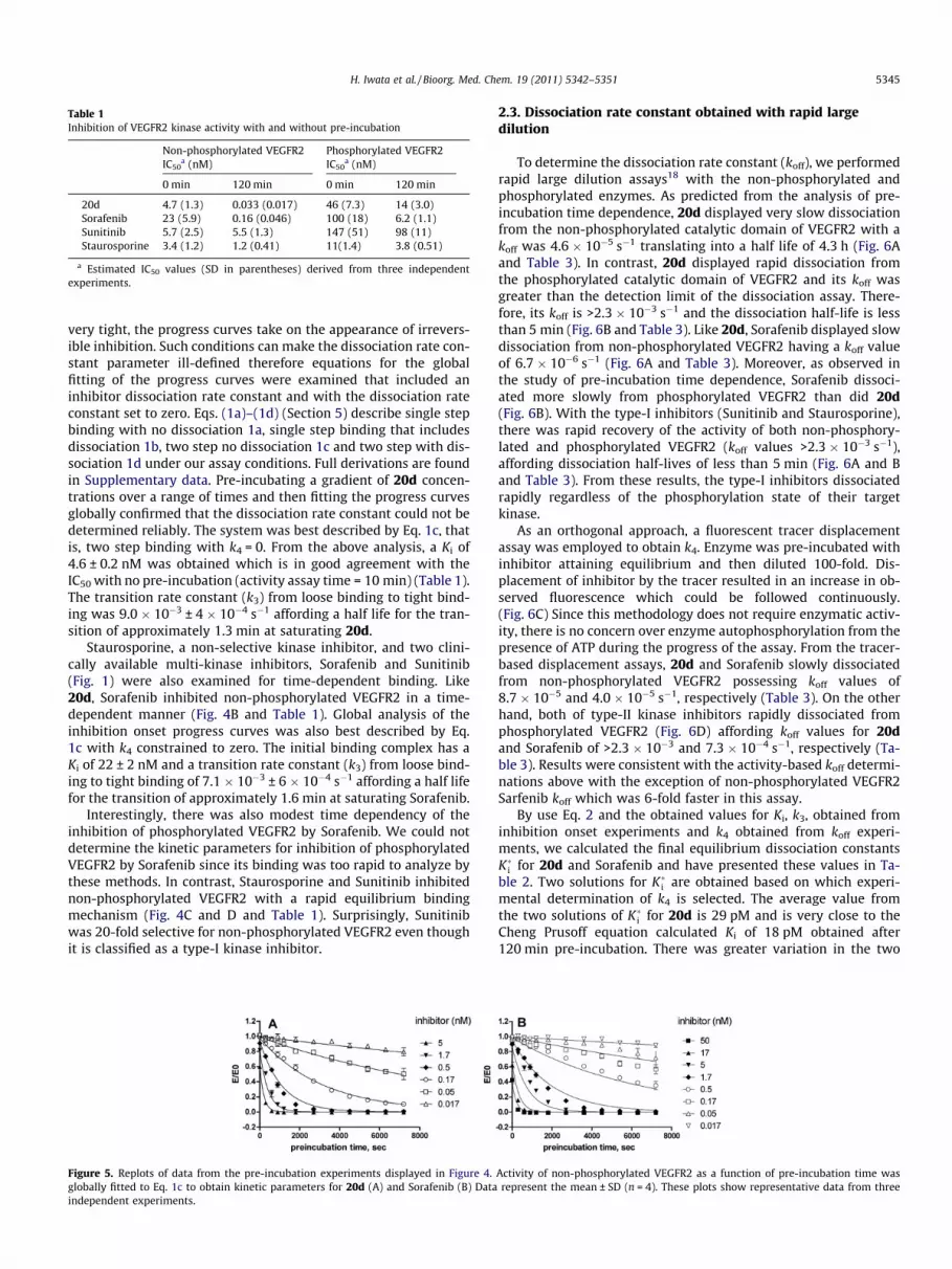
Table 1Inhibition of VEGFR2 kinase activity with and without pre-incubation
Non-phosphorylated VEGFR2IC50
a (nM)Phosphorylated VEGFR2IC50
a (nM)
0 min 120 min 0 min 120 min
20d 4.7 (1.3) 0.033 (0.017) 46 (7.3) 14 (3.0)Sorafenib 23 (5.9) 0.16 (0.046) 100 (18) 6.2 (1.1)Sunitinib 5.7 (2.5) 5.5 (1.3) 147 (51) 98 (11)Staurosporine 3.4 (1.2) 1.2 (0.41) 11(1.4) 3.8 (0.51)
a Estimated IC50 values (SD in parentheses) derived from three independentexperiments.
H. Iwata et al. / Bioorg. Med. Chem. 19 (2011) 5342–5351 5345
very tight, the progress curves take on the appearance of irrevers-ible inhibition. Such conditions can make the dissociation rate con-stant parameter ill-defined therefore equations for the globalfitting of the progress curves were examined that included aninhibitor dissociation rate constant and with the dissociation rateconstant set to zero. Eqs. (1a)–(1d) (Section 5) describe single stepbinding with no dissociation 1a, single step binding that includesdissociation 1b, two step no dissociation 1c and two step with dis-sociation 1d under our assay conditions. Full derivations are foundin Supplementary data. Pre-incubating a gradient of 20d concen-trations over a range of times and then fitting the progress curvesglobally confirmed that the dissociation rate constant could not bedetermined reliably. The system was best described by Eq. 1c, thatis, two step binding with k4 = 0. From the above analysis, a Ki of4.6 ± 0.2 nM was obtained which is in good agreement with theIC50 with no pre-incubation (activity assay time = 10 min) (Table 1).The transition rate constant (k3) from loose binding to tight bind-ing was 9.0 � 10�3 ± 4 � 10�4 s�1 affording a half life for the tran-sition of approximately 1.3 min at saturating 20d.
Staurosporine, a non-selective kinase inhibitor, and two clini-cally available multi-kinase inhibitors, Sorafenib and Sunitinib(Fig. 1) were also examined for time-dependent binding. Like20d, Sorafenib inhibited non-phosphorylated VEGFR2 in a time-dependent manner (Fig. 4B and Table 1). Global analysis of theinhibition onset progress curves was also best described by Eq.1c with k4 constrained to zero. The initial binding complex has aKi of 22 ± 2 nM and a transition rate constant (k3) from loose bind-ing to tight binding of 7.1 � 10�3 ± 6 � 10�4 s�1 affording a half lifefor the transition of approximately 1.6 min at saturating Sorafenib.
Interestingly, there was also modest time dependency of theinhibition of phosphorylated VEGFR2 by Sorafenib. We could notdetermine the kinetic parameters for inhibition of phosphorylatedVEGFR2 by Sorafenib since its binding was too rapid to analyze bythese methods. In contrast, Staurosporine and Sunitinib inhibitednon-phosphorylated VEGFR2 with a rapid equilibrium bindingmechanism (Fig. 4C and D and Table 1). Surprisingly, Sunitinibwas 20-fold selective for non-phosphorylated VEGFR2 even thoughit is classified as a type-I kinase inhibitor.
Figure 5. Replots of data from the pre-incubation experiments displayed in Figure 4.globally fitted to Eq. 1c to obtain kinetic parameters for 20d (A) and Sorafenib (B) Dataindependent experiments.
2.3. Dissociation rate constant obtained with rapid largedilution
To determine the dissociation rate constant (koff), we performedrapid large dilution assays18 with the non-phosphorylated andphosphorylated enzymes. As predicted from the analysis of pre-incubation time dependence, 20d displayed very slow dissociationfrom the non-phosphorylated catalytic domain of VEGFR2 with akoff was 4.6 � 10�5 s�1 translating into a half life of 4.3 h (Fig. 6Aand Table 3). In contrast, 20d displayed rapid dissociation fromthe phosphorylated catalytic domain of VEGFR2 and its koff wasgreater than the detection limit of the dissociation assay. There-fore, its koff is >2.3 � 10�3 s�1 and the dissociation half-life is lessthan 5 min (Fig. 6B and Table 3). Like 20d, Sorafenib displayed slowdissociation from non-phosphorylated VEGFR2 having a koff valueof 6.7 � 10�6 s�1 (Fig. 6A and Table 3). Moreover, as observed inthe study of pre-incubation time dependence, Sorafenib dissoci-ated more slowly from phosphorylated VEGFR2 than did 20d(Fig. 6B). With the type-I inhibitors (Sunitinib and Staurosporine),there was rapid recovery of the activity of both non-phosphory-lated and phosphorylated VEGFR2 (koff values >2.3 � 10�3 s�1),affording dissociation half-lives of less than 5 min (Fig. 6A and Band Table 3). From these results, the type-I inhibitors dissociatedrapidly regardless of the phosphorylation state of their targetkinase.
As an orthogonal approach, a fluorescent tracer displacementassay was employed to obtain k4. Enzyme was pre-incubated withinhibitor attaining equilibrium and then diluted 100-fold. Dis-placement of inhibitor by the tracer resulted in an increase in ob-served fluorescence which could be followed continuously.(Fig. 6C) Since this methodology does not require enzymatic activ-ity, there is no concern over enzyme autophosphorylation from thepresence of ATP during the progress of the assay. From the tracer-based displacement assays, 20d and Sorafenib slowly dissociatedfrom non-phosphorylated VEGFR2 possessing koff values of8.7 � 10�5 and 4.0 � 10�5 s�1, respectively (Table 3). On the otherhand, both of type-II kinase inhibitors rapidly dissociated fromphosphorylated VEGFR2 (Fig. 6D) affording koff values for 20dand Sorafenib of >2.3 � 10�3 and 7.3 � 10�4 s�1, respectively (Ta-ble 3). Results were consistent with the activity-based koff determi-nations above with the exception of non-phosphorylated VEGFR2Sarfenib koff which was 6-fold faster in this assay.
By use Eq. 2 and the obtained values for Ki, k3, obtained frominhibition onset experiments and k4 obtained from koff experi-ments, we calculated the final equilibrium dissociation constantsK�i for 20d and Sorafenib and have presented these values in Ta-ble 2. Two solutions for K�i are obtained based on which experi-mental determination of k4 is selected. The average value fromthe two solutions of K�i for 20d is 29 pM and is very close to theCheng Prusoff equation calculated Ki of 18 pM obtained after120 min pre-incubation. There was greater variation in the two
Activity of non-phosphorylated VEGFR2 as a function of pre-incubation time wasrepresent the mean ± SD (n = 4). These plots show representative data from three

Table 2Kinetic constants from pre-incubation experiments with non-phosphorylated VEGFR2
Kia (nM) Ki
⁄b (nM) Ki⁄c (nM) k3
a (s�1)
20d 4.6 (0.2) 0.024 0.044 9.0�10�3 (4.0 � 10�4)Sorafenib 22 (1.9) 0.021 0.120 7.1 � 10�3 (6.3 � 10�4)Sunitinib 2.9d N.D. N.D. N.D.Staurosporine 1.7d N.D. N.D. N.D.
a Global fit to inhibition onset progress curves to obtain Ki and k3 values (SD inparentheses).
b Values calculated by Eq. 2 using Ki, k3, and k4 from activity based analysis.c Values calculated by Eq. 2 using Ki, k3, with k4 from ligand displacement based
analysis.d Sunitinib and Staurosporine are not time dependent inhibitors, Ki values cal-
culated from IC50 values with no pre-incubation are shown.
Figure 6. Recovery of enzyme activity (A and B) and tracer binding activity (C and D) after 100-fold dilution for estimation of dissociation rate constants. (A) Results of theactivity based the 100-fold dilution assay with non-phosphorylated VEGFR2 and 20d (d), Staurosporine (�), Sorafenib (N), Sunitinib (h), or no inhibitor as a control (j). (B)Results of activity based the 100-fold dilution assay with phosphorylated VEGFR2 and 20d (d), Staurosporine (�), Sorafenib (N), Sunitinib (h), or no inhibitor as a control (j).(C) Results of the fluorescent tracer based 100-fold dilution assay with non-phosphorylated VEGFR2 and 20d (d), Staurosporine (�), Sorafenib (N), Sunitinib (h), or withoutany inhibitor as a control (j) and without inhibitor or enzyme (s). (D) Results of the fluorescent tracer based 100-fold dilution assay with phosphorylated VEGFR2 and 20d(d), Staurosporine (�), Sorafenib (N), Sunitinib (h), or without any inhibitor as a control (j) and without inhibitor or enzyme (s). Representative data from threeindependent experiments are shown. Results represent the mean ± SD (n = 3).
5346 H. Iwata et al. / Bioorg. Med. Chem. 19 (2011) 5342–5351
calculations of K�i for Sorafenib but the average value of 70 pMmatches Cheng Prusoff equation calculated Ki of 80 pM.
The slow dissociative character of 20d and Sorafenib from onlynon-phosphorylated VEGFR2 are confirmed two assay methodolo-gies. The koff value of 20d for dissociation from non-phosphory-lated VEGFR2 was at least 50-fold smaller than the koff forphosphorylated VEGFR2, and the difference was much larger forSorafenib (about 200-fold). These results suggest that both of thetype-II kinase inhibitors investigated in this study were recognized
Table 3Kinetic constants for non-phosphorylated and phosphorylated VEGFR2from enzyme–inhib
Method Non-phosphorylated VEG
koffa (s�1)
20d Activity based 4.6�10�5 (8.9 � 10�6)Ligand displacement 8.7 � 10�5 (6.5 � 10�6)
Sorafenib Activity based 6.7 � 10�6 (1.7 � 10�6)Ligand displacement 4.0 � 10�5 (2.9 � 10�6)
Sunitinib Activity based >2.3 � 10�3c
Ligand displacement >2.3 � 10�3c
Staurosporine Activity based >2.3 � 10�3c
Ligand displacement >2.3 � 10�3c
a Estimated koff values (SD in parentheses).b Values calculated with Eq. 3 using koff values (SD in parentheses).c Values calculated with Eq. 3 using t1/2 < 5 min. Results shown were derived from th
by non-phosphorylated and phosphorylated VEGFR2 but their dis-sociation velocity are highly affected by the state of phosphoryla-tion of the bound enzyme.
2.4. Surface plasmon resonance (SPR) analysis
In the activity based and fluorescent tracer displacement exper-iments, 20d only displayed time-dependent inhibition and slowdissociation in the case of non-phosphorylated VEGFR2. In orderto evaluate these characteristics of 20d through binding kinetics,we performed SPR analysis using a Biacore biosensor with non-phosphorylated and phosphorylated VEGFR2. The sensorgramsare presented in Figure 7. Unfortunately, the sensorgram tracesfor 20d against non-phosphorylated enzyme defied fitting to rea-sonable models. As such, they are best viewed qualitatively. SPRanalysis captures transitions that are much faster than what is ob-servable by the methods described above. From the sensorgrams itis clear that onset of the EI complex was not rapid equilibrium (onthe SPR time scale) for either non-phosphorylated enzyme or thephosphorylated enzyme. As observed in the pre-incubation anddilution experiments, 20d dissociation from the non-phosphory-lated enzyme was considerably slower (indeterminately slow byBiacore) than dissociation from the phosphorylated enzyme. Qual-itatively, the two step slow binding mechanism reveals itself in the
itor complex dilution assays
FR2 Phosphorylated VEGFR2
t1/2b (min) koff
a (s�1) t1/2b (min)
260 (55) >2.3 � 10�3c <5130 (10) >2.3�10�3c <51800 (510) 1.5 � 10�3 (4.2 � 10�4) 8 (3)290 (20) 7.3 � 10�4 (2.8 � 10�4) 18 (9)<5 >2.3 � 10�3c <5<5 >2.3 � 10�3c <5<5 >2.3 � 10�3c <5<5 >2.3 � 10�3c <5
ree independent experiments.

Figure 7. Results of SPR binding experiments with non-phosphorylated VEGFR2 (A, B) and phosphorylated VEGFR2 (C, D). 20d was injected at 2-fold serial dilutions from 1.9to 125 nM for non-phosphorylated VEGFR2 (A) and 2-fold serial dilutions from 1.9 to 500 nM for phosphorylated VEGFR2 (C). Sunitinib was injected at 2-fold serial dilutionsfrom 1.9 to 500 nM for both non-phosphorylated VEGFR2 (B) and phosphorylated VEGFR2 (D). (A) Each sensorgram was globally fitted to a two-state reaction (conformationchange) model except for Rmax values that were obtained by local fitting to the same binding model. (B), (C), and (D) Each sensorgram was fitted to a 1:1 binding with masstransfer model, except for Rmax value that were obtained by local fitting to the same binding model. These results are representative data from three independentexperiments.
Figure 8. Recovery of autophosphorylation of tyrosine residues on VEGFR2 afterwashout of inhibitor. Serum starved KDR/293 cells were incubated with 20d orSunitinib at 100 nM for 1 h, and then washed with inhibitor-free medium. Next, thecells were stimulated with 50 ng/ml of VEGF for 10 min at the indicated times afterwashout, followed by lysis with cell lysis buffer. Total cell lysates were subjected toELISA with phospho tyrosine 1175 antibody. Values were normalized from 0% (thewell containing no inhibitor and without VEGF stimulation) to 100% (the wellcontaining no inhibitor and with VEGF stimulation). Data are shown as the meanand SD (n = 3).
H. Iwata et al. / Bioorg. Med. Chem. 19 (2011) 5342–5351 5347
biphasic nature of inhibitor dissociation from the enzyme-boundcomplex. The loose (rapidly reversible) complex quickly dissociateswhile the EI⁄ tight complex dissociates very slowly on the Biacoretime scale (Fig. 7). Curiously, Sorafenib, the other slow bindinginhibitor, sensorgram traces also defied fitting to reasonable mod-els (Fig. S3). In contrast, the sensorgrams for dissociation of Suniti-nib from non-phosphorylated VEGFR2 fit well to a 1:1 single stepbinding model (Fig. 7B). Moreover, the sensorgram of 20d for phos-phorylated VEGFR2 fit a 1:1 single step binding model well(Fig. 7C). Ka and Kd values of 20d for phosphorylated VEGFR2 weredetermined as 1.6 � 105M�1 s�1 and 3.1 � 10�3 s�1, respectively.With these parameters, KD was calculated as 19 nM. The KD is goodagreement with Ki of 23 nM from enzymatic activity based analy-sis. Additionally, 20d preferentially interacts with non-phosphory-lated VEGFR2, as observed in the activity based experiments, andthis preference seems to be based on the difference of dissociationrate constants for non-phosphorylated and phosphorylated VEG-FR2 (Fig. 7A and C). Interestingly, Sunitinib also demonstrated apreference for non-phosphorylated VEGFR2 as observed in activitybased experiments. The KD value for non-phosphorylated VEGFR2obtained with steady state affinity analysis was about 5-fold smal-ler than KD value for the phosphorylated enzyme.
2.5. Cellular autophosphorylation assay
We assessed the inhibitory activity of 20d in a cellular auto-phosphorylation assay using 293/KDR cells that stably over ex-pressed VEGFR2, but whose proliferation and survival were VEGFindependent. Since autophosphorylation of tyrosine 1175 is con-sidered a requirement for VEGF mediated survival and prolifera-tion,20 we used phosphorylation of this residue to index ofcellular VEGFR2 kinase activity. Phosphorylation was detected by
an enzyme-linked immunosorbent assay (ELISA). As shown in(Fig. 8), 20d and Sunitinib both completely inhibited VEGF-inducedautophosphorylation of tyrosine 1175 on VEGFR2 at 100 nM with-out an inhibitor washout step. Even at 96 h post inhibitor washout,compound 20d completely inhibited VEGF-stimulated phosphory-lation of VEGFR2. In marked contrast washout experiments withSunitinib displayed an increasing VEGFR2 phosphorylation overtime post washout. Clearly 20d displays prolonged inhibitoryactivity with a long residence time on VEGFR2 in a cellular as-say—typical characteristics of a type-II kinase inhibitor.

5348 H. Iwata et al. / Bioorg. Med. Chem. 19 (2011) 5342–5351
3. Discussion
In this study, we used VEGFR2 to characterize the inhibitoryactivity of type-I and type-II kinase inhibitors and to investigatethe reason for the differing inhibitory activity of type-II kinaseinhibitors for non-phosphorylated and phosphorylated enzymes.First, we determined the phosphorylation state of the catalytic do-main of VEGFR2 with or without autophosphorylation. The cata-lytic domain of VEGFR2 that we targeted in this study has 10tyrosine residues. Among these 10 residues, it has been reportedthat at least 3 residues can be phosphorylated21 and it seems thattyrosine 1008 might be phosphorylated according to a study of themouse ortholog enzyme.22 From our LC/MS data, there could be asmany as 6 or 7 phosphorylated residues after autophosphorylationover the surface of the enzyme (Fig. S2). The in vivo presence orrelevance hyperphosphorylation is unknown. However, our immu-noblotting analysis indicated that relevant tyrosine residues on theactivation loop of the catalytic domain of VEGFR2 underwent auto-phosphorylation and there was an obvious difference of phosphor-ylation between non-phosphorylated and phosphorylated VEGFR2(Fig. 2). Thus, we chose to use these two proteins to characterizetype-I and type-II kinase inhibitors.
We are compelled to evaluate the unactivated enzyme activityover a short time period (10 min or less) since longer reactiontimes will cause an unintended autoactivation through autophos-phorylation. The competition of autophoshophorylation withphosphorylation of the intended peptide substrate prevented usfrom employing a more conventional technique to characterizekon such as progress curve analysis. To address these necessitiesof assay sensitivity and avoidance of autoactivation, we performedenzyme assays in AlphaScreen� platform. This approach gave thedesired high sensitivity when performed with MnCl2 containingbuffer in addition to MgCl2 since the catalytic activity of VEGFR2kinase increased.23 In addition, we have derived equations that de-scribe the predicted progress curves for the onset of inhibition for2-phase assay system containing a pre-incubation phase and an as-say phase where inhibition onset still continues but at a reducedrate.
Characterization of the kinase inhibitors within this study wasby use of enzymatic activity and biophysical analyses. As describedpreviously,16 during the course of compound optimization, we per-form assays with the un-phosphorylated/inactive enzyme assess-ing the inhibitory activity of compounds toward this enzymeform. Moreover, as observed in this study, since most type-II inhib-itors preferentially inhibit non-phosphorylated enzyme, screeningsolely against the activated form of the enzyme may underesti-mate a compound’s potential in vivo affinity.
As described above, one of the characteristics of type-II inhibi-tors is interaction with the back pocket site of the target enzyme.This binding mode not only applies to non-phosphorylated VEGFR2but also to phosphorylated VEGFR2 according to a X-ray crystallo-graphic study performed by using a urea based type-II kinaseinhibitor and the auto-phosphorylated catalytic domain of VEG-FR2.24 Thus crystallographic data support the hypothesis thatphosphorylated VEGFR2 can be induced to adopt DFG-out confor-mation in the presence of an inhibitor. Also, binding with theDFG-out conformation of the kinase is not sufficient to cause slowdissociation of an inhibitor since there have been some reports oftype-II kinase inhibitors that do not display slow dissociation.25
Therefore, there is no clear conclusion that the slow dissociationis coupled to the movement of the activation loop from DFG-inform to DFG-out forms of protein conformation.
In this study, we report that 20d is a two-step slow bindinginhibitor against non-phosphorylated VEGFR2, but not for phos-phorylated VEGFR2, by activity based experiments (Figs. 4A, Eand 5). By use of an activity based dilution assay and by use of a
fluorescent tracer displacement based dilution assay, we observethat 20d very slowly dissociated from non-phosphorylated VEG-FR2 (Fig. 6A and C) yet its dissociation from phosphorylated VEG-FR2 was rapid (Fig. 6B and D). These results were also observedwith the other type-II inhibitor Sorafenib (Figs. 4B, F and 6). Fromthese analysis, we obtained K�i of each type-II inhibitor. Except forK�i of Sorafenib by use of k4 from activity based analysis, obtainedK�i values were in good agreement with IC50 with 120 min pre-incubation. The discrepancy of k4 of Sorafenib from each experi-ment is thought to be due to limitations of each method, such asminimal dissociation or reactivation of the enzyme.
These observations were confirmed through binding SPR-basedbiophysical experiments (Fig. 7). However, we could not determinewhether or not the slow binding isomerization step represents aconformational change from DFG-in to DFG-out, so further studiesof VEGFR2 protein or of protein–inhibitor complex conformationswill be required for a more complete understanding.
Studies on the interaction of type-II kinase inhibitors with p38aMAP kinase were previously performed by use of NMR26,27 and itwas concluded that type-II inhibitors only bind p38a in the DFG-out conformation. In contrast, Frembgen–Kesner and Elcock re-cently reported that type-II kinase inhibitors not only bind to theDFG-out conformation of p38a MAP kinase but also the DFG-inconformation, and they suggested the existence of pseudo-DFG-in and pseudo-DFG-out conformations in addition to the generallyaccepted DFG-in and DFG-out conformations through analysiswith molecular dynamics simulation.28 According to the latter re-sult, we suggest that type-II kinase inhibitors bind to the DFG-inconformation first and this may then cause a conformationalchange of the target enzyme.29
As discussed above, it is widely believed that phosphorylatedVEGFR2 adopts mainly a DFG-in conformation in solution. Thisstudy strongly supports the hypothesis that type-II kinase inhibi-tors bind to the DFG-in conformation first since 20d has similarbinding rates for non-phosphorylated and phosphorylated VEGFR2.Upon obtaining binding equilibrium with 20d, both the non-phos-phorylated VEGFR2 and phosphorylated protein adopt DFG-outconformation and thereby only the non-phosphorylated VEGFR2adopts a more stable and inactive conformation.
A similar phenomenon was observed in the study of PYK2 andBIRB-796, a p38a MAP kinase inhibitor.19 BIRB-796 is known toslowly dissociate from p38a9 but it did not show slow dissociativecharacter with PYK2. In this study, we demonstrated that type-IIinhibitors may associate at similar rates with both non-phosphor-ylated VEGFR2 and phosphorylated VEGFR2 and that the type-IIinhibitors display slow dissociative character from the non-phos-phorylated enzyme.
Although p38a MAP kinase requires phosphorylation of itsthreonine residues by MKK6 to display enzymatic activity, theintracellular domain or catalytic domain of RTKs like VEGFR2 haskinase activity even without phosphorylation of the activationloop. Thus, the mechanism of enzymatic activation with a confor-mational change and/or the mechanism of binding to type-II kinaseinhibitors may vary among protein kinases.
Sunintib is a clinically available multikinase inhibitor that iscategorized as a type-I inhibitor. Interestingly, it displayed lessinhibitory activity for phosphorylated VEGFR2 than for the non-phosphorylated enzyme. DiNitto et al. reported that Sunitinibbinds to and stabilizes the inactive form of c-Kit, a tyrosine ki-nase from the same PDGFR superfamily as VEGFR2, and they con-cluded that Sunitinib only shows weak inhibition againstactivated c-Kit when its activation loop is phosphorylated.30 Inour study, a similar phenomenon might be in operation for phos-phorylated VEGFR2, and Sunitinib might be a type-I inhibitor thatpreferentially targets the inactive forms of both c-Kit andVEGFR2.

Scheme 3. Putative inhibitory mechanism of compound 20d for non-phosphory-lated and phosphorylated VEGFR2.
H. Iwata et al. / Bioorg. Med. Chem. 19 (2011) 5342–5351 5349
Recently, it was reported that the inhibitory activity of Axitinib,a type-II VEGFR2 kinase inhibitor, was strongly influenced by thejuxtamembrane region of the enzyme and that Axitinib preferen-tially inhibited VEGFR2 protein with juxtamembrane domainrather than the catalytic domain.31 Thus, there may be some struc-tural regulation of VEGFR2 protein associated with the juxtamem-brane domain. In the present study, however, we chose thecatalytic domain of VEGFR2 because we wished to focus on bindingevents in the ATP-binding pocket to simplify analysis. In addition,it was considered difficult to obtain fully phosphorylated cytoplas-mic domain enzyme through the autophosphorylation step with itsweaker enzymatic activity than with the catalytic domain of theenzyme. Recently, we reported on the slow binding mechanismof TAK-593, another type-II VEGFR/PDGFR inhibitor and Sorafenib.16
In that study, we performed activity and binding based analysesusing the cytoplasmic domain of VEGFR2 with low phosphorylation,which contains the juxtamembrane domain, and we found that bothTAK-593 and Sorafenib displayed two-step slow binding inhibition.Thus, the hypothesis for the behavior of type-II inhibitor suggestedin this study may not only apply to the catalytic domain of VEGFR2but also to the cytoplasmic domain of this enzyme.16
4. Conclusion
In conclusion, 20d is an ATP competitive type-II VEGFR/PDGFRkinase inhibitor that is slow binding with and selective for non-phosphorylated VEGFR2. Its selective binding is driven by slow dis-sociation from non-phosphorylated VEGFR2. This study supportsthe theory that VEGFR2 kinase can adopt the DFG-out conforma-tion (an inactive conformation) and bind with type-II inhibitorseven after phosphorylation of tyrosine residues on its activationloop, but that binding of the inactive conformation with type-IIinhibitors is not very stable, as was observed in the case of non-phosphorylated VEGFR2 (Scheme 3). Based on its slow dissociation,and supported by cellular wash-out experiments 20d displays pro-longed inhibitory activity in vitro and in vivo. These findings willhelp us to better understand the inhibitory mechanisms of type-Iand type-II kinase inhibitors for non-phosphorylated and phos-phorylated kinases and assist in the designing and synthesis of no-vel type-II kinase inhibitors. In particular, with these findings wedemonstrate the importance of the dissociation rate determinationfrom a kinase in distinct phosphorylation condition during the leadoptimization of type-II kinase inhibitors.
5. Materials and methods
5.1. Chemicals
(1-{2-Fluoro-4-[(5-methyl-5H-pyrrolo[3,2-d]pyrimidin-4-yl)oxy]phenyl}-3-[3-(trifluoromethyl)phenyl]urea) (20d) was syn-thesized by Takeda Pharmaceutical Company, Ltd (Osaka, Japan).15
Staurosporine was purchased from Wako Pure Chemicals (Osaka,Japan). Sorafenib (Nexavar�, Bayer, Germany) and Sunitinib (Su-tent�, Pfizer, USA) were obtained from commercial sources.
5.2. Reagents
Ni-NTA resin was purchased from QIAGEN (Hilden, Germany).ATP, DTT, and puromycin were purchased from Sigma–Aldrich(MO, USA). AlphaScreen� Phosphotyrosine (P-Tyr 100) andAlphaScreen� Phosphotyrosine (PT66) assay kits were obtainedfrom PerkinElmer (MA, USA). Biotinylated poly-GluTyr (4:1)was obtained from Cisbio (France). Anti-phospho-KDR/Flk-1/VEGFR2 (Tyr1054) and anti-phospho-KDR/Flk-1/VEGFR2(Tyr1059) were obtained from Millipore (MA, USA). Block Ace�
powder was purchased from Dainippon Sumitomo (Osaka, Japan)and sample treatment for Tris–SDS was purchased from Cosmo-bio (Tokyo, Japan). Phospho-tyrosine mouse mAb (P-Tyr-100),horseradish peroxidase-conjugated anti-rabbit IgG, 20� Lumi-GLO� Reagent and peroxide were obtained from Cell SignalingTechnology (MA, USA), while VEGF Receptor 2 antibody fordetection of total VEGFR2 was purchased from Abcam (Cam-bridge, UK). 5� Kinase Buffer A, Eu-anti-His-tag Antibody and Ki-nase Tracer 236 for the LanthaScreen™ Eu kinase binding assaywere obtained from Invitrogen (CA, USA).
5.3. Cell culture and cellular autophosphorylation assay
Human embryonic kidney 293 cells stably expressing KDR(KDR/293) were purchased from Sibtech (CT, USA) and culturedin Dulbecco’s modified Eagle’s medium (DMEM; Invitrogen) sup-plemented with 10% fetal bovine serum (FBS; Moregate, Brisbane,Australia), 1� penicillin/streptomycin (Invitrogen), and 0.375 lg/ml of puromycin (Sigma–Aldrich) to maintain the expression ofVEGFR2/KDR. Two days before the washout experiments, cellswere seeded at 100,000/well in poly-D-lysine-coated 24-wellplates. After 24 h, the medium was changed to DMEM containing0.5% FBS, 1� penicillin/streptomycin, and 0.375 lg/ml of puromy-cin. Inhibitors prepared in DMSO were diluted with DMEM con-taining 0.5% FBS, 1� penicillin/streptomycin, and 0.375 lg/ml ofpuromycin, and then were added to the wells at 1 h before wash-out. After 1 h of exposure to each inhibitor, the wells were aspi-rated. Then the cells were washed three times with DMEM andstimulated with 50 ng/ml of VEGF (R&D Systems, MN, USA) atthe indicated time after washout. Cellular autophsphorylationwas detected by using a PathScan� Phospho-VEGFR-2 (Tyr1175)Sandwich ELISA Kit (Cell Signaling Technology) according to themanufacturer’s instructions. Wells containing no test compoundthat were stimulated with VEGF and wells containing no com-pound without VEGF stimulation were used as the 100% controland 0% control, respectively.
5.4. VEGFR2 protein expression and auto-phosphorylation
The catalytic domain of human VEGFR2 (residues 806-1171)with deletion of the kinase insert domain (residues 940–989)was expressed as an N-terminal His-tagged protein with the TEVprotease site by using a baculovirus expression system, as de-scribed previously.15 Then 30 lM of purified VEGFR2 with its N-terminal His tag was subjected to auto-phosphorylation with2 mM ATP and 10 mM MgCl2 on ice for 90 min, after which thereaction mixture was desalted by using a HiPrep desalting column.Non-phosphorylated and phosphorylated VEGFR2 were stored at�80 �C.
5.5. Immunoblotting analysis
Protein samples were mixed with an equal volume of sampletreatment for Tris–SDS (Cosmobio) containing 100 mM DTT andheated at 95 �C for 5 min. Then proteins were subjected to electro-phoresis on SDS–polyacrylamide gel with Tris–glycine running

5350 H. Iwata et al. / Bioorg. Med. Chem. 19 (2011) 5342–5351
buffer, followed by electrotransfer to an iBlot PVDF Transfer Stack(Invitrogen) and incubation with 4% (w/v) Block Ace at 4 �C over-night. Then the membrane was washed and incubated at roomtemperature with anti-phospho or anti-VEGFR2 antibody(1:1000) for 1 h, after which it was incubated at room temperaturewith horseradish peroxidase-conjugated anti-rabbit IgG (1:1000)for 1 h and then visualized by using 1� LumiGLO� (Cell SignalingTechnology) Reagent and peroxide.
5.6. Investigation of ATP-competitive inhibition of non-phosphorylated and phosphorylated VEGFR
VEGFR2 kinase activity was determined by using an anti-phos-photyrosine antibody with quantitation by the AlphaScreen� sys-tem32 (PerkinElmer, USA). Enzyme reactions were performed in50 mM Tris–HCl (pH 7.5), 5 mM MnCl2, 5 mM MgCl2, 0.01%Tween-20, and 2 mM DTT with ATP at various concentrations,0.1 lg/ml biotinylated poly-GluTyr (4:1), and 75 pM of non-phos-phorylated or phosphorylated VEGFR2. Reactions were initiatedby adding VEGFR2 kinase to the reaction mixture and test com-pound, and reactions were quenched by the addition of 25 ll of100 mM EDTA, 10 lg/ml each of AlphaScreen� streptavidin donorbeads and acceptor beads in 62.5 mM HEPES (pH 7.4), 250 mMNaCl, and 0.1% BSA. Plates were incubated in the dark overnightand then read by an EnVision 2102 Multilabel Reader (PerkinEl-mer). Wells containing the substrate and the enzyme without20d were used as the total reaction control, while wells containingbiotinylated poly-GluTyr (4:1) and the enzyme without ATP wereused as the basal control. The concentration of 20d producing50% inhibition of the kinase activity of non-phosphorylated andphosphorylated VEGFR2 (IC50) was analyzed using GraphPad Prismversion 5.01 (GraphPad Software, CA, USA). Sigmoidal dose–re-sponse (variable slope) curves were fitted by non-linear regressionanalysis, with the top and bottom of the curve being constrained at100 and 0, respectively.
5.7. Time-dependent inhibition of non-phosphorylated andphosphorylated VEGFR2
In order to assess the time-dependency of inhibition, assayswere performed after pre-incubation of the enzyme with theinhibitor for various times and then conducting an enzymeactivity assay where ATP was present at Kapp
m . AlphaScreen�
system, as described above was used to assay enzyme activity.By determining the IC50 value of 20d as a function of enzyme–inhibitor pre-incubation time prior to initiation of catalysis weobtained a preliminary assessment of time dependent inhibi-tion. IC50 values were calculated by logistic regression analysis(GraphPad Prism 5.01). Rigorous experiments to characterizethe onset of inhibition consisted of the pre-incubation phasefollowed by dilution to 0.6 (15 ll initial to 25 ll final) uponaddition of substrate. The enzyme activity phase of the experi-ments was conducted for 10 min. Eqs. 1a through 1d describethe fractional enzyme activity when accounting for the amountof additional onset of tight binding occurring during the activityphase of the assay. Fractional activity as a function of inhibitorconcentration and pre-incubation time was fit globally to Eqs.1a through 1d and overall quality of fit and consistency withother experimental data used to select the most appropriatebinding model. Derivations of these equations are found inSupplementary data.
Single step with koff = 0
EET¼ e�kon ½I�te
�kon0:6½I�
1þ SKM
10
ð1aÞ
Full expression for single step
EET¼ 1
1þ 0:6½I�koffkon
1þ SKM
� �þ0:6½I�
0:6½I� þ koff
kon1þ S
KM
� � e�ðkon ½I�þkoff Þte� kon
0:6½I�1þ S
KM
þkoff
� �10
ð1bÞTwo step with koff = 0
EET¼ e�k3
½I�½I�þKI
t
1þ 0:6½I�
KI 1þ SKM
� � e
�k30:6½I�
0:6½I�þKI 1þ SKM
� �10
ð1cÞ
Full expression for two step
EET¼ 1
1þ 0:6½I�
KI 1þ SKM
� ��b
aþ bþ a
aþ be�ða
0þbÞte�ðaþbÞ10� �
ð1dÞ
where:
a ¼ k30:6½I�
KI 1þ SKM
� �þ 0:6½I�
a0 ¼ k3½I�
KI þ ½I�b ¼ k4
Note: 0.6 corrects for inhibitor dilution and 10 min was the activityassay time.
With a two-step binding mechanism, the final equilibriumaffinity of the inhibitor (K�i ) is defined by Eq. 2.
K�i ¼ k4K i=ðk3 þ k4Þ ð2Þ
The dissociation half-life (t1/2) was calculated with Eq. 3
t1=2 ¼ 0:693=k4 ð3Þ
Eqs. 2 and 3 were previously described by Copeland.33
5.8. Determination of the dissociation rate constant by the rapidlarge dilution method
To determine the dissociation rate constant (koff) of the inhibi-tors, enzyme–inhibitor dilution assays were performed. The assaysinvolved determination of koff from the kinetics of the recovery ofkinase activity following rapid dilution of the enzyme–inhibitorcomplex.33 Recovery of enzyme activity from a preformed en-zyme–inhibitor complex was measured by using the AlphaScreen�
system. The enzyme (at a 100-fold higher concentration than finalactivity conditions) and the inhibitor (at a 10-fold higher concen-tration than the initial IC50 value) were pre-incubated together inthe reaction buffer for 60 min at room temperature permittingequilibrium formation for tight enzyme–inhibitor complexes. Thenthe complexes were diluted 1:100 in reaction buffer containing1 mM ATP and 0.1 lg/ml biotinylated poly-Glu-Tyr (4:1) to initiatethe kinase reaction. The kinetics of the recovery of kinase activityafter rapid dilution were fitted to Eq. 4.34
Under the above conditions, kobs represents koff of the enzyme–inhibitor complex and the dissociation half-life (t1/2) was calcu-lated from Eq. 3 by replacement of k4 with kobs.
P ¼ mst þ ½ðmi � msÞ=kobs�½1� expð�kobsÞ� þ P0 ð4Þ
P; Product of the kinase reaction (CPS)P0; Background signal when t = 0vi; Initial velocityvs; Steady state velocitykobs; Inhibition rate constant

H. Iwata et al. / Bioorg. Med. Chem. 19 (2011) 5342–5351 5351
5.9. Ligand displacement based dilution assay by use ofLanthaScreen™ Eu kinase binding assay
Dissociation rate constants of inhibitors were determined byuse of a ligand displacement based enzyme–inhibitor dilution as-say. This assay involved determination of koff through the kineticsof recovery of the tracer binding signal after rapid dilution of theenzyme–inhibitor complex. Recovery of the tracer binding signalfrom a preformed enzyme–inhibitor complex was measured byusing the LanthaScreen™ Eu kinase binding assay. 500 nM of VEG-FR2 enzyme (at a 100-fold higher concentration than the bindingassay conditions) and the 1000 nM of inhibitor were incubated to-gether in 1� Kinase Buffer A containing 2 mM DTT and 0.05% BSAfor 60 min at ambient temperature to form the enzyme–inhibitorcomplex. Then the complex was diluted 1:100 in 1� Kinase BufferA containing 2 mM DTT and 5 nM of Eu-anti-His-tag Antibody(Invitrogen) and 100 nM of Kinase Tracer 236 to initiate displace-ment of the inhibitor by an excess of Kinase Tracer 236. The kinet-ics of the recovery of tracer binding activity after rapid dilutionwere normalized as a percentage of tracer bound enzyme (100%;no inhibitor control, 0% no inhibitor and no enzyme control) andfitted to Eq. 5.
Under these conditions, kobs represents koff of the enzyme–inhibitor complex and the dissociation half-life was calculatedwith Eq. 3, replacing k4 by kobs in the same manner as for enzy-matic activity based rapid dilution analysis.
Y ¼ 100� Spanð1� expð�kobstÞÞ ð5Þ
kobs; rate constant for inhibitionSpan; Y value when t =1
5.10. Binding kinetics analysis with a surface plasmonresonance (SPR) biosensor
To orthogonally determine the rate constants of association ordissociation, SPR experiments were performed with a Biacore3000 biosensor (GE Healthcare, Fairfield, CT, USA) at 20 �C in50 mM Tris–HCl (pH 7.5), 5 mM MgCl2, 5 mM MnCl2, 150 mMNaCl, 2 mM DTT, and 0.01% Surfactant P20 (GE Healthcare). Immo-bilization of non-phosphorylated VEGFR2 protein on the CM7 sen-sor chip (GE Healthcare) was conducted with the standard aminecoupling procedure using 1 lM of protein and 10 lM of Stauro-sporine complex in 10 mM actetate (pH 6.0), 2 mM DTT, 5 mMMgCl2, 5 mM MnCl2, and 0.05% Surfactant P20. For phosphorylatedVEGFR2 protein, immobilization was performed under the sameconditions, except that the protein dilution buffer was 10 mM act-etate (pH 5.0), 2 mM DTT, 5 mM MgCl2, and 5 mM MnCl2. Bindingparameters were calculated globally from the obtained sensorgramdata (RU) by fitting to a 1:1 interaction with the mass transfermodel, except for Rmax values that were obtained by local fittingto the same binding model. SPR sensorgrams were analyzed withBiaevaluation software (GE Healthcare).
Acknowledgments
We thank Naoki Miyamoto, Yuya Oguro and Takaharu Hiray-ama for preparing compounds used in this study. We thank ShinjiTsuji and Yumi Hayano for preparation of recombinant humanVEGFR2. We also thank Yumiko Moriya and Misae Abe for perform-ing LC/MS analysis of the recombinant protein, as well as TaekoYoshida for kind assistance with enzymatic assays and Ikuo Miyah-isa for helpful discussion regarding analysis of the data from
kinetic assays. Moreover, we thank Junji Matsui and Naoki Taruifor providing encouragement to undertake this study.
Supplementary data
Supplementary data (amino acid sequence of VEGFR2 is shownin Supplementary Figure S1. Results of LC/MS analysis of recombi-nant VEGFR2 proteins are shown in Supplementary Figure S2. Re-sults of SPR analysis of Sorafenib binding with non-phosphorylated and phosphorylated VEGFR2 are shown in Supple-mentary Figure S3. Derivation of the equations describing onset ofinhibition progress curve analysis is also shown) associated withthis article can be found, in the online version, at doi:10.1016/j.bmc.2011.08.002.
References and notes
1. Manning, G.; Whyte, D. B.; Martinez, R.; Hunter, T.; Sudarsanam, S. Science2002, 298, 1912.
2. Hubbard, S. R.; Till, J. H. Ann. Rev. Biochem. 2000, 69, 373.3. Blume-Jensen, P.; Hunter, T. Nature 2001, 411, 355.4. Hubbard, S. R.; Mohammadi, M.; Schlessinger, J. J. Biol. Chem. 1998, 273, 11987.5. Traxler, P.; Furet, P. Pharmacol. Ther. 1999, 82, 195.6. Schindler, T.; Bornmann, W.; Pellicena, P.; Miller, W. T.; Clarkson, B.; Kuriyan, J.
Science 2000, 289, 1938.7. Nagar, B.; Bornmann, W. G.; Pellicena, P.; Schindler, T.; Veach, D. R.; Miller, W.
T.; Clarkson, B.; Kuriyan, J. Cancer Res. 2002, 62, 4236.8. Kufareva, I.; Abagyan, R. J. Med. Chem. 2008, 51, 7921.9. Pargellis, C.; Tong, L.; Churchill, L.; Cirillo, P. F.; Gilmore, T.; Graham, A. G.; Grob,
P. M.; Hickey, E. R.; Moss, N.; Pav, S.; Regan, J. Nat. Struct. Biol. 2002, 9, 268.10. Liu, Y.; Gray, N. S. Nat. Chem. Biol. 2006, 2, 358.11. Angell, R. M.; Angell, T. D.; Bamborough, P.; Bamford, M. J.; Chung, C. W.;
Cockerill, S. G.; Flack, S. S.; Jones, K. L.; Laine, D. I.; Longstaff, T.; Ludbrook, S.;Pearson, R.; Smith, K. J.; Smee, P. A.; Somers, D. O.; Walker, A. L. Bioorg. Med.Chem. Lett. 2008, 18, 4433.
12. Morphy, R. J. Med. Chem. 2010, 53, 1413.13. Bull, H. G.; Thornberry, N. A.; Cordes, M. H.; Patchett, A. A.; Cordes, E. H. J. Biol.
Chem. 1985, 260, 2952.14. Garvey, E. P.; Schwartz, B.; Gartland, M. J.; Lang, S.; Halsey, W.; Sathe, G.;
Carter, H. L., 3rd; Weaver, K. L. Biochemistry 2009, 48, 1644.15. Oguro, Y.; Miyamoto, N.; Okada, K.; Takagi, T.; Iwata, H.; Awazu, Y.; Miki, H.;
Hori, A.; Kamiyama, K.; Imamura, S. Bioorg. Med. Chem. 2010, 18, 7260.16. Iwata, H.; Imamura, S.; Hori, A.; Hixon, M. S.; Kimura, H.; Miki, H. Biochemistry
2011, 50, 738.17. Dougher, M.; Terman, B. I. Oncogene 1999, 18, 1619.18. Shah, P. P.; Myers, M. C.; Beavers, M. P.; Purvis, J. E.; Jing, H.; Grieser, H. J.;
Sharlow, E. R.; Napper, A. D.; Huryn, D. M.; Cooperman, B. S.; Smith, A. B., 3rd;Diamond, S. L. Mol. Pharmacol. 2008, 74, 34.
19. Han, S.; Mistry, A.; Chang, J. S.; Cunningham, D.; Griffor, M.; Bonnette, P. C.;Wang, H.; Chrunyk, B. A.; Aspnes, G. E.; Walker, D. P.; Brosius, A. D.;Buckbinder, L. J. Biol. Chem. 2009, 284, 13193.
20. Takahashi, T.; Yamaguchi, S.; Chida, K.; Shibuya, M. EMBO J. 2001, 20, 2768.21. Dougher-Vermazen, M.; Hulmes, J. D.; Bohlen, P.; Terman, B. I. Biochem.
Biophys. Res. Commun. 1994, 205, 728.22. Meyer, R. D.; Latz, C.; Rahimi, N. J. Biol. Chem. 2003, 278, 16347.23. Zhao, G.; Peery, R. B.; Yingling, J. M. Anal. Biochem. 2007, 360, 196.24. Miyazaki, Y.; Matsunaga, S.; Tang, J.; Maeda, Y.; Nakano, M.; Philippe, R. J.;
Shibahara, M.; Liu, W.; Sato, H.; Wang, L.; Nolte, R. T. Bioorg. Med. Chem. Lett.2005, 15, 2203.
25. Yang, J.; Zappacosta, F.; Annan, R. S.; Nurse, K.; Tummino, P. J.; Copeland, R. A.;Lai, Z. Biochem. J. 2009, 417, 355.
26. Sullivan, J. E.; Holdgate, G. A.; Campbell, D.; Timms, D.; Gerhardt, S.; Breed, J.;Breeze, A. L.; Bermingham, A.; Pauptit, R. A.; Norman, R. A.; Embrey, K. J.; Read,J.; VanScyoc, W. S.; Ward, W. H. Biochemistry 2005, 44, 16475.
27. Vogtherr, M.; Saxena, K.; Hoelder, S.; Grimme, S.; Betz, M.; Schieborr, U.;Pescatore, B.; Robin, M.; Delarbre, L.; Langer, T.; Wendt, K. U.; Schwalbe, H.Angew. Chem., Int. Ed. 2006, 45, 993.
28. Frembgen-Kesner, T.; Elcock, A. H. J. Mol. Biol. 2006, 359, 202.29. Filomia, F.; De Rienzo, F.; Menziani, M. C. Bioorg. Med. Chem. 2010, 18, 6805.30. DiNitto, J. P.; Deshmukh, G. D.; Zhang, Y.; Jacques, S. L.; Coli, R.; Worrall, J. W.;
Diehl, W.; English, J. M.; Wu, J. C. J Biochem 2010, 147, 601.31. Solowiej, J.; Bergqvist, S.; McTigue, M. A.; Marrone, T.; Quenzer, T.; Cobbs, M.;
Ryan, K.; Kania, R. S.; Diehl, W.; Murray, B. W. Biochemistry 2009, 48, 7019.32. Ullman, E. F.; Kirakossian, H.; Singh, S.; Wu, Z. P.; Irvin, B. R.; Pease, J. S.;
Switchenko, A. C.; Irvine, J. D.; Dafforn, A.; Skold, C. N. Proc. Natl. Acad. Sci. U.S.A.1994, 91, 5426.
33. Copeland, R. A. Methods Biochem. Anal. 2005, 46, 1.34. Morrison, J. F.; Walsh, C. T. Adv. Enzymol. Relat. Areas Mol. Biol. 1988, 61, 201.


![[Biochemical aspects of fetal hypoxia]](https://img.dokumen.tips/doc/110x75/635d79de88f33c6f8200b2b0/biochemical-aspects-of-fetal-hypoxia.jpg)