Embed Size (px)
Citation preview

Biochimica et Biophysica Acta 1840 (2014) 1850–1860
Contents lists available at ScienceDirect
Biochimica et Biophysica Acta
j ourna l homepage: www.e lsev ie r .com/ locate /bbagen
α-Melanocyte-stimulating hormone inhibits angiogenesis throughattenuation of VEGF/VEGFR2 signaling pathway
Wen-Tsan Weng a,1, Shih-Chung Huang b,e,1, Yi-Ling Ma c,e, Hoi-Hung Chan d, Shih-Wei Lin f, Jian-Ching Wu f,Chang-Yi Wu c, Zhi-Hong Wen g, E-Ming Wang e, Chao-Liang Wu a,h,2,⁎, Ming-Hong Tai e,f,i,2,⁎⁎a Institute of Basic Medical Sciences, National Cheng Kung University, Tainan 701, Taiwanb Department of Internal Medicine, Kaohsiung Armed Forces General Hospital, Kaohsiung 802, Taiwanc Division of Nephrology, Kaohsiung Veterans General Hospital, Kaohsiung 813, Taiwand Division of Gastroenterology, Department of Internal Medicine, Kaohsiung Veterans General Hospital, Kaohsiung 813, Taiwane Department of Biological Sciences, National Sun Yat-sen University, Kaohsiung 804, Taiwanf Institute of Marine Biotechnology, National Sun Yat-Sen University, Kaohsiung 804, Taiwang Institute of Marine Biotechnology and Resources, Asia-Pacific Ocean Research Center, National Sun Yat-Sen University, Kaohsiung 804, Taiwanh Department of Biochemistry and Molecular Biology, National Cheng Kung University Medical College, Tainan 701, Taiwani Institute of Biomedical Sciences, National Sun Yat-Sen University, Kaohsiung 804, Taiwan
Abbreviations:α-MSH,α-melanocyte-stimulating hortor; VEGFR2, VEGF receptor 2⁎ Correspondence to: C-L. Wu, Department of Bioche
Tel.: +886 6 2353535x5536, fax: +886 6 274 1694.⁎⁎ Correspondence to: M-H. Tai, Institute of Biomedical5250197.
E-mail addresses: [email protected] (W.-T. Weng)[email protected] (S.-W. Lin), djbluestyle338@hotm(E.-M. Wang), [email protected] (C.-L. Wu), m
1 Both contributed equally to this work as first author.2 Both contributed equally to this work as senior autho
http://dx.doi.org/10.1016/j.bbagen.2014.02.0050304-4165/© 2014 Elsevier B.V. All rights reserved.
a b s t r a c t
a r t i c l e i n f oArticle history:
Received 28 July 2013Received in revised form 6 January 2014Accepted 7 February 2014Available online 14 February 2014Keywords:Proopiomelanocortinα-MSHAngiogenesisHUVECVEGF
Background:Gene therapy of proopiomelanocortin, the precursor ofα-melanocyte-stimulating hormone (α-MSH),suppresses the neovascularization in tumors. However, the roles of α-MSH in angiogenesis remain unclear.Methods: The influence of α-MSH on angiogenesis was evaluated by ex vivo rat aorta and in vivo, including trans-genic zebrafish and chicken chorioallantoic membrane (CAM) assays. The effect of α-MSH on proliferation, matrixmetalloproteinase (MMP) secretion, migration and tube formation was examined using human umbilical veinendothelial cells (HUVECs). The expression of vascular endothelial growth factor (VEGF) and VEGF receptor 2(VEGFR2)was investigated by quantitative RT-PCR, immunoblot and immunofluorescent analysis. Antibodies' neu-tralization was employed to dissect the receptor(s) transmitting α-MSH signaling.Results: Application of α-MSH potently suppressed the microvessels sprouting in organotypic aortic rings. Besides,α-MSH perturbed the vessels development in zebrafish and chicken embryos.α-MSH (0.01–10 nM) inhibited theMMP-2 secretion,migration and tube formation of HUVECswithout affecting proliferation.Mechanistic studies un-
veiledα-MSH decreased the VEGF expression and release in HUVECs. Besides,α-MSH downregulated the VEGFR2expression at transcriptional and translational levels. Importantly,α-MSH attenuated the Akt phosphorylation, butenhanced the expression of PTEN, endogenous antagonist of PI3K/Akt signaling. Expression analysis and antibodyneutralization revealed that MC1-R and MC2-R participated in α-MSH-induced blockage of migration and VEGF/VEGFR2/Akt signaling. However, VEGF supply failed to reverse the anti-angiogenic function of α-MSH.Conclusions: α-MSH inhibits the physiological angiogenesis by attenuating VEGF/VEGFR2/Akt signaling in endo-thelial cells.General significance: α-MSH is a potent angiogenesis inhibitor targeting at endothelial VEGF/VEGFR2 signaling,which may have potential for therapeutic application.© 2014 Elsevier B.V. All rights reserved.
mone;MC-Rs, melanocortin receptors; HUVECs, human umbilical vein endothelial cells; VEGF, vascular endothelial growth fac-
mistry and Molecular Biology, National Cheng Kung University Medical College, 1 University Road, Tainan 70101, Taiwan.
Sciences, National Sun Yat-Sen University, 70 Lien-Hai Rd., Kaohsiung 804, Taiwan. Tel.: +886 7 5252000x5816; fax: +886 7
, [email protected] (S.-C. Huang), [email protected] (Y.-L. Ma), [email protected] (H.-H. Chan),ail.com (J.-C. Wu), [email protected] (C.-Y. Wu), [email protected] (Z.-H. Wen), [email protected]@gmail.com (M.-H. Tai).
r.

1851W.-T. Weng et al. / Biochimica et Biophysica Acta 1840 (2014) 1850–1860
1. Introduction
α-Melanocyte-stimulating hormone (α-MSH) is a tridecapeptidefrom the precursor proopiomelanocortin (POMC) and belongs tomelanocortin family [1]. The amino-acid sequence of α-MSH is highlyconserved across animal species, extending into invertebrates [2]. Theproduction of α-MSH is widespread and mainly generated by thepituitary, skin tissue and immune cells [2]. The biological activitiesof α-MSH are mediated through the G-protein coupled melanocortinreceptors (MC-Rs) which increase intracellular cAMP to exert differentbiological functions. α-MSH mainly binds to activates MC1-, MC3-,MC4- and MC5-R to mediate diverse function [3]. α-MSH is well-knownfor its role in the skin where α-MSH influences human melanocytes tostimulate melanogenesis formation byMC1-R and it can affect pigmen-tation [4]. In addition to pigment regulation, the anti-inflammatory andimmunomodulatory properties of α-MSH have been paid attention inthe last few years [1,3]. Given in pharmacological concentration,α-MSH is extremely effective in preclinical treatment of local and sys-temic inflammatory disorders including sepsis syndrome, acute respira-tory distress syndrome, rheumatoid arthritis, inflammatory boweldisease, and encephalitis [2,5]. Recently, α-MSH has been proposed tobe the important role in food intake, energy balance and protectiverole in hypoxia conditions acting upon MC4-R in the brain [6–8]. De-spite the numerous studies available on the classical roles of α-MSH,it is still unclear whetherα-MSH couldmodulate endothelial functions,especially angiogenesis.
Angiogenesis, the formation of new capillaries from the existing vas-culature, is a key event in physiological processes (such as embryonicdevelopment and wound healing) and pathological states (such astumor growth and metastasis) [9,10]. In recent years, angiogenesisblockage has become a therapeutic strategy for human diseases due toaberrant angiogenesis including cancer, inflammation, cardiac hyper-trophy [9], and peripheral artery disease [10] and ischemic heart dis-eases [11]. Angiogenesis is a multiple cellular process consisting ofendothelial cell proliferation, migration and morphological differentia-tion [12] and is closely regulated by growth factors and intracellularsignaling pathways. One of the most specific and potent inducers asso-ciated with angiogenesis is vascular endothelial growth factor (VEGF),which modulates endothelial proliferation, permeability, and survival[13,14]. VEGF, generally referred to as VEGF-A, is an apparently endothe-lial cell-specific mitogen derived from arteries, veins, and lymphatics[15]. VEGF not only has the potent angiogenic ability to stimulate capil-lary formation in vivo but also has directmitogenic actions on endothelialcells [15]. VEGF exerts its biological functions via activation of the proteintyrosine kinase receptors, VEGF receptor 1(VEGFR1) and VEGFR2 [16].These receptors regulate physiological as well as pathological angiogene-sis. VEGFR2 is predominantly located on the surfaces of endothelial cellsand is thought to initiate intracellular signal transduction regulatingendothelial cell proliferation, migration, and in vivo angiogenesis [16].
Our previous studies indicated systematic POMC gene deliveryreduced tumor angiogenesis in melanoma models and also inhibitedvascularization in rat model of osteoarthritis [17,18]. We further delin-eated that POMC-derived melanocortins such as α-MSH may partici-pate in POMC-mediated melanoma suppression [19]. However, theinfluence ofα-MSHon physiological angiogenesis and function of endo-thelial cells have never been explored. In this study, we evaluated theeffects of α-MSH on endothelial cell functions in vitro and in vivo. Sub-sequently, we delineated the role of VEGF/VEGFR2 signaling inα-MSH-mediated angiogenesis regulation.
2. Materials and methods
2.1. Peptides and antibodies
α-MSHwas purchased fromBachemBioscience Inc. (King of Prussia,PA). VEGF was from Sigma Chemical (St. Louis, MO). All the drugs were
dissolved in normal saline. Antibodies against MC-Rs (MC1-5R), VEGF,VEGFR2, Akt, pAkt (Ser473), PTEN and β-actin were from Santa CruzBiotechnology, Inc. (Santa Cruz, CA).
2.2. Aortic ring assay
This ex vivo angiogenesis assay was performed as previouslydescribed [20]. Thoracic aortas were removed from Sprague–Dawleyrats (male; 8-week-old) and immediately transferred to a culture dishcontaining ice-cold serum-free MCDB131 media (Life technologiesLtd., Paisley, Scotland). The peri-aortic fibroadipose tissue was removedwith micro-dissecting forceps and carefully not to damage the aorticwall. Each aortic ring was sectioned and extensively rinsed in five sub-sequent washes of MCDB131 media. Ring-shaped explants of aortawere then embedded in the 1 ml mixtures of Matrigel and MCDB131(1:1). Then, the aortic rings were polymerized and kept in triplicate at37 °C in the 24 well culture plates. After polymerization, each wellwas added with 1 ml of MCDB131 (Life Technologies Ltd., Paisley,Scotland) supplementedwith 25mMNaHCO3, 2.5% rat serum, 1% gluta-mine, 100 U/ml penicillin, 100 μg/ml streptomycin and PBS or α-MSH(10 nM) or/combined with VEGF (10 ng/ml) to the upper on Matrigel-based embedded aortic ring. The ringswere kept at 37 °C in a humidifiedenvironment for 7 days and the vascular sprouting was examined bymi-croscope equipped with digital images system (Olympus; Tokyo, Japan).The greatest distance from the aortic ring body to the end of the vascularsprouts (sprout length) was measured by NIH Image program at threedistinct points per ring.
2.3. Zebrafish angiogenesis model
Transgenic Tg(fli-1:EGFP) zebrafish embryos, in which EnhancedGreen Fluorescent Proteins (EGFP) is expressed in all endothelial cellsof the vasculature, were used to monitor the effects of α-MSH onembryonic angiogenesis [21]. Zebrafish embryos were generated bynatural pair-wise mating and raised at 28 °C in embryo water (0.2 g/lof Instant Ocean Salt in distilled water). Approximately 20 healthy em-bryos were placed in 6 cm dishes and various concentrations of α-MSHwere separately added into embryowater at 6 h post fertilization (hpf).The embryowater containingα-MSHwas replaced daily. At 72 hpf, theembryos were anesthetized using 0.05% 2-phenoxyethanol in embryowater. The embryos were further observed for blood vesseldevelopment, especially in the intersegmental vessels (ISV) andsubintestinal vessel plexus (SIV), using a microscope with digital im-ages system (Olympus; Tokyo, Japan).
2.4. Chorioallantoic membrane assay (CAM assay)
Based on the previously described protocol [22], fertilized chickeneggs were incubated in aMultiQuip Incubator at 37 °C and constant hu-midity. After 3 day of incubation, a square window (1 cm2) was cut intothe shell with small dissecting scissors to reveal the embryo and CAMvessels under aseptic conditions. The window was resealed with ad-hesive tape, and the eggs returned to the incubator for a further5 days. At day 8 of incubation, 1-mm diameter gelatin sponges(Spongostan; Ethicon) soaked in either PBS, or α-MSH (10 nM)were implanted onto the CAM between branches of blood vessels.Samples were observed until 4 day after placement wherein theywere photographed with a stereomicroscope equipped with a CameraSystem (Leica MZ FL111) to quantify the blood vessels surroundingthe implants (n = 6).
2.5. Endothelial cells cultures
Human umbilical vein endothelial cells (HUVECs) were isolatedfrom umbilical veins and cultured in M199 medium (Life Technologies,Gaithersburg, MD) as previously described [23]. HUVECs were used for
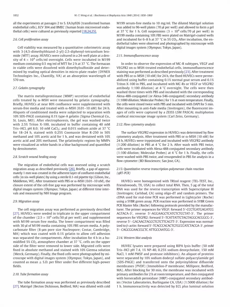
1852 W.-T. Weng et al. / Biochimica et Biophysica Acta 1840 (2014) 1850–1860
all the experiments at passages 2 to 5. EA.hy926 (transformed humanendothelial cells), ECV 304 and BMEC (human brain microvessel endo-thelial cells) were cultured as previously reported [18,24,25].
2.6. Cell proliferation assay
Cell viability was measured by a quantitative colorimetric assaywith 3-[4,5-dimethylthiazol-2-yl]-2,5-diphenyl-tetrazolium bro-mide (MTT) assay. HUVECs were cultured in a 24-well plate at a den-sity of 4 × 10⁴ cells/ml overnight. Cells were incubated in M199medium containing 0.5 mg/ml of MTT for 2 h at 37 °C. The formazanin viable cells were dissolved with dimethylsulfoxide and deter-mined by reading optical densities in micro-plate reader (DYNEXTechnologies Inc., Chantilly, VA) at an absorption wavelength of570 nm.
2.7. Gelatin zymography
The matrix metalloproteinase (MMP) secretion of endothelialcells treated by α-MSH were measured by gelatin zymography.Briefly, HUVECs at near 80% confluence were supplemented withserum-free media and treated with α-MSH (0.01–10 nM) for 24 h.Aliquots of conditioned media were subjected to separation with10% SDS-PAGE containing 0.1% type-A gelatin (Sigma Chemical Co.,St. Louis, MO). After electrophoresis, the gel was washed twicewith 2.5% Triton X-100, incubated in buffer containing 40 mMTris–HCl, pH 8.0; 10 mM CaCl2; and 0.01% sodium azide at 37 °Cfor 18–24 h, stained with 0.25% Coomassie blue R-250 in 50%methanol and 10% acetic acid for 1 h, and was destained with 10%acetic acid and 20% methanol. The gelatinolytic regions by MMPswere visualized as white bands in a blue background and quantifiedby densitometer.
2.8. Scratch wound healing assay
The migration of endothelial cells was assessed using a scratchmigration assay as described previously [26]. Briefly, a gap of approxi-mately 1mmwas created in the adherent layer of confluent endothelialcells (in six-well plates) by using a sterile 0.1-ml pipette tip (Gilson, Inc.,Middleton, WI). After treatment with PBS or α-MSH (0.01–10 nM), theclosure extent of the cell-free gap was performed by microscope withdigital images system (Olympus; Tokyo, Japan) at different time inter-vals and measured by NIH Image program.
2.9. Migration assay
The cell migration assay was performed as previously described[27]. HUVECs were seeded in triplicate in the upper compartmentof the chamber (2.5 × 10⁴ cells/50 μl per well) and supplementedwith M199 serum free media. The lower compartment was filledwith 30 μl of M199 media containing 10% FBS serum media. A poly-carbonate filter (8-μm pore size Nucleopore; Costar, Cambridge,MA) which was coated with 0.1% gelatin to allow cell adhesionwas separated the compartments. After incubation for 4 h in a hu-midified 5% CO2 atmosphere chamber at 37 °C, cells on the upperside of the filter were removed to lower side. Migrated cells werefixed in absolute methanol and stained with 10% Giemsa solution(Merck, Germany). Finally, the fixed cells were photographed by mi-croscope with digital images system (Olympus; Tokyo, Japan), andcounted as mean ± S.D. per filter under five different high-powerfields.
2.10. Tube formation assay
The tube formation assay was performed as previously described[27]. Matrigel (Becton Dickinson, Bedford, MA) was diluted with cold
M199 serum-free media to 10 mg/ml. The diluted Matrigel solutionwas added to 96-well plates (70 μl per well) and allowed to form a gelat 37 °C for 1 h. Cell suspensions (3 × 10⁴ cells/70 μl per well) inM199 media containing 10% FBS were plated on Matrigel-coated wellsand incubated for 6–8 h at 37 °C in 5% CO2. After incubation, the en-dothelial tubes were observed and photographed by microscope withdigital images system (Olympus; Tokyo, Japan).
2.11. Immunofluorescence assay
In order to observe the expression of MC-R subtypes, VEGF andVEGFR2 on α-MSH-treated endothelial cells, immunofluorescencestainingwas performed as described previously [23]. After treatmentwith PBS or α-MSH (10 nM) for 24 h, the fixed HUVECs were perme-abilized using buffer containing 0.1% normal goat serum and 0.1%Triton X-100 in PBS, and incubated with MC-Rs or VEGF or VEGFR2antibody 1:100 dilution) at 4 °C overnight. The cells were thenwashed three times with PBS and incubated with the correspondingAlexa-488-conjugated (or Alexa-546-conjugated) secondary antibody(1:1000 dilution;Molecular Probes) for 1 h at room temperature. Finally,the cells were rinsed twice with PBS and incubated with DAPI for 5 min.After mounting in anti-Fade media (Invitrogen), the fluorescent im-ages of cells were captured by a ZEISS LSM PASCAL multiphotonconfocal microscope image system (Carl Zeiss, Germany).
2.12. Flow cytometry analysis
The surface VEGFR2 expression in HUVECs was determined by flowcytometry analysis. After treatment with PBS or α-MSH (10 nM) for24 h, HUVECs were trypsinized and incubated with VEGFR2 antibody(1:200 dilution) in PBS at 4 °C for 2 h. After wash with PBS twice,cells were incubated with Alexa-488-conjugated secondary antibody(1:100 dilution; Molecular Probes) at 4 °C for 1 h. Finally, the cellswere washed with PBS twice, and resuspended in PBS for analysis in aflow cytometer (BD Biosciences; San Jose, CA).
2.13. Quantitative reverse transcription-polymerase chain reaction(qRT-PCR)
HUVECs were homogenized with TRIzol reagent (TEL-TEST, Inc.,Friendswoods, TX, USA) to collect total RNA. Then, 5 μg of the totalRNA was used for the reverse transcription with Superscriptase III(Invitrogen; Carlsbad, CA) using oligo-dT and random primers. ThecDNA used for real-time PCR was performed in Lightcycler (Roche)using a SYBR green assay. PCR reaction was performed in SYBR GreenPCR Master Mix (Roche) following protocols provided by the manufac-turer. The primer sequences for VEGF: forward 5′-CCCTGATGAGATCGAGTACA-3′, reverse 5′-AGGAAGCTCATCTCTCCTAT-3′. The primersequences for VEGFR2: forward 5′-TCATTATTCTAGTAGGCACGGCG-3′,reverse 5′- GACAAGTAGCCTGTCTTCAGTT-3′. Expression was normal-ized toβ-actin: forward 5′-TCACCCACACTGTGCCCATCTACGA-3′, primer5′- CAGCGGAACCGCTC ATTGCCAATGG-3′.
2.14. Western blot analysis
HUVEC lysates were prepared using RIPA lysis buffer (50 mMTris–HCl pH 7.4, 1% NP-40, 0.25% sodium deoxycholate, 150 mMNaCl, 1 mM PMSF and protease inhibitors). An aliquot of proteinswere separated by 10% sodium dodecyl sulfate-polyacrylamide gel(SDS-PAGE) and transferred onto the polyvinylidene difluoridemembranes (PVDF) (Immobilon-P membrane; Millipore, Bedford,MA). After blocking for 30 min, the membrane was incubated withprimary antibodies for 2 h at room temperature, and then conjugatedwith horseradish peroxidase (HRP)-conjugated secondary antibod-ies (Vector Laboratories, Burlingame CA, USA) (1:5000 dilution) for1 h. Immunoreactivity was detected by ECL plus luminal solution

1853W.-T. Weng et al. / Biochimica et Biophysica Acta 1840 (2014) 1850–1860
(Amersham Biosciences, Piscataway, NJ, USA). The immunobandintensities were quantified by densitometric scanning. The primaryantibodies used in this study were antibodies against MC1-5R, VEGF,VEGFR2, Akt, pAkt (Ser473), PTEN (1:1000 dilution) and β-actin(1:5000 dilution).
2.15. Enzyme-linked immunosorbent assay (ELISA)
To explore VEGF release from α-MSH-incubated HUVECs, we an-alyzed the VEGF concentrations of supernatant by VEGF ELISA kit(R&D Systems Inc.). After treatment with α-MSH (0.01–10 nM) for
Fig. 1. Inhibition of angiogenesis byα-MSH ex vivo and in vivo. (A) Effect ofα-MSHon themicroα-MSH (10 nM) and vessel sprout from various aorta sampleswas observed on day 7. n= 6; Scembryos. Embryos were treated with α-MSH (0.01–10 nM) at 6 hpf (n = 12 per group) thenphotographs of intersegmental vessels (ISV) fluorescence in Tg(fli-1:EGFP) zebrafish treated wfluorescence was quantified and expressed as mean ± SD percentages of control (n = 12; bo(SIV) fluorescence in Tg(fli-1:EGFP) zebrafish treated with α-MSH (0.1–10 nM) were analyvesicle-like structure. The SIV arcades were quantified and expressed as mean± SD (n= 12; bpictures of PBS- orα-MSH-treated CAMwere shown (left panel). Quantification analysis of the npanel). Bars = 3 mm. n = 6 per group. *, p b 0.05 and **, p b 0.01.
24 h, the cultured media of HUVECs were collected following proto-cols provided by manufacturer.
2.16. Measurement of cAMP
After treatment with α-MSH (0.01–10 nM) for 24 h, HUVECswere washed and lysed with 0.01 N HCl. The cellular cAMP expres-sion was measured by cAMP EIA kit (Cayman, Chemical Inc., USA)following protocols provided by the manufacturer. The amount ofcAMP in the cell samples was standardized to the amount of proteinin each sample as determined by the BCA protein assay kit.
vessel sprouting in aorta rings. Rat aortic rings placed inMatrigel were treatedwith PBS orale bar= 2mm. (B) Effect ofα-MSHon angiogenesis in transgenic Tg(fli-1:EGFP) zebrafishmonitored for imaging recording at various time intervals. At 48 hpf, the representativeith α-MSH (0.1–10 nM) were shown (40× magnification; Scale bars, 100 μm). The ISV
ttom left panel). At 72 hpf, the representative photographs of subintestinal vessel plexuszed (100× magnification; Scale bars, 50 μm). White asterisks indicated arcades in theottom right panel). (C) Effect of α-MSH on angiogenesis in CAM assay. The representativeewblood vessel growth (black arrows) in a defined areawas performedmean± SD (right
1854 W.-T. Weng et al. / Biochimica et Biophysica Acta 1840 (2014) 1850–1860
2.17. Statistical analysis
All values were expressed as mean ± standard deviation (SD). Apaired t test was used to assess the statistical differences between thegroups. The differences were considered statistically significant whenp was less than 0.05.
3. Results
3.1. α-MSH perturbs angiogenesis ex vivo and in vivo
We first employed the organotypic aortic rings to evaluate thefunction of α-MSH on angiogenesis in physiological conditions.
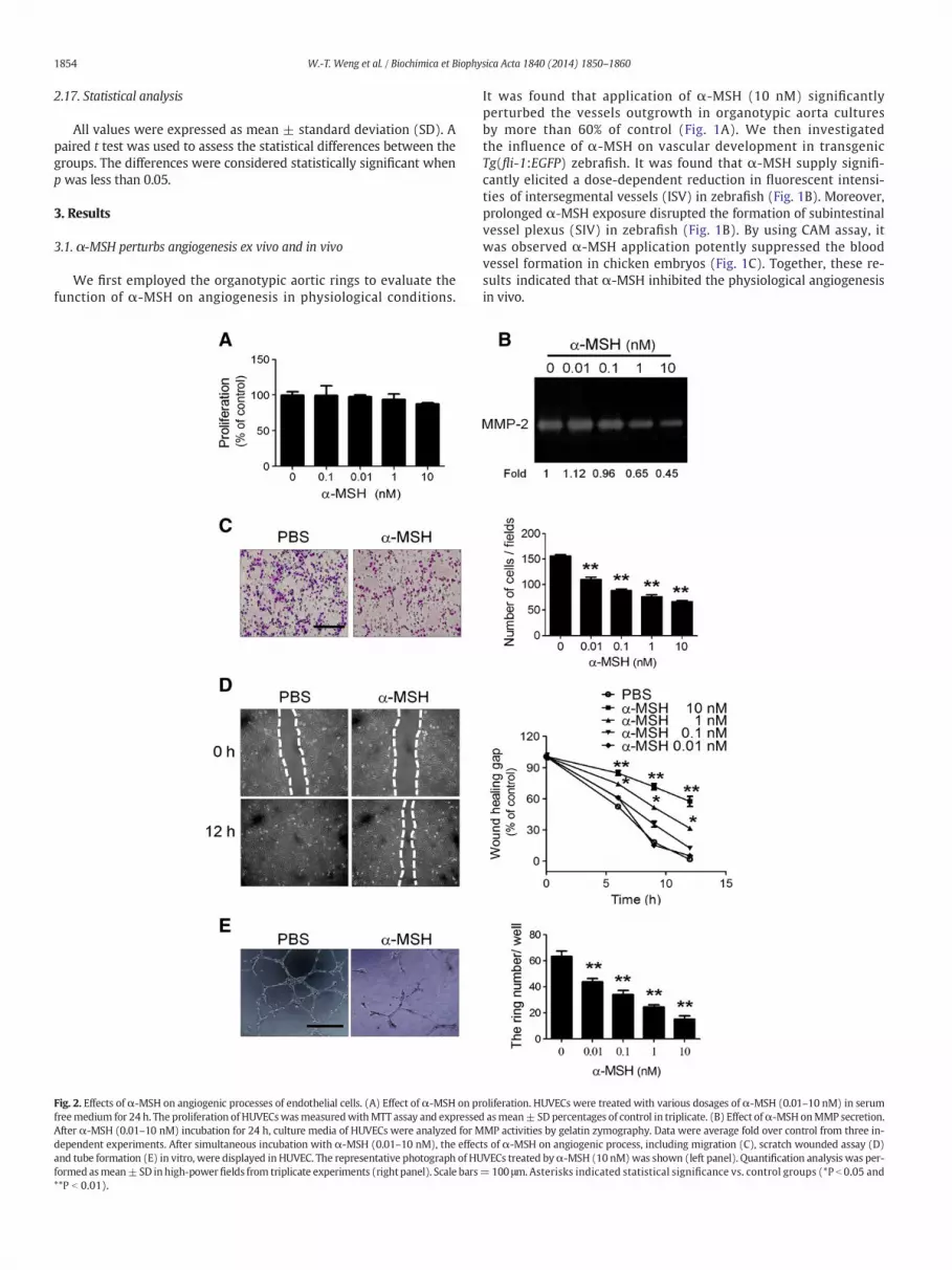
Fig. 2. Effects of α-MSH on angiogenic processes of endothelial cells. (A) Effect of α-MSH on prfreemedium for 24 h. The proliferation of HUVECswasmeasuredwithMTT assay and expresseAfter α-MSH (0.01–10 nM) incubation for 24 h, culture media of HUVECs were analyzed for Mdependent experiments. After simultaneous incubation with α-MSH (0.01–10 nM), the effecand tube formation (E) in vitro, were displayed in HUVEC. The representative photograph of HUformed asmean±SD inhigh-powerfields from triplicate experiments (right panel). Scale bars**P b 0.01).
It was found that application of α-MSH (10 nM) significantlyperturbed the vessels outgrowth in organotypic aorta culturesby more than 60% of control (Fig. 1A). We then investigatedthe influence of α-MSH on vascular development in transgenicTg(fli-1:EGFP) zebrafish. It was found that α-MSH supply signifi-cantly elicited a dose-dependent reduction in fluorescent intensi-ties of intersegmental vessels (ISV) in zebrafish (Fig. 1B). Moreover,prolonged α-MSH exposure disrupted the formation of subintestinalvessel plexus (SIV) in zebrafish (Fig. 1B). By using CAM assay, itwas observed α-MSH application potently suppressed the bloodvessel formation in chicken embryos (Fig. 1C). Together, these re-sults indicated that α-MSH inhibited the physiological angiogenesisin vivo.
oliferation. HUVECs were treated with various dosages of α-MSH (0.01–10 nM) in serumd asmean± SDpercentages of control in triplicate. (B) Effect ofα-MSH onMMP secretion.MP activities by gelatin zymography. Data were average fold over control from three in-ts of α-MSH on angiogenic process, including migration (C), scratch wounded assay (D)VECs treated byα-MSH (10 nM)was shown (left panel). Quantification analysis was per-=100 μm.Asterisks indicated statistical significance vs. control groups (*P b 0.05 and
1855W.-T. Weng et al. / Biochimica et Biophysica Acta 1840 (2014) 1850–1860
3.2. α-MSH suppresses multiple angiogenic processes in endothelial cells
We subsequently evaluated the effects of α-MSH on the distinctangiogenic steps, including proliferation, MMPs secretion, migrationand tube formation, in cultured endothelial cells. By MTT assay, it wasobserved that α-MSH (in physiological concentrations; 0.01–10 nM)had no significant effect on proliferation of endothelial cells (Fig. 2A).Despite lack of effect on endothelial proliferation, α-MSH potentlyinhibited the MMP-2 secretion as revealed by gelatin zymographyanalysis (Fig. 2B). In Boyden chamber assay, α-MSH dose-dependentlyattenuated the migration of endothelial cells with a half maximalinhibitory concentration (IC50) around 0.1–1 nM (Fig. 2C). Consistently,α-MSH significantly perturbed the healing of scratch wound in endo-thelial cells (Fig. 2D). Finally, α-MSH treatment dose-dependentlyabolished the formation of tube-like structure of HUVECs in Matrigelwith an IC50 around 1 nM (Fig. 2E). Together, these results indicatethat α-MSH suppresses MMPs secretion, migration and tube formationof endothelial cells without affecting the cell growth.
3.3. α-MSH reduces VEGF expression in endothelial cells attranscriptional level
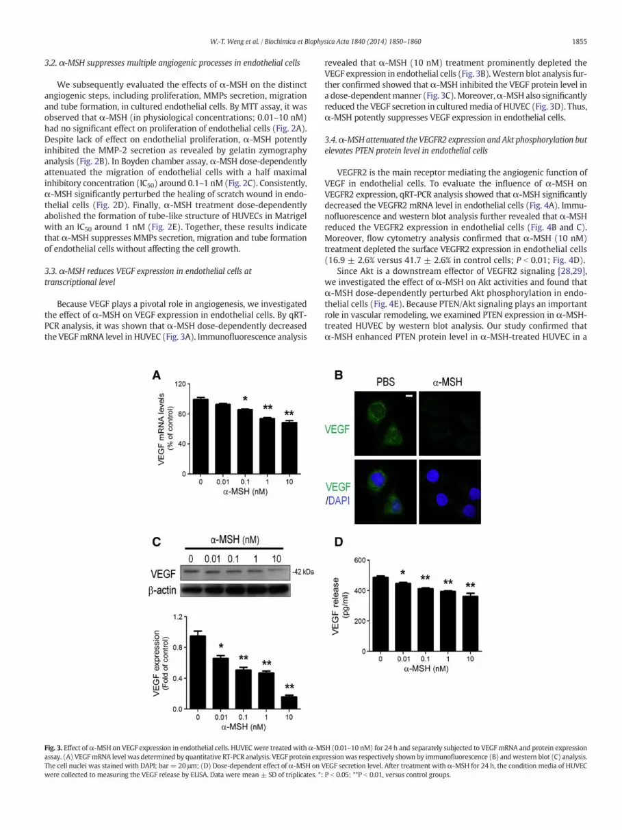
Because VEGF plays a pivotal role in angiogenesis, we investigatedthe effect of α-MSH on VEGF expression in endothelial cells. By qRT-PCR analysis, it was shown that α-MSH dose-dependently decreasedthe VEGFmRNA level in HUVEC (Fig. 3A). Immunofluorescence analysis
Fig. 3. Effect ofα-MSH on VEGF expression in endothelial cells. HUVEC were treated withα-MSassay. (A) VEGFmRNA level was determined by quantitative RT-PCR analysis. VEGF protein expThe cell nuclei was stained with DAPI; bar = 20 μm; (D) Dose-dependent effect of α-MSH on Vwere collected to measuring the VEGF release by ELISA. Data were mean ± SD of triplicates. *:
revealed that α-MSH (10 nM) treatment prominently depleted theVEGF expression in endothelial cells (Fig. 3B).Western blot analysis fur-ther confirmed showed that α-MSH inhibited the VEGF protein level ina dose-dependentmanner (Fig. 3C).Moreover,α-MSH also significantlyreduced the VEGF secretion in culturedmedia of HUVEC (Fig. 3D). Thus,α-MSH potently suppresses VEGF expression in endothelial cells.
3.4.α-MSH attenuated the VEGFR2 expression and Akt phosphorylation butelevates PTEN protein level in endothelial cells
VEGFR2 is the main receptor mediating the angiogenic function ofVEGF in endothelial cells. To evaluate the influence of α-MSH onVEGFR2 expression, qRT-PCR analysis showed that α-MSH significantlydecreased the VEGFR2 mRNA level in endothelial cells (Fig. 4A). Immu-nofluorescence and western blot analysis further revealed that α-MSHreduced the VEGFR2 expression in endothelial cells (Fig. 4B and C).Moreover, flow cytometry analysis confirmed that α-MSH (10 nM)treatment depleted the surface VEGFR2 expression in endothelial cells(16.9 ± 2.6% versus 41.7 ± 2.6% in control cells; P b 0.01; Fig. 4D).
Since Akt is a downstream effector of VEGFR2 signaling [28,29],we investigated the effect of α-MSH on Akt activities and found thatα-MSH dose-dependently perturbed Akt phosphorylation in endo-thelial cells (Fig. 4E). Because PTEN/Akt signaling plays an importantrole in vascular remodeling, we examined PTEN expression in α-MSH-treated HUVEC by western blot analysis. Our study confirmed thatα-MSH enhanced PTEN protein level in α-MSH-treated HUVEC in a
H (0.01–10 nM) for 24 h and separately subjected to VEGF mRNA and protein expressionressionwas respectively shown by immunofluorescence (B) andwestern blot (C) analysis.EGF secretion level. After treatment with α-MSH for 24 h, the condition media of HUVECP b 0.05; **P b 0.01, versus control groups.

Fig. 4. Effect ofα-MSH on VEGFR2 expression and Akt/PTEN signaling in HUVEC. (A) Effect ofα-MSH on VEGFR2mRNA expression. After treatmentwithα-MSH (0.01–10 nM) for 24 h inHUVECs, VEGFR2mRNA levelswere determined by quantitative RT-PCR analysis. Moreover, VEGFR2 protein levelswere shownby immunofluorescence (B) andwestern blot (C) analysis,respectively. The cell nucleiwere visualized using DAPI staining (blue). Scale bar=20 μm. (D) Flow cytometry analysis of surface VEGFR2 expression afterα-MSH treatment. Afterα-MSH(10 nM) treatment for 24 h, the cell surface VEGFR2 expression in endothelial cellswas analyzed by FACScan. (E) Effect ofα-MSHonAkt activation bywestern blot analysis. Quantificationindicated mean fold change compared with the control (n = 3). (F) Effect of α-MSH on PTEN expression by western blot analysis. Quantification indicated mean fold change comparedwith the control (n = 3). All data were mean ± SD of triplicates. *: P b 0.05; **P b 0.01, versus control groups.
1856 W.-T. Weng et al. / Biochimica et Biophysica Acta 1840 (2014) 1850–1860
dose-dependent manner (Fig. 4F). These data demonstrate that α-MSHinduces VEGFR2 down-regulation and a unique profile of Akt inactiva-tion and PTEN upregulation in α-MSH-treated HUVEC.
3.5. MC1-R and MC2-R are involved in α-MSH-induced angiogenesisinhibition in endothelial cells
α-MSH exerts its biological functions through the activationof MC-Rs. We first analyzed the expression of MC-R subtypes inendothelial cells by semi-quantitative RT-PCR and detected the
transcripts of MC1-R, MC2-R, MC4-R and MC5-R in endothelialcells (data not shown). Immunofluorescent analysis revealed thepresence of MC1-R, MC2-R, MC4-R and MC5-R, but not MC3-R, inHUVEC (Fig. 5A). Moreover, western blot analysis of a panel of humanendothelial cells (including EA.hy926, ECV304 and BMEC) showedsimilar results (Fig. 5B). Because cAMP mediates the MC-R signaling,we determined the effect of α-MSH on cAMP concentration in HUVECand found α-MSH dose-dependently stimulated the cAMP level inHUVEC (Fig. 5C). Thus, MC-R/cAMP signaling pathway was involved inα-MSH-mediated effects in endothelial cells.

Fig. 5. The profiles of melanocortin receptors expression and signaling of α-MSH-regulated angiogenesis inhibition in endothelial cells. The protein expression of MC-Rs in culturedendothelial cells was shown in immunofluorescence (A) and western blot (B) analysis, respectively. The cell nuclei were visualized using DAPI staining (blue). Scale bar = 10 μm.(C) Enzyme-linked immunosorbent assay of cAMP levels in α-MSH-incubated HUVECs. After α-MSH (0.01–10 nM) treatment for 24 h, cAMP expression in HUVECs was analyzed bycAMPEIAkit. All datawere expressed as themean±SDof triplicates. (D) Effect ofMC-Rs neutralization onα-MSH-inhibitedmigration. After treatmentwithα-MSH(10nM) or/combinedwith MC-Rs antibody (10 μg/ml), the migratory abilities were assessed by Boyden chamber assay; Data were mean ± SD in high-power fields from triplicate experiments. Scalebars = 100 μm. (D) Effect of MC-1R and MC2-R neutralization on α-MSH-induced inhibition of VEGF/VEGFR2/Akt signaling pathway. After pretreatment with MC1-R or MC2-Rantibody for 30 min, the Akt, pAkt, VEGF and VEGFR2 protein expression were examined by western blot analysis. Quantification indicated mean fold change compared with thecontrol (n = 3). *: P b 0.05; **P b 0.01, versus control groups.
1857W.-T. Weng et al. / Biochimica et Biophysica Acta 1840 (2014) 1850–1860
To delineate which MC-Rs were contributed to the anti-angiogenicsignaling ofα-MSH,we utilized variousMC-Rs' antibodies to investigatethe effect of antibody neutralization onα-MSH-induced inhibition ofendothelial migration. It was found that pretreatment with anti-
MC1-R and anti-MC2-R antibody significantly reversed the α-MSH-induced inhibition of endothelial migration by about 90% and 60%of control, respectively (P b 0.05; Fig. 5D). In contrast, application ofanti-MC4-R or ani-MC5-R had no such effect. Furthermore, western

Fig. 6. Effects of VEGF supply on α-MSH-induced angiogenesis in vitro and ex vivo. After simultaneous incubation with PBS or α-MSH (10 nM) or/combined with VEGF (10 ng/ml), theeffects of α-MSH on VEGF-induced proliferation (A), migration (B), tube formation (C) in vitro were observed in HUVEC. (D) The effects of α-MSH (10 nM) on the VEGF-inducedmicrovessel sprouting in aorta rings assay ex vivo. Data weremean± SD of quadruplicate experiments. (B),(C) Scale bars= 100 μm; (D) Scale bars=2mm. Asterisks indicate statisticalsignificance vs. control group (*P b 0.05 and **P b 0.01). NS indicates no statistical significance.
1858 W.-T. Weng et al. / Biochimica et Biophysica Acta 1840 (2014) 1850–1860
blot analysis revealed thatMC1-R andMC2-R neutralization significant-ly attenuated the α-MSH-induced VEGF/VEGFR2 down-regulation andAkt de-phosphorylation in endothelial cells (Fig. 5E). Interestingly,blocking MC1-R was more efficient in counteracting the function ofα-MSH than MC2-R blockade. These results suggest that both MC1-Rand MC2-R participate in α-MSH-induced angiogenesis inhibition.
3.6. Excessive VEGF supply fails to restore the α-MSH-inducedangiogenesis inhibition
Because α-MSH treatment depleted the cellular VEGF level in endo-thelial cells, we elucidated the influence of exogenous VEGF supply onangiogenic processes in α-MSH-treated endothelial cells and rat aortarings. It was found that, despite of its potent efficacy in stimulating theangiogenic processes in vitro, VEGF supply exerted marginal effect inalleviating the α-MSH-induced inhibition of proliferation (Fig. 6A),migration (Fig. 6B) and tube formation (Fig. 6C) in endothelial cells.Likewise, VEGF supply had no effect on α-MSH-induced inhibition ofvessel sprouting in aorta rings (Fig. 6D). Thus, α-MSH potently sup-presses the angiogenesis even in the presence of excessive VEGF.
4. Discussion
It has been recently established that gene delivery of POMC, the pre-cursor of α-MSH, suppressed the primary and metastatic melanomaand Lewis lung carcinoma through blockade of tumor angiogenesis[19,30,31]. Besides, prophylactic POMC gene transfer attenuated the
plasma leakage in lung during capsaicin-induced neurogenic inflamma-tion [32], and significantly suppressed angiogenesis in a rat model ofosteoarthritis [17]. The anti-neoplastic function of POMC therapy formultiple cancers seems to arise from blockade of tumor vasculatureby POMC-derived melanocortins, particularly α-MSH. The presentstudy first demonstrated that α-MSH at physiological concentration(0.01–10 nM) potently suppressed the vessels sprouting in organotypicaorta rings.Moreover, application ofα-MSHdose-dependently disruptedthe vessels development in zebrafish. It may seem paradoxical how ahuman hormone exerts function in fish at such a low concentration.However, previous studies showed that α-MSH at the concentrationaround nM was sufficient to stimulate melanosome translocationand pigmentation in zebrafish [33,34], suggesting the dose range(0.01–10 nM) ofα-MSH used in our study was adequate and function-al. Besides, given that melanocortin system is highly evolutionarilyconserved in vertebrates [35], human α-MSH may likely exert its anti-angiogenic signaling through zebrafish MC-Rs. Together with previ-ous studies, the present study indicates that α-MSH is an importantendogenous regulator for either the physiological or pathological an-giogenesis in vertebrates.
Our study brings important insights on how α-MSH regulates theangiogenesis at distinct angiogenic steps in vitro. Angiogenesis can bedivided at least into the following steps: MMP secretion, breakdownof endothelial barrier, endothelial migration, endothelial proliferation,and interaction with extracellular matrix/mural cells [12]. Unlikemany anti-angiogenic agents, α-MSH was not cytotoxic and exertedlittle influence on proliferation of stimulated endothelial cells at

1859W.-T. Weng et al. / Biochimica et Biophysica Acta 1840 (2014) 1850–1860
physiological concentrations. Although lacking of cytotoxicity,α-MSHpo-tently repressed various angiogenic processes, including MMP-2 release,migration and tube formation, with an IC50 in the range of 0.1–1 nM.Thus, the high anti-angiogenic efficacy of α-MSH plays a pivotalrole in the anti-tumor and anti-metastatic function of POMC therapy.
To our knowledge, the present study provided the first evidence forthe co-expression ofMC1-R,MC2-R,MC4-R andMC5-R in human endo-thelial cells since previous studies revealed onlyMC1-R in human endo-thelial cells [36,37].Moreover, we further delineated the involvement ofMC1-R and MC2-R, but not MC4-R and MC5-R, in the anti-angiogenicmechanism of α-MSH in endothelial cells. Given that α-MSH preferen-tially binds to MC1-R over MC4-R or MC5-R [38], it seemed reasonableto exclude MC4-R and MC5-R from α-MSH-mediating signaling in en-dothelial cells. Nevertheless, the role of MC2-R in transmitting α-MSHsignaling is intriguing because MC2-R binds only ACTH and has noaffinity for the other melanocortin peptides [39]. To address the roleof MC2-R, pilot study using MC2-R antibody failed to augment the effi-cacy of MC1-R antibody in counteracting α-MSH. Therefore, one likelymechanism is that MC2-R is not directly involved in, yet required forfully activation of MC1-R signaling by α-MSH. Further studies are war-ranted to elucidate the detailed mechanism.
The signaling mechanism underlying α-MSH-mediated angiogene-sis inhibition remains unclear. It has been known that melanocortinsexert their effects by via coupling to heterotrimeric G proteins, andfurther result in adenylase cyclase-dependent cAMP synthesis, proteinkinase A (PKA) activation. α-MSH has elucidated to act as a potent in-hibitor of LPS- or TNF-α-activated MC1-R-mediated NF-κB activationby maintaining the cytosolic IκBα in HDMECs [40]. This may implythat cAMP/PKA-dependent signaling transduction participate in theNF-κB regulation of α-MSH in endothelial cells. In the present study,we found α-MSH significantly reduced Akt activation to affect VEGF/VEGFR2 expression in a dose-dependent manner. Recently, the regula-tion of NF-κB activation through PKA/Akt-dependent pathway hasbeen explored in HUVEC [41]. Therefore, we speculate the insightsthat α-MSH inhibits VEGF/VEGFR2 gene expression by PKA/Akt/NF-κBthrough MC-Rs' activations.
The present study unveils that α-MSH inhibits angiogenesis bydownregulating VEGF/VEGFR2 expression in endothelial cells. How-ever, exogenous VEGF supply failed to reverse the anti-angiogeniceffects of α-MSH in HUVEC. It may implicate the involvement of othermechanisms contributing to the α-MSH-mediated anti-angiogenesisin HUVEC. It has been known that α-MSH reduces the LPS- or TNF-α-induced adhesion molecule expression, in human microvascularendothelial cells [40,42]. And the adhesion molecules may regulatethe spreading, migration, and cell–cell and cell–matrix adhesion, whichbelong to the angiogenesis processes. Therefore, inhibition of celladhesion may be one possible mechanism of α-MSH-mediated anti-angiogenesis in endothelial cells. Additionally, altered endothelin-1(ET-1) homeostasis may be also involved in the anti-angiogenic func-tion of α-MSH in endothelial cells. ET-1 is the most potent angiogenicfactor, and a reduction in ET-1 secretion might directly contribute tothe POMC-mediated angiogenesis inhibition in endothelial cells [18].Thus, we speculate that altered ET-1 homeostasismay be other possibil-ity mechanisms. Another possibility is the nitric oxide (NO) homeosta-sis. Endothelium-derived NO is required for the pro-angiogenic activityof growth factors such as VEGF. Autocrine NO production following ac-tivation of eNOS in endothelial cells promotes cell migration and orga-nization in tubes, a phenomenon participating in the first steps ofangiogenesis. Future studies are warranted to elucidate themechanismunderlying α-MSH-induced angiogenesis inhibition.
In conclusion, the present study has demonstrated that α-MSH in-hibits angiogenesis in vitro and in vivo by attenuating VEGF/VEGFR2/Akt signaling through MC1-R and MC2-R activation in endothelialcells. Our findings support the novel function of α-MSH as an endoge-nous angiogenesis inhibitor, which could be of therapeutic potentialfor pathological conditions due to VEGF/VEGFR2 activation.
Acknowledgements
Thisworkwas supported by grants from theNational Science Council,Taiwan (NSC-98-2320-B-110-004-MY3), Kaohsiung Armed ForcesGeneral Hospital (101-10), Kaohsiung Veterans General Hospital(VGHKS-99-036), and National Sun Yat Sen University-KaohsiungMedical University Joint Research Center (Kaohsiung, Taiwan). Wealso thank Dr. Yow-Ling Shiue for technical assistance to CAM assay.
References
[1] T. Brzoska, T.A. Luger, C.Maaser, C. Abels, M. Bohm, Alpha-melanocyte-stimulating hor-mone and related tripeptides: biochemistry, antiinflammatory and protective effectsin vitro and in vivo, and future perspectives for the treatment of immune-mediated in-flammatory diseases, Endocr. Rev. 29 (2008) 581–602.
[2] S. Gatti, G. Colombo, R. Buffa, F. Turcatti, L. Garofalo, N. Carboni, L. Ferla, L.R.Fassati, J.M. Lipton, A. Catania, alpha-Melanocyte-stimulating hormone protectsthe allograft in experimental heart transplantation, Transplantation 74 (2002)1678–1684.
[3] A. Catania, S. Gatti, G. Colombo, J.M. Lipton, Targeting melanocortin receptors as anovel strategy to control inflammation, Pharmacol. Rev. 56 (2004) 1–29.
[4] J.L. Rees, The melanocortin 1 receptor (MC1R): more than just red hair, Pigment CellRes. 13 (2000) 135–140.
[5] J.M. Lipton, A. Catania, Anti-inflammatory actions of the neuroimmunomodulatoralpha-MSH, Immunol. Today 18 (1997) 140–145.
[6] A.V. Savos, J.M. Gee, D. Zierath, K.J. Becker, alpha-MSH: a potential neuroprotectiveand immunomodulatory agent for the treatment of stroke, J. Cereb. Blood FlowMetab. 31 (2011) 606–613.
[7] Y.K. Yang, C.M. Harmon, Recent developments in our understanding ofmelanocortinsystem in the regulation of food intake, Obes. Rev. 4 (2003) 239–248.
[8] D. Giuliani, L. Minutoli, A. Ottani, L. Spaccapelo, A. Bitto, M. Galantucci, D. Altavilla, F.Squadrito, S. Guarini, Melanocortins as potential therapeutic agents in severe hyp-oxic conditions, Front. Neuroendocrinol. 33 (2012) 179–193.
[9] J. Folkman, Tumor angiogenesis: therapeutic implications, N. Engl. J. Med. 285(1971) 1182–1186.
[10] J. Folkman, Angiogenesis in cancer, vascular, rheumatoid and other disease, Nat.Med. 1 (1995) 27–31.
[11] P.A. D'Amore, Y.S. Ng, Won't you be my neighbor? Local induction of arteriogenesis,Cell 110 (2002) 289–292.
[12] F. Bussolino, A. Mantovani, G. Persico, Molecular mechanisms of blood vessel forma-tion, Trends Biochem. Sci. 22 (1997) 251–256.
[13] D.O. Bates, F.E. Curry, Vascular endothelial growth factor increases hydraulic conduc-tivity of isolated perfused microvessels, Am. J. Physiol. 271 (1996) H2520–H2528.
[14] N. Ferrara, Vascular endothelial growth factor: basic science and clinical progress,Endocr. Rev. 25 (2004) 581–611.
[15] N.M. Pandya, N.S. Dhalla, D.D. Santani, Angiogenesis—a new target for future thera-py, Vascul. Pharmacol. 44 (2006) 265–274.
[16] J. Dai, A.B. Rabie, VEGF: an essential mediator of both angiogenesis and endochon-dral ossification, J. Dent. Res. 86 (2007) 937–950.
[17] P.C. Shen, A.L. Shiau, I.M. Jou, C.H. Lee, M.H. Tai, H.Y. Juan, P.R. Lin, G.S. Liu, C.L. Wu,J.L. Hsieh, Inhibition of cartilage damage by pro-opiomelanocortin prohormoneoverexpression in a rat model of osteoarthritis, Exp. Biol. Med. (Maywood) 236(2011) 334–340.
[18] H.C. Lam, S.M. Kuo, M.J. Chuang, H.M. Keng, P.R. Lin, G.S. Liu, C.M. Hsu, S.L. Howng,M.H. Tai, Blockade of endothelin-1 release contributes to the anti-angiogenic effectby pro-opiomelanocortin overexpression in endothelial cells, Exp. Biol. Med. (May-wood) 231 (2006) 782–788.
[19] G.S. Liu, H.E. Tsai, W.T. Weng, L.F. Liu, C.H. Weng, M.R. Chuang, H.C. Lam, C.S. Wu, R.Tee, Z.H. Wen, S.L. Howng, M.H. Tai, Systemic pro-opiomelanocortin expression in-duces melanogenic differentiation and inhibits tumor angiogenesis in establishedmouse melanoma, Hum. Gene Ther. 22 (2011) 325–335.
[20] Y.S. Bee, S.J. Sheu, Y.L. Ma, H.C. Lin, W.T. Weng, H.M. Kuo, H.C. Hsu, C.H. Tang, J.C.Liou, M.H. Tai, Topical application of recombinant calreticulin peptide, vasostatin48, alleviates laser-induced choroidal neovascularization in rats, Mol. Vis. 16(2010) 756–767.
[21] N.D. Lawson, B.M. Weinstein, In vivo imaging of embryonic vascular developmentusing transgenic zebrafish, Dev. Biol. 248 (2002) 307–318.
[22] D. Ribatti, B. Nico, A. Vacca, M. Presta, The gelatin sponge-chorioallantoic membraneassay, Nat. Protoc. 1 (2006) 85–91.
[23] H.M. Kuo, C.Y. Lin, H.C. Lam, P.R. Lin, H.H. Chan, J.C. Tseng, C.K. Sun, T.F. Hsu, C.C. Wu,C.Y. Yang, C.M. Hsu, M.H. Tai, PTEN overexpression attenuates angiogenic processesof endothelial cells by blockade of endothelin-1/endothelin B receptor signaling,Atherosclerosis 221 (2010) 341–349.
[24] T. Kumagai, T. Ishino, Y. Nakagawa, Acidic sphingomyelinase induced by electro-philes promotes proinflammatory cytokine production in human bladder carcino-ma ECV-304 cells, Arch. Biochem. Biophys. 519 (2012) 8–16.
[25] P.M. Rood, J. Calafat, A.E. von dem Borne, W.R. Gerritsen, C.E. van der Schoot,Immortalisation of human bone marrow endothelial cells: characterisation of newcell lines, Eur. J. Clin. Invest. 30 (2000) 618–629.
[26] N. Zhu, R. Lalla, P. Eves, T.L. Brown, A. King, E.H. Kemp, J.W. Haycock, S. MacNeil,Melanoma cell migration is upregulated by tumour necrosis factor-alpha andsuppressed by alpha-melanocyte-stimulating hormone, Br. J. Cancer 90 (2004)1457–1463.

1860 W.-T. Weng et al. / Biochimica et Biophysica Acta 1840 (2014) 1850–1860
[27] M.H. Tai, S.M. Kuo, H.T. Liang, K.R. Chiou, H.C. Lam, C.M. Hsu, H.J. Pownall, H.H. Chen,M.T. Huang, C.Y. Yang, Modulation of angiogenic processes in cultured endothelialcells by low density lipoproteins subfractions from patients with familial hypercho-lesterolemia, Atherosclerosis 186 (2006) 448–457.
[28] B.H. Jiang, L.Z. Liu, AKT signaling in regulating angiogenesis, Curr. CancerDrug Targets 8(2008) 19–26.
[29] I. Shiojima, K.Walsh, Role of Akt signaling in vascular homeostasis and angiogenesis,Circ. Res. 90 (2002) 1243–1250.
[30] H.E. Tsai, L.F. Liu, G.J. Dusting, W.T. Weng, S.C. Chen, M.L. Kung, R. Tee, G.S. Liu, M.H.Tai, Pro-opiomelanocortin gene delivery suppresses the growth of established Lewislung carcinoma through a melanocortin-1 receptor-independent pathway, J. GeneMed. 14 (2012) 44–53.
[31] G.S. Liu, L.F. Liu, C.J. Lin, J.C. Tseng,M.J. Chuang, H.C. Lam, J.K. Lee, L.C. Yang, J.H. Chan, S.L.Howng, M.H. Tai, Gene transfer of pro-opiomelanocortin prohormone suppressed thegrowth and metastasis of melanoma: involvement of alpha-melanocyte-stimulatinghormone-mediated inhibition of the nuclear factor kappaB/cyclooxygenase-2 pathway,Mol. Pharmacol. 69 (2006) 440–451.
[32] G.S. Liu, H.T. Huang, C.J. Lin, J.Y. Shi, L.F. Liu, R.T. Tseng, W.T. Weng, H.C. Lam, Z.H.Wen, T.L. Cheng, K.S. Hsu,M.H. Tai, Prophylactic proopiomelanocortin expression al-leviates capsaicin-induced neurogenic inflammation in rat trachea, Shock 32 (2009)645–650.
[33] T.Y. Choi, J.H. Kim, D.H. Ko, C.H. Kim, J.S. Hwang, S. Ahn, S.Y. Kim, C.D. Kim, J.H. Lee,T.J. Yoon, Zebrafish as a new model for phenotype-based screening of melanogenicregulatory compounds, Pigment Cell Res. 20 (2007) 120–127.
[34] D.W. Logan, S.F. Burn, I.J. Jackson, Regulation of pigmentation in zebrafish melano-phores, Pigment Cell Res. 19 (2006) 206–213.
[35] J.R. Metz, J.J. Peters, G. Flik, Molecular biology and physiology of the melanocortinsystem in fish: a review, Gen. Comp. Endocrinol. 148 (2006) 150–162.
[36] T.E. Scholzen, T. Brzoska, D.H. Kalden, M. Hartmeyer, M. Fastrich, T.A. Luger, C.A.Armstrong, J.C. Ansel, Expression of functional melanocortin receptors andproopiomelanocortin peptides by human dermal microvascular endothelial cells,Ann. N. Y. Acad. Sci. 885 (1999) 239–253.
[37] T.E. Scholzen, D.H. Kalden, T. Brzoska, T. Fisbeck, M. Fastrich, M. Schiller, M. Bohm, T.Schwarz, C.A. Armstrong, J.C. Ansel, T.A. Luger, Expression of proopiomelanocortinpeptides in human dermal microvascular endothelial cells: evidence for a regulationby ultraviolet light and interleukin-1, J. Invest. Dermatol. 115 (2000) 1021–1028.
[38] P.M. Holloway, H.K. Smith, D. Renshaw, R.J. Flower, S.J. Getting, F.N. Gavins, Targetingthemelanocortin receptor system for anti-stroke therapy, Trends Pharmacol. Sci. 32(2011) 90–98.
[39] M.P. Corander, M. Fenech, A.P. Coll, Science of self-preservation: how melanocortinaction in the brain modulates body weight, blood pressure, and ischemic damage,Circulation 120 (2009) 2260–2268.
[40] T.E. Scholzen, C. Sunderkotter, D.H. Kalden, T. Brzoska, M. Fastrich, T. Fisbeck, C.A.Armstrong, J.C. Ansel, T.A. Luger, Alpha-melanocyte stimulating hormone preventslipopolysaccharide-induced vasculitis by down-regulating endothelial cell adhesionmolecule expression, Endocrinology 144 (2003) 360–370.
[41] S. Balwani, R. Chaudhuri, D. Nandi, P. Jaisankar, A. Agrawal, B. Ghosh, Regulation ofNF-kappaB activation through a novel PI-3K-independent and PKA/Akt-dependentpathway in human umbilical vein endothelial cells, PLoS One 7 (2012) e46528.
[42] D.H. Kalden, T. Scholzen, T. Brzoska, T.A. Luger, Mechanisms of the antiinflammatoryeffects of alpha-MSH. Role of transcription factor NF-kappa B and adhesionmoleculeexpression, Ann. N. Y. Acad. Sci. 885 (1999) 254–261.


