Embed Size (px)
Citation preview

A Failure to Normalize Biochemical and Metabolic Insults DuringMorphine Withdrawal Disrupts Synaptic Repair in MiceTransgenic for HIV-gp120
Veera Venkata Ratnam Bandaru, Neha Patel, Ostefame Ewaleifoh, and Norman J. HaugheyDepartment of Neurology, Division of Neuroimmunology, Johns Hopkins University School ofMedicine, Meyer 6-109, 600 North Wolfe Street, Baltimore, MD 21287, USANorman J. Haughey: [email protected]
AbstractDrug abuse in HIV-infected individuals accelerates the onset and progression of HIV-associatedneurocognitive disorders (HAND). Opiates are a class of commonly abused drugs that haveinteractive effects with neurotoxic HIV proteins that facilitate glial dysfunction, neuronal damageand death. While the combined effects of neurotoxic HIV proteins and morphine have beenextensively studied in the setting of chronic and acute morphine use, very little in known about theeffects of HIV proteins during drug withdrawal. Since opiate withdrawal can induce considerableneuronal stress, we determined the effects of opiates (morphine) on brain redox balance,sphingolipid metabolism and synaptic integrity during both chronic and withdrawal conditions innon-transgenic mice (nTg), and in mice transgenic for the HIV-coat protein gp120 (gp120tg). InnTg mice, we found that chronic morphine increased brain oxidative capacity and inducedsynaptic damage that was largely reversed during drug withdrawal. Gp120tg mice showed asimilar response to chronic morphine, but the diminished oxidative capacity and synaptic damagefailed to normalize during drug withdrawal. In nTg mice, brain sphingolipid content was notaffected by morphine during chronic or withdrawal conditions. In gp120tg mice there was abaseline perturbation in sphingolipid metabolism that manifest as decreased sphingomyelin withaccumulations of the bioactive lipid ceramide. Sphingolipid metabolism was highly reactive tomorphine in gp120tg mice. Chronic morphine increased sphingomyelin content with a consequentreduction in ceramide. During drug withdrawal, these effects reversed, and sphingomyelin levelswere reduced with consequent increases of ceramide. We interpret these findings to suggest thatneuronal repair during morphine withdrawal is inhibited in the setting of gp120 by mechanismsthat involve sustained oxidative insult and accumulations of the highly reactive intermediateceramide.
KeywordsHIV; HAND; Morphine; Opiates; Neuron; Withdrawal; Oxidative stress; Sphingomyelin;Ceramide; Synapse; PSD95
© Springer Science+Business Media, LLC 2011
Correspondence to: Norman J. Haughey, [email protected].
Conflict of interest The authors have no conflicts of interest to report.
NIH Public AccessAuthor ManuscriptJ Neuroimmune Pharmacol. Author manuscript; available in PMC 2012 August 20.
Published in final edited form as:J Neuroimmune Pharmacol. 2011 December ; 6(4): 640–649. doi:10.1007/s11481-011-9289-0.
NIH
-PA Author Manuscript
NIH
-PA Author Manuscript
NIH
-PA Author Manuscript

IntroductionHIV-infected people who also abuse opiates often show faster rates of disease progressionincluding early and rapidly progressing HIV-Associated Neurocognitive Disorders (HAND).Heroin undergoes first pass metabolism in liver to be converted to morphine, makingmorphine the most commonly used agent to study opiate effects in both in vitro and in vivoneurological systems. Nontoxic concentrations of morphine have a variety of effects on CNSfunction that include alterations in cytokine balance, blood brain barrier (BBB) permeabilityand synaptic simplification. Morphine increases inflammatory cytokine release from gliawith consequent reductions in the expression of several tight junctional proteins includingZO-1, Jam-2, occludin and P-glycoprotein expression. These perturbations increase theendothelial adhesion of monocytes and the transendothelial migration of these cells acrossthe BBB where they can modify the biochemical environment in brain parenchyma(Mahajan et al. 2008; Stefano et al. 1998a, b). Morphine also has direct effects on brainresident cells and directly interacts with astrocytes to increase their activation state, anddecreases expression of the protective cytokines IL-8 and MIP-β (Mahajan et al. 2002;Bruce-Keller et al. 2008). Although morphine appears to not induce a generalized death ofneurons, there is evidence that morphine induces the death of GABAergic interneurons,induces a neuronal stress response in cortical and striatal neurons and reduces spine density(Sultana et al. 2010; Fitting et al. 2010).
In addition to direct effects on brain resident cells,morphine potentiates the glial response toa variety of stimuli including HIV-proteins (Hauser et al. 2000; Gurwell et al. 2001). Forexample, morphine in combination with the HIV trans-acting protein Tat perturbsproteosome activity, induces the generation of reactive oxygen species (ROS), increasesprotein oxidation and increases expression of the pro-inflammatory cytokines MCP-1,RANTES and IL-6, although a reduced expression of TNFα, IL-6 and MCP-1 has also beenreported in microglia stimulated with Tat and morphine (El-Hage et al. 2005; Turchan-Cholewo et al. 2009). The combined effects of morphine and Tat induces death of selectpopulations of astrocytes, reduces the survival of glial progenitors, and induces a phenotypeconsistent with activation of apoptotic pathways in oligodendrocytes (Khurdayan et al.2004; Hauser et al. 2009). Likewise, morphine has been shown to enhance neuronal deathinduced by the HIV-1 Tat protein (Gurwell et al. 2001). Less is known about the potentialinteractive effects of morphine with the HIV-coat protein gp120. HIV-gp120 may enhancethe effects of opiates by up-regulating mu opioid receptors on macrophages and glia throughautocrine/paracrine actions that involve TNF-α (Beltran et al. 2006; Chang et al. 2007).Morphine has also been shown to enhance the cytotoxicity of gp120 through p38 mitogen-activated protein kinase signaling, and by increasing the expression of CCR5 and alteringthe function of CXCR4 (Mahajan et al. 2002, 2008; Pitcher et al. 2010; Hu et al. 2005).
Since opiate abuse often involves cycles of intoxication followed by withdrawal, the effectsof morphine withdrawal are likely to contribute to neuronal damage in HIV-infectedindividuals. However, the vast majority of research published to date has used combinationsof HIV proteins and morphine to study interactive effects that occur during acute or chronicmorphine exposure. Withdrawal from morphine is accompanyed by increased neuronalsensitivity to stress, suggesting that neurons would be highly sensitized to the toxic effectsof HIV proteins during drug withdrawal. Consistent with this notion, morphine withdrawalhas been shown to sensitize rodents to LPS-induced lethality by increasing the production ofTNF-α, and by priming proapoptotic pathways in cortex and hippocampus that include Fas,Bad, caspases-8 and −3 signaling (Emeterio et al. 2006). In neurons, withdrawal frommorphine increases the phosphorylation of NMDA receptor subunits NR1 and NR2B, andinduces activation of ERK1/2, calmodulin-dependent kinase II, and cAMP response elementbinding proteins (Liu et al. 2010), demonstrating that neuronal signaling can be dramatically
Bandaru et al. Page 2
J Neuroimmune Pharmacol. Author manuscript; available in PMC 2012 August 20.
NIH
-PA Author Manuscript
NIH
-PA Author Manuscript
NIH
-PA Author Manuscript

modified during morphine withdrawal. In this study we sought to determine the interactiveeffects of the HIV-1 protein gp120, morphine intoxication and drug withdrawal on neuronalstress and synaptic integrity in vivo, using mice transgenic for the HIV-coat protein gp120.
MethodsAnimals and experimental treatments
Six month old C57BL-6 non-transgenic (nTg) mice, and C57BL-6 mice transgenic forgp120 (gp120tg) were used in these studies. Gp120 mice were obtained from Dr. LennartMuke (Gladstone Institute) and express a GFAP promoter driven HIV-1 env gene(HIV-1LAV: Toggas et al. 1994). Morphine was administered by subcutaneous implantationof time-release 25 mg pellets (NIDA, Rockville, MD). These pellets deliver 5 mg/daymorphine for up to 5 days (Bruce-Keller et al. 2008). Mice were sacrificed at 4 days post-implant for studies of morphine intoxication and at 7-days post-implant for studies on drugwithdrawal. A total of 6 mice for each experimental group were used for these studies.Brains were rapidly removed and snap-frozen for biochemical and protein studies.
Fluorometric enzymatic assaysA Cobas Fara II centrifugal fluorometric-analyzer (Roche Diagnostics, Montclair, NJ) wasused to detect and quantify the presence of catalase, super oxide dismutase (SOD),glutathione and uric acid.
GlutathioneThe glutathione assay (OXIS Research, Portland Oregon) measures the amount ofendogenous glutathione using a single substitution reaction in which the thiol group ofglutathione in the sample replaces a halogen on R2 (4-clorolo-1 –methyl- 7- tri fluromethyl-quinolinium methylsulfate) forming a thioether. Under alkaline conditions (pH >13.4), R3then transforms the substitution product (thioester) into a chromophoric thione. The thioneformed has maximal absorbance at 400 nm. The amount of endogenous oxidized glutathione(GSSG) was detected using the enzyme glutathione reductase, and the cofactor NADPH.This assay introduces a radical scavenger M2VP (1-Methyl −2- thioethyl 2-pyidinium) saltthat stabilizes GSH in the sample, ensuring that it is not detected as interference when GSSGis measured. To accomplish this, M2VP forms a stable UN reacting compound withendogenous GSH leaving only GSSG in the sample. Meta-phosphoric acid (5%) was addedto the mixture and a 20 fold dilution of sample was made using the GSSG buffer.Glutathione reductase enzyme and NADPH were mixed in a 1:1:1:1:1 ratio. In this step theendogenous GSSG is converted back to GSH and detected at 412 nm as NADPH that isoxidized to NADP.
Super oxide dismutaseThe SOD assay (Fluka, Switzerland) catalyzes the dismutation of super oxide anions intohydrogen peroxide and elemental oxygen. In this reaction, super oxide anions are producedby the action of xanthine oxidase that converts xanthine to uric acid. Xanthine oxidase alsoconverts the tetrazolium salt (2-4-Iodophenyl-3-4-Nitrophenyl-5-2, 4 –disulfophenyl-2H-Tetrazolium salt) to a water soluble fomazan dye which absorbs light at 550 nm.
Uric acidThis assay uses uricase to produce hydrogen peroxide from uric acid (Equal DiagnosticsLimited, Charlottetown, Canada). The hydrogen peroxide is then converted to a phenoliccompound derivative-quinoneimine, by reacting with (4-dicholoro-2-hydroxy benzene as
Bandaru et al. Page 3
J Neuroimmune Pharmacol. Author manuscript; available in PMC 2012 August 20.
NIH
-PA Author Manuscript
NIH
-PA Author Manuscript
NIH
-PA Author Manuscript

catalyzed by peroxidase. The red colored quinoneimine dye has a maximum absorbance at520 nm.
CatalaseThe catalase assay system (Sigma, USA) is based on the ability of hydrogen peroxide tooxidize an azo chromogen with an accompanying increase in the absorbance to 410–420 nm.In the presence of catalase, the amount of hydrogen peroxide is reduced, thus inhibiting theoxidation of the azo chromogen. This inhibition of chromogen oxidation is thus proportionalto the activity of catalase present in the sample.
Total oxidationIn the total oxidation assay (Applied bioanalytical labs, Sarasota, FL), antioxidants in thesample suppress the oxidation of ABTS (2,2′-azino-di-[3-ethylbenzthiazoline sulphonate])to ABTS+ by metmyoglobin. The amount of ABTS+ is measured by reading absorbance at405 nm. The assay relies on the ability of antioxidants in the sample to inhibit the oxidationof ABTS (2,2′-azino-di-[3-ethylbenzthiazoline sulphonate]) to ABTS+ bymetmyoglobin.Antioxidants present in the sample suppress the absorbance to a degree proportional to theirconcentration. Thus, more antioxidant activity results in less fluorescence.
ImmunoblottingWestern blots were conducted using published procedures (Tabatadze et al. 2010).Hippocampal tissues were homogenized in ice-cold buffer containing 20 mM Tris-HCl (pH7.4), 50 mM NaF, 0.32 M sucrose, 2 mM EDTA, 2 mM EGTA, 0.2 mM sodiumorthovanadate, 1 mM PMSF and protein inhibitor cocktail (Roche diagnostics GmbH,Germany). Proteins (10 µg of total protein per lane) were separated by sodium dodecylsulfate-polyacrylamide gel electrophoresis and transferred to nitrocellulose membranes.Membranes were blocked in Tris-buffered saline (TBS) containing 10 mM Tris-HCl [pH7.4], 150 mM NaCl and 0.1% Tween 20 (TBS-T) for 1 h and incubated with a antibodies tothe post synaptic density-95 protein (PSD95, 1:5000, Oncogene, Cambridge, MA) and β-actin (1:1000, Sigma, St. Louis, MO) overnight at 4°C. Following incubations with primaryantibodies, membranes were washed in TBS and TBS-T and incubated at room temperaturefor 1 h with HRP-conjugated secondary antibodies (1:5000, Goat anti-mouse IgG; 1:5000,Goat anti-rabbit IgG, Promega, WI). Immunoreactivity was visualized using Pico WestSuperSignal (Pierce, Rockford, IL). Optical densities were quantified using NIH ImageJsoftware.
Mass spectrometric measurement of sphingolipidsCrude total lipid content was extracted from brain tissues using a modified Bligh and Dyerprocedure (Bandaru et al. 2007). Each sample tissue sample was weighed and homogenizedat room temperature in deionized water (10 volumes to weight). Lipid content was extractedby the addition of methanol containing 53 mM ammonium formate (3 volumes) with 17 ng/ml C12:0 ceramide and C12:0 sphingomyelin as internal standards, and vortexed followedby chloroform (4 volumes). The mixture was vortexed and centrifuged at 1,000 g for 10min. The chloroform was removed and dried in a vacuum dryer, and stored at −80°C. Thesample was resuspended in 100% methanol for analysis by LC/MS/MS. All extractions wereperformed using borosilicate-coated glass tubes, pipettes, and injectors to reduce thepotential loss of lipids through interactions with plastic.
ESI/MS/MS analyses were performed using methods similar to those used in previousstudies (Haughey et al. 2004; Bandaru et al. 2007). Individual molecular species ofsphingomyelin and ceramide (C12:0 and C16:0-C26:1), were detected and quantified by LC/
Bandaru et al. Page 4
J Neuroimmune Pharmacol. Author manuscript; available in PMC 2012 August 20.
NIH
-PA Author Manuscript
NIH
-PA Author Manuscript
NIH
-PA Author Manuscript
MS/MS using multiple reaction monitoring (MRM). The procedure is based on highperformance liquid chromatography (HPLC) for temporal resolution of compounds, withsubsequent introduction into the mass spectrometer for detection and quantification by mass/charge. Samples were injected using a PAL autosampler into a PerkElmer HPLC equippedwith a 2.6 µm, C18, 100 Å 50×2.1 mm column for ceramides and a 5 µm, C18, 100 Å 100×2mm column for sphingomyelins (Phenomenex, Torrance, CA). The sample was eluted at 0.4mL min−1 for ceramides and 1 ml min−1 for sphingomyelins. The LC column was first pre-equilibrated for 0.5 min with the first mobile phase consisting of 85% methanol, 15% H20,and 5 mM ammonium formate. The column was then eluted with the second mobile phaseconsisting of 99% methanol, 1% formic acid, and 5 mM ammonium formate. The elutedsample was injected into the ion source, and the detection, and quantitation of each analyteis carried out by ESI/MS/MS in MRM mode monitoring the parent compound and productsby ion scan. Method development for the quantitative detection of each analyte wasaccomplished with the aid of reference standards for sphingomyelins and ceramides fromAvanti Polar Lipids (Alabaster, AL). Slight differences in extraction or mass spectrometerefficiencies were normalized using internal standards (ceramide C12:0 and sphingomyleinC12:0) with molecular structures similar to the analytes but not found in mammals. Areaunder the curve for each analyte was defined individually using the Analyst 1.4.2 softwarepackage.
ResultsMice transgenic for gp120 fail to normalize oxidative capacity during morphine withdrawal
Gp120 protein expression was detected in gp120tg mice and not in nTg mice (Fig. 1a).Morphine or placebo tablets were transplanted subcutaneously into 6-month oldnontransgenic (nTg) and gp120-transgenic (gp120tg) mice, and rodents were sacrificed ondays 4 and 7 post-implant. These time points were chosen to mimic chronic and drugwithdrawal conditions. On day 4 post-implant, morphine levels in plasma were 2.7–3.2 ng/ml and were not different in nTg compared with gp120tg mice. Morphine was undetectable7 days post-implant, and was not detected in animals administered placebo (Fig. 1b, c). Aseries of antioxidant and pro-oxidant measures were used to determine redox capacity inthese rodents. Non-transgenic (nTg) and gp120 transgenic (gp120tg) mice administeredplacebos had a similar total oxidation capacity, superoxide dismutase (SOD) activity, uricacid levels and ratios of reduced glutathione (GSH) to oxidized glutathione (Fig. 2a–d).After 4 days of morphine there was a 24.5 ± 5% decrease in the oxidation capacity of nTgmice, and a 41.4±4% decrease in gp120tg mice (Fig. 2a). SOD activity decreased by19.8±6% in nTg mice, and by 21.8±3% in gp120tg mice (Fig. 2b). Uric acid levelsdecreased by 10.0±4% nTg, and by 16.2±3% gp120tg mice (Fig. 2c). The ratio of GSH toGSSG decreased by 46.8±12% in nTg mice and by 55.9±5% in gp120tg mice following 4days of morphine exposure (Fig. 2d). These data show that there were no significantbaseline differences in the antioxidant capacities of gp120tg compared with nTg mice, andthat morphine increased oxidative stress to a similar extent in nTg and gp120tg mice.
During morphine withdrawal in nTgmice, total antioxidant capacity, SOD activity, uric acidlevels, but not the GSH/GSSG ratio normalized (Fig. 2e–h). However, in gp120tg mice, thetotal antioxidant capacity, SOD activity, and the GSH/GSSG ratio did not recover duringmorphine withdrawal (Fig. 2e, f, h). Uric acid levels did normalize in gp120tg mice (Fig.2g). These data demonstrate that morphine reduced endogenous antioxidant defenses, with aconsequent accumulation of free radicals. During morphine withdrawal, oxidative balancelargely normalized in nTg mice, but failed to normalize in gp120tg mice.
Bandaru et al. Page 5
J Neuroimmune Pharmacol. Author manuscript; available in PMC 2012 August 20.
NIH
-PA Author Manuscript
NIH
-PA Author Manuscript
NIH
-PA Author Manuscript
Morphine induced accumulations of sphingomyelin are metabolized to ceramide during drugwithdrawal in mice transgenic for gp120
There were baseline differences in sphingomyelin and ceramide balance in gp120tgcompared with nTg mice. In mice administered placebo, all sphingomyelin speciesmeasured (C16:0–C26:0) were decreased, and all (C18:0–C24:0) but one (C16:0) ceramidespecies were increased in gp120tg compared with nTg mice (Fig. 3a–b). These data areconsistent with previous reports that accumulations of ceramide are induced by gp120application onto cultured neurons (Haughey et al. 2004; Jana and Pahan 2004). We nextdetermined sphingolipid and sterol levels in brains of nTg and gp120tg mice during chromicmorphine administration and during morphine withdrawal. During chronic morphineadministration, levels of all sphingomyelin and ceramide species were unchanged in nTgmice compared with placebo (Fig. 3a–b). In gp120tg mice, chronic morphine administrationresulted in increases of multiple sphingomyelin species (C16:0–C22:1, C26:1) and decreasesin several long chain ceramides (C16:0–C20:0) (Fig. 3a–b). During morphine withdrawal ingp120tg mice, sphingomyelin levels either normalized (C16:0–C22:0) or decreased (C24:0–C26:0) (Fig. 3c) and levels of all ceramides measured were increased (C16:0–C24:0) (Fig.3d) compared to gp120tg mice administered placebo. These data demonstrate thatsphingolipid metabolism is perturbed in gp120tg mice, and these pathways are sensitive tothe effects of chronic morphine and drug withdrawal.
Evidence for synaptic damage in gp120 mice administered morphine
Sphingolipids are highly enriched in brain and play complex roles in regulating neuronalexcitability. The sphingolipids, sphingomyelin, ceramide and sphingosine are regulators ofsynaptic functions, and have been shown to play important roles in synapse formation,neurotransmitter release and synaptic plasticity (Wheeler et al. 2009; Yang 2000; Brann etal. 1999; Ping and Barrett 1998; Inokuchi et al. 1998; Furuya et al. 1998; Furukawa andMattson 1998; Ito and Horigome 1995). Disturbances in sphingolipid metabolism that resultin the accumulations of ceramide have been associated with synaptic damage and activationof death pathways (see Haughey 2010 for a recent review).
We therefore quantified the postysynapticmarker PSD95 to determine if there werealterations in synaptic integrity associated with chronic morphine administration ormorphine withdrawal. Chronic morphine administration decreased PSD95 levels by63.0±10.4% in nTg mice (Fig. 4a), consistent with reports that morphine alone reducesneuronal spine density (Fitting et al. 2010). During morphine withdrawal in nTg mice,PSD95 levels rapidly recovered to levels comparable with nTg mice administered placebo(Fig. 4b). Chronic morphine administration in gp120tg mice resulted in a 42.8±8.1%decrease of PSD95 compared with gp120tg mice administered placebo (Fig. 4c). PSD95 didnot recover during morphine withdrawal in gp120tg mice and remained decreased by46.1±4.3% (Fig. 4d). These data demonstrate that morphine induced synaptic damage inboth nTg and gp120tg mice, but that this neuronal damage rapidly recovered in nTg, but didnot recover in gp120tg mice during the initial period of drug withdrawal.
DiscussionThese findings highlight important differences in the neuronal response to morphineintoxication and drug withdrawal between nTg mice and gp120tg mice. In nTg mice,morphine intoxication increased oxidative stress in hippocampus without significantlyaltering sphingolipid metabolism, and induced neuronal damage that was evidenced bydecreased synaptic integrity. These morphine-induced alterations in redox balance rapidlynormalized in nTg mice during drug withdrawal and there were no apparent effects on
Bandaru et al. Page 6
J Neuroimmune Pharmacol. Author manuscript; available in PMC 2012 August 20.
NIH
-PA Author Manuscript
NIH
-PA Author Manuscript
NIH
-PA Author Manuscript

sphingolipid metabolism or synaptic integrity during drug withdrawal. In contrast, morphineintoxication in gp120tg mice increased oxidative stress, increased multiple species ofsphingomyelin, decreased several species of ceramide, and induced synaptic damage.During drug withdrawal in gp120tg mice, oxidative stress did not normalize in (with theexception of uric acid levels), sphingomyelin levels normalized or were decreased(dependent on the species), ceramide was increased, and synaptic damage did not recover.These findings suggest that in addition to the combined effects of morphine and HIVproteins on glial and neuronal functions in the setting of acute and chronic drug use, thereare additional effects during opiate withdrawal that have implications for theneuropathogenesis of HAND in subjects who abuse opiates.
The primary biochemical differences that we observed in nTg compared with gp120tg micein response to chronic morphine were in sphingolipid metabolism. In gp120tg mice, therewere baseline disruptions of spingolipid metabolism with reductions in sphingomyelinspecies and accumulations of long chain ceramides compared with nTg mice, suggestingthat gp120 itself perturbed the pathways for sphingolipid metabolism by promoting thehydrolysis of sphingomyelin to ceramide. Indeed, gp120 has been shown to activate neutralsphingomyelinase, a hydrolase enzyme that is responsible for breaking down sphingomyelinto ceramide and phosphocholine (Jana and Pahan 2004; Haughey et al. 2004). These initialdisruptions in sphingomyelin and ceramide metabolism appeared to sensitize thesebiochemical pathways to the effects of morphine, since sphingomyelin and ceramide levelsresponded to morphine in gp120tg, but not in nTg mice. In particular, chronic morphineincreased long chain sphingomyelins and decreased all ceramide species measured ingp120tg mice, suggesting that a synthetic pathway became active, and ceramide wasconverted to sphingomyelin. In the pathway for sphingomyelin synthesis, a phosphocholinehead group is transferred from phosphatidylcholine onto ceramide by a phosphatidylcholinetransferase (also known as sphingomyelin synthase). Two sphingomyelin synthasesdesignated as 1 and 2 (SMS1, SMS2) have been identified. Human SMS1 is localized to theGolgi, while SMS2 resides primarily at the plasma membrane (Takeuchi et al. 1995;Huitema et al. 2004; Khoury et al. 2007; Tafesse et al. 2007). Although it is not clear at thistime why sphingomyelin synthesis becomes active during morphine intoxication in gp120tgbut not in nTg mice, one possibility is that the perturbation of sphingolipid metabolism bygp120 sensitizes these pathways to the effects of morphine. For example, gp120 is known toenhance the catabolism of sphingomyelin to ceramide by increasing the activity ofsphingomyleinase (Jana and Pahan 2004; Haughey et al. 2004). Morphine may suppress thisactivity, thus allowing ceramide to be converted to sphingomyelin. In the setting of drugwithdrawal, this inhibition would be removed, and sphingomyelinases could again becomeoveractive, converting this enlarged pool of sphingomyelin to ceramide. Consistent with thisnotion, morphine tolerance has been shown to stimulate enzymatic activities of spinal cordserine palmitoyltransferase, ceramide synthase, and acid sphingomyelinase (enzymesinvolved in de novo and catabolic pathways of ceramide synthesis) leading to nitroxidativestress, increased formation of TNF-α, IL-β and IL-6. Inhibition of ceramide synthesis orpharmacological inhibitors of ceramide, and S1P attenuated ceramide production,nitroxidative stress, and neuroimmune activation.
A second notable difference in the neural response to morphine was the inability of gp120tgmice to normalize brain ROS during drug withdrawal compared with nTg mice. There is agreat deal of experimental evidence which demonstrates that sustained ROS production isdamaging to neurons, and increased oxidative stress contributes to neural damage in brainsof HIV infected subjects (Boven et al. 1999; Turchan et al. 2003; Haughey et al. 2004).Tissue culture and animal models of HAND have repeatedly shown that the HIV-1 proteinsgp120 and Tat induce oxidative stress and increase expression of pro-inflammatorycytokines, and that antioxidants protect neurons from the damaging effects of these
Bandaru et al. Page 7
J Neuroimmune Pharmacol. Author manuscript; available in PMC 2012 August 20.
NIH
-PA Author Manuscript
NIH
-PA Author Manuscript
NIH
-PA Author Manuscript
neurotoxic HIV proteins (Mattson et al. 2005; Haughey and Mattson 2002; Turchan et al.2003; Kruman et al. 1998). There are a number of interesting interactions between ROS andsphingolipid metabolism which suggest that ROS may play a significant role in driving theperturbation of sphingolipid metabolism and neuronal damage in the combined setting ofgp120 and morphine. HIV-gp120 rapidly induces ceramide, then perturbs mitochondrialfunction and increase ROS over a period of hours (see Haughey et al. 2008 for a review),suggesting that an initial accumulation of the highly reactive intermediate ceramide may bea triggering event in gp120 –induced ROS. Indeed, synthetic cell-permeable ceramideanalogs (C2-, C6- and C16-ceramides) rapidly perturb mitochondrial oxidativephosphorylation and induce cytochromoe C release by forming pores in the mitochondrialouter membrane that are permeable to molecules with a molecular mass less than 60,000.(Gudz et al. 1997; Ghafourifar et al. 1999; Siskind et al. 2002). ROS are known to havepotent modulatory effects on sphingolipid metabolism. The activity sphingomyelinases(catalyzes the conversion of sphingomylelin to ceramide) are potently modulated by ROSthat regulates location and function of these enzymes. For example, hydrogen peroxide(H2O2) induces trafficking of a neutral sphingomyelinase (nSMase) to the plasma membranewhere it localizes into lipid raft domains, while the antioxidant glutathione, promotes thetranslocation and accumulation of nSMase in perinuclear regions (Levy et al. 2006).Likewise, peroxynitrate (ONOO−) increases activity of an acidic sphingomyelinase(aSMase) and nitric oxide (NO) can protect cells from apoptotic death by inhibiting theactivity of aSMase (Falcone et al. 2004; Castillo et al. 2007; Barsacchi et al. 2002).However, at high concentrations, NO can induce death by activating both nSMase andaSMase (Huwiler et al. 1999). Thus, the continued presence of ROS during drug withdrawalin gp120 transgenic mice may drive the increased generation of ceramide and impede therepair of synapses during drug withdrawal. Since these experiments were conducted duringearly stages of drug withdrawal, is not possible to conclude if these changes in sphingolipidmetabolism, oxidative stress measures, or failure to repair synapses would recover overlonger periods of time following drug cessation.
Sphingolipid metabolism is disrupted in the CNS of HIV-infected patients resulting inaccumulations of multiple ceramide and sphingomyelin species. Moreover, the temporalprogression of HAND can be predicted, and is associated with changes in sphingolipidmetabolism (Sacktor et al. 2004; Cutler et al. 2004; Haughey et al. 2004; Mielke et al. 2010;Bandaru et al. 2007). Although, the potential interactions of drugs of abuse, and opiates inparticular, on these biomarkers has not yet been addressed, our data suggest that here arelikely to be complex interactions between opiates in the setting of HIV-infection thatdysregulate sphingolipid metabolism and associated signaling pathways in both chronic anddrug withdrawal conditions. In our model system morphine was introduced into a setting inwhich gp120 was actively being expressed. In humans, opiate abuse typically precedes HIV-infection. Our data would suggest that a failure to repair synapses would contribute to rapiddeclines of cognitive function that are apparent in opiate abusers who become infected withHIV. These combined findings suggest that opiate use may prime the CNS to damage that isinduced during drug withdrawal in the setting of HIV infection.
AcknowledgmentsThis work was supported by NIH grants AG034849, AA017408, MH077542.
ReferencesBandaru VV, McArthur JC, Sacktor N, Cutler RG, Knapp EL, Mattson MP, Haughey NJ. Associative
and predictive biomarkers of dementia in HIV-1-infected patients. Neurology. 2007; 68:1481–1487.[PubMed: 17470750]
Bandaru et al. Page 8
J Neuroimmune Pharmacol. Author manuscript; available in PMC 2012 August 20.
NIH
-PA Author Manuscript
NIH
-PA Author Manuscript
NIH
-PA Author Manuscript
Barsacchi R, Perrotta C, Sestili P, Cantoni O, Moncada S, Clementi E. Cyclic GMP-dependentinhibition of acid sphingomyeli-nase by nitric oxide: an early step in protection against apoptosis.Cell Death Differ. 2002; 9:1248–1255. [PubMed: 12404124]
Beltran JA, Pallur A, Chang SL. HIV-1 gp120 up-regulation of the mu opioid receptor in TPA-differentiated HL-60 cells. Int Immunopharmacol. 2006; 6:1459–1467. [PubMed: 16846840]
Boven LA, Gomes L, Hery C, Gray F, Verhoef J, Portegies P, Tardieu M, Nottet HS. Increasedperoxynitrite activity in AIDS dementia complex: implications for the neuropathogenesis of HIV-1infection. J Immunol. 1999; 162:4319–4327. [PubMed: 10201964]
Brann AB, Scott R, Neuberger Y, Abulafia D, Boldin S, Fainzilber M, Futerman AH. Ceramidesignaling downstream of the p75 neurotrophin receptor mediates the effects of nerve growth factoron outgrowth of cultured hippocampal neurons. J Neurosci. 1999; 19:8199–8206. [PubMed:10493721]
Bruce-Keller AJ, Turchan-Cholewo J, Smart EJ, et al. Morphine causes rapid increases in glialactivation and neuronal injury in the striatumof inducibleHIV-1 Tat transgenic mice. Glia. 2008;56:1414–1427. [PubMed: 18551626]
Castillo SS, Levy M, Thaikoottathil JV, Goldkorn T. Reactive nitrogen and oxygen species activatedifferent sphingomyelinases to induce apoptosis in airway epithelial cells. Exp Cell Res. 2007;313:2680–2686. [PubMed: 17498692]
Chang SL, Beltran JA, Swarup S. Expression of the mu opioid receptor in the humanimmunodeficiency virus type 1 transgenic rat model. J Virol. 2007; 81:8406–8411. [PubMed:17553897]
Cutler RG, Haughey NJ, Tammara A, McArthur JC, Nath A, Reid R, Vargas DL, Pardo CA, MattsonMP. Dysregulation of sphingolipid and sterol metabolism by ApoE4 in HIV dementia. Neurology.2004; 63:626–630. [PubMed: 15326233]
El-Hage N, Gurwell JA, Singh IN, Knapp PE, Nath A, Hauser KF. Synergistic increases inintracellular Ca2+, and the release of MCP-1, RANTES, and IL-6 by astrocytes treated withopiates and HIV-1 Tat. Glia. 2005; 50:91–106. [PubMed: 15630704]
Emeterio EP, Tramullas M, Hurle MA. Modulation of apoptosis in the mouse brain after morphinetreatments and morphine withdrawal. J Neurosci Res. 2006; 83:1352–1361. [PubMed: 16496378]
Falcone S, Perrotta C, De Palma C, Pisconti A, Sciorati C, Capobianco A, Rovere-Querini P, ManfrediAA, Clementi E. Activation of acid sphingomyelinase and its inhibition by the nitric oxide/cyclicguanosine 3′,5′-monophosphate pathway: key events in Escherichia coli-elicited apoptosis ofdendritic cells. J Immunol. 2004; 173:4452–4463. [PubMed: 15383576]
Fitting S, Xu R, Bull C, Buch SK, El-Hage N, Nath A, Knapp PE, Hauser KF. Interactive comorbiditybetween opioid drug abuse and HIV-1 Tat: chronic exposure augments spine loss and sublethaldendritic pathology in striatal neurons. Am J Pathol. 2010; 177:1397–1410. [PubMed: 20651230]
Furukawa K, Mattson MP. The transcription factor NF-kappaB mediates increases in calcium currentsand decreases in NMDA- and AMPA/kainate-induced currents induced by tumor necrosis factor-alpha in hippocampal neurons. J Neurochem. 1998; 70:1876–1886. [PubMed: 9572271]
Furuya S, Mitoma J, Makino A, Hirabayashi Y. Ceramide and its interconvertible metabolitesphingosine function as indispensable lipid factors involved in survival and dendriticdifferentiation of cerebellar Purkinje cells. J Neurochem. 1998; 71:366–377. [PubMed: 9648886]
Ghafourifar P, Klein SD, Schucht O, Schenk U, Pruschy M, Rocha S, Richter C. Ceramide inducescytochrome c release from isolated mitochondria. Importance of mitochondrial redox state. J BiolChem. 1999; 274:6080–6084. [PubMed: 10037689]
Gudz TI, Tserng KY, Hoppel CL. Direct inhibition of mitochondrial respiratory chain complex III bycell-permeable ceramide. J Biol Chem. 1997; 272:24154–24158. [PubMed: 9305864]
Gurwell JA, Nath A, Sun Q, Zhang J, Martin KM, Chen Y, Hauser KF. Synergistic neurotoxicity ofopioids and human immunodeficiency virus-1 Tat protein in striatal neurons in vitro.Neuroscience. 2001; 102:555–563. [PubMed: 11226693]
Haughey NJ. Sphingolipids in neurodegeneration. Neuromolecular Med. 2010; 12:301–305. [PubMed:20737248]
Haughey NJ, Mattson MP. Calcium dysregulation and neuronal apoptosis by the HIV-1 proteins Tatand gp120. J Acquir Immune Defic Syndr. 2002; 31(Suppl 2):S55–S61. [PubMed: 12394783]
Bandaru et al. Page 9
J Neuroimmune Pharmacol. Author manuscript; available in PMC 2012 August 20.
NIH
-PA Author Manuscript
NIH
-PA Author Manuscript
NIH
-PA Author Manuscript

Haughey NJ, Cutler RG, Tamara A, McArthur JC, Vargas DL, Pardo CA, Turchan J, Nath A, MattsonMP. Perturbation of sphingolipid metabolism and ceramide production in HIV-dementia. AnnNeurol. 2004; 55:257–267. [PubMed: 14755730]
Haughey NJ, Steiner J, Nath A, McArthur JC, Sacktor N, Pardo C, Bandaru VV. Converging roles forsphingolipids and cell stress in the progression of neuro-AIDS. Front Biosci. 2008; 13:5120–5130.[PubMed: 18508574]
Hauser KF, Houdi AA, Turbek CS, Elde RP, Maxson W 3rd. Opioids intrinsically inhibit the genesisof mouse cerebellar granule neuron precursors in vitro: differential impact of mu and deltareceptor activation on proliferation and neurite elongation. Eur J Neurosci. 2000; 12:1281–1293.[PubMed: 10762357]
Hauser KF, Hahn YK, Adjan VV, Zou S, Buch SK, Nath A, Bruce-Keller AJ, Knapp PE. HIV-1 Tatand morphine have interactive effects on oligodendrocyte survival and morphology. Glia. 2009;57:194–206. [PubMed: 18756534]
Hu S, Sheng WS, Lokensgard JR, Peterson PK. Morphine potentiates HIV-1 gp120-induced neuronalapoptosis. J Infect Dis. 2005; 191:886–889. [PubMed: 15717263]
Huitema K, van den Dikkenberg J, Brouwers JF, Holthuis JC. Identification of a family of animalsphingomyelin synthases. EMBO J. 2004; 23:33–44. [PubMed: 14685263]
Huwiler A, Pfeilschifter J, van den Bosch H. Nitric oxide donors induce stress signaling via ceramideformation in rat renal mesangial cells. J Biol Chem. 1999; 274:7190–7195. [PubMed: 10066779]
Inokuchi J, Mizutani A, Jimbo M, et al. A synthetic ceramide analog (L-PDMP) up-regulates neuronalfunction. Ann N YAcad Sci. 1998; 845:219–224.
Ito A, Horigome K. Ceramide prevents neuronal programmed cell death induced by nerve growthfactor deprivation. J Neurochem. 1995; 65:463–466. [PubMed: 7790893]
Jana A, Pahan K. Human immunodeficiency virus type 1 gp120 induces apoptosis in human primaryneurons through redox-regulated activation of neutral sphingomyelinase. J Neurosci. 2004;24:9531–9540. [PubMed: 15509740]
Khoury CM, Yang Z, Ismail S, Greenwood MT. Characterization of a novel alternatively splicedhuman transcript encoding an N-terminally truncated Vps24 protein that suppresses the effects ofBax in an ESCRT independent manner in yeast. Gene. 2007; 391:233–241. [PubMed: 17331679]
Khurdayan VK, Buch S, El-Hage N, et al. Preferential vulnerability of astroglia and glial precursors tocombined opioid and HIV-1 Tat exposure in vitro. Eur J Neurosci. 2004; 19:3171–3182. [PubMed:15217373]
Kruman I, Nath A, Mattson MP. HIV protein Tat induces apoptosis by a mechanism involvingmitochondrial calcium overload and caspase activation. Exp Neurol. 1998; 154:276–288.[PubMed: 9878167]
Levy M, Castillo SS, Goldkorn T. nSMase2 activation and trafficking are modulated by oxidativestress to induce apoptosis. Biochem Biophys Res Commun. 2006; 344:900–905. [PubMed:16631623]
Liu WT, Han Y, Liu YP, Song AA, Barnes B, Song XJ. Spinal matrix metalloproteinase-9 contributesto physical dependence on morphine in mice. J Neurosci. 2010; 30:7613–7623. [PubMed:20519536]
Mahajan SD, Schwartz SA, Shanahan TC, Chawda RP, Nair MP. Morphine regulates gene expressionof alpha- and beta-chemokines and their receptors on astroglial cells via the opioid mu receptor. JImmunol. 2002; 169:3589–3599. [PubMed: 12244149]
Mahajan SD, Aalinkeel R, Sykes DE, Reynolds JL, Bindukumar B, Fernandez SF, Chawda R,Shanahan TC, Schwartz SA. Tight junction regulation by morphine and HIV-1 tat modulatesblood-brain barrier permeability. J Clin Immunol. 2008; 28:528–541. [PubMed: 18574677]
Mattson MP, Haughey NJ, Nath A. Cell death in HIV dementia. Cell Death Differ. 2005; 12(Suppl 1):893–904. [PubMed: 15761472]
Mielke MM, Bandaru VV, Haughey NJ, Rabins PV, Lyketsos CG, Carlson MC. Serumsphingomyelins and ceramides are early predictors of memory impairment. Neurobiol Aging.2010; 31:17–24. [PubMed: 18455839]
Ping SE, Barrett GL. Ceramide can induce cell death in sensory neurons, whereas ceramide analoguesand sphingosine promote survival. J Neurosci Res. 1998; 54:206–213. [PubMed: 9788279]
Bandaru et al. Page 10
J Neuroimmune Pharmacol. Author manuscript; available in PMC 2012 August 20.
NIH
-PA Author Manuscript
NIH
-PA Author Manuscript
NIH
-PA Author Manuscript

Pitcher J, Shimizu S, Burbassi S, Meucci O. Disruption of neuronal CXCR4 function by opioids:preliminary evidence of ferritin heavy chain as a potential etiological agent in neuro-AIDS. JNeuroimmunol. 2010; 224:66–71. [PubMed: 20627326]
Sacktor N, Haughey N, Cutler R, Tamara A, Turchan J, Pardo C, Vargas D, Nath A. Novel markers ofoxidative stress in actively progressive HIV dementia. J Neuroimmunol. 2004; 157:176–184.[PubMed: 15579295]
Siskind LJ, Kolesnick RN, Colombini M. Ceramide channels increase the permeability of themitochondrial outer membrane to small proteins. J Biol Chem. 2002; 277:26796–26803. [PubMed:12006562]
Stefano GB, Salzet M, Bilfinger TV. Long-term exposure of human blood vessels to HIV gp120,morphine, and anandamide increases endothelial adhesion of monocytes: uncoupling of nitricoxide release. J Cardiovasc Pharmacol. 1998a; 31:862–868. [PubMed: 9641470]
Stefano GB, Salzet M, Rialas CM, Mattocks D, Fimiani C, Bilfinger TV. Macrophage behaviorassociated with acute and chronic exposure to HIV GP120, morphine and anandamide: endothelialimplications. Int J Cardiol. 1998b; 64(Suppl 1):S3–S13. [PubMed: 9687087]
Sultana S, Li H, Puche A, Jones O, Bryant JL, Royal W. Quantitation of parvalbumin+ neurons andhuman immunodeficiency virus type 1 (HIV-1) regulatory gene expression in the HIV-1transgenic rat: effects of vitamin A deficiency and morphine. J Neurovirol. 2010; 16:33–40.[PubMed: 20113193]
Tabatadze N, Savonenko A, Song H, Bandaru VV, Chu M, Haughey NJ. Inhibition of neutralsphingomyelinase-2 perturbs brain sphingolipid balance and spatial memory in mice. J NeurosciRes. 2010; 88:2940–2951. [PubMed: 20629193]
Tafesse FG, Huitema K, Hermansson M, van der Poel S, van den Dikkenberg J, Uphoff A, SomerharjuP, Holthuis JC. Both sphingomyelin synthases SMS1 and SMS2 are required for sphingomyelinhomeostasis and growth in human HeLa cells. J Biol Chem. 2007; 282:17537–17547. [PubMed:17449912]
Takeuchi J, Okada M, Toh-e A, Kikuchi Y. The SMS1 gene encoding a serine-rich transmembraneprotein suppresses the temperature sensitivity of the htr1 disruptant in Saccharomyces cerevisiae.Biochim Biophys Acta. 1995; 1260:94–96. [PubMed: 7999801]
Toggas SM, Masliah E, Rockenstein EM, Rall GF, Abraham CR, Mucke L. Central nervous systemdamage produced by expression of the HIV-1 coat protein gp120 in transgenic mice. Nature. 1994;367:188–193. [PubMed: 8114918]
Turchan J, Pocernich CB, Gairola C, et al. Oxidative stress in HIV demented patients and protection exvivo with novel antioxidants. Neurology. 2003; 60:307–314. [PubMed: 12552050]
Turchan-Cholewo J, Dimayuga FO, Gupta S, Keller JN, Knapp PE, Hauser KF, Bruce-Keller AJ.Morphine and HIV-Tat increase microglial-free radical production and oxidative stress: possiblerole in cytokine regulation. J Neurochem. 2009; 108:202–215. [PubMed: 19054280]
Wheeler D, Knapp E, Bandaru VV, Wang Y, Knorr D, Poirier C, Mattson MP, Geiger JD, HaugheyNJ. Tumor necrosis factor-alpha-induced neutral sphingomyelinase-2 modulates synaptic plasticityby controlling the membrane insertion of NMDA receptors. J Neurochem. 2009; 109:1237–1249.[PubMed: 19476542]
Yang SN. Ceramide-induced sustained depression of synaptic currents mediated by ionotropicglutamate receptors in the hippocampus: an essential role of postsynaptic protein phosphatases.Neuroscience. 2000; 96:253–258. [PubMed: 10683565]
Bandaru et al. Page 11
J Neuroimmune Pharmacol. Author manuscript; available in PMC 2012 August 20.
NIH
-PA Author Manuscript
NIH
-PA Author Manuscript
NIH
-PA Author Manuscript
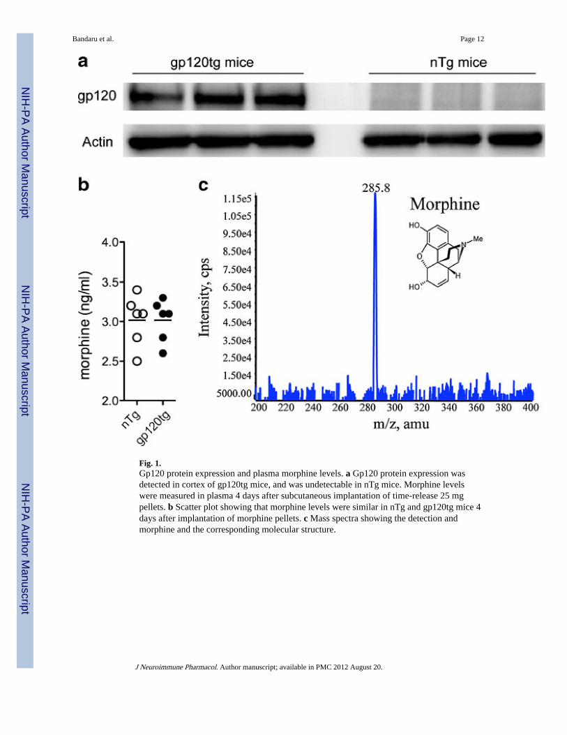
Fig. 1.Gp120 protein expression and plasma morphine levels. a Gp120 protein expression wasdetected in cortex of gp120tg mice, and was undetectable in nTg mice. Morphine levelswere measured in plasma 4 days after subcutaneous implantation of time-release 25 mgpellets. b Scatter plot showing that morphine levels were similar in nTg and gp120tg mice 4days after implantation of morphine pellets. c Mass spectra showing the detection andmorphine and the corresponding molecular structure.
Bandaru et al. Page 12
J Neuroimmune Pharmacol. Author manuscript; available in PMC 2012 August 20.
NIH
-PA Author Manuscript
NIH
-PA Author Manuscript
NIH
-PA Author Manuscript

Fig. 2.Mice transgenic for HIV gp120 fail to normalize morphine-induced increases reactiveoxygen species in brain during drug withdrawal. a–d Chronic morphine administrationincreased total oxidation, and decreased SOD activity, acid levels and the GSH/GSSH ratioin both nTg and gp120tg mice. e–f During drug withdrawal, total oxidation and SODactivity normalized in nTg mice, but not in gp120tg mice. Uric acid levels normalized andthe GSH/GSSG ratio failed to normalize in both groups of mice. ANOVA with Tukey posthoc comparisons (n=6 per group). *p<0.05, **p<0.01, ***p<0.001 compared with placebo.
Bandaru et al. Page 13
J Neuroimmune Pharmacol. Author manuscript; available in PMC 2012 August 20.
NIH
-PA Author Manuscript
NIH
-PA Author Manuscript
NIH
-PA Author Manuscript

Fig. 3.Evidence that morphine induces sphingomyelin synthesis during chronic administration andsphingomyelin catabolism during drug withdrawal in brains of gp120tg mice. a–b Baselinelevels of all sphingomyelin species detected were decreased and all but one ceramide specieswere increased in gp120tg mice compared with nTg mice. Chronic morphine had no effecton sphingomyelin or ceramide metabolism in nTg mice, but increased levels of several longchain sphingomyelins (C16:0-C22:0), and decreased levels of several long chain ceramides(C16:0-C20:0) in gp120tg mice. c–d There were no apparent alterations in sphingomyelin orceramide metabolism during drug withdrawal in nTg mice. In gp120tg mice, brain levels oflong chain sphingomyelin normalized (C16:0-C22:0), and levels of very long chainsphingomyleins were reduced (C24:0-C26:0), and all ceramide species were increasedduring drug withdrawal. ANOVA with Tukey post hoc comparisons (n=6 per group).*p<0.05, **p<0.01, ***p<0.001, ###p<0.001.
Bandaru et al. Page 14
J Neuroimmune Pharmacol. Author manuscript; available in PMC 2012 August 20.
NIH
-PA Author Manuscript
NIH
-PA Author Manuscript
NIH
-PA Author Manuscript

Fig. 4.Morphine-induced synaptic damage does not recover during drug withdrawal in gp120tgmice. a–b Levels of PSD95 in hippocampus were decreased in nTg mice during chronicmorphine administration and recovered to levels similar to those measured in miceadministered placebo. c–d Hippocampal levels of PSD95 were decreased in gp120tg miceduring chronic morphine administration compared with placebo, and did not recover duringdrug withdrawal. For quantitative analysis, the densities of PSD95 were normalized to thecorresponding β-actin density. Students T-test (n=4 per condition). Exact p values areshown.
Bandaru et al. Page 15
J Neuroimmune Pharmacol. Author manuscript; available in PMC 2012 August 20.
NIH
-PA Author Manuscript
NIH
-PA Author Manuscript
NIH
-PA Author Manuscript


