Embed Size (px)
Citation preview
P
Ya
b
a
ARR1AA
KEEEKL
1
tpto(btrcbvica[i
Tm
(
0d
Sensors and Actuators B 152 (2011) 37–48
Contents lists available at ScienceDirect
Sensors and Actuators B: Chemical
journa l homepage: www.e lsev ier .com/ locate /snb
olyphenol-modified glassy carbon electrodes for copper detection
asemin Oztekina,b, Zafer Yazicigil a, Almira Ramanavicieneb, Arunas Ramanaviciusb,∗
Selcuk University, Faculty of Science, Department of Chemistry, Konya, TurkeyVilnius University, Faculty of Chemistry, NanoTechnas – Centre of Nanotechnology and Materials Science, Naugarduko 24, Vilnius, Lithuania
r t i c l e i n f o
rticle history:eceived 29 July 2010eceived in revised form6 September 2010ccepted 23 September 2010vailable online 16 October 2010
a b s t r a c t
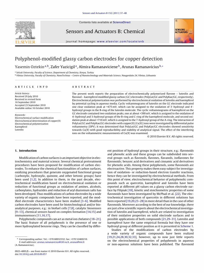
The present work reports the preparation of electrochemically polymerized flavone – luteolin andflavonol – kaempferol modified glassy carbon (GC) electrodes (PolyLut/GC and PolyKae/GC, respectively).Electrochemical polymerization was performed by electrochemical oxidation of luteolin and kaempferolby potential cycling in aqueous media. Cyclic voltammograms of luteolin on the GC electrode indicatedone clear oxidation peak at +475 mV, which can be assigned to the oxidation of 3′-hydroxyl and 4′-hydroxyl groups in the B-ring of the luteolin molecule. The cyclic voltammograms of kaempferol on the
eywords:lectrochemical surface modificationlectrochemical determination of copper(II)lectrochemical polymerizationaempferoluteolin
GC electrode contained two oxidation peaks, one at about +390 mV, which is assigned to the oxidation of4′-hydroxyl and 3-hydroxyl groups of the B-ring and C-ring of the kaempferol molecule, and second oxi-dation peak at about +710 mV, which is assigned to the 7-hydroxyl group of the A-ring. The interaction ofPolyLut/GC and PolyKae/GC electrodes with copper(II) (Cu(II)) ions were investigated by differential pulsevoltammetry (DPV). It was determined that PolyLut/GC and PolyKae/GC electrodes showed sensitivitytowards Cu(II) with good reproducibility and stability of analytical signal. The effect of the interfering
mea
ions on the voltammetric. Introduction
Modification of carbon surfaces is an important objective in elec-rochemistry and material science. Several chemical pretreatmentrocedures have been proposed for modification of carbon elec-rodes. To enhance the chemical functionalities of carbon surfaces,xidizing procedures that generate oxygenated functional groupscarboxylic, hydroxylic, quinone, and other ketonic groups) haveeen used [1,2]. In addition to them, in the past decade, elec-rochemical modification based on electrochemical oxidation oreduction of functional groups as oxidation of amines, alcohols,arboxylates, hydrazides and reduction of aryl diazonium salts haseen developed. Thus modifications of carbon electrodes by a wideariety of organic compounds have been reported, and the mod-fied electrode characteristics have been studied [3–6]. Modifiedarbon electrodes have been used for biotechnological and/or bio-nalytical purposes, e.g.: in biofuel cells [7,8], catalytic biosensors9–13], chemical sensors based on complex formation [14,15] and
mmunosensors [11,16,17].Polyphenolic compounds can act as metal ion chelators [18–21].he basic feature of all polyphenols is the presence of one orore hydroxylated benzene rings. They can be classified by differ-
∗ Corresponding author: Tel.: +370 60032332; fax: +370 52469210.E-mail addresses: [email protected], [email protected]
A. Ramanavicius).
925-4005/$ – see front matter © 2010 Elsevier B.V. All rights reserved.oi:10.1016/j.snb.2010.09.057
surements of Cu(II) was examined.© 2010 Elsevier B.V. All rights reserved.
ent position of hydroxyl groups in their structure, e.g.: flavonoidsand phenolic acids and these groups can be subdivided into sev-eral groups such as flavonols, flavones, flavanols, isoflavones forflavonoids; benzoic acid derivatives and cinnamic acid derivativesfor phenolic acids. Among these polyphenols, some flavonoids areelectroactive. This property makes them easy subject for investiga-tion of oxidation- or reduction-based electron transfer reactions,hence they can be investigated by electrochemical methods. Fromthis point of view, electrochemical behavior of polyphenolic com-pounds such as quercetin, kaempferol and luteolin have beenreported at different pH values on a glassy carbon electrode sur-face by Filipiak [18], kinetic and stoichiometric properties of someflavonoids have been investigated by McPhail et al. [22]. The elec-trochemical investigation of quercetin in different solutions hasbeen reported [19,20,23–28] in more detail than in the case of otherflavonoids. Moreover according to the best of our knowledge, thereare just a few scientific reports about the electrochemical investiga-tion of luteolin and kaempferol, which are related to investigationof their oxidative properties on solid electrode surfaces and topossible applications of both compounds [21,29–31]. Luteolin andkaempferol have the same empirical formula but they have onehydroxyl group at different position as it is presented in Scheme 1.
Studies of the modifications of carbon electrodes bya wide variety of organic compounds have been realized[19,21,24,28,30,32,33], however, up to now just little reportson the electrochemical properties of polyphenols in aqueousor non-aqueous solutions have been published. The flavonoid
38 Y. Oztekin et al. / Sensors and Actu
S(
sis
eGitoc(estmh
2
2
ScS
pmipuwabp
2
tBwewwcB
cheme 1. Structural features of flavonoids: (A) general structure, (B) luteolin andC) kaempferol.
tructure plays a crucial role in the chelation of copper(II) (Cu(II))ons and it could be exploited in the development of analyticalystems.
Therefore the main purpose of this study was to perform thelectrochemical polymerization of luteolin and kaempferol on theC electrode in order to get polyluteolin and polykaempferol mod-
fied GC electrodes (PolyLut/GC and/or PolyKae/GC) and to applyhese modified electrodes in analytical systems for determinationf Cu(II) ions. The objectives of this work were: (i) to create electro-hemically polymerized PolyLut/GC and/or PolyKae/GC electrodes,ii) to characterize modified GC electrodes by cyclic voltammetry,lectrochemical impedance spectroscopy (EIS), ellipsometry andcanning electron microscopy, (iii) to determine the interaction ofhese modified electrodes with Cu(II) by differential pulse voltam-
etry (DPV) and (iv) to investigate the effect of interference of someeavy metal ions.
. Experimental
.1. Chemicals
Kaempferol (96%) and luteolin (98%) were purchased fromigma (St. Louis, USA). The other chemicals used for electro-hemical experiments were purchased from Merck, Riedel andigma–Aldrich companies and were of reagent grade.
The 1.0 mM solutions of flavonoids were prepared in 100 mMhosphate buffer solution (PBS), pH 7.5. The PBS was prepared byixing of 0.05 mM K2HPO4 and 0.05 mM KH2PO4 and then adjust-
ng of pH by addition of KOH or H3PO4. CuSO4·5H2O solutions wererepared at different concentrations (ranging from 1.0 × 10⁻⁶ Mntil 1.0 × 10⁻¹¹ M) in Britton–Robinson (BR) buffer, pH 5.0, whichas prepared by mixing of H3BO3, H3PO4 and CH3COOH and then
djusting of pH by addition of HCl or NaOH. The pH values of theuffer solutions were measured and adjusted with an Orion EA 940H meter.
.2. Electrochemical equipment
A traditional three-electrode cell system was used in all elec-rochemical experiments. The GC electrode Model MF-2012 fromAS (Kenilworth Warwickshire, United Kingdom) was used asorking electrode with a geometric area of 0.071 cm2. The ref-
rence electrodes: Ag/AgCl in saturated KCl (Ag/AgCl/(sat. KCl)),hich was used in aqueous media; or Ag/Ag+ in 10 mM AgNO3,hich was used in non-aqueous media. All experiments were
arried out inside a Faraday cage at room temperature (25 ◦C).efore electrochemical treatment all solutions used in the voltam-
ators B 152 (2011) 37–48
metric system were deaerated with argon (99.999%) for at least5 min. The cyclic voltammetry (CV) technique was applied withPHE 200 software, electrochemical impedance spectroscopy (EIS)was applied with EIS 300 software and differential pulse voltam-metry (DPV) applied with PV 220 software. All were performedusing Potentiostat/Galvanostat Gamry Reference PCI4/750 fromGamry Instruments (Warminster, USA). The thickness of the formedpolymeric layer was measured with a Nanofilm EP3 Imaging Ellip-someter (Nanofilm Technologie GmbH, Goettingen, Germany). Thelayer properties were compared with bare GC and the forma-tion of the layer was discussed. The morphology of polyphenolfilms on GC electrode was investigated by using scanning elec-tron microscope (SEM) Quanta 400F from FEI company (Eindhoven,The Netherlands). The interaction between polyphenols and Cu(II)ions were also investigated with a double beam spectrophotometermodel UV-1800, Shimadzu (Kyoto, Japan).
2.3. Electrode preparation and modification procedures
Steps mentioned in the previously published literature wereperformed before the usage of the GC electrodes in electrochemicalexperiments in order to avoid contamination by oxidation prod-ucts and to obtain a clean renewed electrode surface [6,14,15].The surface of the GC electrode was hand-polished with a 1.0 �m,0.3 �m and 0.05 �m alumina–water slurry using a polishing cloth,the electrodes were rinsed with purified water and then sonicatedin purified water for 10 min. To remove impurities, which could notbe removed with water the electrodes were washed with purifiedwater and later sonicated with a 1:1 (V/V) mixture of acetoni-trile (CH3CN) with isopropylalcohol for 10 min. Then electrodeswere kept in CH3CN. Electrode surfaces were sonicated with CH3CNbefore and after each treatment.
The polished and cleaned GC electrode surfaces as describedabove were modified in 1.0 mM solutions of flavonoids prepared in100 mM PBS, pH 7.5 by CV technique. The modification of the GCelectrode by flavonoids (luteolin and kaempferol) was performed inthe potential range between +100 mV and +900 mV by 30 potentialcycles at potential sweep rate of 100 mV/s. After the modificationof the GC electrode, the surfaces of obtained luteolin-modified GC(PolyLut/GC) and kaempferol-modified GC (PolyKae/GC) electrodeswere washed in order to remove all impurities from the electrodesurface and then they were used for other investigations describedin this paper.
2.4. Electrode characterization
The modified electrodes were characterized by CV, EIS, ellip-sometry and SEM. The characterization with CV technique wasperformed in the presence of: (i) 1.0 mM equiv.-molar ratio ofK3[Fe(CN)6]/K4[Fe(CN)6] (Fe(CN)6
3−/Fe(CN)64−) in BR buffer, pH
2.0, and (ii) 1.0 mM of ferrocene in CH3CN containing 100 mM tetra-butylamoniumtetrafluoroborate (TBATFB) as redox active probes.The cyclic voltammograms of modified electrodes were comparedwith cyclic voltammograms of the bare GC electrode.
The characterization with EIS technique was carried out witha Gamry Reference PCI4/750 potentiostat by EIS 300 software. Forthe EIS measurements performed in 100 mM KCl containing 1.0 mMequiv.-molar ratio of Fe(CN)6
3−/Fe(CN)64−, a sine wave potential
alteration at 5 mV amplitude superimposed on a formal potentialof the redox probe of 220 mV was applied, a wide frequency rangefrom 0.1 Hz to 100 kHz was scanned and the Nyquist plots were
recorded. All potentials are referenced vs. Ag/AgCl/(sat. KCl). TheNyquist plots of modified electrodes were then compared with theEIS data of the bare GC electrode.The reflection of 532 nm laser (green light) at 73◦ was used inellipsometric measurements. The results of these measurements
d Actuators B 152 (2011) 37–48 39
w5alsc
e
2
PbPCisocembptcatt1wesw
2
sbJabwcc
3
3
toFopigie[Bifd
Y. Oztekin et al. / Sensors an
ere recorded as an average of four computations in the area of0 �m × 50 �m. The model was formed with the refractive indicess 3.0841 for graphite, 1.9000 for GC substrate, 1.4100 for organicayer and 1.0000 for air, 1.7820 for graphite, 0.8200 for GC sub-trate, 0.0000 for organic layer and 0.0000 for air are the valuesorresponding to the extinction coefficients in the measurements.
The morphology of polyluteolin and polykaempferol films on GClectrode was investigated by scanning electron microscopy.
.5. Application of polyphenol-modified GC electrodes
Another control for the presence of polyphenol layer on theolyLut/GC and PolyKae/GC electrode surfaces was performedy testing of the complexation-ability of the PolyLut/GC andolyKae/GC electrodes with Cu(II) ions. For this aim, interaction ofu(II) with polyphenol-modified electrodes was accomplished by
mmersing the PolyLut/GC and PolyKae/GC electrodes into Cu(II)olutions with varying concentrations of Cu(II) ions for 1 h inrder to form complexes of Cu(II) with polymerized polyphenolompounds and to create Cu(II)/PolyLut/GC and Cu(II)/PolyKae/GClectrodes, respectively. The bound metal ions on the polyphenol-odified GC electrodes were reduced at −900 mV for 1 min in BR
uffer, pH 5.0. The DPV technique for Cu(II) determination waserformed with the Cu(II)/PolyLut/GC and Cu(II)/PolyKae/GC elec-rodes in BR, pH 5.0, with a Gamry Reference PCI4/750 potentiostatontrolled by PV 220 software. The following parameters werepplied in DPV experiments: the potential range was from −400 mVo +200 mV, the potential sweep rate was 50 mV/s, pulse ampli-ude of 50 mV, the pulse period was 0.05 s and sample period was.0 s. The selectivity of polyphenol modified GC electrodes for Cu(II)as examined in the presence of the interfering ions. The modified
lectrodes for 1 h were treated with 1.0 × 10⁻⁶ M interfering ionolution mixed with 1.0 × 10⁻⁶ M Cu(II) ions. The analyte solutionsere prepared in BR buffer, pH 5.0.
.6. Spectrophotometric experiments
In addition to the electrochemical measurements, the UV–Vispectra were recorded in the range of 220–600 nm with a dou-le beam spectrophotometer model UV-1800, Shimadzu (Kyoto,apan) using quartz cuvettes (1.0 cm optical path) at room temper-ture in order to determine the stoichiometric ratio for the reactionetween polyphenols and Cu(II) ions by Job’s method. The solutionsere prepared by mixing of both components at equal-molar con-
entrations (0.1 mM of each compound) by varying the ratios ofomponents from 1 to 10.
. Results and discussion
.1. Modification of GC electrode
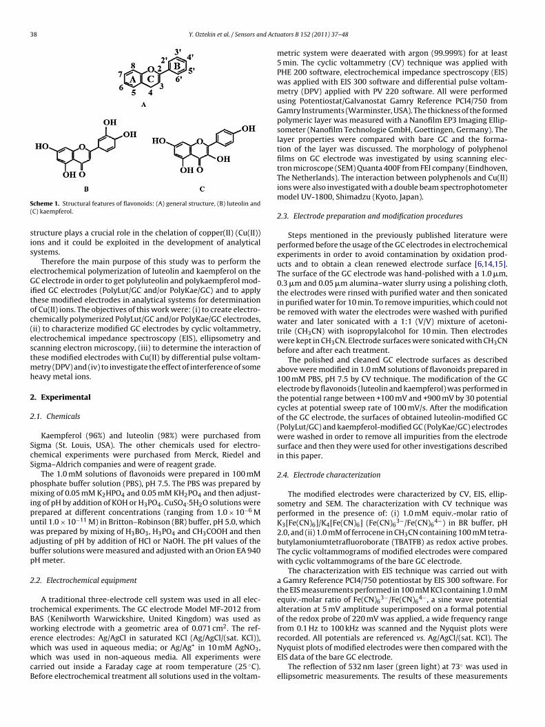
The cyclic voltammograms of luteolin and kaempferol regis-ered by applying 30 potential cycles at the potential sweep ratef 100 mV/s on a bare GC electrode surface and are shown inig. 1. The cyclic voltammograms of luteolin show that luteolinn bare GC exhibits an electrochemically irreversible oxidationeak at lower positive potentials (Epa = +475 mV) (Fig. 1A), which
s attributed to the oxidation of the 3′-hydroxyl and 4′-hydroxylroups in the B-ring of luteolin. Such oxidation of luteolin molecules in an agreement with the research using Raman and surface-nhanced Raman spectra that were reported by Corredor et al.
34]. Due to the different position of the hydroxyl groups in the-ring the motions involved in vibrations of these groups resultedn some differences of Raman spectra. Raman spectra were uniqueor each flavonoid and for the luteolin molecule the most intenseiscriminant bands were located at 1505 cm−1. This vibration could
Fig. 1. Cyclic voltammograms of 1.0 mM (A) luteolin and (B) kaempferol in PBS, pH7.5, vs. Ag/AgCl/(sat. KCl), 1st (a) and 30th (b) potential scan cycle. Potential sweeprate is 100 mV/s.
be attributed to the 3′-hydroxyl group in the B-ring. Anotherspectroscopic characterization has been performed with quercetin(3,3′,4′,5,7-pentahydroxyflavone) by Brown et al. and it has beenmentioned that the oxidation could occur at the 3′-hydroxyl and4′-hydroxyl groups of the B-ring and 3-hydroxyl group of the C-ringfor quercetin [35].
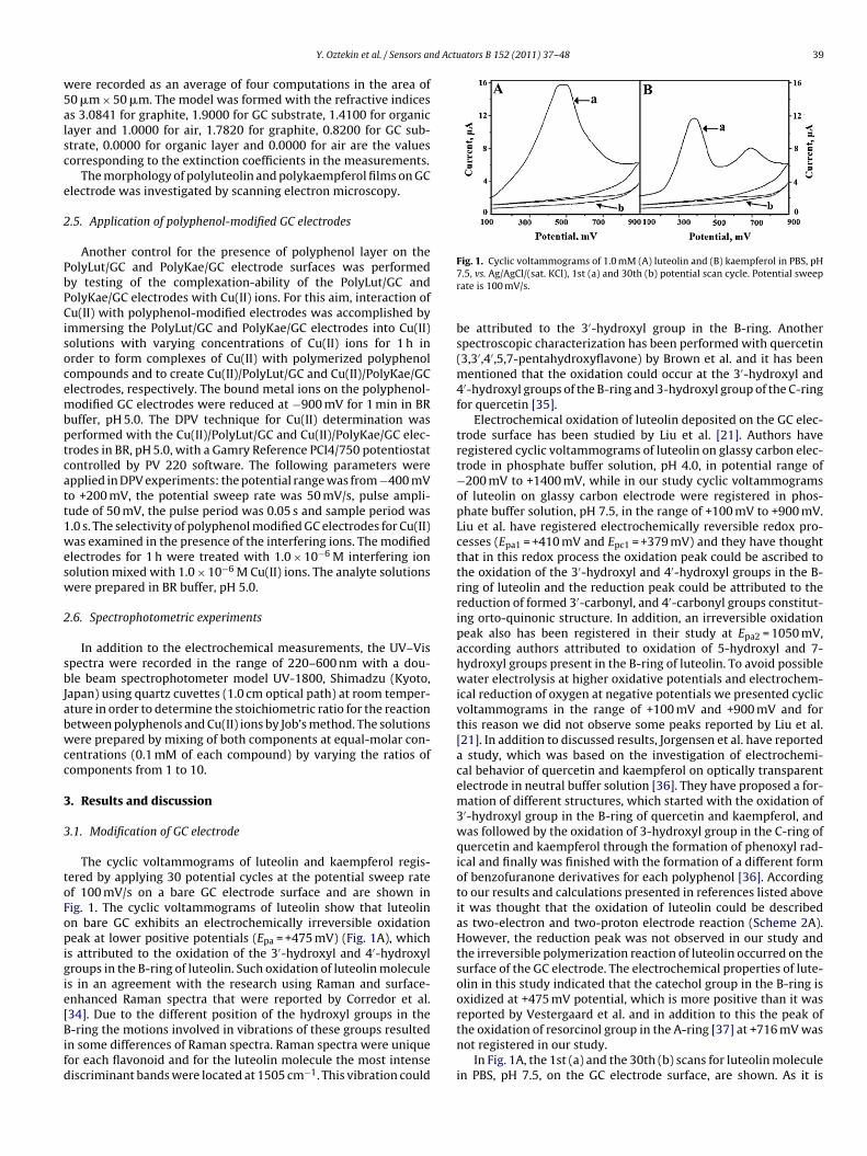
Electrochemical oxidation of luteolin deposited on the GC elec-trode surface has been studied by Liu et al. [21]. Authors haveregistered cyclic voltammograms of luteolin on glassy carbon elec-trode in phosphate buffer solution, pH 4.0, in potential range of−200 mV to +1400 mV, while in our study cyclic voltammogramsof luteolin on glassy carbon electrode were registered in phos-phate buffer solution, pH 7.5, in the range of +100 mV to +900 mV.Liu et al. have registered electrochemically reversible redox pro-cesses (Epa1 = +410 mV and Epc1 = +379 mV) and they have thoughtthat in this redox process the oxidation peak could be ascribed tothe oxidation of the 3′-hydroxyl and 4′-hydroxyl groups in the B-ring of luteolin and the reduction peak could be attributed to thereduction of formed 3′-carbonyl, and 4′-carbonyl groups constitut-ing orto-quinonic structure. In addition, an irreversible oxidationpeak also has been registered in their study at Epa2 = 1050 mV,according authors attributed to oxidation of 5-hydroxyl and 7-hydroxyl groups present in the B-ring of luteolin. To avoid possiblewater electrolysis at higher oxidative potentials and electrochem-ical reduction of oxygen at negative potentials we presented cyclicvoltammograms in the range of +100 mV and +900 mV and forthis reason we did not observe some peaks reported by Liu et al.[21]. In addition to discussed results, Jorgensen et al. have reporteda study, which was based on the investigation of electrochemi-cal behavior of quercetin and kaempferol on optically transparentelectrode in neutral buffer solution [36]. They have proposed a for-mation of different structures, which started with the oxidation of3′-hydroxyl group in the B-ring of quercetin and kaempferol, andwas followed by the oxidation of 3-hydroxyl group in the C-ring ofquercetin and kaempferol through the formation of phenoxyl rad-ical and finally was finished with the formation of a different formof benzofuranone derivatives for each polyphenol [36]. Accordingto our results and calculations presented in references listed aboveit was thought that the oxidation of luteolin could be describedas two-electron and two-proton electrode reaction (Scheme 2A).However, the reduction peak was not observed in our study andthe irreversible polymerization reaction of luteolin occurred on thesurface of the GC electrode. The electrochemical properties of lute-olin in this study indicated that the catechol group in the B-ring isoxidized at +475 mV potential, which is more positive than it wasreported by Vestergaard et al. and in addition to this the peak of
the oxidation of resorcinol group in the A-ring [37] at +716 mV wasnot registered in our study.In Fig. 1A, the 1st (a) and the 30th (b) scans for luteolin moleculein PBS, pH 7.5, on the GC electrode surface, are shown. As it is
40 Y. Oztekin et al. / Sensors and Actuators B 152 (2011) 37–48
eolin a
scacotfi1coeiw
koia(t(ewopp4CocSaocst
atcioBMtAa
Scheme 2. Oxidation mechanism proposed for (A) lut
een from Fig. 1A, the oxidation peak current remarkably decreasesycle-by-cycle (the number in Fig. 1A represents the cycle sequences “a” (1st cycle) and “b” (30th cycle)), suggesting that the peakurrents are adsorption driven or show the characteristic featuresf electrochemical formation of polymeric layer on the GC elec-rode. After the third cycle, the peak currents vary slightly andnally remain almost constant. Therefore, the peak current of thest scan was recorded and used in subsequent comments and cal-ulations. According to our best knowledge it is the first reportf electrochemical deposition of luteolin on the GC electrode bylectrochemical oxidation. Moreover in addition to mentioned fact,t should be noted that electrochemical investigations of luteolin
ere presented by other authors [21,35].The cyclic voltammograms of kaempferol shows that
aempferol on the bare GC electrode surface has two irreversiblexidation peaks (at E1pa = +390 mV, E2pa = +710 mV) (Fig. 1B). As its seen from the cyclic voltammograms of kaempferol in Fig. 1B, therea of the oxidation peak that is observed in more negative regionE1pa = +390 mV) and its area is approximately two times biggerhan the area of the second oxidation peak in more positive regionE2pa = +710 mV). For this reason we thought that the number oflectrons involved in oxidation process at positive potential valueas twice lower if compared with electron number involved into
xidation process occurring at more negative potential. From thisoint of view, we thought that the oxidation peak at more negativeotential (at E1pa = +390 mV) represented in Fig. 1B belongs to′-hydroxyl group in the B-ring and to 3-hydroxyl group in the-ring, while the second oxidation peak is attributed to oxidationf 7-hydroxyl group in the A-ring. The 1st (a) and the 30th (b)ycles of kaempferol in PBS, pH 7.5, are represented in Fig. 1B.imilarly to the cyclic voltammograms of luteolin, the irreversiblenodic peaks of kaempferol decrease gradually with increasef potential cycle number and finally it reaches a steady-stateonditions by the third cycle. This current-potential behaviorhows the characteristic features of electrochemical formation ofhe polykaempferol layer on the GC electrode surface.
Although the 7-hydroxyl group in the A-ring of kaempferol islso present in luteolin structure, however, no oxidation peak forhe 7-hydroxyl group at the same potential was observed in theyclic voltammograms of luteolin. Thus, the presence of the peakn the cyclic voltammograms of kaempferol can be related to thexidation/reduction ability of the 3-hydroxyl group localized in the
-ring and of the 7-hydroxyl group in the A-ring of kaempferol.oreover, after oxidation of the 3-hydroxyl group in the B-ringo 3-carbonyl group the oxidation of the 7-hydroxyl group in the-ring can form the quinonic structure more easy because of suit-ble induction/conjugation effects. No oxidation peak is observed
nd (B) kaempferol on the GC electrode in PBS, pH 7.5.
in the presence of luteolin although its structure has the same7-hydroxyl group in the A-ring as kaempferol. Absence of the 3-hydroxyl group in the B-ring of luteolin, which makes the oxidationof the 7-hydroxyl group in the A-ring easier because of its induc-tion/conjugation effects. Another reason for the oxidation of the7-hydroxyl group in the A-ring of kaempferol could be the forma-tion of radicalic species by an electron extraction from the A-ringand C-ring of kaempferol and a hydrogen leakage from the 7-hydroxyl group in the A-ring turning to the oxidized form of the3-hydroxyl group in the C-ring, which then could be easily rear-ranged into corresponding quinonic form. The herein describedoxidation process takes place more efficiently for kaempferol incomparison with oxidation of luteolin.
The first anodic peak in the cyclic voltammograms of kaempferolin PBS, pH 7.5, has been registered in other studies, while the sec-ond peak of kaempferol has not been observed yet in any publishedscientific reports. Moreover some of our ideas about the oxida-tion products of kaempferol have been supported by investigationspresented by other authors where the cyclic voltammograms ofkaempferol in BR buffer, pH 1.8, showed one oxidation peak at+570 mV followed by the oxygen evolution wave beyond +900 mV[31]. However, in this study [31] no cathodic peak was observedin cyclic voltammograms which is similar to findings reported inour study. Another study on electrochemical behavior of quercetinin hydro-alcoholic solution has been reported by Timbola et al.Authors have registered three oxidation peaks, which were thoughtto be attributed to the oxidation of 3′-hydroxyl and 4′-hydroxylgroups in the B-ring, the 3-hydroxyl group in the C-ring and the5-hydroxyl and the 7-hydroxyl groups in the A-ring [20]. In thisstudy the oxidation process of quercetin has been clearly dividedinto three separated parts for the common functional groups withkaempferol, while in our study only two clear oxidation parts arerepresented as two separated peaks in cyclic voltammograms ofkaempferol. According to our results, the luteolin molecule has onlyone oxidation peak, which is thought to be the oxidation of the 3′-hydroxyl and the 4′-hydroxyl groups in the B-ring. However, thekaempferol molecule has no catechol group in the B-ring and forthis reason no peak within +100 mV and +900 mV was observed.On the other hand, in our study, any oxidation of 5-hydroxyl and 7-hydroxyl groups in the A-ring was observed, as it has been noticedin the study based on the evaluation of the cyclic voltammogramsof quercetin in hydro-alcoholic solution, pH 7.4 [20]. Moreover in
the same study [20] the influence of the potential sweep rate on thevoltammetric profile of 0.8 mM quercetin in hydro-alcoholic solu-tion at pH 7.4 has been investigated, in order to access in moredetail the characteristics of electrochemical and chemical reac-tions taking place at the electrode/solution interfaces. The authors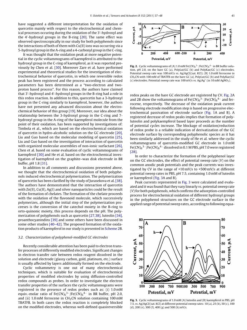
d Actuators B 152 (2011) 37–48 41
hqitot5
thvetpppttghcrhpToLLwCktb
wnoTwowpcompst
3
fisi
tprtreaTo
Fig. 2. Cyclic voltammograms of (A) 1.0 mM Fe(CN)63−/Fe(CN)6
4− in BR buffer solu-tion, pH 2.0, on the bare GC (a), PolyLut/GC (b) and PolyKae/GC (c) electrodes.
in the polyphenol structures on the GC electrode surface in theapplied range of potential sweep rates, according to following equa-
Y. Oztekin et al. / Sensors an
ave suggested a different interpretetation for the oxidation ofuercetin mainly with respect to the electrochemical and chem-
cal processes occuring during the oxidation of the 3′-hydroxyl andhe 4′-hydroxyl groups in the B-ring [20]. The same effect wasbserved spectroscopically in our study for both polyphenols sincehe interactions of both of them with Cu(II) ions was occurring via a-hydroxyl group in the A-ring and a 4-carbonyl group in the C-ring.
It was thought that the oxidation peak at more negative poten-ial in the cyclic voltammograms of kaempferol is attributed to theydroxyl group in the C-ring of kaempferol, as it was reported pre-iously by Chen et al. [30]. Moreover Zare et al. have performedxperimental and theoretical studies for the investigation of elec-rochemical behavior of quercetin, in which one reversible redoxeak has been registered and the process according to calculatedarameters has been determined as a “two-electron and two-roton based process”. For this reason, the authors have claimedhat 3′-hydroxyl and 4′-hydroxyl groups in the B-ring had a role inhis redox reaction. In addition to this, quercetin has a 3-hydroxylroup in the C-ring similarly to kaempferol, however, the authorsave not presented any advanced discussion about the electro-hemical behavior of this group [19]. Moreover, our idea about theelationship between the 3-hydroxyl group in the C-ring and 7-ydroxyl group in the A-ring of the kaempferol molecule from theoint of their oxidation, has been supported by investigations of:imbola et al., which are based on the electrochemical oxidationf quercetin in hydro-alcoholic solution on the GC electrode [20],iu and Guo based on the molecular modeling of quercetin [25],iu and Guo based on the investigation of interaction of quercetinith organized molecular assemblies of non-ionic surfactant [26],hen et al. based on some evaluation of cyclic voltammograms ofaempferol [30] and He et al. based on the electrochemical inves-igation of kaempferol on the graphite–wax disk electrode in BRuffer, pH 1.8 [31].
In addition to all comments and discussions presented above,e thought that the electrochemical oxidation of both polyphe-ols induced electrochemical polymerization. The polymerizationf quercetin has been clarified by the study of Jurasekova et al. [38].he authors have demonstrated that the interaction of quercetinith Zn(II), Cu(II), Ag(I) and silver nanoparticles could be the result
f the formation of chelates. The formation of the chelate proceedsith the oxidation of the flavonoid molecule, which successivelyolymerizes, although the initial step of the polymerization pro-eeses is the conversion of the catechol moiety in the B-ring torto-quinonic moiety, this process depends on the pH. The poly-erization of polyphenols such as quercetin [27,38], luteolin [34],
roanthocyanindins [39] and some others have been discussed inome other studies [40–42]. The proposed formation of the oxida-ion products of kaempferol in our study is presented in Scheme 2B.
.2. Characterization of polyphenol-modified GC electrodes
Recently considerable attention has been paid to electron trans-er processes of differently modified electrodes. Significant changesn electron transfer rate between redox reagent dissolved in theolution and electrode (glassy carbon, gold, platinum, etc.) surfaces usually affected by layers additionally formed on the electrode.
Cyclic voltammetry is one out of many electrochemicalechniques, which is suitable for evaluation of electrochemicalroperties of modified electrodes by using diffusion-controllededox compounds as probes. In order to investigate the electronransfer properties of the surfaces the cyclic voltammograms were
egistered in the presence of redox probes such as: (i) 1.0 mMquiv.-molar ratio of Fe(CN)63−/Fe(CN)64− in BR buffer, pH 2.0,
nd (ii) 1.0 mM ferrocene in CH3CN solution containing 100 mMBATFB. In both cases the redox reaction is completely blockedn the modified electrodes, whereas well-defined quasireversible
Potential sweep rate was 100 mV/s vs. Ag/AgCl/(sat. KCl); (B) 1.0 mM ferrocene inCH3CN with 100 mM of TBATFB on the bare GC (a), PolyLut/GC (b) and PolyKae/GC(c) electrodes. Potential sweep rate was 100 mV/s vs. Ag/Ag+ (in 10 mM AgNO3).
redox peaks on the bare GC electrode are registered by CV. Fig. 2Aand 2B show the voltammograms of Fe(CN)6
3−/Fe(CN)64− and fer-
rocene, respectively. The decrease of the oxidation peak currentfollowing electrode modification step is based on progressive elec-trochemical passivation of electrode surface (Fig. 1A and B). Aregistered decrease of redox peaks implies that formation of poly-luteolin and polykaempferol based layer proceeds as the numberof potential cycles increase. The blockage of oxidation/reductionof redox probe is a reliable indication of derivatization of the GCelectrode surface by corresponding polyphenolic species as it hasbeen similarly addressed in the study of He et al. where the cyclicvoltammograms of quercetin-modified GC electrode in 1.0 mMFe(CN)6
3−/Fe(CN)64− dissolved in 0.1 M PBS, pH 7.0 were registered
[28].In order to characterize the formation of the polyphenol layer
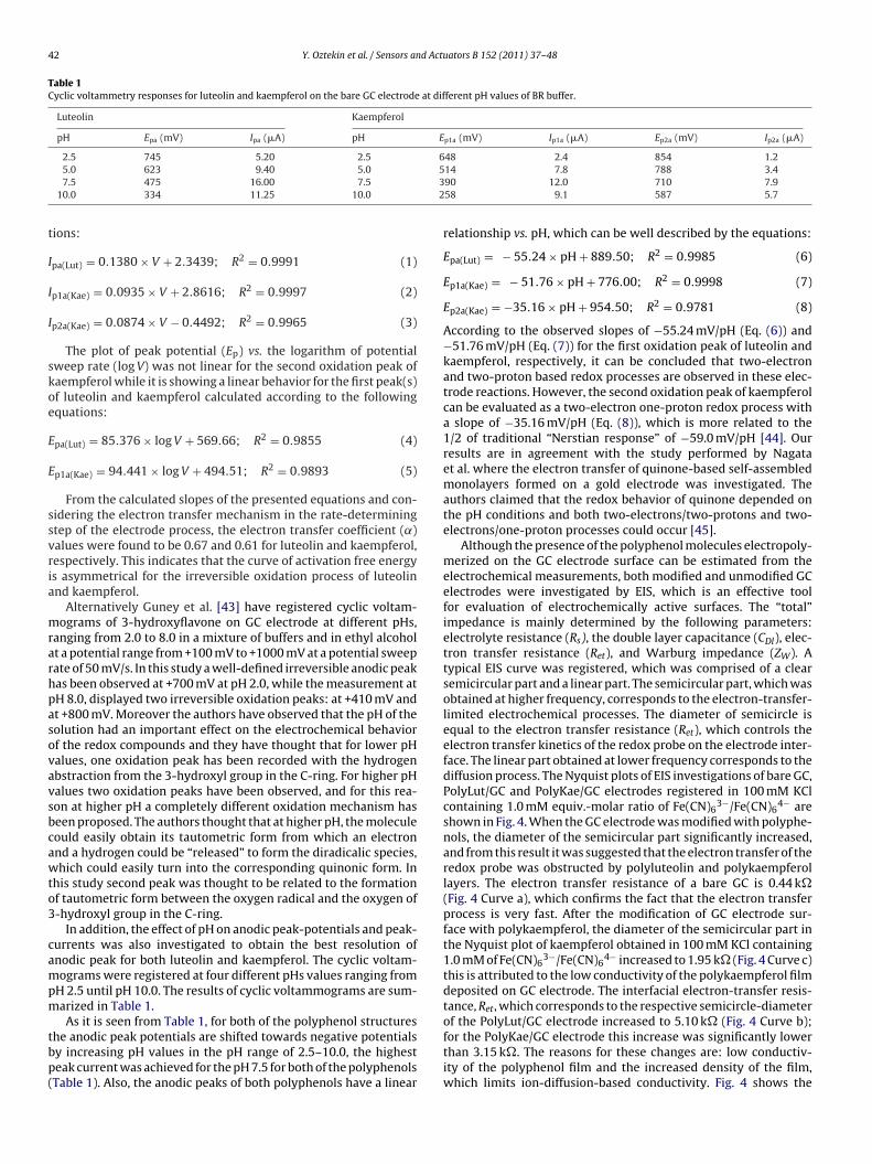
on the GC electrodes, the effect of potential sweep rate (V) on thecommon anodic peak potentials and the peak currents was inves-tigated by CV in the range of +10 mV/s to +500 mV/s at differentpotential sweep rates in PBS, pH 7.5, containing 1.0 mM of luteolinor kaempferol (Fig. 3A and B).
Peak currents represented in Fig. 3 were calculated and evalu-ated and it was found that they vary linearly vs. potential sweep rate(V) for both polyphenols, which confirms the adsorption-controlledprocess for electrochemical oxidation of different hydroxyl groups
Fig. 3. Cyclic voltammograms of 1.0 mM (A) luteolin and (B) kaempferol in PBS, pH7.5, vs. Ag/AgCl/(sat. KCl) at different potential sweep rates: 10 (a), 25 (b), 50 (c), 100(d), 200 (e), 300 (f), 400 (g) and 500 (h) mV/s.
42 Y. Oztekin et al. / Sensors and Actuators B 152 (2011) 37–48
Table 1Cyclic voltammetry responses for luteolin and kaempferol on the bare GC electrode at different pH values of BR buffer.
Luteolin Kaempferol
pH Epa (mV) Ipa (�A) pH Ep1a (mV) Ip1a (�A) Ep2a (mV) Ip2a (�A)
6532
t
I
I
I
skoe
E
E
ssvria
mrarhpasovavsbcawto3
campm
tbp(
2.5 745 5.20 2.55.0 623 9.40 5.07.5 475 16.00 7.5
10.0 334 11.25 10.0
ions:
pa(Lut) = 0.1380 × V + 2.3439; R2 = 0.9991 (1)
p1a(Kae) = 0.0935 × V + 2.8616; R2 = 0.9997 (2)
p2a(Kae) = 0.0874 × V − 0.4492; R2 = 0.9965 (3)
The plot of peak potential (Ep) vs. the logarithm of potentialweep rate (log V) was not linear for the second oxidation peak ofaempferol while it is showing a linear behavior for the first peak(s)f luteolin and kaempferol calculated according to the followingquations:
pa(Lut) = 85.376 × log V + 569.66; R2 = 0.9855 (4)
p1a(Kae) = 94.441 × log V + 494.51; R2 = 0.9893 (5)
From the calculated slopes of the presented equations and con-idering the electron transfer mechanism in the rate-determiningtep of the electrode process, the electron transfer coefficient (˛)alues were found to be 0.67 and 0.61 for luteolin and kaempferol,espectively. This indicates that the curve of activation free energys asymmetrical for the irreversible oxidation process of luteolinnd kaempferol.
Alternatively Guney et al. [43] have registered cyclic voltam-ograms of 3-hydroxyflavone on GC electrode at different pHs,
anging from 2.0 to 8.0 in a mixture of buffers and in ethyl alcoholt a potential range from +100 mV to +1000 mV at a potential sweepate of 50 mV/s. In this study a well-defined irreversible anodic peakas been observed at +700 mV at pH 2.0, while the measurement atH 8.0, displayed two irreversible oxidation peaks: at +410 mV andt +800 mV. Moreover the authors have observed that the pH of theolution had an important effect on the electrochemical behaviorf the redox compounds and they have thought that for lower pHalues, one oxidation peak has been recorded with the hydrogenbstraction from the 3-hydroxyl group in the C-ring. For higher pHalues two oxidation peaks have been observed, and for this rea-on at higher pH a completely different oxidation mechanism haseen proposed. The authors thought that at higher pH, the moleculeould easily obtain its tautometric form from which an electronnd a hydrogen could be “released” to form the diradicalic species,hich could easily turn into the corresponding quinonic form. In
his study second peak was thought to be related to the formationf tautometric form between the oxygen radical and the oxygen of-hydroxyl group in the C-ring.
In addition, the effect of pH on anodic peak-potentials and peak-urrents was also investigated to obtain the best resolution ofnodic peak for both luteolin and kaempferol. The cyclic voltam-ograms were registered at four different pHs values ranging from
H 2.5 until pH 10.0. The results of cyclic voltammograms are sum-arized in Table 1.
As it is seen from Table 1, for both of the polyphenol structureshe anodic peak potentials are shifted towards negative potentialsy increasing pH values in the pH range of 2.5–10.0, the highesteak current was achieved for the pH 7.5 for both of the polyphenolsTable 1). Also, the anodic peaks of both polyphenols have a linear
48 2.4 854 1.214 7.8 788 3.490 12.0 710 7.958 9.1 587 5.7
relationship vs. pH, which can be well described by the equations:
Epa(Lut) = − 55.24 × pH + 889.50; R2 = 0.9985 (6)
Ep1a(Kae) = − 51.76 × pH + 776.00; R2 = 0.9998 (7)
Ep2a(Kae) = −35.16 × pH + 954.50; R2 = 0.9781 (8)
According to the observed slopes of −55.24 mV/pH (Eq. (6)) and−51.76 mV/pH (Eq. (7)) for the first oxidation peak of luteolin andkaempferol, respectively, it can be concluded that two-electronand two-proton based redox processes are observed in these elec-trode reactions. However, the second oxidation peak of kaempferolcan be evaluated as a two-electron one-proton redox process witha slope of −35.16 mV/pH (Eq. (8)), which is more related to the1/2 of traditional “Nerstian response” of −59.0 mV/pH [44]. Ourresults are in agreement with the study performed by Nagataet al. where the electron transfer of quinone-based self-assembledmonolayers formed on a gold electrode was investigated. Theauthors claimed that the redox behavior of quinone depended onthe pH conditions and both two-electrons/two-protons and two-electrons/one-proton processes could occur [45].
Although the presence of the polyphenol molecules electropoly-merized on the GC electrode surface can be estimated from theelectrochemical measurements, both modified and unmodified GCelectrodes were investigated by EIS, which is an effective toolfor evaluation of electrochemically active surfaces. The “total”impedance is mainly determined by the following parameters:electrolyte resistance (Rs), the double layer capacitance (CDl), elec-tron transfer resistance (Ret), and Warburg impedance (ZW). Atypical EIS curve was registered, which was comprised of a clearsemicircular part and a linear part. The semicircular part, which wasobtained at higher frequency, corresponds to the electron-transfer-limited electrochemical processes. The diameter of semicircle isequal to the electron transfer resistance (Ret), which controls theelectron transfer kinetics of the redox probe on the electrode inter-face. The linear part obtained at lower frequency corresponds to thediffusion process. The Nyquist plots of EIS investigations of bare GC,PolyLut/GC and PolyKae/GC electrodes registered in 100 mM KClcontaining 1.0 mM equiv.-molar ratio of Fe(CN)6
3−/Fe(CN)64− are
shown in Fig. 4. When the GC electrode was modified with polyphe-nols, the diameter of the semicircular part significantly increased,and from this result it was suggested that the electron transfer of theredox probe was obstructed by polyluteolin and polykaempferollayers. The electron transfer resistance of a bare GC is 0.44 k�(Fig. 4 Curve a), which confirms the fact that the electron transferprocess is very fast. After the modification of GC electrode sur-face with polykaempferol, the diameter of the semicircular part inthe Nyquist plot of kaempferol obtained in 100 mM KCl containing1.0 mM of Fe(CN)6
3−/Fe(CN)64− increased to 1.95 k� (Fig. 4 Curve c)
this is attributed to the low conductivity of the polykaempferol filmdeposited on GC electrode. The interfacial electron-transfer resis-tance, Ret, which corresponds to the respective semicircle-diameter
of the PolyLut/GC electrode increased to 5.10 k� (Fig. 4 Curve b);for the PolyKae/GC electrode this increase was significantly lowerthan 3.15 k�. The reasons for these changes are: low conductiv-ity of the polyphenol film and the increased density of the film,which limits ion-diffusion-based conductivity. Fig. 4 shows the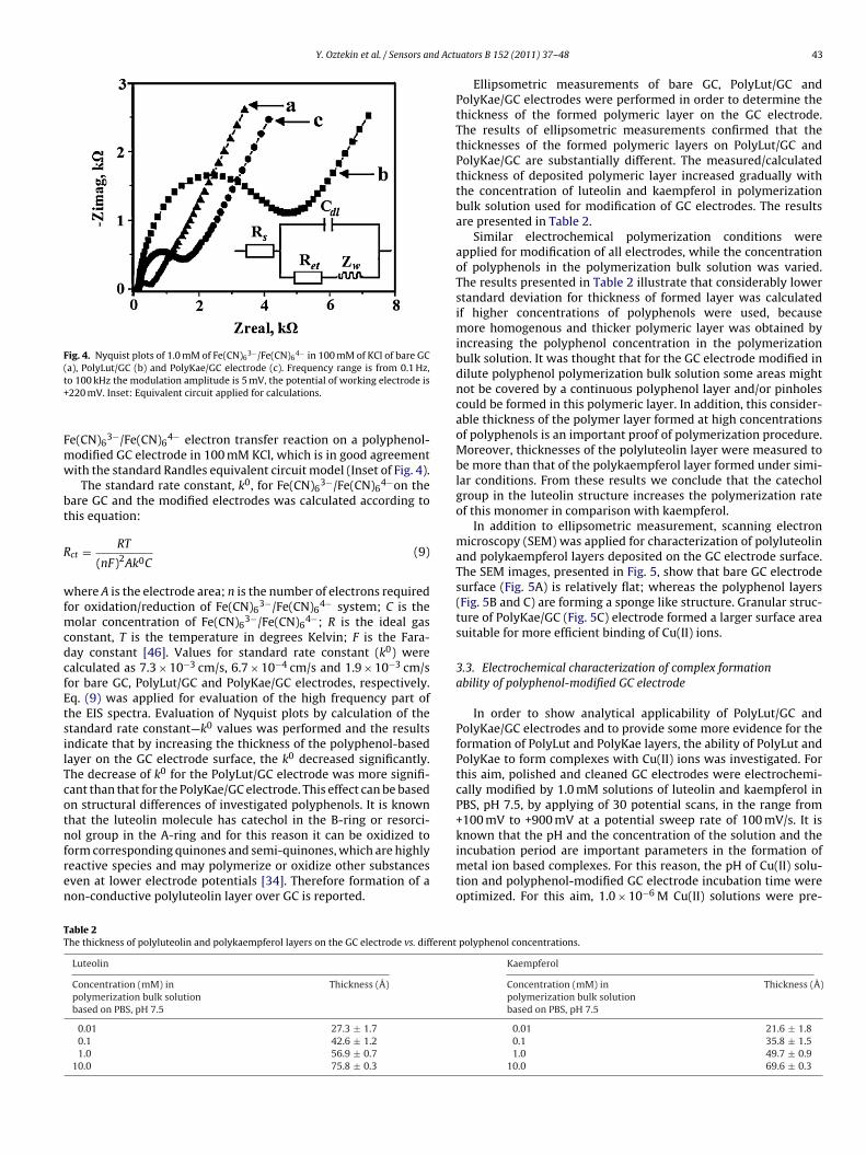
Y. Oztekin et al. / Sensors and Actu
Fig. 4. Nyquist plots of 1.0 mM of Fe(CN)63−/Fe(CN)6
4− in 100 mM of KCl of bare GC(t+
Fmw
bt
R
wfmcdcfEtsilTcotnfren
known that the pH and the concentration of the solution and the
TT
a), PolyLut/GC (b) and PolyKae/GC electrode (c). Frequency range is from 0.1 Hz,o 100 kHz the modulation amplitude is 5 mV, the potential of working electrode is220 mV. Inset: Equivalent circuit applied for calculations.
e(CN)63−/Fe(CN)6
4− electron transfer reaction on a polyphenol-odified GC electrode in 100 mM KCl, which is in good agreementith the standard Randles equivalent circuit model (Inset of Fig. 4).
The standard rate constant, k0, for Fe(CN)63−/Fe(CN)6
4−on theare GC and the modified electrodes was calculated according tohis equation:
ct = RT
(nF)2Ak0C(9)
here A is the electrode area; n is the number of electrons requiredor oxidation/reduction of Fe(CN)6
3−/Fe(CN)64− system; C is the
olar concentration of Fe(CN)63−/Fe(CN)6
4−; R is the ideal gasonstant, T is the temperature in degrees Kelvin; F is the Fara-ay constant [46]. Values for standard rate constant (k0) werealculated as 7.3 × 10⁻³ cm/s, 6.7 × 10⁻⁴ cm/s and 1.9 × 10⁻³ cm/sor bare GC, PolyLut/GC and PolyKae/GC electrodes, respectively.q. (9) was applied for evaluation of the high frequency part ofhe EIS spectra. Evaluation of Nyquist plots by calculation of thetandard rate constant—k0 values was performed and the resultsndicate that by increasing the thickness of the polyphenol-basedayer on the GC electrode surface, the k0 decreased significantly.he decrease of k0 for the PolyLut/GC electrode was more signifi-ant than that for the PolyKae/GC electrode. This effect can be basedn structural differences of investigated polyphenols. It is knownhat the luteolin molecule has catechol in the B-ring or resorci-ol group in the A-ring and for this reason it can be oxidized to
orm corresponding quinones and semi-quinones, which are highlyeactive species and may polymerize or oxidize other substancesven at lower electrode potentials [34]. Therefore formation of aon-conductive polyluteolin layer over GC is reported.
able 2he thickness of polyluteolin and polykaempferol layers on the GC electrode vs. different
Luteolin
Concentration (mM) inpolymerization bulk solutionbased on PBS, pH 7.5
Thickness (Å)
0.01 27.3 ± 1.70.1 42.6 ± 1.21.0 56.9 ± 0.7
10.0 75.8 ± 0.3
ators B 152 (2011) 37–48 43
Ellipsometric measurements of bare GC, PolyLut/GC andPolyKae/GC electrodes were performed in order to determine thethickness of the formed polymeric layer on the GC electrode.The results of ellipsometric measurements confirmed that thethicknesses of the formed polymeric layers on PolyLut/GC andPolyKae/GC are substantially different. The measured/calculatedthickness of deposited polymeric layer increased gradually withthe concentration of luteolin and kaempferol in polymerizationbulk solution used for modification of GC electrodes. The resultsare presented in Table 2.
Similar electrochemical polymerization conditions wereapplied for modification of all electrodes, while the concentrationof polyphenols in the polymerization bulk solution was varied.The results presented in Table 2 illustrate that considerably lowerstandard deviation for thickness of formed layer was calculatedif higher concentrations of polyphenols were used, becausemore homogenous and thicker polymeric layer was obtained byincreasing the polyphenol concentration in the polymerizationbulk solution. It was thought that for the GC electrode modified indilute polyphenol polymerization bulk solution some areas mightnot be covered by a continuous polyphenol layer and/or pinholescould be formed in this polymeric layer. In addition, this consider-able thickness of the polymer layer formed at high concentrationsof polyphenols is an important proof of polymerization procedure.Moreover, thicknesses of the polyluteolin layer were measured tobe more than that of the polykaempferol layer formed under simi-lar conditions. From these results we conclude that the catecholgroup in the luteolin structure increases the polymerization rateof this monomer in comparison with kaempferol.
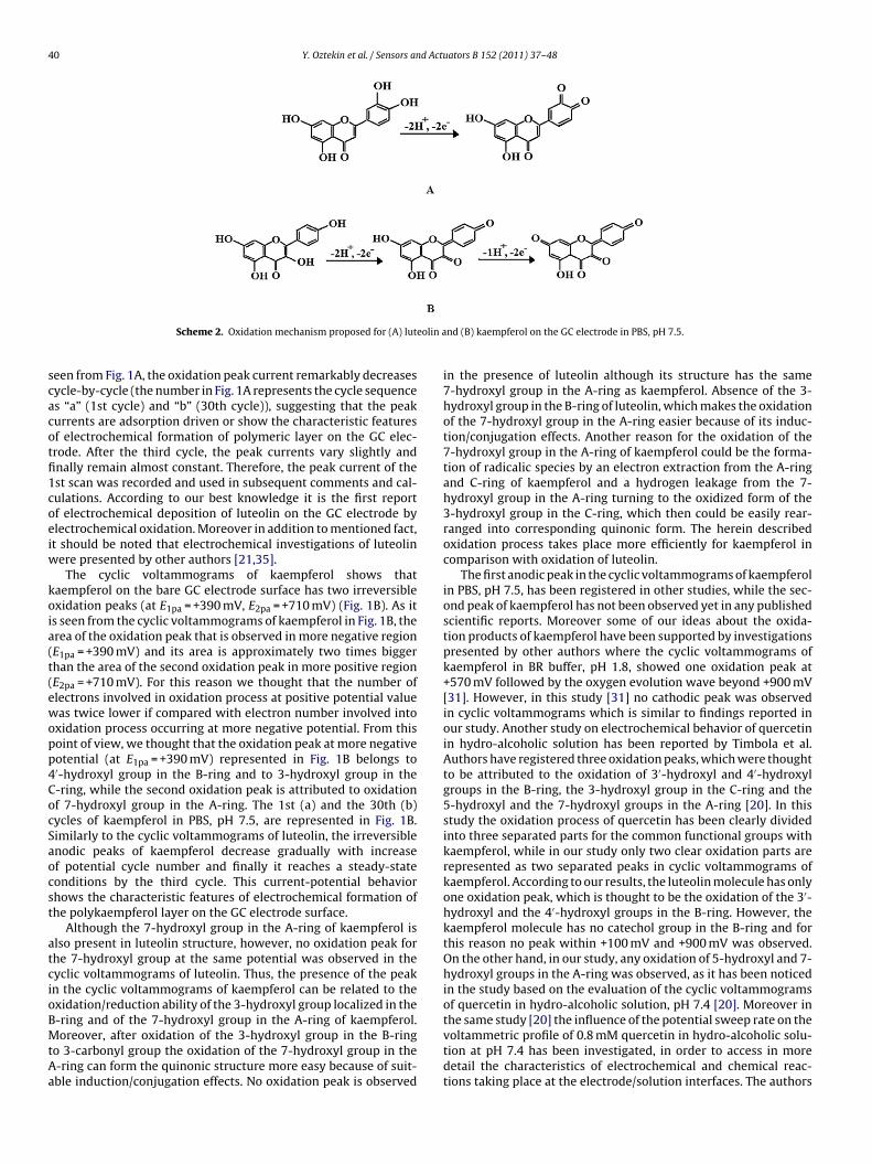
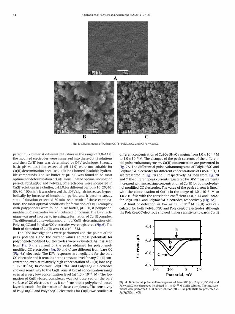
In addition to ellipsometric measurement, scanning electronmicroscopy (SEM) was applied for characterization of polyluteolinand polykaempferol layers deposited on the GC electrode surface.The SEM images, presented in Fig. 5, show that bare GC electrodesurface (Fig. 5A) is relatively flat; whereas the polyphenol layers(Fig. 5B and C) are forming a sponge like structure. Granular struc-ture of PolyKae/GC (Fig. 5C) electrode formed a larger surface areasuitable for more efficient binding of Cu(II) ions.
3.3. Electrochemical characterization of complex formationability of polyphenol-modified GC electrode
In order to show analytical applicability of PolyLut/GC andPolyKae/GC electrodes and to provide some more evidence for theformation of PolyLut and PolyKae layers, the ability of PolyLut andPolyKae to form complexes with Cu(II) ions was investigated. Forthis aim, polished and cleaned GC electrodes were electrochemi-cally modified by 1.0 mM solutions of luteolin and kaempferol inPBS, pH 7.5, by applying of 30 potential scans, in the range from+100 mV to +900 mV at a potential sweep rate of 100 mV/s. It is
incubation period are important parameters in the formation ofmetal ion based complexes. For this reason, the pH of Cu(II) solu-tion and polyphenol-modified GC electrode incubation time wereoptimized. For this aim, 1.0 × 10⁻⁶ M Cu(II) solutions were pre-
polyphenol concentrations.
Kaempferol
Concentration (mM) inpolymerization bulk solutionbased on PBS, pH 7.5
Thickness (Å)
0.01 21.6 ± 1.80.1 35.8 ± 1.51.0 49.7 ± 0.9
10.0 69.6 ± 0.3
44 Y. Oztekin et al. / Sensors and Actuators B 152 (2011) 37–48
GC, (B
ptabCiopC6bstwmnTPl
ppfm(Gc1semslo
for PolyLut/GC and PolyKae/GC electrodes, respectively (Fig. 7A).A limit of detection as low as 1.0 × 10⁻¹¹ M Cu(II) was cal-
culated for both PolyLut/GC and PolyKae/GC electrodes althoughthe PolyKae/GC electrode showed higher sensitivity towards Cu(II)
Fig. 5. SEM immages of (A) bare
ared in BR buffer at different pH values in the range of 3.0–11.0;he modified electrodes were immersed into these Cu(II) solutionsnd then Cu(II) ions was determined by DPV technique. Stronglyasic pH values (that exceeded pH 11.0) were not suitable foru(II) determination because Cu(II) ions formed insoluble hydrox-
de compounds. The BR buffer at pH 5.0 was found to be mostptimal for determination of Cu(II) ions. To find optimal incubationeriod, PolyLut/GC and PolyKae/GC electrodes were incubated inu(II) solutions in BR buffer, pH 5.0, for different periods (10; 20; 40;0; 80; 100 min). It was observed that DPV signals increased hyper-olically by increase of incubation period and it became steadytate if duration exceeded 60 min. As a result of these examina-ions, the most optimal conditions for formation of Cu(II) complexith polyphenols were found in BR buffer, pH 5.0, if polyphenolodified GC electrodes were incubated for 60 min. The DPV tech-
ique was used in order to investigate formation of Cu(II) complex.he differential pulse voltammograms of Cu(II) determination witholyLut/GC and PolyKae/GC electrodes were registered (Fig. 6). Theimit of detection of Cu(II) was 1.0 × 10⁻¹¹ M.
The DPV investigations were performed and the points of theeak potentials and the current values at these potentials forolyphenol-modified GC electrodes were evaluated. As it is seenrom Fig. 6 the current of the peaks obtained for polyphenol-
odified GC electrodes (Fig. 6b and c) are different from bare GCFig. 6a) electrode. The DPV responses are negligible for the bareC electrode and it remains at the constant level for any Cu(II) con-entration even at relatively high concentration of Cu(II) ions (e.g..0 × 10−6 M). In contrast, PolyLut/GC and PolyKae/GC electrodeshowed sensitivity to the Cu(II) ions at broad concentration range
−11
ven at a very low concentration level (at 1.0 × 10 M). The for-ation of Cu(II)-based complexes was not observed on the bareurface of GC electrode; thus it confirms that a polyphenol-basedayer is crucial for formation of these complexes. The sensitivityf PolyLut/GC and PolyKae/GC electrodes for Cu(II) was studied at
) PolyLut/GC and (C) PolyKae/GC.
different concentration of CuSO4·5H2O ranging from 1.0 × 10⁻¹¹ Mto 1.0 × 10⁻⁶ M. The changes of the peak currents of the differen-tial pulse voltammogrms vs. Cu(II) concentration are presented inFig. 7A. The differential pulse voltammograms of PolyLut/GC andPolyKae/GC electrodes for different concentrations of CuSO4·5H2Oare presented in Fig. 7B and C, respectively. As seen from Fig. 7Band C, the different peak currents registered by DPV measurementsincreased with increasing concentration of Cu(II) for both polyphe-nol modified GC electrodes. The value of the peak current is linearwith the concentration of Cu(II) in the range of 1.0 × 10⁻¹¹ M to1.0 × 10−6 M with the correlation coefficient as 0.9944 and 0.9927
Fig. 6. Differential pulse voltammograms of bare GC (a), PolyLut/GC (b) andPolyKae/GC (c) electrodes incubated in 1 × 10⁻¹¹ M Cu(II) solution. The measure-ments were performed in BR buffer solution, pH 5.0, all potentials are presented vs.Ag/AgCl/(sat. KCl).
Y. Oztekin et al. / Sensors and Actuators B 152 (2011) 37–48 45
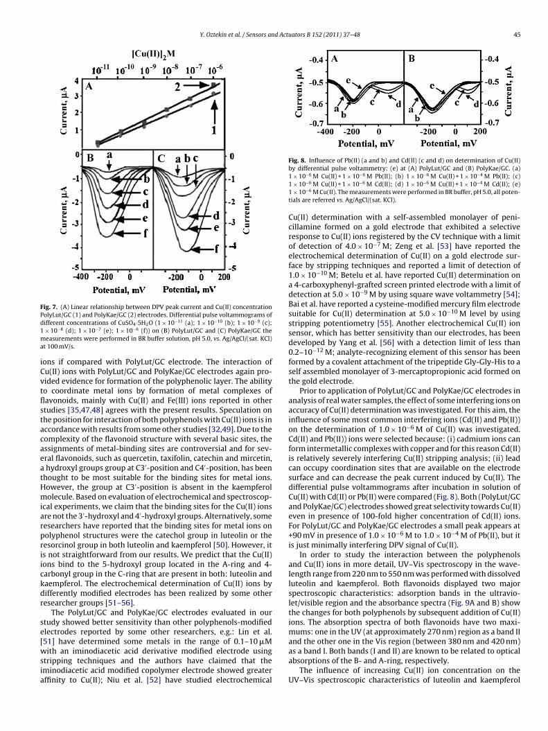
Fig. 7. (A) Linear relationship between DPV peak current and Cu(II) concentrationPd1ma
iCvtflstacaeatHmiarpriickdr
se[wsia
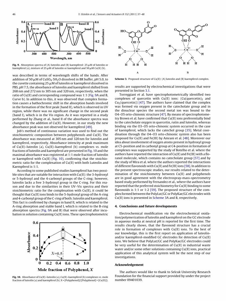
Fig. 8. Influence of Pb(II) (a and b) and Cd(II) (c and d) on determination of Cu(II)by differential pulse voltammetry: (e) at (A) PolyLut/GC and (B) PolyKae/GC. (a)
olyLut/GC (1) and PolyKae/GC (2) electrodes. Differential pulse voltammograms ofifferent concentrations of CuSO4·5H2O (1 × 10⁻¹¹ (a); 1 × 10⁻¹⁰ (b); 1 × 10⁻⁹ (c);× 10−8 (d); 1 × 10⁻⁷ (e); 1 × 10⁻⁶ (f)) on (B) PolyLut/GC and (C) PolyKae/GC theeasurements were performed in BR buffer solution, pH 5.0, vs. Ag/AgCl/(sat. KCl)
t 100 mV/s.
ons if compared with PolyLut/GC electrode. The interaction ofu(II) ions with PolyLut/GC and PolyKae/GC electrodes again pro-ided evidence for formation of the polyphenolic layer. The abilityo coordinate metal ions by formation of metal complexes ofavonoids, mainly with Cu(II) and Fe(III) ions reported in othertudies [35,47,48] agrees with the present results. Speculation onhe position for interaction of both polyphenols with Cu(II) ions is inccordance with results from some other studies [32,49]. Due to theomplexity of the flavonoid structure with several basic sites, thessignments of metal-binding sites are controversial and for sev-ral flavonoids, such as quercetin, taxifolin, catechin and mircetin,hydroxyl groups group at C3′-position and C4′-position, has been
hought to be most suitable for the binding sites for metal ions.owever, the group at C3′-position is absent in the kaempferololecule. Based on evaluation of electrochemical and spectroscop-
cal experiments, we claim that the binding sites for the Cu(II) ionsre not the 3′-hydroxyl and 4′-hydroxyl groups. Alternatively, someesearchers have reported that the binding sites for metal ions onolyphenol structures were the catechol group in luteolin or theesorcinol group in both luteolin and kaempferol [50]. However, its not straightforward from our results. We predict that the Cu(II)ons bind to the 5-hydroxyl group located in the A-ring and 4-arbonyl group in the C-ring that are present in both: luteolin andaempferol. The electrochemical determination of Cu(II) ions byifferently modified electrodes has been realized by some otheresearcher groups [51–56].
The PolyLut/GC and PolyKae/GC electrodes evaluated in ourtudy showed better sensitivity than other polyphenols-modifiedlectrodes reported by some other researchers, e.g.: Lin et al.
51] have determined some metals in the range of 0.1–10 �Mith an iminodiacetic acid derivative modified electrode usingtripping techniques and the authors have claimed that theminodiacetic acid modified copolymer electrode showed greaterffinity to Cu(II); Niu et al. [52] have studied electrochemical
1 × 10⁻⁶ M Cu(II) + 1 × 10⁻⁶ M Pb(II); (b) 1 × 10⁻⁶ M Cu(II) + 1 × 10⁻⁴ M Pb(II); (c)1 × 10⁻⁶ M Cu(II) + 1 × 10⁻⁶ M Cd(II); (d) 1 × 10⁻⁶ M Cu(II) + 1 × 10⁻⁴ M Cd(II); (e)1 × 10⁻⁶ M Cu(II). The measurements were performed in BR buffer, pH 5.0, all poten-tials are referred vs. Ag/AgCl/(sat. KCl).
Cu(II) determination with a self-assembled monolayer of peni-cillamine formed on a gold electrode that exhibited a selectiveresponse to Cu(II) ions registered by the CV technique with a limitof detection of 4.0 × 10⁻⁷ M; Zeng et al. [53] have reported theelectrochemical determination of Cu(II) on a gold electrode sur-face by stripping techniques and reported a limit of detection of1.0 × 10−10 M; Betelu et al. have reported Cu(II) determination ona 4-carboxyphenyl-grafted screen printed electrode with a limit ofdetection at 5.0 × 10⁻⁹ M by using square wave voltammetry [54];Bai et al. have reported a cysteine-modified mercury film electrodesuitable for Cu(II) determination at 5.0 × 10⁻¹⁰ M level by usingstripping potentiometry [55]. Another electrochemical Cu(II) ionsensor, which has better sensitivity than our electrodes, has beendeveloped by Yang et al. [56] with a detection limit of less than0.2–10−12 M; analyte-recognizing element of this sensor has beenformed by a covalent attachment of the tripeptide Gly-Gly-His to aself assembled monolayer of 3-mercaptopropionic acid formed onthe gold electrode.
Prior to application of PolyLut/GC and PolyKae/GC electrodes inanalysis of real water samples, the effect of some interfering ions onaccuracy of Cu(II) determination was investigated. For this aim, theinfluence of some most common interfering ions (Cd(II) and Pb(II))on the determination of 1.0 × 10⁻⁶ M of Cu(II) was investigated.Cd(II) and Pb(II)) ions were selected because: (i) cadmium ions canform intermetallic complexes with copper and for this reason Cd(II)is relatively severely interfering Cu(II) stripping analysis; (ii) leadcan occupy coordination sites that are available on the electrodesurface and can decrease the peak current induced by Cu(II). Thedifferential pulse voltammograms after incubation in solution ofCu(II) with Cd(II) or Pb(II) were compared (Fig. 8). Both (PolyLut/GCand PolyKae/GC) electrodes showed great selectivity towards Cu(II)even in presence of 100-fold higher concentration of Cd(II) ions.For PolyLut/GC and PolyKae/GC electrodes a small peak appears at+90 mV in presence of 1.0 × 10⁻⁶ M to 1.0 × 10⁻⁴ M of Pb(II), but itis just minimally interfering DPV signal of Cu(II).
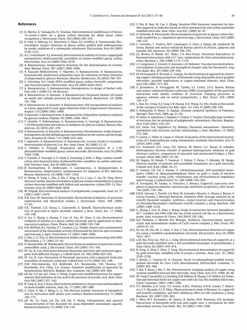
In order to study the interaction between the polyphenolsand Cu(II) ions in more detail, UV–Vis spectroscopy in the wave-length range from 220 nm to 550 nm was performed with dissolvedluteolin and kaempferol. Both flavonoids displayed two majorspectroscopic characteristics: adsorption bands in the ultravio-let/visible region and the absorbance spectra (Fig. 9A and B) showthe changes for both polyphenols by subsequent addition of Cu(II)ions. The absorption spectra of both flavonoids have two maxi-mums: one in the UV (at approximately 270 nm) region as a band II
and the other one in the Vis region (between 380 nm and 420 nm)as a band I. Both bands (I and II) are known to be related to opticalabsorptions of the B- and A-ring, respectively.The influence of increasing Cu(II) ion concentration on theUV–Vis spectroscopic characteristics of luteolin and kaempferol
46 Y. Oztekin et al. / Sensors and Actuators B 152 (2011) 37–48
Fk
watP2rCtir(pca
sakofmomK
bolsstaTAab
Ff
ig. 9. Absorption spectra of (A) luteolin and (B) kaempferol: 25 �M of luteolin oraempferol (a), mixture of 25 �M of luteolin or kaempferol and 50 �M Cu(II) (b).
as described in terms of wavelength shifts of the bands. Afterddition of 50 �M of CuSO4·5H2O dissolved in BR buffer, pH 5.0, tohe cuvette containing 25 �M of luteolin or kaempferol dissolved inBS, pH 7.5, the absorbance of luteolin and kaempferol shifted from68 nm and 272 nm to 305 nm and 320 nm, respectively, when theatio of Cu(II) and corresponding compound was 1:1 (Fig. 9A and B,urve b). In addition to this, it was observed that complex forma-ion causes a bathochromic shift in the absorption bands involvedn the formation of the first peak (band II), which is observed in UVegion, while there was no significant change in the second peakband I), which is in the Vis region. As it was reported in a studyerformed by Zhang et al., band II of the absorbance spectra washanged by the addition of Cu(II). However, in our study the newbsorbance peak was not observed for kaempferol [49].
Job’s method of continuous variation was used to find out thetoichiometric composition between polyphenols and Cu(II). Thebsorbance was measured at 305 nm and 320 nm for luteolin andaempferol, respectively. Absorbance intencity at peak maximumf Cu(II)–luteolin (a), Cu(II)–kaempferol (b) complexes vs. moleractions of luteolin and kaempferol are presented in Fig. 10 and the
aximal absorbance was registered at 1:1 mole fraction of luteolinr kaempferol with Cu(II) (Fig. 10), confirming that the stoichio-etric ratio for the complexation of Cu(II) with both Luteolin and
aempferol is 1:1.According to some published studies kaempferol has two possi-
le sites that are suitable for interaction with Cu(II): the 3-hydroxylr 5-hydroxyl and the 4-carbonyl groups of the C-ring; however,uteolin lacks a free 3-hydroxyl group in the C-ring. For this rea-on and due to the similarities in their UV–Vis spectra and theirtoichiometric ratio for the complexation with Cu(II), it could behought that Cu(II) ions binds to the 5-hydroxyl group of the A-ringnd 4-carbonyl group of the C-ring of both: luteolin and kaempferol.
his fact is confirmed by changes in band II, which is related to the-ring absorption and stable band I, which is related to the B-ringbsorption spectra (Fig. 9A and B) that were observed after incu-ation in solution containing Cu(II) ions. These spectrophotometricig. 10. Absorbance of Cu(II)–luteolin (a), Cu(II)–kaempferol (b) complexes vs. moleraction of luteolin (a) and kaempferol (b). X = [Polyphenol]/([Polyphenol] + [Cu(II)]).
Scheme 3. Proposed structure of Cu(II): (A) luteolin and (B) kaempferol complex.
results are supported by electrochemical investigations that werepresented in Section 3.1.
Torreggiani et al. have spectrophotometrically identified twocomplexes of quercetin with Cu(II) ions: (Cu(quercetin)2 andCu2(quercetin)) [47]. The authors have claimed that the complexwas formed via oxygen present in the catecholate group and inthe dinuclear species the second metal ion was bound to theO4–O5 orto-chinonic structure [47]. By means of spectrophotome-try Brown et al. have confirmed that Cu(II) ions preferentially bindto the catecholate oxygen in quercetin, rutin and luteolin, whereasbinding via the O3–O5 orto-chinonic system occurred in the caseof kaempferol, which lacks the catechol group [35]. Metal coor-dination through the O4–O3 orto-chinonic system also has beenproposed for Cu(II) and Fe(III) by Amrani et al. [48]. Moreover ouridea about involvement of oxygen atoms present in hydroxyl groupat C5-position and in carbonyl group at C4-position in formation ofcomplexes was supported by the study of Botelho et al. where theauthors have reported the interaction of Cu(II) and Fe(III) with a flo-ranol molecule, which contains no catecholate group [57] and bythe study of Mira et al. where the authors reported the interactionsof different flavonoids with Cu(II) and Fe(III) ions [58]. In addition tothe present spectroscopic studies, our results related to the deter-mination of the stoichiometry between Cu(II) and polyphenolsare in good agreement with the electrospray-mass-spectrometrybased study performed by Fernandez et al., where the authors havereported that the preferred stoichiometry for Cu(II) binding to someflavonoids is 1:1 or 1:2 [59]. The proposed structure of the com-plex formed between the PolyLut/GC or PolyKae/GC electrodes withCu(II) ions is presented in Scheme 3A and B, respectively.
4. Conclusions and future developments
Electrochemical modification via the electrochemical oxida-tion/polymerization of luteolin and kaempferol on the GC electrodein aqueous media at neutral pH is reported for the first time. Theresults clearly shows, that the flavonoid structure has a crucialrole in formation of complexes with Cu(II) ions. To the best ofour knowledge, this is the first report on application of luteolin-and/or kaempferol-modified GC electrodes for detection of Cu(II)ions. We believe that PolyLut/GC and PolyKae/GC electrodes couldbe very useful for the determination of Cu(II) in industrial wastewater and/or some other solutions containing Cu(II) ions, practicalapplication of this analytical system will be the next step of ourinvestigations.
Acknowledgement
The authors would like to thank to Selcuk University ResearchFoundation for the financial support provided by under the projectnumber 09401039.
d Actu
R
[
[
[
[
[
[
[
[
[
[
[
[
[
[
[
[
[
[
[
[
[
[
[
[
[
[
[
[
[
[
[
[
[
[
[
[
[
[
[
[
[
[
[
[
[
[
[
[
Y. Oztekin et al. / Sensors an
eferences
[1] K. Morita, A. Yamaguchi, N. Teramae, Electrochemical modifictaion of benzo-15-crown-5-ether on a glassy carbon electrode for alkali metal cationrecognition, J. Electroanal. Chem. 563 (2004) 249–255.
[2] K. Vaik, U. Mäeorg, F.C. Maschion, G. Maia, D.J. Schiffrin, K. Tammeveski, Elec-trocatalytic oxygen reduction on glassy carbon grafted with anthraquinoneby anodic oxidation of a carboxylate substituent, Electrochim. Acta 50 (2005)5126–5131.
[3] A.C. Cruickshank, A.J. Downard, Electrochemical stability of citrate-capped goldnanoparticles elektrostatically assembled on amine-modified glassy carbon,Electrochim. Acta 54 (2009) 5566–5570.
[4] A. Ramanavicius, Amperometric biosensor for the determination of creatine,Anal. Bioanal. Chem. 387 (2007) 1899–1906.
[5] A. Kausaite-Minkstimiene, V. Mazeiko, A. Ramanaviciene, A. Ramanavicius,Enzymatically synthesized polyaniline layer for extension of linear detectionof amperometric glucose biosensor, Biosens. Bioelectron. 26 (2010) 790–797.
[6] Z. Ustundag, A.O. Solak, EDTA modified glassy carbon electrode: preparationand characterization, Electrochim. Acta 54 (2009) 6426–6432.
[7] A. Ramanavicius, A. Ramanaviciene, Hemoproteins in design of biofuel cells,Fuel cells 1 (2009) 25–36 (Review).
[8] A. Ramanavicius, A. Kausaite, A. Ramanaviciene, Enzymatic biofuel cell basedon anode and cathode powered by ethanol, Biosens. Bioelectron. 24 (2008)761–766.
[9] A. Ramanavicius, A. Kausaite, A. Ramanaviciene, Self-encapsulation of oxidasesas a basic approach to tune upper detection limit of amperometric biosensors,Analyst 133 (2008) 1083–1089.
10] A. Kausaite, A. Ramanaviciene, A. Ramanavicius, Polyaniline synthesis catalyzedby glucose oxidase, Polymer 50 (2009) 1846–1851.
11] Y. Oztekin, V. Krikstolaityte, A. Ramanaviciene, Z. Yazicigil, A. Ramanavicius,1,10-Phenanthroline derivatives as mediators for glucose oxidase, Biosens. Bio-electron. 26 (2010) 267–270.
12] A. Ramanavicius, A. Kausaite, A. Ramanaviciene, Potentiometric study of quino-hemoprotein alcohol dehydrogenase immobilized on the carbon rod electrode,Sens. Actuators B: Chem. 113 (2006) 435–444.
13] I. Lapenaite, A. Ramanaviciene, A. Ramanavicius, Current trends in enzymaticdetermination of glycerol, Crit. Rev. Anal. Chem. 36 (2006) 13–25.
14] Y. Oztekin, Z. Yazicigil, Preparation and characterization of a 1,10-phenanthroline-modified glassy carbon electrode, Electrochim. Acta 54 (2009)7294–7298.
15] Y. Oztekin, Z. Yazicigil, A. O. Solak, Z. Ustundag, Z. Kilic, S. Bilge, Surface modifi-cation and characterization of phenanthroline nanofilms on carbon substrate,Surf. Interface Anal., doi:10.1002/sia.3662.
16] B. Kurtinaitiene, D. Ambrozaite, V. Laurinavicius, A. Ramanaviciene, A.Ramanavicius, Amperometric immunosensor for diagnosis of BLV infection,Biosens. Bioelectron. 23 (2008) 1547–1554.
17] K. Wang, H. Yang, L. Zhu, Z. Ma, S. Xing, Q. Lv, J. Liao, C. Liu, W. Xing, Directelectron transfer and electrocatalysis of glucose oxidase immobilized on glassycarbon electrode modified with Nafion and mesoporous carbon FDU-15, Elec-trochim. Acta 54 (2009) 4626–4630.
18] M. Filipiak, Electrochemical analysis of polyphenolic compounds, Anal. Sci. 17(2001) i1667–i1670.
19] H.R. Zare, M. Namazian, N. Nasirizadeh, Electrochemical behavior of quercetinexperimental and theoretical studies, J. Electroanal. Chem. 584 (2005)77–83.
20] A.K. Timbola, C.D. Souza, C. Giacomelli, A. Spinelli, Electrochemical oxida-tion of quercetin in hydro alcoholic solution, J. Braz. Chem. Soc. 17 (2006)139–148.
21] A. Liu, S. Zhang, L. Huang, Y. Cao, H. Yao, W. Chen, X. Lin, Electrochemicaloxidation of luteolin at a glassy carbon electrode and its application in phar-maceutical analysis, Chem. Pharm. Bull. 56 (2008) 745–748.
22] D.B. McPhail, R.C. Hartley, P.T. Gardner, G.G. Duthie, Kinetic and stoichiometricassessment of the antioxidant activity of flavonoids by electron spin resonancespectroscopy, J. Agric. Food Chem. 51 (2003) 1684–1690.
23] Z. Zhu, C. Li, N.Q. Li, Electrochemical studies of quercetin interacting with DNA,Microchem. J. 71 (2002) 57–63.
24] D. Nematollahi, M. Malakzadeh, Electrochemical oxidation of quercetin in ben-zenesulfinic acids, J. Electroanal. Chem. 547 (2003) 191–195.
25] W. Liu, R. Guo, Interaction between flavonoid, quercetin and surfactant aggre-gates with different charges, J. Colloid Interf. Sci. 302 (2006) 625–632.
26] W. Liu, R. Guo, Interaction of flavonoid, quercetin with organized molecularassemblies of nonionic surfactant, Colloid Surf. A 274 (2006) 192–199.
27] E.M. Cherviakovsky, D.A. Bolibrukh, A.V. Baranovsky, T.M. Vlasova, V.P.Kurchenko, A.A. Gilep, S.A. Usanov, Oxidative modification of quercetin byhemeproteins, Biochem. Biophys. Res. Commun. 342 (2006) 459–464.
28] J.B. He, G.P. Jin, Q.Z. Chen, Y. Wang, A quercetin modified biosensor for amper-ometric determination of uric acid in the presence of ascorbic acid, Anal. Chim.Acta 585 (2007) 337–343.
29] B. Yang, K. Arai, F. Kusu, Electrochemical behaviors of quercetin and kaempferolin neutral buffer solution, Anal. Sci. 17 (2001) 987–989.
30] G. Chen, X. Ma, F. Meng, G. Li, The electron transfer reactivity of kaempferoland its interaction with amino acid residues, Bioelectrochemistry 72 (2008)169–173.
31] J.B. He, S.J. Yuan, J.Q. Du, X.R. Hu, Y. Wang, Voltammetric and spectralcharacterization of two flavonols for assay-dependent antioxidant capacity,Bioelectrochemistry 75 (2009) 110–116.
[
ators B 152 (2011) 37–48 47
32] S. Niu, B. Han, W. Cao, S. Zhang, Sensitive DNA biosensor improved by lute-olin copper(II) as indicator based on silver nanoparticles and carbon nanotubesmodified electrode, Anal. Chim. Acta 651 (2009) 42–47.
33] D. Zielinska, B. Pierozynski, Electrooxidation of quercetin at glassy carbon elec-trode studied by a.c. impedance spectroscopy, J. Electroanal. Chem. 625 (2009)149–155.
34] C. Corredor, T. Teslova, M.V. Canamares, Z. Chen, J. Zhang, J.R. Lombardi, M.Leona, Raman and surface-enhanced Raman spectra of chrysin, apigenin andluteolin, Vib. Spectrosc. 49 (2009) 190–195.
35] J.E. Brown, H. Khodr, R.C. Hider, C.A. Rice-Evans, Structural dependence offlavonoid interactions with Cu2+ ions: implications for their antioxidant prop-erties, Biochem. J. 330 (1998) 1173–1178.
36] L.V. Jorgensen, C. Cornett, U. Justesen, L.H. Skibsted, Two electron electrochem-ical oxidation of quercetin and kaempferol changes only the flavonoid C-ring,Free Radic. Res. 29 (1998) 339–350.
37] M. Vestergaard, K. Kerman, E. Tamiya, An electrochemical approach for detect-ing copper-chelating properties of flavonoids using disposable pencil graphiteelectrodes: possible implications in copper-mediated illnesses, Anal. Chim.Acta 538 (2005) 273–281.
38] Z. Jurasekova, A. Torreggiani, M. Tamba, S.S. Cortes, J.V.G. Ramos, Ramanand surface-enhanced Raman scattering (SERS) investigation of the quercetininteraction with metals: evidence of structural changing processes inaqueous soliuton and on metal nanoparticles, J. Mol. Struct. 918 (2009)129–137.
39] L. Han, B.C. Dong, X.J. Yang, C.B. Huang, X.D. Wang, X.J. Wu, Study on flavonoidsin the caryopsis of indica rice Rdh, Agric. Sci. Chin. 8 (2009) 249–256.
40] S. Kobayashi, H. Ritter, D. Kaplan, Enzyme-catalyzed Synthesis of Polymers,Springer, New York, 2006, pp. 51–62.
41] H. Hotta, H. Sakamoto, S. Nagano, T. Osakai, Y. Tsujino, Unusually large numbersof electrons fort he oxidation of polyphenolic antioxidants, Biochim. Biophys.Acta 1526 (2001) 159–167.
42] K.E. Heim, A.R. Tagliaferro, D.J. Bobilya, Flavonoid antioxidants: chemistry,metabolism and structure–activity relationships, J. Nutr. Biochem. 13 (2002)572–584.
43] S. Guney, G. Yildiz, A. Capan, T. Ozturk, Evaluation of the electrochemical prop-erties of 3-hydroxyflavone using voltammetric methods, Electrochim. Acta 55(2010) 3295–3300.
44] S.A. Trammell, D.A. Lowy, D.S. Seferos, M. Moore, G.C. Bazan, N. Lebedev,Heterogenous electron transfer of quinone-hydroquinone solutions at goldelectroce surfaces: comparison of saturated and unsaturated bridges, J. Elec-troanal. Chem. 606 (2007) 33–38.
45] M. Nagata, M. Kondo, Y. Suemori, T. Ochiai, T. Dewa, T. Ohtsuka, M. Nango,Electron transfer of quinone self-assembled monolayers on a gold electrode,Colloids Surf. B 64 (2008) 16–21.
46] V. Ganesh, S.K. Pal, S. Kumar, V. Lakshminarayanan, Self-assembled mono-layers (SAMs) of alkoxyxyanobiphenyl thiols on gold—a study of electrontransfer reaction using cyclic voltammetry and electrochemical impedancespectroscopy, J. Colloid Interf. Sci. 296 (2006) 195–203.
47] A. Torregiani, M. Tamba, A. Trinchero, S. Bonora, Copper(II)–quercetin com-plexes in aqueous solutions: spectroscopic and kinetic properties, J. Mol. Struct.744 (2005) 759–766.
48] F.B.El. Amrani, L. Perello, J.A. Real, M. Gonzalez-Alvarez, G. Alzuet, J. Borras, S.Garcia-Granda, J. Montejo-Bernardo, Oxidative DNA cleavage induced by aniron(III) flavonoid complex: synthesis, crystal structure and characterizationof chlorobis(flavonolato) (methanol) iron(III) complex, J. Inorg. Biochem. 100(2006) 1208–1218.
49] G. Zhang, J. Guo, N. Zhao, J. Wang, Study of interaction between kaempferol-Eu3+ complex and DNA with the use of the neutral red dye as a fluorescenceprobe, Sens. Actuators B: Chem. 144 (2010) 239–246.
50] Q.K. Panhwar, S. Memon, M.I. Bhanger, Synthesis, characterization, spectro-scopic and antioxidation studies of Cu(II)–morin complex, J. Mol. Struct. 967(2010) 47–53.
51] M. Lin, M. Cho, W.-S. Choe, Y. Son, Y. Lee, Electrochemical detection of copperion using a modified copolythiophene electrode, Electrochim. Acta 54 (2009)7012–7017.
52] L.M. Niu, H.Q. Luo, N.B. Li, L. Song, Electrochemical detection of copper(II) at agold electrode modified with a self-assembled monolayer of penicillamine, J.Anal. Chem. 62 (2007) 470–474.
53] B. Zeng, X. Ding, F. Zhao, Y. Yang, Electrochemical determination of copper(II)by gold electrodes modified with N-acetyl-l-cysteine, Anal. Lett. 35 (2002)2245–2258.
54] S. Betelu, C. Vautrin-Ul, A. Chausse, Novel 4-carboxyphenyl-grafted screen-printed electrode for trace Cu(II) determination, Electrochem. Commun. 11(2009) 383–386.
55] Y. Bai, X. Ruan, J. Mo, Y. Xie, Potentiometric stripping analysis of copper usingcysteine modified mercury film electrode, Anal. Chim. Acta 373 (1998) 39–46.
56] W. Yang, D. Jaramillo, J.J. Gooding, D.B. Hibbert, R. Zhang, G.D. Willett, K.J. Fisher,Sub-ppt detection limits for copper ions with Gly-Gly-His modified electrodes,Chem. Commun. (2001) 1982–1983.
57] F.V. Botelho, J.I.A. Leite, V.S. Lemos, A.M.C. Pimenta, H.D.R. Calado, T. Maten-
cio, C.T. Miranda, E.C.P. Maia, Physicochemical study of floranol, its copper(II)and iron(III) complexes, and their inhibitory effect on LDL-oxidation, J. Inorg.Biochem. 101 (2007) 935–943.58] L. Mira, M.T. Fernandez, M. Santos, R. Rocha, M.H. Florencio, K.R. Jennings,Interactions of flavonoids with iron and copper ions: a mechanism for theirantioxidant activity, Free Radic. Res. 36 (2002) 1199–1208.

4 d Actu
[
B
8 Y. Oztekin et al. / Sensors an
59] M.T. Fernandez, M.L. Mira, M.H. Florencio, K.R. Jennings, Iron and copper chela-tion by flavonoids: an electrospray mass spectrometry study, J. Inorg. Biochem.92 (2002) 105–111.
iographies
Assist. Prof. Dr. Yasemin Oztekin received her masterdegree in 2003 with specialization in analytical chem-istry and PhD from Selcuk University, Konya/Turkey. Shewas working in Selcuk University, Faculty of Science andDepartment of Chemistry from 2002. She is doing herpostdoctoral research at Vilnius University, Faculty ofChemistry, NanoTechnas – Centre of Nanotechnology andMaterials Science. She is recently focusing her researchattention on interdisciplinary topics such as membranetechnology, electroanalytical techniques, surface modifi-cation and characterization techniques, biofuel cell andbiosensor applications.
Assoc. Prof. Dr. Zafer Yazicigil received his MSc andPhD degrees in Analytical Chemistry from Selcuk Uni-versity, Konya/Turkey, in 1991 and 1998, respectively.He started his academic career as a research assistantin 1996, he obtained his assistant professor degree in1999, and is currently an associate professor for 2 years
in Selcuk University, Faculty of Science and Departmentof Chemistry. His research fields include chromatographictechniques, membrane technology (electrodialysis, elec-trodeposition, salt splitting), electroanalytical techniques,surface modifications and characterization techniques,sensor applications.ators B 152 (2011) 37–48
Assoc. Prof. Dr. Almira Ramanaviciene is head of thedivision of “Immunotechnology” at the newly establishedState Research Institute “Centre of Innovative Medicine”.Prior to that she headed the Laboratory of “Immunoanal-ysis and Nanotechnology” at Institute of Immunology ofVilnius University, Lithuania. She received her PhD degreein biomedicine in 2002 from the Institute of Immunologyand Vilnius University. In 2008 she completed habilita-tion procedure in physical sciences at Vilnius University.She is serving as FP7 projects expert for the EuropeanCommission and other international foundations. Nowher primary research work is in the field of nanotech-nology, immunoanalysis, conducting polymers, bio- and
immuno-sensors. She is head of many national and international projects relatedto nanotechnology. She is national coordinator of several nanotechnology relatedCOST actions.
Prof. Habil. Dr. Arunas Ramanavicius is a professor andhead of the Research Center for Nanotechnology andMaterials Science – NanoTechnas at Vilnius University,Lithuania. At the same time he is also a senior researcherat the department of organic chemistry, Institute of Chem-istry, State Research Institute Centre for Physical andTechnological Sciences. In 1998 he received the PhDdegree and in 2002 he received the doctor habilitus degreefrom Vilnius University. He is serving as expert-evaluatorin EU FP7 program coordinated by European Commission
and he is technical advisor to many foundations locatedin European and non-European countries: Austria, France,Ireland, Lithuania, Georgia, etc. He has research interestsin various aspects of nanotechnology, bionanotechnology, nanomaterials, biosen-sorics, bioelectronics, biofuel cells and MEMS based analytical devices. He is anational coordinator of several nanotechnology related COST actions.


