Embed Size (px)
Citation preview


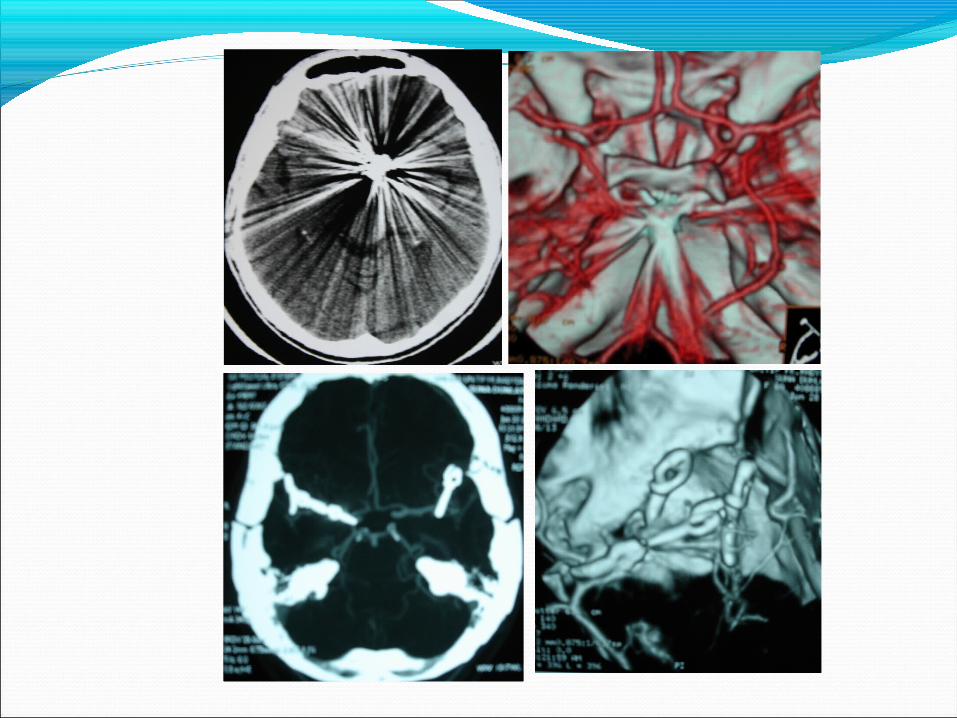
Vasküler Nöroşirürji 1- Anevrizma2- Karotis stenozu3-Arteriovenöz malformasyonlar

Anevrizma Tedavisi
Cerrahi Klipleme
Endovasküler tedavi

International Subarachnoid Aneurysm Trial 2009: Endovascular Coiling of Ruptured Intracranial Aneurysms Has No Significant Advantage Over Neurosurgical ClippingIn the May 2009 issue of The Lancet Neurology, the 5-year follow-up results of the InternationalSubarachnoid Aneurysm Trial (ISAT) were published. The authors concluded that, althoughthe significant difference between coiling and neurosurgical clipping of ruptured intracranialaneurysms in terms of death and severe disability after 1 year has vanished (primaryendpoint), coiling should still be favored over neurosurgical clipping because mortalityrates significantly favored coiling. In this commentary, it is this particular conclusion thatis challenged by combining data from previous ISAT publications with the current 5-yearfollow-up results. This modified intent-to-treat analysis clearly demonstrates that the significantadvantage in terms of mortality in favor of the endovascularly treated patients isno longer present, with a hazard ratio of 0.80 in favor of endovascular treatment (95% confidence
interval: 0.60-1.05; P = .10). Therefore, for everyday clinical practice and decision making, coiling and clipping are to be considered equivalent in the long term.Neurosurgery 66:961-962, 2010 DOI: 10.1227/01.NEU.0000368152.67151.73 www.neurosurgery- online.com
Nicolaas A. Bakker, MD, PhD, Jan D.M. Metzemaekers, MD, PhD Rob J.M. Groen, MD, PhD, Jan Jakob A. Mooij, MD, PhD, J. Marc C. Van Dijk, MD, PhDDepartment of Neurosurgery, University Medical Center Groningen, Groningen, The Netherlands

ISAT has demonstrated that endovascular coiling of rupturedintracranial aneurysms has a significant advantage over neurosurgicalclipping in the first year after treatment. After 5 years, the benefitseems to have vanished, and no significant difference in eitherdisability or mortality remains between the 2 treatment modalities.
Therefore, for everyday clinical practice and decision making, coilingand clipping are to be considered equivalent in the long term.
DisclosureThe authors have no personal financial or institutional interest in any of thedrugs, materials, or devices described in this article.


OUTCOME OF OCULOMOTOR NERVE PALSY FROM POSTERIOR COMMUNICATING ARTERY ANEURYSMS: COMPARISON OF CLIPPING AND COILING
OBJECTIVE: Recovery of posterior communicating artery aneurysm-induced oculomotor nerve palsy (ONP) after aneurysm coiling has been reported. However, the coil mass may compromise recovery of the nerve. Therefore, we compared the outcome of coiling and clipping for this indication.
METHODS: We retrospectively compared the outcomes of ONP in 13 patients, six of whom underwent endovascular coiling and seven of whom underwent surgical clipping.
RESULTS: Six of the seven surgical patients with ONP recovered completely, compared with two of the six patients in the endovascular group. Of the patients with more than 1 year of follow-up, all six surgical patients recovered completely, compared with two of four endovascular patients (P 0.05). In addition, preoperative complete or partial ONP also was associated with degree of resolution by survival analysis (P 0.03). All patients with partial ONP in the surgical group and two of three patients in the endovascular group recovered without residual deficits, whereas three of the four patients with complete ONP in the clipping group and none in the coiling group recovered completely. Regardless of the treatment method, time to complete resolution of ONP was 6 months in both groups.
CONCLUSION: Clipping posterior communicating artery aneurysms was associated with a higher probability of complete recovery from ONP than coiling. Degree of preoperative ONP also affected recovery.
If patients can tolerate surgery, it should be considered the treatment of choice.
KEY WORDS: Endovascular coiling, Oculomotor nerve palsy, Posterior communicating artery aneurysm,Surgical clippingNeurosurgery 58:1040-1046, 2006 DOI: 10.1227/01.NEU.0000215853.95187.5E www.neurosurgery-online.com
Robert F. Spetzler, M.D. Division of Neurological Surgery,Barrow Neurological Institute, St. Joseph’s Hospital and Medical Center,Phoenix, Arizona


Effect of Flow Diversion Treatment on Very Small Ruptured Aneurysms
BACKGROUND: Ruptured aneurysms of < 2 mm are not amenable to endovascular coilingand therefore pose a significant treatment challenge.OBJECTIVE: To test recently introduced flow diverters that allow endovascular reconstructionvia another method and may represent a new treatment option for such lesions.PATIENTS AND METHODS: Three female patients presented with acute subarachnoidhemorrhage. An aneurysm of < 2 mm was identified in all patients as the cause of bleeding.The aneurysms were located at the C2 segment of the internal carotid in 2 patientsand on the basilar bifurcation in the other. All patients had failed early endovascular treatmentattempts. Flow diversion with the SILK flow diverter was offered as an alternative ineach patient.RESULTS: SILK deployment successfully eliminated the aneurysms in all 3 instances. Oneof the aneurysms was excluded from contrast material visualization immediately afterstent deployment. Transient thrombotic complication was observed in the case of the basilarartery aneurysm. It resolved with the administration of intraarterial tirofiban. There wasno treatment-related morbidity, and none of the aneurysms reruptured after SILK implantationduring a clinical follow-up of at least 4 months (range, 4-10 months). Imaging followupshowed complete vessel remodeling in all cases.
CONCLUSION: Flow diversion treatment prevented rebleeding during the follow-up period. Reverse remodeling of the concerned vascular segment with delayed disappearance of the aneurysm was observed in each case.KEY WORDS: Cerebral aneurysm, Flow diversion, SILK, Subarachnoid hemorrhage, UncoilableNeurosurgery 67:789-793, 2010 DOI: 10.1227/01.NEU.0000372920.39101.55 www.neurosurgery- online.com
Zsolt Kulcsár, MDDepartment of Neuroradiology,Hirslanden Clinic,Zurich, SwitzerlandStephan G. Wetzel, MDDepartment of Neuroradiology,University Hospital of Basel,Basel, SwitzerlandLuca Augsburger, PhDLaboratory of Hemodynamics andCardiovascular Technology,Federal Institute of Technology,Lausanne, SwitzerlandAndreas Gruber, MD, PhDDepartment of Neurosurgery,University of Vienna,Vienna, AustriaIsabel Wanke, MD, PhDDepartment of Neuroradiology,Hirslanden Clinic,Zurich, Switzerland; andDepartment of Neuroradiology,University of Essen,Essen, GermanyDaniel Andre Rüfenacht,MD, PhDDepartment of Neuroradiology,Hirslanden Clinic,Zurich, Switzerland


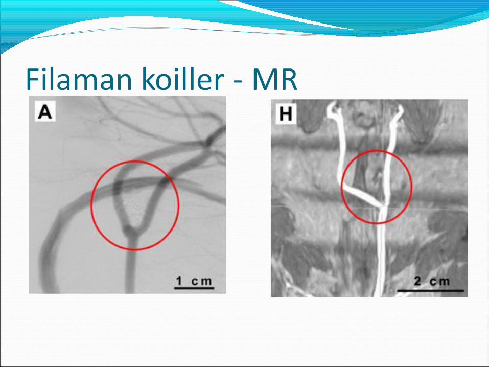
Normal koil Filaman koil


Ibudilast Inhibits Cerebral Aneurysms by Down-Regulating Inflammation-Related Molecules in the Vascular Wall of RatsOBJECTIVE: Phosphodiesterase-4 (PDE-4) is a cyclic adenosine monophosphate–specificenzyme involved in various inflammatory diseases. We studied its role in and the effect ofibudilast, which predominantly blocks PDE-4, on rat cerebral aneurysms.METHODS: Cerebral aneurysms were induced at the anterior cerebral artery–olfactoryartery bifurcation of female rats subjected to hypertension, increased hemodynamic stress,and estrogen deficiency. The effect of ibudilast (30 or 60 mg/kg/d for 3 months) on theircerebral aneurysms was studied by morphological and immunohistochemical assessmentand quantitative real-time polymerase chain reaction assay. In our in vitro study, we grewendothelial cells stimulated by angiotensin II under estrogen-free conditions and examinedthe effect of ibudilast on PDE-4 activation and the cyclic adenosine monophosphate level.RESULTS: Morphological evaluation using vascular corrosion casts showed ibudilastsignificantly suppressed cerebral aneurysms in a dose-dependent manner. In rats withinduced cerebral aneurysms, the gene and protein expression of PDE-4 was high, andendothelial leukocyte adhesion molecules (P-selectin, intracellular adhesion molecule1, and vascular adhesion molecule 1), matrix metalloproteinase-9, and tumor necrosis αwere expressed. Macrophage migration was also increased. Treatment with ibudilastdown-regulated these molecules, suppressed macrophage migration into the aneurysmwall, and inhibited PDE-4 activation and the elevation of cyclic adenosine monophosphatein endothelial cells.CONCLUSION: These results suggest that blocking of PDE4 is associated with the reduction of inflammation-related molecules and macrophage migration, thereby reducing the progression of cerebral aneurysms. It may represent a new conservative therapy to treat patients with cerebral aneurysms.
KEY WORDS: Cerebral aneurysm, Ibudilast, Leukocyte adhesion molecules, Phosphodiesterase inhibitor, Vascular inflammationNeurosurgery 66:551-559, 2010 DOI: 10.1227/01.NEU.0000365771.89576.77 www.neurosurgery- online.com
Kenji Yagi, MD Yoshiteru Tada, MD Keiko T. Kitazato, BS Tetsuya Tamura, MD Junichiro Satomi, MD, PhD Shinji Nagahiro, MD, PhDDepartment of Neurosurgery, Institute of Health Biosciences, The University of Tokushima Graduate School,Tokushima, Japan


Karotis Stenozu
1- Cerrahi tedavi
2- Endovasküler tedavi

Completed1991StatusTrial complete. Initial results published 8/91.
Trial PhasePhase III
SponsorNational Institute of Neurologic Disorders and Stroke, NIH
NASCET North American Symptomatic Carotid Endarterectomy Trial
ResultsThe risk of ipsilateral stroke was reduced significiantly (p=0.045) in patients with carotid stenosis 50-69% who received carotid endarterectomy. Patients with stenosis of 70-99% showed the most significant reduction(p < 0.001) in the rate of ipsilateral stroke while patients with stenosis of <50% did not show a significantly lower rate of ipsilateral stroke.

Perspect Vasc Surg Endovasc Ther. 2006 Dec;18(4):300-3; discussion 304-5. Links Carotid stent trials: past, present, and future.Quirel KDivision of Surgery, Cleveland Clinic, Cleveland, Ohio 44195, USA. [email protected]
Carotid stenting has emerged as a therapeutic alternative to standard carotid endarterectomy in patients with carotid bifurcation disease. The percutaneous modality holds the potential to replace a large proportion of the carotid surgical procedures performed throughout the world. Carotid stenting has undergone technologic advances in the last decade, including improved sheaths and guides, lower profile balloons and stents, and the almost ubiquitous use of dependable distal embolization protection devices. Contemporary data confirm the safety and efficacy of the procedure for patients with high-grade lesions who are at higher-than-normal risk for standard open carotid repair.
Whether lower-risk patients should be offered stenting as an alternative to carotid endarterectomy is a question that must await the results of ongoing clinical trials

Recently, short-term results (120 days) of the International Carotid Stenting Study (ICSS), arandomized multicentrer, international, controlled trial with blinded adjudication of outcomescomparing stenting vs endarterectomy for recently symptomatic carotid artery stenosisdemonstrated no significant difference in disabling stroke or death in patients receivingstenting (4.0%) as compared with CEA (3.2%;
The rate of any stroke or death within 30 days of treatment in the stenting groupwas more than twice the rate recorded in the CEA group and there were also more fatal strokes and fatal myocardial infarctions in the stenting group. As expected, cranial nerve deficits and hematomas were significantly more common in the CEA group.
Carotid Endarterectomy Vs Endovascular Stenting:Recent Results From ICSS and CREST

Carotid Endarterectomy Vs Endovascular Stenting:Recent Results From ICSS and CREST
In-depth functional outcomes may also be important in weighing the risks and benefits of treatments. Previous studies have shown significantly increased rates of non-disabling stroke in patients receiving endovascular therapy,11,13 and recent studies show that these strokes may have significant long-term impact on development of dementia. 14 Furthermore, new stents coming to the market already raise the question as to whether results will already be outdated by the time of CREST publication. In conclusion, the new data from ICSS and CREST continue to support the practice that most patients with carotid stenosis are best treated with endarterectomy, but that stenting is a safe and efficacious alternative in those patients who are deemed poor candidates for surgery.
RICARDO J. KOMOTARROBERT M. STARKE E. SANDER CONNOLLY
International Carotid Stenting Study (ICSS),
Carotid Revascularization Endarterectomy vs Stenting Trial (CREST)
N12 | VOLUME 66 | NUMBER 6 | JUNE 2010 www.neurosurgery-online.com
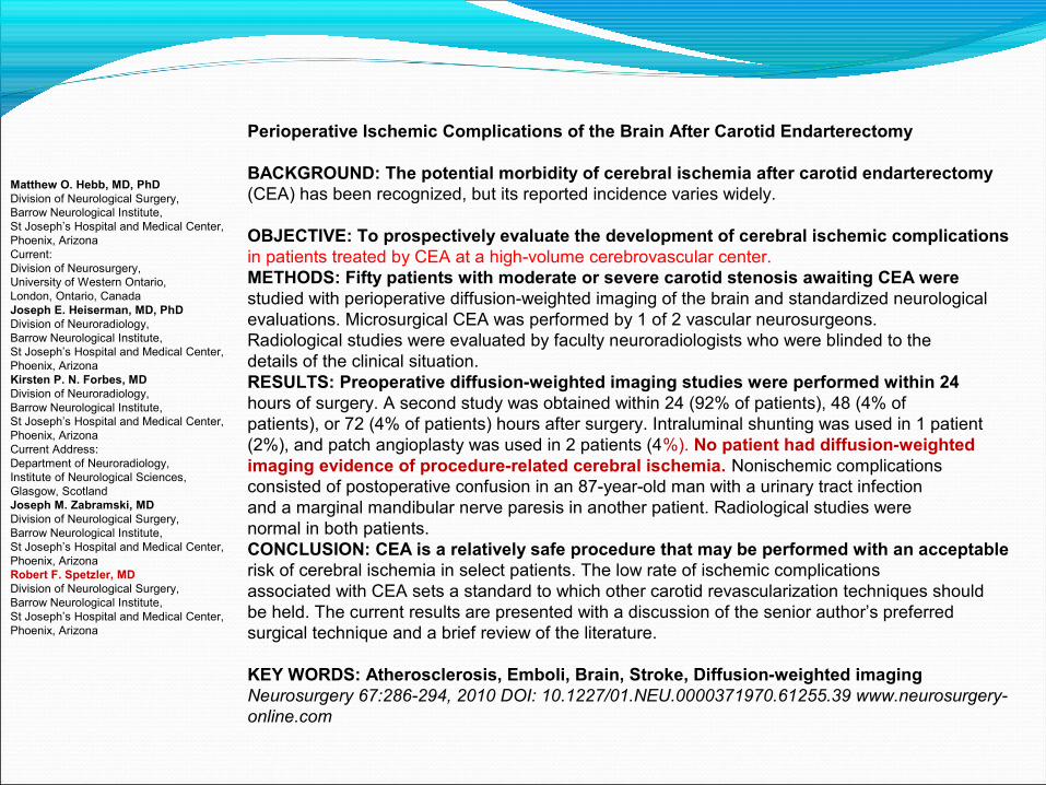
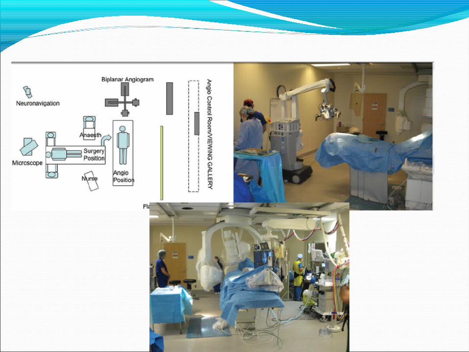
Perioperative Ischemic Complications of the Brain After Carotid Endarterectomy
BACKGROUND: The potential morbidity of cerebral ischemia after carotid endarterectomy(CEA) has been recognized, but its reported incidence varies widely.
OBJECTIVE: To prospectively evaluate the development of cerebral ischemic complicationsin patients treated by CEA at a high-volume cerebrovascular center.METHODS: Fifty patients with moderate or severe carotid stenosis awaiting CEA werestudied with perioperative diffusion-weighted imaging of the brain and standardized neurologicalevaluations. Microsurgical CEA was performed by 1 of 2 vascular neurosurgeons.Radiological studies were evaluated by faculty neuroradiologists who were blinded to thedetails of the clinical situation.RESULTS: Preoperative diffusion-weighted imaging studies were performed within 24hours of surgery. A second study was obtained within 24 (92% of patients), 48 (4% ofpatients), or 72 (4% of patients) hours after surgery. Intraluminal shunting was used in 1 patient(2%), and patch angioplasty was used in 2 patients (4%). No patient had diffusion-weightedimaging evidence of procedure-related cerebral ischemia. Nonischemic complicationsconsisted of postoperative confusion in an 87-year-old man with a urinary tract infectionand a marginal mandibular nerve paresis in another patient. Radiological studies werenormal in both patients.CONCLUSION: CEA is a relatively safe procedure that may be performed with an acceptablerisk of cerebral ischemia in select patients. The low rate of ischemic complicationsassociated with CEA sets a standard to which other carotid revascularization techniques shouldbe held. The current results are presented with a discussion of the senior author’s preferredsurgical technique and a brief review of the literature.
KEY WORDS: Atherosclerosis, Emboli, Brain, Stroke, Diffusion-weighted imagingNeurosurgery 67:286-294, 2010 DOI: 10.1227/01.NEU.0000371970.61255.39 www.neurosurgery- online.com
Matthew O. Hebb, MD, PhDDivision of Neurological Surgery,Barrow Neurological Institute,St Joseph’s Hospital and Medical Center,Phoenix, ArizonaCurrent:Division of Neurosurgery,University of Western Ontario,London, Ontario, CanadaJoseph E. Heiserman, MD, PhDDivision of Neuroradiology,Barrow Neurological Institute,St Joseph’s Hospital and Medical Center,Phoenix, ArizonaKirsten P. N. Forbes, MDDivision of Neuroradiology,Barrow Neurological Institute,St Joseph’s Hospital and Medical Center,Phoenix, ArizonaCurrent Address:Department of Neuroradiology,Institute of Neurological Sciences,Glasgow, ScotlandJoseph M. Zabramski, MDDivision of Neurological Surgery,Barrow Neurological Institute,St Joseph’s Hospital and Medical Center,Phoenix, ArizonaRobert F. Spetzler, MDDivision of Neurological Surgery,Barrow Neurological Institute,St Joseph’s Hospital and Medical Center,Phoenix, Arizona


Arteri venöz malformasyon
1.Cerrahi tedavi
2.Embolizasyon
3. Gama knife

How Safe Is Arteriovenous Malformation Surgery? A Prospective, Observational Study of Surgery As First-Line Treatment for Brain Arteriovenous Malformations
OBJECTIVES: Existing studies reporting the risk of surgery for brain arteriovenous malformations(AVMs) are often biased by the exclusion of patients not offered surgery. In thisstudy, we examine the risk of surgery, including cases excluded from surgery because ofthe high surgical risk.METHODS: Data were collected on 640 consecutively enrolled AVMs in a database thatincluded all patients not considered for surgery.RESULTS: Patients with Spetzler-Martin grade 1 to 2 AVMs (n = 296) were treated with a surgicalrisk of 0.7% (95% confidence interval [CI], 0%-3%); patients with Spetzler-Martin grade3 to 4 AVMs in noneloquent cortex (n = 65) were treated with a surgical risk of 17% (95%CI, 10%-28%). Patients with Spetzler-Martin grade 3 to 5 AVMs in eloquent cortex (n = 168)were treated with a surgical risk of 21% (95% CI, 15%-28%). However, because 14% ofpatients in this series with similar AVMs were refused surgery because of perceived surgicalrisk, these results are not generalizable to the population of patients with similar AVMs.
CONCLUSION: The results of this series suggest that it is reasonable to offer surgery as apreferred treatment option for Spetzler-Martin grade 1 to 2 AVMs. This study also reinforcesthe predictive value of the Spetzler-Martin grading system, with some caveats.
KEY WORDS: Intracranial arteriovenous malformations, Neurosurgical procedures, Research design
Neurosurgery 66:498-505, 2010 DOI: 10.1227/01.NEU.0000365518.47684.98 www.neurosurgery- online.com
Andrew S. Davidson, MS., Michael K. Morgan, MDAustralian School of Advanced Medicine,Macquarie University, Sydney, Australia

CONCLUSIONSTo undertake a valid discussion of the risks of treatment in brain AVMs, an accurate knowledge of the risks and benefits of all management strategies including the natural history is essential. Surgical series are typically limited by biases that affect their validity, and our analysis confirms that a significant selection bias for surgical treatment exists for selected groups of patients with AVMs in this series. In an attempt to compensate for these biases, we describe a rational approach to reporting surgical morbidity by studying the upper 95% CIs and considering patients excluded from treatment.
This approach reveals that surgery can be performed on an unselected group of patients with Spetzler-Martin grade 1 to 2 AVMswith a risk of downgrade due to surgery of less than 3%.
In patients with Spetzler-Martin grade 3 to 4 AVMs in noneloquent cortex, the risk of surgery is less than 30%.
In patients with Spetzler- Martin grade 3 to 5 AVMs in eloquent cortex, the risk of surgery is certainly greater than 16% and may be as high as 41%.
The results of this series are sufficiently reassuring that it remains reasonable to continue to offer surgery as our preferred treatment options for all Spetzler-Martin grade 1 to 2 AVMs.
This study also reinforces the predictive value of the Spetzler- Martin grading system, with some caveats relating to the generalizability of surgical series where a significant number of patients may have been excluded from the reported results.


IA
IV


VE SON SÖZ......
Bir saatliğine mutlu olmak için, şekerleme yapın Bir günlüğüne mutlu olmak için, balık avlamaya gidin Bir aylığına mutlu olmak için, evlenin Bir yıllığına mutlu olmak için, bir servete konun
Tüm yaşam boyunca mutlu olmak için, işinizi sevin...
ÇİN ATASÖZÜ