Embed Size (px)
Citation preview

CHAPTER 19

6 BASIC PROCESSES
1. Ingestion 2. Secretion 3. Mixing and propulsion 4. Digestion 5. Absorption 6. Defecation

LAYERS OF THE GI TRACT
Mucosa- inner lining, direct contact with contents, Submucosa- blood vessels and lymphatic vessels, neutrons
for control Muscularis- consists of skeletal muscle for voluntary
swallowing and smooth muscle in the rest of the GI tract. Serosa and peritoneum- outermost layer, secretes watery
fluid to allow the tract to glide easily against other organs


THE MOUTH
Hard palate- bones of maxillae and palatine of the skull, roof of mouth
Soft palate- muscular, near the back Uvula- prevents swallowed foods and liquids into the
nasal cavity Palatine tonsils- posterior to soft palate near the
opening of the oropharynx.


TONGUE
Forms the floor of the oral cavity. Composed of skeletal muscle. Covered with papillae or taste buds. Scientists recognize 5 taste types. Sweet, sour, salty, bitter and umami.

SALIVARY GLANDS
Parotid glands- between the ears and the masseter muscle, lower jaw
Submandibular glands- base of tongue and floor of mouth
Sublingual glands- anterior to submandibular glands


TEETH
Incisors-cut into food cuspids (canines)- tear and shred food bicuspids (premolars)- crush and grind food Molars- crush and grind


DIGESTION IN THE MOUTH
Mechanical digestion in the mouth results from chewing or mastication in which the food is manipulated by the tongue, ground by the teeth and mixed with saliva.
The food is reduced to a soft, flexible and easily swallowed mass called a bolus.

PHARYNX AND ESOPHAGUS
When food is swallowed, it passes from the mouth into the pharynx.
Muscular contractions move the bolus, into the esophagus.
The esophagus is a muscular tube and connects to the stomach.
It is lined with stratified squamous epithelium and mucous. Swallowing involves the mouth, pharynx and esophagus
helped by saliva and mucus.


Vocabulary Words
Layers of GI tract (4 layers) Salivary glands (3) Mouth (4 structures) Teeth (4 types) Pharynx Esophagus

THE STOMACH
J-shaped enlargement of the GI tract directly below the diaphragm.
Connects the esophagus to the duodenum, the first part of the small intestine.
Serves as a mixing chamber and holding reservoir for food. It is the most elastic part of the GI tract and can
accommodate a large amount of food. The diaphragm pushes and pulls the stomach with each
breath.


STRUCTURE OF THE STOMACH
The stomach is composed of the same four layers as the rest of the GI tract. (mucosa, submucosa, muscularis and serosa.)
When empty, the muscosa lie in large folds called rugae. The stomach is connected to the duodenum by the pyloric
sphincter. The stomach contains many types of cells that secrete various
chemicals.
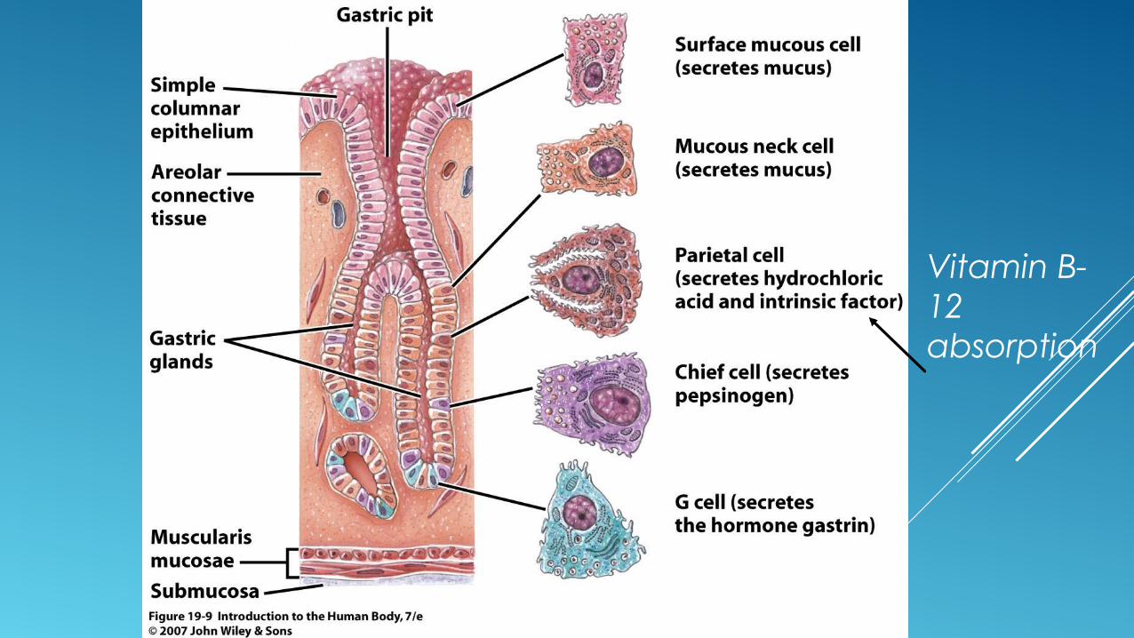
Vitamin B-12 absorption
DIGESTION AND ABSORPTION IN THE STOMACH
Even before food enters the stomach, reflexes from the senses prepare the stomach including injection of various digestion enzymes.
Once the food reaches the stomach, the wall is stretched and pH changes.
Nerve impulses trigger mixing waves which are rippling movements (peristalsis) of the muscles in the stomach.
The waves macerate the food and mix it with the secretions of the gastric juices producing chyme.
Little absorption occurs except for certain medications, water and alcohol.


PANCREAS
Located behind the stomach, the pancreas secretes chemicals via the pancreatic duct into the duodenum which unites with the common bile duct from the liver and gallbladder.
The pancreas is also part of the endocrine system, because it secretes hormones.
Pancreatic juice consists of water, salts, sodium bicarbonate and enzymes.
Neurons and two hormones (secretin and cholecystokinin) regulate the secretion of pancreatic juice.

PancreasSmall intestine


Vocabulary Cards
Stomach Bolus Chyme pancreas

LIVER AND GALLBLADDER
The liver is the second largest organ in the body.
The gallbladder is a pear-shaped sac that hangs from the lower part of the liver.
The liver’s lobes are made of many functional units called lobules.
A lobule consists of specialized cells called hepatocytes arranged around a central vein.
Bile is secreted by hepatocytes that enter bile ducts and eventually form the right and left hepatic ducts.
The purpose of bile is to emulsify (turn into tiny droplets) triglycerides. (fat)


These unite and exit the liver through the common hepatic duct.
The hepatic duct joins with the cystic duct from the gallbladder forming the common bile duct.
The gallbladder stores the bile until it’s used.


FUNCTIONS OF THE LIVER
1. carbohydrate metabolism 2. lipid metabolism 3. protein metabolism 4. processing of drugs and hormones. 5. excretion of bilirubin (bile) 6. storage of vitamins and minerals. 7. activation of vitamin D.


SMALL INTESTINE
Within 2-4 hours of eating a meal, the stomach has emptied its contents into the small intestine where the major events of digestion and absorption occur.
The small intestine is about 10 feet long in a living person and 21 feet in a cadaver. Why the differences?
It is divided into 3 sections. The duodenum attaches to the stomach, the jejunum is the
middle portion and the ileum is attached to the large intestine.


Projections called circular folds are permanent ridges in the muscosa.
They enhance absorption by increasing surface area and causing chyme to spiral rather than go in a straight line as it passes in the small intestine.
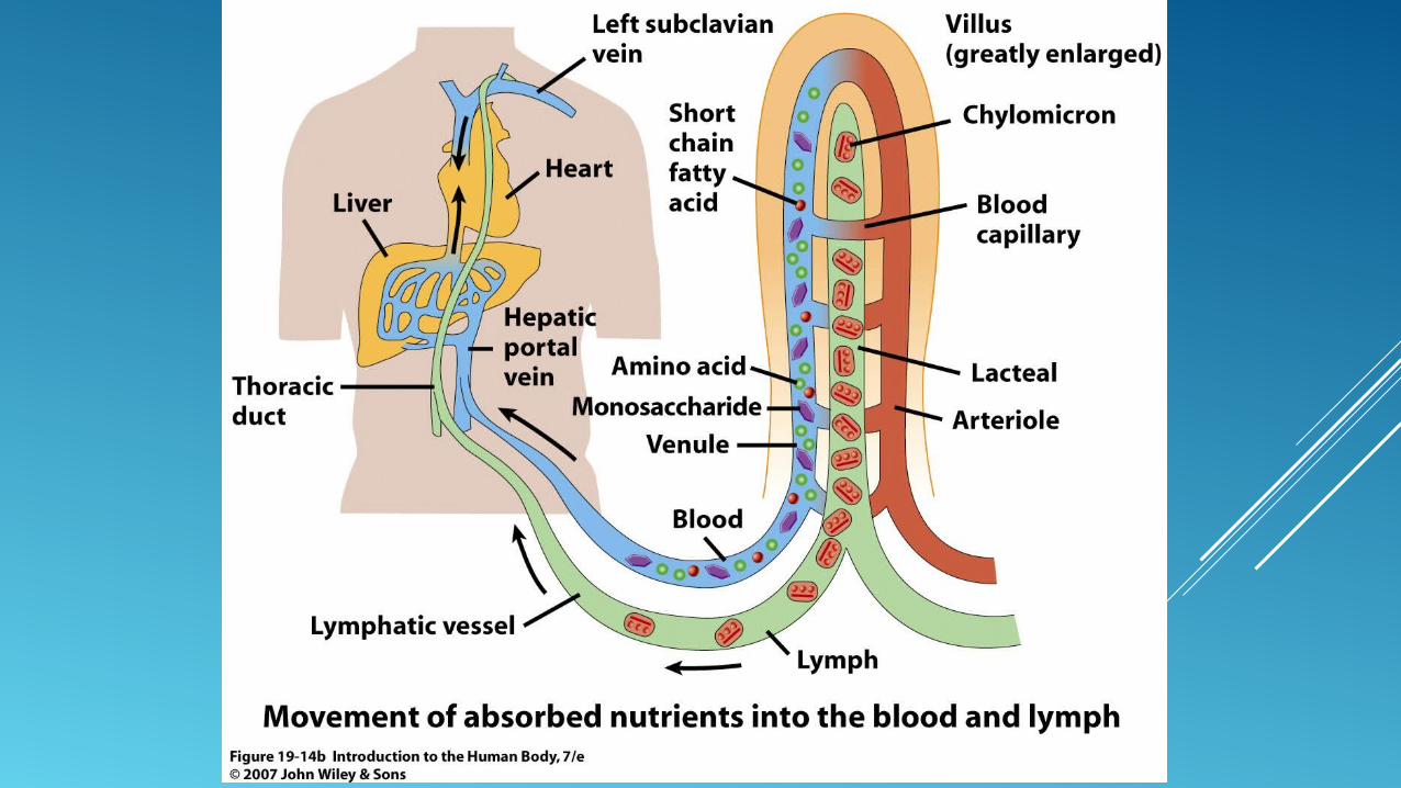
The muscosa contains villi and microvilli.


The villi are numerous fingerlike projections consisting of simple columnar epithelium.
The epithelium contains absorptive cells, mucus-secreting goblet cells, and endocrine cells.
The microvilli digest nutrients and move them to the absorptive cells.


Mechanical Digestion in the Small Intestine
Segmentations are localized contractions that move chyme back and forth, mixing it with digestive juices.
Peristalsis moves the chyme forward into the small intestine and the wave migrates down the small intestine reaching the end in 90 to 120 minutes.
The chyme generally remains in the small intestine 3-5 hours.

Chemical Digestion in the Small Intestine
The chyme entering the small intestine contains partially digested carbohydrates and proteins.
Pancreatic amylase, maltase, sucrase and lactase act upon specific sugars in the chyme and break them into smaller units to be absorbed.
Peptidases break down proteins. Pancreatic lipase breaks down lipids. Nucleases digest nucleotides.

Vocabulary Words
Small intestine (3 parts)
Villi Microvilli Bile Liver Gallbladder Lobules hepatocytes

Large Intestine
The large intestine is about 5 feet in length. The overall functions of the large intestine are
completion of absorption, production of vitamins, the formation of feces and the expulsion of feces.

Structure of the Large Intestine
At beginning of the large intestine, the ileum connects via a sphincter.
The materials from the small intestine pass into the large intestine here.
The first segment of the large intestine is the cecum with an attachment called the appendix.
The appendix contains further digestive enzymes.

The ascending colon goes up the side into the transverse colon which is horizontal. This leads into the descending colon which travels downward.
The sigmoid colon is an S-shaped part which connects to the rectum and anus.
The rectum stores feces. The anus is the opening to the outside of the
body.


The walls of the large intestine differ from the small intestine in that there are no villi or circular folds.
The epithelium contain goblet cells for lubrication and absorptive cells that function primarily in ion and water absorption.
Unlike the rest of the GI tract, the muscularis of the large intestine is bundled in three longitudinal bands giving the intestine a puckered appearance.


Digestion and Absorption
Passage of chyme from the ileium into the cecum is regulated by the sphincter.
Immediately after a meal, a reflex intensifies peristalsis. Mass peristalsis is a characteristic of the large intestine which is
a huge wave driving the contents of the colon into the rectum. Food in the stomach initiates mass peristalsis which usually
takes place 3-4 times a day during or immediately after a meal.

The final stage of digestion occurs in the colon through the activity of bacteria.
Bacteria ferment any remaining carbohydrates and create gases.
These gases contribute to flatus. Several vitamins like B and K are absorbed in the
colon. Bile pigments are broken down into simpler pigments
which give feces the brown color.

The large intestine absorbs a significant amount of water.
By the time chyme has remained in the large intestine for 3-10 hours, it becomes feces.
The defecation reflex results from mass peristalsis of food contents into the rectum sending impulses to the spinal chord which causes contractions and the need to defecate.


24 total Vocabulary Words
Mass peristalsis Large intestine (5
parts) appendix Rectum Anus Enzymes and
hormones (all on one card)