Embed Size (px)
Citation preview
The Digestive System
Chapter 15
Mrs. Carter’s A&P Notes
*Copy these into your notes handouts, and
complete the SG given.
Digestive System Overview
• The oral cavity (mouth) contains the tongue and the teeth.
• Salivary glands produce saliva, a mixture of mucus and
enzyme (amylase), and empty into the oral cavity.
• Located behind the mouth is the throat
(pharynx).
The pharynx is divided into three
parts, and the two digestive components are the
(1) oral pharynx and the (2) laryngopharynx,
which function in swallowing and the passage of
food.
Digestive System Overview:
• Swallowing forces food into the esophagus, the tube that
descends from the pharynx to the stomach.
• The small intestine is a long twisted tube that extends
from the stomach to the large intestine.
– The duodenum receives a liquid mixture of food from the
stomach called chyme and secretions from the liver and the
pancreas.
– The liver produces bile which contains bile salts for the
emulsification of fats.
– The pancreas produces pancreatic juice which contains two
major components, (1) enzymes for digestion and (2)
bicarbonate ions for adjusting the acidic chyme toward
neutral.
– The ileocecal valve regulates the emptying of the small
intestine.
Digestive System Overview • The large intestine is divided into the:
(1) cecum, (2) appendix, (3) colon, (4) rectum, & (5) anal
canal.
– The sigmoid colon joins the rectum, which terminates
at the anal canal. The short anal canal terminates at
the opening to the outside called the anus.
• The wall of the alimentary canal is organized
from the esophagus to the anal canal into four
distinctive layers.
Located from the inside to the
outside, the layers of the wall are called the
– (1) mucosa,
– (2) submucosa,
– (3) muscularis
The Digestive System
Notes
A. The digestive system can be divided
into two major parts:
(1) the alimentary canal
(gastrointestinal, or digestive tract) and
(2) the accessory organs.
1. The digestive tract is the tube that extends from the mouth
to the anus, and it consists of the:
(1) mouth, (2) pharynx, (3) esophagus,
(4) stomach, (5) small intestine, and (6) large intestine.
2. The accessory organs include the (1) teeth,
(2) tongue, (3) salivary glands, (4) liver, (5) gallbladder, and
(6) pancreas.

15.1 Digestive System
1. Function: mechanical and
chemical breakdown of food
including:
***ingestion, digestion,
absorption, elimination,
propulsion, secretion***
2. Consists of alimentary
canal and accessory organs
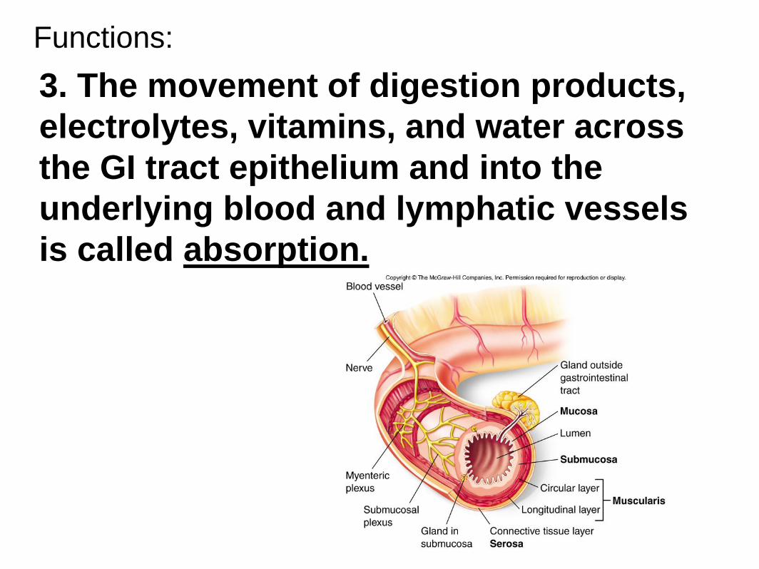
Functions:
3. The movement of digestion products,
electrolytes, vitamins, and water across
the GI tract epithelium and into the
underlying blood and lymphatic vessels
is called absorption.
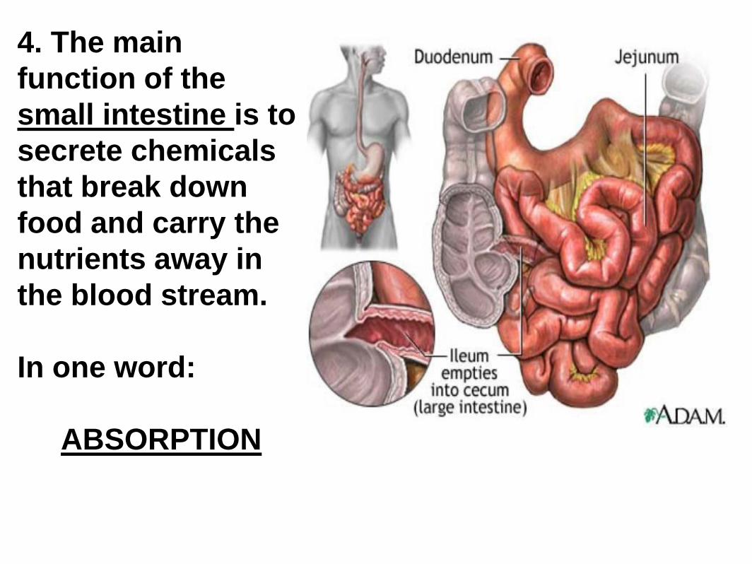
4. The main
function of the
small intestine is to
secrete chemicals
that break down
food and carry the
nutrients away in
the blood stream.
In one word:
ABSORPTION
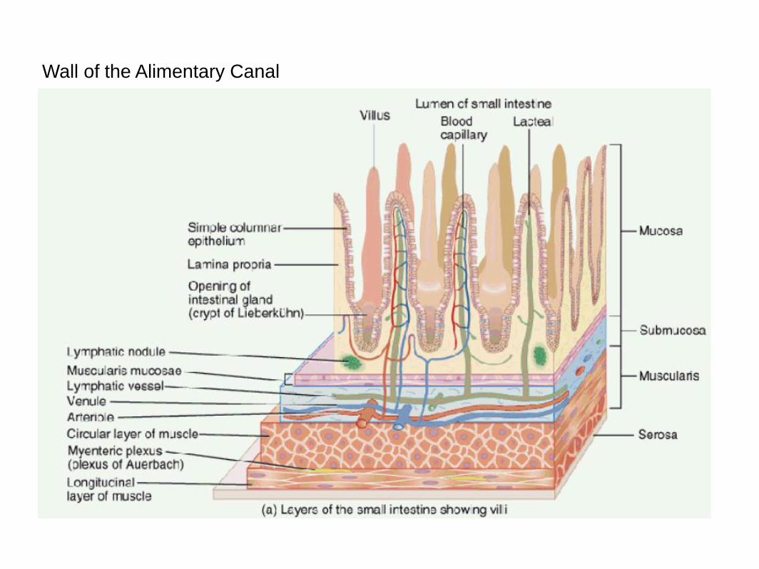
Wall of the Alimentary Canal
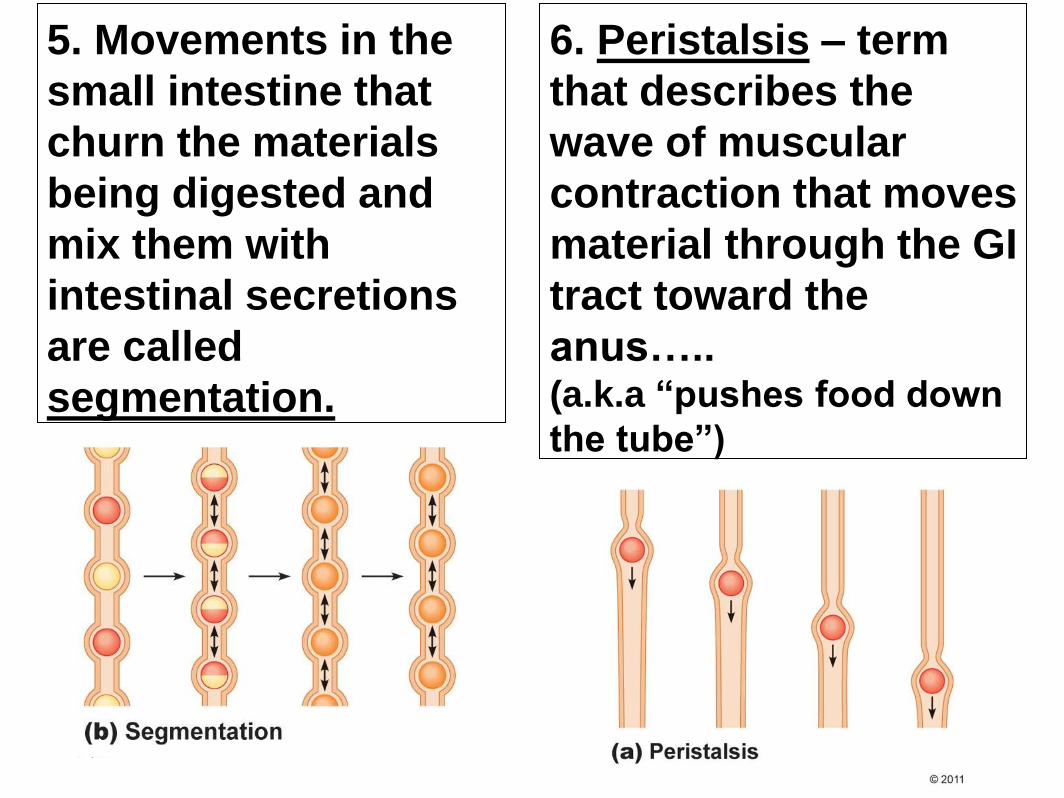
5. Movements in the
small intestine that
churn the materials
being digested and
mix them with
intestinal secretions
are called
segmentation.
6. Peristalsis – term
that describes the
wave of muscular
contraction that moves
material through the GI
tract toward the
anus….. (a.k.a “pushes food down
the tube”)
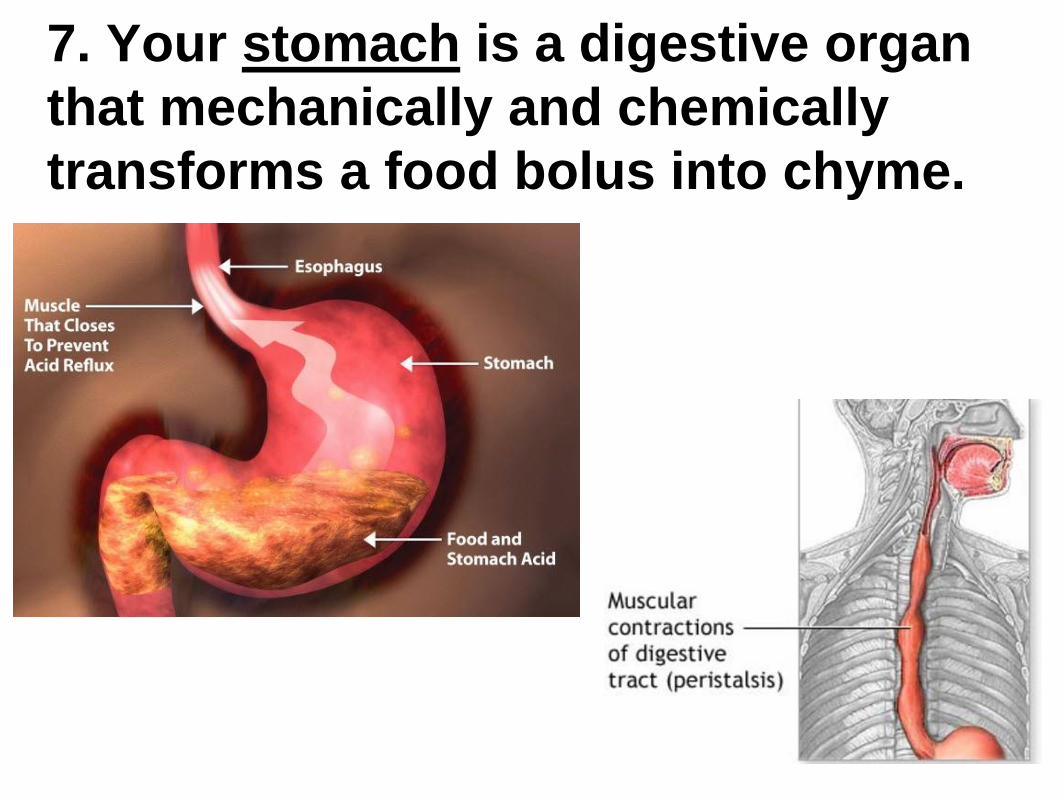
7. Your stomach is a digestive organ
that mechanically and chemically
transforms a food bolus into chyme.
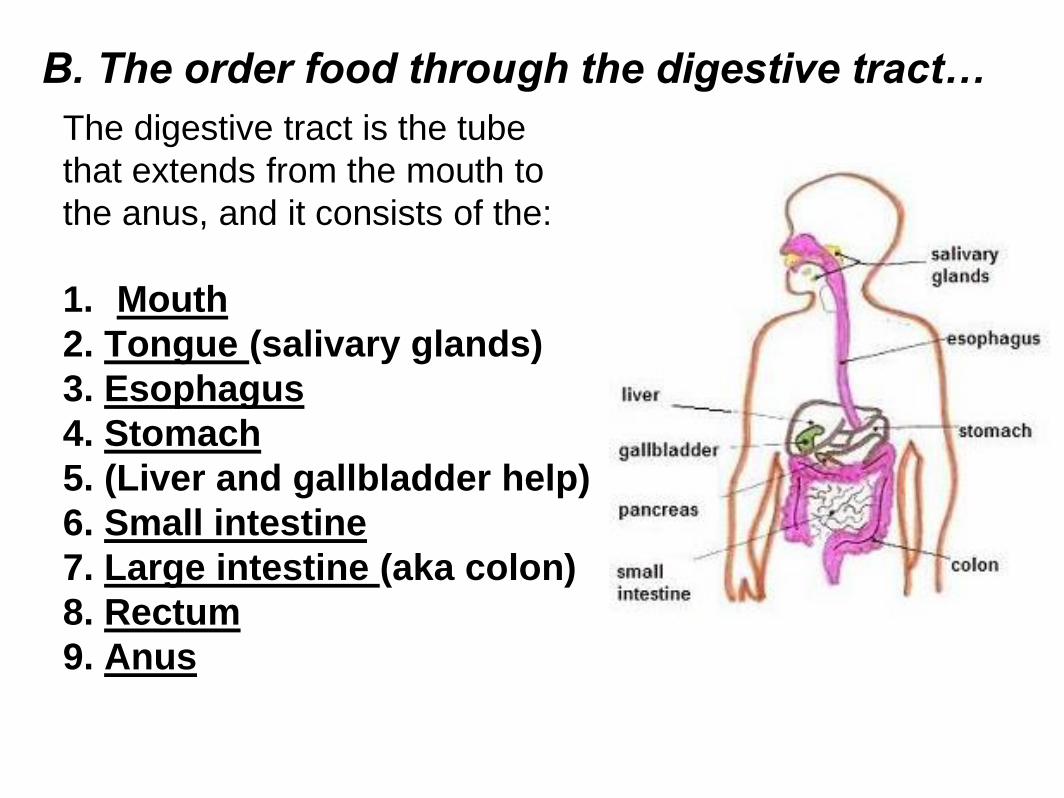
B. The order food through the digestive tract…
The digestive tract is the tube
that extends from the mouth to
the anus, and it consists of the:
1. Mouth
2. Tongue (salivary glands)
3. Esophagus
4. Stomach
5. (Liver and gallbladder help)
6. Small intestine
7. Large intestine (aka colon)
8. Rectum
9. Anus
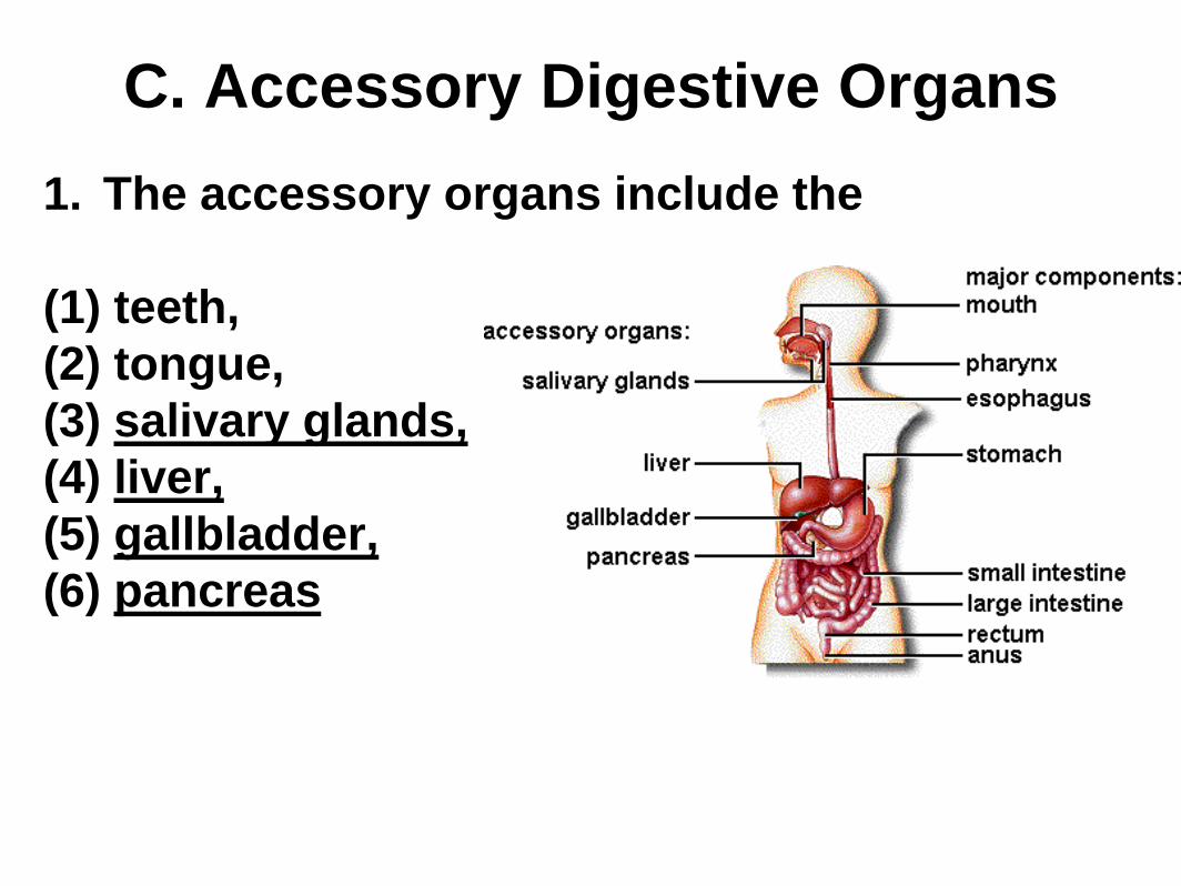
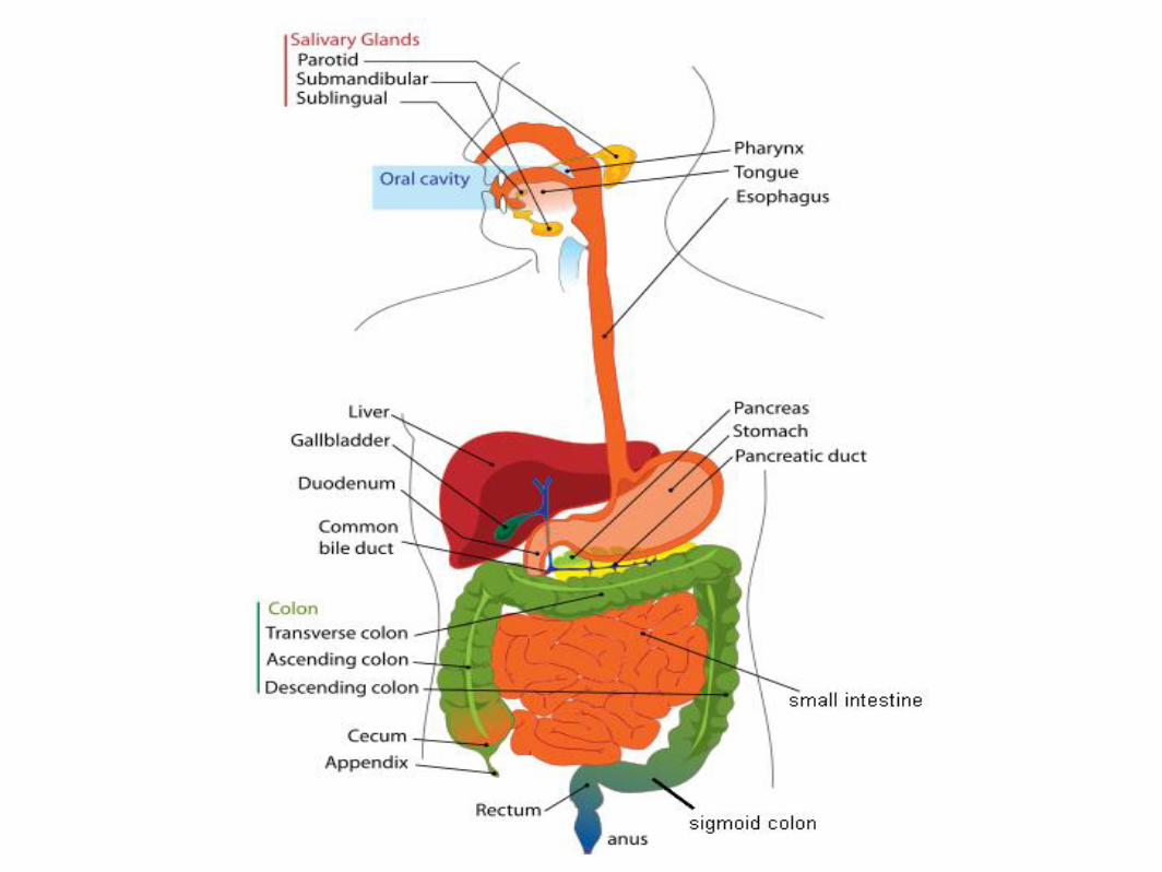
C. Accessory Digestive Organs
1. The accessory organs include the
(1) teeth,
(2) tongue,
(3) salivary glands,
(4) liver,
(5) gallbladder,
(6) pancreas
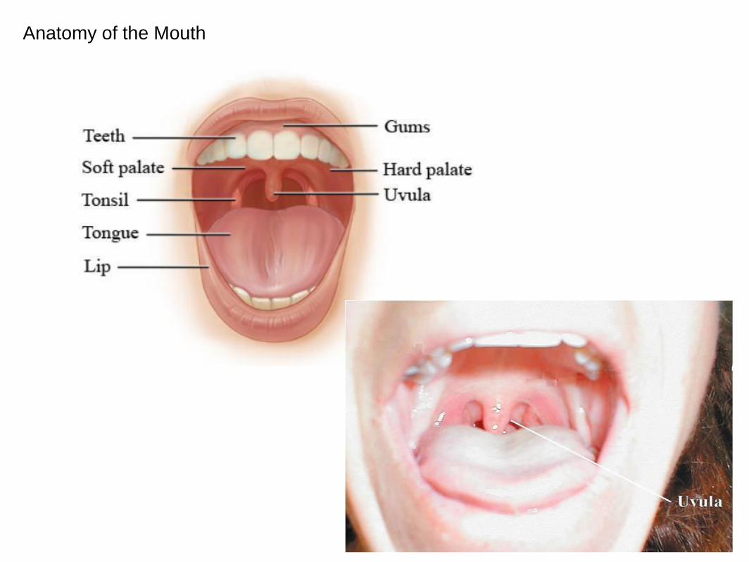
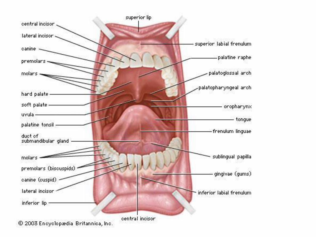
Anatomy of the Mouth
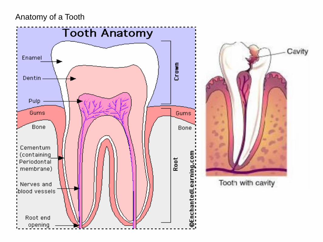
Anatomy of a Tooth
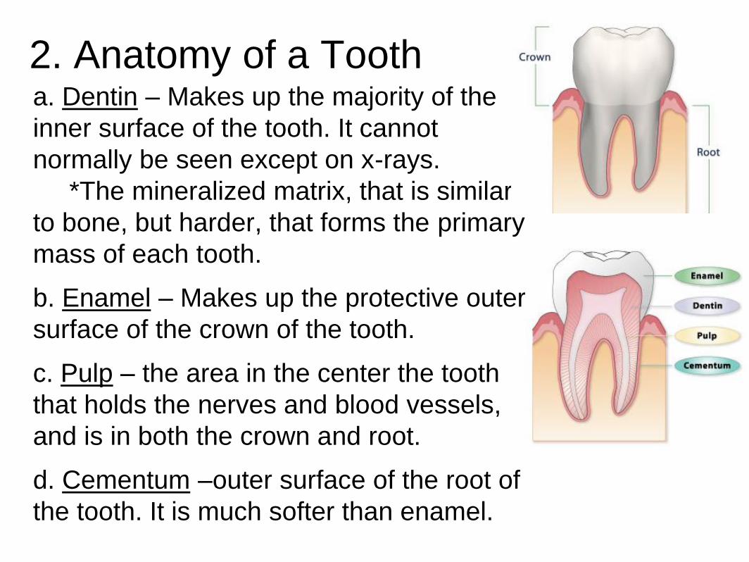
2. Anatomy of a Tooth a. Dentin – Makes up the majority of the
inner surface of the tooth. It cannot
normally be seen except on x-rays.
*The mineralized matrix, that is similar
to bone, but harder, that forms the primary
mass of each tooth.
b. Enamel – Makes up the protective outer
surface of the crown of the tooth.
c. Pulp – the area in the center the tooth
that holds the nerves and blood vessels,
and is in both the crown and root.
d. Cementum –outer surface of the root of
the tooth. It is much softer than enamel.
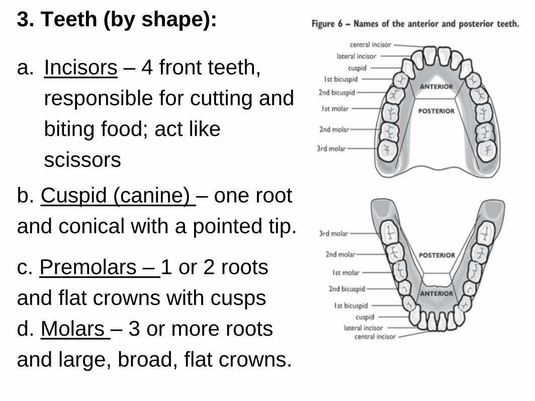
3. Teeth (by shape):
a. Incisors – 4 front teeth,
responsible for cutting and
biting food; act like
scissors
b. Cuspid (canine) – one root
and conical with a pointed tip.
c. Premolars – 1 or 2 roots
and flat crowns with cusps
d. Molars – 3 or more roots
and large, broad, flat crowns.
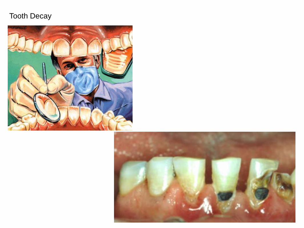
Tooth Decay
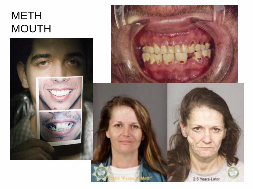
METH
MOUTH

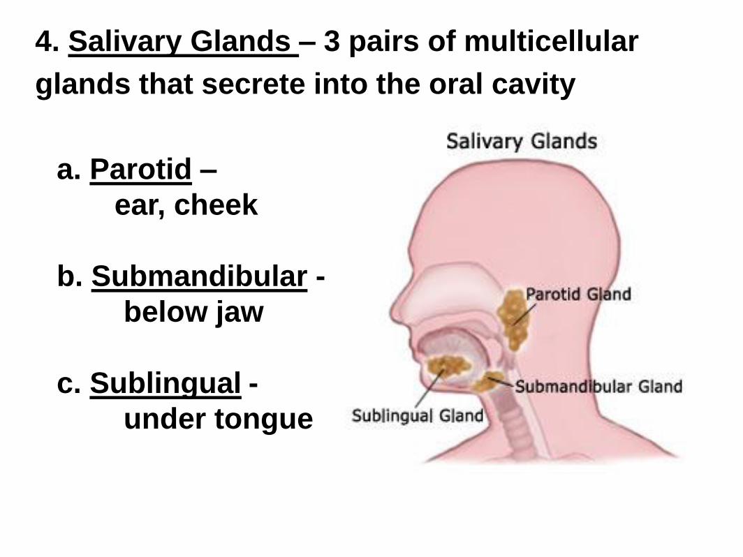
4. Salivary Glands – 3 pairs of multicellular
glands that secrete into the oral cavity
a. Parotid –
ear, cheek
b. Submandibular -
below jaw
c. Sublingual -
under tongue
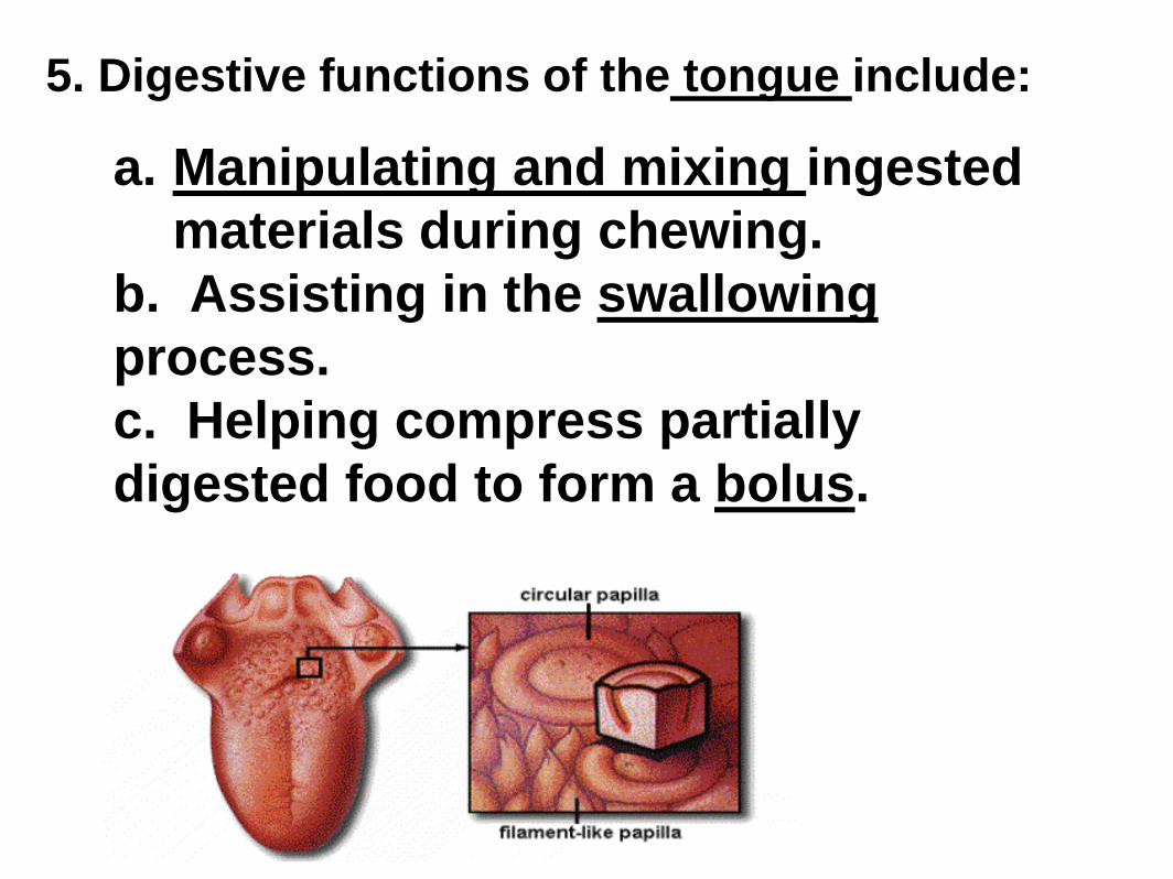
5. Digestive functions of the tongue include:
a. Manipulating and mixing ingested
materials during chewing.
b. Assisting in the swallowing
process.
c. Helping compress partially
digested food to form a bolus.
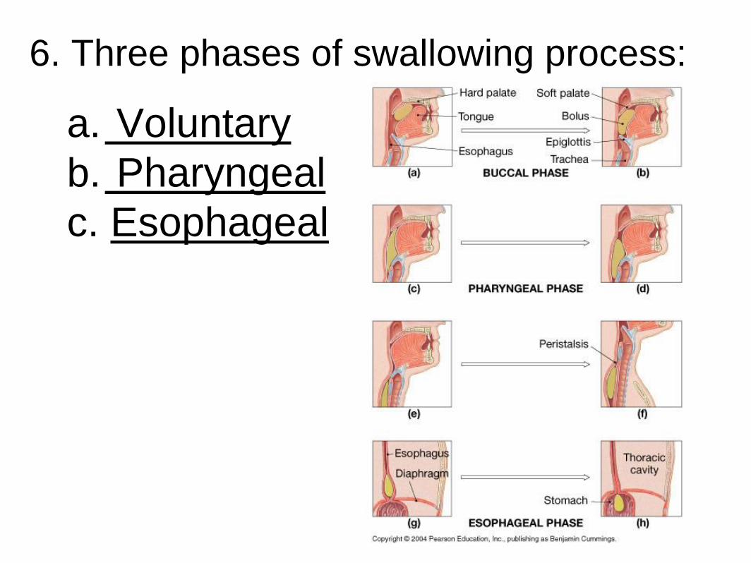
6. Three phases of swallowing process:
a. Voluntary
b. Pharyngeal
c. Esophageal
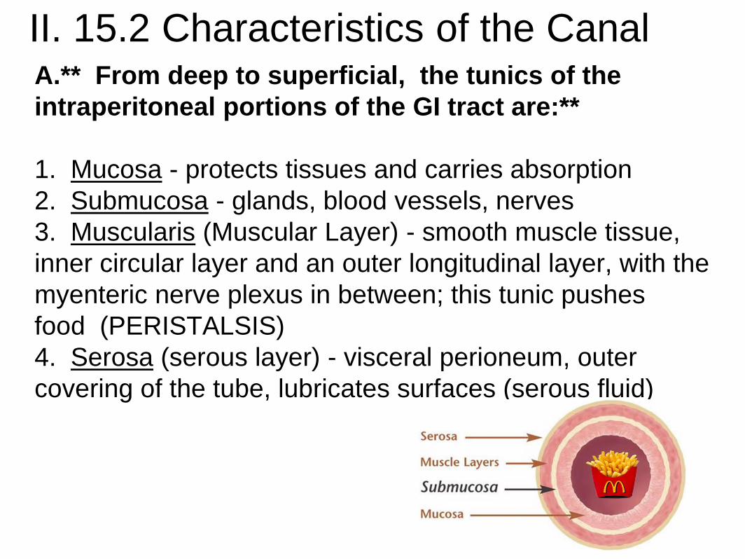
II. 15.2 Characteristics of the Canal A.** From deep to superficial, the tunics of the
intraperitoneal portions of the GI tract are:**
1. Mucosa - protects tissues and carries absorption
2. Submucosa - glands, blood vessels, nerves
3. Muscularis (Muscular Layer) - smooth muscle tissue,
inner circular layer and an outer longitudinal layer, with the
myenteric nerve plexus in between; this tunic pushes
food (PERISTALSIS)
4. Serosa (serous layer) - visceral perioneum, outer
covering of the tube, lubricates surfaces (serous fluid)
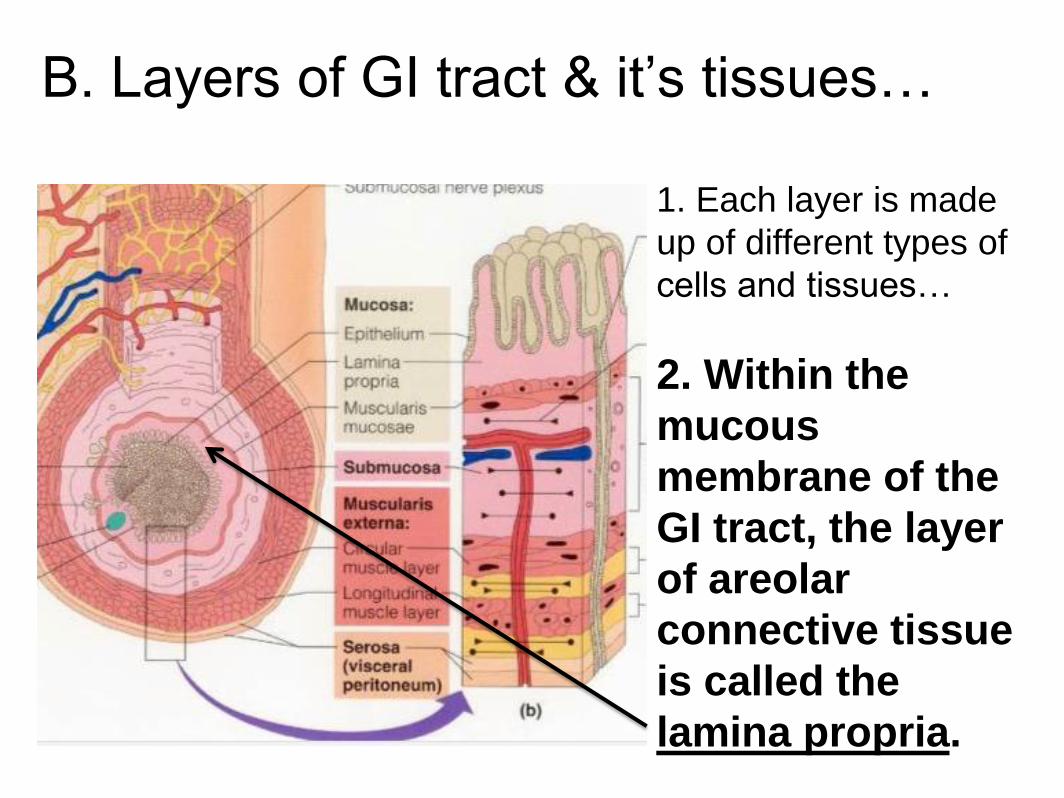
B. Layers of GI tract & it’s tissues…
1. Each layer is made
up of different types of
cells and tissues…
2. Within the
mucous
membrane of the
GI tract, the layer
of areolar
connective tissue
is called the
lamina propria.
Layers of GI tract & it’s tissues…
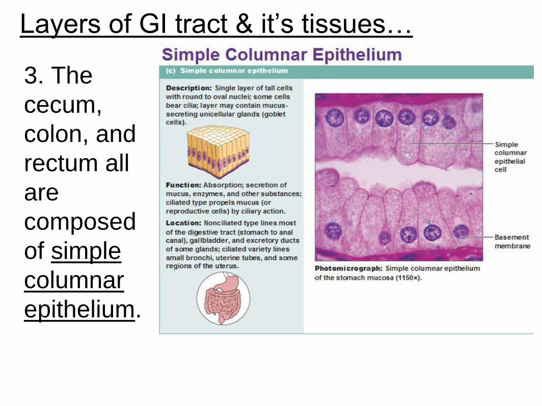
3. The
cecum,
colon, and
rectum all
are
composed
of simple
columnar
epithelium.
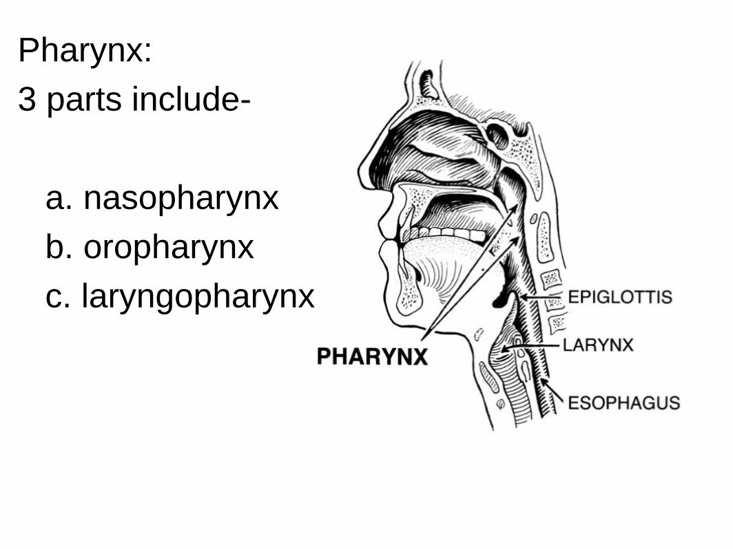
Pharynx:
3 parts include-
a. nasopharynx
b. oropharynx
c. laryngopharynx
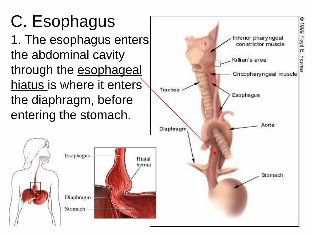
C. Esophagus 1. The esophagus enters
the abdominal cavity
through the esophageal
hiatus is where it enters
the diaphragm, before
entering the stomach.
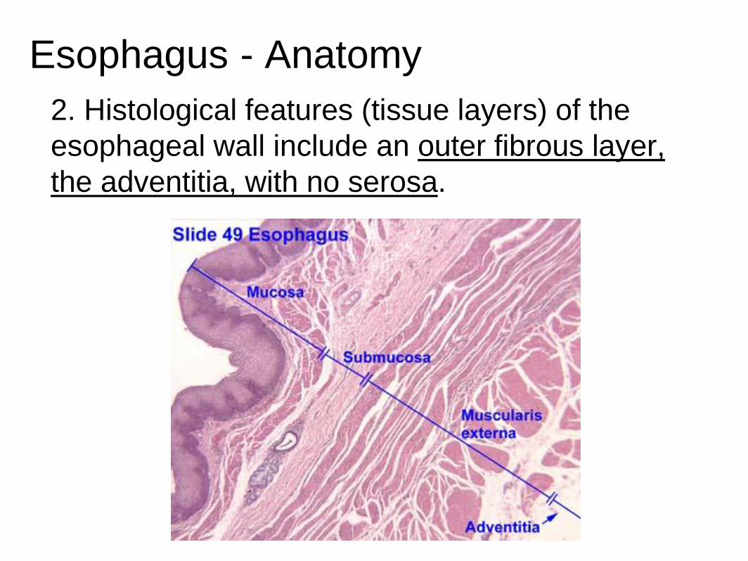
Esophagus - Anatomy
2. Histological features (tissue layers) of the
esophageal wall include an outer fibrous layer,
the adventitia, with no serosa.
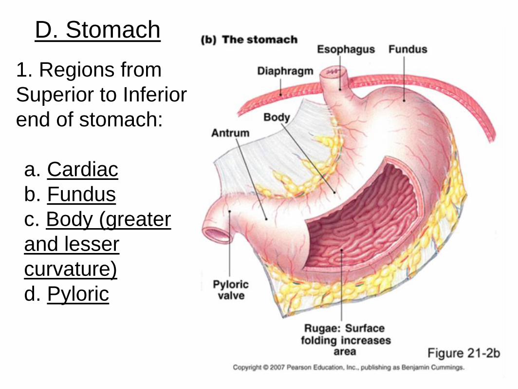
D. Stomach
1. Regions from
Superior to Inferior
end of stomach:
a. Cardiac
b. Fundus
c. Body (greater
and lesser
curvature)
d. Pyloric
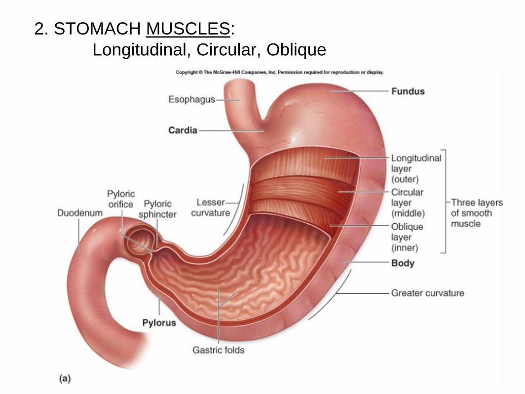
2. STOMACH MUSCLES:
Longitudinal, Circular, Oblique
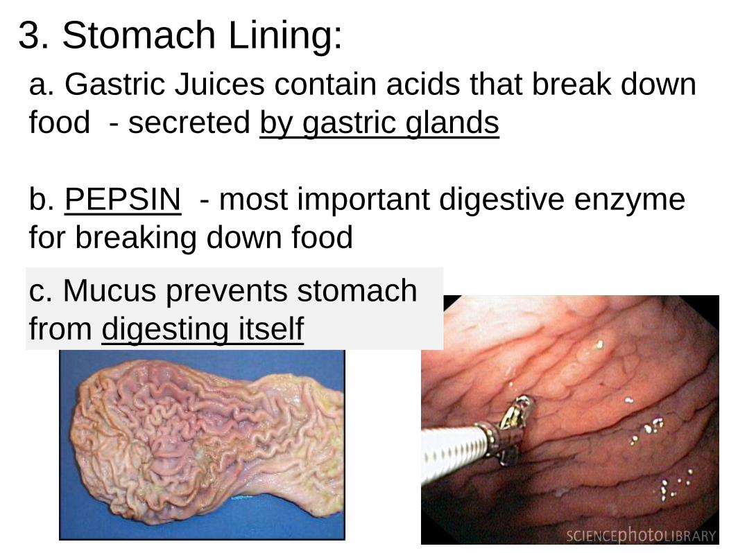
3. Stomach Lining:
a. Gastric Juices contain acids that break down
food - secreted by gastric glands
b. PEPSIN - most important digestive enzyme
for breaking down food
c. Mucus prevents stomach
from digesting itself
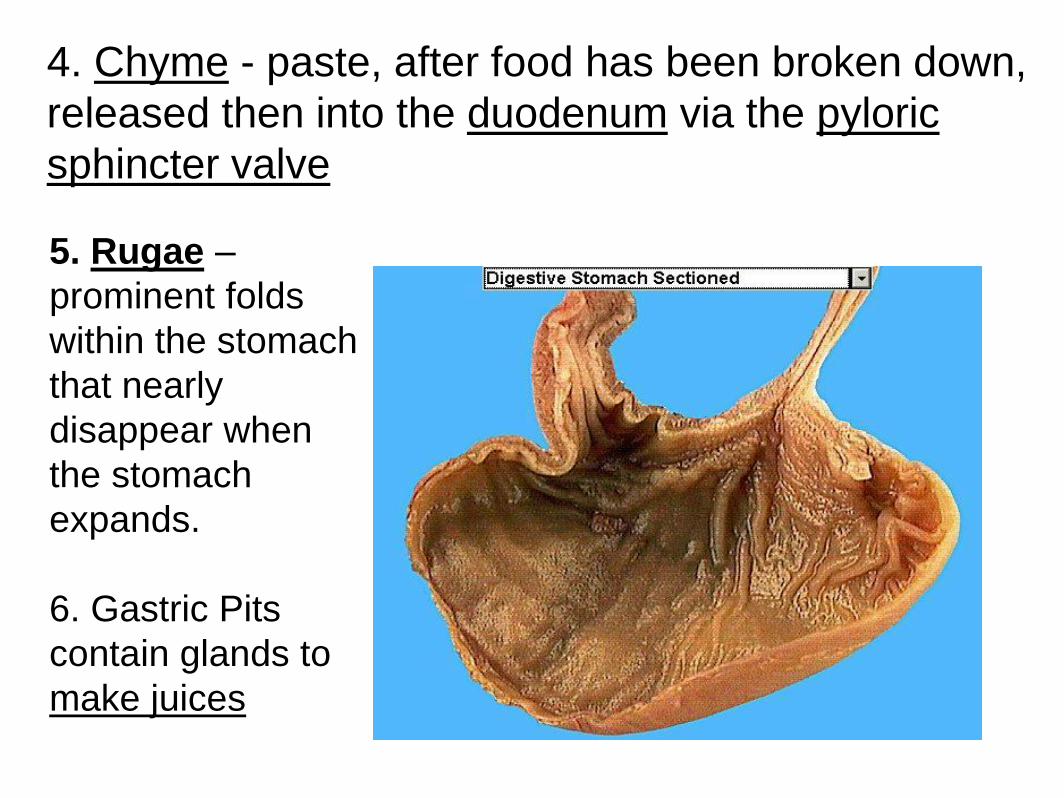
4. Chyme - paste, after food has been broken down,
released then into the duodenum via the pyloric
sphincter valve
5. Rugae –
prominent folds
within the stomach
that nearly
disappear when
the stomach
expands.
6. Gastric Pits
contain glands to
make juices
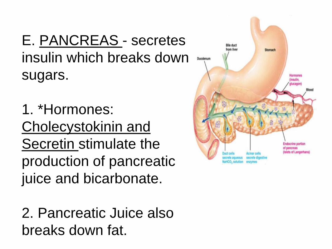
E. PANCREAS - secretes
insulin which breaks down
sugars.
1. *Hormones:
Cholecystokinin and
Secretin stimulate the
production of pancreatic
juice and bicarbonate.
2. Pancreatic Juice also
breaks down fat.
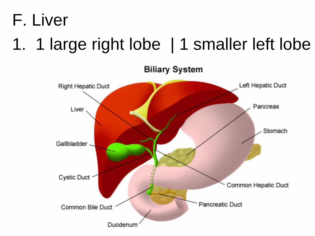
F. Liver
1. 1 large right lobe | 1 smaller left lobe
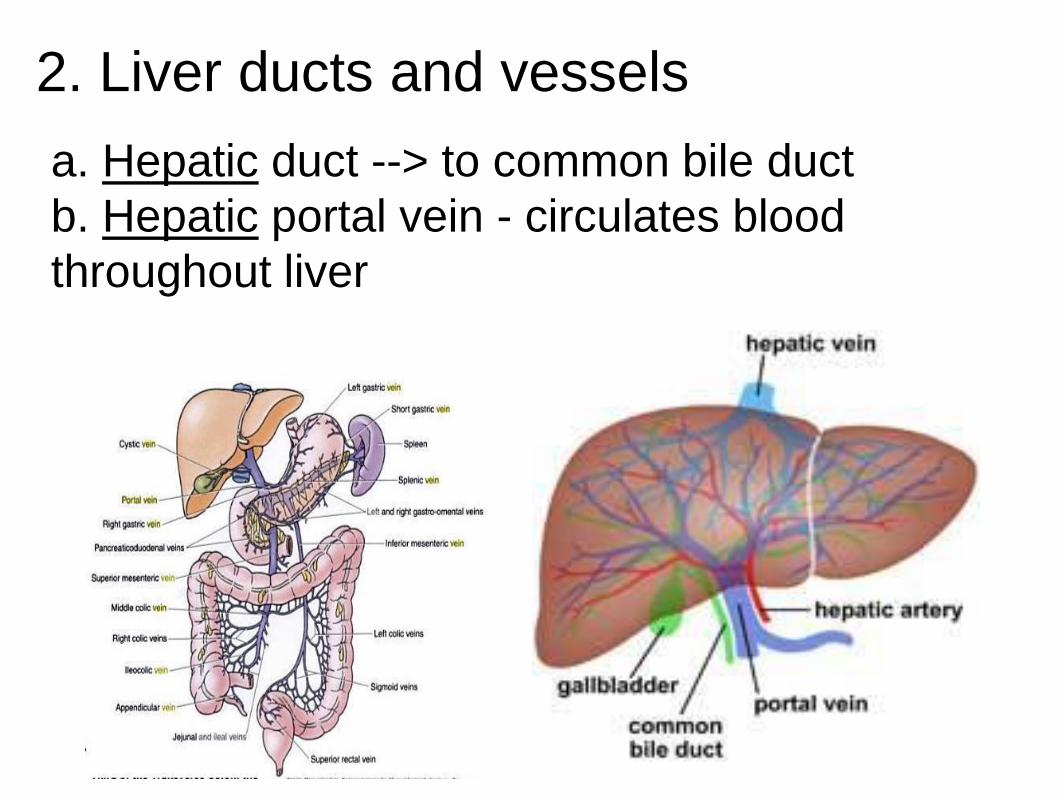
2. Liver ducts and vessels
a. Hepatic duct --> to common bile duct
b. Hepatic portal vein - circulates blood
throughout liver
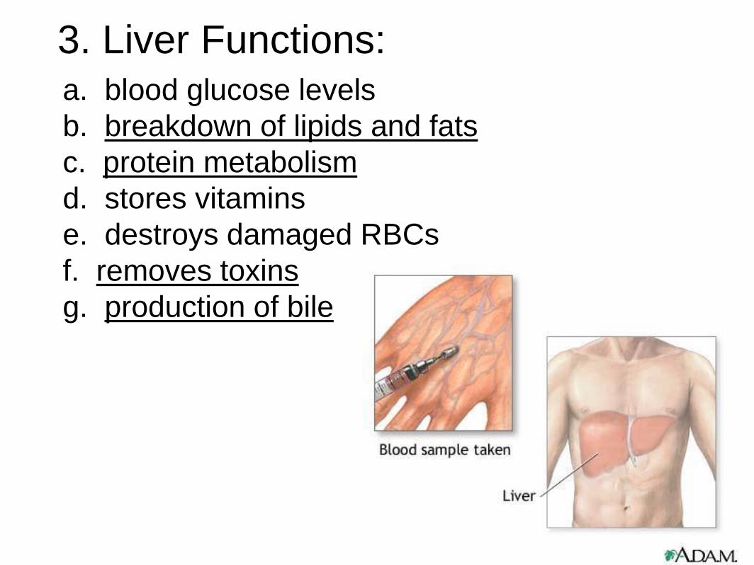
3. Liver Functions: a. blood glucose levels
b. breakdown of lipids and fats
c. protein metabolism
d. stores vitamins
e. destroys damaged RBCs
f. removes toxins
g. production of bile

Remember Bili Lights?
Using bili lights is a therapeutic procedure performed on newborn or
premature infants to reduce elevated levels of bilirubin. If blood levels
of bilirubin become too high, the bilirubin begins to dissolve in the body
tissues, producing the characteristic yellow eyes and skin of jaundice.
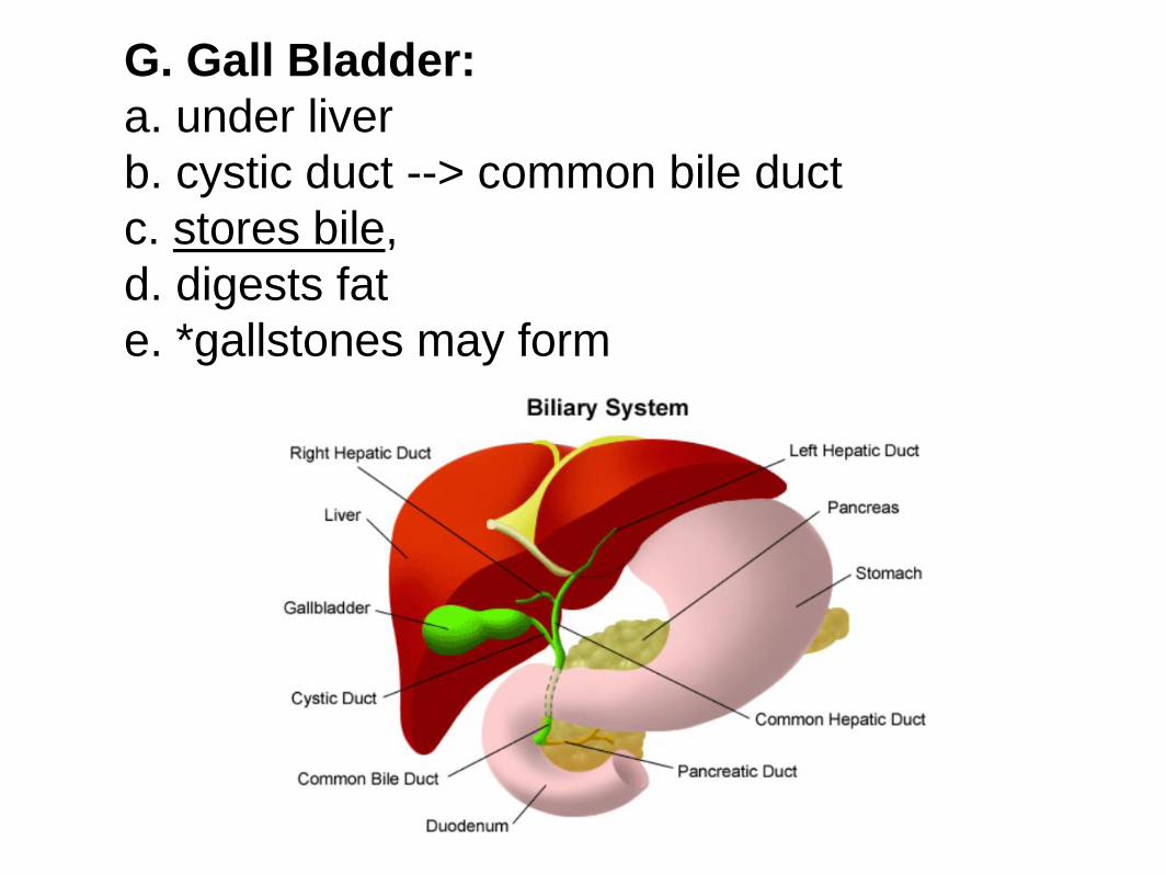
G. Gall Bladder:
a. under liver
b. cystic duct --> common bile duct
c. stores bile,
d. digests fat
e. *gallstones may form
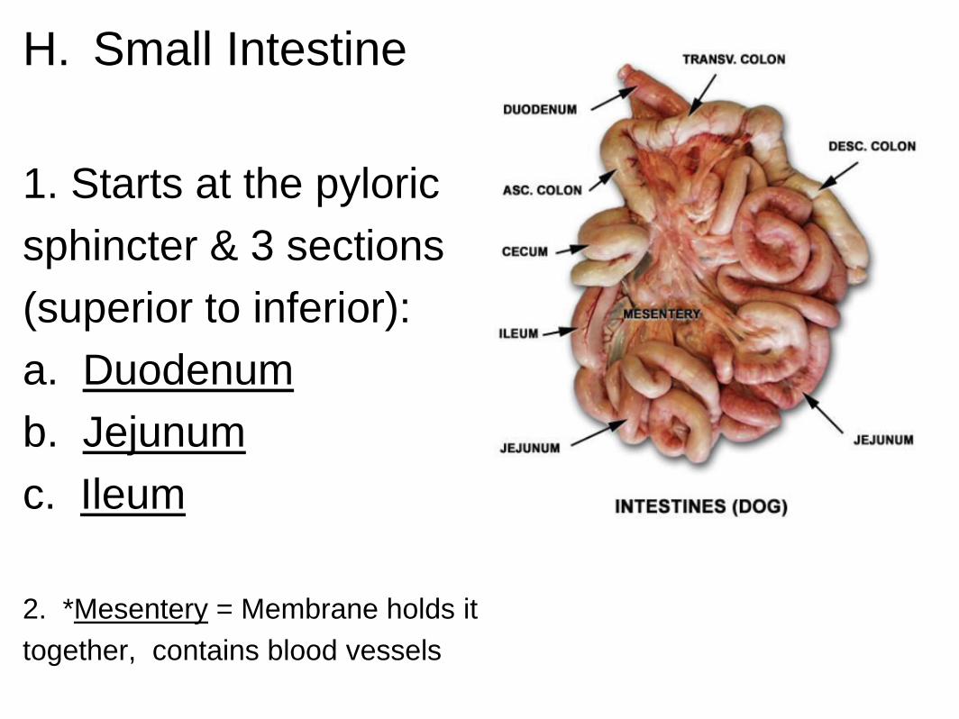
H. Small Intestine
1. Starts at the pyloric
sphincter & 3 sections
(superior to inferior):
a. Duodenum
b. Jejunum
c. Ileum
2. *Mesentery = Membrane holds it
together, contains blood vessels
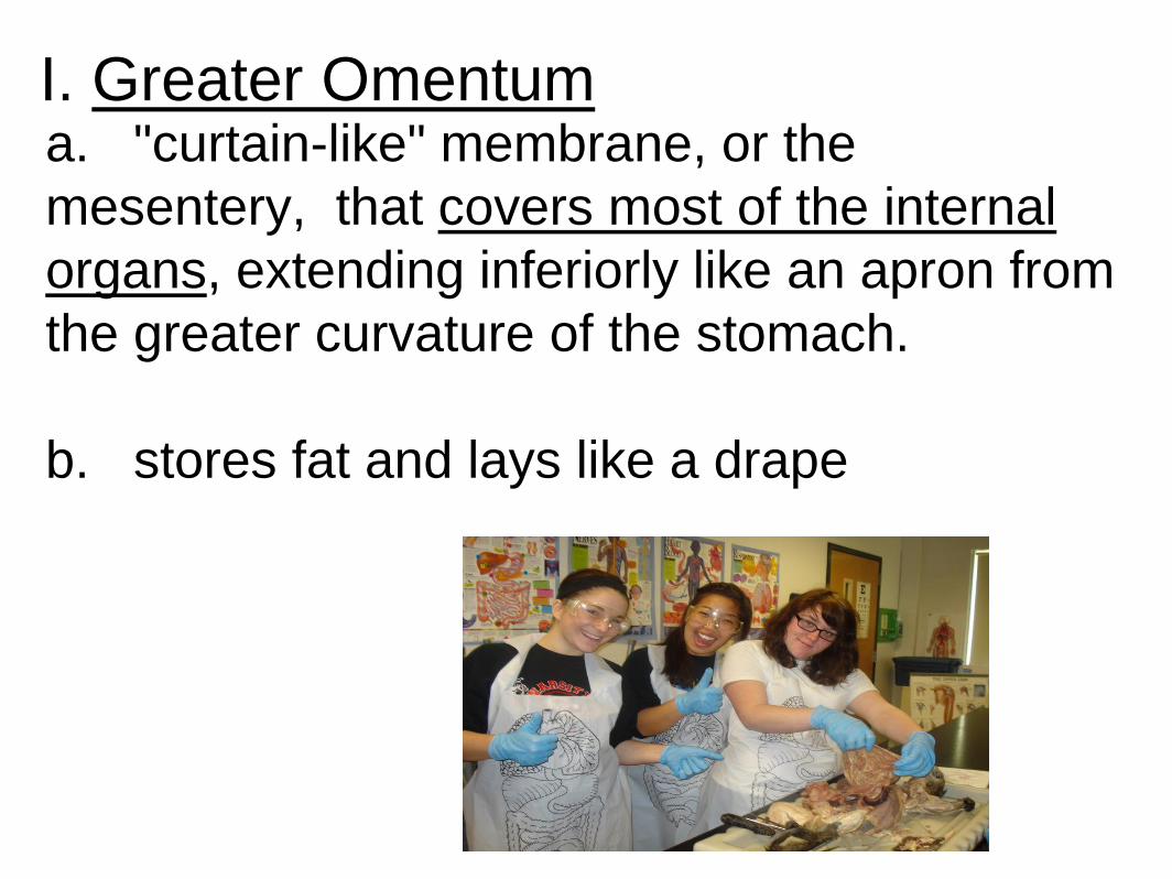
I. Greater Omentum a. "curtain-like" membrane, or the
mesentery, that covers most of the internal
organs, extending inferiorly like an apron from
the greater curvature of the stomach.
b. stores fat and lays like a drape
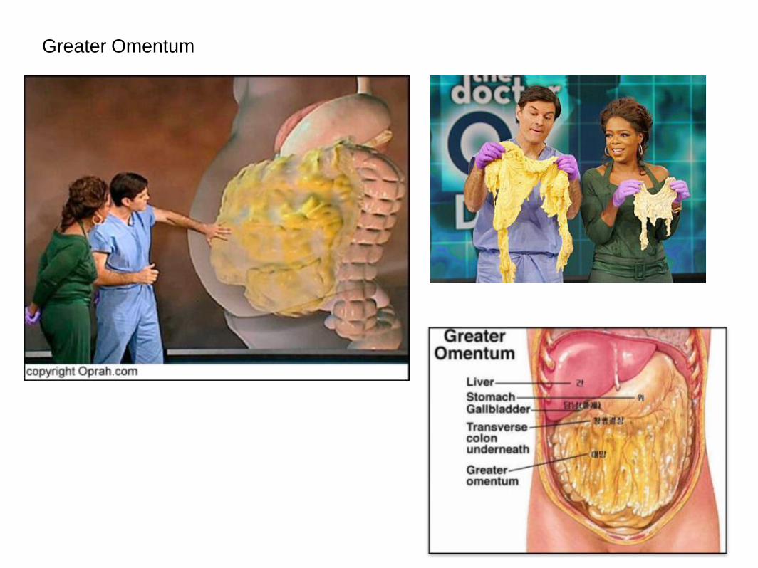
Greater Omentum
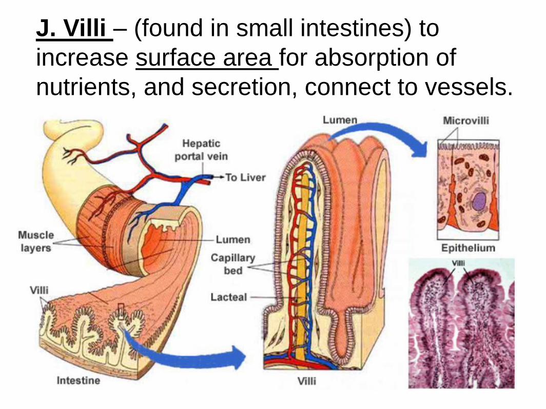
J. Villi – (found in small intestines) to
increase surface area for absorption of
nutrients, and secretion, connect to vessels.
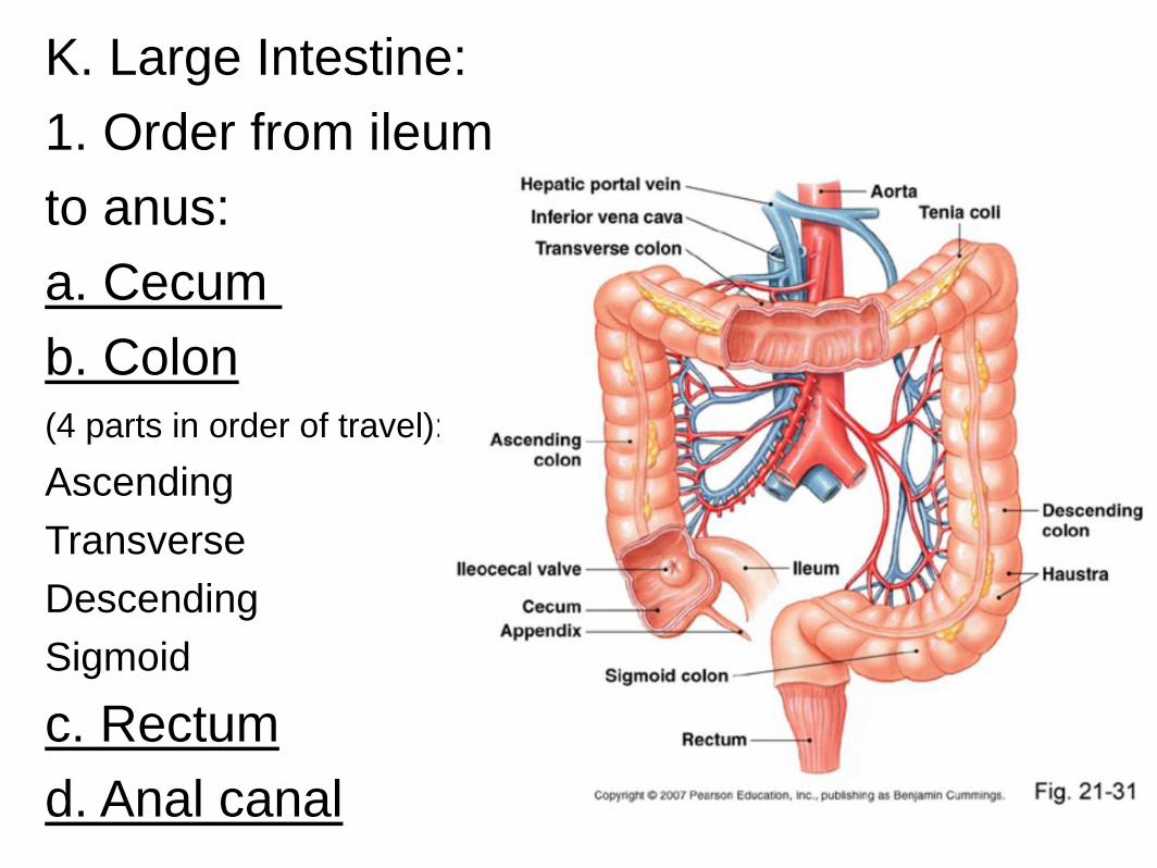
K. Large Intestine:
1. Order from ileum
to anus:
a. Cecum
b. Colon
(4 parts in order of travel):
Ascending
Transverse
Descending
Sigmoid
c. Rectum
d. Anal canal
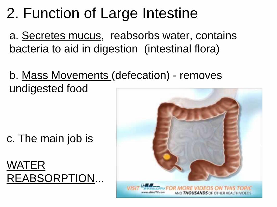
2. Function of Large Intestine
a. Secretes mucus, reabsorbs water, contains
bacteria to aid in digestion (intestinal flora)
b. Mass Movements (defecation) - removes
undigested food
c. The main job is
WATER
REABSORPTION...

3. Mucosa of Large Intestine:
Characterized by:
a. lack of intestinal villi
b. many lymphatic nodules and cells in the lamina
propria
c. numerous goblet cells and intestinal glands
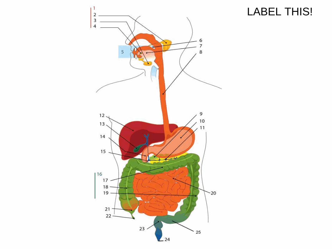
LABEL THIS!
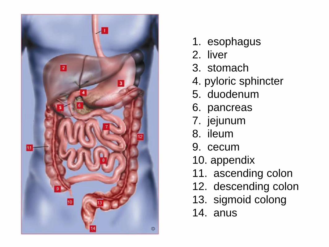
1. esophagus
2. liver
3. stomach
4. pyloric sphincter
5. duodenum
6. pancreas
7. jejunum
8. ileum
9. cecum
10. appendix
11. ascending colon
12. descending colon
13. sigmoid colong
14. anus
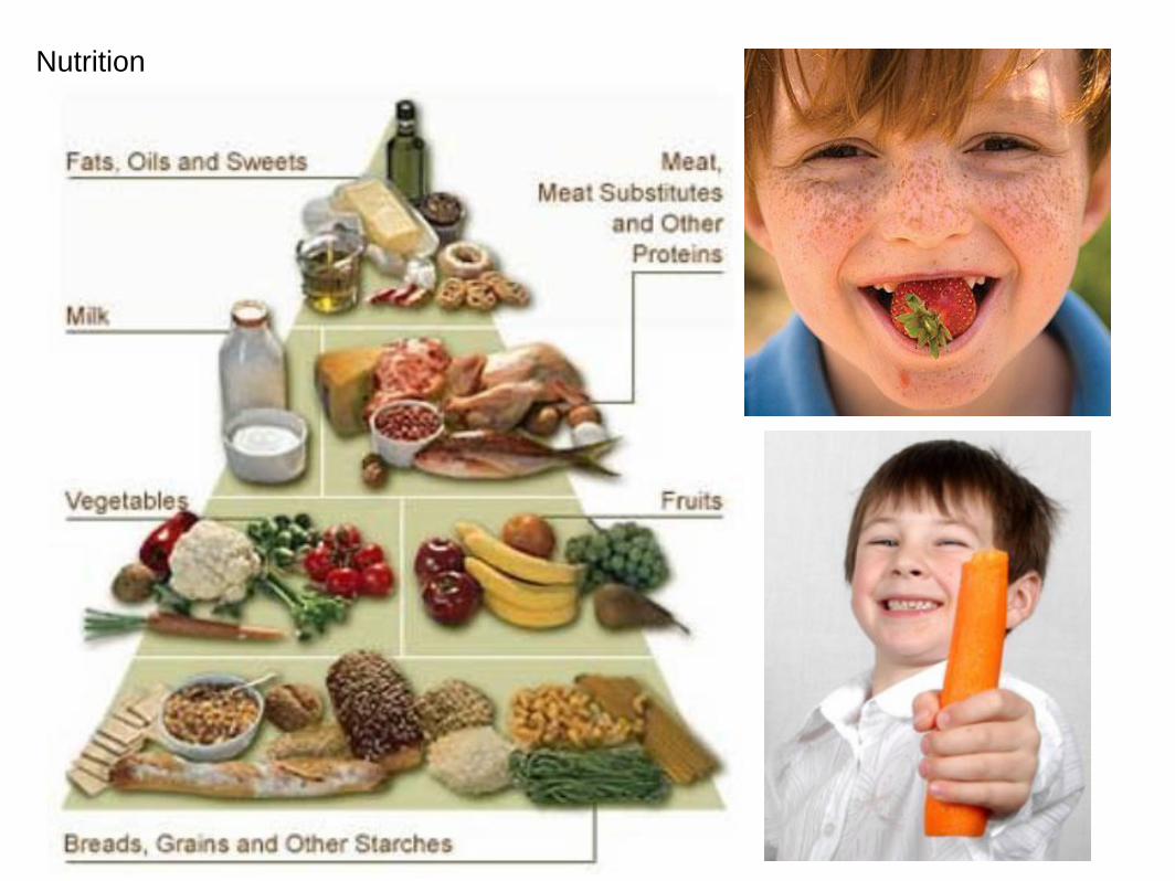
Nutrition
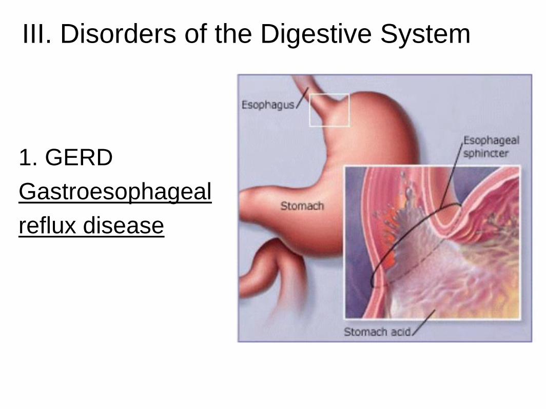
III. Disorders of the Digestive System
1. GERD
Gastroesophageal
reflux disease
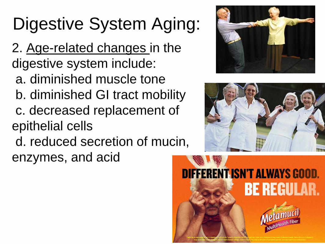
Digestive System Aging:
2. Age-related changes in the
digestive system include:
a. diminished muscle tone
b. diminished GI tract mobility
c. decreased replacement of
epithelial cells
d. reduced secretion of mucin,
enzymes, and acid
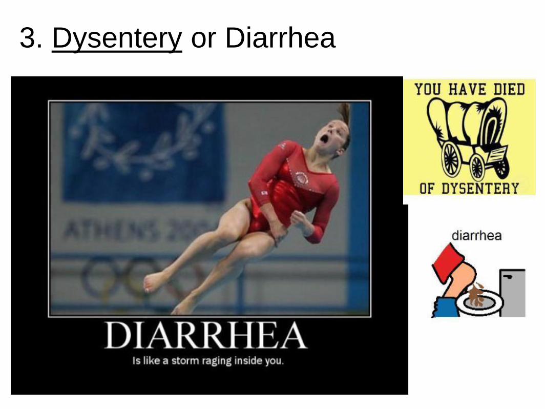
3. Dysentery or Diarrhea
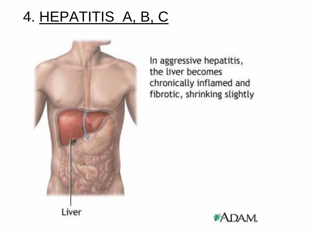
4. HEPATITIS A, B, C
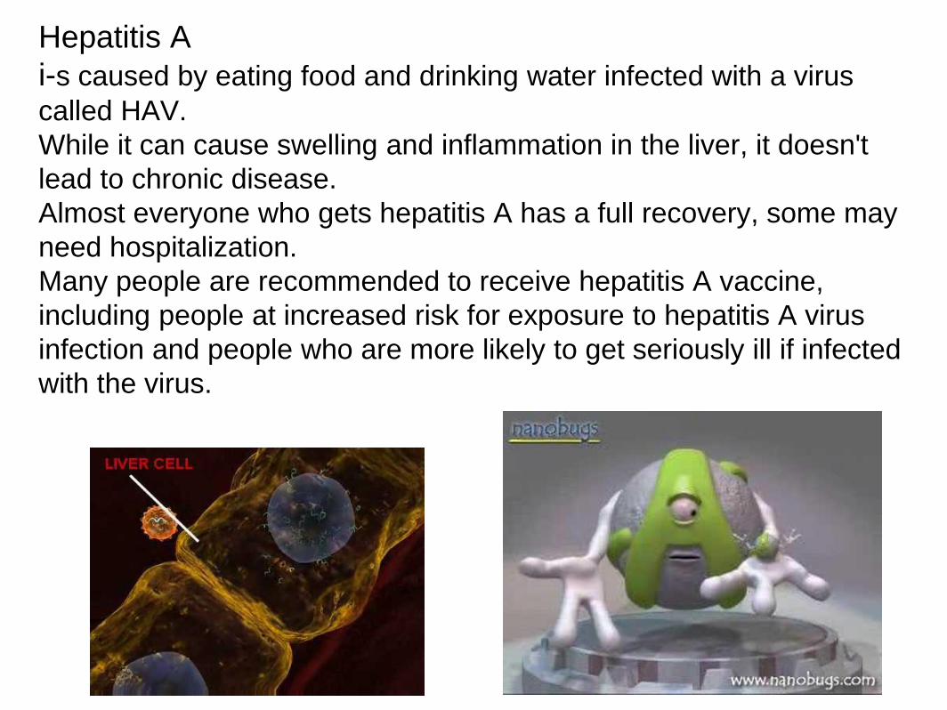
Hepatitis A
i-s caused by eating food and drinking water infected with a virus
called HAV.
While it can cause swelling and inflammation in the liver, it doesn't
lead to chronic disease.
Almost everyone who gets hepatitis A has a full recovery, some may
need hospitalization.
Many people are recommended to receive hepatitis A vaccine,
including people at increased risk for exposure to hepatitis A virus
infection and people who are more likely to get seriously ill if infected
with the virus.

Hepatitis B is caused by the virus HBV.
It is spread by contact with an infected person's blood, semen,
or other body fluid. And, it is a sexually transmitted disease
(STD).
Some people never develop symptoms, others develop chronic
symptoms that stay with them their whole life.
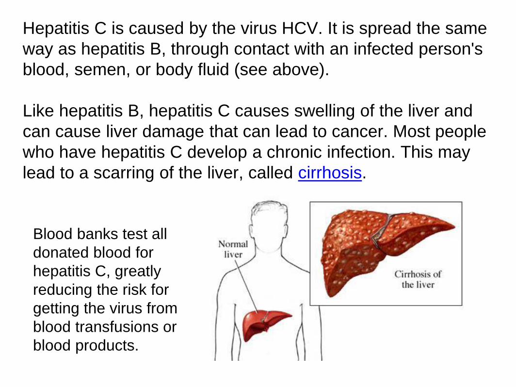
Hepatitis C is caused by the virus HCV. It is spread the same
way as hepatitis B, through contact with an infected person's
blood, semen, or body fluid (see above).
Like hepatitis B, hepatitis C causes swelling of the liver and
can cause liver damage that can lead to cancer. Most people
who have hepatitis C develop a chronic infection. This may
lead to a scarring of the liver, called cirrhosis.
Blood banks test all
donated blood for
hepatitis C, greatly
reducing the risk for
getting the virus from
blood transfusions or
blood products.
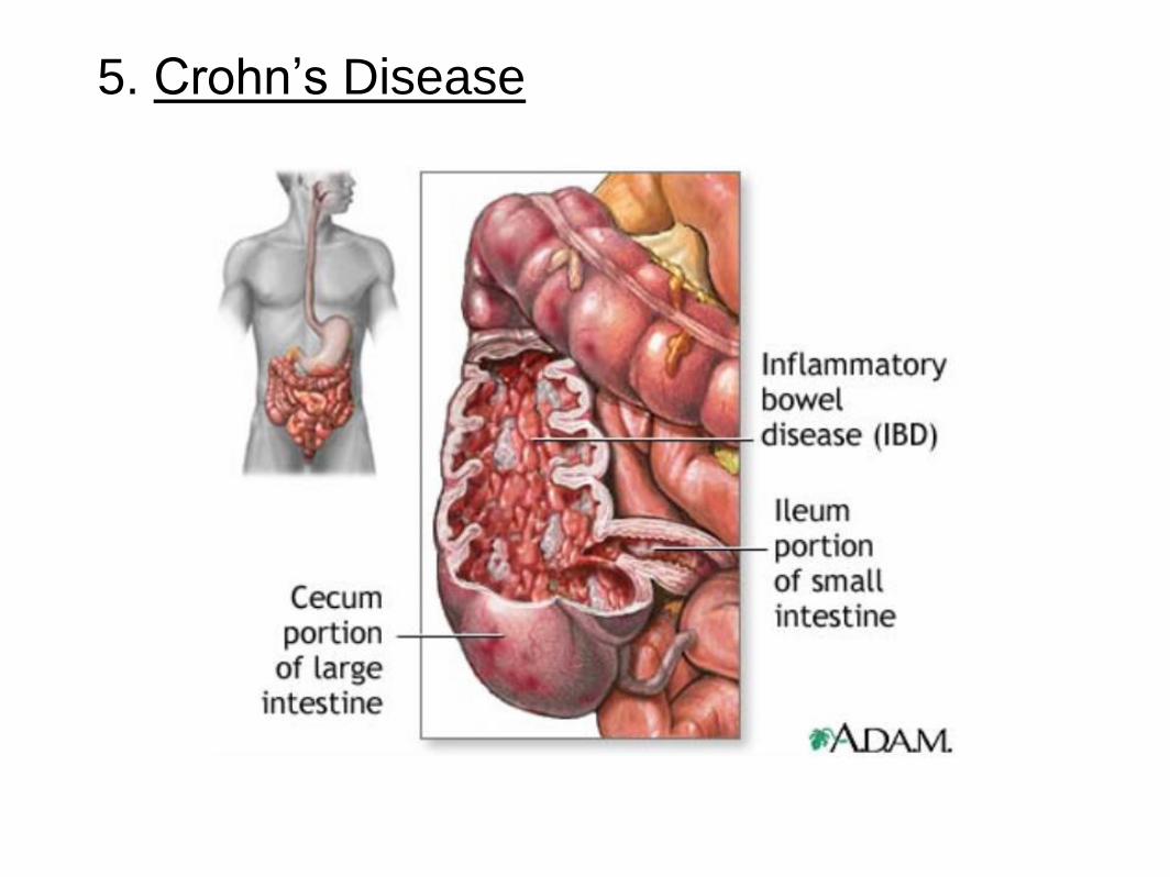
5. Crohn’s Disease
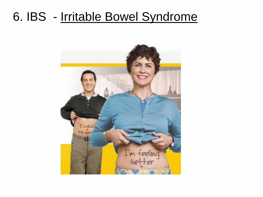
6. IBS - Irritable Bowel Syndrome
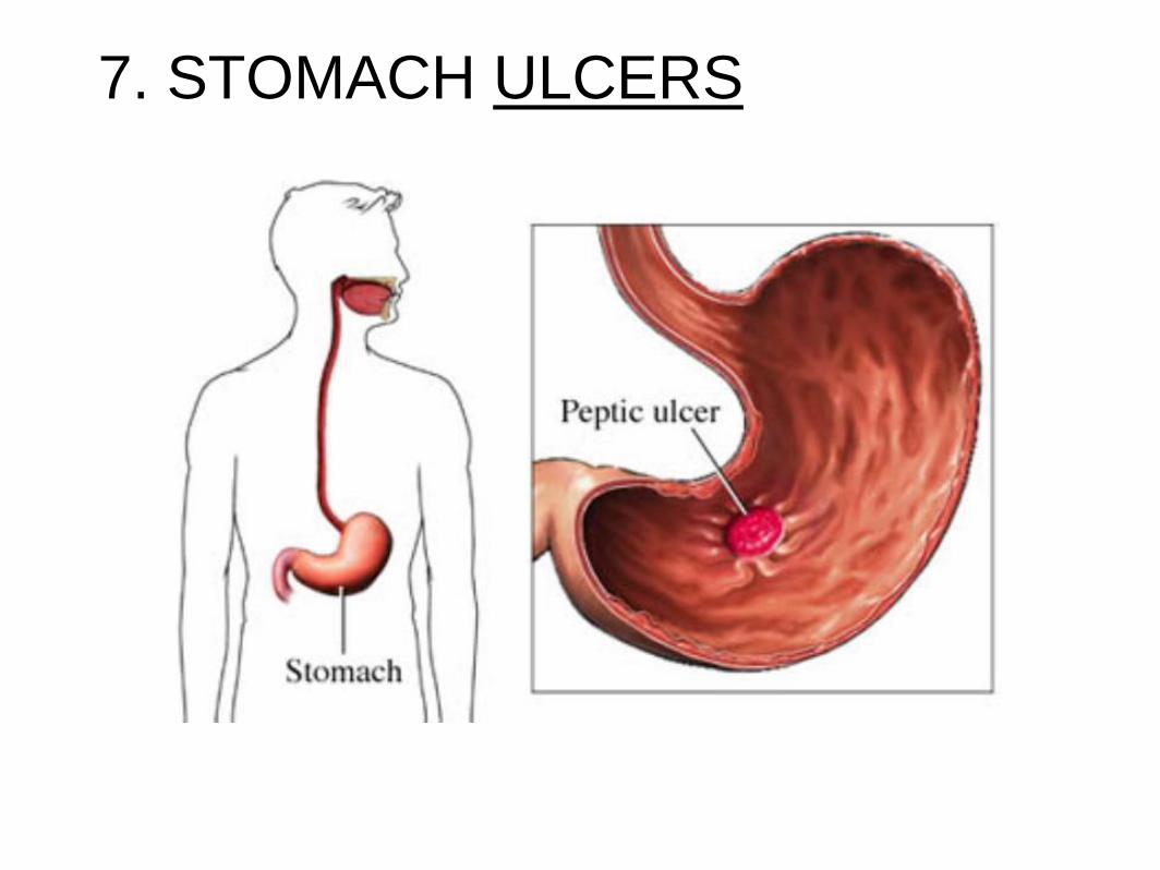
7. STOMACH ULCERS
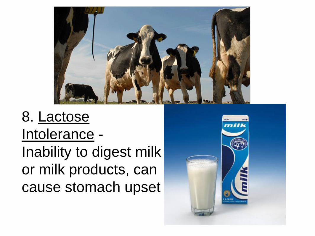
8. Lactose
Intolerance -
Inability to digest milk
or milk products, can
cause stomach upset
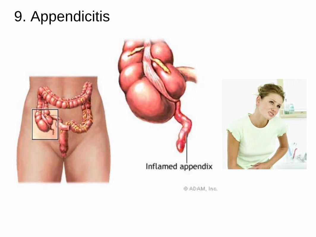
9. Appendicitis
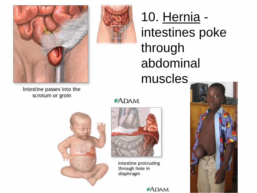
10. Hernia -
intestines poke
through
abdominal
muscles
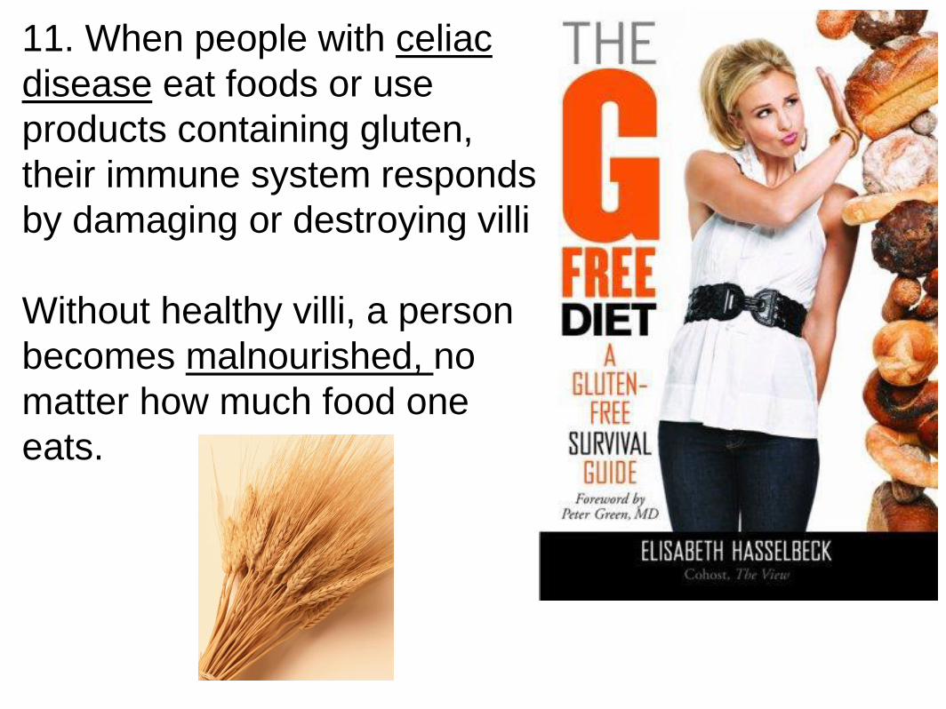
11. When people with celiac
disease eat foods or use
products containing gluten,
their immune system responds
by damaging or destroying villi
Without healthy villi, a person
becomes malnourished, no
matter how much food one
eats.
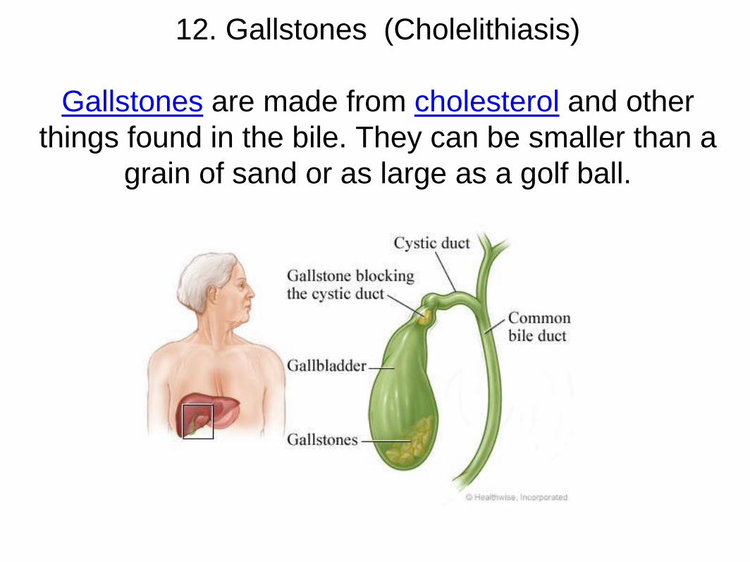
12. Gallstones (Cholelithiasis)
Gallstones are made from cholesterol and other
things found in the bile. They can be smaller than a
grain of sand or as large as a golf ball.

13. Gastric Bypass Surgery Gastric bypass surgery makes
the stomach smaller and causes food
to bypass part of the small intestine.
You will feel full more quickly than
when your stomach was its original
size. This reduces the amount of food
you can eat at one time. Bypassing
part of the intestine reduces how
much food and nutrients are
absorbed. This leads to weight loss.
http://www.hurtbyadoctor.com/Gastric
-Bypass-Surgery-Malpractice-
Lawsuits-Information-Home.htm
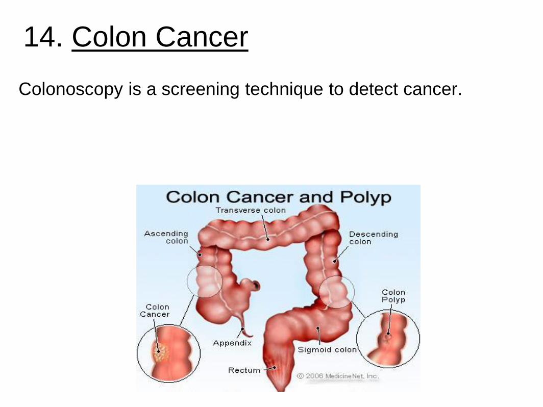
14. Colon Cancer
Colonoscopy is a screening technique to detect cancer.


![2 War in Korea and - PC\|MACimages.pcmac.org/SiSFiles/Schools/AL/MobileCounty/BurnsMiddle/Uploads... · mhe]](https://img.dokumen.tips/doc/110x75/604359a27e69523ab22e65ba/2-war-in-korea-and-pc-mhe-.jpg)