SUBJECT-SPECIFIC MUSCULOSKELETAL MODELING OF THE
LOWER EXTREMITIES IN PERSONS WITH UNILATERAL
CEREBRAL PALSY
by
Olesya Klets
May 2011
Technical reports from
Royal Institute of Technology
KTH Mechanics
SE-100 44 Stockholm, Sweden

ii
iii
SUBJECT-SPECIFIC MUSCULOSKELETAL MODELING OF THE LOWER EXTREMITIES IN PERSONS WITH UNILATERAL CEREBRAL PALSY
Olesya Klets
Department of Mechanics, Royal Institute of Technology
SE-100 44 Stockholm, Sweden
ABSTRACT
The computational musculoskeletal models that are used to study muscle moment-
generating capacities of persons with movement disorders and planning treatment options
must be accurate, and take into account the inter-individual variability of musculoskeletal
geometry.
In Paper I the methods of creating the subject-specific musculoskeletal model of the lower
extremities from magnetic resonance images (MRIs) were developed. The subject-specific
model was used to analyze hip, knee and ankle muscle moment arms (MALs) and muscle-
tendon lengths (MTLs) during gait in a subject with unilateral cerebral palsy (CP), and to
evaluate the accuracy of widespread and commonly-used scaled generic model.
It was found that the scaled generic model delivered accurate values for changes in MTLs in
most muscles. However, the scaled generic and the subject-specific lower extremity
musculoskeletal models showed substantial differences in MALs calculated during gait.
In Paper II subject-specific musculoskeletal models of nine subjects with unilateral CP
were created to study muscles volumes, MTLs and MALs; and to examine the accuracy of
MALs calculated by the scaled generic models.
It was shown that the scaled generic model significantly underestimated hip MALs
discrepancies between the affected and the non-affected sides of the lower extremities.
However, it significantly overestimated hip adduction/abduction of gluteus maximus,
gluteus medius, gluteus minimus, tensor fascia latae and biceps femoris long head; and hip
flexion of adductor longus and rectus femoris in the affected and the non-affected sides.
iv
It was also found that muscle volumes and hip abduction MALs in gluteus medius and
gluteus minimus, hip flexion MALs in iliacus and hip rotation in gluteus maximus were
smaller in the affected side of lower extremities. MTLs in the affected and the non-affected
sides throughout the range of hip motion were similar.
This thesis suggests the need for the subject-specific musculoskeletal models that can
account for variability of muscle attachments and musculoskeletal geometry of persons
with movement disorders. Based on inaccuracies of the scaled generic model reported
here, the generic models that are used to guide treatment decisions must be tested, and
interpreted with care.
Descriptions: cerebral palsy, moment arm, muscle length, hemiplegic, MRI, subject-
specific, musculoskeletal modeling, lower extremities, muscle volume.

v
PREFACE
This thesis is based upon studies conducted during April 2009 to May 2011 at the
Department of Mechanical Engineering, Royal Institute of Technology, Stockholm, Sweden;
and is built on the following papers, which will be referred to in the text by their Roman
numerals.
I. Klets O, Riad J, Broström EW, Gutierrez-Farewik EM. Comparison between a subject-
specific and a scaled generic musculoskeletal model of the lower extremities in a subject
with unilateral cerebral palsy. Clinical Biomechanics, April 2011, submitted.
II. Klets O, Riad J, Broström EW, Gutierrez-Farewik EM. Moment-generating biomechanical
factors of hip muscles in persons with unilateral CP with subject-specific models.
Gait&Posture, May 2011, submitted

vi
Division of work between authors
The research was initiated by Dr. Elena Gutierrez-Farewik (EGF), who was the main
supervisor and co-author in Paper I and Paper II.
Dr. Eva Weidenhielm Broström (EWB) provided the gait analysis. Dr. Jacques Riad (JR)
provided medical imaging data of studied subjects. EWB and JR were clinical advisors.
The methods of building the subject-specific musculoskeletal models were developed by
Olesya Klets (OK). The calculations were done by OK with supervision from EGF. Articles
were written by OK with input from EGF, JR and EWB.
vii
LIST OF ABBREVIATIONS
3D Three-dimensional
CP Cerebral palsy
CT Computer tomography
EMG Electromyography
MTL Muscle-tendon length
MAL Muscle moment arm length
MRI Magnetic resonance imaging

viii
ix
TABLE OF CONTENTS
ABSTRACT ............................................................................................................................................................. iii
PREFACE .................................................................................................................................................................. v
Division of work between authors ............................................................................................................ vi
LIST OF ABBREVIATIONS ............................................................................................................................. vii
1. INTRODUCTION .............................................................................................................................................. 1
1.1. Biomechanics of skeletal muscle ........................................................................................................ 2
1.2. Functional anatomy of the lower extremities ............................................................................... 3
1.3. Cerebral palsy ............................................................................................................................................ 6
1.4. Motion analysis ......................................................................................................................................... 7
1.5. Generic musculoskeletal models ........................................................................................................ 8
1.6. Subject-specific musculoskeletal models ........................................................................................ 9
SCOPE AND AIMS .............................................................................................................................................. 11
2. MATERIALS AND METHODS .................................................................................................................. 13
2.1. SUBJECTS .................................................................................................................................................. 13
2.2. IMAGING CAPTURE .............................................................................................................................. 13
2.3. MOTION CAPTURE ................................................................................................................................ 14
2.4. SCALED GENERIC MODEL.................................................................................................................. 15
2.5. SUBJECT-SPECIFIC MUSCULOSKELETAL MODEL .................................................................... 15
2.5.1. Study I ................................................................................................................................................ 15
2.5.2. Study II ............................................................................................................................................... 15
2.5.3. Three-dimensional reconstruction of muscles and bones models ............................ 16
2.5.3. Specification of joint kinematics and representation of muscle-tendon paths ..... 17
2.5.4. Simulation of gait ........................................................................................................................... 17
x
2.6. DATA ANALYSIS ..................................................................................................................................... 18
3. RESULTS AND DISCUSSIONS .................................................................................................................. 19
Study I ................................................................................................................................................................ 19
Study II ............................................................................................................................................................... 21
4. GENERAL DISCUSSION .............................................................................................................................. 23
Future work ..................................................................................................................................................... 24
OUTLINE OF PAPERS ...................................................................................................................................... 27
ACKNOWLEDGEMENTS ................................................................................................................................ 29
REFERENCES ...................................................................................................................................................... 31
PAPER I
PAPER II
1
1. INTRODUCTION
For an airplane, mechanics enables us to design its
structure and predict its performance. For an organism,
biomechanics helps us to understand its normal function,
predict changes due to alterations, and propose methods
of artificial intervention.
(Fung Y, 1993)
Biomechanics is a modern science with ancient roots. One of the earliest books covering the
concepts of biomechanics was On the Parts of Animal by Aristotle (384-322 B.C.), where the
anatomy and function of internal organs is described. Decartes, a great mathematician,
suggested a physiological theory upon mechanical grounds. Giovanni Alfonso Borelli, the
Italian mathematician and astronomer, successfully explained muscular movement and
body dynamics In On Motion of Animals (De Motu Animalium) (1680).
Newton´s laws of motion are often the basis for biomechanics, since it is difficult to find
anything living that does not involve some mechanical problems. Muscles transmit forces
through tendons, which connect to bones and span joints that have complicated kinematics
[118]. Biomechanical engineers are interested how the geometric relationships among the
muscles and bones transform muscle forces into moments about the joints during a variety
of activities and under many conditions [36].
Quantifying the muscle moment-generating capacities, forces and muscle lengths in various
situations or during different activities may be of value from a medical viewpoint. In sports
medicine, biomechanics can be applied to improve performance with minimal risks to
muscles and joint structures. In orthopedics, which deals with musculoskeletal problems,
biomechanics is used to investigate mechanically the function of diseased muscles or the
effects of musculoskeletal deformities on movement patterns, to explore the relationships
between muscle excitation and movement, to evaluate joint loading during different
movements, to plan a treatment and to adjust post-surgical rehabilitation and physical
therapy with minimal risks to damaged or weak structures [9].
2 1. INTRODUCTION
1.1. Biomechanics of skeletal muscle
Skeletal muscles provide strength and protection to the skeleton by distributing loads and
absorbing shock. The skeletal muscles perform both dynamic and static work. Dynamic
work permits locomotion and the positioning of the body segments in space. Static work
maintains body posture or position [82]. Such abilities usually represent the action of
muscle groups, not of individual muscles.
The tendons and the connective tissues in and around the muscle belly are viscoelastic
structures that help to determine the mechanics characteristics of whole muscle during
contraction [66].
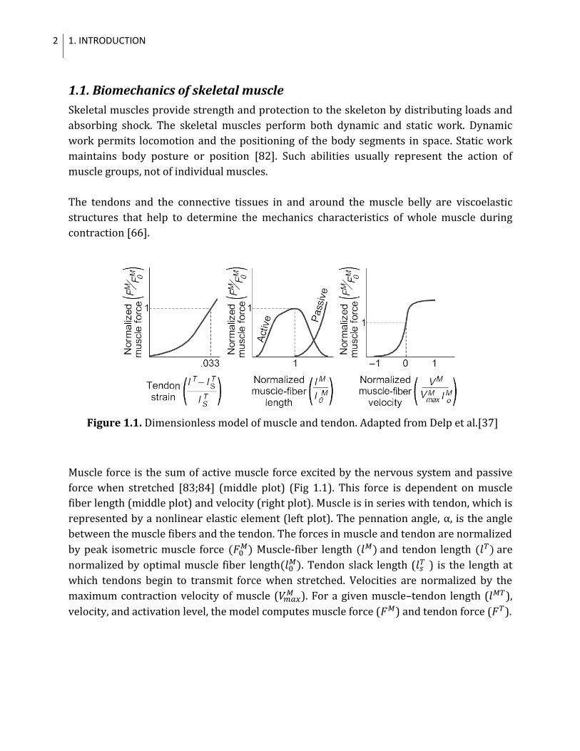
Figure 1.1. Dimensionless model of muscle and tendon. Adapted from Delp et al.[37]
Muscle force is the sum of active muscle force excited by the nervous system and passive
force when stretched [83;84] (middle plot) (Fig 1.1). This force is dependent on muscle
fiber length (middle plot) and velocity (right plot). Muscle is in series with tendon, which is
represented by a nonlinear elastic element (left plot). The pennation angle, α, is the angle
between the muscle fibers and the tendon. The forces in muscle and tendon are normalized
by peak isometric muscle force Muscle-fiber length and tendon length are
normalized by optimal muscle fiber length . Tendon slack length (
) is the length at
which tendons begin to transmit force when stretched. Velocities are normalized by the
maximum contraction velocity of muscle ( ). For a given muscle–tendon length ( ),
velocity, and activation level, the model computes muscle force ( ) and tendon force ( ).
3
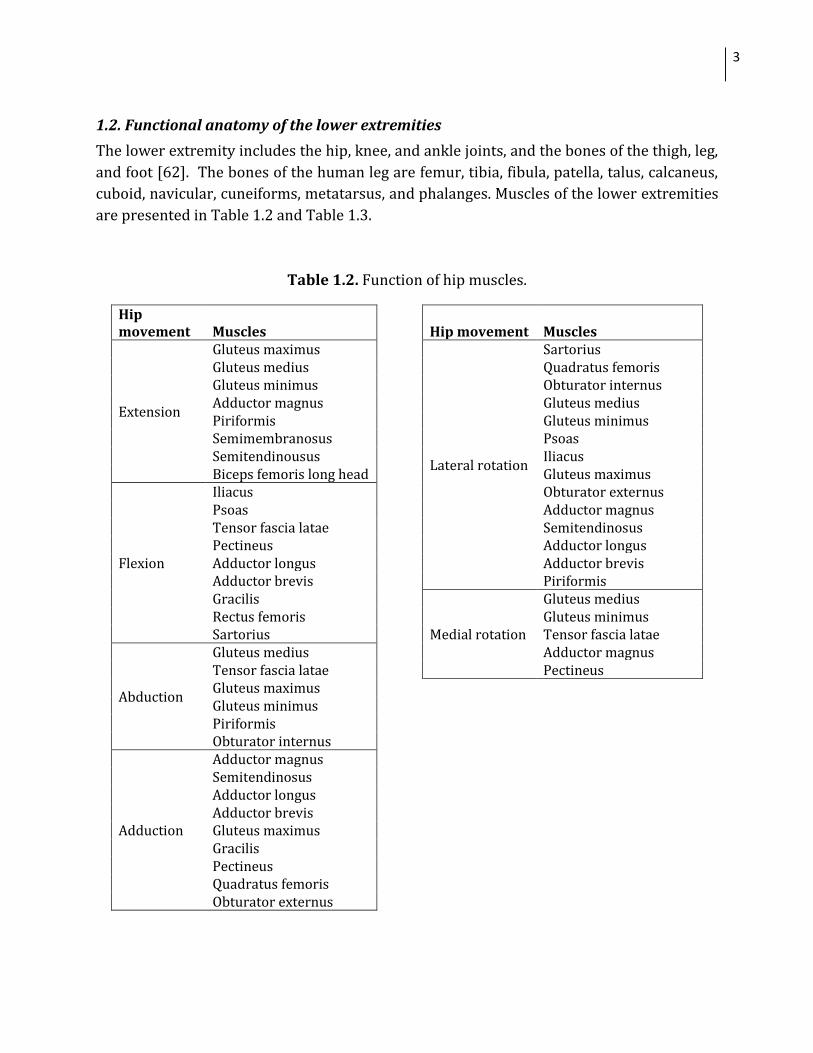
1.2. Functional anatomy of the lower extremities
The lower extremity includes the hip, knee, and ankle joints, and the bones of the thigh, leg,
and foot [62]. The bones of the human leg are femur, tibia, fibula, patella, talus, calcaneus,
cuboid, navicular, cuneiforms, metatarsus, and phalanges. Muscles of the lower extremities
are presented in Table 1.2 and Table 1.3.
Table 1.2. Function of hip muscles.
Hip movement Muscles
Hip movement Muscles
Extension
Gluteus maximus
Lateral rotation
Sartorius Gluteus medius
Quadratus femoris
Gluteus minimus
Obturator internus Adductor magnus
Gluteus medius
Piriformis
Gluteus minimus Semimembranosus
Psoas
Semitendinousus
Iliacus Biceps femoris long head
Gluteus maximus
Flexion
Iliacus
Obturator externus Psoas
Adductor magnus
Tensor fascia latae
Semitendinosus Pectineus
Adductor longus
Adductor longus
Adductor brevis Adductor brevis
Piriformis
Gracilis
Medial rotation
Gluteus medius Rectus femoris
Gluteus minimus
Sartorius
Tensor fascia latae
Abduction
Gluteus medius
Adductor magnus Tensor fascia latae
Pectineus
Gluteus maximus Gluteus minimus Piriformis Obturator internus
Adduction
Adductor magnus Semitendinosus Adductor longus Adductor brevis Gluteus maximus Gracilis Pectineus Quadratus femoris Obturator externus
4 1. INTRODUCTION
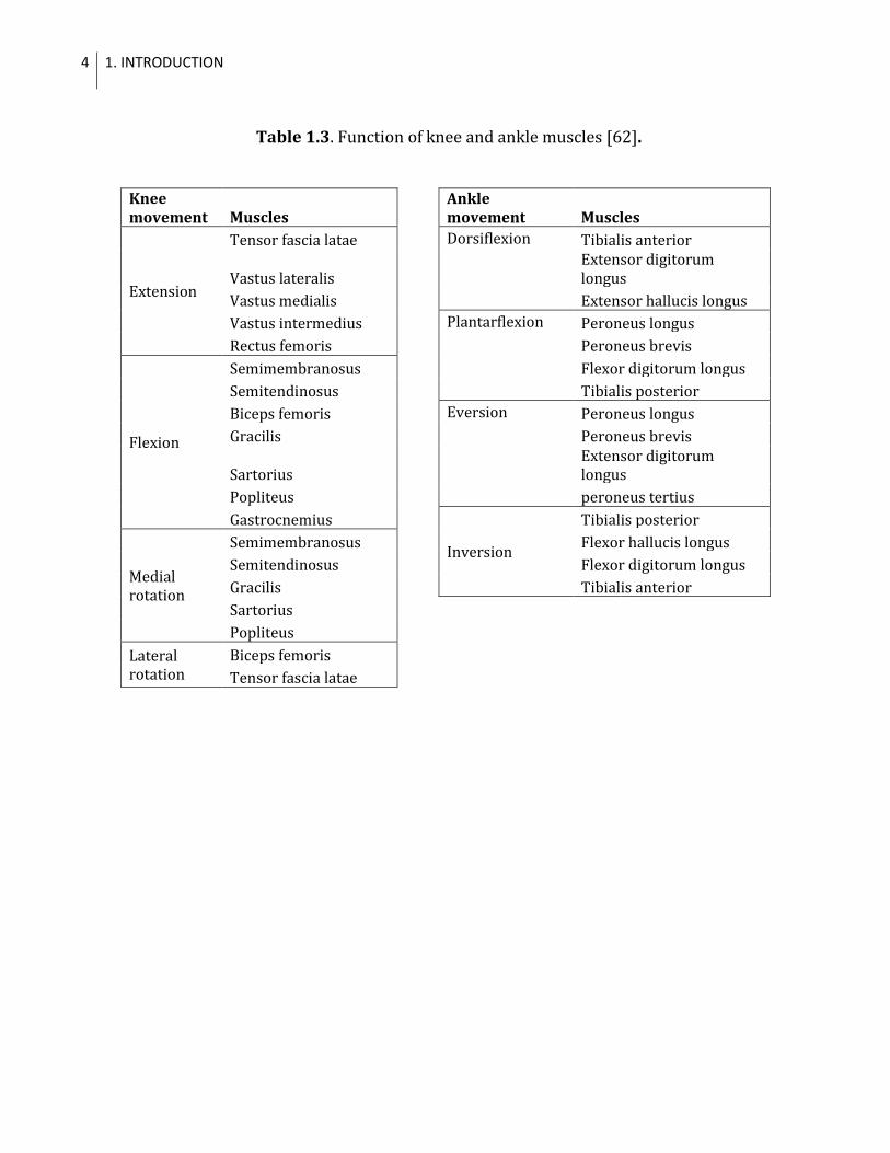
Table 1.3. Function of knee and ankle muscles [62].
Knee movement Muscles
Ankle movement Muscles
Extension
Tensor fascia latae
Dorsiflexion Tibialis anterior
Vastus lateralis
Extensor digitorum longus
Vastus medialis
Extensor hallucis longus
Vastus intermedius
Plantarflexion Peroneus longus
Rectus femoris
Peroneus brevis
Flexion
Semimembranosus
Flexor digitorum longus
Semitendinosus
Tibialis posterior
Biceps femoris
Eversion Peroneus longus
Gracilis
Peroneus brevis
Sartorius
Extensor digitorum longus
Popliteus
peroneus tertius
Gastrocnemius
Inversion
Tibialis posterior
Medial rotation
Semimembranosus
Flexor hallucis longus
Semitendinosus
Flexor digitorum longus
Gracilis
Tibialis anterior
Sartorius Popliteus Lateral
rotation Biceps femoris
Tensor fascia latae
5
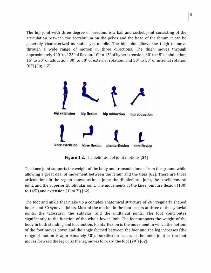
The hip joint with three degree of freedom, is a ball and socket joint consisting of the
articulation between the acetabulum on the pelvis and the head of the femur. It can be
generally characterized as stable yet mobile. The hip joint allows the thigh to move
through a wide range of motion in three directions. The thigh moves through
approximately 120˚ to 125˚ of flexion, 10˚ to 15˚ of hyperextension, 30˚ to 45˚ of abduction,
15˚ to 30˚ of adduction, 30˚ to 50˚ of external rotation, and 30˚ to 50˚ of internal rotation
[62] (Fig. 1.2).
Figure 1.2. The definition of joint motions [34]
The knee joint supports the weight of the body and transmits forces from the ground while
allowing a great deal of movement between the femur and the tibia [62]. There are three
articulations in the region known as knee joint: the tibiofemoral joint, the patellofemoral
joint, and the superior tibiofibular joint. The movements at the knee joint are flexion (130˚
to 145˚) and extension (1˚ to 7˚) [62].
The foot and ankle that make up a complex anatomical structure of 26 irregularly shaped
bones and 30 synovial joints. Most of the motion in the foot occurs at three of the synovial
joints: the talocrural, the subtalar, and the midtarsal joints. The foot contributes
significantly to the function of the whole lower limb. The foot supports the weight of the
body in both standing and locomotion. Plantarflexion is the movement in which the bottom
of the foot moves down and the angle formed between the foot and the leg increases (the
range of motion is approximately 50˚). Dorsiflexion occurs at the ankle joint as the foot
moves forward the leg or as the leg moves forward the foot (20˚) [62].
6 1. INTRODUCTION
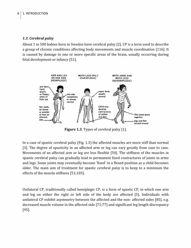
1.3. Cerebral palsy
About 1 in 500 babies born in Sweden have cerebral palsy [2]. CP is a term used to describe
a group of chronic conditions affecting body movements and muscle coordination [116]. It
is caused by damage to one or more specific areas of the brain, usually occurring during
fetal development or infancy [51].
Figure 1.3. Types of cerebral palsy [1].
In a case of spastic cerebral palsy (Fig. 1.3) the affected muscles are more stiff than normal
[3]. The degree of spasticity in an affected arm or leg can vary greatly from case to case.
Movements of an affected arm or leg are less flexible [50]. The stiffness of the muscles in
spastic cerebral palsy can gradually lead to permanent fixed contractures of joints in arms
and legs. Some joints may eventually become 'fixed' in a flexed position as a child becomes
older. The main aim of treatment for spastic cerebral palsy is to keep to a minimum the
effects of the muscle stiffness [51;105].
Unilateral CP, traditionally called hemiplegic CP, is a form of spastic CP, in which one arm
and leg on either the right or left side of the body are affected [5]. Individuals with
unilateral CP exhibit asymmetry between the affected and the non- affected sides [85], e.g.
decreased muscle volume in the affected side [71;77] and significant leg length discrepancy
[95].
7
1.4. Motion analysis
Locomotion is the process of self-propulsion by which one moves from one geographic
position to another [98]. The human body integrates the motions of the various segments
of the body during walking and controls the activity of the muscles so that the metabolic
energy required for a given distance walked is minimized. Different gaits are characterized
by differences in limb movement patterns, overall velocity, forces, kinetic and potential
energy cycles, and changes in the contact with the ground [93].
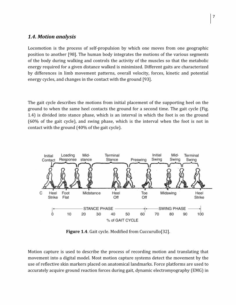
The gait cycle describes the motions from initial placement of the supporting heel on the
ground to when the same heel contacts the ground for a second time. The gait cycle (Fig.
1.4) is divided into stance phase, which is an interval in which the foot is on the ground
(60% of the gait cycle), and swing phase, which is the interval when the foot is not in
contact with the ground (40% of the gait cycle).
Figure 1.4. Gait cycle. Modified from Cuccurullo[32].
Motion capture is used to describe the process of recording motion and translating that
movement into a digital model. Most motion capture systems detect the movement by the
use of reflective skin markers placed on anatomical landmarks. Force platforms are used to
accurately acquire ground reaction forces during gait, dynamic electromyography (EMG) in
8 1. INTRODUCTION
evaluating and recording the electrical activity in skeletal muscles [26;45]. Gait analysis,
often consisting of joint kinematics, kinetics and dynamic EMG data [33;80], is the major
application of a motion capture in orthopedics [16;49;52], and is used to define movement
deviations and the various functional deficits related to complex neuromuscular conditions
such as CP [107]. Postoperatively, it provides an accurate assessment of outcome [53] that
enables objective evaluation of surgeries [48]. However, instrumental errors, anatomical
landmark misplacement [70], and soft tissue artefacts may lead to inaccurate conclusions
made by gait analysis [28;30]. Novel methods have been applied to minimize the effect of
skin movement on the accuracy of recorded markers trajectories on space [4;27;74].
The data from gait analysis (GA) not only should be collected in a standardized way but
also must be calculated with appropriate methods. This requires an accurate underlying
computational musculoskeletal model.
1.5. Generic musculoskeletal models
Computational musculoskeletal models allow quantifying factors (e.g. muscle moment
arms, joint motions) that affect musculoskeletal function, and may help clinicians to
improve clinical outcomes of the necessary treatments [8;10;11;11;14;38;61].
Musculoskeletal models have been used to study stroke [65], spinal cord injury [88],
osteoarthritis [55;56] and neurological deficits such as CP [35]
However, the existing musculoskeletal models in use have limitations. Most of the software
packages for biomechanical analysis of muscle function are based on biomechanical studies
of cadaveric specimens [13;36], and use the musculoskeletal geometry of a healthy,
average-sized adult male with normal musculoskeletal geometry [10;36;38]. These generic
models apply variations in subject size by scaling [40;43;76], based on three-dimensional
positions of markers placed on selected anatomical landmarks and measured during a
static, standing trial. Generic models were used to simulate bone deformities[8],
osteotomies [44;104], and tendon transfer surgeries [40]. However, a recent study has
proved that such models provide inaccurate analysis of muscle function even for a healthy
adult male [102].
The musculoskeletal system is very intricate and large anatomical variations exist among
individuals. The musculoskeletal geometry determines moment arm and thereby the
moment about a joint produced by a given musculotendon force [68;117;118]. Duda et al
[41] have studied how variability of muscle attachments affects muscle moment arms
(MALs). The effects of bone geometry on the moment-generating capacity of the muscles
has been shown by Delp et al.[39] Thus, the different musculoskeletal geometry due to size
9
or pathology can also affect the accuracy of results derived from generic models. A recent
study [100] has demonstrated the inaccuracy of gait kinematics calculated by the scaled
generic models in subjects with increased femoral anteversion. It was reported that the
muscle-tendon length (MTLs) calculated with a generic model are erroneous if compared
with subject-specific models in children with CP and crouch gait [7]. It was shown that
scaled generic models provide inaccurate analysis of MALs and MTLs in CP children with
altered femoral geometry [103].
Since the results of simulations are often sensitive to the accuracy of the functional
musculoskeletal model, individualized musculoskeletal models may be a better alternative
[73;113].
1.6. Subject-specific musculoskeletal models
Defining the geometry of a complex musculoskeletal system is challenging. Medical imaging
techniques such as magnetic resonance imaging (MRI) or computer tomography (CT) are
used to create images of the human body, and to study in vivo the complex geometric
relationships among the muscles, bones, and other structures [14;15;18;24;46;47;57-
59;63;71;96;97;99;106;108;110;111]. The volumes of muscles, which can be derived from
segmented MRIs, are important in examining the atrophy or hypertrophy resulting from
different pathologies, treatments, and strength training [17;46;67;71;77].
An accurate reconstruction of the functional anatomy of the body, required for modern
whole-body biomechanical models, is not trivial [114]. The musculoskeletal geometry for a
specific subject can be extracted from MRI [11;12;15;21;71] or CT-scan images [114].
Three-dimensional reconstructions from CT scans have been used to design orthopedic
implants [60;89;112] and plan orthopedic surgeries [81]. Subject-specific 3D models of
muscles have been created from MRIs to study muscle volumes [57;71;78;87]. Muscle
moment arms have been estimated in vivo from static MRIs [99], however it is time-
consuming and requires extensive imaging protocols to capture the muscle and joint
geometry at different limb positions. Arnold et al. [12] were the first to build subject-
specific models using MRI to analyse the MALs over the range of joint motion.
Subject-specific musculoskeletal modelling also addresses the problem of image
segmentation, which consists of extracting anatomical structures from medical image data
such as MRI. Semiautomatic or fully automatic segmentation methods are fast but
inaccurate since muscle distinction is often difficult or impossible to assess with currently
10
used methods. Thus, muscles volumetric representations are most often and most
accurately acquired by defining muscle contours manually [11;86].
Blemker et al. [22] created volumetric finite-element representations of a muscle and built
the surface data from manually segmented MRIs, combined with description of the
nonlinear stress-strain behaviour of muscle tissue, and developed a new formulation for
representing muscles shape, geometry, and force. Arnold et al. [12] created MRI-based
musculoskeletal models of three lower extremity cadaveric specimens, which included
pelvis, femur, tibia, psoas, semimembranosus, and semitendinosus, from manually
segmented MRIs. Scheys et al. [101] generated a subject-specific musculoskeletal model of
the lower extremities of an able-bodied subject, which included femur, tibia and fibula and
25 muscles’ lines of action using a centroid approach, i.e. the attachment points of the
muscle to the bone were identified by scrolling through the image slices and picking an
appropriate point in the last slice where the muscle is visible.
Automatic segmentation and a 3D region-growing algorithm were applied by Scheys et al.
[103] to define the bone structures. These methods were also used to build the person-
specific models of CP children with presence of femoral anteversion and study the effect of
bone deformities on the accuracy of hip muscles moment arms [12;103]. However, the
entire process of MRI-based modeling is still time-consuming because semi-automatic
segmentation of the muscles has failed thus far.
Since the extensive variations in musculoskeletal geometry exist among individuals, there
is no public software which can perform acquisition of individual musculoskeletal
geometry from medical imaging data and analyse muscle function.
11
SCOPE AND AIMS
The scope of this thesis is focused on the developing and applying subject-specific
musculoskeletal models of the lower extremities to study muscle volumes and
biomechanics parameters of muscles in subjects with unilateral CP.
Study I
The first aim was to develop a workflow to build highly detailed, subject-specific
musculoskeletal model of the lower extremities from MRIs of a person with unilateral CP
that can be exported in software for musculoskeletal computing (SIMM).
The second aim was to calculate MTLs and MALs during gait using the developed
musculoskeletal model.
The third aim was to determine the accuracy of hip, knee and ankle MALs and MTLs during
gait calculated from the scaled generic model by comparing them to those computed from
the subject-specific musculoskeletal model.
Study II
Study II was designed as a wider scale application of the methods developed in Study I.
The first aim was to develop subject-specific musculoskeletal models of the hip joints in
both sides of the lower extremities based on MRIs of nine subjects with unilateral CP.
The second aim was to examine MALs and MTLs over hip abd/adduction, hip
flexion/extension and hip rotation, and muscle volumes calculated by the subject-specific
model.
The third aim was to study the accuracy of MALs calculated by the scaled generic model.

12 SCOPE AND AIMS
13
2. MATERIALS AND METHODS
2.1. SUBJECTS
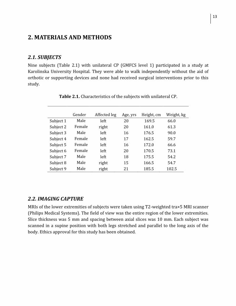
Nine subjects (Table 2.1) with unilateral CP (GMFCS level 1) participated in a study at
Karolinska University Hospital. They were able to walk independently without the aid of
orthotic or supporting devices and none had received surgical interventions prior to this
study.
Table 2.1. Characteristics of the subjects with unilateral CP.
Gender Affected leg Age, yrs Height, cm Weight, kg
Subject 1 Male left 20 169.5 66.0
Subject 2 Female right 20 161.0 61.3
Subject 3 Male left 16 176.5 90.0
Subject 4 Female left 17 162.5 59.7
Subject 5 Female left 16 172.0 66.6
Subject 6 Female left 20 170.5 73.1
Subject 7 Male left 18 175.5 54.2
Subject 8 Male right 15 166.5 54.7
Subject 9 Male right 21 185.5 102.5
2.2. IMAGING CAPTURE
MRIs of the lower extremities of subjects were taken using T2-weighted tra×5 MRI scanner
(Philips Medical Systems). The field of view was the entire region of the lower extremities.
Slice thickness was 5 mm and spacing between axial slices was 10 mm. Each subject was
scanned in a supine position with both legs stretched and parallel to the long axis of the
body. Ethics approval for this study has been obtained.
14 2. MATERIALS AND METHODS
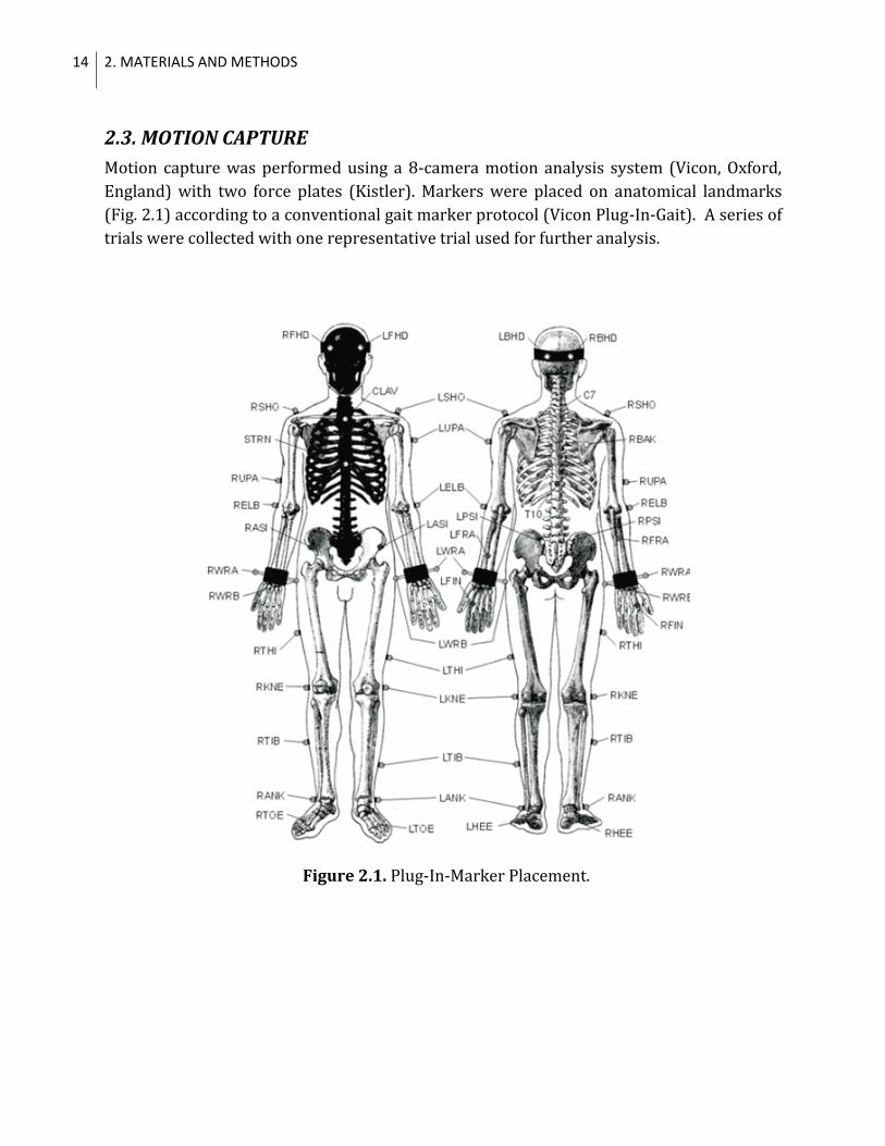
2.3. MOTION CAPTURE
Motion capture was performed using a 8-camera motion analysis system (Vicon, Oxford,
England) with two force plates (Kistler). Markers were placed on anatomical landmarks
(Fig. 2.1) according to a conventional gait marker protocol (Vicon Plug-In-Gait). A series of
trials were collected with one representative trial used for further analysis.
Figure 2.1. Plug-In-Marker Placement.
15
2.4. SCALED GENERIC MODEL
The generic model [36] was scaled in SIMM (Musculographics Inc., Santa Rosa, CA) based
on three-dimensional positions of markers attached to the pelvis, femur, tibia and foot
during a standing trial.
2.5. SUBJECT-SPECIFIC MUSCULOSKELETAL MODEL
2.5.1. Study I
The subject-specific model of the lower extremities was developed from MRIs of Subject 8
(Table 2.1). It included muscles, bones (Table 2.2), and kinematic descriptions of hip, knee
and ankle joints.
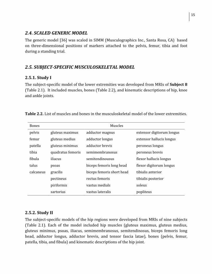
Table 2.2. List of muscles and bones in the musculoskeletal model of the lower extremities.
Bones Muscles
pelvis gluteus maximus adductor magnus extensor digitorum longus
femur gluteus medius adductor longus extensor hallucis longus
patella gluteus minimus adductor brevis peroneus longus
tibia quadratus femoris semimembranosus peroneus brevis
fibula iliacus semitendinousus flexor hallucis longus
talus psoas biceps femoris long head flexor digitorum longus
calcaneus gracilis biceps femoris short head tibialis anterior
pectineus rectus femoris tibialis posterior
piriformis vastus medials soleus
sartorius vastus lateralis popliteus
2.5.2. Study II
The subject-specific models of the hip regions were developed from MRIs of nine subjects
(Table 2.1). Each of the model included hip muscles (gluteus maximus, gluteus medius,
gluteus minimus, psoas, iliacus, semimembranosus, semitendinosus, biceps femoris long
head, adductor longus, adductor brevis, and tensor fascia latae), bones (pelvis, femur,
patella, tibia, and fibula) and kinematic descriptions of the hip joint.
16 2. MATERIALS AND METHODS
2.5.3. Three-dimensional reconstruction of muscles and bones models
Bones and muscles contours were manually outlined by assigning label maps, where each
voxel is a number indicating the type of tissue at that location in 3D Slicer (www.slicer.org),
which is a free open- source software for visualization and image computing and can
perform different medical image processing activities including surface reconstruction
from MRI [54;90;91].
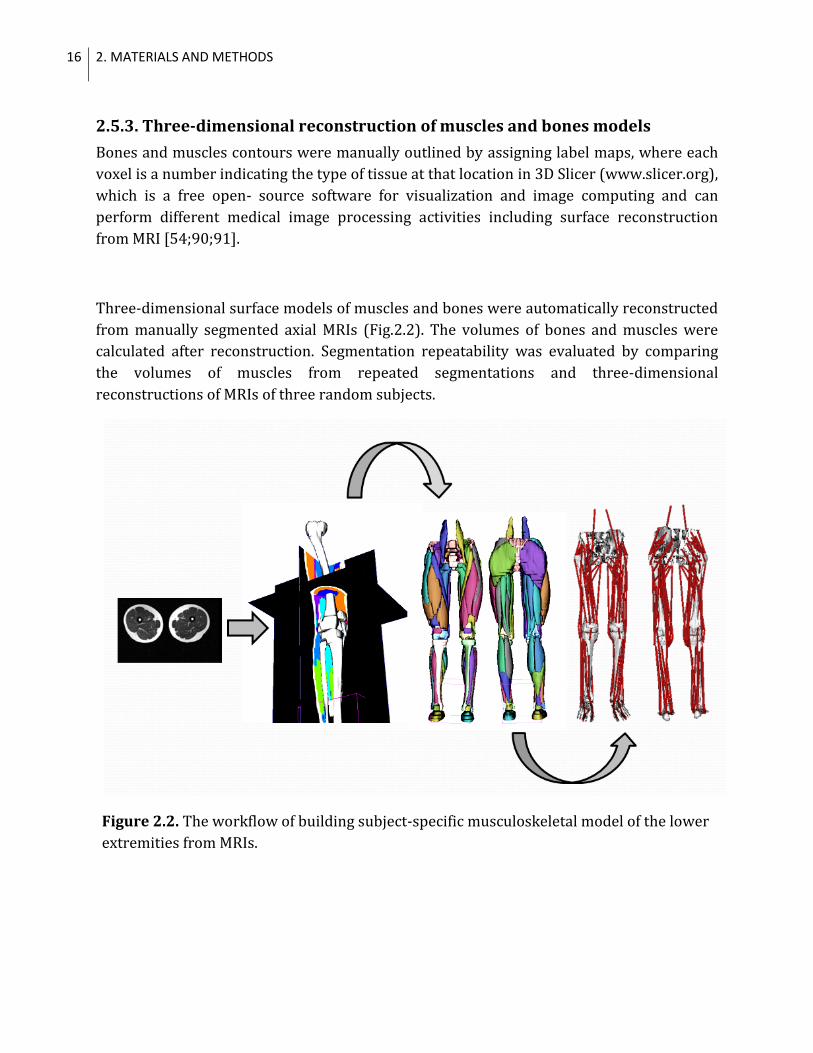
Three-dimensional surface models of muscles and bones were automatically reconstructed
from manually segmented axial MRIs (Fig.2.2). The volumes of bones and muscles were
calculated after reconstruction. Segmentation repeatability was evaluated by comparing
the volumes of muscles from repeated segmentations and three-dimensional
reconstructions of MRIs of three random subjects.
Figure 2.2. The workflow of building subject-specific musculoskeletal model of the lower
extremities from MRIs.
17
2.5.3. Specification of joint kinematics and representation of muscle-tendon
paths
Using a musculoskeletal modeling package [36], we defined the joint kinematics, and
muscle–tendon paths of the model.
Kinematic descriptions of hip, knee and ankle joint were defined based on the patient’s
bone surface geometry in the scanned position. The transformations that relate the
position and orientation of one body segment to another consisted of three translations
and three rotations.
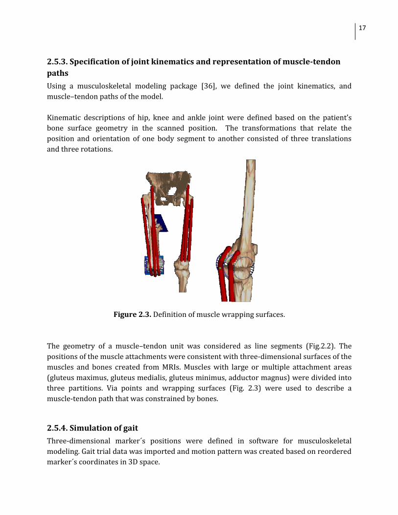
Figure 2.3. Definition of muscle wrapping surfaces.
The geometry of a muscle–tendon unit was considered as line segments (Fig.2.2). The
positions of the muscle attachments were consistent with three-dimensional surfaces of the
muscles and bones created from MRIs. Muscles with large or multiple attachment areas
(gluteus maximus, gluteus medialis, gluteus minimus, adductor magnus) were divided into
three partitions. Via points and wrapping surfaces (Fig. 2.3) were used to describe a
muscle-tendon path that was constrained by bones.
2.5.4. Simulation of gait
Three-dimensional marker´s positions were defined in software for musculoskeletal
modeling. Gait trial data was imported and motion pattern was created based on reordered
marker´s coordinates in 3D space.
18
2.6. DATA ANALYSIS
Study I
The hip, knee and ankle MALs and MTLs of 70 muscles in affected and non-affected sides
during gait were calculated using both the subject-specific model and the scaled generic
model.
Study II
For each subject MALs over hip adduction/abduction, extension/flexion and rotation
ranges of motion in the both affected and the non- affected sides were calculated using both
the scaled generic and the subject-specific models. We calculated the ratio, standard deviation
(SD) between the average values of the MALs, over the range of hip motion, in both sides of the
lower extremities in the generic scaled model and subject-specific model. The Wilcoxon
matched pairs test (significance level of p<0.05) was implemented in Matlab (MathWorks Inc.)
to evaluate the differences in muscle volumes, MALs and MTLs in the affected side vs. the non-
affected side calculated by the subject-specific models, and to evaluate systematic differences
between average hip MALs calculated by the scaled generic modes and subject-specific models.
19
3. RESULTS AND DISCUSSIONS
Study I
We developed the workflow to build a highly detailed, subject-specific model of the entire
lower extremities from MRIs, which can be exported into the software for biomechanical
analysis of muscle function during gait. The study provided a comprehensive evaluation of
muscle volumes, MTLs and MALs of 70 muscles in the entire lower extremities in a subject
with unilateral CP.
During the process of subject-specific modeling we created 3D models of muscles and
bones from axial MRIs, calculated muscle volumes and evaluated the repeatability of MRI
segmentation. The maximum volume error was 12% for tensor fascia latae, since it was
difficult to see muscle-tendon transition. The volume error for other muscles was
approximately 1-4% (the low resolution artifacts and noise in images led to difficulty in
identifying borders between muscles in some of the axial MRIs, which influenced the
precision of calculation of muscle volumes).
All muscle volumes in the affected limb were found to be smaller than in the non-affected
limb, with atrophy being more significant in the shank than in the thigh, with an average
muscle volume discrepancy of 28% and 13% respectively. Our findings confirm those of
Elder et al. [42], Malaiya et al. [77], and Lampe et al. [72]. We also found that maximal MTLs
during gait calculated by the subject-specific model were shorter in the affected side in
adductor longus, adductor brevis, adductor magnus, pectineus, and quadratus femoris
muscles. The decreased muscle volumes in the affected leg may therefore be attributable in
part to shorter muscles, corresponding to findings by Lieber et al.[75].
Muscle tendon lengths during gait
In general, the scaled model delivered accurate enough values for changes in MTLs during
gait for all muscles except adductor magnus, adductor longus, adductor brevis, pectineus,
iliacus, psoas, and quadratus femoris.
20 3. RESULTS AND DISCUSSIONS
Muscle moment arm lengths in the affected side during gait
We found that scaled generic model extremely overestimated MALs for hip medial (gluteus
medius, gluteus minimus, adductor longus, psoas, tensor fascia latae, biceps femoris) and
lateral rotation; for hip abduction and adduction (in semitendinosus, sartorius, biceps
femoris); for hip flexion in adductor brevis; for knee flexion in semimembranosus and
semitendinosus; for ankle flexion in peroneus tertius.
Muscle moment arm lengths discrepancies between the affected and the non-affected sides
Average hip rotation MAL discrepancies between affected and non-affected lower
extremities during gait were underestimated by the scaled generic model in most hip
rotator muscles, except gluteus medialis, by an average of 73%
Average hip abd/adduction MAL discrepancies between affected and non-affected lower
extremities during gait were underestimated by the scaled generic model in most hip add-
/abductors muscles by an average of 62%; and were overestimated in gluteus minimus,
adductor brevis, tensor fascia latae, gracilis, semitendinosus by an average of 53%.
Average hip flexion/extension MAL discrepancies between affected and non-affected lower
extremities during gait were underestimated by the scaled generic model in most hip
flexors/extensors muscles, except semitendinosus, by an average of 71%; and were
overestimated in gluteus medius, gluteus maximus, adductor longus, piriformis, tensor
fascia latae, sartorius by more than 100%.
Average knee flexion/extension MAL discrepancies between affected and non-affected
lower extremities during gait were underestimated by scaled generic model in most knee
flexors/extensors muscles, except sartorius, by 83%; and were overestimated in
semimembranosus and semitendinosus by an average of 33%
Average ankle plantar/dorsiflexion MAL discrepancies between affected and non-affected
lower extremities during gait were underestimated by the scaled generic model in most
ankle flexion muscles by an average of 84%; and were overestimated in peroneus longus by
18%,
21
Study II
We created the subject-specific musculoskeletal models of the lower extremities from MRIs
of nine teenagers and young adults with mild unilateral CP to study muscle volumes, hip
MALs and MTLs.
Since all studied subjects in the present study were highly functioning, the MTLs in the
affected and non-affected sides were very similar. No significant differences between sides
were observed in MTLs of gluteus maximus, gluteus medius, gluteus minimus, psoas,
iliacus, adductor longus and adductor brevis. Average hip adduction/abduction MALs of
rectus femoris, the medial part of gluteus medius, the anterior parts of gluteus medius and
gluteus minimus in the affected side were slightly smaller by an average of 5±1mm; MALs
iliacus, psoas and the medial part of gluteus maximus in the affected side were slightly
larger by an average of 3±1mm.
However muscle volumes in gluteus maximus (p=0.008), adductor longus (p=0.014),
tensor fascia latae (p=0.006), biceps femoris long head (p<0.001), rectus femoris
(p=0.005), semimembranosus (p=0.022), semitendinosus (p=0.020) in the affected side
were significantly smaller than in the non-affected side by an average of 16%%, correlating
with previous findings [71;94].
Fukunaga et al. [46] found a correlation between muscle torque and muscle volume or
PSCA, and smaller torque around hip joint in the affected side. It was reported that the loss
of muscle strength[115] correlates with the volumetric loss of the spastic musculature.
Spastic muscles have also shown power reduction during gait [71;94]. Gluteus medius
and gluteus minimus with smaller muscle volumes and hip abduction MALs in the affected
side therefore can be expected to have lower hip abduction strength comparing with the
non-affected side. Similarly, we can expect iliacus to have lower hip flexion; and, finally,
gluteus maximus to have lower hip rotation strength [19;92].
Comparison of MALs from the scaled generic and the subject-specific models
Hip abduction/adduction MALs of gluteus medius, gluteus minimus, tensor fascia latae and
biceps femoris long head and the anterior part of gluteus maximus were significantly
overestimated by the scaled generic model by an average of 46% (p<0.001, p<0.001,
p=0.001, p=0.001 and p=0.02) in the affected side and by 34% (p=0.04, p=0.005, p=0.006,
p=0.006 and p=0.01) in the non-affected side. Hip abduction/adduction MALs of adductor
longus and semimembranosus were significantly underestimated by the scaled generic
22 4. GENERAL DISCUSSION
model by an average of 15% (p=0.01 and p=0.02) side in the affected, and by 44% (p=0.01
and p=0.02) in the non-affected side. Adduction MALs of psoas in the affected side were
also significantly (p=0.04) underestimated by the scaled generic model by 81%.
Hip flexion/extension MALs of the medial part and the posterior part of gluteus maximus,
the anterior part of gluteus medius, the medial part of gluteus minimus were significantly
underestimated by the scaled generic model by an average of 44% (p=0.02, p=0.02, p=0.02
and p=0.006) in the affected side, and by 47% (p=0.004, p=0.002, p=0.03 and p=0.001) in
the non-affected side. Hip flexion/extension MALs of the medial and the posterior parts of
gluteus medius, adductor brevis and psoas were significantly overestimated by the scaled
generic model by an average of 99% (p=0.004, p=0.01, p=0.01 and p=0.01) in the affected
side and by 113% (p<0.001, p<0.001, p=0.03 and p=0.03) in the non-affected side. Hip
flexion/extension MALs of adductor longus and rectus femoris in the non-affected side
were significantly overestimated by the scaled generic model by an average of 44% with
p=0.03 and p=0.008 respectively.
Hip rotation MALs of the posterior part of gluteus minimus and semitendinosus were
significantly underestimated by the scaled generic model by an average of 65% (with
p=0.004 and p<0.001) in the affected side and by 68% (p<0.001and p<0.001) in the non-
affected side. Hip rotation MALs of the medial part of gluteus maximus (p=0.02) and
semimembranosus (p=0.02) in the non/affected side were significantly underestimated by
the scaled generic model by 22% and 57%. Hip rotation MALs of the posterior part of
gluteus maximus and gluteus medius, the anterior part of gluteus medius and biceps
femoris long head were significantly (p=0.008, p=0.002 and p<0.001 overestimated by the
scaled generic model in the affected side by an average of 65%.
Our results also confirmed that the scaled generic model significantly underestimated hip
MALs differences between the affected and the non- affected sides in most muscles.
Consequently, the scaled generic models may lead to erroneous conclusions about
individual muscle contributions to joint moments.
23
4. GENERAL DISCUSSION
The results of the performed studies have important implications for the accuracy of
assessing muscle function of persons with unilateral CP using the scaled generic model. Our
findings showed that the scaled generic and the subject-specific lower extremity
musculoskeletal models showed substantial dissimilarities in hip, knee and ankle MALs and
MTLs calculated during gait of a subject with unilateral CP and significant differences in
MALs over the range of hip motion in a group of subjects with very mild unilateral CP.
Differences between the subject-specific and the scaled generic models were caused by the
variability in muscle attachment locations [102] and bone geometry that is not taken into
account in the scaling of the generic model. Persons with unilateral CP often have
asymmetric musculoskeletal geometry in the affected side and the non- affected side[49],
e.g. decreased muscle volume in the affected side[71;77] and significant leg length
discrepancy[95]. Nevertheless, the both sides of the lower extremities are symmetrical in
the generic model; axial scaling makes its bones longer/shorter or wider/narrower, but
changes in muscle attachment positions were not taken into account. As a result, the scaled
generic model failed to identify variability of muscle attachments and bone geometry
between the affected and the non- affected sides in subjects with unilateral CP.
The inaccuracies of the generic scaled model were very pronounced in femur, because it
was impossible to assess femoral shape parameters (e.g. neck length, femoral length,
femoral neck angle etc.) in individuals using only scaling based on markers placed on skin .
As a result, muscle attachments and via points were defined by the scaled generic model
with a large error.
It is important to keep in mind some of the limitations of this study. We described muscle-
tendon paths as a series of line segments; because the main goal was to import the subject-
specific model in software that can calculate muscle moments arms and tendon lengths
during motion based on such simplified representation of muscle geometry. This is a
reasonable simplification for muscles with small areas of origin and insertion (e.g. tibialis
posterior). However, it was challenging to use a series of line segments to represent
muscles with broad attachments, like the gluteus maximus. In SIMM models such muscles
are separated into compartments, and multiple paths to represent the muscle [36] are
used. However, it was unclear how many paths to define, where the paths should be
24 4. GENERAL DISCUSSION
located, and how to define via points (and/or wrapping surfaces) so that the models
accurately represent the anatomy. The resulting muscle moment arms may be highly
sensitive to how the constraints are defined. In a future, muscles with large areas of
attachment, multiple origins, or curved paths could be advantageously modelled as
volumetric objects [14;22;23].
The outcome of orthopedic surgery aimed to correct movement abnormalities of persons
with CP [3;29;31;60;81] can be difficult to predict and is sometimes unsuccessful
[25;29;31;105]. Musculoskeletal simulations are needed to analyse the biomechanical
causes of movement abnormalities since this information is important for developing
better treatment plans [9;10]. Despite limitations of this study, we believe that the methods
presented here offer the potential to improve the accuracy of models of the
musculoskeletal system for development of more effective treatment plans of persons with
movement disorders. Based on inaccuracies of scaled generic model reported in our
studies and in recent articles [7;100;102] , the scaled generic models that are used to study
persons with CP must be tested and interpreted with care, in the knowledge of the
underlying limitations of the models and the conditions that determine when, and for
which patients subject-specific models are the better alternative.
Future work
The accuracy of a simulation depends on the accuracy of the defined musculoskeletal
model. The subject-specific musculoskeletal models, based on in vivo measurements of
musculoskeletal geometry and joint kinematics, can help in understanding the causes of
movement deviations [13;37] and assessing treatment options [43].
It is challenging to simulate, explore and predict the biomechanical effects of orthopedic
surgeries using subject-specific musculoskeletal models and dynamic simulations of
individuals with pathological gait [9]. Further advancements in image-based
musculoskeletal modeling will expand the accuracy and utility of models used to study
musculoskeletal and neuromuscular impairments, and to improve the treatment outcome.
Modeling muscle using a series of line segments allows only one length and moment arm to
be estimated for each muscle path. However, variation in moment arms lengths among
fibers within a muscle could greatly influence the muscle’s capacity to generate force:
previous study [64] has demonstrated that such simplified musculoskeletal models do not
25
accurately predict in vivo force–joint angle behaviours of muscles with complex
architectures. By creating volumetric finite-element representations of muscle from the 3D
muscle surface derived from static MRIs, combined with description of the nonlinear
stress-strain behaviour of muscle tissue, a new formulation for representing muscle shape,
geometry, and force can be developed.
The internal architecture of muscles can be also derived from diffusion tensor imaging
combined with tractography methods as it was implemented in recent studies [24;63;108].
Models that represent the 3D arrangement of muscle fibers and allow for variations in fiber
lengths and moment arms [22;23] are needed to more closely represent in vivo muscle
behaviour.
Subject-specific musculoskeletal models can be evaluated by comparing muscle tissue
deformations predicted by volumetric muscle models with tissue deformations derived
from dynamic MRI, and by comparing MALs predicted by models with MALs measured
from dynamic MRIs. Joint kinematics can be prescribed from in vivo, dynamic, loaded
measurements of individual subjects. Acquisition of static MRIs at multiple joint positions
has been applied to studying the mechanics of the many joints [58;59;106].

26 4. GENERAL DISCUSSION
27
OUTLINE OF PAPERS
Paper 1
The purpose of this paper was to develop methods to build a subject-specific
musculoskeletal model of the lower extremities based on MRIs of a subject with unilateral
CP, and to determine whether a scaled generic musculoskeletal model is accurate enough
to characterize MTLs and MALs of 70 muscles in both lower limbs during gait in a subject
with unilateral cerebral palsy.
We found, that the generic models produced accurate values for changes in MTL during gait
for almost all muscles, except adductor longus, adductor magnus, adductor brevis,
quadratus femoris, pectineus, extensor digitorum longus, soleus, lateral gastrocnemius, and
medial gastrocnemius.
MALs computed from the scaled generic model, however, differed considerably from those
computed from the subject-specific model. Upon comparison of hip, knee and ankle MALs
in affected and non-affected sides of the lower extremities, the scaled generic model
generally failed to identify level arm dysfunction in the subject with unilateral CP.
Paper II The aim of this paper was to create the subject-specific modes of the lower extremities
based on MRIs of nine youth adults with unilateral CP to study hip muscle volumes, MTLs
and MALs. Muscle volumes and hip abduction MALs in gluteus medius and gluteus
minimus, hip flexion MALs in iliacus, and hip rotation in gluteus maximus were smaller in
the affected side of lower extremities. Yet, MTLs were very similar in the involved and the
non-involved sides.
We also studied the accuracy of MALs of 36 muscles over the range of hip motion calculated
from generic scaled models, and its ability to identify discrepancy in MALs between the
affected and the non- affected sides. The hip MALs of almost all muscles in the affected leg
were overestimated by the scaled generic. The MALs discrepancies between the affected
and the non- affected sides of the lower extremities were significantly underestimated by
the scaled generic model.

28
29
ACKNOWLEDGEMENTS
This work was supported by Stiftelsen Promobilia and The Swedish Research Council.
I am sincerely thankful to Professor Anders Eriksson and Svetlana Bauer, who gave me the
chance to do a research as a PhD student at Royal Institute of Technology, Department of
Mechanics.
Lanie Gutierrez-Farewik for presenting me the world of orthopedics, biomechanics, and
simulations of the musculoskeletal system. As my principal supervisor, Lanie has been a
constant source of enthusiasm and creativity. Thank you, Lanie, for your steadfast
encouragement to complete this thesis.
Professor Anders Eriksson, my co-supervisor, for introducing me to biomechanics, for great
kindness and support of my studies.
Peter Loan and Steve Piper have been of tremendous assistance in developing the subject-
specific musculoskeletal model. Without their help this project would not have been
possible.
Jacques Riad and Eva W. Broström for sharing their insights into hemiplegic cerebral palsy.
Carolina, Nina and Heide for constant friendship and invaluable help in solving problems
connected with documentation and applications.
I have enjoyed daily interactions with colleagues at Mechanical Department in The Royal
Institute of Technology. Thanks to Eva, Natalia, Rouli and Zeinab for friendship atmosphere
and interesting conversations about life during daily coffee breaks.
Finally, and most importantly, I thank my parents, sister Maria, grandmother Valentina for
their love and support during all my life. Спасибо тебе, мама, что была рядом в трудные
минуты моей жизни и не позволила сломаться под гнетом обрушившихся несчастий.
Тебе я посвящаю эту диссертацию.

30
31
REFERENCES
[1]http://prehealthfig2008.wikispaces.com/Kaitlyn_Cerbral_Palsy. 11-21-2010.
[2]www.vardguiden.se. 8-24-2010.
[3]M.F. Abel, D.L. Damiano, M. Pannunzio, J. Bush, Muscle-tendon surgery in diplegic
cerebral palsy: functional and mechanical changes. Journal of Pediatric Orthopaedics 19
(1999) 366.
[4]E.J. Alexander, T.P. Andriacchi, Correcting for deformation in skin-based marker systems. Journal
of Biomechanics 34 (2001) 355-361.
[5]P.E. Allen, A. Jenkinson, M.M. Stephens, T. O'Brien, Abnormalities in the uninvolved lower limb in
children with spastic hemiplegia: the effect of actual and functional leg-length discrepancy. Journal
of Pediatric Orthopaedics 20 (2000) 88.
[6]A.S. Arnold, F.C. Anderson, M.G. Pandy, S.L. Delp, Muscular contributions to hip and knee
extension during the single limb stance phase of normal gait: a framework for investigating the
causes of crouch gait. Journal of Biomechanics 38 (2005) 2181-2189.
[7]A.S. Arnold, S.S. Blemker, S.L. Delp, Evaluation of a deformable musculoskeletal model for
estimating musclulotendon lengths during crouch gait. Annals of Biomedical Engineering 29 (2001)
263-274.
[8]A.S. Arnold, S.L. Delp, Rotational moment arms of the medial hamstrings and adductors vary with
femoral geometry and limb position: implications for the treatment of internally rotated gait.
Journal of Biomechanics 34 (2001) 437-447.
[9]A.S. Arnold, S.L. Delp. The role of musculoskeletal models in patient assessment and treatment.
Treatment of Gait Problems in Cerebral Palsy, Edited by J.R.Gage , 163-177. 2004. Cambridge Press.
[10]A.S. Arnold, S.L. Delp, Computer modeling of gait abnormalities in cerebral palsy: application to
treatment planning. Theoretical Issues in Ergonomics Science 6 (2005) 305-312.
32 REFERENCES
[11]A.S. Arnold, M.Q. Liu, M.H. Schwartz, Do the hamstrings operate at increased muscle-tendon
lengths and velocities after surgical lengthening? Journal of Biomechanics 39 (2006) 1498-1506.
[12]A.S. Arnold, S. Salinas, D.J. Hakawa, S.L. Delp, Accuracy of muscle moment arms estimated from
MRI-based musculoskeletal models of the lower extremity. Computer Aided Surgery 5 (2000) 108-
119.
[13]E.M. Arnold, S.R. Ward, R.L. Lieber, S.L. Delp, A Model of the Lower Limb for Analysis of Human
Movement. Annals of Biomedical Engineering 38 (2010) 269-279.
[14]D.S. Asakawa, S.S. Blemker, G.T. Rab, A. Bagley, S.L. Delp, Three-dimensional muscle-tendon
geometry after rectus femoris tendon transfer. The Journal of Bone and Joint Surgery 86 (2004)
348.
[15]L. Assassi, C. Charbonnier, J. Schmid, P. Volino, N. Magnenat-Thalmann, From MRI to anatomical
simulation of the hip joint. Computer Animation and Virtual Worlds 20 (2009) 53-66.
[16]R. Baker, Gait analysis methods in rehabilitation. Journal of NeuroEngineering and
Rehabilitation 3 (2006) 4.
[17]T. Bandholm, S. Sonne-Holm, C. Thomsen, J. Bencke, S.A. Pedersen, B.R. Jensen, Calf muscle
volume estimates: implications for botulinum toxin treatment? Pediatric neurology 37 (2007) 263-
269.
[18]P.J. Besl, N.D. McKay, A method for registration of 3-D shapes. IEEE Transactions on pattern
analysis and machine intelligence (1992) 239-256.
[19]A.J. Blazevich, D.R. Coleman, S. Horne, D. Cannavan, Anatomical predictors of maximum
isometric and concentric knee extensor moment. European journal of applied physiology 105
(2009) 869-878.
[20]E.E. Bleck, H.M. Horstmann, Orthopaedic management in cerebral palsy. (1987).
[21]S.S. Blemker, D.S. Asakawa, G.E. Gold, S.L. Delp, Image based musculoskeletal modeling:
Applications, advances, and future opportunities. Journal of Magnetic Resonance Imaging 25 (2007)
441-451.
[22]S.S. Blemker, S.L. Delp, Three-dimensional representation of complex muscle architectures and
geometries. Annals of Biomedical Engineering 33 (2005) 661-673.
33
[23]S.S. Blemker, S.L. Delp, Rectus femoris and vastus intermedius fiber excursions predicted by
three-dimensional muscle models. Journal of Biomechanics 39 (2006) 1383-1391.
[24]S.S. Blemker, A.J. Sherbondy, D.L. Akers, R. Bammer, S.L. Delp, G.E. Gold. Characterization of
skeletal muscle fascicle arrangements using diffusion tensor tractography. Proceedings of the 13th
Annual Meeting of ISMRM, Miami Beach, FL, USA . 2005.
[25]D.C. Borton, K. Walker, M. Pirpiris, G.R. Nattrass, H.K. Graham, Isolated calf lengthening in
cerebral palsy: outcome analysis of risk factors. Journal of Bone and Joint Surgery-British Volume
83 (2001) 364.
[26]T.S. Buchanan, D.G. Lloyd, K. Manal, T.F. Besier, Neuromusculoskeletal modeling: estimation of
muscle forces and joint moments and movements from measurements of neural command. Journal
of applied biomechanics 20 (2004) 367.
[27]A. Cappello, A. Cappozzo, P.F. La Palombara, L. Lucchetti, A. Leardini, Multiple anatomical
landmark calibration for optimal bone pose estimation. Human movement science 16 (1997) 259-
274.
[28]A. Cappozzo, U. la Croce, A. Leardini, L. Chiari, Human movement analysis using
stereophotogrammetry:: Part 1: theoretical background. Gait & posture 21 (2005) 186-196.
[29]H. Chambers, A.L. Lauer, K. Kaufman, J.M. Cardelia, D. Sutherland, Prediction of outcome after
rectus femoris surgery in cerebral palsy: the role of cocontraction of the rectus femoris and vastus
lateralis. Journal of Pediatric Orthopaedics 18 (1998) 703.
[30]L. Chiari, U.D. Croce, A. Leardini, A. Cappozzo, Human movement analysis using
stereophotogrammetry:: Part 2: Instrumental errors. Gait & Posture 21 (2005) 197-211.
[31]M.S. Cornell, N.C. Hatrick, R. Boyd, G. Baird, J.D. Spencer, The hip in children with cerebral palsy:
predicting the outcome of soft tissue surgery. Clinical orthopaedics and related research 340
(1997) 165.
[32]S. Cuccurullo, S.J. Cuccurullo Physical medicine and rehabilitation board review, Demos Medical
Publishing, 2004.
[33]A.F.F. da Silva, Gait and Posture Evaluation in Rehabilitation.
34 REFERENCES
[34]S.L. Delp, Surgery simulation: a computer graphics system to analyze and design
musculoskeletal reconstructions of the lower limb. Stanford University (1990).
[35]S.L. Delp, A.S. Arnold, R.A. Speers, C.A. Moore, Hamstrings and psoas lengths during normal and
crouch gait: Implications for muscle tendon surgery. Journal of Orthopaedic Research 14 (1996)
144-151.
[36]S.L. Delp, J.P. Loan, A graphics-based software system to develop and analyze models of
musculoskeletal structures. Computers in Biology and Medicine 25 (1995) 21-34.
[37]S.L. Delp, J.P. Loan, A computational framework for simulating and analyzing human and animal
movement. Computing in Science & Engineering 2 (2002) 46-55.
[38]S.L. Delp, J.P. Loan, M.G. Hoy, F.E. Zajac, E.L. Topp, J.M. Rosen, An interactive graphics-based
model of the lower extremity to study orthopaedic surgical procedures. Biomedical Engineering,
IEEE Transactions on 37 (2002) 757-767.
[39]S.L. Delp, W. Maloney, Effects of hip center location on the moment-generating capacity of the
muscles. Journal of Biomechanics 26 (1993) 485-499.
[40]S.L. Delp, D.A. Ringwelski, N.C. Carroll, Transfer of the rectus femoris: effects of transfer site on
moment arms about the knee and hip. Journal of Biomechanics 27 (1994) 1201-1211.
[41]G.N. Duda, D. Brand, S. Freitag, W. Lierse, E. Schneider, Variability of femoral muscle
attachments. Journal of Biomechanics 29 (1996) 1185-1190.
[42]G.C.B. Elder, J. Kirk, G. Stewart, K. Cook, D. Weir, A. Marshall, L. Leahey, Contributing factors to
muscle weakness in children with cerebral palsy. Developmental medicine and child neurology 45
(2003) 542-550.
[43]M.D. Fox, J.A. Reinbolt, S. Ounpuu, S.L. Delp, Mechanisms of improved knee flexion after rectus
femoris transfer surgery. Journal of Biomechanics 42 (2009) 614.
[44]S.A. Free, S.L. Delp, Trochanteric transfer in total hip replacement: Effects on the moment arms
and force generating capacities of the hip abductors. Journal of Orthopaedic Research 14 (1996)
245-250.
[45]C. Frigo, R. Shiavi, Applications in movement and gait analysis. Electromyography: Physiology,
Engineering, and Non-Invasive Applications (IEEE Press Series on Biomedical Engineering) by
R.Merletti and P.Parker. (2004). .

35
[46]T. Fukunaga, M. Miyatani, M. Tachi, M. Kouzaki, Y. Kawakami, H. Kanehisa, Muscle volume is a
major determinant of joint torque in humans. Acta Physiologica Scandinavica 172 (2001) 249-255.
[47]T. Fukunaga, R.R. Roy, F.G. Shellock, J.A. Hodgson, M.K. Day, P.L. Lee, H. Kwong-Fu, V.R.
Edgerton, Physiological cross-sectional area of human leg muscles based on magnetic resonance
imaging. Journal of Orthopaedic Research 10 (1992) 926-934.
[48]J.R. Gage, Gait analysis for decision-making in cerebral palsy. Bulletin of the Hospital for Joint
Diseases Orthopaedic Institute 43 (1983) 147.
[49]J.R. Gage Gait analysis in cerebral palsy, Mac Keith Press London, 1991.
[50]J.R. Gage, The clinical use of kinetics for evaluation of pathological gait in cerebral palsy. The
Journal of Bone and Joint Surgery 76 (1994) 622.
[51]J.R. Gage The treatment of gait problems in cerebral palsy, Mac Keith Press London, 2004.
[52]J.R. Gage, P.A. Deluca, T.S. Renshaw, Gait analysis: principles and applications. The Journal of
Bone and Joint Surgery 77 (1995) 1607.
[53]J.R. Gage, D. Fabian, R. Hicks, S. Tashman, Pre-and postoperative gait analysis in patients with
spastic diplegia: a preliminary report. Journal of Pediatric Orthopaedics 4 (1984) 715.
[54]D. Gering, A. Nabavi, R. Kikinis, W. Grimson, N. Hata, P. Everett, F. Jolesz, W. Wells. An integrated
visualization system for surgical planning and guidance using image fusion and interventional
imaging. Medical Image Computing and Computer-Assisted Intervention MICCAI , 809-819 (1999).
[55]H.S. Gill, J.J. O'Connor, Heelstrike and the pathomechanics of osteoarthrosis: a pilot gait study.
Journal of Biomechanics 36 (2003) 1625-1631.
[56]H.S. Gill, J.J. O'Connor, Heelstrike and the pathomechanics of osteoarthrosis: a simulation study.
Journal of Biomechanics 36 (2003) 1617-1624.
[57]B. Gilles, L. Moccozet, N. Magnenat-Thalmann, Anatomical modelling of the musculoskeletal
system from MRI. Medical Image Computing and Computer-Assisted Intervention MICCAI (2006)
289-296.
36 REFERENCES
[58]G.E. Gold, T.F. Besier, C.E. Draper, D.S. Asakawa, S.L. Delp, G.S. Beaupre, Weight bearing MRI of
patellofemoral joint cartilage contact area. Journal of Magnetic Resonance Imaging 20 (2004) 526-
530.
[59]A. Goto, H. Moritomo, T. Murase, K. Oka, K. Sugamoto, T. Arimura, J. Masumoto, S. Tamura, H.
Yoshikawa, T. Ochi, In vivo three dimensional wrist motion analysis using magnetic resonance
imaging and volume based registration. Journal of Orthopaedic Research 23 (2005) 750-756.
[60]J.W. Granholm, D.D. Robertson, P.S. Walker, P.C. Nelson, Computer design of custom femoral
stem prostheses. IEEE Computer Graphics and Applications, (2007) 26-35.
[61]L.G. Hallen, O. Lindahl, The "screw-home" movement in the knee-joint. Acta Orthopaedica 37
(1966) 97-106.
[62]J. Hamill, K.M. Knutzen Biomechanical basis of human movement, Lippincott Williams &
Wilkins Baltimore, MD: 2003.
[63]A.M. Heemskerk, G.J. Strijkers, A. Vilanova, M.R. Drost, K. Nicolay, Determination of mouse
skeletal muscle architecture using three-dimensional diffusion tensor imaging. Magnetic Resonance
in Medicine 53 (2005) 1333-1340.
[64]W. Herzog, H.E.D.J. Keurs, Force-length relation of in-vivo human rectus femoris muscles.
Pflugers Archiv European Journal of Physiology 411 (1988) 642-647.
[65]J.S. Higginson, F.E. Zajac, R.R. Neptune, S.A. Kautz, S.L. Delp, Muscle contributions to support
during gait in an individual with post-stroke hemiparesis. Journal of Biomechanics 39 (2006) 1769-
1777.
[66]A.V. Hill First and last experiments in muscle mechanics, University Press Cambridge, 1970.
[67]K.R.S. Holzbaur, W.M. Murray, G.E. Gold, S.L. Delp, Upper limb muscle volumes in adult subjects.
Journal of Biomechanics 40 (2007) 742-749.
[68]M.G. Hoy, F.E. Zajac, M.E. Gordon, A musculoskeletal model of the human lower extremity: the
effect of muscle, tendon, and moment arm on the moment-angle relationship of musculotendon
actuators at the hip, knee, and ankle. Journal of Biomechanics 23 (1990) 157-169.
37
[69]O. Klets, J. Riad, E. W. Broström, E.M. Gutierrez-Farewik, Comparison between a subject-specific
and a scaled generic musculoskeletal model of the lower extremities in a subject with unilateral
cerebral palsy. Submitted (2011).
[70]U. la Croce, A. Leardini, L. Chiari, A. Cappozzo, Human movement analysis using
stereophotogrammetry:: Part 4: assessment of anatomical landmark misplacement and its effects
on joint kinematics. Gait & posture 21 (2005) 226-237.
[71]R. Lampe, S. Grassl, J. Mitternacht, L. Gerdesmeyer, R. Gradinger, MRT-measurements of muscle
volumes of the lower extremities of youths with spastic hemiplegia caused by cerebral palsy. Brain
and Development 28 (2006) 500-506.
[72]R. Lampe, S. Grassl, J. Mitternacht, L. Gerdesmeyer, R. Gradinger, MRT-measurements of muscle
volumes of the lower extremities of youths with spastic hemiplegia caused by cerebral palsy. Brain
and Development 28 (2006) 500-506.
[73]A. Leardini, C. Belvedere, L. Astolfi, S. Fantozzi, M. Viceconti, F. Taddei, A. Ensini, M.G. Benedetti,
F. Catani, A new software tool for 3D motion analyses of the musculo-skeletal system. Clinical
Biomechanics 21 (2006) 870-879.
[74]A. Leardini, L. Chiari, U.D. Croce, A. Cappozzo, Human movement analysis using
stereophotogrammetry:: Part 3. Soft tissue artifact assessment and compensation. Gait & posture
21 (2005) 212-225.
[75]R.L. Lieber, S. Steinman, I.A. Barash, H. Chambers, Structural and functional changes in spastic
skeletal muscle. Muscle & nerve 29 (2004) 615-627.
[76]M.Q. Liu, F.C. Anderson, M.H. Schwartz, S.L. Delp, Muscle contributions to support and
progression over a range of walking speeds. Journal of Biomechanics 41 (2008) 3243-3252.
[77]R. Malaiya, A.E. McNee, N.R. Fry, L.C. Eve, M. Gough, A.P. Shortland, The morphology of the
medial gastrocnemius in typically developing children and children with spastic hemiplegic
cerebral palsy. Journal of electromyography and kinesiology: official journal of the International
Society of Electrophysiological Kinesiology 17 (2007) 657.
[78]R. Malaiya, A.E. McNee, N.R. Fry, L.C. Eve, M. Gough, A.P. Shortland, The morphology of the
medial gastrocnemius in typically developing children and children with spastic hemiplegic
cerebral palsy. Journal of electromyography and kinesiology: official journal of the International
Society of Electrophysiological Kinesiology 17 (2007) 657.
[79]H. McBurney, N.F. Taylor, K.J. Dodd, H.K. Graham, A qualitative analysis of the benefits of
strength training for young people with cerebral palsy. Developmental Medicine & Child Neurology
45 (2003) 658-663.
38 REFERENCES
[80]R.G. Morris, S.E.M. Lawson, A review and evaluation of available gait analysis technologies, and
their potential for the measurement of impact transmission. Newcastle University (2010).
[81]S.B. Murphy, P.K. Kijewski, M.B. Millis, J.E. Hall, S.R. Simon, H.P. Chandler, The planning of
orthopaedic reconstructive surgery using computer-aided simulation and design. Computerized
Medical Imaging and Graphics 12 (1988) 33-45.
[82]M. Nordin, V.H. Frankel Basic biomechanics of the musculoskeletal system, Lippincott Williams
& Wilkins, 2001.
[84]M. Nordin, T. Lorenz, M. Campello, Biomechanics of tendons and ligaments. Basic biomechanics
of the musculoskeletal system2001) 103-125.
[85]T.F. Novacheck, J.R. Gage, Orthopedic management of spasticity in cerebral palsy. Child's
Nervous System 23 (2007) 1015-1031.
[86]K. Oberhofer, K. Mithraratne, N.S. Stott, I.A. Anderson, Anatomically-based musculoskeletal
modeling: prediction and validation of muscle deformation during walking. The Visual Computer 25
(2009) 843-851.
[87]K. Oberhofer, N.S. Stott, K. Mithraratne, I.A. Anderson, Subject-specific modelling of lower limb
muscles in children with cerebral palsy. Clinical biomechanics (Bristol, Avon) 25 (2010) 88.
[88]C. Paul, M. Bellotti, S. Jezernik, A. Curt, Development of a human neuro-musculo-skeletal model
for investigation of spinal cord injury. Biological cybernetics 93 (2005) 153-170.
[89]S.H. Pettersen, S. Muller, P.O. Ïstbyhaug, A. Aamodt. Comparison of Case Specific Finite Element
Simulations with Strain Gauge Measurements of the Proximal Femur. 16th Annual Symposium of
the International Society for Technology in Arthroplasty, San Francisco, USA . 2003.
[90]S. Pieper, M. Halle, R. Kikinis. 3D Slicer. Biomedical Imaging: Nano to Macro, 2004.IEEE
International Symposium on , 632-635. 2005.
39
[91]S. Pieper, B. Lorensen, W. Schroeder, R. Kikinis. The na-mic kit: Itk, vtk, pipelines, grids and 3d
slicer as an open platform for the medical image computing community. Biomedical Imaging: Nano
to Macro, 2006.3rd IEEE International Symposium on , 698-701. 2006.
[92]J.Z. Popadic Gacesa, D.B. Kozic, N.R. Dragnic, D.G. Jakovljevic, D.A. Brodie, N.G. Grujic, Changes of
functional status and volume of triceps brachii measured by magnetic resonance imaging after
maximal resistance training. Journal of Magnetic Resonance Imaging 29 (2009) 671-676.
[93]M.P. Rani, G. Arumugam, Children Abnormal GAIT Classification Using Extreme Learning
Machine. Global Journal of Computer Science and Technology 10 (2010).
[94]J. Riad, C.M. Modlesky, E.M. Gutierrez-Farewik, F. Miller, E. Weidenhielm Broström, The impact
of muscle volume differences on concentric muscle work during walking in spastic hemiplegic
cerebral palsy. Clinical Orthopaedics and Related Research2011).
[95]J. Riad, E. Broström, Leg length discrepancy in hemiplegic cerebral palsy: A magnetic resonance
imaging assessment. Gait & posture 30 (2009) 93-94.
[96]R.A. Robb, D.P. Hanson, Biomedical image visualization research using the Visible Human
Datasets. Clinical Anatomy 19 (2006) 240-253.
[97]R.A. Robb, D.P. Hanson, R.A. Karwoski, A.G. Larson, E.L. Workman, M.C. Stacy, Analyze: a
comprehensive, operator-interactive software package for multidimensional medical image display
and analysis. Computerized Medical Imaging and Graphics 13 (1989) 433-454.
[98]J. Rose, J.G. Gamble, I. Ovid Technologies Human walking, Williams & Wilkins, 1994.
[99]S.G. Rugg, R.J. Gregor, B.R. Mandelbaum, L. Chiu, In vivo moment arm calculations at the ankle
using magnetic resonance imaging (MRI). Journal of Biomechanics 23 (1990) 495-497.
[100] L. Scheys, K. Desloovere, A. Spaepen, P. Suetens, I. Jonkers, Calculating gait kinematics using
MR-based kinematic models. Gait & posture (2011).
[101] L. Scheys, I. Jonkers, F. Schutyser, S. Pans, A. Spaepen, P. Suetens. Image based methods to
generate subject-specific musculoskeletal models for gait analysis. International Congress Series
1281, 62-67. 2005.
40 REFERENCES
[102]L. Scheys, A. Spaepen, P. Suetens, I. Jonkers, Calculated moment-arm and muscle-tendon
lengths during gait differ substantially using MR based versus rescaled generic lower-limb
musculoskeletal models. Gait & posture 28 (2008) 640-648.
[103] L. Scheys, A. Van Campenhout, A. Spaepen, P. Suetens, I. Jonkers, Personalized MR-based
musculoskeletal models compared to rescaled generic models in the presence of increased femoral
anteversion: effect on hip moment arm lengths. Gait & posture 28 (2008) 358-365.
[104] D.J. Schmidt, A.S. Arnold, N.C. Carroll, S.L. Delp, Length changes of the hamstrings and
adductors resulting from derotational osteotomies of the femur. Journal of Orthopaedic Research
17 (1999) 279-285.
[105] M.H. Schwartz, E. Viehweger, J. Stout, T.F. Novacheck, J.R. Gage, Comprehensive treatment of
ambulatory children with cerebral palsy: an outcome assessment. Journal of Pediatric Orthopaedics
24 (2004) 45.
[106] F.G. Shellock. Functional assessment of the joints using kinematic magnetic resonance
imaging. Seminars in musculoskeletal radiology 7[4], 249-276. 2003.
[107] S.R. Simon. Quantification of human motion: gait analysis--benefits and limitations to its
application to clinical problems. Journal of Biomechanics 37[12], 1869-1880. 2004.
[108] S. Sinha, U. Sinha, V.R. Edgerton, In vivo diffusion tensor imaging of the human calf muscle.
Journal of Magnetic Resonance Imaging 24 (2006) 182-190.
[109] C.W. Spoor, J.L. Van Leeuwen, Knee muscle moment arms from MRI and from tendon travel.
Journal of Biomechanics 25 (1992) 201-206.
[110] I. Sudhoff, J.A. de Guise, A. Nordez, E. Jolivet, D. Bonneau, V. Khoury, W. Skalli, 3D-patient-
specific geometry of the muscles involved in knee motion from selected MRI images. Medical and
Biological Engineering and Computing 47 (2009) 579-587.
[111] J. Teran, E. Sifakis, S.S. Blemker, V. Ng-Thow-Hing, C. Lau, R. Fedkiw, Creating and simulating
skeletal muscle from the visible human data set. IEEE Transactions on Visualization and Computer
Graphics2005) 317-328.
41
[112] M. Viceconti, M. Davinelli, F. Taddei, A. Cappello, Automatic generation of accurate subject-
specific bone finite element models to be used in clinical studies. Journal of Biomechanics 37 (2004)
1597.
[113] M. Viceconti, F. Taddei, L. Montanari, D. Testi, A. Leardini, G. Clapworthy, S. Van Sint Jan,
Multimod Data Manager: A tool for data fusion. Computer methods and programs in biomedicine 87
(2007) 148-159.
[114] M. Viceconti, D. Testi, F. Taddei, S. Martelli, G.J. Clapworthy, S.V.S. Jan, Biomechanics modeling
of the musculoskeletal apparatus: status and key issues. Proceedings of the IEEE 94 (2006) 725-
739.
[115] M.E. Wiley, D.L. Damiano, Lower Extremity strength profiles in spastic cerebral palsy.
Developmental Medicine & Child Neurology 40 (1998) 100-107.
[116] T.F. Winters, J.R. Gage, R. Hicks, Gait patterns in spastic hemiplegia in children and young
adults. The Journal of Bone and Joint Surgery 69 (1987) 437.
[117] G.T. Yamaguchi, F.E. Zajac, A planar model of the knee joint to characterize the knee extensor
mechanism. Journal of Biomechanics 22 (1989) 1-10.
[118] F.E. Zajac, Muscle and tendon: properties, models, scaling, and application to biomechanics
and motor control. Critical reviews in biomedical engineering 17 (1989) 359.
[119] J. Teran, E. Sifakis, S.S. Blemker, V. Ng-Thow-Hing, C. Lau, R. Fedkiw, Creating and
simulating skeletal muscle from the visible human data set. IEEE Transactions on
Visualization and Computer Graphics (2005) 317-328.
[120] M. Viceconti, M. Davinelli, F. Taddei, A. Cappello, Automatic generation of accurate
subject-specific bone finite element models to be used in clinical studies. Journal of
Biomechanics 37 (2004) 1597.
[121] M. Viceconti, F. Taddei, L. Montanari, D. Testi, A. Leardini, G. Clapworthy, S. Van Sint
Jan, Multimod Data Manager: A tool for data fusion. Computer methods and programs in
biomedicine 87 (2007) 148-159.
[122] M. Viceconti, D. Testi, F. Taddei, S. Martelli, G.J. Clapworthy, S.V.S. Jan, Biomechanics
modeling of the musculoskeletal apparatus: status and key issues. Proceedings of the IEEE
94 (2006) 725-739.
42 REFERENCES
[123] M. Viceconti, C. Zannoni, D. Testi, M. Petrone, S. Perticoni, P. Quadrani, F. Taddei, S.
Imboden, G. Clapworthy, The multimod application framework: a rapid application
development tool for computer aided medicine. Computer methods and programs in
biomedicine 85 (2007) 138-151.
[124] M.E. Wiley, D.L. Damiano, Lower Extremity strength profiles in spastic cerebral palsy.
Developmental Medicine & Child Neurology 40 (1998) 100-107.
[125] T.F. Winters, J.R. Gage, R. Hicks, Gait patterns in spastic hemiplegia in children and
young adults. The Journal of Bone and Joint Surgery 69 (1987) 437.
[126] G.T. Yamaguchi, F.E. Zajac, A planar model of the knee joint to characterize the knee
extensor mechanism. Journal of Biomechanics 22 (1989) 1-10.
[127] F.E. Zajac, Muscle and tendon: properties, models, scaling, and application to
biomechanics and motor control. Critical Reviews in Biomedical Engineering 17 (1989)
359.
Recommended