Embed Size (px)
Citation preview

Bio 230 – Articulations & Muscles Lab Materials Page 1 of 18
Articulations and Muscles
KNEE JOINT
The condyles of the knee joint contain stabilizing pads of fibrocartilage referred to as the medial meniscus and lateral meniscus. Attaching from the intercondylar eminence of the tibia to the intercondylar fossa are the anterior cruciate ligament and posterior cruciate ligament. The outer aspects of the knee are protected and held together by the medial (tibial) collateral ligament and lateral (fibular) collateral ligament. The quadriceps muscles attach to the upper part of the patella by the quadriceps tendon that continues as the patellar ligament from the kneecap to the tibial tuberosity.

Bio 230 – Articulations & Muscles Lab Materials Page 2 of 18
SHOULDER JOINT (GLENOHUMERAL) STRUCTURE & MUSCLES
The glenoid fossa is shallow concavity and contains the glenoid labrum which deepens it and aids in stability. With 120 degrees of unassisted flexion, the glenohumeral joint is the most mobile joint in the body. The rotator cuff muscles (teres minor, supraspinatus, infraspinatus, and subscapularis) of the shoulder produce a high tensile force, and help to pull the head of the humerus into the glenoid fossa.
The glenohumeral joint has a loose joint capsule (more so inferiorly) and is at risk of dislocation inferiorly, it attaches to the anatomical neck of the humerus and the anatomical neck of the scapula. The tendon of the long head of the biceps brachii muscle travels inside the capsule to attach to the supraglenoid tubercle of the scapula. Because the tendon is inside the capsule, it requires a synovial tendon sheath to minimize friction.
A number of bursae in the capsule aid mobility. Namely, they are the subdeltoid bursa (between the joint capsule and deltoid muscle), subcoracoid bursa (between joint capsule and coracoid process of scapula), coracobrachial bursa (between subscapularis muscle and tendon of coracobrachialis muscle), subacromial bursa (between joint capsule and acromion of scapula) and the subscapular bursa (between joint capsule and tendon of subscapularis muscle, also known as subtendinous bursa of subscapularis muscle). The bursa are formed by the synovial membrane of the joint capsule. An inferior pouching of the joint capsule between teres minor and subscapularis is known as the axillary recess.
It is important to note that the shoulder joint is a muscle dependent joint as it lacks strong ligaments. The main ligaments of the glenohumeral joint are the glenohumeral ligaments (superior, middle and inferior bands), the coracohumeral ligament and the transverse humeral ligament.

Bio 230 – Articulations & Muscles Lab Materials Page 3 of 18
MUSCULOSKELETAL SYSTEM
The muscles along with the underlying skeletal structures form leverage systems at joints. For each of the joints on the list, indicate if it is a 1st, 2nd or 3rd class lever.
__________ elbow
__________ ankle
__________ atlas-occipital joint
__________ temporomandibular joint
__________ knee
MUSCLE FASCICLE ARRANGEMENT
Give a muscle example of the following fascicle arrangements
unipennate ________________________ parallel strap_____________________________
bipennate _________________________ parallel fusiform __________________________
multipennate ______________________ convergent _______________________________
circular ___________________________

Bio 230 – Articulations & Muscles Lab Materials Page 4 of 18
MUSCLES
Study the arm, leg, male muscle figures, and torso models for the following muscles (bolded), we may also be examining the cadavers for some of these muscles.
MUSCLES OF FACIAL EXPRESSION The platysma is a superficial muscle that covers the anterior neck from the base of the neck to the mandible. Beneath the platysma is the deep buccinator muscle. The orbicularis oris surrounds lips and constricts the mouth. The zygomaticus major and zygomaticus minor attach the mouth to the zygomatic arch and retract and elevate the corner of the mouth. The levator labii superioris elevates the upper lip and originates on the lower margin of the orbit. The mentalis on the chin protrudes the lower lip. The orbicularis oculi circles the eye and closes the eyelids. The frontalis (frontal belly of occipitofrontalis) located on the frontal bone raises the eyebrows.
MUSCLES THAT MOVE THE MANDIBLE
The masseter is the powerful muscle for chewing and attaches from the lower zygomatic arch to the mandible. The temporalis assists the masseter and travels under the zygomatic arch from the skull to the coronoid process of the mandible.

Bio 230 – Articulations & Muscles Lab Materials Page 5 of 18
MUSCLES THAT MOVE THE HEAD AND NECK
The sternocleidomastoid is an extensive muscle that attaches to the sternum and clavicle and inserts on the mastoid process of the temporal bone. The splenius capitis is a posterior muscle that rotates and tilts the neck sideways or extends the neck when both sides contract. The anterior scalene, middle scalene, and posterior scalene muscles are deep lateral neck muscles that flex and bend the neck and elevate the ribs and aid with breathing.

Bio 230 – Articulations & Muscles Lab Materials Page 6 of 18
MUSCLES USED FOR BREATHING The diaphragm is a broad muscular sheet that separates the abdominopelvic cavity from the thoracic cavity and is used for inspiration. The external intercostals lie between the ribs with the internal intercostals running beneath them in a different direction. The serratus posterior, a lower back muscle opposes the diaphragm with its action.

Bio 230 – Articulations & Muscles Lab Materials Page 7 of 18
ABDOMINAL WALL MUSCLES The muscles of the abdominal wall help compress the abdomen and flex the vertebral column. Their fibers run in cross patterns with the external oblique being the most superficia muscle. Beneath it is the internal oblique and the transversus abdominis being the innermost muscle. The muscles from both sides attach medially onto the linea alba. The most medial muscle is the rectus abdominis that runs vertically from the pubic bone to the thoracic cage.

Bio 230 – Articulations & Muscles Lab Materials Page 8 of 18
MUSCLES THAT MOVE THE SHOULDER GIRDLE The anterior muscles that move the shoulder girdle are the lateral serratus anterior muscle and a deep pectoralis minor muscle. The trapezius is a large superficial posterior shoulder and back muscle with the rhomboid major, rhomboid minor, and levator scapulae located beneath the trapezius. The rhomboideus major attaches to the lower medial border of the scapula with rhomboideus minor and levator scapulae attaching higher up on the scapula.
MUSCLES THAT MOVE THE SHOULDER JOINT (ARM)
The large triangular muscle that covers the lateral surface of the shoulder is the deltoid muscle. The posterior latissimus dorsi and anterior pectoralis major are large, broad, superficial muscles. The small coracobrachialis extends from the coracoid process to the medial shaft of the humerus. The teres major is attached to the lower scapula to humerus.
Four muscles attach from the scapular fossae to the greater or lesser tubercles and help rotate the shoulder joint. These four rotator cuff muscles include the supraspinatus, infraspinatus, teres minor and subscapularis. The long head of the triceps brachii and the biceps brachii also move the shoulder but are discussed in the next section.

Bio 230 – Articulations & Muscles Lab Materials Page 9 of 18
MUSCLES THAT MOVE THE ELBOW JOINT The posterior muscles of the arm include the triceps brachii lateral head, the triceps brachii long head, and the triceps brachii medial head. All three extend the elbow. The anterior muscles include the biceps brachii long head and the biceps brachii short head. The short head is the more medial of the two. A lateral brachialis and the distal brachioradialis complete the four flexors of the elbow.

Bio 230 – Articulations & Muscles Lab Materials Page 10 of 18
MUSCLES THAT MOVE RADIUS RELATIVE TO THE ULNA The pronator teres and the pronator quadratus muscles help pronate (cross) the radius over the ulna while the supinator reverses this action. The supinator is deep to the brachioradialis on the proximal portion of the radius while the pronator teres crosses over the insertion of the supinator.
MUSCLES THAT MOVE THE WRIST AND FINGERS The names for the following muscles describe their insertion points and their actions. The superficial posterior forearm muscles from lateral to medial are the extensor carpi radialis longus, extensor carpi radialis brevis, extensor digitorum, and extensor carpi ulnaris. The superficial anterior muscles from lateral to medial are the flexor carpi radialis, palmaris longus, and flexor carpi ulnaris. The three muscles that move the thumb or pollux are the abductor pollicis longus being the most lateral muscle, the extensor pollicis brevis and the extensor pollicis longus. This latter muscle is deep to the extensor muscles and can’t be seen on the model except for its tendon.

Bio 230 – Articulations & Muscles Lab Materials Page 11 of 18

Bio 230 – Articulations & Muscles Lab Materials Page 12 of 18
PELVIC MUSCLES THAT MOVE THE HIP JOINT The largest and most superior hip muscle is the gluteus maximus. Anterior and deep to the gluteus maximus are the gluteus medius and underlying gluteus minimus. Both of which produce abduction and medial rotation of the hip joint. A lateral muscle is the tensor fasciae latae that attaches from the iliac crest to the fascia lata or iliotibial tract that is a band of aponeurosis that extends from the ilium to the tibia.
There are six small lateral rotators of the hip joint and include the piriformis, superior gemellus, obturator internus, inferior gemellus, quadratus femoris, and obturator externus.

Bio 230 – Articulations & Muscles Lab Materials Page 13 of 18
There are five adductor muscles of the hip joint that originate on the rami of the pubis. They include the adductor longus, adductor magnus, adductor brevis, pectineus, and gracilis. The gracilis will be further discussed in the next section.
The psoas major and iliacus make up the medial surface of the pelvis and attach to the lower vertebrae and iliac fossa respectively. They are collectively referred to as the iliopsoas muscle.
MUSCLES THAT MOVE THE HIP JOINT AND/OR KNEE JOINT The muscles that extend the knee are on the anterior thigh and are collectively referred to the quadriceps femoris muscles. They include the vastus lateralis, vastus intermedius, vastus medialis, and rectus femoris. The rectus femoris also flexes the hip joint. All four of these muscles insert on the tibial tuberosity by way of the patella ligament and quadriceps tendon.

Bio 230 – Articulations & Muscles Lab Materials Page 14 of 18
The flexors of the knee joint include the sartorius, gracilis, popliteus, and the hamstring muscles. The hamstring muscles include the biceps femoris short head, biceps femoris long head, semimembranosus, and semitendinosus. The latter three muscles originate on the ischial tuberosity and extend the hip joint as well as flex the knee joint. The biceps femoris muscles have lateral insertion points while the semimembranosus and semitendinosus insert medially on the tibia. The semimembranosus is a large deep muscle with the semitendinosus located superficially on top of the semimembranosus. The popliteus muscle is a small muscle that wraps around the posterior aspect of the tibia within the popliteal fossa. The popliteus unlocks the knee by medially rotating it at the start of flexion of the knee joint. The sartorius is the longest muscle in the body and wraps medially around the anterior thigh from the anterior superior iliac spine to the medial surface of the tibia. The sartorius also moves the hip joint.

Bio 230 – Articulations & Muscles Lab Materials Page 15 of 18
MUSCLES THAT MOVE THE ANKLE, FOOT, AND/OR TOES The calf muscles are strong plantar flexors and include the proximal gastrocnemius and the deeper and more distal soleus. Both attach to the tendon of calcaneus or Achilles tendon. The gastrocnemius also acts on the knee joint. The three peroneus muscles aid the above muscles with plantar flexing and by everting the foot. The three lateral fibularis (peroneus) muscles are the fibularis (peroneus) longus, the shorter and underlying fibularis (peroneus) brevis, and the small and deep fibularis (peroneus) tertius. The tibialis posterior is a deep muscle posterior to the tibia.
The major dorsiflexor of the ankle and foot is the tibialis anterior and it lies lateral to the anterior tibial crest or shin.
The extensor digitorum longus extends the toes while the extensor hallucis longus extends the hallux or big toe. These two muscles are located within the anterior compartment lateral and posterior to the tibialis anterior.
The flexor digitorum longus and flexor hallucis longus flex the toes and big toe respectively and are located near the tibialis posterior.

Bio 230 – Articulations & Muscles Lab Materials Page 16 of 18

Bio 230 – Articulations & Muscles Lab Materials Page 17 of 18
MUSCLE ORIGINS, INSERTIONS, & ACTIONS You will be responsible for the following list of muscles; a chart similar to this one will be on your examination which will have a blank in any of the four locations.
MUSCLE NAME ORIGIN INSERTION ACTION Coracobrachialis coracoid process medial shaft of
humerus adducts and flexes shoulder joint
Deltoid clavicle & acromion and spine of scapula
deltoid tuberosity abducts and flexes shoulder joint
Supraspinatus supraspinous fossa greater tubercle of humerus
abducts shoulder joint
Infraspinatus infraspinous fossa greater tubercle of humerus
lateral rotation of shoulder joint
Teres minor lateral border of scapula greater tubercle of humerus
lateral rotation of shoulder joint
Subscapularis subscapular fossa lesser tubercle of humerus
medial rotation of shoulder joint
Teres major inferior angle of scapula medial lip of intertubercular groove
extends and Adducts shoulder joint
Latissimus dorsi spinous processes of lower thoracic vertebrae and lower ribs
intertubercular groove extends, adducts and medially rotates shoulder joint
Pectoralis major sternum, costal cartilage of ribs 2-6 & clavicle
greater tubercle extends, adducts and medially rotates shoulder joint
Biceps brachii long head
supraglenoid tubercle radial tuberosity flexes and supinates elbow joint
Biceps brachii short head
coracoid process radial tuberosity flexes and supinates elbow joint
Brachioradialis lateral epicondyle of humerus
styloid process of radius
flexes elbow joint
Triceps brachii long head
infraglenoid tubercle olecranon process extends elbow joint
Triceps brachii lateral head
lateral margin of humerus
olecranon process extends elbow joint
Triceps brachii medial head
posterior surface of humerus
olecranon process extends elbow joint
Rectus abdominus pubis costal cartilage of ribs 5-7 & xiphoid process
flexes trunk/lumbar vertebrae at abdomen
Gluteus maximus iliac crest, sacrum and lumbodorsal fascia
below greater trochanter and iliotibial tract
extends and laterally rotates hip joint
Gluteus medius lateral iliac crest greater trochanter abducts and stabilizes hip joint

Bio 230 – Articulations & Muscles Lab Materials Page 18 of 18
Gluteus minimus lateral surface of ilium greater trochanter medially rotates and abducts hip joint
Tensor fasciae latae iliac crest and anterior superior iliac spine
iliotibial tract flexes, abducts and medially rotates hip joint
Adductor longus inferior ramus of pubis middle half of linea aspera
adducts, flexes and medially rotates hip joint
Adductor magnus inferior ramus of pubis linea aspera adducts, flexes and medially rotates hip joint
Gracilis inferior ramus of pubis medial surface of tibia medially rotates & adducts hip joint and flexes knee joint
Biceps femoris long head
ischial tuberosity head of fibula extends hip joint and flexes knee joint
Biceps femoris short head
linea aspera head of fibula flexes knee joint
Semimembranosus ischial tuberosity medial condyle of tibia
extends hip joint and flexes & medially rotates knee joint
Semitendinosus ischial tuberosity medial surface of tibia extends hip joint and flexes & medially rotates knee joint
Sartorius anterior superior iliac spine
medial surface near tibial tuberosity
flexes & laterally rotates hip joint and flexes knee joint
Rectus femoris anterior inferior iliac spine
tibial tuberosity via patellar ligament
flexes hip joint and extends knee joint
Vastus lateralis below greater trochanter tibial tuberosity via patellar ligament
extends knee joint
Vastus intermedius anteriolateral surface of femur
tibial tuberosity via patellar ligament
extends knee joint
Vastus medialis linea aspera tibial tuberosity via patellar ligament
extends knee joint
Tibialis anterior inferior to lateral condyle of tibia & upper 2/3 of the lateral anterior crest
medial cuneiform and first metatarsal of foot
dorsiflexion
Gastrocnemius femoral condyles calcaneus via calcaneal tendon
flexes knee joint and plantar flexes foot
Soleus head and shaft of fibula and shaft of tibia
calcaneus via calcaneal tendon
plantar flexes foot
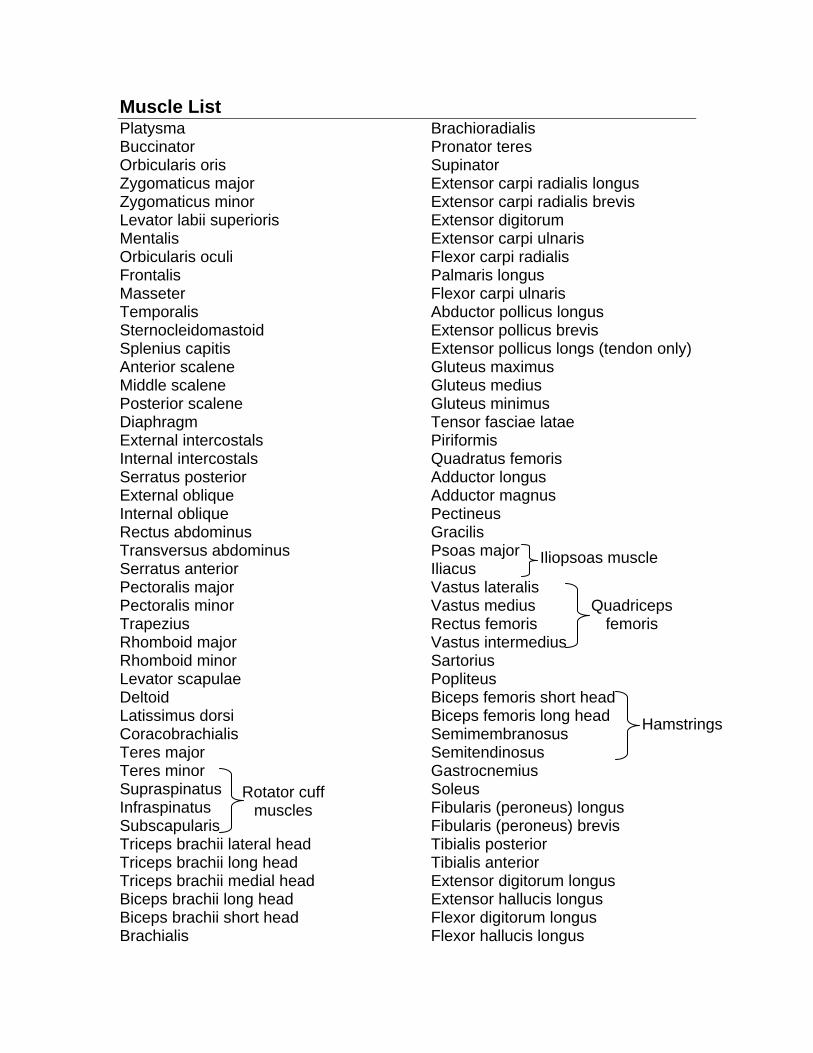
Muscle List Platysma Buccinator Orbicularis oris Zygomaticus major Zygomaticus minor Levator labii superioris Mentalis Orbicularis oculi Frontalis Masseter Temporalis Sternocleidomastoid Splenius capitis Anterior scalene Middle scalene Posterior scalene Diaphragm External intercostals Internal intercostals Serratus posterior External oblique Internal oblique Rectus abdominus Transversus abdominus Serratus anterior Pectoralis major Pectoralis minor Trapezius Rhomboid major Rhomboid minor Levator scapulae Deltoid Latissimus dorsi Coracobrachialis Teres major Teres minor Supraspinatus Infraspinatus Subscapularis Triceps brachii lateral head Triceps brachii long head Triceps brachii medial head Biceps brachii long head Biceps brachii short head Brachialis
Brachioradialis Pronator teres Supinator Extensor carpi radialis longus Extensor carpi radialis brevis Extensor digitorum Extensor carpi ulnaris Flexor carpi radialis Palmaris longus Flexor carpi ulnaris Abductor pollicus longus Extensor pollicus brevis Extensor pollicus longs (tendon only) Gluteus maximus Gluteus medius Gluteus minimus Tensor fasciae latae Piriformis Quadratus femoris Adductor longus Adductor magnus Pectineus Gracilis Psoas major Iliacus Vastus lateralis Vastus medius Rectus femoris Vastus intermedius Sartorius Popliteus Biceps femoris short head Biceps femoris long head Semimembranosus Semitendinosus Gastrocnemius Soleus Fibularis (peroneus) longus Fibularis (peroneus) brevis Tibialis posterior Tibialis anterior Extensor digitorum longus Extensor hallucis longus Flexor digitorum longus Flexor hallucis longus
Rotator cuff muscles
Iliopsoas muscle
Quadriceps femoris
Hamstrings

Joint Structures medial & lateral meniscus anterior & posterior cruciate ligaments medial (tibal) collateral ligament lateral (fibular) collateral ligament patellar ligament tendon of quadriceps femoris glenoid fossa glenoid labrum tendon sheath of biceps brachii long head subacromial bursa subscapular bursa synovial membrane glenohumeral ligaments (superior, middle & inferior) coracohumeral ligament transverse humeral ligament