01 Globe dental 2012.doc 1
Neuroanatomy Suzanne Stensaas
February 16, 2012, 10:00-12:00 p.m. Reading: Waxman Ch. 15. Your histology and gross anatomy books should be useful. Reading: Histology of the Eye from any histology book you have as well as gross anatomy text Much of this you will have to learn on your own and hope that physiology will add some more detail.
THE EYE: RETINA AND GLOBE
Objectives: 1. Describe the production, circulation and removal of aqueous humor. 2. Name the neural elements that compose the retina, and their location and function
within the retina. 3. Explain for what purpose the macula/fovea exists. 4. Ophthalmologists use the term choked or raised disc and depressed or cupped disc.
Explain the pathophysiology that leads to these states. 5. Describe the medical significance of a careful examination of the optic nerve. Why
is it a window on the brain? I. EMBRYOLOGY
A. Two evaginations, the optic vesicles, develop from prosencephalon. 1. Come to be associated with diencephalon. 2. Optic cup results from invagination of optic vesicle. a. Outer layer, retinal pigment epithelium.
b. Inner layer, neural retina. c. Axons from inner layer of optic cup form the optic nerve and grow into
the thalamus
01 Globe dental 2012.doc 2
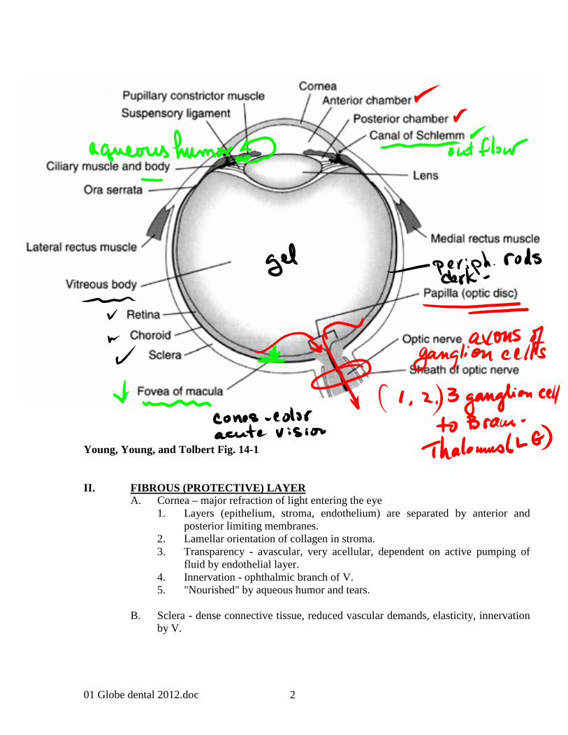
Young, Young, and Tolbert Fig. 14-1 II. FIBROUS (PROTECTIVE) LAYER A. Cornea – major refraction of light entering the eye 1. Layers (epithelium, stroma, endothelium) are separated by anterior and
posterior limiting membranes. 2. Lamellar orientation of collagen in stroma. 3. Transparency - avascular, very acellular, dependent on active pumping of
fluid by endothelial layer. 4. Innervation - ophthalmic branch of V. 5. "Nourished" by aqueous humor and tears. B. Sclera - dense connective tissue, reduced vascular demands, elasticity, innervation
by V.

01 Globe dental 2012.doc 3
III. NUTRITIVE LAYER (UVEA) PIGMENTED AND VASCULAR A. Choroid 1. Very vascular capillary plexus - choriocapillaris. 2. Vascular supply to pigment epithelium and outer segment of retinal
receptors. 3. Adherent to pigment epithelium and sclera.
B. Ciliary Body 1. 70-80 radial ciliary processes. a. Produce aqueous humor, which is low in protein. (1) Metabolic supply for cornea and lens. (2) Helps maintain intraocular pressure.
b. Angle of filtration (1) Trabecular meshwork drains into scleral venous sinuses - canal
of Schlemm. (2) Obstruction leads to glaucoma and blindness due to increased
intraocular pressure on optic nerve = depressed or cupped disc. This collapses the retinal veins leading to retinal edema and additional risk of retinal detachment.
c. Produces zonule fibers that suspend lens = suspensory ligament.
Contraction of ciliary muscles (smooth muscle innervated by III nerve) results in decreased tension on the zonular fibers and increasing curvature of the lens for near vision.
C. Iris 1. Continuation of ciliary body. 2. Sphincter M. near pupil margin - parasympathetic via III N. 3. Dilator M. - radial, lateral - sympathetic via superior cervical ganglion.
4. Separates anterior and posterior chambers

01 Globe dental 2012.doc 4
Young, Young, and Tolbert Fig. 14-2

01 Globe dental 2012.doc 5
IV. LENS
A. Cells become fibrous, condensed, homogeneous, elongated, lose nuclei. B. Continuous addition of cells at equator. C. Avascular - maintains its own metabolism.
D. Capsule – acellular membrane surrounding lens epithelium. E. Contraction of ciliary muscle in ciliary body reduces tension on zonular fibers and
permits rounding up of lens (=accommodation). F. Rigidity of lens increases with age – presbyopia. G. Opacifies with age - cataract. Replacement lenses can be placed inside the
capsule. V. VITREOUS HUMOR (or vitreous body behind the lens)
A. Collagen (fibrils), hyaluronic acid, wandering cells, 99% water. B. Produced by ciliary processes and retinal glial cells. C. Liquifies as part of aging process and retina can detach. D. “Floaters” – aggregations of vitreous material – generally normal but sudden
appearance could be a symptom of retinal disease.
Young, Young, and Tolbert Fig. 14-1

01 Globe dental 2012.doc 6
VI. NEUROECTODERMAL LAYER A. Pigment epithelium - outer layer optic cup. 1. Absorbs light, decreases reflection. 2. Phagocytoses discs in outer segments of photoreceptors. 3. Resynthesizes photopigments. 4. Weak junction with retina is site of retinal detachment.
B. Retina - inner layer of optic cup. 1. Photoreceptors – Rods (B&W) and Cones (Color). Phototransduction takes
place in outer segments of these cells. 2. Bipolar Cells –conduct signal from photoreceptors to inner retina. 3. Ganglion Cells - axons from the optic nerve where impulses originate. 3. Horizontal and amacrine cells - Lateral connections for contrast
enhancement. (Synapses located in outer and inner plexiform layers.) 5. Glial Cells (in retina are called Müller Cells) - span the width of the retina.
Play an important role in retinal homeostasis of ions and neurotransmitters.
Young, Young, and Tolbert Fig. 14-3 C. Blood supply.
1. Central retinal artery – supplies blood to inner layers of retina. 2. Ciliary arteries – supply blood to outer segments of photoreceptors.
Neurectodermal layers
ganglion cells
Bipolar cells
Receptor rod/cone
Pig Ep
Choroid
Sclera

01 Globe dental 2012.doc 7

01 Globe dental 2012.doc 8
VII. RETINAL LANDMARKS A. Macula lutea - area of acute vision. Fovea - small pit in center of macula.
Directly along the visual axis. 1. Cones predominate – color vision. 2. Inner nuclear and ganglion cell layers displaced laterally. 3. No blood vessels or optic nerve fibers internal to photoreceptor layer.
B. Optic Disc (papilla, nerve head), 1.5 mm 1. Ganglion cell axons pierce sclera and leave the globe. 2. Cell layer of retina not present, therefore, there is a "blind spot". 3. The point where the central retinal artery enters and the central retinal vein
leaves. 4. Disc is depressed (cupped) with increased intraocular pressure (i.e., glaucoma). 5. Disc bulges (papilledema - choked with increased intracranial pressure (i.e.,
tumor).
Macula and optic disc

01 Globe dental 2012.doc 9
From Greenberg DA, Aminoff MJ, Simon RP:Clinical Neurology, 2nd ed.Note fovea label is incorrectly placed on the diagram.

01 Globe dental 2012.doc 10
Fovea cross-section
01 Globe dental 2012.doc 11
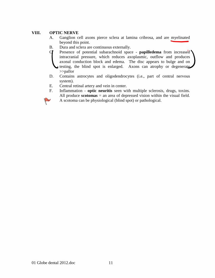
VIII. OPTIC NERVE A. Ganglion cell axons pierce sclera at lamina cribrosa, and are myelinated
beyond this point. B. Dura and sclera are continuous externally. C. Presence of potential subarachnoid space - papilledema from increased
intracranial pressure, which reduces axoplasmic, outflow and produces axonal conduction block and edema. The disc appears to bulge and on testing, the blind spot is enlarged. Axons can atrophy or degenerate >>pallor
D. Contains astrocytes and oligodendrocytes (i.e., part of central nervous system).
E. Central retinal artery and vein in center. F. Inflammation - optic neuritis seen with multiple sclerosis, drugs, toxins.
All produce scotomas = an area of depressed vision within the visual field. A scotoma can be physiological (blind spot) or pathological.
Recommended