DNA and RNA[Introduction, Components,
and Structure]
M A Y A N N R O G O N
Prepared by:

DNA[Introduction, Components,
and Structure]
DNA [Deoxyribonucleic acid]
a polymer of deoxyribo-nucleotides.
Usually double stranded.
And have double-helix structure.
found in chromosomes, mitochondria and
chloroplasts.
It acts as the genetic material in most of the
organisms.
Carries the genetic information

A Few Key Events Led to the Discovery of the Structure of DNA
Friedrich Meischer
DNA as an acidic substance present in nucleus was first identified by Friedrich Meischer in 1868.
He named it as ‘Nuclein’.

James Watson and Francis Crick worked out the three-dimensional structure of DNA, based on work by Rosalind Franklin
In 1953 , James Watson and Francis Crick, described a very simple but famous DoubleHelix model for the structure of DNA.

X-Ray EvidenceRosalind Franklin
British Scientist
Used a technique called X-Ray diffraction
Provided important clues about the structure of DNA

X-Ray Evidence
There were 2 strands
Strands were twisted around each other (helix)
The nitrogen bases are in the middle

The Double Helix• Francis Crick & James Watson
• Trying to understand the structure of DNA by building models.
• Unsuccessful until early 1953, Watson was shown a copy of Franklin’s X-ray pattern.
• “The instant I saw the picture my mouth fell open and my pulse began to race.”– James Watson
• Within weeks Watson and Crick had figured out the structure of DNA
• Published their results in a historic one page paper in April of 1953
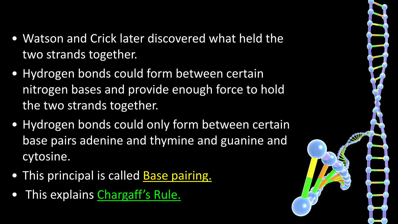
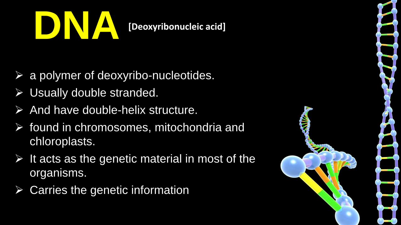
• Watson and Crick later discovered what held the two strands together.
• Hydrogen bonds could form between certain nitrogen bases and provide enough force to hold the two strands together.
• Hydrogen bonds could only form between certain base pairs adenine and thymine and guanine and cytosine.
• This principal is called Base pairing.
• This explains Chargaff’s Rule.

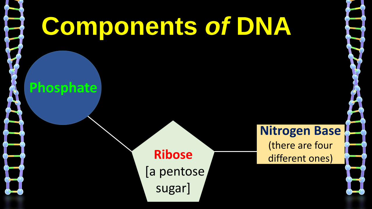
Components of DNA
Deoxyribose[a pentose
sugar]
Nitrogen Base(there are four different ones)
Phosphate

H
OH
OCH2
Base
Phosphate
Ribose
OHH
5′
4′
1′
3′2′
H
A, G, C or U
HH
H
OCH2
Base
Phosphate
Deoxyribose
5′
OHH
4′ 1′
3′ 2′
OO
O
P
O–HH
DNA Nucleotide RNA Nucleotide
A, G, C or T

CCytosine
GGuanine
AAdenosine
TThymine
4 Kinds
of
Nitrogen Bases
Purines
Pyrimidines

Nitrogenous Bases of DNA & RNA

Nucleotide Structure
Nucleotides are formed by the condensation of a sugar, phosphate and one of the 4 bases
The following illustration represents one nucleotide
Phosphate
Deoxyribose
NitrogenousBases

HH
H
OCH2
Base
DNA nucleotide
Phosphate
Deoxyribose
5′
OH
4′ 1′
3′ 2′
OO
O
P
O–HH
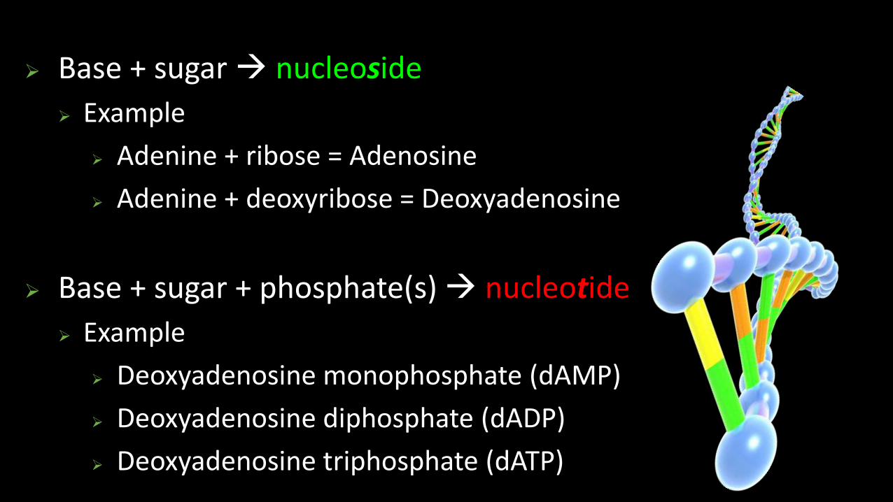
Base + sugar nucleoside
Example
Adenine + ribose = Adenosine
Adenine + deoxyribose = Deoxyadenosine
Base + sugar + phosphate(s) nucleotide
Example
Deoxyadenosine monophosphate (dAMP)
Deoxyadenosine diphosphate (dADP)
Deoxyadenosine triphosphate (dATP)

Nomenclature of Nucleic Acid Components
Base Nucleoside Nucleotide Nucleic
acid
Purines
Adenine Adenosine Adenylate RNA
Deoxyadenosine Deoxyadenylate DNA
Guanine Guanosine Guanylate RNA
Deoxy guanosine Deoxyguanylate DNA
Pyrimidines
Cytosine Cytidine Cytidylate RNA
Deoxycytidine Deoxycytidylate DNA
Thymine Thymidine Thymidylate DNA
(deoxythymidine) (deoxythymidylate)
Uracil Uridine Uridylate RNA

SugarBase
P
SugarBase
P
Nucleotides are linked together by covalent bondscalled phosphodiester linkage.
1
23
4
5
1
23
4
5
A chemical bond that involves sharing a pair of electrons between atoms in a molecule.


Antiparallel strands
The strands run opposite of each other.
The 5’ end always has the phosphate attached.
It is made of two polynucleotide chains, where the backbone is constituted by sugar-phosphate, and the bases project inside.
The two chains have anti- parallel polarity. It means, if one chainhas the polarity 5’-3’, the other has 3’-5’.
5’ 3’
3’ 5’
G C
T A
C G
A T
DNA Double Helix & Hydrogen bondingSalient features of the Double-helix structure of DNA:

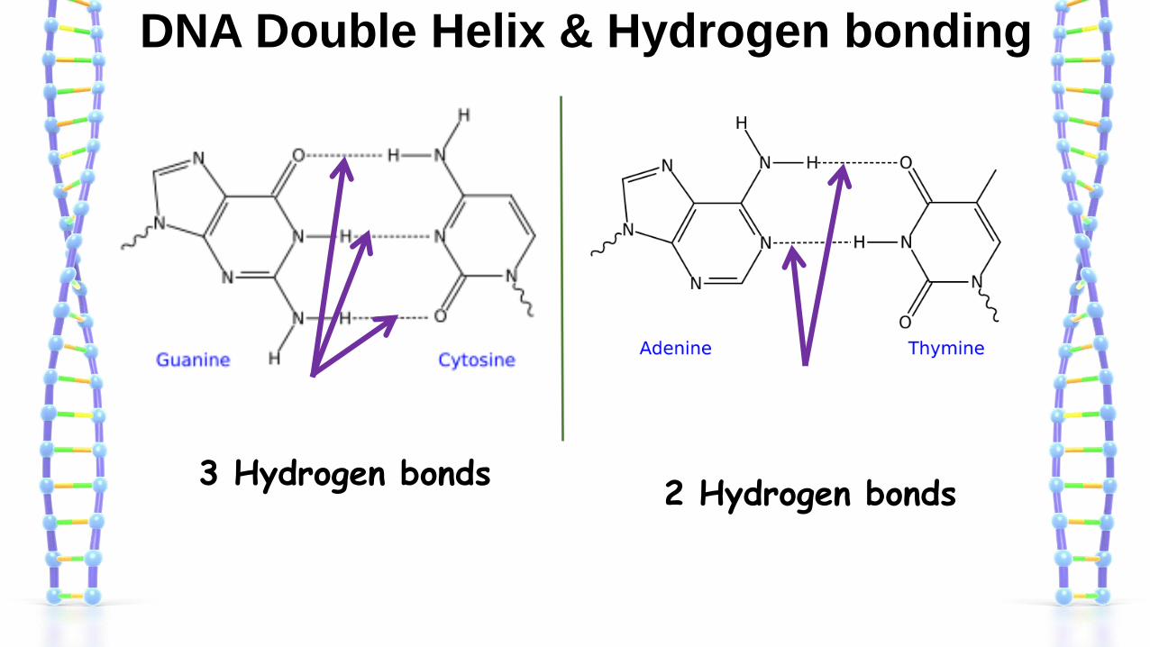
DNA Double Helix & Hydrogen bonding
3 Hydrogen bonds2 Hydrogen bonds
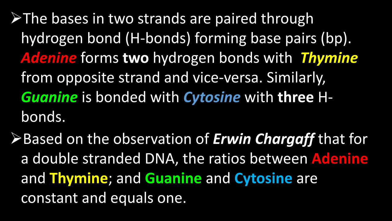
The bases in two strands are paired through hydrogen bond (H-bonds) forming base pairs (bp). Adenine forms two hydrogen bonds with Thyminefrom opposite strand and vice-versa. Similarly, Guanine is bonded with Cytosine with three H-bonds.
Based on the observation of Erwin Chargaff that for a double stranded DNA, the ratios between Adenineand Thymine; and Guanine and Cytosine are constant and equals one.

Hydrogen bond
a chemical bond inwhich a hydrogen atomof one molecule isattracted to anelectronegative atom,especially a nitrogen,oxygen, or fluorineatom, usually ofanother molecule.
24


DNA Double Helix
There are two asymmetrical grooves on the outside of the helix:a)Major grooveb)Minor groove
Groove any furrow(slight depression in the smoothness of a surface)or channel on a bodily structure or part.
Certain proteins can bind within these grooveThey can thus interact with a particular sequence of bases.
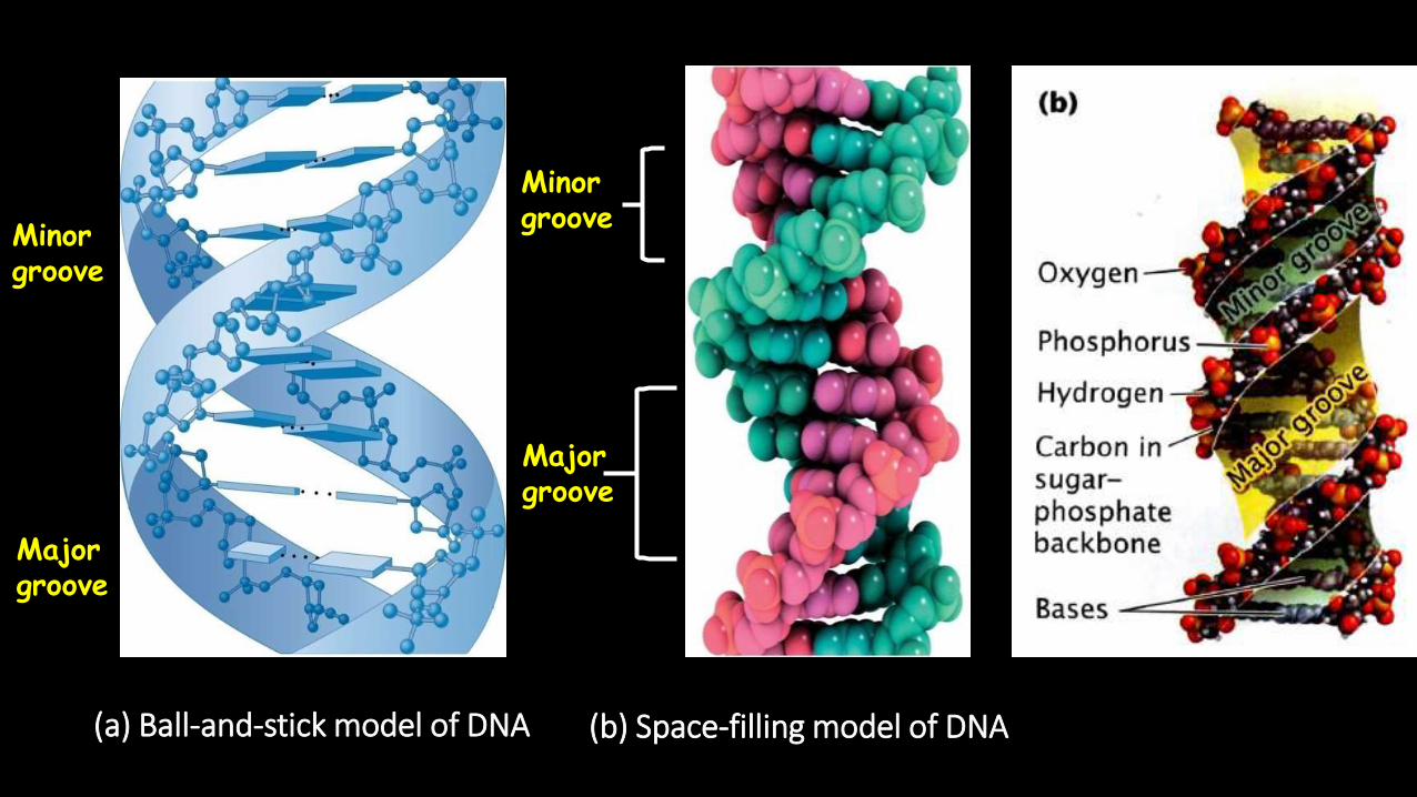
(b) Space-filling model of DNA(a) Ball-and-stick model of DNA
Minorgroove
Majorgroove
Minorgroove
Majorgroove

Structure of Double-helix
B- DNA
A- DNA
Z- DNA
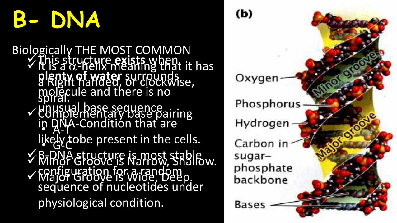
Biologically THE MOST COMMONIt is a -helix meaning that it has
a Right handed, or clockwise, spiral.
Complementary base pairing• A-T• G-C
Minor Groove is Narrow, Shallow.Major Groove is Wide, Deep.
B- DNA
This structure exists when plenty of water surrounds molecule and there is no unusual base sequence in DNA-Condition that are likely tobe present in the cells.
B-DNA structure is most stable configuration for a random sequence of nucleotides under physiological condition.

A- DNA
Right-handed helixWider and flatter than B-DNAIts bases are tilted away from
main axis of moleculeNarrow Deep major Groove and
Broad, Shallow minor Groove.Observed when less water is
present. i.e.Dehydratingcondition.
A-DNA has been observed in two context:• Active site of DNA polymerase
(~3bp)• Gram (+) bacteria undergoing
sporulation

Z- DNA
A left-handed helixSeen in Condition of High salt
concentration.In this form sugar-phosphate
backbones zigzag back andforth, giving rise to the nameZ-DNA(for zigzag).
12 base pairs per turn.
A deep Minor Groove.No Discernible Major Groove.Part of some active genes form
Z-DNA, suggesting that Z-DNA may play a role in regulating gene transcription.


RNA[Introduction, Components,
and Structure]

RNA [Ribonucleic acid]
RNA is a polymer of ribonucleotides linked
together by phosphodiester linkage.
RNA was first genetic material.
In 1967 Carl Woese found the catalytic
properties of RNA and speculated that the
earliest forms of life relied on RNA both
to carry genetic information and to catalyse
biochemical reactions.
Their theories were not validated until the work of
Nobel Prize laureate Thomas R. Cech. In the
1970s, Cech was studying the splicing of RNA in a
single-celled organism, Tetrahymena thermophila,
when he discovered that an unprocessed RNA
molecule could splice itself. He announced his
discovery in 1982 and became the first to show
that RNA has catalytic functions.

RNA [Ribonucleic acid]
RNA exists in several different single-stranded
structures, most of which are directly or indirectly
involved in protein synthesis or its regulation.
It also acts as the genetic material in some
viruses.
It function as messenger(mRNA), adapter(tRNA),
structural(rRNA) and in some cases as a catalytic
molecule(Ribozyme).
RNA strands are typically several hundred to
several thousand nucleotides in length.
Components of DNA
Ribose[a pentose
sugar]
Nitrogen Base(there are four different ones)
Phosphate

CCytosine
GGuanine
AAdenosine
UUracil
4 Kinds
of
Nitrogen Bases
Purines
Pyrimidines

Nucleotide Structure
Nucleotides are formed by the condensation of a sugar, phosphate and one of the 4 bases
The following illustration represents one nucleotide
Phosphate
Ribose
NitrogenousBases

HH
OH
OCH2
Base
RNA nucleotide
Phosphate
Ribose
5′
OH
4′ 1′
3′ 2′
OO
O
P
O–HH

Base + sugar nucleoside Example
Adenine + ribose = Adenosine
Base + sugar + phosphate(s) nucleotide
Example
Adenosine monophosphate (AMP)
Adenosine diphosphate (ADP)
Adenosine triphosphate (ATP)

SugarBase
P
SugarBase
P
Nucleotides are linked together by covalent bondscalled phosphodiester linkage.
1
23
4
5
1
23
4
5
A chemical bond that involves sharing a pair of electrons between atoms in a molecule.

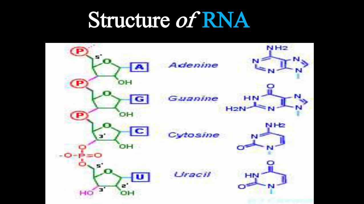
Adenine (A)
Guanine(G)
Uracil (U)
BasesBackbone
Cytosine (C)
O
HHHH
OOO
O –
P CH2
O –
HHHH
OOO
O
P CH2
O –
NH2
H
N
HHHH
OOO
O
P CH2
O –
H
H
HH
OH
HH
OOO
O
P CH2
O –
Sugar (ribose)
Phosphate
5′
4′ 1′
2′3′
5′
4′ 1′
2′3′
5′
4′ 1′
2′3′
5′
4′ 1′
2′3′OH
OH
OH
OH
RNAnucleotide
Phosphodiesterlinkage
3′
5′
NH2
OH
H
H
O
NH O
N
NN
N
N
N
N
N N
N
NH2
H
H
Structure of RNA

Types of RNA
In all prokaryotic and eukaryotic organisms, three main classes of RNA molecules exist
1) Messenger RNA(m RNA)
2) Transfer RNA (t RNA)
3) Ribosomal RNA (r RNA)

Messenger RNA (mRNA) Messenger RNA (mRNA) carries
information about a protein
sequence to the ribosomes, the
protein synthesis factories in the
cell.
It is coded so that every three
nucleotides (a codon) correspond
to one amino acid.
In eukaryotic cells, once
precursor mRNA (pre-mRNA)
has been transcribed from DNA,
it is processed to mature mRNA
This removes its introns—non-
coding sections of the pre-
mRNA
The mRNA is then exported
from the nucleus to the
cytoplasm, where it is bound to
ribosomes and translated into its
corresponding protein form with
the help of tRNA

Transfer RNA (tRNA)
Transfer RNA (tRNA) is a small
RNA chain of about
80 nucleotides
It transfers a specific amino
acid to a
growing polypeptide chain at the
ribosomal site of protein
synthesis during translation
It has sites for amino acid
attachment and
an anticodon region
for codon recognition that binds
to a specific sequence on the
messenger RNA chain through
hydrogen bonding

Ribosomal RNA (rRNA)
Ribosomal RNA (rRNA) is the
catalytic component of the
ribosomes
Three of the rRNA molecules
are synthesized in
the nucleolus, and one is
synthesized elsewher
In the cytoplasm, ribosomal
RNA and protein combine to
form a nucleoprotein called a
ribosome
The ribosome binds mRNA
and carries out protein
synthesis
Several ribosomes may be
attached to a single mRNA at
any time.
Nearly all the RNA found in a
typical eukaryotic cell is rRNA.


RNA vs. DNA
Structurally ,DNA and RNA are nearly identical .However there are three fundametaldifferences that account for the very different functions of two molecules.

Differences between RNA and DNA

REFERENCES:• J. D. Watson and F. H. C. Crick. Molecular structure of nucleic acids: a
structure for deoxyribose nucleic acids. Nature 171:737–738 (1953).
• J. D. Watson and F. H. C. Crick. Genetical implications of the structure of deoxyribonucleic acid.Nature 171:964–967 (1953).
• U.satyanarayana. Structure of DNA and RNA. Biochemistry. Retrieved from: http://www. Slideshare.com./ph
• Lehninger, Micheal M. Cox and David l. Nelson .Principle of biochemistry. Retrieved from: www. Slideshare.com.ph
• Tazeen Anwaar and Uzma Imtiyaz. Presentation on DNA and RNA Structure. Retrieved from: http://www. Slideshare.com./ph

fin
KO
MA
PT
IM
A-
N DI A
Recommended