Embed Size (px)
Citation preview

IntroductionWounding of quiescent epidermis disrupts the basementmembrane, changes cell-cell and cell-substrate adhesion andcell signaling (Borradori and Sonnenberg, 1999; Fuchs et al.,1997; Goldfinger et al., 1999; Martin, 1997; Nguyen et al.,2000a; Woodley et al., 1999). Initial changes in adhesion andsignaling are necessary for subsequent changes in genetranscription and protein translation required for repair of thebasement membrane and migration (Frank, 2004). Thesechanges generate a subpopulation of activated leadingkeratinocytes (LKs) at the wound edge that are distinct fromeither quiescent keratinocytes or following keratinocytes in theoutgrowth (Lampe et al., 1998; Li et al., 2003; Nguyen et al.,2000a; Wood et al., 2002). Here, we investigated the role oflaminin 5, a basement membrane adhesive ligand (Nguyen etal., 2000a; Ryan et al., 1999), in regulating cell signaling andprotein expression in LKs generated by wounding or adhesiondefects in laminin 5.
Changes in outside-in signals through integrin receptors inepidermal wounds contribute to changes in cell motility andprotein expression that define LKs. For example, quiescentepidermal keratinocytes adhere to laminin 5 via integrin α6β4in hemidesmosomes (Carter et al., 1991; Gipson et al., 1993;Goldfinger et al., 1999; Ryan et al., 1999). Adhesion via α6β4does not require actin-dependent interactions that mediate cell
motility (Frank, 2004; Xia et al., 1996). In contrast, woundingexposes the dermis and activates adhesion to dermal collagenvia integrin α2β1 or to fibronectin via integrin α5β1, whichrequire actin rearrangements to mediate cell motility. Targeteddisruption of laminin 5 in mice generates epithelial blisterscaused by failure of laminin 5 to bind integrin α6β4 inhemidesmosomes and/or integrin α3β1 (Nguyen et al., 2000a;Ryan et al., 1999). In the absence of laminin 5, integrin α3β1interacts with an alternative basement membrane ligand,probably laminin 10. The switch from β4 to β1 integrinscorrelates with the discontinuous ‘beads on a string’organization of α6β4 in the basement membrane zone and maygenerate changes in integrin-ligand interactions, cell signalsand/or protein expression similar to wounds and tumors(Nguyen et al., 2000a; Ryan et al., 1999). For example,suspension and re-adhesion of keratinocytes via α6β4 andα3β1 to laminin 5 in vitro activates phosphoinositide 3-OHkinase (PI3K), which regulates epithelial motility and mRNAtranscription (Li et al., 2003; Xia and Karin, 2004). The PI3K-Rac-JNK/p38 pathway participates in initial adhesion ofquiescent human keratinocytes (HKs) to laminin 5 via α6β4and α3β1 (Nguyen et al., 2000b; Xia and Karin, 2004). Initialadhesion on laminin 5 elevates GTP-bound Rho allowing forsubsequent Rho-dependent adhesion on collagen via α2β1(Nguyen et al., 2000b). Integrin α3β1 directs the stabilization
3471
Quiescent epidermis anchors to laminin 5 in the basementmembrane via integrin α6β4. Wounding elevatesexpression of laminin 5, generating leading keratinocytes(LKs) that migrate via β1 integrins. Laminin 5 wasevaluated as a regulator of cell signaling, and mRNA andprotein expression in LKs. An in vitro wound model wasdeveloped based on suspension and re-adhesion ofquiescent human keratinocytes (HKs). DNA microarraysidentified multiple mRNAs elevated 1.5 hours aftersuspension and re-adhesion including activationtranscription factor 3 (ATF3). In vitro and in vivo, levels ofATF3 protein elevate in nuclei of LKs, but not in nuclei ofthe following cells, 2 hours after suspension or woundingbut decline by 12-18 hours post injury. Significantly, nulldefects in laminin 5 or integrin β4 that inhibit anchoragechronically elevate ATF3 in vivo. This suggests thatadhesion to laminin 5, but not other ligands, suppresses
activation. On suspension, ATF3 and other transcripts inthe microarrays are elevated by phosphorylated p38mitogen-activated protein kinase (P-p38), a stress kinasethat regulates mRNA and cell motility. Inhibition of P-p38with SB203580 prevents phosphorylation of ATF2, atranscription factor for ATF3 in LKs. Re-adhesion tolaminin 5 via α6β4 dephosphorylates P-p38 and suppressesATF3 protein relative to cells in suspension. Thus,wounding of quiescent HKs disrupts laminin 5 adhesion toactivate p38, generating mRNA transcripts that defineLKs. Adhesion to deposits of laminin 5 via α6β4 suppressesP-p38 and activation mRNAs including ATF3. Defects inlaminin 5 and α6β4 sustain P-p38 with probablepathological effects on transcription and migration.
Key words: Laminin 5, p38 MAPK, ATF3, Epidermal Wounds
Summary
Wounding activates p38 map kinase and activationtranscription factor 3 in leading keratinocytes Erin G. Harper1,2, Stacy M. Alvares1,3 and William G. Carter1,2,*1Fred Hutchinson Cancer Research Center, 1100 Fairview Avenue, Seattle, WA 98109, USA2Department of Pathobiology Graduate Program, University of Washington, N.E. Pacific Street, Seattle, WA 98195, USA3Program in Molecular and Cellular Biology, University of Washington, N.E. Pacific Street, Seattle, WA 98195, USA*Author for correspondence (e-mail: [email protected])
Accepted 9 May 2005Journal of Cell Science 118, 3471-3485 Published by The Company of Biologists 2005doi:10.1242/jcs.02475
Research Article
Jour
nal o
f Cel
l Sci
ence
3472
of polarized lamellipodium in epithelial cells throughactivation of Rac1 (Choma et al., 2004). This sequence ofadhesion and signaling changes allows HKs to make thetransition from quiescence to activation in wounds. Similarly,ligation of α3β1 in A549 adenocarcinoma cells by laminin10/11 or laminin 5 preferentially activates the Rac-MKK-JNK/p38 stress pathway that mediates epithelial migration (Guet al., 2001; Ono and Han, 2000; Xia and Karin, 2004). Inaddition to adhesion, interaction of cells with integrin ligandsalso regulates mRNA transcription and protein translationthrough activation of p38 MAPK (Balda and Matter, 2003).Ligation of α6β4 activates the Rac-PAK-MKK-p38 pathway topromote expression of IL-6 in thymic epithelial cells (Mainieroet al., 2003). Thus, cell suspension and re-adhesion ontodermal collagen or laminin 5 via integrins activates JNK and/orp38 by phosphorylation to alter protein expression and/or cellmotility (Clark et al., 2003; Ono and Han, 2000).
Here, we sought to: (1) identify changes in mRNAtranscription and protein translation in quiescent keratinocytesas they transition into wound LKs; (2) identify cell signalsnecessary for the transcriptional changes in LKs; (3)understand the role of laminin 5 adhesion in regulating thesignaling and transcriptional changes. We found thatsuspension or wounding of HKs activates p38 in LKs topromote motility and transcription of activation transcriptionfactor 3 (ATF3), a stress-response transcription factor.Significantly, in vivo defects in laminin 5 or α6β4 inhibitquiescent anchorage and chronically activate expression ofATF3. These results suggest that wounding activates p38 as asignificant regulator of adhesion and transcription in LKs.Defects in laminin 5, like wounding, activate p38 leading topathological expression of wound transcripts and migration.
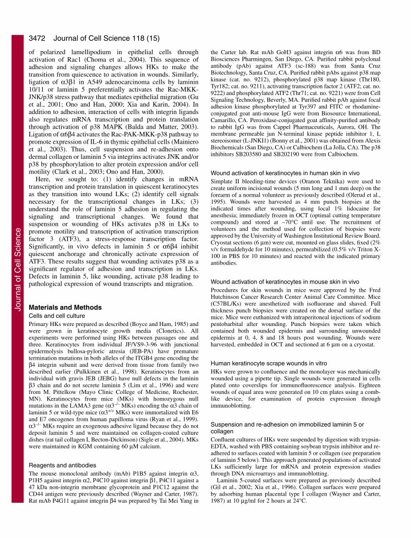
Materials and MethodsCells and cell culturePrimary HKs were prepared as described (Boyce and Ham, 1985) andwere grown in keratinocyte growth media (Clonetics). Allexperiments were performed using HKs between passages one andthree. Keratinocytes from individual JF/VS9-3-96 with junctionalepidermolysis bullosa-pyloric atresia (JEB-PA) have prematuretermination mutations in both alleles of the ITGB4 gene encoding theβ4 integrin subunit and were derived from tissue from family twodescribed earlier (Pulkkinen et al., 1998). Keratinocytes from anindividual with gravis JEB (JEBG) have null defects in the lamininβ3 chain and do not secrete laminin 5 (Lim et al., 1996) and werefrom M. Pittelkow (Mayo Clinic College of Medicine, Rochester,MN). Keratinocytes from mice (MKs) with homozygous nullmutations in the LAMA3 gene (α3–/– MKs) encoding the α3 chain oflaminin 5 or wild-type mice (α3+/+ MKs) were immortalized with E6and E7 oncogenes from human papilloma virus (Ryan et al., 1999).α3–/– MKs require an exogenous adhesive ligand because they do notdeposit laminin 5 and were maintained on collagen-coated culturedishes (rat tail collagen I, Becton-Dickinson) (Sigle et al., 2004). MKswere maintained in KGM containing 60 μM calcium.
Reagents and antibodiesThe mouse monoclonal antibody (mAb) P1B5 against integrin α3,P1H5 against integrin α2, P4C10 against integrin β1, P4C11 against a47 kDa non-integrin membrane glycoprotein and P1C12 against theCD44 antigen were previously described (Wayner and Carter, 1987).Rat mAb P4G11 against integrin β4 was prepared by Tai Mei Yang in
the Carter lab. Rat mAb GoH3 against integrin α6 was from BDBiosciences Pharmingen, San Diego, CA. Purified rabbit polyclonalantibody (pAb) against ATF3 (sc-188) was from Santa CruzBiotechnology, Santa Cruz, CA. Purified rabbit pAbs against p38 mapkinase (cat. no. 9212), phosphorylated p38 map kinase (Thr180,Tyr182; cat. no. 9211), activating transcription factor 2 (ATF2; cat. no.9222) and phosphorylated ATF2 (Thr71; cat. no. 9221) were from CellSignaling Technology, Beverly, MA. Purified rabbit pAb against focaladhesion kinase phosphorylated at Tyr397 and FITC or rhodamine-conjugated goat anti-mouse IgG were from Biosource International,Camarillo, CA. Peroxidase-conjugated goat affinity-purified antibodyto rabbit IgG was from Cappel Pharmaceuticals, Aurora, OH. Themembrane permeable jun N-terminal kinase peptide inhibitor 1, Lstereoisomer (L-JNKI1) (Bonny et al., 2001) was obtained from AlexisBiochemicals (San Diego, CA) or Calbiochem (La Jolla, CA). The p38inhibitors SB203580 and SB202190 were from Calbiochem.
Wound activation of keratinocytes in human skin in vivoSimplate II bleeding-time devices (Oranon Teknika) were used tocreate uniform incisional wounds (5 mm long and 1 mm deep) on theforearm of a normal volunteer as previously described (Olerud et al.,1995). Wounds were harvested as 4 mm punch biopsies at theindicated times after wounding, using local 1% lidocaine foranesthesia; immediately frozen in OCT (optimal cutting temperaturecompound) and stored at –70°C until use. The recruitment ofvolunteers and the method used for collection of biopsies wereapproved by the University of Washington Institutional Review Board.Cryostat sections (6 μm) were cut, mounted on glass slides, fixed (2%v/v formaldehyde for 10 minutes), permeabilized (0.5% v/v Triton X-100 in PBS for 10 minutes) and reacted with the indicated primaryantibodies.
Wound activation of keratinocytes in mouse skin in vivo Procedures for skin wounds in mice were approved by the FredHutchinson Cancer Research Center Animal Care Committee. Mice(C57BL/Ks) were anesthetized with isofluorane and shaved. Fullthickness punch biopsies were created on the dorsal surface of themice. Mice were euthanized with intraperitoneal injections of sodiumpentobarbital after wounding. Punch biopsies were taken whichcontained both wounded epidermis and surrounding unwoundedepidermis at 0, 4, 8 and 18 hours post wounding. Wounds wereharvested, embedded in OCT and sectioned at 6 μm on a cryostat.
Human keratinocyte scrape wounds in vitroHKs were grown to confluence and the monolayer was mechanicallywounded using a pipette tip. Single wounds were generated in cellsplated onto coverslips for immunofluorescence analysis. Eighteenwounds of equal area were generated on 10 cm plates using a comb-like device, for examination of protein expression throughimmunoblotting.
Suspension and re-adhesion on immobilized laminin 5 orcollagenConfluent cultures of HKs were suspended by digestion with trypsin-EDTA, washed with PBS containing soybean trypsin inhibitor and re-adhered to surfaces coated with laminin 5 or collagen (see preparationof laminin 5 below). This approach generated populations of activatedLKs sufficiently large for mRNA and protein expression studiesthrough DNA microarrays and immunoblotting.
Laminin 5-coated surfaces were prepared as previously described(Gil et al., 2002; Xia et al., 1996). Collagen surfaces were preparedby adsorbing human placental type I collagen (Wayner and Carter,1987) at 10 μg/ml for 2 hours at 24°C.
Journal of Cell Science 118 (15)
Jour
nal o
f Cel
l Sci
ence

3473Laminin 5 regulates p38 MAPK and ATF3 in wounds
BSA adhesion assayThe BSA adhesion assay couples deposition of laminin 5 to subsequentadhesion to the deposits and was performed as previously described(Frank, 2004; Gil et al., 2002). MKs from laminin 5 null mice (α3–/–
MKs) and from a wild-type mouse (α3+/+ MKs) were grown toconfluence, suspended and re-plated onto petri dishes coated with HD-BSA, a non-adhesive surface, or onto laminin 5 surfaces. Triton andSDS protein extracts were collected at 2 hour intervals from 0 to 10hours after re-plating. The quiescent cell populations establishedbaseline levels of protein expression and p38 phosphorylation. Extractswere examined by immunoblotting with anti-p38 map kinase and anti-phosphorylated and p38 map kinase antibodies.
Immunofluorescence microscopyScrape wounds in vitro were fixed with 2% formaldehyde, 0.1 Mcacodylate and 0.1 M sucrose and tissue sections were fixed with 2%formaldehyde in PBS for 15 minutes. Coverslips and tissue sectionswere permeabilized with 0.5% Triton X-100 detergent in PBS andblocked with 0.5% HD-BSA for 30 minutes. Wounds were stainedwith the indicated antibodies. Samples were washed with PBS andincubated with affinity-purified FITC or Rhodamine-conjugatedspecies-specific secondary antibodies. Coverslips were mounted withProlong (Molecular Probes, Eugene, OR) and analyzed with a Zeissfluorescent microscope.
ImmunoblottingKeratinocyte populations were sequentially extracted with 1% TritonX-100 in PBS for 10 minutes, followed by extraction with 1% SDSin PBS. Both lysis buffers contained 1 mM PMSF and 2 mM N-ethylmaleimide, 1 mM sodium fluoride and 1 mM sodium orthovanadatein PBS. Triton-soluble and Triton-insoluble protein fractions wereseparated using 12% SDS-PAGE gels (Laemmli, 1970), transferred tonitrocellulose membranes and immunoblotted with the indicatedantibodies. Blots were developed using the EnhancedChemiluminescence kit (Amersham).
Assay of p38 kinase activityP38 MAP kinase was assayed as recommended (Cell SignalingTechnology) as follows: immobilized mAb against P-p38(Thr180/Tyr182) was used to immunoprecipitate P-p38 from cellextracts as indicated in Fig. 7B. An in vitro kinase assay wasperformed using ATF-2 as substrate. P-ATF-2 product was detectedby western blotting using P-ATF-2 (Thr71) antibody.
DNA microarray analysis of mRNA from wounded andquiescent keratinocytesTotal RNA was isolated from the quiescent and activated cellpopulations using a Totally RNA kit (Ambion). Generation of Cy fluor-labeled cDNA was according to a published method (Fazzio et al.,2001) and was co-hybridized to spotted human cDNA microarray chips.cDNA microarray chips were produced by the FHCRC microarrayfacility (Seattle, WA) under direction of Jeff Delrow. cDNA chips wereproduced by PCR amplification of each cDNA clone in a human cDNAlibrary (18,000 genes) representing various tissues and cell types(Research Genetics). A fluorescent image was generated using aGenePix 4000 fluorescent scanner (Axon Instruments). The image wasanalyzed using GenePix Pro microarray acquisition and analysissoftware. Five separate analyses were performed and data wasstatistically analyzed using the Student’s paired t-test.
Semi-quantitative reverse-transcription PCR analysesAdherent quiescent confluent HKs were suspended with trypsin-
EDTA and either re-adhered to laminin 5 or kept in suspension overagarose to prevent adhesion for 2, 9, 24 and 48 hours. RNA wasextracted from the cells with the RNeasy Midi Kit (Qiagen). 2 μg ofeach RNA sample was then reverse-transcribed using theSuperScriptTM First-Strand Synthesis System (Invitrogen). To amplifyATF3 and glyceraldehyde 3-phosphate dehydrogenase (GAPDH) insemi-quantitative PCR, the following primers were used (Syed et al.,2005): ATF3, 5′-CTCCTGGGTCACTGGTGTTT-3′ (forward) and5′-GTCGCCTCTTTTTCCTTTCA-3′ (reverse); GAPDH, 5′-CAT-CACCATCTTCCAGGAGC-3′ (forward) and 5′-GGATGATGTT-CTGGAGAGCC-3′ (reverse). 2 μl aliquots of cDNA were amplifiedby PCR as described (Syed et al., 2005) except that annealingtemperature was 55°C for both ATF3 and GAPDH primers and thenumber of amplification cycles was 23 for both ATF3 and GAPDH.The PCR products were fractionated on a 2% agarose gel andvisualized after ethidium bromide staining. The results were obtainedusing two independent cDNA syntheses from each RNA sample.
ResultsComparison of quiescent keratinocytes to wound-activated LKs by DNA microarrayMicroarrays of cDNA were used to compare mRNA transcriptlevels of quiescent adherent HKs to wound-activated LKs. Adiagram of the manipulations used to activate the quiescentkeratinocytes is presented in Fig. 1A. Quiescent basalkeratinocytes in epidermis were modeled with confluentcultures of primary HKs. Early transcriptional changes inwound-activated LKs were modeled by suspending thequiescent HKs and then re-adhering cells onto laminin 5surfaces. HKs were re-adhered for 1.5 hours prior to purifyingtotal RNA. Using cDNA microarrays, mRNA transcripts in thequiescent HKs were compared to transcripts in HKs re-adherent and spread on laminin 5 (Fig. 1B). Prior studies haveshown that re-adhesion to laminin 5 is mediated by integrinα6β4 and α3β1 whereas spreading and motility are mediatedby α3β1 (Frank, 2004; Xia et al., 1996). The predictivecapability of the wound model and DNA microarray werevalidated by examining transcripts known to be elevated inwounds in vivo (Fig. 1B). The array correctly reported mRNAupregulation of urokinase plasminogen activator (Morioka etal., 1987), α3 chain of laminin 5 (Lampe et al., 1998; Ryan etal., 1994), CD9 (Penas et al., 2000) and ezrin (Crepaldi et al.,1997), each involved in epithelial cell motility. The arrayscorrectly predicted a downregulation of mRNAs fordesmoglien and plakoglobin (Okada et al., 2001),corneodesmosin (Haftek et al., 1997) and serine proteaseinhibitor (Scott et al., 1998), characteristic of quiescent ordifferentiated keratinocytes. We concluded that the in vitrowound model coupled with cDNA microarray comparisonswere capable of accurately reporting physiologicallysignificant changes in equilibrium levels of mRNA transcriptsoccurring in epidermal wounds.
In five separate experiments, the arrays consistently reportedfivefold increases (P<0.05) or higher in levels of four mRNAtranscripts in re-adherent HKs compared to levels in quiescentHKs. These transcripts included activating transcription factor3 (ATF3), tumor necrosis factor α-induced protein 3(TNFαIP3), growth-related oncogene-1 (Gro-1), and urokinaseplasminogen activator. Reproducible increases in mRNAexpression were also detected for syndecan 4, a transmembraneglycoprotein reported to interact with the G-4 domain of
Jour
nal o
f Cel
l Sci
ence

3474
laminin 5 (Utani et al., 2003; Utani et al., 2001); EGR-1, atranscription factor encoded by an immediate early responsegene (Sukhatme et al., 1988); Ras, a GTP binding protein andoncogene (Charvat et al., 1999); L6, a member of thetransmembrane 4 super family and a tumor antigen (Marken etal., 1992; our unpublished work); SOX9, a developmentaltranscription factor (Smith and Koopman, 2004) and PMAIP1,a PMA-induced protein 1. Reproducible but small increases intranscripts for integrins α3, α2 but not β4, were observedwithin 1.5 hours of wounding. In controls (results not shown),suspension and re-adhesion for 3 days further elevatedtranscripts for laminin α3 chain, and β1 integrins. Here, weevaluate cell signals and adhesion events that regulate earlyexpression of ATF3 in wounds.
ATF3 protein is transiently expressed in LKs of woundsin vivo and in vitroATF3 is a member of the ATF/Creb family of transcriptionfactors that is upregulated in response to injury or cell stress(Hai and Hartman, 2001; Hai et al., 1999). Expression of ATF3protein was examined in LKs by immunofluorescencemicroscopy in timed scrape wounds in confluent quiescentHKs (Fig. 2A). Cells were fixed and permeabilized 0, 3, 6 and12 hours post wounding. LKs at the wound margins, but notfollowing cells, upregulated and translocated ATF3 protein tothe nucleus within 3 hours of wounding (Fig. 2Ab), followed
by a decrease within 6 hours (Fig. 2Ac) and a return to baselinelevels within 12 hours of wounding (Fig. 2Ad). Treatment ofscrape wounds with actinomycin D, an inhibitor of mRNAtranscription, or cycloheximide, an inhibitor of proteintranslation, prevented ATF3 protein expression by LKs,confirming regulation at the transcriptional level. LKsexpressing ATF3, but not following cells, spread over theexposed wound edge and localized paxillin in prominent focaladhesions (Fig. 2B). Thus changes in cell adhesion andspreading are concurrent with changes in transcription andtranslation of ATF3 in LKs. In principle, signals that regulateATF3 may also regulate adhesion. Results in Figs 1 and 2establish that wound stress induced by scraping or bydetachment with trypsin elevates levels of ATF3 mRNA andprotein in LKs.
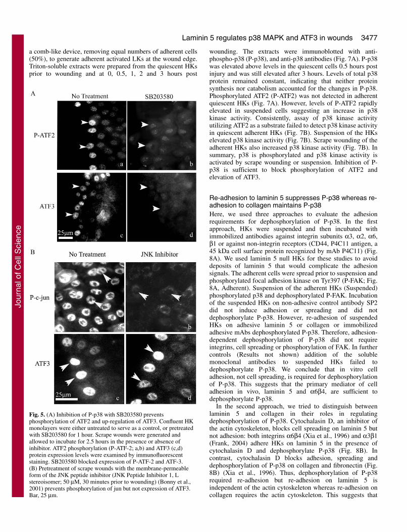
Semi-quantitative reverse-transcription polymerase chainreaction (RT-PCR) was used to confirm the elevation of ATF3mRNA during suspension (Fig. 3A). Quiescent adherent HKsexpressed low levels of ATF3 mRNA that increased by 2 hoursafter suspension confirming the results in Fig. 1B. Levels ofATF3 mRNA returned to baseline within 8 hours aftersuspension with or without re-adhesion.
ATF3 protein in quiescent confluent HKs was also comparedto levels in suspended HKs and suspended/re-adherent HKs(Fig. 3B). ATF3 protein was not detected in quiescent adherentHKs. In suspended HKs, ATF3 was maximal by 2 hours aftersuspension and remained elevated for 6 hours (Fig. 3A) and
Journal of Cell Science 118 (15)
0
5000
10000
15000
20000
25000
A
B
CD9*Ras
L6**
TNFaIP3*
*Ezr
in
ATF3**
alpha
3
Urokin
ase*
*
Lam
inin
alpha
3*
EGR-1*
Synde
can
4*
alpha
2
Gro-1
**
beta
4
Serine
Pro
t Inh
ib.*
Desm
oglie
n*
Corne
odes
mos
in**
ConfluentActivated
Fig. 1. (A) Suspension and re-adhesion ofquiescent HKs activates leadingkeratinocytes for a wound model. HKs weregrown at confluence to generate quiescence.Suspension with trypsin/EDTA activated thekeratinocytes. Re-adhesion onto surfacescoated with laminin 5 was mediated byintegrins α6β4 and α3β1. Re-adhesion wasfollowed by cell spreading via integrinα3β1. Spreading, but not adhesion, wasblocked with Cytochalasin D. (B) cDNAmicroarray analysis of quiescent andsuspended/re-adherent HKs. Levels ofmRNA transcripts in quiescent andsuspended/re-adherent HKs were comparedby cDNA microarray analysis 1.5 hours postsuspension/re-adhesion with spreading.Solid bars represent transcript levels ofactivated suspended/re-adherent HKs,whereas open bars represent transcript levelsin quiescent cells. Transcript levels arereported as mean (±s.e.m.) fluorescent units(y-axis). Significant differences **P<0.05and *P<0.10 in fluorescence levels werefound between the groups indicated and thecontrol. Microarray analysis was performedin five separate suspension/re-adhesionexperiments.
Jour
nal o
f Cel
l Sci
ence
3475Laminin 5 regulates p38 MAPK and ATF3 in wounds
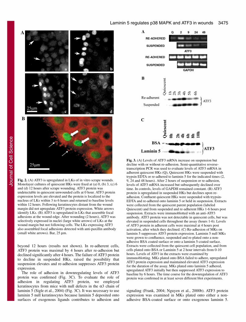
beyond 12 hours (results not shown). In re-adherent cells,ATF3 protein was maximal by 4 hours after re-adhesion butdeclined significantly after 4 hours. The failure of ATF3 proteinto decline in suspended HKs, raised the possibility thatsuspension elevates and re-adhesion suppresses ATF3 proteinexpression.
The role of adhesion in downregulating levels of ATF3protein was confirmed (Fig. 3C). To evaluate the role ofadhesion in regulating ATF3 protein, we employedkeratinocytes from mice with null defects in the α3 chain oflaminin 5 (Sigle et al., 2004) (Fig. 3C). It was necessary to uselaminin 5 null keratinocytes because laminin 5 deposited ontosurfaces of exogenous ligands contributes to adhesion and
signaling (Frank, 2004; Nguyen et al., 2000b). ATF3 proteinexpression was examined in MKs plated onto either a non-adhesive BSA-coated surface or onto exogenous laminin 5.
Fig. 2. (A) ATF3 is upregulated in LKs of in vitro scrape wounds.Monolayer cultures of quiescent HKs were fixed at (a) 0, (b) 3, (c) 6and (d) 12 hours after scrape wounding. ATF3 protein wasundetectable in quiescent unwounded cells at 0 hour. ATF3 proteinexpression levels are elevated and the protein is localized to thenucleus of LKs within 3 to 6 hours and returned to baseline levelswithin 12 hours. Following keratinocytes distant from the woundmargin did not upregulate ATF3 protein expression. White arrowsidentify LKs. (B) ATF3 is upregulated in LKs that assemble focaladhesions at the wound edge. After wounding (2 hours), ATF3 wasselectively expressed in nuclei (large white arrows) of LKs at thewound margin but not following cells. The LKs expressing ATF3also assembled focal adhesions detected with anti-paxillin antibody(small white arrows). Bar, 25 μm.
Fig. 3. (A) Levels of ATF3 mRNA increase on suspension butdecline with or without re-adhesion. Semi-quantitative reverse-transcription PCR was used to evaluate levels of ATF3 mRNA inadherent quiescent HKs (Q), Quiescent HKs were suspended withtrypsin EDTA or re-adhered to laminin 5 for the indicated times (2,9, 24 and 48 hours). After 2 hours of suspension or re-adhesion,levels of ATF3 mRNA increased but subsequently declined overtime. In controls, levels of GAPDH remained constant. (B) ATF3protein is upregulated in suspended HKs but declines upon re-adhesion. Confluent quiescent HKs were suspended with trypsin-EDTA and re-adhered onto laminin 5 or held in suspension. Extractswere collected from the quiescent parent population (labeledQuiescent) and from suspended and re-adherent HKs 1-6 hours postsuspension. Extracts were immunoblotted with an anti-ATF3antibody. ATF3 protein was not detectable in quiescent cells, but waselevated in suspended cells throughout the assay (hours 1-6). Levelsof ATF3 protein in adherent cells were maximal at 4 hours postactivation, after which they declined. (C) Re-adhesion of MKs onlaminin 5 suppresses ATF3 protein expression. Laminin 5 null MKswere grown to confluence, suspended and re-plated onto a non-adhesive BSA coated surface or onto a laminin 5-coated surface.Extracts were collected from the quiescent cell population, and fromcells plated onto BSA or Laminin 5 at 2 hour intervals from 0-10hours. Levels of ATF3 in the extracts were examined byimmunoblotting. MKs plated onto BSA failed to adhere, upregulatedATF3 protein expression and maintained elevated ATF3 expressionfor the duration of the assay. MKs plated onto laminin 5 adhered,upregulated ATF3 initially but then suppressed ATF3 expression tobaseline by 6 hours. The time course for the downregulation of ATF3protein was confirmed in at least seven different blot experiments.
Jour
nal o
f Cel
l Sci
ence

3476
Suspended α3–/– MK upregulated ATF3 protein, but failed todeposit laminin 5 and therefore did not adhere to BSA-coatedsurfaces during the assay (0-10 hour) (Sigle et al., 2004). Incontrast, α3–/– MK plated onto an exogenous laminin 5 surfaceadhered and downregulated ATF3 (Fig. 3C). Similar resultswere also obtained using HKs with null defects in laminin 5(Results not shown). This confirmed that adhesion to laminin5, and probably other adhesive surfaces, suppressed ATF3protein expression elevated by suspension.
To ensure that the elevation in ATF3 was physiologicallyrelevant, incision wounds in mouse skin were examined forATF3 expression at 0, 4 and 18 hours post injury. ATF3 proteinwas undetectable in LKs at the wound edge in the zero timewound (Fig. 4a). Within 4 hours of the wounding, ATF3 waselevated and translocated to the nucleus in LKs (Fig. 4b)whereas expression in adjacent quiescent following cellsremained undetectable. ATF3 returned to the baseline level ofquiescent cells by 18 hours (Fig. 4c). A composite image (Fig.4d) shows expression of ATF3 in LKs at the wound margin 4hours after injury, but absent from the quiescent cells distantto the injury.
In summary, wounding in vivo or in vitro transientlyupregulates ATF3 protein and mRNA in LKs, but ATF3remained at baseline levels in adjacent following or quiescentkeratinocytes. Significantly, suspension activates ATF3expression at both the mRNA and protein levels whereas re-adhesion suppresses the duration of ATF3 expressionparticularly at the protein level. This suggests that adhesion ingeneral, or adhesion to laminin 5, may limit ATF3 expressionto LKs. The role of adhesion in downregulating expression ofATF3 protein will be evaluated elsewhere. Here, we determinewhat upstream cell signals activate expression of ATF3 mRNAand protein.
Wound activation of p38 MAPK phosphorylatesdownstream ATF2 to elevate ATF3We sought to identify cell signals activated by scrape woundingor suspension of HKs that elevate ATF3 in LKs and that may
be suppressed by re-adhesion to laminin 5. Interestingly, p38mitogen-activated kinase (MAPK) has previously beenreported to transcriptionally and/or post-transcriptionallyregulate at least seven out of the 50 mRNA transcripts elevatedat least 1.5 times or higher in LKs in our wound screens (Fig.2, ATF3, TNFαIP3, uPA, GRO1, IL1β, PMAIP1 and SOX9)(Frevel et al., 2003). Thus, p38 is a major contributor toincreases in mRNA levels that define LKs and is also a possibleupstream regulator of ATF3. Phosphorylation of activationtranscription factor 2 (ATF2) by JNK (Cai et al., 2000; Yin etal., 1997; Zhang et al., 2001) and/or by p38 MAPK (Fan et al.,2002) is reported to upregulate ATF3 expression in cell lines.We determined if activation of JNK or p38 MAPK wasnecessary for phosphorylation of ATF2 and elevation of ATF3in LKs of scrape wounds. Confluent cultures of HKs werepretreated with or without specific inhibitors of p38 kinaseactivity, SB203580 or SB202190 (50 μm each, 30 minutesprior to wounding) or membrane-permeable forms of the JNKpeptide inhibitor 1, L stereoisomer (50 μm each, 30 minutesprior to wounding) (Bonny et al., 2001). Following scrapewounding, HKs were incubated in the presence or absence ofinhibitors for 2.5 hours to allow for upregulation ofphosphorylated ATF2 (P-ATF2) and ATF3 by LKs. LKs inscrape wounds pretreated with either SB203580 (Fig. 5Ab) orSB202190 (not shown) failed to upregulated P-ATF2 or ATF3(Fig. 5Ad). In contrast, inhibitors of JNK failed to suppressexpression of ATF3 (Fig. 5Bd) under conditions where theinhibitors did prevent JNK-mediated phosphorylation of Jun(Fig. 5Bb).
Levels of P-p38 were transiently increased in LKs at theedge of in vivo wounds 1 hour after injury but declined within4 hours of injury (Fig. 6B). Activation of p38 requiresphosphorylation of Thr180 and Tyr182 by dual specificitymitogen-activated kinase kinase 3/6 or 4 (Ono and Han, 2000).The anti-P-p38 antibody was specific for p38 phosphorylatedon both Thr180 and Tyr182. Both P-p38 and P-ATF2 wereselectively increased in LKs of in vitro wounds (Fig. 6A). Therole of p38 was confirmed by immunoblotting of scrapewounds (Fig. 7A). Confluent plates of HKs were wounded with
Journal of Cell Science 118 (15)
Fig. 4. ATF3 is upregulated in LKs in epidermal wounds invivo. Cryostat sections were prepared from wounds inneonatal mouse epidermis 0 (a), 4 (b) and 18 hours (c) postwounding. The wounds were stained with an anti-ATF3antibody. White arrows indicate the wound edge.(d) Composite image of a 4 hour wound showing expressionof ATF3 in LKs adjacent to the wound edge but not in thefollowing cells distant to the wound. Bar, 20 μm.
Jour
nal o
f Cel
l Sci
ence
3477Laminin 5 regulates p38 MAPK and ATF3 in wounds
a comb-like device, removing equal numbers of adherent cells(50%), to generate adherent activated LKs at the wound edge.Triton-soluble extracts were prepared from the quiescent HKsprior to wounding and at 0, 0.5, 1, 2 and 3 hours post
wounding. The extracts were immunoblotted with anti-phospho-p38 (P-p38), and anti-p38 antibodies (Fig. 7A). P-p38was elevated above levels in the quiescent cells 0.5 hours postinjury and was still elevated after 3 hours. Levels of total p38protein remained constant, indicating that neither proteinsynthesis nor catabolism accounted for the changes in P-p38.Phosphorylated ATF2 (P-ATF2) was not detected in adherentquiescent HKs (Fig. 7A). However, levels of P-ATF2 rapidlyelevated in suspended cells suggesting an increase in p38kinase activity. Consistently, assay of p38 kinase activityutilizing ATF2 as a substrate failed to detect p38 kinase activityin quiescent adherent HKs (Fig. 7B). Suspension of the HKselevated p38 kinase activity (Fig. 7B). Scrape wounding of theadherent HKs also increased p38 kinase activity (Fig. 7B). Insummary, p38 is phosphorylated and p38 kinase activity isactivated by scrape wounding or suspension. Inhibition of P-p38 is sufficient to block phosphorylation of ATF2 andelevation of ATF3.
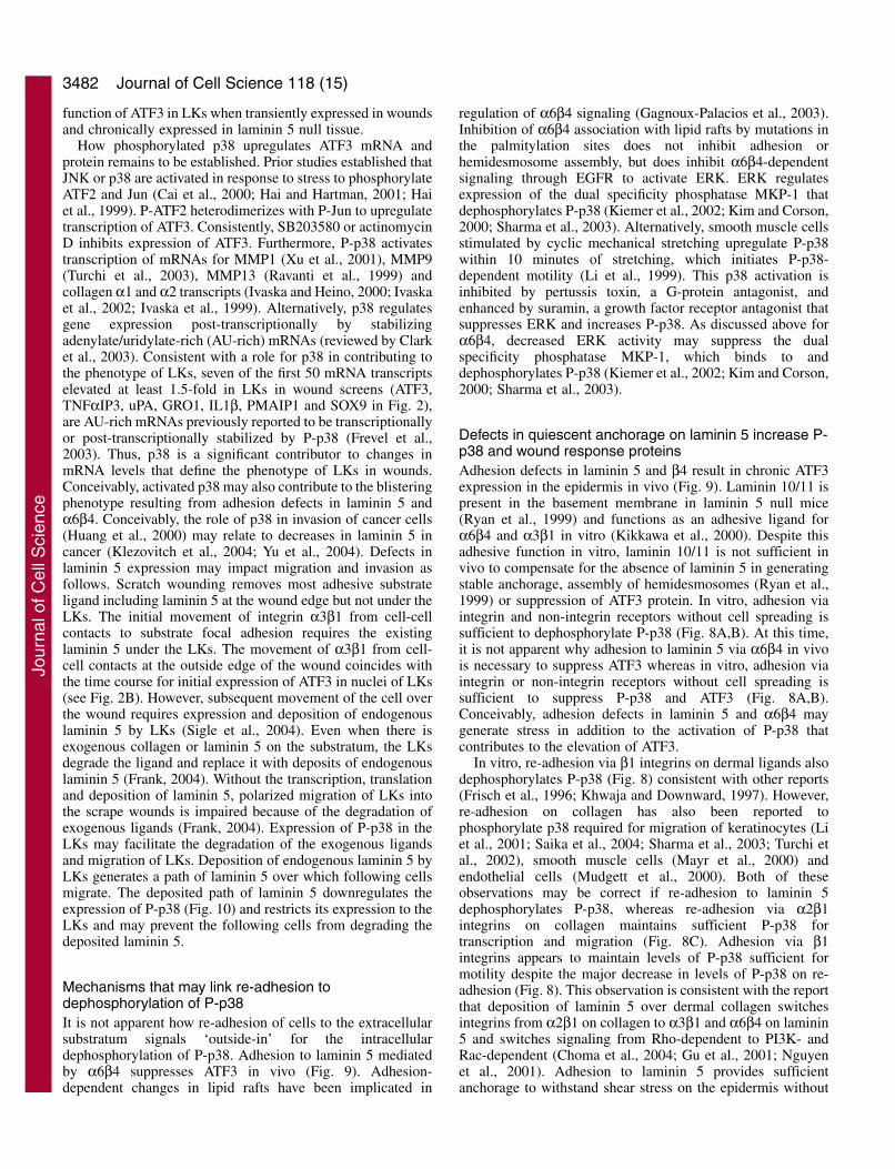
Re-adhesion to laminin 5 suppresses P-p38 whereas re-adhesion to collagen maintains P-p38Here, we used three approaches to evaluate the adhesionrequirements for dephosphorylation of P-p38. In the firstapproach, HKs were suspended and then incubated withimmobilized antibodies against integrin subunits α3, α2, α6,β1 or against non-integrin receptors (CD44, P4C11 antigen, a45 kDa cell surface protein recognized by mAb P4C11) (Fig.8A). We used laminin 5 null HKs for these studies to avoiddeposits of laminin 5 that would complicate the adhesionsignals. The adherent cells were spread prior to suspension andphosphorylated focal adhesion kinase on Tyr397 (P-FAK; Fig.8A, Adherent). Suspension of the adherent HKs (Suspended)phosphorylated p38 and dephosphorylated P-FAK. Incubationof the suspended HKs on non-adhesive control antibody SP2did not induce adhesion or spreading and did notdephosphorylate P-p38. However, re-adhesion of suspendedHKs on adhesive laminin 5 or collagen or immobilizedadhesive mAbs dephosphorylated P-p38. Therefore, adhesion-dependent dephosphorylation of P-p38 did not requireintegrins, cell spreading or phosphorylation of FAK. In furthercontrols (Results not shown) addition of the solublemonoclonal antibodies to suspended HKs failed todephosphorylate P-p38. We conclude that in vitro celladhesion, not cell spreading, is required for dephosphorylationof P-p38. This suggests that the primary mediator of celladhesion in vivo, laminin 5 and α6β4, are sufficient todephosphorylate P-p38.
In the second approach, we tried to distinguish betweenlaminin 5 and collagen in their roles in regulatingdephosphorylation of P-p38. Cytochalasin D, an inhibitor ofthe actin cytoskeleton, blocks cell spreading on laminin 5 butnot adhesion: both integrins α6β4 (Xia et al., 1996) and α3β1(Frank, 2004) adhere HKs on laminin 5 in the presence ofcytochalasin D and dephosphorylate P-p38 (Fig. 8B). Incontrast, cytochalasin D blocks adhesion, spreading anddephosphorylation of P-p38 on collagen and fibronectin (Fig.8B) (Xia et al., 1996). Thus, dephosphorylation of P-p38required re-adhesion but re-adhesion on laminin 5 isindependent of the actin cytoskeleton whereas re-adhesion oncollagen requires the actin cytoskeleton. This suggests that
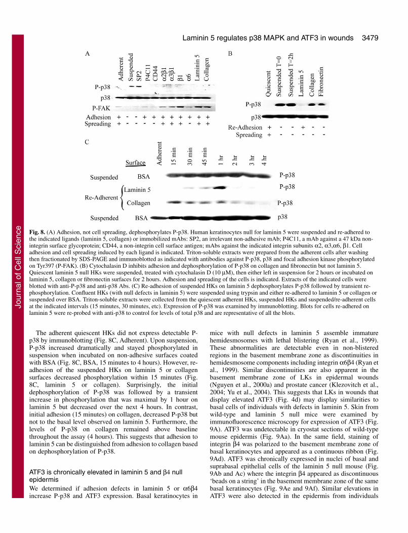
Fig. 5. (A) Inhibition of P-p38 with SB203580 preventsphosphorylation of ATF2 and up-regulation of ATF3. Confluent HKmonolayers were either untreated to serve as a control, or pretreatedwith SB203580 for 1 hour. Scrape wounds were generated andallowed to incubate for 2.5 hours in the presence or absence ofinhibitor. ATF2 phosphorylation (P-ATF-2; a,b) and ATF3 (c,d)protein expression levels were examined by immunofluorescentstaining. SB203580 blocked expression of P-ATF-2 and ATF-3.(B) Pretreatment of scrape wounds with the membrane-permeableform of the JNK peptide inhibitor (JNK Peptide Inhibitor 1, Lstereoisomer; 50 μM, 30 minutes prior to wounding) (Bonny et al.,2001) prevents phosphorylation of jun but not expression of ATF3.Bar, 25 μm.
Jour
nal o
f Cel
l Sci
ence

3478
adhesion to laminin 5 could dephosphorylate P-p38 underconditions when activation integrins like α2β1 and α5β1 donot contribute to adhesion or spreading, as in quiescence.
In the third approach, we compared re-adhesion to laminin5 with re-adhesion to collagen in dephosphorylation of P-p38.Published reports have established that suspension and re-adhesion of cells on collagen activates phosphorylation of p38
that regulates cell motility and transcription (Clark et al., 2003;Ono and Han, 2000). We emphasize that these publishedstudies required suspension of the cells prior to re-adhesion.However, studies here, and elsewhere (Frisch et al., 1996;Khwaja and Downward, 1997), indicate that suspension ofHKs or scrape wounding is sufficient to phosphorylate p38whereas re-adhesion dephosphorylates P-p38 relative to cellsin suspension. As a possible explanation for the difference inresults, we determined if re-adhesion to laminin 5 and collagendiffer in dephosphorylating P-p38.
Journal of Cell Science 118 (15)
Fig. 6 (A) P-p38 and P-ATF2 are elevated in LKs (white arrowheads) ofscrape wounds. Scrape wounds were generated in confluent HK monolayers.Levels of phosphorylated p38 and ATF2 were examined 1 hour postwounding by immunofluorescence microscopy. (B). P-p38 is upregulated inLKs of wounds in vivo. Cryostat sections of human epidermis 0 hours (a), 1hour (b) and 4 hours (c) post wounding were examined for p38phosphorylation through immunofluorescence microscopy. In the control (d),a 0 hour wound section is stained with anti-laminin 5 mAb (C2-5) to identifythe basement membrane. Wound margins are identified with whitearrowheads. Direction of outgrowth is shown by white arrow. Bar, 20 μm.
Fig. 7. (A) Scrape wounding increases phosphorylation of p38 andATF2. HKs were grown to confluence and multiple scrape woundswere generated in the monolayer. Triton-soluble extracts were collectedin non-wound HKs (Quiescent), or in wounded HKs at the time ofwounding, (0 hour), and 30 minutes, 1 hour, 2 hours and 3 hours postwounding. Levels of p38 remained constant but P-p38 increased by 0.5hours after wounding and P-ATF2 increased immediately afterwounding. (B) p38 kinase activity is elevated in suspended or woundedHKs. Confluent quiescent HKs (Quiescent) were assayed for p38kinase after suspension for 3 hours (Suspended) or scrape wounding for6 hours (Wounded). In vitro assay of p38 kinase activity was performedas described (see Materials and Methods) using ATF2 as substrate andimmunoblotting for P-ATF2. Aliquots of the cell extracts were alsoimmunoblotted (Immunoblot) for P-p38 to confirm that levels of P-p38correlated with p38 kinase activity.
Jour
nal o
f Cel
l Sci
ence
3479Laminin 5 regulates p38 MAPK and ATF3 in wounds
The adherent quiescent HKs did not express detectable P-p38 by immunoblotting (Fig. 8C, Adherent). Upon suspension,P-p38 increased dramatically and stayed phosphorylated insuspension when incubated on non-adhesive surfaces coatedwith BSA (Fig. 8C, BSA, 15 minutes to 4 hours). However, re-adhesion of the suspended HKs on laminin 5 or collagensurfaces decreased phosphorylation within 15 minutes (Fig.8C, laminin 5 or collagen). Surprisingly, the initialdephosphorylation of P-p38 was followed by a transientincrease in phosphorylation that was maximal by 1 hour onlaminin 5 but decreased over the next 4 hours. In contrast,initial adhesion (15 minutes) on collagen, decreased P-p38 butnot to the basal level observed on laminin 5. Furthermore, thelevels of P-p38 on collagen remained above baselinethroughout the assay (4 hours). This suggests that adhesion tolaminin 5 can be distinguished from adhesion to collagen basedon dephosphorylation of P-p38.
ATF3 is chronically elevated in laminin 5 and β4 nullepidermisWe determined if adhesion defects in laminin 5 or α6β4increase P-p38 and ATF3 expression. Basal keratinocytes in
mice with null defects in laminin 5 assemble immaturehemidesmosomes with lethal blistering (Ryan et al., 1999).These abnormalities are detectable even in non-blisteredregions in the basement membrane zone as discontinuities inhemidesmosome components including integrin α6β4 (Ryan etal., 1999). Similar discontinuities are also apparent in thebasement membrane zone of LKs in epidermal wounds(Nguyen et al., 2000a) and prostate cancer (Klezovitch et al.,2004; Yu et al., 2004). This suggests that LKs in wounds thatdisplay elevated ATF3 (Fig. 4d) may display similarities tobasal cells of individuals with defects in laminin 5. Skin fromwild-type and laminin 5 null mice were examined byimmunofluorescence microscopy for expression of ATF3 (Fig.9A). ATF3 was undetectable in cryostat sections of wild-typemouse epidermis (Fig. 9Aa). In the same field, staining ofintegrin β4 was polarized to the basement membrane zone ofbasal keratinocytes and appeared as a continuous ribbon (Fig.9Ad). ATF3 was chronically expressed in nuclei of basal andsuprabasal epithelial cells of the laminin 5 null mouse (Fig.9Ab and Ac) where the integrin β4 appeared as discontinuous‘beads on a string’ in the basement membrane zone of the samebasal keratinocytes (Fig. 9Ae and 9Af). Similar elevations inATF3 were also detected in the epidermis from individuals
Fig. 8. (A) Adhesion, not cell spreading, dephosphorylates P-p38. Human keratinocytes null for laminin 5 were suspended and re-adhered tothe indicated ligands (laminin 5, collagen) or immobilized mAbs: SP2, an irrelevant non-adhesive mAb; P4C11, a mAb against a 47 kDa non-integrin surface glycoprotein; CD44, a non-integrin cell surface antigen; mAbs against the indicated integrin subunits α2, α3,α6, β1. Celladhesion and cell spreading induced by each ligand is indicated. Triton-soluble extracts were prepared from the adherent cells after washingthen fractionated by SDS-PAGE and immunoblotted as indicated with antibodies against P-p38, p38 and focal adhesion kinase phosphorylatedon Tyr397 (P-FAK). (B) Cytochalasin D inhibits adhesion and dephosphorylation of P-p38 on collagen and fibronectin but not laminin 5.Quiescent laminin 5 null HKs were suspended, treated with cytochalasin D (10 μM), then either left in suspension for 2 hours or incubated onlaminin 5, collagen or fibronectin surfaces for 2 hours. Adhesion and spreading of the cells is indicated. Extracts of the indicated cells wereblotted with anti-P-p38 and anti-p38 Abs. (C) Re-adhesion of suspended HKs on laminin 5 dephosphorylates P-p38 followed by transient re-phosphorylation. Confluent HKs (with null defects in laminin 5) were suspended using trypsin and either re-adhered to laminin 5 or collagen orsuspended over BSA. Triton-soluble extracts were collected from the quiescent adherent HKs, suspended HKs and suspended/re-adherent cellsat the indicated intervals (15 minutes, 30 minutes, etc). Expression of P-p38 was examined by immunoblotting. Blots for cells re-adhered onlaminin 5 were re-probed with anti-p38 to control for levels of total p38 and are representative of all the blots.
Jour
nal o
f Cel
l Sci
ence

3480
with null defects in the ITGΒ4 gene encoding the β4 subunitof integrin α6β4 (9Bb). Based on elevated ATF3 expression,inherited adhesion defects in laminin 5 or integrin β4 result inthe activation of ATF3 similar to LKs in wounds.
MKs that are null for laminin 5 (α3–/–) fail to adhere to BSAbecause they cannot deposit laminin 5 and as a result, theyexpress ATF3 protein in suspension (Fig. 3C). Here, wedetermined if deposition of laminin 5 is sufficient to promoteadhesion and adhesion-dependent dephosphorylation of P-p38that regulates ATF3 (Fig. 10). Many studies have reported thatP-p38 is required for cell migration (see Discussion).Furthermore, we have previously shown that deposition oflaminin 5 at the rear of migrating LKs is necessary forpolarized migration of LKs (Frank, 2004). First, we determinedif null defects in laminin 5 would elevate levels of P-p38 invitro (Fig. 10). MKs with the wild-type α3 chain of laminin 5(α3+/+ MKs) and mutants null for the α3 chain of laminin 5(α3–/– MKs) were compared for deposition of laminin 5 and
adhesion to the deposits in the BSA adhesion assay (Gil et al.,2002; Sigle et al., 2004). The α3+/+ and α3–/– MKs weresuspended and re-plated onto either a non-adhesive surfacecoated with BSA or an adhesive laminin 5 surface (Fig. 10).Both the α3+/+ and α3–/– MKs adhered on the laminin 5surfaces and dephosphorylated P-p38 with similar kinetics.However, on the non-adhesive BSA surface, the α3+/+ MKsdeposited laminin 5, adhered and dephosphorylated P-p38within 4 hours of contact with the substratum. In contrast, α3–/–
keratinocytes failed to adhere on the BSA surface because theydid not deposit laminin 5 and P-p38 remained phosphorylatedthroughout the assay. We conclude that deposition ofendogenous laminin 5 followed by adhesion to the deposits issufficient to dephosphorylate P-p38. This suggests thatdeposition of laminin 5 by LKs at the rear of the migrating cellmay dephosphorylate p38 at the rear of the migrating LK andin following cells that migrate over the path of depositedlaminin 5. Thus deposition of endogenous laminin 5 is
Journal of Cell Science 118 (15)
Fig. 9. Adhesion defects in laminin 5 or integrin β4 chronically elevate ATF3. (A) Cryostat sections of skin from wild type neonatal mouse(a,d) and laminin 5 null neonatal mouse (b,c,e,f,) were stained with an anti-ATF3 (a,b,c) or anti-integrin β4 antibody (d,e,f). ATF3 protein is notdetectable in wild type mouse epidermis (arrows identify epidermal basal cells). β4 was continuous and polarized to the basement membrane(d, arrow identifies β4 staining in the basement membrane zone). In contrast, laminin 5 null mouse skin (α3–/–) exhibited increased levels ofnuclear ATF3 protein expression (b, c, arrow identifies ATF3 staining). Integrin β4 was polarized to the basement membrane zone in laminin 5null keratinocytes but was discontinuous (e,f) (arrows identify discontinuities in β4 integrin). (B) Cryostat sections of skin from a normalcontrol individual (a) and from an individual with null defects in the INTB4 gene encoding the integrin β4 subunit (b) were stained with anti-ATF3 antibodies. ATF3 was elevated in most cell layers in the β4 null epidermis. Bar, 20 μm (A); 20 μm (B).
Jour
nal o
f Cel
l Sci
ence

3481Laminin 5 regulates p38 MAPK and ATF3 in wounds
sufficient to regulate dephosphorylation P-p38 and this mayimpact the polarity of the LKs and restricted expression ofactivation components like P-p38 and ATF3 to the LKs but notthe following keratinocytes.
DiscussionWe have identified adhesion-dependent changes in thephosphorylation of p38 that regulate protein expression in LKs.These p38-dependent changes in protein expression result fromwounds or defects in laminin 5 or integrin β4. First, we usedan in vitro epidermal wound model and DNA microarrays toidentify mRNAs for ATF3 and other activation components(Fig. 1B). The activation mRNAs are elevated in LKs within1.5 hours of injury. Both in vitro and in vivo, ATF3 protein wasupregulated, translocated to the nucleus of LKs, thendownregulated in a transient window of expression between 2and 12 hours after wounding (Figs 2-4). Phosphorylation of
p38 MAPK and ATF2 was required for ATF3 expression inLKs of scrape wounds (Figs 5 and 6). Like scrape wounding,cell suspension was sufficient to phosphorylate p38 and elevateATF3 protein expression. Significantly, seven of the first 50mRNA transcripts elevated 1.5 times or more in LKs, areregulated by p38 (Fig. 1B). This indicates that p38 is a majorcontributor to initial epidermal wound activation in LKs. Invitro, re-adhesion of suspended cells to laminin 5downregulates both P-p38 and ATF3 independently of cellspreading. Consistently, in vivo adhesion defects in laminin 5or β4 elevated ATF3. We conclude that adhesion to laminin 5via α6β4 in vivo has two functions: first, laminin 5 is necessaryto anchor quiescent epidermis via α6β4 and prevent blistering;second, adhesion to laminin 5 suppresses P-p38 cell signaling,ATF3 and other activation mRNAs. We suggest that adhesionto laminin 5 may limit excessive migration or proteolyticremodeling of the basement membrane and dermis by LKs.Defects in deposition of laminin 5, or destruction of laminin 5in cancers may elevate P-p38 to increase activation mRNAsthat drive migration, stromal proteolysis and cell invasion.Here, we discuss these findings in relation to laminin 5 inregulating p38, transcription and adhesion in wounds, adhesiondefects and cancer.
Function of P-p38 and ATF3 in LKs of woundsATF3 is a member of the ATF/Creb family of transcriptionfactors and is upregulated in response to mechanical injury andstress stimuli (Hai and Hartman, 2001; Hai et al., 1999). Invivo, these stimuli include nerve axotomy (Tsujino et al.,2000), ischemia of the heart (Chen et al., 1996) and kidney(Yin et al., 1997), partial hepatectomy and brain seizure (Chenet al., 1996). In vitro, microtubule binding agents includingtaxol and colchicine (Shtil et al., 1999), genotoxic agentsincluding UV and ionizing radiation, transforming growthfactor β, tumor necrosis factor α and serum each stimulateATF3 expression (Hai and Hartman, 2001; Hai et al., 1999).For example, transforming growth factor β in combination withUV radiation elevates SMAD3 and P-p38, increasing ATF3.Subsequently, ATF3 represses the ID1 transcription factor toinhibit proliferation (Kang et al., 2003). Other target genes forATF3 repression include gadd153/Chop10, a transcriptionfactor that mediates effects of stress (Wang and Ron, 1996;Wolfgang et al., 1997), and E-selectin, an adhesion receptoractivated by TNFα (Nawa et al., 2000). However, the functionof ATF3 is still not established: ATF3 is reported to both inhibit(Kawauchi et al., 2002; Zhang et al., 2002) and promote (Nawaet al., 2002) p53-dependent apoptosis. ATF3 has also beenreported to promote adhesion on collagen (Ishiguro et al.,2000). Studies here demonstrate that ATF3 is upregulated inLKs at the wound margin, whereas quiescent following cellsare unaffected. In principle, the elevation of ATF3 in LKs mayrepress ID1 in LKs, inhibiting cell cycle progression whilecontributing to migration (Natarajan et al., 2003; Onuma et al.,2001; Sharma et al., 2003). Consistently, the laminin 5 nullMKs with elevated ATF3 (Fig. 9) cannot adhere (Fig. 10) anddo not progress through the cell cycle without exogenousligand (Ryan et al., 1999; Sigle et al., 2004). Alternatively,inhibition of p53 by elevated ATF3 in LKs may contribute tothe resistance of LKs to apoptosis in the hostile environmentof the wound. Future studies will attempt to understand the
Fig. 10. Defects in laminin 5 deposition, prevent adhesion anddephosphorylation of P-p38. MKs from laminin 5 null mice (α3–/–)and wild-type mice (α3+/+) were suspended and replated onto either aBSA- or Laminin 5-coated surface. Triton-soluble extracts werecollected at 2 hour intervals from 0 to 10 hours and immunoblottedwith anti-P-p38 and anti-p38 antibodies. Quiescent α3+/+ and α3–/–
MKs did not express phosphorylated p38. When plated onto a BSA-coated surface, α3+/+ MKs deposited laminin 5, adhered to thedeposits on the BSA surface and dephosphorylated p38 within 4hours. In contrast, α3–/– MKs were unable to deposit laminin 5 oradhere to the BSA or dephosphorylate p38 for the duration of theassay. Both α3+/+ MKs and α3–/– MKs rapidly adhered to exogenouslaminin 5 and dephosphorylated P-p38, indicating exogenous laminin5 was sufficient to rescue the ability of α3–/– MKs to adhere anddephosphorylate p38. Total levels of p38 did not change significantlyin either the α3+/+ MK or α3–/– MK populations.
Jour
nal o
f Cel
l Sci
ence
3482
function of ATF3 in LKs when transiently expressed in woundsand chronically expressed in laminin 5 null tissue.
How phosphorylated p38 upregulates ATF3 mRNA andprotein remains to be established. Prior studies established thatJNK or p38 are activated in response to stress to phosphorylateATF2 and Jun (Cai et al., 2000; Hai and Hartman, 2001; Haiet al., 1999). P-ATF2 heterodimerizes with P-Jun to upregulatetranscription of ATF3. Consistently, SB203580 or actinomycinD inhibits expression of ATF3. Furthermore, P-p38 activatestranscription of mRNAs for MMP1 (Xu et al., 2001), MMP9(Turchi et al., 2003), MMP13 (Ravanti et al., 1999) andcollagen α1 and α2 transcripts (Ivaska and Heino, 2000; Ivaskaet al., 2002; Ivaska et al., 1999). Alternatively, p38 regulatesgene expression post-transcriptionally by stabilizingadenylate/uridylate-rich (AU-rich) mRNAs (reviewed by Clarket al., 2003). Consistent with a role for p38 in contributing tothe phenotype of LKs, seven of the first 50 mRNA transcriptselevated at least 1.5-fold in LKs in wound screens (ATF3,TNFαIP3, uPA, GRO1, IL1β, PMAIP1 and SOX9 in Fig. 2),are AU-rich mRNAs previously reported to be transcriptionallyor post-transcriptionally stabilized by P-p38 (Frevel et al.,2003). Thus, p38 is a significant contributor to changes inmRNA levels that define the phenotype of LKs in wounds.Conceivably, activated p38 may also contribute to the blisteringphenotype resulting from adhesion defects in laminin 5 andα6β4. Conceivably, the role of p38 in invasion of cancer cells(Huang et al., 2000) may relate to decreases in laminin 5 incancer (Klezovitch et al., 2004; Yu et al., 2004). Defects inlaminin 5 expression may impact migration and invasion asfollows. Scratch wounding removes most adhesive substrateligand including laminin 5 at the wound edge but not under theLKs. The initial movement of integrin α3β1 from cell-cellcontacts to substrate focal adhesion requires the existinglaminin 5 under the LKs. The movement of α3β1 from cell-cell contacts at the outside edge of the wound coincides withthe time course for initial expression of ATF3 in nuclei of LKs(see Fig. 2B). However, subsequent movement of the cell overthe wound requires expression and deposition of endogenouslaminin 5 by LKs (Sigle et al., 2004). Even when there isexogenous collagen or laminin 5 on the substratum, the LKsdegrade the ligand and replace it with deposits of endogenouslaminin 5 (Frank, 2004). Without the transcription, translationand deposition of laminin 5, polarized migration of LKs intothe scrape wounds is impaired because of the degradation ofexogenous ligands (Frank, 2004). Expression of P-p38 in theLKs may facilitate the degradation of the exogenous ligandsand migration of LKs. Deposition of endogenous laminin 5 byLKs generates a path of laminin 5 over which following cellsmigrate. The deposited path of laminin 5 downregulates theexpression of P-p38 (Fig. 10) and restricts its expression to theLKs and may prevent the following cells from degrading thedeposited laminin 5.
Mechanisms that may link re-adhesion todephosphorylation of P-p38It is not apparent how re-adhesion of cells to the extracellularsubstratum signals ‘outside-in’ for the intracellulardephosphorylation of P-p38. Adhesion to laminin 5 mediatedby α6β4 suppresses ATF3 in vivo (Fig. 9). Adhesion-dependent changes in lipid rafts have been implicated in
regulation of α6β4 signaling (Gagnoux-Palacios et al., 2003).Inhibition of α6β4 association with lipid rafts by mutations inthe palmitylation sites does not inhibit adhesion orhemidesmosome assembly, but does inhibit α6β4-dependentsignaling through EGFR to activate ERK. ERK regulatesexpression of the dual specificity phosphatase MKP-1 thatdephosphorylates P-p38 (Kiemer et al., 2002; Kim and Corson,2000; Sharma et al., 2003). Alternatively, smooth muscle cellsstimulated by cyclic mechanical stretching upregulate P-p38within 10 minutes of stretching, which initiates P-p38-dependent motility (Li et al., 1999). This p38 activation isinhibited by pertussis toxin, a G-protein antagonist, andenhanced by suramin, a growth factor receptor antagonist thatsuppresses ERK and increases P-p38. As discussed above forα6β4, decreased ERK activity may suppress the dualspecificity phosphatase MKP-1, which binds to anddephosphorylates P-p38 (Kiemer et al., 2002; Kim and Corson,2000; Sharma et al., 2003).
Defects in quiescent anchorage on laminin 5 increase P-p38 and wound response proteinsAdhesion defects in laminin 5 and β4 result in chronic ATF3expression in the epidermis in vivo (Fig. 9). Laminin 10/11 ispresent in the basement membrane in laminin 5 null mice(Ryan et al., 1999) and functions as an adhesive ligand forα6β4 and α3β1 in vitro (Kikkawa et al., 2000). Despite thisadhesive function in vitro, laminin 10/11 is not sufficient invivo to compensate for the absence of laminin 5 in generatingstable anchorage, assembly of hemidesmosomes (Ryan et al.,1999) or suppression of ATF3 protein. In vitro, adhesion viaintegrin and non-integrin receptors without cell spreading issufficient to dephosphorylate P-p38 (Fig. 8A,B). At this time,it is not apparent why adhesion to laminin 5 via α6β4 in vivois necessary to suppress ATF3 whereas in vitro, adhesion viaintegrin or non-integrin receptors without cell spreading issufficient to suppress P-p38 and ATF3 (Fig. 8A,B).Conceivably, adhesion defects in laminin 5 and α6β4 maygenerate stress in addition to the activation of P-p38 thatcontributes to the elevation of ATF3.
In vitro, re-adhesion via β1 integrins on dermal ligands alsodephosphorylates P-p38 (Fig. 8) consistent with other reports(Frisch et al., 1996; Khwaja and Downward, 1997). However,re-adhesion on collagen has also been reported tophosphorylate p38 required for migration of keratinocytes (Liet al., 2001; Saika et al., 2004; Sharma et al., 2003; Turchi etal., 2002), smooth muscle cells (Mayr et al., 2000) andendothelial cells (Mudgett et al., 2000). Both of theseobservations may be correct if re-adhesion to laminin 5dephosphorylates P-p38, whereas re-adhesion via α2β1integrins on collagen maintains sufficient P-p38 fortranscription and migration (Fig. 8C). Adhesion via β1integrins appears to maintain levels of P-p38 sufficient formotility despite the major decrease in levels of P-p38 on re-adhesion (Fig. 8). This observation is consistent with the reportthat deposition of laminin 5 over dermal collagen switchesintegrins from α2β1 on collagen to α3β1 and α6β4 on laminin5 and switches signaling from Rho-dependent to PI3K- andRac-dependent (Choma et al., 2004; Gu et al., 2001; Nguyenet al., 2001). Adhesion to laminin 5 provides sufficientanchorage to withstand shear stress on the epidermis without
Journal of Cell Science 118 (15)
Jour
nal o
f Cel
l Sci
ence
3483Laminin 5 regulates p38 MAPK and ATF3 in wounds
activating signals through Rho, p38 and ATF3 or urokinase thatmay promote excessive migration or invasion of stroma (Huanget al., 2000; Montero and Nagamine, 1999). Consistently,active Rho and P-p38 contribute to degradation of substratecollagen (Berdeaux et al., 2004; Turchi et al., 2003). Therefore,deposition of laminin 5 that reduces P-p38 and Rho-dependentadhesion on collagen may also reduce collagen degradation(Frank, 2004) or cell invasion.
We would like to thank Diane Frank, Randy Sigle, Clarence Dunn,Beatrice Knudsen and Paul Lampe for critical review of themanuscript. We would also like to thank Tim King for help and advicewith the cDNA microarrays. This work was supported by NationalInstitutes of Health Grants CA049259 (W.G.C.) and DK59221(J.O./W.G.C.). E.G.H. was supported by a Warren MagnusonScholarship for Biomedical Research and Health Profession (2000)and from an NIH training grant (5T32AI07509) to the Dept ofPathobiology, University of Washington (1998-1999). S.M.A. wassupported in part by PHS NRSA T32 GM07270 from NIGMS.
ReferencesBalda, M. S. and Matter, K. (2003). Epithelial cell adhesion and the
regulation of gene expression. Trends Cell Biol. 13, 310-318.Berdeaux, R. L., Diaz, B., Kim, L. and Martin, G. S. (2004). Active Rho is
localized to podosomes induced by oncogenic Src and is required for theirassembly and function. J. Cell Biol. 166, 317-323.
Bonny, C., Oberson, A., Negri, S., Sauser, C. and Schorderet, D. F. (2001).Cell-permeable peptide inhibitors of JNK: novel blockers of beta-cell death.Diabetes 50, 77-82.
Borradori, L. and Sonnenberg, A. (1999). Structure and function ofhemidesmosomes: more than simple adhesion complexes. J. Invest.Dermatol. 112, 411-418.
Boyce, S. T. and Ham, R. G. (1985). Cultivation, frozen storage, and clonalgrowth of normal human epidermal keratinocytes in serum-free media. J.Tissue Cult. Meth. 9, 83-93.
Cai, Y., Zhang, C., Nawa, T., Aso, T., Tanaka, M., Oshiro, S., Ichijo, H.and Kitajima, S. (2000). Homocysteine-responsive ATF3 gene expressionin human vascular endothelial cells: activation of c-Jun NH(2)-terminalkinase and promoter response element. Blood 96, 2140-2148.
Carter, W. G., Ryan, M. C. and Gahr, P. J. (1991). Epiligrin, a new celladhesion ligand for integrin α3β1 in epithelial basement membranes. Cell65, 599-610.
Charvat, S., Duchesne, M., Parvaz, P., Chignol, M. C., Schmitt, D. andSerres, M. (1999). The up-regulation of vascular endothelial growth factorin mutated Ha-ras HaCaT cell lines is reduced by a farnesyl transferaseinhibitor. Anticancer Res. 19, 557-561.
Chen, B. P., Wolfgang, C. D. and Hai, T. (1996). Analysis of ATF3, atranscription factor induced by physiological stresses and modulated bygadd153/Chop10. Mol. Cell. Biol. 16, 1157-1168.
Choma, D. P., Pumiglia, K. and DiPersio, C. M. (2004). Integrin alpha3beta1directs the stabilization of a polarized lamellipodium in epithelial cellsthrough activation of Rac1. J. Cell Sci. 117, 3947-3959.
Clark, A. R., Dean, J. L. and Saklatvala, J. (2003). Post-transcriptionalregulation of gene expression by mitogen-activated protein kinase p38.FEBS Lett. 546, 37-44.
Crepaldi, T., Gautreau, A., Comoglio, P. M., Louvard, D. and Arpin, M.(1997). Ezrin is an effector of hepatocyte growth factor-mediated migrationand morphogenesis in epithelial cells. J. Cell Biol. 138, 423-434.
Fan, F., Jin, S., Amundson, S. A., Tong, T., Fan, W., Zhao, H., Zhu, X.,Mazzacurati, L., Li, X., Petrik, K. L. et al. (2002). ATF3 inductionfollowing DNA damage is regulated by distinct signaling pathways andover-expression of ATF3 protein suppresses cells growth. Oncogene 21,7488-7496.
Fazzio, T. G., Kooperberg, C., Goldmark, J. P., Neal, C., Basom, R.,Delrow, J. and Tsukiyama, T. (2001). Widespread collaboration of Isw2and Sin3-Rpd3 chromatin remodeling complexes in transcriptionalrepression. Mol. Cell. Biol. 21, 6450-6460.
Frank, D. and Carter, W. G. (2004). Laminin 5 deposition regulateskeratinocyte polarized and persistent migration. J. Cell Sci. 117, 1351-1363.
Frevel, M. A., Bakheet, T., Silva, A. M., Hissong, J. G., Khabar, K. S. and
Williams, B. R. (2003). p38 Mitogen-activated protein kinase-dependentand -independent signaling of mRNA stability of AU-rich element-containing transcripts. Mol. Cell. Biol. 23, 425-436.
Frisch, S. M., Vuori, K., Kelaita, D. and Sicks, S. (1996). A role for Jun-N-terminal kinase in anoikis; suppression by bcl-2 and crmA. J. Cell Biol. 135,1377-1382.
Fuchs, E., Dowling, J., Segre, J., Lo, S.-H. and Yu, Q.-C. (1997). Integratorsof epidermal growth and differentiation: distinct functions for beta 1 andbeta 4 integrins. Curr. Opin. Genet. Dev. 7, 672-682.
Gagnoux-Palacios, L., Dans, M., van’t Hof, W., Mariotti, A., Pepe, A.,Meneguzzi, G., Resh, M. D. and Giancotti, F. G. (2003).Compartmentalization of integrin alpha6beta4 signaling in lipid rafts. J. CellBiol. 162, 1189-1196.
Gil, S. G., Sigle, R. O. and Carter, W. G. (2002). Detection and purificationof instructive extracellular matrix components with monoclonal antibodytechnologies. Methods Cell Biol. 69, 27-52.
Gipson, I. K., Spurr-Michaud, S., Tisdale, A., Elwell, J. and Stepp, M. A.(1993). Redistribution of the hemidesmosome components alpha 6 beta 4integrin and bullous pemphigoid antigens during epithelial wound healing.Exp. Cell Res. 207, 86-98.
Goldfinger, L. E., Hopkinson, S. B., deHart, G. W., Collawn, S.,Couchman, J. R. and Jones, J. C. (1999). The alpha3 lamininsubunit, alpha6beta4 and alpha3beta1 integrin coordinately regulatewound healing in cultured epithelial cells and in the skin. J. Cell Sci. 112,2615-2629.
Gu, J., Sumida, Y., Sanzen, N. and Sekiguchi, K. (2001). Laminin-10/11and fibronectin differentially regulate integrin-dependent Rho and Racactivation via p130(Cas)-CrkII-DOCK180 pathway. J. Biol. Chem. 276,27090-27097.
Haftek, M., Simon, M., Kanitakis, J., Marechal, S., Claudy, A., Serre, G.and Schmitt, D. (1997). Expression of corneodesmosin in the granular layerand stratum corneum of normal and diseased epidermis. Br. J. Dermatol.137, 864-873.
Hai, T. and Hartman, M. G. (2001). The molecular biology and nomenclatureof the activating transcription factor/cAMP responsive element bindingfamily of transcription factors: activating transcription factor proteins andhomeostasis. Gene 273, 1-11.
Hai, T., Wolfgang, C. D., Marsee, D. K., Allen, A. E. and Sivaprasad, U.(1999). ATF3 and stress responses. Gene Exp. 7, 321-335.
Huang, S., New, L., Pan, Z., Han, J. and Nemerow, G. R. (2000). Urokinaseplasminogen activator/urokinase-specific surface receptor expression andmatrix invasion by breast cancer cells requires constitutive p38alphamitogen-activated protein kinase activity. J. Biol. Chem. 275, 12266-12272.
Ishiguro, T., Nagawa, H., Naito, M. and Tsuruo, T. (2000). Inhibitory effectof ATF3 antisense oligonucleotide on ectopic growth of HT29 human coloncancer cells. Jpn J. Cancer Res. 91, 833-836.
Ivaska, J. and Heino, J. (2000). Adhesion receptors and cell invasion:mechanisms of integrin-guided degradation of extracellular matrix. Cell.Mol. Life Sci. 57, 16-24.
Ivaska, J., Reunanen, H., Westermarck, J., Koivisto, L., Kahari, V. M. andHeino, J. (1999). Integrin alpha2beta1 mediates isoform-specific activationof p38 and upregulation of collagen gene transcription by a mechanisminvolving the alpha2 cytoplasmic tail. J. Cell Biol. 147, 401-416.
Ivaska, J., Nissinen, L., Immonen, N., Eriksson, J. E., Kahari, V. M. andHeino, J. (2002). Integrin alpha 2 beta 1 promotes activation of proteinphosphatase 2A and dephosphorylation of Akt and glycogen synthase kinase3 beta. Mol. Cell. Biol. 22, 1352-1359.
Kang, Y., Chen, C. R. and Massague, J. (2003). A self-enabling TGFbetaresponse coupled to stress signaling: Smad engages stress response factorATF3 for Id1 repression in epithelial cells. Mol. Cell 11, 915-926.
Kawauchi, J., Zhang, C., Nobori, K., Hashimoto, Y., Adachi, M. T., Noda,A., Sunamori, M. and Kitajima, S. (2002). Transcriptional repressoractivating transcription factor 3 protects human umbilical vein endothelialcells from tumor necrosis factor-alpha-induced apoptosis through down-regulation of p53 transcription. J. Biol. Chem. 277, 39025-39034.
Khwaja, A. and Downward, J. (1997). Lack of correlation between activationof Jun-NH2-terminal kinase and induction of apoptosis after detachment ofepithelial cells. J. Cell Biol. 139, 1017-1023.
Kiemer, A. K., Weber, N. C., Furst, R., Bildner, N., Kulhanek-Heinze, S.and Vollmar, A. M. (2002). Inhibition of p38 MAPK activation viainduction of MKP-1: atrial natriuretic peptide reduces TNF-alpha-inducedactin polymerization and endothelial permeability. Circ. Res. 90, 874-881.
Kikkawa, Y., Sanzen, N., Fujiwara, H., Sonnenberg, A. and Sekiguchi, K.(2000). Integrin binding specificity of laminin-10/11: laminin-10/11 are
Jour
nal o
f Cel
l Sci
ence
3484
recognized by alpha 3 beta 1, alpha 6 beta 1 and alpha 6 beta 4 integrins. J.Cell Sci. 113, 869-876.
Kim, F. and Corson, M. A. (2000). Adhesion to fibronectin enhances MKP-1 activation in human endothelial cells. Biochem. Biophys. Res. Commun.273, 539-545.
Klezovitch, O., Chevillet, J., Mirosevich, J., Roberts, R. L., Matusik, R. J.and Vasioukhin, V. (2004). Hepsin promotes prostate cancer progressionand metastasis. Cancer Cell 6, 185-195.
Laemmli, U. K. (1970). Cleavage of structural proteins during the assemblyof the head of bacteriophage T4. Nature 227, 680-685.
Lampe, P. D., Nguyen, B. P., Gil, S., Usui, M., Olerud, J., Takada, Y. andCarter, W. G. (1998). Cellular Interaction of integrin a3b1 with laminin 5promotes gap junctional communication. J. Cell Biol. 143, 1735-1747.
Li, C., Hu, Y., Mayr, M. and Xu, Q. (1999). Cyclic strain stress-inducedmitogen-activated protein kinase (MAPK) phosphatase 1 expression invascular smooth muscle cells is regulated by Ras/Rac-MAPK pathways. J.Biol. Chem. 274, 25273-25280.
Li, G., Gustafson-Brown, C., Hanks, S. K., Nason, K., Arbeit, J. M.,Pogliano, K., Wisdom, R. M. and Johnson, R. S. (2003). c-Jun is essentialfor organization of the epidermal leading edge. Dev. Cell 4, 865-877.
Li, W., Nadelman, C., Henry, G., Fan, J., Muellenhoff, M., Medina, E.,Gratch, N. S., Chen, M., Han, J. and Woodley, D. (2001). The p38-MAPK/SAPK pathway is required for human keratinocyte migration ondermal collagen. J. Invest. Dermatol. 117, 1601-1611.
Lim, K. K., Su, W. P., McEvoy, M. T. and Pittelkow, M. R. (1996).Generalized gravis junctional epidermolysis bullosa: case report, laboratoryevaluation, and review of recent advances. Mayo Clin. Proc. 71, 863-868.
Mainiero, F., Colombara, M., Antonini, V., Strippoli, R., Merola, M., Poffe,O., Tridente, G. and Ramarli, D. (2003). p38 MAPK is a critical regulatorof the constitutive and the beta4 integrin-regulated expression of IL-6 inhuman normal thymic epithelial cells. Eur. J. Immunol. 33, 3038-3048.
Marken, J. S., Schieven, G. L., Hellstrom, I., Hellstrom, K. E. and Aruffo,A. (1992). Cloning and expression of the tumor-associated antigen L6. Proc.Natl. Acad. Sci. USA 89, 3503-3507.
Martin, P. (1997). Wound healing – aiming for perfect skin regeneration.Science 276, 75-81.
Mayr, M., Li, C., Zou, Y., Huemer, U., Hu, Y. and Xu, Q. (2000).Biomechanical stress-induced apoptosis in vein grafts involves p38 mitogen-activated protein kinases. FASEB J. 14, 261-270.
Montero, L. and Nagamine, Y. (1999). Regulation by p38 mitogen-activatedprotein kinase of adenylate- and uridylate-rich element-mediated urokinase-type plasminogen activator (uPA) messenger RNA stability and uPA-dependent in vitro cell invasion. Cancer Res. 59, 5286-5293.
Morioka, S., Lazarus, G. S., Baird, J. L. and Jensen, P. J. (1987). Migratingkeratinocytes express urokinase-type plasminogen activator. J. Invest.Dermatol. 88, 418-423.
Mudgett, J. S., Ding, J., Guh-Siesel, L., Chartrain, N. A., Yang, L., Gopal,S. and Shen, M. M. (2000). Essential role for p38alpha mitogen-activatedprotein kinase in placental angiogenesis. Proc. Natl. Acad. Sci. USA 97,10454-10459.
Natarajan, E., Saeb, M., Crum, C. P., Woo, S. B., McKee, P. H. andRheinwald, J. G. (2003). Co-expression of p16(INK4A) and laminin 5gamma2 by microinvasive and superficial squamous cell carcinomas in vivoand by migrating wound and senescent keratinocytes in culture. Am. J.Pathol. 163, 477-491.
Nawa, T., Nawa, M. T., Cai, Y., Zhang, C., Uchimura, I., Narumi, S.,Numano, F. and Kitajima, S. (2000). Repression of TNF-alpha-induced E-selectin expression by PPAR activators: involvement of transcriptionalrepressor LRF-1/ATF3. Biochem. Biophys. Res. Commun. 275, 406-411.
Nawa, T., Nawa, M. T., Adachi, M. T., Uchimura, I., Shimokawa, R.,Fujisawa, K., Tanaka, A., Numano, F. and Kitajima, S. (2002).Expression of transcriptional repressor ATF3/LRF1 in humanatherosclerosis: colocalization and possible involvement in cell death ofvascular endothelial cells. Atherosclerosis 161, 281-291.
Nguyen, B. P., Gil, S. G., Ryan, M. C. and Carter, W. G. (2000a). Depositionof laminin 5 in epidermal woulds regulates integrin signaling and adhesion.Curr. Opin. Cell Biol. 12, 554-562.
Nguyen, B. P., Gil, S. G. and Carter, W. G. (2000b). Deposition of laminin5 by keratinocytes regulates integrin adhesion and signaling. J. Biol. Chem.275, 31896-31907.
Nguyen, B. P., Ren, X. D., Schwartz, M. A. and Carter, W. G. (2001).Ligation of integrin alpha 3beta 1 by laminin 5 at the wound edge activatesRho-dependent adhesion of leading keratinocytes on collagen. J. Biol.Chem. 276, 43860-43870.
Okada, Y., Saika, S., Shirai, K., Hashizume, N., Yamanaka, O., Ohnishi,Y. and Senba, E. (2001). Disappearance of desmosomal components in ratcorneal epithelium during wound healing. Ophthalmologica 215, 61-65.
Olerud, J. E., Odland, G. F., Burgess, E. M., Wyss, C. R., Fisher, L. D. andMatsen, F. A., 3rd (1995). A model for the study of wounds in normalelderly adults and patients with peripheral vascular disease or diabetesmellitus. J. Surg. Res. 59, 349-360.
Ono, K. and Han, J. (2000). The p38 signal transduction pathway: activationand function. Cell. Signal. 12, 1-13.
Onuma, H., Mastui, C. and Morohashi, M. (2001). Quantitative analysis ofthe proliferation of epidermal cells using a human skin organ culture systemand the effect of DbcAMP using markers of proliferation (BrdU, Ki-67,PCNA). Arch. Dermatol. Res. 293, 133-138.
Penas, P. F., Garcia-Diez, A., Sanchez-Madrid, F. and Yanez-Mo, M.(2000). Tetraspanins are localized at motility-related structures and involvedin normal human keratinocyte wound healing migration. J. Invest. Dermatol.114, 1126-1135.
Pulkkinen, L., Rouan, F., Bruckner-Tuderman, L., Wallerstein, R.,Garzon, M., Brown, T., Smith, L., Carter, W. and Uitto, J. (1998). NovelITGB4 mutations in lethal and nonlethal variants of epidermolysis bullosawith pyloric atresia: missense versus nonsense. Am. J. Hum. Genet. 63,1376-1387.
Ravanti, L., Heino, J., Lopez-Otin, C. and Kahari, V. M. (1999). Inductionof collagenase-3 (MMP-13) expression in human skin fibroblasts by three-dimensional collagen is mediated by p38 mitogen-activated protein kinase.J. Biol. Chem. 274, 2446-2455.
Ryan, M. C., Tizard, R., VonDevanter, D. R. and Carter, W. G. (1994).Cloning of the LamA3 gene encoding the α3 chain of the adhesive ligandepiligrin: expression in wound repair. J. Biol. Chem. 269, 22779-22787.
Ryan, M. C., Lee, K., Miyashita, Y. and Carter, W. G. (1999). Targeteddisruption of the LAMA3 gene in mice reveals abnormalities in survivaland late stage differentiation of epithelial cells. J. Cell Biol. 145, 1309-1323.
Saika, S., Okada, Y., Miyamoto, T., Yamanaka, O., Ohnishi, Y., Ooshima,A., Liu, C. Y., Weng, D. and Kao, W. W. (2004). Role of p38 MAP kinasein regulation of cell migration and proliferation in healing cornealepithelium. Invest. Ophthalmol. Vis. Sci. 45, 100-109.
Scott, F. L., Paddle-Ledinek, J. E., Cerruti, L., Coughlin, P. B., Salem, H.H. and Bird, P. I. (1998). Proteinase inhibitor 6 (PI-6) expression in humanskin: induction of PI-6 and a PI-6/proteinase complex during keratinocytedifferentiation. Exp. Cell Res. 245, 263-271.
Sharma, G. D., He, J. and Bazan, H. E. (2003). p38 and ERK1/2 coordinatecellular migration and proliferation in epithelial wound healing: evidence ofcross-talk activation between MAP kinase cascades. J. Biol. Chem. 278,21989-21997.
Shtil, A. A., Mandlekar, S., Yu, R., Walter, R. J., Hagen, K., Tan, T. H.,Roninson, I. B. and Kong, A. N. (1999). Differential regulation of mitogen-activated protein kinases by microtubule-binding agents in human breastcancer cells. Oncogene 18, 377-384.
Sigle, R. O., Gil, S. G., Bhattacharya, M., Ryan, M. C., Yang, T. M.,Brown, T. A., Boutaud, A., Miyashita, Y., Olerud, J. and Carter, W. G.(2004). Globular domains 4/5 of the laminin alpha3 chain mediatedeposition of precursor laminin 5. J. Cell Sci. 117, 4481-4494.
Smith, J. M. and Koopman, P. A. (2004). The ins and outs of transcriptionalcontrol: nucleocytoplasmic shuttling in development and disease. TrendsGenet. 20, 4-8.
Sukhatme, V. P., Cao, X. M., Chang, L. C., Tsai-Morris, C. H.,Stamenkovich, D., Ferreira, P. C., Cohen, D. R., Edwards, S. A., Shows,T. B. and Curran, T. (1988). A zinc finger-encoding gene coregulated withc-fos during growth and differentiation, and after cellular depolarization.Cell 53, 37-43.
Syed, V., Mukherjee, K., Lyons-Weiler, J., Lau, K. M., Mashima, T.,Tsuruo, T. and Ho, S. M. (2005). Identification of ATF-3, caveolin-1, DLC-1, and NM23-H2 as putative antitumorigenic, progesterone-regulated genesfor ovarian cancer cells by gene profiling. Oncogene 24, 1774-1787.
Tsujino, H., Kondo, E., Fukuoka, T., Dai, Y., Tokunaga, A., Miki, K.,Yonenobu, K., Ochi, T. and Noguchi, K. (2000). Activating transcriptionfactor 3 (ATF3) induction by axotomy in sensory and motoneurons: A novelneuronal marker of nerve injury. Mol. Cell. Neurosci. 15, 170-182.
Turchi, L., Chassot, A. A., Rezzonico, R., Yeow, K., Loubat, A., Ferrua,B., Lenegrate, G., Ortonne, J. P. and Ponzio, G. (2002). Dynamiccharacterization of the molecular events during in vitro epidermal woundhealing. J. Invest. Dermatol. 119, 56-63.
Turchi, L., Chassot, A. A., Bourget, I., Baldescchi, C., Ortonne, J. P.,
Journal of Cell Science 118 (15)
Jour
nal o
f Cel
l Sci
ence

3485Laminin 5 regulates p38 MAPK and ATF3 in wounds
Meneguzzi, G., Lemichez, E. and Ponzio, G. (2003). Cross-talk betweenRhoGTPases and stress activated kinases for matrix metalloproteinase-9induction in response to keratinocytes injury. J. Invest. Dermatol. 121, 1291-1300.
Utani, A., Nomizu, M., Matsuura, H., Kato, K., Kobayashi, T., Takeda, U.,Aota, S., Nielsen, P. K. and Shinkai, H. (2001). A unique sequence of thelaminin alpha 3 G domain binds to heparin and promotes cell adhesionthrough syndecan-2 and -4. J. Biol. Chem. 276, 28779-28788.
Utani, A., Momota, Y., Endo, H., Kasuya, Y., Beck, K., Suzuki, N.,Nomizu, M. and Shinkai, H. (2003). Laminin alpha 3 LG4 module inducesmatrix metalloproteinase-1 through mitogen-activated protein kinasesignaling. J. Biol. Chem. 278, 34483-34490.
Wang, X. Z. and Ron, D. (1996). Stress-induced phosphorylation andactivation of the transcription factor CHOP (GADD153) by p38 MAPKinase. Science 272, 1347-1349.
Wayner, E. A. and Carter, W. G. (1987). Identification of multiple celladhesion receptors for collagen and fibronectin in human fibrosarcoma cellspossessing unique alpha and common beta subunits. J. Cell Biol. 105, 1873-1884.
Wolfgang, C. D., Chen, B. P., Martindale, J. L., Holbrook, N. J. and Hai,T. (1997). gadd153/Chop10, a potential target gene of the transcriptionalrepressor ATF3. Mol. Cell. Biol. 17, 6700-6707.
Wood, W., Jacinto, A., Grose, R., Woolner, S., Gale, J., Wilson, C. andMartin, P. (2002). Wound healing recapitulates morphogenesis inDrosophila embryos. Nat. Cell Biol. 4, 907-912.
Woodley, D. T., O’Toole, E., Nadelman, C. M. and Li, W. (1999).
Mechanisms of human keratinocyte migration: lessons for understanding there-epithelialization of human skin wounds. Prog. Dermatol. 33, 1-12.
Xia, Y. and Karin, M. (2004). The control of cell morphogenesis by Junkinases. Trends Cell Biol. 14, 94-101.
Xia, Y., Gil, S. G. and Carter, W. G. (1996). Anchorage mediated by integrinalpha6beta4 to laminin 5 (epiligrin) regulates tyrosine phosphorylation of amembrane-associated 80-kD protein. J. Cell Biol. 132, 727-740.
Xu, J., Clark, R. A. and Parks, W. C. (2001). p38 mitogen-activated kinaseis a bidirectional regulator of human fibroblast collagenase-1 induction bythree-dimensional collagen lattices. Biochem. J. 355, 437-447.
Yin, T., Sandhu, G., Wolfgang, C. D., Burrier, A., Webb, R. L., Rigel, D.F., Hai, T. and Whelan, J. (1997). Tissue-specific pattern of stress kinaseactivation in ischemic/reperfused heart and kidney. J. Biol. Chem. 272,19943-19950.
Yu, H.-M., Frank, D. E., Zhang, J., You, X., Carter, W. G. and Knudsen,B. S. (2004). Basal Prostate epithelial cells stimulate the migration ofprostate cancer cells. Mol. Carcinog. 41, 85-97.
Zhang, C., Kawauchi, J., Adachi, M. T., Hashimoto, Y., Oshiro, S., Aso,T. and Kitajima, S. (2001). Activation of JNK and transcriptional repressorATF3/LRF1 through the IRE1/TRAF2 pathway is implicated in humanvascular endothelial cell death by homocysteine. Biochem. Biophys. Res.Commun. 289, 718-724.
Zhang, C., Gao, C., Kawauchi, J., Hashimoto, Y., Tsuchida, N. andKitajima, S. (2002). Transcriptional activation of the human stress-inducible transcriptional repressor ATF3 gene promoter by p53. Biochem.Biophys. Res. Commun. 297, 1302-1310.
Jour
nal o
f Cel
l Sci
ence