Embed Size (px)
Citation preview

06 (2007) 714–724www.elsevier.com/locate/ydbio
Developmental Biology 3
Wnt signaling is required for antero-posteriorpatterning of the planarian brain
Chiyoko Kobayashi a,b, Yumi Saito c, Kazuya Ogawa a, Kiyokazu Agata a,c,⁎
a RIKEN Center for Developmental Biology, 2-2-3 Minatojima-minamimachi, Chuo-ku, Kobe, Hyogo 650-0047, Japanb Division of Integrative Cell Biology, Institute of Molecular Embryology and Genetics, Kumamoto University, 2-2-1 Honjo, Kumamoto 860-0811, Japan
c Department of Biophysics, Graduate School of Science, Kyoto University, Kitashirakawa-Oiwake, Sakyo-ku, Kyoto 606-8502, Japan
Received for publication 13 November 2006; revised 10 April 2007; accepted 10 April 2007Available online 18 April 2007
Abstract
Wnt signaling functions in axis formation and morphogenesis in various animals and organs. Here we report that Wnt signaling is required forproper brain patterning during planarian brain regeneration. We showed here that one of the Wnt homologues in the planarian Dugesia japonica,DjwntA, was expressed in the posterior region of the brain. When DjwntA-knockdown planarians were produced by RNAi, they could regeneratetheir heads at the anterior ends of the fragments, but formed ectopic eyes with irregular posterior lateral branches and brain expansion. Thissuggests that the Wnt signal may be involved in antero-posterior (A–P) patterning of the planarian brain, as in vertebrates. We also investigatedthe relationship between the DjwntA and nou-darake/FGFR signal systems, as knockdown planarians of these genes showed similar phenotypes.Double-knockdown planarians of these genes did not show any synergistic effects, suggesting that the two signal systems function independentlyin the process of brain regeneration, which accords with the fact that nou-darake was expressed earlier than DjwntA during brain regeneration.These observations suggest that the nou-darake/FGFR signal may be involved in brain rudiment formation during the early stage of headregeneration, and subsequently the DjwntA signal may function in A–P patterning of the brain rudiment.© 2007 Elsevier Inc. All rights reserved.
Keywords: Planarian; WNT signaling; Brain patterning; FGF; nou-darake; Regeneration
Introduction
Planarians are well known to have high regenerative ability(Newmark and Sánchez Alvarado, 2002; Saló and Baguñà,2002; Agata et al., 2003) and to be one of the most primitiveanimals that possess a central nervous system (CNS) (Agata etal., 1998; Agata and Watanabe, 1999). The CNS consists of aninverted U-shaped brain in the anterior region and a pair oflongitudinal ventral nerve cords (VNC) along the body (see Fig.2A). The brain is located more dorsally than the VNC and it hasnine branches on each side. A pair of eyes is located on the dorsalside at the level of the third branch, and the visual axonsterminate between the levels of the 4th and 5th branches. The 6thto 9th branches form a rather distinct cluster, in which lateral
⁎ Corresponding author. Department of Biophysics, Graduate School ofScience, Kyoto University, Kitashirakawa-Oiwake, Sakyo-ku, Kyoto 606-8502,Japan. Fax: +81 75 753 4203.
E-mail address: [email protected] (K. Agata).
0012-1606/$ - see front matter © 2007 Elsevier Inc. All rights reserved.doi:10.1016/j.ydbio.2007.04.010
branches are closely spaced (see Fig. 3B). These branchesinnervate the auricle on each side, probably functioning assensory organs for taste (Agata et al., 1998; Okamoto et al.,2005). In previous studies, we isolated three otd/Otx familygenes which showed a discrete expression pattern along themedio-lateral axis of the brain (Umesono et al., 1997, 1999; seeFig. 9A). Recently, we identified a large number of CNS-relatedgenes by EST and DNA chip analysis, and showed that theplanarian brain appeared to consist of at least four functional andstructural domains along the medio-lateral axis of the brain, andthat many common genes are used in planarians as well asvertebrates for brain formation and function (Cebrià et al.,2002b,c; Mineta et al., 2003; Nakazawa et al., 2003). Throughthat analysis, and RNA interference (RNAi) experiments(Sánchez Alvarado and Newmark, 1999), we identified severalinteresting genes regulating brain formation and function(Cebrià et al., 2002a; Inoue et al., 2004). The most interestinggene discovered so far is nou-darake (Djndk). Djndk is a geneencoding an FGF receptor-like molecule specifically expressed

715C. Kobayashi et al. / Developmental Biology 306 (2007) 714–724
in the head region. Loss of function ofDjndk by RNAi results inectopic brain and eye induction throughout the body. Thisectopic brain formation is suppressed by inhibition of twoplanarian FGF receptor homologues (DjFGFR1 andDjFGFR2).This suggests thatDjndkmay modulate FGF signaling to restrictthe brain to the head region of planarians (Cebrià et al., 2002a).
The Wnt gene family encodes secreted signaling moleculesthat control cell fate specification, proliferation, polarity andmovement during animal development (Cadigan and Nusse,1997; Wodarz and Nusse, 1998; Giudice, 2001; Nordström etal., 2002; Holstein et al., 2003). Here, we report that Wntsignaling is involved in A–P patterning of the brain, but not theVNC, in planarians, and we discuss the molecular cascade ofthe brain regeneration process.
Materials and methods
Animals
A clonal strain of planarian, Dugesia japonica (GI) was used. The colonywas maintained in boiled-and-cooled tap water at room temperature (22 °C) insubdued light and fed beef liver paste once a week. Animals used formicroinjections and in situ hybridization were starved for 1 week.
RNAi experiments
Double-stranded RNA (dsRNA) was synthesized as described by SánchezAlvarado andNewmark (1999). Onemicrogram of pBluescript SK+ containing afull-length cDNA insert was linearized to generate antisense or sense RNAsusing either T7 or T3 RNA polymerase (Fermentus). After RNA synthesis, thesamples were digested with DNaseI for 20 min at 37 °C. The sense and antisenseRNAs were then mixed and denatured for 20 min at 65 °C, and annealed for40 min at 37 °C. The samples were extracted with phenol/chloroform,precipitated with ethanol, and resuspended in 10 μl of diethylpyrocarbonate-treated H2O. Formation of dsRNAwas confirmed by agarose gel electrophoresis.
The schedule of injections for planarians 6–8mm in length consisted of three32-nl injections per day for 3 days. Either dsRNA or H2O was injected into thegastrovascular cavity using a Drummond Scientific Nanoject injector (Broomall,PA). Each injection delivered approximately 109–1011 molecules of dsRNA. Onthe third day, at 2–3 h after the last round of injections, the animals wereamputated transversely at the pre-pharyngeal and post-pharyngeal levels orsagittally along themidline of the body. The resulting fragments were maintainedat RT.
Whole-mount in situ hybridization
Regenerating animals and dsRNA-injected samples were treated with 2%HCl for 5 min and fixed either in 4% paraformaldehyde in 5/8 Holtfreter's
Table 1Summary of planarian Wnt signaling-related genes
Clone Similarity Expressi
Dj_H_014_F24_084.seq.v WNT protein Posteriorregion o
01362_HH Frizzled precursor Anteriorthe phary
03541_HH Dishevelled Brain an01677_HH GSK3 Brain an03133_HH HMG protein TCF/LEF Brain01443_HH β-Catenin General
VNC, ventral nerve cords.
solution or in Carnoy's solution for 2–3 h. Whole-mount in situ hybridizationwas performed as described in previous reports (Umesono et al., 1997; Umesonoet al., 1999; Agata et al., 1998; Kobayashi et al., 1998).
Whole-mount immunostaining
Immunofluorescence staining analysis was carried out as previouslydescribed (Sakai et al., 2000, Inoue et al., 2007). Briefly, intact and regeneratingplanarians were treated with 2% HCl for 5 min to remove excess mucus, thenfixed in Carnoy's solution for 3 h. The samples were rinsed in 100% methanoland bleached overnight at RT in 6% H2O2 in methanol. The bleached sampleswere rehydrated through a graded methanol/PBS series (75%, 50% and 25%) for15 min each and rinsed in PBS containing 0.3% Triton X-100 (PBST; Sigma),and then blocked for 2 h in PBST containing 0.25% BSA (PBSTB; Sigma).Samples were incubated in PBSTB at RT for 10–12 h with the followingprimary antibody: polyclonal antibodies against the planarian GTP bindingprotein homologue (anti-2381HH, used at a 1:1000 dilution), or a planarian anti-arrestin monoclonal antibody specific for planarian visual cells (MA-VC1, usedat a 1:5000 dilution, Sakai et al., 2000). After washing 3 times with PBST for2 h, the samples were washed in PBSTB for 2 h, and then incubated overnight atRT in Alexa 488-labeled secondary anti-rabbit-IgG diluted 1:400 in PBSTB. Fordouble staining, MA-VC1 was visualized using the Zenon One mouse IgG1Labeling Kit (Molecular Probes). After overnight incubation, the samples werewashed for several hours in PBST, mounted and observed with a confocalmicroscope (Zeiss).
Results
Wnt signaling-related genes identified by EST project
From our EST project (Mineta et al., 2003 and unpub-lished sequence list), we found several clones showing highsimilarity to Wnt signaling-related genes, such as Wnt ligand,Frizzled, Dishevelled, GSK3, β-catenin and TCF/LEF(Table 1). Based on the results of sequencing and alignmentanalyses, four of these clones were found to be full-lengthand were named DjwntA, DjfzA, DjdshA and Djβ-cateninA(Table 1). DjwntA encoded a protein that consisted of 402amino acids and showed complete conservation of the patternof 23 cysteine residues characteristic of Wnt family proteins(Accession No. is AB181909; Fig. 1; Cadigan and Nusse,1997). DjfzA, DjdshA and Djβ-cateninA encoded 723-, 795-and 695-amino-acid proteins, respectively. As we have not yetobtained full-length clones of GSK3 and TCF/LEF homo-logues, we substituted the respective clone numbers for theirnames.
on in adult planarian Accession no. Gene name
brain, VNC and proximalf the pharynx
AB181909 DjwntA
brain and distal region ofnx
AB181911 DjfzA
d gastrovascular system AB181910 DjdshAd gastrovascular system AB181914 01677HH
AB181912 03133HHexpression in all cells AB181913 Djβ-cateninA

Fig. 1. Alignment of amino acid sequences of Wnt proteins isolated from planarian (Dj), Xenopus (X), chicken (C), zebrafish (Z), human (h), mouse (m) andurochordate (Ci). Shaded residues represent conserved amino acids and dots indicate gaps introduced to optimize the alignment. The black box indicates the conservedcysteine residues.
716 C. Kobayashi et al. / Developmental Biology 306 (2007) 714–724
Expression patterns of Wnt signaling-related genes in intactplanarians
To investigate the expression patterns of Wnt signaling-related genes in intact planarians, we performed whole-mountin situ hybridization (Fig. 2; Table 1). Expression of DjwntAwas detected in the posterior region of the brain (Fig. 2B,arrowhead), VNC (Fig. 2B, arrow) and the proximal region ofthe pharynx (Fig. 2B, asterisk). Interestingly, DjfzA showed acomplementary expression pattern to DjwntA. It was expressed
in the anterior region of the brain (Fig. 2C, arrowhead), anddistal region of the pharynx (Fig. 2C, asterisk). The expressionof DjdshA and 01677HH (GSK3 homologue) was not as easilyvisualized as that of DjwntA and DjfzA, but it was clear thattheir signals were concentrated exclusively in the brain (Figs.2D, E, arrowhead) and were also detected in the gastro-vascular system. 03133HH (TCF/LEF homologue) wasspecifically expressed in the brain (Fig. 2F, arrowhead).Although the signal of Djβ-cateninA was strongly observed inthe intestinal epithelial cells, it was only weakly detected in

Fig. 2. Expression patterns of Wnt signaling-related genes in intact planarians. (A) Illustration of planarian body structure. Purple, red and yellow areas show thecentral nervous system, pharynx and intestine, respectively. (B) Expression of DjwntA. (C) Expression of DjfzA. (D) Expression of DjdshA. (E) Expression of01677HH (GSK3 homologue). (F) Expression of 03133HH (TCF/LEF homologue). (G) Expression of Djβ-cateninA. Scale bar: 0.2 mm.
717C. Kobayashi et al. / Developmental Biology 306 (2007) 714–724
the brain and pharynx (Fig. 2G, arrow, arrowhead and asterisk,respectively). These expression patterns suggested that Wntsignaling may play roles in brain and pharynx formation inplanarians.
Fig. 3. Ectopic eye and abnormal brain formation in DjwntA (−) planarians. (A, Cregeneration. Arrows indicate a regenerated pair of eyes. Arrowheads indicate generaimmunostaining with the MA-VC1 (visual cell marker; red) and anti-2381HH (brain b(B) Control animal. (D, E) DjwntA (−) animals. (F) A diagram of the positions of secsitu hybridization using eye639 (unpublished) as a probe at 15 days of regenerationpharynx nerve cells. (H) Control animal. (I) DjwntA (−). Scale bar: 0.2 mm.
Ectopic eye formation in DjwntA knockdown planarians
To investigate the function of Wnt signaling-related genes inplanarians, we conducted RNAi experiments. After dsRNA
) A bright-field view of control (A) and DjwntA (−) (C) animals at 7 days ofted ectopic eyes. (B, D, E) Animals regenerated from trunk pieces. Whole mountranch marker; green). Arrowheads indicate termination position of visual axons.tions. (G) The table shows rates of ectopic eye formation. (H, I) Whole-mount infrom head pieces. Regenerated pharynx is surrounded by dots. Arrows indicate

718 C. Kobayashi et al. / Developmental Biology 306 (2007) 714–724
injection, planarians were transversely amputated into threepieces by cutting at different levels along the A–P axis (Fig. 3F)and observed during regeneration. Only DjwntA knockdownplanarians (DjwntA (−) planarians) showed the distinctivephenotype described below, whilst no clear defect was detectedin other knockdown planarians. Within 1 week of regeneration,many of the DjwntA (−) planarians generated several ectopiceyes (Fig. 3C, arrowheads). By the 15th day of regeneration,more than 80% of trunk pieces and 50% of tail pieces showedectopic eye formation (Fig. 3G). However, in head pieces, thefrequency of ectopic eye formation was less than 10%. Theseresults indicate that ectopic eye formation is specific to theregenerating head in DjwntA (−) planarians.
To visualize the morphology of the brain, we performedwhole-mount immunostaining with the visual neuron antibodyMA-VC1 (Sakai et al., 2000) and the brain branch-specificantibody, anti-2381HH (Inoue et al., 2007). As mentioned inthe Introduction, visual axons terminate in the medial region ofthe brain between the levels of the 4th and 5th branches (Fig.3B, arrowheads) and the 6th to 9th branches are tightly packed(Fig. 3B, asterisks) in control animals. In addition to ectopiceye formation in the DjwntA (−) planarians, we detectedelongation of the visual centers (i.e., at the termini of the visualaxons;Figs. 3D, E, arrowheads) and posterior expansion of thebrain with abnormal branch phenotypes, such as irregularbranch intervals (Fig. 3D) and an increase in branch number(Fig. 3E).
Next we observed the process of pharynx regeneration. Theregeneration speed and the size and ability of the organ to ingestfood were normal. One of the types of pharynx nerve cell, whichwas stained with eye639 probe (unpublished), was alsoregenerated at the normal position (Figs. 3H, I arrows). We
Fig. 4. DjwntA/DjfzA signal is important in panels A–P patterning of the planarian bAfter analysis of whole-mount in situ hybridization, the samples were counterstaineDjwntA-expressing regions. (C, D, G, H) DjfzA probe. Orange lines show the expressregion of the brain (A, B) and DjfzAwas expressed up to the DjwntA expression regio(−) planarian, with weak expression remaining at the posterior end of the brain (asterposterior end of the expanded brain. Scale bar: 0.2 mm.
could not observe any clear defect in the pharynx in regeneratingDjwntA (−) planarians.
Brain expansion and anteriorization in DjwntA knockdownplanarians
To investigate why ectopic eyes and abnormal branch patternswere formed in DjwntA (−) planarians, we analyzed theexpression pattern of a Wnt receptor gene, DjfzA, by in situhybridization, since it was expressed in the anterior region of thebrain. All samples were counterstained with the branch-specificantibody described above, anti-2381HH. In the control animals,DjwntA was expressed at the levels of the 7th to 9th branches(Figs. 4A, B asterisks). On the other hand, DjfzAwas detected atthe levels of the 1st to 6th branches (Figs. 4C, D). These resultsconfirmed that the expression patterns ofDjwntA and DjfzAwerecomplementary, with the level of the 6th branch as the cutoffpoint for each. In DjwntA (−) planarians, the DjfzA expressionregion was dramatically elongated to the posterior region of theexpanded brain (6/6, Figs. 4G, H), even though DjwntAexpression was not completely eliminated (Figs. 4E, F asterisk).
To what extent did the DjwntA (−) planarian's brainovergrow? Utilizing the unique regenerative properties ofplanarians to address this question, we amputated the animalssagittally along the midline and then examined the brain size,using the remaining half of the brain as an internal control (Fig.5A). 7 days after amputation, we compared the lengths of theregenerating and remaining brain (Figs. 5B, a and b, res-pectively). In the water-injected animals, the new regeneratingbrain was always shorter or smaller, by an average of 20%,than the control-side of the brain (Figs. 5B, C). However,DjwntA (−) planarians regenerated an elongated brain (longer
rain. All animals were analyzed after 15 days of regeneration from trunk pieces.d with anti-2381HH and MA-VC1. (A, B, E, F) DjwntA probe. Asterisks showion boundary of DjfzA. In control animals, DjwntAwas expressed in the posteriorn (C, D). (E, F) Expression of DjwntAwas clearly down-regulated in the DjwntAisk). (G, H) Expression of DjfzAwas dramatically elongated posteriorly up to the

Fig. 5. Brain expansion in lateral regenerants. (A) Schematic diagram of amputation method. (B) Whole-mount immunostaining with anti-2381HH at 7 days ofregeneration. Arrowheads indicate regenerated eyes. (C) Graph showing length of newly regenerated brain (a) relative to intact brain (b) in (B). (D) Schematic drawingof phenotype of DjwntA RNAi planarians. Scale bar: 0.2 mm.
Fig. 6. Similarities and differences of RNAi phenotypes betweenDjndk (−) andDjwntA (−) animals after 15 days of regeneration. Arrows indicate generated eyes. (A–D)Whole-mount immunostaining with anti-2381HH. (A, B) Trunk pieces. (C, D) Head pieces. (E–G) Trunk pieces. Whole-mount in situ hybridization using glutamatereceptor homologue (1008HH) and 821_HN as probe. Arrowheads indicate expressing area in head periphery. (H–J) Trunk pieces. Whole-mount in situ hybridizationusing 517_HH as a probe. Scale bar: 0.2 mm.
719C. Kobayashi et al. / Developmental Biology 306 (2007) 714–724

720 C. Kobayashi et al. / Developmental Biology 306 (2007) 714–724
by an average of 20% than the control-side brain), with ectopiceye formation (Figs. 5B, C).
In conclusion, DjwntA (−) planarians primarily showedanterior expansion of the brain, resulting in ectopic eyeformation and expansion of the visual center. This wasconfirmed by our finding of the expansion of the DjfzAexpression domain (Fig. 4G), suggesting that DjwntA may beinvolved in A–P patterning of the brain (Fig. 5D).
Relationship between DjwntA and Djndk
As we previously reported, ectopic eye formation and brainexpansion were also observed in nou-darake (Djndk) (−)planarians (Cebrià et al., 2002a). In regenerating Djndk (−)trunk pieces, the brain expanded posteriorly and the number ofbrain branches increased (Fig. 6A). This phenotype was similarto that of regenerating DjwntA (−) trunk pieces (compare Fig.6A with B). However, brain expansion was not observed inregenerating DjwntA (−) head pieces in contrast with regenerat-ing Djndk (−) head pieces (compare Fig. 6C with D). Moreover,intact DjwntA (−) animals (10/10) did not show such a
Fig. 7. Is there cross-talk between ndk/FGFR and DjwntA signals? Whole-mount i1008HH (I–L) as probes. (E–L) Trunk pieces. All animals were analyzed after 15piece; T: tail piece. Scale bar: 0.2 mm.
phenotype for up to 21 days after injection (data not shown).Thus, the DjwntA (−) phenotype seemed to be specific toregenerating brain, whereas the Djndk (−) phenotype affectedboth regenerating and already-formed brains.
To investigate in detail the similarities and differencesbetween the DjwntA (−) and Djndk (−) phenotypes, wecharacterized the DjwntA (−) phenotype in the same manneras Djndk was analyzed in a previous study (Cebrià et al.,2002a). First, we performed in situ hybridization with a probefor 821_HN, which is specifically expressed in the headperiphery until the 5–6th branch of the brain. In both types ofRNAi animals, the expression pattern of this gene was the sameas in control animals (Figs. 6E−G (4/4)), indicating that only thebrain, not the whole anterior region, is expanded in DjwntA (−)animals. Next, we performed in situ hybridization with a probefor 517_HH, which is expressed pan-neurally. In Djndk (−)animals, ectopic brain tissues were seen in the VNC (Fig. 6I);however, they are not seen in the VNC in DjwntA (−) animals(6/6; Fig. 6J). These results suggest that DjwntA has a specificrole in the patterning rather than in the induction of new braintissues.
n situ hybridization using 03133HH (A–D), DjwntA (E, F), Djndk (G, H) anddays of regeneration. Arrows indicate generated eyes. H: head piece; Tr: trunk
721C. Kobayashi et al. / Developmental Biology 306 (2007) 714–724
We were interested in whether Wnt signaling might berelated to ndk/FGFR signaling during brain formation. Toinvestigate whether there was any interaction between these twosignals, we performed combinatory RNAi experiments (Figs.7A–D). In DjwntA (−)/Djndk (−) double knockdown animals,the effect on the extent of posterior brain expansion was notsynergistic (Fig. 7D). Furthermore, DjwntA (−) planariansexpressed Djndk in the expanded brain and non-brain cells (6/6;Fig. 7H), like control animals (Fig. 7G), and conversely Djndk(−) planarians expressed DjwntA in the posteriorly expandedbrain, but at a low level and with a smeared pattern (6/6; Fig.7F), probably because Djndk (−) animals could not define theposterior end of the brain, unlike control animals (Fig. 7E,arrowhead). Previous studies indicated that when bothDjFGFR1- and DjFGFR2-dsRNAs were co-injected withDjndk-dsRNA, ectopic eye formation and brain abnormality,which were observed in Djndk (−) planarians (Fig. 7I), werecompletely suppressed (Fig. 7J). This suggests that FGFRsignaling is essential for the ectopic eye formation and brainabnormality in Djndk (−) planarians. However, ectopic eyeformation and brain abnormality in DjwntA (−) planarians (Fig.7K) were not suppressed even when we co-injected DjFGFR1-and DjFGFR2- with DjwntA-dsRNAs (6/6; Fig. 7L), suggestingthat DjwntA knockdown caused ectopic eye formation andbrain expansion via FGFR-independent signals. From theseobservations, we conclude that ndk/FGFR and Wnt signalswork independently to a certain extent during planarian brainformation.
Brain formation during early regeneration
We previously proposed that the process of regeneration ofthe planarian brain can be divided into four stages (rudiment
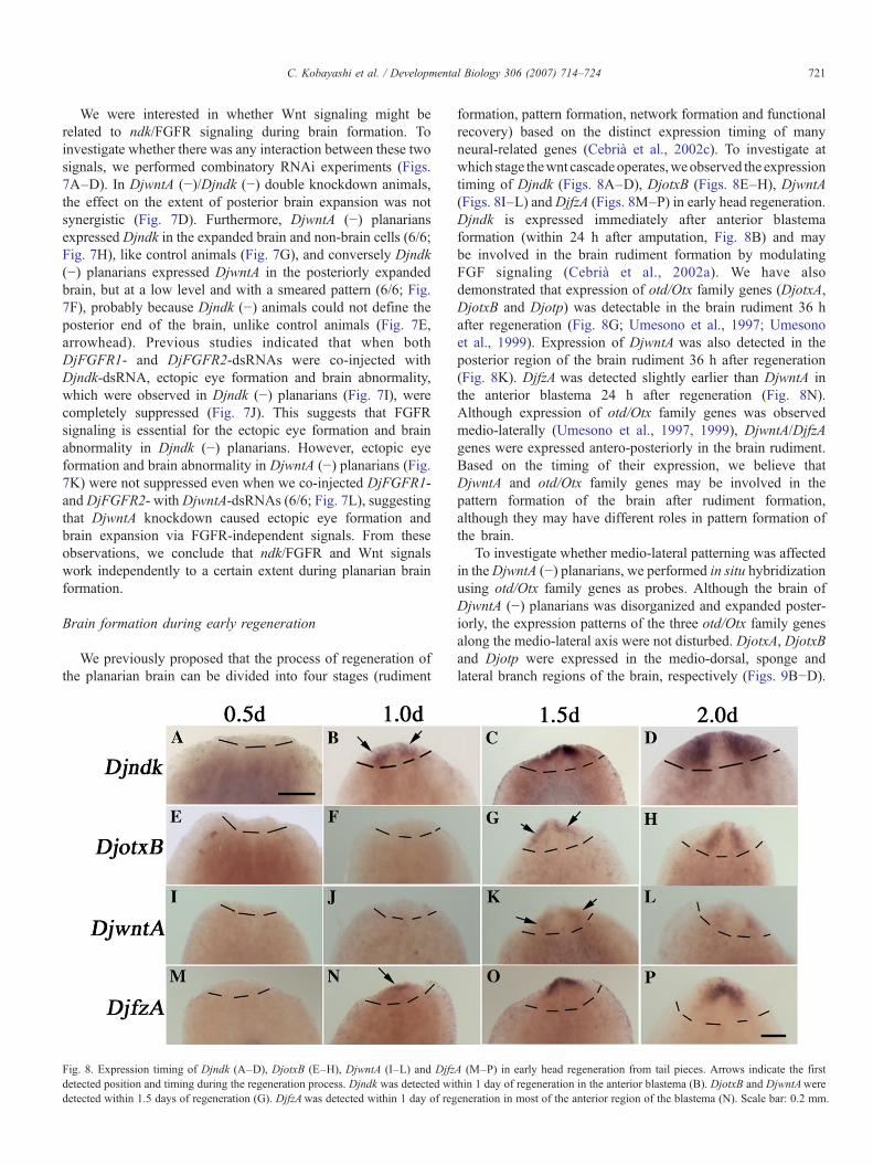
Fig. 8. Expression timing of Djndk (A–D), DjotxB (E–H), DjwntA (I–L) and Djfzdetected position and timing during the regeneration process. Djndk was detected widetected within 1.5 days of regeneration (G). DjfzAwas detected within 1 day of reg
formation, pattern formation, network formation and functionalrecovery) based on the distinct expression timing of manyneural-related genes (Cebrià et al., 2002c). To investigate atwhich stage thewnt cascadeoperates,weobserved the expressiontiming of Djndk (Figs. 8A–D), DjotxB (Figs. 8E–H), DjwntA(Figs. 8I–L) andDjfzA (Figs. 8M–P) in early head regeneration.Djndk is expressed immediately after anterior blastemaformation (within 24 h after amputation, Fig. 8B) and maybe involved in the brain rudiment formation by modulatingFGF signaling (Cebrià et al., 2002a). We have alsodemonstrated that expression of otd/Otx family genes (DjotxA,DjotxB and Djotp) was detectable in the brain rudiment 36 hafter regeneration (Fig. 8G; Umesono et al., 1997; Umesonoet al., 1999). Expression of DjwntA was also detected in theposterior region of the brain rudiment 36 h after regeneration(Fig. 8K). DjfzA was detected slightly earlier than DjwntA inthe anterior blastema 24 h after regeneration (Fig. 8N).Although expression of otd/Otx family genes was observedmedio-laterally (Umesono et al., 1997, 1999), DjwntA/DjfzAgenes were expressed antero-posteriorly in the brain rudiment.Based on the timing of their expression, we believe thatDjwntA and otd/Otx family genes may be involved in thepattern formation of the brain after rudiment formation,although they may have different roles in pattern formation ofthe brain.
To investigate whether medio-lateral patterning was affectedin theDjwntA (−) planarians, we performed in situ hybridizationusing otd/Otx family genes as probes. Although the brain ofDjwntA (−) planarians was disorganized and expanded poster-iorly, the expression patterns of the three otd/Otx family genesalong the medio-lateral axis were not disturbed. DjotxA, DjotxBand Djotp were expressed in the medio-dorsal, sponge andlateral branch regions of the brain, respectively (Figs. 9B−D).
A (M–P) in early head regeneration from tail pieces. Arrows indicate the firstthin 1 day of regeneration in the anterior blastema (B). DjotxB and DjwntAwereeneration in most of the anterior region of the blastema (N). Scale bar: 0.2 mm.

Fig. 9. DjwntA (−) animals do not show altered medio-lateral patterning. (A)Schematic drawing of expression patterns of otd/Otx family genes of planarians,which are related to medio-lateral patterning of the brain. (B−D) Whole-mountin situ hybridization for DjotxA (B), DjotxB (C) and Djotp (D). Trunk pieces.Scale bar: 0.2 mm.
722 C. Kobayashi et al. / Developmental Biology 306 (2007) 714–724
These results suggest that DjwntA does not affect medio-lateralpatterning in the brain.
Discussion
Wnt signaling in planarian
Before starting the analysis of the function of Wnt signalingin planarian regeneration, we expected that we would be able tomanipulate planarian body polarity by modulating Wntsignaling. While we have succeeded in identifying severalWnt signaling-related genes, all of them were expressed in thebrain. Wnt itself (DjwntA) was expressed in the posterior regionof the brain, while a Wnt receptor (DjfzA) showed acomplementary expression pattern to DjwntA and its putativetarget transcription factor (TCF/LEF homologue, 03133HH)was specifically expressed in the brain. RNAi planarians forDjwntA clearly showed defects of brain patterning along theA–P axis. From these observations we concluded that Wntsignaling is involved in brain patterning in planarians, as invertebrates.
Although all of the Wnt signaling-related genes wereexpressed in the brain, only DjwntA showed a clear phenotypefor RNAi. Why didn't the other genes show a phenotype whenknocked down by RNAi? Although DjfzA showed a comple-mentary expression pattern to DjwntA, we could not get anydirect evidence proving that DjfzA is involved in A–Ppatterning by interacting with DjwntA in the brain. One possiblereason is that other redundant genes may compensate for itsloss. Recently, when we increased the size of our EST database,we identified two additional genes which were classified into
frizzled family genes (we named them DjfzB, C), one additionalWnt homologue (DjwntB), two additional dishevelled homo-logous genes, and one additional GSK3 homologue (data notshown). To clarify the molecular pathways involved in Wntsignaling in the planarian brain, we are now trying tocharacterize the remaining genes (frizzled, disheveled andGSK3 homologues) and investigate their function by combina-tory RNAi experiments. It has been well documented that Wntsignaling has an important role in posteriorization of thevertebrate brain (Nordström et al., 2002). Our study suggeststhat invertebrates use a signaling system similar to that ofvertebrates to form a complex brain.
A Wnt homologue was identified from another planarian,Girardia tigrina, and named Gtwnt5 (Marsal et al., 2003).This gene was expressed in the peripheral region of the brainand the VNC, a pattern that is completely different from theexpression pattern of the DjwntA gene. DjwntB was expressedin the peripheral region of the brain from the anterior end topharynx and in the medial region of the brain and the VNC (datanot shown). The homology of Gtwnt5 and DjwntB to DjwntA is30% and 34%, respectively. DjwntA and DjwntB are homo-logues of the Wnt4 class of the human Wnt gene family (K.M.,personal communication), whileGtwnt5 a homolog of the Wnt5class. From these results, we conclude that the three genes aredifferent, suggesting that planarians have at least three types ofWnt family genes.
Relationship between Wnt and FGF signals for brainformation in planarians
In Drosophila, the wingless gene maintains the segmentboundaries (Peifer and Bejsovec, 1992, Siegfried et al., 1994).In the mouse, theWnt1 gene is expressed in the mid–hind brainboundary and mice deficient in the Wnt1 gene lack a midbrainregion as well as the cerebellum (Thomas and Cappecchi, 1990,McMahon et al., 1992, Joyner, 1996). These results suggest thatwingless/Wnt genes may have a conserved role for boundaryformation during evolution.
Interestingly, inDjwntA (−) planarians, we observed not onlydisturbance of A–P patterning of the brain but also posteriorexpansion of the brain. This brain expansion was confirmed bylateral regeneration experiments comparing the size of theregenerating brain after RNAi with that of the remaining half ofthe brain (Fig. 5). How does the planarian determine theposterior boundary of the brain? It is known that FGF is anotherimportant gene for brain boundary formation in mice. The FGF8gene is expressed posteriorly to the Wnt1 expression domain inthe mouse brain to form a positive circuit for the formation of themid–hind brain boundary. Unfortunately, no FGF gene has beenidentified in planarians. However, we have already reported thattwo planarian FGFR homologues (DjFGFR1 andDjFGFR2) arestrongly expressed in the posterior region adjoining the brainboundary (Ogawa et al., 2002). Also, as we mentioned before(Cebrià et al., 2002a), Djndk, which is an FGFR-like moleculespecifically expressed in the head region, modulates FGFsignaling to restrict brain tissues to the head region. Theseobservations strongly suggest that Wnt and FGF signaling may

Fig. 10. Schematic drawing of early head regeneration of planarians. After decapitation, the ndk/FGFR signal is activated within 24 h and a brain rudiment is formed atthe anterior blastema (pink). Genes involved in pattern formation are then activated around 36 h after decapitation. Three otd/Otx family genes are activated along themedio-lateral (M–L) axis. DjwntA and DjfzA are complementarily expressed along the antero-posterior (A–P) axis. Following several additional steps, a complexbrain is completely formed within 5 days.
723C. Kobayashi et al. / Developmental Biology 306 (2007) 714–724
interact with each other to form the brain boundary in planariansas in vertebrates.
We subsequently investigated the relationship between theDjwntA and ndk/FGFR signaling systems, as knockdownplanarians of these genes showed similar phenotypes. BothDjwntA (−) and Djndk (−) planarians showed ectopic eyeformation and brain expansion. However, we found severalphenotypic differences in the two knockdown planarians.Firstly, ectopic eye formation and brain expansion were notdetected in non-amputated DjwntA (−) planarians, whereasnon-amputated Djndk (−) planarians showed the same pheno-type as amputated animals. Secondly, ectopic eyes andexpanded brain were always formed only in the head regionin DjwntA (−) planarians, whereas in Djndk (−) planarians,ectopic eyes and brain were sometimes detected throughout theVNC. Finally, by combinatory RNAi experiments, we foundthat FGFRs are required for ectopic eye formation in the case ofDjndk (−) planarians, but not in DjwntA (−) planarians.Furthermore, in DjwntA/Djndk double knockdown animals,the extent of posterior brain expansion was not synergistic, andthe expression of each of these genes was detectable when theother was knocked down. From these observations, weconclude that the two signals may function independently ofeach other in brain formation and brain patterning.
Does Wnt play a role in the anterior patterning of the brainexclusively, does it play a role in the formation of a borderbetween the brain and VNC, or does it play a role in the A–Ppatterning in all nervous system tissue? To begin to distinguishamong these possibilities, we performed whole-mount in situhybridization using 517_HH and 821 HN as probes in DjwntA(−) in planarians (Fig. 6). The results showed that ectopic braintissues were never seen in the VNC, and the expansion wasspecific to the brain and not to surrounding head tissues. Thisindicates that DjwntA plays a role in the anterior patterning ofbrain, excluding the VNC.
When DjwntA expression was not completely eliminated,the posterior end of the brain could form (Figs. 4E, F). If wecould succeed in overexpressing DjwntA in part of the brain inDjndk (−) planarians, the phenotype of brain expansion mightbe ameliorated, although it is not possible at present to test thispossibility.
How does the planarian brain regenerate?
We have proposed that the regeneration process of theplanarian brain can be divided into four stages based on thedifference of the timing of expression of the neural-relatedgenes: rudiment formation, pattern formation, neural networkformation and functional recovery (Cebrià et al., 2002c; Inoue etal., 2004). As we reported before (Cebrià et al., 2002a),Djndk isthe gene whose expression is detected earliest just after anteriorblastema formation and may be involved in brain rudimentformation by modulating FGF signaling. Recently, we alsoreported that two novel secretory molecules were involved inthe final step of brain regeneration (Inoue et al., 2004). Whenwe made knockdown planarian of these molecules, they couldnot completely recover negative phototactic behavior, eventhough they regenerated a morphologically normal brain.
During regeneration, expression of DjwntAwas observed inthe posterior region of the brain rudiment 36 h afterregeneration. Expression of most of the Wnt signal-relatedgenes began to be detected after formation of the Djndk-positivebrain rudiment. Thus, it seems likely that Wnt signaling isinvolved in A–P patterning after specification of the brainrudiment in the blastema (in the second step of brainregeneration).
Expression of all of the otd/Otx family genes becamedetectable about 36 h after regeneration in the brain rudiment, asseen in the case of DjwntA. The otd/Otx family genes' discreteexpression pattern along the medio-lateral axis can be observed

724 C. Kobayashi et al. / Developmental Biology 306 (2007) 714–724
48 h after regeneration (Umesono et al., 1997, 1999), suggestingthat otd/Otx family genes are involved in medio-lateralpatterning of the brain rudiment. From these expression patternsand RNAi analysis, we propose the following pattern of brainregeneration (Fig. 10). First, the brain rudiment is formed in theanterior blastema using the ndk/FGFR signal system within24 h after amputation. Expression of DjwntA is then activated inthe posterior region of the brain rudiment to define the posteriorboundary and regulate A–P patterning of the brain. At the sametime, the otd/Otx family genes start to be expressed in the brainrudiment, and medio-lateral patterning of brain can beestablished. Finally, following several additional steps, theplanarian brain regenerates completely to form an well-organized and functional brain. These results suggest thatsimilar tool kits are used in the formation of a complex brainfrom invertebrates to vertebrates.
Acknowledgments
We thank all of the colleagues in our laboratory for helpfuldiscussions and especially thank Elizabeth Nakajima andMichael Royle for the critical reading of the manuscript. Thiswork was supported by Grants-in-Aid for Creative BasicResearch from the Ministry of Education, Culture, Sports,Science and Technology, Japan to K. A., Grants-in-Aid forScientific Research on Priority Areas to K. A., and a COE grantfrom MEXT Japan to C. K.
References
Agata, K., Watanabe, K., 1999. Molecular and cellular aspects of planarianregeneration. Semin. Cell Dev. Biol. 10, 77–83.
Agata, K., Soejima, Y., Kato, K., Kobayashi, C., Umesono, Y., Watanabe, K.,1998. Structure of the planarian central nervous system (CNS) revealed byneuronal cell markers. Zool. Sci. 15, 433–440.
Agata, K., Tanaka, T., Kobayashi, C., Saitoh, Y., 2003. Intercalary regenerationin planarians. Dev. Dyn. 226, 308–316.
Cadigan, K.M., Nusse, R., 1997. Wnt signaling: a common theme in animaldevelopment. Genes Dev. 11, 3286–3305.
Cebrià, F., Kobayashi, C., Umesono, Y., Nakazawa, M., Mineta, K., Ikeo, K.,Gojobori, T., Sánchez Alvarado, A., Agata, K., 2002a. FGFR-related genenou-darake restricts brain tissues to the head region of planarians. Nature419, 620–624.
Cebrià, F., Kudome, T.Y., Nakazawa, M., Mineta, K., Ikeo, K., Gojobori, T.,Agata, K., 2002b. The expression of neural-specific genes reveals thestructural and molecular complexity of the planarian central nervous system.Mech. Dev. 116, 199–204.
Cebrià, F., Nakazawa, M., Mineta, K., Ikeo, K., Gojobori, T., Agata, K., 2002c.Dissecting planarian central nervous system regeneration by the expressionof neural-specific genes. Dev. Growth Differ. 44, 135–146.
Giudice, G., 2001. Conserved cellular and molecular mechanisms in develop-ment. Cell Biol. Int. 25, 1081–1090.
Holstein, T.W., Hobmayer, E., Technau, U., 2003. Cnidarians: an evolutionarilyconserved model system for regeneration? Dev. Dyn. 226, 257–267.
Inoue, T., Kumamoto, H., Okamoto, K., Umesono, Y., Sakai, M., SánchezAlvarado, A., Agata, K., 2004. Morphological and functional recovery of theplanarian photosensing system during head regeneration. Zool. Sci. 21,275–283.
Inoue, T., Hayashi, T., Takechi, K., Agata, K., 2007. Clathrin-mediatedendocytic signals are required for the regeneration of, as well as homeostasisin, the planarian CNS. Development 134, 1679–1689.
Joyner, A.L., 1996. Engrailed,Wnt and Pax genes regulate midbrain–hindbraindevelopment. Trends Genet. 12, 15–20.
Kobayashi, C., Kobayashi, S., Orii, H., Watanabe, K., Agata, K., 1998.Identification of two distinct muscles in the planarian Dugesia japonica bytheir expression of myosin heavy chain genes. Zool. Sci. 15, 855–863.
Marsal, M., Pineda, D., Saló, E., 2003. Gtwnt5 a member of the wnt familyexpressed in a subpopulation of the nervous system of the planarian Gir-ardia tigrina. Gene Expr. Patterns 3, 489–495.
McMahon, A.P., Joyner, A.L., Bradley, A., 1992. The midbrain–hindbrainphenotype of Wnt-1−/Wnt-1− mice results from stepwise deletion of en-grailed expressing cells by 9.5 days postcoitum. Cell 68, 581–595.
Mineta, K., Nakazawa, M., Cebrià, F., Ikeo, K., Agata, K., Gojobori, T., 2003.Origin and evolutionary process of the CNS elucidated by comparativegenomics analysis of planarian ESTs. Proc. Natl. Acad. Sci. U. S. A. 100,7666–7671.
Nakazawa, M., Cebrià, F., Mineta, K., Ikeo, K., Agata, K., Gojobori, T., 2003.Search for the evolutionary origin of a brain: planarian brain characterizedby microarray. Mol. Biol. Evol. 20, 784–791.
Newmark, P., Sánchez Alvarado, A., 2002. Not your father's planarian: aclassic model enters the era of functional genomics. Nat. Rev., Genet. 3,210–219.
Nordström, U., Jessell, T.M., Edlund, T., 2002. Progressive induction of caudalneural character by graded Wnt signaling. Nat. Neurosci. 5, 525–532.
Ogawa, K., Kobayashi, C., Hayashi, T., Orii, H., Watanabe, K., Agata, K., 2002.Planarian fibroblast growth factor receptor homologs expressed in stem cellsand cephalic ganglions. Dev. Growth Differ. 44, 191–204.
Okamoto, K., Takeuchi, K., Agata, K., 2005. Neural projections in planarianbrain revealed by fluorescent dye tracing. Zool. Sci. 22, 535–546.
Peifer, M., Bejsovec, A., 1992. Knowing your neighbors: cell interactionsdetermine intrasegmental patterning in the Drosophila. Trends Genet. 8,243–249.
Sakai, F., Agata, K., Orii, H., Watanabe, K., 2000. Organization andregeneration ability of spontaneous supernumerary eyes in planarians –eye regeneration field and pathway selection by optic nerves. Zool. Sci. 17,375–381.
Saló, E., Baguñà, J., 2002. Regeneration in planarians and other worms: newfindings, new tools and new prospectives. J. Exp. Zool. 292, 528–539.
Sánchez Alvarado, A., Newmark, P., 1999. Double-stranded RNA specificallydisrupts gene expression during planarian regeneration. Proc. Natl. Acad.Sci. U. S. A. 96, 5049–5054.
Siegfried, E., Wilder, E.L., Perrimon, N., 1994. Components of winglesssignaling in Drosophila. Nature 367, 76–80.
Thomas, K.R., Cappecchi, M.R., 1990. Targeted disruption of the murine int-1proto-oncogene resulting in severe abnormalities in midbrain and cerebellardevelopment. Nature 346, 847–850.
Umesono, Y., Watanabe, K., Agata, K., 1997. A planarian orthopedia homologis specifically expressed in the branch region of both the mature andregenerating brain. Dev. Growth Differ. 39, 723–727.
Umesono, Y., Watanabe, K., Agata, K., 1999. Distinct structure domains in theplanarian brain defined by the expression of evolutionarily conservedhomeobox genes. Dev. Genes Evol. 209, 18–30.
Wodarz, A., Nusse, R., 1998. Mechanisms of WNT signaling in development.Annu. Rev. Cell Dev. Biol. 14, 59–88.