Embed Size (px)
Citation preview

W3: Pregnancy-Related Musculoskeletal Conditions:
The Pelvic Floor and Linea Alba Connection Workshop Chair: Sinéad Dufour, Canada
28 August 2018 09:00 - 10:30
Start End Topic Speakers
09:00 09:15 Morphology and biomechanics of the lina alba and pelvic floor
– connecting the system
Cynthia Chiarello
09:15 09:30 Overview of the scientific literature base Kari Bø
09:30 09:45 Exploring the ‘’gaps’’: Knowledge users vs scientific literature Stephanie Bernard
09:45 10:00 Primary care approach to conservative care provision for
pregnancy-related DRA: Interprofessional considerations
Sinéad Dufour
10:00 10:30 Discussion: Future research directions & practice implications Sinéad Dufour
Kari Bø
Stephanie Bernard
Cynthia Chiarello
Aims of Workshop
Pregnancy-related musculoskeletal tissue injury is common and ranges from strain on the pelvic ligaments to injury to the pelvic
floor and changes associated with fascial system, including widening of the linea alba, called diastasis rectus abdominis (DRA).
Exploring the topic of pregnancy-related musculoskeletal conditions, specifically from the perspective of understanding the
potential relationship and relevance of pelvic floor function to the linea alba, is important. In most cases, conservative
management strategies are established as first line care. This workshop will overview pregnancy-related musculoskeletal tissue
changes and injuries with a focus on the pelvic floor muscle and the linea alba according to our past and evolving understanding
of DRA.
Learning Objectives
1. Understand pregnancy-related musculoskeletal tissue changes and associated conditions such as diastasis rectus
abdominis and differentiate those that require intervention (conservative management) and those that do not.
2. Identify scientific update on evidence pertaining to various aspects of pregnancy-related diastasis rectus abdominis.
3. Determine the current evidence-informed and integrative conservative care principles for pregnancy-related DRA from a
primary health care perspective and with an emphasis on the roles of physiotherapists and primary care providers.
Learning Outcomes
After this workshop, participants will be able to understand the need to mount a cohesive evidence-based approach in the
conservative management for pregnancy-related musculoskeletal tissue concerns that involve the pelvic floor and linea alba, such
as diastasis rectus abdominis. Furthermore, participants should have a greater understanding of how to determine when
pregnancy-related musculoskeletal changes to the linea alba and pelvic floor require conservative intervention, and when they do
not impact function. The audience will appreciate the inter-relation of tissue function through the perinatal stage of which the
pelvic floor is central. As such, participants will have enhanced clinical reasoning that will lend to both prevention and
management of pregnancy-related musculoskeletal conditions like diastasis rectus abdominis.
Target Audience
Primary care practitioners, specifically: physiotherapists, midwives, obstetricians, nurses and other allied healthcare professionals
interested in understanding pregnancy-related musculoskeletal tissue changes and associated impairments.
Advanced/Basic
Basic
Conditions for Learning
This is an interactive workshop that does not have a restriction on the number of delegates.
Suggested Learning before Workshop Attendance
This is an entry level workshop and no particular preparation is required.

Suggested Reading
1. Sperstad, Tennfjord, Hilde, Ellstrom-Engh,Bo. Diastasis recti abdominis during pregnancy and 12 months after birth:
prevalence, risk factors and report of lumbopelvic pain. British Journal of Sports Medicine. 2016;0:1–6. doi:10.1136/bjsports-
2016-096065
2. Lee, Hodges. Behaviour of the linea alba in a curl up task in diastasis rectus abdominis: an observational study. Journal of
Orthopaedic & Sports Physical Therapy. 2016; 46(6):580–589 DOI: 10.2519/jospt.2016.6536
3. Spitznagle, Leong. Dillen. Prevalence of diastasis recti abdominis in a urogynecological patient population. International
Urogynecology Journal.2007; 18(3):321-328.
4. Chiarello & McAully. Mind the Gap: A comprehensive approach for the evaluation of the intervention of diastasis recti
abdominis. Combined Sections Meeting American Physical Therapy Association, Las Vegas Nevada, February, 2014.
5. Keeler J, Albrecht M, Eberhardt L et al. Diastasis recti abdominis: a survey of women’s health specialists for current physical
therapy clinical practise for postpartum women. J Women’s Health Phys Ther. 2012; 36:131-142.
6. Dufour, Bernard, and Murray-Davis. Establishing best practice principles for the conservative management of pregnancy-
related diastasis rectus abdominis: results from Phase 1 of a consensus study. International Continence Society Conference,
Florence Italy, September, 2107.
Speakers Presentation Summaries
Morphology and biomechanics of the lina alba and pelvic floor – connecting the system
Cynthia Chiarello
Cynthia will begin the workshop by exploring the anatomical and functional interdependence of the muscular and connective
tissue components of the trunk. The “pressurized can” concept will be described as a theoretical model for the potential
association between diastasis rectus abdominis (DRA) and pelvic floor dysfunction. Several concepts will be introduced and
related to explain the integrated function of the anterior abdominal wall and pelvic floor. The morphology and biomechanics of
the linea alba will be presented with attention to muscular and fascial connections. Synergistic and feed-forward trunk muscular
mechanisms will be presented along with load transfer and support across the pelvis. These foundational concepts will provide a
rational for intervention strategies.
email: [email protected]
Country: United States
Profession: Physical Therapist
Experience & Qualifications:
Dr. Chiarello is an Assistant Professor at Columbia University, Department of Rehabilitative and Regenerative Medicine, Doctoral
Program in Physical Therapy where she teaches kinesiology, biomechanics and orthopedics. She is the Editor-in-Chief of the
Journal of Women’s Health Physical Therapy, the peer reviewed publication of the Section on Women’s Health of the American
Physical Therapy Association. Dr. Chiarello received a BS in Biology and Psychology from SUNY Fredonia, a MS in Physical
Therapy from Duke University and a PhD in Pathokinesiology from New York University. Her basic science research has examined
the linea alba in cadavers as a foundation for the characteristics of diastasis rectus abdominis. Her clinical research examines the
relationship between diastasis rectus abdominis, low back pain, and abdominal muscle function in pregnant and post-partum
women. Her current research includes studies investigating exercise for pelvic girdle pain in pregnancy, and ultrasound imaging
of inter-recti distance and abdominal muscle contraction in functional positions.
Overview of the scientific literature
Kari Bø
Kari will give an overview on the scientific literature on prevalence and complications of diastasis recti abdominis and the effect
of treatment of abdominal and pelvic floor muscle exercises for diastasis. A special emphasis will be on the connection or lack
there of non between the pelvic floor muscles and the abdominal muscles from a treatment perspective. To date there are few
published studies in this area and newer experimental research raises questions regarding the evidence for the commonly used
clinical physiotherapy protocols that emphasize training of the transverse abdominal and pelvic floor muscles to address
pregnancy-related DRA.
e-mail: [email protected]
Country: Norway
Profession: Physiotherapist and Exercise Scientist
Experience & Qualifications
Professor Kari Bø obtained her PhD on pelvic floor muscle training in 1990 and was appointed professor of Exercise Science and
Physiotherapy in 1997. Since then, she has been elected rector (head) of the Norwegian School of Sport Sciences, (specialized
university) in Oslo 2013-2017, and was the first vice president of the International Organization of Physical Therapists in
Women’s Health, WCPT 1999-2007. Further, she has been the vice president of the Norwegian Council for Physical Activity for 8
years, giving direct advice to the Norwegian Minister of Health. Kari has published > 260 scientific papers on pelvic floor
dysfunction, treatment of incontinence and low back- and pelvic girdle pain, exercise during pregnancy and after childbirth,
diastasis recti abdominis, measurement methodology, fitness and women’s health and has given > 260 invited international
keynote presentations. She has been awarded with the most prestige's award from the World Confederation of Physiotherapy
and the ICS Lifelong Achievement award for her research and education on the pelvic floor and incontinence.
Exploring the “gaps”: knowledge users vs. scientific literature
Stéphanie Bernard
Stephanie Bernard will introduce participants to who are knowledge users, how meaningful it can be to have knowledge users
participating in the various steps of research processes, and at the different research methods that have been used in the DRA
literature to inform readers of practice-based evidence they have provided. Additionally, she will explore the findings from
practice-based inquiry that also integrate principles from basic science research and contrasts them with non-practice-based
evidence from the literature, highlighting the areas of coherence and dissemblance between the two. This will lead to a more
complete understanding of what we know regarding the conservative management of pregnancy-related DRA, as well as how
can further research help fill the identified gaps in evidence.
Email: [email protected]
Country: Canada
Profession: Physiotherapist
Experience & Qualifications:
Ms. Stephanie Bernard is a Physiotherapist with expertise in pelvic floor therapy and a Doctoral candidate at Université Laval in
Québec, Canada. She has been a clinician for over 12 years, where she treats various pelvic floor dysfunctions, with a special
interest for patients with pregnancy-related musculoskeletal dysfunctions and pelvic floor disorders after cancer. She lectures at
both Université Laval and Université de Montréal since 2014. She is the Past Editor-in-Chief of the Women’s Health Division of
the Canadian Physiotherapy Association, and a member of the ICS Working Group on Terminology of pelvic floor function and
dysfunction.
Primary care approach to conservative care provision for pregnancy-related DRA: Interprofessional considerations
Sinéad Dufour
Sinéad will close the workshop with a translation of the collective evidence discussed (basic science, clinical science and
practice-based) applied to a clinical vignette. The vignette has been developed by the collaborators to operationalize in a
pragmatic way how the state of the evidence related pregnancy-related dysfunction in the linea alba (aka DRA) can be applied.
The vignette will be presented to span two different points in time through the perianal care period and will be explored from
the perspective of different primary care providers. Aspects or both assessment and management will be discussed. As such,
relation patient education, lifestyle counselling and exercise prescription considerations will all considered. Pregnancy-related
musculoskeletal conditions are common and can be managed more effectively with conservative approaches by all relevant
primary care providers. Many barriers to optimal care exist and lack of clarity of the state of the evidence as well as how to
clinically apply the current evidence represent barriers we hope to ameliorate through this workshop.
Email: [email protected]
Country: Canada
Profession: Physiotherapist
Experience & Qualifications:
Dr. Sinéad Dufour is Assistant Clinical Professor in the Faculty of Health Science at McMaster University. She teaches and
conducts research in both the Schools of Medicine , which houses the Midwifery Education Program and School of Rehabilitation
Science. She completed her MScPT at McMaster University (2003), her PhD in Health and Rehabilitation Science at Western
(2011), and returned to McMaster to complete a post-doctoral fellowship (2013). Her current research interests include:
conservative approaches to manage pelvic floor dysfunction, pregnancy-related pelvic-girdle pain, and interprofessional
collaborative practice models of service provision to enhance pelvic health. Sinéad stays currently clinically through her work as
the Director of Pelvic Health Services at The World of my Baby (the WOMB) in Ontario Canada and is member of the
urogynecology committee of the Society of Obstetricians and Gynecologists of Canada.

1
Morphology and
Biomechanics of the Linea
Alba and Pelvic FloorCynthia M. Chiarello, PT, PhD
Editor-in-Chief, Journal of Women’s Health Physical Therapy
Assistant Professor of Rehabilitation and Regenerative Medicine at
CUMC, Program in Physical Therapy, Columbia University, New York,
USA
Objectives:
After completing this session, you will be able to:
• Illustrate the integrated function of the musculofascial
system in providing support and stability to the trunk
and pelvis.
• Explore theoretical models which present the
interrelated mechanical function of the trunk.
• Summarize mechanical properties of some trunk
structures.
Trunk Function
• Contains organs
• Respiration
• The musculoskeletal trunk functions as a mechanical unit
• Supports weight
• Transmits forces
• Produces and controls intra-abdominal pressure
• Maintains stability
• Form and force closure
Trunk
Top Diaphragm
Anterior
Abdominal Muscles
Deep → TA, IO
Superficial → EO, RA
Connective Tissue
Linea Alba
Aponeuroses
Posterior
MusclesLocal → Multifidus
Global → ES
Connective Tissue Thoracolumbar Fascia
Bottom Pelvic Floor
Levator Ani
Fascia & Ligaments
Anatomical Trunk Components
Deep SuperficialModel for Integrated Function of the Trunk“Abdominal Canister”
Top:
Diaphragm
Front:
Abdominal
Muscles & CT
Back:
Extensor
Muscles & CT
Bottom:
Pelvic Floor
Muscles & CT

2
From: Saunders K. Recent Advances in Understanding
Pelvic-Floor Tissue of Women With and Without Pelvic
Organ Prolapse: Considerations for Physical Therapists
Phys Ther. 2017;97(4):455-463. doi:10.1093/ptj/pzx019
Phys Ther | © 2017 American Physical Therapy Association
Pelvic floor support
• Pelvic organs (boat) supported by the levator ani muscle (water) and stabilized by the ligaments (cables).
• Levator ani weakness causes increased reliance on supportive connective tissue
Model for Integrated Function of the Trunk
“Abdominal Canister” Lee, 2014
• Ideal trunk function requires a balance between movement and stiffness
• Impaired function of one part of the canister can diminish load transfer negatively impacting support mechanisms
• Synergistic muscle activation of pelvic floor musculature, the deep abdominal muscles and diaphragm
• intra-abdominal pressure
• Lumbo-pelvic stability
• Continence
• Muscle forces are transferred through fascial attachments.
Model for Integrated Function of the Trunk
• General principle: a disorder in one component of the canister (back
pain, incontinence, respiratory problems) is associated with the
development of a condition in another component.
• Longitudinal cohort study identified that the presence and development of a
disorder in one system (back pain {BP}, incontinence {UI}, respiratory
problems, or GI symptoms) is associated with the development of conditions
in other systems (Smith, Russell, Hodges, 2014)
• pre-existing and newly developed UI was a risk factor for the future development
of BP
• Women with breathing problems were more likely develop BP & women with BP
more likely to develop breathing problems
Is there a relationship between Diastasis Rectus
Abdominis and Incontinence or Pelvic floor dysfunction?
Diastasis Rectus Abdominis (DRA)
The “abnormal” midline separation of the right and left rectus abdominis muscles along the linea alba.
– Appears as a visible increase in the width of the linea alba or Inter-Recti Distance (IRD).
– Connective tissue alterations of the linea alba (Szczesny, 2006)
– Damage of the fixation of the rectus muscles (Axer, 2001)
DRA Measurement: TechniquePalpation
• Palpation & Calipers
• Ultrasound
• Clinical Relevance
Chiarello, McAuley, 2013
The Abdominal Wall
• Function
• Abdominal Muscles
• Abdominal wall
connective tissue
• Components• Linea Alba - tendinous fibers
from abdominal muscles
• Anterior Rectus Sheath
• Posterior Rectus Sheath
Neumann. Kinesiology
of the Musculoskeletal
System: Foundations
for Rehabilitation, 2nd
Edi. Mosby, 2010.

3
Morphology of Linea Alba and Rectus
Sheaths (Axer et al., 2001)
1. Collagen fibers - 3-D highly structured meshwork of collagen
1. Oblique Fiber Layer
2. Transverse Fiber Layer
3. Irregular Fiber Layer
❑ Distinct Craniocaudal regions❑Supraumbilical
❑Umbilical
❑Transition zone
❑ Infra-arcuate
❑ LA Gender differences❑Thickness ♂ > ♀
❑ Infraumbilical• Width ♀ > ♂
• ♀ more transverse (relative to oblique) bundles
Mechanical Function of Linea Alba
• Withstand abdominal pressure (Konerding et al., 2011)
• Stabilizing the abdominal wall (Gräßel et al, 2005).
• Sustain and transmit muscle contraction forces (Brown & McGill, 2008)
• Stiffest structure, most work (Hernández-Gascón et al., 2013)
• Most important component for stability of
the abdominal wall (Hernández-Gascón et al, 2013)
• Sustains the highest stresses under
physiological load (Hernández-Gascón et al, 2013)
Mechanical Behavior of Linea Alba
• Anisotropic & Non-linear
• Transverse direction is stiffer
(smaller compliance) than
longitudinal (Gräßel et al, 2005; Cooney et al 2016).
• LA is 3x stiffer for small strains 4x stiffer
for large strains (Tran et al, 2016)
• Collagen & elastin adapted to the
type of loading. Microstructure
strongly affected the mechanical
response of the linea alba (Levillain et
al 2016)
• Dorsal layer mechanical resistance to
transverse load
• Ventral layer resists transverse and
longitudinal
Pelvic Floor
• Pelvic floor muscles
• Tonic contraction, transverse load bearing, antigravity
support
• Passive support through connections to endopelvic
fascia
Pregnancy: Connective Tissue Changes
• Peripheral joint laxity
increases during pregnancy
• Hormonally mediated
connective tissue changes
• Pregnancy can result in lasting
changes in knee joint laxity
Connective Tissue Mechanical Properties
• Both the pelvic floor and the anterior abdominal wall are
composed of both muscle and connective tissue.
• CT provides support for the organs
• Muscular components of both the pelvic floor and the
abdomen attach to the connective tissue
• A decrease in the stiffness or strength of this connective
tissue would predispose an individual to dysfunction as
abnormal displacements under normal physiological
pressures could occur impairing muscular contraction
efficiency.
Copy and paste logo art as needed

4
Summary
• Mechanical support for the anterior abdominal wall and the pelvic floor both depend on the precise interplay between muscular contraction and adequate tension of the ligament and fascial connective tissue.
• Weakened muscles leads to impaired function
• Lax connective tissue leads to impaired function
• The “Canister” theoretical model presents the potential interdependence between pelvic floor and anterior abdominal function.
• In pregnancy, hormonally mediated changes in connective tissue may lead to decreased support
ReferencesAxer H, Keyserlingk DG, Prescher A. Collagen fibers in linea alba and rectus sheaths. I. General scheme and morphological
aspects. J Surg Res. 2001a; 96:127-134. http://dx.doi.org/10.1006/jsre.2000.6070
Bhattarai A, Staat M. Modelling of soft connective tissues to investigate female pelvic floor dysfunctions. Comput Math
Methods Med. 2018;2018:9518076. doi: 10.1155/2018/9518076.
Chiarello CM, McAuley JA. Concurrent validity of calipers and ultrasound imaging to measure interrecti distance. J Orthop
Sports Phys Ther. 2013;43:495-503. doi.org/10.2519/ jospt.2013.4449
Förstemann T, Trzewik J, Holste J, Batke B, Konerding MA, Wolloscheck T, Hartung C. Forces and deformations of the
abdominal wall--a mechanical and geometrical approach to the linea alba. J Biomech. 2011;44(4):600-6. doi:
10.1016/j.jbiomech.2010.11.021.
Gräßel D, PrescherA, Fitzek S, Keyserlingk DVG, Axer, H. Anisotropy of human linea alba: a biomechanical study. J Surg
Res. 2005;124,118–125. doi.org/10.1016/j.jss.2004.10.010.
Hernández-Gascón B1, Mena A, Peña E, Pascual G, Bellón JM, Calvo B. Understanding the passive mechanical behavior of
the human abdominal wall. Ann Biomed Eng. 2013;41(2):433-44. doi: 10.1007/s10439-012-0672-7.
Hodges PW; Sapsford R; Pengel LH. Postural and respiratory functions of the pelvic floor muscles. Neurourol Urodyn. 2007;
26:362–371.
Konerding MA, Bohn M, Wolloscheck T, Batke B, Holste JL, Wohlert S, Trzewik J, Förstemann T, Hartung C. Maximum forces
acting on the abdominal wall: experimental validation of a theoretical modeling in a human cadaver study. Med Eng Phys.
2011;33(6):789-92. doi: 10.1016/j.medengphy.2011.01.010.
Leblanc DR, Schneider M, Angele P, Vollmer G, Docheva D. The effect of estrogen on tendon and ligament metabolism and
function. J Steroid Biochem Mol Biol. 2017 Sep;172:106-116. doi: 10.1016/j.jsbmb.2017.06.008.
References
Lee DG. New perspectives from the Integrated Systems Model for treating women with pelvic girdle pain, urinary incontinence,
pelvic organ prolapse and/or diastasis rectus abdominis. J Assoc Chartered Physiotherapists Women’s Health, 2014;
(114): 10-10.
Levillain A, Orhanta M, Turquierb F, Hoc T. Contribution of collagen and elastin fibers to the mechanical behavior of an
abdominal connective tissue, J Mech Behav Biomed Mater. 2016;61:308-317. doi: 10.1016/j.jmbbm.2016.04.006.
Peng Y, Miller BD, Boone TB, Zhang Y. Modern Theories of Pelvic Floor Support : A Topical Review of Modern Studies on
Structural and Functional Pelvic Floor Support from Medical Imaging, Computational Modeling, and Electromyographic
Perspectives. Curr Urol Rep. 2018;19(1):9. doi: 10.1007/s11934-018-0752-9.
Saunders K. Recent Advances in Understanding Pelvic-Floor Tissue of Women With and Without Pelvic Organ Prolapse:
Considerations for Physical Therapists, Phys Ther, 2017;97,(4):455–463. https://doi.org/10.1093/ptj/pzx019
Silva MET, Brandão S, Parente MPL, Mascarenhas T, Natal Jorge RM. Biomechanical properties of the pelvic floor muscles of
continent and incontinent women using an inverse finite element analysis. Comput Methods Biomech Biomed Engin.
2017 Jun;20(8):842-852. doi: 10.1080/10255842.2017.
Smith MD; Russell A; Hodges PW. The relationship between incontinence, breathing disorders, gastrointestinal symptoms,
and back pain in women: a longitudinal cohort study. Clin J Pain 2014; 30:162–167. doi:10.1097/AJP.0b013e31828b10fe.
Smith MD; Russell A; Hodges PW. Is there a relationship between parity, pregnancy, back pain and incontinence? Int
Urogynecol J Pelvic Floor Dysfunct. 2008;19(2):205-11. doi: 10.1007/s00192-007-0421-x
Tran D, Mitton D, Voirin D, Turquier F, Beillas P. Contribution of the skin, rectus abdominis and their sheaths to the structural
response of the abdominal wall ex vivo. J Biomech. 2014;47(12):3056-63. doi: 10.1016/j.jbiomech.2014.06.031.
Tran D, Podwojewski F, Beillas P, Ottenio M, Voirin D, Turquier F, Mitton D. Abdominal wall muscle elasticity and abdomen
local stiffness on healthy volunteers during various physiological activities. J Mech Behav Biomed Mater. 2016;60:451-
459. doi: 10.1016/j.jmbbm.2016.03.001.

12.09.2018
1
The pelvic floor and linea alba connection: Overview of the scientific literature ICS 2018
Kari Bø
Professor, Ph.D
PT, Exercise scientist
Norwegian School of Sport Sciences
Dept of Sports Medicine
Akershus University Hospital
Dept of Obstetrics and Gynecology
Affiliations to disclose†:
Funding for speaker to attend:
Self-funded
Institution (non-industry) funded
Sponsored by:
Kari Bø
Kari Bo
x
† All financial ties (over the last year) that you may have with any business organisation with respect to the subjects mentioned during your presentation
Measurement methods van de Water & Benjamin-16
• Palpation finger width: Kw
0.7/0.5 Mota et al-13
• Calipers: ICC: 0.9 (intra) Boxer&Jones-97
• Ultrasound: ICC 0.9/0.7-0.9 Mota et a-12
• NO consensus on:
• where to measure alongthe linea alba
• cut off point for diastasis
• What is NORMAL? Mota et al-17
• Usually ≥ "two finger widths"Forskning.no
Diastasis and the PFM? Prevalence of diastasis recti in a urogynecological patient populationSpiznagle et al -07
• 541 patients seeking help for PFD (myofacial pelvic pain,UI,FI,POP), mean age 52.5 years (SD 16.6)
• Evaluated diastasis with finger width
•Results• Prevalence 52% in middle-aged women
• 35% of nulliparous
• DRA women were older, reported higher parity, had weaker PFM, Caucasian/African, menopausal, using hormonal replacement therapy, abdominal surgery
PFM function & DRA in 300 first time pregnant women Bø et al-16
•At gestational week 21 women with diastasis had significantly better PFM function:
• VRP: mean diff: 3.06 cm H2O (95% CI: 0.70;5.42)
• MVC: mean diff: 5.09 cm H2O (95% CI: 0.76;9.42)
• Endu.:mean diff: 47.08 cmH2Osec (95% CI: 15.18; 78.99)
•However: No statistically sign differences in VRP, PFM strength or endurance between women with and without diastasis at 6 weeks, 6 months or 12 months postpartum
Is there any connection between the TrA and the pelvic floor/pelvic floor muscles?
NO muscular or facial connection between the two muscle groups
Part of same canister – what does it mean?
Weak connective tissue my be the common link between prevalence of PFD (urinary incontinence and
pelvic organ prolapse) and diastesis recti abdominis
Cause effect relationship?
12.09.2018
2
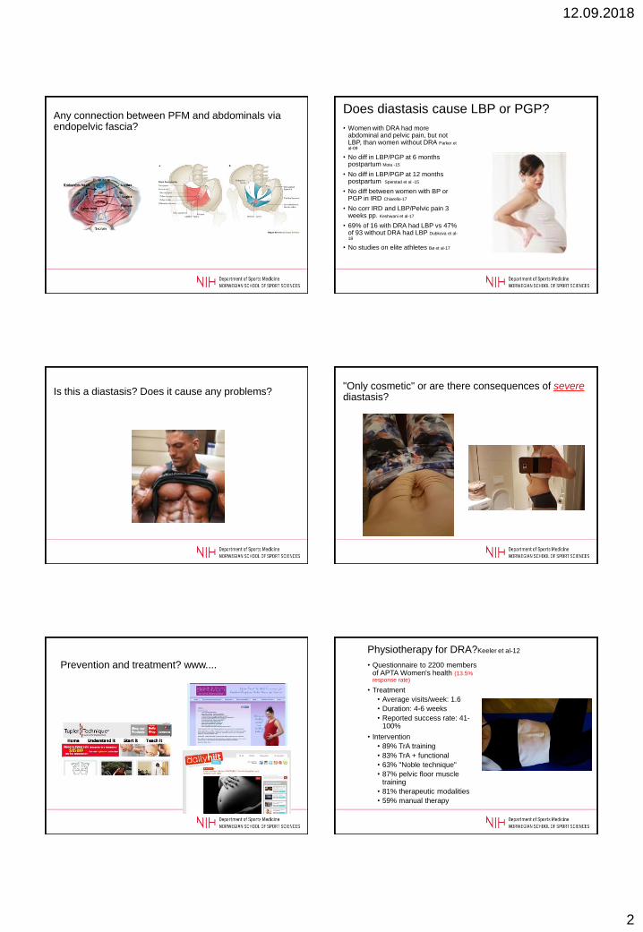
Any connection between PFM and abdominals via endopelvic fascia?
Does diastasis cause LBP or PGP?
• Women with DRA had more abdominal and pelvic pain, but not LBP, than women without DRA Parker et al-09
• No diff in LBP/PGP at 6 months postpartum Mota -15
• No diff in LBP/PGP at 12 months postpartum Sperstad et al -15
• No diff between women with BP or PGP in IRD Chiarello-17
• No corr IRD and LBP/Pelvic pain 3 weeks pp. Keshwani et al-17
• 69% of 16 with DRA had LBP vs 47% of 93 without DRA had LBP Dubkova et al-18
• No studies on elite athletes Bø et al-17
Is this a diastasis? Does it cause any problems?"Only cosmetic" or are there consequences of severe diastasis?
Prevention and treatment? www....
Physiotherapy for DRA?Keeler et al-12
• Questionnaire to 2200 members of APTA Women's health (13.5% response rate)
• Treatment
• Average visits/week: 1.6
• Duration: 4-6 weeks
• Reported success rate: 41-100%
• Intervention
• 89% TrA training
• 83% TrA + functional
• 63% "Noble technique"
• 87% pelvic floor muscle training
• 81% therapeutic modalities
• 59% manual therapy

12.09.2018
3
"Manual therapy":59% Keeler et al 2012
• Myofascial release: 46%
• Triggerpoint release: 36%
• Muscle energy technique:33%
• Visceral manipulation: 21%
• Other: "Joint mobilization":• Sacrum
• Innominate
• Lumbar spine
• Coccyx
• Pelvic symphysis
• Theory/mechanism???
Prevention and treatment of DRA during pregnancy?
• No RCTs in the general pregnant population or in athletes
• One retrospective study Chiarello et al-
05
• Can stretched abdominals be trained?
• At GW 36 length of abdominal muscles ↑ with mean 115%. Change in angle of insertion, reducing ability to generate torque Gilleard & Brown -96
• Which exercises?
Drawing in vs sit up/abdominal crunch?
• "Drawing in" widens the IRD Mota et al-12, Sancho et al -15,
Mota et al 15, Lee & Hodges – 17, Theodorsen et al-17
• PFM contraction widens the IRD Theodorsen et al-17, Lee & Hodges
-17
• Sit up/curl up narrows the IRD Mota et al-12, Sancho et al-15,
Pascoal et al-14, Chiarello et al-16, Lee & Hodges-17
RCT DRA Mesquita et al-99
• Published in Portuguise in BJPT
• 50 women after vaginal delivery, age: 18-40 years old
• Intervention
• Two sessions 6 (10 repetitions) and 18 hours (20 repetitions) after birth
• Basal respiration, pelvic tilt with isometric contraction of TrA, exercises for abdominal obliques, PFM contractions
• Results (caliper)
• Intervention: from 3.45 (± 0.43) cm to 2.64 (± 0.45) cm
• Control: from 3.16 (± 0.26) cm to 2.99 (± 0.28) cm
• P < 0.05 between groups at 18 hours
RCT DRA Walton et al-16
• 9 women with either vaginal birth or CS
• Randomized to 3 visits/week for 6 weeks (3x10 reps + progression during period)
• Plank OR
• Modified sit up
• In addition; Both groups had pelvic tilt, PFMT, obliques, external support
• Results (ultrasound and caliper)
• Sign reduction in both groups (only at navel)
• No diff between groups
RCT DRA Emanuelson et al-16
•89 participants (2 men) 18-40 years old
•Randomized to• Surgery with mesh
• Surgery with Quill
• Exercise: 3 times/week for 3 months with physical therapist: rectus abdominis, obliques, TrA
•Results (ruler, SF-36,pain,abdominal strength (VAS,
Biodex system)
• Surgery better than exercise

12.09.2018
4
RCT DRA Kamel & Yousif -17
• 60 women (25-35 years) at 2 months pp after normal vaginal delivery and DRA >2.5 cm
• Randomized to physical therapy 3 times/wk for 8 wk• A. Abdominal exercise + Neuromuscular el stim (frequency 80
pulses/min, pulse width 0.1–0.5 ms, on:off ratio of 5s:10s, 30 minutes)
• B. Abdominal exercise (20 reps (+ increased by 4 reps /wk) of sit-ups, reverse sit-ups, reverse trunk twists, and U-seat) + 5 times respiration + TrA (increased per/wk)
• Results (ultrasound)• IRD: 50% vs 26% reduction in A vs B
• Sign intra-group improvement in other measures, strength sign better improvement in A
Norwegian single blind RCT Gluppe et al, Phys Ther -18
• Control: usual care
• 4 month group training once a week
• Strength training:
• 5 sets of PFM exercises in different positions
• 3 sets of abdominal exercises
• 3 sets of back exercises
• Strength training of arms and legs
• Ergonomics: lifting technique
• Posture, breathing and body awareness
• Stretching of shoulder and neck
• Total body relaxation
• Home PFMT: 3 sets of 8-12 contractions/ day
Pilot RCT on TrA and kinesiotape Tuttle et al-18
• 30 women 6-12 weeks postpartum with 2 finger palpable diastasis
• Randomized to 12 weeks of
• TrA training n=10 (4-5 x 10 in 4 positions)
• Kinesiotape n=8
• TrA + kinesiotape n=5
• Control n=7
• Primary outcome: IRD assessed with ultrasound
• Secondary outcome: LBP (Roland Morris) & PFD (PDFI-20)
• Results
• Statistical significant better results of TrA and TrA + kinesiotape compared to kinesiotape alone and control
• No diff in LBP or PFD
Evidence for PP abdominal training for DRA:Benjamin et al 2014
• 8 studies; 1 RCT (Mesquita et al-99)
• Poor quality
• «Based on the available evidence and quality of this evidence, non-specific exercise may or may not help to prevent or reduce diastasis of the rectus abdominal muscle during the ante- and postnatal periods»
• 5 new RCTs did not change this statement (Walton et al-16, Emanuelson et al-
16, Kamel &Yousif et al -17, Gluppe et al-18, Tuttle et al-18)
• No studies on elite athletes
• Dangerous exercises?
• Urgent need for high quality RCTs
• INTERVENTION???
Thank you for your attention!

1
The ‘’gaps’’ between evidence from knowledge users and evidence-based literature
Stéphanie Bernard,MSc, PT
Doctoral candidate atUniversité Laval, Québec, Canada
Affiliations to disclose†:
Funding for speaker to attend:
Self-funded
Institution (non-industry) funded
Sponsored by:
Université Laval, Québec
Centre de recherche en réadaptation et intégration sociale (CIRRIS)
† All financial ties (over the last year) that you may have with any business organisation with respect to the subjects mentioned during your presentation
x : (FRQS – Repar)
www.ulaval.ca
DRA and physiotherapy
3 www.ulaval.ca 4
Objectives
1. Understanding the advantages of integrating knowledge users in the DRA-related research ;
2. Understanding agreement and disagreement between evidence from knowledge users and evidence-based literature ;
3. Determine the current evidence-informed and integrative conservative care principles for pregnancy-related DRA from a primary health care perspective ;
4. Recognize future directions for research and clinical practice.
www.ulaval.ca
Who are knowledge users?
«(…) an individual who is likely to be able to use research results to make informed decisions about health policies, programs and/or practice.»1
Canadian Institutes of
Health Research, 2016
5
© CC BY-SA
www.ulaval.ca
Taking knowledge users’ knowledge
Participation of knowledge users in researchpresents many benefits:2
o Bidirectional knowledge exchange
o Increased quality of research output
o Enhanced utilization of results in clinicalpractice
6

2
www.ulaval.ca 7
Evidence frompractitioners
(«practice-based» evidence)
Identification of areas of agreement
and disagreement
Evidence fromthe literature
www.ulaval.ca
Taking knowledge users’ knowledge
Scientific research methods can be used to report on knowledge from knowledge users:
1. Delphi Consensus3 study (Dufour et al 2018)
2. Survey4 (Keeler et al 2012)
8
www.ulaval.ca
During pregnancy…
9
©BougeottePlacotine
www.ulaval.ca
What can we do during pregnancy to prevent persistant post-partum DRA?
10
Keeler
• Posture training
• Isolated, and functional abdominal x’s
Dufour
• Postures and activity patterns that ↓
IAP
• Inner unit x’s
• Tension-free diaphragmatic breathing
• Avoid concentric abdominal x’s
©HealthyfamiliesBC
IAP: Intra-abdominal pressure
www.ulaval.ca
What can we do during pregnancy to prevent persistant post-partum DRA?
11
Agreement with literature ?
Yes :
• Heavy lifting >20x/wk possibly ↑ risk to develop DRA5 (LoE: 2b)
• Abdominal x’s during pregnancy reducespresence of DRA by 35%7 (LoE:3)
www.ulaval.ca
What can we do during pregnancy to prevent persistant post-partum DRA?
12
Agreement with literature ?
No :
• Curl-up exercise (concentric x’s) producesa narrowing of IRD in pregnant women.8
(LoE =2b)
Reducing IRD during pregnancy?17, 24
Posture ?
Breathing ?
LA: Linea alba

3
www.ulaval.ca
During pregnancy…
✓ Encourage physical activity and movementpatterns that do not excessively maintainhigh IAP to avoid persistent postpartum DRA
✓ Encourage inner unit exercises to favorizepostpartum recovery for DRA
13 www.ulaval.ca
Intrapartum phase…
14
©SarahTailleur
www.ulaval.ca
What can we do during delivery to preventpersistant post-partum DRA?
15
Keeler
• Not assessed
Dufour
• Advocate mobility during labour
• Avoid directed pushing techniques that ↑ IAP for sustained periods
• Advocate for sacrum freeing ratherthan recumbent birth positions
©NaturalBirthBlog
www.ulaval.ca
What can we do during delivery to preventpersistant post-partum DRA?
16
Agreement with literature ?
Undemonstrated
• ↓ # episiotomies with sacrum-free birthing
positions9, 10 (LoE: 1b)• ↓ severe perineal tear with sacrum-free
position10 (LoE: 2b)
• Co-activation patterns exist between the abdominals and the PFM21, 22
www.ulaval.ca
Intrapartum recommendations
? Advocate for sacrum-free birthing to decrease risk for severe perinealtrauma
➢ Document birthing positions in future DRA research
17
©KayaBirthingSeat
www.ulaval.ca
Early postpartum…
18
©Babylux
4
www.ulaval.ca
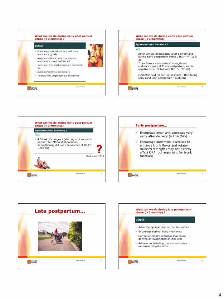
What can we do during early post-partum phase (< 3 months) ?
19
Dufour
©BougeottePlacotine
• Encourage optimal posture and body
mechanics (↓ IAP)
• Avoid exercises in which continence
mechanism is not maintained
• Inner unit x’s, leading to more functional
x’s
• Avoid concentric abdominal x’
• Tension-free diaphragmatic breathing
www.ulaval.ca
What can we do during early post-partum phase (< 3 months)?
20
Agreement with literature ?
Yes :
• Inner unit x’s immediately after delivery and during early postpartum phase ↓ IRD11, 12 (LoE:
2b)
• Trunk flexors and rotators’ strength and endurance are ↓ at 7 wks postpartum, and isnegatively correlated with IRD13 (LoE: 2b)
• Isometric hold (in curl-up position) ↓ IRD during early (and late) postpartum14 (LoE:3b)
www.ulaval.ca
What can we do during early post-partum phase (< 3 months)?
21
Agreement with literature ?
No:
• A 16-wk x’s program (starting at 6 wks post partum) for PFM and abdominalsstrengthening did not ↓ prevalence of DRA15
(LoE: 1b)
©GopherPerformance
Keshwani, 2018
www.ulaval.ca
Early postpartum…
✓ Encourage inner unit exercises veryearly after delivery (within 24h).
✓ Encourage abdominal exercises to enhance trunk flexor and rotatormuscles strength (may not directlyaffect DRA, but important for trunkfunction)
22
www.ulaval.ca
Late postpartum…
23
©BougeottePlacotine
www.ulaval.ca
What can we do during late post-partum phase (> 3 months) ?
24
Dufour
• Advocate optimal posture (neutral spine)
• Encourage optimal body mechanics
• Correct or modify exercises that cause doming or invagination of linea alba
• Address contributing thoracic and pelvicmovement impairments

5
www.ulaval.ca
What can we do during late post-partum phase (> 3 months) ?
25
Keeler
• General inner unit training
• Inner unit x’s during functionalactivities
• Noble technique
• PFM x’s
• Manual release of restrictedmyofascial tissue
• Use of abdominal binders
www.ulaval.ca
What can we do during late post-partum phase (> 3 months) ?
26
Agreement with literature ?
Yes :
• ↓ Trunk flexors and rotators’ strength and endurance at 6 months, and trunk rotation torque at 12-month13, 19
• Negatively correlated with IRD13 (LoE: 2b)
• Correlation b/w distortion of LA and IRD19
• A global-approach (movement impairmentsystem) can reduce IRD20 (LoE: 4)
www.ulaval.ca
What can we do during late post-partum phase (> 3 months) ?
27
Agreement with literature ?
No:
• Physiotherapy exercise programs can besuccessful at reducing IRD during a contraction, but not at rest. May play a rolein the laxity of the ventral musculature.23
www.ulaval.ca
Late postpartum…
✓ Encourage abdominal exercises to enhancetrunk flexor and rotator muscles strength.
✓ Avoid invagination or doming of the lineaalba during exercises.
28
www.ulaval.ca
Moving knowledge into action
Engaging knowledge users in the elaboration, the completion and dissemination of the results processes of research will be helpful for:
1. Evidence-informed practice
2. Research relevant to the needs of both providers and patients
3. Documenting the effects of interventions that reflect the practices of clinicians
4. Increase our understanding of what we are actually doing
29 www.ulaval.ca
Thank you!30
6
www.ulaval.ca
References1. Canadian Institutes of Health Research: More about Knowledge Translation at
CIHR. http://www.cihr-irsc.gc.ca/e/39033.html] Accessed Feb 11th, 2012.
2. Jagosh, J., A. C. Macaulay, P. Pluye, J. Salsberg, P. L. Bush, J. Henderson, E. Sirett, G. Wong, M. Cargo, C. P. Herbert, S. D. Seifer, L. W. Green and T. Greenhalgh (2012). "Uncovering the Benefits of Participatory Research:
Implications of a Realist Review for Health Research and Practice." The Milbank Quarterly 90(2): 311-346.
3. Dufour, S., Bernard, S., Graham, N., Murray-Davis, B. Establishing expert-based recommendations for the conservative management of pregnancy-related diastasis rectus abdominis: results of a Delphi consensus study. JOWHPT. In revision.
4. Keeler, J., M. Albrecht, L. Eberhardt, L. Horn, C. Donnelly and D. Lowe (2012). "Diastasis Recti Abdominis: A Survey of Womenʼs Health Specialists for Current Physical Therapy Clinical Practice for Postpartum Women." Journal of Womenʼs
Health Physical Therapy 36(3): 131-142.
5. Sperstad, J. B., M. K. Tennfjord, G. Hilde, M. Ellstrom-Engh and K. Bo (2016). "Diastasis recti abdominis during pregnancy and 12 months after childbirth: prevalence, risk factors and report of lumbopelvic pain." Br J Sports Med 50(17): 1092-1096.
31 www.ulaval.ca
References6. Gilleard, W. L. and J. M. M. Brown (1996). "Structure and function of the
abdominal muscles in primigravid subjects during pregnancy and the immediate postbirth period." Physical Therapy 76(7): 750.
7. Chiarello, C. M., L. A. Falzone, K. E. McCaslin, M. N. Patel and K. R. Ulery (2005). "The effects of an exercise program on diastasis recti abdominis in pregnant
women." Journal of Women’s Health Physical Therapy 29(1): 11-16.
8. Mota, P., A. G. Pascoal, A. I. Carita and K. Bo (2015). "The Immediate Effects on Inter-rectus Distance of Abdominal Crunch and Drawing-in Exercises During Pregnancy and the Postpartum Period." J Orthop Sports Phys Ther 45(10): 781-788.
9. Edqvist, M., E. Blix, H. K. Hegaard, O. Á. Ólafsdottir, I. Hildingsson, K. Ingversen, M. Mollberg and H. Lindgren (2016). "Perineal injuries and birth positions among 2992 women with a low risk pregnancy who opted for a homebirth." BMC Pregnancy and Childbirth 16: 196.
10. Nasir, A., R. Korejo and K. J. Noorani (2007). "Child birth in squatting position." J Pak Med Assoc 57(1): 19-22.
11. Mesquita, L. A., A. V. Machado and A. V. Andrade (1999). "Physiotherapy for reduction of diastasis of the recti abdominis muscles in the postpartum period." Revista Brasileira de Ginecologia e Obstetrícia 21(5): 267-272.
32
www.ulaval.ca
References12. Tuttle, L. J., J. Fasching, A. Keller, M. Patel, C. Saville, R. Schlaff, A. Walker, M.
Mason and S. P. Gombatto (2018). "Noninvasive Treatment of Postpartum Diastasis Recti Abdominis: A Pilot Study." Journal of Women’s Health Physical Therapy 42(2): 65-75.
13. Liaw, L. J., M. J. Hsu, C. F. Liao, M. F. Liu and A. T. Hsu (2011). "The relationships
between inter-recti distance measured by ultrasound imaging and abdominal muscle function in postpartum women: a 6-month follow-up study." J OrthopSports Phys Ther 41(6): 435-443.
14. Sancho, M. F., A. G. Pascoal, P. Mota and K. Bo (2015). "Abdominal exercises affect inter-rectus distance in postpartum women: a two-dimensional ultrasound study." Physiotherapy 101(3): 286-291.
15. Gluppe, S. L., G. Hilde, M. K. Tennfjord, M. E. Engh and K. Bø (2018). "Effect of a Postpartum Training Program on the Prevalence of Diastasis Recti Abdominis in Postpartum Primiparous Women: A Randomized Controlled Trial." Physical
Therapy 98(4): 260-268.
16. Fernandes da Mota, P. G., A. G. Pascoal, A. I. Carita and K. Bo (2015). "Prevalence and risk factors of diastasis recti abdominis from late pregnancy to 6 months postpartum, and relationship with lumbo-pelvic pain." Man Ther 20(1): 200-205.
33 www.ulaval.ca
References
17. Mota, P., A. G. Pascoal, A. I. Carita and K. Bo (2018). "Normal width of the inter-recti distance in pregnant and postpartum primiparous women." Musculoskelet Sci Pract 35: 34-37.
18. Keshwani, N., S. Mathur and L. McLean (2018). "Relationship Between Interrectus Distance and Symptom Severity in Women With Diastasis Recti Abdominis in the Early Postpartum Period." Phys Ther 98(3): 182-190.
19. Nicole Hills. The Physical Implications of Diastasis Recti Abdominis Among Women at the End of Their First Postpartum Year. Doctoral Thesis, School of Rehabilitation Therapy, University of Queens, 2018.
20. Kurz, J. and D. Borello-France (2017). "Movement System Impairment-Guided Approach to the Physical Therapist Treatment of a Patient With Postpartum Pelvic Organ Prolapse and Mixed Urinary Incontinence: Case Report." Physical Therapy 97(4): 464-477.
21. Madill, S. J. and L. McLean (2008). "Quantification of abdominal and pelvic floor muscle synergies in response to voluntary pelvic floor muscle contractions." J Electromyogr Kinesiol18(6): 955-964.
22. Neumann, P. and V. Gill (2002). "Pelvic floor and abdominal muscle interaction: EMG activity and intra-abdominal pressure." Int Urogynecol J Pelvic Floor Dysfunct 13(2): 125-132.
23. Mommers, E. H. H., J. E. H. Ponten, A. K. Al Omar, T. S. de Vries Reilingh, N. D. Bouvy and S. W. Nienhuijs (2017). "The general surgeon's perspective of rectus diastasis. A systematic review of treatment options." Surg Endosc 31(12): 4934-4949.
24. Lee, D. and P. W. Hodges (2016). "Behavior of the Linea Alba During a Curl-up Task in Diastasis Rectus Abdominis: An Observational Study." J Orthop Sports Phys Ther 46(7): 580-589. 34

9/12/2018
1
Practical Application: Research to Practice
Sinéad Dufour, PT PhD
Assistant Clinical ProfessorSchool of Rehabilitation Science
Michael G. DeGroote School of Medicine
McMaster University
CANADA
Affiliations to disclose†:
Funding for speaker to attend:
Self-funded
Institution (non-industry) funded
Sponsored by:
Sinéad Dufour PT PhD
X
† All financial ties (over the last year) that you may have with any business organisation with respect to the subjects mentioned during your presentation
Objectives
• Apply the presented research to a clinical case
• Two points in time
– Pregnancy
– Post-partum
• Two provider perspectives
– Family Physician (General Practitioner)
– Pelvic Health Physiotherapist
Pregnancy – Family Physician
Saphia is a 36 year old women in her second trimester of her third pregnancy. She has a three-year old son and had a miscarriage (8 weeks gestation) approximately 1 year ago. She presents with pain that she describes as being close to her groin that moves from one side to the other and is most irritable when she is getting dressed or changing positions at night. She also mentions that she is really worried about her “core” as she states she had DRA after the birth of her first child which never ”got better”. Saphia wants to know if there is anything she can do now to prevent worsening of the DRA as well as address the pain she is having in her pelvis.
Evidence-informed approach?
• We treat people, not conditions
– Saphia presents with both pregnancy-related PGP and DRA
• We need an evidence-informed, tailored approach that engages Saphia’s preferences
– What assessment strategies are appropriate for pregnancy-related PGP and DRA?
– What treatment strategies are appropriate for pregnancy-related PGP and DRA?
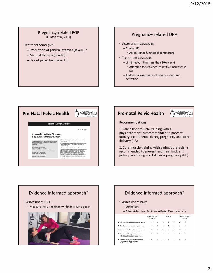
Pregnancy-related PGP(Clinton et al, 2017)
Assessment Strategies
• Physical orthopedic tests
– Pain provocation tests
– Functional tests (stroke test and active straight leg raise)
• Self-report measures
– Pelvic Girdle Questionnaire
– Fear Avoidance Belief Questionnaire
– Pain Catastrophizing Scale
9/12/2018
2
Pregnancy-related PGP(Clinton et al, 2017)
Treatment Strategies
–Promotion of general exercise (level C)*
–Manual therapy (level C)
–Use of pelvic belt (level D)
Pregnancy-related DRA
• Assessment Strategies
– Assess IRD
• Assess other functional parameters
• Treatment Strategies
– Limit heavy lifting (less than 20x/week)
• Attention to sustained/repetitive increases in IAP
– Abdominal exercises inclusive of inner unit activation
Pre-Natal Pelvic Health Pre-natal Pelvic Health
Recommendations
1. Pelvic floor muscle training with a physiotherapist is recommended to prevent urinary incontinence during pregnancy and after delivery (I-A)
2. Core muscle training with a physiotherapist is recommended to prevent and treat back and pelvic pain during and following pregnancy (I-B)
Evidence-informed approach?
• Assessment DRA:
– Measure IRD using finger width in a curl up task
Evidence-informed approach?
• Assessment PGP:
– Stoke Test
– Administer Fear Avoidance Belief Questionnaire

9/12/2018
3
Evidence Informed ApproachFamily Physician
Assessment Findings• IRD = 4 finger widths• Stoke Test = positive• Fear Avoidance Belief Score = 13
Findings consistent with pregnancy-related PGP and pre-existing DRA
Evidence Informed ApproachFamily Physician
Management
• Counselling related to – General movement (not fearing movement)
– Avoiding sustained or repeated strain through the abdominal wall such as straining on the toilet, heavy lifting or other similar activities that increase IAP.
• Refer to pelvic health physiotherapist for further Ax and Rx– PGP, global pelvic health promotion, prevention of UI
Post-Partum – Pelvic Health PT
Saphia is a 36 year old women who has been referred to you from her family doctor. She is 16 weeks post-partum with her second child. She had DRA after the birth of her first child and feels is it worse now after having another baby. She was referred during her pregnancy but never came. She denies having any issues with bladder control although when probed does indicate she leaks a small amount of urine when she sneezes. She indicates that she feels like she would probably have bladder control issues with exercise but that she has not returned to any exercise (other than walking) as she has researched about DRA on line is scared to exercise – she doesn’t want to make her DRA worse.
Evidence-informed approach?
• We treat people, not conditions– Saphia presents with both stress UI and DRA
• We need an evidence-informed, tailored approach that engages Saphia’s preferences– What assessment are the appropriate assessment
strategies for SUI and DRA?
– What are the appropriate treatment strategies for SUI and DRA?
Stress Urinary Incontinence
• Assessment Strategies – Pelvic Floor Muscle Strength (oxford scale)
– Presence of pre-contraction pelvic floor muscle reflex (the knack)
– Self-report measures
• Treatment Strategies– Pelvic floor muscle training represents the first
line intervention for stress, urge or mixed urinary incontinence (Doumalin et al, 2014 ).
• Level 1A evidence!
Pregnancy-related DRA
• Assessment Strategies
– Assess IRD
• Assess other functional parameters
• Treatment Strategies
– Encourage abdominal exercises to enhance trunk flexor and rotator muscles strength
• Inner unit exercise
– Apply a global movement approach
• Inclusive of self-monitoring
9/12/2018
4
Evidence Informed ApproachPelvic Health Physiotherapist
Assessment Findings• Oxford score = 2/5 (squeeze no lift) • No presence of pre-contraction of pelvic floor
contraction with cough• IRD = 3 finger widths • Doming noted through abdominal wall during
curl up taskPresentation consistent with pelvic floor dysfunction and pregnancy-related DRA
Evidence Informed ApproachPelvic Health Physiotherapist
Management
• Counselling related to
–General movement (not fearing movement)
– Self-monitoring of LA and IAP
• Abdominal exercises inclusive of inner unit work following established PFMT protocol
– individually tailored
Closing Remarks
• Evidence guiding practice for pregnancy-related DRA is lacking.
• Women with DRA will often present with other pelvic health concerns which are not necessarily correlated but need to be addressed in a concordant manner.
• Practice-based research has the potential to inform future RCTs to clarify what treatment interventions are the most effective.