Embed Size (px)
Citation preview

University of Naples “Federico II”
Ph.D in Chemical Sciences
XXX Cycle
Thesis in Physical chemistry
“Effect of polyunsaturated fatty acids
on the structure and dynamics of lipid bilayers
and on the interaction with peptides”
Candidate
Augusta De Santis
Tutors Advisor
Prof. Luigi Paduano
Prof. Gerardino D’Errico
Prof.ssa Angelina Lombardi
2014-2017


Tables of contents
Introduction I
Chapter 1: Biomembranes 1
1.1. The fluid mosaic model 3
1.2. Lipid polymorphism and its significance 7
1.3. Lipid molecular geometry 11
1.4. Polyunsaturated fatty acids 12
1.5. References 15
Chapter 2: Amyloidogenic proteins and peptides 17
2.1. Definition of amyloid fibril protein 19
2.2. Protein misfolding and amyloid formation 20
2.2.1. Aβ and α-synuclein: two case studies 22
2.3. The role of membranes 23
2.3.1. The role of cholesterol 25
2.3.2. The effect of omega-3 fatty acids 26
2.4. Nucleophosmin-1: an unexpected amyloid protein 28
2.5. References 33
Chapter 3: Effect of DHA-phospholipids on Lα phases 39
3.1. Work outline 41
3.2. Materials and methods 42
3.3. Effect of SDPC and DDPC on POPC lipid membranes 48
3.3.1. Electron spin resonance analysis 48
3.3.2. Neutron reflectivity analysis 53
3.3.3. Cryogenic transmission electron microscopy 56
3.4. Effect of SDPC and DDPC on POPC/Chol lipid membranes 57
3.4.1. Electron spin resonance analysis 57
3.4.2. Neutron reflectivity analysis 63
3.5. Conclusion 67
3.6. References 68
Chapter 4: Amyloidogenic peptides from NMP-1 and model membranes 71
4.1. Work outline 73

4.2. Materials and methods 74
4.3. Effects of lipid membranes Ld and Lo on NPM1 peptides 76
4.3.1. Circular dichroism 76
4.3.2. Electron spin resonance 78
4.4. Effects of lipid membranes Ld and Lo containing DDPC on NPM1
peptides 82
4.4.1. Circular dichroism 82
4.4.2. Electron spin resonance 84
4.5. Conclusion 89
4.6. References 92
Chapter 5: Aβ 1-42 and model membranes 93
5.1. Work outline 95
5.2. Materials and methods 96
5.3. Sample condition design 99
5.4. Circular dichroism 99
5.5. Electron spin resonance 102
5.6. Conclusion 107
5.7. References 110
Appendix A: Effect of polyunsaturated free fatty acids on Lβ phases 113
A.1. Work outline 115
A.2. Materials and methods 116
A.3. Differential scanning calorimetry 118
A.4. Laurdan General Polarization method 121
A.5. Confocal microscopy of GUVs 124
A.6. Conclusion 126
A.7. References 127
Appendix B: Neutron reflectivity profiles 129
B.1. Neutron reflectivity profiles for SDPC and DDPC in POPC
lipid membranes 131
B.2. Neutron reflectivity profiles for SDPC and DDPC in POPC/Chol
lipid membranes 132
Publications 133

I
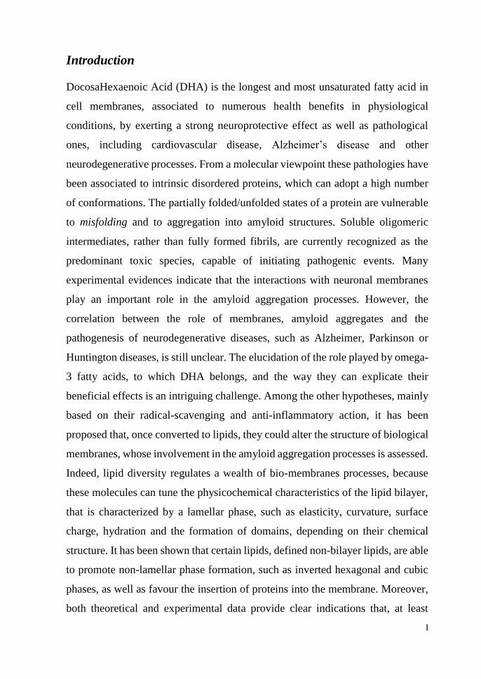
Introduction
DocosaHexaenoic Acid (DHA) is the longest and most unsaturated fatty acid in
cell membranes, associated to numerous health benefits in physiological
conditions, by exerting a strong neuroprotective effect as well as pathological
ones, including cardiovascular disease, Alzheimer’s disease and other
neurodegenerative processes. From a molecular viewpoint these pathologies have
been associated to intrinsic disordered proteins, which can adopt a high number
of conformations. The partially folded/unfolded states of a protein are vulnerable
to misfolding and to aggregation into amyloid structures. Soluble oligomeric
intermediates, rather than fully formed fibrils, are currently recognized as the
predominant toxic species, capable of initiating pathogenic events. Many
experimental evidences indicate that the interactions with neuronal membranes
play an important role in the amyloid aggregation processes. However, the
correlation between the role of membranes, amyloid aggregates and the
pathogenesis of neurodegenerative diseases, such as Alzheimer, Parkinson or
Huntington diseases, is still unclear. The elucidation of the role played by omega-
3 fatty acids, to which DHA belongs, and the way they can explicate their
beneficial effects is an intriguing challenge. Among the other hypotheses, mainly
based on their radical-scavenging and anti-inflammatory action, it has been
proposed that, once converted to lipids, they could alter the structure of biological
membranes, whose involvement in the amyloid aggregation processes is assessed.
Indeed, lipid diversity regulates a wealth of bio-membranes processes, because
these molecules can tune the physicochemical characteristics of the lipid bilayer,
that is characterized by a lamellar phase, such as elasticity, curvature, surface
charge, hydration and the formation of domains, depending on their chemical
structure. It has been shown that certain lipids, defined non-bilayer lipids, are able
to promote non-lamellar phase formation, such as inverted hexagonal and cubic
phases, as well as favour the insertion of proteins into the membrane. Moreover,
both theoretical and experimental data provide clear indications that, at least

II
transiently, non-lamellar structural intermediates must exist in vivo. With the aim
to understand the functional role of DHA, we characterized lipid membranes with
different phospholipid containing this fatty acid, the 1-stearoyl-2-
docosahexanoyl-glycerophosphocholine, SDPC, and the 1,2-docosahexanoyl-
glycerophosphocholine, DDPC, (Figure 1) embedded, at different scale levels,
from micro-structure to overall morphology. We used lipid membranes composed
by pure 1-palmitoyl-2-oleoyl-phosphatidylcholine (POPC) and in combination
with Cholesterol (Chol) (Figure 1), because they are characterized by different
liquid crystalline meso-structures (e.g., ordered, Lo, vs. disordered, Ld).
Figure 1: structures of the phospholipids under investigation.
A combination of two physico-chemical techniques, Electron Spin Resonance
(ESR) spectroscopy with the spin-labelling approach and Neutron Reflectivity
(NR), was used to achieve our aims.
Moving from the characterization of the lipid systems, we attempt to rationalize
the DHA-phospholipids effect the DDPC, on peptide/lipid interaction, focusing
on the role played in amyloidogenic processes. Through a combined ESR and
Circular Dichroism (CD) approach, the interaction of model membranes,
containing DDPC, with different amyloidogenic peptides have been investigated.
Two novel short amyloidogenic peptides derived from nucleophosmin-1 (NPM-
1), a nucleolar protein with a likely involvement in Huntington Disease and
Amyloid Leukaemia, corresponding to helix H2 and the N-terminal extended H2,

III
Nterm, regions of NPM1 have been chosen. Then, the work has been scaled up to
more biomimetic lipid membrane containing DDPC, to study the interaction with
peptide hallmark of Alzheimer’s disease: Aβ 1–42 aggregation process.
Nevertheless, also the role of free fatty acid, DHA, should be considered; indeed,
it has been found vesicles cycling, which is an important biological event,
involves the interplay between membrane lipids and proteins, among which the
enzyme phospholipase A2 (PLA2) plays a critical role. In nervous cells, PLA2
was also shown to control the metabolic transformation of phospholipid (PL)
molecules that contain polyunsaturated fatty acids. In addition, it has been found,
using raftlike lipid mixtures, that line tension and elastic properties govern
budding formation after the addition of phospholipase A2 (PLA2), which explains
why docosahexaenoic acid (DHA)-containing phosphatidylcholine (PC), but not
Oleic Acid (OA)-containing PC, are able to exhibit liquid-ordered (Lo) domain
budding. In this context, very little is known about the interaction of omega-3 free
fatty acids with model membranes, thus their effect, i.e. DHA and OA as control
on DPPC membranes were investigated during a period of five months spent
abroad at Biofisika Institute of UPV/EHU and the main results reported in the
appendix.

Chapter 1:
Biomembranes


Chapter 1: Biomembranes
3
1. 1. The fluid mosaic model
In 1972, Singer and Nicolson conceived a model, which is known as the fluid
mosaic model, to describe the structure of plasma membranes1.
Biological membranes can be considered as a pseudo-two-dimensional liquid in
which both lipids and membrane-associated protein molecules move laterally or
sideways throughout the membrane to allow for function. The plasma membrane
was postulated to be composed of different kinds of lipid and protein and the
overall random appearance of the composite made the membrane looks like a
mosaic (Figure 1.1).
Figure 1.1: The Fluid Mosaic Model for cell membrane.
Natural lipids are amphipathic biomolecules usually insoluble in water, but
soluble in organic solvents. Due to this amphipathic character, when diluted in
aqueous solutions they generally tend to self-aggregate into bilayer structures,
becoming essential constituents of cell membranes2. Lipids comprise a large
number of different molecules each of them possesses different properties that
play an important role in biological functions.
Glycerophospholipids, sphingolipids and sterols are crucial structural
components of cell membranes and depending on their chemical structure can

Chapter 1: Biomembranes
4
modulate the physical properties of plasma membranes. In this scenario, the
understanding of the physiological significance of lipid diversity in biological
membranes is a key issue in membrane research.
Glycerophospholipids
The general structure of a glycerophospholipid is reported in Figure 1.2; they
typically consist on a hydrophilic head characterized by a glycerol esterified in
the 3rd position by a negatively charged phosphate group, which can be esterified
by an alcohol derived molecule (R) and in the 1st and 2nd positions by hydrophobic
tails (R1, R2).
Figure 1.2: Structures of a general phospholipid R1 and R2 represent the different possibilities of
fatty acid chains, while R represents the different possibilities for the polar head group.
The R group attached to the phosphate characterizes the hydrophilic headgroup,
and according to it, glycerophospholipids are classified:
Phosphatidylcholine (PC);
Phosphatidylethanolamine (PE);
Phosphatidylglycerol (PG);
Phosphatidylserine (PS);
Phosphatidylinositol (PI).
Regarding the hydrophobic part (R1 and R2), glycerophospholipids can
incorporate fatty acids (FA) of various lengths and unsaturation degree, which
define the membrane fluidity.
It is clear that all the possible combinations between different headgroup and
different chains give rise to an infinite variety of phospholipids that play a

Chapter 1: Biomembranes
5
decisive role for many cell functions; indeed, their relative abundance depends on
both the type of living organisms and tissues indicating its biological significance.
Sphingolipids
Johann L.W. Thudichum first isolated a brain-derived compound with mysterious
properties that he called sphingolipid with regard to the likewise enigmatic
sphinx3. Nowadays, structurally well-characterised sphingolipids represent a
crucial lipid group for many central biological mechanisms. Sphingolipids are
structurally based on sphingosine, (2S, 3R, 4E) -2-amino-4-octadecene-1,3-diol
(Figure 1.3) and have been considered for decades as inert structural components
of cell plasma membranes.
Figure 1.3: Sphingosine structure.
Recently, the unravelling of basic lipid metabolism and the discovery of the
sphingolipid signalling pathway4,5, made sphingolipids the object of great interest
for many scientists6. Nowadays, several sphingolipids are considered as bioactive
molecules and are assumed to act as secondary messengers in many signalling
pathways. These principal bioactive sphingolipids are: sphingosine, ceramide and
their related phosphate analogues sphingosine-1-phosphate and ceramide-1-
phosphate7. Binding of more complex structures based on one or more sugar
residues produces largest sphingolipids, namely the glycosphingolipids, which
can be classified into two groups, cerebrosides and gangliosides, depending on
the number of sugar residues and the presence of sialic acids. A further important
discovery was the sphingolipid involvement in the generation of lateral structures
or membrane domains, among which the most relevant was the proposal of
sphingolipid and cholesterol-based membrane rafts8. The simple formation and/or

Chapter 1: Biomembranes
6
functional role of these rafts has originated a huge amount of experimental work,
but with important controversy to their real existence and possible roles.
Deep studies of lipid raft generation have resulted in research of other
sphingolipids with similar behaviour on the generation of such structures, as is
the case of ceramide.
Sterols
An additional major group of lipid constituents of cell membranes are sterols; they
are found in animals, plants and eukaryotic microorganisms, constituting as well
as high percentage of total membrane lipids.
The cholestane structure characterizes this group of lipids and consists on a
hydrocarbon tail linked to a on four-ring structure in which a single hydroxyl
group exists, giving the molecule its amphipathic character (Figure 1.4).
Figure 1.4: Structures of cholestane group.
The most abundant sterol in animal tissues is cholesterol and it is principally
found in plasma membranes where it can reach up to 30 mol % of total lipid. In
red blood cells and ocular lens membranes its amount is significantly increased,
accounting for 50 and 80 mol % of total lipid respectively9. Several studies have
shown that cholesterol has been implicated in the modulation of the activities of
some integral membrane proteins10,11,12 and in the regulation of cellular proteins
through the oxysterol-binding protein13; in addition, it is involved in cellular
processes, particular cell signaling in, through specific interactions with the
components of major signaling pathways14. The main hypothesis on the

Chapter 1: Biomembranes
7
mechanism of action is the regulation of physical properties of the membrane,
most notably the formation of cholesterol-rich lipid rafts, deriving from the
association with sphingolipids and saturated phospholipids15.
1.2. Lipids polymorphism and its significance
Cell membrane lipids are basically organised in unidimensional lamellar structure
due to the same amphipathic property, which allow them to constitute and
modulate the bilayer structure, which is the platform of fluid mosaic model.
However, some crucial biological processes are characterised by the formation of
more complex non-lamellar structures, such as the inverted hexagonal or cubic
phases. From macromolecule transversal motion across the membrane to cell
division, numerous processes require the generation of these unstable structures.
Membrane fusion and fission processes16,17, that include virus infection, vesicular
transport from the ER to the Golgi or endocytic and exocytic transport, are good
examples of energetically unfavourable events that require the formation of non-
lamellar structures. However, lipid polymorphism is not only associated to two-
dimensional structural changes. For instance, the formation of laterally segregated
structures within a membrane, where protein complexes can adopt a more stable
conformation18 is dependent on unidimensional alterations of lipid order or
rigidity.
Owing to their low water solubility, when dispersed in an aqueous solution lipids
self-aggregate into different structures, basically depending on the solution ionic
strength, pH, temperature, pressure, hydration level and on the lipid nature and
morphological geometry. In this extent, lipids exist in a particular phase. Phases
are thermodynamic idealisations defined under equilibrium conditions and
characterised by particular physical properties.
Lipids phases are classified according to three general criteria:
i) kind of lattice;
ii) lipid acyl chain order (ordered or disordered);

Chapter 1: Biomembranes
8
iii) overall structure curvature (normal or inverted).
An accepted nomenclature to distinguish the various lipid phases is that proposed
back in 1968 by Vittorio Luzzati based on x-ray studies19: 1D micellar (M), 1D
lamellar (L), 2D hexagonal (H), 2D oblique (P), 3D cubic (Q) and 3D crystalline
(C). Finally, the lipid phases are classified owing to the overall structure
curvature, being of type I or type II, and represented as well by the subscript I or
II respectively. Type I conformation denotes an overall positive curvature with
the hydrophobic acyl chains towards the centre and away from the aqueous
environment, while type II, or inverted, reflects and overall negative curvature
with the polar lipid headgroups towards the centre and the acyl chains in close
contact with the aqueous environment.
Figure 1.5: Overview of the different structures. A) micelle; B) inverted micelle; C) hexagonal;
D) inverted hexagonal; E) lamellar; F) cubic; G) inverted cubic.
The most biologically relevant phases are: lamellar (L), hexagonal (H), micellar
(M) and cubic (Q). Permutations between phases are called phase transitions and
occur when the specific physical parameters that define a certain phase are
changed. Phase transitions can be induced upon changes in system temperature

Chapter 1: Biomembranes
9
(thermotropic transitions), pressure (barotropic transitions) or solvent proportions
(lyotropic transitions). Of great importance for many biological processes is the
characterisation of the thermotropic transitions in pure or lipid mixtures,
principally in those exhibiting thermotropic transitions in temperature ranges
close to that of physiological relevance (37 ºC). These transitions are strongly
dependent on the lipid acyl chain length and unsaturation level. For example,
lipids with unsaturated acyl chains hardly display lamellar gel-fluid thermotropic
phase transitions at temperatures higher than 0 ºC, reflecting a complete fluidity
under physiological conditions.
Micellar phase (M)
This one-dimensional phase is inherent to lipids with inverted-cone-shaped
structures (see lipid molecular geometry at the end of this section) as is the case
for Lyso-phospholipids (phospholipids that lack one of the acyl chains). This kind
of lipids self-aggregate in aqueous environments into micelles with the acyl
chains located away from the aqueous medium and towards the centre of the
micelle. These lipids display properties opposite to those of lipids with high
propensity for hexagonal phases, but both kinds may be crucial for the above-
mentioned biological mechanisms to be undertaken. For instance, in a membrane
fission event, when the final neck is generated, the high curvature inherent to the
neck will require lipids with opposite shapes to be in the two opposite layers of
the membrane for proper fission. A typical parameter to refer to a micelle-forming
lipid is the critical micellar concentration (CMC), which reflects the concentration
at which a lipid monomer-micelle transition occurs. The generation or
introduction of high amounts of a lipid with these characteristics into a specific
region within a membrane can cause an overall destabilisation of the bilayer
structure, inducing the formation of positive curvature in the external monolayer
and possible membrane solubilisation and subsequent micelle. This behaviour is
inherent to molecules with detergent-like properties20.

Chapter 1: Biomembranes
10
Lamellar phases (Lα)
The lamellar phases are classified according to acyl chain order as: disordered or
fluid (Ld), ordered and perpendicular to the plane of the membrane (gel) (Lβ),
ordered and tilted with respect to the plane of the membrane (Pβ´). Later, a new
lamellar phase was introduced with intermediate properties between fluid and gel,
namely, liquid-ordered phase (Lo). These subscripts are used to classify the
different phases only in a unidimensional lamellar lattice, as the only phase that
can be organised with their acyl chains in an ordered or disordered configuration.
Biological membranes under equilibrium conditions are composed of a lipid
bilayer in a lamellar configuration, where the most significant phases are: fluid,
liquid-disordered, or liquid-crystalline (Lα). In a Lα phase, lipids are free to
diffuse laterally or rotationally, and display their acyl chains in a complete
disordered state with high flexibility. In this phase, the acyl chains present low
proportions of trans carbon- carbon (C-C) bound conformers, and at least one of
the acyl chains is normally unsaturated. Certain lipids can be present in a lamellar
gel Lβ phase. In this phase, the acyl chains are highly ordered and display high
proportions of trans C-C conformers, being almost immobile and not allowing
lateral or rotational motion21. The Lβ to Lα phase transition is usually known as
the main phase transition. Lipids with high gel-fluid thermotropic phase
transitions are usually composed of saturated acyl chains, and present these acyl
chains in a perpendicular disposition to the plane of the membrane. The third
phase is called liquid-ordered (Lo), and reflects a phase with intermediate
properties between the gel and the fluid phases. It was recently named to denote
lipid mixtures of sphingolipids or glycerolipids with sterols, and it is a phase in
which lateral diffusion is allowed but there is a low level of gauche conformers.
This is the phase in which “membrane rafts” are supposed to exist22.
Hexagonal phases (H)
The formation of non-lamellar structures is apparently contradictory to the normal
function of a natural cell membrane. However, lipids with the ability to form such

Chapter 1: Biomembranes
11
phases have been related to several important mechanisms such as fusion or
fission processes. Among non-lamellar structures, the hexagonal phase appears as
one of the most relevant conformations. Hexagonal phases are fluid 2-D structures
that can be of type I (HI) or type II (HII). In both cases, large parallel cylinders
with a hexagonal packing are formed. In the normal or HI type, lipids arrange with
the acyl chains disposed to the centre of the cylinder. HII is called inverted
hexagonal phase and presents the polar headgroups to the centre of the cylinder
and the acyl chains towards the external environment and in close contact with
hydrophobic tails from other cylinders.
The biological relevance of these structures becomes significant in processes as
membrane lipid- and protein-mediated pore and/or channel formation, which
appear to recruit non-lamellar forming lipids for proper structure stability, and
membrane fusion and fission events, in which lipids with a high propensity for
hexagonal phases are required both for the contact sites between membranes in
fusion mechanisms and for the final neck generation prior to fission events.
Cubic phases (Q)
The last, but not least, kind of biologically relevant phase refers to the periodic
three dimensional cubic phases19. Seven classes of cubic phases are known,
among which the most important two are the micellar and the bicontinuous type
I and II. The bicontinuous type II cubic phase (QII) represents a highly convoluted
lipid bilayer, which subdivides 3-D space into two unconnected polar labyrinths
divided by an apolar septum. In the case of the bicontinuous type I cubic phase
(QI) the septum and the two labyrinths are filled by the polar and the apolar
medium, respectively.
1.3. Lipid molecular geometry
The formation of the various structures is strongly influenced by the lipid
molecular geometry. Jacob Israelachvili described how differences in the cross-
sectional area between the polar headgroup and the hydrophobic tail of the

Chapter 1: Biomembranes
12
different lipids could determine the overall structure of the lipid aggregates23. On
this basis, lipids can be classified into three groups by having: cone, cylindrical
or inverted-cone shapes. This fact is summarised by the morphological parameter
Ns defined in Equation 1.1:
𝑵𝑺 =𝑽𝑪
𝒂𝑪∙𝑳𝑪 Eq. 1.1
where “V” is the volume of the lipid molecule, “ao” is the area of the molecule in
the lipid-water interface and “Lc” the length of the extended acyl chain.
Considering these parameters and the lipid cross-sectional area in the
hydrophobic tail “aH”, which would reflect a volume of V = aHLc for a lipid
displaying cylindrical shape, we can review the different morphological
geometries as:
a0 = aH (Ns = 1), the molecule presents a cylindrical shape;
a0 < aH (Ns > 1), the molecule presents a cone shape;
a0 > aH (Ns < 1), the molecule presents an inverted cone shape.
As described in Figure 1.5, pure lipids in aqueous solutions will self-aggregate
into different structures depending on their molecular geometry. Cylindrical
shapes displaying lipids as PC or SM will become organised in the form of
lamellar bilayers. Cone shaped lipids as PE or sterols will tend to form hexagonal
arrangements, while inverted-cone shaped lipids as is the case of lyso-
phospholipids will aggregate into micelles. Thus, it could be summarised that
lipid polymorphism reflects the lipid behaviour under specific physical conditions
in which long-range ordered structures are generated with the lipids in a given
phase.
1.4. Polyunsaturated fatty acids
Polyunsaturated fatty acids (PUFA) contain more than one double bond in one
acyl chain. PUFA includes many important compounds such as essential fatty
acids (EFA). Essential fatty acids are indispensable for the health of mammalian

Chapter 1: Biomembranes
13
animals. It can only be ingested from food and cannot be synthesized in human
body. Only two EFAs are known for humans: alpha-linolenic acid (an omega-3
fatty acid) and linoleic acid (an omega-6 fatty acid). Docosahexaenoic acid
(DHA) with 22-carbons and six double bonds is the most polyunsaturated fatty
acid commonly found in biological systems and it can be synthesized from alpha-
linolenic acid or obtained directly from fish oil24. In the inner leaflet of some
animal cell plasma membranes PUFAs comprise up to ~50% of sn-2 fatty acyl
chains25. In retinal rod outer segment disk membranes DHA comprises ~50% of
the total acyl chains, with this high percentage required for optimal rhodopsin
function26. DHA is also found at high concentrations in certain other membranes,
including synaptosomes27 and sperm28. The importance of DHA and PUFAs for
human health has been well-studied29: spectroscopic, computational, and other
biophysical methods30 have established significant PUFA effects on membrane
properties. It has been shown that phospholipid containing DHA with a
phosphoethanolamine headgroup, or a di-polyunsaturated phospholipid in
particular condition, can give a non-lamellar phase31. In addition, PUFAs seem
to have a weaker interaction with cholesterol compared with saturated or
monounsaturated acyl chains. A relatively low solubility of cholesterol in PUFA-
containing membranes was measured using both X-ray diffraction and solid-state
2H NMR32. By the investigation of model membranes with increasing number of
components, it has been proposed that DHA could be directly involved in
inducing lateral phase separations into DHA-rich/cholesterol-poor and DHA-
poor/cholesterol-rich lipid domains33. It becomes clear that despite the abundance
and importance of DHA-containing lipids such as SDPC, SDPE (1-stearoyl-2-
docosahexaenoyl-sn-glycero-3-phosphatidylethanolamine), PDPC (1-palmitoyl-
2-docosahexaenoyl-sn-glycero-3-phosphocholine), and PDPE (1-palmitoyl-2-
docosahexaenoyl-sn-glycero-3-phosphatidylethanolamine), DDPC and DDPE
very few different PUFA containing lipid compositions have been examined,
especially if it is taken into consideration that the lipid geometry affects

Chapter 1: Biomembranes
14
supramolecular organization. The vast majority of studies conducted on DHA-
containing lipids have focused on only a few sample compositions, most
commonly 1/1/1 = DHA-containing lipid/SM/Chol34,35,36. Feigenson et al.37
presented the first mixing behaviour overall possible compositions of these three-
component mixtures, including the key regions of immiscibility, can be described
by use of a triangular phase diagram. The phase diagram is a Type II mixture, that
is, having three macroscopic phase separation regions Ld + Lo; Ld + Lo + Lβ; and
Ld+ Lβ, showing the same immiscibility regions observed for the lipid mixtures
contained POPC instead of SDPC. In this context the aim of the present work is
to asses if a different behaviour exists depending on the phospholipids containing
the DHA: different lipid mixtures in the presence of different amount of mono-
esterified and di-esterified phospholipids containing DHA were analysed.

Chapter 1: Biomembranes
3
1.5. References
1 Singer, S. J. & Nicolson, G. L. The fluid mosaic model of the structure of cell membranes.
Science 175, 720-731 (1972).
2 Alberts, B. Molecular biology of the cell. 4th edn, (Garland Science, 2002).
3 Chun, J. & Hartung, H. P. Mechanism of action of oral fingolimod (FTY720) in multiple
sclerosis. Clinical neuropharmacology 33, 91-101, doi:10.1097/WNF.0b013e3181cbf825
(2010).
4 Kolesnick, R. N. & Paley, A. E. 1,2-Diacylglycerols and phorbol esters stimulate
phosphatidylcholine metabolism in GH3 pituitary cells. Evidence for separate mechanisms of
action. The Journal of biological chemistry 262, 9204-9210 (1987).
5 Okazaki, T., Bell, R. M. & Hannun, Y. A. Sphingomyelin turnover induced by vitamin D3 in
HL-60 cells. Role in cell differentiation. The Journal of biological chemistry 264, 19076-
19080 (1989).
6 Maggio, B. The surface behavior of glycosphingolipids in biomembranes: a new frontier of
molecular ecology. Progress in biophysics and molecular biology 62, 55-117 (1994).
7 Goni, F. M. & Alonso, A. Biophysics of sphingolipids I. Membrane properties of sphingosine,
ceramides and other simple sphingolipids. Biochimica et biophysica acta 1758, 1902-1921,
doi:10.1016/j.bbamem.2006.09.011 (2006).
8 Simons, K. & Ikonen, E. Functional rafts in cell membranes. Nature 387, 569-572,
doi:10.1038/42408 (1997).
9 Li, L. K., So, L. & Spector, A. Membrane cholesterol and phospholipid in consecutive
concentric sections of human lenses. Journal of lipid research 26, 600-609 (1985).
10 Barrantes, F. J. Cholesterol effects on nicotinic acetylcholine receptor: cellular aspects. Sub-
cellular biochemistry 51, 467-487, doi:10.1007/978-90-481-8622-8_17 (2010).
11 Levitan, I., Fang, Y., Rosenhouse-Dantsker, A. & Romanenko, V. Cholesterol and ion
channels. Sub-cellular biochemistry 51, 509-549, doi:10.1007/978-90-481-8622-8_19 (2010).
12 Paila, Y. D. & Chattopadhyay, A. Membrane cholesterol in the function and organization of
G-protein coupled receptors. Sub-cellular biochemistry 51, 439-466, doi:10.1007/978-90-481-
8622-8_16 (2010).
13 Wang, P. Y., Weng, F. & Anderson, R. G. W. OSBP is a cholesterol-regrulated scaffolding
protein in control of ERK1/2 activation. Science 307, 1472-1476,
doi:10.1126/science.1107710 (2005).
14 Sheng, R. et al. Cholesterol modulates cell signaling and protein networking by specifically
interacting with PDZ domain-containing scaffold proteins. Nature communications 3, 1249,
doi:10.1038/ncomms2221 (2012).
15 Lingwood, D. & Simons, K. Lipid Rafts As a Membrane-Organizing Principle. Science 327,
46-50, doi:10.1126/science.1174621 (2010).
16 Burger, K. N. Greasing membrane fusion and fission machineries. Traffic 1, 605-613 (2000).
17 Chernomordik, L. V., Zimmerberg, J. & Kozlov, M. M. Membranes of the world unite! The
Journal of cell biology 175, 201-207, doi:10.1083/jcb.200607083 (2006).
18 Cullis, P. R. & de Kruijff, B. Lipid polymorphism and the functional roles of lipids in
biological membranes. Biochimica et biophysica acta 559, 399-420 (1979).
19 Luzzati, V. Biological significance of lipid polymorphism: the cubic phases. Current opinion
in structural biology 7, 661-668 (1997).
20 Sot, J., Goni, F. M. & Alonso, A. Molecular associations and surface-active properties of short-
and long-N-acyl chain ceramides. Biochimica et biophysica acta 1711, 12-19,
doi:10.1016/j.bbamem.2005.02.014 (2005).
21 Marsh, D. Molecular motion in phospholipid bilayers in the gel phase: long axis rotation.
Biochemistry 19, 1632-1637 (1980).
22 Brown, R. E. Sphingolipid organization in biomembranes: what physical studies of model
membranes reveal. Journal of cell science 111 ( Pt 1), 1-9 (1998).

Chapter 1: Biomembranes
4
23 Israelachvili, J. N., Marcelja, S. & Horn, R. G. Physical principles of membrane organization.
Quarterly reviews of biophysics 13, 121-200 (1980).
24 Simopoulos, A. P. Human requirement for N-3 polyunsaturated fatty acids. Poultry science
79, 961-970 (2000).
25 Wassall, S. R. & Stillwell, W. Polyunsaturated fatty acid-cholesterol interactions: domain
formation in membranes. Biochimica et biophysica acta 1788, 24-32,
doi:10.1016/j.bbamem.2008.10.011 (2009).
26 Lin, D. S., Anderson, G. J., Connor, W. E. & Neuringer, M. Effect of Dietary N-3 Fatty-Acids
Upon the Phospholipid Molecular-Species of the Monkey Retina. Investigative ophthalmology
& visual science 35, 794-803 (1994).
27 Anderson, R. E., Feldman, L. S. & Feldman, G. L. Lipids of ocular tissues. II. The
phospholipids of mature bovine and rabbit whole retina. Biochimica et biophysica acta 202,
367-373 (1970).
28 Stillwell, W., Shaikh, S. R., Zerouga, M., Siddiqui, R. & Wassall, S. R. Docosahexaenoic acid
affects cell signaling by altering lipid rafts. Reproduction, nutrition, development 45, 559-579,
doi:10.1051/rnd:2005046 (2005).
29 Escriba, P. V. et al. Membrane lipid therapy: Modulation of the cell membrane composition
and structure as a molecular base for drug discovery and new disease treatment. Progress in
lipid research 59, 38-53, doi:10.1016/j.plipres.2015.04.003 (2015).
30 Gawrisch, K., Eldho, N. V. & Holte, L. L. The structure of DHA in phospholipid membranes.
Lipids 38, 445-452 (2003).
31 Shaikh, S. R., Brzustowicz, M. R., Stillwell, W. & Wassall, S. R. Formation of inverted
hexagonal phase in SDPE as observed by solid-state (31)P NMR. Biochemical and biophysical
research communications 286, 758-763, doi:10.1006/bbrc.2001.5454 (2001).
32 Brzustowicz, M. R. et al. Controlling membrane cholesterol content. A role for
polyunsaturated (docosahexaenoate) phospholipids. Biochemistry 41, 12509-12519 (2002).
33 Shaikh, S. R., Kinnun, J. J., Leng, X., Williams, J. A. & Wassall, S. R. How polyunsaturated
fatty acids modify molecular organization in membranes: insight from NMR studies of model
systems. Biochimica et biophysica acta 1848, 211-219, doi:10.1016/j.bbamem.2014.04.020
(2015).
34 Shaikh, S. R., Locascio, D. S., Soni, S. P., Wassall, S. R. & Stillwell, W. Oleic- and
docosahexaenoic acid-containing phosphatidylethanolamines differentially phase separate
from sphingomyelin. Biochimica et biophysica acta 1788, 2421-2426,
doi:10.1016/j.bbamem.2009.08.019 (2009).
35 Shaikh, S. R., Brzustowicz, M. R., Gustafson, N., Stillwell, W. & Wassall, S. R.
Monounsaturated PE does not phase-separate from the lipid raft molecules sphingomyelin and
cholesterol: role for polyunsaturation? Biochemistry 41, 10593-10602 (2002).
36 Georgieva, R. et al. Docosahexaenoic acid promotes micron scale liquid-ordered domains. A
comparison study of docosahexaenoic versus oleic acid containing phosphatidylcholine in raft-
like mixtures. Biochimica et biophysica acta 1848, 1424-1435,
doi:10.1016/j.bbamem.2015.02.027 (2015).
37 Konyakhina, T. M. & Feigenson, G. W. Phase diagram of a polyunsaturated lipid mixture:
Brain sphingomyelin/1-stearoyl-2-docosahexaenoyl-sn-glycero-3-phosphocholine/cholestero
l. Biochimica et biophysica acta 1858, 153-161, doi:10.1016/j.bbamem.2015.10.016 (2016).

Chapter 2:
Amyloidogenic
peptides and proteins


Chapter 2: Amyloidogenic peptides and proteins
19
2.1. Definition of amyloid fibril proteins
The chemical diversity of amyloid and amyloidosis has been evident since the
mid-1970s1 and the number of known human amyloid proteins has steadily
increased in the last 40 years. Recently, the Nomenclature Committee of the
International Society of Amyloidosis (ISA) has reviewed the number of known
amyloid fibril proteins and the recommendations for clinical classification of
amyloidosis syndromes2.
Briefly, an amyloid fibril protein is a protein that is deposited as insoluble fibrils,
mainly in the extracellular spaces of organs and tissues because of sequential
changes in protein folding that result in a condition known as amyloidosis.
From a structural point of view, amyloid fibrils are unbranched, several
micrometer long and with a diameter of the order of 10 nm. The existence of
differences in amino acid sequences and native structure of the aggregating
proteins and peptides arises from the ISA report; thus, three criteria have been
identified to define a protein aggregate as an amyloid fibril:
- green birefringence upon staining with Congo Red;
- the fibrillar morphology;
- a universal cross β-sheet quaternary structural organization, in which β-strands
are oriented perpendicular to the fibril axis.
In addition, amyloids are very stable, highly resistant to proteolytic degradation
and exhibit remarkable mechanical properties with Young’s moduli in the range
of several GPa3,4,5.
It has been supposed that the amyloid state could be adopted by many polypeptide
sequences and therefore it represents an alternative to the native state of proteins.
Thus, it is hypothesized that the cross β-sheet conformation is likely to be the
putative cause of the general unique properties of amyloids6.
Given the high number of diseases associated with amyloid fibrils, the conversion
of normally soluble proteins and peptides into amyloid deposits has emerged in
recent years as a subject of fundamental importance. In science disciplines,

Chapter 2: Amyloidogenic peptides and proteins
20
ranging from physics and chemistry to biology and medicine, with the aim of
reaching a thorough understanding of the mechanisms by which protein
aggregation occurs and sometimes induces pathogenic behaviour. However, the
molecular origin and mechanistic link between amyloid formation and disease
aetiology remain unclear.
This lack of comprehension is mainly due to the formidable experimental
challenge that is associated with unrevealing amyloid properties and the
mechanism of their formation. In vitro biophysical studies, mainly based on
Thioflavin assay (ThT), infrared spectroscopy and Circular Dichroism7,8 have
been fundamental to shed light into the molecular processes underlying protein
aggregation and consequently misfolding diseases.
2.2. Protein misfolding and amyloid formation
The native state of a protein was initially associated with a compact globular
conformation possessing a rigid and highly ordered 3D structure. It was
demonstrated that the structure of globular proteins is encoded in their amino
acids sequences and that these proteins spontaneously fold following a diffusional
research of a conformational free energy minimum, which corresponds to the
native state4. Later on, it was found that only a part of proteins possesses a
globular conformation and it has been reported that human proteome can encode
proteins with more than 40 consecutive disordered residues. These proteins are
termed intrinsically disordered proteins (IDPs) and they lack a well-defined 3D
structure under physiological conditions. Due to the absence of a precise third
structure, IDPs are characterized by a high flexibility that allows them to interact
with multiple partners and hence to exert multiple biological functions9,10.
Globular proteins can also adopt intermediate conformations simply because of
thermodynamic fluctuations, which correspond to local minima in their energy
landscape11,12. It is also supposed that in some cases, partially unfolded states
could retain a biological function, such as cellular trafficking and translocations

Chapter 2: Amyloidogenic peptides and proteins
21
through mitochondrial and nuclear membranes13. The partially folded/unfolded
states of a protein, independently of its globular or naturally unfolded
conformation, are vulnerable to misfolding and to aggregation into amyloid
structures7. This condition is promoted by conditions that destabilize the native
fold of the protein, such as high temperature, high pressure, low pH, organic
solvents, natural or post-translational mutations.
The fibrillation process typically takes the form of a nucleation-dependent
polymerization reaction14 and a schematic representation of the aggregation
process is reported in Figure 2.115.
Figure 2.1: Schematic representation of amyloid aggregation process.
This model supposes that the formation of oligomeric structures is necessary to
nucleate the first proto-fibrillar structures, ultimately leading to the formation of
the mature amyloid fibrils. This process is typically described by a sigmoidal
reaction time course, commonly measured by ThT fluorescence and light
scattering assays16,17. According to the classical nucleation process, a primary
nucleation step is necessary for the formation of aggregates, but in the case of
amyloid fibrils several secondary steps can also be involved. In these secondary
processes, the formation of the nuclei is catalysed either by the fragmentation of
the already formed fibrils or through a surface secondary nucleation mechanism,

Chapter 2: Amyloidogenic peptides and proteins
22
whereby the existing fibrils act as the nucleation of further nuclei at their
surface18. Nevertheless, the conventional “first-misfolding-then-aggregation”
paradigm is the generally accepted process of amyloid formation, but several
observations have shown that the misfolding process could take place after a first
step in which native monomers aggregate, i.e. “first-aggregation-then-
misfolding”. These native oligomers undergo a structural misfolding to form the
early cross sheet aggregate whereas the final amyloid fibrillar structures only form
in a second step. Despite the initial difference, both pathways are conceptually
similar: a misfolded state, monomeric or oligomeric, is necessary to nucleate the
formation of the universal amyloidogenic cross β-sheet structure15.
2.2.1. Aβ and α-synuclein: two case studies
It is widely accepted that Alzheimer and Parkinson disease are associated to
intrinsically disordered proteins: the amyloid precursor protein and α-synuclein,
respectively19.
The first indication that protein misfolding and aggregation were involved in
neurodegenerative diseases came from post-mortem neuropathological studies.
Almost a century ago, Alois Alzheimer described the typical neuropathological
hallmarks of the disease that takes his name: neuritic amyloid plaques and
neurofibrillary tangles. In the case of Alzheimer disease, amyloid plaques are
deposited extracellularly in the brain parenchyma and around the cerebral vessel
walls, and their main component is a 40- or 42-residue peptide: amyloid-β protein
(Aβ)20. It has been observed, in patients with PD, that the cytoplasm of neurons
from the substantia nigra contains aggregates called Lewy bodies21, and the major
constituents of these aggregates were fragments of α-synuclein. Support for a
causal role of protein misfolding in neurodegenerative diseases has come more
recently from genetic studies22. Mutations in the genes that encode the protein
components of fibrillar aggregates are genetically associated with the inherited
forms of all neurodegenerative diseases.

Chapter 2: Amyloidogenic peptides and proteins
23
Mutations in the respective fibrillar proteins have been found in AD and PD. The
generation of transgenic animal models bearing mutant forms of the human genes
encoding the fibrillar protein have provided good evidence for the contribution of
protein misfolding to disease pathogenesis.
Nevertheless, the pathologies of neurodegenerative diseases are still unclear;
indeed, it should be taken into account that many pathways are possible before
the mature fibril formation, due to the wide number of species that can perform a
function in the biological systems. For example, it is well assessed that Aβ 1-42
and α-synuclein interacts with membranes, in particular with lipid rafts, that are
characterized by a high content in gangliosides, sphingomyelin and
cholesterol23,24 however, the role of this interaction in the complex mechanism of
cytotoxicity for neuronal cells is still under investigation. In this respect many the
efforts are addressed to deciphering the unique mechanism for the
neurodegenerative related pathologies.
2.3. The role of membranes
Soluble oligomeric intermediates, rather than fully formed fibrils, are currently
recognized as the predominant toxic species, capable of initiating pathogenic
events25. Many experimental evidences indicate that the interactions with
neuronal membranes play an important role in the Aβ 1-42 toxicity26, but until
now the valence (positive or negative) of the membrane action is an open
question. Considerable attention has also been focused on the membrane
associated state of α-synuclein, which has been suggested to be of great
significance in both physiological and pathological contexts. It is indeed evident
that α-synuclein exists in vivo in an equilibrium between cytosolic and
membrane-bound states, with membrane partitioning being tightly regulated27.
A particularly intriguing issue in this context is the mechanism by which the
affinity of those amyloidogenic protein to lipid membranes is modulated. On one
hand, the membrane biophysical properties, such as electrostatic potential,

Chapter 2: Amyloidogenic peptides and proteins
24
hydrophobicity, water content, rigidity and curvature, are supposed to affect the
amyloidogenic process. Many studies have been carried out with many lipid
membranes, consisting on different phospholipids and thus characterized by
different lipid phases28.
In the case of Aβ 1-42 self-aggregation the membrane surface has been proposed
to catalyse the first stages of the, acting as template29, or that lipid bilayers can
solubilize preformed Aβ fibrils, leading to the formation of neurotoxic oligomers
able to form unregulated ion channels among the phospholipids, which favours
membrane disruption. In any case, both processes probably coexist, the
prevalence of one of them being determined by the membrane composition26.
For α-synuclein, there is strong evidence that the population of the bound state is
regulated by the intrinsic structural properties of α-synuclein and by the
composition and the physical properties of the membrane bilayer, but even in this
case it is difficult to rationalize, the proper effect of the lipid membranes in terms
of biophysical properties30.
Although the literature on the effect of lipid bilayer on the aggregation process of
amyloid peptide is wide, the question is controversial and still debated.
In the last ten years, it has been proposed that several distinct amyloid proteins
could generate a common oligomer structure31, then a common pathogenesis for
various neurodegenerative diseases can occur32. In this respect, the discovery of
amyloid pores33 has given a robust structural background for the so-called
“calcium hypothesis” of Alzheimer’s disease, a concept that has been initially
proposed in the early 1990’s34 and has recently gained renewed interest.
Such membrane-embedded structures have been initially described as a class of
“annular protofibrils” sharing structural similarities with bacterial cytolysins16.
These annular protofibrils, formed by both Aβ and α-synuclein (the protein
associated with Parkinson disease), were recognized as a new type of “amyloid”
assembly and logically referred to as “amyloid pores”. From a functional point of
view, amyloid pores behave as Ca2+ selective channels responsible for a

Chapter 2: Amyloidogenic peptides and proteins
25
dysregulated entry of Ca2+ in the cytoplasm of brain cells. The structure of
amyloid pores has been extensively studied by ultrastructural methods including
atomic force microscopy, and by in silico approaches35. The constant in the two
theories is the affinity that Aβ and α-synuclein has for lipid usually associated to
lipid rafts, in particular the cholesterol molecule.
2.3.1. The role of cholesterol
Cholesterol is known to bind to the amyloid protein precursor and α-synuclein29,36
and to regulate its membrane insertion37, 38, once the sterol molecule is embedded
in lipid rafts. Recently we have shown that cholesterol is required for the assembly
of amyloid pores formed by various Aβ peptides: cholesterol promoted the
insertion of Aβ in the plasma membrane, induced α-helical structuration, and
forced the peptide to adopt a tilted topology that favour the oligomerization
process. This finding is consistent with in vitro studies indicating that Aβ can form
ion channels in planar lipid membranes only in presence of at least 30%
cholesterol39. Also in the case of α-synuclein, it has been shown that, three distinct
regions of this protein (the N-terminal, central and C-terminal segments) interact
in very different ways with lipid bilayers as a result of their different structural
and dynamical properties. The central segment of the protein (residues 26–97),
which can be described as a membrane-sensor region, has intermediate dynamical
properties. It is legitimate to assume, however, based on EPR measurements and
transferred NOE data40, that this membrane-sensor region adopts α-helical
structure when transiently bound to a lipid membrane surface. A combined study
carried out on both the Aβ and α-synuclein using a combination of molecular
modelling and physicochemical approaches, shows that they contain both proteins
contain a cholesterol-binding domain35.
So far two linear consensus cholesterol-binding domains have been characterized.
The first one is the CRAC domain (an acronym standing for “Cholesterol
Recognition/interaction Amino acid Consensus sequence”41), which is a short

Chapter 2: Amyloidogenic peptides and proteins
26
motif which fulfils the simple algorithm (L/V)-X1-5-(Y)- X1-5-(K,R). Although
the CRAC motif has been found in various proteins that bind cholesterol, the
simplicity of the consensus sequence defined by only three specific amino acids
and up to ten undefined residues (thus referred to as “X” in the algorithm) has
raised some scepticism about its predictive value. However, the cholesterol-
binding activity of CRAC has been carefully established by mutational studies42.
The second consensus cholesterol-binding motif is a reversed version of the
CRAC algorithm, i.e. (K/R)-X1-5-(Y/F)- X1-5-(L,V) that was logically coined
“CARC”43. This new cholesterol-binding motif has been discovered in the human
nicotinic acetylcholine receptor, whose trans-membrane domains do not display
the CRAC motif. Despite all these CRAC domains may have the intrinsic
capability to bind cholesterol, their location outside the membrane renders such
interactions highly unlikely in vivo.
The findings suggest the intriguing possibility of a dual lipid control of membrane
permeabilization by amyloid proteins and the roles of cholesterol in the formation
of Ca2+-permeable amyloid pores.
2.3.2. The effect of omega-3 fatty acids
The adherence to a Mediterranean diet profile, including fish, vegetables, fruits,
coffee, and light-to-moderate alcohol intake, seems to reduce the incidence of
these pathologies44. The beneficial effects of fish are at least partially ascribed to
the high content of polyunsaturated fatty acids (PUFAs), and specifically of
omega-3 ones45.
However, the effectiveness of these molecules is still controversial, and their
mechanism of action remains elusive. Among other hypotheses, mainly based on
the PUFAs radical-scavenging and anti-inflammatory action46,47, it has been
proposed that omega-3 fatty acids, once converted to phospholipids, could alter
the structure and functionality of neuronal membranes, whose involvement in AD
aetiology is widely accepted48. DHA is found in extraordinarily high

Chapter 2: Amyloidogenic peptides and proteins
27
concentration in the plasma membranes of neural tissues, reaching up to 50% of
the total acyl chains and it is an omega-3 fatty acid that is essential for normal
brain growth and cognitive function49. Furthermore, its decline has been
associated with the loss of memory and learning, suggesting that the omega-3
fatty acids modulate the membranes properties and influence the molecular
mechanism of AD50. Despite being an abundant fatty acid in brain phospholipids,
DHA cannot be de novo synthesized in brain and must be imported across the
blood–brain barrier, but mechanisms for DHA uptake in brain have remained
enigmatic. Recently, it has been identified a member of the major facilitator
superfamily - Mfsd2a (previously an orphan transporter) - as the major transporter
for DHA uptake into brain51. Mfsd2a is found to be expressed exclusively in
endothelium of the blood–brain barrier of micro-vessels. Lipidomic analysis
indicates that Mfsd2a-deficient (Mfsd2a-knockout) mice show markedly reduced
levels of DHA in brain accompanied by neuronal cell loss in hippocampus and
cerebellum, as well as cognitive deficits and severe anxiety, and microcephaly.
Unexpectedly, cell-based studies indicate that Mfsd2a transports DHA in the form
of lyso-phosphatidylcholine (LPC), but not free fatty acid, in a sodium-dependent
manner. Moreover, the phosphor-zwitterionic headgroup of LPC is critical for
transport. Many studies tried to understand at a molecular level which is the role
played by DHA in the amyloid fibrils formation, in the case of Aβ. Recently, the
group were I conduced this Ph.D. project found that DHA containing
phospholipids are able to solubilize different fragments of Aβ, suggesting that the
fluidification effects of such fatty acids in able to internalize peptides in the
bilayer, inducing the arrest in the aggregation process 52,53. Katsaras also shown
that the unique conformation of DHA is able to segregates cholesterol in the
centre of the bilayer54 and recent works conduced on a more complex lipid
membrane models, containing brain sphingomyelin and cholesterol, demonstrate
that DHA has the ability to rearrange phase separated microdomain55.

Chapter 2: Amyloidogenic peptides and proteins
28
In this context, the understanding of the effect of phospholipid containing DHA
on dynamics and microstructures of lipid membranes and how those effects can
regulate the interaction with amyloid oligomers is crucial. Indeed, even if the role
of cholesterol and gangliosides is assessed, the effect of DHA phospholipids on
those lipid organization and the consequent interaction with amyloid peptides or
protein is still unclear.
2.4. Nucleophosmin-1: an unexpected amyloid protein
Nucleophosmin-1 (NPM1) is an abundant multifunctional protein and a nuclear
chaperon56 essential for embryonic development and prevalently localized in
nucleoli57,58. It integrates with proteins containing arginine-rich linear motifs (R-
motifs) and ribosomal RNA (rRNA) within the nucleolus59. Its N-terminal region
is an oligomerization domain mainly involved in its chaperone activity, the central
portion is an IDR (Intrinsically Disordered Region) that crucially assists60 the
DNA/RNA recognition by the C-terminal domain (CTD)61,62 that is constituted
by a three-helix bundle in which helices H1 and H3 are almost coaxial, whereas
the connecting helix H2 is tilted, by ∼45°, with respect to other helices (Figure
2.2). The bundle is stabilized by a small hydrophobic core, formed by four
aromatic residues: Phe268, Phe276, Trp288 and Trp290 63. NPM1 is involved in
several biological processes such as ribosome biogenesis, tumor suppression and
nucleolar stress response64. It was found over-expressed or mutated in tumors of
different histological origin and in various hematological malignancies 57,58,65. It
is highly expressed in the brain and in neurons but, herein, its functions are poorly
understood: emerging evidences indicate that the nucleolus, of which NPM1 is an
important component, has a crucial role in neuronal development and
maintenance as well as in neurodegenerative diseases66. It was reported that its
expression is increased in the striatum of the R6/2 transgenic and 3-nitropropionic
acid (3-NP) injected mouse models of Huntington’s disease (HD). Its ectopic
expression on cultured neurons indicated that increasing NPM1 levels are toxic

Chapter 2: Amyloidogenic peptides and proteins
29
to both healthy cerebellar granule cells and cortical neurons and that this toxicity
depends on its cytoplasmic localization and oligomerization status67.
Figure 2.2: Three-helix bundle in which helices H1 and H3 are almost coaxial, whereas the
connecting helix H2 is tilted, by ∼45°, with respect to other helices.
Moreover, protein localization in the nucleoplasm or in other cellular
compartments was reported for cells exposed to different types of stress68,69 and
appeared driven by the expression levels of its interactors70. NPM1 is recognized
as the most frequently mutated gene in acute myeloid leukemia (AML) patients,
~ 30% of cases71-76,56. The leukemogenic potential of these mutations still needs
to be elucidated: they make the protein unable to bind to G-quadruplexes77 and
cause the aberrant accumulation of this protein in the cytoplasm of leukemic cells
(hence the term NPM cytoplasmic positive NPMc+)75,56,63,78,79. The cytoplasmatic
mislocation of NPM1 causes the loss of its nuclear functions and the impairment
of the activities of its interactors. Additionally, AML-NPM1 mutants acquire new
properties such as the ability to interact and inhibit the cell death activity of
caspase-6 and caspase-8 in the cytoplasm80. The induction of the most abundant
AML variant, named NPM1-mutA, into Myeloid ELF1-like factor (MEF/ELF4)
-overexpressing NIH3T3 cells facilitates leukemic transformation. In addition,
clinical leukemia samples with NPM1 mutations presented higher Human Double

Chapter 2: Amyloidogenic peptides and proteins
30
Minute 2 homolog (HDM2) mRNA expression, with respect to healthy cells,
suggesting a direct link between NPM1 mutants and HDM2 expression in
leukemogenesis81. Recently it was found that NPM1-mutA enhances the
adhesive, migratory and invasive potential of THP-1 leukemia cells in vitro, that
it directly interacts with Ki-ras2 Kirsten rat sarcoma viral oncogene homolog (K-
Ras) and activated MEK/ERK signaling82 and that its enforced expression inhibits
myeloid differentiation of leukemic cells, whereas knockdown of NPM1-mutA
has the opposite effect83.
In order to gain insights into Structure-Activity Relationships (SAR) of isolated
protein fragments, NPM1-CTD was previously dissected into three peptides
corresponding to the three helices60 and demonstrated that the H284 and several
H3-AML mutated variants85 form amyloid-like assemblies endowed with fibrillar
morphology and -sheet structures that resulted toxic in cell viability assays. In
particular, we showed that the H2 sequence is the most amyloidogenic region of
NPM186 while the extension of the H2 sequence beyond its N-terminus, in the
Nterm H2 peptide, delayed aggregation and its associated cytotoxicity in SH-
SY5Y cell lines through the influx of extracellular Ca2+ ions assay (Figure 2.3).
The underlying hypothesis of the investigations carried out in this project, relies
in the structural destabilization of the helical bundle of CTD due to AML
mutations63 that causes the exposure of both amyloidogenic regions, H2 and
mutated H3 region leading to the formation of toxic aggregates. It is widely
accepted that the disruption of intracellular Ca2+ homeostasis is one of the earliest
biochemical consequences of the interaction of prefibrillar aggregates with cell
membranes87-89. Furthermore, the oligomer toxicity of amyloidogenic species not
only depends on specific structural properties of the oligomers themselves, but
also on the biochemical and biophysical properties of the membrane they interact
with, in a delicate and complex interplay between the structural and
physicochemical features of both26,90. Indeed, the influence of cholesterol on the
toxicity of the amyloidogenic Aβ-peptide (1-40) and, more specifically, on the

Chapter 2: Amyloidogenic peptides and proteins
31
membranotropic propensity of Aβ-peptide fragments91 has been largely
investigated with controversial results92.
Figure 2.3: circular dichroism spectra over time of A) H2 peptide and B) Nterm H2.
Further it was demonstrated that cholesterol can promote the insertion of Aβ in
the plasma membrane, induce α-helical structuration and force sequences to adopt
a tilted topology that favoured the oligomerization process93. In this context both
the modifications of the rigidity of the membrane due to the presence of
cholesterol and the direct interaction with cholesterol should be considered94. In
this last direction previous investigations delineated two consensus amino acidic
sequences termed CRAC [(L/V)-X1−5-(Y)-X 1−5-(K/R)] and CARC [(K/R)-X 1−5-
(Y/F)-X 1−5-(L/V)]41 that, even if not exhaustive, are often recurrent in peptides
known to bind cholesterol. In the present work, to shed light on the cytotoxicity
mechanisms of amyloidogenic regions of NPM1, their interaction with model
lipid membranes were investigated, focusing on both the aggregation behaviour
in the presence of Large Unilamellar Vesicles (LUVs) mimicking the cell
membrane and the effects that these interactions have on the structure and
dynamic of the LUV itself95
A B

Chapter 2: Amyloidogenic peptides and proteins
32
2.5. References
1 Westermark, P. et al. A primer of amyloid nomenclature. Amyloid : the international journal
of experimental and clinical investigation : the official journal of the International Society of
Amyloidosis 14, 179-183, doi:10.1080/13506120701460923 (2007).
2 Sipe, J. D. et al. Amyloid fibril proteins and amyloidosis: chemical identification and clinical
classification International Society of Amyloidosis 2016 Nomenclature Guidelines. Amyloid :
the international journal of experimental and clinical investigation : the official journal of the
International Society of Amyloidosis 23, 209-213, doi:10.1080/13506129.2016.1257986
(2016).
3 Knowles, T. P. J., Vendruscolo, M. & Dobson, C. M. The amyloid state and its association
with protein misfolding diseases. Nat Rev Mol Cell Bio 15, 384-396, doi:10.1038/nrm3810
(2014).
4 Dobson, C. M. Protein folding and misfolding. Nature 426, 884-890, doi:10.1038/nature02261
(2003).
5 Lashuel, H. A., Overk, C. R., Oueslati, A. & Masliah, E. The many faces of alpha-synuclein:
from structure and toxicity to therapeutic target. Nature reviews. Neuroscience 14, 38-48,
doi:10.1038/nrn3406 (2013).
6 Suh, Y. H. & Checler, F. Amyloid precursor protein, presenilins, and alpha-synuclein:
molecular pathogenesis and pharmacological applications in Alzheimer's disease.
Pharmacological reviews 54, 469-525 (2002).
7 Ruggeri, F. S. et al. Influence of the beta-sheet content on the mechanical properties of
aggregates during amyloid fibrillization. Angewandte Chemie 54, 2462-2466,
doi:10.1002/anie.201409050 (2015).
8 Zandomeneghi, G., Krebs, M. R., McCammon, M. G. & Fandrich, M. FTIR reveals structural
differences between native beta-sheet proteins and amyloid fibrils. Protein science : a
publication of the Protein Society 13, 3314-3321, doi:10.1110/ps.041024904 (2004).
9 Habchi, J., Tompa, P., Longhi, S. & Uversky, V. N. Introducing protein intrinsic disorder.
Chemical reviews 114, 6561-6588, doi:10.1021/cr400514h (2014).
10 Turoverov, K. K., Kuznetsova, I. M. & Uversky, V. N. The protein kingdom extended: ordered
and intrinsically disordered proteins, their folding, supramolecular complex formation, and
aggregation. Progress in biophysics and molecular biology 102, 73-84,
doi:10.1016/j.pbiomolbio.2010.01.003 (2010).
11 Onuchic, J. N., Luthey-Schulten, Z. & Wolynes, P. G. Theory of protein folding: the energy
landscape perspective. Annual review of physical chemistry 48, 545-600,
doi:10.1146/annurev.physchem.48.1.545 (1997).
12 Baldwin, A. J. et al. Metastability of Native Proteins and the Phenomenon of Amyloid
Formation. J Am Chem Soc 133, 14160-14163, doi:10.1021/ja2017703 (2011).
13 Chua, C. E. & Tang, B. L. Rabs, SNAREs and alpha-synuclein--membrane trafficking defects
in synucleinopathies. Brain research reviews 67, 268-281,
doi:10.1016/j.brainresrev.2011.03.002 (2011).
14 Nayak, A., Sorci, M., Krueger, S. & Belfort, G. A universal pathway for amyloid nucleus and
precursor formation for insulin. Proteins 74, 556-565, doi:10.1002/prot.22169 (2009).
15 Ruggeri, F. S., Habchi, J., Cerreta, A. & Dietler, G. AFM-Based Single Molecule Techniques:
Unraveling the Amyloid Pathogenic Species. Current pharmaceutical design 22, 3950-3970
(2016).
16 LeVine, H., 3rd. Quantification of beta-sheet amyloid fibril structures with thioflavin T.
Methods in enzymology 309, 274-284 (1999).
17 Cohen, S. I., Vendruscolo, M., Dobson, C. M. & Knowles, T. P. From macroscopic
measurements to microscopic mechanisms of protein aggregation. Journal of molecular
biology 421, 160-171, doi:10.1016/j.jmb.2012.02.031 (2012).
18 Kakio, A., Nishimoto, S., Yanagisawa, K., Kozutsumi, Y. & Matsuzaki, K. Interactions of
amyloid beta-protein with various gangliosides in raft-like membranes: importance of GM1

Chapter 2: Amyloidogenic peptides and proteins
33
ganglioside-bound form as an endogenous seed for Alzheimer amyloid. Biochemistry 41,
7385-7390 (2002).
19 Soto, C. Unfolding the role of protein misfolding in neurodegenerative diseases. Nature
reviews. Neuroscience 4, 49-60, doi:10.1038/nrn1007 (2003).
20 Glenner, G. G. & Wong, C. W. Alzheimer's disease: initial report of the purification and
characterization of a novel cerebrovascular amyloid protein. 1984. Biochemical and
biophysical research communications 425, 534-539, doi:10.1016/j.bbrc.2012.08.020 (2012).
21 Forno, L. S. Neuropathology of Parkinson's disease. Journal of neuropathology and
experimental neurology 55, 259-272 (1996).
22 Hardy, J. & Gwinn-Hardy, K. Genetic classification of primary neurodegenerative disease.
Science 282, 1075-1079 (1998).
23 Park, J. Y. et al. ON THE MECHANISM OF INTERNALIZATION OF alpha-SYNUCLEIN
INTO MICROGLIA: ROLES OF GANGLIOSIDE GM1 AND LIPID-RAFT. Glia 57, S131-
S131 (2009).
24 Kakio, A., Nishimoto, S., Kozutsumi, Y. & Matsuzaki, K. Formation of a membrane-active
form of amyloid beta-protein in raft-like model membranes. Biochemical and biophysical
research communications 303, 514-518 (2003).
25 Sengupta, U., Nilson, A. N. & Kayed, R. The Role of Amyloid-beta Oligomers in Toxicity,
Propagation, and Immunotherapy. Ebiomedicine 6, 42-49, doi:10.1016/j.ebiom.2016.03.035
(2016).
26 Cecchi, C. & Stefani, M. The amyloid-cell membrane system. The interplay between the
biophysical features of oligomers/fibrils and cell membrane defines amyloid toxicity.
Biophysical chemistry 182, 30-43, doi:10.1016/j.bpc.2013.06.003 (2013).
27 Bodner, C. R., Dobson, C. M. & Bax, A. Multiple tight phospholipid-binding modes of alpha-
synuclein revealed by solution NMR spectroscopy. Journal of molecular biology 390, 775-
790, doi:10.1016/j.jmb.2009.05.066 (2009).
28 Valincius, G. et al. Soluble amyloid beta-oligomers affect dielectric membrane properties by
bilayer insertion and domain formation: implications for cell toxicity. Biophysical journal 95,
4845-4861, doi:10.1529/biophysj.108.130997 (2008).
29 Matsuzaki, K. Physicochemical interactions of amyloid beta-peptide with lipid bilayers.
Biochimica et biophysica acta 1768, 1935-1942, doi:10.1016/j.bbamem.2007.02.009 (2007).
30 Fantini, J., Carlus, D. & Yahi, N. The fusogenic tilted peptide (67-78) of alpha-synuclein is a
cholesterol binding domain. Biochimica et biophysica acta 1808, 2343-2351,
doi:10.1016/j.bbamem.2011.06.017 (2011).
31 Jang, H. et al. Structural Convergence Among Diverse, Toxic beta-Sheet Ion Channels. J Phys
Chem B 114, 9445-9451, doi:10.1021/jp104073k (2010).
32 Kayed, R. et al. Common structure of soluble amyloid oligomers implies common mechanism
of pathogenesis. Science 300, 486-489, doi:DOI 10.1126/science.1079469 (2003).
33 Lashuel, H. A., Hartley, D., Petre, B. M., Walz, T. & Lansbury, P. T., Jr. Neurodegenerative
disease: amyloid pores from pathogenic mutations. Nature 418, 291, doi:10.1038/418291a
(2002).
34 Pollard, H. B., Rojas, E. & Arispe, N. A new hypothesis for the mechanism of amyloid toxicity,
based on the calcium channel activity of amyloid beta protein (A beta P) in phospholipid
bilayer membranes. Annals of the New York Academy of Sciences 695, 165-168 (1993).
35 Di Scala, C. et al. Common molecular mechanism of amyloid pore formation by Alzheimer's
beta-amyloid peptide and alpha-synuclein. Sci Rep-Uk 6, doi:Artn 2878110.1038/Srep28781
(2016).
36 Fortin, D. L. et al. Lipid rafts mediate the synaptic localization of alpha-synuclein. The Journal
of neuroscience : the official journal of the Society for Neuroscience 24, 6715-6723,
doi:10.1523/JNEUROSCI.1594-04.2004 (2004).
37 Lahdo, R. & De La Fourniere-Bessueille, L. Insertion of the amyloid precursor protein into
lipid monolayers: effects of cholesterol and apolipoprotein E. The Biochemical journal 382,
987-994, doi:10.1042/BJ20040777 (2004).

Chapter 2: Amyloidogenic peptides and proteins
34
38 Kubo, S. et al. A combinatorial code for the interaction of alpha-synuclein with membranes.
The Journal of biological chemistry 280, 31664-31672, doi:10.1074/jbc.M504894200 (2005).
39 Di Scala, C. et al. Mechanism of cholesterol-assisted oligomeric channel formation by a short
Alzheimer beta-amyloid peptide. Journal of neurochemistry 128, 186-195,
doi:10.1111/jnc.12390 (2014).
40 Vilar, M. et al. The fold of alpha-synuclein fibrils. P Natl Acad Sci USA 105, 8637-8642,
doi:10.1073/pnas.0712179105 (2008).
41 Li, H. & Papadopoulos, V. Peripheral-type benzodiazepine receptor function in cholesterol
transport. Identification of a putative cholesterol recognition/interaction amino acid sequence
and consensus pattern. Endocrinology 139, 4991-4997, doi:10.1210/endo.139.12.6390 (1998).
42 Epand, R. M. Cholesterol and the interaction of proteins with membrane domains. Progress in
lipid research 45, 279-294, doi:10.1016/j.plipres.2006.02.001 (2006).
43 Fantini, J. & Barrantes, F. J. How cholesterol interacts with membrane proteins: an exploration
of cholesterol-binding sites including CRAC, CARC, and tilted domains. Frontiers in
physiology 4, 31, doi:10.3389/fphys.2013.00031 (2013).
44 Singh, B. et al. Association of mediterranean diet with mild cognitive impairment and
Alzheimer's disease: a systematic review and meta-analysis. Journal of Alzheimer's disease :
JAD 39, 271-282, doi:10.3233/JAD-130830 (2014).
45 Cunnane, S. C. et al. Fish, docosahexaenoic acid and Alzheimer's disease. Progress in lipid
research 48, 239-256, doi:10.1016/j.plipres.2009.04.001 (2009).
46 Bazinet, R. P. & Laye, S. Polyunsaturated fatty acids and their metabolites in brain function
and disease. Nature reviews. Neuroscience 15, 771-785, doi:10.1038/nrn3820 (2014).
47 Hjorth, E. et al. Omega-3 fatty acids enhance phagocytosis of Alzheimer's disease-related
amyloid-beta42 by human microglia and decrease inflammatory markers. Journal of
Alzheimer's disease : JAD 35, 697-713, doi:10.3233/JAD-130131 (2013).
48 Williamson, R. & Sutherland, C. Neuronal Membranes are Key to the Pathogenesis of
Alzheimer's Disease: the Role of Both Raft and Non-Raft Membrane Domains. Curr
Alzheimer Res 8, 213-221 (2011).
49 Shaikh, S. R., Kinnun, J. J., Leng, X. L., Williams, J. A. & Wassall, S. R. How polyunsaturated
fatty acids modify molecular organization in membranes: Insight from NMR studies of model
systems. Bba-Biomembranes 1848, 211-219, doi:10.1016/j.bbamem.2014.04.020 (2015).
50 Escriba, P. V. et al. Membrane lipid therapy: Modulation of the cell membrane composition
and structure as a molecular base for drug discovery and new disease treatment. Progress in
lipid research 59, 38-53, doi:10.1016/j.plipres.2015.04.003 (2015).
51 Nguyen, L. N. et al. Mfsd2a is a transporter for the essential omega-3 fatty acid
docosahexaenoic acid. Nature 509, 503-+, doi:10.1038/nature13241 (2014).
52 Vitiello, G., Di Marino, S., D'Ursi, A. M. & D'Errico, G. Omega-3 fatty acids regulate the
interaction of the Alzheimer's abeta(25-35) peptide with lipid membranes. Langmuir : the ACS
journal of surfaces and colloids 29, 14239-14245, doi:10.1021/la403416b (2013).
53 Emendato, A. et al. Preferential interaction of the Alzheimer peptide Abeta-(1-42) with
Omega-3-containing lipid bilayers: structure and interaction studies. FEBS letters 590, 582-
591, doi:10.1002/1873-3468.12082 (2016).
54 Kucerka, N. et al. The functional significance of lipid diversity: orientation of cholesterol in
bilayers is determined by lipid species. J Am Chem Soc 131, 16358-16359,
doi:10.1021/ja907659u (2009).
55 Konyakhina, T. M. & Feigenson, G. W. Phase diagram of a polyunsaturated lipid mixture:
Brain sphingomyelin/1-stearoyl-2-docosahexaenoyl-sn-glycero-3-phosphocholine/cholestero
l. Biochimica et biophysica acta 1858, 153-161, doi:10.1016/j.bbamem.2015.10.016 (2016).
56 Falini, B. et al. Altered nucleophosmin transport in acute myeloid leukaemia with mutated
NPM1: molecular basis and clinical implications. Leukemia 23, 1731-1743,
doi:10.1038/leu.2009.124 (2009).

Chapter 2: Amyloidogenic peptides and proteins
35
57 Maggi, L. B., Jr. et al. Nucleophosmin serves as a rate-limiting nuclear export chaperone for
the Mammalian ribosome. Molecular and cellular biology 28, 7050-7065,
doi:10.1128/MCB.01548-07 (2008).
58 Colombo, E., Alcalay, M. & Pelicci, P. G. Nucleophosmin and its complex network: a possible
therapeutic target in hematological diseases. Oncogene 30, 2595-2609,
doi:10.1038/onc.2010.646 (2011).
59 Mitrea, D. M. et al. Nucleophosmin integrates within the nucleolus via multi-modal
interactions with proteins displaying R-rich linear motifs and rRNA. eLife 5,
doi:10.7554/eLife.13571 (2016).
60 Scognamiglio, P. L. et al. G-quadruplex DNA recognition by nucleophosmin: new insights
from protein dissection. Biochimica et biophysica acta 1840, 2050-2059,
doi:10.1016/j.bbagen.2014.02.017 (2014).
61 Gallo, A. et al. Structure of nucleophosmin DNA-binding domain and analysis of its complex
with a G-quadruplex sequence from the c-MYC promoter. J Biol Chem 287, 26539-26548,
doi:10.1074/jbc.M112.371013 (2012).
62 Arcovito, A. et al. Synergic role of nucleophosmin three-helix bundle and a flanking
unstructured tail in the interaction with G-quadruplex DNA. J Biol Chem 289, 21230-21241,
doi:10.1074/jbc.M114.565010 (2014).
63 Grummitt, C. G., Townsley, F. M., Johnson, C. M., Warren, A. J. & Bycroft, M. Structural
consequences of nucleophosmin mutations in acute myeloid leukemia. The Journal of
biological chemistry 283, 23326-23332, doi:10.1074/jbc.M801706200 (2008).
64 Okuwaki, M., Iwamatsu, A., Tsujimoto, M. & Nagata, K. Identification of
nucleophosmin/B23, an acidic nucleolar protein, as a stimulatory factor for in vitro replication
of adenovirus DNA complexed with viral basic core proteins. Journal of molecular biology
311, 41-55, doi:10.1006/jmbi.2001.4812 (2001).
65 Grisendi, S., Mecucci, C., Falini, B. & Pandolfi, P. P. Nucleophosmin and cancer. Nature
reviews. Cancer 6, 493-505, doi:10.1038/nrc1885 (2006).
66 Hetman, M. & Pietrzak, M. Emerging roles of the neuronal nucleolus. Trends in neurosciences
35, 305-314, doi:10.1016/j.tins.2012.01.002 (2012).
67 Pfister, J. A. & D'Mello, S. R. Regulation of Neuronal Survival by Nucleophosmin 1 (NPM1)
Is Dependent on Its Expression Level, Subcellular Localization, and Oligomerization Status.
J Biol Chem 291, 20787-20797, doi:10.1074/jbc.M116.723015 (2016).
68 Yogev, O., Saadon, K., Anzi, S., Inoue, K. & Shaulian, E. DNA damage-dependent
translocation of B23 and p19 ARF is regulated by the Jun N-terminal kinase pathway. Cancer
Res 68, 1398-1406, doi:10.1158/0008-5472.CAN-07-2865 (2008).
69 Dhar, S. K. & St Clair, D. K. Nucleophosmin blocks mitochondrial localization of p53 and
apoptosis. The Journal of biological chemistry 284, 16409-16418,
doi:10.1074/jbc.M109.005736 (2009).
70 Zoughlami, Y., van Stalborgh, A. M., van Hennik, P. B. & Hordijk, P. L. Nucleophosmin1 Is
a Negative Regulator of the Small GTPase Rac1. PloS one 8, doi:ARTN e68477
10.1371/journal.pone.0068477 (2013).
71 Balusu, R. et al. Targeting levels or oligomerization of nucleophosmin 1 induces
differentiation and loss of survival of human AML cells with mutant NPM1. Blood 118, 3096-
3106, doi:10.1182/blood-2010-09-309674 (2011).
72 Falini, B. et al. Immunohistochemistry predicts nucleophosmin (NPM) mutations in acute
myeloid leukemia. Blood 108, 1999-2005, doi:10.1182/blood-2006-03-007013 (2006).
73 Falini, B. et al. Cytoplasmic nucleophosmin in acute myelogenous leukemia with a normal
karyotype. The New England journal of medicine 352, 254-266, doi:10.1056/NEJMoa041974
(2005).
74 Falini, B. et al. Translocations and mutations involving the nucleophosmin (NPM1) gene in
lymphomas and leukemias. Haematologica 92, 519-532 (2007).

Chapter 2: Amyloidogenic peptides and proteins
36
75 Falini, B., Nicoletti, I., Martelli, M. F. & Mecucci, C. Acute myeloid leukemia carrying
cytoplasmic/mutated nucleophosmin (NPMc+ AML): biologic and clinical features. Blood
109, 874-885, doi:10.1182/blood-2006-07-012252 (2007).
76 Okuwaki, M. et al. Function of homo- and hetero-oligomers of human
nucleoplasmin/nucleophosmin family proteins NPM1, NPM2 and NPM3 during sperm
chromatin remodeling. Nucleic acids research 40, 4861-4878, doi:10.1093/nar/gks162 (2012).
77 Chiarella, S. et al. Nucleophosmin mutations alter its nucleolar localization by impairing G-
quadruplex binding at ribosomal DNA. Nucleic Acids Res 41, 3228-3239,
doi:10.1093/nar/gkt001 (2013).
78 Scaloni, F., Gianni, S., Federici, L., Falini, B. & Brunori, M. Folding mechanism of the C-
terminal domain of nucleophosmin: residual structure in the denatured state and its
pathophysiological significance. FASEB journal : official publication of the Federation of
American Societies for Experimental Biology 23, 2360-2365, doi:10.1096/fj.08-128306
(2009).
79 Scaloni, F., Federici, L., Brunori, M. & Gianni, S. Deciphering the folding transition state
structure and denatured state properties of nucleophosmin C-terminal domain. Proceedings of
the National Academy of Sciences of the United States of America 107, 5447-5452,
doi:10.1073/pnas.0910516107 (2010).
80 Leong, S. M. et al. Mutant nucleophosmin deregulates cell death and myeloid differentiation
through excessive caspase-6 and -8 inhibition. Blood 116, 3286-3296, doi:10.1182/blood-
2009-12-256149 (2010).
81 Ando, K. et al. Mutations in the nucleolar phosphoprotein, nucleophosmin, promote the
expression of the oncogenic transcription factor MEF/ELF4 in leukemia cells and potentiates
transformation. J Biol Chem 288, 9457-9467, doi:10.1074/jbc.M112.415703 (2013).
82 Xian, J. et al. Nucleophosmin Mutants Promote Adhesion, Migration and Invasion of Human
Leukemia THP-1 Cells through MMPs Up-regulation via Ras/ERK MAPK Signaling.
International journal of biological sciences 12, 144-155, doi:10.7150/ijbs.13382 (2016).
83 Zou, Q. et al. The human nucleophosmin 1 mutation A inhibits myeloid differentiation of
leukemia cells by modulating miR-10b. Oncotarget 7, 71477-71490,
doi:10.18632/oncotarget.12216 (2016).
84 Di Natale, C. et al. Nucleophosmin contains amyloidogenic regions that are able to form toxic
aggregates under physiological conditions. FASEB journal : official publication of the
Federation of American Societies for Experimental Biology, doi:10.1096/fj.14-269522 (2015).
85 Scognamiglio, P. L. et al. Destabilisation, aggregation, toxicity and cytosolic mislocalisation
of nucleophosmin regions associated with acute myeloid leukemia. Oncotarget,
doi:10.18632/oncotarget.10991 (2016).
86 Russo, A. et al. Insights into amyloid-like aggregation of H2 region of the C-terminal domain
of nucleophosmin. Biochimica et biophysica acta 1865, 176-185,
doi:10.1016/j.bbapap.2016.11.006 (2017).
87 Kourie, J. I. Mechanisms of prion-induced modifications in membrane transport properties:
implications for signal transduction and neurotoxicity. Chem Biol Interact 138, 1-26,
doi:S0009279701002289 [pii] (2001).
88 Demuro, A. et al. Calcium dysregulation and membrane disruption as a ubiquitous neurotoxic
mechanism of soluble amyloid oligomers. J Biol Chem 280, 17294-17300, doi:M500997200
[pii]10.1074/jbc.M500997200 (2005).
89 Baglioni, S. et al. Prefibrillar amyloid aggregates could be generic toxins in higher organisms.
The Journal of neuroscience : the official journal of the Society for Neuroscience 26, 8160-
8167, doi:10.1523/JNEUROSCI.4809-05.2006 (2006).

Chapter 2: Amyloidogenic peptides and proteins
37
90 Evangelisti, E. et al. Membrane lipid composition and its physicochemical properties define
cell vulnerability to aberrant protein oligomers. Journal of cell science 125, 2416-2427,
doi:10.1242/jcs.098434 (2012).
91 D'Errico, G. et al. Interaction between Alzheimer's Abeta(25-35) peptide and phospholipid
bilayers: the role of cholesterol. Biochimica et biophysica acta 1778, 2710-2716,
doi:10.1016/j.bbamem.2008.07.014 (2008).
92 Wood, W. G., Li, L., Muller, W. E. & Eckert, G. P. Cholesterol as a causative factor in
Alzheimer's disease: a debatable hypothesis. J Neurochem 129, 559-572,
doi:10.1111/jnc.12637 (2014).
93 Di Scala, C., Chahinian, H., Yahi, N., Garmy, N. & Fantini, J. Interaction of Alzheimer's beta-
Amyloid Peptides with Cholesterol: Mechanistic Insights into Amyloid Pore Formation.
Biochemistry 53, 4489-4502, doi:10.1021/bi500373k (2014).
94 Vitiello, G. et al. Fusion of raft-like lipid bilayers operated by a membranotropic domain of
the HSV-type I glycoprotein gH occurs through a cholesterol-dependent mechanism. Soft
Matter 11, 3003-3016, doi:10.1039/c4sm02769h (2015).
95 Hane, F., Drolle, E., Gaikwad, R., Faught, E. & Leonenko, Z. Amyloid-beta aggregation on
model lipid membranes: an atomic force microscopy study. Journal of Alzheimer's disease :
JAD 26, 485-494, doi:10.3233/JAD-2011-102112 (2011).

Chapter 3:
Effect of DHA-phospholipids
on Lα phases


Chapter 3: Effect of DHA-phospholipids on Lα phases
41
3.1. Work outline
DocosaHexaenoic Acid (DHA) is the longest and most unsaturated fatty acid
present in cell membranes, playing a pivotal role in the prevention of
cardiovascular diseases and cancer and exerting a strong neuroprotective effect1.
Among other hypotheses, mainly based on their radical-scavenging and anti-
inflammatory action2 it has been proposed that omega-3 fatty acids, to which
DHA belongs, once converted to lipids could alter the structure of biological
membranes, whose involvement in major biological processes, such as signaling
and protein trafficking, is reported. However, how one molecule can be associated
to many different beneficial effects is still unclear.
Lipid diversity regulates a wealth of biomembranes processes, because these
molecules can tune the physicochemical characteristics of the lipid bilayer, such
as elasticity, curvature, surface charge, hydration and the formation of domains
by specific interactions involving the chemical structure, conformation and
dynamics of the lipid head groups and acyl chains. For instance, it has been shown
that certain lipids can promote non-lamellar phase formation, such as inverted
hexagonal and cubic phases, as well as favor insertion of proteins into the
membrane. These lipids, including sphingolipids and lysolipids, are defined non-
bilayer lipids. Moreover, both theoretical and experimental data provide clear
indications that, at least transiently, non-lamellar structural intermediates must
exist in vivo3.
With the aim to understand the functional role of DHA, two different
phospholipids containing this fatty acid, i.e. 1-stearoyl-2-docosahexanoyl-sn-
glycero-phpsphocholine and 1,2-didocosahexanoyl-glycero-phosphocholine,
reported in Figure 3.1, were characterized at different scale levels, from
microstructure to overall morphology. The effect on liquid crystalline phases, Lα,
a liquid disordered (Ld) one, consisting on 1-palmitoyl-2-oleyl-sn-glycero-
phpsphocholine (POPC), and a liquid ordered (Lo) phase, consisting on
POPC/Cholesterol with a mole ratio 6/4 (POPC/Chol), was investigated by means

Chapter 3: Effect of DHA-phospholipids on Lα phases
42
of different physico-chemical techniques such as Electron Spin Resonance (ESR)
spectroscopy with the spin-labelling approach and Neutron Reflectivity (NR).
Figure 3.1: structures of the two phospholipids under investigation.
3.2. Materials and methods
Materials
Chloroform, dichloromethane and methanol, HPLC-grade solvents, were
obtained from Merck (Darmstadt, Germany). 1-Palmitoyl-2-oleoyl-sn-glycero-3-
phosphatidylcholine (POPC), 1-d31-Palmitoyl-2-oleoyl-sn-glycero-3-
phosphatidylcholine (dPOPC), 1-stearoyl-2-docosahexaenoyl-sn-glycero-3-
phosphocholine (SDPC), 1,2-didocosahexaenoyl-sn-glycero-3-phosphocholine
(DDPC) were obtained from Avanti Polar Lipids (Birmingham, AL, USA). Spin-
labelled phosphatidylcholines (1-palmitoyl-2-stearoyl-(n-doxyl)-sn-glycero-3-
phosphocholine, n-PCSL) with the nitroxide group in the positions 5, 7, 10 and
14 of the acyl chain and 25-doxyl-cholesterol were also obtained from Avanti
Polar Lipids. Cholesterol (Chol), D2O (99% purity) and Phosphate Buffer Saline
(PBS) tabs were provided from Sigma Aldrich (St. Louis, MO).
Lipid mixtures preparation
All lipid mixtures were prepared mixing appropriate amounts of lipids, dissolved
in a chloroform/methanol (2:1 v/v) in a round-bottom test tube. For Electron Spin
Resonance measurements spin-labeled phosphatidylcholines (n-PCSL) were
alternatively added to the lipid mixture (1% mol% on the total lipid) by mixing
appropriate amounts of a spin-label solution in ethanol (1 mg/mL) with the lipid
organic mixture. A thin lipid film was produced by evaporating the solvents with
dry nitrogen gas and final traces of solvents were removed by subjecting the

Chapter 3: Effect of DHA-phospholipids on Lα phases
43
sample to vacuum desiccation for at least 3 h. The samples were then hydrated
with the proper amount of buffer (i.e. PBS, Phosphate buffer 10mM) pH=7.4,
repeatedly vortexed, avoiding any further treatment of the samples.
Supported lipid bilayer.
Neutron reflectometry measurements were performed on lipid bilayers deposited
on monocrystalline silicon supports through the vesicle fusion protocol4. A
vesicle suspension, 0.5 mM, prepared in PBS-D2O was injected in the solid-liquid
reflectometry flow cell and equilibrated with the silicon support surface for 30
min. Then pure D2O was injected into the cell, leading to the rupture of the
vesicles and formation of the desired planar lipid bilayers.
Control for lipid breakdown
PUFA breakdown is readily initiated by free-radical formation and subsequent
propagation of a chain-reaction that proceeds autocatalytically. In order to
minimize the lipid breakdown our protocol produces consist on reducing factors
such as oxygen, light and iron ions. All physical handling of lipids is performed
in an inert atmosphere under low light conditions. All solvents and buffers were
deoxygenated by bubbling with inert gas to carry away dissolved oxygen. The
UV spectroscopic assay for lipid breakdown was used. Samples for UV
spectroscopic assay were prepared by a dilution of the aqueous MLVs
suspensions. Lipids without conjugated double bonds have a single strong
absorption peak at ~200 nm. Breakdown-induced conjugation yields a strong peak
at 235 nm. The ratio of peak heights at 235/200 was used to monitor breakdown.
Electron Spin Resonance (ESR) Spectroscopy
ESR spectra were recorded with a 9 GHz Bruker Elexys E-500 spectrometer
(Bruker, Rheinstetten, Germany). The capillaries containing MLVs suspensions,
were placed in a standard 4 mm quartz sample tube containing light silicone oil
for thermal stability. All the measurements were performed at 25 °C. Spectra were
recorded using the following instrumental settings: sweep width, 100 G;
resolution, 1024 points; time constant, 20.48 ms; modulation frequency, 100 kHz;

Chapter 3: Effect of DHA-phospholipids on Lα phases
44
modulation amplitude, 1.0 G; incident power, 6.37 mW. Several scans, typically
16, were accumulated to improve the signal-to-noise ratio. For the spectra
simulation the gii values for the coupling between the electron spin and the
magnetic field were these which gave the best fitting for all spectra: gii=2.0075,
2.006, 2.003. In some cases, due to fitting improvements, 2.0027 was used instead
of 2.003 for gzz. About the Aii values for the coupling between the electron spin
and the nitrogen nuclear spin, which give a measure of the environmental polarity,
we maintained constant Axx=7.1 G and Ayy=7.2 G values, which were also well
fitting all the spectra, while it was needed to change the Azz value from one to
another system, where we list the <A>=(Axx+Ayy+Azz)/3 values. In order to
quantitatively analyse the spectra, we calculated the order parameter, S, using a
spectra parameterization method defined in Equation 3.1.
S = T∥− T⊥
Tzz− Txx ∙
aN
a′N Eq.3.1
Where:
• Txx and Tzz are the principal elements of the real hyperfine splitting tensor
in the spin Hamiltonian of the spin-label, which can be measured from the
corresponding single-crystal EPR spectrum and are reported in the
literature (Txx=6.1 G and Tzz=32.4 G)5;
• 𝑇∥ and 𝑇⊥ are two phenomenological hyperfine splitting parameters which
can be determined experimentally for each spin-labeled phospholipid as
shown in Figure 3.2 (note that T⊥ =2T⊥
′ +1.16
2) 6.
• aN is the isotropic hyperfine coupling constants for the spin-label in
crystal state, given by aN = 1
3∙ (Tzz − 2Txx);
• a’N is the spin-label isotropic hyperfine coupling constant given by a′N =
1
3∙ (T∥ − 2T⊥).

Chapter 3: Effect of DHA-phospholipids on Lα phases
45
Figure 3.2: Two phenomenological hyperfine splitting parameters for 5-PCSL spectrum.
S is a measure of the local orientational ordering of the labelled acyl chain with
respect to the normal to the bilayer surface, therefore is an index of the acyl chain
mobility and then of their microstructural state.
Neutron Reflectivity (NR)
NR allows determining structure and composition of layers at interfaces.
Measurements were performed on the D17 reflectometer7 at the high flux reactor
of the Institute Laue-Langevin (ILL, Grenoble, France) in time-of-flight mode
using a spread of wavelengths between 2 and 20 Å with two incoming angles of
0.8 and 3.2° and on Maria reflectometer8 of the Jülich Centre for Neutron Science
at MLZ (Garching, Germany) varying the incidence angle of the incoming beam
using two different wavelengths: 12 Å for the low-q region and 6 Å for the high-
q region up to 0.20 Å−1, with a wavelength spread Δλ/λ = 0.1. The specular
reflection at the silicon/water interface, R(q), defined as the ratio between the
reflected and the incoming intensities of a neutron beam, is measured as a function
of the wave vector transfer, q, perpendicular to the reflecting surface. R(q) is
related to the scattering length density across the interface, ρ(z), which depends
on the composition of the adsorbed species. The neutron scattering length density,
ρ(z), is defined by the following relation:
𝜌(𝑧) = ∑ 𝑛𝑗(𝑧)𝑏𝑗𝑗 Eq.3.2

Chapter 3: Effect of DHA-phospholipids on Lα phases
46
where nj(z) is the number of nuclei per unit volume and bj is the scattering length
of nucleus j. The scattering lengths of the constituent fragments of any species
adsorbed at the surface are the fundamental quantities from which the interfacial
properties and microstructural information on the lipid bilayer are derived. The
theoretical ρ values of the used lipids were calculated through Eq.3.2, are shown
in Table 3.1. The volume chain was calculated according with Koenig et al.9
Table 3.1: Parameters used for the fitting of reflectivity profiles.
Measurement of a sample in different solvent contrasts greatly enhances the
sensitivity of the technique10. Samples were measured using H2O, SMW (silicon-
matched water) and D2O as solvent contrasts. SMW (ρ = 2.07 × 10⁻⁶ Å-2) is a
mixture of 38 vol % D2O (ρ = 6.35 × 10⁻⁶ Å-2) and 62 vol % H2O (ρ = -0.56 × 10-
6 Å-2) with the same refraction index for neutrons as a bulk silicon. Neutron
Reflectivity profiles were analysed by box model, Figure 3.3, fitting starting with
simulations from the AuroreNR program10. The supported membrane is modelled
as a series of boxes corresponding to the different bilayer regions. The program
allows the simultaneous analysis of reflectivity profiles from the same sample in
different water contrasts, characterizing each box by its thickness, scattering
length density (ρ), solvent volume fraction, and interfacial roughness. All the
parameters were varied until the optimum fit to the data was found. Although
more than one model could be found for a given experimental curve, the number
of possible models was greatly reduced by a prior knowledge of the system, which
d31-POPC POPC DDPC SDPC Chol
Vheadgroup (Å3)
ρheadgroup (× 10-6 Å-2)
Vchains (Å3)
ρchains (× 10-6 Å-2)
322.1
1.86
933.7
3.1
322.1
1.86
933.7
-0.36
322.1
1.86
1137.36
0.36
322.1
1.86
1093.52
-0.018
602
0.22

Chapter 3: Effect of DHA-phospholipids on Lα phases
47
allows defining upper and lower limits of the parameters to be optimized, by the
elimination of the physically meaningless parameters, and most importantly by
the use of different isotopic contrasts.
Figure 3.3: Schematic representation of the five boxes model used for the NR curves fit.
The bare silicon substrate was characterized first in terms of thickness and
roughness of the native oxide layer. The set of NR profiles were calculated for a
uniform single layer model (the silicon oxide layer) of thickness 10 ± 1 Å,
roughness 3 ± 1 Å, and a scattering length density of 3.41 × 10⁻⁶ Å-2,
corresponding to 100% SiO2. This step was followed by the characterization of
the lipid bilayers with different composition.
Cryogenic transmission electron microscopy
Cryogenic transmission electron microscopy (cryo-TEM) images were carried out
at the Heinz Maier-Leibnitz Zentrum, Garching, Germany on a JEOL 200 kV
JEM-FS2200 with a field emission gun (FEG). A small drop of the sample
solution was applied on a copper EM grid with a holey carbon film, and excess
solution was blotted with a filter paper, leaving thus a thin sample film spanning
the holes in the carbon film. The samples used for the Cryo-TEM characterization
were the same used for the ESR characterization.

Chapter 3: Effect of DHA-phospholipids on Lα phases
48
3.3. Effect of SDPC and DDPC on POPC lipid membranes
3.3.1. Electron spin resonance analysis
The study was realized by analyzing the ESR spectra of phosphatidylcholine spin-
labeled at different positions, n, in the sn-2 chain (n-PCSL, n = 5, 7, 10 and 14)
incorporated in the membranes at 1% by weight on total lipids. The analysis of
the spectra gives information about the local microstructure and polarity
experienced by the radical reporter groups11. Initially, the 5-PCSL spectrum of
POPC alone (black lines in Figure 3.4) and in the presence of increasing amount
of SDPC (Panel A) and DDPC (Panel B) in the lipid mixtures were acquired.
Figure 3.4: ESR spectra of 5-PCSL in POPC bilayers (black lines) containing SDPC, in Panel A,
and DDPC, in Panel B, at different molar fractions.
Inspection of Figure 3.4, shows that the 5-PCSL spectrum, in all the investigated
samples, presents a clearly defined axially anisotropic line shape, as detectable
from the splitting of the high- and low-field peaks. The anisotropy is the index of
the restricted motion of the label due to the packing near the hydrophilic region
and it is preserved in all the investigated samples. In the case of high amount of
DDPC (Panel B) a shift of the high field minimum is observed which reflects the
reduction of the anisotropy.

Chapter 3: Effect of DHA-phospholipids on Lα phases
49
In the case of the 14-PCSL, representing the motion in the hydrophobic section,
the spectrum shows a three-line quasi-isotropic line shape (Figure 3.5), which is
more sensitive to the lipid composition of the mixtures.
Figure 3.5: ESR spectra of 14-PCSL in POPC bilayers (black lines) containing SDPC, in Panel
A, and DDPC, in Panel B, at different molar fractions.
Indeed, a typical slow-motion spectrum, with broad signals, is observed in the
case of POPC alone. When the molar fraction of phospholipids containing DHA
increase, the peaks appear narrower. Notably, when the DHA is present up to
50%, (more than 50% DDPC, Panel B) significant reduction of the peaks
broadening occur, which is an indication of a locally unrestrained motions.
In order to quantitatively analyze the spectra, we evaluated the order parameter,
S, and the isotropic hyperfine coupling constant, a’N, using a spectra
parameterization method reported in Section 3.2.5.
S is a measure of the local orientational ordering of the labeled acyl chain with
respect to the normal to the bilayer surface, while a’N, is an index of the
micropolarity experienced by the nitroxide; in Figure 3.6 the values of these
parameters for 5-PCSL are reported for increasing amount of SDPC and DDPC,
respectively.

Chapter 3: Effect of DHA-phospholipids on Lα phases
50
Figure 3.6: Trend of the order parameter, S, and a’N for 5-PCSL. Left: POPC containing SDPC
at different molar fraction; right: POPC containing DDPC at different molar fraction.
For both phospholipids SDPC and DDPC, a very weak and gradual S decrease is
observed moving from 0% to 100%. At the same time a’N values remain almost
constant; these evidences suggest that the microstructure and local polarity
experienced by the acyl chain segments close to the headgroups is not affected by
the presence of omega-3 species.
Moving deep into the hydrophobic portion of the bilayer, S and a’N for the 14-
PCSL are reported in Figure 3.7. The S trends show a different behaviour
depending on the phospholipid containing DHA: a less decrease occurs between
40 and 60 % of SDPC compared to the addition of DDPC. For the di-esterified
phospholipid containing DHA, this result indicates a dramatic reduction of that
the ordering of the acyl chain termini. In the case of a bilayer rich in DHA (more
than 50% DDPC), even the a’N significantly changes, the increase indicates that
the local environment is more polar. A similar result was already observed by 2H
NMR study of perdeuterated phospholipids containing DHA: the order parameter
obtained show a lower value compared with monounsaturated and saturated acyl
chains12. In order to investigate the phase that characterizes the bilayers with
different composition of DHA, a combined use of the different spin-labels, that
permits the entire membrane profile to be monitored, from the interface to the
deep interior, was used.
Reporting the S parameter against the nitrosyl label position on the acyl chain, it
is possible to obtain a trend that describes the phase of the lipid bilayer13.

Chapter 3: Effect of DHA-phospholipids on Lα phases
51
Figure 3.7: Trend of the order parameter, S, and a’N for 5-PCSL. Left: POPC containing SDPC
at different molar fraction; right: POPC containing DDPC at different molar fraction.
For this reason, the 7-PCSL and 10-PCSL were incorporated into the lipid
aggregates and the spectra were acquired. The lipid bilayer compositions under
investigation were choose considering 20% of DHA (40%SDPC and 20%
DDPC), an average amount of DHA contained in cell membranes, and higher
concentrations of DHA exceeding the 50% to deepen the investigation. Thus, to
study the effect of a highly containing DHA model membranes and to compare
the two phospholipids effect POPC lipid system in presence of 100% SDPC and
at 80% DDPC were considered (Figure 3.8). The flexibility gradient for the
POPC alone is a characteristic hallmark of the disordered state of liquid-
crystalline phospholipid bilayers, Ld14. The addition of 20% of DHA (40% SDPC
or 20% DDPC) to the POPC system, show the same profile. On the contrary, when
the DHA fraction is 50% (100% of SDPC), it induces a small reduction of the
order parameter for all spin-labelled lipids. These results point to a weak
enhancement of the POPC bilayer fluidity. The flexibility gradient for the POPC
alone is a characteristic hallmark of the disordered state of liquid-crystalline
phospholipid bilayers, Ld14. The addition of 20% of DHA (40% SDPC or 20%
DDPC) to the POPC system, show the same profile. On the contrary, when the
DHA fraction is 50% (100% of SDPC), it induces a small reduction of the order
parameter for all spin-labelled lipids.

Chapter 3: Effect of DHA-phospholipids on Lα phases
52
Figure 3.8: n-PCSL profiles for POPC bilayers containing 40% and 100% SDPC, left, and 20%
and 80% of DDPC, right.
These results point to a weak enhancement of the POPC bilayer fluidity.
Interestingly, when the amount the DHA reaches the 80 % (80% DDPC, Figure
3.8B) significant changes in the S trend are observed. Specifically, a strong
reduction is observed for 10- and 14-PCSL, indicating a dramatic increase of the
segmental chain mobility. According to Hardman et al. this gradient profile is
associated to a different mesophase, the HII. Furthermore, the a’N values for 14-
PCSL (Figure 3.7B) confirm a more polar environment near the nitroxide group,
which are consistent with the exposure of the hydrophobic tails to the water
solution, suggesting one again a different organization of lipids in a phase that is
not lamellar. The ability of phospholipid containing DHA to give an HII phase
was already reported but for a phospholipid with a different headgroup: the
phosphor-ethanolamine; such headgroup occupy a smaller volume than
phosphocholine, allowing a cone shape lipid with a form factor less than 1, that
are recognized to be non-lamellar lipids15,16. The ability of phospholipid
containing DHA to give an HII phase was already reported but for a phospholipid
with a different headgroup: the phosphor-ethanolamine; such headgroup occupy
a smaller volume than phosphocholine, allowing a cone shape lipid with a form
factor less than 1, that are recognized to be non-lamellar lipids15,16. It is also
known that the DHA can assume hairpin-like or coiled conformations, that can be
described by the acyl chain tilted near the headgroup than induce a higher area

Chapter 3: Effect of DHA-phospholipids on Lα phases
53
per lipid of such fatty acid17. Therefore, the hypothesis is that the DDPC can give
an inverted phase at room temperature and in physiological pH.
3.3.2. Neutron reflectivity analysis
First, supported lipid bilayer of pure POPC were realized and measured in the
three different solvent contrasts D2O, SMW and H2O that greatly enhances the
sensitivity of the technique10;the reflectivity profiles and related best fits are
shown in Figure 3.9.
Figure 3.9: Experimental data and the best fitting curves in D2O, black, SMW, blue and H2O, red,
for pure POPC.
The method was adopted in all the cases: lipid bilayers with different molar
fraction of either SDPC or DDPC were investigated to discriminate the effects of
DHA on the structural organization of the biomembranes; all the experimental
data and relative fit curves are reported in the Appendix B. The experimental data
were modelled with a five-layer model (as already described in Figure 3.3). In
particular, the lipid molecules were considered as composed by two different
regions with different scattering length density, i.e. the polar headgroup region
and the apolar acyl chains region. Glycerolphosphocholine headgroups represent
the polar portions of the phospholipid molecules, facing the support and the bulk
aqueous solvent. The acyl chains represent the hydrophobic portion of the bilayer,
which stays away from the aqueous solvent. In the case of the investigated

Chapter 3: Effect of DHA-phospholipids on Lα phases
54
samples, the scattering length density (SLD) of the different bilayer regions was
first calculated considering the scattering length of POPC, SDPC, DDPC and the
molecular volumes of the two phospholipids (Table 3.1) and then adjusted during
the fitting procedures. The experimental data in D2O, with the best fitting curves,
and the scattering length density, ρ, profiles are shown in Figure 3.10.
Figure 3.10: Experimental data and the best fitting curves in D2O and relative SLD profiles for
lipid systems: A) POPC, B) POPC/ SDPC 60/40, C) POPC/DDPC 80/20, D) SDPC
and E) POPC/DDPC 20/80, respectively.

Chapter 3: Effect of DHA-phospholipids on Lα phases
55
The profiles indicate that almost in all the cases a single lipid bilayer has formed
on the SiO2 substrate, thus it is possible to proceed with the data analysis. The
parameters obtained from the best fit procedure, the thickness, the roughness plus
the solvent content expressed as volume percent, are reported in Table 3.1.
Table 3.2: Parameters derived from model fitting of the reflectivity profiles.
Pure POPC bilayers present a total thickness of 44 ± 3 Å, consistent with the
results reported in a previous work18. The presence of DHA, depending on the
phospholipids in which it is contained, SDPC or DDPC, influences the POPC
lipid bilayer. At 40% SDPC (Panel B), an increase of the thickness (~ 4 Å) and
Interfacial
Layer
Thickness
(Å)
solvent
(%)
Roughness
(Å)
POPC
inner headgroups
chain region
outer headgroup
8±1
28±1
8±1
30±5
-
30±5
6±1
7±1
6±1
40% SDPC
inner headgroups
chain region
outer headgroup
7±1
30±1
10±1
27±5
2±1
40±5
7±1
5±1
7±1
100% SDPC
inner headgroups
chain region
outer headgroup
8±1
31±1
10±1
25±5
5±1
45±5
5±1
5±1
5±1
20% DDPC
inner headgroups
chain region
outer headgroup
8±1
32±1
12±1
30±5
5±2
45±5
7±1
5±1
9±1
80% DDPC
inner headgroups
chain region
outer headgroup
6±1
16±1
6±1
55±10
15±10
55±10
10±1
13±1
10±1

Chapter 3: Effect of DHA-phospholipids on Lα phases
56
an asymmetric solvent content in the two headgroup layers is observed. These
effects are more pronounced in the system including DDPC at the same DHA
content (Panel D). Notably, at 80% DDPC the high solvent content in the tail
region and the high roughness observed are inconsistent with a supported bilayer,
supporting the hypothesis that DHA-containing phospholipid induce an
asymmetric reorganization of the lipid structure, eventually causing, in the
presence of 80% DDPC, the formation of non-bilayer structures and the
consequent detachment of the lipid system from the support (Panel E).
3.3.3. Cryogenic transmission electron microscopy
From ESR and NR, it is clear DHA affects in different ways the bilayer structure
and dynamics depending on the phospholipid is incorporated. The DDPC can
fluidify a liquid disordered phase and to rearrange the bilayer structure when this
phospholipid reaches a concentration higher that 50%. To get morphological
information on the lipid aggregates containing DDPC, Cryo-TEM images were
acquired at MLZ by Dr. Marie-Sousai Appovau.
Figure 3.11: Vesicles containing A) pure POPC and B) POPC/DDPC 80/20.
In Figure 3.11 images of pure POPC vesicles and containing 20% DDPC are
reported. The sample consisting on pure POPC shows unilamellar vesicles with
a mean radius of 60 nm, with some multilamellar structure. In the case of 20% of
DDPC the extrusion process does not give stable unilamellar vesicles as in the
case of pure POPC; indeed, in Panel B rearranged and elongated structures are

Chapter 3: Effect of DHA-phospholipids on Lα phases
57
observed, suggesting that tis small amount of DHA is able to promote fusion
processes. On the other hand, an unexpected result was obtained for the system
containing 80% mol% and shown in Figure 3.12; the coexistence of different
lipid aggregates are observed.
Figure 3.12: Cryo-TEM images of the lipid formulation POPC/DDPC 20/80 A) showing the
coexistence of different type of aggregates; B) very polydispersed and irregular
vesicles; C) very small and monodispersed aggregates resembling micellar
aggregates with a mean radius 10 nm.
Small and monodispersed particles with a mean radius 10 nm, resembling
micellar aggregates, are observed (Figure 3.12 A and C). Even big aggregates
with an elongated morphology, which contain water in the inner core (light
portion of the images), are observed indicating that DDPC has a different
aggregation property. The presence of micellar-type was quite unexpected,
because in this condition the polyunsaturated fatty acids is not able to give a long
range HII mesophase, as hypothesized in previous study15.
3.4. Effect of SDPC and DDPC on POPC/Chol lipid membranes
3.4.1. Electron spin resonance analysis
Once assessed the effect of the two phospholipids containing DHA on the
microstructural properties of lipid bilayers in a liquid disordered phase, the same
study was carried out on a liquid ordered phase, formed by POPC/Chol 6/4. The
choice was made considering that this lipid system is characterized by Lo phase19,

Chapter 3: Effect of DHA-phospholipids on Lα phases
58
which is associated to biological relevant mesophases called lipid raft. The study
was realized by analyzing the ESR spectra of phosphatidylcholine spin-labeled at
different positions, n, in the sn-2 chain (n-PCSL, n = 5, 7, 10 and 14) and a spin
labelled cholesterol analogue, 25-doxyl-cholesterol (CNO), incorporated in the
membranes at 1% by weight on total lipids. As already reported for the
investigation on a Ld phase (see previous section), first, the 5-PCSL spectra for
POPC/Chol alone (black lines in Figure 3.13 and 3.14, respectively) and in the
presence of increasing amount of SDPC (Panel A) and DDPC (Panels B) in the
lipid mixtures were analyzed. Inspection of Figure 3.13 shows that the 5-PCSL
spectra in all the investigated samples present a clearly defined axially anisotropic
line shape, as detectable from the splitting of the high- and low-field peaks. For
this spin-label, scarce effects of SDPC are observed; indeed, no variation of the
line shape was observed (Panel A).
Figure 3.13: ESR spectra of 5-PCSL in POPC/Chol bilayers (black lines) containing SDPC, in
Panel A, and DDPC, in Panel B, at different molar fractions.
In contrast, in the case of DDPC, when DHA is present as 80% of total acyl chains,
the spectrum present decrease in the anisotropy, as can be detected by the shift of
the low field maximum and high field minimum (Figure 3.14, Panel B). Once

Chapter 3: Effect of DHA-phospholipids on Lα phases
59
again, to investigate what is happening in the hydrophobic portion of the bilayer,
the 14-PCSL was used; the spectra for the same lipid mixtures studied by 5-PCSL
are reported in Figure 3.14.
Figure 3.14: ESR spectra of 14-PCSL in POPC/Chol bilayers (black lines) containing SDPC, in
Panel A, and DDPC, in Panel B, at different molar fractions.
The spectra for the POPC/Chol lipid systems has a different line shape compared
with POPC alone, the spectrum shows a quasi-anisotropic one due to the presence
of cholesterol. Indeed, the 14-PCSL is more sensitive than 5-PCSL and shows the
restricted motion of acyl chains. Even in this case, notably, the major effects in
the dynamics of the bilayer are observed in the presence of DDPC (Panel B); the
spectra for high amount of DHA are more isotropic describing the unrestrained
motions of the 14-PCSL. As described in previous section, S and a’N, for 5-PCSL
and 14-PCSL are reported in Figure 3.15 and Figure 3.16, respectively. In the
case of SDPC, moving from 0% to 100%, S and a’N, remain constant, thus this
phospholipid does not affect the hydrophilic part of the bilayer. On the other hand,
when DDPC is added a gradual decrease of the order is observe, associated to a
more polar environment, indeed a’N increases up to 50% of DDPC (Panel B).

Chapter 3: Effect of DHA-phospholipids on Lα phases
60
Figure 3.15: Trend of the order parameter, S, and a’N for 5-PCSL. Left: POPC/Chol containing
SDPC at different molar fraction; right: POPC/Chol containing DDPC at different
molar fraction.
A threshold concentration of DHA (60% DDPC), as also recorded for a Ld phase,
induces a steep decrease of the order parameter, S of both 5- and 14-PCSL.
However, in this case, the associated a’N is not affected, probably due to the
presence of cholesterol.
Figure 3.16: Trend of the order parameter, S, and a’N for 5-PCSL. Left: POPC/Chol containing
SDPC at different molar fraction; right: POPC/Chol containing DDPC at different
molar fraction.
In Figure 3.17, the order parameters versus the label position, for different
concentration of SDPC and DDPC are reported. The lipid mixtures compositions
were chosen in agreement with the samples in the Ld phase already investigated.
The profiles show a gradient of the order parameter values, with a reduced slope
compared with Ld phase, that is characteristic of a Lo phase. Similarly, to the effect
on the pure POPC, the high amount of DDPC causes a change in the n-PCSL

Chapter 3: Effect of DHA-phospholipids on Lα phases
61
profile, from a gradient corresponding to a liquid ordered phase to a profile
associated to a disordered phase.
Figure 3.17: n-PCSL profiles for POPC/Chol bilayers containing 40% and 100% SDPC, left, and
20% and 80% of DDPC, right.
Brzustowich and co-workers studied the molecular organization of cholesterol
into bilayer consisting on DDPC, and found that this small sterol has a low
solubility in such systems, corresponding to 8% mol% compared to 55% in SDPC
bilayer20. Katsaras et al in 2009 proposed a different location of cholesterol in
highly polyunsaturated lipid bilayer: the molecule reside in the centre of the
bilayer21. The evidences show that cholesterol is not excluded by the membranes
due to the presence of SDPC, because the line-shape in the case of 5-PCSL a 14-
PCSL remains almost the same. In addition, the n-PCSL profile show that a Lo
phase still exists. In the case of high amounts of di-esterified DDPC a spectra line-
shape variation occurs and it is associated to an overall reduction of the order
parameter for the entire bilayer. Thus, these evidences confirm that highly
polyunsaturated lipid mixtures assume a lamellar phase due to the presence of
small amount of cholesterol. To further investigate the cholesterol dynamics, once
the composition of the bilayer changes, ESR spectra of a cholesterol analogue
spin-labelled at position 25 (CNO spin label) embedded in the POPC/Chol
bilayers (as 1% of the total lipid acquired (Figure 3.18). Indeed, the analysis of

Chapter 3: Effect of DHA-phospholipids on Lα phases
62
CNO ESR spectra allows to get almost straight information on the positioning of
cholesterol in the investigated membranes22.
Figure 3.18: ESR spectra of CNO in POPC/Chol bilayers (black lines) containing SDPC, in Panel
A, and DDPC, in Panel B, at different molar fractions.
CNO spectrum in POPC/Chol presents an almost isotropic line-shape, like the 14-
PCSL spectrum, ruling out the possibility of cholesterol segregation leading to
domain formation.
Figure 3.19: Trend of the order parameter, S, and a’N for CNO. Left: POPC/Chol containing
SDPC at different molar fraction; right: POPC/Chol containing DDPC at different
molar fraction.

Chapter 3: Effect of DHA-phospholipids on Lα phases
63
The effects of the polyunsaturated become clearer reporting S and a’N as function
of the phospholipid amount (Figure 3.19). In the case of SPDC, the S value
remains almost constant going from pure POPC to 100% SDPC, indicating that
the change in lipid composition does not affect the cholesterol content or its
position. For the DDPC, the S decreases, associated to a higher a’N, for the CNO
spin label, show the unrestricted dynamics of the little acyl chain present in the
cholesterol, that occurs in a more polar environment, suggesting that the sterol
squeezed out of the membrane.
3.4.2. Neutron reflectivity analysis
Even in this case, supported lipid bilayer used as control, d31-POPC/Chol, were
realized and measured in the three different solvent contrasts D2O, SMW and
H2O; the reflectivity profiles and related best fits are shown in Figure 3.20.
Figure 3.20: Experimental data and the best fitting curves in D2O, black, SMW, blue and H2O,
red, for pure d31-POPC/Chol.
Notably, the profiles are quite different from pure POPC, mainly for two reasons:
i) the POPC molecule carry in 1-sn- position a palmitoyl perdeuterated chain; ii)
the supported lipid bilayer is supposed to be more rigid, thus thicker. In this case
the d31-POPC was used; indeed, it has a scattering length density completely
different from the DHA acyl chains and cholesterol molecules giving an enhanced
contrast that allows a best fit.

Chapter 3: Effect of DHA-phospholipids on Lα phases
64
Figure 3.21: Experimental data and the best fitting curves in D2O and relative SLD profiles for
lipid systems: A) POPC/Chol, B) POPC/SDPC 60/40 40% Chol, C) POPC/DDPC
80/20 40% Chol, D) SDPC/Chol and E) POPC/DDPC 20/80 40% Chol, respectively.
In Figure 3.21 the experimental data in D2O,which is the most informative one,
with the best fitting curves, and the scattering length density, ρ, profiles are
reported, like section 3.3.2; all the experimental data and relative fit curves are
reported in Appendix B. It has to be highlighted that the profiles show a lipid
deposition in all the cases. The data obtained from the best fit are listed in Table

Chapter 3: Effect of DHA-phospholipids on Lα phases
65
3.3. d31-POPC/Chol bilayer present a thickness of 48 ± 1 Å, according to the
values reported in literature for same systems18. Indeed, the presence of
cholesterol causes an increase of the thickness of the hydrophobic region, going
from 28 ± 2 Å (pure POPC) to 32 ± 2 Å. At the same time, the scattering length
density value corresponding to this region decreases from the assessed 3.1 10-6 Å-
2 of pure deuterated d31-POPC to 2.08 × 10⁻⁶ Å-2. The change of the scattering
length density in the hydrophobic core is associated to a decrease in the scattering
length density of headgroups, suggesting that cholesterol positions is between the
hydrophilic and hydrophobic region23. Also for this lipid system, the presence of
SDPC influences the overall lipid bilayer. The replacement of 40% of d31-POPC
with SDPC decreases the thickness (~2Å) and the roughness in the chain region
and decrease the solvent content and the roughness in the head portion, as reported
in Table 3.3; the trend is confirmed when the total amount of d31-POPC is
replaced by SDPC. The results rule out that probably the cholesterol is disclosed
in the hydrophilic region of the bilayer. In the case of DDPC, the d31-POPC /Chol
undergoes to a significant change of the bilayer structural properties. Specifically,
a small amount of this phospholipid - 20% - decreases (~4 Å) the thickness value
for chain region and induce an asymmetry in the bilayer, as in the case of POPC.
The outer headgroup region become thicker and also the roughness increases.
Also, the scattering length density of acyl chains region varies from 2.08·10-6Å-2
for d31-POPC/Chol to 1.75·10-6Å-2. A significant increase in the roughness
values of both hydrophilic and hydrophobic regions was also observed, and a high
solvent volume content was found in both regions (see Table 3.3), when the
DDPC reaches the 80%, confirming that a great amount of DDPC effectively
modified the structural organization and lipid packing also in the case of
POPC/Chol biomembranes. The unique features of DHA to assume a hairpin like
conformation can explain the realization of a bilayer thicker than d31-POPC/Chol
and with scattering length density that exclude cholesterol from the composition
of bilayer.

Chapter 3: Effect of DHA-phospholipids on Lα phases
66
Combining these results with the once obtained by ESR, probably the low amount
of cholesterol, that is soluble in the bilayer24, is able to induce a lamellar structure
that DDPC alone is not able to assume.
Table 3.3: Parameters derived from model fitting of the reflectivity profiles.
Nevertheless, further investigations are necessary to define the positioning of
cholesterol in such systems, because the results are still controversial.
Interfacial
Layer
Thickness
(Å)
solvent
(%)
Roughness
(Å)
d31-POPC/Chol
inner headgroups
chain region
outer headgroup
8±1
32±1
8±1
35±5
-
35±5
7±1
6±1
7±1
40% SDPC
inner headgroups
chain region
outer headgroup
7±1
30±1
8±1
20±5
5±1
25±5
5±1
3±1
5±1
100% SDPC
inner headgroups
chain region
outer headgroup
7±1
28±1
7±1
35±5
5±1
35±5
5±1
3±1
5±1
20% DDPC
inner headgroups
chain region
outer headgroup
7±1
28±1
12±1
15±5
5±2
35±5
5±1
8±1
8±1
80% DDPC
inner headgroups
chain region
outer headgroup
8±1
24±1
10±1
15±5
10±5
35±5
10±1
5±1
6±1

Chapter 3: Effect of DHA-phospholipids on Lα phases
67
3.5. Conclusions
Polyunsaturated phospholipids, in particular omega-3 class, play a key role in
many biological processes and non-specific functions have been proposed for this
class of fatty acids. The effects of Docosahexaenoic acid (DHA), once converted
to phospholipids, on the membrane reorganization and on the ability to induce
lipid phases separation were extensively studied.
In this work the dynamics of the binary mixture POPC/SDPC or POPC/DDPC,
where all the phospholipids have the same headgroup, to identify the role of the
omega-3 polyunsaturated fatty chains were investigated. In addition, the effects
of the SDPC and DDPC on the ability to realize a liquid ordered phase such as
POPC/Chol 6/4 were investigated.
The results provide the evidence that the phospholipids in which DHA is esterified
(SDPC or DDPC), affect differently a Ld or Lo phase. The DDPC, that bear two
DHA chains, shows the most interesting behaviour: even small amounts of DDPC
can induce changes in the lamellar lipid arrangement. When 20% of DDPC is
added to a Ld phase consisting of POPC, the bilayer becomes rougher and
characterized by a higher content of water; when it is added to a cholesterol-
containing Lo phase, it induces the same changes, re-locating the cholesterol
closer to the aqueous environment. Interestingly, addition of higher amounts of
DDPC to either Ld or Lo bilayers causes more dramatic effects: in the former case,
when 50% of POPC is substituted with DDPC, nonlamellar structures form; in
the latter, lamellar structures are thinner than POPC/Chol ones and characterized
by a scattering length density indicating that cholesterol is not embedded in
bilayer anymore, but squeezed out of the membrane.
The DDPC unique ability of assuming different conformations resulting in
different features of the membrane can explain the role it plays in neuronal
membranes, whose involvement in amyloidogenic processes is widely accepted.

Chapter 3: Effect of DHA-phospholipids on Lα phases
68
3.6. References
1 Laye, S. Polyunsaturated fatty acids, neuroinflammation and well being. Prostaglandins, leukotrienes, and essential fatty acids 82, 295-303, doi:10.1016/j.plefa.2010.02.006 (2010).
2 Bazinet, R. P. & Laye, S. Polyunsaturated fatty acids and their metabolites in brain function and disease. Nature reviews. Neuroscience 15, 771-785, doi:10.1038/nrn3820 (2014).
3 Goni, F. M. The basic structure and dynamics of cell membranes: an update of the Singer-Nicolson model. Biochimica et biophysica acta 1838, 1467-1476, doi:10.1016/j.bbamem.2014.01.006 (2014).
4 Lind, T. K., Cardenas, M. & Wacklin, H. P. Formation of Supported Lipid Bilayers by Vesicle Fusion: Effect of Deposition Temperature. Langmuir 30, 7259-7263, doi:10.1021/la500897x (2014).
5 Gordon, L. M. & Sauerheber, R. D. Studies on spin-labelled egg lecithin dispersions. Biochimica et biophysica acta 466, 34-43 (1977).
6 Aloia, R. C. E., Curtain, C. C. E. & Gordon, L. M. E. Methods for studying membrane fluidity. (Alan R Liss Inc., 1988).
7 Rheinstadter, M. C., Ollinger, C., Fragneto, G., Demmel, F. & Salditt, T. Collective dynamics of lipid membranes studied by inelastic neutron scattering. Physical review letters 93, 108107, doi:10.1103/PhysRevLett.93.108107 (2004).
8 Stefan Mattauch, A. K., Sabine Pütter. MARIA: Magnetic reflectometer with high incident angle. Journal of large-scale research facilities 1, doi:http://dx.doi.org/10.17815/jlsrf-1-29 (2015).
9 Koenig, B. W. & Gawrisch, K. Specific volumes of unsaturated phosphatidylcholines in the liquid crystalline lamellar phase. Biochimica et biophysica acta 1715, 65-70, doi:10.1016/j.bbamem.2005.07.006 (2005).
10 Fragneto, G., Thomas, R. K., Rennie, A. R. & Penfold, J. Neutron reflection from hexadecyltrimethylammonium bromide adsorbed on smooth and rough silicon surfaces. Langmuir 12, 6036-6043, doi:Doi 10.1021/La9604644 (1996).
11 Sankaram, M. B., Marsh, D. & Thompson, T. E. Determination of fluid and gel domain sizes in two-component, two-phase lipid bilayers. An electron spin resonance spin label study. Biophysical journal 63, 340-349, doi:10.1016/S0006-3495(92)81619-3 (1992).
12 Holte, L. L., Peter, S. A., Sinnwell, T. M. & Gawrisch, K. 2H nuclear magnetic resonance order parameter profiles suggest a change of molecular shape for phosphatidylcholines containing a polyunsaturated acyl chain. Biophysical journal 68, 2396-2403, doi:10.1016/S0006-3495(95)80422-4 (1995).
13 Hubbell, W. L. & McConnell, H. M. Molecular motion in spin-labeled phospholipids and membranes. J Am Chem Soc 93, 314-326 (1971).
14 Galdiero, S. et al. Role of membranotropic sequences from herpes simplex virus type I glycoproteins B and H in the fusion process. Biochimica et biophysica acta 1798, 579-591, doi:10.1016/j.bbamem.2010.01.006 (2010).
15 Shaikh, S. R., Brzustowicz, M. R., Stillwell, W. & Wassall, S. R. Formation of inverted hexagonal phase in SDPE as observed by solid-state (31)P NMR.

Chapter 3: Effect of DHA-phospholipids on Lα phases
69
Biochemical and biophysical research communications 286, 758-763, doi:10.1006/bbrc.2001.5454 (2001).
16 Sjolund, M., Rilfors, L. & Lindblom, G. Reversed hexagonal phase formation in lecithin-alkane-water systems with different acyl chain unsaturation and alkane length. Biochemistry 28, 1323-1329 (1989).
17 Gawrisch, K., Eldho, N. V. & Holte, L. L. The structure of DHA in phospholipid membranes. Lipids 38, 445-452 (2003).
18 Vitiello, G. et al. Cholesterol modulates the fusogenic activity of a membranotropic domain of the FIV glycoprotein gp36. Soft Matter 9, 6442-6456, doi:10.1039/c3sm50553g (2013).
19 Ionova, I. V., Livshits, V. A. & Marsh, D. Phase Diagram of Ternary Cholesterol/Palmitoylsphingomyelin/Palmitoyloleoyl-Phosphatidylcholine Mixtures: Spin-Label EPR Study of Lipid-Raft Formation. Biophysical journal 102, 1856-1865, doi:10.1016/j.bpj.2012.03.043 (2012).
20 Shaikh, S. R., Locascio, D. S., Soni, S. P., Wassall, S. R. & Stillwell, W. Oleic- and docosahexaenoic acid-containing phosphatidylethanolamines differentially phase separate from sphingomyelin. Biochimica et biophysica acta 1788, 2421-2426, doi:10.1016/j.bbamem.2009.08.019 (2009).
21 Kucerka, N. et al. The functional significance of lipid diversity: orientation of cholesterol in bilayers is determined by lipid species. J Am Chem Soc 131, 16358-16359, doi:10.1021/ja907659u (2009).
22 Marsh, D. Molecular motion in phospholipid bilayers in the gel phase: long axis rotation. Biochemistry 19, 1632-1637 (1980).
23 Luchini, A. et al. Neutron Reflectometry reveals the interaction between functionalized SPIONs and the surface of lipid bilayers. Colloids and surfaces. B, Biointerfaces 151, 76-87, doi:10.1016/j.colsurfb.2016.12.005 (2017).
24 Lee, C. W., Brzustowicz, M. R., Zerouga, M., Stillwell, W. & Wassall, S. R. Molecular organization of cholesterol in polyunsaturated phospholipid membranes: A solid state NMR investigation. Biophysical journal 80, 516a-516a (2001).

Chapter 4:
Amyloidogenic peptides
from NMP-1
and model membranes

Chapter 4: Amyloidogenic peptides from NMP-1 and model membranes
73
4.1. Work outline
Nucleophosmin-1 (NPM1) is an abundant multifunctional protein, implicated in
a variety of biological processes and in the pathogenesis of several human
malignancies. Its C-terminal domain (CTD) is endowed with a three helix bundle
and it has been demonstrated that several regions within it, including those
associated with Acute Myeloid Leukemia (AML), have a strong tendency to form
amyloid-like assemblies with β-sheet structures and cellular toxicity. In
particular, the central helix of the bundle (H2- residues 264-277) resulted the most
amyloidogenic region; it has a strong tendency to form amyloid-like assemblies
endowed with fibrillar morphology and β-sheet structure toxic to neuroblastoma
cells, while the addition of a short loop at the N-terminus of the helix (peptide N-
term, residues 259-277) delays the aggregation process. To shed light in the
cytoxic processes of two peptide sequences covering H2 and understand if the
polyunsaturated fatty acid DHA which neuroprotective effect is assessed, the
conformational and aggregation behaviour of H2 and Nterm H2, in the presence
of model membranes, has been investigated. Different lipid membranes,
composed by 1-palmitoyl-2-oleoyl-phosphatidylcholine (POPC), Cholesterol
(Chol) and 1,2-didocosahexaenoyl-phosphatidylcholine (DDPC), characterized
by different liquid crystalline mesostructures (e.g., ordered, Lo, vs. disordered,
Ld), have been prepared. To gain insights into the microstructural changes of the
lipid bilayer and the conformational rearrangement of the interacting peptides, a
combined approach including Electron Spin Resonance (ESR) spectroscopy and
Circular Dichroism (CD) was used. It has been found that the model membranes
interact with the H2 an Nterm H2 and their presence induce a change in
conformation and aggregation process of these peptides.

Chapter 4: Amyloidogenic peptides from NMP-1 and model membranes
74
4.2. Materials and methods
Materials
Chloroform, dichloromethane and methanol, HPLC-grade solvents, were
obtained from Merck (Darmstadt, Germany). 1-Palmitoyl-2-oleoyl-sn-glycero-3-
phosphatidylcholine (POPC), 1,2-didocosahexaenoyl-sn-glycero-3-
phosphocholine (DDPC) were obtained from Avanti Polar Lipids (Birmingham,
AL, USA). Spin-labelled phosphatidylcholines (1-palmitoyl-2-stearoyl-(n-
doxyl)-sn-glycero-3-phosphocholine, n-PCSL) with the nitroxide group in the
positions 5, 7, 10 and 14 of the acyl chain and 25-doxyl cholesterol were also
obtained from Avanti Polar Lipids. Cholesterol (Chol), Sodium Phosphate mono
and di-hydrate and 1,1,1,3,3,3-hexafluoro-2-propanol (HFIP) were provided from
Sigma Aldrich (St. Louis, MO).
H2 derived peptides, whose sequences are reported in Table 4.1, were synthesized
and purified as already reported 1. Purified peptides were pretreated with HFIP
for 2 h (50 % v/v) and then lyophilized and stored at -20 °C until use. The sample
was then freeze-dried and dissolved filtered phosphate buffer 10mM pH 7, and
then the UV and CD spectra were recorded to assess the starting concentration.
Table 4.1: Peptides sequences
H2 264VEAKFINYVKNCFR277
Nterm H2 259GSLPKVEAKFINYVKNCFR277
Large unilamellar vesicles
Large unilamellar vesicles (LUVs), used for ESR spectroscopy and CD
spectroscopy, were prepared as described in Section 3.2.2 and then extruded
through a 0.1 μm polycarbonate membranes 21 times. Briefly, the lipid mixtures
were prepared mixing appropriate amounts of lipids, dissolved in a
chloroform/methanol (2:1 v/v) in a round-bottom test tube. Spin-labeled

Chapter 4: Amyloidogenic peptides from NMP-1 and model membranes
75
phosphatidylcholines (n-PCSL) and CNO were added to the lipid mixture (5%
mol% on the total lipid) by mixing appropriate amounts of a spin-label solution
in ethanol (1 mg/mL) with the lipid organic mixture.
A thin lipid film was produced by evaporating the solvents with dry nitrogen gas
and final traces of solvents were removed by subjecting the sample to vacuum
desiccation for at least 3 h. The samples were then hydrated with the proper
amount of Phosphate buffer 10mM pH=7.4, in order to obtain the final
concentration of 500 μM of lipids. The luvs solutions used for the ESR and CD
characterization were the same.
Electron Spin Resonance (ESR) Spectroscopy
ESR spectra were recorded with a 9 GHz Bruker Elexys E-500 spectrometer
(Bruker, Rheinstetten, Germany). The capillaries containing LUVs suspensions,
in absence and presence of the investigated peptides, were placed in a standard 4
mm quartz sample tube containing light silicone oil for thermal stability. All the
measurements were performed at 25 °C. Spectra were recorded using the
following instrumental settings: sweep width, 100 G; resolution, 1024 points;
time constant, 20.48 ms; modulation frequency, 100 kHz; modulation amplitude,
1.0 G; incident power, 6.37 mW. Several scans, typically 32, were accumulated
to improve the signal-to-noise ratio. The spectra were quantitatively analysed by
the parametrization method already described in section 3.2.
Circular Dichroism (CD)
CD spectra of H2, Nterm H2, were acquired with a Jasco J-810 spectropolarimeter
(Jasco international Co. Ltd, Tokyo, Japan) in the range of 200–260 nm using a
0.1 cm path length quartz cuvette at 20 °C in continuous scanning mode (20
nm/min, with a 4.0 s response and a 1.0 nm band width). Spectra corresponding
only to the solvent or lipid bilayers in the absence of the peptide have been
recorded thoroughly during the measurements and subtracted from the
corresponding peptide/lipid bilayer spectrum. The data were accumulated over 3
runs, the presented data being the average. The results are not converted to molar

Chapter 4: Amyloidogenic peptides from NMP-1 and model membranes
76
ellipticity because in the resence of an aggregation process is not possible to
determine the peptide concentration.
4.3. Effects of lipid membranes Ld and Lo on conformation of
NPM1 peptides
4.3.1. Circular dichroism
The CD characterization of H2 derived peptides in phosphate buffer was already
reported1 H2 exhibited a characteristic β-sheet profile with a slight decrease in
the overall signal, within 24 h, due to aggregation, while Nterm H2 presented
features suggesting of a mixture of different secondary structure conformations
(-helix, β-sheet and random coil) with an evolution toward an enrichment of the
β-structure content.
In the present study the influence of membrane models on the conformation of
H2 peptides was investigated.
Freshly dissolved solutions of the H2 or Nterm H2 peptides were added to LUV
solutions of either POPC or POPC/Chol, to get a lipid/peptide molar ratio of 10.
Upon the addition of peptides, CD spectra were recorded (Figure 4.1, black lines)
and the samples were monitored for 4 h.
At t=0 the two peptides present different spectra also depending on the lipid
bilayer: H2 presents CD spectra with a single minimum at ~ 215 nm in the
presence of pure POPC (Figure 4.1A) and with two minima at around 208 and
220 nm, in the presence of POPC/Chol (Figure 4.1B). Those results show
differences in the peptide conformations, probably due to the interaction of the
peptide H2 with POPC and POPC/Chol bilayers; while POPC do not affect the
conformation and aggregation properties of the peptide, which preserves a β-sheet
structure and aggregates with time, the presence of cholesterol induces a slight
conformational transition, from a β-structure to a more α-helical conformation.
Since the decrease of Cotton effect is due to a variation of nominal concentration

Chapter 4: Amyloidogenic peptides from NMP-1 and model membranes
77
of the peptide upon aggregation, the presence of cholesterol seems to have also a
delaying aggregation effect on both derived H2 sequences.
Figure 4.1: Circular dichroism spectra over time of H2 (A and B) and Nterm H2 (C and D) in the
presence of POPC and POPC/Chol lipid systems (A and C and B and D, respectively).
These results clearly show that the helical conformation of the native H2 region
could have a protective role with respect to amyloid-like aggregation as
previously suggested: indeed wt NPM1-CTD unfolds through compact states
retaining residual -helical structure just in the H2 region2-4.
On the other hand, Nterm H2 is characterized by a spectrum with a minimum at
~ 200 nm for POPC system (Figure 4.1C) while, for POPC/Chol, also a shoulder
at 222 nm is observed suggesting a mixed state random coil -helix (Figure
4.1D). In the presence of POPC, Nterm H2 shows the onset of the shoulder at 222
nm, starting from 60 min, and a slight shift of the minimum toward higher
wavelength that makes CD profiles more similar to those shown in POPC/Chol

Chapter 4: Amyloidogenic peptides from NMP-1 and model membranes
78
over time. For Nterm H2, the effects of lipid model membranes seem to be less
significant: no
conformational change is detected but only a suppression or delay of the
aggregation process, both in the presence of POPC and POPC/Chol LUVs.
4.3.2. Electron spin resonance
To deepen the influence of protein regions on lipid membrane ordering and
dynamics ESR investigations, using purposely selected spin-labelled lipids were
carried out. The ESR spin labels approach, described in Chapter 3 was adopted:
spin-labeled at the position 5 and 14 along the acyl chain (5-PCSL and 14-PCSL,
respectively), embedded in the two LUV systems (as 5% of the total lipid molar
content) were acquired. From the spectra analysis, it is possible to estimate the
order parameter S, an index of the mobility of the acyl chains, with higher values
associated to more rigid systems. Comparison between the S values allows the
local ordering of the two lipid systems in the absence and in the presence of the
peptides to be investigated. Time course samples prepared for CD experiments
were also analysed by means of ESR, by monitoring the S changes immediately
after addition of the peptides and for the following 4 h.
For the POPC lipid system, in the presence of H2, S remains constant over time
for both 5- and 14-PCSL (Figure 4.2A). This result indicates that the acyl chain
mobility is not significantly affected by the peptide, neither in the hydrophilic
region nor in the hydrophobic one. Upon the addition of Nterm H2 (Figure 4.2C),
S suddenly increases with respect to the values registered in the peptide absence;
then for 5-PCSL it decreases with time, going back to a value similar to that of
the lipid alone; on the contrary, for 14-PCSL it remains constant, i.e. slightly
higher with respect to the starting one. In the case of POPC/Chol system,
immediately upon the H2 addition, the order parameter of spin labels decreases,
passing through a minimum before reaching a constant value after 2 h (Figure
4.2B). This trend is the same for both spin labels, but it is more evident for 14-

Chapter 4: Amyloidogenic peptides from NMP-1 and model membranes
79
PCSL than for 5-PCSL. In the presence of Nterm H2, we observe an increased S
value, more marked with respect to POPC alone (Figure 4.2D).
After 1 h, the strong increase observed for the hydrophilic label 5-PCSL remains
constant and S for the inner hydrophobic 14-PCSL decreases, reaching a lower
value with respect to that of the bilayer in the absence of the peptide.
Figure 4.2: Order Parameter, S, of 5-PCSL (black scale) and 14-PCSL (red scale) in POPC (A
and C) and POPC/Chol (B and D) lipid systems in the presence of H2 (A and B) and
Nterm H2 (C and D), respectively. The order parameter in POPC and POPC/Chol
before addition of the peptides is reported in the figures as an open symbol for
comparison.
These data clearly indicate that both H2 and Nterm H2 exert stronger effects on
POPC/Chol with respect to POPC bilayers: these differences can arise i) from the
presence of cholesterol-rich crystalline domains within POPC/Chol system, that
mediate the interaction of amyloidogenic peptides with membranes, ii) from
changes in the physical properties of cholesterol-rich membranes (e.g., increased

Chapter 4: Amyloidogenic peptides from NMP-1 and model membranes
80
lipid ordering, reduced lipid mobility), iii) or also from specific interaction with
the peptide sequences5. Thus, to unveil the mechanisms of the cholesterol effects,
ESR spectra of a cholesterol analogue spin-labelled at position 25 (CNO spin
label) embedded in the POPC/Chol bilayers (as 3% of the total lipid molar
content), in the absence and in the presence of H2 peptides were acquired. Indeed,
the analysis of CNO ESR spectra allows to get almost straight information on the
positioning of cholesterol in the investigated membranes6. CNO spectrum in
POPC/Chol presents an almost isotropic line shape, similar to the 14-PCSL
spectrum, ruling out the possibility of cholesterol segregation leading to domain
formation; time course measurements do not show any significant line shape
variation, suggesting that no cholesterol re-arrangement occurs6.
On the contrary, immediately upon the addition of either H2 or Nterm H2 a
change of the order parameter is observed (Figure 4.3). Indeed, the addition of
H2 causes a decrease of S and this value remains constant over time (Figure
4.3A), following the same trend already observed for 5- and 14-PCSL in the same
system. Nterm H2 addition determines an increase of S that reaches a constant
value upon 2 h, following the same trend of the hydrophilic label 5-PCSL.
Figure 4.3: Order Parameter, S, of CNO in POPC/Chol lipid systems in the presence of H2 (A)
and Nterm H2 (B), respectively. The order parameter in POPC/Chol before addition of
the peptides is reported in the figures as an open symbol for comparison.

Chapter 4: Amyloidogenic peptides from NMP-1 and model membranes
81
To obtain further information from the EPR spectra we calculated the hyperfine
coupling constant a’N, an index of the environment polarity experienced by the
labels7, the values obtained for the three spin labels in POPC/Chol system, in the
presence of either H2 or Nterm H2, are reported in Figure 4.4A and B,
respectively.
Figure 4.4: Hyperfine coupling constant, a’N, for 5-PCSL (black), 14-PCSL (red) and CNO (blue)
in POPC/Chol in the presence of H2 (A) and Nterm H2 (B), respectively. The
Hyperfine coupling constant in POPC/Chol before addition of the peptides is reported
in the figures as an open symbol for comparison
In the absence of peptides the a’N values of the three spin labels are different,
following the order 5-PCSL > CNO > 14-PCSL, indicating that the expected
polarity gradient exists moving from the hydrophilic headgroups to the inner of
the bilayer7.
The addition of H2 peptide causes a marked change in the a’N calculated from 5-
PCSL and 14-PCSL spectra: in the former case it decreases while in the latter
increases. On the contrary a’N for CNO remains constant. Notably the variation
of a’N over time, for the three spin labels, indicates that, after 240 min, it reaches
very similar values for the three systems, suggesting that in the presence of the
peptide the typical bilayer polarity gradient is suppressed.
For what concern Nterm H2, the only significant change of a’N is observed for
CNO: it increases just after the addition of the peptide and remains constant with
time.

Chapter 4: Amyloidogenic peptides from NMP-1 and model membranes
82
Summarizing, the presence of H2 peptide causes a fluidification of the lipid
bilayer, in particular of the inner core, as clearly indicated by the decrease of the
order parameter for the three spin labels. On the contrary, an almost opposite
effect is caused by the interaction with Nterm H2 peptide where the increase of
the order parameter for 5-PCSL and CNO points towards a stiffening of the outer
regions of the bilayer. It is worth to stress that this stiffening effect appears as a
transient event in the POPC system (in particular when the outer region of the
membrane is under observation) while it is permanent in the POPC/Chol system.
ESR well highlights a deeper interaction of the cholesterol containing model
membrane with respect to the pure POPC one.
4.4. Effects of lipid membranes Ld and Lo containing DPC on
NPM1 peptides
4.4.1. Circular dichroism
Freshly dissolved solutions of the H2 or Nterm H2 peptides were added to LUV
solutions of either POPC/DDPC or POPC/Chol, where the 20% of POPC is
replaced by DDPC, to get a lipid/peptide molar ratio of 10. Upon the addition of
peptides, CD spectra were recorded (Figure 4.5, black lines) and the samples
were monitored for 4 h.
At t=0 H2 presents CD spectra with a single minimum at ~ 215 nm in the presence
of pure POPC/DDPC as in the case of POPC alone (Figure 4.5A). Interestingly
after 60 min the intensity of the signal remains unchanged indicating that an
interaction with the membranes is occurring; indeed, H2 undergoes to a rapid
aggregation process in POPC LUVs or in phosphate buffer. However, this
interaction seems to be transient because a reduced signal is registered over time.
On the contrary, in the case of POPC/DDPC/Chol, H2 shows a spectrum at t=0
minute characterized by a minimum at ~218 nm (Figure 4.5B) and that there is
an inhibition of the decrease of Cotton effect.

Chapter 4: Amyloidogenic peptides from NMP-1 and model membranes
83
For Nterm H2, the effects of lipid model membranes seem to induce a
conformational change: at t= 0 min the Nterm H2 is characterized by a spectrum
with a minimum at ~ 200 nm in the presence of POPC/DDPC system (Figure
4.5C), with a less pronounced shoulder at 222 nm compared with the Nterm H2
in phosphate buffer.
Figure 4.5: Circular dichroism spectra over time of H2 (A and B) and Nterm H2 (C and D) in the
presence of POPC/DDPC and POPC/DDPC/Chol lipid systems (A and C and B and
D, respectively).
After 60 min a decrease of the signal intensity is observed, but is suppressed over
time. At 0 min the signal undergoes to a slight shift to higher wavelengths and it
associated to a reduce intensity, which suggests the starting point of an
aggregation process. In the presence of POPC/DDPC/Chol, Nterm H2 shows a
spectrum similar to the one observed in the presence of POPC/Chol, that is
characterized by a shoulder at 222 nm that becomes more pronounced over time.

Chapter 4: Amyloidogenic peptides from NMP-1 and model membranes
84
Even in this case after 60 min the intensity at 200 nm decreases, but is also shifted
to higher wavelength; the shift remains over time and, also, the intensity remains
constant. Summarizing the two peptides H2 and Nterm H2 show different
conformational changes in the presence of lipid mixtures containing DDPC with
respect to phosphate buffer. The behavior also differs from what happens in the
presence of POPC or POPC/Chol; indeed, when DDPC replace the 20% of POPC,
it delays the aggregation process, and the effect is even more marked when
cholesterol is present in the lipid mixtures, but with no changes in conformations
associated.For Nterm H2 the effect of DDPC is even more clear: POPC/DDPC
induces a more random coil conformation and inhibits over 4 h the β-sheet
conformation and such inhibition is also observed in the presence of
POPC/DDPC/Chol.
4.4.1. Electron spin resonance
To deepen the influence of protein regions on lipid membrane ordering and
dynamics ESR investigations, using purposely selected spin-labelled lipids were
carried out. The ESR spin labels approach, described in previous section was
adopted and the order parameter S and hyperfine coupling constant a’N were
calculated and reported in Figure 4.6. Comparison between the S values allows
the local ordering of the two lipid systems in the absence and in the presence of
the peptides to be investigated. Time course samples prepared for CD experiments
were also analysed by means of ESR, by monitoring the S changes immediately
after addition of the peptides and for the following 4 h. For the POPC/DDPC lipid
system, in the presence of H2, S increases after 60 min for both 5- and 14-PCSL
(Figure 4.6A), but such change is transient, because S returns to the starting
value. This result indicates that, during the first h, the acyl chain mobility is
affected by the peptide, both in the hydrophilic region and in the hydrophobic one,
but the perturbation is labile, because it occurs just in the first h. Upon the addition
of Nterm H2 (Figure 4.6C), S for 5-PCSL decreases with respect to the values

Chapter 4: Amyloidogenic peptides from NMP-1 and model membranes
85
registered in the peptide absence; then it increases with time, going back to a value
similar to that of the lipid alone; on the contrary, for 14-PCSL it remains constant,
i.e. slightly higher with respect to the starting one. In the case of
POPC/DDPC/Chol system, immediately upon the H2 addition, the order
parameter of spin labels increases, instead of decreases as in the case of
POPC/Chol (Figure 4.6B).
Figure 4.6: Order Parameter, S, of 5-PCSL (black scale) and 14-PCSL (red scale) in POPC/DDPC
(A and C) and POPC/DDPC/Chol (B and D) lipid systems in the presence of H2 (A
and B) and Nterm H2 (C and D), respectively. The order parameter in POPC/DDPC
and POPC/DDPC/Chol before addition of the peptides is reported in the figures as
an open symbol for comparison.
For 5-PCSL once H2 is added an increase of the S value is observed; it reaches a
maximum and after 2 h tend to a constant value higher than lipid mixtures in the
absence of the peptide. The 14-PCSL, that bear the label in the hydrophobic
portion of the membrane, the S value follow the same trend of 5-PCSL during the

Chapter 4: Amyloidogenic peptides from NMP-1 and model membranes
86
first h. Interestingly, over the 4 h, S is lower than the starting value. These
observed data suggest an internalization of the peptide in the hydrophilic portion
of the membrane and that, upon the interaction of the peptide with bilayer, the
hydrophobic portion is fluidified, probably because the cholesterol molecule is
subtracted from the center of the bilayer. On the contrary, when Nterm H2 is
added to the lipid system POPC/DDPC/Chol, S does not increase as soon as the
peptide is added, like H2 peptide, but after 60 min (Figure 4.6D), when a strong
increase is observed for both for the hydrophilic 5-PCSL and the inner
hydrophobic 14-PCSL; however, as in the case of POPC/Chol, the perturbation is
more marked in the outer hydrophilic portion of the bilayer. These results clearly
indicate that both H2 and Nterm H2 exert stronger effects on the bilayer
containing Chol, as already shown in the case of the lipid mixtures POPC and
POPC/Chol. Thus, even in this case, to unveil the mechanisms of the cholesterol
effects, ESR spectra of a cholesterol analogue spin-labelled at position 25 (CNO
spin label) embedded in the POPC/Chol bilayers (as 5% of the total lipid molar
content), in the absence and in the presence of H2 peptides were acquired. Indeed,
the analysis of CNO ESR spectra allows to get almost straight information on the
positioning of cholesterol in the investigated membranes. CNO spectrum in
POPC/Chol presents an almost isotropic line shape, similar to the 14-PCSL
spectrum, ruling out the possibility of cholesterol segregation leading to domain
formation; time course measurements do not show any significant line shape
variation, suggesting that no cholesterol re-arrangement occurs6. On the contrary,
immediately upon the addition of either H2 or Nterm H2 a change of the order
parameter is observed (Figure 4.7). Indeed, the addition of either H2 or Nterm
H2 causes an increase of S and this value remains constant over time, following
the same trend already observed for 5- and 14-PCSL in the same system.
However, Nterm H2 addition determines a stronger effect on the S that reaches a
maximum value after 1 h and upon 2 h follows the same trend of the hydrophilic
label 5-PCSL, where S seems to decrease.
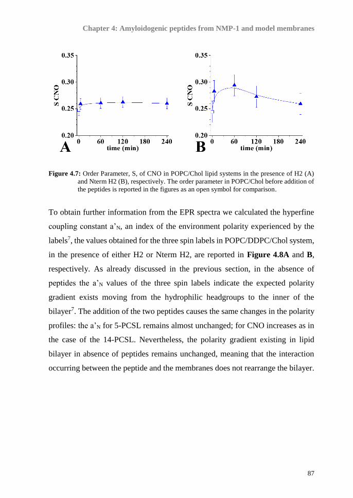
Chapter 4: Amyloidogenic peptides from NMP-1 and model membranes
87
Figure 4.7: Order Parameter, S, of CNO in POPC/Chol lipid systems in the presence of H2 (A)
and Nterm H2 (B), respectively. The order parameter in POPC/Chol before addition of
the peptides is reported in the figures as an open symbol for comparison.
To obtain further information from the EPR spectra we calculated the hyperfine
coupling constant a’N, an index of the environment polarity experienced by the
labels7, the values obtained for the three spin labels in POPC/DDPC/Chol system,
in the presence of either H2 or Nterm H2, are reported in Figure 4.8A and B,
respectively. As already discussed in the previous section, in the absence of
peptides the a’N values of the three spin labels indicate the expected polarity
gradient exists moving from the hydrophilic headgroups to the inner of the
bilayer7. The addition of the two peptides causes the same changes in the polarity
profiles: the a’N for 5-PCSL remains almost unchanged; for CNO increases as in
the case of the 14-PCSL. Nevertheless, the polarity gradient existing in lipid
bilayer in absence of peptides remains unchanged, meaning that the interaction
occurring between the peptide and the membranes does not rearrange the bilayer.

Chapter 4: Amyloidogenic peptides from NMP-1 and model membranes
88
Figure 4.8: Hyperfine coupling constant, a’N, for 5-PCSL (black), 14-PCSL (red) and CNO (blue)
in POPC/DDPC/Chol in the presence of H2 (A) and Nterm H2 (B), respectively. The
hyperfine coupling constant in POPC/DDPC/Chol before addition of the peptides is
reported in the figures as an open symbol for comparison.

Chapter 4: Amyloidogenic peptides from NMP-1 and model membranes
89
4.5. Conclusion
Peptides and proteins often show a conversion from their soluble forms into
fibrillar aggregates, giving rise to pathological conditions ranging from
neurodegenerative disorders to systemic amyloidosis and, in the fibrillar process,
early oligomers are the most toxic species. Recently, it was demonstrated that
several regions of the C-terminal domain of NPM1 form amyloid toxic
aggregates. The sequence covering the H2 region of the CTD resulted already
toxic at 0 and 1 h of aggregation, whereas the Nterm H2 peptide containing the
linker loop connecting H2 with H1, appeared weakly toxic at these time points,
causing the main effects after 15 h of aggregation time. Herein the biophysical
features of the aggregation of several NPM1 regions were investigated in the
presence of model membranes containing or not cholesterol. This study has the
aim to elucidate the different toxic behaviours exhibited by the two sequences
covering the H2 region of NPM1. The interaction between NPM1 regions and
LUVs mimicking membranes was firstly characterized in terms of conformational
effect of LUVs, by means of circular dichroism: while POPC bilayers do not
affect the conformation and aggregation properties of the peptides, which
preserves a β-sheet structure and aggregates with time, the presence of cholesterol
induces a slight conformational transition. For Nterm H2, the effects of lipid
model membranes seem to be less significant: no conformational change is
detected but only a suppression or delay of the aggregation process, both in the
presence of POPC and POPC/Chol LUVs. In this context it is worth to note that
Nterm H2 peptide presents a cholesterol binding consensus sequence (the CARC
sequence (K/R)-X 1−5-(Y/F)-X 1−5-(L/V)), but it seems able to discriminate worse
between the two lipid model membranes than H2. This is likely due to the
presence of two strong helical breaking residues (Gly and Pro) at the beginning
of the Nterm H2 that hampers such a fold of the peptide, while CRAC and CARC
sequences are known to effectively bind cholesterol when located within a α-
helical stretch. On the contrary, H2 is able to fold as a distorted helix in the

Chapter 4: Amyloidogenic peptides from NMP-1 and model membranes
90
presence of cholesterol, suggesting that it can bind cholesterol despite the
irregular CRAC sequence (since it presents one more residue between Val and
Tyr with respect to the consensus sequence). These differences may also justify
peptide different effects on POPC/Chol systems: H2 that likely binds cholesterol
through the CRAC sequence determines a sensitive fluidification of the whole
bilayer, as already observed for similar sequences, causing the loss of a polarity
gradient moving from the outer to the inner of the membrane. On the contrary
Nterm H2 causes a stiffening of central and outer regions of the bilayer, and a
partial fluidification of the inner hydrophobic region. The addition of the DHA
containing phospholipid DDPC to both POPC and POPC/Chol shows to have an
effect on both peptides.
When the mega-3 phospholipid is substituted in the liquid disordered phase, it
role out a solubilization effect on the H2 and Nterm H2, that is more marked for
the less amyloidogenic Nterm H2, due to the conformation changes that can be
observed: indeed, it shows a more random coiled conformation. Nevertheless, for
both peptides the interaction is labile, and mainly localized on the vesicles
surface; indeed, it is observed that the order parameter associated to the 5-PCSL-
the label positioned near the hydrophylic portion of the bilayer- is the most
affected during the first h of observation. Over time, the perturbation caused by
the peptides to the bilayer is suppressed.
Notably, when DDPC is added to this lipid system, no aggregation occurs but no
helical conformation of H2, which is supposed to be a transient step associated to
pore formation in the membranes, is promoted. The ability of DHA to solubilize
H2 and at the same time inhibit a possible step for pore formation can be an
explanation of the beneficial effects associated to DHA. Indeed, recently the pore
formation was considered as a plausible mechanism of pathogenesis of
neurodegenerative processes. In this respect, although a deeper investigation
should be performed to determine the selective binding that occurs between
amyloidogenic peptides and omega-3 phospholipids, the presented results could

Chapter 4: Amyloidogenic peptides from NMP-1 and model membranes
91
help in the comprehension of such affinity, contributing to build a robust scientific
platform for the biomedical and pharmaceutical exploitation of these molecules.

Chapter 4: Amyloidogenic peptides from NMP-1 and model membranes
92
4.6. References
1 Di Natale, C. et al. Nucleophosmin contains amyloidogenic regions that are able to form toxic
aggregates under physiological conditions. FASEB journal : official publication of the
Federation of American Societies for Experimental Biology, doi:10.1096/fj.14-269522 (2015).
2 Scaloni, F., Gianni, S., Federici, L., Falini, B. & Brunori, M. Folding mechanism of the C-
terminal domain of nucleophosmin: residual structure in the denatured state and its
pathophysiological significance. Faseb J 23, 2360-2365, doi:10.1096/fj.08-128306 (2009).
3 Scaloni, F., Federici, L., Brunori, M. & Gianni, S. Deciphering the folding transition state
structure and denatured state properties of nucleophosmin C-terminal domain. Proc Natl Acad
Sci U S A 107, 5447-5452, doi:10.1073/pnas.0910516107 (2010).
4 Chiarella, S., Federici, L., Di Matteo, A., Brunori, M. & Gianni, S. The folding pathway of a
functionally competent C-terminal domain of nucleophosmin: protein stability and denatured
state residual structure. Biochem Biophys Res Commun 435, 64-68,
doi:10.1016/j.bbrc.2013.04.038 (2013).
5 Cecchi, C. & Stefani, M. The amyloid-cell membrane system. The interplay between the
biophysical features of oligomers/fibrils and cell membrane defines amyloid toxicity.
Biophysical chemistry 182, 30-43, doi:10.1016/j.bpc.2013.06.003 (2013).
6 Marsh, D. Molecular motion in phospholipid bilayers in the gel phase: long axis rotation.
Biochemistry 19, 1632-1637 (1980).
7 Vitiello, G. et al. Fusion of raft-like lipid bilayers operated by a membranotropic domain of
the HSV-type I glycoprotein gH occurs through a cholesterol-dependent mechanism. Soft
Matter 11, 3003-3016, doi:10.1039/c4sm02769h (2015).

Chapter 5:
Aβ 1-42 and model membranes


Chapter 5: Aβ 1-42 and model membranes
95
5.1. Work outline
Many experimental evidences indicate that the interactions with neuronal
membranes play an important role in the Aβ 1-42 toxicity1,2 and the study of the
interactions between Aβ peptides and membrane model systems is a very
important topic in the recent literature.
AD is also elicited by the interplay of genetic, environmental and dietary
factors3,4. The adherence to a Mediterranean diet profile, including fish,
vegetables, fruits, coffee, and light-to-moderate alcohol intake, seems to reduce
the incidence of this and other neurodegenerative pathologies5. The beneficial
effects of fish are at least partially ascribed to the high content of polyunsaturated
fatty acids (PUFA), and specifically of omega-3 ones6. However, the
effectiveness of these molecules is still controversial, and their mechanism of
action remains elusive. Among other hypotheses, mainly based on the radical-
scavenging and anti-inflammatory action of PUFA7,8 it has been proposed that
omega-3 fatty acids, once converted to lipids, could alter the structure and
functionality of neuronal membranes, whose involvement in AD aetiology is
widely accepted2.
Docosahexaenoic acid (DHA) is found in extraordinarily high concentration in
the plasma membranes of neural tissues, reaching up to 50% of the total acyl
chains9,10 and its decline has been associated with the loss of memory and
learning, suggesting that the omega-3 fatty acids modulate the membranes
properties and influence the molecular mechanism of AD11,12. Recently, it has
been proposed that omega-3 lipids disturb the lipid packing in the membrane,
favoring a deeper internalization of Aβ fragments among the lipid acyl chains13
offering a possible explanation for the inhibition of Aβ self-aggregation operated
by omega-3 fatty acids.
To get light in this complex scenario, the interaction of Aβ 1-42 with bilayers
whose lipid composition reproduces the main components of neuronal
membranes14,15, including both zwitterionic and anionic phospholipids,

Chapter 5: Aβ 1-42 and model membranes
96
cholesterol, sphingomyelin and gangliosides has been investigated. Moreover, the
effect of the inclusion of the omega-3 lipid 1,2-didocosahexaenoyl-sn-glycero-3-
phosphocholine DDPC in the bilayer composition is analysed in detail.
An integrated experimental approach was applied for this study. The effect of
complex lipid bilayers on Aβ 1-42 conformation/self-aggregation was monitored
by Circular Dichroism (CD); the influence of Aβ 1-42 interaction on the
membrane microstructure and fluidity was studied by Electron Spin Resonance
(ESR) measurements; the disrupting effect of this interaction on membrane
integrity was studied by a previously established FRET-based membrane leakage
assay.
5.2. Materials and methods
Materials
Chloroform, dichloromethane and methanol, HPLC-grade solvents, were
obtained from Merck (Darmstadt, Germany). 1-Palmitoyl-2-oleoyl-sn-glycero-3-
phosphatidylcholine (POPC), 1-Palmitoyl-2-oleoyl-sn-glycero-3-phospho-L-
serine (POPS), N-octadecanoyl-D-erythro-sphingosylphosphorylcholine (SM),
ganglioside (GM1) ovine brain sodium salt, 1,2-didocosahexaenoyl-sn-glycero-
3-phosphocholine (DDPC) were obtained from Avanti Polar Lipids (Birmingham,
AL, USA). Spin-labeled phosphatidylcholines (1-palmitoyl-2-stearoyl-(n-doxyl)-
sn-glycero-3-phosphocholine, n-PCSL) with the nitroxide group in the positions
5, 7, 10 and 14 of the acyl chain were also obtained from Avanti Polar Lipids.
Aβ 1-42 peptide was synthesized as previously reported and retreated with TFA,
in order to dissolve any fibrillar aggregates, as previously described [14]. The
sample was then freeze-dried and redissolved in pure HFIP, and then the UV and
CD spectra were recorded to assess the acid treatment success.
Table1: Peptides sequences.
Aβ 1-42 1DAEFRHDSGYEVHHQKLVFFAEDVGSNKGAIIGLMVGGVVIA42

Chapter 5: Aβ 1-42 and model membranes
97
Lipid mixtures preparation
All lipid mixtures were prepared mixing appropriate amounts of lipids, dissolved
in a chloroform/methanol (2:1 v/v) in a round-bottom test tube. For Electron Spin
Resonance measurements spin-labeled phosphatidylcholines (n-PCSL) were
alternatively added to the lipid mixture (1% mol% on the total lipid) by mixing
appropriate amounts of a spin-label solution in ethanol (1 mg/mL) with the lipid
organic mixture. A thin lipid film was produced by evaporating the solvents with
dry nitrogen gas and final traces of solvents were removed by subjecting the
sample to vacuum desiccation for at least 3 h. The samples were then hydrated
with the proper amount of buffer (i.e. Phosphate buffer 10mM) pH=7.4,
repeatedly vortexed, avoiding any further treatment of the samples.
Large unilamellar vesicles
Large unilamellar vesicles (LUVs) used for the supported bilayer, ESR
spectroscopy and CD spectroscopy in the case of peptides interaction study with
lipid membranes, were prepared as described in Section 5.2.1 and then extruded
through a 0.1 μm polycarbonate membranes 21 times.
For Aβ 1-42 investigation, LUVs consisting on POPC/POPS/Chol/SM/GM1
(8/2/9/1/1 mol/mol), henceforth indicated as neuronal membrane-like lipid
bilayers, NML-LB, POPC/POPS/Chol/SM/GM1/DDPC (4/2/9/1/1/5mol/mol),
henceforth indicated as O3-LB and POPC/POPS/Chol/SM/GM1/DDPC
(6/2/9/1/1/2mol/mol), henceforth indicated as O3L-LB, were prepared in 20mM
phosphate buffer pH 7.4, freshly degassed by nitrogen purging, at total
concentration of 0.4 mg/ml.
Electron spin resonance
ESR spectra were recorded with a 9 GHz Bruker Elexys E-500 spectrometer
(Bruker, Rheinstetten, Germany). The capillaries containing MLVs suspensions,
were placed in a standard 4 mm quartz sample tube containing light silicone oil
for thermal stability. All the measurements were performed at 25 °C. Spectra were
recorded using the following instrumental settings: sweep width, 100 G;

Chapter 5: Aβ 1-42 and model membranes
98
resolution, 1024 points; time constant, 20.48 ms; modulation frequency, 100 kHz;
modulation amplitude, 1.0 G; incident power, 6.37 mW. Several scans, typically
16, were accumulated to improve the signal-to-noise ratio. For the spectra
simulation the gii values for the coupling between the electron spin and the
magnetic field were those which gave the best fitting for all spectra: gii=2.0075,
2.006, 2.003. In some cases, due to fitting improvements, 2.0027 was used instead
of 2.003 for gzz. About the Aii values for the coupling between the electron spin
and the nitrogen nuclear spin, which give a measure of the environmental polarity,
we maintained constant Axx=7.1 G and Ayy=7.2 G values, which were also well
fitting all the spectra, while it was needed to change the Azz value from one to
another system, where we list the <A>=(Axx+Ayy+Azz)/3 values. In order to
quantitatively analyse the spectra, we calculated the order parameter, S, using a
spectra parameterization method defined in Equation 3.1.
𝑆 = 𝑇∥− 𝑇⊥
𝑇𝑧𝑧− 𝑇𝑥𝑥 ∙
𝑎𝑁
𝑎′𝑁 Eq.3.1
as already discussed in section 3.2.
Circular Dichroism
CD spectra of H2, Nterm H2, Aβ 1-42 (0.1 mg/ml final concentration) were
acquired with aJasco J-810 spectropolarimeter (Jasco international Co. Ltd,
Tokyo, Japan) in the range of 200–260 nm using a 0.1 cm path length quartz
cuvette at 20 °C in continuous scanning mode (20 nm/min, with a 4.0 s response
and a 1.0 nm band width). Spectra corresponding only to the solvent or lipid
bilayers in the absence of the peptide have been recorded thoroughly during the
measurements and subtracted from the corresponding peptide/lipid bilayer
spectrum. The data were accumulated over 3 runs, the presented data being the
average. The results are not converted to molar ellipticity because in the presence
of an aggregation process is not possible to determine the peptide concentration.

Chapter 5: Aβ 1-42 and model membranes
99
5.3. Sample condition design
Six different conditions were explored in this work; Aβ 1-42 concentration was
0.020 M phosphate buffer, pH 7.4, 80/20 (V/V), in order to check peptide
behaviour in a polar environment; 0.020 M phosphate buffer, pH 7.4 to check a
polar environment. Four different liposomes composition were studied: 0.4 mg/ml
of lipid bilayers formed only by palmytoil-oleil-phosphocholine (POPC-LB); 0.4
mg/ml of neuronal membrane mimicking lipid bilayer (NML-LB)15,16; 0.4 mg/ml
of NML-LB containing also 25% w/w of the unsaturated fatty acid DDPC (O3-
LB); 0.4 mg/ml of NML-LB containing 10% w/w of DDPC (O3L-LB). The two
last mixtures were prepared with two different amounts of DDPC to evaluate its
specific potential effect on Aβ 1-42 solubility and structure. The detailed
liposome compositions are reported in the experimental section. The chemical
structure of all lipids used for this study are reported in Figure 5.1.
Figure 5.1: Chemical structures of all lipids used in liposomes formulation.
5.4. Circular Dichroism
In consequence of the generally low solubility of Aβ 1-42 and fast aggregation in
aqueous solutions, we have checked the peptide conformation in experimental
conditions already known to induce canonical secondary structures in Aβ, i.e.

Chapter 5: Aβ 1-42 and model membranes
100
HFIP/water mixture and phosphate buffer. Both the controls spectra gave the
expected results, showing the typical shape of α-helix and β-sheet respectively,
thus confirming the absence of pre-existing aggregates in the dry peptide film.
Interestingly, in the presence of POPC-LB the spectrum of the peptide showed
the shape of a β-structure, though the minimum was shifted at a slightly higher
wavelength, according to the presence of aggregated forms17. In contrast, in the
presence of more complex lipid bilayers (NML-LB, O3-LB and O3L-LB), only a
low intensity CD signal was detected and no regular secondary structure could be
associated to the spectrum. The CD spectra of peptide upon dissolution in every
condition are reported in Figure 5.2. Since the kinetics of aggregation/dissolution
processes and conformational transitions of Aβ 1-42 are quite slow, the CD
spectra of the peptide in the different conditions was monitored over several days.
Figure 5.2 CD spectra of A Aβ 1-42 just after dissolution in the conditions studied.
The results obtained are reported in Figure 5.3 for each liposome conditions. The
control experiments gave the expected results: the spectrum in HFIP/water
mixture was unaffected over the whole incubation time; on the other hand, when

Chapter 5: Aβ 1-42 and model membranes
101
the peptide was dissolved in phosphate buffer, the signal intensity decreases over
time, becoming almost undetectable after 14 days, in agreement with the expected
formation of insoluble aggregates in aqueous environments.
Figure 5.3: CD spectra time evolution of Aβ 1-42 interacting with different lipid bilayer: Panel A
POPC-LB; Panel B NML-LB; Panel C O3L-LB; Panel D O3-LB.
Figure 5.3 A shows that the shape and intensity of the spectra acquired in POPC-
LB over time were constant, suggesting that the soluble aggregate concentration
does not change. However, the presence of insoluble, CD silent forms cannot be
excluded. In the presence of NML-LB bilayers, the low intensity of the CD signal
indicates that only a small fraction of the peptide was dissolved (Panel B), and
no significant increase is appreciated over time. Yet, the spectrum shape changes
during the incubation time, confirming that the lipid bilayer is able to interact with
the peptide2. Unlike the previous cases, in the lipid bilayer O3-LB, the peptide
slowly but extensively dissolves after several days, as highlighted by a consistent

Chapter 5: Aβ 1-42 and model membranes
102
increase in the CD spectrum intensity. The spectrum assumes a shape similar to
that observed in the presence of POPC, except for a further red-shift of the
minimum, suggesting the presence of higher size soluble aggregates.
Interestingly, the time necessary to appreciate changes in the CD spectrum
decreases when the amount of omega-3 lipid present in the lipid mixture
increases. Indeed, while the presence of higher concentration of DDPC (Panel D)
induces the peptide film dissolution after 21 days incubation, at lower DDPC
concentration (Panel C) the spectrum shape changes slightly over 28 days
incubation, but no clear trend can be appreciated, as in the case of NML-LB.
These evidences suggest the presence of membrane interacting peptide aggregates
slowly evolving over time when omega-3 lipids present in the lipid bilayer
formulation. However, the kinetics of this process critically depends on the
amount of omega-3 present in the lipid phase.
5.5. Electron spin resonance
The changes in lipid ordering and dynamics was studied using the spin label
approach. Two spin-labeled phosphocholines were used as reporter: 5-PCSL and
14-PCSL. The former bears the radical in proximity of the polar head, just
underneath the hydrophilic surface, while in the latter the label is close to the acyl
chain terminus, thus spectra are representative of the inner apolar region
behaviour.
The spectra of 5- and 14-PCSL in POPC bilayers are reported in Figure 5.4,
respectively Panel A and Panel B (black lines). 14-PCSL generates an almost
isotropic three-line spectrum. In contrast, a clearly defined anisotropy is observed
for 5-PCSL, as highlighted by the splitting of the low- and high-field signals.
Thus, the rotational motion of the label is hindered in the relatively ordered
external region of the bilayer, while it is almost free in the bilayer interior. A
quantitative analysis of the spectra was accomplished by determining the lipid
segmental order parameter (S), as reported in the literature18. The S value is 0.64

Chapter 5: Aβ 1-42 and model membranes
103
for 5-PCSL and 0.14 for 14-PCSL. These evidences indicate that POPC-LB
presents a marked rigidity and ordering decrease in going from the lipid polar
head to the acyl tails termini, as expected for membranes in the liquid crystalline
disordered state (Ld)19. In the case of the NML-LB, both spin-labelled lipids show
anisotropic spectra (Figure 5.4). The S value is 0.74 for 5-PCSL and 0.41 for 14-
PCSL, showing that these bilayers are more rigid than the previous ones, and the
lipid ordering gradient in going from the polar head to the acyl tails termini is
lower. This indicates that NML-LB are in the liquid crystalline ordered state (Lo).
Also in the presence of DDPC both spin-labelled lipids have anisotropic spectra;
the two omega3 containing lipid bilayers show the same lineshape, spectra for the
O3-LB mixture are shown in Figure 5.4. No additional spectral component,
evocative of domain formation20, was detected. The rigidity of the membrane is
only slightly reduced with respect to NML-LB even in the presence of the higher
amount of omega-3 lipids, as the S value is 0.68 for 5-PCSL and 0.38 for 14-
PCSL. This indicates that inclusion of DDPC in the lipid mixture does not change
the liquid crystalline organization of the bilayer, which remains in the Lo state. In
a previous work was found that DDPC is able to drive the Lo/Ld transition in the
case of a POPC/SM/Chol bilayer13. Moreover, omega-3 lipid bearing two
polyunsaturated acyl chains, like DDPC, are reported to drive the segregation of
cholesterol, with formation of lipid domains21,22. The resistance of NML-LB to
undergo any transition is probably ascribable to the presence of GM1, which is
recognized as a major player in the modulation of later ordering within biological
membranes23, limiting domain growth. The presence of Aβ 1-42 in the system
affects the POPC-LB organization13,19, as revealed by the spectra of 5- and 14-
PCSL registered soon after samples preparation (Figure 5.4, red lines). In the case
of 5-PCSL a slight reduction of the spectrum anisotropy is observed, as confirmed
by the decrease of the S value by ~0.02. An opposite behaviour is observed for
14-PCSL: the spectrum anisotropy increases and S raises by ~0.04. However, no
evidence of a second component in the spectrum is observed, as expected for

Chapter 5: Aβ 1-42 and model membranes
104
membrane-penetrating peptides. Thus, the experimental evidences point to a
positioning of the peptide close to the membrane interface, causing perturbation
in the lipid dynamic and ordering that propagates to the bilayer inner core.
Figure 5.4: EPR spectra of the two spin reporter used in this study, 5-PCSL (Panel A) and 14-
PCSL (Panel B) of the investigated liposome compositions. Black lines: EPR spectra
of liposomes without Aβ 1-42. Red lines: EPR spectra just after peptide addition.
Cyan lines: EPR spectra after 21 days of incubation.
Even for NML-LB the presence of Aβ 1-42 in the system causes a small reduction
of the 5-PCSL spectrum anisotropy and a consequent slight S decrease of ~0.02.
However, almost no variation is found in the 14-PCSL spectrum. These results
indicate a weak interaction of the peptide with the membrane interface, which
does not affect the inner bilayer structure. In the case of the omega-3 lipid
containing lipid bilayers, similarly to what observed for NML-LB, the presence
of Aβ 1-42 causes a reduction of the 5-PCSL spectrum anisotropy, as confirmed
by the reduction of 0.02 in the S value, while no effect is registered for 14-PCSL.

Chapter 5: Aβ 1-42 and model membranes
105
Thus, inclusion of DDPC does not affect peptide-membrane interaction in just-
prepared samples.
Figure 5.5 Panel A) and for 14PCSL
(Panel B) for each lipid bilayers studied when interacting with Aβ 1-42.
The evolution over time of the 5- and 14-PCSL spectra in all the considered
bilayers was followed both in the absence and in the presence of Abet42. No
spectra change was observed in the peptide absence. In the case of O3L-LB and
O3-LB, this indicates no DDPC oxidation to occur, in line with the long-term
stability of omega-3 lipids in liposomes reported in the literature24. For samples
containing the peptide significant changes of the spectra with time were observed,
depending on the bilayer lipid composition. In the case of POPC a progressive
reduction of the 5-PCSL spectrum anisotropy is observed, as shown by the trend
of ΔS reported in Figure 5.5. As an example, the spectra of the two spin labels in
POPC-LB in the presence of the peptide after 21 days are shown in Figure 5.4
(cyan lines). In contrast, the 14-PCSL S value remains almost constant (Figure
5.5). The 5-PCSL and 14-PCSL spectra in NML-LB exposed to Aβ 1-42 remain
almost unaltered, indicating that the weak interaction of the peptide with the
membrane does not strengthen with time. In the presence of omega-3 containing

Chapter 5: Aβ 1-42 and model membranes
106
lipid bilayers, for both the formulations considered (O3L-LB and O3-LB), the 5-
PCSL spectrum exposed to Aβ 1-42 remains almost constant whilst, interestingly,
in the case of 14-PCSL the spectrum anisotropy becomes markedly more evident
after three weeks and, consequently, the S value significantly increases (Figure
5.5). These results indicate an increasing membrane perturbation of the bilayer
inner core due to the interaction with the peptide, modulated by the amount of
omega-3 lipids in the membrane, as it is elicited after a smaller time-lag for O3-
LB than for O3L-LB systems (Figure 5.5D). Internalization of Aβ fragments has
been observed in the case of POPC/SM/Chol bilayers enriched in DDPC, which,
differently from the ones used in this study, are in the Ld state13. This could
indicate that the peptide selectively interacts with omega-3 lipids, almost
independently of the lipid bilayer state.

Chapter 5: Aβ 1-42 and model membranes
107
5.6. Conclutions
In conclusion, by using CD spectroscopy, we have preliminarily verified that, in
phosphate buffer and in the absence of lipids, the peptide adopts the expected β
conformation and undergo the subsequent self-aggregation in insoluble
aggregates. The effect of single-component lipid bilayer has been also
investigated by using POPC, a zwitterionic phospholipid abundant in eukaryotic
cells, often chosen for membrane-peptide interaction studies. Our results show
that POPC-LB stabilize for several weeks a soluble, aggregated form of Aβ 1-42,
having a small but constant concentration. The CD spectrum is indeed very
reproducible over time, but has a low intensity and shows a shift of the β-sheet
minimum toward higher lambda values, which can be the effect of a β aggregated
but soluble structure, with a scattering25 due to the presence of high molecular
weight aggregates. ESR results show that these aggregates interact with the
membrane interface, increasingly disturbing its organization, while the bilayer
interior is only indirectly affected. Interestingly, biomimetic lipid bilayers (NML-
LB) do not present the ability to solubilize the peptide, although CD spectra
suggest the presence of a small amount of peptide deprived of regular secondary
structure also associated with the lipid phase. Our results indicate that, in the
presence of GM1, large and likely heterogeneous aggregates interacting with the
membrane interface quickly form. Furthermore, the experimental evidences
clearly show that these insoluble aggregates are not able to penetrate in the lipid
bilayer2. This could be connected with the rigidity of the lipid phase (in the Lo
state), which in this case is much higher than that of POPC-LB (in the Ld state).
Inclusion of DDPC in the lipid composition does not affect the initial behavior of
the membrane in the presence of Aβ 1.42: only a limited perturbation of O3-LB
interface is observed, probably due to a weak interaction with large, insoluble
aggregates. However, CD data clearly show that, over time, an increasing fraction
of the peptide convert to soluble, lipid associated, aggregates, able to interact with
the bilayer.

Chapter 5: Aβ 1-42 and model membranes
108
Figure 3.7 Scheme of the different interaction model proposed for Aβ 1-42 and neuronal
membrane biomimetic lipid bilayers (left side), and omega-3 containing biomimetic bilayers (right
side).
The β-sheet minimum indeed undergoes a further shift toward high wavelength
with respect to POPC-LB, suggesting an increasing of the scattering contribution
in the spectra. Furthermore, ESR spectra indicate that these aggregates are
significantly embedded in the bilayer, differently from the superficial interaction
in case of NML-LB, as schematised in Figure 3.7.
The comparison of the data obtained by introducing two different DDPC amounts
in the lipid bilayer formulation indicates that qualitatively the effect of omega-3
lipid presence is independent by the amount of this component, however the
extent of the perturbation and the kinetics of the process are enhanced when the
omega-3 lipid concentration increases. It is to be highlighted that the dramatic
enhancement of the Aβ 1-42 affinity for the membrane due to DDPC cannot be
straightforwardly related to changes of the bilayer properties, considering that this
lipid causes only a slight reduction of the bilayer ordering. This result could
induce to speculate about a peptide molecular preference for the omega-3 lipid,
which, however, should be better investigated.
Moreover, the interaction between Aβ 1-42 and the investigated lipid bilayers
causes a strong permeabilization of the latter. Considering the three molecular
models proposed for the Aβ 1-42 membrane disrupting activity, detailed
discussed in section 1.7, the results obtained suggest that the ion channel
mechanism is the most likely for the conditions studied, even if for the NML-LB

Chapter 5: Aβ 1-42 and model membranes
109
only subtle changes in the bilayer microstructure was detected by ESR. In the
carpeting mechanism, a critical peptide concentration should be reached before
the membrane permeabilization occurs; but, as shown in figure 3.6, the initial
trend of the titration is almost linear, without any concentration lag phase at the
beginning. On the other hand, in the detergent mechanism, the peptide
disassembles the lipid vesicles almost completely, forming smaller species such
as micelles, but such a dramatic effect on the bilayer microstructure was not
detected by ESR analysis.

Chapter 5: Aβ 1-42 and model membranes
110
5.7. References
1 Matsuzaki, K. Physicochemical interactions of amyloid beta-peptide with lipid bilayers.
Biochimica et biophysica acta 1768, 1935-1942, doi:10.1016/j.bbamem.2007.02.009 (2007).
2 Williamson, R. & Sutherland, C. Neuronal Membranes are Key to the Pathogenesis of
Alzheimer's Disease: the Role of Both Raft and Non-Raft Membrane Domains. Curr
Alzheimer Res 8, 213-221 (2011).
3 Hu, N. et al. Nutrition and the risk of Alzheimer's disease. BioMed research international
2013, 524820, doi:10.1155/2013/524820 (2013).
4 Reitz, C. & Mayeux, R. Alzheimer disease: epidemiology, diagnostic criteria, risk factors and
biomarkers. Biochemical pharmacology 88, 640-651, doi:10.1016/j.bcp.2013.12.024 (2014).
5 Singh, B. et al. Association of mediterranean diet with mild cognitive impairment and
Alzheimer's disease: a systematic review and meta-analysis. Journal of Alzheimer's disease :
JAD 39, 271-282, doi:10.3233/JAD-130830 (2014).
6 Cunnane, S. C. et al. Fish, docosahexaenoic acid and Alzheimer's disease. Progress in lipid
research 48, 239-256, doi:10.1016/j.plipres.2009.04.001 (2009).
7 Bazinet, R. P. & Laye, S. Polyunsaturated fatty acids and their metabolites in brain function
and disease. Nature reviews. Neuroscience 15, 771-785, doi:10.1038/nrn3820 (2014).
8 Hjorth, E. et al. omega-3 fatty acids enhance phagocytosis of Alzheimer's disease-related
amyloid-beta42 by human microglia and decrease inflammatory markers. Journal of
Alzheimer's disease : JAD 35, 697-713, doi:10.3233/JAD-130131 (2013).
9 Stillwell, W., Shaikh, S. R., Zerouga, M., Siddiqui, R. & Wassall, S. R. Docosahexaenoic acid
affects cell signaling by altering lipid rafts. Reproduction, nutrition, development 45, 559-579,
doi:10.1051/rnd:2005046 (2005).
10 Wassall, S. R. & Stillwell, W. Polyunsaturated fatty acid-cholesterol interactions: domain
formation in membranes. Biochimica et biophysica acta 1788, 24-32,
doi:10.1016/j.bbamem.2008.10.011 (2009).
11 Escriba, P. V. et al. Membrane lipid therapy: Modulation of the cell membrane composition
and structure as a molecular base for drug discovery and new disease treatment. Progress in
lipid research 59, 38-53, doi:10.1016/j.plipres.2015.04.003 (2015).
12 Prasad, M. R., Lovell, M. A., Yatin, M., Dhillon, H. & Markesbery, W. R. Regional membrane
phospholipid alterations in Alzheimer's disease. Neurochemical research 23, 81-88 (1998).
13 Vitiello, G., Di Marino, S., D'Ursi, A. M. & D'Errico, G. Omega-3 fatty acids regulate the
interaction of the Alzheimer's abeta(25-35) peptide with lipid membranes. Langmuir : the ACS
journal of surfaces and colloids 29, 14239-14245, doi:10.1021/la403416b (2013).
14 Calderon, R. O., Attema, B. & Devries, G. H. Lipid-Composition of Neuronal Cell-Bodies and
Neurites from Cultured Dorsal-Root Ganglia. Journal of neurochemistry 64, 424-429 (1995).
15 Soderberg, M., Edlund, C., Kristensson, K. & Dallner, G. Fatty acid composition of brain
phospholipids in aging and in Alzheimer's disease. Lipids 26, 421-425 (1991).
16 Hennebelle, M. et al. Ageing and apoE change DHA homeostasis: relevance to age-related
cognitive decline. The Proceedings of the Nutrition Society 73, 80-86,
doi:10.1017/S0029665113003625 (2014).
17 Martins, I. C. et al. Lipids revert inert Abeta amyloid fibrils to neurotoxic protofibrils that
affect learning in mice. The EMBO journal 27, 224-233, doi:10.1038/sj.emboj.7601953
(2008).
18 Vitiello, G., Grimaldi, M., D'Ursi, A. M. & D'Errico, G. The iAbeta5p beta-breaker peptide
regulates the Abeta(25-35) interaction with lipid bilayers through a cholesterol-mediated
mechanism. Biochemical and biophysical research communications 417, 88-92,
doi:10.1016/j.bbrc.2011.11.061 (2012).
19 Vitiello, G. et al. Cholesterol modulates the fusogenic activity of a membranotropic domain
of the FIV glycoprotein gp36. Soft Matter 9, 6442-6456, doi:10.1039/c3sm50553g (2013).

Chapter 5: Aβ 1-42 and model membranes
111
20 Georgieva, R. et al. Docosahexaenoic acid promotes micron scale liquid-ordered domains. A
comparison study of docosahexaenoic versus oleic acid containing phosphatidylcholine in raft-
like mixtures. Biochimica et biophysica acta 1848, 1424-1435,
doi:10.1016/j.bbamem.2015.02.027 (2015).
21 Brzustowicz, M. R. et al. Controlling membrane cholesterol content. A role for
polyunsaturated (docosahexaenoate) phospholipids. Biochemistry 41, 12509-12519 (2002).
22 Shaikh, S. R., Kinnun, J. J., Leng, X., Williams, J. A. & Wassall, S. R. How polyunsaturated
fatty acids modify molecular organization in membranes: insight from NMR studies of model
systems. Biochimica et biophysica acta 1848, 211-219, doi:10.1016/j.bbamem.2014.04.020
(2015).
23 Puff, N., Watanabe, C., Seigneuret, M., Angelova, M. I. & Staneva, G. Lo/Ld phase
coexistence modulation induced by GM1. Biochimica et biophysica acta 1838, 2105-2114,
doi:10.1016/j.bbamem.2014.05.002 (2014).
24 Rasti, B., Jinap, S., Mozafari, M. R. & Yazid, A. M. Comparative study of the oxidative and
physical stability of liposomal and nanoliposomal polyunsaturated fatty acids prepared with
conventional and Mozafari methods. Food Chem 135, 2761-2770,
doi:10.1016/j.foodchem.2012.07.016 (2012).
25 Bustamante, C., Tinoco, I., Jr. & Maestre, M. F. Circular differential scattering can be an
important part of the circular dichroism of macromolecules. Proc Natl Acad Sci U S A 80,
3568-3572 (1983).

Appendix A:
Effect of polyunsaturated free fatty
acids on Lβ phases


Appendix A: Effect of polyunsaturated free fatty acids on Lβ phases
115
A.1. Work outline
The dietary intake of Omega3 fatty acids such as DHA and EPA is linked to the
prevention of cancer, hearth diseases and is associated to brain development1,2.
Among other hypotheses, mainly based on their radical-scavenging and anti-
inflammatory action3, it has been proposed that omega-3 fatty acids, once
converted to lipids4, could alter the structure and functionality of biological
membranes, whose involvement in major biological processes, such as signaling
and protein trafficking, is widely reported. Thus, the effects of different lipids
containing DHA on both the Ld and Lo phases have been investigated and the
results are presented in chapter 3. Nevertheless also the role of free fatty acids
should be taken into account; indeed the it has been found vesicle cycling, which
is an important biological event, involves the interplay between membrane lipids
and proteins, among which the enzyme phospholipase A2 (PLA2) plays a critical
role5. Phospholipase A2 family consists on a group of enzymes that hydrolyze
the ester bond at the sn-2 position in glycerophospholipids, inducing the release
of a free fatty acid (FA) and lysophospholipid. In nervous cells, PLA2 was also
shown to control the metabolic transformation of phospholipid (PL) molecules
that contain PUFAs6. In addition, Georgieva et al.7, using raft-like lipid mixtures,
demonstrated that line tension and elastic properties govern budding formation
after the addition of phospholipase A2 (PLA2), which explains why DHA-
containing phosphatidylcholine (PC), but not oleic acid (OA)- containing PC, are
able to exhibit liquid-ordered (Lo) domain budding. Very little is known about
the interaction of free fatty acids with model membranes, thus the effect of
polyunsaturated fatty acids, i.e. DHA and EPA, and also OA as control on DPPC
membranes were investigated. The study was carried out by means of differential
microCalorimetry; in addition, generalized polarization for Laurdan label
imbedded in DPPC and eggPC was investigated in 0rder to obtain information on
the fluidity of membranes and cconfocal microscopy of GUVs containing DHA
were acquired to verify the existence of microdomains.

Appendix A: Effect of polyunsaturated free fatty acids on Lβ phases
116
A.2. Materials and methods
Materials
Chloroform, dichloromethane and methanol, HPLC-grade solvents, were
obtained from Merck (Darmstadt, Germany). EggPC, 1,2-dipalmitoyl-sn-glycero-
3-phosphatidylcholine (DPPC) and 1-palmytoyl-2-docosahexaenoyl-sn-glycero-
3-phosphocholine (PDPC) were obtained from Avanti Polar Lipids (Birmingham,
AL, USA). Docosahexaenoic acid, Eicosapentaenoic acid and oleic acid were
provided from Sigma Aldrich (St. Louis, MO).
Differential Scanning microcalorimetry (DSC)
All measurements were performed in a VP-DSC high-sensitivity scanning
microcalorimeter (MicroCal, Northampton, MA). Both lipid and buffer solutions
were fully degassed before loading into the appropriate cell. Buffer was 20 mM
PIPES, 150 mM NaCl, 1 mM EDTA, pH 7.4. Lipid suspensions were loaded into
the microcalorimeter in the form of multilamellar vesicles. A final amount of
0.5ml at 0.5mM total lipid concentration was loaded into the calorimeter, and up
to six heating scans were performed at 45 °C/h between 10 and 60 °C for all
samples. Lipid concentration was determined as lipid phosphorus, and used
together with data from the last scan, to obtain normalized thermograms. The
software Origin 7.0 (MicroCal), provided with the calorimeter, was used to
determine the different parameters for the scans. The software PeakFit v4.12 was
used for curve-fitting.
Fluorescence spectroscopy
The emission maxima of LAURDAN in phospholipid bilayers depend upon the
phase state of the phospholipids, being blue in the gel (maximum emission =440
nm) and green in the liquid crystalline phase (maximum emission =490 nm)
(Figure A.1). This shift of the emission spectrum has been attributed to dipolar
relaxation processes occurring in the phospholipid liquid–crystalline phase but
not in the gel phase8. The generalized polarization (GP) of Laurdan was measured

Appendix A: Effect of polyunsaturated free fatty acids on Lβ phases
117
in an Aminco Bowman Series 2 spectrofluorometer (ThermoFisher Scientific,
Waltham, MA) equipped with thermoregulated cell holders.
Figure A.1: A) Laurdan emission spectra in both Lβ and Lα phases; B) schematic representation
of Laurdan label inserted in a lipid layer9.
The GP was calculated by the equation A.1:
𝐺𝑃 =𝐼440−𝐼490
𝐼440+𝐼490 Eq. A.1
where I440 and I490 are the emission intensities at 440 and 490 nm, respectively,
when exciting at 360 nm. The final probe/lipid molar ratio was 1/1000.
Confocal microscopy of GUVs
Giant Unilamellar Vesicles (GUVs) were prepared using the electroformation
method. For vesicle observation, a home-made chamber was used that allows
direct GUV visualization under the microscope10. Stock lipid solutions (0.2
mg/mL total lipid containing 0.2 mol % lissamine rhodamine PE (Rho-PE)) were
prepared in a chloroform/methanol (2:1, v/v) solution. Then, 3 μL of the
appropriate lipid stocks were added onto the surface of Pt electrodes and solvent
traces were removed by placing the chamber under high vacuum for at least 2 h.
The Pt electrodes were covered with 400 mL Millipore filtered Milli-Q water
previously equilibrated at 75°C. The Pt wires were connected to an electric-wave
generator (TG330 function generator, Thurlby Thandar Instruments, Huntington,
United Kingdom) under AC field conditions (10 Hz, 0.9 V) for 2 h at 75°C. The
generator and the water bath were switched off, and vesicles were left to
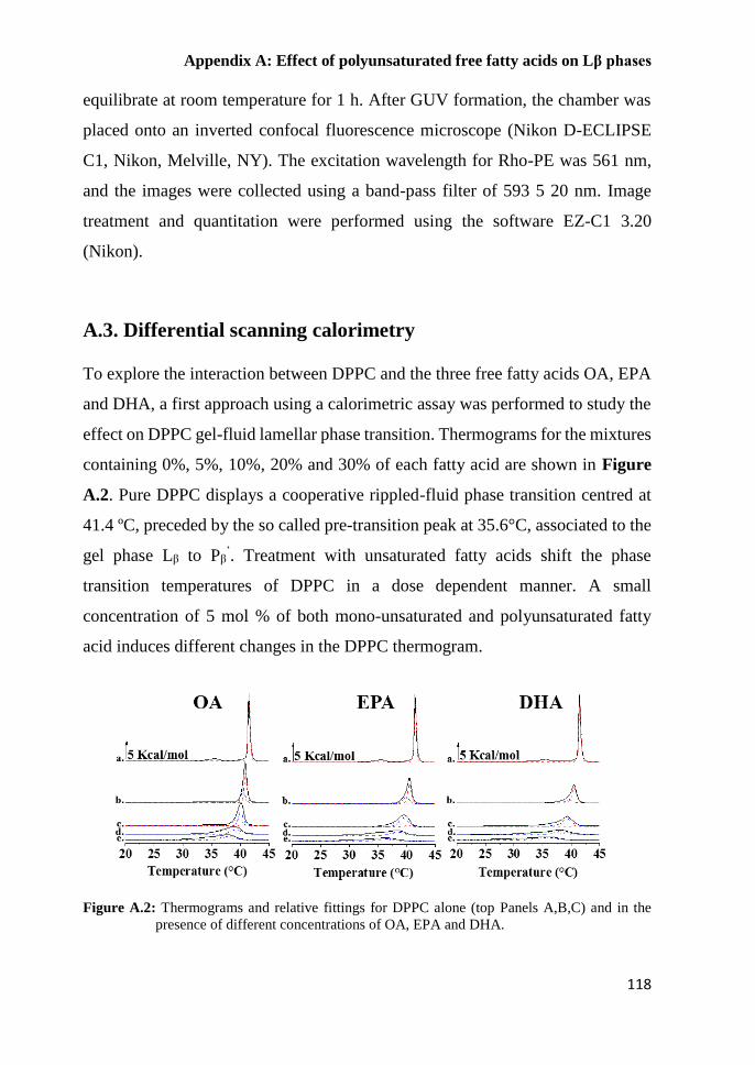
Appendix A: Effect of polyunsaturated free fatty acids on Lβ phases
118
equilibrate at room temperature for 1 h. After GUV formation, the chamber was
placed onto an inverted confocal fluorescence microscope (Nikon D-ECLIPSE
C1, Nikon, Melville, NY). The excitation wavelength for Rho-PE was 561 nm,
and the images were collected using a band-pass filter of 593 5 20 nm. Image
treatment and quantitation were performed using the software EZ-C1 3.20
(Nikon).
A.3. Differential scanning calorimetry
To explore the interaction between DPPC and the three free fatty acids OA, EPA
and DHA, a first approach using a calorimetric assay was performed to study the
effect on DPPC gel-fluid lamellar phase transition. Thermograms for the mixtures
containing 0%, 5%, 10%, 20% and 30% of each fatty acid are shown in Figure
A.2. Pure DPPC displays a cooperative rippled-fluid phase transition centred at
41.4 ºC, preceded by the so called pre-transition peak at 35.6°C, associated to the
gel phase Lβ to Pβ’. Treatment with unsaturated fatty acids shift the phase
transition temperatures of DPPC in a dose dependent manner. A small
concentration of 5 mol % of both mono-unsaturated and polyunsaturated fatty
acid induces different changes in the DPPC thermogram.
Figure A.2: Thermograms and relative fittings for DPPC alone (top Panels A,B,C) and in the
presence of different concentrations of OA, EPA and DHA.

Appendix A: Effect of polyunsaturated free fatty acids on Lβ phases
119
The OA causes the shift of the main transition peak and it is still the pre-transition
one. On the contrary, EPA and DHA cause an asymmetry in the main peak with
a shoulder at lower temperatures, more marked for DHA, on the DPPC phase
transition. In the case of poly-unsaturated the disappearance of the pre-transition
peak is also detected. Increasing the amount of each fatty acid, the sharp peak,
associated with DPPC main transition, became more broad and asymmetric until
the completely disappearance in the case of 30% DHA. Thermogram transition
temperatures, enthalpies, and widths at half-height (reported in Figure A.2) were
determined using the software ORIGIN (Microcal) provided with the calorimeter.
Figure A.3: Panel A: Transition temperatures; Panel B: The peak width, at half of its height; Panel
C: ΔH; for DPPC in the presence of OA (green), EPA (red) and DHA (blue).
Concerning the transition temperatures, Figure A.3 A shows that Tm decreases in
a dose dependent way for all the three fatty acids, suggesting that these molecules
can fluidify the DPPC membrane. The T1/2 (Panel B) describes the cooperativity
of the transition: the higher is the T1/2 value the less cooperative is the process;
thus, increasing the amount of the fatty acids the process become less cooperative
but with a different behaviour for mono-unsaturated OA and poly-unsaturated
EPA and DHA suggesting a heterogeneity affect the lipid systems. The ΔH
variations also indicate that the more fluid phase occurs in the presence of poly-
unsaturated fatty acids, indeed a less heat in necessary to obtain the transition. In
order to rationalise those results, the thermograms investigation was deepened;

Appendix A: Effect of polyunsaturated free fatty acids on Lβ phases
120
indeed, due to the peak shape curves, fittings were performed by the software
GRAMS_32 Spectra Notebase. Thermograms were fitted to the smallest number
of Gaussian curves compatible with a statistically convergent solution with an R2
correlation coefficient >0.99; the best fittings are reported as dotted lines in
Figure A.2. It comes out that three different components (excluding the pre-
transition) characterize the lipid mixtures as soon as the fatty acids are added
(thermograms b. in Figure A.2). To quantitatively analyse the behaviour of those
components and thus to understand their origin, the transition temperature, the
half high temperature and ΔH were reported for each component in Figure A.4.
Figure A.4: Tm and T1/2 for the three components arising in the presence of OA (A, D), EPA (B,
E) and DHA (C, F)
For all the parameters the averaged values are reported in black and the three
components, detected by the fitting procedures, in different colours: red, blue and
green. In all the investigated samples, the component indicated in red disappears
upon the addition of 10% OA or EPA or DHA; such component shows to have
the same Tm and T1/2 of DPPC transition and those values remain constant until

Appendix A: Effect of polyunsaturated free fatty acids on Lβ phases
121
they disappear; thus, it is ascribed to the Pβ’ phase of DPPC alone. On the other
hand, the components reported in blue and green, appear when the fatty acids are
added and evolve with the increase of fatty acids molar fraction; in both cases, the
transition temperatures decrease, indicating a fluidification of lipid system, but
the effect is higher for the green one. Even the cooperativity of the process
changes differently: it is much less in the case of the green component. Looking
at the enthalpies variation of those two components (Figure A.5) it is possible to
see that the associated ΔH increase when the ΔH for the DPPC alone decrease,
and then they reach a constant value, which is higher for the green component,
increasing the % of fatty acids. Those results suggest that upon the addition of a
treashold concentration of each fatty acid, the DPPC phase is more fluid and it is
possible to discriminate between an enriched unsaturated fatty acid phase and
enriched saturated phase. Notably, DHA is the fatty acid that most affect the
DPPC bilayer, probably due to the high number of unsaturation.
Figure A.5: ΔH for the three components arising in the presence of OA (A, D), EPA (B, E) and
DHA (C, F)
A.4. Laurdan General Polarization method
In order to estimate the packing effect of single lipid species, we analysed simple
one component lipid mixtures consisting on DPPC with known phase behavior11.
Having confirmed that Laurdan emission spectrum has the λmax at 440 nm, the GP
value were calculated and are reported in black in Figure A.6.

Appendix A: Effect of polyunsaturated free fatty acids on Lβ phases
122
To understand the effect of OA, EPA and DHA, the GP values for different
concentration of each fatty acid were calculated and reported in Figure A.6.
The addition of monounsaturated OA with exception of the 30% OA, causes a
little increase of GP of Laurdan at all investigated temperatures; indeed, the 30%
of OA reduces the GP at 37°C of the lipid mixtures compared to DPPC alone. On
the contrary, lower amount of polyunsaturated fatty acids, 20%, can reduce the
GP with higher effect, clear at 37°C. Reporting the GP for a wide range of the
three fatty acids at a physiological temperature, the effects of polyusaturated fatty
acids become clearest (Figure A.7). The GP value for DPPC at 37°C and the
relative statistical error are reported as black dotted lines. The results show that
the 20% of polyunsaturated EPA and DHA affect the hydration near the label
instead of OA that does not alter the properties of DPPC alone. For the 20% of
each fatty acid the temperature scan was performed for DPPC, and EggPC,
reported in Figure A.8 A and B, respectively.
Figure A.6: Laurdan GP value at different temperature of DPPC alone (black squares) and in the
presence of different concentrations of OA (A), EPA (B), DHA (C).
From the inspection of Figure A.8A, the two transitions for DPPC alone, reported
in black, are clear, and show that reporting the GP values versus temperature is
an efficient technique to investigate the phase transitions in lipid mixtures. The
presence of OA, EPA or DHA induce different effects on the transition:
- at temperature below 37°C, OA induces an increase of DPPC GP, while EPA
and DHA reduce it;

Appendix A: Effect of polyunsaturated free fatty acids on Lβ phases
123
- at 37°C, GP in the presence of OA is the same observed for DPPC, but the
polyunsaturated fatty acids shift the GP at lower value;
- at temperature higher than 37°C GP tend to a constant value for all the systems.
Thus, from fluorescence investigation it is possible to assesses that 20% of OA,
EPA or DHA at low temperature fluidify the DPPC gel phase to a P’β , indeed the
pretransition is supressed; further comparing the GP values at 37°C, the Laurdan
label resides in a more polar environment when PUFAs are added to the lipid
mixtures suggesting that the lipids headgroups are less packed.
Figure A.7: Laurdan GP value of different percentage of the FFAs in a DPPC bilayer at 37°C.
The Laurdan GP value and the relative standard deviation for DPPC alone are the
dotted lines.
The overall effect for the three fatty acids is to shift the transition temperature, as
already shown by DSC thermograms. In contrast, in the case of these low amounts
of free fatty acids, no effects are shown on a liquid disordered phase, as in the
case of eggPC.

Appendix A: Effect of polyunsaturated free fatty acids on Lβ phases
124
Figure A.8: Temperature scan of Laurdan GP value in absence and in presence of 20 % (mol%)
of the FFAs in a DPPC bilayer (A) and in an EggPC bilayer (B).
A.5. Confocal microscopy of GUVs
Giant Unilamellar Vesicles (GUVs) of DPPC alone and DPPC/DHA with molar
fractions were prepared by electroformation protocol and observed by confocal
microscopy at room temperatures. In Figure A.8 the images related to DPPC
GUVs are reported.
Figure A.9: Confocal microscopy images of DPPC GUVs acquired at 25°C. The scale is 500 nm.
The vesicles show a rough surface mainly due to gel phase that characterizes
DPPC alone. Interestingly when 30% of DHA is present in the lipid mixtures the
vesicles are smoother. The fluidification effects of DHA, already observed by
DSC measurements and by fluorescence spectroscopy, were confirmed.

Appendix A: Effect of polyunsaturated free fatty acids on Lβ phases
125
Notably no domains are observed indicating that free fatty acid have a different
ability in the lipid reorganization with respect to the phospholipid containing
DHA.
Figure A.10: Confocal microscopy images of DPPC/DHA 70/30 GUVs acquired at 25°C. The
scale is 500 nm

Appendix A: Effect of polyunsaturated free fatty acids on Lβ phases
126
A.6. Conclusions
The effects of Docosahexaenoic acid (DHA), once converted to phospholipids,
on the membrane reorganization and on the ability to induce lipid phases
separation were extensively studied. However, a little is known about the effects
that free fatty acids have on the lipid organization. The properties that omega3
fatty acids, EPA and DHA, can exert on a gel phase have been investigated and a
comparison with the monounsaturated OA has been carried out.
The results, obtained from a calorimetry study combined with the evaluation of
GP values, show that both the polyunsatutared fatty acids and monounsaturated
OA affect the gel phase of DPPC in a dose dependent manner and that DHA and
EPA have a stronger fluidification effect on a gel phase compared to oleic acid.
Interestingly, even if the omega3 free fatty acids are able to fluidify the gel phase,
are not able to induce a lipid segregation as shown by the confocal images.
Combining the results obtained with those reported in literature for phospholipids
containing DHA or EPA12,13, it is possible to assess that the different geometries
of phospholipids and the free fatty acids differently affect the lipid packing of
DPPC.

Appendix A: Effect of polyunsaturated free fatty acids on Lβ phases
127
A.7. References
1 Connor, W. E. Importance of n-3 fatty acids in health and disease. Am J Clin Nutr 71, 171s-
175s (2000).
2 Laye, S. Polyunsaturated fatty acids, neuroinflammation and well being. Prostaglandins,
leukotrienes, and essential fatty acids 82, 295-303, doi:10.1016/j.plefa.2010.02.006 (2010).
3 Bazinet, R. P. & Laye, S. Polyunsaturated fatty acids and their metabolites in brain function
and disease. Nature reviews. Neuroscience 15, 771-785, doi:10.1038/nrn3820 (2014).
4 Hulbert, A. J., Kelly, M. A. & Abbott, S. K. Polyunsaturated fats, membrane lipids and animal
longevity. J Comp Physiol B 184, 149-166, doi:10.1007/s00360-013-0786-8 (2014).
5 Gama, M. A. S. et al. Conjugated linoleic acid-enriched butter improved memory and up-
regulated phospholipase A(2) encoding-genes in rat brain tissue. J Neural Transm 122, 1371-
1380, doi:10.1007/s00702-015-1401-9 (2015).
6 Lee, J. C., Simonyi, A., Sun, A. Y. & Sun, G. Y. Phospholipases A2 and neural membrane
dynamics: implications for Alzheimer's disease. Journal of neurochemistry 116, 813-819,
doi:10.1111/j.1471-4159.2010.07033.x (2011).
7 Georgieva, R. et al. Phospholipase A2-Induced Remodeling Processes on Liquid-
Ordered/Liquid-Disordered Membranes Containing Docosahexaenoic or Oleic Acid: A
Comparison Study. Langmuir : the ACS journal of surfaces and colloids 32, 1756-1770,
doi:10.1021/acs.langmuir.5b03317 (2016).
8 Krasnowska, E. K., Gratton, E. & Parasassi, T. Prodan as a membrane surface fluorescence
probe: Partitioning between water and phospholipid phases. Biophysical journal 74, 1984-
1993 (1998).
9 Ashrafzadeh, P. & Parmryd, I. Methods applicable to membrane nanodomain studies? Essays
Biochem 57, 57-68, doi:10.1042/Bse0570057 (2015).
10 Jimenez-Rojo, N., Garcia-Arribas, A. B., Sot, J., Alonso, A. & Goni, F. M. Lipid Bilayers
Containing Sphingomyelins and Ceramides of Varying N-Acyl Lengths: a Glimpse into
Sphingolipid Complexity. Biophysical journal 106, 288a-288a (2014).
11 de Almeida, R. F., Fedorov, A. & Prieto, M. Sphingomyelin/phosphatidylcholine/cholesterol
phase diagram: boundaries and composition of lipid rafts. Biophysical journal 85, 2406-2416,
doi:10.1016/S0006-3495(03)74664-5 (2003).
12 Dumaual, A. C., Jenski, L. J. & Stillwell, W. Liquid crystalline/gel state phase separation in
docosahexaenoic acid-containing bilayers and monolayers. Bba-Biomembranes 1463, 395-
406, doi:Doi 10.1016/S0005-2736(99)00235-7 (2000).
13 Williams, J. A. et al. Docosahexaenoic and Eicosapentaenoic Acids Segregate Differently
between Raft and Nonraft Domains. Biophysical journal 103, 228-237,
doi:10.1016/j.bpj.2012.06.016 (2012).

Appendix B:
Neutron reflectivity Profiles


Appendix B: Neutron Reflectivity Profiles
131
B.1. Neutron reflectivity profiles for SDPC and DDPC in POPC
lipid membranes
Supported lipid bilayers consisting on a binary mixture either SDPC/POPC or
DDPC/POPC were realized as described in section 3.2 and measured in the three
different solvent contrasts D2O, SMW and H2O that greatly enhances the
sensitivity of the technique10;the reflectivity profiles and related best fits are
shown in Figure B.1.
Figure 3.10: Experimental data and the best fitting curves in D2O (black), SMW (blue) and H2O
(red)for lipid systems: A) POPC/SDPC 60/40, B) POPC/DDPC 80/20, C) SDPC
and D) POPC/DDPC 20/80, respectively.

Appendix B: Neutron Reflectivity Profiles
132
B.2. Neutron reflectivity profiles for SDPC and DDPC in
POPC/Chol lipid membranes
Supported lipid bilayers consisting on a ternary mixture characterized by a
constant amount of cholesterol (40%) and either SDPC or DDPC combined with
POPC were realized as described in section 3.2 and measured in the three
different solvent contrasts D2O, SMW and H2O that greatly enhances the
sensitivity of the technique;the reflectivity profiles and related best fits are shown
in Figure B.1.
Figure 3.10: Experimental data and the best fitting curves in D2O (black), SMW (blue) and H2O
(red)for lipid systems: A) POPC/SDPC 60/40 40% Chol, B) POPC/DDPC
80/2040% Chol, C) SDPC 40% Chol and D) POPC/DDPC 20/80 40% Chol,
respectively.

133
Publications
• De Santis, A.; Russo Krauss, I.; Vitiello, G., Ottaviani, M.F.; Paduano, L.;
D'Errico, G.,
Omega-3 fatty lipids modulate the microstructural properties of
biomembranes
In preparation
• De Santis, A.; La Manna, S.; Russo Krauss, I.; Malfitano, A.M.; Novellino,
E.; Federici, L.; Di Matteo, A.; D’Errico, G.; Marasco, D. Nucleophosmin-1
aggregating regions associated with Acute Myeloid Leukemia interact
differently with lipid membranes. Biochimica et biophysica acta 2017
accepted with major revisions.
Abstract
Nucleophosmin-1 (NPM1) is an abundant multifunctional protein, implicated
in a variety of biological processes and in the pathogenesis of several human
malignancies. Its C-terminal domain (CTD) is endowed with a three helix
bundle and we demonstrated that several regions within it, including those
associated with Acute Myeloid Leukemia (AML), have a strong tendency to
form amyloid-like assemblies with β-sheet structures and cellular toxicity.
In particular the central helix of the bundle (H2) resulted the most
amyloidgenic region: here with the dual aim to better model the processes of
cytoxicity of two peptide sequences covering H2 and to get clues of a
potential involvement of the interaction between CTD (in the wild type and
AML-mutA variants) and cellular membrane, we investigated the interaction
of CTD-NPM1 regions with models lipid membranes through ANS binding
assay, as well as by Surface Plasmon Resonance, Circular Dichroism and
Electron Spin Resonance spectroscopies.
These investigations demonstrated a fine interplay between self-aggregation
process of peptides and their interaction with the membranes. Particularly,
cholesterol plays a key role in driving the membrane interaction with
amyloidogenic sequences: H2 peptide showed a specific interaction with
cholesterol, determining a sensitive fluidification of the bilayer, while its N-
term elongated variant non-specifically interacts with membrane causing a
stiffening of the bilayer. Noticeably CTD variants also presents different
mechanisms of interaction with lipid membranes.
Presented results corroborate that the aggregation of several regions of
NPM1-CTD could be implicated in diseases where NPM1 is mutated (i.e.
AML) and/or where its overexpression is cytotoxic.

134
• Russo Krauss, I.; Imperatore, R.; De Santis, A.; Luchini, A.; Paduano, L.;
D'Errico, G., Structure and dynamics of cetyltrimethylammonium chloride-
sodium dodecylsulfate (CTAC-SDS) catanionic vesicles: High-value nano-
vehicles from low-cost surfactants. Journal of colloid and interface science
2017, 501, 112-122.
Abstract
Hypothesis: Catanionic vesicles based on large-scale produced surfactants
represent a promising platform for the design of innovative, effective and
relatively inexpensive nano-vehicles for a variety of actives. Structural,
dynamic and functional behavior of these aggregates is finely tuned by the
molecular features of their components and can be opportunely tailored for
their applications as drug carriers.
Experiments: Here we investigate the aggregates formed by CTAC and SDS,
two of the most diffused surfactants, by means of Dynamic Light Scattering,
Small Angle Neutron Scattering and Electron Paramagnetic Resonance
spectroscopy (EPR). The exploitation of these aggregates as nano-vehicles is
explored using the poorly water-soluble antioxidant trans-resveratrol (t-
RESV), testing t-RESV solubility and antioxidant activity by means of UV,
fluorescence spectroscopy and EPR.
Findings: The presence of a large stability region of catanionic vesicles on
the CTAC-rich side of the phase diagram is highlighted and interpreted in
terms of the mismatch between the lengths of the surfactant tails and of first
reported effects of the chloride counterions. CTAC-SDS vesicles massively
solubilize t-RESV, which in catanionic vesicles exerts a potent antioxidant
and radical-scavenging activity. This behavior arises from the positioning of
the active at the surface of the vesicular aggregates thus being sufficiently
exposed to the external medium.
• Lombardi, L.; Stellato, M. I.; Oliva, R.; Falanga, A.; Galdiero, M.;
Petraccone, L.; D'Errico, G.; De Santis, A.; Galdiero, S.; Del Vecchio, P.,
Antimicrobial peptides at work: interaction of myxinidin and its mutant
WMR with lipid bilayers mimicking the P. aeruginosa and E-coli
membranes. Scitific Report 2017, 7 doi:10.1038/srep44425.
Abstract
Antimicrobial peptides are promising candidates as future therapeutics in
order to face the problem of antibiotic resistance caused by pathogenic
bacteria. Myxinidin is a peptide derived from the hagfish mucus displaying
activity against a broad range of bacteria. We have focused our studies on the
physico-chemical characterization of the interaction of myxinidin and its
mutant WMR, which contains a tryptophan residue at the N-terminus and
four additional positive charges, with two model biological membranes

135
(DOPE/DOPG 80/20 and DOPE/DOPG/CL 65/23/12), mimicking
respectively Escherichia coli and Pseudomonas aeruginosa membrane
bilayers. All our results have coherently shown that, although both myxinidin
and WMR interact with the two membranes, their effect on membrane
microstructure and stability are different. We further have shown that the
presence of cardiolipin plays a key role in the WMR-membrane interaction.
Particularly, WMR drastically perturbs the DOPE/DOPG/CL membrane
stability inducing a segregation of anionic lipids. On the contrary, myxinidin
is not able to significantly perturb the DOPE/DOPG/CL bilayer whereas
interacts better with the DOPE/DOPG bilayer causing a significant
perturbing effect of the lipid acyl chains. These findings are fully consistent
with the reported greater antimicrobial activity of WMR against P.
aeruginosa compared with myxinidin.
• Randino, R.; Grimaldi, M.; Persico, M.; De Santis, A.; Cini, E.; Cabri, W.;
Riva, A.; D'Errico, G.; Fattorusso, C.; D'Ursi, A. M.; Rodriquez, M.,
Investigating the Neuroprotective Effects of Turmeric Extract: Structural
Interactions of beta-Amyloid Peptide with Single Curcuminoids. Scientific
Report 2016, 6, doi:10.1038/srep38846.
Abstract
A broad biophysical analysis was performed to investigate the molecular
basis of the neuroprotective action of Curcuma longa extracts in Alzheimer's
disease. By combining circular dichroism and electron paramagnetic
resonance experiments with molecular modeling calculations, the minor
components of Curcuma longa extracts, such as demethoxycurcumin (2,
DMC), bisdemethoxycurcumin (3, BDMC) and cyclocurcumin (4, CYC),
were analyzed in a membrane environment mimicking the phospholipid
bilayer. Our study provides the first evidence on the relative role of single
curcuminoids interacting with Aβ-peptide. When the CYC and curcumin
metabolite tetrahydrocurcumin (5, THC) were inserted into an anionic lipid
solution, a significant modification of the Aβ CD curves was detected. These
data were implemented by EPR experiments, demonstrating that CYC
reaches the inner part of the bilayer, while the other curcuminoids are
localized close to the membrane interface. Computational studies provided a
model for the curcuminoid-Aβ interaction, highlighting the importance of a
constrained "semi-folded" conformation to interact with Aβ analogously to
the pattern observed in α-helical coiled-coil peptide structures. This
combined approach led to a better understanding of the intriguing in vitro and
in vivo activity of curcuminoids as anti-Alzheimer agents, paving a new path
for the rational design of optimized druggable analogues.

136
• Oliva, R.; Emendato, A.; Vitiello, G.; De Santis, A.; Grimaldi, M.; D'Ursi,
A. M.; Busi, E.; Del Vecchio, P.; Petraccone, L.; D'Errico, G., On the
microscopic and mesoscopic perturbations of lipid bilayers upon interaction
with the MPER domain of the HIV glycoprotein gp41. Biochimica et
biophysica acta 2016, 1858 (8), 1904-13.
Abstract
The effect of the 665-683 fragment of the HIV fusion glycoprotein 41,
corresponding to the MPER domain of the protein and named gp41MPER,
on the microscopic structure and mesoscopic arrangement of palmitoyl oleoyl
phosphatidylcholine (POPC) and POPC/sphingomyelin (SM)/cholesterol
(CHOL) lipid bilayers is analyzed. The microscopic structuring of the
bilayers has been studied by Electron Spin Resonance (ESR) spectroscopy,
using glycerophosphocholines spin-labelled in different positions along the
acyl chain. Transitions of the bilayer liquid crystalline state have been also
monitored by Differential Scanning Calorimetry (DSC). Changes of the
bilayers morphology have been studied by determining the dimension of the
liposomes through Dynamic Light Scattering (DLS) measurements. The
results converge in showing that the sample preparation procedure, the
bilayer composition and the peptide/lipid ratio critically tune the lipid
response to the peptide/membrane interaction. When gp41MPER is added to
preformed liposomes, it positions at the bilayer interface and the lipid
perturbation is limited to the more external segments. In contrast, if the
peptide is mixed with the lipids during the liposome preparation, it assumes
a trans-membrane topology. This happens at all peptide/lipid ratios for fluid
POPC bilayers, while in the case of rigid POPC/SM/CHOL membranes a
minimum ratio has to be reached, thus suggesting peptide self-aggregation to
occur. Peptide insertion results in a dramatic increase of the lipid ordering
and bilayer stiffening, which reflect in significant changes in liposome
average dimension and distribution. The biological implications of these
findings are discussed.
• Emendato, A.; Spadaccini, R.; De Santis, A.; Guerrini, R.; D'Errico, G.;
Picone, D., Preferential interaction of the Alzheimer peptide Abeta-(1-42)
with Omega-3-containing lipid bilayers: structure and interaction studies.
FEBS letters 2016, 590 (4), 582-91.
Abstract
Many age-related neurodegenerative diseases, including Alzheimer Disease
(AD), are elicited by an interplay of genetic, environmental, and dietary
factors. Food rich in Omega-3 phospholipids seems to reduce the AD
incidence. To investigate the molecular basis of this beneficial effect, we

137
have investigated by CD and ESR studies the interaction between the
Alzheimer peptide Aβ-(1-42) and biomimetic lipid bilayers. The inclusion of
1,2-didocosahexaenoyl-sn-glycero-3-phosphocholine does not change
significantly the bilayers organization, but favors its Aβ-(1-42) interaction.
The Omega-3 lipid amount modulates the effect intensity, suggesting a
peptide selectivity for membranes containing polyunsatured fatty acids
(PUFA) and providing hints for the mechanism and therapy of AD.