Embed Size (px)
Citation preview

UNIVERSITI PUTRA MALAYSIA
GENERATION OF A PANEL OF MONOCLONAL ANTIBODIES AGAINST THE HAEMAGGLUTININ-NEURAMINIDASE
GLYCOPROTEIN OF NEWCASTLE DISEASE VIRUS STRAIN AF2240
LEE LIN KIAT.
FBSB 2005 12

GENERATION OF A PANEL O F MONOCLONAL ANTIBODIES AGAINST THE HAEMAGGLUTININ-NEURAMINIDASE GLYCOPROTEIN OF
NEWCASTLE DISEASE VIRUS STRAIN AF2240
BY
LEE LIN KIAT
Thesis Submitted to the School of Graduate Studies, Universiti Putra Malaysia, in Fulfilment of the Requirements for the Degree of Master of Science
September 2 ~ 0 5

Dedicated to Dad, Mom and Christine

Abstract of thesis presented to the Senate of Universiti Putra Malaysia in fulfilment of the requirement for the degree of Master of Science
GENERATION OF A PANEL OF MONOCLONAL ANTIBODIES AGAINST THE HAEMAGGLUTININ-NEURAMINIDASE
GLYCOPROTEIN OF NEWCASTLE DISEASE VIRUS STRAIN AF2240
BY
LEE LIN KIAT
September 2005
Chairperson: Professor Datin Khatijah Mohd. Yusoff, PhD
Faculty: Biotechnology and Biomolecular Sciences
The Malaysian velogenic-viscerotropic Newcastle disease virus (NDV) strain
AF2240 is responsible for high mortality and morbidity. Monoclonal antibodies
(mAbs) have been known to be useful in the identification of NDV due to their
binding specificity, their homogeneity and their ability to be produced in
unlimited quantities. It is, however, very difficult to obtain mAbs which are
specific to NDV commercially. Therefore, this project is to develop mAbs
against the local NDV strain AF2240. This velogenic-viscerotropic viral strain is
a reference strain that has often been used for vaccine development.
Hybridoma cells were created by fusing NDV-hyperimmunised Balblc
splenocytes with Sp210-Ag14 (Sp2) myeloma cells using polyethylene glycol
with the molecular weight of 1450 (PEG 1450). Positive clones were screened by
ELISA. High titre producing clones were selected from a series of limiting
dilutions. MAbs from stable hybridomas were further characterised by western

blot analysis, haemagglutination-inhibition test (HI) and haemolysis-inhibition
test (HLI).
Eight hybridoma cell lines producing mAbs against the haemagglutinin-
neuraminidase (HN) glycoprotein of NDV strain AF2240 were generated.
Isotyping showed that mAbs 1B9, 2D6 and 9D7 were IgG1, mAbs 1D5, 5A10
and 5F10 were IgG2a and mAbs 2G3 and 5E10 were IgG3. Kappa (K) light
chains were found in all mAbs. They can be divided into two groups: (1) mAbs
lD5, 5A10 and 5E10 which recognised conformational and linearised epitopes
and (2) mAbs 1B9, 2D6 and 9D7 which recognised only conformational
epitopes. All mAbs showed positive results in the HI test but not HLI test
conforming that they were specific to the HN protein and not the fusion (F)
protein.

Abstrak tesis yang dikemukakan kepada Senat Universiti Putra Malaysia sebagai mernenuhi keperluan untuk ijazah Master Sains
PENGENERASIAN SATU PANEL ANTIBODI MONOKLONAL TERHADAP GLIKOPROTEIN HEMAGGLUTININ-NEURAMINIlIASE
VIRUS PENYAKIT NEWCASTLE STRAIN AF2240
Oleh
LEE LIN KIAT
September 2005
Pengerusi: Profesor Datin Khatijah Mohd. Yusoff, PhD
Fakulti: Bioteknologi dan Sains Biomolekul
Virus penyakit sampar ayam atau virus penyakit Newcastle (NDV) strain
ternpatan AF2240 rnenyebabkan kernatian yang tinggi. Antibodi rnonoklonal
(mAb) sudah diketahui dengan kegunaannya dalam pengenalian NDV
berasaskan specifikasi ikatan, keseragaman dan keupayaan dihasilkan dalam
jurnlah yang banyak. Namun demikian, rnAb susah diperolehi secara komersil.
Maka, projek ini bertujuan menghasilkan rnAb terhadap NDV ternpatan. Virus
strain velogenic-viscerotropic ini rnenjadi strain rujukan yang kerap digunakan
dalam penghasilan vaksin.
Sel hibridorna telah dihasilkan melalui pergabungan sel limfosit Balblc yang
telah dipertingkatkan imrnunisasinya terhadap NDV dengan sel milorna SpYO-
Ag14 (Sp2) menggunakan polietilen gliko dengan berat molekul 1450 (PEG
1450). Klon positif diasai daripada teknik ELISA. Klon yang menghasilkan titer
tinggi akan dipilih daripada satu siri pencairan terhad. MAb daripada hibridoma

yang stabil akan dicirikan secara lanjutan melalui analisis 'western blot', ujian
penghalangan hemagglutinin (HI) dan ujian penghalangan hemolisis (HLI).
Lapan rangkaian sel hibridoma yang menghasilkan mAb terhadap NDV strain
AF2240 glikoprotein hemagglutinin-neuraminidase (HN) telah digenerasikan.
Pengkelasan antibodi telah menunjukkan mAb 1B9, 2D6 dan 9D7 dari sub-kelas
IgG1, rnAb 1D5, 5A10 dan 5FlO dari sub-kelas IgG2a dan rnAb 2G3 dan 5E10
dari sub-kelas IgG3. Semua rnAb mempunyai rantai ringan jenis kappa (K). Ia
juga boleh dibahagikan kepada dua kumpulan: (1) mAb 1D5, 5A10 dan 5E10
yang mengenali epitop konformasi and linear serta (2) mAb 1B9, 2D6 dan 9D7
yang mengenali epitop konformasi sahaja. Semua mAb menunjukkan keputusan
positif terhadap ujian HI dan sebaliknya untuk ujian HLI bagi mengesahkan mAb
adalah specifik terhadap protein HN dan bukan protein fusion (F).

ACKNOWLEDGEMENTS
I would like to express my sincere gratitude and appreciation to my supervisors
Professor Datin Dr. Khatijah Mohd. Yusoff and Professor Dr. Abdul Manaf Ali
for their valuable advice, technical guidance and encouragement.
Special thanks to the staff especially Mr. Karim, Mr. Hussain, Mr. Ibrahim, Mr.
Zamros and Mdm. Sharipah from Department of Microbiology and Department
of Biochemistry, Faculty of Biotechnology and Biomolecular Sciences, UPM.
Not forgetting, Mr. Othman for his excellent job in maintaining the animal
house.
I wish to acknowledge the guidance and support from Associate Professor Dr.
Tan Wen Siang, Dr. Majid Eshahgi, Dr. Heilly Chong, Dr. Michael Aigner, Dr.
Wong Sing King, Dr. Lim Yang Mooi and Dr. Kho Chiew Ling; my lab seniors;
Firoozeh, Swee Tin, Raha, Geok Hun, Eddie, Lalita, Nawien, Onie, Rafidah,
Riha, Suhana, Thong Chuan, Yan Peng and Zul; my collogues; Budy, h a , Kah
Fai, Kie Hie, Max, Mokrish, Pala, Taznim, Wani and Wawa.
My gratitude also goes out to my friends; Ainon, Bok Hui, Chin Woi, Judy, Han
Koh, Hazalina, Keng Fei, Kim Seng, Lee Kheng, Lih Ling, May Ling, Michelle,
Sheau Wei, Tiong San and Watti. Support from Mdm. Maria Chew (Culture
Lab), Mr. W. H. Looi (Roche Diagnostic), Mr. C. H. Ti (Trans Techno), Ms.
vii

Angie Yip (Research Instruments) and Mr. C. W. Lye (Biodiagnostic) are always
appreciated.
I am indeed indebted to Janet Loh, my buddies Amos and Leslie for sharing their
knowledge and experience with me. My parents and Christine for their
unconditional sacrifice and love.
I wish to extend my appreciation to everyone, although not individually named
here, who had contributed directly or indirectly to my project and thesis. Finally,
I would like to thank the Ministry of Science, Technology and Innovation of
Malaysia (MOSTI) for providing me the PASCA scholarship and IRPA grant no.
01-02-04-003 BTWERl006 for supporting this study.
Without all of you, it would not be possible for me to complete my project and
thesis. Thank you all for your support and unconditional love. May God bless
you all for your kindness.
. . . V l l l

This thesis submitted to the Senate of Universiti Putra Malaysia and has been accepted as fulfilment of the requirement for the degree of Master of Science. The members of the Supervisory Committee are as follows:
KHATWAH MOHD. YUSOFF, PhD Professor Department of Microbiology Faculty of Biotechnology and Biomolecular Sciences Universiti Putra Malaysia (Chairman)
ABDUL MANAF ALI, PhD Professor Department of Cell and Molecular Biology Faculty of Biotechnology and Biomolecular Sciences Universiti Putra Malaysia (Member)
AINI IDERIS, PhD ProfessorIDean School of Graduate Studies Universiti Putra Malaysia

DECLARATION
I hereby declare that the thesis is based on my original work except for quotations and citations which have been duly acknowledged. I also declare that it has not been previously or concurrently submitted for any other degree at UPM or other institutions.

TABLEOFCONTENTS
DEDICATION ABSTRACT ABSTRAK ACKNOWLEDGEMENTS APPROVAL DECLARATION LIST O F TABLES LIST O F FIGURES LIST O F ABBREVIATIONS
CHAPTER
INTRODUCTION
LITERATURE REVIEW 2.1 Basic Concepts in Hybridoma Technology
2.1.1 Somatic Cell Hybridisation 2.1.2 Immunisation Strategies 2.1.3 Cell Fusion 2.1.4 Hybridoma Selection 2.1.5 Screening 2.1.6 Cloning of Hybridomas 2.1.7 Cryopreservation of Hybridomas 2.1.8 Scale Up Production of mAbs 2.1.9 Antibody Purification
2.2 Application of Monoclonal Antibodies 2.2.1 Research and Diagnostic Applications 2.2.2 Therapeutic Applications
2.3 Newcastle Disease Virus
METHODOLOGY 3.1 Source of Viruses and Cell 3.2 Source of Chemicals and Biochemicals 3.3 Virus Cultivation and Purification 3.4 Viral Profiles
3.4.1 Bradford Assay 3.4.2 Haemaggutination (HA) Test 3.4.3 SDS-PAGE Protein Profile
3.5 Myeloma Cultivation 3.6 Immunisation
3.6.1 Immunogen Preparation and Immunisation 3.6.2 Antibody Titre
3.7 Hybridoma Development 3.7.1 Splenectomy 3.7.2 Cell Fusion 3.7.3 Hybridoma Selection
Page . . 11 ... 111
v vii ix xi
xiv xv xvi
xii

3.7.4 Screening for Antibody Producing Hybridoma 3.7.5 Limiting Dilution 3.7.6 Expansion and Cryopreservation
3.8 Ascites Fluid and Purification 3.8.1 Ascites Production 3.8.2 Antibody Purification and Storage 3.8.3 Antibody Concentration Determination 3.8.4 Antibody Titre Determination
3.9 Characterisation of Monoclonal Antibodies 3.9.1 Antibody Isotyping Determination 3.9.2 Heavy and Light Chains Determination 3.9.3 Immunoblot Analysis 3.9.4 Haemagglutination-Inhibition (HI) Test 3.9.5 Haemolysis-Inhibition (HLI) Test 3.9.6 Cross-Reactivity Test
RESULTS 4.1 Production of Hybridoma 4.2 Characterisation of Selected Hybridomas
4.2.1 Isotyping 4.2.2 Antibody Purity 4.2.3 Immunoblotting against HN Glycoprotein 4.2.4 Haemagglutination-Inhibition and Haemolysis-
Inhibition Tests 4.2.5 Cross-Reactivity Test for NDV Inter-Strains 4.2.6 Cross-Reactivity Test for Avian Viruses
DISCUSSION 5.1 Production of Monoclonal Antibodies against HN
Glycoprotein 5.2 Characterisation of Selected Hybridomas
CONCLUSION
REFERENCES BIODATA OF THE AUTHOR
... X l l l
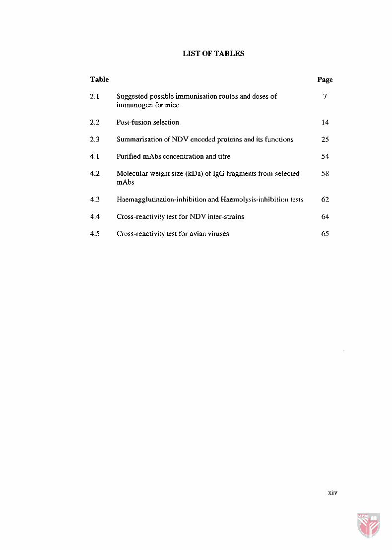
LIST OF TABLES
Table
2.1 Suggested possible immunisation routes and doses of immunogen for mice
Puss-fusion selection
Summarisation of NDV encoded proteins and its functions
Purified mAbs concentration and titre
Molecular weight size (kDa) of IgG fragments from selected m Abs
Haemagglutination-inhibition and Haemolysis-inhibition tests
Cross-reactivity test for NDV inter-strains
Cross-reactivity test for avian viruses
Page
7
xiv

LIST OF FIGURES
Figure
Flow diagram on the protocol of generation of monoclonal antibodies
Flow diagram on hybridoma selection
Schematic diagram of the virion structure of Newcastle disease virus
Schematic diagram of antibody isotyping determination
Balblc antiserum titres throughout the immunisation period
Proliferation of single hybridoma into high density colony
Absorbance values of selected mAbs after each limiting dilution
Isotyping classes and subclasses of selected mAbs
IgG heavy and light chains of selected mAbs
4.6a NDV protein profile
4.6b Immunoblotting against selected mAbs
Page
9

LIST OF ABBREVIATIONS
ABTS
ADCC
AEV
AIV
AP
APS
BCIP
BSA
"C
CAV
CD
cm
CMV
CO?_
dHzO
DMEM
DMSO
DNA
dTMP
EBV
EDTA
ELISA
2,2'-azinobis(3-ethylbenzthiazoline-6-sulf0nic acid) diammonium
antibody-dependent cell-mediated cytotoxicity
avian encephalomyelitis virus
avian influenza virus
alkaline phosphatase
ammonium persulfate
5-bromo-4-chloro-3-indolyl phosphate
bovine serum albumin
Celsius
chicken anaemia virus
cluster of differentiation
centimetre
cytomegalovirus
carbon dioxide
distilled water
Dulbecco's Modified Eagle's Medium
dimethylsulfoxide
deoxyribonucleic acid
deoxythymidine monophosphate
Epstein-Barr virus
ethylenediaminetetraacetic acid
enzyme-linked immunosorbent assay
fusion (glycoprotein)
xvi
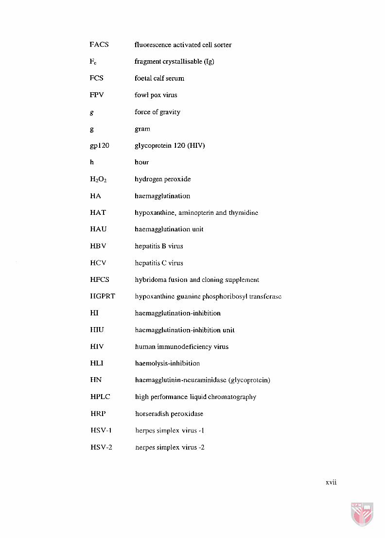
FACS
Fc
FCS
FPV
HAT
HAU
HBV
HCV
HFCS
HGPRT
HI
HIU
HIV
HLI
HN
HPLC
HRP
HSV- 1
HSV-2
fluorescence activated cell sorter
fragment crystallisable (Ig)
foetal calf serum
fowl pox virus
force of gravity
gram
gl ycoprotein 120 (HIV)
hour
hydrogen peroxide
haemagglutination
hypoxanthine, aminopterin and thymidine
haemagglutination unit
hepatitis B virus
hepatitis C virus
hybridoma fusion and cloning supplement
hypoxanthine guanine phosphoribosyl transferase
haemagglutination-inhibition
haemagglutination-inhibition unit
human immunodeficiency virus
haemolysis-inhibition
haemagglutinin-neuraminidase (glycoprotein)
high performance liquid chromatography
horseradish peroxidase
herpes simplex virus -1
herpes simplex virus -2
xvii

IBDV
IBND
IBV
ICPI
Ig
ILTV
im
IMP
ip
iv
IVPI
kb
L
m Ab
mAbs
mg
min
mL
hypoxanthine and thymine
infectious bursa1 disease virus
bivalent vaccines of IBV and NDV
infectious bronchitis virus
intracerebral pathogenicity index
immunoglobulin
infectious laryngotracheitis virus
intramuscular
inosine monophosphate
intraperitoneal
intravenous
intravenous pathogenicity index
kilobase
large (protein)
microgram
microlitre
micrometre
matrix (protein)
molar
milliampere
monoclonal antibody
monoclonal antibodies
milligram
minute (time)
millilitre
xviii

mm
mM
MOPC
NA
NBT
ND
NDV
NEAA
NI
nm
nM
NP
NT
NTE
P
p Abs
PB S
PBST
PEG
PE
P W P
RBC
RNA
rpm
RSV
millimetre
millimolar
mineral oil plasmacytoma
neurarninidase
nitro-blue tetrazolium chloride
Newcastle disease
Newcastle disease virus
non-essential amino acids
neutralisation index
nanometre
nanomolar
nucleoprotein
neutralisation titre
natrium Tris EDTA
phosphoprotein
polyclonal antibodies
phosphate buffered saline
phosphate buffered saline - Tween-20
polyethylene glycol
picogram
p-nitrophenyl phosphate disodium
red blood cell
ribonucleic acid
revolutions per minute
respiratory sjmcytial virus
xix

RT
RT-PCR
S
SC
room temperature
reverse transcription-polymerase chain reaction
second (time)
subcutaneous
TK
Tris
uv
v
vlv
wlv
SDS sodium dodecyl sulphate
SDS-PAGE sodium dodecyl sulphate - polyacrylarnide gel electrophoresis
TEMED N, N, N', N'-tetramethylethylenediamine
thymidine kinase
Tris-(hydroxymethy1)-aminomethane
ultraviolet
volt
volume for volume
weight for volume
CHAPTER 1
INTRODUCTION
Newcastle disease (ND) is regarded throughout the world as one of the two most
important diseases of poultry and other birds, the other disease being the highly
pathogenic avian influenza. Its etiologic agent, the Newcastle disease virus
(NDV), is a member of the family Paramyxovirihe and has been assigned to the
genus Avulavirus in the subfamily Paramyxovirinae (Mayo, 2002; Peeters and
Koch, 2002). Alexander (1989) had grouped NDV into five pathotypes based on
their pathogenic signs: (1) viscerotropic velogenic NDV which causes
hemorrhagic lesions in the gut; (2) neurotropic velogenic NDV shows respiratory
and neurological signs but no gut lesions; (3) mesogenic NDV produces low
mortality with acute respiratory disease and nervous signs in some birds; (4)
lentogenic NDV shows mild and in apparent respiratory infections and (5)
asymptomatic enteric NDV, avirulent viruses that appear to replicate primarily in
the intestinal tract. Regardless of outbreaks or farms under constant surveillance,
lack of obvious clinical signs or field experts will require confirmatory diagnosis
for further identification and characterisation of the virus.
Diagnosis of ND started from the conventional techniques including virus
isolation; in vivo estimation of pathogenicity through intracerebral pathogenicity
index (ICPI) and intravenous pathogenicity index (IVPI) in one day old chicks
and six weeks old chickens, respectively (Alexander, 1988); in vitro studies on
the fusion protein cleavage site (Aldous and Alexander, 2001) and serological

tests like haemagglutination (HA) test and haemagglutination-inhibition (HI)
test. These conventional methods are perceived as slow, laborious and required
in vivo techniques. Since NDV has a 15.19 kb single-stranded negative RNA
genome, reverse transcription-polymerase chain reaction (RT-PCR) was used to
amplify the specific gene region using universal primers, and pathotype-specific
primers or nested PCR (Aldous and Alexander, 2001). Real-time PCR that can
detect minute amount (10 pg) of DNA (Tan et al., 2004) and biopanning with a
fusion phage that carried specific amino acid sequence to interact with surface
glycoproteins (Ramanujam et al., 2004) have also been performed.
Besides molecular-based techniques, monoclonal antibodies (mAbs) were
intensively used in identification and differentiation of NDV strains [Iorio and
Bratt, 1983 (Australia-Victoria strain); Nishikawa et al., 1983 (D2& Russell and
Alexander, 1983 (Ulster 2C); Ishida et al., 1985 (Miyadera and Taka); Abenes et
al., 1986 (Sato); Erdei e t al., 1987 (La Sota); Yusoff et al., 1988 (Beaudette C);
Jestin et al., 1989 (Ploufragan); Panshin et al., 1999 (Israel)]. MAbs were
employed to study the antigenic differentiation among strains where single amino
acid changes at the directed epitope can be detected (Chambers et al., 1988;
Yusoff et al., 1989). The mAb era began in 1975 with a report in Nature by
Kohler and Milstein entitled 'Continuous cultures of fused cells secreting
antibody of predefined specificity'. It was reported that they could fuse
immortalized myeloma cells with splenocytes which secreted a specific antibody
of interest. The cell line that produces such antibodies is termed hybridoma.
Since then, mAbs have become increasingly valuable in both research and
therapeutic applications. The usefulness of mAbs can be characterised into three

main points: their specificity of binding, their homogeneity and their ability to be
produced in unlimited quantities (Harlow and Lane, 1988). Since the antibodies
produced are from one specific hybridoma cell, their identical properties make
them very powerful in their ability to detect any specific epitope. Alexander et al.
(1997) had allocated over 1500 NDV into different groups based on their ability
to react with different panels of mAbs. MAbs have also been used to distinguish
vaccine viruses from epizootic viruses in a given area (Srinivasappa et al., 1986).
Nevertheless, it is very difficult to obtain mAbs which are specific to NDV
commercially. Therefore, the objectives of this study are:
a) to generate a panel of murine n~onoclonal antibodies against NDV strain
AF2240 and
b) to characterise the selected hybridoma clones.

CHAPTER 2
LITERATURE REVIEW
Kohler and Milstein (1975) had successfully developed a technique that allows
the growth of cells secreting antibodies with a defined specificity. In this
technique the splenocytes isolated from an immunised animal, is fused with
myeloma cells, a type of tumour cell. These hybrid cells or better known as
hybridomas can be cultured in vitro. Antibodies secreted from hybridomas are
known as monoclonal antibodies (mAbs).
Basic Concepts in Hybridoma Technology
2.1.1 Somatic Cell Hybridisation
The techniques of somatic cell fusion used by Kohler and Milstein (1975) to
generate hybridomas secreting anti-sheep red blood cell with inactivated Sendai
virus was a breakthrough in the field of cell biology. Cell fusion between
lymphocytes and myelomas was associated with the presence of Sendai virus
fusion glycoprotein, making it possible for their membrane to coalesce,
cytoplasm to intermingle and multinucleated homokaryons and heterokaryons
were formed (Gordon, 1975). Thus, different viruses with surface glycoproteins
such as Semliki Forest virus, vesicular stomatitis virus, fowl plaque virus and
influenza virus (White et al., 1980, 1981) were applied to study cell fusion
mechanisms. Another breaktitrough was using Epstein-Barr virus (EBV) to fuse

human peripheral blood lymphocytes with human plasmacytomas to generate
human mAbs (Roder, 1986).
The tumour caused by malignantly transformed antibody secreting cells is known
as myeloma or plasmacytoma. According to Goding (1996), pathologists may
make a morphological distinction between these terms but they may be regarded
as biologically identical. Mineral oil or pristane were found to be potent inducers
of myeloma in Balblc mouse. Myelomas that were isolated using this approach
were termed mineral oil plasmacytoma (MOPC) (Potter and Boyce, 1962; Potter,
1972). Myelomas that are used as fusion partners should not produce antibodies
to avoid production of hybridomas that secrete more than one type of antibody.
Harlow and Lane (1988) have recommended the following cell lines as gooa
fusion partners and were successfully being used in the cited articles: FO (Davis
et al., 1982; Mao and France, 1984); FOX-NY (Lane, 1985); NS1/1-Ag4-1
(Nishikawa et al., 1983; Russell and Alexander, 1983; Yusoff et al., 1988;
Llames et al., 2000); Sp210-Ag14 (Iorio and Bratt, 1983; Long et al., 1986;
Letellier et al., 2001; Fontes et al., 2005) and X63Ag8.653 (Davis et al., 1982;
Srinivasappa et al., 1986).
Splenocytes are obtained from the immunised mouse through splenectomy. A
spleen from an immunised mouse contains approximately 5 x lo7 to 2 x 10'
splenocytes (Harlow and Lane, 1988). Marusich (1988) reported that 1 x lo5
splenocytes were sufficient to generate hybridomas. Hybridoma cells are created
by fusing splenocytes from an imnlunised animal with myeloma cells to enable
the hybridomas to possess both the antibody secreting properties of the parent