Embed Size (px)
Citation preview

Type of file: PDF Size of file: 0 KB Title of file for HTML: Supplementary Information Description: Supplementary Figures Type of file: PDF Size of file: 0 KB Title of file for HTML: Peer Review File Description:
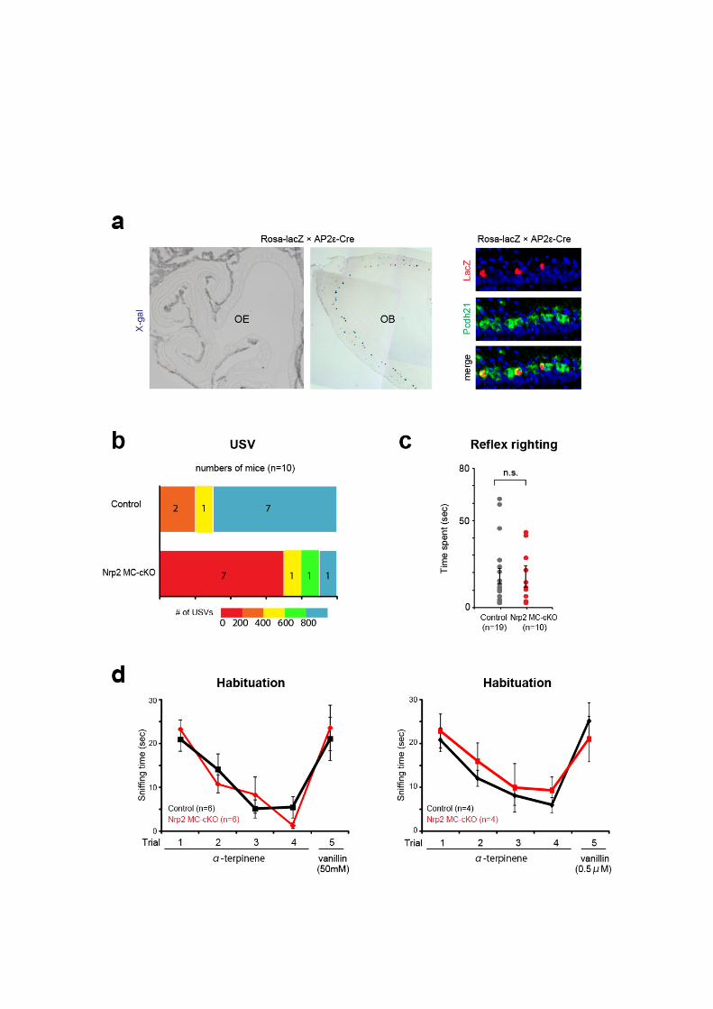
Supplementary Figure. 1 Odor sensing is not affected by the MC-specific cKO of Nrp2.
(a) Detection of the AP2e promoter activity (left). The Tg AP2ε-Cre mouse was crossed with
the ROSA-shutter-lacZ mouse for Cre-mediated lacZ induction. Coronal sections of the OE
and a parasagittal section of the OB at P14 were analyzed by X-gal staining. MCs were
stained blue in the OB. LacZ+ PCdh21+ cells in the MCL (right). Parasagittal OB section
were immunostained with antibodies against β-gal and Pcdh21. (b) Distribution of numbers of
male USV. USV was counted in the presence of a female intruder for duration of 6 min in
each experiment. (c) Locomotor abilities test. Mice were placed on their back. The time
they spent to recover from this position, to being upright and on four paws, was measured
(reflex righting). No difference was found between the cKO and heterozygous littermates in
their recovery time lengths. (d) Habituation-dishabituation test. In this test, α-terpinene was
presented in four consecutive trials for duration of 1 min. The inter-trial interval was 10 min.
Then, a novel odor vanillin (50 mM or 0.5 µM) was presented. Habituation is defined by a
progressive decrease in sniffing towards the repeated presentation of the same odor stimulus.
Dishabituation is defined by reinstatement of sniffing when the novel odor is presented.
Graphs demonstrate amounts of time that the male cKO and control littermates spend on
sniffing a piece of filter paper spotted with vanillin or α-terpinene. Trials with vanillin as the
repetitive odor and α-terpinene as the novel odor yielded similar results. No difference is
found between the cKO and control mice. Error bars are ±SE. Error bars are ±SE. n.s.:
not significant (Student’s t test).
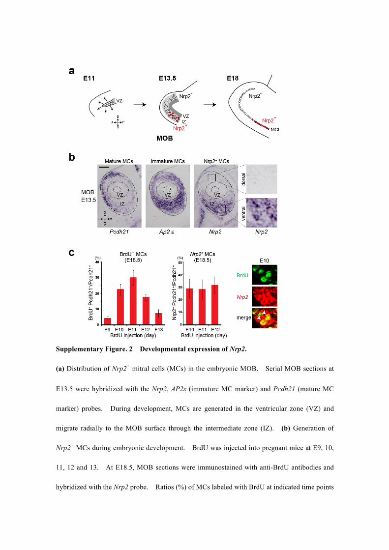
Supplementary Figure. 2 Developmental expression of Nrp2.
(a) Distribution of Nrp2+ mitral cells (MCs) in the embryonic MOB. Serial MOB sections at
E13.5 were hybridized with the Nrp2, AP2ε (immature MC marker) and Pcdh21 (mature MC
marker) probes. During development, MCs are generated in the ventricular zone (VZ) and
migrate radially to the MOB surface through the intermediate zone (IZ). (b) Generation of
Nrp2+ MCs during embryonic development. BrdU was injected into pregnant mice at E9, 10,
11, 12 and 13. At E18.5, MOB sections were immunostained with anti-BrdU antibodies and
hybridized with the Nrp2 probe. Ratios (%) of MCs labeled with BrdU at indicated time points
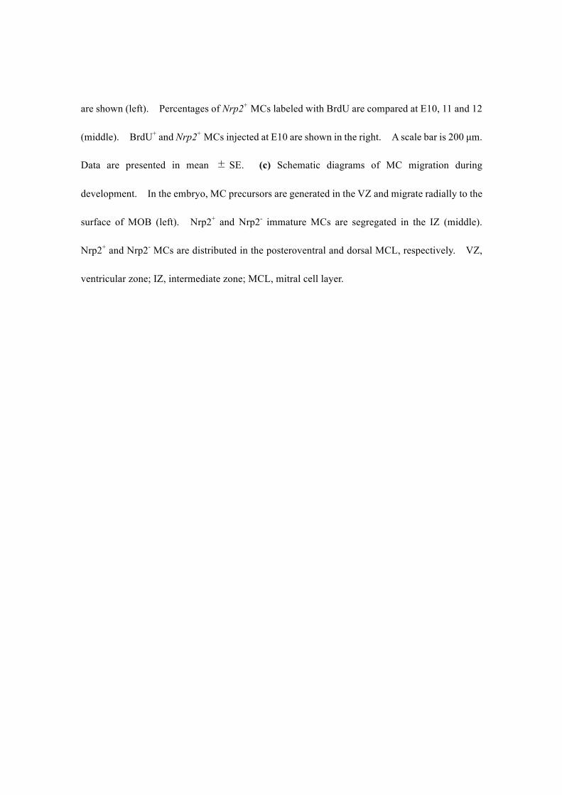
are shown (left). Percentages of Nrp2+ MCs labeled with BrdU are compared at E10, 11 and 12
(middle). BrdU+ and Nrp2+ MCs injected at E10 are shown in the right. A scale bar is 200 µm.
Data are presented in mean ± SE. (c) Schematic diagrams of MC migration during
development. In the embryo, MC precursors are generated in the VZ and migrate radially to the
surface of MOB (left). Nrp2+ and Nrp2- immature MCs are segregated in the IZ (middle).
Nrp2+ and Nrp2- MCs are distributed in the posteroventral and dorsal MCL, respectively. VZ,
ventricular zone; IZ, intermediate zone; MCL, mitral cell layer.
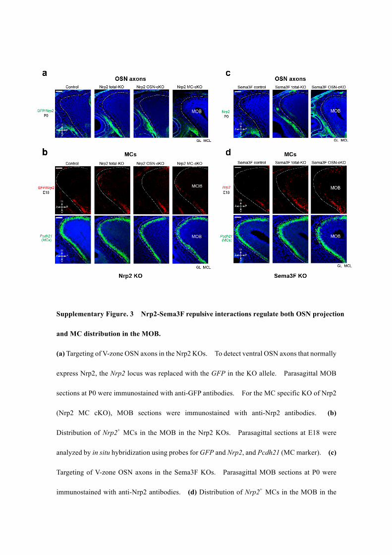
Supplementary Figure. 3 Nrp2-Sema3F repulsive interactions regulate both OSN projection
and MC distribution in the MOB.
(a) Targeting of V-zone OSN axons in the Nrp2 KOs. To detect ventral OSN axons that normally
express Nrp2, the Nrp2 locus was replaced with the GFP in the KO allele. Parasagittal MOB
sections at P0 were immunostained with anti-GFP antibodies. For the MC specific KO of Nrp2
(Nrp2 MC cKO), MOB sections were immunostained with anti-Nrp2 antibodies. (b)
Distribution of Nrp2+ MCs in the MOB in the Nrp2 KOs. Parasagittal sections at E18 were
analyzed by in situ hybridization using probes for GFP and Nrp2, and Pcdh21 (MC marker). (c)
Targeting of V-zone OSN axons in the Sema3F KOs. Parasagittal MOB sections at P0 were
immunostained with anti-Nrp2 antibodies. (d) Distribution of Nrp2+ MCs in the MOB in the

Sema3F KOs. Parasagittal MOB sections at E18 were analyzed by in situ hybridization using
probes for Nrp2 and Pcdh21 (MC marker). In all figures, broken lines indicate the boundary of
the glomerular layer (GL) and mitral-cell layer (MCL) in the MOB. Scale bars are 200 µm.
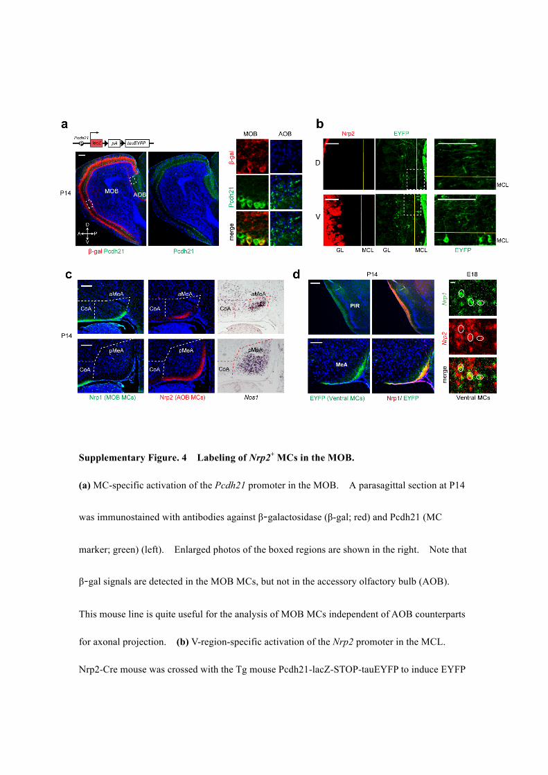
Supplementary Figure. 4 Labeling of Nrp2+ MCs in the MOB.
(a) MC-specific activation of the Pcdh21 promoter in the MOB. A parasagittal section at P14
was immunostained with antibodies against β-galactosidase (β-gal; red) and Pcdh21 (MC
marker; green) (left). Enlarged photos of the boxed regions are shown in the right. Note that
β-gal signals are detected in the MOB MCs, but not in the accessory olfactory bulb (AOB).
This mouse line is quite useful for the analysis of MOB MCs independent of AOB counterparts
for axonal projection. (b) V-region-specific activation of the Nrp2 promoter in the MCL.
Nrp2-Cre mouse was crossed with the Tg mouse Pcdh21-lacZ-STOP-tauEYFP to induce EYFP
specifically in the MOB MCs. A coronal MOB section at P14 was immunostained with
antibodies against GFP. EYFP-positive MCs (green) are found in the ventral (V), but not in
the dorsal (D) region of the MCL. (c) Nrp2+ MOB MCs send their axons to the anterior MeA
but not to the posterior MeA. Coronal sections of the OC at P14 were immunostained with
anti-Nrp1 and anti-Nrp2 antibodies to detect MOB- and AOB-MC axons, respectively. Note
that Nrp2 is not expressed in MOB MCs at this stage (P14). Thus, Nrp2 can be used as an
AOB-MC marker. Serial OC sections were also analyzed by in situ hybridization using the
Nos1 probe, a marker for MeA. (d) Detection of Nrp1 (MOB MC marker) in the Nrp2+
(EYFP+) ventral MC axons (green, left column). Coronal sections of the OC at P14 were
immunostained with anti-GFP and anti-Nrp1 antibodies. GFP+ cells are all positive for Nrp1.
Scale bars are 100 µm in (a-c & d left) and 10 µm in (d right).
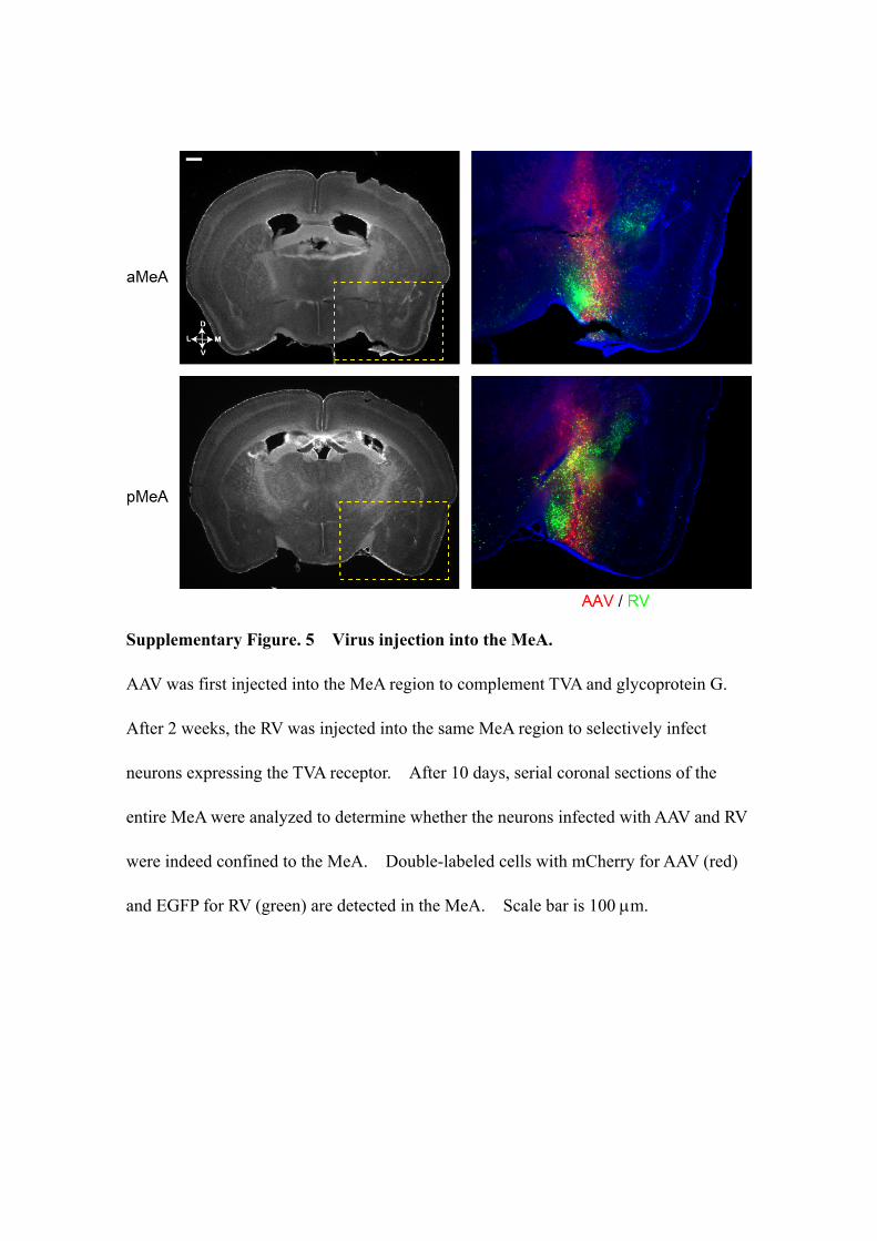
Supplementary Figure. 5 Virus injection into the MeA.
AAV was first injected into the MeA region to complement TVA and glycoprotein G.
After 2 weeks, the RV was injected into the same MeA region to selectively infect
neurons expressing the TVA receptor. After 10 days, serial coronal sections of the
entire MeA were analyzed to determine whether the neurons infected with AAV and RV
were indeed confined to the MeA. Double-labeled cells with mCherry for AAV (red)
and EGFP for RV (green) are detected in the MeA. Scale bar is 100 µm.
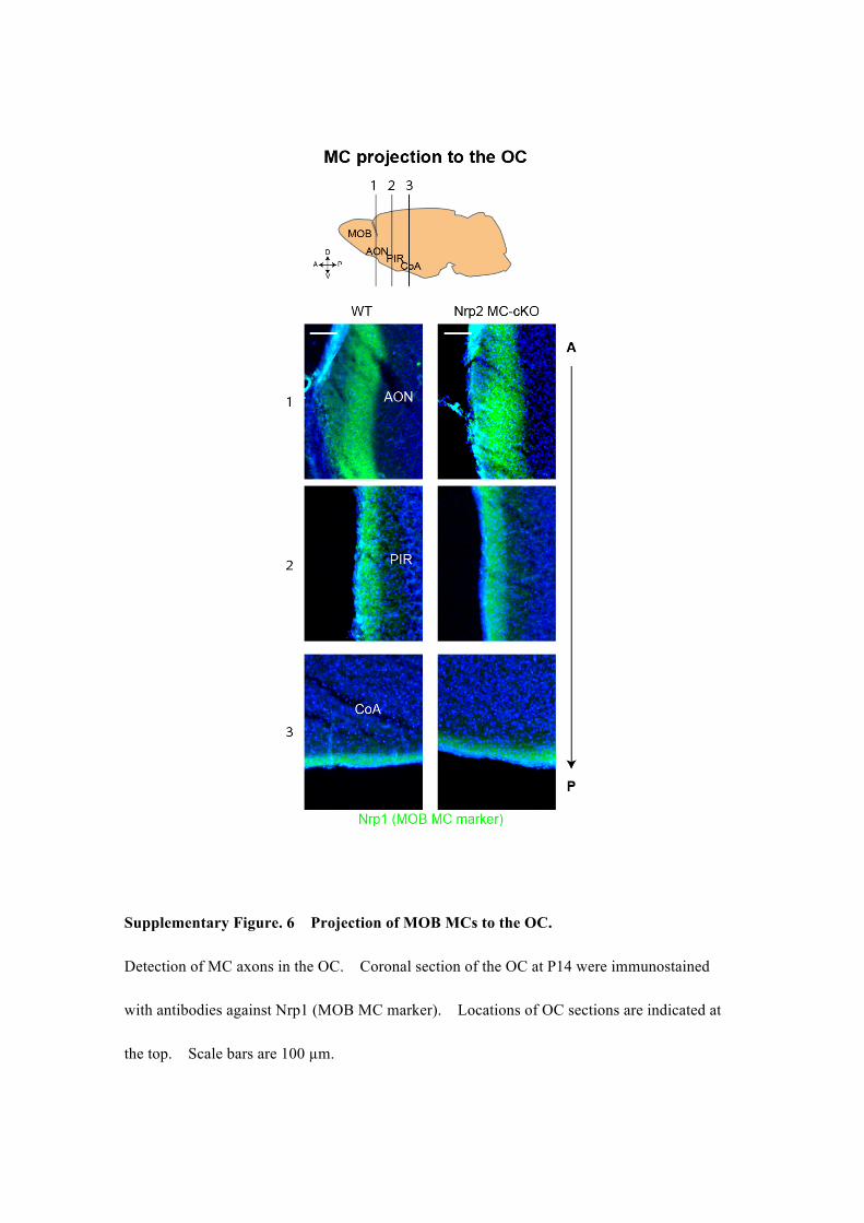
Supplementary Figure. 6 Projection of MOB MCs to the OC.
Detection of MC axons in the OC. Coronal section of the OC at P14 were immunostained
with antibodies against Nrp1 (MOB MC marker). Locations of OC sections are indicated at
the top. Scale bars are 100 µm.
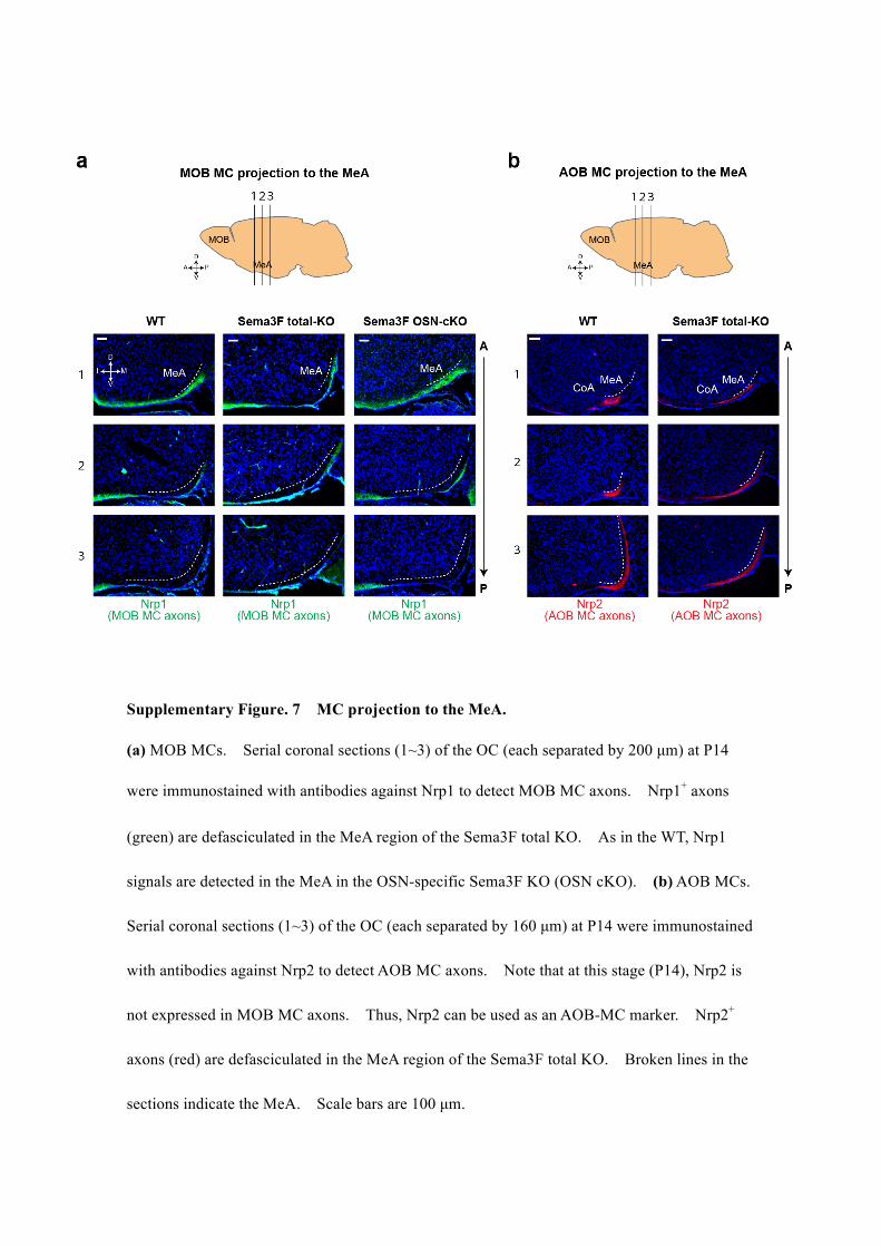
Supplementary Figure. 7 MC projection to the MeA.
(a) MOB MCs. Serial coronal sections (1~3) of the OC (each separated by 200 µm) at P14
were immunostained with antibodies against Nrp1 to detect MOB MC axons. Nrp1+ axons
(green) are defasciculated in the MeA region of the Sema3F total KO. As in the WT, Nrp1
signals are detected in the MeA in the OSN-specific Sema3F KO (OSN cKO). (b) AOB MCs.
Serial coronal sections (1~3) of the OC (each separated by 160 µm) at P14 were immunostained
with antibodies against Nrp2 to detect AOB MC axons. Note that at this stage (P14), Nrp2 is
not expressed in MOB MC axons. Thus, Nrp2 can be used as an AOB-MC marker. Nrp2+
axons (red) are defasciculated in the MeA region of the Sema3F total KO. Broken lines in the
sections indicate the MeA. Scale bars are 100 µm.

Supplementary Figure. 8 In utero electroporation in the embryonic OB.
Plasmid vectors containing the EGFP (pGFP) with or without human Nrp2 cDNA (phNrp2)
were electroporated into the WT embryonic MOB at E11. Coronal MOB sections at P0 were
isolated and immunostained with antibodies against GFP, Tbr1, Tbr2, and Tbx21 (MC markers).
A fraction of MCs ectopically expressed EGFP. Scale bars are 100 µm (left) and 20 µm
(right).
In all extended data, dimensions are: D, dorsal; V, ventral; A, anterior; P, posterior; L, lateral; M,
medial.