Embed Size (px)
Citation preview

Two Dipolar �-Helices within Hormone-encoding Regions ofProglucagon Are Sorting Signals to the Regulated SecretoryPathway*
Received for publication, March 10, 2014 Published, JBC Papers in Press, April 11, 2014, DOI 10.1074/jbc.M114.563684
Leonardo Guizzetti‡, Rebecca McGirr§, and Savita Dhanvantari‡§¶�1
From the Departments of ‡Medical Biophysics, ¶Pathology, and �Medicine, University of Western Ontario, London, Ontario N6A 3K7and the §Metabolism/Diabetes and Imaging Programs, Lawson Health Research Institute, London, Ontario N6A 4V2, Canada
Background: The constituent peptides of proglucagon contain �-helices that may target proglucagon to secretory granules.Results: Sorting of proglucagon processing intermediates is context-dependent, despite containing two sorted domains ofglucagon and glucagon-like peptide 1.Conclusion: Proglucagon sorting is directed by two dipolar �-helices within glucagon and GLP-1.Significance: Hormone domains, rather than disordered prodomains, encode proglucagon sorting signals.
Proglucagon is expressed in pancreatic � cells, intestinal Lcells, and some hypothalamic and brainstem neurons. Tissue-specific processing of proglucagon yields three major peptidehormones as follows: glucagon in the � cells and glucagon-likepeptides (GLP)-1 and -2 in the L cells and neurons. Efficientsorting and packaging into the secretory granules of the regu-lated secretory pathway in each cell type are required for nutri-ent-regulated secretion of these proglucagon-derived peptides.Our previous work suggested that proglucagon is directed intogranules by intrinsic sorting signals after initial processing toglicentin and major proglucagon fragment (McGirr, R., Guiz-zetti, L., and Dhanvantari, S. (2013) J. Endocrinol. 217, 229 –240), leading to the hypothesis that sorting signals may be pres-ent in multiple domains. In the present study, we show that the�-helices within glucagon and GLP-1, but not GLP-2, act assorting signals by efficiently directing a heterologous secretoryprotein to the regulated secretory pathway. Biophysical charac-terization of these peptides revealed that glucagon and GLP-1each encode a nonamphipathic, dipolar �-helix, whereas thehelix in GLP-2 is not dipolar. Surprisingly, glicentin and majorproglucagon fragment were sorted with different efficiencies,thus providing evidence that proglucagon is first sorted to gran-ules prior to processing. In contrast to many other prohormonesin which sorting is directed by ordered prodomains, the sortingdeterminants of proglucagon lie within the ordered hormonedomains of glucagon and GLP-1, illustrating that each prohor-mone has its own sorting “signature.”
Proglucagon is an endocrine prohormone that is expressed inpancreatic � cells, intestinal L cells, and select neurons of thehypothalamus and brainstem. It is the precursor for the peptidehormones glucagon, glucagon-like peptides (GLP)-1 and
GLP-2. Glucagon is the main glucose counter-regulatory hor-mone, principally stimulating hepatic gluconeogenesis and gly-cogenolysis to maintain euglycemia (1). Conversely, GLP-1 andGLP-2 are secreted from intestinal L cells in response to nutri-ent ingestion; GLP-1 stimulates insulin secretion in a glucose-dependent manner, and GLP-2 increases intestinal blood flowand nutrient absorption (2). Oxyntomodulin, which containsthe sequence of glucagon plus a 6-amino acid C-terminal exten-sion (Fig. 1), is also postprandially secreted from L cells and actsas a potent appetite suppressant (3). Therefore, all three hor-mones exert distinct metabolic actions to maintain nutrienthomeostasis.
The post-translational processing of proglucagon by prohor-mone convertases follows a strict temporal sequence, in whichan initial cleavage at K70R71 yields two fragments, glicentinand major proglucagon fragment (MPGF),2 in both � cells andL cells (Fig. 1) (4, 5). Pancreatic � cells produce glucagonthrough cleavage of glicentin by PC2 (4, 6), and PC1/3-medi-ated processing yields glicentin, oxyntomodulin, GLP-1, andGLP-2 within L cells and neurons (7–9). There is evidence thatproglucagon processing in � cells is altered under conditions of� cell injury such that bioactive GLP-1 is produced (10). Each ofthese peptide hormones must be stored in dense-core secretorygranules, a compartment unique to endocrine and neuroendo-crine cells, for nutrient-regulated secretion. It is well docu-mented by pulse-chase and immunoelectron microscopy stud-ies that the final stages of processing occur in the secretorygranules (5, 11, 12), and therefore, the sorting of proglucagon tosecretory granules is essential for the production of its bioactivepeptide hormones. However, it is not known if the initial cleav-age of proglucagon to glicentin and MPGF occurs before orafter sorting to granules. One component of a sorting mecha-nism that appears to be common to a number of prohormonesis a sorting signal that is contained within the prohormone
* This work was supported by a Discovery Grant from the Natural Sciencesand Engineering Research Council of Canada, the Lawson Health ResearchInstitute Internal Research Fund, and an Early Researcher Award from theOntario Ministry of Research and Innovation.
1 To whom correspondence should be addressed: Lawson Health ResearchInstitute, 268 Grosvenor St., London, Ontario N6A 4V2, Canada. E-mail:[email protected].
2 The abbreviations used are: MPGF, major proglucagon fragment; PGDP,proglucagon-derived peptide; CPE, carboxypeptidase E; CgA, chromogra-nin A; pro-CART, pro-cocaine and amphetamine-regulated transcript;GRPP, glicentin-related pancreatic polypeptide; PCC, Pearson’s correlationcoefficient.
THE JOURNAL OF BIOLOGICAL CHEMISTRY VOL. 289, NO. 21, pp. 14968 –14980, May 23, 2014© 2014 by The American Society for Biochemistry and Molecular Biology, Inc. Published in the U.S.A.
14968 JOURNAL OF BIOLOGICAL CHEMISTRY VOLUME 289 • NUMBER 21 • MAY 23, 2014
by guest on May 28, 2019
http://ww
w.jbc.org/
Dow
nloaded from

sequence. If the initial processing of proglucagon precedes sort-ing to granules, then a sorting signal must be present in each ofglicentin and MPGF, leading to the intriguing possibility thatproglucagon contains at least two sorting signals that are spa-tially segregated.
Several types of prohormone sorting signals have beendescribed that mediate specific interactions with membrane-bound sorting receptors or co-target with processing enzymes.Proinsulin (13, 14) undergoes aggregation mediated by hydro-phobic residues. A disulfide-bonded loop exposes two acidicamino acid residues comprising a sorting signal within pro-opiomelanocortin (15, 16), proinsulin (14), proenkephalin (17),and pro-brain-derived neurotrophic factor (18) and interactswith the sorting receptor carboxypeptidase E (CPE). Pairedbasic amino acids that are cleavage sites for prohormone con-vertases serve as sorting signals in pro-neuropeptide Y (19),pro-renin (20), progastrin (21), pro-neurotensin (22) and pro-VGF (nonacronymic) (23), suggesting that these prohormonesare co-targeted with their processing enzymes. Finally,amphipathic �-helix regions/domains are required for the sort-ing of pro-somatostatin (24) and pro-cocaine and ampheta-mine-regulated transcript (pro-CART) (25). Any or all of thesesorting signals may exist within a single prohormone and maysynergize to increase sorting efficiency (23, 26, 27).
Of these various known sorting signals, proglucagon con-tains two predicted types, significant �-helical content withinglucagon, GLP-1 and GLP-2, as documented by their knowncrystal or NMR structures (28 –30) and a dibasic amino acidsequence within the �-helix of glucagon (17RR18; proglucagon(49 –50)). Interestingly, unlike other prohormones, the �-heli-ces lie within ordered hormone-encoding regions and not in aprodomain (31). Additionally, these hormone domains are evo-lutionarily conserved, particularly regarding their biophysicalcharacteristics (32). We have previously identified 17RR18 andthe �-helix within glucagon as putative sorting signals, and ourresults also suggest that processing of proglucagon to glicentinand MPGF precedes sorting (33). Therefore, in this study, weinvestigated the possibility that proglucagon contains multiplesorting signals in the different hormone domains. To this end,we have extensively characterized the role of each predicted�-helix within proglucagon in sorting to the regulated secretorypathway in the well characterized neuroendocrine PC12 cellline. Our study reveals that two nonamphipathic �-helixdomains within the sequences of glucagon and GLP-1 are nec-essary and sufficient to target proglucagon to granules. We alsocombine these results to a model of proglucagon processingand sorting in � and L cells.
EXPERIMENTAL PROCEDURES
Plasmid Construction and Reagents—Fusion proteins wereconstructed using proglucagon-derived peptide sequencesattached to the 3� end of the cDNA encoding the CH2/CH3domains of mouse IgG-2b (termed Fc), preceded by the pro-renin signal peptide (a kind gift from Dr. T. Reudelhuber, Mon-treal, Quebec, Canada) (Fig. 2), as described previously (24).Proglucagon-derived DNA sequences were amplified from Syr-ian hamster pre-proglucagon cDNA (a kind gift from Dr. D.Steiner, Chicago; GenBankTM accession J00059.1). All primers
were purchased from Sigma, and the specific primers used inthis study for PCR amplification or site-directed mutagenesiscan be found in Table 1. All fusion constructs were constructedin pcDNA3.1 (Invitrogen).
The cDNA sequence of Fc was selectively amplified using theFc primers (Table 1) and ligated to the pcDNA3.1 backbone,between HindIII and BamHI restriction sites. To construct anFc expression plasmid, an in-frame stop codon was mutatedbetween the coding region and the HindIII restriction site,using the Fc stop primers (Table 1). Mutagenesis reactions wereperformed using the QuikChange II site-directed mutagenesiskit (Agilent Technologies, Mississauga, Ontario, Canada)according to the manufacturer’s protocol.
The Fc-wild-type glucagon fusion construct was generated(“Fc-WT glucagon”; Fig. 2), in which glucagon cDNA wasamplified using specific primers for glucagon (Table 1), ligatedbetween the EcoRI and NotI restriction sites, and joined by a10-amino acid linker (Table 1). To determine possible sortingsignals, the sequence of glucagon was mutated in two ways (Fig.2) as follows: two leucines, Leu-14 and Leu-26, which are pos-tulated to stabilize the �-helix, were mutated to L14P,L26P(“Fc-LP glucagon”) by specific amplification and mutationusing the respective L14P and L26P glucagon primers (Table 1);and the dibasic sequence R18R19 was changed to R18R19Q(“Fc-RQ glucagon”) using the R18Q mutagenesis primers(Table 1).
Subsequent proglucagon-derived peptide constructs used asimilar Fc expression system, in which Fc was ligated into theNheI and HindIII restriction sites. Expression constructs weregenerated for the following peptides, GLP-1(1–37), GLP-1(7–37), GLP-2, oxyntomodulin, glicentin, and MPGF (referred toas Fc-GLP-1(1–37), Fc-GLP-1(7–37), Fc-GLP-2, Fc-OXM, Fc-glicentin and Fc-MPGF, respectively; Figs. 1 and 2), and ligatedinto the BamHI and EcoRI restriction sites. An internal EcoRIcut site was silently mutated using the GLP-1 E27 primers(Table 1). From Fc-GLP-2, we generated site-directed pointmutations, specifically changing five acidic amino acids toeither neutral, D3Q, or basic, D8K, E9K, N11K, and D15K(referred to as Fc-dipolar GLP-2; Figs. 1 and 2 and Table 1).These mutations were chosen to mimic the dipolar nature ofthe glucagon �-helix, which share less than 40% homology, and
FIGURE 1. Post-translational processing of proglucagon to its derivedpeptides. Numbers indicate amino acid positions relative to proglucagon,beginning at the N-terminal amino acid. The top and bottom rows representprocessing sites and removal of spacer amino acids between peptides. Majorhormone products produced from tissue-specific processing by prohormoneconvertases are shown at bottom. GRPP, glicentin-related pancreatic poly-peptide; IP, intervening peptide; GLP, glucagon-like peptide; MPGF, majorproglucagon fragment; PC, prohormone convertase.
Proglucagon Is Sorted by Dipolar �-Helices
MAY 23, 2014 • VOLUME 289 • NUMBER 21 JOURNAL OF BIOLOGICAL CHEMISTRY 14969
by guest on May 28, 2019
http://ww
w.jbc.org/
Dow
nloaded from

also to keep the �-helix intact. Finally, each of these constructswas terminated by an in-frame stop codon, introduced either bysite-directed mutagenesis or PCR amplification. All resultswere confirmed by sequencing at the London Regional Geno-mics Facility, University of Western Ontario.
Cell Culture and Transient Transfections—Wild-type PC12cells (a kind gift from Dr. W. J. Rushlow, University of WesternOntario, London, Ontario, Canada) were maintained in highglucose (25 mM) DMEM (Invitrogen), supplemented with 15%horse serum (Invitrogen) and 2.5% FBS (Invitrogen). �TC1-6cells (a kind gift from Dr. C. B. Verchere, University of BritishColumbia, Vancouver, British Columbia, Canada) were cul-tured as described previously (34). Cells were transfected usingLipofectamine 2000 (Invitrogen). To prepare cells for micros-copy, cells were grown on glass coverslips coated with rat tailtype I collagen (100 �g/ml; Sigma) at a density of 4�105 cells/cm2 the day prior to transfection. For secretion assays, cellswere grown in poly-D-lysine-coated 6-well tissue culture dishes(Corning Glass). Cells were allowed to grow for 48 h followingtransfection.
Secretion Experiments—On the day of the experiment, mediawere changed to high glucose DMEM supplemented with 1%dialyzed FBS. After preincubation, cells were incubated for 3 hin the same medium (“3 h basal”) followed by 15-min incuba-tions without (“�K”) and with (“�K”) 55 mM KCl to stimulategranule exocytosis (35). Cells were quickly rinsed in Hanks’buffered salt solution between incubations. All media (1 ml persample) were collected on ice, with fresh protease inhibitormixture (Roche Applied Science), 2 �g/ml aprotinin, 55 mM
Tris, 1 mM EDTA, pH 7.4, for immunoprecipitation; cell lysateswere collected, and protein concentration was quantified asdescribed previously (34).
Immunoprecipitation, Western Blot, and Secretion Index—The media and cell lysates were applied to 50 �l of proteinA-Sepharose (GE Healthcare) and incubated at 4 °C overnightwith rotation, after which beads were recovered, and proteinwas eluted by heating to 70 °C for 10 min. The immunoprecipi-tated proteins were separated on 10% NuPAGE pre-cast gels(Invitrogen) or SDS-PAGE and transferred to nitrocellulosemembranes. Fc-immunoreactive bands were visualized byincubating membranes with goat anti-mouse IgG HRP-conju-gated antibody (1:5000 concentration; Invitrogen) followed bySuperSignal chemiluminescent substrate (Thermo-Fisher Sci-entific, Toronto, Ontario, Canada). Bands were quantified bydensitometry as described previously (26). Secretion indexeswere expressed as a ratio of stimulated to basal secretion, nor-malized to total protein (22), and were used for statisticalanalysis.
Immunocytochemistry—Cells were processed for immuno-cytochemistry as described previously (26). Slides were incu-bated with antibodies against the secretory granule marker,chromogranin A (CgA) (1:100; Abcam, Cambridge, MA), or thesynaptic-like microvesicle marker, synaptophysin (1:250;Abcam). AlexaFluor488 IgG (Invitrogen) was used to visualizethe reporter, Fc, and AlexaFluor594 IgG for the CgA or synap-tophysin antibody. Coverslips were mounted using a ProLongGold anti-fade mounting medium (Invitrogen).
Image Acquisition and Analysis—Immunofluorescenceimages were acquired using a Zeiss LSM 510 Duo Vario confo-cal microscope (Zeiss Canada Inc., Toronto, Ontario, Canada)and a �63 1.4 NA Plan-Apochromat oil differential interfer-ence contrast objective lens using the Zen 2009 software (ZeissCanada Inc.). Three coverslips per transfection were imaged foranalysis. Image analysis was conducted using FIJI version 1.46h(36), a distribution of ImageJ (National Institutes of Health,Bethesda), using the co-localization 2 plug-in within FIJI.Regions of interest were manually drawn around distinct singleor multicell bodies, positive for Fc and either chromogranin Aor synaptophysin. Co-localization of these pixels from eachpseudo-colored image were used to calculate Pearson’s corre-lation coefficient, as described previously (33). To generate athree-dimensional rendering of the spatial localization ofFc-WT glucagon and Fc-dipolar GLP-2, the Imaris softwarepackage (Bitplane AG, Zurich, Switzerland) was used. Thethree-dimensional voxel information was used to assign 0.25–0.30-�m spheres to computed point sources of light in eachchannel. Only the co-localized spots are shown, as determinedby spatial overlap within a maximum distance of 0.30 �m. Cor-relation coefficients from each experiment were treated as oneexperimental data set (n � 30 –35).
Secondary Structure Predictions and Biophysical PropertyCalculations—Secondary structure predictions (see Table 2)were carried out with the PSI-PRED algorithm (version 3.1)(37). Percent helical content was calculated as the ratio of total�-helical residues to the peptide length. The corresponding pIvalue was calculated using the ExPASy Bioinformatics Portal(38), and the mean hydrophobic moment was calculated usingthe method of Eisenberg et al. (39). Hydrophobic cluster anal-ysis was carried out by the method of Gaboriaud et al. (40).
Statistical Analyses—Differences were assessed using a one-way analysis of variance with Tukey’s HSD post hoc test. Statis-tical significance was accepted at the level of p � 0.05, and theresults are expressed as the means � S.E. Statistical analyseswere performed using GraphPad Prism version 5.02 (GraphPadSoftware Inc, La Jolla, CA).
RESULTS
Rationale for Using PC12 Cells as a Model of Hormone Traf-ficking and Secretion—Although in our previous publication weobserved abundant localization of proglucagon in secretorygranules in Neuro2a cells (33), these cells did not respond to anysecretagogue (K�, Ba2�, dibutyryl cyclic AMP, and isobutyl-methylxanthine alone or in combination) in our hands. Becauseshowing regulated secretion of our fusion constructs was a nec-essary part of our study, we sought another model of a cell typewith a regulated secretory pathway. We avoided the use of � orL cell lines so as not to confound our results with endogenousproglucagon and derived peptides due to the multistep natureof proglucagon processing. We chose the PC12 neuroendo-crine cell line because of the following: 1) They have a very wellcharacterized regulated secretory pathway. 2) They expressCPE, which we have shown to be a sorting receptor for proglu-cagon in � cells. 3) They lack significant PC1/3 and PC2 activity,thus allowing us to assay individual proglucagon-derived pep-tides for sorting independently of processing. PC12 cells have
Proglucagon Is Sorted by Dipolar �-Helices
14970 JOURNAL OF BIOLOGICAL CHEMISTRY VOLUME 289 • NUMBER 21 • MAY 23, 2014
by guest on May 28, 2019
http://ww
w.jbc.org/
Dow
nloaded from

been used to characterize the sorting of multiple classes of neu-ropeptides and hormones that agree with the mechanism intheir native tissues, including the following: proinsulin and pro-enkephalin (41); pro-brain-derived neurotrophic factor (18,42); pro-CART (25); pro-neurotensin (43); pro-opiomelano-cortin (17); and pro-renin (44). In fact, the regulated secretorypathway in PC12 cells is better characterized than in either ofthe accepted models of proglucagon processing, �TC1-6 andGLUTag cells, and they have many key proteins in common.PC12 and �TC1-6 cells express chromogranin A, and human �cells additionally express chromogranin B and secretograninsIII (34, 45, 46), whereas L cells express CgB and secretograninsII, III, and V (47), thus showing similarities in sorting machin-ery. The exocytosis machinery is also similar, with both PC12and �TC1-6 cells expressing the SNARE proteins syntaxin-1a,VAMP2, SNAP25 (34, 48), and the SNARE-associated proteinsMunc13-1 and Munc18-1 (49, 50), whereas AP-1 and AP-3 areexpressed by PC12 and mouse � cells (51). More recently, it hasbeen shown that the GLUTag L cell model also expressesSNAP25, VAMP-1, -2 and -3, syntaxin-1a, and Munc18-1 (52).The literature therefore strongly supports the use of the PC12cell line as a model for the sorting of proglucagon to the regu-lated secretory pathway in � and L cells.
Glucagon Contains an �-Helix Sorting Signal—To identifysorting signals contained within glucagon, we transfected PC12cells with either Fc alone or the Fc-WT glucagon fusion con-structs described in Fig. 2A. Expression of fusion constructs wasconfirmed by Western blot (Fig. 2B). First, we determined theextent of regulated secretion of fusion proteins from PC12 cellsusing 55 mM K� as a secretagogue. The KCl secretagoguecauses depolarization of the plasma membrane, triggering arapid calcium-dependent fusion of secretory granules with theplasma membrane, resulting in exocytosis of granule cargo. Alack of response to secretagogue stimulation (i.e. secretionindex equal to unity) indicates constitutive secretion, whereas asignificantly elevated secretion index indicates the ability ofglucagon to direct Fc into secretory granules of the regulatedsecretory pathway (53). Second, we examined the extent towhich the Fc fusion proteins were sorted to secretory granulesby quantitative co-localization with the secretory granulemarker, CgA. Taken together, these experiments specificallydetermined the nature of sorting signals within glucagon thatdirect it to granules.
As expected, the fragment of the mouse IgG heavy chain, Fc,was secreted in a constitutive manner as shown by the lack ofK�-stimulated release and a secretion index of 1 (Fig. 3, A andB). In contrast, fusion of Fc to WT glucagon resulted in regu-lated secretion, as indicated by a robust secretory response to 55mM K� (Fig. 3A) and a secretion index that was significantlyelevated (p � 0.05) compared with Fc alone (Fig. 3B). There-fore, WT glucagon contains a signal that is sufficient to sort Fcto granules. We then investigated the structural nature of thesorting signal within glucagon by mutating the �-helix (Fc-LPglucagon) and the role of the 17RR18 motif in sorting (Fc-RQglucagon). The secretion of Fc-LP glucagon secretion did notincrease upon secretagogue stimulation (Fig. 3B), and thesecretion index was not significantly different from Fc alone(Fig. 3B). In contrast, Fc-RQ glucagon showed similarly regu-
lated secretion to WT glucagon (Fig. 3A) and significantlygreater secretion index (p � 0.05) compared with Fc alone (Fig.3B). These results suggest that the �-helix within glucagon, andnot the dibasic site, may serve as a sorting signal to direct pro-glucagon into granules.
To identify the subcellular distribution of the fusion proteins,we conducted immunofluorescence confocal microscopy tovisualize Fc immunoreactivity and the extent of co-localizationwith the secretory granule marker, CgA. Co-localization wasquantified as the fluorescence intensity co-variance between Fc
FIGURE 2. Schematic depiction and expression of fusion proteins. A,fusion proteins were composed of a signal peptide (SP), mouse IgG-2b heavychain (Fc), and a proglucagon-derived peptide (PGDP). A control consisted ofFc alone immediately followed by a stop codon. The listed peptides wereindividually fused in-frame with Fc to generate fusion proteins. Numbers indi-cate amino acid positions relative to the PGDP sequence. Amino acid muta-tions and position are indicated within schematic illustration. Dashed boxesindicate the groups of related peptides. Fc-LP glucagon decreases helicalcontent of glucagon, whereas Fc-RQ glucagon changes the dibasic sequence.Fc-dipolar GLP-2 mutations introduce a positively charged surface within the�-helix to mimic the charge distribution of glucagon/GLP-1. B, expression offusion proteins in PC12 cells by Western blot. Far right lane shows expressionof Fc-WT glucagon in �TC1-6 cells.
Proglucagon Is Sorted by Dipolar �-Helices
MAY 23, 2014 • VOLUME 289 • NUMBER 21 JOURNAL OF BIOLOGICAL CHEMISTRY 14971
by guest on May 28, 2019
http://ww
w.jbc.org/
Dow
nloaded from

and CgA immunofluorescence, using Pearson’s correlationcoefficient (PCC), as described previously (33). Fc alone had apara-nuclear staining pattern characteristic of Golgi localiza-tion (arrowhead, Fig. 3C). The corresponding measured fluo-rescence correlation of Fc and CgA (Fig. 3D) appears high, but itlikely reflects the fact that both Fc and CgA are co-traffickedthrough the Golgi under steady-state conditions, rather thanlocalization of Fc in granules. In contrast, Fc-WT glucagonexpression was localized in CgA� granules along the cellperiphery and toward the tips of the cell processes (arrow, Fig.3C), a pattern that indicates localization in secretory granules(33). Pearson’s correlation of Fc-WT glucagon with CgA wassignificantly greater than Fc alone (p � 0.01; Fig. 3D), thusdemonstrating the sorting of Fc-WT glucagon to secretorygranules. When the �-helix of glucagon was disrupted in Fc-LPglucagon, Fc immunoreactivity was predominantly localized tothe Golgi, and the corresponding Pearson’s correlation was notsignificantly different from Fc alone (Fig. 3D). Finally, Fc-RQ
glucagon was localized within CgA� secretory granules in apunctate pattern similar to that of Fc-WT glucagon (arrow, Fig.3C). Pearson’s correlation of Fc-RQ glucagon with CgA wassignificantly greater than Fc alone (p � 0.001) (Fig. 3D) and notsignificantly different from Fc-WT glucagon. Re-constructionof the co-localized sub-volume showed similar Pearson’s corre-lation coefficients, and we also show co-localized spots (0.25–0.30 �m in size) consistent with secretory granules from thestack (Fig. 3E). Taken together, our results indicate that the�-helix within glucagon is a necessary and sufficient sortingsignal, whereas the dibasic 17RR18 motif is not required forsorting.
To show that the sorting of the Fc constructs is not an artifactof the cell type, we repeated secretion and immunofluorescenceexperiments using Fc-WT glucagon in �TC1-6 cells, a gluca-gon-secreting cell line (34). Fc-WT glucagon exhibited a similardegree of stimulated secretion with 15 mM arginine (secretionindex (SI) � 2.3 � 0.1 versus 1.1 � 0.2 for Fc alone, p � 0.01) to
FIGURE 3. Glucagon contains a necessary �-helical sorting signal. PC12 cells were transfected with either Fc alone or Fc-glucagon fusion constructs. A,Western blot analysis of regulated secretion. PC12 cells were incubated for 15 min without (�K; constitutive) and then with (�K; stimulated) 55 mM K�. Mediawere immunoprecipitated for Fc prior to Western blot analysis. Representative blots are shown (n � 6). B, secretion indexes from A. The dashed line indicatesa secretion index of unity. Values are means � S.E. (n � 6). **, p � 0.01; *, p � 0.05, versus Fc alone. C, subcellular localization of Fc-glucagon fusion proteins.Arrows indicate co-localization of Fc with CgA. Arrowheads denote Golgi localization. Scale bar, 10 �m. D, PCC for co-localization between Fc and CgA. Valuesare means � S.E. (n � 30 –35). ***, p � 0.001. E, three-dimensional reconstructed volume from confocal stacks of Fc-WT glucagon depicting co-localizationbetween Fc:CgA (translucent yellow) as well as identified spots of co-localization consistent with secretory granules.
Proglucagon Is Sorted by Dipolar �-Helices
14972 JOURNAL OF BIOLOGICAL CHEMISTRY VOLUME 289 • NUMBER 21 • MAY 23, 2014
by guest on May 28, 2019
http://ww
w.jbc.org/
Dow
nloaded from

that seen in PC12 cells stimulated with 55 mM K�. Similar val-ues were also observed in �TC1-6 cells with the localization ofFc-WT glucagon in CgA� granules (PCC � 0.80 � 0.03 versus0.51 � 0.03 for Fc alone, p � 0.001) as in PC12 cells. As shownin Fig. 2B, the level of expression of Fc-WT glucagon in �TC1-6cells was within the range observed for PC12 cells. These resultsvalidate the use of PC12 cells and Fc-PGDP constructs to iden-tify sorting signals in proglucagon.
GLP-1, but Not GLP-2, Efficiently Targets Fc to SecretoryGranules—Because our previous work indicated that the pro-cessing of proglucagon to glicentin and MPGF may precedeentry into granules (33), we tested the possibility that progluca-gon may contain sorting signals within its other constituentpeptides. Fc fusion proteins of the glucagon-like peptides,
GLP-1 and GLP-2, were constructed and expressed in PC12cells. Both GLP-1(1–37) and GLP-1(7–37) were included so asto test the role of the N-terminal six amino acids of full-lengthGLP-1. Both Fc-GLP-1(1–37) (p � 0.001) and Fc-GLP-1(7–37)(p � 0.001) exhibited robust K�-stimulated secretion com-pared with the constitutively secreted Fc reporter (Fig. 4, A andB). Surprisingly, Fc-GLP-2 did not exhibit regulated secretion(Fig. 4, A and B). Immunofluorescence microscopy showed thatboth forms of Fc-GLP-1 directed Fc to granules, as evidencedby a punctate fluorescence pattern along the cell periphery andtoward the tips of cell processes (arrow and inset, Fig. 4C).There was significant correlation between CgA and Fc fluores-cence for Fc-GLP-1(1–37) (p � 0.001) and Fc-GLP-1(7–37)(p � 0.001) (Fig. 4D). Fc-GLP-2 showed a stronger para-nuclear
FIGURE 4. GLP-1 is sorted to secretory granules by a sorting signal within GLP-1(7–37). PC12 cells were transfected with Fc-GLP-1 or Fc-GLP-2 fusionconstructs. A, Western blot analysis of regulated secretion. PC12 cells were incubated for 15 min without (�K; constitutive) and then with (�K; stimulated) 55mM K�. Media were immunoprecipitated for Fc prior to Western blot analysis. Representative blots are shown (n � 6). B, secretion indexes from A. The dashedline indicates a secretion index of unity. Values are means � S.E. (n � 6). ***, p � 0.001 versus Fc alone, #, p � 0.001 versus Fc-GLP-2. C, subcellular localizationof Fc-GLP-1, Fc-GLP-2, and Fc-dipolar GLP-2 fusion proteins. Arrows indicate co-localization of Fc with CgA. Scale bar, 10 �m. D, PCC for co-localization betweenFc and CgA. Values are means � S.E. (n � 30 –35). ***, p � 0.001 versus Fc alone; #, p � 0.001 versus Fc-GLP-2; ns, compared with Fc alone. E, three-dimensionalreconstructed volume from confocal stacks of Fc-dipolar GLP-2 depicting co-localization between Fc:CgA (translucent yellow), as well as identified spots ofco-localization consistent with secretory granules.
Proglucagon Is Sorted by Dipolar �-Helices
MAY 23, 2014 • VOLUME 289 • NUMBER 21 JOURNAL OF BIOLOGICAL CHEMISTRY 14973
by guest on May 28, 2019
http://ww
w.jbc.org/
Dow
nloaded from

localization and, interestingly, was also present in punctate ves-icles that appeared to be distinct from those that were immu-nopositive for CgA (arrowhead and inset, Fig. 4C). There was nosignificant difference in co-localization with CgA and Fc com-pared with Fc alone (Fig. 4D), consistent with the lack ofK�-stimulated secretion. Therefore, our data suggest that GLP-1(7–37) contains sufficient information for granule sorting.However, GLP-2 is not sorted efficiently into dense-core secre-tory granules and may instead be routed to another vesiclecompartment in PC12 cells.
Dipolar �-Helix GLP-2 Mutant Sorts to Secretory Granules—Because GLP-2 shares only 38 and 32% homology with gluca-gon and GLP-1, respectively, it possible that the sequence con-text contributes to the differences in the sorting of glucagonand GLP-1 compared with GLP-2. We therefore introducedpoint mutations in GLP-2 that would mimic the charge distri-bution within the sequence of glucagon by changing four acidicamino acids in the �-helix to basic lysines (see “ExperimentalProcedures” and Table 1), termed “Fc-dipolar GLP-2” (Table 2).In contrast to Fc-GLP-2, Fc-dipolar GLP-2 showed a robustresponse to 55 mM K� (Fig. 5A), and the secretion index wassimilar to that of Fc-WT glucagon and significantly greater thanFc alone (p � 0.05) and Fc-GLP-2 (p � 0.05) (Fig. 5B). Immu-nofluorescence microscopy showed that Fc-dipolar GLP-2 waslocalized to CgA� granules (Fig. 5C). The extent of co-localiza-
tion between Fc-dipolar GLP-2 and CgA was significantlygreater than Fc alone (p � 0.001) and WT GLP-2 (p � 0.001)(Fig. 5D). Reconstruction of the co-localized sub-volumeshowed similar Pearson’s correlation coefficients (PCC �0.78 � 0.06 versus 0.85 � 0.02) and co-localized spots 0.25–0.30 �m in size from the stack were consistent with localizationin secretory granules (Fig. 4E). Therefore, altering the chargedistribution of the �-helix of GLP-2 was sufficient to direct Fcto secretory granules.
Biophysical Properties of Glucagon, GLP-1, and GLP-2 �-Hel-ices Determine Sorting Efficiency—Despite the fact that theamino acid sequences of glucagon, GLP-1(7–37), and GLP-2(1–33) are all highly conserved and contain a predominantly�-helical structure, our results clearly show that the �-helixalone is not sufficient to target PGDPs to granules. We deter-mined the biophysical nature of the helices by calculating thehydrophobicity and charge distribution for the helical portionof each peptide. The hydrophobic clusters within wild-type glu-cagon (Table 2 and Fig. 3) were disrupted within Fc-LP gluca-gon and remained intact in Fc-RQ glucagon, indicating that theleucines are important in the formation of larger hydrophobicclusters. Therefore, the signal within glucagon must consist ofan intact �-helix. We then conducted hydrophobic clusteranalysis of glucagon, GLP-1, and GLP-2 and did not observe anydifferences in either size or location of hydrophobic clusters
TABLE 1Primer pairs used for cloning and mutagenesisThe underlined sequence indicates the restriction sites used for cloning. Boldface sequences indicate site-directed mutations. A conservative mutation was made withinGLP-1 to remove an internal EcoRI restriction site. Boldface and underlined text indicates a stop codon.
Fusion construct Oligonucleotide sequence pair (5�3 3�)
Fc (for construction of Fc alone and Fc-glucagon) Forward, 5�-AAGCTTGGCATGGATCAATTCReverse, 5�-GGATCCAGGCCTACCCGCAGA
Fc stop mutation Forward, 5�-GACCATCTCCCGGTCTCCGGGTTAGCCTGGATCCReverse, 5�-GGACTAGTGGATCCAGGCTAACCCGGAGACCGGGAG
Fc-glucagon frame shift Forward, 5�-CCTGGATCCACTCAGTCCAGTGTGGTGGReverse, 5�-CCACCACACTGGACTGAGTGGATCCAGG
Glucagona Forward, 5�-GAATTCCATTCACAGGGAACAReverse, 5�-GCGGCCGCCTAGGTGTTCATCAG
L26P-glucagon amplification Forward, 5�-GAATTCCATTCACAGGGAACAReverse, 5�-GCGGCCGCCTAGGTGTTCATCGG
R18Q-glucagon mutagenesis Forward, 5�-AAATACCTGGACTCCCGCCAAGCCCAAGATTTTGReverse, 5�-CAAAATCTTGGGCTTGGCGGGAGTCCAGGTATTT
L14P-glucagon mutagenesis Forward, 5�-TACAGCAAATACCCGGGACTCCCGCCGAGCCReverse, 5�-GGCTCGGCGGGAGTCCGGGTATTTGCTGTA
Fc (for construction with GLP-1, GLP-2, glicentin, MPGF) Forward, 5�-GCTAGCATGGATCAATTCCGATGGReverse, 5�-AAGCTTACCCGGAGACCGGGAGATGG
GLP-1(1–37)b Forward, 5�-GGATCCCACGATGAGTTTGAGAGGReverse, 5�-GAATTCTCCTCTGCCTTTCACC
GLP-1(7–37)b Forward, 5�-GGATCCCACGCTGAAGGGACCReverse, 5�-GAATTCTCCTCTGCCTTTCACC
GLP1 Glu-27 mutagenesis Forward, 5�-GGCCAGGCTGCAAAGGAGTTCATTGCTTGGReverse, 5�-CCAAGCAATGAACTCCTTTGCAGCCTGGCC
GLP-1 stop codon mutagenesis Forward, 5�-GGTGAAAGGCAGAGGATGAGAATTCTGCA GATATCCTTAAGReverse, 5�-AAGGATATCTGCAGAATTCTCATCCTCTGCC TTTCACCAGC
GLP-2b Forward, 5�-GGATCCCATGCGGACGGCTCCTTCReverse, 5�-GAATTCGTCAGTGATTTTGGTTTG
GLP-2 stop codon mutagenesis Forward, 5�-CAAACCAAAATCACTGACTAGGAATTCTGCAGA TATCCTTAAGTReverse, 5�-AGGATATCTGCAGAATTCCTAGTCAGTGATTTT GGTTTGAATC
D3Q-GLP-2 mutagenesis Forward, 5�-GGATCCCATGCGCAGGGCTCCTTCTCCReverse, 5�-GGAGAAGGAGCCCTGCGCATGGGATCC
D8K,E9K,N11K,D15K GLP-2 mutagenesis Forward, 5�-GCTCCTTCTCCAAGAAGATGAAGACGATTCTCAAG AGTCTTGCCReverse, 5�-GGCAAGACTCTTGAGAATCGTCTTCATCTTCTTGGA GAAGGAGC
Glicentinb Forward, 5�-GGATCCCATTCCCTTCAGGACACGGAGGReverse, 5�-GAATTCCTAGCGTTTGGCAATGTTGTTCCTGTTC
MPGFb Forward, 5�-GGATCCCACGATGAGTTTGAGAGGCACGCReverse, 5�-GAATTCCTATTTCTTGTCAGTGATTTTGGTTTGAATCA
Oxyntomodulinb Forward, 5�-CTCGGATCCCATTCACAGGGAACATTCACCAGTGACTACAGReverse, 5�-GTGAATGGGATCCGAGCTCGGTACCAAGCTTACCCG
a Constructs used a flexible 10-amino acid linker (sequence GSTQSSVVEF).b Constructs used a flexible 8-amino acid linker (sequence KLGTELGS).
Proglucagon Is Sorted by Dipolar �-Helices
14974 JOURNAL OF BIOLOGICAL CHEMISTRY VOLUME 289 • NUMBER 21 • MAY 23, 2014
by guest on May 28, 2019
http://ww
w.jbc.org/
Dow
nloaded from

TABLE 2Biophysical properties of major proglucagon-derived peptidesAmino acid sequences of the peptides used in this study are shown, with the wild-type or mutant sequence indicated. Underlined portions of sequence correspond to�-helical content of the peptides. Stars indicate proline (hydrophobic); diamonds indicate glycine (uncharged, hydrophobic); open squares indicate threonine (uncharged,polar), and dotted squares indicate serine (uncharged, polar). Enclosed amino acids represent hydrophobic patches.
Proglucagon Is Sorted by Dipolar �-Helices
MAY 23, 2014 • VOLUME 289 • NUMBER 21 JOURNAL OF BIOLOGICAL CHEMISTRY 14975
by guest on May 28, 2019
http://ww
w.jbc.org/
Dow
nloaded from

(Table 2) between these highly conserved sequences (54). These�-helices are flanked by highly conserved N- and C-terminaltails, indicating that these �-helices are in a similar peptidecontext. The mean hydrophobic moments for the �-helixregions of glucagon, GLP-1, and GLP-2 were similar, reflectingthe degree of amphiphilicity of these helices. However, there
were significant differences in net charge of the �-helices. Thecalculated pI values for the glucagon and GLP-1 �-heliceswere greater than that of GLP-2 (Table 2), suggesting the netcharge (electrical polarization), rather than hydrophobicity, is amore important determinant of proglucagon sorting (Table 2).Finally, based on the charged amino acid distribution, glucagon
FIGURE 5. MPGF is efficiently sorted into secretory granules by the sorting signal contained within GLP-1. PC12 cells were transfected with Fc-glicentin,Fc-MPGF, or Fc-OXM fusion constructs. A, Western blot analysis of regulated secretion. PC12 cells were incubated for 15 min without (�K; constitutive) and thenwith (�K; stimulated) 55 mM K�. Media were immunoprecipitated for Fc prior to Western blot analysis. Representative blots are shown (n � 6). B, secretionindexes from A. The dashed line indicates a secretion index of unity. Values are means � S.E. (n � 6). **, p � 0.01 versus Fc alone. C, subcellular localization ofFc-glicentin, Fc-MPGF, and Fc-OXM fusion proteins. Arrows indicate co-localization of Fc with CgA. Arrowheads denote Golgi localization. Scale bar, 10 �m. D,PCC for co-localization between Fc and CgA. Values are means � S.E. (n � 30 –35). **, p � 0.01 versus Fc alone; ns, compared with Fc alone.
Proglucagon Is Sorted by Dipolar �-Helices
14976 JOURNAL OF BIOLOGICAL CHEMISTRY VOLUME 289 • NUMBER 21 • MAY 23, 2014
by guest on May 28, 2019
http://ww
w.jbc.org/
Dow
nloaded from

and GLP-1 have a net polarization along the length of theirhelices, and GLP-2 has a more uniform negative charge distri-bution. By introducing a dipolar mutation to GLP-2, the chargedistribution resembled that of glucagon/GLP-1, thus recon-structing a net polarization within GLP-2. Our results demon-strate that efficient targeting of glucagon (Fig. 3), GLP-1 (Fig. 4),and the GLP-2 dipolar mutant (Fig. 4) to granules is determinedby dipolar �-helices, which contain distinct positive and nega-tive patches to polarize the length of the helix, and is sufficientto target glucagon and GLP-1 to secretory granules.
MPGF, but Not Glicentin, Sorts to Secretory Granules—It hasbeen documented that initial processing of proglucagon occursat 70KR71 early in the secretory pathway (5), possibly in theGolgi, to yield glicentin and MPGF (Fig. 1). In this scenario, theprocessing of proglucagon to glicentin and MPGF may precedesorting to granules. We therefore examined the sorting behav-ior of glicentin and MPGF with the hypothesis that processingat 70KR71 would occur prior to sorting. Surprisingly, however,secretion of Fc-glicentin was not stimulated by 55 mM K� (Fig.5A), and its secretion index was similar to Fc alone (Fig. 5B),indicating that glicentin was not sorted to the regulated secre-tory pathway. In contrast, secretion of Fc-MPGF was signifi-cantly stimulated by 55 mM K� (p � 0.001) (Fig. 5, A and B).These results were corroborated by analyses of subcellularlocalization. Fc-glicentin showed very little co-localization withCgA (arrow and inset, Fig. 5C). Quantification of Pearson’s cor-relation coefficients showed Fc-MPGF had a significantlyhigher value than Fc-glicentin (p � 0.01) and Fc alone (p �0.01) (Fig. 5D). Therefore, our data demonstrate that MPGF,but not glicentin, is sorted to granules, thus implying that pro-glucagon must be sorted to granules prior to being cleaved toglicentin and MPGF. This is an intriguing finding because bothglicentin and MPGF contain sorting signals (glucagon andGLP-1, respectively), yet they are sorted quite differently. Theseresults suggest the following: 1) the sorting signal within thesequence of GLP-1 is sufficient to direct MPGF to secretorygranules, and 2) the sorting signal within glucagon is masked bythe N-terminal GRPP (Fig. 1).
Oxyntomodulin Sorts to Secretory Granules—To determinewhether GRPP is masking the sorting signal within glucagon,we generated Fc-OXM (Fig. 2). Secretion of Fc-OXM was sig-nificantly stimulated by 55 mM K� (p � 0.01) (Fig. 5, A and B),in contrast to Fc-glicentin. Immunofluorescence microscopy ofFc-OXM showed co-localization with CgA� granules (Fig. 5C),and quantification of Pearson’s correlation coefficient showedthat Fc-OXM had a significantly greater value than Fc alone(p � 0.01; Fig. 5, C and D). Therefore, the sorting signal withinglucagon is sufficient to direct oxyntomodulin to granules.These results are consistent with the hypothesis that GRPPmasks the glucagon sorting signal in the context of glicentin(Fig. 1), thus providing a mechanism by which glicentin is notsorted to granules.
DISCUSSION
Highly efficient sorting of proglucagon is required for thematuration of the proglucagon-derived peptides and subse-quent storage within secretory granules. Proglucagon is aunique prohormone from the perspective of its structural orga-
nization. Several prohormones, such as pro-thyrotropin-re-leasing hormone and pro-gonadotropin-releasing hormone,have structured prodomains, whereas the active hormonedomain(s) are completely disordered (31). In contrast, proglu-cagon exhibits disordered prodomains (GRPP, IP-1, and IP-2),with mostly ordered hormone domains, as our previous workhas shown (33). Additionally, the sequences of glucagon,GLP-1, and GLP-2 are highly conserved with respect to theircharge distribution (32). With this information in hand, wewished to characterize how proglucagon is targeted for regu-lated secretion by identifying the relevant sorting signalsencoded within the ordered hormone domains of proglucagon.We constructed fusion proteins linking each PGDP to areporter, Fc. Our results demonstrate that both glucagon andGLP-1 contain dipolar �-helices in which charged residues aredistributed around hydrophobic patches and that these helicesdirect sorting to granules. In contrast, GLP-2, which containsan �-helix that is not polarized, is very inefficiently sorted. Sur-prisingly, the sorting of glicentin, which contains the sequenceof glucagon and therefore the identified dipolar �-helix, wasinefficient, whereas MPGF maintained its sorting efficiency.Oxyntomodulin was sorted efficiently to secretory granules,thus demonstrating that the N-terminal sequence of glicentinmasked the sorting signal contained within the �-helix of glu-cagon. We conclude that proglucagon contains two sufficientsorting signals contained within the sequences of glucagon andGLP-1, in the form of a dipolar �-helix, and that the �-helix ofglucagon is masked after proglucagon is processed to glicentin.
In our previous studies of proglucagon trafficking usingNeuro2a cells, our index of sorting efficiency was the co-local-ization between proglucagon and the cis/medial-Golgi marker,p115 (33). The high correlation value of R18Q-proglucagon ledus to conclude that the dibasic 17RR18 sequence within gluca-gon could contribute to sorting. In this study, we calculatedco-localization of the Fc constructs with the granule-residentprotein CgA. Here, a high correlation reflected more efficientco-localization in granules, indicating that the 17RR18 sequencemay not be a factor in the sorting of proglucagon to granules orthat it may be cell type-specific. However, it is important to notethat the sorting of the �-helix mutant of glucagon was calcu-lated to be inefficient in both systems, indicating that the �-he-lix within glucagon is a primary sorting signal for proglucagonregardless of the cell type.
Previously identified �-helical sorting signals indicate thattheir amphipathic nature directs sorting of prohormones andtheir processing enzymes to the regulated secretory pathway.Prohormones containing granule-targeting amphipathic heli-ces include pro-somatostatin (24) and pro-CART (25). Thesorting signals of the prohormone processing enzymes PC1/3(53, 55), PC2 (56), and CPE (57) are also amphipathic �-helices.Our previous work showed that reducing the �-helical contentwithin glucagon reduced proglucagon sorting efficiency inNeuro2a cells (33). We now show that proglucagon containstwo sorting signals in the form of nonamphipathic �-heliceswith a unique arrangement of hydrophobic patches andcharged residues. Dikeakos et al. (27) addressed sorting deter-minants by using synthetic �-helices, finding that the testedamphipathic helices with a charged face, or a nonamphipathic
Proglucagon Is Sorted by Dipolar �-Helices
MAY 23, 2014 • VOLUME 289 • NUMBER 21 JOURNAL OF BIOLOGICAL CHEMISTRY 14977
by guest on May 28, 2019
http://ww
w.jbc.org/
Dow
nloaded from
helix with a substantial hydrophobic patch and segregatedcharged residues, were efficiently sorted to granules. Theyinferred two important features of helical sorting signals as fol-lows: segregation of charged residues from hydrophobicpatches is essential, and the degree of hydrophobicity correlateswell with sorting efficiency. Although synthetic �-helices weresorted with as few as five charged residues (27), our data showedthat as few as three charged residues within the sequences ofglucagon and GLP-1 can direct sorting. The pI values of thehelices within glucagon and GLP-1 are more similar thanGLP-2 to the granule lumen environment, pH 5.5, possibly aid-ing their targeting to granules. Our hydrophobic cluster analy-ses show a dipolar charge distribution segregated from hydro-phobic patches within glucagon and GLP-1, which were able tosort to granules. In contrast, the helix within GLP-2 has slightlydifferent characteristics; although the nature of the hydropho-bic patches is identical to those in glucagon and GLP-1, thecharge distribution is not dipolar, consisting of only negativelycharged residues along the helix. This difference resulted invery inefficient sorting, suggesting that charge distribution ismore important than hydrophobicity for the nonamphipathic�-helices of proglucagon. We may now estimate the minimalhydrophobic domain required for sorting, in which a large, con-tiguous hydrophobic face (27) can be reduced to two discon-tiguous patches of 3 to 4 residues on opposing faces of the helix.This inference is supported by the recent finding that the pro-CART helix contains a smaller hydrophobic face relative to syn-thetic helices (25). This underscores the importance of the�-helix as a platform for sorting signal construction in general,and in proglucagon, the sorting information is encoded by thedipolar distribution of electric charge in relation to hydropho-bic patches along the helix surface.
The differences in sorting efficiency between glicentin andMPGF suggest a context-dependent regulation of sorting whenconsidering that glicentin does not efficiently sort to granules,despite containing a sorting signal within the sequence of glu-cagon. After proglucagon is initially processed at the inter-do-main cleavage site, 70KR71, glucagon is flanked by the sequencesof GRPP and IP-1 (Fig. 1). We used the PSI-PRED server (37) toanalyze the predicted secondary structure of glicentin, and itrevealed that IP-1 is disordered when not joining glucagon toGLP-1, a characteristic of � loops (58). The N-terminal GRPPdomain is also highly disordered and enriched in acidic resi-dues. Our previous model of proglucagon shows this disor-dered region masks the basic N-terminal residues of the gluca-gon helix (33), and this study confirms this masking by showingthat removal of the GRPP domain results in the targeting of Fcto granules. Conformational masking has been demonstratedin moesin, in which an �-helical domain regulates the degree ofunmasking between its N- and C-terminal ligand-bindingdomains (59), and in the prohormone protachykinin, in whichthe negatively charged pro-region masks the positively chargedproduct, calcitonin gene-related peptide (60). We now showthat glicentin experiences a similar conformational masking byGRPP. However, MPGF experiences no such masking becauseIP-2, which links the helices of GLP-1 to GLP-2, appears to bepartially helical (5, 33), thus maintaining the availability of theGLP-1 helix for efficient targeting to granules.
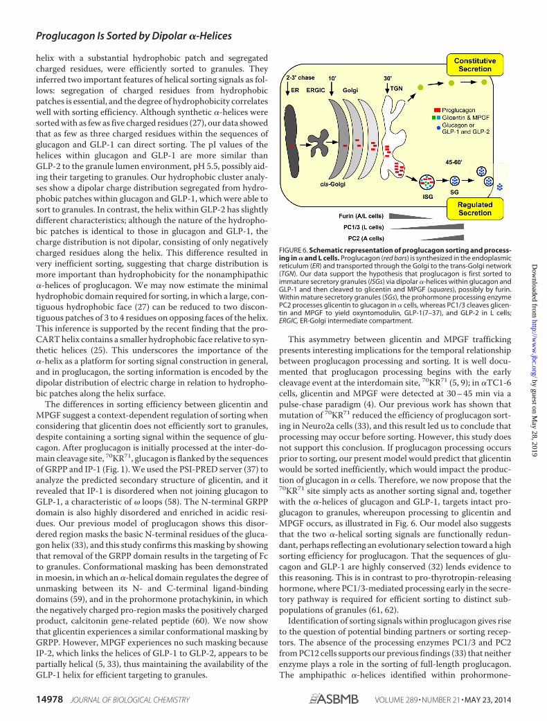
This asymmetry between glicentin and MPGF traffickingpresents interesting implications for the temporal relationshipbetween proglucagon processing and sorting. It is well docu-mented that proglucagon processing begins with the earlycleavage event at the interdomain site, 70KR71 (5, 9); in �TC1-6cells, glicentin and MPGF were detected at 30 – 45 min via apulse-chase paradigm (4). Our previous work has shown thatmutation of 70KR71 reduced the efficiency of proglucagon sort-ing in Neuro2a cells (33), and this result led us to conclude thatprocessing may occur before sorting. However, this study doesnot support this conclusion. If proglucagon processing occursprior to sorting, our present model would predict that glicentinwould be sorted inefficiently, which would impact the produc-tion of glucagon in � cells. Therefore, we now propose that the70KR71 site simply acts as another sorting signal and, togetherwith the �-helices of glucagon and GLP-1, targets intact pro-glucagon to granules, whereupon processing to glicentin andMPGF occurs, as illustrated in Fig. 6. Our model also suggeststhat the two �-helical sorting signals are functionally redun-dant, perhaps reflecting an evolutionary selection toward a highsorting efficiency for proglucagon. That the sequences of glu-cagon and GLP-1 are highly conserved (32) lends evidence tothis reasoning. This is in contrast to pro-thyrotropin-releasinghormone, where PC1/3-mediated processing early in the secre-tory pathway is required for efficient sorting to distinct sub-populations of granules (61, 62).
Identification of sorting signals within proglucagon gives riseto the question of potential binding partners or sorting recep-tors. The absence of the processing enzymes PC1/3 and PC2from PC12 cells supports our previous findings (33) that neitherenzyme plays a role in the sorting of full-length proglucagon.The amphipathic �-helices identified within prohormone-
FIGURE 6. Schematic representation of proglucagon sorting and process-ing in � and L cells. Proglucagon (red bars) is synthesized in the endoplasmicreticulum (ER) and transported through the Golgi to the trans-Golgi network(TGN). Our data support the hypothesis that proglucagon is first sorted toimmature secretory granules (ISGs) via dipolar �-helices within glucagon andGLP-1 and then cleaved to glicentin and MPGF (squares), possibly by furin.Within mature secretory granules (SGs), the prohormone processing enzymePC2 processes glicentin to glucagon in � cells, whereas PC1/3 cleaves glicen-tin and MPGF to yield oxyntomodulin, GLP-1(7–37), and GLP-2 in L cells;ERGIC, ER-Golgi intermediate compartment.
Proglucagon Is Sorted by Dipolar �-Helices
14978 JOURNAL OF BIOLOGICAL CHEMISTRY VOLUME 289 • NUMBER 21 • MAY 23, 2014
by guest on May 28, 2019
http://ww
w.jbc.org/
Dow
nloaded from

processing enzymes PC1/3 (26, 63), PC2 (56), and CPE (16) areknown to associate with the cholesterol-rich microdomains ofgranule membranes. However, we could not demonstrate bind-ing of purified proglucagon to liposomes (data not shown) andtherefore hypothesize that proglucagon may bind to granuleproteins. It is possible that granins bind prohormones, such aspro-opiomelanocortin (64). We have some evidence thatproglucagon sorting involves interaction with CPE in � cells(33). Studies investigating the roles of other granin proteins insorting proglucagon are currently underway.
In conclusion, we have shown that proglucagon contains twodipolar nonamphipathic �-helices with relatively small hydro-phobic faces that act as sorting signals for entry into secretorygranules of endocrine cells. Our data support a mechanism bywhich proglucagon is sorted to granules prior to the initialcleavage event that results in the production of glicentin andMPGF (Fig. 6). That these sorting domains lie within theordered domains of encoded proglucagon-derived peptides,and not in a disordered prodomain that characterizes manyother prohormones, highlights the unique sorting “signature”of proglucagon and further emphasizes the disparate nature ofsorting signals that lie within prohormones and other proteinsdestined for the secretory granules of the regulated secretorypathway.
Acknowledgments—We thank Dr. Jimmy D. Dikeakos (University ofWestern Ontario) and Dr. Robert B. Mackin (Creighton University,Omaha, NE) for critical reading of the manuscript and Karen Nygardfor assistance with confocal microscopy.
REFERENCES1. Jiang, G., and Zhang, B. B. (2003) Glucagon and regulation of glucose
metabolism. Am. J. Physiol. Endocrinol. Metab. 284, E671–E6782. Dong, C. X., and Brubaker, P. L. (2012) Ghrelin, the proglucagon-derived
peptides and peptide YY in nutrient homeostasis. Nat. Rev. Gastroenterol.Hepatol. 9, 705–715
3. Cohen, M. A., Ellis, S. M., Le Roux, C. W., Batterham, R. L., Park, A.,Patterson, M., Frost, G. S., Ghatei, M. A., and Bloom, S. R. (2003) Oxyn-tomodulin suppresses appetite and reduces food intake in humans. J. Clin.Endocrinol. Metab. 88, 4696 – 4701
4. Rouille, Y., Westermark, G., Martin, S. K., and Steiner, D. F. (1994) Prog-lucagon is processed to glucagon by prohormone convertase PC2 in �TC1-6 cells. Proc. Natl. Acad. Sci. U.S.A. 91, 3242–3246
5. Dey, A., Lipkind, G. M., Rouille, Y., Norrbom, C., Stein, J., Zhang, C.,Carroll, R., and Steiner, D. F. (2005) Significance of prohormone conver-tase 2, PC2, mediated initial cleavage at the proglucagon interdomain site,Lys70-Arg71, to generate glucagon. Endocrinology 146, 713–727
6. Furuta, M., Zhou, A., Webb, G., Carroll, R., Ravazzola, M., Orci, L., andSteiner, D. F. (2001) Severe defect in proglucagon processing in islet A-cells of prohormone convertase 2 null mice. J. Biol. Chem. 276,27197–27202
7. Damholt, A. B., Buchan, A. M., Holst, J. J., and Kofod, H. (1999) Progluca-gon processing profile in canine L cells expressing endogenous prohor-mone convertase 1/3 and prohormone convertase 2. Endocrinology 140,4800 – 4808
8. Dhanvantari, S., and Brubaker, P. L. (1998) Proglucagon processing in anislet cell line: effects of PC1 overexpression and PC2 depletion. Endocri-nology 139, 1630 –1637
9. Dhanvantari, S., Seidah, N. G., and Brubaker, P. L. (1996) Role of prohor-mone convertases in the tissue-specific processing of proglucagon. Mol.Endocrinol. 10, 342–355
10. O’Malley, T. J., Fava, G. E., Zhang, Y., Fonseca, V. A., and Wu, H. (2014)
Progressive change of intra-islet GLP-1 production during diabetes devel-opment. Diabetes. Metab. Res. Rev. 10.1002/dmrr.2534
11. Ravazzola, M., Perrelet, A., Unger, R. H., and Orci, L. (1984) Immunocy-tochemical characterization of secretory granule maturation in pancreaticA-cells. Endocrinology 114, 481– 485
12. Noe, B. D., Baste, C. A., and Bauer, G. E. (1977) Studies on proinsulin andproglucagon biosynthesis and conversion at the subcellular level. II. Dis-tribution of radioactive peptide hormones and hormone precursors insubcellular fractions after pulse and pulse-chase incubation of islet tissue.J. Cell Biol. 74, 589 – 604
13. Huang, X. F., and Arvan, P. (1995) Intracellular transport of proinsulin inpancreatic beta-cells. Structural maturation probed by disulfide accessi-bility. J. Biol. Chem. 270, 20417–20423
14. Dhanvantari, S., Shen, F.-S., Adams, T., Snell, C. R., Zhang, C., Mackin,R. B., Morris, S. J., and Loh, Y. P. (2003) Disruption of a receptor-mediatedmechanism for intracellular sorting of proinsulin in familial hyperproin-sulinemia. Mol. Endocrinol. 17, 1856 –1867
15. Cool, D. R., Normant, E., Shen, F., Chen, H. C., Pannell, L., Zhang, Y., andLoh, Y. P. (1997) Carboxypeptidase E is a regulated secretory pathwaysorting receptor: genetic obliteration leads to endocrine disorders in Cpe-(fat) mice. Cell 88, 73– 83
16. Zhang, C. F., Snell, C. R., and Loh, Y. P. (1999) Identification of a novelprohormone sorting signal-binding site on carboxypeptidase E, a regu-lated secretory pathway-sorting receptor. Mol. Endocrinol. 13, 527–536
17. Loh, Y. P., Maldonado, A., Zhang, C., Tam, W. H., and Cawley, N. (2002)Mechanism of sorting proopiomelanocortin and proenkephalin to theregulated secretory pathway of neuroendocrine cells. Ann. N.Y. Acad. Sci.971, 416 – 425
18. Lou, H., Kim, S.-K., Zaitsev, E., Snell, C. R., Lu, B., and Loh, Y. P. (2005)Sorting and activity-dependent secretion of BDNF require interaction of aspecific motif with the sorting receptor carboxypeptidase e. Neuron 45,245–255
19. Brakch, N., Allemandou, F., Cavadas, C., Grouzmann, E., and Brunner,H. R. (2002) Dibasic cleavage site is required for sorting to the regulatedsecretory pathway for both pro- and neuropeptide Y. J. Neurochem. 81,1166 –1175
20. Brechler, V., Chu, W. N., Baxter, J. D., Thibault, G., and Reudelhuber, T. L.(1996) A protease processing site is essential for prorenin sorting to theregulated secretory pathway. J. Biol. Chem. 271, 20636 –20640
21. Bundgaard, J. R., Birkedal, H., and Rehfeld, J. F. (2004) Progastrin is di-rected to the regulated secretory pathway by synergistically acting basicand acidic motifs. J. Biol. Chem. 279, 5488 –5493
22. Feliciangeli, S., Kitabgi, P., and Bidard, J. N. (2001) The role of dibasicresidues in prohormone sorting to the regulated secretory pathway. Astudy with proneurotensin. J. Biol. Chem. 276, 6140 – 6150
23. Garcia, A. L., Han, S.-K., Janssen, W. G., Khaing, Z. Z., Ito, T., Glucksman,M. J., Benson, D. L., and Salton, S. R. (2005) A prohormone convertasecleavage site within a predicted �-helix mediates sorting of the neuronaland endocrine polypeptide VGF into the regulated secretory pathway.J. Biol. Chem. 280, 41595– 41608
24. Mouchantaf, R., Kumar, U., Sulea, T., and Patel, Y. C. (2001) A conserved�-helix at the amino terminus of prosomatostatin serves as a sorting signalfor the regulated secretory pathway. J. Biol. Chem. 276, 26308 –26316
25. Blanco, E. H., Lagos, C. F., Andres, M. E., and Gysling, K. (2013) An am-phipathic �-helix in the prodomain of cocaine and amphetamine regu-lated transcript peptide precursor serves as its sorting signal to the regu-lated secretory pathway. PLoS One 8, e59695
26. Lacombe, M.-J., Mercure, C., Dikeakos, J. D., and Reudelhuber, T. L.(2005) Modulation of secretory granule-targeting efficiency by cis andtrans compounding of sorting signals. J. Biol. Chem. 280, 4803– 4807
27. Dikeakos, J. D., Lacombe, M.-J., Mercure, C., Mireuta, M., and Reudelhu-ber, T. L. (2007) A hydrophobic patch in a charged �-helix is sufficient totarget proteins to dense core secretory granules. J. Biol. Chem. 282,1136 –1143
28. Sasaki, K., Dockerill, S., Adamiak, D. A., Tickle, I. J., and Blundell, T. (1975)X-ray analysis of glucagon and its relationship to receptor binding. Nature257, 751–757
29. Underwood, C. R., Garibay, P., Knudsen, L. B., Hastrup, S., Peters, G. H.,
Proglucagon Is Sorted by Dipolar �-Helices
MAY 23, 2014 • VOLUME 289 • NUMBER 21 JOURNAL OF BIOLOGICAL CHEMISTRY 14979
by guest on May 28, 2019
http://ww
w.jbc.org/
Dow
nloaded from

Rudolph, R., and Reedtz-Runge, S. (2010) Crystal structure of glucagon-like peptide-1 in complex with the extracellular domain of the glucagon-like peptide-1 receptor. J. Biol. Chem. 285, 723–730
30. Venneti, K. C., and Hewage, C. M. (2011) Conformational and molecularinteraction studies of glucagon-like peptide-2 with its N-terminal extra-cellular receptor domain. FEBS Lett. 585, 346 –352
31. Dirndorfer, D., Seidel, R. P., Nimrod, G., Miesbauer, M., Ben-Tal, N., En-gelhard, M., Zimmermann, R., Winklhofer, K. F., and Tatzelt, J. (2013) The�-helical structure of prodomains promotes translocation of intrinsicallydisordered neuropeptide hormones into the endoplasmic reticulum.J. Biol. Chem. 288, 13961–13973
32. Irwin, D. M. (2001) Molecular evolution of proglucagon. Regul. Pept. 98,1–12
33. McGirr, R., Guizzetti, L., and Dhanvantari, S. (2013) The sorting of pro-glucagon to secretory granules is mediated by carboxypeptidase E andintrinsic sorting signals. J. Endocrinol. 217, 229 –240
34. McGirr, R., Ejbick, C. E., Carter, D. E., Andrews, J. D., Nie, Y., Friedman,T. C., and Dhanvantari, S. (2005) Glucose dependence of the regulatedsecretory pathway in �TC1-6 cells. Endocrinology 146, 4514 – 4523
35. Taupenot, L. (2007) Analysis of regulated secretion using PC12 cells. Curr.Protoc. Cell Biol. Chapter 15, Unit 15.12
36. Schindelin, J., Arganda-Carreras, I., Frise, E., Kaynig, V., Longair, M., Pi-etzsch, T., Preibisch, S., Rueden, C., Saalfeld, S., Schmid, B., Tinevez, J.-Y.,White, D. J., Hartenstein, V., Eliceiri, K., Tomancak, P., and Cardona, A.(2012) Fiji: an open-source platform for biological-image analysis. Nat.Methods 9, 676 – 682
37. McGuffin, L. J., Bryson, K., and Jones, D. T. (2000) The PSIPRED proteinstructure prediction server. Bioinformatics 16, 404 – 405
38. Artimo, P., Jonnalagedda, M., Arnold, K., Baratin, D., Csardi, G., de Castro,E., Duvaud, S., Flegel, V., Fortier, A., Gasteiger, E., Grosdidier, A., Hernan-dez, C., Ioannidis, V., Kuznetsov, D., Liechti, R., Moretti, S., Mostaguir, K.,Redaschi, N., Rossier, G., Xenarios, I., and Stockinger, H. (2012) ExPASy:SIB bioinformatics resource portal. Nucleic Acids Res. 40, W597–W603
39. Eisenberg, D., Schwarz, E., Komaromy, M., and Wall, R. (1984) Analysis ofmembrane and surface protein sequences with the hydrophobic momentplot. J. Mol. Biol. 179, 125–142
40. Gaboriaud, C., Bissery, V., Benchetrit, T., and Mornon, J. P. (1987) Hydro-phobic cluster analysis: an efficient new way to compare and analyseamino acid sequences. FEBS Lett. 224, 149 –155
41. Normant, E., and Loh, Y. P. (1998) Depletion of carboxypeptidase E, aregulated secretory pathway sorting receptor, causes misrouting and con-stitutive secretion of proinsulin and proenkephalin, but not chromograninA. Endocrinology 139, 2137–2145
42. Moller, J. C., Kruttgen, A., Heymach, J. V., Jr., Ghori, N., and Shooter, E. M.(1998) Subcellular localization of epitope-tagged neurotrophins in neu-roendocrine cells. J. Neurosci. Res. 51, 463– 472
43. Feliciangeli, S., and Kitabgi, P. (2002) Insertion of dibasic residues directsa constitutive protein to the regulated secretory pathway. Biochem. Bio-phys. Res. Commun. 290, 191–196
44. Chidgey, M. A., and Harrison, T. M. (1990) Renin is sorted to the regulatedsecretory pathway in transfected PC12 cells by a mechanism which doesnot require expression of the pro-peptide. Eur. J. Biochem. 190, 139 –144
45. Stridsberg, M., Grimelius, L., and Portela-Gomes, G. M. (2008) Immuno-histochemical staining of human islet cells with region-specific antibodiesagainst secretogranins II and III. J. Anat. 212, 229 –234
46. Hosaka, M., and Watanabe, T. (2010) Secretogranin III: a bridge betweencore hormone aggregates and the secretory granule membrane. Endocr. J.57, 275–286
47. Habib, A. M., Richards, P., Cairns, L. S., Rogers, G. J., Bannon, C. A.,Parker, H. E., Morley, T. C., Yeo, G. S., Reimann, F., and Gribble, F. M.
(2012) Overlap of endocrine hormone expression in the mouse intestinerevealed by transcriptional profiling and flow cytometry. Endocrinology153, 3054 –3065
48. Wu, Y., Gu, Y., Morphew, M. K., Yao, J., Yeh, F. L., Dong, M., and Chap-man, E. R. (2012) All three components of the neuronal SNARE complexcontribute to secretory vesicle docking. J. Cell Biol. 198, 323–330
49. Xia, F., Leung, Y. M., Gaisano, G., Gao, X., Chen, Y., Fox, J. E., Bhat-tacharjee, A., Wheeler, M. B., Gaisano, H. Y., and Tsushima, R. G.(2007) Targeting of voltage-gated K� and Ca2� channels and solubleN-ethylmaleimide-sensitive factor attachment protein receptor pro-teins to cholesterol-rich lipid rafts in pancreatic alpha-cells: effects onglucagon stimulus-secretion coupling. Endocrinology 148, 2157–2167
50. Han, G. A., Malintan, N. T., Collins, B. M., Meunier, F. A., and Sugita, S.(2010) Munc18-1 as a key regulator of neurosecretion. J. Neurochem. 115,1–10
51. Sirkis, D. W., Edwards, R. H., and Asensio, C. S. (2013) Widespread dys-regulation of peptide hormone release in mice lacking adaptor proteinAP-3. PLoS Genet. 9, e1003812
52. Li, S. K., Zhu, D., Gaisano, H. Y., and Brubaker, P. L. (2014) Role of vesicle-associated membrane protein 2 in exocytosis of glucagon-like peptide-1from the murine intestinal L cell. Diabetologia 54, 809 – 818
53. Jutras, I., Seidah, N. G., and Reudelhuber, T. L. (2000) A predicted �-helixmediates targeting of the proprotein convertase PC1 to the regulated se-cretory pathway. J. Biol. Chem. 275, 40337– 40343
54. Seino, S., Welsh, M., Bell, G. I., Chan, S. J., and Steiner, D. F. (1986) Mu-tations in the guinea pig preproglucagon gene are restricted to a specificportion of the prohormone sequence. FEBS Lett. 203, 25–30
55. Dikeakos, J. D., Di Lello, P., Lacombe, M.-J., Ghirlando, R., Legault, P.,Reudelhuber, T. L., and Omichinski, J. G. (2009) Functional and structuralcharacterization of a dense core secretory granule sorting domain fromthe PC1/3 protease. Proc. Natl. Acad. Sci. U.S.A. 106, 7408 –7413
56. Assadi, M., Sharpe, J. C., Snell, C., and Loh, Y. P. (2004) The C terminus ofprohormone convertase 2 is sufficient and necessary for Raft associationand sorting to the regulated secretory pathway. Biochemistry 43,7798 –7807
57. Dhanvantari, S., Arnaoutova, I., Snell, C. R., Steinbach, P. J., Hammond, K.,Caputo, G. A., London, E., and Loh, Y. P. (2002) Carboxypeptidase E, aprohormone sorting receptor, is anchored to secretory granules via a C-terminal transmembrane insertion. Biochemistry 41, 52– 60
58. Fetrow, J. S. (1995) Omega loops: nonregular secondary structures signif-icant in protein function and stability. FASEB J. 9, 708 –717
59. Li, Q., Nance, M. R., Kulikauskas, R., Nyberg, K., Fehon, R., Karplus, P. A.,Bretscher, A., and Tesmer, J. J. (2007) Self-masking in an intact ERM-merlin protein: an active role for the central �-helical domain. J. Mol. Biol.365, 1446 –1459
60. Ma, G.-Q., Wang, B., Wang, H.-B., Wang, Q., and Bao, L. (2008) Shortelements with charged amino acids form clusters to sort protachykinininto large dense-core vesicles. Traffic 9, 2165–2179
61. Mulcahy, L. R., Vaslet, C. A., and Nillni, E. A. (2005) Prohormone-conver-tase 1 processing enhances post-Golgi sorting of prothyrotropin-releasinghormone-derived peptides. J. Biol. Chem. 280, 39818 –39826
62. Perello, M., Stuart, R., and Nillni, E. A. (2008) Prothyrotropin-releasinghormone targets its processing products to different vesicles of the secre-tory pathway. J. Biol. Chem. 283, 19936 –19947
63. Dikeakos, J. D., and Reudelhuber, T. L. (2007) Sending proteins to densecore secretory granules: still a lot to sort out. J. Cell Biol. 177, 191–196
64. Sun, M., Watanabe, T., Bochimoto, H., Sakai, Y., Torii, S., Takeuchi, T.,and Hosaka, M. (2013) Multiple sorting systems for secretory granulesensure the regulated secretion of peptide hormones. Traffic 14, 205–218
Proglucagon Is Sorted by Dipolar �-Helices
14980 JOURNAL OF BIOLOGICAL CHEMISTRY VOLUME 289 • NUMBER 21 • MAY 23, 2014
by guest on May 28, 2019
http://ww
w.jbc.org/
Dow
nloaded from

Leonardo Guizzetti, Rebecca McGirr and Savita DhanvantariSorting Signals to the Regulated Secretory Pathway
-Helices within Hormone-encoding Regions of Proglucagon AreαTwo Dipolar
doi: 10.1074/jbc.M114.563684 originally published online April 11, 20142014, 289:14968-14980.J. Biol. Chem.
10.1074/jbc.M114.563684Access the most updated version of this article at doi:
Alerts:
When a correction for this article is posted•
When this article is cited•
to choose from all of JBC's e-mail alertsClick here
http://www.jbc.org/content/289/21/14968.full.html#ref-list-1
This article cites 63 references, 20 of which can be accessed free at
by guest on May 28, 2019
http://ww
w.jbc.org/
Dow
nloaded from