Embed Size (px)
Citation preview

i
THE SECRETORY APPARATUS OF
CERATOTHECA TRILOBA (PEDALIACEAE):
MORPHOLOGY AND CHEMICAL
COMPOSITION OF THE SECRETION
Taariq Karim
A research report submitted to the Faculty of Science and Agriculture, University of KwaZulu-
Natal, in fulfilment of the requirements for the degree of Master of Science in Biological
Sciences (Plant Science).
January 2012

ii
DECLARATION
I, Taariq Karim, declare that this dissertation is my own work. It is being submitted in fulfillment
of the requirements for the degree of Master of Science in Biological Sciences (Plant Sciences)
at the University of KwaZulu-Natal. It has not been submitted before for any degree or
examination at this or any other university.
.....................................................
Taariq Karim
...........day of................2012

iii
DEDICATION
This dissertation is dedicated to my parents who have supported me and always motivated me to
do my very best.

iv
ABSTRACT
Ceratotheca triloba (Bernh.) E. Mey. ex Hook. f., commonly known as the wild foxglove, is a
fast growing annual that is indigenous to southern Africa. The surface of the plant is covered
with fine, hair-like trichomes, which exude sticky, aromatic substances. The plant is traditionally
used to treat various abdominal ailments, insect infestation of the skin and leaf extracts are
administered to induce abortion. In order to provide a scientific basis for the medicinal properties
of C. triloba, this study was initiated to characterise the morphology and ultrastructure of the
foliar trichomes and to determine the chemical composition of the secretion. Phytochemical tests
and a range of microscopic techniques including stereo microscopy, scanning electron
microscopy, transmission electron microscopy, histochemistry and fluorescence microscopy
were used. Leaf surface imaging with stereo microscopy and SEM indicated the presence of two
morphologically distinct glandular trichomes, capitate and peltate. The capitate trichomes are
tall, consisting of a single basal cell, 2-4 stalk cells, a neck cell and a head that is made up of four
secretory cells. Capitate trichomes of up to 14 stalk cells were observed on the abaxial side of
flower petals. The peltate trichomes consisted of a basal cell, a single stalk cell and a
multicellular head. The peltate trichome head generally consisted of four cells, but occasionally
were seven or eight-celled. Fully developed trichomes were found to be concentrated on
emergent leaves and probably serve protective function. The density of trichomes decreased as
the leaf expanded. TEM showed that the secretory cells of peltate trichomes contained
centralised nuclei and numerous peripheral vacuoles. Numerous mitochondria and ER cisternea
were found throughout the cytoplasm. Osmiophilic plastids which were found near nuclei and
golgi apparatus were in close proximity to the peripheral vacuoles. Similar observations were
made for the stalk cells of both trichomes. The head cell of capitate trichomes however,
contained large amounts of osmiophilic substances, ER cisternae and mitochondria. The head
cells of peltate trichomes store the secretory product in vacuoles and secrete them upon cell
rupture or cell dissolution. The secretory product of capitate trichomes is secreted through the
cell membrane via an eccrine mode of secretion. Peltate and capitate trichomes appear to be
secreting similar compounds, but the composition of each compound in the secretory material
may vary between the trichomes. Histochemical and phytochemical tests reveal that the
secretions are comprised of mucilage, phenolic compounds, lipids, flavonoids, tannins, saponins

v
and fixed oils which may contribute to the medicinal properties of C. triloba. The observations
made in this study provide useful information for additional research in the Pedaliaceae, and
specifically in C. triloba. Future studies should isolate the active compounds for antimicrobial
and antioxidant testing. Cytotoxicity testing should also be undertaken to test safety and efficacy
of the active compounds.

vi
CONFERENCE CONTRIBUTIONS FROM THIS DISSERTATION
Karim, T., Naidoo, Y., Naidoo, G., 2009. Foliar trichomes of Ceratotheca triloba (Pedaliaceae):
The morphology and chemical composition of the secretion. 4th Global Summit on Medicinal
and Aromatic Plants, Kuching, Sarawak, Malaysia.

vii
ACKNOWLEDGEMENTS
I would like to thank my supervisor, Dr. Yougasphree Naidoo, for her support, expertise and
patience throughout this research. Her commitment will always be remembered and appreciated.
I would also like to thank my co-supervisor, Prof. G. Naidoo for his guidance and expertise
throughout this research and for the award of a NRF grantholder‟s bursary.
I would like to thank the following people:
The staff of The Electron Microscope Units at the University of KwaZulu-Natal, Westville and
Pietermaritzburg campuses.
C.T. Sadashiva for assistance with phytochemistry.
N. Kasim for assistance in preparing phytochemical and histochemical reagents.
Dr. S.H.A Ibrahim for assistance with transmission electron microscopy.
Finally, a special thank you is extended to my family and friends for their moral support. They
were a source of motivation and encouragement when this project seemed an impossible task.

viii
TABLE OF CONTENTS
Page
DECLARATION ii
DEDICATION iii
ABSTRACT iv
CONFERENCE CONTRIBUTIONS FROM THIS DESSERTATION vi
ACKNOWLEDGEMENTS vii
TABLE OF CONTENTS viii
LIST OF TABLES ix
LIST OF FIGURES x
LIST OF ABBREVAITIONS xii
CHAPTER 1: INTRODUCTION 1
CHAPTER 2: LITERATURE REVIEW 5
CHAPTER 3: MATERIALS AND METHODS 18
CHAPTER 4: RESULTS 26
CHAPTER 5: DISCUSSION 63
CHAPTER 6: CONCLUSION 76
REFERENCES 78

ix
LIST OF TABLES
Page
Table 1: Results of histochemical and fluorescence tests for the identification of
compounds in the secretory product.
51
Table 2: Results of phytochemical tests of crude methanolic extracts of powdered
leaves of C. triloba.
62

x
LIST OF FIGURES
Page
Figure 1: Ceratotheca triloba (Bernh.) E. Mey. ex Hook. f. growing along a
roadside at the University of Kwa Zulu-Natal, Westville Campus.
3
Figure 2: Glandular trichomes of the Lamiaceae 10
Figure 3: Mucilage hairs of Sesamum indicum and Ceratotheca melanosperma. 17
Figure 4: Capitate trichomes observed on leaves of C. triloba. 29
Figure 5: Peltate trichome on leaves of C. triloba. 31
Figure 6: Development of peltate trichomes on leaves of C. triloba. 33
Figure 7: SEM showing a decrease in density of trichomes as leaves mature. 35
Figure 8: Trichome frequency on the adaxial surface of leaves at three stages of
development, emergent, young and mature.
37
Figure 9: Trichome frequency on the abaxial surface of leaves at three stages of
leaf development, emergent, young and mature.
37
Figure 10: Surface imaging of trichomes on flower petals of C. triloba. 39
Figure 11: Light micrographs of transverse sections of leaves of C. triloba stained
with Sudan Black.
42
Figure 12: Light micrographs of transverse sections of leaves of C. triloba stained
with Sudan III and IV.
44
Figure 13: Light micrographs of transverse sections of leaves of C. triloba stained
with Ruthenium Red.
46
Figure 14: Autofluorescence of transverse sections of leaves of C. triloba. 48
Figure 15: Light micrographs of transverse sections of leaves of C. triloba stained
with ferric trichloride.
50
Figure 16: Transmission electron micrographs of a capitate trichome. 55

xi
Figure 17: Transmission electron micrographs of head cells of peltate trichomes. 57
Figure 18: TEM of cell wall region of a peltate trichome. 59
Figure 19: TEM showing vesicle formation in peltate trichomes. 61
Figure 20: Illustration showing different pathways of secretion in secretory cells. 72
Figure 21: Illustration showing likely origin of protein-carbohydrate mucilage in
mango fruit ducts.
73

xii
LIST OF ABBREVIATIONS
Bc Basal cell
C Capitate trichome
Ch Chloroplast
Cp Cellular pedestal
CPS Capitate trichome in the post secretory phase
Cw Cell wall
DC Developing capitate trichome
DP Developing peltate trichome
ER Endoplasmic reticulum
ESEM Environmental scanning electron microscopy/micrograph
Gb Golgi body
L Lipid drop
LM Light microscopy/micrograph
M Mitochondria
Mc Mucilage
Me Multimembranous structure
N Nucleus
Nc Neck cell
Nm Nuclear membrane
Nu Nucleolus
O Oil drop
P Peltate trichome
Pd Plastids
Pn Phenolic Compounds
PPS Peltate trichome in the post secretory phase
SC Subcuticular chamber
Sc Secretory cell
Sec Secretory material

xiii
SEM Scanning electron microscopy/micrograph
Sh Secretory head
St Stalk cell
TEM Transmission electron microscopy/micrograph
V Vacuole
Vc Vesicle
Vn Vein
WHO World Health organisation

1
CHAPTER 1: INTRODUCTION
1.1 ETHNOBOTANICAL MEDICINE
Traditional medicine can be defined as any non-western form of medication or medical practice.
It is a very broad term as it incorporates the use of animal, plant and mineral based products for
the diagnosis and treatment of ailments. Therefore, to describe medicinal practices involving the
use of plants and plant products only, the term ethnomedicine or ethnobotanical medicine will be
used instead.
The use of plants for medicine can be dated back in fossil records some 60 000 years ago to the
Middle Paleolithic age and the development of ethnomedicinal practices can be traced back
through documents described in ancient Ayurvedic and Chinese literature. The World Health
Organisation (WHO) estimates that 85% of the world population still rely on some sort of plant
product for medicine (Obi et al., 2006). Even in developed countries, the use of drugs containing
plant products is also common (Fabricant and Farnsworth, 2001). In Germany the consumption
of plants for medicinal purposes has risen by 20%. In the USA, 25% of all drugs dispensed by
pharmacies contained plant active compounds (Farnsworth et al., 1985). Despite this,
pharmaceutical companies in the USA have not shown much interest in ethnomedicinal research.
Developing countries, on the other hand, have invested in ethnomedicinal research which is
beneficial as they have a rich source of information on traditional medicinal plant species which
are used by the local people (Fabricant and Farnsworth, 2001).
An approach to researching plants for medicinal practices is by utilizing the information acquired
from traditional healers. The information obtained from ancient ethnomedicinal literature has
been useful in modern medicinal research. A survey carried out by Fabricant and Farnsworth
(2001) to determine if ethnomedicinal information had any use in modern day medicinal research
showed that 122 plant-derived pure compounds were identified as established drugs used in
modern medicine. Of these, 80% are still being used to treat the original ailments as described in
ethnomedicinal literature. It was also found that these compounds were isolated from only 94
plants. The number of flowering plant species occurring globally is estimated to be 250 000,

2
leaving many plants to be studied and disease treating compounds still to be discovered (Ayensu
and DeFilipps, 1978).
1.2 DESCRIPTION OF Ceratotheca triloba
Ceratotheca triloba (Bernh.) E. Mey. ex Hook. f., commonly known as the wild foxglove, is a
fast growing annual that is indigenous to southern Africa (Fig. 1). The plant is also known as the
South African foxglove because it is the only species of Ceratotheca in South Africa. The plant
thrives in disturbed habitats and is commonly observed in dry grasslands along roadsides.
Plants of C. triloba may be bushy or may grow as a single stem that can reach a height of 2 m,
depending on the soil moisture. The entire plant is covered in fine hair-like structures which
exude sticky, aromatic substances. The plants display either pink or white flowers that grow in
pairs along the uppermost part of stems. The petals have five lobes with the lowermost being
longer than the rest and displaying streaks of thin lines leading into the flower. The soft leaves
expand to about 50 mm in length and divide into three lobes at the base with bluntly serrated
margins. Seeds of the plant are protected by a capsule that displays three horns at the tip.
In South Africa, leaf extracts of C. triloba are used by traditional healers to treat diarrhoea,
nausea and painful abdominal and menstrual cramps, whereas in Zimbabwe, the same leaf
extracts are administered to induce abortion. Furthermore, in Botswana, the entire plant of C.
triloba is used to treat insect infestations of the skin and pediculosis. Despite its potential
medicinal value, no significant scientific research has been carried out on this plant.

3
Figure. 1. Ceratotheca triloba (Bernh.) E. Mey. ex Hook. f. growing along a roadside at the
University of Kwa Zulu-Natal, Westville Campus. a) whole plant, b) mature three lobed leaf, and
c) pink flower with five lobes; the lower most lobe has streaks of lines running into the corolla
tube.
a
c
b

4
1.3 RATIONALE OF THIS STUDY
Trichomes have been studied previously because of their functional usefulness in nature and the
economic value of the secretory product in industry (Valkama et al., 2003). Trichomes are highly
variable structures that provide a mechanical and chemical barrier against herbivores and the
secretory product that they produce has been used in the food, fragrance and pharmaceutical
industry (Fabricant and Farnsworth, 2001; Valkama et al., 2003). Trichome morphological traits
have also been key characteristics in plant taxonomic studies. Therefore the morphology of
trichomes, chemical nature of the secretory product, and how the secretory product is produced
are vital aspects that need to be addressed.
1.4 AIMS AND OBJECTIVES
The genus Ceratotheca has been neglected in terms of trichome research. Not much is known
about the trichomes or the chemical properties of the secretions they produce. Since C. triloba
has been used by traditional healers for a variety of ailments, this study was undertaken with the
following aim and objectives:
Aim
To provide a scientific basis for the medicinal properties of C. triloba.
Objectives
a. characterise the foliar trichomes of C. triloba according to morphology and distribution
b. determine the chemical composition of the secretory product
c. provide a description of the secretory process
d. provide a phytochemical assessment of crude leaf extracts

5
CHAPTER 2: LITERATURE REVIEW
Secretory tissues in plants can be defined as multicellular structures or specialised single cells
that eliminate specific substances to the external environment. These structures secrete
specialised compounds which may aid in pollination, defence against herbivory and pathogens,
and regulation of water and ions. In all cases, secretory structures serve as part of the metabolic
pathway of plants, and in plant interrelations with the external environment. These structures
include salt secreting glands, laticifers, nectaries, mucilage secreting cells, and trichomes (Fahn,
1979). There is great variation in structure and function of secretory tissues between species of
plants. In addition, development and composition of the secretion can also vary. This review will
focus on plant trichomes which are the most diverse group of secretory structures. The botanical
literature has listed more than 300 morphologically distinct types of trichomes which have been
used in plant classification (Kelsey et al., 1984).
Trichomes, derived from the Greek word trikhoma which means „growth of hair‟, are fine hair-
like outgrowths found on the aerial regions of most angiosperms, some gymnosperms and
bryophytes (Johnson, 1975). They are highly variable structures that are in contact with the
external environment and therefore function in response to various biotic and abiotic stimuli
(Tooker et al., 2010). Trichomes serve as the first line of defence against plant natural enemies
and they may attract and guide pollinators (Young et al., 1984; Wagner, 1991). They also reflect
UV radiation, and assist in drought tolerance by reducing leaf temperature and preventing
excessive water loss (Levin, 1973; Wagner, 1991; Werker, 2000). In addition to variation in
structure and function, differences may also occur in trichome development (Werker, 2000;
Serna and Martin, 2006).
2.1. EVOLUTION OF PLANT TRICHOMES
Co-evolution between species is widely accepted as one of the fundamental mechanisms that
produced the Earth‟s diversity (Ehrlich and Raven, 1964). The process of co-evolution is a
closely linked interaction between species. Interactions such as pollination, predation and
mutualism between species may bring about genetic and morphological changes in one species

6
triggered by changes in the other (Ehrlich and Raven, 1964; Janzen, 1980). According to the co-
evolutionary hypotheses, characteristics of plants that offer protection such as spines, thorns and
trichomes evolved due to selection pressures imposed by natural enemies on plants (Juniper and
Southwood, 1986). Therefore, plants evolved trichomes as a defence strategy against herbivores
and plant pathogens.
Arabidopsis thaliana grown in an environment free of herbivores and pathogens displayed a
decrease in production of two known characteristics that offer protection against natural enemies
i.e. glucosinolate concentration and trichome density (Mauricio and Rausher, 1997).
Glucosinolate is a secondary compound that is toxic to herbivores and pathogens (Mauricio and
Rausher, 1997; Rosa and Rodrigues, 1999; Tierens et al., 2001) and trichome density provides
defence against plant‟s natural enemies (Ambrosio et al., 2008). These results indicate that
selection pressure enforced by plant natural enemies causes an increase in defence
characteristics, which is supportive of the co-evolution hypothesis.
In contrast to co-evolution, the sequential evolution hypothesis considers the resistant features of
trichomes as incidental and points to abiotic selections pressures, such as temperature and UV
radiation, as the underlying cause of trichome evolution (Mauricio and Rausher, 1997). Unlike
the co-evolution hypothesis, evidence to support the sequential evolution hypothesis is
circumstantial.
2.2. TRICHOME CLASSIFICATION
Trichomes are highly diverse structures that vary in function, location, density, distribution,
ability to secrete and timing of secretion. These differences can occur between species of plants,
between plants within a single species and between organs of an individual plant. Classification
of trichomes using one of the above characteristics is difficult because of overlap between
species of plants and neither characteristic on its own is sufficient (Werker, 2000). The function
of a trichome may vary according to its location on the plant. Trichomes located on vegetative
organs may serve to repel herbivory, whereas trichomes located on reproductive organs may
serve a dual role as attractants to pollinators as well as repellents to herbivores.

7
2.2.1 Morphological variation
a) Non-glandular trichomes
Non-glandular trichomes are generally classified according to their morphology. They range
from unicellular to multicellular structures that can be uniseraite, biseriate or multiseriate,
branched or unbranched (Werker et al., 1994). The tips of non-glandular trichomes may be
tapering or blunt (Werker, 2000). Thickness of cell walls may vary within and between
trichomes and the hardening material within the walls may vary resulting in varying strength
(Werker, 2000). Trichome length may differ between different plants, between organs of the
same plants or within a single organ (Economou-Amilli et al., 1982).
Occasionally more than one type of non-glandular trichome may be found on leaves as in the
case of Salvia blepharophylla and Coridothymus capitatus (Economou-Amilli et al., 1982; Bisio
et al., 1999). In S. blepharophylla, the three types were found: 1. multicellular, long trichomes
with tapering tips, located along the leaf margins; 2. multicellular, uniseriate, short trichomes
(usually 4-5 celled) with swollen bases located on the midrib and veins; and 3. unicellular
papillae with enlarged bases and tapering tips, which were common on the abaxial surface of
leaves. The reason for this variation in trichome morphology, however, is not known.
b) Glandular trichomes
Glandular trichomes include a variety of glands that synthesise, accumulate and secrete essential
oils (Croteau, 1987; Gershenzon et al., 1989). In order to classify these trichomes, the chemical
composition of the secretion and the secretory process are considered in relation to trichome
location and function.
The structure of glandular trichomes range from unicellular to multicellular and these can be
uniseriate or multiseriate (Werker, 2000). The uniseriate, multicellular glandular trichome
comprises of a base of one to a few cells, a stalk of one to many cells and a secretory head made
up of one to many secretory cells (Fahn, 1988). Occasionally, a neck cell is located between the
secretory head cells and stalk cells, and is morphologically different from the two.

8
In species of Lamiaceae, glandular trichomes are generally characterised in two major groups,
the capitate and the peltate trichomes, named according to the shape of their secretory heads
(Fahn, 1988). Capitate (Latin – capitis meaning “of a head”) trichomes usually have a globose
secretory head made up of 1-4 cells, subtended by a stalk of one to many cells. They have been
termed “short term trichomes” because they develop and release their secretion early in leaf
development, thereby providing protection against herbivory even before any form of
mechanical disturbance occurs (Werker, 2000). Peltate (Latin – peltata meaning “shield shaped”)
trichome secretory heads are usually flattened and round made up of 4-18 cells subtended by a
single stalk cell located at the centre of the secretory head. These have been defined as “long
term trichomes” since they accumulate oil that is only release upon mechanical disruption
(Werker, 2000). Similar forms of trichomes have been observed in other families such as the
Cannabaceae (Dayanandan and Kaufman, 1976), Fabaceae (Zoric et al., 2009), Curcurbitaceae
(Kolb and Müller, 2004) and Pedaliaceae (Naidoo et al., 2006).
Variation in trichome structure may occur between species within a family or genus. Mentha
peperita and Leonotis leonurus of the family Lamiaceae possess both capitate and peltate
trichomes (Fahn, 1988; Ascensão et al., 1995, Fig. 2). Both contain peltate trichomes consisting
of a single basal cell, a stalk cell and a secretory head made up of eight secretory cells arranged
in a circle (Fahn, 1988; Ascensão et al., 1995). However, other species of the same family such
as Satureja thymbra, have peltate trichomes of similar structure but the head contains more cells
arranged in two concentric circles (Bosabalidis, 1990).
Differences in trichome structure may also be found within a single genus as in the case of Salvia
blepharophylla (Bisio et al., 1999) and S. aurea (Serrato-Valenti et al., 1997), which contain
peltate trichomes with secretory heads of four cells arranged in a quadrant, and eight cells
arranged in a circle respectively. The capitate trichomes of S. aurea and S. blepharophylla are
however, similar (Serrato-Valenti et al., 1997; Bisio et al., 1999) comprising of a single stalk cell
and a bicellular secretory head. In contrast, those found on leaves and flowers of S. scleria and S.
dominica are highly diverse, comprising of six different forms differing in structure, size,
location, and manner of secretion (Werker et al., 1985a). In addition, the short stalk capitate
trichomes of Plectranthus madagascariensis (Ascensão et al., 1998) and P.annua (Ascensão et

9
al., 1999) also differ with respect to the number of head cells, with the former possessing a
bicellular head and the latter a unicellular one.
Variation may also extend to trichomes located on the same organ of a plant and between groups
of cells within a single trichome. Meyberg et al., (1991), reported that capitate trichomes found
on leaves of Nicotiana tobaccum secreted different products even though they appeared to be
morphologically analogous. Some trichomes secreted a resinous compound while the others
secreted oil droplets. The exact functions of the different groups of cells that make up a trichome,
and between cells of the same group are not clear. Ultrastructural studies indicated that trichomes
with multicellular secretory heads may contain cells that secrete different substances than their
neighbouring head cells (e.g. Artemisia annua, Duke and Paul, 1993) whereas other studies have
highlighted the possibility that some head cells may not even be active in secretion (e.g. Mallotus
philipinensis, Roth, 1977). Ultrastructural studies have also shown differences in cells of the
stalk with the lowermost cells being highly vacuolated and the uppermost cells having dense
cytoplasm (Turner et al., 2000).

10
Figure 2. Glandular trichomes of Lamiaceae. a) Peltate trichome of Mentha peperita with oil
drop (O) in subcuticular chamber (SC). The side walls of the stalk are highly cutinised (arrow) to
prevent apoplastic backflow. b) Capitate trichome with a single stalk cell and unicellular
secretory head. (from Fahn, 1988)
O
SC
a
b

11
c) Integrated forms of glandular and non-glandular trichomes
In most cases, trichomes can be clearly differentiated into glandular and non-glandular.
However, there exist trichomes that cannot be characterised explicitly into either group. In the
case of certain species of Lamiaceae, unbranched glandular trichomes are clearly differentiated
from branched non-glandular trichomes. However, there are instances in which compound
glandular trichomes are branched having many non-glandular branches with one glandular
branch (e.g. Phlomis, Azizian and Cutler, 1982; Rosmarinus officinalis, Werker et al., 1985c;
Hyptis, Rudall, 1980). In addition, Ascensão et al., (1995) reported that the stalk walls of capitate
trichomes in Leonotus leonurus displayed cuticular warts which are a characteristic of the non-
glandular trichomes of that species. It was concluded by Ascensão et al., (1995) that these
capitate trichomes represented an intermediate form between glandular and non-glandular
trichomes. Fahn (1988) suggested that these findings may also provide some answers with
regards to the evolution of these structures.
2.2.2 Variation in the process of secretion
In Lamiaceae, the peltate trichomes accumulate the secretory product in a subculticular chamber,
which forms when the cuticle detaches from the cell wall of the secretory cells (Werker et al.,
1985a, b). These peltate trichomes are assumed to release their contents when the elevated
cuticle is damaged, usually by plant herbivores. Ceratotheca sesamoides of the family
Pedaliaceae has peltate trichomes that lack a subcuticular chamber. They secrete mucilage when
the secretory cell ruptures (Abels, 1975) or dissolves (Ihlenfeldt, 2001), resulting in cell death.
Capitate trichomes may also accumulate oils in a subcuticular chamber and release them either
by cuticular rupture or by diffusion through micropores in the cuticle. The secretory product of
the “short stalked capitate trichome” of Plectranthus madagascariensis accumulates in a small
subcuticular space and is probably released by diffusion through micropores, whereas “the long
stalked capitate trichome” releases its secretory product via cuticular rupture (Ascensão et al.,
1998). Similar variations were also reported for other species of Lamiaceae (e.g. Ocimum
basilicum, Werker et al., 1993; Plectranthus annua, Ascensão et al., 1999) The “capitate Type Ι
hairs” of S. sclarea however lack a subcuticular chamber and release their contents through

12
micropores in the cell wall and cuticle (Werker et al., 1985a). Accumulation of secretory product
in the cell lumen and direct diffusion through the cell wall were also observed for capitate
trichomes in S. aurea (Serrato-Valenti et al., 1997) and O. basilicum (Werker et al., 1993). Kolb
and Müller (2004) observed that the capitate trichomes found on leaves of Cucurbita pepo var.
styriaca released their secretion when they interacted with the non-glandular trichomes and
bristles which indicates that secretion in this species is dependant on trichome density.
2.2.3 Trichome function
a) Protection
Plants may require protection from a variety of abiotic (extreme temperature, intense light and
water loss) and biotic (herbivores, pathogens and alleopathy from plant competitors) factors.
Trichomes provide protection against all of the above threats, especially when they densely cover
plant organs. The effect of trichome density on herbivore resistance has been noted in Thithonia
diversifolia (Ambrosio et al., 2008).
In the case of abiotic factors, a direct correlation has been observed between environmental
stresses and the density of trichomes found on the plant organ (Clausen et al., 1940). Johnson,
1975 reported that Potentilla (Rosaceae) planted in sunny sites contained more pubescent
trichomes than those grown in shady sites. Warming (1909) has already emphasised the point
that a greater number of trichomes will appear in plants that are subjected to a harsher moisture
regime. He also noted the differences in trichome cover between plants of the same species
which are found in xeric and mesic habitats. He observed that the plants which are found in xeric
habitats contained more trichomes. In addition, the resin secreted by glandular trichomes, in
Betula pendula found at high altitudes, serves as a moisture repellent, protecting the plant against
the harsh winter and spring (Lapinjoki et al., 1991). Non-glandular trichomes may serve as
structural defences against plant herbivores. Gilbert (1971) observed a highly effective defence
strategy of Passiflora adenopoda against the heliconiine butterfly larvae. The hook-like
trichomes killed the larvae by entrapping them, followed by inducing many puncture wounds in
the larval integument.

13
Glandular trichomes provide a variety of defence strategies by secreting specialised compounds
that may either trap or poison herbivores, or provide an antimicrobial effect against pathogens.
These compounds include terpenoids (Dawson et al., 1966; Bakker et al., 1972; Turner et al.,
2000), flavonoids (Kelsey et al., 1984), tannis (Feeny, 1968) alkaloids (Rodriguez et al., 1984)
and phenolics (Yu et al., 1992). Non-volatile compounds such as alkaloids, tannins and phenols
may act as a deterrent to herbivory. In a study carried out by Harley and Thorsteinson (1967),
several alkaloids (including nornicotine, lobeline, tomatine and hordinine) that were added to the
diet of the two striped grasshopper, Melanoplus bivittatus, either repelled the insects or killed
them. Similarly, phenolic compounds such as the coumarins, which are produced by Melilotus
species, have an inhibitory effect on the vegetable weevil (Matsumoto, 1962). Some glandular
trichomes secrete mucilage that interacts with phenols to trap herbivorous insects. Tingey and
Gibson (1978) reported a defence strategy of Solanum polyadenium and S. berthaultii against the
aphid Empoasca fabea. It was reported that the mouthparts and tarsi were encased by a sticky
substance which hardened, resulting in starvation and death of the aphid. This is probably due to
the release of phenolic compounds which are oxidised to quinines by polyphenol oxidase. The
resulting quinines polymerise and trap insects (Yu et al., 1992).
Tannins interact with proteins to form tannin-protein complexes, which reduce the total protein
that can be ingested by herbivores. Since these complexes reduce the dietery content in plants
and most angiosperms contain tannins, it is highly possible that insects use tannins as a negative
feeding cue. In addition, chemoreceptors found on legs and mouthparts of insects detect possible
fatal chemicals on the elevated head cells of trichomes (Dethier, 1963). This prevents feeding as
the insect realises the host is unsuitable for consumption. In this way trichomes serve as a
deterrent to herbivores by advertising the presence of highly toxic chemicals.
b) Attraction
Much of the research characterising trichomes has focused on the vegetative organs with the
reproductive organs being neglected. Trichomes on flowers serve conflicting roles in deterring
natural enemies and attracting pollinators (possibly the natural enemies of other plants). In some
species of Lamiaceae, large glandular trichomes were observed on stamens, under the anther
lobes (Cannabis sativa, Mahlberg et al., 1984; Leonotus leonurus, Ascensão et al., 1995).

14
Whether these trichomes secrete compounds that attract pollinators or whether they deter
herbivores, or both, is not known.
Many authors have observed differences in trichome structure and distribution between
vegetative and reproductive organs. This may be indicative of differences in function. In
Cannabis sativa, stalked capitate trichomes were observed on floral organs, whereas the
vegetative organs displayed various other trichomes (Mahlberg et al., 1984). In many species,
the calyx, which covers the flower during development, is covered in glandular and non-
glandular trichomes similar to those found on the vegetative organs. These trichomes are
probably for protection as described previously. The trichomes on the calyx of Origanum
vulgare (Werker et al., 1985b) are different from those on vegetative organs with respect to the
ratio of the various types of glandular trichomes and the proportion of the chemical constituents
they secrete, thus indicating that trichomes on the calyx may also serve as attractants. In Salvia
dominica and S. sclarea, both the reproductive and vegetative organs are covered in capitate and
peltate trichomes. The morphology of peltate trichomes is uniform between the two organs
whereas the capitate trichomes differ in structure, size and mode of secretion. The main
components of the essential oil, neryl acetate and linalyl acetate, also differed between vegetative
and reproductive organs (Werker et al., 1985a).
Variations in trichome occurrence and distribution were also observed within a flower. In some
species of Lamiaceae such as Origanum vulgare (Werker et al., 1985b) and Leonotus leonurus
(Ascensão et al., 1995), peltate trichomes were the only types observed on the outside of corolla
tubes whereas the inside of the tubes displayed a variety of other glandular trichomes. Variation
in trichome structure and distribution between and within organs may indicate differences in
function. However, evidence to support this is inferred and more work is required to clarify these
issues.

15
2.3 TRICHOME DEVELOPMENT
Most of the information regarding trichome development is drawn from studies on mutants of
Arabidopsis thaliana. These studies show that the first cells (protodermal cells) to differentiate
from the epidermis develop into trichomes. Trichomes are therefore generated in early leaf
development and the rate of differentiation is rapid (Duke and Paul, 1993; Werker, 1993). This
often provides protection to the leaf primordia which are more susceptible to herbivory. In Inula
viscosa, fully developed trichomes were observed on leaves not more than 2 mm long (Werker
and Fahn, 1981). Whether all the trichomes produced on leaf primordia are predetermined or
develop as the leaf ages is still not clear.
The various processes in trichome development are discussed in the following section.
Development of other protuberances, such as emergences and „false hairs‟ will not be considered
as they are not true trichomes.
2.3.1 Development of single cells
The epidermal cell, with a dense cytoplasm and a large nucleus, divides periclinally to form a
protrusion above the epidermis. These cells can then undergo further periclinal divisions to form
a stalk and anticlinal divisions to form secretory head cells (Werker, 2000). Uphof (1962)
characterised the periclinal development of a multicellular, uniseriate trichome according to
direction of division of the existing stalk cells. Acropetal division occurs when the uppermost
stalk cell divides to form a new cell, whereas basipetal division occurs when the lowermost stalk
cell undergoes division. In Pelargonium scabrum, capitate stalked trichomes result from a
development of the protodermal cell which divides periclinally to form a single stalk cell
(Oosthuizen and Coetzee, 1983). Further stalk cells develop from acropetal division. The peltate
trichome in species of Lamiaceae undergoes two periclinal, acropetal divisions to form a stalk
cell and a single secretory cell which undergoes further anticlinal division (e.g. Origanum
dictamnus, Bosabaladis and Tsekos, 1982a; Leonotus leonurus, Ascensão et al., 1995).

16
A common misconception with biseriate and multiseriate trichomes is that they all develop from
more than one cell. In most biseriate trichomes, especially those of the Asteraceae, initial
division occurs from a single protodermal cell which undergoes an anticlinal division to form
two cells along the leaf epidermis (e.g. Holocarpha virgata, Carlquist, 1958; Artemisia
campestris, Ascensão and Pais, 1987; Stevia rabaudiana, Montiero et al., 2001). These cells then
divide periclinally to form a fully developed biseriate trichome. There are variations to this form
of development. In Helichrysum aureonitens, the first division is periclinal but the anticlinal
division occurs soon after (Afolayan and Meyer, 1995).
In most trichomes, in addition to differentiation of the protodermal cell, other epidermal cells
may differentiate around the protodermal cell. These generally form a ring of differentiated cells
normally referred to as a cellular pedestal, which provides support and anchors the trichome in
the epidermis (Werker, 2000). Some cellular pedestals may even grow above the epidermis
thereby elevating the trichome.
2.3.2 Development from multiple cells
Initially only one protodermal cell divides anticlinally to form four cells, which undergo
numerous periclinal divisions to form a developed trichome. In addition to the single
protodermal cell, many epidermal cells also differentiate and divide together to form the
developing trichome, and in most cases add to the structure as in Helicteres hirsute (Uphof,
1962).
2.4 THE FAMILY PEDALIACEAE
The Pedaliaceae is a small family, consisting of 17 genera and 80 species. It is a predominantly
African family with the exception of a few species, such as Pedalium murex, which is found in
Yemen (Cronquist, 1981; Wood and Haig-Thomas, 1997). Sesamum is the largest genera
consisting of 20 species, most of which are well documented for their oily seeds. Taxonomically,

17
the genera Sesamum and Ceratotheca are the closest, with the horns on seed capsules of
Ceratotheca the only characteristic separating the two (Bruce, 1953).
Much of the research on this family focused on the oil producing seeds of Sesamum. In terms of
research on trichomes, the Pedaliaceae has been somewhat neglected. Solereder (1908) described
the mucilage hairs found on leaves of Sesamum indicum and Ceratotheca melanosperma. These
trichomes were peltate, consisting of a head made up of four cells arranged in a cross (Figure 3).
A capitate trichome was also described as having a multicellular stalk and a small spherical head
of four cells which were shaped like small palisade cells. Similar observations were made for C.
sesamoides. Naidoo et al., (2006) described glandular trichomes found on leaves of
Harpagophytum procumbens consisting of a multicellular stalk and head made up of four cells
that were arranged in a quadrant, which secreted viscous material.
Figure 3. Mucilage hairs of Sesamum indicum and Ceratotheca melanosperma. (from Solereder,
1908)
a b

18
CHAPTER 3: MATERIALS AND METHODS
3.1 COLLECTION AND PROPAGATION OF PLANT MATERIAL
Intact plants of Ceratotheca triloba were collected from the University of KwaZulu-Natal
(UKZN), Westville campus and Umhlanga Rocks (290 43‟ 33.6” S, 31
0 4‟ 12” E) and transported
to the greenhouse for propagation in pots. Voucher specimens were prepared and kept at the
School of Biological and Conservation Sciences Ward Herbarium (Voucher specimen no.
KARIM - 1).
3.2 SAMPLING
For light microscopy and scanning and transmission electron microscopy, samples were taken
from mature plants in the field from the tip, mid and basal regions of leaves. Three stages of leaf
development were sampled in this study i.e. the emergent (width < 2 mm, length < 9 mm) young
(width 2 < 18 mm, length 9 < 26 mm) and mature (width > 18 mm, length > 26 mm) leaves.
3.3 PREPARATION FOR SCANNING ELECTRON MICROSCOPY (SEM)
Three techniques were used for SEM studies: freeze-drying, chemical fixation and environmental
SEM.
3.3.1 Freeze-drying
Leaf segments were prepared by quenching in liquid nitrogen slush and freeze drying for 96 h in
an Edwards Modulyo freeze dryer at -40 0C to -60
0C at a vacuum of 10
-1 Torr. After securing
samples on brass stubs with carbon conductive tape, they were sputter coated with gold in an
atmosphere of argon with a Polaron Sputter coater unit SC500. The leaf segments were viewed
with a Leo 1450 SEM at 5 kV and a working distance of 15 mm.

19
3.3.2 Chemical fixation
Segments of leaf blades and petals were prepared for chemical fixation for SEM. These samples
underwent a primary fixation in a solution of 2.5 % glutaraldehyde (4 0C) buffered with 0.1 M
phosphate buffer (pH 7.2) for 2-24 h followed by a post fixation at room temperature in 0.5%
osmium tetroxide (OsO4) for 1 h. Samples were then dehydrated in a graded alcohol series of 30
%, 50 %, 75 % and 100 % ethanol. Critical-point-dried samples were then secured on brass stubs
with carbon conductive tape and sputter coated with gold and viewed as described previously.
3.3.3 Environmental scanning electron microscopy (ESEM)
Segments of fresh leaves and petals were placed on brass stubs with carbon conductive tape and
viewed under a Philips XL 30 ESEM at 15 kV and a working distance of approximately 13 mm.
3.4 PREPARATION FOR TRANSMISSION ELECTRON MICROSCOPY (TEM)
Two standard methods (differing in embedding time and the embedding medium) of sample
preparation were used for TEM.
3.4.1 Method 1
Leaf segments (< 0.5 mm2) were prepared by prefixing in a solution of 2.5% glutaraldehyde (4
0C) buffered with 0.1 M phosphate buffer (pH 7.2) for 2-24 h followed by a post fixation at room
temperature in 0.5% osmium tetroxide (OsO4) for 1 h. The samples were washed three times for
5 min before and after post fixation in phosphate buffer followed by dehydration in a graded
acetone series (i.e twice for 5 min each in 30 %, 50 %, 75 % and 100 % acetone). The leaf
segments were allowed to stand in equal parts Spurr‟s (1969) resin and acetone for 4 h followed
by Spurr‟s resin only for 24 h. Samples were then polymerized in whole resin at 70 0C for 8 h.
Ultrathin sections were obtained with a Reichhert Ultracut E microtome using glass knives and
collected on uncoated 200 square mesh copper grids. The sections were stained with 2 %
aqueous uranyl acetate (10 min) followed by Reynold‟s (1963) lead citrate (10 min). Sections
were viewed with a Jeol 1010 TEM.

20
3.4.2 Method 2
Leaf segments (< 0.5 mm2) were prepared by prefixing in a solution of 3 % glutaraldehyde (4
0C)
buffered with 0.05 M cacodylate buffer for 8 h followed by post fixation at room temperature in
2 % OsO4 for 2 – 4 h. The samples were washed twice for 30 min before and after post fixation
in cacodylate buffer (0.05 M), followed by dehydration in a graded alcohol series of 10 min each
in 10 %, 30 %, 50 % and 70 % ethanol. The samples were allowed to stand in 70 % ethanol
overnight at 4 0C and further dehydrated for 10 min each in 80 % and 90 % ethanol followed by
three 10 min changes in 100 % ethanol. The leaf segments were then washed twice in propylene
oxide for 30 min and taken through increasing concentrations of Epon resin, diluted with
propylene oxide (i.e. 25 % - 2 h, 50 % - 2 h, 75 % - 12 h and 100 % - 24 h). Leaf material was
then embedded in 100 % resin at 70 0C for 48 h. Ultrathin sections were obtained with a
Reichhert Ultracut E microtome using glass knives and collected on uncoated 200- square mesh
copper grids. The sections were stained with 2 % aqueous uranyl acetate (10 min) followed by
Reynold‟s (1963) lead citrate (10 min). Sections were viewed with a Jeol 1010 TEM.
3.5 LIGHT MICROSCOPY (LM)
For light microscopy, thin, hand cut sections and sections embedded in wax or Spurr‟s (1969)
resin were used.
3.5.1 Wax embedded material
Excised leaf samples (~ 3 mm2) were initially placed in formaldehyde (70 %) for 48 h.
Thereafter, the leaf samples were dehydrated with eight incubations in a graded ethanol series (3
x 70 % - 30 min, 80 % - 2 h, 90 % - 4 h, 100 % - 90 min, 100 % - overnight, 100 % - 2 h). The
samples were then placed in a graded series of xylene: ethanol mixtures (25:75, 50:50, 75:25) for
an hour each before being placed in two xylene incubations for 15 min each. Paraplast Plus®
wax pellets (McCormick Scientific) were added four times at 3 h intervals, followed by a final
addition of wax before sectioning.

21
Wax sections were de-waxed by exposing them to three 2 min baths of xylene (100 %), three 2
min baths of ethanol (100 %), a single 2 min bath in 70 % ethanol and a final 2 min wash in tap
water.
3.5.2 Spurr embedded material
Leaf material was prepared in Spurr‟s (1969) resin (according to the standard protocol 3.4.1) and
semi-thin sections were placed on pre-cleaned glass slides. Monitor sections (1 µm) were stained
with 1 % Toluidine Blue - O (Feder and O‟Brien, 1968)
3.6 FLUORECSENCE MICROSCOPY
3.6.1 Cellulose cell walls
Excised leaf segments were placed in a non-toxic, 0.01 % solution of Cellufluor for 5 h, followed
by washing in tap water for 30 min. Thereafter, the leaf segments were hand sectioned
transversely and the sections mounted in 70 % glycerine on pre-cleaned slides before viewing
with ultraviolet light. Untreated sections, viewed under UV light, were used as controls.
3.6.2 Phenols and lignin
Untreated freehand cross sections of leaves were viewed with UV light for the detection of
phenols (Mabry et al., 1970) and lignins. Sections for fluorescence microscopy were viewed and
imaged with a Nikon Eclipse 80i Microscope fitted with an Ultra Violet (UV) filter assembly
(excitation wavelength 330 – 380 nm).
3.7 HISTOCHEMISTRY
3.7.1 Pectin and mucilage (Johansen, 1940)
Freehand cross-sections of fresh leaves were stained with ruthenium red for 10 min for detection
of mucilage in the secretion and pectins in the cell walls.

22
3.7.2 Phenolic compounds (Johansen, 1940)
For the detection of phenolic compounds, transverse sections of fresh leaves and wax embedded
material were stained with 10 % ferric trichloride. A stock solution of ferric trichloride was
prepared by diluting 10 g ferric trichloride pellets in 100 ml distilled water, followed by filtration
through Whatman No.1 filter paper.
3.7.3 Lipids, cutin and suberin (Pearse, 1968)
For the detection of lipids, hand sections and wax embedded sections of leaf material were
stained with aqueous solution of Sudan III and IV for 15 min. The stain was washed off with 70
% ethanol and the sections were mounted in 70 % glycerol.
Sudan Black B was also used to detect lipids. Sections were stained in an alcoholic solution of
Sudan Black B for 30 min in a moist environment. The stained sections were washed with 70 %
ethanol and mounted in 70 % glycerine. Controls leaves were pre-treated with methanol and
chloroform (1:1) (Lison, 1960)
3.7.4 Alkaloids (Furr and Mahlberg, 1981)
Sections of wax embedded leaf material were stained with Dittmar reagent (1 g potassium
iodide, 1 g sodium nitrite, 30 ml HCl and 30 ml H2O) for the detection of alkaloids. Leaf sections
that were pre-treated with tartaric acid were used as controls (Johansen, 1940)
3.7.5 Terpenoids (David and Carde, 1964)
Leaf sections, embedded in wax, were treated with Nadi reagent (David and Carde, 1964) for the
detection of terpenoids. Stock solutions of solution A (1 % α-naphthol in 95 % ethanol) and
solution B (1 % N, N-26 dimethyl-p-phenylenediamine HCl in water) were prepared and a
mixture of equal volumes of each were used for staining. Sections stained with Nadi reagent
were incubated in the dark for 60 min followed by a wash with sodium phosphate buffer for 2
min. Leaf sections that were pre-treated with 2 N HCl were used as controls (Mace et al., 1974)
All sections for histochemistry were examined with a Nikon Eclipse 80i Microscope using bright
field illumination. Images were viewed and saved with NIS Elements – D (version 3.0) software.

23
3.8 PRELIMINARY PHYTOCHEMICAL TESTS
3.8.1 Preparation of crude leaf extract (European Pharmacopiea, 1975)
Fresh leaves were dried at room temperature for one week and then ground down to a powder.
Extracts were prepared by continuous extraction method a soxhlet apparatus and methanol as the
organic solvent. The extracted solution was filtered, concentrated with a rotary evaporator, and
stored at 4 0C for further use.
3.8.2 Phytochemical screening of leaf extracts
Preliminary phytochemical tests were carried out for the following phytocompounds:
a) Monosaccharides
Fehling’s solution
For the detection of monosaccharides, extracts (3 ml) were mixed with an equal volume of
Fehling‟s solution. Copper sulphate solution (34.66 g copper sulphate dissolved in 500 ml
distilled water) and alkaline tartrate solution (173 g potassium sodium tartrate and 50 g NaOH
dissolved in 500 ml water) were prepared. Both solutions were mixed with equal volumes before
use. A brown colour reaction was indicative of monosaccharides.
Benedict’s solution
Extracts were also mixed with Benedict‟s solution for the detection of monosaccharides.
Benedict‟s solution is made up of two solutions: solution 1 (173 g sodium citrate and 100 g
sodium carbonate in 800 ml distilled water while heating) and solution 2 (17.3 g copper sulphate
in 100 ml distilled water). Solution 2 is added to solution 1 and made up to 1 L. The
development of a reddish-brown precipitate is indicative of monosaccharides.
b) Oligosaccharides and polysaccharides
Plant methanolic extracts (3 ml) were added to water and filtered. The filtrate was then subjected
to Molisch‟s reagent (15 g α-naphthol dissolved in 100 ml absolute ethanol). The formation of
reddish-brown rings indicated the presence of di- and polysaccharides.

24
c) Flavonoids
Methanolic extracts (5 ml) were mixed with a solution of lead acetate (2 ml). Flocculent white
precipitate indicated the presence of flavonoids.
d) Tannins
Breamer‟s test was performed for the detection of tannins. Plant methanolic extract (3 ml) was
mixed with a 10 % alcoholic ferric trichloride solution. A dark blue or greenish grey colouration
of the solution indicated the presence of tannins.
e) Alkaloids
Draggendorf’s reagent
A drop of extract was spotted on a silica gel plate, followed by spraying with Draggendorf‟s
reagent. An orange colouration of the spot indicated the presence of alkaloids.
Stock solutions were prepared as follows: Solution A – bismuth nitrate (0.17 g) dissolved in
glacial acetic acid (2 ml) and distilled water (8 ml), and Solution B – potassium iodide (4 g)
dissolved in glacial acetic acid (10 ml) and distilled water (20 ml). Both solutions were mixed in
equal volumes and diluted to 100 ml.
Hager’s reagent
Methanolic extracts were treated with Hager‟s reagent for the detection of alkaloids. Stock
solution of Hager‟s reagent was prepared by dissolving 1 g of picric acid in 100 ml distilled
water. The development of a yellow precipitate indicated the presence of alkaloids.
Wagner’s reagent
Methanolic extracts were treated with Wagner‟s reagent (2 g iodine and 6 g potassium iodide
dissolved in 100 ml distilled water). A reddish-brown precipitate was a positive result for
alkaloids.

25
f) Saponins
The foam test was carried out for the detection of saponins. Methanolic extract (2 ml) was mixed
with 20 ml distilled water and shaken. A 1 cm layer of foam, occurring at the top of the mixture,
indicated the presence of saponins.
g) Amino acids
Methanolic extract (1 ml) was mixed with distilled water and ninhydrin solution (1 ml) was
added. A blue colour change indicated the presence of amino acids.
h) Fats and fixed oils
A drop of extract was placed between two pieces of filter paper. Occurrence of oil stains
indicated the presence of oils.
3.9 IMAGE ANALYSES – TRICHOME COUNTS AND MEASUREMENTS
Images obtained from SEM were analysed with iTEM software (Soft Imaging System GmBH,
Münster, Germany). Trichomes were counted in quadrants for each treatment (differentiated
according to region on the adaxial and abaxial surfaces of leaves and the stage of development)
with a total number of 20 images per treatment. The length of the stalk of 100 capitate trichomes
and the diameter of the secretory head were measured randomly from developing and mature
peltate trichomes.
3.10 STATISTICAL ANALYSES
All statistical analyses were performed with the SPSS statistical software package for windows
(Rel. 15.0.0. 2006. Chicago: SPSS Inc.). The frequency (count mm-2
) of trichomes was compared
between treatments with a one-way analyses of variance (ANOVA) using the Tukeys Multiple
Comparisons Test to compare differences among treatments. The diameter of peltate trichomes
between developing and mature trichomes was compared with an independent sample t-test. A
value of p < 0.05 was considered statistically significant.

26
CHAPTER 4: RESULTS
4.1 TRICHOMES
4.1.1 Trichome distribution
The leaves of C. triloba possess two morphologically distinct trichomes, capitate (Fig. 4 a) and
peltate (Fig. 5), both of which appear to be glandular. Trichomes occur on the adaxial and
abaxial surfaces of leaves, with capitate dominant on the adaxial surface and peltate predominant
on the abaxial surface. Distribution of capitate trichomes is uniform on both surfaces of the leaf.
However, peltate trichomes were mostly observed along the grooves on the adaxial surfaces and
on the veins on the abaxial surface (Fig. 7 a-f).
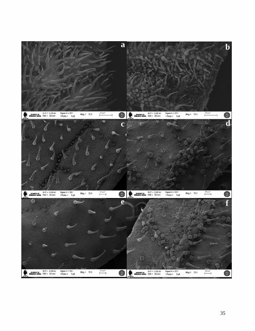
Emergent leaves were densely covered in trichomes and trichome density decreased as the leaf
matured. The frequency (count mm-2
) of trichomes on the emergent leaves was significantly
higher on both side of the leaf surface as compared to the frequency on the young and mature
leaves (Fig. 8 and 9). On the abaxial surface, the frequency of trichomes was significantly higher
at the tip (82.17 ± 3.61 mm-2
, Mean ± SE) but this decreased as the leaf matured (22.07 ± 2.79
mm-2
). Trichome frequency was similar on the mid and base regions of the adaxial and abaxial
surfaces. Capitate and peltate trichomes also occur on the abaxial surface of flower petals (Fig.
10), while the adaxial surface was bare.
4.1.2 Capitate trichomes
Capitate trichomes on leaves were approximately 248.10 ± 4.53 μm in length, unseriate and
normally comprised of a single basal cell, 3-5 stalk cells, a neck cell and a secretory head
containing vertical walls, arranging the head in a quadrant of four small secretory cells (Fig. 4 a,
b and c). The stalk is made up of long cells at the base which gradually become shorter towards
the secretory head. Trichomes with up to 8 stalk cells were observed along the leaf margin but
these were not common. Wax sections stained with Toluidine Blue showed that the basal cells of
capitate trichomes appeared to be highly vacuolated while the cells closer to the secretory head
were denser with cytoplasm and probably organelles (Fig. 4 b). The trichome is anchored to the
leaf epidermis by a pedestal made up of a ring of approximately 8-12 cells with very thick cell

27
walls (Fig. 4 d). Capitate trichome outer walls appear to lack cuticular warts and other forms of
microornamentation.
There were two morphologically distinct types of capitate trichomes found on petals and
therefore, for the purpose of petals alone these trichomes were classified as capitate Type I and
Type II. The morphology of the capitate Type I trichome was similar to the capitate trichomes
found on leaves, whereas the capitate Type II trichomes had stalks that were longer in length and
comprised of approximately 14 cells (Fig. 10 a and b). The secretory heads of both types of
capitate trichomes were arranged in quadrants, similar to those found on leaves.
Capitate trichomes appear to develop rapidly on leaf pimordia since fully developed forms were
only observed on emergent through to mature leaves whereas developing capitate trichomes were
rare (Fig. 7 a-f). Development appears to occur from a single epidermal cell which probably
undergoes many periclinal divisions to form the stalk and two anticlinal divisions to form the
four-celled secretory head. This development was not observed directly. However, observations
of developing trichomes on young leaves lead us to this assumption (Fig. 4 a).
4.1.3 Peltate trichomes
Fully developed peltate trichomes comprise a basal cell, a unicellular stalk and a multicellular
secretory head (Fig. 5). The peltate trichome head consisted typically of four cells although some
were seven or even eight-celled (Fig. 6 e and f). Developing trichomes had a secretory head
made up of 2 to 4-cells (diameter = 31.21 μm), and these were significantly smaller than the fully
developed 4-celled peltate trichome (diameter = 74.32, t = 8.263, p < 0001). Development
appears to occur from a single epidermal cell which undergoes 3 periclinal divisions to form a
basal cell, a stalk cell and a unicellular secretory head. The secretory head then divides
periclinally to form a four celled head which expands in size resulting in a developed four celled
peltate trichome. This four celled head may undergo further periclinal division to form a head of
up to eight cells (Fig. 6). The peltate trichomes found on petals were morphologically similar to
those found on leaves.

28
PLATE 1
Figure 4. Capitate trichomes observed on leaves of C. triloba.
(a) SEM of a developed capitate trichome (C) and developing capitate (DC) trichome on a
young leaf. Stalk cells (arrows) are differentiated early in leaf development and then
expand in size.
(b) Toluidine Blue O stained section of capitate trichome. A single basal cell (Bc), four stalk
cells (St), a neck cell (Nc) and a multicellular secretory head (Sc) can be seen. Cells
closer to the secretory head appear to contain more cytoplasm and probably more
organelles than the cells closer to the epidermis. A cellular pedastal is also visible (Cp)
(c) SEM showing the secretory head (S) of capitate trichome with four secretory cells.
(d) SEM of the basal region of capitate trichomes showing a single basal cell (Bc) attached to
an epidermal ring of cells called the cellular pedestal (Cp). A stalk cell (St) and stomata
(arrows) can also be seen.

29
c
S
c
DC
C
a
Bc
St
Nc
Sc
Cp
b
Cp
St
Bc
d

30
PLATE 2
Figure 5. Peltate trichome on leaves of C. triloba.
(a) ESEM of developed four-celled peltate trichome (P) and developing 2-celled peltate
trichome (DP) with a single stalk cell (St).
(b) Toluidine Blue O stained section of two peltate trichomes showing a single basal cell
(Bc), a unicellular stalk (St) and a multicellular secretory head (Sh).

31
DP
P
St
Bc
St
Sh
b
a

32
PLATE 3
Figure 6. Development of peltate trichomes on leaves of C. triloba.
(a) Developing peltate trichome (DP) with bicellular head. Trichome basal cell (Bc) and
stomata (arrows) can also be seen.
(b) Developing peltate trichome with 3-celled secretory head.
(c) Developed 4-celled peltate trichome (P) and a peltate trichome in the post secretory phase
(PPS) located on leaf vein (Vn).
(d) A peltate trichome with a secretory head made up of six cells.
(e) A 7-celled peltate trichome.
(f) Abaxial surface of leaf showing an 8-celled peltate trichome (arrow) on leaf vein (Vn).

33
CA
Bc DP
P
PPS
Vn
Vn
a
f e
d c
b
34
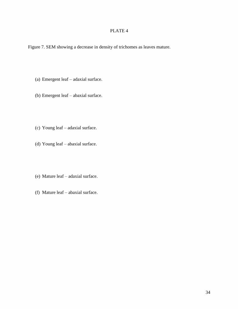
PLATE 4
Figure 7. SEM showing a decrease in density of trichomes as leaves mature.
(a) Emergent leaf – adaxial surface.
(b) Emergent leaf – abaxial surface.
(c) Young leaf – adaxial surface.
(d) Young leaf – abaxial surface.
(e) Mature leaf – adaxial surface.
(f) Mature leaf – abaxial surface.
35
C
e
b a
d c
f

36
Figure 8. Trichome frequency on the adaxial surface of leaves at three stages of
development, emergent, young and mature. The entire leaf area was sampled.
Values are mean ± SE. Bars with different letters are significantly different at p <
0.05 using Tukey‟s multiple range test.
Figure 9. Trichome frequency on the abaxial surface of leaves at three stages of leaf
development, emergent, young and mature. The entire leaf area was sampled.
Values are mean ± SE. Bars with different letters are significantly different at p <
0.05 using Tukey‟s multiple range test.

37
Emergent
Young
Mature
Emergent
Young
Mature
a
a
c c
b
c
c c c
a
d
b
d
b
d d
c
cd
a
b

38
PLATE 5
Figure 10. Surface imaging of trichomes on flower petals of C. triloba.
(a) Stereo micrograph of abaxial surface of petal showing capitate Type II trichomes (arrows).
These trichomes are longer in length as compared to the Type I capitate trichomes. A
variation in morphology could indicate a difference in function.
(b) SEM of petal showing capitate Type II trichomes (arrows).

39
a
b

40
4.2 HISTOCHEMISTRY AND FLUORESCENCE MICROSCOPY
4.2.1 Cell walls
Histochemical analyses of capitate and peltate trichome cell walls revealed the presence of lignin
(Fig. 14), pectin (Fig. 13) and cellulose (results not presented). Sections stained with Sudan
III/IV and Sudan Black B tested positive for suberin or cutin in the single stalk cell of peltate
trichomes and the neck cell of capitate trichomes. A continuous layer of cuticle along the leaf
surface was observed with the Sudan dyes (Fig. 11 and 12).
4.2.2 Identification of secretory product
Histochemical analyses of the secretory cells of trichomes with the Sudan dyes, Ruthenium Red
and ferric trichloride indicated the presence of lipids (Fig. 11 and 12), mucilage (Fig. 13) and
phenolic compounds (Fig. 14 and 15) respectively (Table 1). Both capitate and peltate trichomes
appeared to secrete copious amounts of mucilage which are released when the secretory cells
burst or the cell wall dissolves (Fig. 13).
Sections stained with the Nadi reagent and Ditmarr reagent produced negative results, indicating
the absence of terpenoids and alkaloids respectively (results not presented).

41
PLATE 6
Figure 11. Light micrographs of transverse sections of leaves of C. triloba stained with Sudan
Black B.
(a) Capitate trichome with lipid drop (L) in the secretory cell and a continuous layer of
cuticle along the epidermis (long arrow).
(b) Peltate trichome stained with Sudan Black showing lipid drop (L) in secretory cell. Note
continuous layer of cuticle along the epidermis (long arrows).

42
L
L
a
b

43
PLATE 7
Figure 12. Light micrographs of transverse sections of leaves of C. triloba stained with Sudan III
and IV.
(a) Capitate trichome with positively stained lipid (L) scattered in the secretory cell and a
continuous layer of cuticle along the epidermis (arrow).
(b) Peltate trichome stained with Sudan III/IV showing lipid drop (L) in secretory cell. Note a
continuous layer of cuticle along the epidermis (arrows).

44
28
b
L
L
a
b

45
PLATE 8
Figures 13. Light micrographs of transverse sections of leaves of C. triloba stained with
Ruthenium Red.
(a) Free-hand cross section of capitate trichome showing secreted mucilage (Mc), stained
pink, on secretory cells.
(b) Light micrograph of wax embedded transverse section stained with Ruthenium Red.
Mucilage (Mc) stained pink can be seen around peltate trichomes that are in the post
secretory phase (PPS). Pink stain in the cell walls was also observed which indicates the
presence of pectins (arrows)

46
PPS
PPS
Mc
Mc
Mc
Mc
a
b

47
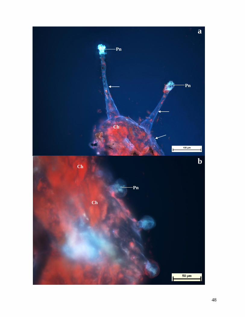
PLATE 9
Figures 14. Autofluorescence of transverse sections of leaves of C. triloba.
(a) Free-hand cross section of leaf showing capitate trichomes exhibiting a pale blue
autoflourescence in the secretory cells, indicating the presence of phenolic compounds
(Pn). Lignin can also be seen in cell walls indicated by a white fluorescence (arrows) and
chloroplasts (Ch), which fluoresces red.
(b) Peltate trichome secretory cells. A pale autofluorescence is observed, which is indicative
of phenolic compounds (Pn). Chloroplast (Ch) fluoresces red.
48
Pn
Pn
Ch
Pn
Ch
Ch
a
b

49
PLATE 10
Figures 15. Light micrographs of transverse sections of leaves of C. triloba stained with ferric
trichloride.
(a) Free-hand section showing capitate trichome containing phenolic compounds (Pn)
indicated by a green stain. A second capitate trichome can also be seen in the post
secretory phase (CPS).
(b) Peltate trichomes containing phenolic compounds (arrows) indicated by a green stain.

50
CPS
Pn
b
a

51
Table 1. Results of histochemical and fluorescence tests for the identification of compounds in the secretory product of C. triloba.
+/- indicates presence/absence of compound
++ indicates intense reaction
Compound group Test Peltate Capitate Reaction Observed
Lipids Sudan III/IV + + Red colouration in head cells
Sudan Black B + + Blue colouration in head cells of trichomes
Mucilage Ruthenium Red ++ + Pink stained mucilage in and around
peltate trichomes and on capitate trichome
heads.
Phenols Ferric trichloride + ++ Deep green colouration
Autofluorescence + ++ Pale blue fluorescence
Alkaloids Dittmar‟s reagent - - Pink colouration
Terpenoids Nadi reagent - - Black colouration

52
4.3 ULTRASTRUCTURE
Samples for TEM were prepared using two methods that differed with the type of fixative used
and the duration that the samples were fixed for and dehydrated. Observations of capitates
trichomes using TEM indicated that the secretory cells of these trichomes contained large
amounts of electron dense lipophilic material that hindered the ability to observe cellular detail
(Fig. 16 a). Despite the fact that cellular detail was difficult to observe, mitochondria and
endoplasmic reticulum cisternae were observed in secretory head cells.
The cytoplasm of stalk cells of capitate trichomes differed from head cells, being characterised
by less lipophilic material and more vacuoles (Fig. 16 a). Oval shaped plastids were observed
bordering the nucleus and containing smaller, light and dark stained lipophilic material (Fig. 16 a
and b). Numerous mitochondria and endoplasmic reticulum were also observed in stalk cells in
close proximity to the nucleus (Fig. 16 b). The outer cell walls of the secretory head cells were
thicker as compared with the cell wall of the stalk cells (Fig. 16 a). However, the cell wall of the
head cells appeared to be arranged with loose fibrils in contrast to the more sturdy cell walls of
the stalk. There appeared to be cell wall intrusion between the head and neck cells (Fig. 16 a).
The secretory head cells of the peltate trichomes contained centralized nuclei and numerous large
vacuoles in the cytoplasm (Fig. 17 a). Oval shaped, light and dark stained electron dense plastids,
endoplasmic reticulum cisternae and mitochondria were observed in close proximity to the
nucleus (Fig. 17 b). Endoplasmic reticulum cisternae, golgi bodies and mitochondria were also
observed scattered throughout the cytoplasm, especially around vacuoles that were closer to the
cell wall (Fig. 18 b). These vacuoles were closely associated with multimembranous structures
found in the cytoplasm, which appeared to push against the vacuoles (Fig. 18 a and b). Small
vesicles developed in the cytoplasm with the simultaneous degradation of the multimembranous
structures (Fig. 19 a). The vesicles appeared to fuse with the vacuoles (Fig. 19 b). Vesicle
formation and translocation into vacuoles could indicate the production and storage of secretory
product respectively. Loose fibrils in the cell walls in peltate trichomes were not observed, as in
the case of the outer cell walls of capitate trichomes.

53
The cytoplasm of stalk cells of peltate trichomes appeared to be similar to the secretory cells
(Fig. 17 a). These cells contained a centralized nucleus surrounded by electron dense plastids and
mitochondria, numerous cytoplasmic vacuoles and golgi bodies along the periphery of the cells.
The walls of the stalk cells appeared to be thicker than the cell walls of secretory cells.
.
4.4 PHYTOCHEMICAL TESTS
4.4.1 Phytochemical screening of plant extract
Plant extracts tested positive for oligosaccharides, polysaccharides, flavonoids, saponins, tannins
and fixed oils. Monosaccharides, amino acids and alkaloids were absent in extracts (Table 2).

54
PLATE 11
Figure 16. Transmission electron micrographs of a capitate trichome.
(a) Section through the head (Sh) and stalk cells (St). Copious amounts of electron dense
secretory material (Sec) and dense cytoplasm can be seen in the secretory cells of the
head. A thin cell wall (Cw) separates the two head cells, and stalk and head cells. The
outer cell wall of the secretory cells appears to be arranged with loose fibrils. Between
the head and stalk cells, an intrusion of the cell wall was observed (arrows). Stalk cells
contain a central nucleus (N), bordered by electron dense plastids (Pd). Numerous
vacuoles (V) were observed throughout the cytoplasm.
(b) Section through the stalk cell showing nucleus (N) with nuclear membrane (Nm).
Electron dense plastids (Pd), numerous mitochondria (M), small vacuoles (V) and
endoplasmic reticulum (ER) were also observed surrounding the nucleus .

55
Cw
Sec V
N
Pd V V
Pd
N
V
V
M
M
a
b
Cw
Sh
St
ER
ER
Nm

56
PLATE 12
Figure 17. Transmission electron micrographs of head cells of peltate trichomes.
(a) Section showing two head cells (Sh) and the stalk cell (St). Central nucleus (N) with
nucleolus (Nu) can be seen in the head cell, with numerous peripheral vacuoles (V) in the
cytoplasm. Light and dark stained electron dense plastids (Pd) surround the nucleus.
(b) Section through the secretory head cell showing nucleus (N) with nucleolus (Nu) and
nuclear membrane (Nm). Light and dark stained plastids (Pd), mitochondria (M),
endoplasmic reticulum (ER) cisternae and vacuoles (V) can also be seen surrounding the
nucleus.

57
M
N
Nu
V
Pd
V
Pd
Nu
N
Pd
V
M
b
a
Sh
St
Nm
ER

58
PLATE 13
Figure 18. TEM of cell wall region of a peltate trichome.
(a) Section showing outer cell wall (Cw) of peltate trichome with invagination of the
cytoplasm (arrows) into vacuoles (V).
(b) High magnification section showing loosening of the cell wall material (arrows) in the
cell wall (Cw). The cytoplasm, which appears to be pushing into the vacuole (V),
contains vesicles (Vc) and a multimembranous structure (Me). Mitochondria (M) and
endoplasmic reticulum cisternae (ER) cisternae can also be seen.

59
a
V
V
M
M
V
Vc
Cw
b Me
ER
Cw
a

60
PLATE 14
Figure 19. TEM showing vesicle formation in peltate trichomes.
(a) Multimembranous structure (Me) appears to be invaginated into a vacuole (V) near the
cell call (Cw). Vesicles (Vc) are beginning to develop in the cytoplasm. These structures
are surrounded by golgi body (Gb), mitochondria (M) and endoplasmic reticulum (ER).
(b) Vacuole (V) contains numerous vesicles (Vc) after multimembranous structure degrades.
The degradation of the membranous structures could form the viscous secretory material
which is then stored in vesicles and vacuoles before secretion.

61
a
b
Cw
Gb
Gb M
M
M Gb
M
Me
ER
Vc
V
Cw
V
V a
Cw
Vc
V
b

62
Table 2. Results of phytochemical tests of crude methanolic extracts of powdered leaves of C. triloba.
++ indicates intense reaction
Compound group Test Reaction Observed Present(+)/Absent(-)
Oligo- and Polysaccharides Molisch‟s Formation of a reddish ring ++
Monosaccharides Fehling‟s No reaction -
Benedict‟s No reaction -
Flavonoids Lead acetate Flocculent white precipitate +
Tannins Braemer‟s Greenish to grey colouration +
Alkaloids Draggendorff‟s Yellow colouration -
Hager‟s No reaction -
Wagner‟s No reaction -
Saponins Foam Layer of foam after shaking an aqueous solution for
15 minutes
+
Amino acids Ninhydrin No reaction -
Fixed oils and fats Filter paper Oily residue on filter paper +

63
CHAPTER 5: DISCUSSION
Reports of trichome morphology have been limited to Harpagophytum procumbens (Naidoo et
al., 2006), Sesamum indicum and Ceratotheca melanosperma (Solereder, 1908) while
phytochemical studies on the Pedaliaceae are limited to C. sesamoides, Pedalium murex
(Sahayaraj et al., 2008), H. procumbens (Georgiev et al., 2010) and a few Sesamum spp.
including S. indicum (Bankole et al., 2007), S. radiatum (Shittu et al., 2006) and S. angolense
(Potterat et al., 1988).
This study was undertaken to investigate the morphology of foliar trichomes of C. triloba and to
determine the chemical constituents of the secretory product. Two morphologically distinct
trichomes were observed on leaves of C. triloba, capitate and peltate. A characteristic feature of
Lamiaceae is the occurrence of peltate and capitate trichomes (Ascensão et al., 1999), and since
C. triloba displays both types, the characteristics of trichomes found on C. triloba will be
discussed in conjunction with other species of Pedaliaceae and other families, including
Lamiaceae.
The peltate trichomes found on leaves of C. triloba were uniseriate and comprised of a basal cell,
a unicellular stalk and a “shield-like‟ secretory head made up of approximately four secretory
cells arranged in a quadrant. Peltate trichomes with up to eight secretory cells were also observed
but these were rare. Solereder (1908), made a similar observation for foliar peltate trichomes
found on C. melanosperma as consisting of a “single short stalk cell, although it may be
exceptionally long and multicellular; the head is peltate, and consists of four cells (in isolated
cases also of 5-8) arranged like a cross, their wall projecting convexly at the margin of the
head” (see figure 3). Single stalk cells were a common feature for peltate trichomes, whereas
multicellular long stalks were not. The stalk of peltate trichomes on leaves of C. triloba was
always observed to be unicellular, similar to those observed in species of Lamiaceae (e.g.
Leonotis leonurus, Ascensão et al., 1995; Salvia aurea, Serrato-Valenti et al., 1997).
Multicellular long stalks in peltate trichomes have been observed in other species of the
Pedaliaceae such as Rogeria adenophylla (Solereder, 1908); whereas multicellular short stalks
were the common feature in the bi- and multiseriate trichomes of the Asteraceae (e.g. Artemisia

64
campestris, Ascensão and Pais, 1987; Stevia rabaudiana, Montiero et al., 2001). Four-celled
peltate trichomes were also common among many species of Lamiaceae such as the Ocimum
species (Werker et al, 1993).
Capitate trichomes on leaves of C. triloba were uniseriate and comprised a single basal cell,
multicellular stalk consisting of longer cells at the base and gradually shorter cells towards the
head. The small spherical secretory head consisted of vertical walls that divided the head into a
quadrant. Head cells similar to these were described for C. melanosperma as palisade-like in
appearance (Solereder, 1908). Capitate trichomes, comprising of a four-celled secretory head,
were not common in Lamiaceae with capitate trichomes in species of this family having either a
unicellular or bicellular head (Werker et al., 1985a; Serrato-Valenti et al., 1997; Bisio et al.,
1999). In Harpagophytum procumbens (Naidoo et al., 2006), a close relative of Ceratotheca, the
leaves lacked typical capitate trichomes since these trichomes comprised of a large, flat secretory
head as compared to the small, spherical head observed in Ceratotheca. These trichomes
appeared to be an intergraded form since they have a secretory head of a peltate trichome
subtended by a long stalk. Intergraded forms of trichomes have been reported previously for
capitate trichomes in species of Lamiaceae (e.g. Salvia sclarea and S. dominica, Werker et al.,
1985a). Non-glandular trichomes were not observed on leaves of C. triloba. This trend has been
observed in other species of the Pedaliaceae (e.g. C. melanosperma, Solereder, 1908;
Harpagophytum procumbens, Naidoo et al., 2006).
Both the capitate and peltate trichomes were observed on the adaxial and abaxial surface of
leaves throughout leaf development. The higher frequency of trichomes on emergent leaves as
compared to the frequency of trichomes on the young and mature leaves might indicate that
trichomes served a more important role in preventing herbivory on the vulnerable emergent
leaves (Fig. 8 and 9). In addition, the relative higher frequency of trichomes at the tip region of
emergent leaves indicated that trichomes might be of higher value at the tips of leaves, which
were relatively more exposed to herbivores and environmental factors. However, the basal region
on the abaxial surface of young leaves was significantly higher than the tip region. Ascensão and
Pais (1987) suggested that trichomes develop in early leaf development and the final number of
trichomes remained constant throughout leaf expansion. Hence, trichome density on the

65
expanding leaf decreased as the leaf matured. However, this did not explain the variation in
trichome frequency between the different regions (tip, mid and base regions) as observed on
leaves of C. triloba. One could assume that different parts of a leaf expand at different rates,
thereby presenting varying trichome frequencies in different regions. Werker et al., (1993)
suggested that in Ocimum basilicum, glandular hairs were produced in early organ development
and the rate of differentiation appeared to be very rapid across the surface. Differentiation
remains as long as an organ contains a meristematic region. Therefore, as in the case of leaves
which contain both meristematic and mature regions, a difference in trichome frequency will be
noted.
This difference might also relate to the function of the „short term‟ capitate and „long term‟
peltate trichomes as described by Werker (2000). The „short term‟ capitate trichomes, which
secreted material during early leaf development, could densely cover meristematic regions
whereas the long term peltate trichomes might be found on the more established regions. This
was noted on leaves of C. triloba in which peltate trichomes were concentrated on the abaxial
surface along the veins and along the grooves on the adaxial surface. These regions were well
established, being protected by epidermal cells that had thick pectinised walls. Another possible
reason for the trichomes to develop on the veins is assumed that they might provide a quicker
and energetically cheaper mode to transport raw materials to the secretory head cells.
The rapid differentiation of capitate trichomes in early leaf development might provide a
protective barrier on emergent leaves, which were more susceptible to biotic and abiotic
pressures (Woodman and Fernandes, 1991). There is a direct correlation between environmental
stresses on a plant and trichome density on that same plant (Johnson, 1975). Plants of Potentilla
(Rosaceae) grown in shady sites contained fewer trichomes than those grown in sunny sites
indicating the importance of trichome density in protecting plants against high temperatures
(Clausen et al., 1940). Trichomes are associated with reflecting light and appeared to play a role
in preventing excessive water loss. In addition to abiotic pressure, trichomes density appeared to
be also important against biotic threats such as herbivory. Fernandes (1994) noted that density of
capitate trichomes on leaves was one of the major factors influencing the number of herbivores

66
feeding on plants. Emergent leaves therefore required a higher density of trichomes to prevent or
reduce herbivory. These mapped scenarios are also assumed to occur in C. triloba.
Segments of flower petals were also prepared for SEM to compare trichomes on petals with
those observed on leaves. The occurrence of different trichomes on different plant organs might
be due to difference in function between these trichomes (Werker et al., 1985a). Since trichomes
on vegetative organs helped to prevent herbivory, one must consider the potential dual role
which is played by trichomes which are found on flowers in repelling herbivores while at the
same time attracting pollinators. The capitate Type II trichome found only on flower petals of C.
triloba could possibly be secreting substances that might attract pollinators, while the peltate and
capitate Type I trichomes is assumed to function in a similar way to those found on leaves, in
order to repel herbivores. If this is the case, then the main components of the secretory product
should differ accordingly between the two types of capitate trichomes in order to provide the
differences in function.
In Salvia dominica and S. sclarea, in addition to differences in shape and occurrence of capitate
trichomes between vegetative and reproductive organs, there was also a difference in the main
components of the essential oil, being neryl acetate and linalyl acetate (Werker et al., 1985a).
Both the reproductive and vegetative organs of these species were covered in capitate and peltate
trichomes. The morphology of peltate trichomes was uniform between the two organs whereas
the capitate trichomes differed in structure, size and mode of secretion. In addition, Mahlberg et
al., (1984) reported that capitate stalked trichomes were only observed on flower petals of
Cannabis sativa while other glandular trichomes were found on leaves. Similar observations in
differences in trichome occurrence and distribution between reproductive and vegetative organs
were reported for Salvia officinalis (Venkatachalam et al., 1984) while, in Origanum vulgare, no
difference it trichomes occurrence was observed between the reproductive and vegetative organs
(Werker et al., 1985b).
The development of both capitate and peltate trichomes in C. triloba appeared to originate from a
single protodermal cell which divided periclinally to form a protrusion above the epidermis.
These cells then have gone through further periclinal divisions to form a stalk and anticlinal

67
divisions to form secretory head cells. Uphof (1962) characterised the periclinal development of
a multicellular, uniseriate trichome according to direction of division of the existing stalk cells.
Acropetal division appeared to occur when the uppermost stalk cell divided to form a new cell,
whereas basipetal division is assumed to occur when the lowermost stalk cell has gone through
division. In Pelargonium scabrum, capitate stalked trichomes resulted from a development of the
protodermal cell which divided periclinally to form a single stalk cell (Oosthuizen and Coetzee,
1983). Further stalk cells occurred from acropetal division. The peltate trichome in species of
Lamiaceae underwent two periclinal, acropetal divisions to form a stalk cell and a single
secretory cell which underwent further anticlinal division (e.g. Origanum dictamnus, Bosabalidis
and Tsekos, 1982a; Leonotus leonurus, Ascensão et al., 1995). It is not clear whether the stalk in
either the peltate or the capitate trichomes on leaves of C. triloba developed via acropetal or
basipetal division. In addition to differentiation of the protodermal cell, other epidermal cells
appeared to develop around the protodermal cell in capitate trichomes. These generally form a
ring of differentiated cells normally referred to as a cellular pedestal, which provides support and
anchors the trichome in the epidermis.
Observations indicated that the mode in which the trichomes on leaves of C. triloba secreted
their product differed substantially from that in the Lamiaceae. In most species of Lamiaceae, the
peltate trichomes accumulated secretory product in a subcuticular chamber which formed when
the cuticle detached from the cell wall of the secretory cells (Werker et al., 1985a, b). These
peltate trichomes appeared to release their contents either through the elevated cuticle or when
the elevated cuticle was damaged, which usually occured via mechanical disturbances which
might be caused by herbivores (Schnepf, 1969). Peltate trichomes of C. triloba, however, lacked
a subcuticular chamber and secretory material which appeared to be stored in numerous vacuoles
in the cell lumen is assumed to be only secreted upon breakage of the cell wall. Similar
observations were found in C. sesamoides where the peltate trichomes secretes mucilage when
the secretory cell ruptured (Abels, 1975) or dissolved (Ihlenfeldt, 2001), resulting in cell death.
In other species of the Pedaliaceae such as Harpagophytum procumbens (Naidoo et al., 2006),
the glandular trichomes, which also appeared to lack a subcuticular chamber, secreted viscous
mucilage through micropores to the exterior. Peltate trichomes of C. triloba therefore appeared
to secrete substances through a deviation of the granulocrine pathway of secretion, by employing

68
vesicles and vacuoles for storage only but not fusing with the plasmalemma as the final step in
secretion. The secretory process of peltate trichomes will be discussed in more detail using TEM
data.
Similarly, the capitate trichomes found on leaves of C. triloba lacked a subcuticular chamber
which is also a common feature for capitate trichomes of the Lamiaceae. In Lamiaceae, capitate
trichomes are also reported to accumulate oils in the subcuticular chamber and released them
either by cuticular rupture or diffusion through micropores in the cuticle (Ocimum basilicum,
Werker et al., 1993; P. annua, Ascensão et al., 1999). The secretory product of the “short stalked
capitate trichome” of P. madagascariensis appeared to accumulate in a small subcuticular space
and is probably released by diffusion through micropores whereas “the long stalked capitate
trichome” is assumed to release its secretory product via cuticular rupture (Ascensão et al.,
1998). The capitate trichomes of C. triloba appeared to produce copious amounts of secretory
material which are not stored in vacuoles as in the case of peltate trichomes. It therefore
appeared that the capitate trichomes employed an eccrine pathway of secretion through the
plasmalemma and cell wall. The process of secretion for capitate trichomes will be discussed
further with TEM.
Although histochemicals tests are not absolutely specific, they can be useful in indicating the
major components of cells walls and the secretory material of trichomes in situ. Histochemical
analyses with Sudan III/IV and Sudan Black indicated the presence of suberin or cutin in the
lateral walls of the single stalk cells of peltate trichomes and neck cell of capitate trichomes. The
cell walls of the stalk cells of both trichomes also tested positive for pectins (see figure 13) and
cellulose with the Ruthenium Red and Calcofluor White tests respectively. The process
occurring could be similar to that of the walls of developing endodermis cells in root hairs,
which might have highly complex wall arrangements to prevent the backflow of water.
Similarly, the cell walls in stalk cells could prevent the backflow of secretory product (Bruni and
Modenesi, 1983; Fahn, 1988). This might be important since secretory material which is
produced by trichomes could be potentially harmful to the rest of the plant (Fahn, 1988).

69
Sections tested with Ruthenium Red indicated that the secretory cells of peltate trichomes
secreted copious amounts of mucilaginous polysaccharides while the capitate trichomes appeared
to secrete it to a lesser degree (see figure 13). Similar observations were made by Solereder
(1908) who referred to the peltate trichomes on C. melanosperma as „mucilage hairs‟. The
function of the mucilage is not clear but the presence of viscous secretions could act as a
lubricant in facilitating leaf growth and expansion (Modensi et al., 1984). The slimy layer on
leaves might even reflect light and thus prevent excessive water loss (Gaff, 1997). Viscous
secretions have also been shown to trap phytophagous insects. Gibson (1971) demonstrated the
resistance of Solanum polyadenium, S. berthaultii and S. tarijense to the aphid Empoasca fabea.
The glandular trichomes on these plants secreted viscous substances that trapped the aphids
which were eventually immobilized and starved to death. Tingey and Gibson (1978) showed that
the viscous substances polymerized to trap the aphids, encasing the legs and tarsi.
Polymerization was shown to occur from the interaction of polysaccharides with phenolic
compounds.
There have been many reports on the medicinal uses of relatives of C. triloba, such as C.
sesamoides and Sesamum angustifolium because of their mucilaginous nature. In Sudan, food is
served with a gelatinous sauce made from these plants to make food more palatable (Bedigian,
2004). The same has been found in Nigeria and Kenya. These plants have also been used for
their gelatinous property as lubricants to ease difficult childbirth (Adjanohoun, 1986).
Positively stained wax sections with Sudan III/IV and Sudan Black in the secretory cells of both
capitate and peltate trichomes indicated the presence of lipids. The ferric trichloride test for
phenolic compounds is based on the reaction of orto-dihydroxyphenols with ferric ions to form deep
green or black deposits. A positive reaction for phenols was observed in the head cells of capitate
and peltate trichomes (see figure 15). In addition to this, blue autofluorescence was observed in
head cells which might also indicate the presence of phenolic compounds (Mabry et al., 1970).
Ecologically, phenolic compounds act as deterrents to herbivory. Matsumoto, (1962) showed that
a coumarin which is produced by Melilotus species, had an inhibitory effect on the vegetable
weevil. Gilbert et al., (1967) demonstrated the inhibitory feeding effect of juglone which is
produced by Carya ovata, to the bark beetle, Scolytus multistriatus. Medicinally, phenolic

70
compounds are known for their antioxidant activities as free radical scavengers (López-vélez et
al., 2003). Plant phenolics are also well known for preventing cancer and coronary heart disease
(Rice-Evans et al., 1997; Roginsky, 2003). This is suspected to justify a use of C. triloba as a
traditional medicine; however, this suggestion might be subject of future extensive investigation
which is not in the scope of this study.
TEM of peltate trichomes showed that secretory head cells contained centralized nuclei and
numerous large vacuoles that appeared to contain mucilage taking up most of the space in the
cell lumen (see Figure 17 a). Solereder (1908) similarly described the lumina of peltate
trichomes in C. melanosperma with mucilage occupying the larger part of the cell lumen and the
remains of the protoplast making up a very small space. The presence of endoplasmic reticulum
cisternae, golgi apparatus and mitochondria scattered throughout the cytoplasm, especially
around vacuoles could be an indication of mucilage production (see Figure 18 b). It is well
known from many investigations that the golgi apparatus is involved in mucilage production and
transport and that many dictyosomes are present in mucilage producing cells. Many of these
studies described a granulocrine method of secretion in which mucilage was transported via
vesicles to the apoplast (Trachtenberg and Fahn, 1981; Schnepf et al., 1983). Figure 20 describes
the different modes of secretion, including granulocrine secretion which employed vesicles that
fused with cell membranes to release substances to the apoplast. Granulocrine secretion however,
did not appear to occur in peltate trichomes of C. triloba. In these trichomes, mucilage appeared
to be stored in vacuoles and was only released once the cell wall broke. An elevated cuticle and
subcuticular chamber which were characteristics of the secretory process for trichomes of
Lamiaceae (Ascensão 2010) were not observed in this study. The above observations emphasised
the fact that secretory products in the peltate trichomes were not secreted through the cell
membrane, but rather stored until the cell wall ruptured (Abels, 1975) or dissolved (Ihlenfeldt,
2001) as in the case of C. sesamoides.
Cell wall rupture can be caused by direct mechanical disturbance whereas cell wall dissolution,
resulting in cell death, is most probably caused by the plant itself. A recent study by Tooker et
al., (2010), on the tomato plant Solanum lycopersicum, showed that trichomes on the leaf surface
acted as „early warning‟ senses. Any disturbance, such as movement on the surface of plants,

71
induced rapid molecular signalling to prepare the plant for herbivore attack. Similarly, peltate
trichomes on C. triloba could have been informed of a potential attack and thus took the
necessary steps for cell wall dissolution and apoptosis. Both the capitate and peltate trichomes
could act as sensors. This process appeared to be efficient in preventing or reducing feeding
before it actually happened.
Mucilage appeared to be produced from an interaction between vacuoles and multimembranous
structures, which appeared to push against the vacuoles (Figure 18 a and b). Small vesicles
developed in the cytoplasm with the simultaneous degradation of the multimembranous
structures. The vesicles appeared to then fuse with each other to form pseudovacuoles or
established vacuoles (Figure 19 b). This was observed for the glandular trichomes of Pharbitis
nil (Unzelman and Healey, 1974) in which many protein-carbohydrate filled vesicles were
enclosed by a network of ER. The rough endoplasmic reticulum (RER), which extended to the
plasmalemma, sequestered vesicles to the apoplast while smooth endoplasmic reticulum (SER)
promoted vesicle fusion into larger vesicles, probably for storage. Kristen (1976) noted that in
placental papillae of Aptenia cordifolia ER cisternea, which have lost their membrane-bound
ribosomes, appeared to promote pseudovesicle formation in the organelle-free parts of the
cytoplasm. There was no evidence of vesicle transport into the apoplast. Figure 21 shows how
pseudovacuoles are produced for the synthesis and storage of mucilage in mango fruits (Joel and
Fahn, 1980).
In the secretory head cells of peltate trichomes oval shaped, light and dark stained electron dense
plastids, endoplasmic reticulum and mitochondria in close proximity to the nucleus is probably
responsible for the production of lipids and phenolic compounds. The most common
ultrastructural feature of a cell secreting lipophilic substances was the occurrence of osmiophilic
plastids which were surrounded by ER and mitochondria (Fahn and Evert, 1974; Joel and Fahn,
1980; Bosabalidis and Tsekos 1982b; Fahn, 1988). Two kinds of plastids, differing in shape and
osmiophilic density, were observed in head cells of peltate glands. Whether each plastid
produced a different substance is not known. However, Fahn (1988) suggested that in a cell
different substances were produced by different organelles.

72
Figure 20. Illustration showing different pathways of secretion in secretory cells. (a) Eccrine
secretion. (b and c) Granulocrine secretion, substances are secreted via vesicles that fuse with the
plasmalemma. (From Fahn 1979).
a c b

73
Figure 21. Illustration showing likely origin of protein-carbohydrate mucilage in mango fruit
ducts. (From Joel and Fahn, 1980)

74
The stalk cells of peltate trichomes were ultrastructurally similar to the secretory head cells.
Even though plasmodesmata were not observed between the head cells and stalk cells, there is
still a possibility that they are present. This could mean cell to cell transport via a symplastic
pathway.
The observations noted for the secretory cells of capitate trichomes, having abundant amounts of
osmiophilic material in the cytoplasm (see Figure 16 a), indicated that the secretory product such
as lipids and phenolics are produced in organelles and are released into the ground cytomatrix.
This has also been noted in the oil gland of Origanum dictamnus (Bosabalidis and Tseko, 1982a)
and the calyx glands of Plumbago capensis (Rachmilevitz and Joel, 1976). The observed
mitochondria and ER probably produced lipophilic substances (Vassilyev, 1970; Fahn and
Benayoun, 1976).
The ultrastructure of stalk cells of capitate trichomes differed from capitate head cells and was
ultrastructurally similar to the cells of peltate trichomes. The stalk cells of capitate trichomes
contained peripheral vacuoles and a centralized nucleus which was surrounded by osmiophilic
plastids, numerous mitochondria and ER (see Figure 16 a and b). The occurrence of loose fibres
in the outer cell wall of secretory cells seemed to indicate that these cells secreted substances in a
similar way to capitate trichomes of Lamiaceae (Ascensão, 2010) even though capitate trichomes
in C. triloba also lacked an elevated cuticle. A basic eccrine pathway could be an explanation for
secretion in the capitate trichomes of C. triloba. There appeared to be cell wall intrusion between
the head cells and neck cells, probably to prevent backflow of the secretory product (Fahn,
2000).
Preliminary phytochemical tests on crude methanolic extracts indicated the presence of oligo-
and polysaccharides, flavonoids, saponins, tannins and fixed oils. The oligosaccharides and
polysaccharides are probably mucilage or mucilaginous products. The flavonoids, which consist
of a large group of compounds that normally occur as glycosides, contain numerous phenolic
hydroxyl functional groups attached to a ring structure (Rice-Evans et al., 1997). They are
known antioxidants since vitamins C and E belong to this group (Rice-Evans et al., 1997).
Flavonoids were also reported to play a role in inhibiting diarrhoea and easing painful abdominal

75
cramps (Schuier et al., 2005). These ailments have been treated by traditions healers with leaf
extracts of C. triloba (Steenkamp, 2003).
Ecologically, tannins prevented herbivory by interacting with proteins to form tannin-protein
complexes which reduced the total protein that could be ingested by herbivores. Since these
complexes reduced the dietary content in plants and most angiosperms contain tannins, it was
highly possible that insects used tannins as a negative feeding cue. Medicinally, tannins have
been reported to have anti-cancer and antioxidant properties (Motar et al., 1985; Erdèlyi et al.,
2005). They were also used in the treatment of skin lesions and ulcers by protecting the wound and
preventing excessive blood loss (Bruneton, 1999; Aiyegoro and Okoh, 2010). Tannins are another
group of compounds that are known for treating diarrhoea and other stomach ailments (Bruneton,
1999). Saponins prevent inflammation and display antimicrobial properties (Bruneton, 1999;
Fawole et al., 2009). This evidence is indirectly supported the possible contention that C. triloba
is used in traditional medicine due to its tannin and saponin content.
Characterisation of trichomes on leaves of C. triloba has highlighted the potential uses of this
plant for future research. The morphological characteristics of trichomes observed on leaves of
C. triloba could be used in plant systematic studies. Both the capitate and peltate trichomes
secreted compounds that were medicinally important and could be used commercially. The mode
of secretion of peltate trichomes, in which mucilage appeared to be produced and stored, was
unique and the same mode could be manipulated using biotechnological tools for the production
of other commercially important compounds.

76
CHAPTER 6: CONCLUSION
This study was initiated to characterise the morphology and ultrastructure of the foliar trichomes
and to determine the chemical composition of the secretion in order to provide a scientific basis
for the medicinal properties of C. triloba.
Leaf surface imaging indicated the presence of two morphologically distinct glandular
trichomes: capitate and peltate. These trichomes were of similar morphology to those observed
on C. melanosperma and Sesamum indicum. The capitate trichomes were tall, consisting of a
single basal cell, 2-4 stalk cells, a neck cell and a head that is made up of four secretory cells.
The peltate trichomes consisted of a basal cell, a single stalk cell and a multicellular head. In
most cases the peltate trichome head consisted typically of four cells, but some were seven or
even eight-celled. Fully developed trichomes were found to be concentrated on emergent leaves
probably to provide protection against herbivory and to prevent excessive water loss. The density
of these trichomes decreased as the leaf expanded because capitate trichomes, also known as
short term trichomes, matured early in leaf development and appeared to provide protection in
emergent leaves against various biotic and abiotic factors. Peltate trichomes however, developed
as the leaf matured and probably provided long term protection to leaves against herbivory.
The ultrastructure of the stalk cells of both trichomes and the secretory head cells of peltate
trichomes were similar. These cells consisted of centralised nuclei surrounded by osmiophilic
plastids which are known producers of lipophilic secondary product. The cytomatrix contained
numerous vacuoles surrounded by dictyosomes. Mitochondria and ER cisternea were found
throughout the cytoplasm, in close proximity to the nucleus and vacuoles. The head cell of
capitate trichomes however, contained large amounts of osmiophilic substances, ER and
mitochondria. Peltate and capitate trichomes appeared to be secreting similar compounds, but the
composition of each compound in the secretory material might vary between the trichomes.
Histochemical and phytochemical tests indicated that the major components of the secretory
product were mucilage and phenolic compounds. Other components included tannins, saponins

77
and flavonoids. It is assumed that these compounds contribute to the medicinal value of C.
triloba.
Future studies should focus on isolating and quantifying the compounds in the secretory product.
Antioxidant screening coupled with cyotoxicity tests should be undertaken to evaluate the safety
of the secretory product for commercial and medicinal use. Similar studies on other species of
the Pedaliaceae could identify interesting plant compounds that may be of medicinal value.

78
CHAPTER 7: REFERENCES
Abels, J., 1975. The genera Ceratotheca Endl. and Dicerocaryum Boj. In: Monographs of the
African Pedaliaceae iii iv. Memórias da Sociedade Broteriana 25, Coimbra.
Adjanohoun, E.J., 1986. Contributions aux Etudes Ethnobotaniqueset Floristiquesa u Togo.
Medecine Traditionellee t Pharmacopee. A gence de Cooperation Culturelle et Technique, Paris
Afolayan, A.J., Meyer, J.J.M., 1995. Morphology and ultrastructure of secreting and
nonsecreting foliar trichomes of Helichrysum aureonitens (Asteraceae). International Journal of
Plant Sciences 156, 481-487.
Aiyegoro, O.A., Okoh, A.I., 2010. Preliminary phytochemical screening and in vitro antioxidant
activities of the aqueous extract of Helichrysum longifolium DC. BMC Complementary and
Alternative Medicine 10, 21-28.
Ambrósio, S.R., Oki, Y., Heleno, V.C.G., Chaves, J.S., Nascimento, P.G.B.D., Lichston, J.E.,
Constantino, M.G., Varanda, E.M., Costa, F.B.D., 2008. Constituents of glandular trichomes of
Tithonia diversifolia: Relationships to herbivory and antifeedant activity. Phytochemistry 69,
2052–2060
Ascensão, L., Pais, M.S., 1987. Glandular trichomes of Artemisia campestris (ssp. maritima):
ontogeny and histochemistry of the secretory product. Botanical Gazette 148, 221-227.113
Ascensão, L., Marques, N., Pais, M.S., 1995. Glandular trichomes on vegetative and
reproductive organs of Leonotis leonurus (Lamiaceae). Annals of Botany 75, 619-626.

79
Ascensão, L., Figueiredo, A.C., Barroso, J.G., Pedro, L.G., Schripsema, J., Deans, S.G.,
Scheffer, J.J.C., 1998. Plectranthus madagascariensis: Morphology of the glandular trichomes,
essential oil composition, and its biological activity. International Journal of Plant Sciences
159(1), 31-38
Ascensão, L., Mota, L., De M. Castro, M., 1999. Glandular trichomes on the leaves and flowers
of Plectranthus ornatus: morphology, distribution and histochemistry. Annals of Botany 84, 437-
447.
Ascensão, L., 2010. Essential oil and oleoresin-secreting trichomes in Lamiaceae: Morphological
and ultrastructural features. Proceedings of the 17th International Microscopy Congress, Rio de
Janeiro, Brazil.
Ayensu, E.S., DeFilipps, R.A., 1978. Endangered and threatened plants of the United States.
Smithsonian Institution, Washington, DC.
Azizian, D., Cutler, D.F., 1982. Anatomical, cytological and phytochemical studies on Phlomis
L. and Eremostachys Bunge (Labiatae). Botanical Journal of the Linnean Society 85, 249-281.
Bakker, H.J., Ghisalberi, E.L., Jefferies, P.R., 1972. Biosynthesis of diterpenes in Beyeria
leschenaltii. Phytochemistry 11, 2221-2231.
Bankole, M.A., Shittu, L.A.J, Ahmed, T.A, Bankole, M.N., Shittu, R.K., Kpela, T., Ashiru, O.A.,
2007. Synergistic antimicrobial activities of phytoestrogens in crude extracts of two sesame
species against some common pathogenic microorginasms. African Journal of Traditional,
Complementary and Alternative Medicines 4(4), 427–433.
Bedigian, D., 2004. Slimy leaves and oily seeds: Distribution and use of wild relatives of Sesame
in Africa. Economic Botany 58, 3-33.

80
Bisio, A., Corallo, A., gastaldo, P., Romussi, G., Ciarallo, G., Fontana, N., De Tommasi, N.,
Profumo, P., 1999. Glandular hairs and secreted material in Salvia blepharophylla Brandegee ex.
Epling grown in Italy. Annals of Botany 83, 441-452.
Bosabalidis, A.M., 1990. Glandular trichomes in Satureja thymbra leaves. Annals of Botany 65,
71-78.
Bosabalidis, A., Tsekos, I., 1982a. Glandular scale development and essential oil secretion in
Origanum dictamnus L. Planta 156, 496-504.
Bosabalidis, A., Tsekos, I., 1982b. Ultrastructural studies on the secretory cavities of Citrus
deliciosa Ten. II. Development of the oil-accumulating central space of the gland and process of
active secretion. Protoplasma 112, 63-70.
Bruce, E.A., 1953. Notes on African Pedaliaceae. Kew Bulletin 8, 417-429.
Bruneton, J., 1999. Terpenoids and Steroids, in: Pharmacognosy, Phytochemistry, Medicinal
Plants. Intercept Limited, England, pp. 385-405.
Bruni, A., Modenesi, P., 1983. Development, oil storage and dehiscence of peltate trichomes in
Thymus vulgaris (Lamiaceae). Nordic Journal of Botany 3, 245-251.
Carlquist, S., 1958. Structure and ontogeny of glandular trichomes of Madinae (Compositae).
American Journal of Botany 45, 675-682.
Clausen, J.D., Keck, D., Heisey W.M., 1940. Experimental studies on the nature of species. l.
Effects of varied environments on western north American plants. Washington, D.C., Camegie
Institute of Washington, 52: 452.
Cronquist, A., 1981. An Integrated System of Classification of Flowering Plants. Columbia
University Press, New York.

81
Croteau, R., 1987. Biosynthesis and catabolism of monoterpenoids. Chemical Reviews 87(5),
929-954.
David, R., Carde, J.P., 1964. Coloration differentielle des inclusions lipidiqueet terpniques des
pseudophylles de Pin maritime au moyen du reactif Nadi. Comptes Rendus de l‟ Academie des
Sciences, Paris 258, 1338-1340.
Dawson, R.M., Jarsus, M.W., Jefferiet, P.R., Payne, S.G., Rosich, R.S., 1966. Acidic
constituents of Dodonea lobulata. Australian Journal of Chemistry 19: 21-22.
Dayanandan, P., Kaufman, P.B., 1976. Trichomes of Cannabis sativa L. (Cannabaceae).
American Journal of Botany 63, 578-591.
Dethier, V.G., 1963. The Physiology of Plant Senses. Wiley, New York.
Duke, S.O., Paul R.N., 1993. Development and fine structure of the glandular trichomes of
Artemisia annua L. International Journal of Plant Science 154, 107–118.
Economou-Amilli, A., Vokou, D.,Anagnostidis, K., Margaris, N.S., 1982. Leaf morphology of
Thymus capitatus (Labiatae) by scanning electron microscopy. In: Margaris, N., Koedam, A.,
Vokou, D. (Eds), Aromatic Plants: Basic and Applied Aspects. Martinus Nijhoff Publishes, The
Hague, pp. 13-24.
Ehrlich P.R., Raven, P.H., 1964. Butterflies and Plants: A Study in Coevolution. Evolution
18(4), 586-608.
Erdèlyi, K., Kiss, A., Bakondi, E., Bai, P., Szabó, C., Gergely, P., Erdödi, F., Virag, .L., 2005.
Gallotannin inhibits the expression of chemokines and inflammatory cytokines in A549 cells.
Molecular Pharmacology 68(3), 895-904.
European Pharmacopoiea, 1975. Maissonneuve SA. Sainte-Ruffine 3, pp. 68.

82
Fabricant, D.S., Farnsworth, N.R., 2001. The Value of Plants Used in Traditional Medicine for
Drug Discovery. Environmental Health Perspectives 109(1), 69-75.
Fahn, A., 1979. Secretory tissues in plants. Academic Press, London, pp. 158-221.
Fahn, A.,1988. Secretory tissues in plants. New Phytologist 108, 229-257.
Fahn, A., 2000. Structure and function of secretory cells. In: Hallahan, D.L., Gray, J.C. (Eds.),
Advances in Botanical Research. Plant Trichomes. Academic Press, New York, London, pp. 37–
75.
Fahn, A., Evert, R.F., 1974. Ultrastructure of the secretory ducts of Rhus glabra L. American
Journal of Botany 61, 1-14.
Fahn, A., Benayoun, J., 1976. Ultrastructure of resin ducts in Pinus halepensis: development,
possible sites of resin synthesis, and mode of its elimination from the protoplast. Annals of
Botany 40, 857-863.
Farnsworth, N.R., Akerele, O., Bingel, A.S., Soejarto, D.D., Guo, Z., 1985. Medicinal Plants in
Therapy. Bulletin of the World Health Organisation 63(6), 965-981.
Fawole, O.A., Ndhlala, A.R., Amoo, S.O., Finnie, J.F., Van Staden, J., 2009. Anti-inflammatory
and phytochemical properties of twelve medicinal plants used for treating gastro-intestinal
ailments in South Africa. Journal of Ethnopharmacology 123, 237-243.
Feder, N., O‟Brien, T.P., 1968. Plant microtechnique: some principles and new methods.
American Journal of Botany 55, 123-142.
Feeny, P.P., 1968. Seasonal changes in the tannin content of oak leaves. Phytochemistry 7, 871-
880.

83
Fernandes, G.W., 1994. Plant mechanical defences against insect herbivory. Revista Brasileira
de Entomologia 38, 421–433.
Furr, M., Mahlberg, P.G., 1981. Histochemical analyses of laticifers and glandular trichomes in
Cannabis sativa. Journal of Natural Products 44, 153-159.
Gaff, D.F., 1997. Mechanisms of dessication tolerance in resurrection vascular plants. In: Basra,
A.S., Basra R.K. (Eds.), Mechanisms of environmental stress resistance in plants. Harwood
Academic Publishers, Amsterdam, pp 43-58.
Georgiev, M., Alipieva, K., Pashova, S., Denev, P., Angelova, M., Kerns, G., Bley, T., 2010.
Antioxidant activity of devil‟s claw cell biomass and its active constituents. Food Chemistry 121,
967–972.
Gershenzon, J., Maffei, M., Croteau, R., 1989. Biochemical and histochemical localization of
monoterpene biosynthesis in the glandular trichomes of spearmint (Mentha spicata). Plant
Physiology 89, 1351–1357.
Gibson, R.W., 1971. Glandular hairs providing resistance to aphids in certain wild potato
species. Annals of Applied Biology 68, 113-19.
Gilbert, L.E., 1971. Butterfly-plant co-evolution: Has Passiflora adenopoda won the selectional
race with heliconine butterflies? Science 172, 585-86.
Gilbert, B.L., Baker, J.E., Harris, D.M., 1967. Juglone (5-hydroxy-1, 4-napthoquinone) from
Carya ovata, a deterrent to feeding by Scolytus multistriatus. Journal of Insect Physiology 13,
1453-1459.

84
Harley, K.L., Thorsteinson, A.J., 1967. The influence of plant chemicals on the feeding behavior,
development, and survival of the two-striped, Melanoplus bivittatus. Canadian Journal of
Zoology 45, 305-19.
Ihlenfeldt, H.D., 2001. Fitting pieces together Pterodiscus Hooker (Pedaliaceae) in tropical NE
Africa, A case study. In: Friis, I., Ryding, O. (Eds.), Biodiversity research in the horn of Africa
region. The Royal Danish Academy of Sciences and Letters, Copenhagen, pp. 63-74.
Janzen, D.H., 1980. When is it coevolution? Evolution 34, 611–612.
Joel, D.M., Fahn, A., 1980. Ultrastructure of resin ducts of Mangifera indica L. (Anacardiaceae).
3. Secretion of protein-polysaccharide mucilage in the fruit. Annals of Botany 46, 785-790.
Johnson, H.B., 1975. Plant pubescence: an ecological perspective. Botanical Review 41, 233–
258.
Johansen, D.A., 1940. Plant microtechnique. McGraw-Hill, New York.
Juniper, B.E., Southwood, T.R.E., 1986. Insects and the plant surface. Edward Arnold, London.
Kelsey, R.G., Reynolds, G.W., Rodriguez, E., 1984. The chemistry of biologically active
constituents secreted and stored in plant glandular trichomes. In: Rodriguez, E., Healey, P.L.,
Mehta, I. (Eds.), Biology and chemistry of plant trichomes. Plenum Press, New York, pp. 187–
241.
Kristen, U., 1976. Die Morphologie der Schleimsekretion in Fruchtknoten von Aptenia
cordifolia. Protoplasma 89, 221-233.
Kolb, D., Müller, M., 2004. Light, conventional and environmental scanning electron
microscopy of the trichomes of Cucurbita pepo subsp. pepo var. styriaca and histochemistry of
glandular secretory products. Annals of Botany 94, 515–526.

85
Lapinjoki, S.P., Elo, H.A., Taipale, H.T., 1991. Development and structure of resin glands on
tissues of Betula pedula Roth. During growth. New Physiologist 117, 219-223.
Levin, D.A., 1973. The role of trichomes in plant defense. Quarterly Review of Biology 48, 3-
15.
Lison, L., 1960. Histochemieetcytochemie animals. Principesetmethods 1(2), Gauthier-Villars,
Paris.
López-vélez, M., Martinez-Martinez, F., Del Valle-Ribes, C., (2003). The study of phenolic
compounds as natural antioxidants in wine. Critical Reviews in Food Science and Nutrition 43,
233-244.
Mabry, T.J., Markham, K.R., Thomas, M.B., 1970. The Systematic Identification of Flavonoids.
Springer, Berlin, Heidelberg.
Mace, M.E., Bell, A.A., Stipanovic, R.D., 1974. Histochemistry and isolation of gossypol and
related terpenoids in roots of cotton seedlings. Phytopathology 64, 1297-1302.
Mahlberg, P. ., Hammond, C.T., Turner, J.C., Hemphill, J.K., 1984. Structure, development and
composition of glandular trichomes of Cannabis sativa L. In: Rodriguez, E., Healey, P.L. Mehta,
I. (Eds), Biology and Chemistry of Plant Trichomes. Plenum Press, New York and London, pp.
23-51.
Matsumoto, Y., 1962. A dual effect of coumarin, olfactory attraction and feeding inhibition, on
the vegetable weevil adult, in relation to the uneatablity of sweet clover leaves. Japanese Journal
of Applied Entomology and Zoology 6, 141-149.
Mauricio, R., Rausher, M.D., 1997. Experimental manipulation of putative selective agents
provides evidence for the role of natural enemies in the evolution of plant defense. Evolution 51,
1435–1444.

86
Meyberg, M., Krohn, S., Brummer, B., Kristen, U., 1991. Ultrastructure and secretion of
glandular trichomes of tobacco leaves. Flora 185, 357–363.
Modenesi, P., Serrato-Valenti, G., Bruni, A., 1984. Development and secretion of clubbed
trichomes in Thymus vulgaris L. Flora 175, 211-219.
Monteiro, W.R., Castro, M.D.M., Mazzoni-Viveiros, S.C., Mahlberg, P.G., 2001. Development
and some histochemical aspects of foliar glandular trichomes of Stevia
rebaudiana (Bert.) Bert. – Asteraceae. Revista Brasileira de Botânica 24(3), 349-357.
Motar, M.L.R., Thomas, G., Barbosa Fillo, J.M., 1985. Effects of Anacardium occidentale stem
bark extract on in vivo inflammatory models. Journal of Ethnopharmacology 95(2-3), 139-142.
Naidoo, Y., Xuma, T., Jordaan, A., Naidoo, G., 2006. Further investigation of leaf secretory
activity in Harpagophytum procumbens. Proceedings 16th
International Microscopy Congress,
(IMC 16), pp. 462.
Obi, R.K., Iroagba, I.I., Ojiako O.A., 2006. Virucidal potential of some edible Nigerian
vegetables. African Journal of Biotechnology 5(19), 1785-1788.
Oosthuizen, L., Coetzee, J., 1983. Morphogenesis of trichomes of Pelargonium scabrum. Suid-
Afrikaans Tydskrift vir Plantkunde 2, 305-310.
Pearse, A.G.E., 1968. Histochemistry: Theoretical and Applied (3rd Ed.). Churchill Livingstone,
London, 1, pp. 1-759.
Potterat, O., Msonthi, J.D., Hostettmann, K., 1988. Four Iridoid glucosides and a
phenylpropanoid glycoside from Sesamum angolense. Phytochemistry 27, 2677-2679.
Rachmilevitz, T., Joel, D.M., 1976. Ultrastructure of the calyx glands of Plumbago capensis
Thunb. in relation to the process of secretion. Israel Journal of Botany 25, 127-139.

87
Reynolds, E.S., 1963. The use of lead citrate at high pH as an electron-opaque stain in electron
microscopy. Journal of Cell Biology 17, 208-212.
Rice-Evans, C.A., Miller, N.J., Panganga, G., 1997. Antioxidant properties of phenolic
compounds. Trends in Plant Science 2(4), 152-159.
Rodriguez, E., Healey, P.L., Mehta, I., 1984. Biology and Chemistry of Plant Trichomes. Plenum
Press, New York and London, pp. 53-69.
Roginsky, V., 2003. Chain breaking antioxidant activity of natural polyphenols as determined
during the chain oxidation of methyl linoleate in triton X-100 micelles. Archives of Biochemistry
and Biophysics 414, 261-270.
Rosa, E.A.S., Rodrigues, P.M.F., 1999. Towards a more sustainable agriculture system: the
effect of glucosinolates on the control of soil-borne diseases. Journal of Horticultural Science
and Biotechnology 74, 667–674.
Roth, I., 1977. Fruits and Angiosperms. Encyclopedia of Plant Anatomy. Gebrüder Borntraeger,
Berlin, Stuttgart.
Rudall, P.J., 1980. Leaf anatomy of the subtribe Hyptidinae (Labiatae). Botanical Journal of the
Linnean Society 80, 319-340.
Sahayaraj, K., Venkateshwari, M., Balasubramanian, R., 2008. Insecticidal and antifeedant effect
of Pedalium murex Linn. Root and on Spodoptera litura (Fab.) (Lepidoptera: Noctuidae).
Journal of Agricultural Technology 4(2), 73-80.
Schnepf, E., 1969. Sekretion und Exkretion bei Pflanzen. In: Protoplasmatologia, Bd. 8,
Springer, Vienna and New York.

88
Schnepf, E., Deichgraber, G., Barthlott, W., 1983. On the fine strucutre of the liquid producing
floral gland of the orchid, Coryanthes speciosa. Nordic Journal of Botany 3, 479-491.
Schuier, M., Sies, H., Billek, B., Fischer, H., 2005. Cocoa-Related Flavonoids Inhibit CFTR
Mediated Chloride Transport across T84 Human Colon Epithelia. Journal of Nutrition135(10),
2320-2325.
Serna, L., Martin, C., 2006. Trichomes: different regulatory networks lead to convergent
structures. Trends in Plant Science 11, 274–280.
Serrato-Valenti, G., Bisio, A., Cornara, L., Ciarallo, G., 1997. Structural and histochemical
investigation of the glandular trichomes of Salvia aurea L. leaves, and chemical analysis of the
essential oil. Annals of Botany 79, 329-336.
Solereder, H., 1908. Systematic Anatomy of the Dicotyledons (Vol. 1). Clarendon Press, Oxford,
pp: 611-613.
Shittu, L.A.J., Bankole, M.A., Ahmed, T., Akinsanya, M. A., Bankole, M.N., Shittu, R. K,
Ashiru O.A., 2006. Differential antimicrobial activity of the various crude leaves extracts of
Sesame radiatum against some common pathogenic microorganisms. Scientific Research Essay
1, 108-111.
Steenkamp, V., 2003. Traditional herbal remedies used by South African women for
gynaecological complaints. Journal of Ethnopharmacology 86, 97-108.
Spurr, A.R., 1969. A low-viscosity epoxy resin embedding medium for electron microscopy.
Journal of Ultrastructural Research 26, 31-43.

89
Tierens, K.F.M.J., Thomma, B.P.H.J., Schmidt, M.B.J., Kistner, K., Porzel, A., Mauch-Mani, B.,
Cammue, B.P.A., Broekaert, W.F., 2001. Study of the role of antimicrobial glucosinolate-
derived isothiocyanates in resistance of Arabidopsis to microbial pathogens. Plant Physiology
125, 1688–1699.
Tingey, W.M., Gibson, W.R., 1978. Feeding and mobility of the potato leafhopper impaired by
glandu1ar trichomes of Solanum berthaultii and S. polyadenium. Journal of Economic
Entomology 71, 856-858.
Tooker, J.F., Peiffer, M., Luthe, D.S., Felton, G.W., 2010. Trichomes as sensors.
Detecting activity on the leaf surface. Plant Signaling & Behavior 5(1), 73-75.
Trachtenberg, S., Fahn, A., 1981. The mucilage cells of Opuntiaficus-indica (L) Mill:
development, ultrastructure, and mucilage secretion. Botanical Gazette 142, 206-213.
Turner, G.W., Gershenzon, J., Croteau, R.B., 2000. Development of peltate glandular trichomes
in peppermint. Plant Physiology 124, 665-679.
Unzelman, J.M. Healey, P.T., 1974. Development, structure and occurrence of secretory
trichomes of Pharbitis. Protoplasma 80, 285-303.
Uphof, J.C.T., 1962. Plant Hairs. Encyclopaedia of Plant Anatomy IV, Vol. 5, pp. 1-206.
Gebrüder Bortraeger, Berlin, Nikolassee.
Valkama, E., Salminem, J.P., Koricheva, J., Pihlaja, K., 2003. Comparative analysis of
leaf trichome structure and composition of epicuticular flavonoids in Finnish Birch species.
Annals of Botany 91, 643–655.
Vassilyev, A.E., 1970. On the localization of terpenoids synthesis in plant cells. Rastitel'nye
Resursy 6, 29-45.

90
Venkatachalam, K.V., Kjonaas, R., Croteau, R., 1984. Development and essential oil content of
secretory glands of sage (Salvia officianalis). Plant Physiology 76, 148-150.
Wagner, G.J., 1991. Secreting glandular trichomes: More than just hairs. Plant Physiology 96,
675-679.
Warming, E., 1909. Oecology of plants: an introduction to the study of plant communities.
London, Oxford University, pp. 422.
Werker, E., Fahn, A., 1981. Secretory hairs of Inula viscosa (L.) Ait.- development,
ultrastructure and secretion. Botanical Gazette 142, 461-476.
Werker, E., Ravid, U., Putievsky, E., 1985a. Glandular hairs and their secretions in the
vegetative and reproductive organs of Salvia sclarea and S. dominica. Israel Journal of Botany
34, 239-252.
Werker, E., Putievsky, E., Ravid, U., 1985b. The essential oils and glandular hairs in different
chemotypes of Origanum vulgare L. Annals of Botany 55, 793-801.
Werker, E., Ravid, U., Putievsky, E., 1985c. Structure of glandular hairs and identification of the
main components of their secreted material in some species of the Labiatae. Israel Journal of
Botany 34, 31-45.
Werker, E., 1993. Function of essential oil-secreting glandular hairs in aromatic plants of the
Lamiaceae-A review. Flavour and Fragrance Journal 8, 249-255.
Werker, E., Putievsky, E., Ravid, U., Dudai, N., Katzir, I., 1993. Glandular hairs and essential oil
in developing leaves of Ocimum basilicum L. (Lamiaceae). Annals of Botany 71, 43-50.
Werker, E., 2000. Trichome diversity and development, in: Hallahan D.L., Gray, J.C. (Eds.),
Advances in Botanical Research, Academic Press Limited, 31, pp. 1-30.

91
Wood, J.R.I., Haig-Thomas, H., 1997. A Hand-Book of the Flora of Yemen. Royal Botanic
Gardens, Kew.
Woodman, R.L., Fernandes, G.W., 1991. Differential mechanical defense: herbivory,
evapotranspiration, and leafhairs. Oikos 60, 11-19.
Young, A.M., Schaller, M., Strand, M., 1984. Floral nectaries and trichomes in relation to
pollination in some species of Theobroma and Herrania (Sterculiaceae). American Journal of
Botany 71(4), 466-480.
Yu, H., Kowalski, S.P., Steffens, J.C., 1992. Comparison of polyphenol oxidase expression in
glandular trichomes of Solanum and Lycopersicon species. Plant Physiology 100, 1885–1890.
Zoric, L., Merkulov, L., Lukovic, J., Boza, P., Polic, D., 2009. Leaf epidermal characteristics of
Trifolium L. species from Serbia and Montenegro. Flora 204, 198-209.