Embed Size (px)
Citation preview

The Secretory Pathway
Becky DutchMolecular and Cellular Biochemistry
1. ER - translation2. ER- protein modifications
3. Discussion Section4. Golgi apparatus
5. Vesicular transport

Lecture 5:Transport Vesicles
Reading: Alberts Chapter 13 Lodish Sections 17.10

. Lodish, Fig 17-13
Vesicular Transport
1. How are these vesicles formed? How are different proteins incorporated?
2. How are these vesicles targeted?
3. How do they fuse with their target?

Lodish 17-50
Three types of coated vesicles known:
Each have different coat protein: clathrin COP I COP II
Each involved in specific cellular transport pathways

Lodish 17-51
Types of vesicles and their target locations
All three types have similar vesicle budding: Coat proteins polymerize around the cytosolic face of budding vesicle Coat and adapter proteins help select cargo GTP-binding protein regulates the rate of vesicle formation
Lodish 17-56
COP I vesicles
Coat protein formed from coatamers: cytosolic complex with seven subunits.Polymerize on surface of vesicles to drive formation. Dissociate from vesicle after formation.Golgi transport - retrograde and likely anterograde Also retrograde Golgi to ER transport

Lodish 17-57
Cell-free system for studying Golgi transport
Cultured cells missing one of the processing enzymes - this will allow differentiation of the two populations of Golgi

Lodish 17-57
Cell-free system for studying Golgi transport
Infect mutant cells with VSV - makes only one viral glycoprotein - VSV G
Addition of N-acetylglucos. to VSV G wlll only happen if transport to wt Golgi stack occurs
Assay used to identify and study function of proteins involved in Golgi vesicular transport

Lodish 17-58
Formation of COP I vesiclesCell-free system just described
very helpful in determing roles
1. ARF - small GTPase, releases GDP and binds GTP - Golgi attached enyzme that promotes this unknown2. ARF-GTP binds receptors on Golgi membrane3. COP I coatamers bind to ARF, other protein on cytosolic face.4. Fatty acyl CoA helps budding mechanism unknown.5. If non-hydrolyzable GTP used - vesicles form and release, but COP I never disassociates

Role of COP I vesicles
Retrograde transport - Golgi to ERKDEL receptor and other membrane proteins to be returned to ER - have KKXX sequence at end of C-terminus. This binds COP and . This sequence necessary and sufficient to drive transport to ER.Yeast mutants lacking COP and can’t do retrograde transport
Retrograde transport - in Golgi Moving specific proteins trans to medial, medial to cis
Anterograde transport in Golgi COP I vesicles with lots of cargo, no KDEL - fast track

COP II vesicles
ER to Golgi transport Cell-free extracts of yeast rough ER plus cytosol and ATP - vesicles form - COP II
Formation - similar to COP I. Sec12 catalyzes exchange of GDP for GTP in th Sar I protein. Complex forms with Sec23 and Sec24 proteins, followed by binding of Sec13, Sec31, then Sec16.
Contain a family of 24kDa proteins that selectively bind soluble proteins bound for Golgi. Integral membrane proteins to be transported generally have Asp-X-Glu sequence - binds to one or more COP II proteins

Alberts 13-36
Exocytosis: TGN to Cell Surface
Constituitive and Regulated secretion
Clathrin vesicles

Alberts 13-39
Exocytosis of secretory vesiclesSecretory vesicles very densely packed - can release
large amounts of material
Regulated secretion - vesicles move from TGN to site of secretion. Can be a long distance (nerve cells)
Triggered release - signal to secrete can be hormone binding receptor, electrical excitation. Increases in Ca2+ often important.

Alberts 13-41
Mast cell - example of regulated exocytosis
Histamine released in response to binding of specific ligands
Gives many of symptoms of allergic reactionsMast cell incubated in solution with ligand-Response all over cell
If ligand islocalized toone spot -response willbe localized.
Lodish 17-59
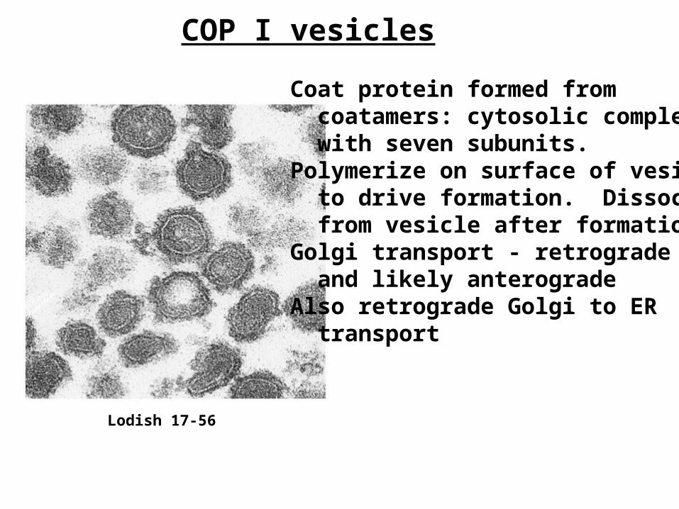
Targetting and Fusion
Common motifs for all types of vesicles - fusion after depolymerization; conserved set of proteins that promote targetting and fusionV-SNARE - in transport vesicle - important for targetingT-SNARE - on target, along with ubiquitous SNAP-25V-SNARE, T-SNARE, SNAP25 form complex - fusionOther proteins involved - NSF (ATPase), SNAP proteins

Rab proteins - regulators of vesicular trafficRab proteins - GTP binding proteins
Approx. 200 amino acids - structure similar to Ras
Bind and hydrolyze GTP - this is hypothesized to regulate rate of vesicular fusion
GDI - catalyzes GDP/GTP exchange of Rabs - this leads to conformational change in Rab that lets it bind vesicle
GTP hydrolysis leads to release of Rab after membrane fusion

Structures of many of these proteins recentlydetermined
Synaptobrevin=VAMP = v-SNARE; syntaxin=t-SNARE; synaptotagmin - Ca2+ binding protein; SNARE complex = portions of synaptobrevin, syntaxin and SNAP-25
Brunger, Curr. Op. Struct.Biol. (2001) 11:163-173

SNARE complex has several states - zipper model
A - closed state; B - binary - syntaxin, SNAP25; C; D - ternary - with synaptobrevin in complex

Syncytia assay of wt SV5 F and the F Tail- mutant.
Viral fusion proteins - best-understood examplesSingle protein systems which promote high level membrane fusion

Lodish 17-60
Viral fusion proteins undergo conformationalchanges upon triggering of fusion

Similar complexes containing heptad repeatregions form in a number of viral fusion proteins

Formation of these heptad repeat complexes critical for membrane fusion

Lodish 17-61
Steps for fusion pore formation
A group of pH activated HA spikes work together to form fused membranes
HA protein inactive after this process - unlike SNARES, which recycle

Relation of SNARE to viral fusion proteins
Complexes containing coiled-coils fundamental to both systems
Skehal and Wiley, Cell (1998) 95: 871-874
. Lodish, Fig 17-13
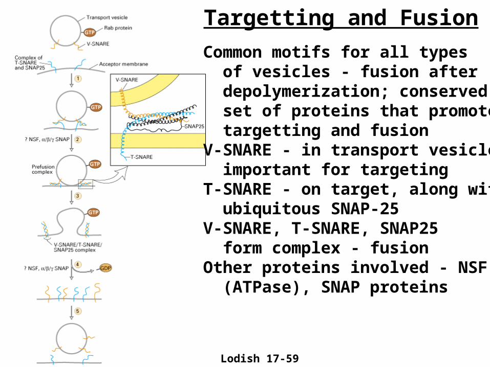
Secretory pathway -critical for cellular
function