Embed Size (px)
Citation preview

Proteomic analysis of the excretory-secretory proteins of the
Trichinella spiralis L1 larva, a nematode parasite of skeletal
muscle
Mark W. Robinson and Bernadette Connolly
School of Medical Sciences, Institute of Medical Sciences, University of
Aberdeen, Aberdeen AB25 2ZD, Scotland, UK
Running title: Excretory-secretory proteins of the Trichinella spiralis muscle
larva
Correspondence: Dr Bernadette Connolly, School of Medical Sciences, Institute
of Medical Sciences, University of Aberdeen, Aberdeen AB25 2ZD, Scotland,
UK
Email: [email protected]
Telephone: +44 1224 555825; Fax: +44 1224 555844;
Abbreviations: ES, excretory-secretory
Keywords: nematode/Trichinella/muscle larva/MALDI-TOF/ Tandem Mass
Spectrometry

Abstract
Trichinella spiralis is an intracellular nematode parasite of mammalian skeletal
muscle. Infection of the muscle cell leads to the formation of a host-parasite
complex that results in profound alterations to the host cell and a re-alignment of
muscle-specific gene expression. The role of parasite excretory-secretory (ES)
proteins in mediating these effects is currently unknown, largely due to the
difficulty in identifying and assigning function to individual proteins. In this
study, a global proteomics approach was used to analyse the ES proteins from T.
spiralis muscle larvae. Following 2-dimensional electrophoresis (2-DE) of ES
proteins, matrix-assisted laser desorption/ionization-time of flight (MALDI-TOF)-
mass spectrometry and liquid chromatography tandem mass spectrometry (LC-
MS/MS) were used to identify the peptide spots. Specific Trichinella EST
databases were assembled and used to analyse the data. Despite the current
absence of a Trichinella genome-sequencing project, 43 out of 52 protein spots
analysed were identified and included the major secreted glycoproteins. Other
novel proteins were identified from matches with sequences in the T. spiralis
database. Our results demonstrate the value of proteomics as a tool for the
identification of Trichinella ES proteins and in the study of the molecular
mechanism underpinning the formation of the host-parasite complex during
Trichinella infections.

1 Introduction
Profound alterations in skeletal muscle can occur as a result of injury, disease or
infection with pathogens. One of the most extreme cases is observed in cells
infected with the nematode parasite, Trichinella spiralis, an intracellular parasite
specific for mammalian skeletal muscle. The newborn L1 larva migrates from
the intestine of the host to the skeletal muscle where it infects and encapsulates
within a portion of the myofiber and develops into the infective or muscle L1
larva [1]. During this process an intimate host-parasite complex, the nurse cell,
is formed. Infection of the terminally-differentiated cell by T. spiralis leads to
profound changes, including re-entry into the cell cycle with subsequent
suspension at the G2/M border, down-regulation of muscle-specific gene [2] and
muscle-specific protein [3] expression, dissolution of myofibrils, proliferation of
secretory organelles and nuclear enlargement [4]. These changes to the infected
muscle cell suggest that the differentiated state in the skeletal muscle cell is
repressed and that post-infection a process of de-differentiation is initiated. This
is followed by re-differentiation into an alternative cell type, one that is adapted
to support the parasite until its transmission to the next host. The molecular
mechanisms underlying the re-programming of the skeletal muscle are currently
unknown although there is circumstantial evidence implicates larval excretory-
secretory (ES) proteins in the formation and/or maintenance of the nurse cell. ES
proteins collected from both newborn larvae and muscle larvae elicit
morphological and structural changes to cultured primary rat myocytes [5].

Furthermore, antigens sharing epitopes with secreted Trichinella proteins have
been detected within isolated host cell nuclei where they co-localise with nuclear
chromatin complexes [6, 7, 8]. However, the abundance of these proteins has
been too low to allow identification and therefore confirmation of the parasite
origin of these antigens awaits data connecting them to Trichinella-encoded
genes. Although several biochemical activities have been observed in the
Trichinella ES fraction, including DNA-binding [9], DNA endonuclease [10,
11], protease [12, 13] and kinase activities [14, 15], relatively few of the proteins
responsible or the genes encoding them have been characterised to date.
The identification and characterisation of ES proteins is of fundamental
importance to understanding the mechanism of parasite-induced de-
differentiation. Recently, mass spectrometric analysis has led to the identification
of two ES proteins, a 67 kDa 5'-nucleotidase [16] and a putative serine protease
[17]. This suggests that the application of proteomics may circumvent many of
the problems encountered with the identification of ES proteins in the past.
Proteomic analysis has been successfully used to identify ES proteins from other
helminth parasites such as the nematode Haemonchus contortus [18] and the
trematode Fasciola hepatica [19, 20]. In the current study, we have used data
collected from 2-DE and mass spectrometry (MS) to analyse and identify the ES
proteins from T. spiralis muscle larvae. This represents the first study which
attempts to comprehensively identify Trichinella ES proteins and, as such, will
provide valuable information for further studies.

2 Materials & Methods
2.1 Parasites
T. spiralis (ISS390) parasites were maintained in female ICR/CD1 mice and
muscle larvae were isolated from infected mice as previously described [21].
2.2 Collection and preparation of ES proteins
Isolated T. spiralis muscle larvae were washed several times in pre-warmed RPMI
1640 medium (Invitrogen) and resuspended at 5000 muscle larvae ml-1 in RPMI
containing 2 mM L-glutamine, 100 U ml-1 penicillin, 100 g ml-1 streptomycin.
The parasites were maintained at 37C / 5% CO2 for up to 20 h after which the
larvae were allowed to sediment and the culture medium containing the ES
protein was collected [22]. Where necessary, the larvae were resuspended in fresh
culture medium and incubated for a further 20 h under the same conditions. ES
proteins were concentrated from the culture supernatants by precipitation with
methanol / chloroform [23]. Normally, 400 l of culture supernatant was used for
1-DE and 1.6 ml for 2-DE, although volumes were adjusted to ensure equal
loading between successive gels. Protein samples were resuspended in buffer
(100 mM Tris-HCl, pH 6.8, 200 mM DTT, 4% SDS, 20% glycerol) prior to 1-DE
or re-swelling buffer (7 M urea, 4% CHAPS, 2 M thio-urea, 0.3% DTT, 2%
resolyte ampholytes pH 4-7) prior to 2-DE.
2.3 De-glycosylation of ES proteins
ES proteins were de-glycosylated using PNGase F (New England Biolabs)
according to the manufacturer’s instructions. Briefly, precipitated ES proteins
were resuspended in 80 l 1X denaturation buffer (supplied with enzyme) and
heated to 95C for 10 mins. Following denaturation 8 l NP-40, 8 l G7 buffer
(supplied with enzyme) and 5 l PNGase F were added and the samples incubated
at 37C overnight. The control sample was identical except for the omission of
PNGase F. The ES proteins were then precipitated as above and resuspended in
an appropriate buffer for electrophoresis.
2.4 1-D and 2-D electrophoresis
ES protein samples were analysed by 1-DE using NuPage® Novex® 4-12% Bis-
Tris gels (Invitrogen) and NuPage® LDS sample buffer plus Sample Reducing
Agent (Invitrogen) were added to the samples prior to electrophoresis. 1-DE
analysis was also used to equalise ES protein samples prior to 2-DE. For 2-DE,
the ES protein samples were loaded onto pH 4-7 IPG strips (Amersham
Biosciences) for separation in the first dimension. The samples were re-hydrated
overnight at room temperature followed by step voltage focussing (0.01 h 200 V;
1.5 h 3500 V; 1.05 h 3500 V) at 22C and the strips were incubated in 7mls of
equilibrating buffer (50 mM Tris, 6 M Urea, 30% glycerol, 2% SDS) containing
10 mg ml-1 DTT for 30 min. Following this, a further equilibration step was
performed with equilibrating buffer containing 25 mg ml-1 iodoacetamide for 30
min. For separation in the second dimension, the IPG strips were laid on 10-15%
non-linear gradient polyacrylamide gels and run at 75 V for 1 h and then at 150 V
until 140 volt-hours were reached. After separation, proteins were visualised by
staining with Colloidal Coomassie Blue G250 (Sigma) as described elsewhere
[24].
2.5 Immunoblotting
ES proteins separated by 1-DE and 2-DE were transferred to Hybond-C pure
nitrocellulose membranes (Amersham Biosciences) using a Novex XCell
Surelock Mini-Cell blot module apparatus (Invitrogen) for 1 h at 30 V.
Following transfer, the membranes were incubated in blocking solution (TSBT:
20 mM Tris-HCl, 150 mM NaCl, 1% Tween-20, pH 7.6) containing 5% skimmed
milk for 3 h at room temperature. Primary antibody (mAb 18H) was diluted
1:1500 in TBST containing 1% skimmed milk and applied to the membranes
overnight at 4C. After washing in TBST (4 X 10 min), the anti-mouse-IgG
secondary antibody conjugated to alkaline phosphatase (Sigma) was applied at a
dilution of 1:10000 in TBST for 1 h. The blots were developed using 5-bromo-4-
chloro-3-indolyl phosphate / nitro blue tetrazolium substrate (Sigma).
2.6 Mass Spectrometry
MALDI-TOF-MS was performed at the University of Aberdeen Promteome
Facility using a protocol adapted from a published method [25]. Briefly, protein
spots were excised from the stained 2-D gels and subjected to in-gel proteolytic
digestion with trypsin (Sigma) using an Investigator ProGest instrument

(Genomic Solutions). The resulting tryptic fragments were guanidylated with
methylisourea, desalted and concentrated using an Investigator ProMS
instrument (Genomic Solutions) before analysis using a PerSpective Biosystems
Voyager-DE STR MALDI-TOF mass spectrometer, operating in reflectron mode
at 20 kV accelerating voltage. Spectra were internally calibrated using trypsin
autodigestion products. LC-MS/MS was performed at the Sir Henry Wellcome
Functional Genomics Facility, University of Glasgow. After in-gel tryptic
digestion, the resulting peptides were solubilized in 0.5 % formic acid and
fractionated by nanoflow HPLC on a C18 reverse phase column, eluting with a
continuous linear gradient to 40% acetonitrile over 20 minutes. The eluate was
analysed by online electrospray tandem MS using a Qstar Pulsar instrument
(Applied Biosystems). A 3-second survey scan preceded each MS/MS data
collection cycle of four 3-second product ion scans, giving a duty cycle of 15
seconds. For tandem MS performed by the FingerPrints Proteomics Facility,
University of Dundee, tryptic peptides were analysed on an Applied Biosystems
4700 Proteomics Analyser (MALDI-TOF-TOF-MS-MS system) in MS reflector
mode to generate a peptide mass fingerprint (PMF). Five of the most intense
peptide ions were selected for further analysis by MS/MS. The MS and MS/MS
data were combined and used by the Applied Biosystems GPS system (Global
Proteomics Server) and MASCOT (Matrix Science) to search the databases.

2.7 Database searching
The PMF profiles obtained from MALDI-TOF-MS were used to search databases,
run locally using MS-Fit (Protein Prospector) against a custom-made Trichinella
EST database and the NemaCluster database for Trichinella [26]. The following
parameters were used: methionine oxidation, cysteine carbamidomethylation and
lysine guanidylation. The peptide mass tolerance was initially set at 50 ppm but
was increased to 150 ppm when searching EST databases. Similarly, up to 3
missed cleavages were allowed when searching EST databases. Searches were
also carried out against all mouse sequences in the NCBInr database in order to
identify potential host proteins. Database searches using LC-MS/MS data were
carried out using MASCOT (Matrix Science).
3 Results
3.1 2-DE analysis of Trichinella ES proteins
ES proteins secreted by isolated T. spiralis muscle larvae during in vitro
cultivation were precipitated from culture supernatants collected following 6 h, 20
h and 40 h incubation in RPMI medium and analysed by 2-DE (Fig. 1A-C). The
most prominent ES proteins in the 6 h sample migrated in the 40-60 kDa range.
These corresponded to the major glycosylated proteins in the 40 h ES sample as
shown by Western blotting and immuno-analysis with mAb 18H, a monoclonal
antibody that is specific for tyvelose-containing glycans epitopes [27] found on
some Trichinella ES proteins (Fig 1D). This was consistent with previous
observations that the major immuno-dominant ES proteins of the T. spiralis
muscle larva are glycosylated and in this size range [28, 29, 30]. Following later
cultivation times, both the number of protein spots and the overall complexity of
the ES profile increased, notably in the lower molecular mass range (ie 15-40
kDa). The 2-DE profiles shown are representative of T. spiralis ES proteins and
gel-to-gel reproducibility was very high between independent samples (for
example, compare Fig. 1C and Fig. 2).
3.2 Identification of Proteins by 2-DE and Mass Spectrometry
Using the current sample preparation method and pH range, approximately 125
Coomassie Blue-stained spots were visible on 2-D gels of T. spiralis 40 h ES
proteins (Fig. 1C). In order to identify the major ES proteins, a total of 52
different prominent spots were excised from 2-D gels (Fig 2) and a proteomic
analysis of selected spots was carried out using MALDI-TOF-MS. The resulting
PMF data were used to search databases. As relatively few Trichinella sequences
are available in the public cDNA and protein databases, searches using these were
largely unsuccessful. Consequently, a custom-made database was compiled from
all Trichinella sequences, both cDNA and EST, deposited in GenBank. Latterly,
the Trichinella EST cluster database compiled as part of the Parasitic Nematode
EST project [26] was also used for searches. In line with other studies, a match to
a Trichinella protein was considered significant where there were at least 5
matched peptides and 15% coverage of the protein sequence [31]. For matches to
ESTs, fewer peptide matches and a lower percent coverage were tolerated in
making a putative assignment of protein identity. Other criteria used to assign

spots to proteins included matching the calculated theoretical molecular mass and
pI values of the protein, where possible, with the observed position of the peptide
on the 2-D gel. Because of the limited availability of complete cDNA sequence
data all of the spot identifications initially assigned on the basis of PMF data were
considered tentative until confirmation could be obtained by tandem MS. To do
this LC-MS/MS was used to supplement the PMF data and to assign identities to
those proteins un-identified by MALDI-TOF alone. In these cases, searches were
carried out using MASCOT (MatrixScience) and MOWSE scores >70 were
considered to be significant.
Using the above criteria for PMF data, positive matches to Trichinella proteins or
ESTs were obtained for 23 peptide spots (Table 1), although in 2 cases (spots 8
and 10) the PMF-based assignment was considered borderline. The identities of
22 were confirmed by LC-MS/MS; spot 12 was excluded as the identification
based on the PMF data was to a complete cDNA and was considered robust. A
further 20 spots were successfully identified using LC-MS/MS alone giving a
total of 43 out of 52 Trichinella ES proteins identified using MS. The remaining
9 spots were un-matched to any Trichinella sequence currently in the database.
The 43 significant matches corresponded to only 13 different proteins encoded by
Trichinella cDNAs or ESTs; 4 to known T. spiralis ES proteins, 3 to predicted ES
proteins and the remaining 6 to putative proteins encoded by novel ESTs (Table
1). Twelve spots (1, 8-12, 26 and 31-35), ranging in size from 44.5 to 69.0 kDa

and with pI values ranging from 5.14 - 6.64, were assigned to a cDNA encoding
the T. spiralis serine protease, TspSP-1 [32, 17]. The predicted size of TspSP-1
based on the cDNA is 48 kDa, however, anti-TspSP-1 antibodies detect native
proteins of 50, 64 and 70 kDa by 1-DE and evidence from de novo amino acid
sequencing and tandem MS, suggests that several isoforms of the protein are
present in the ES [17]. Two spots (2 and 7) were identified as the major secreted
glycoprotein p43; the identification of two isoforms of this protein differing in pI
was again consistent with previous observations [29]. A further three spots (5, 27
and 28) were identified as the secreted 45 kDa antigen and one spot (13) was
assigned to the secreted 5'-nucleotidase [16, 33]. In these cases, there was close
agreement between the predicted molecular mass and pI with those observed by 2-
DE.
Spots assigned to three different hypothetical Trichinella proteins were also
identified, confirming that these proteins are expressed and secreted. Seven spots
(3 and 16-21) were assigned to ORF 9.10. Spots 3 and 16 shared molecular mass
and pI values of 48.5 kDa and 4.94 / 4.86, respectively, whilst the other ORF 9.10
spots formed a distinct group of lower molecular mass (~35 kDa) with pI values
in the range of 4.94 – 5.78. Five spots (39-43) with approximate molecular mass
of 22.5 – 24.6 kDa and pI values ranging from 4.92 – 6.39 were identified as the
ORF 17.20 protein. In addition, spot 46 was assigned to the ORF 11.30 protein.
All of these are novel proteins that have yet to be characterised. The remaining
twelve spots were assigned to proteins encoded by Trichinella ESTs, all of which

appeared to be novel. Spots 45 and 49-52 were assigned to two related ESTs
(BG353021 and BG353717), which were subsequently found on contig TS01176
in the NemaGene database. A complete ORF was identified within TS01176
encoding a novel 15.7 kDa protein with a putative N-terminal signal peptide. The
theoretical molecular mass for this protein matched that observed for this group of
spots. The ESTs BG520944 (spot 6), BG520575 (spots 14 and 15), BQ540916
(spots 29 and 30) and BG345905 (spot 38) all encoded protein fragments that
included an N-terminal signal peptide, as predicted by the SignalP algorithm [34].
Thus 12 of the identified proteins were predicated to be secretory proteins due the
presence of an N-terminal signal peptide. The single exception was EST
BQ542714 (spot 36), which encoded a protein fragment lacking the signal
sequence though this could be due to the fact that the EST probably derives from
a truncated cDNA lacking 5’ gene sequence information.
Despite the successful identification of 43 ES proteins, a few notable exceptions
were not identified including the 53 kDa secreted glycoprotein [35] and the
secreted nucleoside diphosphate kinase [15]. These proteins have predicted pI
values of 8.42 and 7.94 respectively and therefore will not resolve on the 2-D gels
used in this study.
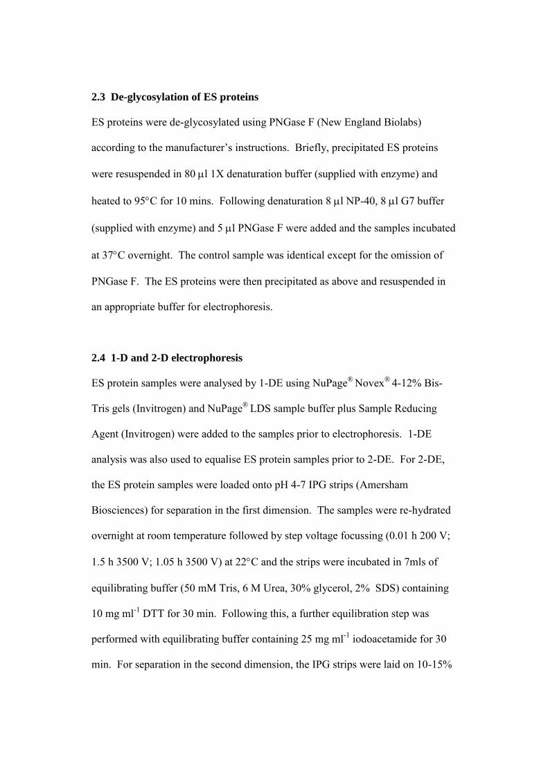
3.3 Glycosylation of T. spiralis ES proteins

The major tyvelosylated T. spiralis ES glycoproteins resolved as approximately
30 immuno-reactive peptides following 2-DE and Western analysis with the anti-
tyvelose monoclonal Ab18H (Fig 1D). Fifteen of these tyvelose-bearing proteins
were identified by MS including the secreted 45 kDa antigen, glycoprotein p43
and the TspSP-1 serine protease. Following treatment with PNGase F, which
cleaves N-linked glycans many ES proteins, including those not recognised by the
mAb 18H antibody, changed position with regard to their molecular weight and pI
(Fig 3). Overall, there was a net movement of spots into the 30-40 kDa and 15
kDa molecular mass regions indicating that a number of peptides had decreased in
mass following de-glycosylation. Therefore, a large subset of the ES proteins
appears to be glycosylated with N-linked glycans. These included the secreted 45
kDa and those encoded by the ESTs BG520575, BQ540916, BG353717 and
BG353021. Peptide spots that did not change their position following de-
glycosylation included those identified as the ORF 9.10 protein, the secreted 5'-
nucleotidase, the ORF 17.20 and ORF 11.30 proteins, and those spots encoded by
BG354905 and BG520944.
4 Discussion
To date few Trichinella ES proteins have been analysed in terms of their
biological activity, due largely to problems in identifying individual proteins. Yet
these proteins function at the host-parasite interface and are probably crucial for
successful parasitism both during the muscle stage and the intestinal stage during
which a new infection is established in a host animal. In the current study, we

have successfully used 2-DE and proteomic analysis tools to identify ES proteins
from the muscle larva, despite the absence of a genome-sequencing project for
Trichinella. Using PMF data from MALDI-TOF-MS and LC-MS/MS and a
custom-assembled Trichinella sequence database, we assigned identities to 43 out
of the 52 ES peptide spots analysed. The majority of the ES proteins identified
contain a predicted N-terminal signal peptide which supports the identifications
and verifies that they are secretory proteins. The use of 2-DE, coupled with
protein identification, has also been informative with regard to determining the
overall constitution and complexity of the ES. The 43 peptide spots identified
represented only 13 different proteins, indicating that several isoforms of many of
the proteins were present. There are a number of possible explanations for the
different isoforms including post-translational modification, splice variants and
protein processing. For example, a large subset of T. spiralis ES proteins is
glycosylated and differential glycosylation may account for some of the isoforms.
The significance of differentially glycosylated variants may become more
apparent once the roles of individual ES proteins have been elucidated. For
instance, it has been suggested that glycosylation may influence the activity of the
TspSP-1 serine protease, as several N-glycosylation sites are found near the active
site of the protein [17].
The question of the function of the muscle larva ES proteins remains to be
determined. Undoubtedly, some of these proteins will play a role in the
establishment or maintenance of the nurse cell and the observations that ES
proteins localise to the surface of the parasite [36] and to the infected host nuclei
[6, 7, 8]support this conclusion. However, tyvelose-bearing ES glycoproteins
may also play a significant role in the intestinal phase. These glycoproteins
originate in the muscle larva in a specialised secretory organ, the stichosome and
are deposited in the intestinal epithelium during the establishment of a new
infection [37, 38]. Furthermore, antibodies raised against Tyvelose are protective
and result in rapid expulsion of parasites from the intestine on administration of a
challenge infection [39, 40]. The application of proteomics in the present study
has provided some much-needed basic data which will allow more focussed
studies on the functions of individual Trichinella ES proteins to be carried out.
These studies are in progress and will be the subject of future communications.
The identification of the ES proteins is critical to understanding the host-parasite
interaction and may have broader implications to the study of the fundamental
biological mechanisms including those underlying maintenance of the
differentiated state in mammalian cells.
Acknowledgements
We would like to thank Dr. P. Cash, E. Argo, E. Ratray and I. Davidson of the
Aberdeen Proteome Facility, Dr. R. Burchmore of the Sir Henry Wellcome
Functional Genomics Facility and D. Lamont and K. Beattie of the FingerPrints
Proteomics Facility. We are also grateful to Dr J. Appleton for the gift of the
mAb 18H. This work was supported by a grant from the BBSRC.

5 References
[1] Despommier, D. D., in: Campbell, W. C. (Ed.) Biology in Trichinella and
Trichinosis, Plenum Press, New York 1983, pp. 75-151.
[2] Jasmer, D. P., J.Cell Biol. 1993, 121, 785-793.
[3] Jasmer, D. P., Bohnet, S., Prieur, D. J., Exp. Parasitol. 1991, 72, 321-331.
[4] Despommier, D., Am. J. Pathol. 1975, 78, 477-496.
[5] Leung, R. K., Ko, R. C., J. Helminthol. 1997, 71, 113-118.
[6] Lee, D. L., Ko, R. C., Yi, X. Y., Yeung, M. H., Parasitology 1991, 102, 117-123.
[7] Yao, C., Jasmer, D. P., Infect. Immun. 2001, 69, 4065-4071.
[8] Yao, C., Jasmer, D. P., Mol. Biochem. Parasitol. 1998, 92, 207-218.
[9] Mak, C. H., Ko, R. C., Parasitology 2001, 123, 301-308.
[10] Mak, C. H., Chung, Y. Y., Ko, R. C., Parasitology 2000, 120, 527-533.
[11] Mak, C. H.,Ko, R. C., Eur. J. Biochem. 1999, 260, 477-481.
[12] Lun, H. M., Mak, C. H., Ko, R. C., Parasitol. Res. 2003, 90, 27-37.
[13] Criado-Fornelio, A., Armas-Serra, C., Gimenez-Pardo, C., Casado-Escribano, N. et al., Vet. Parasitol. 1992, 45, 133-140.
[14] Arden, S. R., Smith, A. M., Booth, M. J., Tweedie, S., et al., Mol. Biochem
.Parasitol. 1997, 90, 111-119.
[15] Gounaris, K., Thomas, S., Najarro, P., Selkirk, M. E., Infect .Immun. 2001, 69, 3658-3662.
[16] Gounaris, K., Infect. Immun. 2002, 70, 4917-4924.
[17] Romaris, F., North, S. J., Gagliardo, L. F., Butcher, B. A., et al., Mol.
Biochem. Parasitol. 2002, 122, 149-160.
[18] Yatsuda, A. P., Krijgsveld, J., Cornelissen, A. W., Heck, A. J., de Vries, E., J. Biol. Chem. 2003, 278, 16941-16951.
[19] Jefferies, J. R., Campbell, A. M., van Rossum, A. J., Barrett, J., Brophy, P. M., Proteomics. 2001, 1, 1128-1132.
[20] Bernal, D., de la Rubia, J. E., Carrasco-Abad, A. M., Toledo, R., et al., FEBS Lett. 2004, 563, 203-206.
[21] Connolly, B., Ingram, L. J., Smith, D. F., Exp. Parasitol. 1995, 80, 488-498.
[22] Kuratli, S., Hemphill, A., Lindh, J., Smith, D. F., Connolly, B., Mol.
Biochem. Parasitol. 2001, 115, 199-208.
[23] Wessel, D.Flugge, U. I., Anal. Biochem. 1984, 138, 141-143.

[24] Anderson, N. L., Esquer-Blasco, R., Hofmann, J. P., Anderson, N. G., Electrophoresis 1991, 12, 907-930.
[25] Shevchenko, A., Wilm, M., Vorm, O., Mann, M., Anal. Chem. 1996, 68, 850-858.
[26] Wylie, T., Martin, J. C., Dante, M., Mitreva, M. D., et al., Nucleic Acids
Res. 2004, 32 (Database issue), D423-D426.
[27] Reason, A. J., Ellis, L. A., Appleton, J. A., Wisnewski, N., et al., Glycobiology 1994, 4, 593-603.
[28] Denkers, E. Y., Wassom, D. L., Krco, C. J., Hayes, C. E., J. Immunol. 1990, 144, 3152-3159.
[29] Denkers, E. Y., Wassom, D. L., Hayes, C. E., Mol. Biochem. Parasitol. 1990, 41, 241-249.
[30] Wu, Z., Nagano, I., Takahashi, Y., Parasitology 1999, 118, 615-622.
[31] Cohen, A. M., Rumpel, K., Coombs, G. H., Wastling, J. M., Int. J.
Parasitol. 2002, 32, 39-51.
[32] Nagano, I., Wu, Z., Nakada, T., Boonmars, T., Takahashi, Y., J. Parasitol. 2003, 89, 92-98.
[33] Gounaris, K., Selkirk, M. E., Sadeghi, S. J., Mol. Biochem. Parasitol. 2004, 136, 257-264.
[34] Bendtsen, J. D., Nielsen, H., von Heijne, G., Brunak, S., J. Mol. Biol. 2004, 340, 783-795.
[35] Zarlenga, D. S.Gamble, H. R., Mol. Biochem. Parasitol. 1990, 42, 165-174.
[36] McVay, C. S., Tsung, A., Appleton, J., Infect. Immun. 1998, 66, 1941-1945.
[37] Capo, V., Silberstein, D., Despommier, D. D., J. Parasitol. 1986, 72, 931-938.
[38] ManWarren, T., Gagliardo, L., Geyer, J., McVay, C., et al., Infect. Immun. 1997, 65, 4806-4812.
[39] Appleton, J. A., Schain, L. R., McGregor, D. D., Immunology 1988, 65, 487-492.
[40] Arasu, P., Ellis, L. A., Iglesias, R., Ubeira, F. M., Appleton, J. A., Mol.
Biochem. Parasitol. 1994, 65, 201-211.

Figure Legends
Figure 1. 2-DE analysis T. spiralis muscle larvae ES proteins in the pH range 3-7
and separated on 10-15% non-linear gradient polyacrylamide gels in the second
dimension. 2-D gels stained with colloidal Coomassie blue G250 of ES proteins
collected after 6 h (A), 20 h (B) or 40 h (C) of in vitro culture. (D) Immunoblot of
2-D gel of 40 h ES proteins, probed with the anti-tyvelose mAb 18H.
Figure 2. Coomassie-stained 2-D gel of 40 h ES proteins showing the positions of
the 52 protein spots subjected to analysis by MS. Spots were numbered and those
identified are listed in Table 1.
Figure 3. Comparison of the 2-DE profiles of 40 h ES proteins that have been
treated (+PNGase) or mock-treated (-PNGase) with PNGase F. Spots identified
by MALDI-TOF-MS following de-glycosylation: (a) secreted 5'-nucleotidase; (b)
ORF 9.10; (c) 45 kDa antigen; (d) BG520575; (e) TspSp-1; (f) ORF 17.20. The
ORF 17.20 peptide spots that do not change position on treatment with PNGase
are marked with arrowheads.

Figure 1
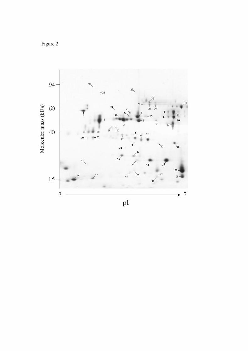
Figure 2

Figure 3

Table 1. Identification of T. spiralis ES proteins identified by MALDI-TOF and LC-MS/MS
Spot Trichinella Protein
Accession Number
TheoreticalMr / pI1
ObservedMr / pI
Coverage (%)
Matched Peptides
MOWSE Score2
1 Serine protease AY02897
4
48.0/6.33 54.0/5.44 24 4 274 2 gp43 M95499 38.0/5.95 47.1/5.48 25 7 433 3 ORF 9.10 U88241 47.6/5.60 48.5/4.94 440 5 45kDa antigen U01847 31.0/4.76 47.1/4.21 17 5 326 6 Unknown BG520944 57.0/3.75 362 7 gp43 M95499 38.0/5.95 47.1/6.04 13 5 270 8 Serine protease AY02897
4
48.0/6.33 55.8/6.44 7 3 121 9 Serine protease AY02897
4
48.0/6.33 55.8/6.64 175 10 Serine protease AY02897
4
48.0/6.33 51.0/6.64 12 4 214 11 Serine protease AY02897
4
48.0/6.33 51.0/6.44 244 12 Serine protease AY02897
4
48.0/6.33 44.5/6.58 42 7 ND 13 5'-nucleotidase AY12757
1
62.0/6.13 62.8/6.86 15 6 1117 14 Unknown BG520575 41.5/4.60 24 6 218 15 Unknown BG520575 41.5/4.69 25 10 263 16 ORF 9.10 U88241 47.6/5.60 48.5/4.86 32 10 404 17 ORF 9.10 U88241 47.6/5.60 36.4/4.94 38 5 392 18 ORF 9.10 U88241 47.6/5.60 35.0/5.36 423 19 ORF 9.10 U88241 47.6/5.60 32.5/5.36 21 5 405 20 ORF 9.10 U88241 47.6/5.60 33.2/5.55 35 5 336 21 ORF 9.10 U88241 47.6/5.60 33.2/5.78 35 6 543 26 Serine protease AY02897
4
48.0/6.33 54.0/5.14 163 27 45kDa antigen U01847 31.0/4.76 38.0/3.92 87 28 45kDa antigen U01847 31.0/4.76 38.0/4.00 14 6 106 29 Unknown BQ540916 35.0/3.89 22 5 496 30 Unknown BQ540916 35.0/4.00 27 6 417 31 Serine protease AY02897
4
48.0/6.33 65.0/5.64 175 32 Serine protease AY02897
4
48.0/6.33 69.0/5.83 210 33 Serine protease AY02897
4
48.0/6.33 65.0/5.83 211 34 Serine protease AY02897
4
48.0/6.33 65.0/6.01 201 35 Serine protease AY02897
4
48.0/6.33 47.1/5.69 105 36 Unknown BQ542714 28.2/5.00 239 38 Unknown BG354905 31.0/6.67 214 39 ORF 17.20 U88239 19.5/6.13 24.6/4.92 211 40 ORF 17.20 U88239 19.5/6.13 24.6/5.33 47 9 338 41 ORF 17.20 U88239 19.5/6.13 22.5/5.42 179 42 ORF 17.20 U88239 19.5/6.13 22.5/5.89 40 16 208 43 ORF 17.20 U88239 19.5/6.13 22.5/6.39 37 9 320 45 Unknown BG353021 15.7/6.283 18.2/6.10 256 46 ORF 11.30 U88238 12.7/4.17 15.0/3.44 397 49 Unknown BG353021 15.7/6.283 16.0/6.06 257 50 Unknown BG353021 15.7/6.283 18.2/6.89 57 7 387 51 Unknown BG353717 15.7/6.283 16.3/6.89 39 6 239 52 Unknown BG353021 15.7/6.283 18.2/5.36 310
1 Theoretical molecular mass (kDa) and pI 2 A MOWSE score of >70 was used to assign identity to a protein analysed by LC-MS/MS 3 Molecular mass and pI of predicted protein encoded on contig TS01176