Embed Size (px)
Citation preview
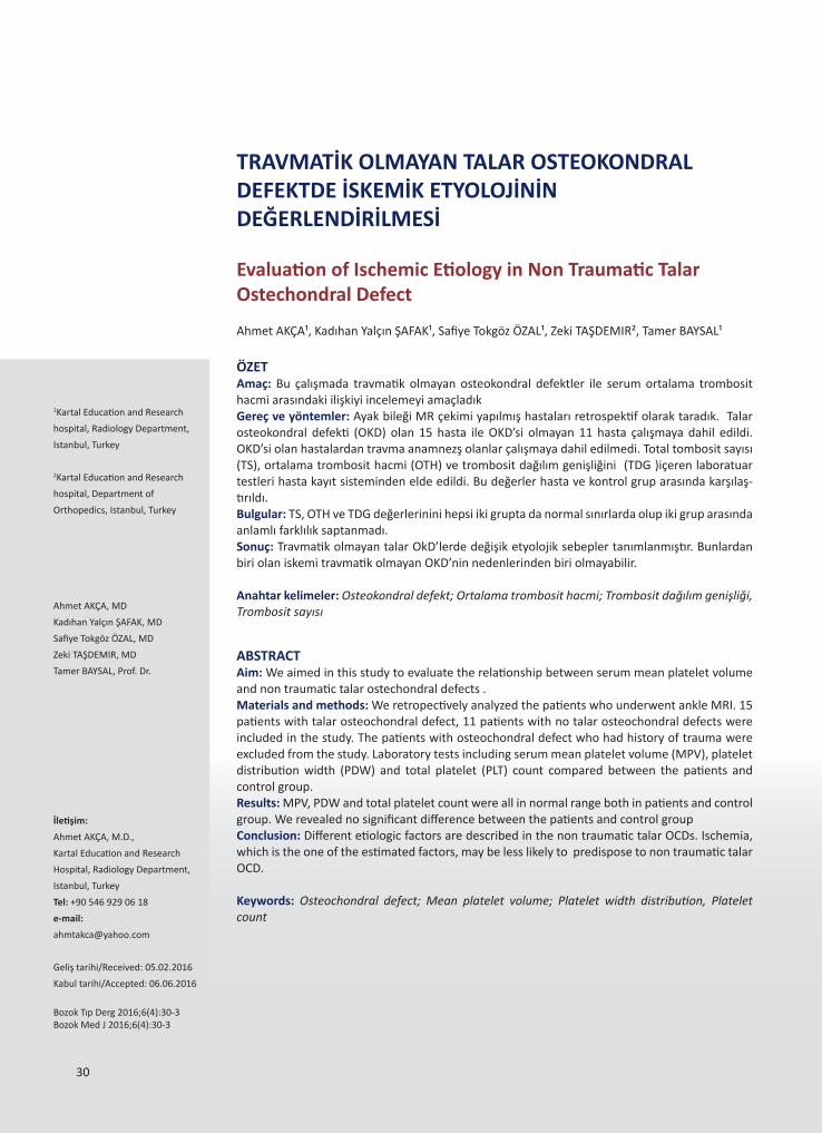
ÖZETAmaç: Bu çalışmada travmatik olmayan osteokondral defektler ile serum ortalama trombosit hacmi arasındaki ilişkiyi incelemeyi amaçladıkGereç ve yöntemler: Ayak bileği MR çekimi yapılmış hastaları retrospektif olarak taradık. Talar osteokondral defekti (OKD) olan 15 hasta ile OKD’si olmayan 11 hasta çalışmaya dahil edildi. OKD’si olan hastalardan travma anamnezş olanlar çalışmaya dahil edilmedi. Total tombosit sayısı (TS), ortalama trombosit hacmi (OTH) ve trombosit dağılım genişliğini (TDG )içeren laboratuar testleri hasta kayıt sisteminden elde edildi. Bu değerler hasta ve kontrol grup arasında karşılaş-tırıldı.Bulgular: TS, OTH ve TDG değerlerinini hepsi iki grupta da normal sınırlarda olup iki grup arasında anlamlı farklılık saptanmadı.Sonuç: Travmatik olmayan talar OkD’lerde değişik etyolojik sebepler tanımlanmıştır. Bunlardan biri olan iskemi travmatik olmayan OKD’nin nedenlerinden biri olmayabilir.
Anahtar kelimeler: Osteokondral defekt; Ortalama trombosit hacmi; Trombosit dağılım genişliği, Trombosit sayısı
ABSTRACTAim: We aimed in this study to evaluate the relationship between serum mean platelet volume and non traumatic talar ostechondral defects .Materials and methods: We retropectively analyzed the patients who underwent ankle MRI. 15 patients with talar osteochondral defect, 11 patients with no talar osteochondral defects were included in the study. The patients with osteochondral defect who had history of trauma were excluded from the study. Laboratory tests including serum mean platelet volume (MPV), platelet distribution width (PDW) and total platelet (PLT) count compared between the patients and control group.Results: MPV, PDW and total platelet count were all in normal range both in patients and control group. We revealed no significant difference between the patients and control group Conclusion: Different etiologic factors are described in the non traumatic talar OCDs. Ischemia, which is the one of the estimated factors, may be less likely to predispose to non traumatic talar OCD. Keywords: Osteochondral defect; Mean platelet volume; Platelet width distribution, Platelet count
İletişim:
Ahmet AKÇA, M.D.,
Kartal Education and Research
Hospital, Radiology Department,
Istanbul, Turkey
Tel: +90 546 929 06 18
e-mail:
Geliş tarihi/Received: 05.02.2016
Kabul tarihi/Accepted: 06.06.2016
Bozok Tıp Derg 2016;6(4):30-3Bozok Med J 2016;6(4):30-3
TRAVMATİK OLMAYAN TALAR OSTEOKONDRAL DEFEKTDE İSKEMİK ETYOLOJİNİN DEĞERLENDİRİLMESİ
Evaluation of Ischemic Etiology in Non Traumatic Talar Ostechondral Defect
Ahmet AKÇA, MD
Kadıhan Yalçın ŞAFAK, MD
Safiye Tokgöz ÖZAL, MD
Zeki TAŞDEMIR, MD
Tamer BAYSAL, Prof. Dr.
Ahmet AKÇA¹, Kadıhan Yalçın ŞAFAK¹, Safiye Tokgöz ÖZAL¹, Zeki TAŞDEMIR², Tamer BAYSAL¹
1Kartal Education and Research
hospital, Radiology Department,
Istanbul, Turkey
2Kartal Education and Research
hospital, Department of
Orthopedics, Istanbul, Turkey
30
31
INTRODUCTION
Increased mean platelet volume (MPV) has been defi-ned as an independent risk factor in the development of thromboembolism (1). There are many studies in-vestigating the role of MPV in occlusive diseaseses like stroke (2-4) and coronary vascular disease (5-7). Seve-ral possible etiological factors for talar ostechondral defect (OCD) have been described including traumatic, embolic, hereditary, endocrine, developmental and idi-opathic (8,9). We aimed in this study to evaluate the role of MPV in non traumatic talar OCDs which can be ischemic in etiology.
MATERIALS AND METHODS
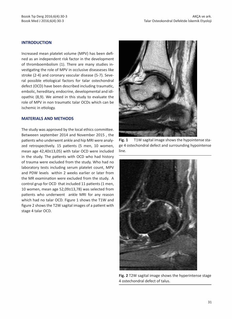
The study was approved by the local ethics committee. Betweeen september 2014 and November 2015 , the patients who underwent ankle and hip MRI were analy-zed retrospectively. 15 patients (5 men, 10 women, mean age 42,40±13,05) with talar OCD were included in the study. The patients with OCD who had history of trauma were excluded from the study. Who had no laboratory tests including serum platelet count, MPV and PDW levels within 2 weeks earlier or later from the MR examination were excluded from the study. A control grup for OCD that included 11 patients (1 men, 10 women, mean age 52,09±13,78) was selected from patients who underwent ankle MRI for any reason which had no talar OCD. Figure 1 shows the T1W and figure 2 shows the T2W sagital images of a patient with stage 4 talar OCD.
Fig. 1 T1W sagital image shows the hypointense sta-ge 4 ostechondral defect and surrounding hypointense line.
Fig. 2 T2W sagital image shows the hyperintense stage 4 ostechondral defect of talus.
AKÇA ve ark.Talar Osteokondral Defektde İskemik Etyoloji
Bozok Tıp Derg 2016;6(4):30-3Bozok Med J 2016;6(4):30-3

32
MR examinations were re-analyzde by two radiologists who were experienced in musculosceletal imaging and who were blinded to clinical and laboratory findings of the patients. Laboratory findings including MPV, PDW and total PLT count were obtained from the hospital in-formation system. All of the MR images were obtained using a 1,5-Tesla MRI scanner (Philips Achievea Intera Release, Einthoven, Netherlands). Sagital T1 (TR:340, TE: 9, FOV:200x200, Matrix: 256x128), axial T1 (TR: 428, TE: 10, FOV:110x160, Matrix:256x86 ), sagital T2 (TR: 2470, TE: 84, FOV:200x200, Matrix:256x128), ax-ial T2 (TR: 661, TE: 23, FOV:143x180, Matrix:256x102 ) and coronal PD (TR: 1520, TE: 28, FOV:125x200, Matrix:256x110 ) sequences were acquired for each patient. Serum total PLT count, MPV and PWD values were compared between the patients and control groups. Statistical analyses were performed using the SPSS software version 20. The variables were investi-gated using analytic methods (Kolmogorov-Simirnov test) to determine whether or not they are normally distributed. Descriptive analyses were presented us-ing means and standart deviations for normally dis-tributed variables. Since the MPV, PDW and PLT count values were normally distributed; the Student’s t-test was used to compare these parameters between the patient and control groups. A p-value of less than 0.05 was considered to show a statistically significant result.
RESULTS
The total PLT count, MPV values and PWD values were 256,87±65,35 cells/μL , 9,04±1,19(fL) and 16,39±1,03 (fL) respectively in patients with OCD and 281,18±106,131 cells/μL , 9,55±1,08 (fL) and 15,85±2,32 (fL) respectively in control group. There was no significant difference between the patients and control group. (p for MPV=0,279, p for PWD= 0,423, p for PLT = 0,477)
DISCUSSION
Plug formation following the vessel wall injury is the main function of platelets (10).MPV which is the most commonly used measure of platelet size (11), is an in-dicator for platelet reactivity and function (12). Small platelets have lower functional capabilities. Large
platelets produce more thromboxane A2 (13,14) and have more fibrinogen receptors (15). Elevated MPV is associated with increased thromboxane synthesis, platelet aggregation, and increased expression of ad-hesion molecules (11). All of these information shows that mean platelet volume (MPV) is an important risk factor for the development of atherothrombosis and embolism (16). There are a lot of studies investigating the role of MPV in vascular occlusive diseases (2-7). Several possible aetiological factors for talar OCD have been described including traumatic, embolic, heredi-tary, endocrine, developmental and idiopathic (8,9). The majority of the cases are associated with previous trauma including involves repetitive and prolonged joint overloading or a sudden impact from high com-pressive stress (17,18). Ischaemia, subsequent necrosis and possibly genetics are aetiologic factors in non-trau-matic OCDs (19). A detailed anamnesis was obtained from all patients whether or not they had any trauma including ankle. To evaluate the ischemia as an etiolog-ic factor in non traumatic OCDs, we excluded all of the patients with history trauma. We expected MPV values to be higher in patients compared with the control group. However, we found no significant difference in total PLT count, MPV values and PDW values between the patients and control group. In conclusion, we think that ischemia is less likely an etiologic factor in idiopathic OCDs. Patients probably do not remember the trauma, especially the repeti-tive microtrauma. In the other hand ischemia may not be related with thrombus formation. We believe that larger studies are needed to investigate the role of isch-emia in the etiology of non traumatic OCDs.
REFERENCES
1. Zandecki M, Genevieve F, Gerard J, Godon A. Spurious counts and spurious results on haematology analysers: a review. Part I: platelets. Int J Lab Hematol. 2007 Feb;29(1):4-20. 2. Mayda-Domaç F, Misirli H, Yilmaz M. Prognostic role of mean platelet volume and platelet count in ischemic and hemorrhagic stroke. J Stroke Cerebrovasc Dis. 2010 Jan; 19 (1) : 66-72. 3. Bath P, Algert C, Chapman N, Neal B; PROGRESS Collaborative Group. Association of mean platelet volume with risk of stroke among 3134 individuals with history of
AKÇA ve ark.Talar Osteokondral Defektde İskemik Etyoloji
Bozok Tıp Derg 2016;6(4):30-3Bozok Med J 2016;6(4):30-3

33
cerebrovascular disease. Stroke. 2004 Mar;35(3):622-6. 4. Turfan M, Erdogan E, Ertas G, Duran M, Murat SN, Celik E et al. Usefulness of mean platelet volume for predicting stroke risk in atrial fibrillation patients. Blood Coagul Fibrinolysis. 2013 Jan;24(1):55-8. 5. Tavil Y, Sen N, Yazici H, Turfan M, Hizal F, Cengel A et al. Coronary heart disease is associated with mean platelet volume in type 2 diabetic patients. Platelets. 2010;21(5):368-72. 6. Sansanayudh N, Anothaisintawee T, Muntham D, McEvoy M, Attia J, Thakkinstian A. Mean platelet volume and coronary artery disease: a systematic review and meta-analysis. Int J Cardiol. 2014 Aug 20;175(3):433-40. 7. Khode V, Sindhur J, Kanbur D, Ruikar K, Nallulwar S. Mean platelet volume and other platelet volume indices in patients with stable coronary artery disease and acute myocardial infarction: A case control study. J Cardiovasc Dis Res. 2012 Oct;3(4):272-5. ja8. Berndt AL, Harty M. Transchondral fractures (osteochondritis dissecans) of the talus. J Bone Joint Surg [Am] 1959;41-A:988- 1020. 9. Davidson AM, Steele HD, MacKenzie DA, Penny JA. A review of twenty- one cases of transchondral fracture of the talus. J Trauma 1967;7:378-415. 10. Kamath S, Blann AD, Lip GY. Platelet activation: assessment and quantification. Eur Heart J. 2001 Sep; 22(17):1561-71.11. Otunctemur A, Bozkurt M, Besiroglu H, Polat EC, Ozcan L, Ozbek E. Erectile Dysfunction Is Positively Correlated with Mean Platelet Volume and Platelet Count, but Not with Eosinophil Count in Peripheral Blood. Urol J. 2015 Nov 14;12(5):2347-52.12. Coskun A, Yavasoglu I, Sargin G, Ok IM, Bircan M, Avcil M et al. The role of mean platelet volume in patients with non-specific abdominal pain in an emergency department. Prz Gastroenterol. 2015;10(3):156-9. 13. Martin JF, Trowbridge EA, Salmon G, Plumb J. The biological significance of platelet volume: Its relationship to bleeding time, thromboxane B2 production and megakaryocyte nuclear DNA concentration. Thromb Res. 1983;32:443-60. 14. Thompson CB, Eaton KA, Princiotta SM, Rushin CA, Valeri CA. Size dependent platelet subpopulations: relationship of platelet volume to ultrastructure, enzymatic activity, and function. Br J Haematol 1982; 50: 509-19.15. Schoene N W. Design Criteria: tests used to assess platelet function. Am J Clin Nutr 1997; 65 (Supplement): 1665– 8S. 16. Park Y, Schoene N, Harris W. Mean platelet volume as an indicator of platelet activation: methodological issues. Platelets. 2002 Aug-Sep;13(5-6):301-6.17. Verhagen RA, Struijs PA, Bossuyt PM, van Dijk CN. Systematic review of treatment strategies for osteochondral
defects of the talar dome. Foot Ankle Clin. 2003 Jun;8(2):233-42, viii-ix.18. Chen HS, Chen SC, Wang HJ, Lin SY, Kang JH. Ultrasonographic Findings and Clinical Characteristics of Two Patients With Talar Osteochondritis Dissecans. Journal of Medical Ultrasound. 2011 Jun;19(2: 47-51)19. Schachter AK, Chen AL, Reddy PD, Tejwani NC. Osteochondral lesions of the talus. J Am Acad Orthop Surg 2005 May;13(3):152-8.
AKÇA ve ark.Talar Osteokondral Defektde İskemik Etyoloji
Bozok Tıp Derg 2016;6(4):30-3Bozok Med J 2016;6(4):30-3