Embed Size (px)
Citation preview

Transformation of a transposon construct into tomato for functional
genomics studies
Dragana Avirovik
Thesis submitted to the faculty of the Virginia Polytechnic Institute and
State University in partial fulfillment of the requirements for the degree of
Master of Science
In
Biological Sciences
Andy Pereira
Christopher B. Lawrence
Dorothea B.C. Tholl
August 29, 2013
Blacksburg, Virginia
Keywords: Genetic transformation, functional genomics, expression of
genes, Agrobacterium tumefaciens, Solanum lycopersicum.
Copyright 2013, Dragana Avirovik

Transformation of a transposon construct into tomato for functional
genomics studies
Dragana Avirovik
ABSTRACT
Tomato (Solanum lycopersicum) is a member of the Solanaceae
family. In this research project tomato, more specifically the M82 cultivar
was chosen as a model plant for Agrobacterium-mediated gene transfer by
cotyledon inoculation. Our objective was to transform tomato with a T-DNA
construct bearing a transposon from maize that can be used for mutagenesis
when it transposes or moves around the genome of the tomato. The vector
used is a two-component in-cis Ac-Ds system which needs a single
transformation event. It was proved that it worked in Arabidopsis and rice
according to Trijatmiko (2005). The construct consists of the BAR gene
conferring resistance to herbicide Basta, hygromycin (HYG) gene conferring
resistance to the antibiotic hygromycin and the green fluorescent protein
(GFP) gene, which are driven by specific plant promoters. The selectable
marker genes such as HYG and BAR were used to select the rare
transformation events by making the transformed tomato tissue resistant to
the toxic chemicals (antibiotic and herbicide) compared to the untransformed
tissue in which growth was inhibited. The results described consist of
developing a transformation protocol which enabled the production of
transgenic tomato lines by the help of the antibiotic augmetin
(amoxicillin/clavulanic acid). The transgenic lines were tested through
polymerase chain reaction (PCR) and herbicide bioassays.

iii
Acknowledgements
First I want to express my thanks to my mom and dad; they have
given a lot of their time and money to educate my brother and I in the United
States. I cannot even imagine what my live would be if I had never stepped
foot on American soil and given the chance to shine and grow as a person. I
have to mention my brother Dragan. He is the one who made me fight and
excel. I had to watch him spend hours and hours in his studies and research
and I believe he will do great things in life. Having him as part of my family
I had and wanted to achieve the same level of excellence. My husband
Andrew who changed me in more ways than one, and put up with me when I
doubt anyone would. Dr. Farnsworth well you have been the best puppy I
have had, coming home after a hard day you would welcome me with a
smile and big slobbery kisses, and I love you. Last but not least, thank you to
my committee advisors. Dr. Andy Pereira you believed in me, you gave me
the chance to be part of your lab and research. I am very grateful Dr.
Christopher Lawrence for always being there. Dr. Andy Pereira, Dr.
Christopher Lawrence and Dr. Dorothea Tholl you all provided me with the
advice when I needed it and your constructive criticisms for my thesis.

iv
Table of contents
Chapter 1: Introduction and Literature review…………………………… 1
1.1 Tomato (Solanum lycopersicum)……………………………….1
1.2 Tomato genomics and functional genomics ………………….1
1.3 Genetic transformation …………………………………………2
1.4 Agrobacterium-mediated gene transfer …………………………4
1.5 Transfer-DNA (T-DNA) into plant cells ……………………….5
1.6 Vector Construction …………………………………………….6
1.7 Green fluorescent protein (GFP) gene ………………………….8
1.8 BAR gene ……………………………………………………….10
1.9 Hygromycin (HYG) gene …………………………………….....11
1.10 Schematic representation of the different methods used for gene
Transfer in plants ………………………………………………………….12
Chapter 2: Materials and Methods………………………….……………..14
2.1 Research for developing the most efficient
protocol……………………………………….……………………………14
2.2 Research on antibiotic selection ………………………………..15
2.3 Research on hormone treatment for transformation ……………16
2.4 Seed sterilization ……………………………………………….17
2.5 Germination medium …………………………………………..18
2.6 Pre-Induction medium ………………………………………….18
2.7 The A. tumefaciens strain growth ……………………………….19
2.8 Co-Cultivation medium ………………………………………...19
2.9 Shoot and regeneration medium (S+R) ……………………......20
2.10 Rooting medium ………………………………………………21
2.11 DNA extraction from plant tissue ……………………………..24
2.12 Plasmid DNA purification …………………………………….26

v
2.13 Analysis of selectable markers of Ac-Ds in-trans ATag system in
primary tomato transformants through PCR………………………………26
2.14 Gel electrophoresis ……………………………………………28
2.15 Screening for Basta herbicide resistance ……………………...28
2.16 Extraction of seeds from fruits ………………………………..28
2.17 Screening progeny plants with Basta herbicide treatment …….29
Chapter 3: Results …………………………………………………………30
3.1 Transformation of Ac-Ds in cis two-component system in
tomato………………………………………………………………………30
3.2 Visualization expression of the GFP gene in transformed tomato
lines ………………………………………………………………………..34
3.3 Polymerase Chain Reaction (PCR) for determining the presence of
the desired genes …………………………………………………………..36
3.4 Green house testing for Basta resistance in the transformed lines
of tomatoes ………………………………………………………………...41
3.5 Higher efficiency using Augmetin as a hormone ……………….46
3.6 Conclusion ……………………………………………………...47
References …………………………………………………………..49
Appendix A: Annotated list of figures ……………………………...53

vi
List of Figures
Figure 1: Tumor formation by Agrobacterium tumefaciens………………6
Figure 2: Activation tag constructs of the Ac-Ds in cis two-component
system ............................................................................................................7
Figure 3: The chromophore in Aequorea green fluorescent protein (GFP)
……………………………………………………………………………..9
Figure 4: In Aequorea the aequorin protein binds calcium oxidizes
coelenterazine. The oxidation of the coelenterazine releases energy and
stimulates the GFP to emit green light in vivo ……………………………..9
Figure 5: Chemical structure of Phosphinothricin (PPT)………………….10
Figure 6: Glutamine Synthase/ Glutamate Synthase Cycle……………..…11
Figure 7: Chemical Structure of Hygromycin………………………..……12
Figure 8: Scheme for developing a transgenic
plant………………..……………………………………………………....13
Figure 9: Schematic representation of Agrobacterium-mediated
transformation in tomato …………………………………………………..23
Figure 10: Cotyledon cuttings ……………………………………………..31
Figure 11: Transformed Tomato plant in of soil …………………………..32
Figure 12: Pictorial representation for the generation of tomato
transformants at different stages of the process. Figure 12 A represents a
germinated tomato seedling with two cotyledons ready for cuttings. Figure
12B. Figures 12 C and D show the formation of transformed tomato shoots
at different stages of development on S+R medium. Figure 12C is taken at
the beginning of shoot formation, and Figure 12D is taken at the end of A.

vii
tumefaciens treatment and beginning of shoot elongation. Figure 12E above
shows the S+R medium in a jar containing the transformed tomato explants
with no more A. tumefaciens present. Figure 12F is the end product of the
transformation protocol. It shows the transformed tomato plant which is
moved from medium containing the necessary requirements for growth to its
natural medium, soil................................…………………………………..33
Figure 13: Images of GFP gene detection on tomato leaves by Fluorescence
microscopy …………………………………………………..…………….35
Figure 14: Pictorial representation of the 1kb DNA ladder from New
England Biolabs on the left and 1kb DNA ladder from Promega on the
right ………………………………………………………………………38
Figure 15: Images taken from Gels represents the results of three different
PCR’s. The Image 1 shows the results for the detection of the GFP gene by
the use of 35S- F primer and GFP-R primer. In contrast Image 2 shows the
results for the detection of the BAR gene by the use of BAR-F and BAR-R
primers. And Image 3 represents the results for the HYG gene detection by
the use of HYGAP-F and HYGAP-primers ……………………………..39
Figure 16: Treatment of control and transformed tomato with BASTA
herbicide at 0.001% concentration at Day 0 …………………………….43
Figure 17: Treatment of control and transformed tomato with BASTA
herbicides at 0.002% concentration at day 0…………………………….43
Figure 18: Treatment of control and transformed tomato with BASTA
herbicide with 0.001% concentration at Day 5 ………………………….44
Figure 19: Treatment of control and transformed tomato with BASTA
herbicide with 0.002% concentration at Day 5 ………………………….44
Figure 20: Treatment of control and transformed tomato with BASTA
herbicide with 0.001% concentration at Day 14 ………………………..45

viii
Figure 21: Treatment of control and transformed tomato with BASTA
herbicide with 0.002% concentration at Day 14 ………………………..45
List of Tables
Table 1: Media composition for transformation of tomato……………….22
Table 2: Data collection of trials done and how many transformed lines were
developed …………………………………………………………………46
Table 3: The p-value once the chi-square distribution was done ………...46
Table 4: Summary table of the results from the developed transformed
tomato lines ……………………………………………………………....48

1
Chapter 1
Introduction and Literature review
1.1 Tomato (Solanum lycopersicum)
In this study tomato (Solanum lycopersicum) was chosen as the plant for
genetic transformation, using Agrobacterium-mediated gene transfer by cotyledon
inoculation. Tomato is part of the Solanaceae family, which also includes other
important crops such as eggplant, potato, tobacco etc. that are part of this family as
well (Diez, 249). Tomato is a diploid species (having two sets of chromosomes)
2n= 24. There are many different varieties of tomatoes available around the world
with different traits such as: external and internal quality of fruits, shelf life,
resistance to biotic and abiotic stresses. We chose for our research to study a
tomato as our model system, more specifically the M82 cultivar. The M82 cultivar
has been used for a lot of tomato breeding compared to other genotypes such as
microtom, moneymaker, beefsteak, cherry and many others.
1.2 Tomato genomics and functional genomics
The tomato genome has been sequenced by the International Tomato
Genome Sequencing Project that started in 2004 and was published in May 31st,
2012. To study the function of genes discovered by sequencing, methods to make
mutants in tomato are required. Mutants can be loss-of-function to identify mutants
with a knockout mutant phenotypes, or gain-of-function to get phenotypes of genes
that are redundant and therefore no knockout phenotypes would be obtained.
Activation tagging is a method of gain-of-function. It is used to generate mutations
in plants or plant cells. These mutations can be dominant by a random insertion of
T-DNA or transposons, which can carry different activation tag (ATag) elements
that can cause transcriptional activation of plant genes. To get a good chance of
activating genes of interest with ATag T-DNA insertions, a very large number of

2
transformed tomato plants need to be generated. The use of transposon ATag
inserts needs a few transformants that can be used to generate transposon ATags by
transposition in later generations. The activated chosen gene can be further
analyzed in the ATag plant and can be used for functional analysis by functional
genomics (Goossens, 2002).
1.3 Genetic transformation
Genetic transformation of plants since the early ‘80s have given plant
biologists a valuable tool to study plants as well as modify them in efficient ways.
However, the ability to alter the genetic makeup of living things is something that
not all people accept as natural. Modern plant biotechnology can facilitate proper
delivery, integration and expression of genes into plant cells, first in vitro to
regenerated plants in-vivo (Ziemienowics, 2001). The development of genetically
modified plants can improve crops for grain yield and fruit production, engineer
tolerance to herbicides, abiotic stress, and resistance to disease. If we expect the
public to understand biotechnology and its importance then we should first educate
them from the basis of the simplest scientific terms in biological sciences: genes,
promoters, terminators, transcription, translation, gene modification and genetic
transformation.
A gene is a sequence of nucleotides in DNA on a chromosome that encodes
a protein, or an RNA molecule that regulates the transcription of such a sequence.
A protein encoding gene has genetic information that is passed from the gene to
RNA to protein. The DNA from a specific gene is transcribed to RNA through
transcription, and then the RNA is translated to a protein with a specific function.
The gene has a genetic code consisting of nucleotides in series of blocks of
information called codons. Each codon corresponds to an amino acid in the
encoded protein. For the DNA to be transcribed to RNA there are two sites that are

3
important on the DNA: promoter and terminator. The promoter allows a specific
protein to bind and initiate transcription, the terminator is the other site where
transcription is terminated, the end product is a mRNA that will later go through
translation to produce a functional protein.
Manipulating the genetic makeup in plants can be done through a few
different methods such as: Agrobacterium- mediated gene transfer, immersion of
totipotent explants and microprojectile bombardment. Agrobacterium- mediated
gene transfer and microprojectile bombardment are the two types of methods that
are most common in the research field.
Microprojectile bombardment is a method where different instruments can
produce high-velocity particles that can penetrate the cell walls of the desired plant
and introduce the DNA. This type of method has no bias to the kind of plant, it has
enabled the injection of DNA into some of the most difficult un-orthodox plants
that are very sensitive to temperature and cannot be stored (Davey et al., 2010).
Immersion of the totipotent explants or a plant tissue which is capable of
growing to a mature plant is done by specific procedures such as “floral dip”.
“Floral dip” is a technique in which the flowers of the plant are submerged in
Agrobacterium tumefaciens, followed by growth of the plant until it reaches
maturity, the extraction and germination of seeds and selection of transformed
seedlings. There is also a related technique where if the flowers of the plant are too
big for floral dip, they can be floral sprayed.
Agrobacterium- mediated gene transfer is the most widely used method for
transformation. This method has been utilized to produce transgenic plants for
commercial applications and for the studies of development and growth of plants.

4
1.4 Agrobacterium- mediated gene transfer
In this research we have chosen to use Agrobacterium- mediated gene
transfer instead of the microprojectile bombardment for the following reasons: it is
less expensive; our choice of plant is Solanum lycopersicum (tomato) a dicot and
not an un-orthodox plant (monocot). Dicots or dicotyledons are flowering plants
which have two embryonic leaves or cotyledons. In contrast projectile
bombardment is used on monocots plants that have only one embryonic leaf. The
Agrobacterium- mediated gene transfer is achieved by the use of a soil bacterium
from the genus Agrobacterium. Several species from this genus can transfer a
specific DNA known as T-DNA (transfer DNA) that can be genetically engineered
to carry a specific DNA of interest to plant cells. Although for a successful transfer
of T-DNA from a bacterium to the plants genome certain steps have to be
followed. First is to choose an appropriate bacterial host, second the plant
cotyledons have to be wounded before they are infected with the bacterial host.
Third the bacterium must come in contact with the plant cells in a step called
“inoculation”. This is when the T-DNA transfer occurs from the Agrobacterium
inner and outer membrane and cell wall to the plant cells. Forth when the T-DNA
transfer enters the plant cell it has to move into the nuclear membrane and
integrate into the plant chromosome. Once the integration of the T-DNA as
occurred the Agrobacterium is killed by the treatment of the plant cells with
5
antibiotics and selective agents. Selective agents are used so that it promotes the
growth of only those plant cells (transgenic plant cells) that have the T-DNA
integrated in the plant genome. Once the Agrobacterium has been completely
killed, the plant tissue is moved to different media for shoot and root elongation,
until they are ready to be moved to soil and exposed to the natural environment. In
order to carry out Agrobacterium –mediated gene transfer in tomato, an
appropriate vector has to be constructed.
1.5 Transfer-DNA (T-DNA) into plant cells
To mediate the transfer of T-DNA into plant cells, an appropriate bacterial
host has to be chosen. Agrobacterium tumefaciens is the bacterium that invades
plant cells by tumor formation. For a successful T-DNA transfer the plant cells
have to be wounded, the bacterium has to attach to the plant cells walls, and the T-
DNA has to be transferred from the bacterium to the plant. Then, the T-DNA has
to be integrated into the nuclear genome of the plant; the genes from the T-DNA
have to be expressed in the plant which can then lead to changes in phenotype of
the new transgenic plant (Hohn, 1989). A successful invasion of the T-DNA in the
plant can be visualized as a tumor/callus formed around the plant tissue that has
been infected by the bacterium. Figure 1 represents the tumor formations caused by
A. tumefaciens.

6
Figure 1: Tumor formation by Agrobacterium tumefaciens
1.6 Vector Construction
In this research the objective is to transform tomato with a T-DNA construct
bearing a transposon from maize that can be used for mutagenesis when it
transposes or moves around the genome. The vector construct used in this research
is a two-component in-cis Ac-Ds system (Figure 2) which needs a single
transformation event, and was proven to work in Arabidopsis and rice (Marsch-
Martinez et al., 2002, Trijatmiko, 2005). The Ac-Ds in-cis ATag construct was
shown to work in rice, reported in a thesis. The molecular analyses showed high
transposition activity. The Ac-Ds in-cis ATag construct is compromised of Ac and
Ds component. The Ac is immobile and the Ds is a mobile component that
contains a selectable marker (BAR gene). Rice transformants were developed and
screened and they showed active transposition of the DsAtag insert in rice
(Trijatmiko, 2005).

7
Figure 2:Activation tag constructs of the Ac-Ds in cis two-component system. The marker
genes (dark shaded), BAR (Basta resistance), Hyg (hygromycin resistance), and GFP
(green fluorescent protein) are driven by the promoters (light shaded) CaMV 35S and rice
GOS with the arrows showing the direction of transcription. Abbreviations used are: RB
and LB are the T-DNA right and left borders, transposon termini of Ds are labeled as LJ
and RJ (left junction, right junction). The TPase denotes the Ac transposase and 35S 4Enh
the tetramer of the 35S enhancer. Trijatmiko, Kurniawan R. (2005). “Comparative
analysis of drought resistance genes in Arabidopsis and rice”. [ PhD. thesis, Wageningen
University, 2005], 115. http://edepot.wur.nl/121694 (accessed Aug. 20th, 2013) Fair Use
determination attached.
The marker genes (dark shaded), BAR (Basta resistance), Hyg (hygromycin
resistance), and GFP (green fluorescent protein) are driven by the promoters (light
shaded) CaMV 35S and rice GOS with the arrows showing the direction of
transcription (Trijatmiko, 2005). The positions of primers for use in empty donor
site (EDS) PCR that can reveal excision are indicated by arrows below the
construct. Abbreviations used are: RB and LB are the T-DNA right and left
borders, transposon termini of Ds are labeled as LJ and RJ (left junction, right
junction). The TPase denotes the Ac transposase and 35S 4Enh the tetramer of the
35S enhancer (Trijatmiko, 2005). Transformation is a rare event and to select for

8
its occurrence the vector construct used contains selectable marker and/or reporter
genes. Selectable marker genes confer resistance to toxic chemicals in transformed
plants. The selection agent (antibiotic, herbicide) should fully inhibit the growth of
untransformed tissue/plants, which will lead to the tissue/plant death, and in
contrast do minimal damage on the transformed tissue/plant. Reporter genes in
contrast to selection genes do not cause resistance to the chemical agent for the
survival of plant cells. Reporter gene constructs allow the detection of products due
to specific catalyzed reactions.
1.7 Green fluorescent protein (GFP) gene
The reporter gene used in the transposon bearing construct is the green
fluorescent protein (GFP) gene.The expression of the GFP gene produces a protein
GFP which can be used for detection under flurocesnce microscope. This detection
shows that the GFP gene is integrated in the genome of the tomato. The green
fluorescent protein is responsible for the green luminescence in the Aequorea
victoria jellyfish (Parsher,1992). As shown in Figure 3 the modified amino acids
of Ser-Tyr-Gly in the polypeptide compose the chromophore in the GFP protein
that causes- that intense fluorescence (Parsher,1992).

9
Figure 3: The chromophore in Aequorea green fluorescent protein (GFP). Prasher D.C.,
Ward ,W.W., Prendergast,F.G. and Cormier, M.J. (1992). "Primary structure of the
Aequorea victoria green-fluorescent protein." Gene 111: 229-233. (Accessed Aug. 20th,
2013) Fair Use determination attached.
For bioluminescence to occur Ca++
binds to a quorin which oxidizes
coelenterazine, which is a luciferin or light emitting molecule that transfers the
released energy to the green fluorescent protein, in the reaction shown in Figure
4(Parsher,1995). The teritary sturcture of the GFP is a barrel structre. The barrel
sturcture keeps the luciferin away from
the solvents, making the GFP capable
of emiting green light.
Figure 4: In Aequorea the aequorin protein
binds calcium oxidizes coelenterazine. The
oxidation of the coelenterazine releases
energy and stimulates the GFP to emit
green light in vivo. Prasher, D. C. (1995).
"Using GFP to see the light." Elsevier
Science Ltd 11(8): 320-323. (Accessed Aug.
20th, 2013) Fair Use determination
attached.

10
1.8 BAR gene
The BAR gene was selected to be one of the two selectable markers in our
vector. The BAR gene was isolated from Streptomyces hygroscopius and encodes
the phosphinothricin acetyltransferase enzyme. Once the BAR gene is expressed in
the transgenic plant it confers resistance to phosphinothricin (PPT), which is a
type of herbicide. Phosphinothricin (PPT) is an irreversible inhibitor of glutamine
synthase (GS). When the plant is sprayed with this type of an herbicide sensitive
tissue will undergo necrosis, whereas resistant tissue will have minimal damage.
According to Lea and Ridley (1990) PPT binds to the active site on the glutamine
synthase enzyme and inhibits its function. PPT is a competitive inhibitor that
competes with glutamate for the enzyme’s active site (Goodman & Donn, 1992).
With the inhibition of glutamine synthase by PPT, the Glutamine
synthase/Glutamate synthase pathway is stopped and the ammonia assimilation is
disrupted. As ammonia accumulates in the plant cells because of the deficiency in
glutamate photosynthesis will be inhibited and the end result is plant cell death
(Forde, 2007).
Figure 5: Chemical Structure of Phosphinothricin (PPT). Goodman, Howard M., Donn,
Gunter (1992). “Plant cells resistant to herbicidal Glutamine synthetase inhibitors” United
States Patent
1.http://www.google.com/patents?hl=en&lr=&vid=USPAT5145777&id=QMgaAAAAEBAJ
&oi=fnd&dq=Donn,+G.,Goodman,H.+%281987%29.+%E2%80%9CPlant+cells+resistant
+to+herbicidal+glutamine+synthetase+inhibitors%E2%80%9Dglutamine+synthetase+inhi
bitors%E2%80%9D+1987&printsec=abstract#v=onepage&q&f=false (Accessed Aug 20th
,
2013) Fair Use determination attached.

11
Figure 6: Glutamine Synthase/Glutamate Synthase Cycle. Goodman, Howard M.,
Donn, Gunter (1992). “Plant cells resistant to herbicidal Glutamine synthetase inhibitors”
United States
Patent1.http://www.google.com/patents?hl=en&lr=&vid=USPAT5145777&id=QMgaAAA
AEBAJ&oi=fnd&dq=Donn,+G.,Goodman,H.+%281987%29.+%E2%80%9CPlant+cells+r
esistant+to+herbicidal+glutamine+synthetase+inhibitors%E2%80%9Dglutamine+syntheta
se+inhibitors%E2%80%9D+1987&printsec=abstract#v=onepage&q&f=false (Accessed
Aug 20th
, 2013) Fair Use determination attached.
1.9 Hygromycin (HYG) gene
The hygromycin gene was chosen for the vector as a selectable agent
because it is widely used in producing transgenic monocot plants, for which the
transposon vector was constructed. In tissue expressing the HYG gene, the activity
of the hygromycin antibiotic added to the medium will be inhibited. This will in
turn stop the inhibition of protein synthesis due to mistranslation of the 70S
ribosome cause by hygromycin (http://www.invivogen.com/hygromycin). The
transgenic plants which express the HYG gene will be able to resist the activity of

12
the hygromycin antibiotic. In contrast the plants without the HYG gene will be
affected by the activity of the Hygromycin antibiotic which in turn will kill the
cells and the plant would die. The figure below-shows the chemical structure of
the hygromycin molecule.
.
Figure 7: Chemical structure of Hygromycin. Invivogen.
http://www.invivogen.com/hygromycin (Accessed Aug 20th
, 2013) Fair Use determination
attached.
1.10 Schematic representation of the different methods used for gene transfer
in plants
Figure 6 is a diagrammatic representation of the different methods used to
develop transgenic plants. The description starts with the physical methods usedto
inject the proper vector into a chosen bacterium Agrobacterium tumefaciens strain.
The A. tumefaciens strain is then used to infect the recipient plant cells. Those
plant cells allow invasion of the bacterium for the T-DNA transfer are further
grown on different media (with selectable and reporter agents). Once the

13
transgenic plant has regenerated and rooted it is moved to soil for further growth
and testing (Ganeshan & Chibbar, 2010).
Figure 8: Scheme for developing a transgenic plant. Chibbar, S. G. (2010). "Gene
Transfer Methods." Transgenic Crop Plants: 57-83. (Accessed Aug 20th
, 2013) Fair Use
determination attached.

14
Chapter 2
Materials and Methods
2.1 Research for developing the most efficient protocol
To develop tomato for functional genomics studies using transformation and
insertion mutagenesis techniques, a suitable transformation methodology has to be
selected. Finding an efficient transformation protocol that results in the
development of sufficient number of genetically modified plants is a very long-
drawn-out process. There are many published protocols available that are quite
similar to each other but that differentiate themselves in some ways. Researchers
chose to use different antibiotics and hormones at various concentrations (Atares,
2011), or a transformation method by fruit injection rather than floral dipping
(Yasmeen, 2009). Since there are quite a number of protocols available online, we
had to test a few before we found the right one to be used. The protocols found had
a great deal of similarities, such as for the seed germination, co-cultivation, shoot
induction and elongation, and root elongation. Most of them used Murashige and
Skoog Salts (MS), which is a basal salt mixture that is used by many laboratories
for cultivation of plant cells as a plant growth medium. Myo-Inositol is another
compound used for tomato transformation because it is a good source for
production of secondary messengers in eukaryotic cells, as well as a part of signal
transduction in cells. The greatest difficulties were in finding the right antibiotics
and hormones for our protocol. The importance of the antibiotics was when used
they should sufficiently inhibit the Agrobacterium tumefaciens activity after the
tomato transformation. The purpose of adding specific hormones was to stimulate
cell elongation, cell division and cell proliferation before plant root formation.

15
2.2 Research on antibiotic selection
The vector construct (Figure 2) containing the marker genes (BAR-gene for
tolerance to phoshinothricin (PPT), HYG-Hygromycin and GFP-green fluorescent
protein) the hygromycin was chosen as one the antibiotics used for selection of
transformants in media. The other two antibiotics selected in the protocol were
carbenicillin and augmentin. Carbenicillin was chosen because it is one of widely
used antibiotics in plant genetic transformation having a wide range of effects on
inhibition of bacterial infection, which in this case was Agrobacterium tumefaciens
activity. At the beginning of our research we used cefatoxime instead of augmentin
(amoxicillin sodium salt/potassium clavulanate), but after months of experimenting
the tomato transformation was not efficient. The effects of different antibiotics
were researched on the regeneration of tomato (Mamidala & Nanna, 2009), and
this suggested that cefatoxime could have the largest detrimental effect on the
regeneration when used at high concentration (300-400 mg/L) (Atares, 2011).
Two other concentrations were used , 250mg/l as well as 50mg/l, in each medium
until we doubled the carbenicillin concentration and took out the cefatoxime
antibiotic in the root formation medium. Using such a high concentration of
cefatoxime was highly expensive (10 g for $1000 from Sigma), and the use of the
low concentration was not as efficient on the plant regeneration. Cefatoxime is
expensive and also its efficiency is low as it did not produce regeneration of
transgenic lines that we required. Not only that cefatoxime had an effect over the
Agrobacterium tumefaciens growth but it also inhibited plant regeneration. Several
cotyledons that turned necrotic when treated with cefatoxime compared to the
cotyledons that were treated with augmentin instead of cefatoxime. Higher
efficiency of developing genetically transformed tomato was obtained by the use of
augmentin as shown previously (Sun Hyeon-Jin et al, (427)), with the price more
reasonable compared to cefatoxime. Kanamycin was not used in the protocol since

16
the vector construct did not contain the Kan gene as a selectable agent. Kanamycin
was used for the growth of A.tumefaciens in YEP medium at 28º Celsius.
2.3 Research on hormone treatment for transformation
Selecting the right hormones was down to knowing what the functions of the
different hormones available are and whether the specific hormone functions are
compatible with the transformation protocol. For instance there are protocols that
use NAA (1-naphthalene acetic acid), which is an auxin- that helps in plant growth
and development. NAA is used in some protocols for root formation and vegetative
propagation. According to the research paper by Cortina & Culianez-Macia, 2004,
NAA was used in the pre-induction and co-cultivation medium which is for callus
formation and not root formation, so we decided to not use NAA (1-naphthalene
acetic acid) as a hormone. Another hormone called 6-Benzylaminopurine also
known as BAP was used by the Cortina and Culianez-Macia labs (2004), the Harry
Klee's Lab, Mike Hasegawa and Ray Bressan labs in the pre-induction and co-
cultivation mediums. According to Phytotechnology Laboratories BAP is used to
stimulate in vitro shoot development. The shoot is part of higher plants above
ground; once the shoot is formed it helps to produce leaves, stems and later flowers
on the plant. BAP was used on few transformations, but its presence did not
improve transformation efficacy, so this hormone was abandoned. Another auxin
indole-3-butyric acid (IBA) has been used as a hormone in the rooting medium
(Zolman, Yoder and Bartel, 2000), helping newly transformed shoots develop
roots. IBA did not show any greater efficacy in the formation of roots compared to
a root regeneration media containing no IBA. In the end IAA (indole-3-acetic acid)
was chosen in the protocol. Indole-3-acetic acid (IAA) is an auxin that stimulates
cell elongation in combination with cytokinins such as zeatin riboside (ZR). Zeatin
riboside in tissue culture stimulates cell division in the cambium and it also

17
promotes shoot proliferation, slows the aging process, and inhibits root formation
(Sambrook and Russell, 2001).
2.4 Seed sterilization
Seed of M-82 and Micro-Tom cultivars were chosen and sterilized before
they were added to sterile media that had been autoclaved. Seed sterilization is
important to make sure that no bacterial contaminants are brought into the freshly
autoclaved medium, and also to help make the seed ready for germination. The
seeds contain two parts: the outer coat and the inner embryo that develops into the
plant. The embryo in the seed is immature and can grow into a mature plant if
allowed to grow under proper environmental conditions. During seed germination
the seed embryo develops into a seedling and through environmental pathways will
grow into a shoot. To induce seed germination as seeds are sterilized, they are
submerged in water that allows water to penetrate the seed coat and cause the seed
to swell, which will help the embryo grow. The seeds are sterilized with 70 %
ethanol for two minutes in a 5 ml microcentrifuge tube, and by the help of a 1ml
micropipette the solution is mixed that causes the seed to move in the solution. The
mixing helps in making sure that the seed are cleaned thoroughly. After two
minutes the 70 % ethanol is discarded and double- distilled water (ddH2O) is added
to the 5 ml microcentrifuge tube. The seeds are washed four times with ddH2O, and
further sterilized with 10 % clorox bleach (sodium hydrochlorite NaClO) for ten
minutes. The microcentrifuge tube with the seeds is put on a platform shaker ten
minutes. The next steps is discarding the 10 % clorox bleach from the
microcentrifuge tube and add ddH2O to it. The seed are washed further five to six
more times. Once the last wash of ddH2O is discarded the seeds are patted dry with
a sterilized filter paper and moved to a germination medium.

18
2.5 Germination medium
Germination medium is used to plant the sterilized seeds and leave them for
few days so that the seed can germinate and a seedling is formed. The plant will be
kept in this medium until it has grown cotyledons, the cotyledons will be used for
the transformation experiments. The germination medium consists of 1L of
mixture of different chemicals. Murashige & Skoog basal salt is used (MS) at 4.43
g/L solution. Sucrose is used as source of carbon for the plant when it grows, at 30
g/L solution. Plant grade agar is a gelatinous substance, which after the medium is
autoclaved and cooled, solidifies but can be melted again by heating. In our
germination medium we used 8 g of plant agar for 1 L. We used 1000µl of 1000X
vitamin solution, 10µl of thiamine- HCl for the 1L of medium. Thiamine (vitamin
B1) HCl is used to help convert carbohydrates into fuel to produce energy for the
plant. Once all of the chemicals are added to 1L of ddH2O the pH of the solution
has to be adjusted to 5. 8. Finally the 1L germination medium is autoclaved at a
Liquid 20, and then the liquid is cooled and poured into autoclavable PhytoCap’s
(jars). The seeds planted in the germination medium are left in a growth chamber at
25º C for 10-14 days.
2.6 Pre-Induction medium
Pre-induction medium is where the cotyledons are left on for a day at 25 0 C
in the growth chamber. The pre-induction medium is consisted of Basal medium
(4.43 g Murashige & Skoog (MS) salt, 30 g sucrose, 8g plant agar, 1000µl 1000X
vitamin solution, 10 µl thiamine HCl in 1L of solution). The only difference
between the germination medium and the pre-inducation medium is that in this
medium we add 2, 4-Dichlorophenoxyacetic acid (2, 4-D), which is an auxin. This
plant hormone is used so that it can be absorbed by the leaves of the plant and

19
transferred to the meristem of the plant and help in plant development. The
cotyledons are cut with a surgical scalpel. These cuttings help wound the cotyledon
once they are submerged in an A. tumefaciens. the invasion of the bacterium would
be more successful.
2.7 The A. tumefaciens strain growth
The A. tumefaciens strain containing the two-component in-cis Ac-Ds
system first is grown in YEP medium for 48 hours. The YEP medium contains 10
grams of yeast extract, 10 grams of bacto peptone and 5 grams of sodium chloride
(NaCl) in 1L of ddH2O. Before the YEP solution is autoclaved at Liquid 20 cycle
the pH is adjusted to 7.1. Once the YEP medium is autoclaved and cooled
50mg/ml concentration of kanamycin antibiotic is added and then 500µl of freeze
dried A. tumefaciens strain containing the two-component in-cis Ac-Ds system.
The strain is grown overnight in a shaker at 280 C. The next day 1 ml of the YEP
medium containing the A. tumefaciens strain is taken and added to more YEP
medium containing kanamycin. This new liquid medium is left at a shaker at 28
Celsius for 3 to 4 hours or until the OD600 =0.4-0.6. Once the OD600 is reached the
bacteria are centrifuged in a 50 ml centrifuge tube for 15 minutes. The YEP liquid
medium is discarded and to the 50 ml tube Basal medium (4.43 g Murashige &
Skoog (MS) salt, 30 g sucrose, 8g plant agar, 1000µl 1000X vitamin solution, 10
µl thiamine HCl in 1L of solution) is added. The A. tumefaciens strain pellet is
dissolved in the Basal medium and then the cotyledons are soaked in the Basal
medium containing the A. tumefaciens strain for 10 minutes.
2.8 Co-Cultivation medium
After 24 hours have passed the cotyledons from the Pre-Induction media are
moved and soaked in Basal liquid medium (4.43 g Murashige & Skoog (MS) salt,

20
30 g sucrose, 8g plant agar, 1000µl 1000X vitamin solution, 10 µl thiamine HCl in
1L of solution) with the A. tumefaciens strain containing the two-component in-cis
Ac-Ds system for 10mintues. After that time period the cotyledons are taken out
of the liquid medium, patted dry with a sterilized filter paper and moved to new
Petri dishes containing Basal medium with 0.2 M acetosyringone. Acetosyringone
is a phenolic inducer which binds to a receptor in the A.tumefaciens. Activation of
the receptor allows virulence and higher transformation efficiency in plants. The
Petri dishes are kept in dark for 3 days in a growth chamber at 25 0 C.
2.9 Shoot and regeneration medium(S+R)
Once the cotyledons have been in the phase of infection for three days in a
dark growth chamber at 250 C, they are moved to a new medium – S+R (shoot and
regeneration) medium – containing basal media (with the exception of using 4g of
gelrite instead of 8g of plant agar in 1 L solution because only approximately half
the amount of gellan gum as agar is needed to reach equivalent gel strength, and
the gel is transparent). Once the medium has been autoclaved and cooled, the
following hormones and antibiotics are added: 0.5 mg/ml of IAA (indole-3 acetic
acid), 1.5 mg/ml ZR (zeatin riboside), 250 mg/ml carbenicillin, and 10mg/ml of
hygromycin. Indole-3 acetic acid (IAA) is added as mentioned earlier it stimulates
cell elongation and, in combination with cytokinins like zeatin riboside in tissue
culture, stimulates cell division in the cambium. Zeatin riboside also promotes
shoot proliferation, slows the aging process, and inhibits root formation (Sambrook
and Russell, 2001). Hygromycin is used because the two-component in-cis Ac-Ds
system construct contains a selectable gene HYG. If the cotyledons can survive the
10 mg/ml concentration of hygromycin then they have the HYG gene integrated in
the plant genome. Carbenicilllin is used as an antibiotic at 250 mg/ml
concentration. The antibiotic will act against the further growth of A.tumefaciens

21
on the tomato cotyledons after the bacterium has formed tumor. Also this
antibiotic will allow shoot regeneration of tomato to occur without having the
A.tumefaciens invading this growth. In this medium we also used 375 mg l-1
augmentin as a second antibiotic. Earlier in our project we used cefatoxime
although its efficacy was not as desired so we exchanged it for augmentin. After
only couple of transformation experiments we noticed that its activity not only it
destroyed the A.tumefaciens activity but we also developed more transformants
compared to when we used the cefatoxime as an antibiotic source.
2.10 Rooting medium
Once the shoot has regenerated and has elongated enough (between 5-10
cm), it is moved to a medium that only contains Basal medium (instead of 4.43 g
of MS salt, 2.215 g of MS salt will be used; and instead of plant agar, gelrite will
be used as mentioned earlier). No hormones are added such as the: 0.5 mg/ml of
IAA (indole-3 acetic acid), 1.5 mg/ml ZR (zeatin riboside). Only carbenicillin with
250 mg/ml concentrationand augmentin with 375 mg l-1
concentration was added
in the medium. These antibiotics were used so that no bacterial contamination
occurs. The absence of ZR and IAA allowed the shoot to grow more and develop
roots (hence the name Rooting medium).
This transformation protocol was modified with regard to the zeatin riboside
(ZR ) concentration and the use of hygromycin instead of kanamycin in
comparison to the original (Qui D et al. 2007). The Table below presents a
representation of the transformation protocol. The “+” sign in the Table means that
the specific chemical substance is present in the solution and the “-“means absent.

22
Table 1: Media composition for transformation of tomato
The detailed protocol above is minimized in a schematic form in Figure 9
bellow. The figure shows the steps followed in the transformation protocol tomato
with an A.tumefaciens containing the plasmid with the genes constructed for the
specific research project carried out. The protocol below shows the different
concentrations of hormones and antibiotics needed for an efficient transformation
protocol. . It also shows the time period of each step in the protocol for explants to
stay in each specific medium. If results are observed in the proper time period, then
the explants are moved to fresh medium. The reason is because the nutrients,
hormones and antibiotics are depleted after specific time, if a new medium is not
prepared and the cotyledons are left in an old medium for three weeks, the
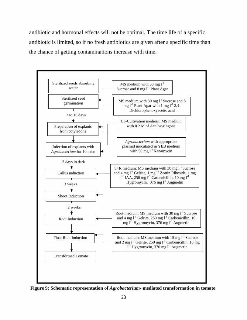
23
antibiotic and hormonal effects will not be optimal. The time life of a specific
antibiotic is limited, so if no fresh antibiotics are given after a specific time than
the chance of getting contaminations increase with time.
Figure 9: Schematic representation of Agrobacterium- mediated transformation in tomato
Sterilized seeds absorbing
water
Sterilized seed
germination
Preparation of explants
from cotyledons
Infection of explants with
Agrobacterium for 10 mins
Agrobacterium with appropriate
plasmid inoculated in YEB medium
with 50 mg l-1
Kanamycin
Co-Cultivation medium: MS medium
with 0.2 M of Acetosyringone
7 to 10 days
MS medium with 30 mg l-1
Sucrose and 8 mg l-1
Plant Agar
Callus induction
3 days in dark
S+R medium: MS medium with 30 mg l-1
Sucrose
and 4 mg l-1
Gelrite, 1 mg l1 Zeatin Riboside, 1 mg
l-1
IAA, 250 mg l-1
Carbenicillin, 10 mg l-1
Hygromycin, 376 mg l-1
Augmetin
Shoot Induction
Root Induction
Root medium: MS medium with 30 mg l-1
Sucrose
and 4 mg l-1
Gelrite, 250 mg l-1
Carbenicillin, 10
mg l-1
Hygromycin, 376 mg l-1
Augmetin
Final Root Induction Root medium: MS medium with 15 mg l-1
Sucrose
and 2 mg l-1
Gelrite, 250 mg l-1
Carbenicillin, 10 mg
l-1
Hygromycin, 376 mg l-1
Augmetin
3 weeks
2 weeks
MS medium with 30 mg l-1
Sucrose and 8
mg l-1
Plant Agar with 1 mg l-1
2,4- Dichlorophenoxyacetic acid
Transformed Tomato

24
Once the transgenic tomato plants are selected, they have to be tested
whether they contain the genes that have been integrated in the tomato genome:
BAR, HYG and GFP gene. Tissue DNA has to be extracted from the transgenic
lines to test whether the genes are present. The section below explains that
extraction protocol
2.11 DNA extraction from plant tissue:
DNA extraction from plant tissues is important because for the primary
transgenic tomatoes to be screened, a DNA needs to be isolated for different
molecular analyses. The following is the protocol used during the research project:
i. From the primary transformant, tissue is collected in a sterile 1.5 ml tube,
and then frozen under liquid nitrogen.
ii. The sample tubes are taken in the lab, and Tissue lyser from Qiagen is used
to disrupt biological samples through high speed shaking for 90 seconds at
1/s Frequency 25.
iii. Once the samples are ground into fine powder, they are left on ice and
isolation buffer added at room temperature.
iv. 400µl of 1x Isolation buffer is added to the samples: 2x Isolation buffer for 1
L (0.6 M NaCl- 35.064 g, 100 mM Tris pH=7.5- 100 ml 1M tris, 40mM
EDTA- 14.8896g, 4% N-Lauroyl Sacrcosine- 40g, 1% Sodium Dodecyl
Sulfate (SDS)- 100ml 10 % SDS), 10M urea (200ml 120g Urea) is added to
the 2 X isolation buffer 1:1(v:v) to get the 1X isolation buffer and then the
samples are vortexed.
v. 400 µl Phenol-Chloroform with 1 % Isomylalcohol and 0.1 %
hydroxyquinoline is added to the samples. The stock solution is made fresh
and consists of 25ml Phenol, 25ml Chloroform, 0.1 % Hydroxyquinoline
25
and 500 µl Isomylalcohol. The samples are inverted a few times, then
centrifuge for 5 min at 10,000 rpm.
vi. The supernatant is removed and transferred to new sterile tubes.
vii. 250 µl Isopropanol is added to the samples and mixed. The samples are left
to sit at room temperature for 5 minutes, and then centrifuged for 10 minutes
at 10,000 rpm.
viii. The liquid is carefully removed without disturbing the pallet, then 200 µl of
70 % cold EtOH (ethanol) is added to the samples, and the tubes centrifuged
for 5 minutes at 10,000 rpm.
ix. The liquid is removed again without losing the pellet and the tubes are left at
room temperature for 30 minutes until the ethanol has evaporated
x. 50 µl of TE with RNase (50 µl of TE and 10 µg/ml RNase A) pH=8 is
added, the concentrations of the samples are checked and, if the samples are
not used immediately, they are kept at - 20 ºC.(Pereira and Aarts, 1998)
SDS is used to aid in lysing cells during DNA extraction. EDTA is used to
chelate divalent metal ions to inhibit DNAses and to destabilize cell membrane for
lysis. N-Lauryl Sarcosine is used for solubilization and separation of membrane
proteins and glycoproteins, useful in concentrated salt solutions like NaCl for cell
lysis. Once phenol and chloroform is added and then centrifuged, the end result is
an upper aqueous phase and lower organic phase. Nearly all of the DNA/RNA is
present in the aqueous phase; and nearly all of the protein and other cell
constituents partition in the interphase and organic phase. The addition of
isopropanol to the aqueous DNA solution precipitates the DNA into a pellet after it
is centrifuged (DNA is insoluble in salt and isopropanol). Ethanol 70% is used to
purify and/or concentrate DNA from aqueous solutions. RNase A in TE is used to
eliminate RNA from the DNA extracted samples (Sambrook and Russell, 2001).

26
This protocol is used to extract the DNA from the tissues of the primary
transformants and the wild type. The wild type DNA is used as negative control for
analyzing the selectable markers of the Ac-Ds in-trans system from the primary
transformants through Polymerase chain reaction (PCR). A plasmid DNA for
positive control can be extracted from the A. tumefaciens containing the Ac-Ds in-
trans system. This plasmid DNA will be used in the PCR, and the extraction
protocol is shown bellow.
2.12 Plasmid DNA purification
To isolate the plasmid DNA from the A. tumefaciens containing the Ac-Ds
in-trans system, the strain is grown over-night in YEP media (10 g Bacto-yeast
extract, 10g Bacto Peptone, 5 g of NaCl in 1L volume pH= 7). Bacto peptone is
used as a nitrogen source for microorganisms (BD bionutrients technical manual).
Bacto yeast is used to deliver water, vitamins, amino acids, peptides, and
carbohydrates to the medium (Advanced bioprocessing, 3rd
edition). Once the
bacteria have grown, plasmid DNA purification is done using the Qiaprep spin
mini prep kit from Qiagen. The plasmid DNA is isolated for use in the PCR
reaction as a positive control.
2.13 Analysis of selectable markers of Ac-Ds in-trans ATag system in primary
tomato transformants through PCR
Polymerase chain reaction (PCR was used to detect the marker genes from
the vector construct in the transformed tomato lines.
i. The Initialization step.
ii. The Denaturing step, which denatures the samples at a high temperature
(94-96 ºC).

27
iii. The Annealing step, where the temperature is lowered to 50-65 ºC, which
allows the right primers (Forward and Reverse primers) to anneal to their
complementary sequences.
iv. The Elongation step, where the temperature will be raised to 72 ºC,
which allows the Taq polymerase to attach to each priming site and to
extend a new DNA strand.
Steps ii, iii, and iv will be repeated for the number of cycles chosen. After the PCR
reaction is completed, the samples are run on Ethidium bromide-stained agarose
gel. When viewed under UV light, the DNA bands are visible. About 50-100ng of
DNA is used for the PCR reaction from each primary transformed tomato tissue
sample and positive and negative control sample.
PCR is carried out using the following primers:
For the Hygromycin gene
HYGAP-F ( 5’- AAA AGT TCG ACA GCG TCT CCG ACC-3
’)
HYGAP-R( 5’- TCT ACA CAG CCA TCG GTC CAG ACG-3
’)
For the GFP gene
35S-F (5’-ATC CCA CTA TCC TTC GCA AGA CCC-3
’)
GFP-R(5’- GCT TGT CGG CCA TGA TAT AGA CG-3
’)
For the BAR gene
BAR1-F(5’-ACC ATG AGC CCA GAA CGA CGC-3
’)
BAR1-R(5’- CAG GCT GAA GTC CAG CTG CCA G-3
’)
The PCR conditions include a denaturing step at 94 ºC for 4 minutes followed by
35 cycles of 30 seconds at 94 ºC; 30 seconds at 53. 5 ºC for Hygromycin gene, 52
ºC for GFP gene and 57.5 ºC for BAR gene; 1 minute at 72 ºC ending with
elongation step of 5 minutes at 72 ºC.
28
2.14 Gel electrophoresis
Agarose is used for making an agarose gel for electrophoresis.. Agarose is a
linear polypeptide made up of repeating units of agarobiose. Once the gel has been
made a small volume of 5µl of DNA is mixed with dye and then is added to the
well of the gel. The agarose gel is subjected to an electrical filed for the separation
of nucleic acids down the agarose matrix by their negatively charged molecules.
Once the electrical filed has been applied on the agarose gel at specific voltage for
a specific time, the gel is moved and is viewed under UV light and the DNA can be
visualized since it was bonded with Ethidium Bromide.
2.15 Screening for Basta herbicide resistance
The primary tomato transformants that test positive for all selectable
markers (BAR, GFP and HYG gene) are further screened for Basta resistance. The
Basta treatment is done by painting young leaves at different concentrations and
following the affect of the herbicide for a time period of 0 days to 14 days. The
leaves are treated at different concentrations: 0.001%, 0.002%, 0.003%, 0.004%,
0.005%, 0.008% and 0.01%.
2.16 Extraction of seeds from fruits
Once fruits are formed from the wild type and transgenic tomatoes, the
following protocol (Downie et al., 2004) explains how to extract these seeds from
the fruit:
1. Slice tomato in two halves.
2. Remove seed-gel mass into beaker glass.
3. Add 0.1 M HCl to seed-gel mass and stir regularly for 1 hour.

29
4. Pour seed-gel mass into sieve.
5. Rinse seeds thoroughly with tap water to remove all gel tissues.
6. Spread the seeds on tissue paper and let them dry over night(Downie et
al., 2004)
2.17 Screening progeny plants with Basta herbicide treatment
The seeds from the progeny of the primary transformants are grown in soil.
Once they are mature, they are tested for Basta herbicide resistance the same way
as the primary transformants. A polymerase chain reaction is performed to
determine whether the offspring contain the genes from the two-component in-cis
Ac-Ds system.

30
Chapter 3
Results
3.1 Transformaton of the Ac-Ds in cis two-component system in tomato
The goal of this project was to develop tomato transformant lines with a transposon
construct for functional genomics analysis. The Ac-Ds in cis two-component
transposon construct is meant for activation tagging, where insertions of the Ds-
ATag element containing a CaMV 35S enhancer near a tomato gene would be able
to enhance expression of surrounding adjacent genes and provide a gain-of-
function phenotype. Transformations were done with Agrobacterium tumefaciens
bearing a T-DNA with the in cis Ac-Ds ATag construct. The T-DNA contained the
BAR, HYG and GFP genes as described earlier. The Ac-Ds vector construct
containing the marker genes were used for A. tumefaciens mediated
transformation. Seed of M-82 cultivar were germinated and grown on media and
cotyledons were harvested for transformation. The A. tumefaciens strain containing
the two-component in-cis Ac-Ds system was grown in YEP medium. The
cotyledons were cut into three parts as shown in Figure 10 below.

31
Figure 10: Cotyledon cuttings
The reason for cutting the tomato cotyledons is to cause wounds to the tomato
cotyledons to allow proper attachment of the A. tumefaciens to the plant cell walls,
which will allow transfer of T-DNA to the nuclear genome of the tomato plant.
Once the T-DNA with the genes are incorporated in the chromosomal DNA of the
tomato plant they will be expressed and will cause resistance to hygromycib
antibiotic, detection of the GFP protein, and resistance to the herbicides. Once the
infection of A. tumefaciens has occurred the resistance of the transformed tissue to
hygromycin will allow growth of the callus. After some time a shoot will emerge
from the callus with the appropriate hormones. At this point the A. tumefaciens
growth has to be inhibited. This inhibition can be accomplished with the addition
of different concentrations of antibiotics in the S+R shoot medium. These
antibiotics that kill/inhibit growth of Agrobacterium are combinations of
carbenicillin, cefatoxime and augmentin. Hygromycin antibiotic at concentration
of 10mg/l Hygromycin is used to select for transformation and growth of callus
and shoots. If the growth of A. tumefaciens is inhibited but the shoot development
is not repressed, it proves that that shoot allows the resistance of the Hygromycin
because of the expression of the HYG gene in the plant. With the removal of A.

32
tumefaciens the shoot will continue to grow and develop. Once the shoot is
developed enough it has four to six leaves, and the plant can be moved to a rooting
medium where only the specific antibiotics are added to the medium and the two
hormones are removed. Indole-3-Acetic Acid (IAA) is an auxin in combination
with cytokinins such as zeatin riboside it slows the aging process, it causes shoot
elongation but it inhibits the root formation. That is why zeatin riboside (1 mg/l)
and IAA (1 mg /l) hormones are removed from the S+R rooting medium. The
transformed plant is moved to the soil for further growth and experimentation once
it has grown roots and overgrown the environment where it is kept in. The figure
here shows the transformed tomato in pot of soil in a controlled environment such
as a growth chamber. The tomato plant is kept in the growth chamber for two
more weeks for further development and then moved to the Virginia Tech’s main
greenhouse located on Washington Street.
Figure 11: Transformed Tomato plant in of soil

33
Figure 12: Pictorial representation for the generation of tomato transformants at
different stages of the process. Figure 12 A represents a germinated tomato seedling with
two cotyledons ready for cuttings. Figure 12B. Figures 12 C and D show the formation of
transformed tomato shoots at different stages of development on S+R medium. Figure
12C is taken at the beginning of shoot formation, and Figure 12D is taken at the end of
A. tumefaciens treatment and beginning of shoot elongation. Figure 12E above shows the
S+R medium in a jar containing the transformed tomato explants with no more A.
tumefaciens present. Figure 12F is the end product of the transformation protocol. It
shows the transformed tomato plant which is moved from medium containing the
necessary requirements for growth to its natural medium, soil.

34
3.2 Visualization expression of the GFP gene in transformed tomato lines
Once the transformed tomato lines are selected they are grown in pots of soil
in growth chambers and then transferred to greenhouses, to be ready for further
experiments. The first laboratory experiment that we did was detection of the GFP
reporter gene. If the transformed tomato contains the two-component in-cis Ac-Ds
system integrated in the tomato genome then expression of the transformed genes
(BAR, GFP and HYG) can be monitored by appropriate assays. Expression of the
GFP reporter gene can be detected by the intense fluorescence observed through a
fluorescence microscope. Untransformed tomato tissue has background
fluorescence. We tested tomato leaves and roots for GFP, but found background
fluorescence. However, the newly developed young leaves showed low
background fluorescence. The leaves were removed from the plants with forceps,
and then immediately added to a tube containing isopropyl alcohol. This
isopropanol treatment removed the chlorophyll of the plant without damaging the
leaf. After removal of chlorophyll by isopropanol treatment the leaves are cleared
and GFP expression can be detected by the fluorescence microscope.

35
Figure 13: Images of GFP gene detection of tomato leaves by Fluorescence microscopy. The
arrows in each image points toward the control tomato leaf (GFP gene not present), the
rest are leaves from transformed tomatoes( containing the GFP gene). There is a
distinction in fluorescence intensity between the transformed tomato leaves and the control
leaves
The Figure 13 above represents a few of the fluorescence microscopy
images that were taken during the process of detecting expression of the GFP gene

36
in the transformed tomato lines. There are distinctive differences between the
transformed tomato lines and the control tomato. A control tomato refers to a
tomato plant that has not been involved in any way with the transformation
protocol. The control tomato also a M82 cultivar represents a group of tomatoes
that can be usually found in a natural environment to which there have been no
experimental techniques preformed on them. The brightness seen on the leaves in
the images shown in Figure 13 is due to the fluorescence of the GFP gene which is
been expressed in the leaves and veins. In contrast to the leaves of the transformed
lines as well as progeny lines, the control tomato leaves have low GFP
fluorescence seen under the Fluorescence microscope.
3.3 Polymerase Chain Reaction (PCR) for determining the presence of the
desired genes
The DNA extracted from the putative transformed tomato lines was used in
further test to determine the presence of the BAR, HYG and GFP genes in the
tomato lines.
Figure 15 represents a pictorial representation of the PCR process that is done by a
PCR thermal cycler. As described in Chapter 3 we used six specific primers that
are complementary to the maker genes: HYG, BAR and GFP genes based on our
knowledge of the sequences in the construction of the Ac-Ds in-cis ATag construct.
The sequences of six primers in the genes are shown below.
Hygromycin(HYG) gene:
HYGAP-F (5’- AAA AGT TCG ACA GCG TCT CCG ACC-3’)
HYGAP-R (5’- TCT ACA CAG CCA TCG GTC CAG ACG-3’)
GFP gene :

37
35S-F (5’-ATC CCA CTA TCC TTC GCA AGA CCC-3
’)
GFP-R (5’- GCT TGT CGG CCA TGA TAT AGA CG-3
’)
BAR gene:
BAR1-F (5’-ACC ATG AGC CCA GAA CGA CGC-3
’)
BAR1-R (5’- CAG GCT GAA GTC CAG CTG CCA G-3
’)
The PCR conditions we used in the PCR thermal cycler were the following:
For the denaturing step we set it up at a 94 ºC temperature for 4 minutes;
Followed by 35 cycles of 30 seconds at 94 ºC temperature;
30 seconds at
53. 5 ºC temperature for Hygromycin gene;
52 ºC temperature for GFP gene;
57.5 ºC temperature for BAR gene;
And 1 minute at 72 ºC temperature;
The final elongation step was set up for 5 minutes at 72 ºC temperature;
Ending with a infinite time at 4 ºC temperature;
After the PCR was done, the PCR products were loaded on an agarose gel for
electrophoresis. Once the gel electrophoresis was finished the gel was visualized
under UV light and photographed. Comparison of the observed bands to expected
sizes was used to determine the presence of the desired genes and if the tomato
lines were truly transformed. Both the GFP and HYG genes are expected to show
fragments of size close to 500 base pair (bp) and the BAR gene 900 bp. Base pairs
are used to measure the size of an individual DNA fragment by comparing to the
standard DNA ladder marker of known size fragments. We are interested in 3
fragments indicative of the 3 marker genes of interest in the construct.

38
Figure 14: Pictorial representation of the 1kb DNA ladder from New England Biolabs on
the left and 1kb DNA ladder from Promega on the right
The Figure 14 above represents two different 1kb DNA ladder from two
different companies that were used to determine fragment sizes. By knowing the
sizes for the genes being tested and comparing them to the 1kb ladder bands we are
able to determine whether they match the expected size. This can further prove that
the desired genes are present in the genome of the specific tomato line and that the
PCR reaction yielded the results we were expecting. The Figure 15 below
represents our result on determining whether the putative transformed lines
growing in the green house or growth chambers were transformed as expected.

39
Figure 15: Images taken from Gels represents the results of three different PCR’s. The
Image 1 shows the results for the detection of the GFP gene by the use of 35S- F primer and
GFP-R primer. In contrast Image 2 shows the results for the detection of the BAR gene by
the use of BAR-F and BAR-R primers. And Image 3 represents the results for the HYG
gene detection by the use of HYGAP-F and HYGAP-primers.
Image 1: GFP gene detection
Lane 1: 1kb DNA ladder
Lane 2: Control (negative control)
Lane 3: Transformant A3
Lane 4: Transformant C1
Lane 5: Progeny C 1.1
Lane 6: G-38 (positive control)
Image 2: BAR gene detection
Lane 1: 1 kb DNA ladder
Lane 2: Control 2 (negative control)
Lane 3: Transformant 8 1.1
Lane 4: Transformant 8 1.2
Lane 5: Transformant 8 2.3
Lane 6: G 38( positive control)
Image 3: HYG gene detection
Lane 1: 1 kb DNA ladder
Lane 2: Control 2(negative control)
Lane 3: Transformant A1
Lane 4: Transformant A3
Lane 5: Transformant C1
Lane 6: Transformant C 1.1
Lane 7: Transformant C 1.3
Lane 8: Progeny C 1.2
Lane 9: G38(positive control)

40
In Figure 15, Image 1 Lane 2 through Lane 6 represent the five different
PCR samples: Control, Transformant A3, Transformant C1, progeny C1.1 and G38
respectively. As it can be seen the band representing the G-38 PCR sample shows
the high fluorescence and compared to Lane 1 which is the 1 kb DNA Ladder and
it has a size close to 500 bp. Comparing the rest of the bands in lines 2-5 we see
that the sizes clearly match the one to the positive control (G 38). With this we can
conclude that all the lines in this PCR reaction: Transformant A3, Transformant
C1, progeny C1.1 contain the GFP gene because it has the same band size and
presence when compared to the G38 sample our positive control. G38 sample is
said to be our positive control because it contains our vector of interest: Ac-Ds in-
cis ATag system.
Image 2 shows the image of the Agarose gel containing PCR samples for the
detection of the BAR gene. Lanes 2 through 6 containing the samples of the
following tomato lines: Control 2, Transformant (T) 8 1.1, T 81.2, T 8 2.3 and G38
respectively. As it can be seen the positive control sample G38 has the brightest
fluorescence, which compared to the 1kb DNA Ladder shows a size of
approximately 1000 bp. By looking at the other samples in Lanes 3-5 we can
conclude that T 8 1.1, T81.2, T8 2.3 contain the BAR gene as well since they also
have a band of size approx. 1000 bp.
Image 3 shows the image of the agarose gel on which PCR samples were run
for the detection of the HYG gene. The lanes 2-9 represent the following PCR
samples: Control (as negative control), TA1, TA3, T C1, TC1.1, TC1.3, Progeny C
1.2 and G38 (as positive control) respectively. The transformants A1, A3, C1.1,
C1.3 and Progeny C1.2 are different from the ones above (T 8 1.1, T 81.2, T 8 2.3)
is that these were developed earlier, the last band being the G 38 sample. The
highest band has a size of ~500 bp, the size of fragment we expect for the HYG

41
gene. On comparison to the samples in lanes 3-8 it can be concluded that they also
have a band of size ~500 bp. The control sample or negative control shows no band
and indicates that there is no contamination or PCR carry-over in the reactions..
Based on these results we can conclude that the PCR samples for the following
tomato lines: Transformant A1, Transformant A3, Transformant C1, Transformant
C1.1, Transformant C1.3, Progeny C 1.2 contain the HYG gene in their gnome.
3.4 Greenhouse testing for BASTA resistance in the transformed lines
Once we had proof that the genes of interest in the construct were present in
the tomato genome of the transformed lines, experimentation was continued under
the greenhouse conditions. From the different PCR experiments performed we
determined which tomato lines had all the expected transformed gene fragments.
As mentioned in earlier chapters the BAR gene is the most useful since it is present
in the transposon that can move around the genome and make mutants. When this
gene is present in the tomato genome we should expect certain resistance to the
herbicide. The BAR gene on our Ac-Ds in-cis ATag system is located on the Ds
transposon, which has a 35S 4Enh which is a tetramer of the 35S enhancer
element, and the BAR gene. After transposition of the Ds-BAR, the insert can
move around the genome to different positions. The BAR gene confers resistance
to phosphinothricin (PPT), which is a competitive inhibitor that competes for the
active site on the glutamine synthase enzyme and does not allow glutamate to
attach to its receptor. This will cause ammonia to accumulate, inhibit
photosynthesis and cause death of the plant. By expression of the BAR gene the
plant can be tolerant to phosphinothricin (PPT) or in other words the plant becomes
resistant to the herbicide.

42
To test for herbicide resistance we chose the lines of tomato that were tested
positive by PCR and had the genes of interest. To develop an assay, we tested
spraying, painting and treating excised leaves with the herbicide. We concluded
that painting was the most appropriate as the herbicide injury would be localized.
After 2 days we could determine the effect the herbicide on the plant, and did not
run the risk of damaging the plants. By painting one leaf at a time with different
concentrations of herbicide we could test best concentrations on the same plant.
The effect of herbicide was tested on individual transformed tomato lines and
compared to control plants. Control plants correspond to tomato plants from the
same cultivar but which had not put under the transformation protocols and did not
contain any of the desired marker genes: BAR, HYG and GFP. When the control
plants were painted at certain concentration of herbicide they would not be
resistant or tolerant to the effects of the BASTA the competitive inhibitor.
One of the most successful BASTA herbicide screenings was using the following
two concentrations: 0.001% and 0.002% for a 14-day period. Figures 17 and 18
show the tomato lines, both primary transgenic and control, on day 0. Figures 19
and 20 show the affect of the Basta herbicide by day 7. Figures 21-22 show day 14.
Notice that on day 14, the leaf of the control plant treated with BASTA herbicide
at 0.002% has wilted and the size has decreased by half from day 0. When
compared to the control, there were no signs of effect from BASTA herbicide on
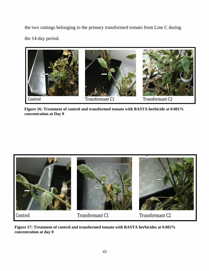
43
the two cuttings belonging to the primary transformed tomato from Line C during
the 14-day period.
Figure 16: Treatment of control and transformed tomato with BASTA herbicide at 0.001%
concentration at Day 0
Figure 17: Treatment of control and transformed tomato with BASTA herbicides at 0.002%
concentration at day 0
44
Figure 18: Treatment of control and transformed tomato with BASTA herbicide with 0.001%
concentration at Day 5
Figure 19: Treatment of control and transformed tomato with BASTA herbicide with 0.002%
concentration at Day 5

45
Figure 20: Treatment of control and transformed tomato with BASTA herbicide with 0.001%
concentration at Day 14
Figure 21: Treatment of control and transformed tomato with BASTA herbicide with 0.002%
concentration at Day 14

46
3.5 Higher efficiency using Augmetin as a hormone
To determine whether the transformation protocol using augmetin as a
hormone instead of cefatoxime for the development of transgenic tomato lines is
more productive we used the data collected during the research project as shown in
table 2, we determined that the protocol that used augmentin as a hormone
increased the transformation protocol efficiency.
Table 2: Data collection of trials done and how many transformed lines were developed
The result we acquired was a p-value of 8.17 * 10-19
as shown in Table 3.
Since the p-value is smaller than 0.05 we can regard it as statistically different.
There is a clear significant deviation between the two protocols. First when we
used the protocol without the augmetin hormone there were less number of
transformed tomato lines developed compared to how many trials were done. In
contrast when we used a transformation protocol with augmetin we developed
more transformed tomato lines in shorter time and in fewer trials.
Trials without Augmetin Transformed without
augmetin
33 2
Trials with augmetin Transformed with augmetin
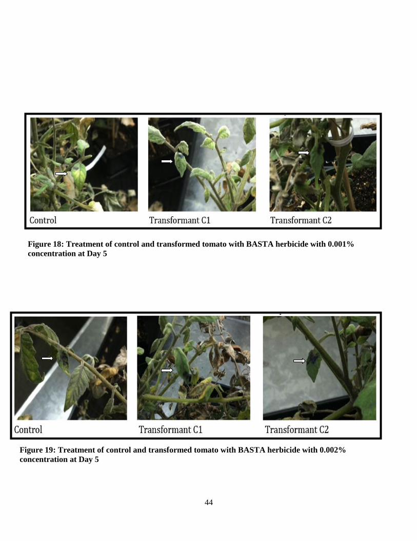
8 3
CHI Test
X^2 8.17035E-19
Table 3: The p-value once the chi-square distribution was done

47
3.6 Conclusion
With the project coming to an end, we collected our data and determined
that the transformation protocol we decided to use was successful. We were able
to get transformed tomato lines in a shorter period when using 376 mg l-1
augmetin.
The hormone augmentin was found to be very successful in the development of
transgenic tomato lines. By the use of the hormone the number of transformed
tomatoes increased. Not only its efficiency was improved but also it was more cost
effective. The price for purchasing augmetin was cheaper compared to the
hormones we used in the past such as cefatoxime. We learned that the only way to
use cefatoxime was to have it in higher concentration (around 150 to 200 mg/l).
Even with the higher concentration of cefatoxime, our results were negligent; we
lost more plants by cross contamination and its inability to inhibit the growth of
A.tumefaciens.
Once the transgenic tomato lines were developed, they were further tested to
detect whether the reporter gene and selectable markers genes were present.
Polymerase chain reactions showed that all three GFP, HYG and BAR genes were
present. We further showed that the fluorescence of the GFP gene was present in
the transformed tomato lines with the help of a fluorescence microscope.
Additionally, we performed BASTA herbicide resistance analysis. These analyses
showed that the transgenic tomato lines had a certain degree of resistance when
treated with an herbicide at 0.001% and 0.002 % concentrations. The Table bellow
shows the summary of the all the transformed tomato lines that were developed. In
the table it is shown which lines were grown by using cefatoxime or augmetin. The
rest of the results show the detection of the marker genes (BAR, HYG and GFP).
48
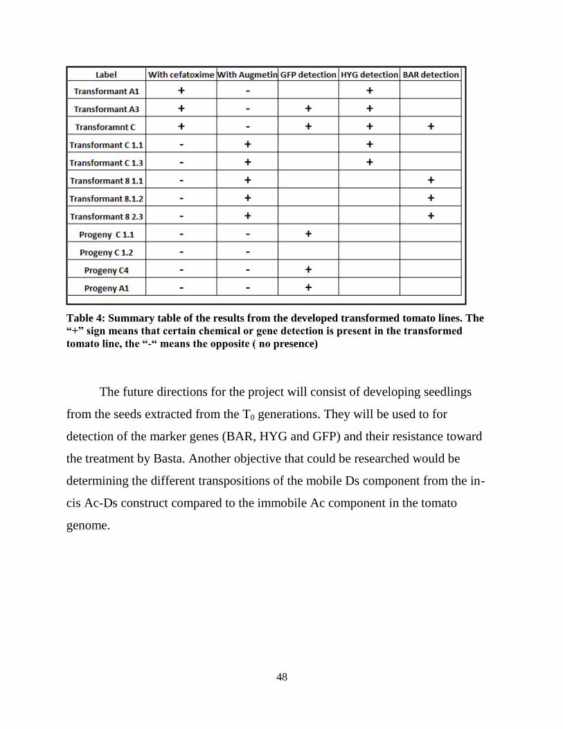
Table 4: Summary table of the results from the developed transformed tomato lines. The
“+” sign means that certain chemical or gene detection is present in the transformed
tomato line, the “-“ means the opposite ( no presence)
The future directions for the project will consist of developing seedlings
from the seeds extracted from the T0 generations. They will be used to for
detection of the marker genes (BAR, HYG and GFP) and their resistance toward
the treatment by Basta. Another objective that could be researched would be
determining the different transpositions of the mobile Ds component from the in-
cis Ac-Ds construct compared to the immobile Ac component in the tomato
genome.

49
References
Altpeter,F., Beachy,R., Bock,R. (2005). "Particle bombardment and the genetic
enhancement of crops: myths and realities." Molecular Breeding 15: 305-
327.
Asma, F. M. V. "Growth and Storage of Agrobacterium." Methods in Molecular
Biology 44: 1-7.
Atares A., Moyano E., Morales B., Schleicher P. (2011). “ An insertional
mutagenesis programme with an enhancer trap for the identification and
tagging of genes involved in abiotic stress tolerance in the tomato wild-
related species S. pennellii” Plant cell Rep 30(10): 1865-1879.
Bandopadhyay, R., Singh,D. and Mukhopadhyay,K. (2010). "Levels and Stability
of Expression of Transgenes." Transgenic Crop Plants: 145-186.
Bansal, P. K.(2010). "Efficient production of transgenic tomatoes via
Agrobacterium-mediated transformation." Biologia Plantarum 54(2): 344-
348.
Chatchawankanphanich, S. I. O. (2005). "Augmentin as an alternative antibiotic for
growth suppression of Agrobacterium for tomato (Lycopersicon esculentum)
transformation." Plant Cell, Tissue and Organ Culture 82: 213–220.
Chibbar, S. G. (2010). "Gene Transfer Methods." Transgenic Crop Plants: 57-83.
Choudhry, A. (2009). "In Planta Transformation of Tomato." Plant Mol Biol Rep
27: 20-28.
Cortina, C., Culianes-Macia,F.A. (2004). “ Tomato trasnforamtionand transgenic
plant production” Plant cell Tissue and Organ Culture 76( 269-275).
Dang Thi Van, N. F. (2010). "Development of a simple and effective protocol for
Agrobacterium tumefaciens mediated leaf disc transformation of commercial
tomato cultivars." GM Crops 1-5: 312-321.
Davey, M.R., Nageswara Rao,M., Kourmpetli,S., Bhattacharya,A. and Kole,C.
"Generation and Deployment of Transgenic Crop Plants: An Overview."
Transgenic Crop Plants,: 1-29.
Donn, G.,Goodman,H. (1987). “Plant cells relsistant to herbicidal glutamine
synthetae inhibitors” Espacenet.
Forde,B.G.,Lea,P.J. (2007). “ Glutamate in plants:metabolism, regulation and
signaling”. Journal of Experimental Botany 58(2): 2339-2358.
Goodman, Howard M., Donn, Gunter (1992). “Plant cells resistant to herbicidal
Glutamine synthetase inhibitors” United States Patent 1.
http://www.google.com/patents?hl=en&lr=&vid=USPAT5145777&id=QMg
aAAAAEBAJ&oi=fnd&dq=Donn,+G.,Goodman,H.+%281987%29.+%E2%
80%9CPlant+cells+resistant+to+herbicidal+glutamine+synthetase+inhibitor

50
s%E2%80%9Dglutamine+synthetase+inhibitors%E2%80%9D+1987&prints
ec=abstract#v=onepage&q&f=false
González-Moro, B., Lacuesta, M., González-Murua,C., Mun˜oz-Rueda.A., (2000).
"Glutamine synthetase from mesophyll and bundle sheath maize cells
isoenzyme complements and different sensitivities to phosphinothricin."
Plant Cell Reports 19: 1127-1134.
Goossens A., Hakkinen S.T., Laakaso I.(2002). “A functional genomics approach
toward the understanding of secondary metabolism in plant cells”
Proceedings of National Academy of Sciences of the United States of
America 100(14): 8595-8600.
Guo-Qing Song, K. C. S., Callow,P.W., Baughan,R. and Hancock,J.M. (2008).
"Evaluation of a Herbicide-resistant Trait Conferred by the Bar Gene Driven
by Four Distinct Promoters in Transgenic Blueberry Plants." J.
AMER.SOC.HORT.SCI. 133(4): 605-611.
Guo-Qinsgo Ngth,I.O.,Amaguchi, N.E.I. (2004). "Efficient Agrobacteium
tumefaciens- mediated transformation of sweet potato (Ipomoea Batatas(L.)
lam.) from stem explants using a two-step kanamycin-hygromycin selection
method" In Vitro Cell. Dev. Biol.-Plant 40: 359-365.
Haiying Liang, P. A. K., Nain, N., Powell,W.A. and Carlson, J.E. (2010).
"Selection and Screening Strategies." Transgenic Crop Plants: 85-132.
Hyeon-Jin Sun , S. U., Watanabe,S. and Ezura,H. (2006). "A Highly Efficient
Transformation Protocol for Micro-Tom, a Model Cultivar for Tomato
Functional Genomics." Plant Cell Physiol. 43(3): 426–431.
Ieamkhang,S. & Chatchawankanphanich.O. (2005). "Augmentin as an alternative
antibiotic for growth suppression of Agrobacterium for tomato
(Lycopersicon esculentum) transformation." Plant Cell, Tissue and Organ
Culture 82: 82: 213–220.
Kim,C.K., Park,S.H., Burrell,A.M. (2004). "Agrobacterium tumefaciens-mediated
transformation of Rosa hybrida using the green fluorescent protein (GFP)
gene." Plant Cell, Tissue and Organ Culture 78: 107-111.
Klee,H., Hasegawa,M. and Bressan, R. “ Ttransformation of Tomato Plants”
Retrived from University of Florida Horticultural Sciences department webi
site:
http://www.hos.ufl.edu/meteng/HansonWebpagecontents/Tomatotransforma
tioprotocol.html
Knapp, J.E., Chandlee,J.M. (2000). "Transformation of three genera of orchid
using the bar gene as a selectable marker." Plant Cell Reports 19: 893-898.
Kobayashi, Y.. (2004). "Production of transgenic lily plants by Agrobacterium-
mediated transformation." Plant Cell Reports 22: 359-364.

51
Kohli,A., Twyman,R.M.,(2010)” Transgene integration, expression and stability in
plants:strategies for improvments.” Transgenic Crop Plants: 201-237.
Li Sun, H. C., Weihong Xu, Yuanlei Hu, and Zhongping Lin (2002). "CaMV 35S
Promoter Directs β-Glucuronidase Expression in Ganoderma lucidum and
Pleurotus citrinopileatus." Molecular Technology 20: 239-244.
Lohar, D.P., Buzas,D.M., Gresshoff,P.M. and Stiller, J.(2001). "Transformation of
Lotus japonicus using the herbicide resistance bar gene as a selectable
marker." Journal of Experimental Botany 52(361): 1697-1702.
Mamidala,P., Nanna,R.S. ( 2009). “ Influence of antibiotics on regeneration
efficiency in tomato” Plant Omics Journal 2(4): 135-140
Milojevic, J., Tubic,D.L., Vinterhalter, B. (2012). "Hygromycin promotes somatic
embryogenesis in spinach." Plant Cell Tiss Organ Cult 109: 573-579.
Nadolska-Orczk,A., Ptvetakiewicz,A., (2000). "Agrobacterium-mediated
transformation of cereals- from technique development to its application."
Acta Physiologiae Plantarum 22(1): 77-88.
Nanna, P. M. (2009). "Influence of antibiotics on regeneration efficiency in
tomato." Plant Omics Journal 2(4): 135-140.
Nuez, M. J. D. "Tomato." 249-323.
Perez-Garcia, A., Canton, F.R., Cazorla, F.M., Codina,J.C., (1998). "Light-
dependent changes of tomato glutamine syntheses in response to P.syringae
infection or phosphinothricin treatment." Physiological Plantarum 102(3-77-
384).
Prasher ,D.C., Ward ,W.W., Prendergast,F.G. and Cormier, M.J. (1992). "Primary
structure of the Aequorea victoria green-fluorescent protein." Gene 111:
229-233.
Prasher, D. C. (1995). "Using GFP to see the light." Elsevier Science Ltd 11(8):
320-323.
Scalco,C.B., Pasquali G. (2011). "Evaluation of different versions of the gene
encoding green fluorescent protein (GFP) in plants." Scalco et al. BMC
Proceedings 5(7): 183.
Sharma, K.K. (2005). "Genetic Transformation Technology: Status and Problems."
In Vitro Cell. Dev. Biol.-Plant 41: 102-112.
Stirling, H. P. "DNA Extraction from tissue." Methods in Molecular Biology 226:
33-34.
Sung H. Park, S.C.R., Zapata, C., Srivatanakul, M., Smith,R.H. (1998). "Cross-
protection and selectable marker genes in plant transformantion" In Vitro
Cell. Dev. Biol.-Plant 34: 117-121.
Tzvi Tzfira, S. V. K. and Citovsky,V. (2007). "Advanced Expression Vector
Systems: New Weapons for Plant Research and Biotechnology." Plant
Physiology 145: 1087-1089.
52
Yasmeen, A. (2009). "An improved protocol for the regeneration and
transformation of tomato (cv Rio Grande)." Acta Physiol Plant 31: 1271–
1277.
Yi-Hong Wang, M. A. C. (2008). "Agrobacterium-Mediated Transformation of
Tomato Elicits Unexpected Flower Phenotypes with Similar Gene
Expression Profiles." PLoS ONE 3(8).
Yun-Jeong Han, Y.-M. K., Lee,L.-Y., Kim,S.J., Cho, K.-C., Chandrasekhar,T.,
Song ,P.-S., Woo, Y.-M., Kim,J.-I. (2009). "Production of purple-colored
creeping bentgrass using maize transcription factor genes Pl and Lc through
Agrobacterium-mediated transformation." Plant Cell 26: 397–406.
YU, X. Y. (2006). "Transformation of multiple soybean cultivars by infecting
cotyledonary-node with Agrobacterium tumefaciens." African Journal of
Biotechnology 5(20): 1989-1993.
Zhangsun,D., Chen,R. and Tang,K. (2007). "Improved Agrobacterium-mediated
genetic transformation of GNA transgenic sugarcane." Biologia 62(4):
386—393.
Ziemienowicz, A. "Plant seleetable markers and reporter genes." Acta
Physiologiae Plantarum 23(3): 363-374.
Zolman,B.K., Yoder,A. and Bartel,B. ( 2000) “Genetic Analysis of Indole-3-
butyric Acid resposnses in A. thaliana reveals four mutant classes” Genetics
society of America: 1324-1337

53
Appendix A
Annotated List of Figures
Figure 2
Trijatmiko, Kurniawan R. (2005). “Comparative analysis of drought resistance
genes in Arabidopsis and rice”. [ PhD. thesis, Wageningen University, 2005], 115.
http://edepot.wur.nl/121694 (accessed Aug. 20th, 2013) Fair Use determination
attached.
Figure 3
Prasher ,D.C., Ward ,W.W., Prendergast,F.G. and Cormier, M.J. (1992). "Primary
structure of the Aequorea victoria green-fluorescent protein." Gene 111: 229-233.
(Accessed Aug. 20th, 2013) Fair Use determination attached.
Figure 4
Prasher, D. C. (1995). "Using GFP to see the light." Elsevier Science Ltd 11(8):
320-323. (Accessed Aug. 20th, 2013) Fair Use determination attached.
Figure 5
Goodman, Howard M., Donn, Gunter (1992). “Plant cells resistant to herbicidal
Glutamine synthetase inhibitors” United States Patent 1.
http://www.google.com/patents?hl=en&lr=&vid=USPAT5145777&id=QMgaAAA
AEBAJ&oi=fnd&dq=Donn,+G.,Goodman,H.+%281987%29.+%E2%80%9CPlant
+cells+resistant+to+herbicidal+glutamine+synthetase+inhibitors%E2%80%9Dglut
amine+synthetase+inhibitors%E2%80%9D+1987&printsec=abstract#v=onepage&
q&f=false (Accessed Aug 20th, 2013) Fair Use determination attached.
Figure 6
Goodman, Howard M., Donn, Gunter (1992). “Plant cells resistant to herbicidal
Glutamine synthetase inhibitors” United States Patent 1.
http://www.google.com/patents?hl=en&lr=&vid=USPAT5145777&id=QMgaAAA
AEBAJ&oi=fnd&dq=Donn,+G.,Goodman,H.+%281987%29.+%E2%80%9CPlant
+cells+resistant+to+herbicidal+glutamine+synthetase+inhibitors%E2%80%9Dglut
amine+synthetase+inhibitors%E2%80%9D+1987&printsec=abstract#v=onepage&
q&f=false (Accessed Aug 20th, 2013) Fair Use determination attached.

54
Figure 7
Invivogen. http://www.invivogen.com/hygromycin (Accessed Aug 20th
, 2013) Fair
Use determination attached.
Figure 8
Chibbar, S. G. (2010). "Gene Transfer Methods." Transgenic Crop Plants: 57-83.
(Accessed Aug 20th
, 2013) Fair Use determination attached.


![An Active Ac/Ds Transposon System for Activation …An Active Ac/Ds Transposon System for Activation Tagging in Tomato Cultivar M82 Using Clonal Propagation1[W][OA] Jared D. Carter,](https://img.dokumen.tips/doc/110x75/5eb1672acea3ec2d5d5099c5/an-active-acds-transposon-system-for-activation-an-active-acds-transposon-system.jpg)