Embed Size (px)
Citation preview
Thermoluminesence of gamma rays irradiated CaSO4 nanorods dopedwith different elements
Numan SalahCenter of Nanotechnology, King Abdulaziz University, Jeddah 21589, Saudi Arabia
H I G H L I G H T S
� Nanorods of CaSO4 activated by Ag, Cu, Dy, Eu and Tb were synthesized.� They were studied for their TL response to a wide range of γ-rays.� The doped samples with Dy, Eu and Tb have thinner nanorods than the others.� Tb doped sample is found to be a highly TL sensitive with a glow peak at 270 1C.� Eu doped sample has a linear TL response in the range i.e. 10 Gy–10 kGy.
a r t i c l e i n f o
Article history:Received 21 October 2013Accepted 30 June 2014Available online 9 July 2014
Keywords:CaSO4
NanorodsThermoluminescenceGamma raysDopants
a b s t r a c t
Nanorods of calcium sulfate (CaSO4) activated by Ag, Cu, Dy, Eu and Tb were synthesized by the co-precipitation technique. They were irradiated by γ-rays in a wide range of exposures and studied fortheir thermoluminesence (TL) properties. The as-synthesized samples were characterized by scanningelectron microscopy (SEM), X-ray diffraction (XRD) and photoluminescence (PL) emission spectra. SEMimages show that the samples doped with rare earths elements (i.e. Dy, Eu and Tb) have thinnernanorods than the other samples, while XRD pattern shows a complete crystalline structures in amonoclinic phase. The TL glow curves of these samples show two components. The first one include lowtemperature glow peaks at around 125 1C, while the second component shows high temperature peaksin the range 230–270 1C. These glow peaks diver from sample to sample by their TL intensity. The TLresults are promising, particularly that of Tb and Eu. Tb doped sample is found to be a highly TL sensitivewith a prominent glow peak at around 270 1C, while Eu has created very active, high dense electrontraps. The later shows quite linear response in the whole studied exposures i.e. 10 Gy–10 kGy. Theseresults show that Eu or Tb doped CaSO4 nanorods might be proper candidates as dosimeters for highdoses of ionizing radiations used in irradiation of foods and seeds.
& 2014 Elsevier Ltd. All rights reserved.
1. Introduction
Nanomaterials have attracted considerable attention due totheir unique fundamental physical and chemical properties andpotential applications in various fields of recent technologies.Large number of workers from different areas, mainly from thefield of luminescence have produced different nanostructurematerials and investigated their structural and optical properties(Nalwa, 2000). Recent articles on luminescent nanostructures haveshowed that they have a potential application in different fieldssuch as a material for color center laser and dosimeters forionizing radiations (Alharbi et al., 2013).
Thermoluminescence (TL) or thermally stimulated lumines-cence is the thermally stimulated emission of light from a solidmaterial, following the prior absorption of energy from ionizingradiation. This phenomenon has been employed for detection/measurement of absorbed radiation, dating of archeological speci-mens and detecting defects in solids. TL is a common techniqueused for the dosimetry of different ionizing radiations. One of themost common material used as a dosimeter is CaSO4:Dy phosphor.It is synthesized by Yamashita et al. (1971), and used as adosimeter in many countries. That is due to its high sensitivity,simple glow curve structure. It also has low fading, easy method ofpreparation and excellent reusability. There have been extensiveinvestigations on this phosphor to enhance its TL properties(Lakshmanan et al., 2002; Atone et al., 1995; Azorin Juan et al.,1984). But, only few studies were reported on the nanostructure
Contents lists available at ScienceDirect
journal homepage: www.elsevier.com/locate/radphyschem
Radiation Physics and Chemistry
http://dx.doi.org/10.1016/j.radphyschem.2014.06.0270969-806X/& 2014 Elsevier Ltd. All rights reserved.
E-mail addresses: [email protected], [email protected]
Radiation Physics and Chemistry 106 (2015) 40–45
form of this material (Salah et al., 2006; Shinsho et al., 2008;Zahedifar and Mehrabi, 2010; Zahedifar et al., 2011). The effect ofdifferent dopants/impurities on the TL properties of this promisingnanomaterial is rarely appeared.
The nanostructure form of CaSO4 was synthesized for the firsttime by Salah et al. (2006). Nanocubes of this material werefabricated by the chemical co-precipitation method and studiedfor their TL response to gamma rays. The result on TL studies ofCaSO4:Dy nanomaterial showed promising features such as alinear/supralinear response over a wide range of exposures(0.1 Gy–5 kGy). However, it is not sensitive for low doses and hasseveral TL glow peaks, which are undesirable in radiation dosi-metry. It is well known that incorporation of small amounts ofdopants (normally in PPM level) can activate/modify the electrical,optical and magnetic properties of the host material. Theseimpurities have different abilities to create electron traps in thematrix. Therefore, doping the nanostructure form of CaSO4 byother elements might improve its TL performance and provide ananomaterial that has better dosiemtric properties. Only a singlereport by Zahedifar and Mehrabi (2010) has presented a work ondoping CaSO4 nanosheets with cerium. They could significantlyimprove the TL sensitivity of this nanostructure material, but in avery small range of exposures at the lower doses side (less than15 Gy) and saturate at higher doses. Further reports on doping thenanostructure form of CaSO4 with other elements and study its TLare rarely appeared.
In this work CaSO4 nanorods doped with different elementswere produced and studied for their TL properties. They weredoped with Ag, Cu, Dy, Eu and Tb. The produced nanomaterialwere characterized by scanning electron microscopy (SEM), X-raydiffraction (XRD) and photoluminescence (PL) emission spectra.They were exposed to different doses of gamma rays from a 173Cssource and investigated for their dosimetric properties using theTL technique. The TL results are promising particularly that of Euand Tb doped samples. Tb doped sample is found to be a highly TLsensitive with a prominent glow peak at around 270 1C, while Euhas created very active electron traps. The later showed quitelinear response in the whole studied exposures i.e. 10 Gy–10 kGy.
2. Experimental procedure
Nanorods of CaSO4 were prepared by the chemical co-precipitation method as adopted earlier by Salah et al. (2006)with a slight modification. Calcium chloride was used instead ofcalcium acetate, while the remaining materials and steps are thesame. The used impurities in this experiment are Ag, Cu, Dy, Euand Tb. They were used in chloride form, except those of rareearths and Ag dopants, where hydrated chlorides (for rare earths)and nitrate compound (For Ag) are implemented. A typical con-centration of 0.5 mol% is used for all these impurities. In a typicalcase for Tb doped sample, 66.3 mg of TbCl3 � xH2O was completelydissolved in 40 ml of triply distilled deionized water, then to thissolution 6.6 g of (NH4)2SO4 was added and stirred for 2 h. Anothersolution of 7.35 g CaCl2 �2H2O was made by dissolving thisquantity in a mixture of 10 ml triply distilled deionized waterand 200 ml ethanol. This solution was then added drop wise witha continues stirring to the solution of ammonium sulfate. Theformed precipitate was filtered out, then washed several timeswith triply distilled deionized water. Nanocrystalline powdersamples thus obtained were dried at 80 1C in an oven for 4 h.Similarly, the other doped samples were prepared.
Morphology of the pure and doped CaSO4 powder sampleswere studied by SEM using a field emission scanning electronmicroscopy (FESEM), JSM-7500F (JEOL – Japan) operated at 15 kV.The XRD of pure CaSO4 powder sample was also recorded, using
an Ultima-IV (Rigaku, Japan) diffractometer with Cu Kα radiation.For taking TL, samples were exposed to gamma rays from a 137Cssource for various doses (10 Gy–10 kGy) at room temperature. Thedose rate for gamma rays irradiation was 0.15 Gy/s. TL glow curveswere recorded after one hour of gamma rays exposure using aHarshaw TLD reader (Model 3500) by taking 5 mg of sample eachtime. These glow curves were recorded under nitrogen atmo-sphere at a heating rate of 5 1C/sec. Photoluminescence (PL)emission spectra of the samples were recorded at an excitationwavelength of 325 nm using a fluorescence spectrofluorophot-ometer, model RF-5301 PC, Shimadzu, Japan. The same amount ofsample was taken during the PL measurement. All the sampleswere excited by the same wavelength i.e. 325 nm. This measure-ment was performed at room temperature.
3. Results and discussion
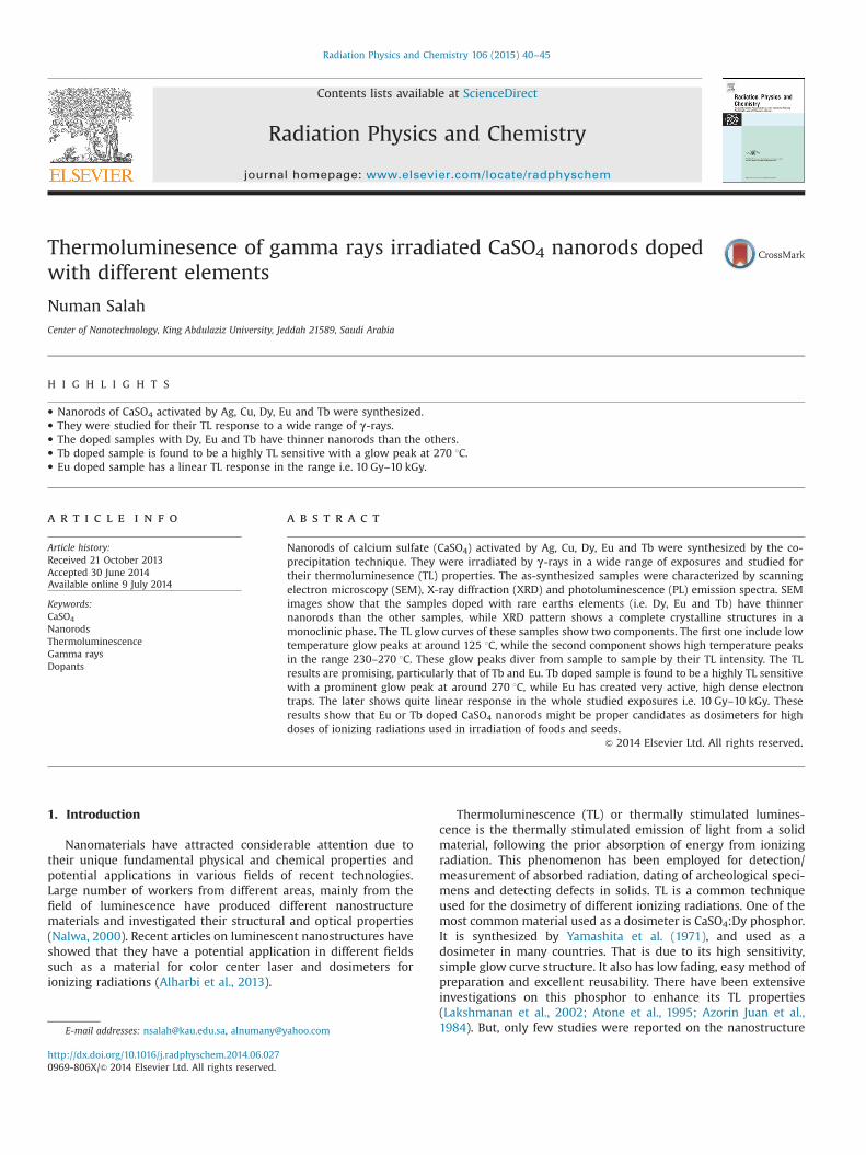
Fig. 1 shows SEM images of pure CaSO4 and Tb doped sample ata concentration of 0.5 mol% (images a and b, respectively). SEMimage of the pure one (a) shows nanorods with diameters in therange of 60–200 nm and lengths varying from 200 nm to 2 μm.Similarly, Ag and Cu doped samples were found to have same sizesand shapes, while those doped with Tb (b) are of smallerdiameters (50–80 nm). The samples doped with Dy and Eu havea morphology similar to that of Tb. It is worth mentioning thatthe used rare earth impurities in this experiment are in hydrated
Fig. 1. SEM images of the as-synthesized pure CaSO4 (a) and Tb (b) doped sampleat a concentration of 0.5 mol%.
N. Salah / Radiation Physics and Chemistry 106 (2015) 40–45 41
chloride forms as mentioned above. This form has higher solubilityin water than un-hydrated one, therefore, it is possible that therare earth ions might get dissolved completely and incorporatedwithin the host of CaSO4 interstitially or substitutionally. Thiswell-incorporation of rare earths ions perhaps could allow agrowth in one direction and limits this growth in the other one;thus shows thinner nanorods. In other words these rare earthsions perhaps could act as controlling agents for size growth. Theother impurities i.e. Ag and Cu were used in un-hydrated forms,therefore could not incorporated well and have not showed anycontrol on the growth process. Their result are similar to that ofthe pure sample. The un-doped sample of CaSO4 has no controllingagent like these rare earths, therefore have showed normalgrowth.
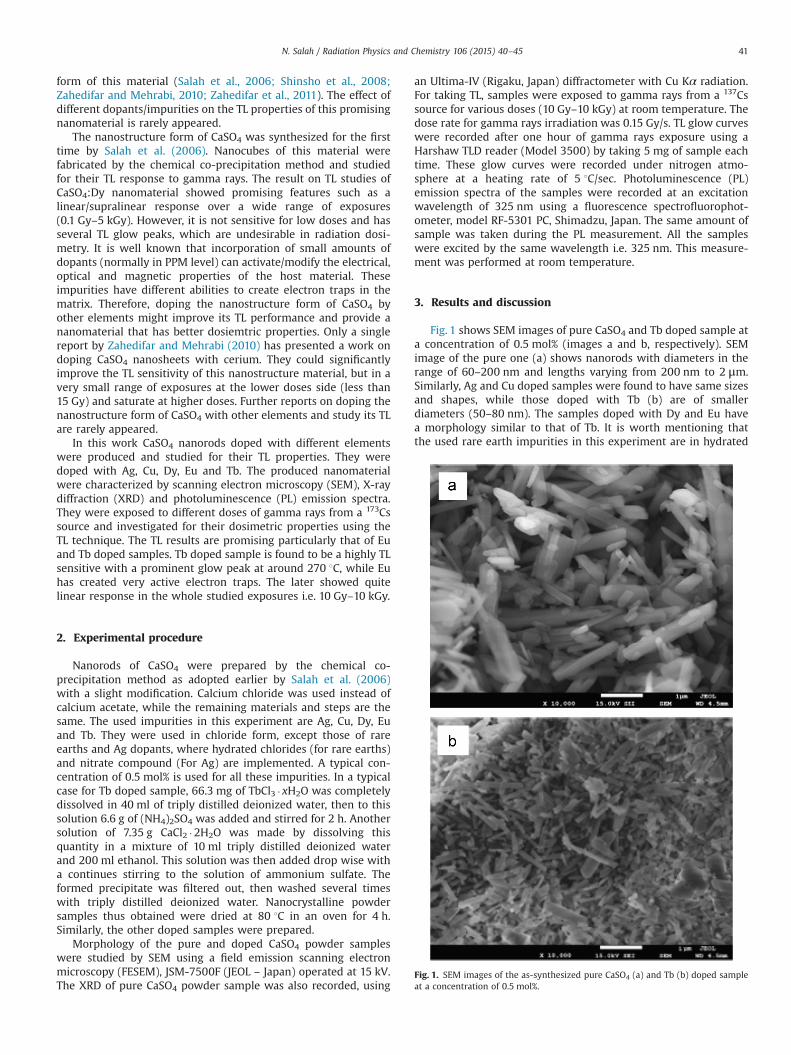
XRD pattern of the as-synthesized CaSO4 nanorods is presentedin Fig. 2. The XRD result shows several diffracted peaks with hklvalues. This indicates a complete crystalline structure in a mono-clinic phase (DB Card number 01-076-5973). This result is differentthan that reported earlier (Salah et al., 2006; Zahedifar andMehrabi, 2010), where orthorhombic lattice structures were pro-duced corresponds to JCPDS Card no. 06-0226. This change in thecrystal structure might be due to the modification induced in thepresent experiment, where calcium chloride are used instead ofcalcium acetate. The XRD results of the doped samples are similarto that of the pure one. No significant changes or variations areobserved amongst these samples. It is possible that there is aformation of small part of CaO phase. This phase might formedduring the reaction process and shows a diffracted peak at around291 (Nirmala and Suresh, 2013). This peak overlaps with that at 291(hkl¼041) of CaSO4 nanorods.
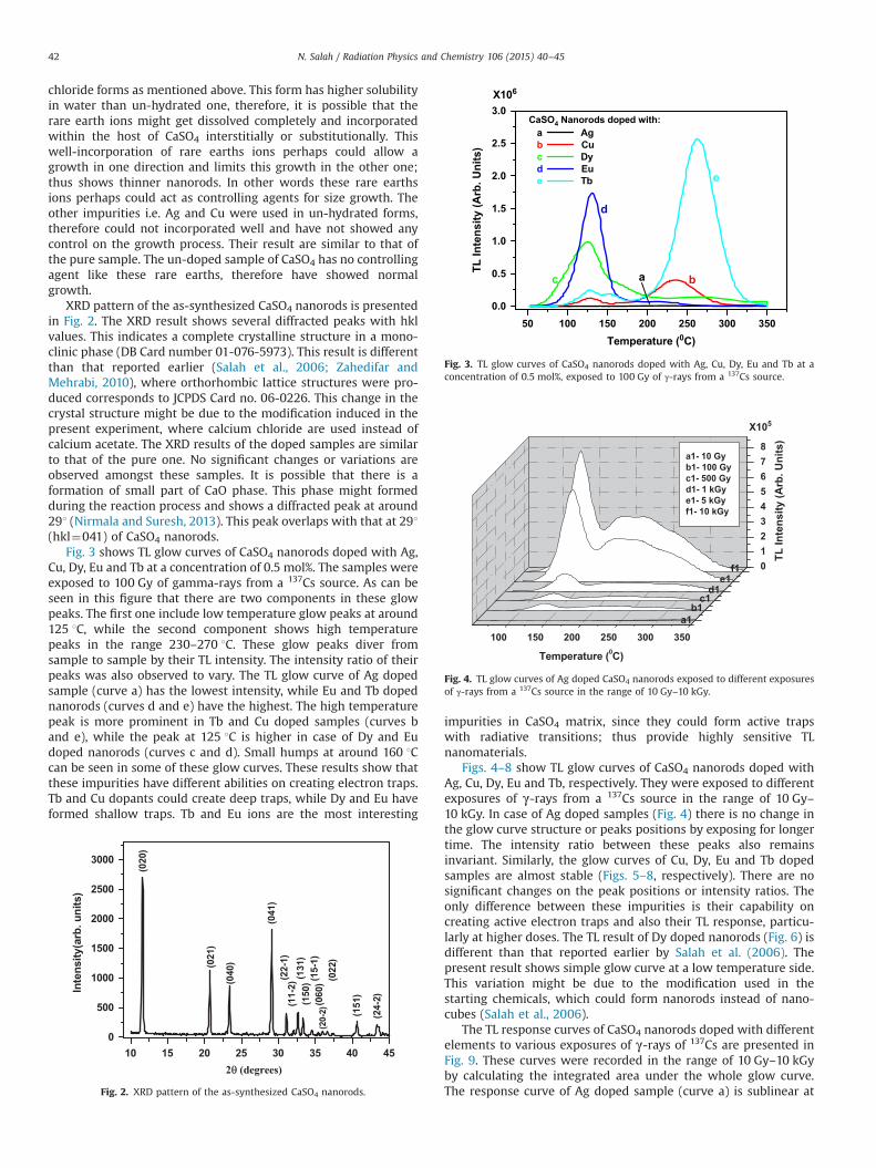
Fig. 3 shows TL glow curves of CaSO4 nanorods doped with Ag,Cu, Dy, Eu and Tb at a concentration of 0.5 mol%. The samples wereexposed to 100 Gy of gamma-rays from a 137Cs source. As can beseen in this figure that there are two components in these glowpeaks. The first one include low temperature glow peaks at around125 1C, while the second component shows high temperaturepeaks in the range 230–270 1C. These glow peaks diver fromsample to sample by their TL intensity. The intensity ratio of theirpeaks was also observed to vary. The TL glow curve of Ag dopedsample (curve a) has the lowest intensity, while Eu and Tb dopednanorods (curves d and e) have the highest. The high temperaturepeak is more prominent in Tb and Cu doped samples (curves band e), while the peak at 125 1C is higher in case of Dy and Eudoped nanorods (curves c and d). Small humps at around 160 1Ccan be seen in some of these glow curves. These results show thatthese impurities have different abilities on creating electron traps.Tb and Cu dopants could create deep traps, while Dy and Eu haveformed shallow traps. Tb and Eu ions are the most interesting
impurities in CaSO4 matrix, since they could form active trapswith radiative transitions; thus provide highly sensitive TLnanomaterials.
Figs. 4–8 show TL glow curves of CaSO4 nanorods doped withAg, Cu, Dy, Eu and Tb, respectively. They were exposed to differentexposures of γ-rays from a 137Cs source in the range of 10 Gy–10 kGy. In case of Ag doped samples (Fig. 4) there is no change inthe glow curve structure or peaks positions by exposing for longertime. The intensity ratio between these peaks also remainsinvariant. Similarly, the glow curves of Cu, Dy, Eu and Tb dopedsamples are almost stable (Figs. 5–8, respectively). There are nosignificant changes on the peak positions or intensity ratios. Theonly difference between these impurities is their capability oncreating active electron traps and also their TL response, particu-larly at higher doses. The TL result of Dy doped nanorods (Fig. 6) isdifferent than that reported earlier by Salah et al. (2006). Thepresent result shows simple glow curve at a low temperature side.This variation might be due to the modification used in thestarting chemicals, which could form nanorods instead of nano-cubes (Salah et al., 2006).
The TL response curves of CaSO4 nanorods doped with differentelements to various exposures of γ-rays of 137Cs are presented inFig. 9. These curves were recorded in the range of 10 Gy–10 kGyby calculating the integrated area under the whole glow curve.The response curve of Ag doped sample (curve a) is sublinear at
10 15 20 25 30 35 40 450
500
1000
1500
2000
2500
3000
(24-
2)
(151
)
(022
)(2
0-2)
(060
)(15-
1)(1
50)(1
31)
(11-
2)(2
2-1)
(041
)
(040
)(021
)
(020
)
Inte
nsity
(arb
. uni
ts)
2θ (degrees)
Fig. 2. XRD pattern of the as-synthesized CaSO4 nanorods.
50 100 150 200 250 300 3500.0
0.5
1.0
1.5
2.0
2.5
3.0
e
d
c b
X106
a
CaSO4 Nanorods doped with: a Ag
b Cuc Dy d Eue Tb
TL In
tens
ity (A
rb. U
nits
)
Temperature (0C)
Fig. 3. TL glow curves of CaSO4 nanorods doped with Ag, Cu, Dy, Eu and Tb at aconcentration of 0.5 mol%, exposed to 100 Gy of γ-rays from a 137Cs source.
100 150 200 250 300 350
012345678
f1e1
d1c1
b1a1
X105
TL In
tens
ity (A
rb. U
nits
)
Temperature (0C)
a1- 10 Gyb1- 100 Gyc1- 500 Gyd1- 1 kGye1- 5 kGyf1- 10 kGy
Fig. 4. TL glow curves of Ag doped CaSO4 nanorods exposed to different exposuresof γ-rays from a 137Cs source in the range of 10 Gy–10 kGy.
N. Salah / Radiation Physics and Chemistry 106 (2015) 40–4542

low doses, then almost linear in the intermediate region andfinally saturates above 5 kGy. Almost similar behaviors wereobserved in case of Dy and Tb doped samples (curves c and e).Cu doped nanomaterial (curve b) has a sublinear response curve atlow doses and saturates at around 5 kGy. These behaviors aredifferent than that of Eu doped nanorods (curve d). The responsecurve of this impurity is quite linear in the whole investigated
range i.e. 10 Gy–10 kGy without showing any saturation. Thisstrange result has not been reported in any nanomaterials studiedearlier (Salah et al., 2006, 2007a, 2007b, 2011a, 2011b; Lochab etal., 2007a, 2007b; Sahare et al., 2007) in this wide range ofexposures. This is a remarkable result for Eu doped nanomaterialto be further investigated by exposing to higher doses above10 kGy and also study the effect of other parameters like dopantconcentration, heating treatment, etc.
The other remarkable TL result in CaSO4 nanorods is the hightemperature peak of Tb doped sample (in the range 230–270 1C),which is very prominent (Fig. 3). For dosimetry propose the glowpeaks at this position are preferred due to their low fading.Moreover, this sample is found to be the most sensitive to gammarays. It also has simple glow curve structure similar to that of thecommercially available CaSO4:Dy, therefore might be anotherchoice for dosimetry applications in a wide range of exposures/doses ( i.e. 10 Gy–5 kGy).
The fading effect on the most sensitive samples of CaSO4
nanorods, which are Eu, Dy and Tb doped materials was studiedand presented in Fig. 10. This effect was studied for the area underthe curves for over 1 month. The samples were stored at roomtemperature (300 K) and without caring for the light of the roomtemperature. The shown fading curves are of Eu and Tb dopedsamples. The total fading for Eu doped sample over this period ofstorage was around 12% (curve a). Dy doped sample showedsimilar result to that of Eu, therefore has not been shown in thisfigure. The total fading in Tb doped sample is much lower, which is
100 150 200 250 300 350
0.00.51.01.52.02.53.03.54.0
f2e2
d2c2
b2a2
a2- 10 Gyb2- 100 Gyc2- 500 Gyd2- 1 kGye2- 5 kGyf2- 10 kGy
Temperature (0C)
TL In
tens
ity (A
rb. U
nits
)
X106
Fig. 5. TL glow curves of Cu doped CaSO4 nanorods exposed to different exposuresof γ-rays from a 137Cs source in the range of 10 Gy–10 kGy.
100 150 200 250 300 350
0
2
4
6
8
f3e3
d3c3
b3a3
X107TL
Inte
nsity
(Arb
. Uni
ts)
Temperature (0C)
a3- 10 Gyb3- 100 Gyc3- 500 Gyd3- 1 kGye3- 5 kGyf3- 10 kGy
Fig. 6. TL glow curves of Dy doped CaSO4 nanorods exposed to different exposuresof γ-rays from a 137Cs source in the range of 10 Gy–10 kGy.
100 150 200 250 300 350
0
1
2
3
4
5
6
f4e4
d4c4
b4a4
TL In
tens
ity (A
rb. U
nits
)
Temperature (0C)
X107
a4- 10 Gyb4- 100 Gyc4- 500 Gyd4- 1 kGye4- 5 kGyf4- 10 kGy
Fig. 7. TL glow curves of Eu doped CaSO4 nanorods exposed to different exposuresof γ-rays from a 137Cs source in the range of 10 Gy–10 kGy.
100 150 200 250 300 350
0
2
4
6
8
10
12
14
f5e5
d5c5
b5a5
X107
TL In
tens
ity (A
rb. U
nits
)
Temperature (0C)
a5- 10 Gyb5- 100 Gy c5- 500 Gyd5- 1 kGy e5- 5 kGy f5- 10 kGy
Fig. 8. TL glow curves of Tb doped CaSO4 nanorods exposed to different exposuresof γ-rays from a 137Cs source in the range of 10 Gy–10 kGy.
101 102 103 104 105
106
107
108
109
e
dc
b
a
CaSO4 Nanorods doped with: a Ag
b Cuc Dy d Eue Tb
TL In
tens
ity (A
rb. U
nits
)
Exposure (Gy)
Fig. 9. TL response curves of CaSO4 nanorods doped with different elements to γ-rays of 137Cs in the range of 10 Gy–10 kGy.
N. Salah / Radiation Physics and Chemistry 106 (2015) 40–45 43
6% (curve b). This is due to the high temperature peak at 270 1C.The structure of the TL glow curves of these doped samples did notchange much after several cycles of exposures and readout.
The saturation effect in TL response of different nanomaterialshas been reported earlier (Salah et al., 2007a, 2007b, 2011a, 2011b;Lochab et al., 2007a, 2007b; Sahare et al., 2007). The linearity orlinear response in a very wide range of exposures/doses wasascribed to the tiny size of the nanomaterials. The track interactionmodel (TIM) (Mahajna and Horowitz, 1997) might be suitable inthis case. It can be used to explain the linear/sublinear response inglow curves of nanomaterials, particularly in a long span ofexposures. In this model, the number or density of electron trapsformed by the ionizing radiation in a track depends on the length/path of the tracks inside the host (not only on the cross-section ofthe tracks). In case of a single crystal/microcrystalline powder,irradiation with high energy (e.g. gamma rays with energy of theorder of MeV) could create a track equal to dimensions of thecrystal/crystallites while penetrating through it. This might be ofthe order of few mm. But, in the case of nanocrystallites, thelength of these tracks will be very small. This length will be onlyless than 100 nm (dimensions of the nanomaterial). In this casethe number of electron traps created in the nanostructure will bemuch less than that in the case of a single crystal or microcrystal-line powder sample at low exposures. However, as “the doseincreases, more overlapping tracks occur, which might not giveextra TL and saturation occurs in the case of a single crystal/microcrystalline powder material. But, in the case of nanostruc-tures, there still exist some particles that would have been missedwhile being targeted by the high-energy radiation, due to the verytiny size of the particles. This will, however, slow down theprocess of generating the competing traps at different levels,giving rise to a good linearity over a very wide range of doses”(Salah et al., 2011a). In the present study the density of electrontraps created by Eu ions in the host of CaSO4 nanorod might bevery high result on providing a TL response with no saturation inthis range, which might occur at exposures higher than 10 kGy.
Fig. 11 shows PL emission spectra of CaSO4 nanorods dopedwith different impurities. The PL emission spectrum of the puresample shows a broad band at 370–550 nm (curve a). This bandmight be created in the host of CaSO4 nanorods due to oxygendefects, which might be created during the synthesis process.(Somma et al., 2009) have reported that oxygen defects/contam-inations can lead to such emissions, but at the higher wavelengthside of the visible region. The sample doped with Ag (curve b) hasalmost similar band but with smaller PL intensity. Similarly, inCu doped sample the same band can be seen, but with slightshift to the UV side, particularly at the top of this peak. The Dy
doped sample has the same band of pure one, but with smaller PLintensity in addition to another small one at around 572 nm (curved). Dysprosium ion might get introduced into the host of CaSO4
matrix in its 3þ form (Dy3þ). This element with this chemicalstate is a well known activator mostly shows its emission in thevisible region. This ion was reported by several authors to havetwo emissions at around 485 and 572 nm (Li et al., 2007). In thepresent Dy doped sample the first band might get overlapped withthe broad band at 370–550 nm, while the second one is shown asa small band at around 572 nm. Similar trend is observed in Eudoped sample (curve e) with emission of three small bandspeaking at 580, 590 and 617 nm. These bands are the well-known emissions of Eu3þ ion (Sivaiah and Buddhudu, 2011). Theycan be assigned to 5D0-
7F0, 5D0-7F1 and 5D0-
7F2 transitions ofEu3þ ion, respectively.
The emission spectrum of Tb doped sample (curve f) shows twostrong sharp emissions located at 488 and 544 nm along with twosmaller one at around 585 and 620 nm. These bands are the well-known emissions of Tb3þ ions. They can be assigned to the5D4-
7F6, 5D4-7F5, 5D4-
7F4 and 5D4-7F3 transitions of Tb3þ
ion, respectively (Parchur et al., 2012; Sato et al., 2013). There is asmall sharp peak at around 375 nm is observed in the pure anddoped samples. This peak cannot be assigned to any impurity,since it is observed in the pure sample. Perhaps a small portion ofoxide phase is formed during the syntheses part as mentionedabove and showed this emission. This emission is very close to thatof the near band edge emission of CaO nanocrystals (Nirmala andSuresh, 2013).
It is clear from Fig. 3 that the TL glow peaks of these dopedsamples are more prominent in case of rare earth doped samples(i.e. Dy, Eu and Tb) than those of other doped samples (Ag and Cu).It is possible that these rare earth ions in their 3þ form couldinduce active electron traps in the host of CaSO4. Moreover, theseactivators have strong emissions in the visible range, therefore,could show enhanced TL intensities. These 3þ ions were reportedto be efficient activators in TL process. Bajpai et al. (in press) havedoped SiO2 with different elements and found that Eu3þ is themost efficient dopant showing maximum TL intensity. Tb dopedsample has strong emission bands as observed from the PL result(Fig. 10), even more prominent than those of the other dopants.The electron traps created by Tb ions might be higher in numberand are deeper than those created by other ions. The electron–holerecombination process in this case might be more efficient. There-fore, Tb doped sample showed enhanced TL intensity with strong
0 5 10 15 20 25 3050
60
70
80
90
100
110
120
b
a
Are
a un
der t
he c
urve
(Nor
mal
ized
)
Storage time (days)
a CaSO4:Eub CaSO4:Tb
Fig. 10. Fading curves in CaSO4 nanorods doped with Eu and Tb.
400 500 600 7000
100
200
300
400 c
f
d
b1
e
a
a Pure CaSO4b CaSO4:Agc CaSO4:Cud CaSO4:Dye CaSO4:Euf CaSO4:Tb
PL In
tens
ity (A
rb. U
nits
)
Wavelength (nm)
Fig. 11. PL emission spectra of the as-synthesized nanorods of pure CaSO4 anddoped samples with different impurities at a concentration of 0.5 mol%.
N. Salah / Radiation Physics and Chemistry 106 (2015) 40–4544

TL glow peak at higher temperature. The samples doped with Euand Dy ions might have good number of shallow traps results ongood TL intensities.
From the application point of few, the easy method of prepara-tion, good sensitivity, simple glow curve structure and linear TLresponse over a wide range of exposure are some of the goodcharacteristics of the presented Eu and Tb doped CaSO4 nanorods.Therefore, might be proper candidates as dosimeters for highdoses of ionizing radiations that have several applications such asin irradiation of foods and seeds. But, further studies are stillneeded to address other factors such as dopant concentrations,heat treatments, etc.
4. Conclusions
Nanorods of CaSO4 doped with Ag, Cu, Dy, Eu and Tb weresynthesized by the co-precipitation technique and exposed to awide range of gamma rays exposures. They were studied for theirTL properties. The TL glow curves of these doped samples showedtwo components. The first one include glow peaks at a lowtemperature at around 125 1C, while the second componentshowed high temperature peaks in the range 230–270 1C. Theseglow peaks diver from sample to sample by their TL intensity. TheTL results of Tb and Eu are remarkable. Tb doped sample is foundto be a TL sensitive with a prominent glow peak at around 270 1C,while Eu has created very active, high dense electron traps. Thelater showed quite linear response in the whole studied exposuresi.e. 10 Gy–10 kGy. This implies that Eu and Tb doped CaSO4
nanorods might be proper candidates as dosimeters for high dosesof ionizing radiations used in irradiation of foods and seeds.
Acknowledgments
The author is thank full for Dr. S. P. Lochab (IUAC, New DelhiIndia) for exposing the nanomaterials to gamma rays.
References
Alharbi, N.D., Salah, N., Habib, S.S., Alarfaj, E., 2013. Synthesis and characterizationof nano- and microcrystalline cubes of pure and Ag-doped LiF. J. Phys. D: Appl.Phys. 46, 035305.
Atone, M.S., Moharil, S.V., Gundu Rao, T.K., 1995. Effective co-dopants for CaSO4:Dyand CaSO4:Tm phosphors. J. Phys. D: Appl. Phys. 28, 1263.
Azorin Juan, N., Gonzalez Genoveva, M., Gutierrez Alicia, C., Salvi Roberto, P.C.,Changfeng, W., 1984. Preparation and dosimetric properties of a highlysensitive CaSO4:Dy thermoluminescent dosimeter. Health Phys. 46, 269–274.
Bajpai, N., Tiwari, A., Khan, S.A., Kher, R.S., Bramhe, N., Dhoble, S.J. Effects of rareearth ions (Tb, Ce, Eu, Dy) on the thermoluminescence characteristics of sol-gel
derived and γ-irradiated SiO2 nanoparticles, Luminescence, http://dx.doi.org/10.1002/bio.2604, in press.
Lakshmanan, A.R., Jose, M.T., Ponnusamy, V., Vivek Kumar, P.R., 2002. Luminescencein CaSO4:Dy phosphor - dependence on grain agglomeration, sintering tem-perature, sieving and washing. J. Phys. D: Appl. Phys. 35, 386.
Li, Y., Chang, Y., Lin, Y., Chang, Y., Lin, Y., 2007. Synthesis and luminescent propertiesof Ln3þ (Eu3þ , Sm3þ , Dy3þ)-doped lanthanum aluminum germanate LaAl-Ge2O7 phosphors. J. Alloys Compd. 439, 367–375.
Lochab, S.P., Pandey, A., Sahare, P.D., Chauhan, R.S., Salah, Numan, Ranjan, Ranju,2007a. Nanocrystalline MgB4O7:Dy for high dose measurement of gammaradiation. Phys. Status Solidi A 204, 2416–2425.
Lochab, S.P., Sahare, P.D., Chauhan, R.S., Salah, Numan, Ranjan, R., Pandey, A., 2007b.Thermoluminescence and photoluminescence study of nanocrystallineBa0.97Ca0.03SO4 : Eu. J. Phys. D: Appl. Phys. 40, 1343.
Mahajna, S., Horowitz, Y.S., 1997. The unified interaction model applied to thegamma ray induced supralinearity and sensitization of peak 5 in LiF:Mg,Ti(TLD-100). J. Phys. D: Appl. Phys. 30, 2603.
Nalwa, H.S., 2000. Handbook of Nanostructured Materials and Nanotechnology,vols. 1–5. Academic, CA, San Diego.
Nirmala, P.N., Suresh, G., 2013. Influence of the particle size on the opticalproperties of CaO thin film. Int. J. Recent Sci. Res. 4, 1320.
Parchur, A.K., Prasad, A.I., Ansari, A.A., Rai, S.B., Ningthoujam, R.S., 2012. Lumines-cence properties of Tb3þ-doped CaMoO4 nanoparticles: annealing effect, polarmedium dispersible, polymer film and core–shell formation. Dalton Trans. 41,11032–11045.
Sahare, P.D., Ranju, Ranjan, Salah, Numan, Lochab, S.P., 2007. K3Na(SO4)2:Eunanoparticles for high dose of ionizing radiation. J. Phys. D: Appl. Phys. 40, 759.
Salah, Numan, Sahare, P.D., Lochab, S.P., Kumar, Pratik, 2006. TL and PL studies onCaSO4:Dy nanoparticles. Radiat. Meas. 41, 40–47.
Salah, Numan, Sahare, P.D., Rupasov, A.A., 2007a. Thermoluminescence of nano-crystalline LiF:Mg, Cu, P. J. Lumin. 123, 357–364.
Salah, Numan, Lochab, S.P., Ranjan, Ranju, Kanjilal, D., Sami S., Habib, A leynikov, V.E., Rupasov, A.A., 2007b. Nanoparticles of K2Ca2)SO4(3:Eu as effective detectorsfor swift heavy ions. J. Appl. Phys. 102, 64904.
Salah, Numan, Habib, Sami S., Khan, Zishan H., Djouider, Fathi, 2011a. Thermo-luminescence and Photoluminescence of ZrO2 Nanoparticles. Radiat. Phys.Chem. 80, 923–928.
Salah, Numan, Habib, Sami S., Khan, Zishan H, 2011b. Nanoparticles of Al2O3:Cr as asensitive thermoluminescent material for high exposures of gamma raysirradiations. Nucl. Instrum. Methods Phys. Res. Sect. B 269, 401–404.
Sato, S., Kamei, S., Uematsu, K., Ishigaki, T., Toda, K., Sato, M., Sasaoka, H., Ooka, M.,Nishimura, K., 2013. Synthesis and luminescence properties of rare earth dopedNa3AlP3O9N oxynitridophosphate phosphor. J. Ceram. Process. Res. 14, s74–s76.
Shinsho, K., Harada, K., Yamamoto, Y., Urushiyama, A., 2008. Differences in glowcurves structure of nano- and microcrystals of CaSO4:Dy measured at a lowheating rate. Radiat. Meas. 43, 236–240.
Sivaiah, K., Buddhudu, S., 2011. Light-emission in Tb3þ and Eu3þ: PVP polymerfilms. Indian J. Pure Appl. Phys. 49, 377–381.
Somma, F., Montereali, R.M., Vincenti, M.A., Polosan, S., Secu, M., 2009. Radiationinduced defects in Pb2þ -doped LiF crystals. Phys. Procedia 2, 211–221.
Yamashita, T., Nada, N., Ohishi, H., Kitamura, S., 1971. Calcium sulfate activated bythulium or dysprosium for thermoluminescence dosimetry. Health Phys. 21,295–300.
Zahedifar, M., Mehrabi, M., 2010. Thermoluminescence and photoluminescence ofcerium doped CaSO4 nanosheets. Nucl. Instrum. Methods Phys. Res. Sect. B 268,3517.
Zahedifar, M., Mehrabi, M., Harooni, S., 2011. Synthesis of CaSO4:Mn nanosheetswith high thermoluminescence sensitivity. Appl. Radiat. Isot., 69; pp.1002–1006.
N. Salah / Radiation Physics and Chemistry 106 (2015) 40–45 45