Embed Size (px)
Citation preview

Sosna et al. Cell Communication and Signaling 2013, 11:76http://www.biosignaling.com/content/11/1/76
RESEARCH Open Access
The proteases HtrA2/Omi and UCH-L1 regulateTNF-induced necroptosisJustyna Sosna1†, Susann Voigt1†, Sabine Mathieu1, Dieter Kabelitz1, Ahmad Trad2, Ottmar Janssen1,Catherine Meyer-Schwesinger3, Stefan Schütze1 and Dieter Adam1*
Abstract
Background: In apoptosis, proteolysis by caspases is the primary mechanism for both initiation and executionof programmed cell death (PCD). In contrast, the impact of proteolysis on the regulation and execution ofcaspase-independent forms of PCD (programmed necrosis, necroptosis) is only marginally understood. Likewise,the identity of the involved proteases has remained largely obscure. Here, we have investigated the impact ofproteases in TNF-induced necroptosis.
Results: The serine protease inhibitor TPKC protected from TNF-induced necroptosis in multiple murine andhuman cells systems whereas inhibitors of metalloproteinases or calpain/cysteine and cathepsin proteases had noeffect. A screen for proteins labeled by a fluorescent TPCK derivative in necroptotic cells identified HtrA2/Omi(a serine protease previously implicated in PCD) as a promising candidate. Demonstrating its functional impact,pharmacological inhibition or genetic deletion of HtrA2/Omi protected from TNF-induced necroptosis. Unlike inapoptosis, HtrA2/Omi did not cleave another protease, ubiquitin C-terminal hydrolase (UCH-L1) duringTNF-induced necroptosis, but rather induced monoubiquitination indicative for UCH-L1 activation.Correspondingly, pharmacologic or RNA interference-mediated inhibition of UCH-L1 protected from TNF-inducednecroptosis. We found that UCH-L1 is a mediator of caspase-independent, non-apoptotic cell death also indiseased kidney podocytes by measuring cleavage of the protein PARP-1, caspase activity, cell death and cellmorphology. Indicating a role of TNF in this process, podocytes with stably downregulated UCH-L1 provedresistant to TNF-induced necroptosis.
Conclusions: The proteases HtrA2/Omi and UCH-L1 represent two key components of TNF-induced necroptosis,validating the relevance of proteolysis not only for apoptosis, but also for caspase-independent PCD. SinceUCH-L1 clearly contributes to the non-apoptotic death of podocytes, interference with the necroptotic propertiesof HtrA2/Omi and UCH-L1 may prove beneficial for the treatment of patients, e.g. in kidney failure.
Keywords: Tumor necrosis factor, Necroptosis, Programmed necrosis, Apoptosis, Proteases, HtrA2/Omi, UCH-L1,Kidney failure
BackgroundCleavage of proteins by caspases is essential for the apop-totic elimination of unwanted or potentially harmful cellsand thus for the survival and homeostasis of multicellularorganisms [1]. Whereas apoptosis represents the primaryroute to programmed cell death (PCD) in most phy-siological settings, non-apoptotic, caspase-independent
* Correspondence: [email protected]†Equal contributors1Institut für Immunologie, Christian-Albrechts-Universität zu Kiel,Michaelisstr. 5, 24105 Kiel, GermanyFull list of author information is available at the end of the article
© 2013 Sosna et al.; licensee BioMed Central LCommons Attribution License (http://creativecreproduction in any medium, provided the or
forms of PCD have been discovered which can act as abackup mechanism to allow cell suicide under condi-tions where the caspase machinery is inhibited (e.g. inapoptosis-resistant tumor cells or after virus infection)[2,3]. As the main mode of caspase-independent PCD,programmed necrosis (also termed “necroptosis” whenmediated by the kinases RIPK1 and RIPK3) has emergedas an important and physiologically relevant response invital processes, e.g. the elimination of chondrocytes,virus infection, bacterial infection [4] or the homeostasisof T cell populations [5]. Moreover, programmed ne-crosis has been described to trigger pathophysiological
td. This is an Open Access article distributed under the terms of the Creativeommons.org/licenses/by/2.0), which permits unrestricted use, distribution, andiginal work is properly cited.

Sosna et al. Cell Communication and Signaling 2013, 11:76 Page 2 of 18http://www.biosignaling.com/content/11/1/76
alterations such as neurodegeneration [6], β-cell elimi-nation from pancreatic islets/development of diabetes,loss of hypertrophic cardiomyocytes during heart failure[7], Crohn’s disease [8], acute pancreatitis, ischemicinjury and inflammation [4,9,10].At the molecular level, the signaling pathways of pro-
grammed necrosis and necroptosis are still incompletelyunderstood. The best studied model of programmed ne-crosis, necroptosis mediated by the 55 kDa tumor necrosisfactor (TNF) receptor (TNF-R1) depends on the activityof the kinases RIPK1 and RIPK3 and the protein MLKL.These essential core components relay the necroptotic sig-nal to further downstream effectors such as PGAM5L/Sand Drp-1, thereby inducing mitochondrial fragmentation[11]. Independently, production of reactive oxygen spe-cies, e.g. by mitochondria or by the NADPH oxidaseNox1, lipid peroxidation, enzymes of the energy meta-bolism, the deubiquitinase CYLD and the Bcl-2 familymember Bmf have been suggested as further mediators ofnecroptosis [12]. In addition, our own group has pre-viously identified the sphingolipid ceramide as a keyeffector of TNF-induced necroptosis [13,14]. Moreover,we have been able to show in a very recent study that, incontrast to previous assumptions [12], TNF-inducednecroptosis is not mediated by the “PARP pathway”(a signaling cascade that involves overactivation of theDNA repair enzyme PARP-1, depletion of intracellularNAD+ and ATP, release of apoptosis-inducing factor frommitochondria, DNA fragmentation and cell death). Rather,necroptosis induced by TNF and the PARP pathwayrepresent two independent and distinct routes to pro-grammed necrosis [15].In contrast to apoptosis, which depends essentially on
the proteolytic activity of caspases, the role of proteolyticevents for both regulation and execution of necroptosis/programmed necrosis is considerably less well charac-terized. Aside from a negative regulation of necroptosisby caspase-8 via cleavage and inactivation of RIPK1 [3],lysosomal proteases such as cathepsin B, D, calpains,granzymes and cys-cathepsins can substitute for caspasesin some, but not all forms of programmed necrosis [16].Also, the endoplasmic reticulum (ER) can induce pro-grammed necrosis in response to cellular stress or un-controlled release of calcium through calpain proteases[16,17]. Several groups (including our own) have inde-pendently observed that serine protease inhibitors such astosyl phenylalanyl chloromethyl ketone (TPCK) can in-hibit both necroptosis/programmed necrosis [18-21] andapoptosis [22]. For apoptosis, serine proteases have beenfound to complement or augment the function of cas-pases, e.g. granzyme B can stimulate apoptosis by cleavageof several procaspases, the pro-apoptotic protein Bid, orinhibitor of caspase-activated DNAse (ICAD) in cyto-toxic T lymphocytes and natural killer cells [22]. For
necroptosis/programmed necrosis, the identity of therelevant serine proteases and that of their substrates hasremained largely obscure.Here, we have identified the serine protease HtrA2/Omi
as a key protease that mediates TNF-induced necroptosis.HtrA2/Omi is the mammalian homologue of the bacterialHtrA endoprotease and highly conserved from bacteria tomammalians. In the latter, HtrA2/Omi is involved in thedegradation of misfolded proteins during conditions ofcellular stress (e.g. ER stress, heat shock and ischemia/reperfusion) [23]. Deletion of HtrA2/Omi or mutationsaffecting its activity have been associated with neuro-degeneration and Parkinson’s disease in mouse models[24] and patients [25]. In response to apoptotic stimuli,HtrA2/Omi is released from mitochondria into the cyto-plasm, where it promotes apoptosis by binding andinhibiting IAP (inhibitor of apoptosis) proteins, thusreleasing active caspases from their natural inhibitors.Independently, HtrA2/Omi degrades IAPs, the caspase-8inhibitor Pea-15 and the anti-apoptotic protein HAX-1through its serine protease activity, further promotingapoptosis [25].In contrast to apoptosis, the molecular details of how
HtrA2/Omi participates in necroptotic signaling arelargely unknown. It has been reported that HtrA2/Omican mediate caspase-independent PCD via its serineprotease activity, e.g. upon interleukin-3 deprivation ofthe mouse pro-B cell line Ba/F3 [25], in imatinib-treatedhuman leukemic cells [26], or in cytomegalovirus infec-tion [27]. However, except for one study reporting clea-vage and inactivation of RIPK1 by HtrA2/Omi [25], thesubstrates of HtrA2/Omi in necroptosis/programmednecrosis are unknown.In the course of this study, we have identified ubiqui-
tin C-terminal hydrolase (UCH-L1) as a second proteasewhich participates in TNF-induced necroptosis down-stream of HtrA2/Omi. UCH-L1 belongs to the family ofcysteine proteases and functions as a deubiquitinasewhich generates, binds and stabilizes ubiquitin mono-mers, and thus can replenish the cellular monoubiquitinpool. Independently, UCH-L1 may act as an ubiquitinligase [28], and may even have functions independent ofthe ubiquitin-proteasome system [29]. UCH-L1 is mainlyexpressed in neuronal tissues (very abundantly in thebrain), in synovial membranes and in cells of the testis,ovaries, and kidney [28,30]. Abnormal expression ofUCHL1 is found in many forms of cancer, includinglung, colorectal, and pancreatic cancers, and may be re-lated to tumor progression [29]. Aberrant expression ofUCH-L1 has also been associated with neurodegene-rative diseases, ischemic and traumatic brain injury [31].Accordingly, and similar to HtrA2/Omi, mutations inUCH-L1 have been associated with Parkinson’s disease,as well as with other neurodegenerative disorders such

Sosna et al. Cell Communication and Signaling 2013, 11:76 Page 3 of 18http://www.biosignaling.com/content/11/1/76
as Alzheimer’s disease [30,32]. De novo expression ofUCH-L1 is involved in podocyte injury and proteinuriain the kidney, possibly mediated through activation ofthe transcription factor NF-κB [30,31]. However, the truein vivo functions as well as the physiological substratesof UCHL1 remain unclear at present [29].In this study, we have investigated the role of proteases
in the regulation of TNF-induced necroptosis and esta-blish two non-caspase proteases, the serine proteaseHtrA2/Omi and the deubiquitinase UCH-L1 as regulatorsof this form of PCD, simultaneously identifying two novelpotential targets for therapeutic intervention.
ResultsInhibition of serine proteases, but not metalloproteases,cathepsin or calpain/cysteine proteases protects fromTNF-induced necroptosisIn a first set of experiments, we investigated the effects ofdifferent protease inhibitors on TNF-induced necroptosis.As shown in Figure 1A, TPCK, an inhibitor of chymotryp-sin-like serine proteases significantly protected murineL929Ts fibrosarcoma cells (a tumor necrosis factor-relatedapoptosis-inducing ligand (TRAIL)-sensitive L929 sublinederived in our laboratory [33]) from TNF-induced ne-croptosis, consistent with a previous study in parentalL929 cells [21]. We found that TPKC also significantlydiminished TNF-induced necroptosis in murine NIH3T3fibroblasts cells as well as in human leukemic JurkatT cells and in human HT-29 colorectal adenocarcinomacells (Figure 1A) as further established cell systems fornecroptosis [14,15,34]. We next investigated whetherTNF-induced necroptosis is regulated by metallopro-teinases. However, TAPI-1, an inhibitor of TACE (TNF-αconverting enzyme, ADAM17) and other metallopro-teinases, as well as GM 6001 and marimastat, two furtherbroad-spectrum inhibitors of matrix metalloproteinases,had no inhibitory effect on TNF-induced necroptosis inL929Ts or NIH3T3 cells (Figure 1B). Likewise, inhibitorsof the cysteine proteases cathepsin B/L (zFA-fmk), ca-thepsin B (Ca-074 Me), cathepsin L (zFF-fmk), as well asthe broad-spectrum calpain/cysteine protease inhibitorE-64 did not protect L929Ts cells from TNF-inducednecroptosis (Figure 1C), in line with previous findings[14,15,33]. In summary, these results suggest that chymo-trypsin-like serine proteases participate in TNF-inducednecroptosis in a cell type- and species-independent man-ner whereas inhibition of metalloproteinases, cathepsinsand calpain/cysteine proteases has no major impact in thisform of PCD.
A screen for serine proteases relevant in TNF-inducednecroptosis reveals HtrA2/Omi as a promising candidateTo identify the TPCK-sensitive serine protease(s) thatregulate TNF-dependent necroptosis, we adapted an
approach that had been previously employed to success-fully identify proteases relevant for endoplasmic reticulum(ER) stress-induced caspase-independent PCD [35]. Forthis purpose, we induced necroptosis in L929Ts cells(to activate the relevant serine proteases) in the presenceof a cell-permeable, active-site-directed, fluorescentlylabeled TPCK-derivative (FAM-FFCK), aiming to affinity-label only the subset of serine proteases that are activatedduring TNF-induced necroptosis. Lysates from the cellswere separated by two-dimensional (2D) gel electropho-resis, and labeled protein spots were analyzed by massspectrometry. Out of the analyzed 128 protein spots, 80could be identified with high (“confirmed”) and 28 withlesser confidence (“candidate”). However, showing thelimitations of this method and obviously due to anonspecific background binding of FAM-FFCK, most ofthe 108 proteins turned out to be non-proteases. Never-theless, the mitochondrial serine protease HtrA2/Omi wasidentified in this screen with high confidence (Figure 2),and we considered it as the most promising candidate,because it had been already associated with both caspase-dependent as well as caspase-independent PCD [25].
HtrA2/Omi mediates TNF-induced necroptosisTo investigate whether HtrA2/Omi was indeed function-ally involved in the execution of TNF-induced ne-croptosis, we performed a first set of experiments inwhich we blocked the serine protease activity of HtrA2/Omi with the specific inhibitor Ucf-101 [36]. As shownin Figure 3A, treatment with Ucf-101 uniformlyprotected L929Ts, HT-29 and Jurkat I42 (a FADD-deficient, TNF-R2-transfected Jurkat subline which rapidlyundergoes necroptosis in response to TNF [37]) cells fromTNF-induced necroptosis, strongly suggesting that theserine protease activity of HtrA2/Omi is required for thisprocess. Notably, incubation of L929Ts cells with Ucf-101in combination with TPCK did not confer a stronger pro-tection from necroptosis than the individual application ofeach inhibitor (Figure 3B), suggesting that both inhibitorsdo not act in an additive manner but rather via the samesignaling pathway or even the same target (i.e. HtrA2/Omi). However, since results obtained with pharmaco-logical inhibitors should be interpreted with a certain cau-tion due to their potential nonspecific effects, we soughtto further substantiate the function of HtrA2/Omi inTNF-induced necroptosis by selectively targeting its ex-pression using RNA interference. As shown in Figure 3C,transfection of murine L929Ts or human Jurkat I42 cellswith the corresponding siRNAs clearly downregulated theexpression of HtrA2/Omi (although not completely).However, we did not detect a corresponding inhibition ofTNF-induced necroptosis; i.e. loss of intracellular ATPmeasured as a marker for cell death was not prevented byHtrA2/Omi-specific siRNAs relative to a negative control

Figure 1 Inhibition of serine proteases, but not metalloproteases, cathepsin or calpain/cysteine proteases protects from TNF-inducednecroptosis. A. Cells were stimulated or not with 100 ng/ml TNF for 5 (L929Ts), 16 (NIH3T3, HT-29), or 20 h (Jurkat) with optional addition of 20(L929Ts, NIH3T3, HT-29) or 50 μM (Jurkat) of the broad-spectrum caspase inhibitor zVAD-fmk to prevent apoptosis, 2 (Jurkat) or 5 μg/ml (HT-29)cycloheximide (CHX) to sensitize for necroptosis [14] and 50 (L929Ts, NIH3T3) and 25 μM (Jurkat, HT-29) TPCK, or 50 μM of the necroptosisinhibitor necrostatin-1 (Nec-1, to confirm necroptosis). Subsequently, the cells were analyzed for loss of membrane integrity as a marker for celldeath by PI staining and flow cytometry. Asterisks indicate statistical significance (t-test), *p < 0.05, **p < 0.01, ***p < 0.001. Micrographs show themorphology of untreated vs. necroptotic vs. L929Ts cells protected by TPCK. Scale bar: 100 μm. B. L929Ts and NIH3T3 cells were preincubated for2 h with TAPI-1, GM 6001 and marimastat and subsequent addition of TNF/zVAD as in A before cell death was analyzed. C. L929Ts cells wereincubated with TNF/zVAD as in A with optional addition of 20 μM zFA-fmk, CA-074 Me, E-64 or (in a separate experiment) zFF-fmk before celldeath was analyzed. For all flow cytometric analyses of membrane integrity, we measured the percentage out of a total of 10,000 analyzed cellsthat show loss of membrane integrity (this is calculated as 100% minus the percentage of intact, large PI-negative cells to account fordisintegrated dead cells that have lost their PI staining again due to diffusion). For all figures, representative data from one out of at least two ormore experiments are shown and error bars indicate the standard deviations (SD) from at least triplicate determinations.
Sosna et al. Cell Communication and Signaling 2013, 11:76 Page 4 of 18http://www.biosignaling.com/content/11/1/76
siRNA (Figure 3C). As one possible explanation for thisresult, the achieved reduction of HtrA2/Omi expression(and thus activity) might not yet be sufficient to inhibitthe death response. Alternatively, this result might indi-cate lack of a role for HtrA2/Omi in TNF-inducednecroptosis and leave the possibility that cell death is
mediated by TPCK-sensitive serine proteases other thanHtrA2/Omi. Regardless of either interpretation, theseresults were not consistent with the data obtained bypharmacological inhibition with Ucf-101. To resolve thisdiscrepancy, we obtained and analyzed mouse embryonicfibroblasts (MEF) from HtrA2/Omi-deficient mice in a
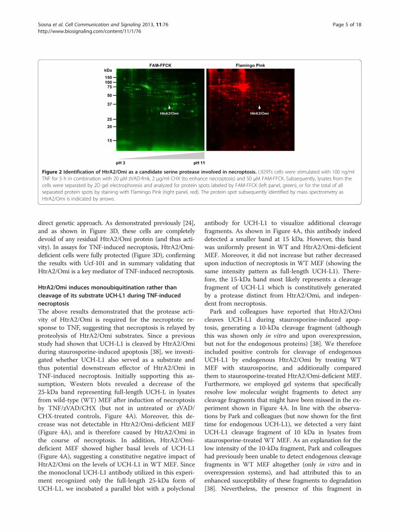
Figure 2 Identification of HtrA2/Omi as a candidate serine protease involved in necroptosis. L929Ts cells were stimulated with 100 ng/mlTNF for 5 h in combination with 20 μM zVAD-fmk, 2 μg/ml CHX (to enhance necroptosis) and 50 μM FAM-FFCK. Subsequently, lysates from thecells were separated by 2D gel electrophoresis and analyzed for protein spots labeled by FAM-FFCK (left panel, green), or for the total of allseparated protein spots by staining with Flamingo Pink (right panel, red). The protein spot subsequently identified by mass spectrometry asHtrA2/Omi is indicated by arrows.
Sosna et al. Cell Communication and Signaling 2013, 11:76 Page 5 of 18http://www.biosignaling.com/content/11/1/76
direct genetic approach. As demonstrated previously [24],and as shown in Figure 3D, these cells are completelydevoid of any residual HtrA2/Omi protein (and thus acti-vity). In assays for TNF-induced necroptosis, HtrA2/Omi-deficient cells were fully protected (Figure 3D), confirmingthe results with Ucf-101 and in summary validating thatHtrA2/Omi is a key mediator of TNF-induced necroptosis.
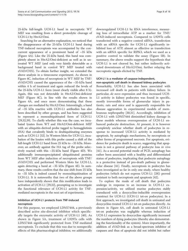
HtrA2/Omi induces monoubiquitination rather thancleavage of its substrate UCH-L1 during TNF-inducednecroptosisThe above results demonstrated that the protease acti-vity of HtrA2/Omi is required for the necroptotic re-sponse to TNF, suggesting that necroptosis is relayed byproteolysis of HtrA2/Omi substrates. Since a previousstudy had shown that UCH-L1 is cleaved by HtrA2/Omiduring staurosporine-induced apoptosis [38], we investi-gated whether UCH-L1 also served as a substrate andthus potential downstream effector of HtrA2/Omi inTNF-induced necroptosis. Initially supporting this as-sumption, Western blots revealed a decrease of the25-kDa band representing full-length UCH-L in lysatesfrom wild-type (WT) MEF after induction of necroptosisby TNF/zVAD/CHX (but not in untreated or zVAD/CHX-treated controls, Figure 4A). Moreover, this de-crease was not detectable in HtrA2/Omi-deficient MEF(Figure 4A), and is therefore caused by HtrA2/Omi inthe course of necroptosis. In addition, HtrA2/Omi-deficient MEF showed higher basal levels of UCH-L1(Figure 4A), suggesting a constitutive negative impact ofHtrA2/Omi on the levels of UCH-L1 in WT MEF. Sincethe monoclonal UCH-L1 antibody utilized in this experi-ment recognized only the full-length 25-kDa form ofUCH-L1, we incubated a parallel blot with a polyclonal
antibody for UCH-L1 to visualize additional cleavagefragments. As shown in Figure 4A, this antibody indeeddetected a smaller band at 15 kDa. However, this bandwas uniformly present in WT and HtrA2/Omi-deficientMEF. Moreover, it did not increase but rather decreasedupon induction of necroptosis in WT MEF (showing thesame intensity pattern as full-length UCH-L1). There-fore, the 15-kDa band most likely represents a cleavagefragment of UCH-L1 which is constitutively generatedby a protease distinct from HtrA2/Omi, and indepen-dent from necroptosis.Park and colleagues have reported that HtrA2/Omi
cleaves UCH-L1 during staurosporine-induced apop-tosis, generating a 10-kDa cleavage fragment (althoughthis was shown only in vitro and upon overexpression,but not for the endogenous proteins) [38]. We thereforeincluded positive controls for cleavage of endogenousUCH-L1 by endogenous HtrA2/Omi by treating WTMEF with staurosporine, and additionally comparedthem to staurosporine-treated HtrA2/Omi-deficient MEF.Furthermore, we employed gel systems that specificallyresolve low molecular weight fragments to detect anycleavage fragments that might have been missed in the ex-periment shown in Figure 4A. In line with the observa-tions by Park and colleagues (but now shown for the firsttime for endogenous UCH-L1), we detected a very faintUCH-L1 cleavage fragment of 10 kDa in lysates fromstaurosporine-treated WT MEF. As an explanation for thelow intensity of the 10-kDa fragment, Park and colleagueshad previously been unable to detect endogenous cleavagefragments in WT MEF altogether (only in vitro and inoverexpression systems), and had attributed this to anenhanced susceptibility of these fragments to degradation[38]. Nevertheless, the presence of this fragment in

Figure 3 HtrA2/Omi mediates TNF-induced necroptosis. A. Cells were left without, or pretreated for 2 h with 25 (Jurkat I42) or 50 μM(L929Ts, HT-29) of Ucf-101. Subsequently, cells were further incubated for 5 (L929Ts), 16 (HT-29) or 6 h (Jurkat I42) without or with addition of100 ng/ml TNF, 20 (L929Ts, HT-29) or 50 μM (Jurkat I42) zVAD-fmk and 5 μg/ml CHX (HT-29) before cell death was analyzed. ***p < 0.001.Micrographs show the morphology of untreated vs. necroptotic cells vs. L929Ts cells protected by Ucf-101. Scale bar: 100 μm. B. L929Ts cellswere left untreated or treated with TNF/zVAD with or without Ucf-101 as in A with addition of 50 μM TPCK or not and analyzed as inA. C. Cells were transfected with siRNAs specific for murine (L929Ts) or human HtrA2/Omi (Jurkat I42), or a negative control siRNA (siCtr). After 48or 72 h, cells were treated with 100 ng/ml TNF and 20 (L929Ts) or 50 μM (Jurkat I42) zVAD-fmk for another 5 (L929Ts) or 6 h (Jurkat I42) beforethe decrease of intracellular ATP levels was determined as a marker for cell death. Control Western blots of transfected but untreated cells wereperformed to verify downregulation of endogenous murine or human HtrA2/Omi. Detection of actin served as a loading control. D. Upper panel:wild-type (WT) and HtrA2/Omi-deficient MEF were stimulated with 100 ng/ml TNF, 20 μM zVAD-fmk and 1 μg/ml CHX for 16 h before cell deathwas determined. ***p < 0.001. Control Western blots show the presence or absence of murine HtrA2/Omi. Detection of actin served as a loadingcontrol. Lower panel: micrographs show the morphology of untreated and TNF/zVAD/CHX-treated WT vs. HtrA2/Omi-deficient MEF.Scale bar: 100 μm.
Sosna et al. Cell Communication and Signaling 2013, 11:76 Page 6 of 18http://www.biosignaling.com/content/11/1/76

Figure 4 HtrA2/Omi induces monoubiquitination rather than cleavage of its substrate UCH-L1 during TNF-induced necroptosis.A. Wild-type (WT) and HtrA2/Omi-deficient MEF were left untreated or stimulated for 16 h with 20 μM zVAD-fmk and 1 μg/ml CHX in thepresence (to induce necroptosis) or absence (for control) of 100 ng/ml TNF before UCH-L1 was detected with a monoclonal antibody thatrecognizes the full-length 25-kDa form of UCH-L1 (mAB UCH-L1) or, on a parallel blot, with a polyclonal antibody to detect all cleavage fragmentsof UCH-L1 (pAb UCH-L1). An asterisk marks the 35-kDa band corresponding to the predicted size of monoubiquitinated UCH-L1. B. WT andHtrA2/Omi-deficient MEF were stimulated as in A, and additionally with 0.5 μM staurosporine for 8 h. Lysates were separated on 10–20% w/vTris-Tricine gels (Biorad) to resolve low molecular weight proteins and immunoblotted with pAb UCH-L1. The blot was deliberately overexposedto visualize faint cleavage fragments. The 10-kDa UCH-L1 cleavage fragment generated by HtrA2/Omi during staurosporine-induced apoptosis isindicated (arrow, red box). C. WT and HtrA2/Omi-deficient MEF were treated with TNF/zVAD/CHX as in A for the indicated times and analyzed forproteins reactive with pAb UCH-L1 by Western blot. An asterisk marks the appearance of the 35-kDa band identical to the predicted size ofmonoubiquitinated UCH-L1. D. Lysates from WT and HtrA2/Omi-deficient MEF were incubated with 20 μM of an HA-tagged ubiquitin-derivedprobe (HAUbVME) in 50 mM Tris, 150 mM NaCl, pH 8.0 for 90 min at 37°C and subsequently analyzed by immunoblotting with HA antibody andreanalyzed with pAB UCH-L1. In panels A-D, detection of actin served as a loading control. E. An immunoprecipitation was performed usinglysates from necroptotic WT MEF (treated with TNF/zVAD/CHX as in A) and an antibody for ubiquitin. Subsequently, UCH-L1 was detected byWestern blot using pAb UCH-L1.
Sosna et al. Cell Communication and Signaling 2013, 11:76 Page 7 of 18http://www.biosignaling.com/content/11/1/76
staurosporine-treated WT but not in HtrA2/Omi-defi-cient MEF (Figure 4B) confirmed that UCH-L1 is cleavedby HtrA2/Omi in staurosporine-induced apoptosis. Incontrast, the 10-kDa fragment was clearly absent in all
lysates from both WTand HtrA2/Omi-deficient MEF ana-lyzed for TNF-induced necroptosis as well as the accom-panying controls (Figure 4B). Given these results, weconsidered it unlikely that the observed decrease of the
Sosna et al. Cell Communication and Signaling 2013, 11:76 Page 8 of 18http://www.biosignaling.com/content/11/1/76
25-kDa full-length UCH-L1 band in necroptotic WTMEF was resulting from a direct proteolytic cleavage ofUCH-L1 by HtrA2/Omi.Searching for an alternative explanation, we noticed that
the disappearance of the 25-kDa UCH-L1 band duringTNF-induced necroptosis was accompanied by the con-current appearance of a prominent band of ~35 kDa(Figure 4A). Like the 25-kDa band, this band was com-pletely absent in HtrA2/Omi-deficient as well as in un-treated WT MEF (and only very faintly detectable as abackground band in control WT MEF treated withzVAD/CHX). To obtain further insight, we extended theabove analysis in a timecourse experiment. As shown inFigure 4C, induction of necroptosis in WT MEF by TNF/zVAD/CHX caused the appearance of the ~35-kDa bandwithin 4 h of treatment and again reduced the levels ofthe 25-kDa UCH-L1 form (most clearly visible after 8 h).Again, this was not detectable in HtrA2/Omi-deficientMEF (Figure 4C), in line with the results shown inFigure 4A, and once more demonstrating that thesechanges are mediated by HtrA2/Omi. Interestingly, a bandof ~35 kDa reactive with UCH-L1 antibodies has alsobeen described by other groups, and has been suggestedto represent a monoubiquitinated form of UCH-L1[29,32,39]. To clarify whether this was the case, we incu-bated lysates from WT and HtrA2/Omi-deficient MEFwith an ubiquitin-derived probe tagged to hemagglutinin(HA) that covalently binds to deubiquitinating enzymessuch as UCH-L1 [32]. In Western blots for UCH-L1, incu-bation of the lysates with this probe caused a shift of thefull-length UCH-L1 band from 25 kDa to ~35 kDa. More-over, an antibody against the HA tag of the probe selec-tively reacted with this ~35-kDa band (Figure 4D). Weadditionally immunoprecipitated ubiquitinated proteinsfrom WT MEF after induction of necroptosis with TNF/zVAD/CHX and performed Western blots for UCH-L1,again detecting a band at ~35 kDa (Figure 4E). In sum-mary, these results confirm that the size shift from 25 kDato ~35 kDa is indeed caused by monoubiquitination ofUCH-L1. It is noteworthy that two of the above groupshave independently shown that this modification leads toactivation of UCH-L1 [29,32], prompting us to investigatethe functional relevance of UCH-L1 activity for TNF-mediated necroptosis in the next set of experiments.
Inhibition of UCH-L1 protects from TNF-inducednecroptosisFor this purpose, we employed LDN57444, a previouslydescribed active site-directed inhibitor which specific-ally targets the enzymatic activity of UCH-L1 [40]. Asshown in Figure 5A, treatment of L929Ts cells withLDN57444 significantly protected from TNF-mediatednecroptosis. To exclude that this was due to nonspecificeffects of this pharmacological inhibitor, we additionally
downregulated UCH-L1 by RNA interference, measur-ing loss of intracellular ATP as a marker for TNF/zVAD-induced necroptosis. Compared to L929Ts cellstransfected with a negative control siRNA, transfectionwith an siRNA specific for UCH-L1 significantly in-hibited loss of ATP, almost as effective as transfectionwith an siRNA specific for RIPK3, which we used as apositive control to validate the assay (Figure 5B). Insummary, the above results support the hypothesis thatUCH-L1 is not cleaved by, but rather indirectly acti-vated downstream of HtrA2/Omi, further relaying thenecroptotic signals elicited by TNF.
UCH-L1 is a mediator of caspase-independent,non-apoptotic cell death in diseased kidney podocytesRemarkably, UCH-L1 has also been associated withincreased cell death in patients with kidney failure. Inparticular, de novo expression and thus increased UCH-L1 activity in kidney podocytes was found in specific,mostly irreversible forms of glomerular injury in pa-tients, rats and mice and is apparently responsible fordisease aggravation in experimental models of mem-branous nephropathy [30,31]. Accordingly, inhibition ofUCH-L1 with LDN57444 diminished kidney damage inthese models whereas overexpression of UCH-L1 en-hanced podocyte destruction. At present, it is howevercompletely unclear whether death of podocytes in re-sponse to increased UCH-L1 activity is mediated byapoptosis, by autophagic mechanisms, by necroptosis orother forms of programmed necrosis. For apoptosis, evi-dence for podocyte death is scarce, suggesting that apop-tosis is not a general pathway of podocyte loss in vivo[41]. As a second potential mode of PCD, autophagy hasrather been associated with a healthy and differentiatedstatus of podocytes, implicating that podocyte autophagyis a protective instead of pro-death pathway in glome-rular disease [41]. Finally, necroptosis in podocytes hasbeen investigated so far in only one study, where healthypodocytes (which do not express UCH-L1 [28]) provedresistant to both necroptosis and apoptosis [42].To explore the mode of cell death that podocytes
undergo in response to an increase in UCH-L1 ex-pression/activity, we utilized murine podocytes stablytransduced with a doxycycline-inducible overexpressionconstruct for UCH-L1 (UCH-L1 tet-on podocytes). In afirst approach, we investigated cell death in untreated anddoxycycline-treated UCH-L1 tet-on podocytes directly. Asshown in Figure 6A, cell death in untreated UCH-L1tet-on podocytes was negligible whereas induction ofUCH-L1 expression by doxycycline significantly increasedthe numbers of dying podocytes (thereby also demonstrat-ing the functionality of the system). More importantly, theaddition of zVAD-fmk as a broad-spectrum inhibitor ofcaspases and thus of apoptosis did not inhibit but rather

Figure 5 Inhibition of UCH-L1 protects from TNF-induced necroptosis. A. L929Ts cells were prestimulated for 3 h with 50 μM of the UCH-L1inhibitor LDN57444 or left unstimulated before addition of 100 ng/ml TNF and 20 μM zVAD-fmk for 5 h. Subsequently, cell death was analyzedby PI staining and flow cytometry. Asterisks indicate statistical significance (t-test), ***p < 0.001. Micrographs additionally show the morphology ofuntreated L929Ts cells vs. necroptotic cells vs. cells protected by LDN57444. Scale bar: 100 μm. B. L929Ts cells were transfected with an siRNAthat does not elicit an RNAi response (negative control, siCtr), with an siRNA specific for murine UCH-L1, and with an siRNA specific for murineRIPK3 (positive control for protection from necroptosis, siRIPK3) as described in “Methods.” 48 h after transfection, cells were treated with100 ng/ml TNF and 20 μM zVAD-fmk for another 5 h before the decrease of intracellular ATP levels was determined as a marker for cell death.ATP levels are shown relative to controls that were not treated with TNF/zVAD. Asterisks indicate statistical significance (t-test), **p < 0.01,***p < 0.001.
Sosna et al. Cell Communication and Signaling 2013, 11:76 Page 9 of 18http://www.biosignaling.com/content/11/1/76
enhanced UCH-L1-dependent cell death. We and othershave previously observed this effect of zVAD-fmk innecroptosis [14,33,43], excluding that de novo expressionand thus increased UCH-L1 activity causes death ofpodocytes by apoptosis but rather pointing to pro-grammed necrosis/necroptosis as the responsible suicideprogram.To extend these results, we investigated cleavage of
PARP-1, a DNA-associating repair enzyme which isinactivated in apoptosis by caspase-3-dependent proces-sing of the mature 116-kDa protein to an 89-kDa clea-vage product [44]. When we analyzed lysates fromUCH-L1 tet-on podocytes treated with doxycycline for72 h or not in Western blots, the full-length 116-kDaPARP-1 band was uniformly visible in all samples, to-gether with a pattern of additional bands. However, thispattern did not change upon treatment with doxycycline(Figure 6B). In particular, the characteristic disappea-rance of the full-length 116-kDa PARP-1 band as well asthe corresponding increase of the 89-kDa cleavage frag-ment that we have previously observed for apoptosis in
multiple studies [13,15,33], and which is also shown forcontrol in L929Ts cells (Figure 6B) could not be de-tected. Given that caspase-3 acts downstream of allother apoptotic caspases as the central effector caspaseof both extrinsic and intrinsic apoptosis, these resultsprovided a second line of evidence that caspase activa-tion and thus apoptosis seems not to occur duringUCH-L1-mediated death of kidney podocytes.To address this point in more detail, we directly mea-
sured the activity of caspase-3 and caspase-8 (as the majorinitiator caspase activated by death receptors). As shownin Figure 6C, no increase in caspase-3 or caspase-8 activitybeyond the already present basal levels was detectable indoxycycline-treated (i.e. UCH-L1-overexpressing, WesternBlot in Figure 6C) vs. untreated UCH-L1 tet-on podocytesor vs. negative controls (doxycycline-treated podocytesthat are stably transduced with empty vector; tet-podocytes). In contrast, the activity of both caspaseswas clearly increased in positive control lysates fromdoxycycline-treated tet- podocytes treated with cyto-chrome c and dATP to validate the assay, in summary
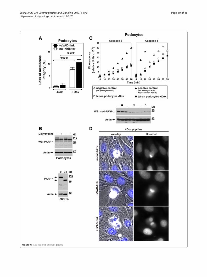
Figure 6 (See legend on next page.)
Sosna et al. Cell Communication and Signaling 2013, 11:76 Page 10 of 18http://www.biosignaling.com/content/11/1/76

(See figure on previous page.)Figure 6 UCH-L1 as a mediator of caspase-independent, non-apoptotic cell death in diseased kidney podocytes. A. UCH-L1 tet-onpodocytes were treated with 20 ng/ml doxycycline for 72 hours (+Dox) or not (−Dox) and 50 μM zVAD-fmk or no inhibitor. Cell death wasmeasured by trypan blue staining. ***p < 0.001. B. UCH-L1 tet-on podocytes were left untreated (−) or treated with doxycycline as in A (+) beforePARP-1-reactive bands were detected by immunoblotting. Cell lysates from untreated and apoptotic L929Ts cells (Co, treated with 100 ng/ml TNFand 2 μg/ml CHX for 1 h) are shown as controls. Full-length and cleaved PARP-1 is marked by arrows. Detection of actin served as a loadingcontrol. C. Aliquots from the stimulations in B were also analyzed for caspase activity. As negative control, tet- podocytes were treated withdoxycycline as in A; as positive control, lysates from doxycyline-treated tet- podocytes were incubated with cytochrome c and dATP to activatecaspases. Subsequently, the activity of caspases-3 and -8 was determined by measuring the cleavage of fluorogenic substrates (zIETD-afc andzDEVD-afc) over 70 minutes. The Western blot below shows that UCH-L1 is indeed upregulated in doxycycline-treated but not untreated UCH-L1tet-on podocytes and also not in doxycycline-treated negative control tet- podocytes. Treatment with doxycycline was performed as inA. UCH-L1 was detected with mAb UCH-L1, detection of actin served as a loading control. D. Cell death was induced in UCH-L1 tet-on podocytesby treatment with doxycycline as in A in the presence of 50 μM zVAD-fmk (+zVAD-fmk, middle and lower micrographs) or no inhibitor(upper micrographs). Micrographs show the morphology of dying cells within a monolayer of healthy cells (overlay, nuclei are stained blue), andin parallel the nuclear morphology of the same cells after staining with Hoechst dye (Hoechst). Original magnification: x 400.
Sosna et al. Cell Communication and Signaling 2013, 11:76 Page 11 of 18http://www.biosignaling.com/content/11/1/76
further corroborating the assumption that UCH-L1-me-diated death of podocytes occurs without activation ofcaspases and thus in a non-apoptotic manner.Finally, when analyzed by microscopy, doxycycline-
treated UCH-L1 tet-on podocytes did not display typicalapoptotic changes such as membrane blebbing, type 2chromatin condensation and accumulation of frag-mented chromatin at the nuclear periphery which wehad earlier observed for apoptosis in other cell systems[33]. Rather, only an incomplete, lumpy condensation ofchromatin was detectable that has previously been as-sociated with programmed necrosis/necroptosis ratherthan apoptosis [16]. Moreover, and as shown above forcell death, the addition of zVAD-fmk did not affect thechanges in the cellular and nuclear morphology ofpodocytes caused by doxycycline-induced overexpressionof UCH-L1 (Figure 6D). Altogether, these results ruleout caspase-dependent apoptosis but rather favor caspase-independent, non-apoptotic forms of cell death such asprogrammed necrosis or necroptosis as the most probablecause for UCH-L1-mediated podocyte death.
Inhibition of UCH-L1 protects podocytes fromTNF-induced necroptosisAs a central proinflammatory cytokine, TNF may alsocontribute to inflammatory reactions in the kidney andthus to subsequent podocyte injury. We thereforewanted to determine whether UCH-L1 can act as a me-diator of TNF-induced necroptosis not only in L929Tscells (as shown in Figure 5), but also in podocytes. Forthis purpose, we analyzed podocytes stably transfectedwith an shRNA construct that causes permanent knock-down of UCH-L1 or with a scrambled negative controlshRNA [30]. As shown in Figure 7, podocytes with stabledownregulation of UCH-L1 were significantly protectedfrom TNF-induced cell death when compared to controlpodocytes. Moreover, and identical to podocyte deathcaused by UCH-L1 overexpression (Figure 6A), the
addition of zVAD-fmk did not prevent TNF-induced celldeath, demonstrating that TNF indeed elicits necroptosisin podocytes, and that UCH-L1 represents a down-stream mediator of the necroptotic signaling cascade ofTNF also in podocytes.
DiscussionThe impact of caspase-independent, non-apoptotic PCDsuch as necroptosis/programmed necrosis has become in-creasingly clear in the last years. This is particularly truefor pathological processes, for example renal [42], cardiacand retinal ischemia/reperfusion injury, hyperacute shock[45], brain damage or pancreatitis [12], Huntington’s,Parkinson’s and Alzheimer’s disease, epilepsy, musculardystrophy, as well as for the destruction of cells by patho-gens such as vaccinia virus, HIV, Shigella and Salmonella[2,12,46,47]. The option to therapeutically interfere withnecroptosis/programmed necrosis has raised great ex-pectations [12]. In consequence, a better knowledge of thestill incompletely understood signaling pathways andthe associated components will facilitate future stra-tegies to interfere with damage induced by necroptosis/programmed necrosis (e.g. in shock, stroke, myocardialinfarction or kidney failure). Here, we have identifiedthe proteases HtrA2/Omi and UCH-L1 as two suchcomponents of TNF-induced necroptosis, and thus re-vealed two novel targets for therapeutic intervention,e.g. by future Ucf-101- or LDN57444-derived drugssuited for use in patients.Based upon the results of our study, we propose the
model shown in Figure 8 to integrate HtrA2/Omi andUCH-L1 into the known signaling pathways of TNF-induced necroptosis. In this model, binding of TNF toTNF-R1 induces activation of the kinases RIPK1, RIPK3,and of MLKL as components of the necrosomal corecomplex. Notably, we have been unable to detectHtrA2/Omi as part of the necroptotic TNF-R1 signalingcomplex in preliminary experiments (D. A. and S. S.,

Figure 7 Inhibition of UCH-L1 protects podocytes fromTNF-induced necroptosis. Podocytes stably transfected with anshRNA construct that causes permanent knockdown of UCH-L1(shUCH-L1) or with a scrambled negative control shRNA (shCtr) weretreated with 100 ng/ml TNF in the presence of 50 μM zVAD-fmk orvehicle for 3 h before loss of membrane integrity as a marker for celldeath was measured by trypan blue staining. Asterisks indicatestatistical significance (t-test), *p < 0.05. The Western blot below wasperformed to demonstrate the permanent knockdown of UCH-L1 inshUCH-L1 podocytes, but not in shCtr podocytes. UCH-L1 wasdetected with mAb UCH-L1, detection of actin served as a loadingcontrol. For each stable transfectant, lysates from four independentflasks were analyzed.
Figure 8 HtrA2/Omi and UCH-L1 as novel components ofTNF-induced necroptosis. The scheme depicts the proposed rolesof HtrA2/Omi and UCH-L1 in TNF-induced necroptosis. Binding ofTNF to TNF-R1 triggers activation of the necrosomal core complexconsisting of RIPK1, RIPK3 and MLKL. Subsequently, the proteinsPGAM5L/S and Drp-1 form the mitochondrial attack complex,resulting in the intramitochondrial activation of HtrA2/Omi.Activated HtrA2/Omi then (by cleavage of yet unidentifiedintramitochondrial substrates) indirectly causes monoubiquitinationand activation of UCH-L1, and finally, necroptosis. Accordingly,inhibition of HtrA2/Omi (Ucf-101, knockout) or UCH-L1 (LDN57444,siRNA) protects from necroptosis. Please see Discussion forfurther details.
Sosna et al. Cell Communication and Signaling 2013, 11:76 Page 12 of 18http://www.biosignaling.com/content/11/1/76
unpublished observations), and no other study has yetreported an association of HtrA2/Omi with componentsof the TNF-R1 signaling complex during necroptosis.This is also consistent with reports showing that, in con-trast to apoptosis, HtrA2/Omi is not released from mito-chondria during TNF-induced necroptosis [23,48]. Insummary, these findings argue against a direct inter-action of HtrA2/Omi with RIPK1, RIPK3 or MLKL butinstead suggest that HtrA2/Omi is activated indirectlywithin the mitochondria. As the most likely mechanism,MLKL has been found to activate the phosphatasesPGAM5L/S, which in turn couple to the mitochondrialprotein Drp-1, and as a mitochondrial attack complex[11], may cause the subsequent intramitochondrialactivation of HtrA2/Omi. Consistent with a function ofHtrA2/Omi in TNF-induced necroptosis despite this
intramitochondrial localization, inhibition of HtrA2/Omiactivity by Ucf-101 or by genetic deletion (knockout)blocks the necroptotic signal of TNF (this was similarlyobserved for Ucf-101 in an independent study in neutro-phils, where the authors also concluded that HtrA2/Omimediates necroptosis through its serine protease proper-ties from within the mitochondria [48]). Downstream ofHtrA2/Omi, our data identify UCH-L1 as another, novelcomponent of the signaling cascade. In contrast tostaurosporine-induced apoptosis, where HtrA2/Omitranslocates into the cytosol and directly cleaves andthus inactivates UCH-L1 [38], the intramitochondriallocalization of HtrA2/Omi during TNF-induced

Sosna et al. Cell Communication and Signaling 2013, 11:76 Page 13 of 18http://www.biosignaling.com/content/11/1/76
necroptosis prevents a direct interaction of both pro-teins. Rather, and also explaining why we did not see adirect cleavage (and thus inactivation) of UCH-L1 byHtrA2/Omi, HtrA2/Omi seems to act indirectly, by yetunknown mechanism (e.g. cleavage of unidentified in-tramitochondrial substrates), causing the monoubiqui-tination and activation of UCH-L1, finally resulting innecroptosis (which can accordingly be blocked viaLDN57444 or by RNA interference).As a side note, UCH-L1 belongs to the family of cyst-
eine proteases, and we wondered why the broad-spectrumcalpain/cysteine protease inhibitor E-64 did not conferany significant protection from TNF-induced necroptosisin the experiments performed in this study (Figure 1C) orin additional control experiments (D. A., J. S. and S. V.,unpublished observations). To the best of our knowledge,inhibition of UCH-L1 by E-64 has also not been shown inany other study. As a possible explanation, UCH-L1 is an“atypical” cysteine protease because its active site ismisaligned when compared to productive cysteine pro-teases [29]. Therefore, a general cysteine protease inhibi-tor such as E-64 may have only limited impact on theactivity of UCH-L1, in contrast to a specific inhibitor suchas LDN57444 or to inhibition of UCH-L1 by RNA inter-ference (which clearly protected L929Ts cells or podocytesfrom TNF-induced necroptosis, Figure 5A-B, Figure 7).We would also like to point out that HtrA2/Omi and
UCH-L1 obviously represent important, but most cer-tainly not the only factors transmitting the necroptoticdeath signals of TNF downstream of RIPK1/RIPK3/MLKL. Whereas HtrA2/Omi is expressed ubiquitously[23], the expression of UCH-L1 is restricted to certaintissues [28,30]. Therefore, in tissues that do not expressUCH-L1, necroptosis must be relayed by additional, in-dependent factors. Notably, the brain is an organ wherea rapid and efficient apoptotic elimination of cells isdangerous, and where alternative, caspase-independentforms of PCD predominate [16]. The brain is also theorgan with the highest expression of UCH-L1 in theentire body [32], suggesting that a deregulation of UCH-L1 activity in the brain may contribute to necroptoticdamage, e.g. after traumatic injury [31] or after stroke(i.e. ischemia/reperfusion damage). Interestingly, bothUCH-L1 as well as HtrA2/Omi have been associatedwith Parkinson’s disease, although a connection tonecroptosis has not been investigated so far. Moreover,recent studies have found that necroptosis is also thepredominant damage mechanism in ischemia/reper-fusion damage in the kidney [42,49], in summary indi-cating that both brain and kidney are organs wheretherapeutic strategies aiming to interfere with thenecroptotic actions of HtrA2/Omi and UCH-L1 may beworthwhile options to consider for the future, e.g. withregard to stroke or kidney failure.
ConclusionsWe have identified the proteases HtrA2/Omi and UCH-L1as two crucial components of TNF-induced necroptosis,and thus provided evidence that proteolysis is not onlycritical for the regulation and execution of apoptosis, butalso essential for caspase-independent forms of PCD. Amodel that integrates HtrA2/Omi and UCH-L1 into theknown signaling cascades of TNF-mediated necroptosis isshown in Figure 8. With HtrA2/Omi and UCH-L1, wehave also revealed two novel targets for therapeutic inter-vention, which may assist in developing strategies for thetreatment of damage induced by necroptosis/programmednecrosis (e.g. in organs such as the kidney and the brain,caused by stroke or kidney failure).
MethodsReagentsHighly purified human recombinant TNF was provided byBASF Bioresearch (Ludwigshafen, Germany). Benzylo-xycarbonyl-Val-Ala-Asp(OMe)-fluoromethylketone (zVAD)was from Bachem (Bubendorf, Switzerland). TPCK, ma-rimastat, benzyloxycarbonyl-Phe-Ala-fluoromethylketone(zFA-fmk) and trans-Epoxysuccinyl-L-leucylamido(4-gua-nidino)butane (E-64), were purchased from Sigma(Deisenhofen, Germany), necrostatin-1, TAPI-1, GM6001,5-[5-(2-nitrophenyl)furfurylidine]-1,3-diphenyl-2-thiobarbi-turic acid (Ucf-101), benzyloxycarbonyl-Phe-Phe-fluoro-methylketone (zFF-fmk) and LDN57444 from MerckMillipore (Darmstadt, Germany), and N-[L-3-trans-(pro-pylcarbamoyl)-oxirane-2-carbonyl]-L-Ile-L-Pro methyl ester(CA-074 Me) from Biomol (Hamburg, Germany). Car-boxyfluorescein-labeled phenylalanine chloromethyl ketone(FAM-FFCK) was from Immuno Chemistry Technologies(Bloomington, MN, USA). Staurosporine was obtainedfrom Selleckchem (Munich, Germany), Ubiquitin vinyl me-thyl ester, HA-tag (HaUbVME) from Enzo Life Sciences(Lausen, Switzerland).
Cell cultureL929Ts is a TRAIL-sensitive L929 subline derived in ourlaboratory [33]. NIH3T3 cells naturally expressing RIPK3and therefore sensitive to necroptosis have been pre-viously described [15,50,51]. Jurkat and HT-29 cells wereobtained from the American Type Culture Collection(ATCC, Manassas, VA, USA). Jurkat I42 cells were a kindgift from Francis Ka-Ming Chan (Worcester, MA, USA).Immortalized MEF deficient for HtrA2/Omi [24] and theirWT counterparts were originally generated by JulianDownward (London, U. K.) and kindly provided byThomas Langer (Cologne, Germany). Cells were culti-vated in DMEM (NIH3T3, MEF), or a mixture of Click’s/RPMI 1640 medium (all other cell lines) supplementedwith 10% v/v fetal calf serum and 2 mM glutamine at 37°Cin a humidified incubator containing 5% w/v CO2. Media

Sosna et al. Cell Communication and Signaling 2013, 11:76 Page 14 of 18http://www.biosignaling.com/content/11/1/76
were additionally supplemented with 1 mM sodium pyru-vate (HT-29) and 50 μg/ml each of streptomycin and peni-cillin. Murine podocytes (a kind gift from K. H. Endlich,Greifswald) were cultured as described [52]. For differenti-ation, podocytes were cultured for 14 days under non-permissive conditions (37°C, 7.4% w/v CO2, RPMI 1640supplemented with 10% v/v fetal calf serum, 10 mM N-2-hydroxyethylpiperazine-N0-2-ethanesulfonic acid, 1 mMsodium pyruvate, 100 U/ml penicillin, 100 mg/mlstreptomycin).
Flow cytometric analysis of membrane integrityCells were seeded in twelve-well plates at 5 x 10⁴ cells/well. Following treatment, both detached and adherentcells were collected by centrifugation. The cells wereresuspended in PBS/5 mM EDTA containing 2 μg/mlpropidium iodide (PI), and the red fluorescence wasmeasured on a FACSCalibur flow cytometer (BectonDickinson).
Statistical analysisp values were calculated using Student’s t-test. Statisticalsignificance is denoted by *p < 0.05, **p < 0.01, ***p < 0.001.
MicroscopyFor documentation of cell morphology, images fromunfixed cells were obtained using an Axiovert 10 micro-scope (Zeiss, Oberkochen, Germany) and a DS-5M-L1digital sight camera system (Nikon, Düsseldorf, Germany).
2D gel electrophoresis, image analysis and spot pickingThe two-dimensional gel electrophoresis was essentiallyperformed as described before [53]. After harvesting, cellswere lysed on ice for 10 min in TNE buffer (50 mM TrispH 8.0, 1% v/v NP40, 2 mM EDTA) containing 10 μg/mlprotease inhibitor cocktail (Roche, Mannheim, Germany).For protein precipitation, trichloroacetic acid (TCA) wasadded to the protein lysate to a final concentration of10% v/v . The mixture was incubated for 30 min on iceand centrifuged at 10,000 × g at 4°C for 20 min. Thesupernatant was removed, ice-cold acetone was added towash the pellet and the sample was centrifuged as above.After removal of the supernatant, the pellet was air driedand resuspended in lysis buffer (pH 8.5) containing 7 Murea, 2 M thiourea, 30 mM Tris, 4% w/v CHAPS. Thesupernatant containing the solubilized proteins was reco-vered after centrifugation for 20 min at 20,000 × g at 4°C.A total amount of 250 μg of protein was mixed with re-hydration buffer (7 M urea, 2 M thiourea, 4% w/vCHAPS, 2% v/v immobilized pH gradient (IPG) bufferpH 3–11 and 2% w/v DTT) and applied by cup-loadingonto 24 cm non-linear pH 3–11 IPG gel strips forisoelectric focusing (IEF). The second dimension wasperformed on 26 × 20 cm large 12.5% w/v gels after
reduction and alkylation using the Ettan DALTsix largevertical electrophoresis system from GE Healthcare(Munich, Germany). The gels were removed from the glassplates, mounted on a non-backed gel frame, and scannedon a Typhoon Trio imager (GE Healthcare) at green fluo-rescence. Subsequently, the gels were stained overnightwith Flamingo Pink (Bio-Rad, Munich, Germany), andscanned again at red fluorescence. The obtained imageswere analyzed using Image Master 6.0 (GE Healthcare).Selected spots were picked with a 2 mm picking head. Thepicked gels were again scanned to verify the correct loca-tion of the punched spots.
In-gel tryptic digestion and mass spectrometryThe punched gel spots were sequentially washed withwater, with 50 mM ammonium bicarbonate (ABC) in50% v/v MeOH, with 70% v/v acetonitrile (ACN), anddehydrated in pure ACN. ACN was evaporated in aSpeedVac centrifuge (ThermoFisher Scientific, Dreieich,Germany), and the dry gel pieces were subjected to in-geldigestion with 100 ng porcine sequencing-grade trypsin(Serva, Heidelberg, Germany) in 25 mM ABC at 37°Covernight. For peptide extraction, 20 μl of 0.1% v/vtrifluoroacetic acid (TFA) in ACN was added and the sam-ples were sonicated for 15 min. The supernatants were re-moved and the gel spots were again incubated with 20 μlof 0.1% v/v TFA in ACN for 10 min. The supernatants ofboth steps were combined, dried in a SpeedVac centrifuge,redissolved in 0.8 μl MALDI matrix solution (3.2 mg/mlα-cyanohydroxycinnamic acid (Sigma) in 65% v/v ACN/0.1% v/v TFA), spotted onto 384-well stainless steelMALDI plates and air-dried. Spectra were acquired on anAB SCIEX MALDI-TOF/TOF 5800 (AB SCIEX, Darm-stadt, Germany) mass spectrometer in positive ion mode.For MS measurements, 2000 shots were accumulated inthe mass range of 800–4000 m/z. Default calibration wasperformed using the 4700 Proteomics Analyzer Stan-dards Kit, while MS measurements were calibrated in-ternally using trypsin and contaminant peaks (842.509,2211.105, 2225.120 and 2807.315 Da). Precursor selec-tion for MS/MS analysis was achieved using the 4000Series Explorer Software (AB SCIEX) with acquisition ofthe 20 most intense precursors (S/N > 20), beginningwith the strongest first. All MS/MS spectra were ac-quired with 1 KV collision energy at ambient air (CIDmedium: 1.25 x 10⁻⁶ Torr) using 3000 laser shots. Forpeptide identification, MALDI-TOF/TOF MS/MS rawfiles were searched using ABSciex GPS software (Version3.6, build 332) with the following pre-filter settings: onlypeaks within a mass range from 60 Da to the precursormass minus 35 Da and S/N ratio above 10 were used.Spectra were searched with Mascot (version 2.2.04, MatrixScience, London, U.K) against the Swissprot databaseusing Mus musculus as a taxonomy filter (15 Feb 2011,
Sosna et al. Cell Communication and Signaling 2013, 11:76 Page 15 of 18http://www.biosignaling.com/content/11/1/76
16345 sequences) and the following parameters: precursortolerance, 50 ppm; MSMS tol, 0.3 Da; max missed cleav-ages 2. Oxidation (M) was set as a variable modification,while carbamidiomethylation (C) was set as a fixed modi-fication. Proteins were considered identified when either 2peptides were identified with a confidence interval ≥ 99%(p < 0.01) or 3 peptides ≥ 95% (p < 0.05).
RNA interferenceThe validated siRNA specific for human HtrA2/Omi(ID # s654), the predesigned siRNAs specific for murineHtrA2/Omi (ID # s82292, s82292) murine UCH-L1(ID # s75710), murine RIPK3 (ID # s80755) as well asthe negative control siRNA (ID # AM4611) were ob-tained from Life Technologies, Darmstadt, Germany.L929Ts cells were transfected with 150 pmol siRNA byAmaxa nucleofection (Lonza, Cologne, Germany), usingsolution V and program T-20. Jurkat I42 cells weretransfected with 30 pmol siRNA and HiPerFect transfec-tion reagent (Qiagen, Hilden, Germany).
Measurement of intracellular ATP levelsThe intracellular ATP content of cells was determinedwith the Cell Titer Glo Assay Kit (Promega, Mannheim,Germany) following the instructions of the manufacturer.
ImmunoblotsUnless otherwise indicated, cells were harvested aftertreatment and lysed at 4°C in TNE buffer containing150 mM NaCl, 10 μg/ml protease inhibitor cocktail,1 mM sodium orthovanadate and 5 mM NaF. Identicalamounts of cell protein per lane were resolved by electro-phoresis on SDS polyacrylamide gels. After electrophor-etic transfer to nitrocellulose, reactive proteins weredetected using antisera specific for actin (sc-1615, SantaCruz, Heidelberg, Germany; A1978, Sigma), HtrA2/Omi(ab32092, Abcam, Cambridge, UK), UCH-L1 (rat mono-clonal, clone U104, generated as outlined below, or rabbitpolyclonal, CL95101, Cedarlane, Burlington, Ontario,Canada), HA (1187423, Roche), PARP-1 (9542, CellSignaling, Danvers, MA, USA) and the ECL detection kit(GE Healthcare). Equal loading as well as efficiency oftransfer was routinely verified for all Western blots byPonceau S staining, and by reprobing the membranes foractin.
Generation of monoclonal UCH-L1 antibodiesWistar rats were initially immunized intraperitoneally(i.p.) with 100 μg of purified UCH-L1 (kindly providedby Gregory A. Petsko, Waltham, MA, USA) in 60 μlphosphate buffer saline (PBS) emulsified with 40 μl ofGerbu adjuvant MM (Gerbu Biotechnik, Heidelberg,Germany). The rats were boosted i.p. on days 14 and 21with 50 μg of purified protein emulsified with 20% v/v of
the adjuvant. The last two doses (50 μg UCH-L1 in PBS)were administered on days 28 and 29 without adjuvant,while the fusion was done on day 30. Spleen cells fromimmunized animals were collected and fused withAg8.653 myeloma cells using polyethylene glycol 1500(Roche). The fused cells were cultured in selectionmedium (HAT, Sigma) for 10 days and screened byELISA for anti-UCH-L1 antibodies. Hybridoma clonesproducing anti-UCH-L1 monoclonal antibodies (mAbs)were then cultivated in serum-free medium and themAbs were purified using protein G affinity chromato-graphy (GE Healthcare). The isotype of the anti-UCH-L(U104) clone (IgG1, λ) was determined by using ELISArat mAb isotyping kit (ThermoFisher).
ImmunoprecipitationsCellular lysates were precleared with GammaBind G-sepharose (GE Healthcare) and immunoprecipitation wasperformed over night on ice using anti-ubiquitin IgG1monoclonal antibody (MAB1510, Merck Millipore, 1:100dilution). After collection of the immunecomplexes withGammaBind G-sepharose and three washing steps in lysisbuffer, the immunoprecipitated proteins were analyzed bySDS-PAGE and Western blot.
Generation of stably transfected podocytes with inducibleoverexpression or downregulation of UCH-L1For inducible overexpression of UCH-L1, the Retro-XTet-On Advanced Inducible Expression System (Clontech,Mountain View, CA, USA) was used according to themanufacturers’ instructions. Briefly, wildtype murineUCH-L1 was amplified by polymerase chain reaction frommurine podocytes using the following primers: mUCHL1-pRetro-fw 5′CTAGGCGGCCGCGCCACCATGCAGCTGAAGCCGATGGA′3; mUCHL1-pRetro-rev 5′CTAGACGCGTTTAAGCTGCTTTGCAGAGAG′3 and subse-quently cloned into the multiple cloning site of thepRetroX-Tight-Pur vector using NotI and MluI (Thermo-Fisher). The sequence of UCH-L1 was verified by sequen-cing (Eurofins MWG Operon, Ebersberg, Germany). Forvirus production, phoenix ecotropic packaging cells weretransfected using DNA/CaCl2 precipitation with thepRetroX-Tet-On Advanced vector, with the pRetroX-Tight-Pur-UCH-L1 vector or the pRetroX-TightPur emptyvector as a control, respectively. The virus-containingsupernatant of the pRetroX-Tet-On transfected phoenixcells was transferred to a 10 cm plate containing podocytetarget cells at around 50% to 60% confluence; the infectionsteps were repeated twice. Selection for integration of thepRetroX-Tet-On Advanced expression plasmid was per-formed with G418 (500 μg/ml, Life Technologies) for7 days. Afterwards, the virus-containing supernatant of thepRetroX-Tight-Pur-UCH-L1 transfected phoenix cells wastransferred to the pRetroX-Tet-On Advanced transduced

Sosna et al. Cell Communication and Signaling 2013, 11:76 Page 16 of 18http://www.biosignaling.com/content/11/1/76
podocyte target cells; the infection steps were againrepeated twice. Selection for integration of the pRetroX-Tight-Pur-UCH-L1 plasmid was performed with pu-romycin (1.5 μg/ml, Sigma). For negative controlexperiments, the pRetroX-Tight-Pur vector was trans-duced without insert (tet-) into the pRetroX-Tet-OnAdvanced expressing podocytes. For induction of UCH-L1overexpression, UCH-L1 tet-on or tet- podocytes werecultured in the presence of tetracycline free medium (PAN-Biotech, Aidenbach, Germany) supplemented with 20 ng/mldoxycycline or without doxycycline for control. For stableknockdown experiments, shRNA627 to murine UCH-L1or scrambled shRNA for control was overexpressed inpodocytes as described before [30].
Analysis of caspase activity, cell death, and cellular andnuclear morphology in podocytes105 differentiated UCH-L1 tet-on or tet- podocytes wereplated in 6-well plates in tetracycline-free RPMI 1640medium (Life Technologies) supplemented with 10% v/vfetal calf serum, 10 mM N-2-hydroxyethylpiperazine-N0-2-ethanesulfonic acid, 1 mM sodium pyruvate, 100 U/mlpenicillin and 100 mg/ml streptomycin. UCH-L1 over-expression was induced with 20 ng/ml doxycycline for72 hours or not. For measurements of caspase activity,cells were collected and lysed in a buffer containing10 mM Hepes pH 7.4, 142 mM KCl, 5 mM MgCl2, 1 mMEGTA, 0.2% v/v NP40, 1 mM DTT and 2 mM Pefabloc(Roche). To generate positive controls, 20 μg of cells lysatewere equilibrated for 1 h at 30°C after the addition of1 mM dATP and 10 μM cytochrome c to permit activa-tion of caspases. Subsequently, 100 μl of caspase buffer(20 mM Pipes, 100 mM NaCl, 10 mM DTT, 1 mM EDTA,0.1% w/v CHAPS, 10% w/v sucrose, pH 7.2) containing100 μM zDEVD-afc (benzyloxycarbonyl-Asp(OMe)-Glu(OMe)-Val-DL-Asp(OMe)-7-aminotrifluoromethylcouma-rin, Merck Millipore) or zIETD-afc benzyloxycarbonyl-Ile-Glu(OMe)-Thr-DL-Asp(OMe)-7-aminotrifluoromethylcoumarin (Merck Millipore) were added to 10 μl of cyto-solic extract (10 μg protein) and incubated at 37°C. Therelease of afc was measured as emission at 505 nm uponexcitation at 405 nm using an Infinite M200 fluorime-ter equipped with a thermostated plate reader (Tecan,Crailsheim, Germany). For measurements of podocytedeath, viable and dead cells were detached with trypsinand counted in a Neubauer chamber after 0.1% w/vtrypan blue (Life Technologies) staining. The percen-tage of dead cells was calculated and plotted as mean +/−SEM, n = 12 per condition. To analyze cellular and nu-clear morphology, cells were stained with Hoechst dye(10 μg/ml, Life Technologies) for 5 min and DNA conden-sation in UCH-L1 tet-on podocytes with or without in-duced UCH-L1 overexpression for 72 hours was evaluated
under an Axio Observer A1 microscope (Zeiss) using theaxiovision software (Zeiss).
Analysis of TNF-induced cell death in podocytesDifferentiated sh627 and scrambled shRNA control po-docytes were plated at a density of 104 cells per 6-wellplate. After 48 hours, cells were treated with 100 ng/mlmurine TNF (PeproTech, Hamburg, Germany) with ad-dition of 50 μM zVAD-fmk or vehicle (ethanol) as con-trol for 3 hours. Cells were detached with trypsin andthe amount of dead and living cells was counted in aNeubauer chamber following staining with 0.1% w/v try-pan blue. The percentage of dead cells was calculatedand plotted as mean +/− SEM, n = 12 per condition.
Competing interestsThe authors declare that they have no competing interests.
Authors’ contributionsJS, SV, DK, OJ, CMS, SS and DA designed research; JS, SV, SM, AT and CMSperformed research; JS, SV, DK, AT, OJ, CMS, SS and DA analyzed data, DAwrote the manuscript. All authors read and approved the final manuscript.
AcknowledgementsWe thank Parvin Davarnia for excellent technical assistance, MelanieNebendahl for help with 2D gel electrophoresis, and the Z2-project of theSFB 877 (Bart van den Berg, Tomas Koudelka and Andreas Tholey) forperforming mass spectrometry and protein identification. This work wassupported by grants from the Deutsche Forschungsgemeinschaft (SFB 877,project B2, to D. A. and S. S., and projects Z2, Z3) and by a fellowship fromthe German Academic Exchange Service (DAAD, A/08/79433, to J. S.).
Author details1Institut für Immunologie, Christian-Albrechts-Universität zu Kiel,Michaelisstr. 5, 24105 Kiel, Germany. 2Biochemisches Institut,Christian-Albrechts-Universität zu Kiel, Olshausenstr. 40, 24098 Kiel,Germany. 3Zentrum für Innere Medizin, Nephrologie, UniversitätsklinikumHamburg-Eppendorf, Martinistr. 52, 20246 Hamburg, Germany.
Received: 19 July 2013 Accepted: 1 October 2013Published: 3 October 2013
References1. Degterev A, Yuan J: Expansion and evolution of cell death programmes.
Nat Rev Mol Cell Biol 2008, 9:378–390.2. Festjens N, Vanden Berghe T, Vandenabeele P: Necrosis, a
well-orchestrated form of cell demise: signalling cascades, importantmediators and concomitant immune response. Biochim Biophys Acta2006, 1757:1371–1387.
3. Mocarski ES, Upton JW, Kaiser WJ: Viral infection and the evolution ofcaspase 8-regulated apoptotic and necrotic death pathways. Nat RevImmunol 2012, 12:79–88.
4. Han J, Zhong CQ, Zhang DW: Programmed necrosis: backup to andcompetitor with apoptosis in the immune system. Nat Immunol 2011,12:1143–1149.
5. Ch’en IL, Tsau JS, Molkentin JD, Komatsu M, Hedrick SM: Mechanisms ofnecroptosis in T cells. J Exp Med 2011, 208:633–641.
6. Chavez-Valdez R, Martin LJ, Northington FJ: Programmed necrosis: aprominent mechanism of cell death following neonatal brain injury.Neurol Res Int 2012, 2012:257563.
7. Dorn GW 2nd: Molecular mechanisms that differentiate apoptosis fromprogrammed necrosis. Toxicol Pathol 2013, 41:227–234.
8. Declercq W, Takahashi N, Vandenabeele P: Dual face apoptotic machinery:from initiator of apoptosis to guardian of necroptosis. Immunity 2011,35:493–495.

Sosna et al. Cell Communication and Signaling 2013, 11:76 Page 17 of 18http://www.biosignaling.com/content/11/1/76
9. Kaczmarek A, Vandenabeele P, Krysko DV: Necroptosis: the release ofdamage-associated molecular patterns and its physiological relevance.Immunity 2013, 38:209–223.
10. Kang TB, Yang SH, Toth B, Kovalenko A, Wallach D: Caspase-8 blocks kinaseRIPK3-mediated activation of the NLRP3 inflammasome. Immunity 2013,38:27–40.
11. Chan FK, Baehrecke EH: RIP3 finds partners in crime. Cell 2012,148:17–18.
12. Vandenabeele P, Galluzzi L, Vanden Berghe T, Kroemer G: Molecularmechanisms of necroptosis: an ordered cellular explosion. Nat Rev MolCell Biol 2010, 11:700–714.
13. Strelow A, Bernardo K, Adam-Klages S, Linke T, Sandhoff K, Krönke M,Adam D: Overexpression of acid ceramidase protects from tumornecrosis factor-induced cell death. J Exp Med 2000, 192:601–611.
14. Thon L, Möhlig H, Mathieu S, Lange A, Bulanova E, Winoto-Morbach S, Schütze S,Bulfone-Paus S, Adam D: Ceramide mediates caspase-independentprogrammed cell death. FASEB J 2005, 19:1945–1956.
15. Sosna J, Voigt S, Mathieu S, Lange A, Thon L, Davarnia P, Herdegen T,Linkermann A, Rittger A, Chan FK, et al: TNF-induced necroptosis andPARP-1-mediated necrosis represent distinct routes to programmednecrotic cell death. Cell Mol Life Sci 2013. epub ahead of print,doi: 10.1007/s00018-013-1381-6.
16. Leist M, Jäättelä M: Four deaths and a funeral: from caspases toalternative mechanisms. Nat Rev Mol Cell Biol 2001, 2:589–598.
17. Bröker LE, Kruyt FA, Giaccone G: Cell death independent of caspases: areview. Clin Cancer Res 2005, 11:3155–3162.
18. Mansat V, Bettaieb A, Levade T, Laurent G, Jaffrezou JP: Serine proteaseinhibitors block neutral sphingomyelinase activation, ceramidegeneration, and apoptosis triggered by daunorubicin. FASEB J 1997,11:695–702.
19. Dünstl G, Weiland T, Schlaeger C, Nüssler A, Künstle G, Wendel A: Activationof an alternative death receptor-induced signaling pathway in humanhepatocytes under caspase arrest. Arch Biochem Biophys 2007,462:140–149.
20. Egger L, Schneider J, Rhême C, Tapernoux M, Häcki J, Borner C: Serineproteases mediate apoptosis-like cell death and phagocytosis undercaspase-inhibiting conditions. Cell Death Differ 2003, 10:1188–1203.
21. Vercammen D, Vandenabeele P, Beyaert R, Declercq W, Fiers W: Tumournecrosis factor-induced necrosis versus anti-Fas-induced apoptosis inL929 cells. Cytokine 1997, 9:801–808.
22. Robertson JD, Zhivotovsky B: New methodology is a key to progress.Cell Cycle 2002, 1:119–121.
23. van Loo G, van Gurp M, Depuydt B, Srinivasula SM, Rodriguez I, Alnemri ES,Gevaert K, Vandekerckhove J, Declercq W, Vandenabeele P: The serineprotease Omi/HtrA2 is released from mitochondria during apoptosis.Omi interacts with caspase-inhibitor XIAP and induces enhancedcaspase activity. Cell Death Differ 2002, 9:20–26.
24. Martins LM, Morrison A, Klupsch K, Fedele V, Moisoi N, Teismann P,Abuin A, Grau E, Geppert M, Livi GP, et al: Neuroprotective role of theReaper-related serine protease HtrA2/Omi revealed by targeted deletionin mice. Mol Cell Biol 2004, 24:9848–9862.
25. Vande Walle L, Wirawan E, Lamkanfi M, Festjens N, Verspurten J, Saelens X,Vanden Berghe T, Vandenabeele P: The mitochondrial serine proteaseHtrA2/Omi cleaves RIP1 during apoptosis of Ba/F3 cells induced bygrowth factor withdrawal. Cell Res 2010, 20:421–433.
26. Okada M, Adachi S, Imai T, Watanabe K, Toyokuni SY, Ueno M,Zervos AS, Kroemer G, Nakahata T: A novel mechanism for imatinibmesylate-induced cell death of BCR-ABL-positive human leukemic cells:caspase-independent, necrosis-like programmed cell death mediated byserine protease activity. Blood 2004, 103:2299–2307.
27. McCormick AL, Roback L, Mocarski ES: HtrA2/Omi terminatescytomegalovirus infection and is controlled by the viral mitochondrialinhibitor of apoptosis (vMIA). PLoS Pathog 2008, 4:e1000063.
28. Meyer-Schwesinger C, Meyer TN, Münster S, Klug P, Saleem M, Helmchen U,Stahl RAK: A new role for the neuronal ubiquitin C-terminal hydrolase-L1(UCH-L1) in podocyte process formation and podocyte injury in humanglomerulopathies. J Pathol 2009, 217:452–464.
29. Boudreaux DA, Maiti TK, Davies CW, Das C: Ubiquitin vinyl methyl esterbinding orients the misaligned active site of the ubiquitin hydrolaseUCHL1 into productive conformation. Proc Natl Acad Sci USA 2010,107:9117–9122.
30. Meyer-Schwesinger C, Meyer TN, Sievert H, Hoxha E, Sachs M,Klupp EM, Münster S, Balabanov S, Carrier L, Helmchen U, et al:Ubiquitin C-terminal hydrolase-l1 activity induces polyubiquitinaccumulation in podocytes and increases proteinuria in ratmembranous nephropathy. Am J Pathol 2011, 178:2044–2057.
31. Zhang H, Sun Y, Hu R, Luo W, Mao X, Zhao Z, Chen Q, Zhang Z: Theregulation of the UCH-L1 gene by transcription factor NF-kappaB inpodocytes. Cell Signal 2013, 25:1574–1585.
32. Cartier AE, Djakovic SN, Salehi A, Wilson SM, Masliah E, Patrick GN:Regulation of synaptic structure by ubiquitin C-terminal hydrolase L1.J Neurosci 2009, 29:7857–7868.
33. Thon L, Mathieu S, Kabelitz D, Adam D: The murine TRAIL receptor signalscaspase-independent cell death through ceramide. Exp Cell Res 2006,312:3808–3821.
34. He S, Wang L, Miao L, Wang T, Du F, Zhao L, Wang X: Receptor interactingprotein kinase-3 determines cellular necrotic response to TNF-alpha.Cell 2009, 137:1100–1111.
35. Egger L, Madden DT, Rhême C, Rao RV, Bredesen DE: Endoplasmicreticulum stress-induced cell death mediated by the proteasome.Cell Death Differ 2007, 14:1172–1180.
36. Cilenti L, Lee Y, Hess S, Srinivasula S, Park KM, Junqueira D,Davis H, Bonventre JV, Alnemri ES, Zervos AS: Characterizationof a novel and specific inhibitor for the pro-apoptotic proteaseOmi/HtrA2. J Biol Chem 2003, 278:11489–11494.
37. Chan FKM, Shisler J, Bixby JG, Felices M, Zheng L, Appel M, Orenstein J,Moss B, Lenardo MJ: A role for tumor necrosis factor receptor-2 andreceptor-interacting protein in programmed necrosis and antiviralresponses. J Biol Chem 2003, 278:51613–51621.
38. Park DW, Nam MK, Rhim H: The serine protease HtrA2 cleaves UCH-L1and inhibits its hydrolase activity: implication in the UCH-L1-mediatedcell death. Biochem Biophys Res Commun 2011, 415:24–29.
39. Meray RK, Lansbury PT Jr: Reversible monoubiquitination regulates theParkinson disease-associated ubiquitin hydrolase UCH-L1. J Biol Chem2007, 282:10567–10575.
40. Liu Y, Lashuel HA, Choi S, Xing X, Case A, Ni J, Yeh LA, Cuny GD,Stein RL, Lansbury PT Jr: Discovery of inhibitors that elucidate therole of UCH-L1 activity in the H1299 lung cancer cell line. Chem Biol2003, 10:837–846.
41. Tharaux PL, Huber TB: How many ways can a podocyte die? Semin Nephrol2012, 32:394–404.
42. Linkermann A, Bräsen JH, Himmerkus N, Liu S, Huber TB, Kunzendorf U,Krautwald S: Rip1 (receptor-interacting protein kinase 1) mediatesnecroptosis and contributes to renal ischemia/reperfusion injury.Kidney Int 2012, 81:751–761.
43. Vercammen D, Beyaert R, Denecker G, Goossens V, van Loo G, Declercq W,Grooten J, Fiers W, Vandenabeele P: Inhibition of caspases increases thesensitivity of L929 cells to necrosis mediated by tumor necrosis factor.J Exp Med 1998, 187:1477–1485.
44. Lazebnik YA, Kaufmann SH, Desnoyers S, Poirier GG, Earnshaw WC:Cleavage of poly(ADP-ribose) polymerase by a proteinase withproperties like ICE. Nature 1994, 371:346–347.
45. Cauwels A, Janssen B, Waeytens A, Cuvelier C, Brouckaert P: Caspaseinhibition causes hyperacute tumor necrosis factor-induced shock viaoxidative stress and phospholipase A2. Nat Immunol 2003,4:387–393.
46. Millay DP, Sargent MA, Osinska H, Baines CP, Barton ER, Vuagniaux G,Sweeney HL, Robbins J, Molkentin JD: Genetic and pharmacologicinhibition of mitochondrial-dependent necrosis attenuates musculardystrophy. Nat Med 2008, 14:442–447.
47. Cho YS, Challa S, Moquin D, Genga R, Ray TD, Guildford M, Chan FK:Phosphorylation-driven assembly of the RIP1-RIP3 complex regulatesprogrammed necrosis and virus-induced inflammation. Cell 2009,137:1112–1123.
48. Blink E, Maianski NA, Alnemri ES, Zervos AS, Roos D, Kuijpers TW:Intramitochondrial serine protease activity of Omi/HtrA2 is required forcaspase-independent cell death of human neutrophils. Cell Death Differ2004, 11:937–939.
49. Linkermann A, Bräsen JH, Darding M, Jin MK, Sanz AB, Heller JO,De Zen F, Weinlich R, Ortiz A, Walczak H, et al: Two independentpathways of regulated necrosis mediate ischemia-reperfusion injury.Proc Natl Acad Sci USA 2013, 110:12024–12029.

Sosna et al. Cell Communication and Signaling 2013, 11:76 Page 18 of 18http://www.biosignaling.com/content/11/1/76
50. Zhang DW, Shao J, Lin J, Zhang N, Lu BJ, Lin SC, Dong MQ, Han J: RIP3, anenergy metabolism regulator that switches TNF-induced cell death fromapoptosis to necrosis. Science 2009, 325:332–336.
51. Lüschen S, Ussat S, Scherer G, Kabelitz D, Adam-Klages S: Sensitization todeath receptor cytotoxicity by inhibition of Fas-associated death domainprotein (FADD)/caspase signaling - Requirement of cell cycleprogression. J Biol Chem 2000, 275:24670–24678.
52. Schiwek D, Endlich N, Holzman L, Holthöfer H, Kriz W, Endlich K: Stableexpression of nephrin and localization to cell-cell contacts in novelmurine podocyte cell lines. Kidney Int 2004, 66:91–101.
53. Schmidt H, Gelhaus C, Lucius R, Nebendahl M, Leippe M, Janssen O:Enrichment and analysis of secretory lysosomes from lymphocytepopulations. BMC Immunol 2009, 10:41.
doi:10.1186/1478-811X-11-76Cite this article as: Sosna et al.: The proteases HtrA2/Omi and UCH-L1regulate TNF-induced necroptosis. Cell Communication and Signaling2013 11:76.
Submit your next manuscript to BioMed Centraland take full advantage of:
• Convenient online submission
• Thorough peer review
• No space constraints or color figure charges
• Immediate publication on acceptance
• Inclusion in PubMed, CAS, Scopus and Google Scholar
• Research which is freely available for redistribution
Submit your manuscript at www.biomedcentral.com/submit