Embed Size (px)
Citation preview

n engl j med 361;1 nejm.org july 2, 2009 11
The new england journal of medicineestablished in 1812 july 2, 2009 vol. 361 no. 1
M-Type Phospholipase A2 Receptor as Target Antigen in Idiopathic Membranous Nephropathy
Laurence H. Beck, Jr., M.D., Ph.D., Ramon G.B. Bonegio, M.D., Gérard Lambeau, Ph.D., David M. Beck, B.A., David W. Powell, Ph.D., Timothy D. Cummins, M.S., Jon B. Klein, M.D., Ph.D., and David J. Salant, M.D.
A BS TR AC T
From the Boston University School of Medicine, Boston (L.H.B., R.G.B.B., D.M.B., D.J.S.); Institut de Pharmacologie Molé culaire et Cellulaire, Unité Mixte de Recherche 6097, Centre National de la Recherche Scientifique, and Université de Nice Sophia Antipolis, Valbonne, France (G.L.); and the University of Louisville School of Medicine, Louisville, KY (D.W.P., T.D.C., J.B.K.). Address reprint requests to Dr. Salant at Evans Biomedical Research Center, Rm. 504, 650 Albany St., Boston, MA 02118, or at [email protected].
N Engl J Med 2009;361:1121.Copyright © 2009 Massachusetts Medical Society.
BACKGROUND
Idiopathic membranous nephropathy, a common form of the nephrotic syndrome, is an antibody-mediated autoimmune glomerular disease. Serologic diagnosis has been elusive because the target antigen is unknown.
METHODS
We performed Western blotting of protein extracts from normal human glomeruli with serum samples from patients with idiopathic or secondary membranous neph-ropathy or other proteinuric or autoimmune diseases and from normal controls. We used mass spectrometry to analyze the reactive protein bands and confirmed the identity and location of the target antigen with a monospecific antibody.
RESULTS
Serum samples from 26 of 37 patients (70%) with idiopathic but not secondary membranous nephropathy specifically identified a 185-kD glycoprotein in non-reduced glomerular extract. Mass spectrometry of the reactive protein band detect-ed the M-type phospholipase A2 receptor (PLA2R). Reactive serum specimens rec-ognized recombinant PLA2R and bound the same 185-kD glomerular protein as did the monospecific anti-PLA2R antibody. Anti-PLA2R autoantibodies in serum samples from patients with membranous nephropathy were mainly IgG4, the predominant immunoglobulin subclass in glomerular deposits. PLA2R was expressed in podocytes in normal human glomeruli and colocalized with IgG4 in immune deposits in glomeruli of patients with membranous nephropathy. IgG eluted from such deposits in patients with idiopathic membranous nephropathy, but not in those with lupus membranous or IgA nephropathy, recognized PLA2R.
CONCLUSIONS
A majority of patients with idiopathic membranous nephropathy have antibodies against a conformation-dependent epitope in PLA2R. PLA2R is present in normal podocytes and in immune deposits in patients with idiopathic membranous neph-ropathy, indicating that PLA2R is a major antigen in this disease.
The New England Journal of Medicine Downloaded from nejm.org at HSD on March 23, 2015. For personal use only. No other uses without permission.
Copyright © 2009 Massachusetts Medical Society. All rights reserved.

T h e n e w e ngl a nd j o u r na l o f m e dic i n e
n engl j med 361;1 nejm.org july 2, 200912
Idiopathic membranous nephropathy, a common cause of the nephrotic syndrome in adults, is an organ-specific autoimmune
disease. Despite extensive investigation, a target antigen has been elusive. Studies of membranous nephropathy in a rat model (Heymann’s nephri-tis) established that the subepithelial immune de-posits that characterize the disease are formed in situ, as a result of capping and shedding of the target antigen, megalin, from the basal surface of podocytes when it forms a complex with circu-lating antimegalin antibodies.1-8 Although mega-lin is not expressed on human podocytes, we hy-pothesized that a similar process, albeit with an unknown antigen, is operative in human mem-branous nephropathy. Additional evidence from the work of Debiec et al.9,10 supports the in situ formation of glomerular immune deposits in pa-tients with membranous nephropathy. They de-scribed an alloimmune form of membranous nephropathy in neonates born to mothers with a deficiency of neutral endopeptidase, a protein ex-pressed on podocytes, to which the mothers had been sensitized during previous pregnancies.
To identify the target antigen in patients with idiopathic membranous nephropathy, we used circulating antibodies from adults with this dis-ease to detect normal glomerular proteins by us-ing Western blotting. Subsequent analysis with the use of mass spectrometry and confirmation with the use of protein-specific reagents allowed for the identification and characterization of the predominant protein detected by these circulat-ing antibodies.
Me thods
Serum and Kidney-Tissue Samples
We collected and stored serum samples from pa-tients with membranous nephropathy, those with other glomerular or autoimmune disorders, and normal controls (healthy volunteers). The samples were assigned codes to render them anonymous. The institutional review board of the Boston Uni-versity School of Medicine approved the study, and written informed consent was obtained from all the subjects. In addition, serum specimens from deceased donors with preserved renal func-tion were obtained from the New England Organ Bank, as were kidneys, from deceased donors, that were unsuitable for transplantation. These
samples were exempt from the requirement of informed consent, according to the institutional review board’s approved use of organs and tissues from deceased donors for research.
Idiopathic membranous nephropathy was diag-nosed by means of renal biopsy in patients who lacked features suggestive of secondary membra-nous nephropathy, such as antinuclear antibodies or hepatitis B antibodies in the serum (Table 1 in the Supplementary Appendix, available with the full text of this article at NEJM.org).
Glomeruli were isolated from the kidneys that were unsuitable for transplantation with the use of graded sieving,11 and glomerular proteins were extracted in radioimmunoprecipitation-assay buf-fer (Boston BioProducts). Contaminating IgG was removed through incubation with Immobilized Protein G Plus (Fisher Scientific). Peptide N-glyco-si dase F (New England BioLabs) was used to de-glycosylate glomerular proteins. Glomerular glyco-proteins were partially purified through absorption by immobilized wheat-germ agglutinin (Vector Laboratories) and elution with N-acetyl glucos-amine.
Western Blotting
Human glomerular extract or recombinant PLA2R expressed by human cells (see the Methods sec-tion of the Supplementary Appendix) was electro-phoresed under nonreducing conditions and trans-ferred to nitrocellulose membranes, according to standard protocols. Human serum was used as the primary antibody, at a dilution of 1:100, un-less otherwise indicated. A guinea pig polyclonal antibody against the M-type phospholipase A2 re-ceptor (hereafter referred to simply as PLA2R) was raised against full-length purified rabbit PLA2R.12 Sheep antibodies against the four IgG subclasses were used as recommended by the manufacturer (Binding Site). Detecting antibodies were peroxi-dase-conjugated donkey antibodies against human IgG, guinea pig IgG (Jackson ImmunoResearch Laboratories), or sheep IgG (Sigma). Immuno-globulins were acid-eluted from the remnant cores of kidney-biopsy specimens from patients with membranous nephropathy, lupus membranous nephropathy, or IgA nephropathy (see the Meth-ods section of the Supplementary Appendix). This eluted IgG was used to immunoblot the human glomerular extract or recombinant PLA2R di-rectly.
The New England Journal of Medicine Downloaded from nejm.org at HSD on March 23, 2015. For personal use only. No other uses without permission.
Copyright © 2009 Massachusetts Medical Society. All rights reserved.

Phospholipase A2 Receptor in Idiopathic Membr anous Nephropathy
n engl j med 361;1 nejm.org july 2, 2009 13
Mass Spectrometry Analysis and Data Interpretation
We excised regions of interest from gels identical to those used for Western blotting and performed in-gel tryptic digestion, as previously described.13 We analyzed the resulting peptides by using a modified version of a method that couples liquid chromatography with tandem mass spectrome-try14 and entered the acquired data into existing databases of human proteins to identify relevant proteins in our patients.
Immunoprecipitation
Reactive or nonreactive human serum was incu-bated with human glomerular extract, immuno-precipitated with Immobilized Protein G Plus, electrophoresed in the presence of a reducing agent, and blotted with guinea pig anti-PLA2R antibodies.
Immunohistologic Analysis
Cryosections of normal human kidney and kidney-biopsy specimens obtained from patients with membranous nephropathy and made anonymous were fixed in methanol and acetone and blocked with 10% bovine serum albumin. We detected PLA2R by using guinea pig anti-PLA2R antibodies (at a dilution of 1:150), followed by a Cy3-conju-gated secondary antibody (see the Supplementary Appendix), and established specificity by preab-sorbing the primary antibody with a recombinant fragment of rabbit PLA2R protein15 containing C-type lectinlike domains (CTLDs) 4, 5, and 6. We used a commercial rabbit antibody against human agrin at a dilution of 1:25 (Santa Cruz Biotechnology), followed by an Alexa Fluor 488–conjugated secondary antibody (Invitrogen), to delineate the glomerular basement membrane. To detect podocyte nuclei, we used rabbit antibod-ies against Wilms’ tumor 1 antigen at a 1:100 dilu-tion (Santa Cruz Biotechnology). We detected IgG4 in immune deposits from patients with membra-nous nephropathy by using sheep anti-IgG4 anti-bodies at a dilution of 1:500 (Binding Site), fol-lowed by a fluorescein isothiocyanate–conjugated secondary antibody. Cell nuclei were visualized by including a nuclear stain (Hoechst 33342, Mo-lecular Probes) in the mounting medium. Immu-nofluorescence images were obtained on a micro-scope (TE-2000, Nikon) linked to a Peltier-cooled CCD camera (CoolSNAP HQ, Photometrics). Con-
focal microscopy was performed with the use of a spinning-disk confocal instrument (UltraVIEW LCI, PerkinElmer).
Statistical Analysis
Chi-square analysis was performed to compare observed with expected results. The null hypoth-esis was that a positive signal on Western blotting would occur in a similar proportion of subjects in each group, on the basis of chance alone.
R esult s
Identification of a Target Protein in Glomerular Extracts
We performed Western blotting, under nonreduc-ing conditions, of extracts of normal human glomeruli from donor kidneys with serum sam-ples from 37 patients with idiopathic membranous nephropathy. A 185-kD protein band was detected in samples from 26 patients (70%) (Fig. 1). An identical protein band was seen in glomerular ex-tracts prepared from all individual donor kidneys tested thus far. In contrast, serum from 30 normal controls, 15 patients with proteinuric conditions other than membranous nephropathy (diabetic nephropathy or focal and segmental glomerulo-sclerosis), and 7 patients with other autoimmune disorders did not react with this antigen when assayed under identical conditions (Fig. 1, and Table 1 in the Supplementary Appendix). More-over, none of the serum samples from eight pa-tients with secondary membranous nephropathy (six with lupus membranous nephropathy and two with hepatitis B–associated membranous neph-ropathy) were reactive with the 185-kD antigen (Fig. 1B). Chi-square analysis indicated an ex-tremely low probability of such a distribution re-sulting from chance alone (P = 2×10⁻¹² for the whole series; P = 0.004 for secondary vs. idiopathic membranous nephropathy). Treatment with pep-tide N-glycosidase F caused a substantial shift in the mobility of this antigen, to approximately 145 kD, indicating that it is heavily glycosylated. All serum samples that were initially reactive with the native 185-kD band also recognized the small-er, deglycosylated band (Fig. 1A).
Both the native and N-deglycosylated forms of the antigen could be bound and substantially en-riched through absorption by wheat-germ agglu-tinin (data not shown). The binding of the de-
The New England Journal of Medicine Downloaded from nejm.org at HSD on March 23, 2015. For personal use only. No other uses without permission.
Copyright © 2009 Massachusetts Medical Society. All rights reserved.

T h e n e w e ngl a nd j o u r na l o f m e dic i n e
n engl j med 361;1 nejm.org july 2, 200914
glycosylated form may reflect the presence of residual N-linked carbohydrates that are inacces-sible to peptide N-glycosidase F under nonreduc-ing conditions. Native and deglycosylated glom-erular proteins were eluted from wheat-germ agglutinin and separated by means of gel electro-phoresis. The regions corresponding to the 185-kD and 145-kD antigen bands on the Western blots
were excised, subjected to in-gel tryptic digestion, and analyzed by means of mass spectrometry. On the basis of the assumption that peptide se-quences corresponding to the putative target antigen would be identified in both the 185-kD and 145-kD samples, we generated a working list of candidate proteins (see both Table 2 and Fig. 1 in the Supplementary Appendix). We used available antibodies and recombinant proteins to test candidate antigens and found that anti-bodies against PLA2R identified a glomerular-protein band of the same size as that recognized by the serum from a patient with membranous nephropathy (Fig. 2A).
ANTI-PLA2R antibodies in Patients with Membranous Nephropathy
Cell-expressed recombinant human PLA2R was immunoblotted with serum from a patient with membranous nephropathy. A distinct band was detected that was slightly smaller than the corre-sponding band from extracts of normal human glomeruli (Fig. 2A). When both types of samples were deglycosylated, however, they migrated to the same position, suggesting a minor difference in overall glycosylation between the native pro-tein and the recombinant form. Western-blot analysis of the same samples with the use of a monospecific polyclonal antibody against PLA2R revealed an identical pattern (Fig. 2A). The spec-ificity of serum samples from patients with mem-branous nephropathy for recombinant PLA2R was established by demonstrating that they had no reactivity with extracts of cells transfected with an irrelevant plasmid (Fig. 2A in the Supplemen-tary Appendix). All serum samples from patients with membranous nephropathy that reacted with the 185-kD glycoprotein from human glomeruli also recognized recombinant PLA2R, further sug-gesting that the 185-kD band from human glom-eruli is indeed PLA2R.
We next confirmed that the serum samples from patients with membranous nephropathy and the anti-PLA2R antibody identified the same pro-tein. As shown in Figure 2B (and Fig. 2B in the Supplementary Appendix), immunoprecipitates of human glomerular extract incubated with reactive serum samples from patients with membranous nephropathy yielded a 185-kD protein that was detected by anti-PLA2R antibodies on Western blotting.
22p3
B Reactivity to the 185-kD Protein
A Western Blotting
40
No.
of S
ubje
cts
30
35
25
20
10
5
15
0
No. of SubjectsReactive serumNonreactive serum
2611
08
022
030
AUTHOR:
FIGURE:
JOB:
4-CH/T
RETAKE
SIZE
ICM
CASE
EMail LineH/TCombo
Revised
AUTHOR, PLEASE NOTE: Figure has been redrawn and type has been reset.
Please check carefully.
REG F
Enon
1st
2nd3rd
Beck
1 of 6
06-18-09
ARTIST: ts
36024 ISSUE:
IdiopathicMN
SecondaryMN
OtherDiseases
Normal
NonreactiveReactive
PNGase F
185 kD
MN1
MN1 MN2 MN3 MN4 MN5
MN2
MN3
MN4
MN5
FGS1
FGS2
DN1DN2
DN3
185 kD145 kD
− + − + − + − + − +
70%
0% 0% 0%
Figure 1. Results of Western Blotting of Glomerular Proteins with Serum from Patients with Idiopathic Membranous Nephropathy.
The top of Panel A shows the results of Western blotting of extract of human glomerular proteins with serum samples from each of five patients with idiopathic membranous nephropathy (MN1 through MN5) and five patients with other proteinuric conditions (two with focal and segmental glomerulosclerosis [FGS1 and FGS2] and three with diabetic nephropathy [DN1, DN2, and DN3]). Serum samples from the five patients with membranous nephropathy all recognized a band of approximately 185 kD, whereas the samples from the patients with other diseases did not. The bottom of Panel A shows the results of Western blotting, with reactive serum samples from the five patients with membranous nephropathy, of glomerular proteins that were deglycosylated with peptide Nglycosidase F (PNGase F+) or not deglycosylated (PNGase F−). All five samples showed the 185kD native antigen and a deglycosylated protein of approximately 145 kD. Panel B shows the specificity of the reactivity of serum samples to the 185kD antigen.
The New England Journal of Medicine Downloaded from nejm.org at HSD on March 23, 2015. For personal use only. No other uses without permission.
Copyright © 2009 Massachusetts Medical Society. All rights reserved.

Phospholipase A2 Receptor in Idiopathic Membr anous Nephropathy
n engl j med 361;1 nejm.org july 2, 2009 15
None of the positive serum specimens retained reactivity to the 185-kD protein when the glom-erular extract was electrophoresed under reduc-ing conditions (Fig. 3A and data not shown). Moreover, recombinant PLA2R and the native glomerular protein share the same reduction-sensitive epitope (Fig. 3A). Both the monospe-cific anti-PLA2R antibodies and the serum sam-ples from patients with membranous nephropathy strongly detected nonreduced recombinant and native glomerular PLA2R. Under reducing con-ditions, the polyclonal antibodies still detected both recombinant and native PLA2R (albeit less robustly), but the serum samples from the pa-tients with membranous nephropathy did not. This suggests that autoantibodies in patients with membranous nephropathy uniquely recognize a conformation-dependent epitope in both the na-tive and recombinant antigen.
The immunoglobulin profile in patients with membranous nephropathy is typical of the type 2 helper T–cell response, with the glomerular im-mune deposits containing predominantly IgG4-subclass antibodies.16,17 Similarly, serum samples from patients with membranous nephropathy are highly enriched in IgG4 autoantibodies that react with recombinant PLA2R or the native protein in glomerular extract. Although IgG4 is the pre-dominant subclass of anti-PLA2R IgG antibod-ies, other subclasses are also present in smaller amounts (Fig. 3B).
Glomerular Location of PLA2R
Although we hypothesized that the target anti-gen resides on the surface of podocytes, we first discounted the possible formation of circulating immune complexes with soluble PLA2R. Neither serum samples from patients with membranous nephropathy nor serum samples from controls had any detectable PLA2R, even after enrichment by means of lectin binding (Fig. 3A in the Supple-mentary Appendix). Nor could we detect circulat-ing immune complexes of PLA2R–IgG through ei-ther precipitation with polyethylene glycol 600018 or protein G immunoprecipitation of IgG in se-rum samples from either group (Fig. 3B and 3C in Supplementary Appendix). Conversely, we de-tected PLA2R in human glomeruli (Fig. 4A) and, more specifically, in podocytes through immuno-fluorescence microscopy with the monospecific anti-PLA2R antibody (Fig. 4D through 4G). PLA2R
expression in podocytes was further confirmed by positive staining with Wilms’ tumor 1 antigen of PLA2R-positive cells in normal human glom-eruli (Fig. 4G), as well as by the detection of PLA2R in cultured immortalized human podo-cytes19 through the polymerase-chain-reaction assay and Western blotting (Fig. 4 in the Supple-
22p3
B Immunoprecipitation
A Western Blotting
AUTHOR:
FIGURE:
JOB:
4-CH/T
RETAKE
SIZE
ICM
CASE
EMail LineH/TCombo
Revised
AUTHOR, PLEASE NOTE: Figure has been redrawn and type has been reset.
Please check carefully.
REG F
Enon
1st2nd3rd
Beck
2 of 6
06-18-09
ARTIST: ts
36024 ISSUE:
PNGase F
Anti-PLA2R Antibody MN Serum
HGE rPLA2R
148 kD
250 kD
98 kD
− + + −
Anti-PLA2R Antibody
HGE rPLA2R
− + + −
PLA2R
MN1
MN2
MN6
MN3
MN7
MN8
NC1NC2
No Ser
um
Anti–Human IgG Antibody
IgG, HeavyChain
Figure 2. Identification of the 185-kD Antigen in Human Glomeruli.
Panel A shows the results of Western blotting of samples of human glomerular extract (HGE) and recombinant phospholipase A2 receptor (rPLA2R), deglycosylated with peptide Nglycosidase F (PNGase F+) or not deglycosylated (PNGase F−), with either a reactive serum sample from a patient with membranous nephropathy (MN) or a polyclonal antibody raised against the Mtype phospholipase A2 receptor (PLA2R). The recombinant protein migrated to a slightly lower position than the native glomerular protein, although deglycosylation with peptide Nglycosidase F caused both the recombinant and native isoforms to migrate to the same position. Panel B shows that immunoprecipitation of native PLA2R occurred in a reactive serum sample from each of five patients with membranous nephropathy (MN1, MN2, MN3, MN6, and MN7) but not in a nonreactive sample (MN8) and not in nonreactive serum samples from normal controls (NC1 and NC2) or in the absence of serum. The immunoprecipitates were then electrophoresed under reducing conditions and subjected to Western blotting with antibodies against PLA2R (top) or against human IgG (bottom). In the reactive samples from all five patients with membranous nephropathy, PLA2R was immunoprecipitated from human glomerular extract, whereas there was no immunoprecipitation in nonreactive samples from one patient and from the controls. No PLA2R was detected when human serum was omitted from the reaction. The bottom image shows that the immunoprecipitates of samples from controls and patients with membranous nephropathy contained at least equivalent amounts of IgG.
The New England Journal of Medicine Downloaded from nejm.org at HSD on March 23, 2015. For personal use only. No other uses without permission.
Copyright © 2009 Massachusetts Medical Society. All rights reserved.

T h e n e w e ngl a nd j o u r na l o f m e dic i n e
n engl j med 361;1 nejm.org july 2, 200916
mentary Appendix). The specificity of PLA2R staining was shown by the near-complete inhibi-tion of the glomerular signal after preincubation of anti-PLA2R antibody with a recombinant frag-ment of PLA2R containing CTLDs 4, 5, and 6 (Fig. 4B, 4E, and 4H). In contrast to podocytes, me-sangial cells in normal human kidney tissue did not show staining for PLA2R (Fig. 5 in the Sup-plementary Appendix).
Experimental models of membranous neph-ropathy have shown capping and shedding of megalin from the surface of podocytes and ac-cumulation of megalin–antimegalin antibody complexes in subepithelial immune deposits. On the basis of these data, we sought evidence that PLA2R and IgG colocalize in the glomerular im-mune deposits in patients with membranous nephropathy. As shown through confocal analy-sis of cryosections of kidney-biopsy specimens from patients with membranous nephropathy (Fig. 5A through 5F, with control samples shown in Fig. 6 and 7 in the Supplementary Appendix), PLA2R is present and colocalizes with IgG4 in a fine granular pattern that is typical of membra-nous nephropathy. As in the normal kidney, the staining of PLA2R could be blocked by the recom-binant PLA2R fragment (Fig. 5G). Although the intensity of staining varied among the specimens from eight patients with membranous nephrop-athy, all revealed the same granular pattern of PLA2R (data not shown). In contrast, PLA2R and IgG4 did not colocalize in a biopsy specimen from a patient with lupus membranous nephropa-thy, examined under identical conditions (Fig. 5I).
To confirm that the IgG in the glomeruli from patients with membranous nephropathy was re-active with PLA2R, we eluted IgG from biopsy specimens and used it in Western blotting with native and recombinant PLA2R. IgG was suc-cessfully eluted from four biopsy samples from patients with idiopathic membranous nephropa-thy, one from patients with lupus membranous nephropathy, and two from patients with IgA nephropathy (Fig. 8 in the Supplementary Ap-pendix). The IgG eluted from the samples from patients with idiopathic membranous nephropa-thy specifically detected the appropriately sized PLA2R bands in human glomerular extract and in cell lysates that were positive for recombinant PLA2R, whereas the IgG eluted from the three other samples from patients with immune-com-plex glomerular disease did not (Fig. 5J and 5K).
16p6
B Specificity of Anti–PLA2R Antibody
A Western Blotting
AUTHOR:
FIGURE:
JOB:
4-CH/T
RETAKE
SIZE
ICM
CASE
EMail LineH/TCombo
Revised
AUTHOR, PLEASE NOTE: Figure has been redrawn and type has been reset.
Please check carefully.
REG F
Enon
1st
2nd
3rd
Beck
3 of 6
06-18-09
ARTIST: ts
36024 ISSUE:
Nonreduced
Reduced
Anti-PLA2R Antibody MN Serum
HGE rPLA2R HGE rPLA2R
MN1
1 2 3 4
MN2
MN3
MN4
MN5
MN6
IgG Subclass
Figure 3. Characterization of the Antibody Response to Reduced and Nonreduced Phospholipase A2 Receptor (PLA2R).
Panel A shows a Western blot in which equal amounts of human glomerular extract (HGE) and equal amounts of recombinant PLA2R (rPLA2R) were electrophoresed under reducing and nonreducing conditions. Western blotting was performed with the use of a reactive serum sample from a patient with membranous nephropathy (MN5) or a polyclonal antiPLA2R antibody and detected with appropriate secondary antibodies. Both the serum sample from the patient with membranous nephropathy and the polyclonal antiPLA2R antibody reacted with the native and recombinant PLA2R in the nonreduced state, but the reduced proteins were reactive with only the antiPLA2R antibody. Panel B shows the IgGsubclass specificity to PLA2R. HGE was blotted initially with serum samples from six patients with membranous nephropathy (MN1 through MN6), followed by sheep antibodies specific for each human IgG subclass (1 through 4), and was detected with peroxidaseconjugated antisheep IgG antibody. The arrowheads indicate the fully glycosylated native PLA2R. The predominant IgG subclass that reacted with the native antigen was IgG4, with varying amounts of reactivity seen for IgG1, IgG2, and IgG3. Identical results were obtained with the use of recombinant PLA2R instead of HGE (not shown).
The New England Journal of Medicine Downloaded from nejm.org at HSD on March 23, 2015. For personal use only. No other uses without permission.
Copyright © 2009 Massachusetts Medical Society. All rights reserved.
Phospholipase A2 Receptor in Idiopathic Membr anous Nephropathy
n engl j med 361;1 nejm.org july 2, 2009 17
33p9
A B C
AUTHOR:
FIGURE:
JOB:
4-CH/T
RETAKE
SIZE
ICM
CASE
EMail LineH/TCombo
Revised
AUTHOR, PLEASE NOTE: Figure has been redrawn and type has been reset.
Please check carefully.
REG F
Enon
1st2nd3rd
Beck
4 of 6
06-18-09
ARTIST: ts
36024 ISSUE:
D E
F G
H
PLA2R
PLA2R
Cys-R
8
7
6
5
4
3
2
1
PLA2RHoechstAgrin HoechstAgrin
WT1 PLA2R
Blocked PLA2R
Blocked PLA2R Hoechst
US
US
USUS
FNII
CTLD
IC
8
7
IC
TM
*
*
*
*
*
*
*
*
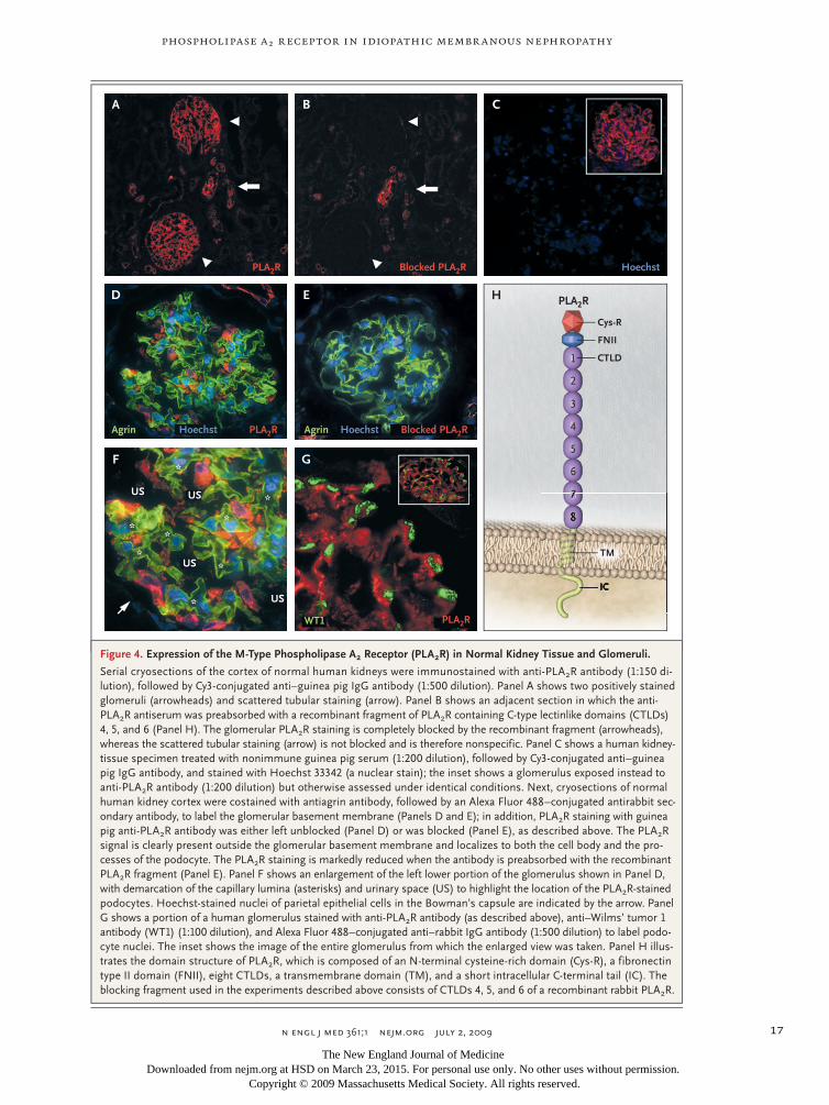
Figure 4. Expression of the M-Type Phospholipase A2 Receptor (PLA2R) in Normal Kidney Tissue and Glomeruli.
Serial cryosections of the cortex of normal human kidneys were immunostained with antiPLA2R antibody (1:150 dilution), followed by Cy3conjugated anti–guinea pig IgG antibody (1:500 dilution). Panel A shows two positively stained glomeruli (arrowheads) and scattered tubular staining (arrow). Panel B shows an adjacent section in which the antiPLA2R antiserum was preabsorbed with a recombinant fragment of PLA2R containing Ctype lectinlike domains (CTLDs) 4, 5, and 6 (Panel H). The glomerular PLA2R staining is completely blocked by the recombinant fragment (arrowheads), whereas the scattered tubular staining (arrow) is not blocked and is therefore nonspecific. Panel C shows a human kidneytissue specimen treated with nonimmune guinea pig serum (1:200 dilution), followed by Cy3conjugated anti–guinea pig IgG antibody, and stained with Hoechst 33342 (a nuclear stain); the inset shows a glomerulus exposed instead to antiPLA2R antibody (1:200 dilution) but otherwise assessed under identical conditions. Next, cryosections of normal human kidney cortex were costained with antiagrin antibody, followed by an Alexa Fluor 488–conjugated antirabbit secondary antibody, to label the glomerular basement membrane (Panels D and E); in addition, PLA2R staining with guinea pig antiPLA2R antibody was either left unblocked (Panel D) or was blocked (Panel E), as described above. The PLA2R signal is clearly present outside the glomerular basement membrane and localizes to both the cell body and the processes of the podocyte. The PLA2R staining is markedly reduced when the antibody is preabsorbed with the recombinant PLA2R fragment (Panel E). Panel F shows an enlargement of the left lower portion of the glomerulus shown in Panel D, with demarcation of the capillary lumina (asterisks) and urinary space (US) to highlight the location of the PLA2Rstained podocytes. Hoechststained nuclei of parietal epithelial cells in the Bowman’s capsule are indicated by the arrow. Panel G shows a portion of a human glomerulus stained with antiPLA2R antibody (as described above), anti–Wilms’ tumor 1 antibody (WT1) (1:100 dilution), and Alexa Fluor 488–conjugated anti–rabbit IgG antibody (1:500 dilution) to label podocyte nuclei. The inset shows the image of the entire glomerulus from which the enlarged view was taken. Panel H illustrates the domain structure of PLA2R, which is composed of an Nterminal cysteinerich domain (CysR), a fibronectin type II domain (FNII), eight CTLDs, a transmembrane domain (TM), and a short intracellular Cterminal tail (IC). The blocking fragment used in the experiments described above consists of CTLDs 4, 5, and 6 of a recombinant rabbit PLA2R.
The New England Journal of Medicine Downloaded from nejm.org at HSD on March 23, 2015. For personal use only. No other uses without permission.
Copyright © 2009 Massachusetts Medical Society. All rights reserved.

T h e n e w e ngl a nd j o u r na l o f m e dic i n e
n engl j med 361;1 nejm.org july 2, 200918
33p9
kD
*
*
A B C
AUTHOR:
FIGURE:
JOB:
4-CH/T
RETAKE
SIZE
ICM
CASE
EMail LineH/TCombo
Revised
AUTHOR, PLEASE NOTE: Figure has been redrawn and type has been reset.
Please check carefully.
REG F
Enon
1st
2nd3rd
Beck
5 of 6
06-18-09
ARTIST: ts
36024 ISSUE:
D E F
G H I
J K
PLA2R
PLA2R
Blocked PLA2R
IgG4
IgG4
IgG4
PLA2R+IgG4
PLA2R+IgG4
PLA2R+IgG4
HGE
250
148
98
64
50
36
MN1 LMN
MN2 IgAN1
MN3 IgAN2
MN4
HGErP
LA2R
HGErP
LA2R
Figure 5. Colocalization of the M-Type Phospholipase A2 Receptor (PLA2R) and IgG4 and Reactivity of Eluted IgG4.
Confocal microscopic analysis of cryosections of a kidneytissue specimen from a patient with membranous nephropathy revealed the presence of PLA2R (Panel A) and IgG4 (Panel B), which were colocalized in the peripheral capillary walls and the glomerular basement membrane (Panel C). Panels D, E, and F are enlarged images of the boxed areas in Panels A, B, and C, respectively. The asterisks in Panel F indicate the capillary lumina. The antiPLA2R antibody was blocked with the recombinant fragment (Panel G), with staining virtually eliminated, despite the continued presence of IgG4 (Panel H). Panel I is a confocal image of representative glomerular capillary loops in a biopsy specimen from a patient with lupus membranous nephropathy; PLA2R and IgG4 are not colocalized. IgG4 was detected with sheep anti–human IgG4 antibody (1:500 dilution) and rabbit anti–sheep IgG antibody (1:500 dilution). IgG was eluted from biopsy cores from patients with membranous nephropathy (MN), lupus membranous nephropathy (LMN), or IgA nephropathy (IgAN). This eluted IgG was used to immunoblot human glomerular extract (HGE) or recombinant PLA2R (rPLA2R) (Panel J). Only IgG eluted from the MN samples identified the native and recombinant PLA2R. Panel K shows that the IgG eluted from the MN3 biopsy sample recognized only those bands corresponding to PLA2R.
The New England Journal of Medicine Downloaded from nejm.org at HSD on March 23, 2015. For personal use only. No other uses without permission.
Copyright © 2009 Massachusetts Medical Society. All rights reserved.

Phospholipase A2 Receptor in Idiopathic Membr anous Nephropathy
n engl j med 361;1 nejm.org july 2, 2009 19
Association with Disease Activity
We analyzed serum specimens collected serially from several patients in whom treatment-induced or spontaneous remission occurred. We found that, in general, autoantibodies against PLA2R were present when there was clinically signifi-cant disease activity, as measured by urinary pro-tein and serum albumin levels (Fig. 6, and Fig. 9 in the Supplementary Appendix). In the patients with remission, there was a decline or disappear-ance of anti-PLA2R antibodies before the protei-nuria fully resolved.
Discussion
Our findings show that PLA2R is a major target antigen in idiopathic membranous nephropathy. Seventy percent of our patients with biopsy-proven idiopathic membranous nephropathy had IgG anti-bodies (predominantly of the IgG4 subclass) that reacted with a reduction-sensitive epitope present on PLA2R, a glycoprotein constituent of normal human glomeruli. The distribution of PLA2R in normal glomeruli suggests that it is located on podocytes, as are megalin in rat models of mem-branous nephropathy and neutral endopeptidase in patients with alloimmune neonatal membra-nous nephropathy. PLA2R colocalizes with IgG4 in immune deposits of kidney-biopsy specimens from patients with idiopathic membranous neph-ropathy, and IgG eluted from such biopsy speci-mens is reactive with PLA2R. Moreover, serial studies in a limited number of patients suggest that the presence of circulating anti-PLA2R anti-bodies corresponds to disease activity. Finally, the specificity of the reactivity of anti-PLA2R anti-bodies in patients with idiopathic membranous nephropathy, as opposed to secondary forms of membranous nephropathy or other proteinuric diseases, suggests that the antibodies are most likely the cause, rather than a consequence, of podocyte injury and proteinuria.
There are several possible reasons why anti-PLA2R antibodies are not detected in all patients with idiopathic membranous nephropathy. First, there may be target antigens other than PLA2R; however, screening by means of Western blotting revealed no consistent alternative candidates. Sec-ond, there may be cryptic epitopes that were not revealed under our extraction and electrophoretic conditions. Third, even though all our patients had some degree of proteinuria, a proportion may
have no longer had immunologically active dis-ease at the time of sample collection. Residual proteinuria in patients with membranous neph-ropathy is most likely due to altered podocyte architecture that results from remodeling of the glomerular basement membrane in the advanced disease state, as shown by the transplantation of kidneys from rats with active Heymann’s nephri-tis into healthy rats.20 Although proteinuria from
22p3
Uri
nary
Pro
tein
:Cre
atin
ineImmunosuppressive treatment
6
Alb
umin
(g/d
l) 4
3
1
5
2
0
6
4
3
1
5
2
0
Serum albumin
Urinary protein:creatinine
December2005
April2006
November2006
June2007
December2005
April2006
November2006
June2007
B
A
AUTHOR:
FIGURE:
JOB:
4-CH/T
RETAKE
SIZE
ICM
CASE
EMail LineH/TCombo
Revised
AUTHOR, PLEASE NOTE: Figure has been redrawn and type has been reset.
Please check carefully.
REG F
Enon
1st2nd
3rd
Beck
6 of 6
06-18-09
ARTIST: ts
36024 ISSUE:
PNGase F − + − + − + − +
148 kD
250 kD
98 kD
Anti–Human IgG Antibody
PLA2R
IgG
Figure 6. Antibody against the M-Type Phospholipase A2 Receptor (PLA2R) and Disease Activity in a Patient with Membranous Nephropathy.
Serum samples were collected serially from a patient with membranous nephropathy who had clinical remission after receiving immunosuppressive treatment with cyclophosphamide and prednisone (see the Clinical Vignettes section in the Supplementary Appendix). Panel A shows a decline in the urinary protein level (as measured by the proteintocreatinine ratio) and an increase in the serum albumin level. The results of Western blotting of serum samples taken from the patient at the same times as the laboratory measurements (Panel B) show reactivity to PLA2R, either the 185kD native form not deglycosylated by peptide Nglycosidase F (PNGase F−) or the 145kD deglycosylated form (PNGase F+) in the initial serum sample (from December 2005) only. Equal loading is verified by the nonspecific detection of a 98kD band. Equal volumes of the four serial serum samples were electrophoresed and blotted with anti–human IgG antibody (bottom), with a progressive increase in total IgG after the patient entered a period of remission from membranous nephropathy.
The New England Journal of Medicine Downloaded from nejm.org at HSD on March 23, 2015. For personal use only. No other uses without permission.
Copyright © 2009 Massachusetts Medical Society. All rights reserved.

T h e n e w e ngl a nd j o u r na l o f m e dic i n e
n engl j med 361;1 nejm.org july 2, 200920
the transplanted kidneys lessened in the absence of antimegalin antibodies, a moderate level of residual proteinuria persisted indefinitely in the recipient rats.
Although anti-PLA2R antibodies are predomi-nantly of the IgG4 isotype, which corresponds to the isotype found in immune deposits in patients with idiopathic membranous nephropathy,16,21 the other three IgG subclasses were variably pres-ent in serum but in smaller amounts. The presence of anti–neutral endopeptidase IgG1 and IgG4 antibodies, but not IgG4 alone, corresponds to the development of proteinuria in patients with alloimmune membranous nephropathy10; how-ever, we could not correlate the presence or ab-sence of anti-PLA2R IgG1 antibodies with the severity or duration of disease.
PLA2R is a type I transmembrane receptor and one of four mammalian members of the mannose-receptor family.22,23 All four have a conserved domain structure (Fig. 4H) and undergo endo-cytic recycling,12 which may provide a constant source of accessible PLA2R at the podocyte mem-brane for immune-complex formation. PLA2R was initially identified as a binding protein for se-creted phospholipase A2 (PLA2) and has a high binding affinity for several types of secreted PLA2 in certain species.24 However, human PLA2R has a different affinity for the more common types of secreted PLA2,25 and its exact physiolog-ical role remains unclear.24 Human PLA2R was originally cloned from kidney tissue, in which it is expressed at high levels25; our studies suggest that its expression in human kidneys is largely confined to glomerular podocytes. In contrast, PLA2R is weakly expressed in the glomerulus in normal rats and is up-regulated in experimental models of mesangial proliferative glomerulone-phritis.26 In addition, we observed that human anti-PLA2R autoantibodies do not detect PLA2R in rodent or rabbit glomerular extract (data not shown).
Anti-PLA2R autoantibodies from unrelated pa-tients react with a similar reduction-sensitive epitope. This commonality implies that the auto-antigen exists only when PLA2R is in a configu-ration dependent on intramolecular disulfide bonds and that the autoantibody response is re-stricted to this conformation. A similarly restrict-ed immune response to a reduction-sensitive epitope has been described for Goodpasture’s epitope in the noncollagenous domain NC1 in
type IV collagen α3.27,28 The finding of such a restricted response in patients with membranous nephropathy has interesting implications for the proximal events that initiate the immune re-sponse and limit epitope spreading to the rest of the PLA2R molecule. Since members of the mannose-receptor family exist in both extended and bent conformations that confer distinct li-gand binding and multimerization capacities,29 these different conformations may also play a role in exposure of the epitope in PLA2R.
The role of PLA2R in the pathogenesis of mem-branous nephropathy is currently unknown. The antigen may simply be a target for antibody bind-ing and complement-mediated podocyte injury, as in experimental models of membranous neph-ropathy.30,31 Alternatively, the antibodies may act as receptor agonists or antagonists, altering podocyte architecture and barrier function. Be-cause PLA2R is also present in the lungs and on leukocytes,32,33 it will be of interest to determine why the pathologic features are limited to the kidney.
Although there is a shorter, alternatively spliced PLA2R transcript that is predicted to encode a soluble form of the receptor,25 we could not de-tect such a form in the serum samples from ei-ther normal controls or patients with membra-nous nephropathy. We also found no evidence of circulating complexes of soluble or shed PLA2R and anti-PLA2R antibodies. These findings are consistent with many earlier studies in which circulating immune complexes were not detected in patients with idiopathic membranous neph-ropathy3 and further support the concept that immune deposits form in situ in those with the disease.
In conclusion, we identified circulating auto-antibodies against PLA2R in a large proportion of patients with idiopathic membranous nephropa-thy whom we examined. This protein is expressed on normal glomerular podocytes and is present in the glomerular immune deposits in patients with the disease. These findings and the charac-teristics of PLA2R strongly suggest that it is a major target antigen in patients with idiopathic membranous nephropathy. Moreover, the speci-ficity of these findings for idiopathic membra-nous nephropathy, in contrast to secondary forms of membranous nephropathy or other proteinu-ric kidney diseases, may have implications for the diagnosis and treatment of the disease.
The New England Journal of Medicine Downloaded from nejm.org at HSD on March 23, 2015. For personal use only. No other uses without permission.
Copyright © 2009 Massachusetts Medical Society. All rights reserved.

Phospholipase A2 Receptor in Idiopathic Membr anous Nephropathy
n engl j med 361;1 nejm.org july 2, 2009 21
Supported by grants from the National Institute of Diabetes and Digestive and Kidney Diseases (DK067658 and DK30932, to Dr. Salant, and DK076743, to Dr. Powell), Amgen (to Dr. Beck), the Halpin Foundation (to Dr. Beck), Centre National de la Recherche Scientifique and Association pour la Recherche sur le Cancer (to Dr. Lambeau), and the Department of Veterans Affairs (to Dr. Klein).
Dr. Beck reports receiving grant support from Amgen and hav-ing a patent pending for a diagnostic immunoassay to detect anti-PLA2R antibodies in membranous nephropathy; Dr. Lambeau, holding patents related to the therapeutic use of secretory PLA2 proteins and their inhibitors; and Dr. Salant, receiving consult-
ing fees from Questcor Pharmaceuticals, Cormedix, and DiObix and having a patent pending for a diagnostic immunoassay to detect anti-PLA2R antibodies in membranous nephropathy. No other potential conflict of interest relevant to this article was reported.
We thank Kathleen Dashner for help with tissue processing for immunofluorescence microscopy, Dr. Eric Boilard for help in preparing the human recombinant PLA2R and rabbit PLA2R frag-ment consisting of CTLDs 4 through 6, the New England Organ Bank for providing human kidneys for research purposes, the families of the organ donors, and the patients and volunteers who provided us with their serum for these studies.
References
Van Damme BJ, Fleuren GJ, Bakker 1. WW, Vernier RL, Hoedemaeker PJ. Experi-mental glomerulonephritis in the rat in-duced by antibodies directed against tubu-lar antigens. V. Fixed glomerular antigens in the pathogenesis of heterologous im-mune complex glomerulonephritis. Lab Invest 1978;38:502-10.
Couser WG, Steinmuller DR, Stilmant 2. MM, Salant DJ, Lowenstein LM. Experi-mental glomerulonephritis in the isolated perfused rat kidney. J Clin Invest 1978; 62:1275-87.
Couser WG, Salant DJ. In situ immune 3. complex formation and glomerular injury. Kidney Int 1980;17:1-13.
Kerjaschki D, Farquhar MG. The path-4. ogenic antigen of Heymann nephritis is a membrane glycoprotein of the renal prox-imal tubule brush border. Proc Natl Acad Sci U S A 1982;79:5557-61.
Idem.5. Immunocytochemical localiza-tion of the Heymann nephritis antigen (GP330) in glomerular epithelial cells of normal Lewis rats. J Exp Med 1983;157: 667-86.
Makker SP, Singh AK. Characteriza-6. tion of the antigen (gp600) of Heymann nephritis. Lab Invest 1984;50:287-93.
Raychowdhury R, Niles JL, McCluskey 7. RT, Smith JA. Autoimmune target in Hey-mann nephritis is a glycoprotein with ho-mology to the LDL receptor. Science 1989; 244:1163-5.
Saito A, Pietromonaco S, Loo AK, Far-8. quhar MG. Complete cloning and sequenc-ing of rat gp330/“megalin,” a distinctive member of the low density lipoprotein re-ceptor gene family. Proc Natl Acad Sci U S A 1994;91:9725-9.
Debiec H, Guigonis V, Mougenot B, et 9. al. Antenatal membranous glomerulone-phritis due to anti-neutral endopeptidase antibodies. N Engl J Med 2002;346:2053-60.
Debiec H, Nauta J, Coulet F, et al. Role 10. of truncating mutations in MME gene in fetomaternal alloimmunisation and ante-natal glomerulopathies. Lancet 2004;364: 1252-9.
Salant DJ, Cybulsky AV. Experimental 11. glomerulonephritis. Methods Enzymol 1988;162:421-61.
Zvaritch E, Lambeau G, Lazdunski M. 12. Endocytic properties of the M-type 180-kDa receptor for secretory phospholipases A2. J Biol Chem 1996;271:250-7.
Powell DW, Rane MJ, Joughin BA, et al. 13. Proteomic identification of 14-3-3zeta as a mitogen-activated protein kinase-acti-vated protein kinase 2 substrate: role in dimer formation and ligand binding. Mol Cell Biol 2003;23:5376-87.
Powell DW, Weaver CM, Jennings JL, 14. et al. Cluster analysis of mass spectrome-try data reveals a novel component of SAGA. Mol Cell Biol 2004;24:7249-59.
Lambeau G, Ancian P, Barhanin J, 15. Lazdunski M. Cloning and expression of a membrane receptor for secretory phospho-lipases A2. J Biol Chem 1994;269:1575-8.
Doi T, Mayumi M, Kanatsu K, Suehiro 16. F, Hamashima Y. Distribution of IgG sub-classes in membranous nephropathy. Clin Exp Immunol 1984;58:57-62.
Kuroki A, Iyoda M, Shibata T, Sugisaki 17. T. Th2 cytokines increase and stimulate B cells to produce IgG4 in idiopathic mem-branous nephropathy. Kidney Int 2005;68: 302-10.
Brandslund I, Siersted HC, Jensenius 18. JC, Svehag SE. Detection and quantitation of immune complexes with a rapid poly-ethylene glycol precipitation complement consumption method (PEG-CC). Methods Enzymol 1981;74:551-71.
Saleem MA, O’Hare MJ, Reiser J, et al. 19. A conditionally immortalized human pod-ocyte cell line demonstrating nephrin and podocin expression. J Am Soc Nephrol 2002;13:630-8.
Makker SP, Kanalas JJ. Course of trans-20. planted Heymann nephritis kidney in nor-mal host: implications for mechanism of proteinuria in membranous glomerulo-nephropathy. J Immunol 1989;142:3406-10.
Haas M. IgG subclass deposits in 21. glomeruli of lupus and nonlupus mem-branous nephropathies. Am J Kidney Dis 1994;23:358-64.
East L, Isacke CM. The mannose re-22. ceptor family. Biochim Biophys Acta 2002; 1572:364-86.
Lambeau G, Lazdunski M. Receptors 23. for a growing family of secreted phospho-
lipases A2. Trends Pharmacol Sci 1999;20: 162-70.
Rouault M, Le Calvez C, Boilard E, et 24. al. Recombinant production and proper-ties of binding of the full set of mouse secreted phospholipases A2 to the mouse M-type receptor. Biochemistry 2007;46: 1647-62.
Ancian P, Lambeau G, Mattéi MG, 25. Lazdunski M. The human 180-kDa recep-tor for secretory phospholipases A2: mo-lecular cloning, identification of a secreted soluble form, expression, and chromo-somal localization. J Biol Chem 1995;270: 8963-70.
Beck S, Beck G, Ostendorf T, et al. Up-26. regulation of group IB secreted phospho-lipase A(2) and its M-type receptor in rat ANTI-THY-1 glomerulonephritis. Kidney Int 2006;70:1251-60.
Hellmark T, Burkhardt H, Wieslander J. 27. Goodpasture disease: characterization of a single conformational epitope as the target of pathogenic autoantibodies. J Biol Chem 1999;274:25862-8.
Hudson BG. The molecular basis of 28. Goodpasture and Alport syndromes: bea-cons for the discovery of the collagen IV family. J Am Soc Nephrol 2004;15:2514-27.
Llorca O. Extended and bent confor-29. mations of the mannose receptor family. Cell Mol Life Sci 2008;65:1302-10.
Nangaku M, Shankland SJ, Couser 30. WG. Cellular response to injury in mem-branous nephropathy. J Am Soc Nephrol 2005;16:1195-204.
Cybulsky AV, Quigg RJ, Salant DJ. Ex-31. perimental membranous nephropathy re-dux. Am J Physiol Renal Physiol 2005;289: F660-F671.
Granata F, Petraroli A, Boilard E, et al. 32. Activation of cytokine production by se-creted phospholipase A2 in human lung macrophages expressing the M-type recep-tor. J Immunol 2005;174:464-74.
Silliman CC, Moore EE, Zallen G, et al. 33. Presence of the M-type sPLA(2) receptor on neutrophils and its role in elastase re-lease and adhesion. Am J Physiol Cell Physiol 2002;283:C1102-C1113.Copyright © 2009 Massachusetts Medical Society.
The New England Journal of Medicine Downloaded from nejm.org at HSD on March 23, 2015. For personal use only. No other uses without permission.
Copyright © 2009 Massachusetts Medical Society. All rights reserved.