Embed Size (px)
Citation preview

H,,i,,li ,,,,,n,,! #,, F',<,~i,C \',!W, ,lYYii 46. 501 50:
c i'4'Ii Ik l~i1~1\11 4>wc, "
---.- __.--
BRIT STIC SURGERY ----___ II --.--
The galea frontalis myofascial flap in anterior fossa CSF leaks
H. COW. A. Cerejo. A. Baptista, R. Vaz. M. Goncalves. A. Guimarges. J. Amarante. C‘. Cru7 and F. Guimaraes
Pltrstic. .Cutyr~,~~ trnrr Nrwroswgu~~ Dtparttmwt,s. Hospital S. .10&t. MMetiiwI Sdtool. Opwto. P~wtlrgtrl
~__ - -~.--~~
.S1’,VM.41< 2’. We report the clinical use of galea frontalis myofascial flaps in the treatment of anterior fossa cerebrospinal fluid leaks after trauma. This flap provides an adequately sized and vascularised barrier between the cranial and nasal cavities through which the cells of the inflammatory response reach the target area. This technique was used in 9 cases with complete success; in 5 out of 9 patients, repair of an anterior cranial base bone defect was also performed with split calvarial bone grafts, harvested from the frontal craniotomy bone. In all patients, neither recurrence of the CSF leakage nor postoperative meningitis or its recurrence were observed.
The aetiology of an anterior fossa cerebrospinal fluid leak can be classified as traumatic and non-traumatic. Trrtutmrtic~. Head trauma is a frequent cause of anterior fossa CSF leakage. Its incidence varies from 0.56-7 90. according to the series, regardless of the presence of a skull fracture. but it doubles if a skull fracture is presem.’ Most of the skull fracture patterns are frontal and frontotetnporal, although anterior fossa CSF leaks may occur with skull fractures in other locations. There ih not alwavs correlation of side between the fracture and the CSF leakage.’ Traumatic anterior fossa (CSF leak is more commonly located in the ethmold binus region rather than the frontal sinus region (ratio 3 : I ). although within the ethmoid region. the cribriform plate itself is less often fractured than the ethmoid sinus roof.” Maxillofacial injuries. such as Le Folrt II and III fractures are commonly associated with CSF leaks, with or without direct cranial Lraum;a : ‘.” cerebrospinal fluid rhinorrhea and pneumocephalus may accompany these fractures and if carefully sought are found in at least 25 Oio of these injuries.”
latrogenic (.3F leaks can also occur and cases related to remo\:al of nasal polyps or middle tur- binate during endonasal surgery, rasping the frontal \inus and removal of ethmoid-orbital osteomas. have been reported.’ .l:ott-/r’trurttnti(,. Tumours are another cause of anterior thssa CSF leakage, either by direct erosion or a more distant el-fect from increased intracranial pressure.‘.!’ Inflammation is a less frequent cause. as are osteo- myelitis, influenza and arachnoiditis.“’
Memngitis IS a major risk of anterior fossa CSF leakage. Incidence varies from 8.6% to 41 ‘Ib of cases. if a CSF leak or intracranial air or both are present. Meningitis has been reported before the clinical onset of CS F leakage and may occur in the nonleaking phase of intermittent CSF leakage.” A delay in the onset of CSF leakage after trauma makes the risk of meningitis accompanying the leak 2-8 times greater than if the onset of C‘SF leakage is immediate.”
Meningitis may originate from a broad spectrum of
bacterial agents: however, most adult cases are pro- duced by Diplococcus ptxwtwtliuc~ or .Stri~l,toc’oc.c.us or Stllph?,loc.occ,us organisms. with h:ci.~.scritl twttittgititli.s and Hctuophilus it$furtt:u seen less often.‘:’
Other les< frequent complications include hae- matomas. usually subdural. caused by the tearing of bridging veins when cerebral tijaue collapses, as intracranial pressure is kept abnormally low by CSF leak.’ ’
C‘ott.srr’l’rrtir.c,. The basis for this approach is the possibility for spontaneous cessation of the leah. Some series give incidence of such spontaneous cessation up to 80 O/b.‘;’ Most leaks stop within I4 days. but longer periods (up to 7 weeks) have been reported.‘” There is no consensus regarding the length of time IO be allowed for cessation of CSF leakage. Some think that 1 week is reasonable, others have waited 4 4 weeks and others believe that immediate surgical repair is mandatory.” The use of a lumbar subarachnoid catheter for CSF drainage is very helpful in allowing closure of a CSF nasal fistula, particularly in post traumatic cases.’
In our unit, when a patient presents with CSF rhinorrhea. initial managernent is with positioning. with head and trunk elevation. If this measure alone is not enough to stop leakage in 14 to 38 h. a lumbar catheter for CSF drainage is inserted Like other authors. we consider it safe IO keep this in place for 10 days; if leakage persists after this period. surgery is considered. There are cases in w-hich leakage stops with external lumbar drainage. but reappears some days later in a continuous or intermittent pattern; these patients are also considered for surgery.
Regardingmaxillofacial injuries, an important point must be made that in the presence of Le Fort I1 and III fractures. early stabilisation should always bc under- taken; it is now generally accepted that stabilisation of a mobile midface itself helps to prevent a pumping action and thereby reduces ingress of nasal secretion and infection through the dural det’ect and facilitates
504 British Journal of Plastic Surgery
bone graft
’ j\,
B
C D Fig. 1
Figure l-Diagram showing the anatomical concept of the myo- fascial frontalis galeal flap. with or without calvarial split bone grafts. for the treatment of anterior fossa CSF leaks.
uneventful dural healing in the majority of cases of this type of injury.“,” Intrucranial surgical approaches. Successful intra- cranial repair has been credited to Dandy,lH who sutured fascia lata grafts over bone defects. Others
Table 1 Patient details
have also used fascia lata.“’ absorbable gelatin sponge and temporal muscle fascia.’ Other methods have been used with different rates of success, such as the use of tibia1 periosteal graft,“’ pericranial fascia graft and iodine-soaked packs extradurally,“” bone wax alone”” and a portion of falx cerebri folded down and sutured.‘”
Since January 1988, based on the combined work of our Neurosurgical and Plastic Surgery Departments, we have evolved a surgical method for treatment of anterior fossa CSF leakage. A myofascial frontalis galeal flap can be distally based on the supratrochlear and supraorbital vessels, allowing transposition from an extracranial to intracranial position, so creating a vascularised barrier between the dura mater and the anterior fossa defect. In addition, in some of the clinical cases, calvarial bone grafts were harvested from the frontal craniotomy bone and modelled to fill the bone defect of the cribiform plate and adjacent regions (Fig. 1).
Material and methods
In this study, we include 9 patients (Table 1) with a history of previous head trauma. In all of them, an intracranial-intradural approach was performed. with transposition of a myofascial frontalis galeal vascular- ised flap. Repair of the skull base defect with calvarial bone grafts was performed as well in 5 of these patients. Three patients presented with evidence of CSF rhinor- rhea after head trauma; all of these underwent CT head scan showing basal cranial fractures (Table 2). The remaining 6 patients showed no CSF leakage on admission, but presented later with meningitis. The time between cranial trauma and the onset of men- ingitis was quite variable, ranging from 3 months to 3 years. The causal microorganisms were Pneumococcus (5 cases) and Haemolytic Streptococcus (1 case). All of the 6 patients were treated with appropriate anti-
Case 1 Case 2 Case 3 Case 4 Case 5 Case 6 Case 7 Case 8 Case 9
fpneumococcus + streptococcus + pneumococcus + pneumococcus + pneumococcus + pneumococcus
3 weeks 3 weeks 4 weeks 2 years 3 months I .5 years 3 years 2 years 6 months
3.5 years 2 years 3 years I .5 years I .5 years
3.5 years I year 1 year 1.5 years
Table 2 Patients with CSF leakage on admission
Neuroradiological Imwtigatiorz - C.T. scan Findings on Surgq.
Case I
Case 2
Case 3
Bilateral frontal contusions Ethmoidai fracture Bilateral frontal contusions Multiple basal fractures Multiple basal fractures
Bilateral ethmoidal bone defect
Right anterior cranial base bone defect
Bilateral ethmoidal bone defect
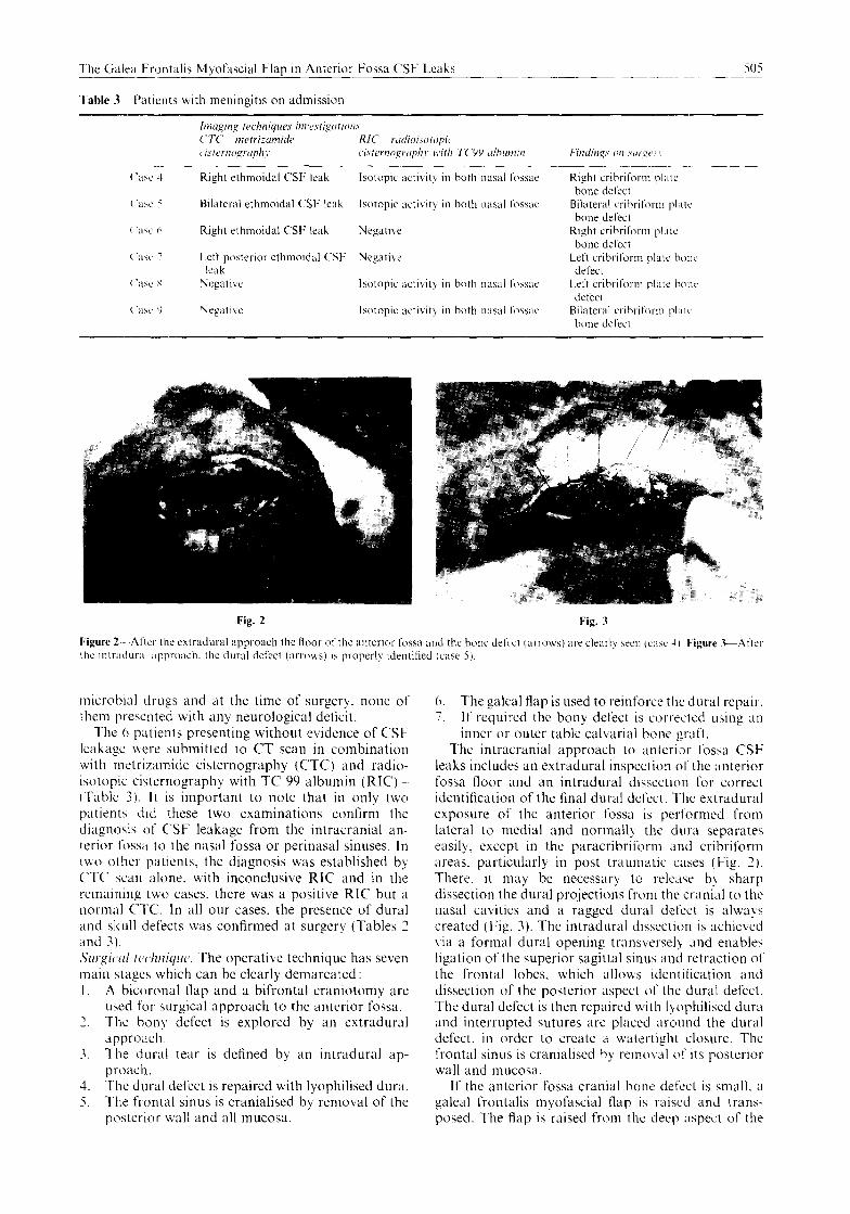
Table 3 Patients with meningitis on admission
Right ethmoidal CSF leak Isotopic activity 111 both nasal fossae Right crihriform plate bone defect
Bilateral ethmoidal CSF leak lwtoplc activit) 111 both nasal fossae Bilateral crlbriform pl,rrr bone detect
Right ethmoidal CSI: leak Negative Right crihriform plate bone deltct
Left posterior ethmoidal CSI- Negative Left cribrlform plate hone leak defect
Yegatlvc Isotopic activlt) in both nasal fossae Left cribl-lt‘orm plate honi, defect
Negative Isotopic activity 111 both nxal f~wae Bilateral crihritorm pl;~ic hone defect
Fig. 2 Fig. 3
Figure 2-After the extradural approach the floor of the unterlor fossa and the hanc defect (arrows) are clearly seen IC;I>C 1). Figure .GAftet 111~ Intradurxl .~pproach. the dual defect (arrows) I\ proper11 Identilied (cast 5).
microbial drugs and at the time of surger!. none of them presented with any neurological defictt.
The h patients presenting without evidence of CSF leakage were submitted to CT scan in combination with metrizamide cisternography (CTC) and radio- isotopic cisternography with TC 99 albumin (RIC) (Table 3). It is important to note that in only two patient> did these two examinations confirm the diagnosis of CSF leakage from the intracranial an- terior fossa to the nasal fossa or perinasal sinuses. In two other patients, the diagnosis was established by CTC scan alone. with inconclusive RIC and in the remaining two cases, there was a positive RIG but a normal CTC. In all our cases, the presence of dural and sk~11l defects was confirmed at surgery (Tables 2 and 3). .~~r:L~rl /c,chniclur. The operative technique has seven main stages which can be clearly demarcated : 1. A bicoronal flap and a bifrontal craniotomy are
uicd for surgical approach to the anterior fossa. ’ The bon!, defect is explored by an extradurat -.
approach 3. The dural rear is defined by an intradural ap-
proach. 4. The dural defect is repaired with tyophilised dura. 5. The frontal sinus is cranialised by removal of the
posterior wall and all mucosa.
6. The galeal Hap is used to reinforce the dural repair. 7. If required the bony defect is corrected using an
inner or outer table call,arial bone graft. The intracranial approach to anterior fosxa CSF
leaks includes an extradural inspection of the anterior fossa floor and an intradural dissection for correct identification of the final dural defect. The extradural exposure of the anterior fossa is performed from lateral to medial and normally. the dura separates easily, except in the paracribrltorm and cribriform areas. particularly in post traumatic c;Lses (Fig. 2). There, it may be necessary to release b! sharp dissection the dural projections from the cranial to the nasal cavities and a ragged dural dercct is always created (Fig. 3). The intradural dissection is achieved \,ia a formal dural opening transvcrscl) and enables ligation of the superior sagittal sinus and retraction of the frontal lobes, which allows identitication and dissection of the posterior aspect of the dural defect. The dural defect is then repaired with Iyophilised dura and interrupted sutures are placed around the dural defect. in order to create II watertight closure. The frontal sinus is cranialised by remo\ral of its posterior wall and mucosa.
If the anterior fossa cranial bone defect is small. a galeal frontalis myofascial flap is raised and trans- posed. The Asp is raised from the deep aspect of the

506 British Journal of Plastic Surgery
Fig. 4
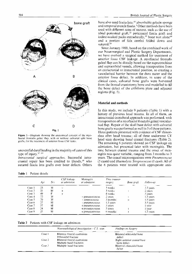
Figure 4-Harvesting of the galeal frontalis myofascial flap (case 6). intracranial extradural environment (case 5).
Fig. 5
Figure S-The transposition of the myoFdscia1 flap from extracranial to
Fig. 6
Figure &Harvesting of the outer table split bone grafts from th reconstruction of the right anterior cranial base defect (case 2).
bicoronal flap. Two longitudinal myogaleal incisions are performed about 6-8 cm apart, until the sub- cutaneous tissue is visualised and by sharp knife dissection, the myofascial flap is carefully raised, including the supraorbital and supratrochlear vessels (Fig. 4). After careful haemostasis, the muscle flap is ready for transposition to the anterior cranial base. normally through a slit-type bone window created in the frontal bone flap (Fig. 5).
However, if the anterior cranial base bone defect is considered large (more than 5 mm width), a split thickness bone graft is harvested from the frontal craniotomy bone flap (Fig. 6). After careful modelling of the bone graft, its fixation is usually possible by finger pressure (Fig. 7). Then, the frontalis myofascial flap is performed as described above. Although we have been using an outer table cranial bone graft, the contour defect of the frontal bone has not been a problem.
The transverse dural incision is closed with con- tinuous non-absorbable suture, transcutaneous intra- dural and extradural drains are inserted, the frontal bone flap is repositioned and fixed and all wounds are closed.
Fig. 7
e frontal cramotomy bone (case 4). Figure 7-Bone graft (arrow) for
Follow-up
The time of follow-up was between 1 and 3.5 years. We have observed neither recurrence of CSF leakage nor any episode of meningitis.
Discussion
The intracranial surgical approach is the method of choice for treatment of anterior fossa CSF leaks; it gives a wide exposure of the anterior cranial base, transforming an inaccessible area to an easily ac- cessible one and enabling the surgeon to perform the necessary extra and intradural dissections to identify the dural defect which can be properly repaired.
Whitaker et al.‘” considered CSF rhinorrhea following transcranial surgery as a dangerous com- plication with a high risk of meningitis. Dural tears and CSF leaks have also long been implicated as strong associates of meningitis and David et d.“” recommended that in conditions requiring transcranial surgery, all dural tears be meticulously repaired, the preferred method being to insert a generous graft of

The G&x Frontaiis Myofascial Flap in Anterior Fossa CSF Leak, 50:
lyophilised dura. temporal fascia or pericranium into the subdural space and suture the margins of the tear to this graft. Jackson’)’ has recommended the use of free vascularised flaps of omentum for large cranial base dejects and it has been stated” that the single most effBecti\c step to reduce the incidence of infections in transcranial cases was use of the vascularised galea frontalis myofascial flap. This flap is easy and quick to elevate. pro\ ides adequate size for nasal-cranial sep- aration in most cases and leaves an inconspicuous donor defect. It can be used after the resection of skull base tumours and frontofxial advancements.
Folloning this. be have been using this Aap as an anatomical and immunological barrier in the treat- ment of anterior fossa CSF leaks. Anatomically. the galea frontalis flap creates a barrier between the dura and the anterior cranial base. avoiding f-uture dural adherence and mvagination into the cribiform plate and paracribriform areas. Immunologically. the galea frontalis myof’dscial flap. being a well vascularised tissue. acts as ;I bridge along which the blood factors and cells of the inflammatory response can reach the target field.
Acknowledgements
The ;iuthors would like to thank Mr Albert0 Alfaia from the Anatom! Department of Oporto University for providing the medical Illustrations and Architect Jorge Castro for the line drawings Our Gncere thanks go to Mrs Ana Medv and Mra t-ernand,l Lenha. the author‘s uife. for typing the manuscript.
References
I. Gurtjllan ES. Webster JE. The surgical management of trau- maric cramonaal fistulas. Surg Clin North !\m lY.53: I : IIS 25
2 Lenin W C‘erebrospinal fluid rhinorrhea in closed head injurie>. BI-.I Surp lYS4.42: l-18.
.t. Mollcy TP. Hetherington RF. Traumatic cerebrospinal fluid rblnorrhca and otorrhea. pneumocephalus and meningitis. Surg (i~necol Obstet 1057: 104: 88~~98.
-I Mxtrxs H. Kuderna H. Combined cramofacial fracture< J Maulloll~c Surs lY80: 8: 52 8.
5 Mel\ 111~ I.<‘. Derome P. Concomitant dislocation of the face and skull J Manillofac Surg 1978: 6: 1~8.
h. Jacob\ J B. Pu\ky MS. Traumatic pneumocephalur. Laryngo- scope 19x0: YO. 515~20.
7 C‘ushlng H Experiences with orbito-ethmoidal osteomatu hiving intr~wanial complications. Surg Gynccol Ohstet lY17: 43 721 32.
X Schxhtcr MM. Rovit RL. Schachter JM. Rhinorrhea and h!droccphalus. observations on spontaneous cerebrospinal Rued fi\tuLl In patients with increased intracranial pressure .Acta Radiolog 1969; 9. 101-16.
‘) Fronr D. Penning L. Occult spontaneous cerebrospinal tluid rhlnorrhca diagnosed by isotope clsternopraphy. Neuro- rxiiolop~ lY71 : 7: 167 ~9
IO Sam ML.. Kramer R. Cerebrospinal rhmorrhea pathological lindinf\ L.aryngoacopc 1940: 50: I I67 77.
II
12.
13.
I3
IS
16.
17.
IX.
19
20
71
‘2.
3 7
24.
2’.
26.
27
2X.
Robinson R(i. Cerebrospinal fluid rhlnorrhw. nlenmgiiib and pneumo cephalus due to non-missile inlurw Au\t NZ J Surf 1970: 3Y: 328 34.
Mincy JE. Post traumatic cerehrosplnal Hued ll\tula of the frontal fosaa. J Trauma 1966: h: 61 li 22
Hand WL. Sanford JP. Post traumatic l-wren II menmgitis. Ann Intern Med lY70; 72: 860 74.
Woodford JE. Bogdanowicr W. Saunders RI... Traumatic intracranxd hematomas. role (of CSF Icahaye .JAMA 1074: 727: 1152 4.
Raaf J. Pohi traumatic cerebro,pinal t’uld Ical \ Arch Sur@ 1967: Y5: 63X 51.
Ltvni S. Spontaneous cerebrospinal rhlnorrhea. BMJ 1+3X: I 1208 Y.
Manson PN. Su CT. Hoopes JE. St!-uctural pillar\ of the facial skeleton. Plast Reconstr Surg 19X0: 66. 53 i?O.
Reynolds JR. Late complication\ vs. method of treatment 111 a large series of midfacial fractures. Plact Recon\rr Surs ly7X: 62: X71 8
Dandy WE. Pneumocephalus (~ntracranal pncumatcxxle or vcrocele). Arch Surg 1926: 12. 949 X2.
Eden K. Traumatic cerebrospinal rhinorrhea repail of the tistula by a transfrontal intradurnl operation. Hr J SUI-g ly4’2; 2Y 799 in3.
Gisw~e W. Post-traumatic cerebro<plnal r hinc)l rhea m Ith case report. BI J Surg 1940: 27: 717 22
Lawon .A. \ case of cerebrosplnal rhlnorrhra !bllou~ng on a multiple fracture of the skull which in\olvetl Ihe left frontal sinus and left orbit. Transaction5 Ophthalmologic Socieo U.K. 1034. 53: !07- 15.
Graham TO. Cerebrospinal rhinorrhea. J L,~rynpol Otol ly37 51: ‘44 7
German WJ. Cerebrospinal rhrnorrhca Sui-gcal rcpalr. J Neurosurg ly44; I : 60 6.
Whitaker LA. Mum-o IR. Salver KE. Jackson IT. Ortif- Monabterlo F. Marchac D. (‘omh~ned report of problems and complications in 793 ainiofaclal ,>pt:r;ition>. Plast Reconstr Sure 1979; 64: IYX~ 204.
David DJ. Poswillo DE, Simpson DA. The (‘r~inlosyn~)stuse4 Causes. Natural History and Manasemtwt Berlin: Springer- V&ig. 19X2.
Jackson IT Infection followinp fronts\-supr,l~.,rbital advance- ment. Pcl-spectives in Plastic Surgery IyX7. I yi 8
Jackson IT. Adharn MN. Marsh WR. Use of rhe galcal frontali\ m~of&ial flap in cramofaci;tl surge:! PIat Recon\tr Sury 1~x6. 77 owio.
The Authors
HorPcio Costa, MD, Consultant Plastic Surgeon: tlrmerl~ Registrar and British Council Research Fellow in Plastic Surgery. Cannie\- burn Hospital, Glasgow. Scotland.
Antbnio Cerejo, MD, Consultant Neurosur~con. Antbnio Baptista, MD, Consultant Yeurosur~eon. Rui Val. MD, Consultant Neurosurgeon, Maia Goyalves. MD, Senior Consultant Nctuxxurgwn. Antdnio GuimarBes, MD, Senior Consultant Playr~c Surgeon. Jo& Amarante, PhD, Professor in Plastic Surgcrt Celso Cruz, PhD, Professor 11, Neurosureer>. Flbio GuimarHes, MD, Semor Con\ultant Plastic Suryeoll.
Plastic Surgery and Neurosurger); Dcpartmcats. IHwpital S Jo&>. Medical School. Oporto. Portug.11.
Requests for reprints to: Hordcio Cotta. MD Cc)llsultant Plastic Surgeon, Rua do Corvo. no 323 .4rcotelo. Praia d.1 Ciranla 4405 \‘aladarea. Portugal
Paper received 8 December lY92. Accepted 7 May 1093. after revision