Embed Size (px)
Citation preview

ORIGINAL ARTICLE
The accordion gracilis muscleflap: a new design forcoverage of recurrent andcomplicated ischeal pressuresoresAhmed H El-Sabbagh
El-Sabbagh AH. The accordion gracilis muscle flap: a new design for coverage of recurrent and complicated ischealpressure sores. Int Wound J 2011; 8:447–453
ABSTRACTManagement of patients with large or recurrent pressure ulcerations can be complicated by the lack of availablelocal flap, whether already used or because adjacent lesions make such flap insufficient for complete coverage.In this article, the gracilis muscle was modified to cover large defects without help from its cutaneous territory.Twelve ischeal pressure sores were treated between August 2007 and 2009 with the modified gracilis muscle flapin a single-staged procedure. Five ulcers were recurrent and seven patients have associated pressure ulcers. Allreconstructions were successful. Mean patient age was 35 years and nearly all patients had multiple significantcomorbidities, including associated ulcers, diabetes and urethrocutaneous fistula. All flaps and donor sites healeduneventfully. There was one complication presented as cellulites at the donor site. Follow-up in some cases extendup to 1·5 years. No recurrence was observed. The accordion gracilis muscle flap is a handy, safe and fast flap forreconstruction of recurrent, difficult ischeal pressure sores.
Key words: Accordion flap • Gracilis muscle • Ischeal pressure sores • Recurrent and complicated ulcers
INTRODUCTIONIschial pressure sores represent a challeng-ing problem for the reconstructive surgeon.Multiple techniques have been described tocover these defects including rotation flaps,island musculocutaneous flaps, V-Y advance-ment flaps, gluteal thigh flaps, perforator flapsand free flaps (1–9).
Pickrell (10) had described the transpositionof the gracilis muscle in order to replace
Author: AH El-Sabbagh, MD, Department of Plastic andReconstructive Surgery, Plastic Surgery Unit, Mansoura UniversityHospital, Mansoura, EgyptAddress for correspondence: Dr AH El-Sabbagh, Depart-ment of Plastic and Reconstructive Surgery, Plastic Surgery Unit,Mansoura University Hospital, Mansoura, EgyptE-mail: [email protected]; [email protected]
an insufficient anal sphincter as early in thefifties past century. Later, the gracilis muscleflap has been used extensively for variousreconstructive purposes, such as soft-tissuecoverage, chronic osteomyelitis reconstructionand free functioning muscle transplantations(11–13).
Key Points
• in this article, a modificationof the gracilis muscle flap isdescribed by increasing thewidth of the muscle withoutstealing from its depth
However, there are limitations when usingthe gracilis muscle as a pedicled flap: limiteddistal muscle bulk and a short pedicle length.Both factors may be critical to successfulreconstruction with the gracilis muscle becausethe most distal portion is often the most crucialfor wound closure.
In this article, a modification of the gracilismuscle flap is described by increasing thewidth of the muscle without stealing from
© 2011 The Author© 2011 Blackwell Publishing Ltd and Medicalhelplines.com Inc • International Wound Journal • Vol 8 No 5 447

Gracilis muscle for ischeal pressure sore
its depth. This modification maintains theadvantages of the gracilis muscle as it providesbetter vascularity, making the flap moreresistant to pressure-induced ischaemia andfilling the cavity of the ulcer completely.
PATIENTS AND METHODSTwelve patients who were treated for ischealpressure sore and reconstructed using anaccordion gracilis muscle flap between August2007 and 2009 were included in the study.Empirical broad-spectrum antibiotics to coverGram positive, Gram negative and anaerobicbacteria were administered intravenously.These regimens were changed on the basis ofsensitivity tests. All patients underwent early,aggressive surgical debridement. Single-stagesurgical reconstruction was performed.
Key Points
• this modification maintains theadvantages of the gracilis mus-cle as it provides better vas-cularity, making the flap moreresistant to pressure-inducedischaemia and filling the cavityof the ulcer completely
• twelve patients who weretreated for ischeal pressuresore and reconstructed usingan accordion gracilis muscleflap between August 2007 and2009 were included in the study
• the main cause of the ischealpressure sore was spinal cordinjury
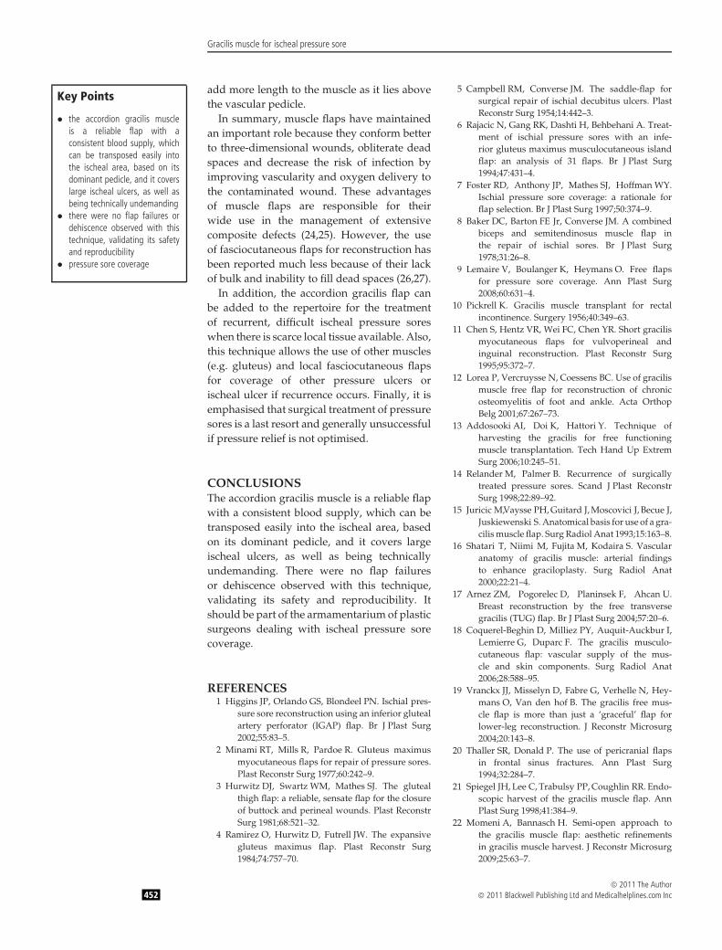
Surgical techniqueThe patient is placed in the supine positionwith hips slightly flexed and moderatelyabducted. The standard longitudinal incision ismade for male patients (ten patients) and twointerrupted zigzag incisions for female patients(two patients). The gracilis muscle is identifiedand a plane is developed on either side ofthe gracilis for proper pedicle localisation. Thetwo minor secondary pedicles are ligated anddivided. The muscle is then divided distallyto facilitate proximal dissection and isolationof the pedicle. Once the proximal pedicleis identified, the adductor longus muscle iselevated and careful dissection of the pediclecontinues deep to the adductor longus musclefor small distance. The motor branch of themuscle is carefully preserved (Figure 1).
The patient is turned to the prone position.The ischeal ulcer is debrided extensively. After
Figure 1. The motor nerve is carefully preserved.
excision of the infected bursa, the ischealtuberosity is reduced and rasped to a smoothcontour and careful haemostasis is made.
A capacious pocket is formed between theulcer cavity and the flap incision by a combinedsharp and blunt dissection. The distal end ofgracilis muscle is held by 2/0 non absorbablesuture material. A big haemostat is introducedfrom the ulcer cavity into the flap incisionthrough the tunnel and the suture is grasped.The gracilis flap is then introduced freely out-side the cavity of the ulcer. Then gracilis muscleis bent like an accordion and sutured side-by-side using 4/0 absorbable sutures (Figure 2).The cavity of the ulcer is obliterated by fixingthe modified muscle by 2/0 non absorbablesuture. The needle is introduced from outsidethe cavity of the ulcer to the muscle and thenreturned back outside the cavity. All suturesare located on the skin outside the ulcer. Theflap incision is closed in two layers.
The subcutaneous tissue is closed by 2/0 nonabsorbable suture and the skin is closed with3/0 non absorbable material. Closed suctiondrainage is applied for 5 days.
RESULTSThere were a total of 12 patients in this study(Table 1). Most of the patients were male (tenmale and two females). The main cause ofthe ischeal pressure sore was spinal cordinjury. The defect size ranged from 7 × 5 cmto 10 × 7 cm. All patients were admitted forcare of ulcers and improving their generalconditions. Seven of the 12 patients had anassociated pressure sores in the gluteal andheel areas (Figures 3 and 4). Five patients hada previous repair with local fasciocutaneousflaps (Figure 5).
Figure 2. The gracilis muscle is bent like an accordion.
© 2011 The Author448 © 2011 Blackwell Publishing Ltd and Medicalhelplines.com Inc

Gracilis muscle for ischeal pressure sore
Table 1 Patients’ summary
Patient Age Sex Size of undermined cavity Primary disease Associated morbidity Follow-up (months)
1 12 Male 7 × 5∗ Spina bifida – 82 36 Male 9 × 8 Spinal cord injury Urethrocutaneous fistula 183 44 Male 10 × 9∗ Spinal cord injury – 24 51 Male 12 × 7 Spinal cord injury Gluteal ulcers 125 16 Female 6 × 6 Spinal cord injury Heel ulcers 106 18 Male 8 × 5 Spinal cord injury – 37 29 Male 10 × 6 Spinal cord injury Gluteal ulcers 58 23 Female 7 × 7∗ Spina bifida – 89 60 Male 4 × 6∗ Spinal cord injury Gluteal ulcers, diabetes mellitus 6
10 55 Male 10 × 9 Spinal cord injury Heel ulcers, diabetes mellitus 611 48 Male 9 × 7∗ Spinal cord injury Gluteal ulcers 412 33 Male 6 × 5 Spinal cord injury Heel ulcers 2
∗Previous repair.
A B
Figure 3. (A) Ischeal ulcer associated with trochanteric ulcer. (B) Obliteration of the cavity by the gracilis muscle with closure oftrochanteric pressure sore (2 weeks postoperatively).
A B
Figure 4. (A) Ischeal pressure sore associated with urethrocutaneous fistula and trochanteric pressure sore. (B) Complete healingof the ulcer cavity (6 months postoperatively).
All patients underwent surgical reconstruc-tion in a single stage with good coverage ofthe defects. There was no need to estimatethe size of the ulcer before raising the gra-cilis muscle. Complete obliteration and fillingof all cavities was performed by bendingthe muscle like an accordion. No skin grafts
were put over the muscle. All flaps survivedcompletely.
Key Points
• all patients underwent surgicalreconstruction in a single stagewith good coverage of thedefects
• complete obliteration and fillingof all cavities was performedby bending the muscle like anaccordion
• the cavities of the ulcers showedcomplete closure in all caseswithin 4–6 weeks
The cavities of the ulcers showed completeclosure in all cases within 4–6 weeks. Onlytopical antibiotic ointment was applied overthe muscle twice daily, till the cavity of theulcer creep and close. There was improvement
© 2011 The Author© 2011 Blackwell Publishing Ltd and Medicalhelplines.com Inc 449

Gracilis muscle for ischeal pressure sore
A B
Figure 5. (A) Recurrent ischeal ulcer. (B) Complete obliteration of the cavity of the ulcer (3 weeks postoperatively).
in the general condition of all patients and theassociated repaired ulcers, mainly trochanteric,healed with excellent outcome.
Key Points
• ischial ulcers historically havebeen the most difficult pressureulcers to treat, with a lowsuccess rate for conservativetherapy and a high recurrencerate after surgical treatment
• the ischial pressure ulcer is usu-ally characterised by a relativelyminor skin defect associatedwith a large, penetrating ischealtuberosity
• the majority of primary orrecurrent pressure sores canbe adequately covered with apedicled flap
• obliteration and filling of thecavity of the ulcer is the key forsuccessful reconstruction andprevention of recurrence
No recurrence was observed up to 1·5 years.No wound dehiscence was seen. Cellulitesdeveloped only in one patient, which wastreated conservatively. No negative commentswere accepted from the donor site. No othercomplications were observed.
DISCUSSIONIschial ulcers historically have been the mostdifficult pressure ulcers to treat, with alow success rate for conservative therapyand a high recurrence rate after surgicaltreatment (14). Despite numerous surgicalmethods presented previously, coverage ofischial defects continues to be a challengingproblem in reconstructive surgery. The ischialpressure ulcer is usually characterised by arelatively minor skin defect associated witha large, penetrating ischeal tuberosity. Themajority of primary or recurrent pressure sorescan be adequately covered with a pedicledflap.
The classic gracilis flap based on the dom-inant pedicle is an excellent, time-honouredflap. The vascular anatomy of the gracilis mus-cle has been investigated and dealt with ina number of articles. Most of these studiesfocus on the so-called dominant pedicle, whichis a branch of the adductor artery (73% ofcases), the medial circumflex artery (19%) or
both (8%). Although variable in number andorigin, the proximal dominant pedicle is con-stant in location (15,16). The limited dissectionto the proximal pedicle was safely carried outin all cases. This mobilisation of the pedicleallows for significantly good reach of the mus-cle to the ischeal cavity. In all cases, the motornerve was preserved in order to prevent mus-cle atrophy. The preservation of the bulkinessof the muscle is vital. Obliteration and fill-ing of the cavity of the ulcer is the key forsuccessful reconstruction and prevention ofrecurrence.
The size of the gracilis muscle was alwaysan obstacle for coverage of big defects. Thetransverse myocutaneous gracilis flap havebeen described to augment the gracilis musclein large and wide defects (17,18). Actually,this flap offers a valuable alternative therapycompared with other standard flaps. However,I do not consider the muscle to be a majorcontributor to the overall tissue volume butrather a vascular pedicle to the skin flap.
Reconstructive microsurgery has used thegracilis muscle flap as a first choice inmoderate-to-large-sized reconstructions of thelower extremity in young patients withhigh velocity traumas that require soft-tissuecoverage of 10 cm in width and 10–20 cmin length procedure. After the epimysiumis cut and the muscle is spread open, this‘gracile’ muscle becomes wider than expected,especially in physically active patients. Thelargest flap used measured 25 cm in length
© 2011 The Author450 © 2011 Blackwell Publishing Ltd and Medicalhelplines.com Inc

Gracilis muscle for ischeal pressure sore
with a width of 12 cm at its broadest upperthird. In older patients with chronic wounds ofsmall width to length ratio, the gracilis musclealso offers an excellent source of tissue forreconstruction of the lower extremity (19).
Thaller and Donald (20) describe harvestingpericranial flaps up to 10 × 14 cm by extendingthe posterior dissection. The flap is folded uponitself ’like an accordion’ to seal off the nasalcavity and obliterate the sinus.
This previous elegant work elaboratesthe idea to fill and obliterate the cavity of theischeal ulcer by folding and suturing the gra-cilis muscle side-by-side like an accordion. Thegracilis muscle is harvested first before excisionof the ulcer with no worry about the size of theulcer.
Several approaches have been described tominimise the length of incision for harvestingthe gracilis muscle. Beginning with the Endo-scopic techniques that are gaining popularity inplastic surgery, these techniques were adoptedinitially in aesthetic surgery and graduallyexpanded to reconstructive surgery. However,this technology requires special instrumentsand video equipment and entails a steep learn-ing curve in the acquisition of the skills neededfor endoscopic surgery.
In consideration of these prerequisites, mini-mal invasive approaches to the gracilis musclehave been developed that avoid endoscopicassistance. An average incision length of 7 cmis reported with these techniques (21). Also,the semi-open approach to the gracilis muscleflap has been described. It is characterised byeasy performance and a short incision in theinconspicuous groin area with resultant bettercosmoses (22). In this study, the standard lon-gitudinal incision was applied to male patients(10 cases) and two interrupted incisions forfemale patients (2 cases). None of the patientsexpressed concern about the scar that resultsfrom skin incision.
No functional morbidity after gracilis muscleharvest is observed because all cases were notambulatory. It is documented that there is rel-atively low morbidity in ambulatory patientsbecause of the compensatory effect of ham-strings and adductor muscles. There wereno major complications, such as seroma orhematoma formation. Only one patient devel-oped cellulites and treated conservatively. Anegative suction was applied for 5–7 days and
removed when the suction is less than 30 cc inamount and yellow serous in colour.
Key Points
• in this study, the standard lon-gitudinal incision was appliedto male patients (10 cases) andtwo interrupted incisions forfemale patients (2 cases)
• no functional morbidity aftergracilis muscle harvest isobserved because all cases werenot ambulatory
• there were no major compli-cations, such as seroma orhematoma formation
• there are some drawbacksrelated to this technique suchas changing the position of theanaesthetised patient duringoperation
• also, leaving the muscleexposed may jeopardise the flapfrom dryness of the superficiallayer of muscle and urine andfaecal contamination
• the main limitation of this studyis the short follow-up (maximum1·5 years)
All cases were complicated before doingsurgery (comorbid and recurrent ulcers). Incases associated with other ulcers in the sameregion, the use of local flap to cover the gracilismuscle may prevent the patient from its uselater on. No need to say that those patients arepotentially liable to ulceration.
Dealing with recurrent ischeal ulcers, afteruse of fasciocutanous flaps, makes the use ofgracilis muscle ideal and logic as regard thereconstructive ladder. Use of fasciocutaneousflaps again is difficult and dangerous due toprevious incisions and dissections that mayjeopardise its vascularity. Also, coverage of theulcer by gracilis muscle is more reliable andmore long lasting.
The skin island over the proximal part ofgracilis is reliable and can be used with themuscle. However, all cases were large andgrafting of the donor site will be inevitable.This will add more morbidity to the patientwho is already critical in this series.
There are some drawbacks related to thistechnique such as changing the position ofthe patient during operation. This may bedangerous in anaesthetised patients. Also,leaving the muscle exposed may jeopardisethe flap from dryness of the superficial layerof muscle and urine and faecal contamination.Only topical antibiotic ointment was appliedto the muscle twice daily till the cavity ofthe ulcer creep on the gracilis muscle andclose. Although this takes 4–6 weeks, all caseswere complicated and were admitted mainlyto improve their poor general condition. Carecan be taken on outpatient basis. No skingrafting was applied over the muscle. It oftenhealed poorly due to frequent urinary andfaecal contamination causing skin macerationand breakdown (23). The main limitation ofthis study is the short follow-up (maximum1·5 years).
The flap usually offers enough volume andwidth to reconstruct large ischeal ulcers. In allcases modification of the shape of the musclewas successful to cover the ulcer with noneed to estimate the size of the ulcer beforeharvesting the flap. Also, preservation of thenerve maintains the bulkiness of the muscleand acts as a stent to prevent twisting of thevascular pedicle. Cutting of the nerve will not
© 2011 The Author© 2011 Blackwell Publishing Ltd and Medicalhelplines.com Inc 451
Gracilis muscle for ischeal pressure sore
add more length to the muscle as it lies abovethe vascular pedicle.
Key Points
• the accordion gracilis muscleis a reliable flap with aconsistent blood supply, whichcan be transposed easily intothe ischeal area, based on itsdominant pedicle, and it coverslarge ischeal ulcers, as well asbeing technically undemanding
• there were no flap failures ordehiscence observed with thistechnique, validating its safetyand reproducibility
• pressure sore coverage
In summary, muscle flaps have maintainedan important role because they conform betterto three-dimensional wounds, obliterate deadspaces and decrease the risk of infection byimproving vascularity and oxygen delivery tothe contaminated wound. These advantagesof muscle flaps are responsible for theirwide use in the management of extensivecomposite defects (24,25). However, the useof fasciocutaneous flaps for reconstruction hasbeen reported much less because of their lackof bulk and inability to fill dead spaces (26,27).
In addition, the accordion gracilis flap canbe added to the repertoire for the treatmentof recurrent, difficult ischeal pressure soreswhen there is scarce local tissue available. Also,this technique allows the use of other muscles(e.g. gluteus) and local fasciocutaneous flapsfor coverage of other pressure ulcers orischeal ulcer if recurrence occurs. Finally, it isemphasised that surgical treatment of pressuresores is a last resort and generally unsuccessfulif pressure relief is not optimised.
CONCLUSIONSThe accordion gracilis muscle is a reliable flapwith a consistent blood supply, which can betransposed easily into the ischeal area, basedon its dominant pedicle, and it covers largeischeal ulcers, as well as being technicallyundemanding. There were no flap failuresor dehiscence observed with this technique,validating its safety and reproducibility. Itshould be part of the armamentarium of plasticsurgeons dealing with ischeal pressure sorecoverage.
REFERENCES1 Higgins JP, Orlando GS, Blondeel PN. Ischial pres-
sure sore reconstruction using an inferior glutealartery perforator (IGAP) flap. Br J Plast Surg2002;55:83–5.
2 Minami RT, Mills R, Pardoe R. Gluteus maximusmyocutaneous flaps for repair of pressure sores.Plast Reconstr Surg 1977;60:242–9.
3 Hurwitz DJ, Swartz WM, Mathes SJ. The glutealthigh flap: a reliable, sensate flap for the closureof buttock and perineal wounds. Plast ReconstrSurg 1981;68:521–32.
4 Ramirez O, Hurwitz D, Futrell JW. The expansivegluteus maximus flap. Plast Reconstr Surg1984;74:757–70.
5 Campbell RM, Converse JM. The saddle-flap forsurgical repair of ischial decubitus ulcers. PlastReconstr Surg 1954;14:442–3.
6 Rajacic N, Gang RK, Dashti H, Behbehani A. Treat-ment of ischial pressure sores with an infe-rior gluteus maximus musculocutaneous islandflap: an analysis of 31 flaps. Br J Plast Surg1994;47:431–4.
7 Foster RD, Anthony JP, Mathes SJ, Hoffman WY.Ischial pressure sore coverage: a rationale forflap selection. Br J Plast Surg 1997;50:374–9.
8 Baker DC, Barton FE Jr, Converse JM. A combinedbiceps and semitendinosus muscle flap inthe repair of ischial sores. Br J Plast Surg1978;31:26–8.
9 Lemaire V, Boulanger K, Heymans O. Free flapsfor pressure sore coverage. Ann Plast Surg2008;60:631–4.
10 Pickrell K. Gracilis muscle transplant for rectalincontinence. Surgery 1956;40:349–63.
11 Chen S, Hentz VR, Wei FC, Chen YR. Short gracilismyocutaneous flaps for vulvoperineal andinguinal reconstruction. Plast Reconstr Surg1995;95:372–7.
12 Lorea P, Vercruysse N, Coessens BC. Use of gracilismuscle free flap for reconstruction of chronicosteomyelitis of foot and ankle. Acta OrthopBelg 2001;67:267–73.
13 Addosooki AI, Doi K, Hattori Y. Technique ofharvesting the gracilis for free functioningmuscle transplantation. Tech Hand Up ExtremSurg 2006;10:245–51.
14 Relander M, Palmer B. Recurrence of surgicallytreated pressure sores. Scand J Plast ReconstrSurg 1998;22:89–92.
15 Juricic M,Vaysse PH, Guitard J, Moscovici J, Becue J,Juskiewenski S. Anatomical basis for use of a gra-cilis muscle flap. Surg Radiol Anat 1993;15:163–8.
16 Shatari T, Niimi M, Fujita M, Kodaira S. Vascularanatomy of gracilis muscle: arterial findingsto enhance graciloplasty. Surg Radiol Anat2000;22:21–4.
17 Arnez ZM, Pogorelec D, Planinsek F, Ahcan U.Breast reconstruction by the free transversegracilis (TUG) flap. Br J Plast Surg 2004;57:20–6.
18 Coquerel-Beghin D, Milliez PY, Auquit-Auckbur I,Lemierre G, Duparc F. The gracilis musculo-cutaneous flap: vascular supply of the mus-cle and skin components. Surg Radiol Anat2006;28:588–95.
19 Vranckx JJ, Misselyn D, Fabre G, Verhelle N, Hey-mans O, Van den hof B. The gracilis free mus-cle flap is more than just a ‘graceful’ flap forlower-leg reconstruction. J Reconstr Microsurg2004;20:143–8.
20 Thaller SR, Donald P. The use of pericranial flapsin frontal sinus fractures. Ann Plast Surg1994;32:284–7.
21 Spiegel JH, Lee C, Trabulsy PP, Coughlin RR. Endo-scopic harvest of the gracilis muscle flap. AnnPlast Surg 1998;41:384–9.
22 Momeni A, Bannasch H. Semi-open approach tothe gracilis muscle flap: aesthetic refinementsin gracilis muscle harvest. J Reconstr Microsurg2009;25:63–7.
© 2011 The Author452 © 2011 Blackwell Publishing Ltd and Medicalhelplines.com Inc

Gracilis muscle for ischeal pressure sore
23 Tiwari IN, Seth HP, Mehdiratta KS. Reconstructionof the scrotum by thigh flaps. Plast Reconstr Surg1980;66:605–7.
24 Guzman Stein G, Fix RJ, Vasconez LO. Muscle flapcoverage for the lower extremity. Clin Plast Surg1991;18:545–52.
25 Fix RJ, Vasconez LO. Fasciocutaneous flaps inreconstruction of the lower extremity. Clin PlastSurg 1991;18:571–82.
26 Yaremchuk MJ, Brumback RJ, Manson PN, BurgessAR, Poka A, Weiland AJ. Acute and defini-tive management of traumatic osteocutaneousdefects of the lower extremity. Plast ReconstrSurg 1987;80:1–14.
27 Khouri RK, Shaw WW. Reconstruction of the lowerextremity with microvascular free flaps: a 10 yearexperience with 304 consecutive cases. J Trauma1989;29:1086–94.
© 2011 The Author© 2011 Blackwell Publishing Ltd and Medicalhelplines.com Inc 453