Embed Size (px)
Citation preview

Synthesis and evaluation of in vitro anticancer activity of
novel solasodine derivatives
Xiao Ming Zha a,b, Fei Ran Zhang c, Jia Qi Shan a, Yi Hua Zhang a,Jun O. Liu c,*, Hong Bin Sun a,*
a Center for Drug Discovery, School of Pharmacy, China Pharmaceutical University, Nanjing 210009, Chinab Jiangsu Center for Drug Screening, China Pharmaceutical University, Nanjing 210009, China
c Department of Pharmacology and Oncology, Johns Hopkins University School of Medicine, Baltimore, MD 21205, USA
Received 23 February 2010
Abstract
Solasodine 11 is a steroidal alkaloid with various biological activities. Herein, 8 novel solasodine derivatives were synthesized
and their effect on prostate cancer cell proliferation was assessed in vitro. Significant improvement in antiproliferative activity was
achieved among some of the synthetic analogs. In particular, 19 exhibited the most potent inhibitory effect against the proliferation
of PC-3 cell line (IC50 = 3.91 mmol/L).
# 2010 Jun O. Liu. Published by Elsevier B.V. on behalf of Chinese Chemical Society. All rights reserved.
Keywords: Solasodine; Derivatives; In vitro anticancer
Steroidal alkaloids and their glycosides are known to possess a variety of biological activities including anti-tumor
[1–4], antifungal [5], anti-inflammatory [6], teratogenic [7], antiviral [8] and antiestrogen [9] activities. Solasodine, a
steroidal alkaloid extracted from Solanum nigrum L and Solanum dulcamara L, has recently attracted extensive
attention due to its bioactivities. Liu reported that solasodine and its hydrochloride exhibited potent anticancer
activities [10]. A phase IIA clinical trial of SBP002 (Coramsine1), a proprietary plant preparation containing
solasonine and solamargine (both are solasodine glycoalkaloids), has been completed for treatment of certain skin
cancers [11].
Previously we reported two efficient routes of solasodine and 12-oxosoladulcidine synthesis starting from readily
available diosgenin and hecogenin, respectively [12,13]. In this manuscript, we report the synthesis of a series of novel
solasodine derivatives with their anti-tumor activities and the structure of pseudodiosgenin heptacyclic triazoline 3
determined by X-ray crystallographic analysis.
The synthesis of solasodine derivatives is summarized in Scheme 1. Solasodine 11 is synthesized from readily
available diosgenin 1 in 25% overall yield as described previously [12]. Uhle reported the conversion of 2 to
pseudodiosgenin heptacyclic triazoline 3 with 3 equiv. of KN3 in DMF at 100 8C in 50 h. The reaction is likely to
proceed through the displacement of tosyl group by azide followed by a 1,3-dipolar cycloaddition to the
www.elsevier.com/locate/cclet
Available online at www.sciencedirect.com
Chinese Chemical Letters 21 (2010) 1087–1090
* Corresponding authors.
E-mail addresses: [email protected] (J.O. Liu), [email protected] (H.B. Sun).
1001-8417/$ – see front matter # 2010 Jun O. Liu. Published by Elsevier B.V. on behalf of Chinese Chemical Society. All rights reserved.
doi:10.1016/j.cclet.2010.04.020
dihydrofuranoid olefinic bond of ring E [14]. Previously, we found that the reaction temperature played a pivotal role
in this reaction. No reaction took place under 50 8C [12]. When the reaction was carried out at 50–70 8C for 2 h, the
azido derivative 4 was obtained in an almost quantitative yield. When the reaction temperature was elevated to 100 8Cand the reaction time was prolonged to 40 h, 4 was further converted to 3. However, the exact stereochemical structure
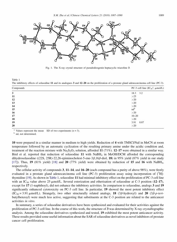
of 3 had not been determined. Thus, we confirmed the structure of 3 using X-ray crystallographic analysis (21b-
methyl) (Fig. 1) [15].
Having established the facile route from 2 to 4, we decided to carry out a limited SAR study to explore the influence
of C-3 substituents and the E ring on the activities of solasodine analogs.
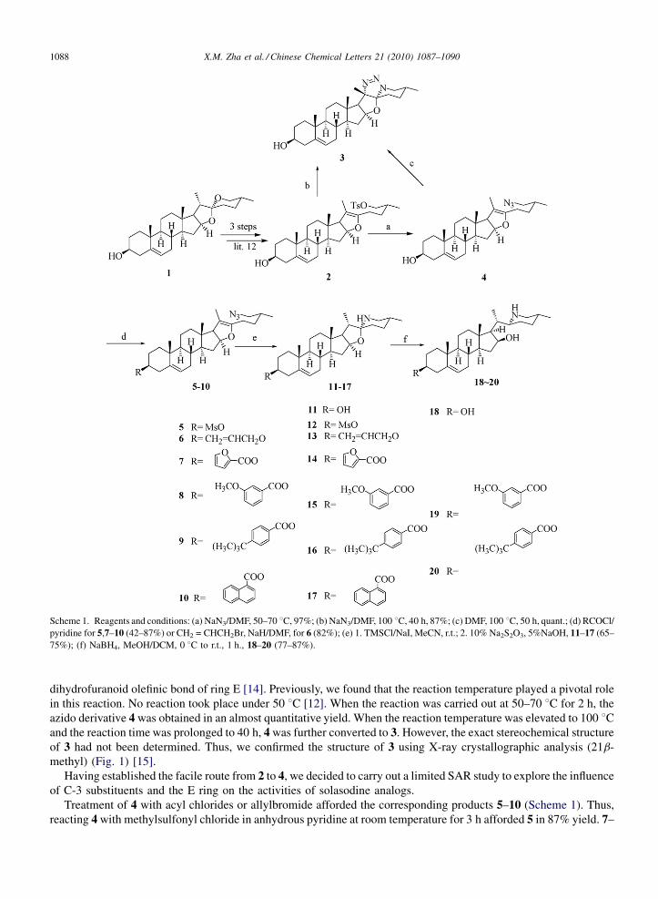
Treatment of 4 with acyl chlorides or allylbromide afforded the corresponding products 5–10 (Scheme 1). Thus,
reacting 4 with methylsulfonyl chloride in anhydrous pyridine at room temperature for 3 h afforded 5 in 87% yield. 7–
X.M. Zha et al. / Chinese Chemical Letters 21 (2010) 1087–10901088
Scheme 1. Reagents and conditions: (a) NaN3/DMF, 50–70 8C, 97%; (b) NaN3/DMF, 100 8C, 40 h, 87%; (c) DMF, 100 8C, 50 h, quant.; (d) RCOCl/
pyridine for 5,7–10 (42–87%) or CH2 = CHCH2Br, NaH/DMF, for 6 (82%); (e) 1. TMSCl/NaI, MeCN, r.t.; 2. 10% Na2S2O3, 5%NaOH, 11–17 (65–
75%); (f) NaBH4, MeOH/DCM, 0 8C to r.t., 1 h., 18–20 (77–87%).
10 were prepared in a similar manner in medium to high yields. Reduction of 4 with TMSCl/NaI in MeCN at room
temperature followed by an automatic cyclization of the resulting primary amine under the acidic condition and,
treatment of the reaction mixture with Na2S2O3 solution, afforded 11 (71%). 12–17 were obtained in a similar way.
Bird et al. reported that reduction of solasodine 11 with NaBH4 in MeOH/DCM afforded the corresponding
dihydrosolasodine ((22S, 25R)-22,26-epiminocholest-5-ene-3b,16b-diol, 18) in 95% yield (87% yield in our study
[17]). Thus, 19 (81% yield) [18] and 20 (77% yield) were obtained by reduction of 15 and 16 with NaBH4,
respectively.
The cellular activity of compounds 3, 11–14, and 16–20 (each compound has a purity of above 98%), were firstly
evaluated in a prostate gland adenocarcinoma cell line (PC-3) proliferation assay using incorporation of [3H]-
thymidine [19]. As shown in Table 1, solasodine 11 had minimal inhibitory effect on the proliferation of PC-3 cell line
with an IC50 value above 25 mmol/L. Several esterization and etherisation of solasodine at C-3 position (12–17),
except for 17 (1-naphthoyl), did not enhance the inhibitory activities. In comparison to solasodine, analogs 3 and 19
significantly enhanced cytotoxicity on PC-3 cell line. In particular, 19 showed the most potent inhibitory effect
(IC50 = 3.91 mmol/L). Strangely, two other structurally related analogs, 18 (3b-hydroxyl) and 20 (3b-p-tert-
butylbenzoyl) were much less active, suggesting that substituents at the C-3 position are related to the anticancer
activities in vitro.
In summary, a series of solasodine derivatives have been synthesized and evaluated for their activities against the
proliferation of PC-3 cell line. In the course of this work, the structure of 3 was determined by X-ray crystallographic
analysis. Among the solasodine derivatives synthesized and tested, 19 exhibited the most potent anticancer activity.
These results provided some useful information about the SAR of solasodine derivatives as novel inhibitors of prostate
cancer cell proliferation.
X.M. Zha et al. / Chinese Chemical Letters 21 (2010) 1087–1090 1089
Fig. 1. The X-ray crystal structure of pseudodiosgenin heptacyclic triazoline 3.
Table 1
The inhibitory effects of solasodine 11 and its analogues 3 and 12–20 on the proliferation of a prostate gland adenocarcinoma cell line (PC-3).
Compounds PC-3 cell line (IC50a, mmol/L)
3 18.3 � 3.2
11 >25
12 >20
13 >20
14 >20
15 ndb
16 >20
17 10–20
18 >20
19 3.91 � 0.87
20 >20
a Values represent the mean �SD of two experiments (n = 3).b nd, not determined.

Acknowledgment
We thank Dr Sensen Lin (Jiangsu Center for Drug Screening, China Pharmaceutical University) for discussion
about the biological aspects of this manuscript.
References
[1] S.M. Kupchan, S.J. Barboutis, J.R. Knox, et al. Science 150 (1965) 1827.
[2] K.R. Lee, N. Kozukue, J.S. Han, et al. J. Agric. Food Chem. 52 (2004) 2832.
[3] B.E. Cham, B. Daunter, Cancer Lett. 55 (1990) 221;
B. Daunter, B.E. Cham, Cancer Lett. 55 (1990) 209.
[4] N. Takanori, K. Chieko, Y.Y. Lee, et al. Biol. Pharm. Bull. 19 (1996) 564.
[5] Y.H. Chen, Q.T. Zhou, D.L. Bai, Acta Pharm. Sin. 33 (1998) 436.
[6] S. Emmanuel, S. Ignacimuthu, R. Perumalsamy, et al. Fitoterapia 77 (2006) 611.
[7] W. Gaffield, R.F. Keeler, Chem. Res. Toxicol. 9 (1996) 426.
[8] B. Samuel, S. Switz, Dtsch. Apoth. Z 136 (1996) 19.
[9] L.C. Chang, K.P.L. Bhat, E. Pisha, et al. J. Nat. Prod. 61 (1998) 1257.
[10] L. Liu, China Patent Application: CN1552724, 2004.;
L. Liu, China Patent Application: CN1629182, 2005.
[11] M. Millward, A. Powell, S. Tyson, et al. J. Clin. Oncol. 23 (2005) 218s.
[12] X.M. Zha, H.B. Sun, J. Hao, et al. Chem. Biodiv. 4 (2007) 25.
[13] X.M. Zha, Y.W. Hou, H.B. Sun, et al. Chem. Biodiv. 4 (2007) 1557.
[14] F.C. Uhle, J. Org. Chem. 32 (1967) 1596.
[15] Crystallographic data (excluding structure factors) has been deposited with the Cambridge Crystallographic Data Centre as supplementary
publication numbers CCDC 689358 (compound 3). Copies of the data can be obtained, free of charge, on application to CCDC, 12 Union Road,
Cambridge CB2 1EZ. UK (fax: +44 (0)1223 336033 or E-mail: [email protected] or http://www.ccdc.cam.ac.uk).
[16] G.J. Bird, D.J. Collins, F.W. Eastwood, et al. Aust. J. Chem. 32 (1979) 783.
[17] Analytical data for compound 18: Mp 264–265 8C. IR (KBr): 3417, 2933, 1460, 1046 cm�1. 1H NMR (300 MHz, CDCl3): d 5.36 (brd, 1H),
4.42 (m, 1H), 3.53 (m, 1H), 3.00 (brd, 1H), 2.57 (brd, 1H), 1.06 (d, 3H, J = 7.1 Hz), 1.02 (s, 3H), 0.93 (s, 3H), 0.83 (d, 3H, J = 6.6 Hz). 13C
NMR (300 MHz, CDCl3): d 140.8; 121.6; 71.8; 71.2; 62.8; 59.8; 54.5; 54.4; 50.3; 42.8; 42.4; 40.2; 37.3; 36.6; 35.9; 35.9; 33.7; 31.9; 31.7; 31.6;
31.5; 31.5; 27.5; 20.9; 19.4; 19.0; 13.5. TOF-MS (m/z): 416.3 ([M+H]+). (The analytical data of 18 were identical with the data reported: see
Ref. [16]).
[18] Analytical data for compound 19: Mp. >300 8C. IR (KBr): 3433, 2948, 1719, 1276, 756 cm�1. 1H NMR (300 MHz, CDCl3): d 7.63 (m, 1H);
7.55 (m, 1H), 7.31 (m, 1H), 7.09 (m,1H), 5.42 (brd, 1H), 4.84 (m, 1H), 4.48 (m, 1H), 3.85 (s, 3H), 3.20 (brd, 1H), 2.85 (brd, 1H), 2.46 (brd, 2H),
1.13 (d, 3H, J = 7.2 Hz), 1.07 (s, 3H), 0.93 (s, 3H), 0.88 (d, 3H, J = 6.4 Hz). 13C NMR (300 MHz, CDCl3): d 165.8; 159.6; 139.7; 132.3; 129.3;
122.6; 122.0; 119.2; 114.2; 77.2; 74.7; 62.3; 58.6; 55.5; 54.3; 53.3; 50.1; 42.8; 40.0; 38.2; 37.1; 36.7; 34.9; 33.0; 31.8; 31.6; 30.2; 27.9; 27.0;
20.8; 19.4; 19.1; 18.2; 13.4. TOF-HRMS (m/z): calcd for C35H52NO4 ([M+H]+) 550.3896, found 550.3878.
[19] PC-3 cells were obtained from ATCC and were cultured in RPMI medium, supplemented with 10% FBS and 50 units/mL penicillin plus 50 mg/
mL streptomycin. Cells were grown in a humidified incubator at 37 8C in an atmosphere of 5% CO2. In cell proliferation analysis, cultured PC-
3 cells were seeded into 96-well plates before treatment with drug for 28 h. 0.5 mCi [3H]-thymidine was added for the last 6 h before cells were
trypsinized and harvested by Cell Harvester (Tomtech, CT). The scintillation counts from the isotope incorporated in the newly synthesized
DNA was counted by Beta-Counter (PerkinElmer). Data were collected and analyzed by GraphPad Prism (La Jolla, CA) 4.0 software.
X.M. Zha et al. / Chinese Chemical Letters 21 (2010) 1087–10901090

![Ferrocene derivatives: Potential anticancer material · 2. Randles-Sevcik equation was used for calculating diffusion coefficient for the reversible redox process [13, 14]: ip = (269,000)](https://img.dokumen.tips/doc/110x75/5e6e8f7bc0c3d577da051471/ferrocene-derivatives-potential-anticancer-material-2-randles-sevcik-equation.jpg)