Embed Size (px)
Citation preview

on August 22, 2006
ww
w.jcb.org
Dow
nloaded from
Synemin and Vimentin are Components of
Intermediate Filaments in Avian Erythrocytes
BRUCE L. GRANGER, ELIZABETH A. REPASKY, and ELIAS LAZARIDES Division of Biology, California Institute of Technology, Pasadena, California 91125
ABSTRACT Synemin, a high-molecular-weight protein associated with intermediate filaments in muscle, and vimentin, an intermediate-filament subunit found in many different cell types, have been identified by immunologic and electrophoretic criteria as components of intermediate filaments in mature avian erythrocytes. Desmin, the predominant subunit of intermediate filaments in muscle, has not been detected in these cells. Two-dimensional immunoautoradiography of proteolytic fragments of synemin and vimentin demonstrates that the erythrocyte proteins are highly homologous, if not identical, to their muscle counterparts. Double immunofluorescence reveals that erythrocyte synemin and vimentin co-localize in a cytoplasmic network of sinuous filaments that extends from the nucleus to the plasma membrane and resists aggregation by Colcemid. Erythrocytes that are attached to glass cover slips can be sonicated to remove nuclei and nonadherent regions of the plasma membrane; this leaves elliptical patches of adherent membrane that retain mats of vimentin- and synemin-containing intermediate filaments, as seen by immunofluorescence and rotary shadowing. Similarly, mechanical enucleation of erythrocyte ghosts in suspension allows isolation of plasma membranes that retain a significant fraction of the synemin and vimentin, as assayed by electrophoresis, and intermediate filaments, as seen in thin sections. Both synemin and vimentin remain insoluble, along with spectrin and actin, in solutions containing nonionic detergent and high salt. However, brief exposure of isolated membranes to distilled water releases the synemin and vimentin together in nearly pure form, before the release of significant amounts of spectrin and actin. These data suggest that avian erythrocyte intermediate fil-aments are somehow anchored to the plasma membrane; erythrocytes may thus provide a simple system for the study of intermediate filaments and their mode of interaction with membranes. In addition, these data, in conjunction with previous data from muscle, indicate that synemin is capable of associating with either desmin or vimentin and may thus perform a special role in the structure or function of intermediate filaments in erythrocytes as well as in muscle.
Mature avian erythrocytes are nucleated, biconvex, elliptical discs that contain relatively few cytoplasmic organelles. Removal of hemoglobin from these cells by hypotonic lysis (15) reveals an equatorial bundle of microtubules known as the marginal band (5), a submembranous spectrin-actin shell (9), as well as a residual network of cytoplasmic ftlaments that surrounds the mitochondria and extends from the nucleus to the plasma membrane (27). This latter network of fl.l.aments is probably a component of the "trans marginal band material" noted in many nonmammalian vertebrate erythrocytes (13). These ftlaments appear to be of the type known as intermediate flla.ments (33, 39), due to their characteristic ultrastructural morphology and insolubility in nonionic detergents (59, 63).
THE )OURNAL OF CELL BIOLOGY • VOLUME 92 fEBRUARY 1982 299-312 ©The Rockefeller University Press. 0021-9525/82/02/0299/14$1.00
The close association of these ftlaments with the plasma membrane and nucleus, as shown by electron microscopy, suggests that they might function to maintain the shape of the cell or position the nucleus within the cell {13, 27, 59, 63).
We have examined these flla.ments biochemically, immunologically, and ultrastructurally and have determined that they are composed predominantly of vimentin, an intermediate ftlament subunit common to many different cell types (18, 39). The other major component of these flla.ments is synemin, a high molecular weight protein originally isolated from avian smooth muscle in association with desmin and subsequently shown to co-localize with desmin and vimentin in skeletal muscle (25). Double immunofluorescence shows that erythro-
299

on August 22, 2006
ww
w.jcb.org
Dow
nloaded from
cyte synemin and vimentin also coexist in a network of cytoplasmic ftlaments. The electrophoretic and immunologic criteria used to identify vimentin and synemin in these cells fail to detect desmin, the major intermediate filament subunit of muscle (40, 53) that is also found in some nonmuscle cells (19, 55, 58).
Various cell fractionation procedures based on differential centrifugation have indicated that synemin and vimentin sediment with both nuclear and membrane fractions. In this study we have concentrated on those intermediate ftlaments that remain associated with the erythrocyte plasma membrane after mechanical enucleation of the cells. These fllaments resist dissociation from the membranes by sonication and treatment with high salt and nonionic detergent, suggesting that they are in some way anchored to the membrane cytoskeleton, perhaps to the spectrin-actin network. However, we have found that the ftlaments can be selectively removed from the membranes by treatment with low ionic strength solutions and that the predominant proteins thus released are vimentin and synemin.
The evidence presented here that synemin associates with vimentin in erythrocytes, in conjunction with the evidence presented previously that synemin associates with desmin in smooth muscle and desmin and vimentin in skeletal muscle (25), suggests that synemin may be capable of associating with different intermediate fllament subunits in different cell types. Because synemin is not detectable in all cell types, it may play a special role in the structure or function of certain types of intermediate ftlaments or filaments in certain cell types.
MATERIALS AND METHODS
Preparation of Erythrocyte Membranes
White leghorn chickens were given intravenous injections of 3-4 mg (500-700 USP units) of heparin (17) and then -50 mg of sodium pentobarbital. Blood was collected from the neck vein into a solution ofO.Ol% heparin, 5 mM HEPES (N· 2-hydroxyethylpiperazine-N'-2-ethanesulfonic acid), 155 mM choline chloride (pH 7.1 at room temperature). Phosphate-buffered saline was simultaneously injected into a wing vein, and the perfusate was also collected until it became relatively clear. Alternatively, blood for some experiments was drawn from wing veins or neck veins of uninjected chickens and collected in 1-2 vol of the above heparin-containing solution. Blood cells were pelleted by centrifugation for 5 min at 1,000 g, and the top white layer of cells (buffy coat) and supernatant were removed by aspiration. The erythrocyte pellet, exclusive of a dark-red layer adhering to the bottom of the centrifuge tube, was resuspended in 155 mM choline chloride, 5 mM HEPES (pH 7 .I at room temperature) and recentrifuged. Again, the supernatant, buffy coat, and dark-red layer were discarded. This cycle was repeated for a total of 4-8 washes, and was performed either at room temperature or at4°C.
The fmal pellet of erythrocytes was rapidly resuspended in at least 10 vol of ice-cold hypotonic lysis buffer (Buffer H) [5 mM Tris-Cl (pH 7.5), 5 mM NaN3,
5 mM MgCI2, I mM EGTA (ethyleneglycol-bis(.B-amino-ethyl ether) N,N'-tetraacetic acid), I mM dithioerythritol (OTE) or dithiothreitol (OTT), 0.5 mM pheny!methyl sulfonyl fluoride (PMSF)). MgCh was included to keep the nuclei intact, EGTA and PMSF were included as protease inhibitors, and OTE (or OTT) was found to increase both the yield and size of the plasma membrane fragments (see below). The resulting nucleated erythrocyte ghosts were pelleted by centrifugation for 5 min at approximately 10,000 g, then resuspended in at least I 0 vol of the same solution. This cycle was repeated for a total of three or four washes in Buffer H.
For the preparation of plasma membranes, the final pellet was resuspended in 2-4 vol of Buffer H, loaded into a syringe, and forced rapidly through a 23 gauge hypodermic needle bent into the shape of a Z (with two 30° angles). Centrifugation for 10-20 min ati,OOO gin a swinging bucket rotor resulted in three layers: a firm, white pellet of free nuclei on the bottom, a loose, pink layer of undisrupted cells in the middle, and a supernatant containing soluble proteins and membrane fragments. The middle layer was resuspended in Buffer H and again forced through the needle and centrifuged to give the three layers. This was usually repeated three times; the yield of membrane fragments increased with each cycle. The supernatants from each centrifugation were combined and recentrifuged for 30 min at 100-200 g to remove any remaining nuclei. The supernatant of this low
300 THE jOURNAL OF CELL BIOLOGY • VOLUME 92, 1982
speed centrifugation was respun for 10 min at 20,000 g to pellet the membranes which were subsequently resuspended in 10-20 vol of Buffer Hand stored on ice. Contaminating nuclei visible by phase contrast microscopy could be removed by another low speed centrifugation. After storage for a month on ice, little change was seen in the electrophoretic protein proflle as judged by one dimensional SOS-polyacrylamide gel electrophoresis (SOS-PAGE) (see below).
Selective Extraction of Synemin and Vimentin From Erythrocyte Membranes
The suspension of erythrocyte membranes was mixed with 10-30 vol of2 mM EOTA 10 mM Tris (pH adjusted with HCI to 7.4 at 0°C); after 10 min on ice the membranes were pelleted by centrifugation at 20,000 g for 10 min. The pellet was resuspended (with a Pasteur pipette) in distilled water at o•c and centrifuged as above. The supernatant was then lyophilized directly for electrophoretic analysis, or first recentrifuged for 1-5 h in a Beckman SW 50.1 swinging bucket rotor at 50,000 RPM (Beckman Instruments, Inc., Fullerton, Calif.).
Polyacrylamide Gel Electrophoresis
One-dimensional SOS-PAGE was based on the discontinuous, Tris-glycine system of Laemmli (37), as modified and described previously (29). Separating gels were II x 14 x 0.16 em and contained 12.5% acrylamide and 0.11% N,N'methylene-bisacrylamide. Samples were solubilized with I% SOS, 125 mM TrisCl (pH 6.8), 10% glycerol, 1% 2-mercaptoethanol, I mM EOTA, 0.004% bromophenol blue("!% SOS sample buffer"), and immediately placed in a boiling water bath for -1 min.
Two-dimensional isoelectric focusing (IEF)/SOS-PAGE was performed according to the method of O'Farrell (48), as modified and described previously (29), except that nonidet P-40 (NP-40) was omitted from all gels and samples (this enhanced the resolution of the isoelectric variants of several proteins of interest in this system). Second-dimension SOS slab gels were as described above.
Phosphorylation
Incorporation of [32P)-phosphate into erythrocyte proteins was performed essentially as described by Beam et al. (4) and Alper et al. (2). Blood was collected from the wing vein of an adult hen turkey in cold heparinized choline chloride buffer and washed as described above. Erythrocytes were then washed once at room temperature with 40 vol of 157.5 mM NaCI, 2.5 mM KCI, 11.1 mM D·
glucose, 10 mM HEPES (brought to pH 7.65 at room temperature with NaOH). I ml of packed cells was resuspended in 9 ml of this solution; 2 mCi of 32Pphosphoric acid (New England Nuclear, Boston, MA; 100 1'1 of carrier-free in 20 mM HCI) were then added and the suspension was incubated in an orbital shaker bath at 39°C. After 3.5 h the suspension was divided in half; to one-half was added 50 pl of 0.1 mM OL-isoproterenol-HCI (Sigma Chemical Co., St. Louis, MO) in the above solution (fmal concentration I I'M). Both aliquots were incubated for another 20 min at 39°C and then processed in parallel for gel electrophoresis. The erythrocytes were spun down and washed once with 40 vol of the above solution at 39°C, then lysed with 80 vol of 10 mM Tris-Cl (pH 8.0), 5 mM MgCI,, I mM EGTA, I mM o-phenanthroline, 0.5 mM PMSF, I mM NaF at ooc. The nucleated ghosts were spun down and washed once with this solution, then disrupted by one passage through a bent hypodermic needle as described above. Intact cells and nuclei were removed by low-speed centrifugation, and free membrane fragments were collected by high-speed centrifugation. The membrane pellet was boiled in I% SOS sample buffer and analyzed by SOSPAGE. After staining and destaining, the gel was dried on ftlter paper and exposed to Kodak X-Omat R XR5 film at room temperature for 10 d without an intensifying screen.
In a separate experiment, erythrocytes were collected, washed and labeled as described above, except that the labeling period was 20 h and no isoproterenol was added. Cells were lysed with 5 mM sodium phosphate, 5 mM MgC!,, I mM EGTA, I mM PMSF (pH 7.4 at 4°C) and disrupted as described above. The membrane fraction was dissolved in I 0 M urea containing I% 2-mercaptoethanol, analyzed by IEF/SOS-PAGE, and autoradiographed.
lmmunoautoradiography
Immunoautoradiography was performed as described previously (25). Gels were incubated with antisera diluted 1,000-fold, followed by radioiodinated protein A (4-7 I'Ci 125I/I'g protein A; each gel was incubated with 20-30 ,.ci in 100 ml of solution). The dried gels were exposed to x-ray film for the following times with(+) or without(-) an intensifying screen: Fig. 3b, 27 h (-);Fig. 3c, 15 h (+);Fig. 4b, 36 h (-);Fig. 4c; 55 h (+);Fig. Sb, 405 h (-);Fig. 5d, 42 h (+).
on August 22, 2006
ww
w.jcb.org
Dow
nloaded from
Samples for electrophoresis were prepared as follows. Fig. 3: The white cell layer (buffy coat) was collected from the first centrifugation of heparinized chicken blood and recentrifuged twice in HEPES-buffered saline containing I mM EDT A to give an erythrocyte-free preparation. The cells were mixed witb 40 vol of ethanol at 0°C, pelleted, and boiled in 1% SDS sample buffer. Whole adult chicken-gizzard smootb-muscle tissue was frozen and pulverized in liquid nitrogen, thawed in etbanol, pelleted, and boiled in 1% SDS sample buffer. Turkey erythrocyte membranes were extracted at 0° C for 20 min witb I 0 mM Tris, I mM EDTA, I M NaCI, 1% 2-mercaptoethanol, 1% Triton X-100, and tbe residue was pelleted and boiled in 1% SDS sample buffer. Chicken erythrocyte membranes were extracted at 0°C for 70 min with 10 mM Tris, 2 mM EDTA, 0.5 M KCI, 1% Triton X-100 (pH 7.4), and the residue was pelleted and boiled in 1% SDS sample buffer. Another aliquot of chicken erythrocyte membranes was washed at 0°C with 10 mM Tris-2 mM EDTA (pH 7.4) and tben treated for 4 h at 0°C with 10 mM Tris-1 mM EGTA (pH 7.4); tbe extract was dialyzed against water, lyophilized, and boiled in 1% SDS sample buffer.
Fig. 4: Chicken erythrocyte membranes were treated with 60 vol of ethanol to permeabilize the vesicles, pelleted, and dissolved at room temperature in a saturated urea solution containing I% 2-mercaptoethanol.
Fig. 5: Chicken erythrocytes were extracted twice at o•c with 1% Triton X-100, 150 mM NaCI, 2 mM EGTA, 5 mM MgC!,, I mM sodium tetrathionate, I mM E-amino-n-caproic acid, I mM o-phenanthroline, I mM PMSF, 10 mM TrisCl (pH 7.2) and rinsed twice with this solution without the Triton X-100. The resulting cytoskeletons were then extracted with this latter solution containing 6 M urea for 1.5 hat 0°C. This extract was dialyzed against water, and the resulting precipitate was collected and dissolved in a saturated urea solution containing 0.5% 2-mercaptoethanol. Whole adult chicken-gizzard smooth-muscle tissue was frozen and pulverized in liquid nitrogen, thawed in ethanol, pelleted, boiled for 30 s in I 0 1-11 of I% SDS, and then dissolved in 90 1-11 of saturated urea containing 1% 2-mercaptoethanol.
Immunofluorescence
Glass cover slips were pretreated with Alcian Blue to promote erythrocyte adhesion (54). Cover slips were cleaned, simmered for 5 min in 0.1% Alcian Blue SGX (Sigma Chemical Co., St. Louis, Mo.), rinsed with distilled water, and air dried. Washed erythrocytes in the choline chloride/HEPES buffer were allowed to settle on the cover slips for 5-10 min at room temperature; nonadherent cells were removed by rinsing with the same solution.
For the sonication experiments, cover slips with attached erythrocytes were hypotonically lysed at room temperature in Buffer H and then placed in Buffer F [130 mM KCI, 5 mM NaCl, I mM NaN3, 5 mM MgC!,, I mM EGTA, 20 mM potassium phosphate (pH 7 .5)). Cover slips were laid face-up in a beaker of buffer F and sonicated for 20 sat 20 watts using a Braunsonic 1510 sonicator (B. Braun Instruments, San Francisco, Calif.) with a 4-mm titanium probe tip positioned 3-4 em above the cells. A glass rod was used to hold the cover slips in position on the bottom of tbe beaker during sonication. Incubations witb antisera and subsequent washes were all performed at room temperature in buffer TM (0.5% Triton X-100. 130 mM NaCI, 5 mM KCI, 5 mM NaN3, 5 mM MgCI, I mM EGTA, 10 mM Tris-Cl (pH 7.5)].
Alternatively, sonication was performed in Buffer Hand antibody incubations and washes in Buffer F without the MgCI2 and EGTA (Fig. Sa,b,c,h).
To investigate the Colcemid sensitivity of the filaments, turkey erythrocytes attached to cover slips were incubated for 16 h at 37°C in growth medium (Eagle's minimal essential medium, nonessential amino acids, 15% horse serum, 5% chick embryo extract, 0.0 I% streptomycin and I 00 U penicillin/ml) containing 5 1-1M Colcemid (demecolcine; Calbiochem-Behring Corp., La Jolla, Calif.). Control cells were treated identically, except that they were not exposed to Colcemid, Cover slips were placed in Buffer TM containing 0.5 mM PMSF for I min at room temperature to make cytoskeletons, then fixed for 10 min at room temperature in Buffer F containing 2% formaldehyde. Subsequent incubations and washes were done in Buffer TM containing a 0.5 mM PMSF, Similar results were obtained with unfixed cells, with cells incubated in suspension rather tban attached to cover slips, and with cells incubated with 100 )LM Colcemid.
Rabbit anti-vimentin was prepared using antigen from embryonic chicken skeletal muscle cytoskeletons (24). Rabbit anti-desmin was prepared using desmin present in a low-salt extract of chicken-gizzard smooth muscle (25). Rabbit antisynemin was prepared using antigen obtained from chicken-gizzard intermediatemament proteins that had been solubilized and precipitated three times in acetic acid (25). All antigens were ultimately purified by preparative SDS-PAGE before injection.
Conjugation of anti-vimentin with rhodamine B was performed as described (25). Double immunofluorescence was performed by the indirect/direct method (32). Fluorescein-conjugated goat anti-rabbit IgG was purchased from MilesVeda, Ltd. (Rehovot, Israel) and diluted \50-fold for use. Primary antisera were partially purified by precipitation with ammonium sulfate at 50% saturation and used at -1/30 serum concentration.
Cover slips were mounted in 90% glycerol in Tris-buffered saline and photographed witb Kodak Tri-X ftlm using a Leitz phase/epifluorescence microscope and fllter modules K and N2. The patches of plasma membrane in Fig. 8 s are visible by phase-contrast microscopy because an air bubble was present under that portion of the cover slip.
Electron Microscopy
For thin sectioning, chicken erythrocyte membranes were pelleted and ftxed with 1% glutaraldehyde, 5 mM MgCh. 10 mM sodium phosphate (pH 7.5), postftxed in 1% Os04 , 0.1 M sodium cacodylate (pH 7.4), stained at 60°C with 1% uranyl acetate, 0.1 M sodium maleate (pH 5.15), dehydrated in ethanol and propylene oxide, and embedded in a 9/16/1 mixture of 1,2,7,8-diepoxyoctane (Aldrich; Milwaukee, Wis.)/nonenyl succinic anhydride (ICN K/K Laboratories, Inc., Plainview, N. Y.)/DMP-30. The resin was cured for 2 d at 60°C and sectioned with glass knives on a Reichert OmU2 ultramicrotome. Sections were stained for 3 min with 0.2% lead citrate, examined with a Philips EM201 at 80 kV, and photographed on 35-mm film.
Shadowed replicas were made as follows: Chicken erythrocytes adhering to Alcian Blue-coated cover slips were lysed and sonicated as described under Immunofluorescence. The cover slips were immersed in buffered I% glutaraldehyde followed by I% Os04, dehydrated witb ethanol, dried in a carbon dioxide critical-point drier, rotary shadowed with platinum/palladium (80/20) at an angle of 6°, and then carbon coated. Replicas were separated from cover slips with 5% HF, washed witb water, mounted on 300-mesh copper grids, and viewed by transmission electron microscopy.
RESULTS
Membrane Fractionation and Ultrastructure
The study of minor protein components of avian erythrocytes is hampered by the relative abundance of hemoglobin and chromatin in these cells. Removal of these two components makes biochemical, immunological and ultrastructural characterization of the remaining structures easier. Hypotonic lysis (to remove hemoglobin) and subsequent mechanical enucleation (to remove chromatin) gives a preparation of membranes that can be studied in a manner analogous to the study of the simpler mammalian erythrocyte ghosts. To this end, chicken and turkey erythrocytes were isolated from fresh blood by differential centrifugation and lysed in a low osmolarity buffer containing magnesium ions to keep the nuclei intact (27). These nucleated ghosts were then disrupted by passage through a bent hypodermic needle, and a membrane fraction was separated from the nuclei and unbroken cells by differential centrifugation. This membrane fraction is the main object of this study and will hereafter be referred to as "erythrocyte membranes."
Representative thin sections of the erythrocyte membranes are shown in Fig. 1. The preparation is composed primarily of plasma membranes, both complete and in pieces (compare with sections of whole cells in references 4, 5, 63, 66). Close examination reveals that in a given thin section many of the membranes have ftlaments associated with them. These ftlaments are -9 nm in diameter and are therefore classified as intermediate filaments. They are present on the cytoplasmic side of the plasma membrane fragments and often appear to be in close apposition to the protein network (analogous to the spectrin network in mammalian erythrocytes) just inside the lipid bilayer. This is especially apparent in grazing sections of the membrane in these relatively thick sections. The ftlaments are usually curved and randomly distributed and do not exhibit any obvious association with specific cell structures.
Thin sections of the free nuclei (not shown) also reveal intermediate filaments associated with these structures, as previously shown by Woodcock (63). It has not been determined what proportion of the filaments remains with the membranes and what proportion with the nuclei, due to the difficulty of
GRANGER ET AL. Synemin and Vimentin in Avian Erythrocytes 301
on August 22, 2006
ww
w.jcb.org
Dow
nloaded from
FIGURE 1 Thin sections of chicken erythrocyte membranes. (a) Medium-power view of pelleted membranes showing that filaments can be seen associated with many, but not all, membranes in a given section (X 11,000). ( b, c) High-power views. Note cytoplasmic filaments and frequent close associations between filaments and membranes ( b: X 38,000; c: x 47,000). Bars, 500 nm.
enucleating the ghosts quantitatively and completely separating the resulting fractions. Such a quantitation is also complicated by the fact that the proportion of the ftlaments associated with the nucleus before cell disruption that remains associated with the nuclei after fractionation is probably a function of the severity of the disruption procedure and the extent of loss of the outer nuclear membrane. However, based on various biochemical and ultrastructural data (see below), it appears that on the average less than half of the cell's intermediate ftlaments end up in the membrane fraction.
It is apparent from these thin sections that the erythrocyte membrane fraction contains low levels of contamination by fragments of structures other than the plasma membrane. Even though this enucleation procedure results in free nuclei that appear to be intact by phase-contrast microscopy, the relatively fragile outer nuclear membrane may become partially fragmented and fractionate with the plasma membranes (27, 65). Fragments of mitochondrial membranes may also be present in this fraction. However, because our studies were concerned primarily with intermediate ftlaments rather than specific membrane proteins, further purification of the membrane fraction was not deemed necessary for subsequent biochemical studies. The purpose of the fractionation was to remove chromatin that would have physically interfered with the membrane extraction experiments, and this was accomplished. Negligible amounts of histone could be seen when the membrane fraction was analyzed by SDS-PAGE, and nuclear membrane lamins (21, 52) could not be detected by IEF/SDS-PAGE, showing that the level of contaminating material was low.
Electrophoretic Analysis of Membrane Fraction
Analysis of the protein composition of avian erythrocyte membranes was performed with regard to the voluminous work on mammalian erythrocyte ghosts. Similarities between the two systems include two major high-molecular-weight proteins in avian membranes that correspond to the mammalian erythrocyte spectrins (see Figs. 2 and 6). Avian a-spectrin comigrates by SDS-PAGE with mammalian a-spectrin, but the f3 variant has a higher mobility and can be resolved into a closely spaced doublet on underloaded gels (not shown). Both systems contain
302 THE jOURNAL Of CELL BIOLOGY • VOLUME 92, 1982
actin at 42,000 daltons as well as a broad band of membrane proteins around 100,000 daltons (Band 3; reference 16). Among the characteristic differences are the presences in avian membranes of goblin, a hormonally-regulated phosphoprotein (4), and of the intermediate ftlament proteins, vimentin and synemin. The presence of these two intermediate ftlament components in association with avian erythrocyte membranes was demonstrated by two-dimensional gel electrophoresis (IEF I SDS-PAGE; see Fig. 4a). Erythrocyte vimentin coelectrophoreses in this gel system with vimentin identified in other avian cell types (19, 24); the identification of synemin was tentative at this stage and required immunological and biochemical confirmation, as described below. Desmin was not detected on these electrophoretograms.
Initial biochemical studies of erythrocyte membranes began with attempts to remove peripherally bound proteins from the membrane lipid bilayer. We found that the solubilization or release of any protein components from the erythrocyte membranes, without the use of detergents or strongly chaotropic agents, required the removal of divalent cations. Therefore, before most biochemical experiments, the magnesium ions present in the membrane suspension (in the hypotonic lysis buffer) were removed from the membranes by washing with a low-salt buffer containing EDT A.
Treatment of membranes with solutions of very low ionic strength was expected to release spectrin, by analogy to the mammalian erythrocyte system (42, 43). However, it was observed that if such an extraction was performed briefly at 0°C, then the primary protein released was vimentin. Fig. 2 a shows a two-dimensional gel of the extract obtained by treatment of chicken erythrocyte membranes with distilled water for 30 min on ice. In addition to the four or five isoelectric variants of vimentin at 52,000 daltons, are synemin at 230,000 daltons and actin at 42,000 daltons. Identification of the 230,000 dalton polypeptide as synemin is based on its immunological crossreactivity with smooth muscle synemin and its immunoautoradiographic peptide map, both as detailed below, as well as its copurification with vimentin. No desmin can be detected on this gel. Distilled water was found to be the optimal solvent for extraction of relatively pure vimentin and synemin, but other low ionic strength solutions (eg., 1-2 mM EDTA or EGTA
on August 22, 2006
ww
w.jcb.org
Dow
nloaded from
a s - -
v
/J"rA
f
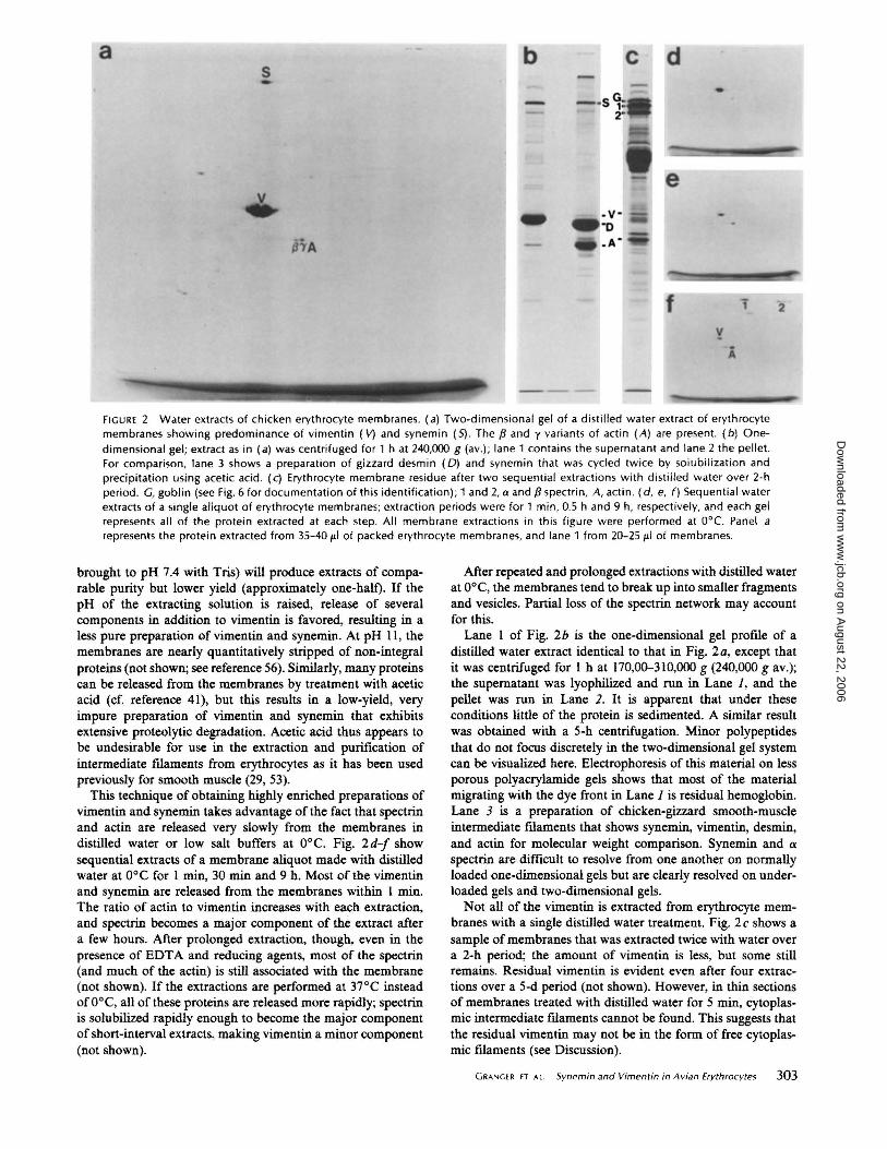
----- -FIGURE 2 Water extracts of chicken erythrocyte membranes. (a) Two-dimensional gel of a distilled water extract of erythrocyte membranes showing predominance of vimentin ( \1) and synemin ( 5) . The f3 and y variants of actin (A) are present. ( b) Onedimensional gel; extract as in (a) was centrifuged for 1 hat 240,000 g (av.); lane 1 contains the supernatant and lane 2 the pellet. For comparison, lane 3 shows a preparation of gizzard des min ( 0) and synemin that was cycled twice by solubi lization and precipitation using acetic acid . (c) Erythrocyte membrane residue after two sequential extractions with distilled water over 2-h period. G, goblin (see Fig. 6 for documentation of this identification) ; 1 and 2, a and f3 spectrin, A, actin . ( d, e, f) Sequential water extracts of a single aliquot of erythrocyte membranes; extraction periods were for 1 min, 0.5 h and 9 h, respect ively, and each gel represents all of the protein extracted at each step. All membrane extractions in this figure were performed at 0°C. Panel a represents the protein extracted from 35-401.11 of packed erythrocyte membranes, and lane 1 from 20-251.11 of membranes.
brought to pH 7.4 with Tris) will produce extracts of comparable purity but lower yield (approximately one-half). If the pH of the extracting solution is raised, release of several components in addition to vimentin is favored, resulting in a less pure preparation of vimentin and synemin. At pH 11 , the membranes are nearly quantitatively stripped of non-integral proteins (not shown; see reference 56). Similarly, many proteins can be released from the membranes by treatment with acetic acid (cf. reference 41), but this results in a low-yield, very impure preparation of vimentin and synemin that exhibits extensive proteolytic degradation. Acetic acid thus appears to be undesirable for use in the extraction and purification of intermediate fl.laments from erythrocytes as it has been used previously for smooth muscle (29, 53).
This technique of obtaining highly enriched preparations of vimentin and synemin takes advantage of the fact that spectrin and actin are released very slowly from the membranes in distilled water or low salt buffers at 0°C. Fig. 2d-f show sequential extracts of a membrane aliquot made with distilled water at 0°C for 1 min, 30 min and 9 h. Most of the vimentin and synemin are released from the membranes within 1 min. The ratio of actin to vimentin increases with each extraction, and spectrin becomes a major component of the extract after a few hours. After prolonged extraction, though, even in the presence of EDT A and reducing agents, most of the spectrin (and much of the actin) is still associated with the membrane (not shown). If the extractions are performed at 37°C instead of 0°C, all of these proteins are released more rapidly; spectrin is solubilized rapidly enough to become the major component of short-interval extracts, making vimentin a minor component (not shown).
After repeated and prolonged extractions with distilled water at 0°C, the membranes tend to break up into smaller fragments and vesicles. Partial loss of the spectrin network may account for this.
Lane I of Fig. 2b is the one-dimensional gel proflle of a distilled water extract identical to that in Fig. 2a, except that it was centrifuged for 1 hat 170,00-310,000 g (240,000 g av.); the supernatant was lyophilized and run in Lane 1, and the pellet was run in Lane 2. It is apparent that under these conditions little of the protein is sedimented. A similar result was obtained with a 5-h centrifugation. Minor polypeptides that do not focus discretely in the two-dimensional gel system can be visualized here. Electrophoresis of this material on less porous polyacrylamide gels shows that most of the material migrating with the dye front in Lane 1 is residual hemoglobin. Lane 3 is a preparation of chicken-gizzard smooth-muscle intermediate ftlaments that shows synemin, vimentin, desmin, and actin for molecular weight comparison. Synemin and a spectrin are difficult to resolve from one another on normally loaded one-dimensional gels but are clearly resolved on underloaded gels and two-dimensional gels.
Not all of the vimentin is extracted from erythrocyte membranes with a single distilled water treatment. Fig. 2c shows a sample of membranes that was extracted twice with water over a 2-h period; the amount of vimentin is less, but some still remains. Residual vimentin is evident even after four extractions over a 5-d period (not shown). However, in thin sections of membranes treated with distilled water for 5 min, cytoplasmic intermediate ftlaments cannot be found. This suggests that the residual vimentin may not be in the form of free cytoplasmic ftlaments (see Discussion).
GRANGER ET AL Synemin and Vi men tin in Avian Erythrocytes 303
on August 22, 2006
ww
w.jcb.org
Dow
nloaded from
Immunological Characterization of Erythrocyte Intermediate Filaments
The technique of immunoautoradiography (10), which uses antibodies to detect protein antigens in polyacrylamide gels, was used in this study for three purposes: (a) to determine whether the erythrocyte intermediate-ftlament subunits were antigenically crossreactive with their muscle counterparts; (b) as a form of peptide map analysis to determine whether the subunits in erythrocytes were homologous or identical to their muscle counterparts; and (c) to detect these antigens in gels with a sensitivity much greater than that afforded by Coomassie Blue staining. Antisera used in this study were all elicited against chicken muscle proteins, purified by SDS-PAGE, and each appears to be specific for its respective antigen as assayed by two-dimensional immunoautoradiography (24, 25).
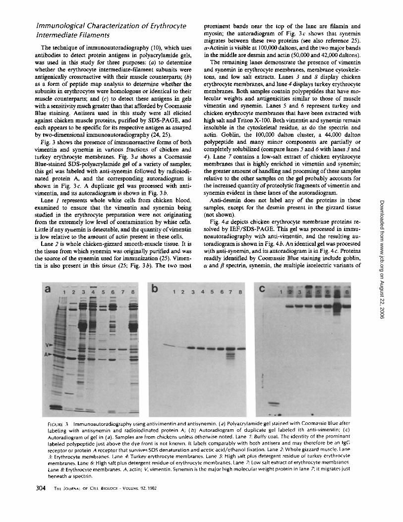
Fig. 3 shows the presence of immunoreactive forms of both vimentin and synemin in various fractions of chicken and turkey erythrocyte membranes. Fig. 3 a shows a Coomassie Blue-stained 80S-polyacrylamide gel of a variety of samples; this gel was labeled with anti-synemin followed by radioiodinated protein A, and the corresponding autoradiogram is shown in Fig. 3 c. A duplicate gel was processed with antivimentin, and its autoradiogram is shown in Fig. 3 b.
Lane 1 represents whole white cells from chicken blood, examined to ensure that the vimentin and synemin being studied in the erythrocyte preparation were not originating from the extremely low level of contamination by white cells. Little if any synemin is detectable, and the quantity ofvimentin is low relative to the amount of actin present in these cells.
Lane 2 is whole chicken-gizzard smooth-muscle tissue. It is the tissue from which synemin was originally purified and was the source of the synemin used for immunization (25). Vimentin is also present in this tissue (25; Fig. 3 b). The two most
prominent bands near the top of the lane are ftlamin and myosin; the autoradiogram of Fig. 3 c shows that synemin migrates between these two proteins (see also reference 25). a-Actinin is visible at 100,000 daltons, and the two major bands in the middle are desmin and actin (50,000 and 42,000 daltons).
The remaining lanes demonstrate the presence of vimentin and synemin in erythrocyte membranes, membrane cytoskeletons, and low salt extracts. Lanes 3 and 8 display chicken erythrocyte membranes, and lane 4 displays turkey erythrocyte membranes. Both samples contain polypeptides that have molecular weights and antigenicities similar to those of muscle vimentin and synemin. Lanes 5 and 6 represent turkey and chicken erythrocyte membranes that have been extracted with high salt and Triton X-100. Both vimentin and synemin remain insoluble in the cytoskeletal residue, as do the spectrin and actin. Goblin, the 100,000 dalton cluster, a 44,000 dalton polypeptide and many minor components are partially or completely solubilized (compare lanes 5 and 6 with lant:s 3 and 4). Lane 7 contains a low-salt extract of chicken erythrocyte membranes that is highly enriched in vimentin and synemin; the greater amount of handling and processing of these samples relative to the other samples on the gel probably accounts for the increased quantity of proteolytic fragments ofvimentin and synemin evident in these lanes of the autoradiogram.
Anti-desmin does not label any of the proteins in these samples, except for the desmin present in the gizzard tissue (not shown).
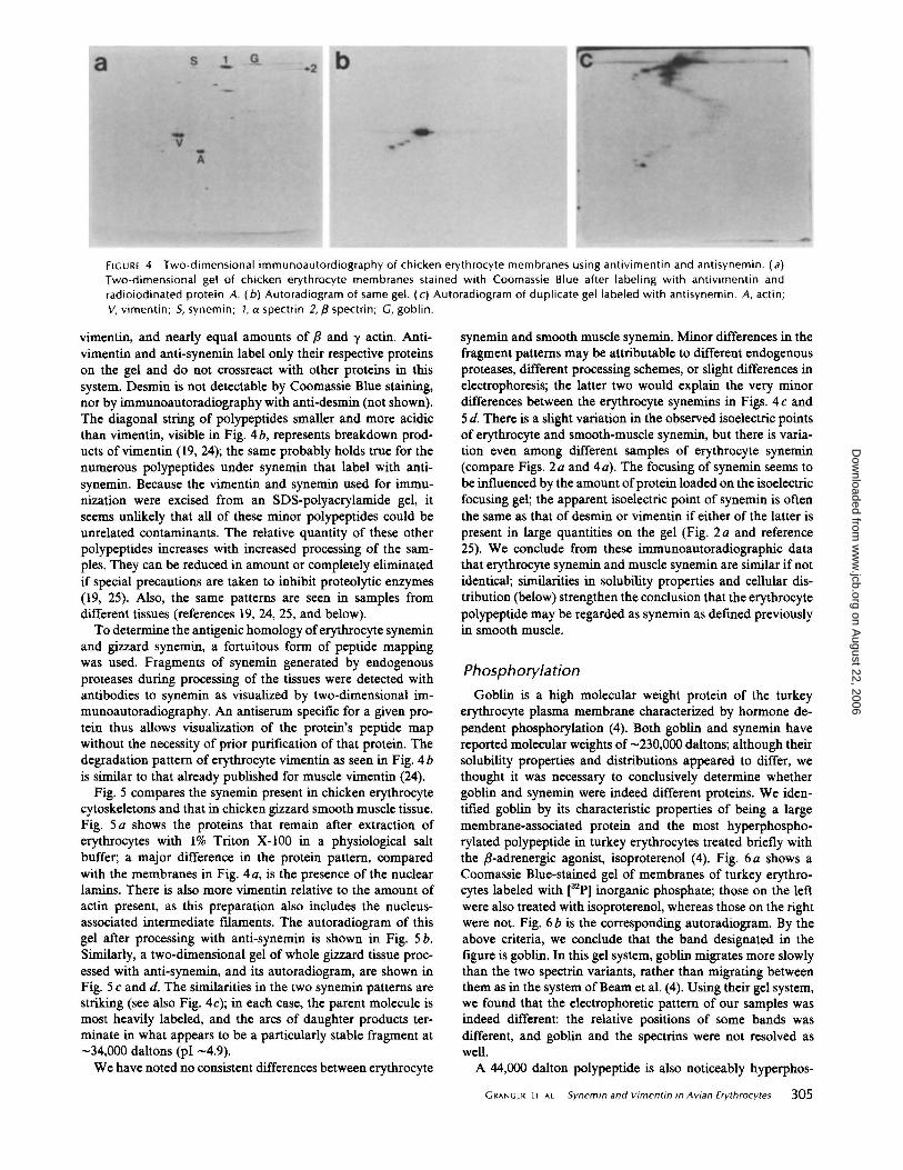
Fig. 4a depicts chicken erythrocyte membrane proteins resolved by IEF/SDS-PAGE. This gel was processed in immunoautoradiography with anti-vimentin, and the resulting autoradiogram is shown in Fig. 4 b. An identical gel was processed with anti-synemin, and its autoradiogram is in Fig. 4 c. Proteins readily identified by Coomassie Blue staining include goblin, a and {3 spectrin, synemin, the multiple isoelectric variants of
a 12345678 b 12345678 c
- -· FIGURE 3 lmmunoautoradiography using antivimentin and antisynemin. (a) Polyacrylamide gel stained with Coomassie Blue after labeling with antisynemin and radioiodinated protein A; (b) Autoradiogram of duplicate gel labeled ith anti-vimentin; (c) Autoradiogram of gel in (a). Samples are from chickens unless otherwise noted. Lane 1: Buffy coat. The identity of the prominant labeled polypeptide just above the dye front is not known. It labels comparably with both antisera and may therefore be an lgG receptor or protein A receptor that survives SDS denaturation and acetic acid/ethanol fixation. Lane 2: Whole gizzard muscle. Lane 3: Erythrocyte membranes. Lane 4: Turkey erythrocyte membranes. Lane 5: High salt plus detergent residue of turkey erythrocyte membranes. Lane 6: High salt plus detergent residue of erythrocyte membranes. Lane 7: Low salt extract of erythrocyte membranes. Lane 8: Erythrocyte membranes. A, actin; V, vimentin. Synemin is the major high molecular weight protein in lane 7; it migrates just
beneath a spectrin.
304 THE jOURNAL OF CELL BIOLOGY • VOlUME 92, 1982
on August 22, 2006
ww
w.jcb.org
Dow
nloaded from
b G s a •2
v -A
FIGURE 4 Two-dimensional immunoautordiography of chicken erythrocyte membranes using antivimentin and antisynemin. (a) Two-dimensional gel of chicken erythrocyte membranes stained with Coomassie Blue after labeling with antivimentin and radioiodinated protein A. (b) Autoradiogram of same gel. (c) Autoradiogram of duplicate gel labeled with antisynemin. A, actin; V, vimentin; 5, synemin; 1, a spectrin 2, /3 spectrin; G, goblin.
vimentin, and nearly equal amounts of {3 and y actin. Antivimentin and anti-synemin label only their respective proteins on the gel and do not crossreact with other proteins in this system. Desmin is not detectable by Coomassie Blue staining, nor by immunoautoradiography with anti-desmin (not shown). The diagonal string of polypeptides smaller and more acidic than vimentin, visible in Fig. 4b, represents breakdown products ofvimentin (19, 24); the same probably holds true for the numerous polypeptides under synemin that label with antisynemin. Because the vimentin and synemin used for immunization were excised from an SDS-polyacrylamide gel, it seems unlikely that all of these minor polypeptides could be unrelated contaminants. The relative quantity of these other polypeptides increases with increased processing of the samples. They can be reduced in amount or completely eliminated if special precautions are taken to inhibit proteolytic enzymes (19, 25). Also, the same patterns are seen in samples from different tissues (references 19, 24, 25, and below).
To determine the antigenic homology of erythrocyte synemin and gizzard synemin, a fortuitous form of peptide mapping was used. Fragments of synemin generated by endogenous proteases during processing of the tissues were detected with antibodies to synemin as visualized by two-dimensional immunoautoradiography. An antiserum specific for a given protein thus allows visualization of the protein's peptide map without the necessity of prior purification of that protein. The degradation pattern of erythrocyte vimentin as seen in Fig. 4 b is similar to that already published for muscle vimentin (24).
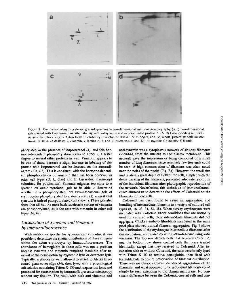
Fig. 5 compares the synemin present in chicken erythrocyte cytoskeletons and that in chicken gizzard smooth muscle tissue. Fig. 5 a shows the proteins that remain after extraction of erythrocytes with 1% Triton X-100 in a physiological salt buffer; a major difference in the protein pattern, compared with the membranes in Fig. 4a, is the presence of the nuclear lamins. There is also more vimentin relative to the amount of actin present, as this preparation also includes the nucleusassociated intermediate filaments. The autoradiogram of this gel after processing with anti-synemin is shown in Fig. 5 b. Similarly, a two-dimensional gel of whole gizzard tissue processed with anti-synemin, and its autoradiogram, are shown in Fig. 5 c and d. The similarities in the two synemin patterns are striking (see also Fig. 4c); in each case, the parent molecule is most heavily labeled, and the arcs of daughter products terminate in what appears to be a particularly stable fragment at -34,000 daltons (pi -4.9).
We have noted no consistent differences between erythrocyte
synemin and smooth muscle synemin. Minor differences in the fragment patterns may be attributable to different endogenous proteases, different processing schemes, or slight differences in electrophoresis; the latter two would explain the very minor differences between the erythrocyte synemins in Figs. 4 c and 5 d. There is a slight variation in the observed isoelectric points of erythrocyte and smooth-muscle synemin, but there is variation even among different samples of erythrocyte synemin (compare Figs. 2 a and 4 a). The focusing of synemin seems to be influenced by the amount of protein loaded on the isoelectric focusing gel; the apparent isoelectric point of synemin is often the same as that of desmin or vimentin if either of the latter is present in large quantities on the gel (Fig. 2 a and reference 25). We conclude from these immunoautoradiographic data that erythrocyte synemin and muscle synemin are similar if not identical; similarities in solubility properties and cellular distribution (below) strengthen the conclusion that the erythrocyte polypeptide may be regarded as synemin as defmed previously in smooth muscle.
Phosphorylation
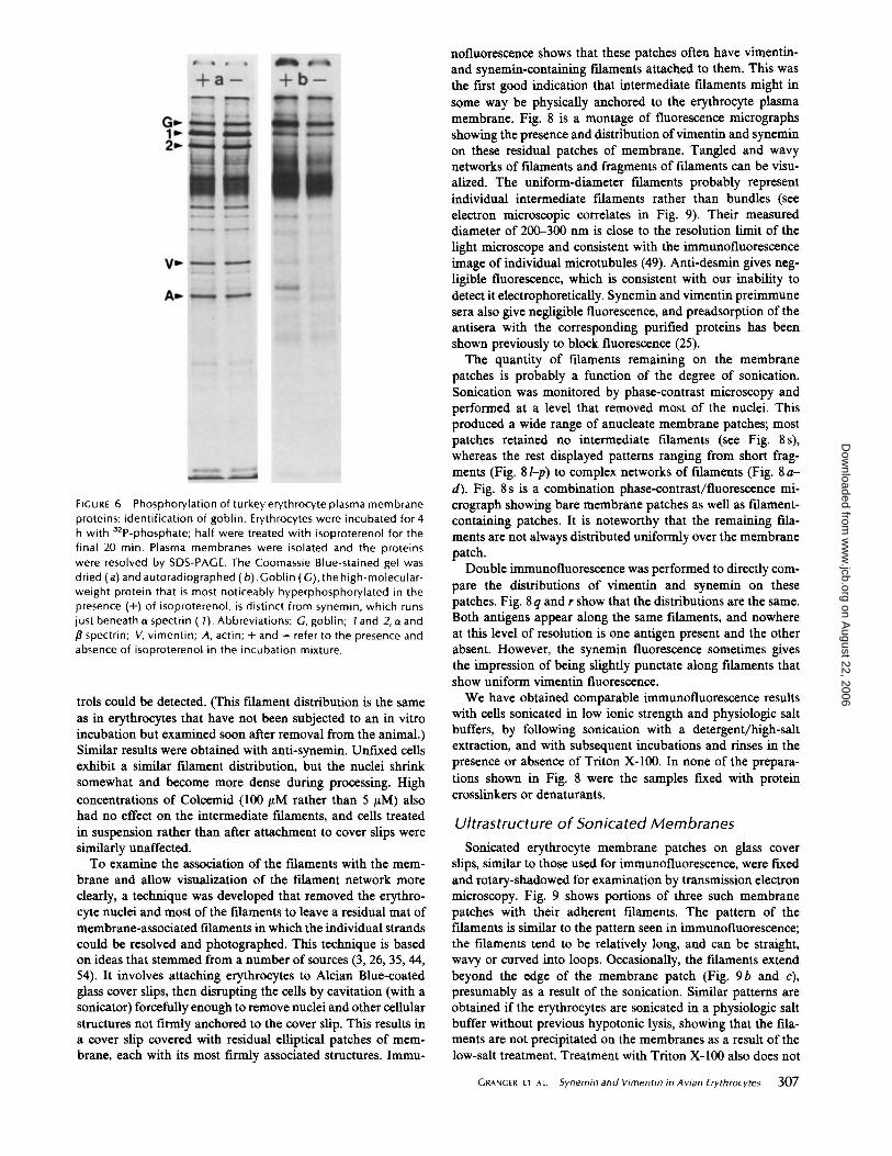
Goblin is a high molecular weight protein of the turkey erythrocyte plasma membrane characterized by hormone dependent phosphorylation (4). Both goblin and synemin have reported molecular weights of -230,000 daltons; although their solubility properties and distributions appeared to differ, we thought it was necessary to conclusively determine whether goblin and synemin were indeed different proteins. We identified goblin by its characteristic properties of being a large membrane-associated protein and the most hyperphosphorylated polypeptide in turkey erythrocytes treated briefly with the {3-adrenergic agonist, isoproterenol ( 4). Fig. 6 a shows a Coomassie Blue-stained gel of membranes of turkey erythrocytes labeled with [32P] inorganic phosphate; those on the left were also treated with isoproterenol, whereas those on the right were not. Fig. 6 b is the corresponding autoradiogram. By the above criteria, we conclude that the band designated in the figure is goblin. In this gel system, goblin migrates more slowly than the two spectrin variants, rather than migrating between them as in the system of Beam et al. (4). Using their gel system, we found that the electrophoretic pattern of our samples was indeed different: the relative positions of some bands was different, and goblin and the spectrins were not resolved as well.
A 44,000 dalton polypeptide is also noticeably hyperphos-
GRANGER ET AL. Synemin and Vimentin in Avian Erythrocytes 305
on August 22, 2006
ww
w.jcb.org
Dow
nloaded from
a
L.
v -A
c s
A
L
·F ·M
b
FIGURE 5 Comparison of erythrocyte and gizzard synemins by two-dimensional immunoautoradiography. (a, c) Two-dimensional gels stained with Coomassie Blue after labeling with antisynemin and radioiodinated protein A. ( b, d) Corresponding autoradiograms. Samples are (a) a Triton X-100 insoluble cytoskeleton of chicken erythrocytes, and (c) whole gizzard smooth muscle tissue. A, actin; 0, desmin; V, vimentin; L, lamins A, 8, and C (references 21 and 52); M, myosin; 5, synemin; F, filamin.
phorylated in the presence of isoproterenol (4), and this hormone-dependent phosphorylation seems to apply to a lesser degree to several other proteins as well. Vimentin appears to be one of these, because a slight increase in labeling of this protein with isoproterenol can be detected on the autoradiogram (Fig. 6b). This is consistent with the hormone-dependent phosphorylation of vimentin that has been observed in other cell types (D. L. Gard and E. Lazarides, manuscript submitted for publication). Synemin migrates too close to a spectrin on one-dimensional gels to be able to determine whether it is phosphorylated, but two-dimensional gels of erythrocytes phosphorylated to a steady state (1) suggest that synemin is indeed phosphorylated (not shown). These gels also show that all but the most basic isoelectric variant of vimentin are phosphorylated, as is the case with vimentin in other cell types (46, 47).
Localization of Synemin and Vimentin by Immunofluorescence
With antibodies specific for synemin and vimentin, it was possible to determine the spatial distributions of these antigens within the avian erythrocyte by immunofluorescence. The abundance of hemoglobin in these cells was not a problem because synemin and vimentin remained insoluble after removal of the hemoglobin by hypotonic lysis or detergent lysis. Typically, erythrocytes were allowed to attach to Alcian Bluecoated glass cover slips (54), then lysed with a physiological salt solution containing Triton X-100 and magnesium ions, and processed for examination by immunofluorescence microscopy without any fixation. The result with both anti-vimentin and
306 THE )OURNAL OF CELL BIOLOGY • VOLUME 92, 1982
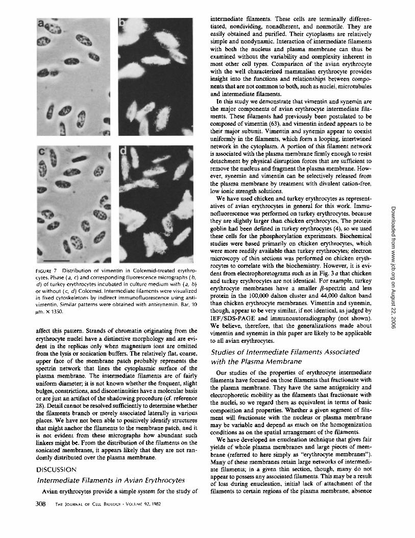
anti-synemin was a cytoplasmic network of sinuous filaments extending from the nucleus to the plasma membrane. This network gave the impression of being composed of a small number of long filaments, since relatively few free ends could be seen. A high concentration of fllaments was often noted near the poles of the nuclei (Fig. 7 d). However, the small size and relatively great depth of field of the cells, coupled with the dense packing of the filaments, prevented adequate resolution of the individual filaments after photographic reproduction of the network. Nevertheless, this technique of immunofluorescence allowed us to determine the effects of Colcemid on the fllaments in these cells.
Colcemid has been found to cause an aggregation and bundling of intermediate filaments in a variety of cultured cell types (8, 18, 23, 31, 33, 38). When turkey erythrocytes were incubated with Colcemid under conditions that are normally used for cultured cells, their intermediate filaments did not aggregate. Chicken embryo fibroblasts incubated in the same petri plate showed normal filament aggregation. Fig. 7 shows the distribution of the erythrocyte intermediate fllaments after this incubation, as revealed by immunofluorescence using antivimentin. The top row depicts cells that received Colcemid, and the bottom row shows control cells that were treated identically, except that they received no Colcemid. After incubation with or without Colcemid, the cells were briefly lysed with Triton X-100 to remove hemoglobin, then fixed with formaldehyde to ensure preservation of filament distribution. There was no obvious Colcemid-induced aggregation of the fllaments, and what appeared to be individual filaments could clearly be seen extending to the plasma membrane. No consistent difference between the Colcemid-treated cells and con-
on August 22, 2006
ww
w.jcb.org
Dow
nloaded from
.. +a-
v~ --
A~ -
FIGURE 6 Phosphorylation of turkey erythrocyte plasma membrane proteins: identification of goblin. Erythrocytes were incubated for 4 h with 32P-phosphate; half were treated with isoproterenol for the final 20 min. Plasma membranes were isolated and the proteins were resolved by SDS-PAGE. The Coomassie Blue-stained gel was dried (a) and auto radiographed (b). Goblin (C), the high-molecularweight protein that is most noticeably hyperphosphorylated in the presence (+) of isoproterenol, is distinct from synemin, which runs just beneath a spectrin ( 1). Abbreviations: C, goblin; 1 and 2, a and {3 spectrin; V, vimentin; A, actin;+ and- refer to the presence and absence of isoproterenol in the incubation mixture.
trots could be detected. (This fllament distribution is the same as in erythrocytes that have not been subjected to an in vitro incubation but examined soon after removal from the animal.) Similar results were obtained with anti-synemin. Unfixed cells exhibit a similar fll.ament distribution, but the nuclei shrink somewhat and become more dense during processing. High concentrations of Colcemid (100 p,M rather than 5 p,M) also had no effect on the intermediate fllaments, and cells treated in suspension rather than after attachment to cover slips were similarly unaffected.
To examine the association of the fJ.J.aments with the membrane and allow visualization of the fJ.J.ament network more clearly, a technique was developed that removed the erythrocyte nuclei and most of the fJ.J.aments to leave a residual mat of membrane-associated fJ.J.aments in which the individual strands could be resolved and photographed. This technique is based on ideas that stemmed from a number of sources (3, 26, 35, 44, 54). It involves attaching erythrocytes to Alcian Blue-coated glass cover slips, then disrupting the cells by cavitation (with a sonicator) forcefully enough to remove nuclei and other cellular structures not firmly anchored to the cover slip. This results in a cover slip covered with residual elliptical patches of membrane, each with its most firmly associated structures. Immu-
nofluorescence shows that these patches often have vimentinand synemin-containing fJ.J.aments attached to them. This was the first good indication that intermediate fJ.J.aments might in some way be physically anchored to the erythrocyte plasma membrane. Fig. 8 is a montage of fluorescence micrographs showing the presence and distribution ofvimentin and synemin on these residual patches of membrane. Tangled and wavy networks of fJ.J.aments and fragments of fll.aments can be visualized. The uniform-diameter fllaments probably represent individual intermediate fll.aments rather than bundles (see electron microscopic correlates in Fig. 9). Their measured diameter of 200-300 nm is close to the resolution limit of the light microscope and consistent with the immunofluorescence image of individual microtubules (49). Anti-desmin gives negligible fluorescence, which is consistent with our inability to detect it electrophoretically. Synemin and vimentin preimmune sera also give negligible fluorescence, and preadsorption of the antisera with the corresponding purified proteins has been shown previously to block fluorescence (25).
The quantity of fllaments remaining on the membrane patches is probably a function of the degree of sonication. Sonication was monitored by phase-contrast microscopy and performed at a level that removed most of the nuclei. This produced a wide range of anucleate membrane patches; most patches retained no intermediate fllaments (see Fig. 8 s), whereas the rest displayed patterns ranging from short fragments (Fig. 8/-p) to complex networks of filaments (Fig. Sad). Fig. 8 s is a combination phase-contrast/fluorescence micrograph showing bare membrane patches as well as fllamentcontaining patches. It is noteworthy that the remaining fJ.J.aments are not always distributed uniformly over the membrane patch.
Double immunofluorescence was performed to directly compare the distributions of vimentin and synemin on these patches. Fig. 8q and r show that the distributions are the same. Both antigens appear along the same fll.aments, and nowhere at this level of resolution is one antigen present and the other absent. However, the synemin fluorescence sometimes gives the impression of being slightly punctate along filaments that show uniform vimentin fluorescence.
We have obtained comparable immunofluorescence results with cells sonicated in low ionic strength and physiologic salt buffers, by following sonication with a detergent/high-salt extraction, and with subsequent incubations and rinses in the presence or absence of Triton X-100. In none of the preparations shown in Fig. 8 were the samples fixed with protein crosslinkers or denaturants.
Ultrastructure of Sonicated Membranes
Sonicated erythrocyte membrane patches on glass cover slips, similar to those used for immunofluorescence, were ftxed and rotary-shadowed for examination by transmission electron microscopy. Fig. 9 shows portions of three such membrane patches with their adherent fllaments. The pattern of the fllaments is similar to the pattern seen in immunofluorescence; the fll.aments tend to be relatively long, and can be straight, wavy or curved into loops. Occasionally, the fllaments extend beyond the edge of the membrane patch (Fig. 9 b and c), presumably as a result of the sonication. Similar patterns are obtained if the erythrocytes are sonicated in a physiologic salt buffer without previous hypotonic lysis, showing that the fllaments are not precipitated on the membranes as a result of the low-salt treatment. Treatment with Triton X-100 also does not
GRANGER ET AL. Synemin and Vimentin in Avian Erythrocytes 307
on August 22, 2006
ww
w.jcb.org
Dow
nloaded from
FIGURE 7 Distribution of vimentin in Colcemid-treated erythrocytes. Phase (a, c) and corresponding fluorescence micrographs ( b, d) of turkey erythrocytes incubated in culture medium with (a, b) or without ( c, d) Colcemid. Intermediate filaments were visualized in fixed cytoskeletons by indirect immunofluorescence using antivimentin. Similar patterns were obtained with antisynemin. Bar, 10 ~tm. X 1350.
affect this pattern. Strands of chromatin originating from the erythrocyte nuclei have a distinctive morphology and are evident in the replicas only when magnesium ions are omitted from the lysis or sonication buffers. The relatively flat, coarse, upper face of the membrane patch probably represents the spectrin network that lines the cytoplasmic surface of the plasma membrane. The intermediate filaments are of fairly uniform diameter; it is not known whether the frequent, slight bulges, constrictions, and discontinuities have a molecular basis or are just an artifact of the shadowing procedure ( cf. reference 28). Detail cannot be resolved sufficiently to determine whether the fllaments branch or merely associated laterally in various places. We have not been able to positively identify structures that might anchor the fllaments to the membrane patch, and it is not evident from these micrographs how abundant such linkers might be. From the distribution of the fll.aments on the sonicated membranes, it appears likely that they are not randomly distributed over the plasma membrane.
DISCUSSION
Intermediate Filaments in Avian Erythrocytes
Avian erythrocytes provide a simple system for the study of
308 THE JOURNAL OF CELL BIOLOGY • VOLUME 92, 1982
intermediate fllaments. These cells are terminally differentiated, nondividing, nonadherent, and nonmotile. They are e~sily obtained and p~rified. Their cytoplasms are relatively sunple and nondynarmc. Interaction of intermediate fll.aments with both the nucleus and plasma membrane can thus be examined without the variability and complexity inherent in m~st other cell types. Comparison of the avian erythrocyte wtth the well characterized mammalian erythrocyte provides insight into the functions and relationships between components that are not common to both, such as nuclei, microtubules and intermediate fllaments.
In this study we demonstrate that vimentin and synemin are the major components of avian erythrocyte intermediate fll.aments. These ftlaments had previously been postulated to be composed ofvimentin (63), and vimentin indeed appears to be their major subunit. Vimentin and synemin appear to coexist uniformly in the fll.aments, which form a looping, intertwined network in the cytoplasm. A portion of this fll.ament network is associated with the plasma membrane firmly enough to resist detachment by physical disruption forces that are sufficient to remove the nucleus and fragment the plasma membrane. However, synemin and vimentin can be selectively released from the plasma membrane by treatment with divalent cation-free, low ionic strength solutions.
We have used chicken and turkey erythrocytes as representatives of avian erythrocytes in general for this work. Immunofluorescence was performed on turkey erythrocytes, because they are slightly larger than chicken erythrocytes. The protein goblin had been defined in turkey erythrocytes (4), so we used these cells for the phosphorylation experiments. Biochemical studies were based primarily on chicken erythrocytes, which were more readily available than turkey erythrocytes; electron microscopy of thin sections was performed on chicken erythrocytes to correlate with the biochemistry. However, it is evident from electrophoretograms such as in Fig. 3 a that chicken and turkey erythrocytes are not identical. For example, turkey erythrocyte membranes have a smaller ,8-spectrin and less protein in the 100,000 dalton cluster and 44,000 dalton band than chicken erythrocyte membranes. Vimentin and synemin, though, appear to be very similar, if not identical, as judged by IEF/SDS-PAGE and immunoautoradiography (not shown). We believe, therefore, that the generalizations made about vimentin and synemin in this paper are likely to be applicable to all avian erythrocytes.
Studies of Intermediate Filaments Associated with the Plasma Membrane
Our studies of the properties of erythrocyte intermediate fllaments have focused on those fllaments that fractionate with the plasma membrane. They have the same antigenicity and electrophoretic mobility as the ftlaments that fractionate with the nuclei, so we regard them as equivalent in terms of basic composition and properties. Whether a given segment of ftlament will fractionate with the nucleus or plasma membrane may be variable and depend as much on the homogenization conditions as on the spatial arrangement of the fll.aments.
We have developed an enucleation technique that gives fair yields of whole plasma membranes and large pieces of membrane (referred to here simply as "erythrocyte membranes"). Many of these membranes retain large networks of intermediate filaments; in a given thin section, though, many do not appear to possess any associated ftlaments. This may be a result of loss during enucleation, initial lack of attachment of the ftlaments to certain regions of the plasma membrane, absence

on August 22, 2006
ww
w.jcb.org
Dow
nloaded from
FIGURE 8 Immunofluorescence of turkey erythrocyte intermediate filaments. Erythrocytes adhering to cover slips were hypotonically lysed and sonicated to remove overlying membranes and nuclei. Intermediate filaments remaining attached to the resulting patches of plasma membrane were visualized by immunofluorescence using antibodies to synemin (a, r) or vimentin (b-q, s). Specimens were not fixed, and all but a, b, c, and h were treated with Triton X-100. Micrographs a-pare indirect immunofluorescence images; q and rdemonstrate colocalization of vimentin ( q) and synemin ( r) by double immunofluorescence. Micrograph s is a combination phase/fluorescence image showing the distribution of vimentin on the elliptical patches of plasma membrane; note that many patches are devoid of filaments. Bars, 5 JLm. a- r, X 3040; s, X 1330.
FIGURE 9 Platinum replicas of sonicated chicken erythrocyte ghosts. Samples were prepared as in Fig. 8, then fixed, dried, and rotary shadowed with platinum for examination by transmission electron microscopy. Intermediate filaments can be seen on patches of plasma membrane that remained attached to the cover slip during sonication. In b and c, a portion of the filaments have fallen beyond the edge of the membrane patch. Magnification: Bar, 1 JLm. X 16,000.
from certain regions of the erythrocyte cytoplasm, or a close apposition to the plasma membrane that renders the ftlaments unresolvable.
Previous studies involving isolation of the avian erythrocyte plasma membrane by differential centrifugation after mechanical disruption of the cells have relied on pressure-release homogenization (7, 14, 61), sonication (4, 27), rotating blades (11, 66), or a tight-fitting Dounce (12, 22) or Potter-Elvehjem homogenizer (6). However, the presence offt.\aments associated with the isolated membrane fragments was noted only rarely
(27), and, in comparisons to mammalian erythrocyte membranes, the presence of an extra polypeptide, similar in molecular weight to vim en tin, was rarely mentioned ( 11 ). Some of these disruption techniques produce very small membrane fragments that may be largely stripped of filaments; alternatively, the ftlaments may assume a distribution or configuration in which they are not readily identifiable by electron microscopy. The gentler disruption techniques appear to produce membrane fragments similar to those in this study, but associated ftlaments have tended to escape detection. Intermediate
GRANG ER ET AL Synemin and Vimentin in Avian Erythrocytes 309

on August 22, 2006
ww
w.jcb.org
Dow
nloaded from
ftlaments have been most apparent in detergent-insoluble cytoskeletons of whole erythrocytes examined by thin sectioning or negative staining (59, 63).
Treatment of avian erythrocyte membranes with certain low ionic strength solutions removes the associated intermediate fJ.laments. Filaments can no longer be found with the membranes in thin sections, and the low-salt extract contains nearly pure vimentin and synemin. This release seems to depend on low ionic strength and absence of divalent cations and be independent of reducing agents or nonionic detergents. Our highest yields have been obtained using distilled water. Roughly 60-90% of the vimentin is released after 1 min of extraction with distilled water. Selective release of vimentin and synemin, as compared to spectrin and actin, is enhanced by low temperature and brevity of treatment. Because the released vimentin and synemin cannot be sedimented by centrifugation for 5 h at 240,000 g, yet appear to comigrate in a gel ftltration column with an exclusion limit of 15 million daltons (unpublished observations), they must exist in solution as some sort of multimeric complex or oligomer. This implies that the ftlaments break down or partially depolymerize during or after release from the membranes. Solubility in low salt has similarly been described for other preparations of native intermediate fllaments (29, 30, 50, 51, 55, 57). These extraction conditions may thus be resulting in a dissolution of the ftlaments rather than a dissociation of the ftlaments from the membranes. It is conceivable that these extraction conditions have no disruptive effect on the anchorage points of the ftlaments to the membranes, which would explain why some of the vimentin remains with the membranes after extensive extraction with water. This vimentin may be a distinct population associated with anchorage points in the form of tightly bound monomers or oligomers or short segments of ftlament not resolvable in thin sections. These extraction data thus do not permit a conclusion about the nature of attachment of the ftlaments to the membranes. It can only be stated, based on the physical data of enucleation and sonication, that at least some of the intermediate ftlaments in avian erythrocytes are somehow anchored to the plasma membrane, and that this attachment is stable in the presence of physiologic salt, high salt, and nonionic detergent.
Comparison of Erythrocyte Proteins
An aspect of comparative biochemistry exemplifled by this study is the difficulty of comparing protein proftles of a given preparation by different SDS-PAGE systems. Although useful for general comparisons, different gel systems may not be directly comparable with regard to speciflc polypeptides. There has classically been disagreement between different investigators about calculated molecular weights; even the relative positions of different polypeptides may not be consistent in different gel systems (for example, the high molecular weight proteins shown in this paper-see Results). This stresses caution in identifYing a polypeptide solely by its mobility on an SDS-polyacrylamide gel Our electrophoretic proflles of avian erythrocyte membrane proteins differ from those of other laboratories, which also differ among themselves (2, 4, 7, 11, 12, 34, 60, 61); some of these differences have been noted and attributed to endogenous proteases or proteases present in contaminating leukocytes (II, 34, 61 ). Extrapolation from one class to another (for example, mammalian (16) to avian erythrocyte membranes) may not be justilled either and may lead to erroneous identillcation of polypeptides. Two-dimensional gel
310 THE jOURNAL OF CELL BIOLOGY· VOLUME 92,1982
electrophoresis makes polypeptide identillcation less ambiguous, because another parameter (isoelectric point) is taken into account and has proved useful for several proteins in this study. Nevertheless, other (nonelectrophoretic) evidence for the identity of a protein band on a gel is essential. We have used immunologic and solubility properties, in addition to electrophoretic characteristics, to identifY synemin and vimentin in avian erythrocytes, and phosphorylation characteristics to identifY goblin (4). Determination of why similarly prepared samples show not only different relative mobilities but also different relative amounts using different gel systems awaits further study.
The Effects of Co/cemid
One indication of a functional or interactive difference between the intermediate ftlaments of avian erythrocytes and most other cell types grown in vitro is the insensitivity of the former to Colcemid. Treatments with Colcemid that will cause aggregation and perinuclear bundling of intermediate ftlaments in most cultured cells (8, 18, 23, 31, 33, 38) appear to have no effect on the ftlaments of erythrocytes. Colcemid sensitivity might thus be a function of how dynamic a cell is, or perhaps its state of differentiation, as appears to be likely for skeletal muscle cells (20, 25) but not be an intrinsic property of the ftlaments themselves. Related to this may be the observation that chick erythrocyte marginal band microtubules are resistant to depolymerization by Colcemid (5).
Intermediate Filament Proteins
Synemin was originally found to be associated with intermediate ftlaments in smooth and skeletal muscle (25). Here we show that synemin is not a muscle-speciflc protein but is present as well in at least one nonmuscle cell, the mature avian erythrocyte. The original study also raised the possibility that synemin was a desmin-associated polypeptide; here we show that synemin can also exist and associate with vimentin. In both muscle cells and erythrocytes, synemin appears to be a component of the same ftlaments that contain desmin and vimentin, as determined by double immunofluorescence. Densitometric scans ofCoomassie Blue-stained polyacrylamide gels of preparations of vimentin and synemin from chicken erythrocytes give a vimentin-to-synemin ratio of -50:1. This is similar to the ratio obtained for desmin and synemin in smooth muscle and suggests a constant stoichiometry between synemin and intermediate ftlaments of different subunit composition. This ratio is a very rough estimate, not taking into account differential proteolysis of the proteins during processing and possible nonlinearity in dye binding and densitometry, and should therefore not be regarded as the true ratio. It is useful, however, for rough comparisons of different systems.
We have taken advantage of a novel form of two-dimensional peptide mapping to compare synemins from different tissues. This combination of partial hydrolysis of tissue proteins by endogenous proteases and two-dimensional immunoautoradiography has demonstrated a high degree of homology between synemins from avian smooth muscle and erythrocytes. Both molecules exhibit an S-shaped string of fragments that terminates in a proteolytically stable peptide of 34,000 daltons. This technique is extremely sensitive, detecting peptides much too scarce to be seen by Coomassie Blue staining, but does not resolve the high molecular weight peptides sufficiently to allow detailed comparisons. Also, slight variations from gel to gel do
on August 22, 2006
ww
w.jcb.org
Dow
nloaded from
not allow us to conclude that the synemins we are examining are identical. Minor differences in the maps may be artifactual or may reflect functional differences in the molecule, perhaps related to whether synemin is found in association with desmin or with vimentin.
These data do not address the question of whether synemin is an integral or an associated ftlament protein, or what its properties are independent of desmin and vimentin. The large size and paucity of synemin relative to desmin and vimentin tend to favor a role for synemin as an associated polypeptide. Perhaps it is analogous to the high molecular weight polypeptide of neuroftlaments, which appears to be wrapped helically around the core ftlament (62), where it may function to stabilize the ftlament, promote assembly (45), or mediate interactions with other molecules or organelles.
The presence of nonmicrotubular ftlaments in nucleated erythrocytes has been known for some time (27, 36), but only recently were they identified as intermediate ftlaments (59, 63). These ftlaments were usually noted and studied in relation to the nucleus or nuclear membrane. In this paper we show that they also exhibit a close association with and apparent anchorage to the plasma membrane, and that they contain the intermediate-filament subunits vimentin and synemin. Nucleated erythrocytes may thus be an ideal model system for the study of ftlament-membrane interactions and for examining intermediate ftlament nucleation, assembly, and deployment during differentiation.
We thank David L. Gard for his helpful comments on the manuscript, and Dr. Jean-Paul Revel and Mr. Patrick F. Koen for their help with the electron microscopy.
This work was supported by grants from the National Institutes of Health (NIH) (GM 06965), National Science Foundation, Muscular Dystrophy Association of America, and a Biomedical Research Support Grant to the Division of Biology, California Institute of Technology. B. L. Granger was also supported by a Predoctoral Training Grant from the NIH (GM 07616), and E. A. Repasky by a Postdoctoral Fellowship from the NIH (GM 07401). E. Lazarides is the recipient of a Research Career Development Award from the NIH.
Received for publication 13 July 1981, and in revised form 5 October 1981.
REFERENCES
I. Alper, S. L., K. G. Beam, and P. Grecngard. 1980. Hormonal control of Na+ -K• cotransport in turkey erythrocytes. Multiple site phosphorylation of goblin, a high molecular weight protein of the plasma membrane. /. Bioi. Chem. 255:4864-4871.
2. Alper, S. L., H. C. Palfrey, S. A. DeRiemer, and P. Grecngard. 1980. Hormonal control of protein phosphorylation in turkey erythrocytes. Phosphorylation by cAMP-dependent and ca•• -dependent protein kinases of distinct sites in goblin, a high molecular weight protein of the plasma membrane. /. Bioi. Chem. 255:11029-11039.
3. Batten, B. E., J. J. Aalberg, and E. Anderson. 1980. The cytoplasmic fllamentous network in cultured ovarian granulosa cells. Cell. 21:885-895.
4. Beam, K. G., S. L. Alper, G. E. Palade, and P. Greengard. 1979. Hormonally regulated phosphoprotein of turkey erythrocytes. Localization to plasma membrane./. Cell Bioi. 83: 1-15.
5. Behnke, 0. 1970. A comparative study of microtubules of disk-shaped blood cells. J. U/tra3truct. Res. 31:61-75.
6. Bilezikian, J. P., and G. D. Aurbach. 1973. A .B-adrenergic receptor of the turkey erythrocyte. I. Binding of catecbolatnine and relationship to adenylate cyclase activity. J. Bioi. Chem. 248:5577-5583.
7. Blanchet, J.P. 1974. Chicken erythrocyte membranes: Comparison of nuclear and plasma membranes from adults and embryos. Exp. Cell Res. 84:159-166.
8. Blose, S. H., and S. Chacko. 1976. Rings of intermediate (100 A) fllament bundles in the perinuclear region of vascular endolhelial cells. Their mobilization by colcemid and mitosis. J. Cell Biol 70:459-466.
9. Branton, D., C. M. Cohen, and J. Tyler. 1981. Interaction of cytoskeletal proteins on Ihe human erylhrocyte membrane. Cell. 24:24-32.
I 0. Burridge, K. 1978. Direct identification of specific glycoproteins and antigens in sodiwn dodecyl sulfate gels. Methods Enzymol. 50:54-64.
II. Caldwell. A. B. 1976. Proteins ofthe turkey erythrocyte membrane. Biochermstry. 15:2711-2718.
12. Chan, L.-N. L. 1977. Changes in the composition of plasma membrane proteins during differentiation of embryonic chick erythroid cells. Proc. Nat/. Acad. Sci. U.S. A. 74:1062-1066.
13. Cohen, W. D. 1978. Observations on Ihe marginal band system of nucleated erythrocytes. J. Cell Bioi. 78:260-273.
14. Davoren, P.R., and E. W. Sutherland. 1963. The cellular location of adenyl cyclase in the pigeon erythrocyte. J. Bioi. Chem. 238:3016-3023.
15. Dodge, J. T., C. Mitchell. and D. J. Hanahan. 1963. The preparation and chemical characteristics of hemoglobin-free ghosts of human erythrocytes. Arch. Biochem. Biophys. 100:119-130.
16. Fairbanks, G., T. L. Steck, and D. F. H. Wallach. 1971. Electrophoretic analysis of the major polypeptides of the human erythrocyte membrane. Biochemistry. 10:2606-2617.
17. Fourie, F. leR. 1977. Effects of anticoagulants on the hematocrit, osmolarity and pH of avian blood. Poult. ScL 56:1842-1846.
18. Franke, W. W., E. Schmid, M. Osborn, and K. Weber. 1978. Different intermediate-sized fllaments distinguished by immunofluorescence microscopy. Proc. Nat/. Acad. Sci. U. S. A. 75:5034-5038.
19. Gard, D. L., P. B. Bell. and E. Lazarides. 1979. Coexistence ofdesmin and Ihe fibroblastic intermediate ftlament subunit in muscle and nonmuscle cells: identification and comparative peptide analysis. Proc. Nat/. Acad. Sci. U. S. A. 76:3894-3898.
20. Gard, D. L., and E. Lazarides. 1980. The synthesis and distribution of desmin and vimentin during myogenesis in vitro. Cell. 19:263-275.
21. Gerace, L., and G. Blobel. 1980. The nuclear envelope lamina is reversibly depolymerized during mitosis. Cell. 19:277-287.
22. Ginsberg. B. H., C. R. Kahn, and J. Roth. 1976. The insulin receptor of Ihe turkey erythrocyte. Characterization of the membrane-bound receptor. Biochim. Biophys. Acta. 443:227-242.
23. Goldman, R. D. 1971. The role of Ihree cytoplasmic fibers in BHK-21 cell motility. I. Microtubules and Ihe effects of Colcbicine. J. Cell Bioi. 5 I :752-762.
24. Granger, B. L., and E. Lazarides. 1979. Desmin and virnentin coexist at Ihe periphery of the myofibril Z disc. Cell. 18:1053-1063.
25. Granger, B. L., and E. Lazarides. 1980. Synemin: a new high molecular weight protein associated wilh desmin and vimentin f!laments in muscle. Cell. 22:727-738.
26. Hall. C. E. 1956. Visualization of individual macromolecules with Ihe electron microscope. Proc. Nat/. Acad. Sci. U. S. A. 42:801-806.
27. Harris, J. R., and J. N. Brown. 1971. Fractionation of the avian erythrocyte: an ultrastructural study. J. Ultrastruct. Res. 36:8-23.
28. Heuser, J. E., and M. W. Kirschner. 1980. Filament organization revealed in platinum replicas offr~-dried cytoskeletons. /.Cell Bioi. 86:212-234.
29. Hubbard, B. D., and E. Lazarides. 1979. Copurification of actin and desmin from chicken smooth muscle and their copolymerization in vitro to intermediate fllaments. /. Cell Bioi. 80:166-182.
30. Huiall, T. W., R. M. Robson, N. Arakawa, and M. H. Stromer. 1980. Desmin from avian smoolh muscle. Purification and partial characterization. J. Bioi. Chem. 255:6981-6989.
31. Hynes, R. 0., and A. T. Desiree. 1978a. 10 mm f!laments in normal and transformed cells. Cell. 13:151-163.
32. Hynes, R. 0., and A. Destree. 1978b. Relationships between fibronectin (LETS protein) and actin. Cell. 15:875-886.
33. Ishikawa, H., R. Bischoff, and H. Holtzer. 1968. Mitosis and intermediate-sized f!laments in developing skeletal muscle. J. Cell Bioi. 38:538-555.
34. Jackson, R. C. 1975. The exterior surface of the chicken erythrocyte. J. Bioi. Chem. 250: 617-622.
35. Jacobson, B.S., and D. Branton. 1977. Plasma membrane: rapid isolation and exposure of the cytoplasmic surface by use of postively charged beads. Science (Wash. D. C.). 195: 302-304.
36. Koehler, J. K. 1968. Freeze-etching observations on nucleated erythrocytes wilh special reference to the nuclear and plasma membranes. z. Zellforsch. Mikrosk. Anat. 85:1-17.
37. Laemmli, U.K. 1970. Cleavage of structural proteins during Ihe assembly of the head of bacteriophage T4. Nature (Lond.). 227:680-685.
38. Lazarides, E. 1978. Tbe distribution of desmin (100 A) f!laments in primary cultures of embryonic chick cardiac cells. Exp. Cell Res. 112:265-273.
39. Lazarides, E. 1980. Intermediate f!laments as mechanical integrators of cellular space. Nature (Lond.). 283:249-256.
40. Lazarides, E., and B. D. Hubbard. 1976. Immunological characterization of the subunit of the 100 A fllaments from muscle cells. Proc. Nat/. Acad. Sci. U. S. A. 73:4344-4348.
41. Maddy, A. H., and P. G. Kelly. 1971. Acetic acid as a solvent for erythrocyte membrane proteins. Biochim. Biophys. Acta. 241:290-301.
42. Marchesi, S. L., E. Steers, V. T. Marchesi, and T. W. Tillack. 1970. Physical and chemical properties of a protein isolated from red cell membranes. Biochemistry. 9:50-57.
43. Marches~ V. T., and E. Steers. 1968. Selective solubilization of a protein component oflhe red cell membrane. Science (Wash. D. C). 159:203-204.
44. Marek, L. F., R. 0. Kelley, B. D. Perdue, and P. 0. Zamora. 1979. Role of the cytoskeleton in fibroblast attachment to poly-t-lysine treated substrates. J. Cell Bioi. 83 (2, Pt. 2): 324a (Abstr.).
45. Moon, H. M., T. Wisniewski, P. Men, J. DeMartini, and H. M. Wisniewski. Partial purification of neurofllament subunits from bovine brains and studies on neurofllament assembly. J. Cell Bioi. 89:560-567.
46. O'Connor, C. M., D. R. Balzer, and E. Lazarides. 1979. Phosphorylation of subunit proteins of intermdiate fllaments from chicken muscle and nonmuscle cells. Proc. Nat/. Acad. Sci. U. S. A. 76:819-823.
47. O'Connor, C. M., D. L. Gard, and E. Lazarides. 1981. Phosphorylation of intermediate fllament proteins by cAMP-dependent protein kinases. Cell. 23:135-143.
48. O'Farrell. P. H. 1975. High resolution two-dimensional electrophoresis of proteins. J. Bioi. Chem. 250:4007-4021.
49. Osborn, M., R. E. Webster, and K. Weber. 1978. Individual microtubules viewed by immunofluorescence and electron microscopy in Ihe same PtK2 cell./. Cell Bioi. 77:R27-R34.
50. Rueger, D. C., J. S. Huston, D. Dahl, and A. Bignami. 1979. Formation of 100 A fllaments from purified glial fibrillary acidic protein in vitro. J. Mol. Bioi. 135:53-68.
51. Scblaepfer, W. W. 1978. Observations on the disassembly of isolated mammalian neurofllaments. /. Cell Bioi. 76:50-56.
52. Shelton, K. R., L. L. Higgins, D. L. Cochran, J. J. Ruffolo, and P.M. Egle. 1980. Nuclear latnios oferythrocyte and liver./. Bioi. Chem. 255:10978-10983.
53. Small. J. V., and A. Sobieszek. 1977. Studies on the funCtion and composition of the t(}.
nm (tOO-A) fllaments of vertebrate smoolh muscle. J. Cell Sci. 23:243-268. 54. Sommer, J. R. 1977. To cationize glass. J. Cell Bioi. 75 (2, Pt. 2): 245a (Abstr.). 55. Starger, J. M., W. E. Brown, A. E. Goldman, and R. D. Goldman. 1978. Biochemical and
immunological analysis of rapidly purified t(}.nm f!laments from baby hamster kidney (BHK-21) cells. J. Cell Bioi. 78:93-109.
56. Steck, T. L., and J. Yu. 1973. Selective solubilization of proteins from red blood cell membranes by protein perturbants. /. Supramo/. Struct. I :220-232.
57. Steinert, P. M., W. W. Idler, F. Cabral, M. M. Gottesman, and R. D. Goldman. 1981. In
GRANGER ET Al. Synemin and Vi men tin in Avian Erythrocytes 311

on August 22, 2006
ww
w.jcb.org
Dow
nloaded from
vitro assembly of homopolymer and copolymer filaments from intermediate filament subunits of muscle and fibroblastic cells. Proc. Nat/. A cad. Sci. U. S. A. 78:3692-3696.
58. Tuszynski, G. P., E. D. Frank, C. H. Damsky, C. A. Buck, and L. Warren. 1979. The detection of smooth muscle desmin-like protein in BHK,1/CJa fibroblasts. J. Bioi. Chem. 254:6138---{)143.
59. Vinanen, 1., M. Kurkinen, and V.-P. Lehto. 1979. Nucleus-anchoring cytoskeleton in chicken red blood cells. Cell Bioi. Int. Rep. 3:157-162.
60. Weise, M. J., and L.-N. L. Chan. 1978. Membrane protein synthesis in embryonic chick erythroid cells. J. Bioi. Chem. 253:1892-1897.
61. Weise, M. J., and V. M. Ingram. 1976. Proteins and glycoproteins of membranes from developing chick red cells. J. Bioi. Chem. 251 :6667---{)673.
62. Willard, M., and C. Simon. 1981. Antibody decoration ofneurofilaments. J. Cell Bioi. 89:
312 THE )OURNAL OF CELL BIOLOGY· VOLUME 92,1982
198-205. 63. Woodcock, C. L. F. 1980. Nucleus-associated intermediate filaments from chicken eryth
rocytes. J. Cell Bioi. 85:881-889. 64. Yu, J., D. A. Fischman, and T. L. Steck. 1973. Selective solubilization of proteins and
phospholipids from red blood cell membranes by nonionic detergents. J. Supramo/. Struct. 1:233-248.
65. Zentgraf, H., B. Deumling, and W. W. Franke. 1969. Isolation and characterization of nuclei from bird erythrocytes. Exp. Cell Res. 56:333-337.
66. Zentgraf, H., B. Deumling, E.-D. Jarasch, and W. W. Franke. 1971. Nuclear membranes and plasma membranes from hen erythrocytes. I. Isolation, characterization, and comparison. J. Bioi. Chem. 246:2986-2995.