Embed Size (px)
Citation preview
International Journal of
Molecular Sciences
Review
Small GTPases of the Ras and Rho Families Switchon/off Signaling Pathways inNeurodegenerative Diseases
Alazne Arrazola Sastre 1,2, Miriam Luque Montoro 1, Patricia Gálvez-Martín 3,4 ,Hadriano M Lacerda 5, Alejandro Lucia 6,7, Francisco Llavero 1,6,* and José Luis Zugaza 1,2,8,*
1 Achucarro Basque Center for Neuroscience, Science Park of the Universidad del País Vasco/Euskal HerrikoUnibertsitatea (UPV/EHU), 48940 Leioa, Spain; [email protected] (A.A.S.);[email protected] (M.L.M.)
2 Department of Genetics, Physical Anthropology, and Animal Physiology, Faculty of Science and Technology,UPV/EHU, 48940 Leioa, Spain
3 Department of Pharmacy and Pharmaceutical Technology, Faculty of Pharmacy, University of Granada,180041 Granada, Spain; [email protected]
4 R&D Human Health, Bioibérica S.A.U., 08950 Barcelona, Spain5 Three R Labs, Science Park of the UPV/EHU, 48940 Leioa, Spain; [email protected] Faculty of Sport Science, European University of Madrid, 28670 Madrid, Spain;
[email protected] Research Institute of the Hospital 12 de Octubre (i+12), 28041 Madrid, Spain8 IKERBASQUE, Basque Foundation for Science, 48013 Bilbao, Spain* Correspondence: [email protected] (F.L.); [email protected] (J.L.Z.)
Received: 25 July 2020; Accepted: 29 August 2020; Published: 31 August 2020�����������������
Abstract: Small guanosine triphosphatases (GTPases) of the Ras superfamily are key regulatorsof many key cellular events such as proliferation, differentiation, cell cycle regulation, migration,or apoptosis. To control these biological responses, GTPases activity is regulated by guanine nucleotideexchange factors (GEFs), GTPase activating proteins (GAPs), and in some small GTPases also guaninenucleotide dissociation inhibitors (GDIs). Moreover, small GTPases transduce signals by theirdownstream effector molecules. Many studies demonstrate that small GTPases of the Ras family areinvolved in neurodegeneration processes. Here, in this review, we focus on the signaling pathwayscontrolled by these small protein superfamilies that culminate in neurodegenerative pathologies,such as Alzheimer’s disease (AD) and Parkinson’s disease (PD). Specifically, we concentrate onthe two most studied families of the Ras superfamily: the Ras and Rho families. We summarizethe latest findings of small GTPases of the Ras and Rho families in neurodegeneration in order tohighlight these small proteins as potential therapeutic targets capable of slowing down differentneurodegenerative diseases.
Keywords: GTPases; neurodegeneration; Alzheimer; Parkinson; Ras; Rap; Rho; Rac; Cdc42
1. Introduction
Eukaryotic cells permanently maintain communication with the extracellular medium throughsome molecules such as growth factors, hormones, peptides, and ions. These signaling mediators(agonists or antagonists) can bind to specific receptors, which promote the internalization of themessage that spreads via signaling cascades in order to drive a specific cellular response [1].
The above-mentioned processes share common elements that are recruited in the presence of thedifferent stimuli and have an active role in both several and different responses. One of those recruited
Int. J. Mol. Sci. 2020, 21, 6312; doi:10.3390/ijms21176312 www.mdpi.com/journal/ijms
Int. J. Mol. Sci. 2020, 21, 6312 2 of 23
elements are small guanosine triphosphatases (GTPases) of the Ras superfamily. Those proteins arekey regulators of many important and different cellular events that take place in eukaryotic cells suchas proliferation, differentiation, or apoptosis [2,3].
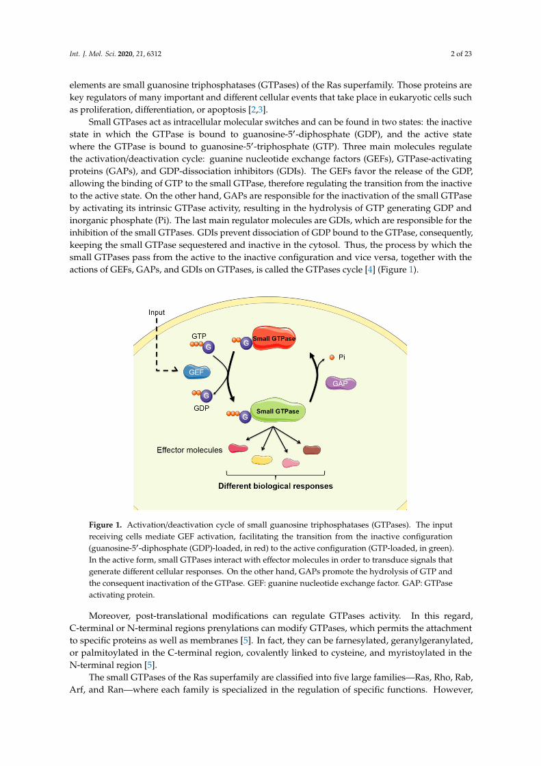
Small GTPases act as intracellular molecular switches and can be found in two states: the inactivestate in which the GTPase is bound to guanosine-5′-diphosphate (GDP), and the active statewhere the GTPase is bound to guanosine-5′-triphosphate (GTP). Three main molecules regulatethe activation/deactivation cycle: guanine nucleotide exchange factors (GEFs), GTPase-activatingproteins (GAPs), and GDP-dissociation inhibitors (GDIs). The GEFs favor the release of the GDP,allowing the binding of GTP to the small GTPase, therefore regulating the transition from the inactiveto the active state. On the other hand, GAPs are responsible for the inactivation of the small GTPaseby activating its intrinsic GTPase activity, resulting in the hydrolysis of GTP generating GDP andinorganic phosphate (Pi). The last main regulator molecules are GDIs, which are responsible for theinhibition of the small GTPases. GDIs prevent dissociation of GDP bound to the GTPase, consequently,keeping the small GTPase sequestered and inactive in the cytosol. Thus, the process by which thesmall GTPases pass from the active to the inactive configuration and vice versa, together with theactions of GEFs, GAPs, and GDIs on GTPases, is called the GTPases cycle [4] (Figure 1).
Int. J. Mol. Sci. 2020, 21, x FOR PEER REVIEW 2 of 23
proteins are key regulators of many important and different cellular events that take place in eukaryotic cells such as proliferation, differentiation, or apoptosis [2,3].
Small GTPases act as intracellular molecular switches and can be found in two states: the inactive state in which the GTPase is bound to guanosine-5′-diphosphate (GDP), and the active state where the GTPase is bound to guanosine-5′-triphosphate (GTP). Three main molecules regulate the activation/deactivation cycle: guanine nucleotide exchange factors (GEFs), GTPase-activating proteins (GAPs), and GDP-dissociation inhibitors (GDIs). The GEFs favor the release of the GDP, allowing the binding of GTP to the small GTPase, therefore regulating the transition from the inactive to the active state. On the other hand, GAPs are responsible for the inactivation of the small GTPase by activating its intrinsic GTPase activity, resulting in the hydrolysis of GTP generating GDP and inorganic phosphate (Pi). The last main regulator molecules are GDIs, which are responsible for the inhibition of the small GTPases. GDIs prevent dissociation of GDP bound to the GTPase, consequently, keeping the small GTPase sequestered and inactive in the cytosol. Thus, the process by which the small GTPases pass from the active to the inactive configuration and vice versa, together with the actions of GEFs, GAPs, and GDIs on GTPases, is called the GTPases cycle [4] (Figure 1).
Figure 1. Activation/deactivation cycle of small guanosine triphosphatases (GTPases). The input receiving cells mediate GEF activation, facilitating the transition from the inactive configuration (guanosine-5′-diphosphate (GDP)-loaded, in red) to the active configuration (GTP-loaded, in green). In the active form, small GTPases interact with effector molecules in order to transduce signals that generate different cellular responses. On the other hand, GAPs promote the hydrolysis of GTP and the consequent inactivation of the GTPase. GEF: guanine nucleotide exchange factor. GAP: GTPase activating protein.
Moreover, post-translational modifications can regulate GTPases activity. In this regard, C-terminal or N-terminal regions prenylations can modify GTPases, which permits the attachment to specific proteins as well as membranes [5]. In fact, they can be farnesylated, geranylgeranylated, or palmitoylated in the C-terminal region, covalently linked to cysteine, and myristoylated in the N-terminal region [5].
The small GTPases of the Ras superfamily are classified into five large families—Ras, Rho, Rab, Arf, and Ran—where each family is specialized in the regulation of specific functions. However, it
Figure 1. Activation/deactivation cycle of small guanosine triphosphatases (GTPases). The inputreceiving cells mediate GEF activation, facilitating the transition from the inactive configuration(guanosine-5′-diphosphate (GDP)-loaded, in red) to the active configuration (GTP-loaded, in green).In the active form, small GTPases interact with effector molecules in order to transduce signals thatgenerate different cellular responses. On the other hand, GAPs promote the hydrolysis of GTP andthe consequent inactivation of the GTPase. GEF: guanine nucleotide exchange factor. GAP: GTPaseactivating protein.
Moreover, post-translational modifications can regulate GTPases activity. In this regard,C-terminal or N-terminal regions prenylations can modify GTPases, which permits the attachmentto specific proteins as well as membranes [5]. In fact, they can be farnesylated, geranylgeranylated,or palmitoylated in the C-terminal region, covalently linked to cysteine, and myristoylated in theN-terminal region [5].
The small GTPases of the Ras superfamily are classified into five large families—Ras, Rho, Rab,Arf, and Ran—where each family is specialized in the regulation of specific functions. However,
Int. J. Mol. Sci. 2020, 21, 6312 3 of 23
it has been described that GTPases from different families can cooperate with each other in manycircumstances [6,7].
The Ras family is implicated in cellular growth control and metabolism [2,3], and it cooperateswith the Rho family in the regulation of the cell cycle, gene expression, and cellular transformation [2,3].The Rho family is specialized in actin cytoskeleton reorganization. The Rab family is responsible forthe intracellular vesicle and membrane trafficking, whereas the Arf family regulates the formation ofthe vesicles and the intracellular transport [2,3]. Lastly, the Ran family controls the nucleocytoplasmictransport and microtubule organization [2,3].
Depending on the context (i.e., cell type or the stimulus), activated GTPases lead to cellularresponses such as proliferation, differentiation, motility, survival, or apoptosis [2,3]. Taking intoaccount the importance of these responses at a cellular physiology level, mutations or alterations in theRas superfamily of GTPases have been associated with different diseases, such as cancer [8], as well asvascular [9] and neurodegenerative diseases [10].
Neurodegenerative diseases are characterized by the progressive loss of specific subsets ofneurons [11]. The original trigger of neuronal death is still unknown, although aging is considered arisk factor for neurodegenerative diseases. Many of these disorders share an abnormal accumulationof misfolded peptides or proteins in the brain and spinal cord. These deposits of insoluble proteinscould accumulate with time, and they could be more toxic when neurons are old, as they lose theability to degrade such proteins. Apart from protein seeding and propagation, the pathologicalhallmarks behind a neurodegenerative disease include altered protein quality control, dysfunctionalmitochondrial homeostasis, autophagy and lysosomal dysregulation, stress granules, synaptic toxicity,neuroinflammation due to dysregulated glial cells, and maladaptive innate immune responses [11].All these features culminate in neuronal death.
Neurodegenerative diseases are classified depending on main clinical characteristic, anatomicdistribution of the neuronal loss, or fundamental molecular alteration (e.g., the protein that isbeing accumulated, as well as its neuroanatomical distribution). Hence, depending on whichprotein is accumulated, neurodegenerative diseases can be classified in different groups: amyloidosisdiseases, Tau protein impairments, and α-synucleinopathies that are characterized by the accumulationand aggregation of β-amyloid (Aβ) peptides, Tau protein impairments, and α-Synuclein (α-syn)accumulation, respectively [11].
The misfolded proteins mentioned above are hallmarks of the most prevalent (and, therefore,most studied) neurodegenerative diseases, such as Alzheimer’s disease (AD) and Parkinson’s disease(PD) [11]. AD and PD are the two most common neurodegenerative diseases.
The most common form of dementia, AD, is a progressive and irreversible neurodegenerativedisorder [12]. It is characterized by the abnormal accumulation of both Tau protein and Aβ peptides.The aggregation of hyperphosphorylated Tau protein provokes the formation of neurofibrillary tangles(NFT) in the intracellular region [13]. NFT formation typically parallels neuronal loss and, therefore,it has been associated with AD severity. The accumulation and spreading of Aβ peptides (called senileplaques) are also a hallmark of AD. The accumulation and aggregation of both proteins induce thedegeneration of hippocampal and cerebral cortex neurons, resulting in the typical AD symptoms thatinclude memory loss, spatiotemporal disorientation, or behavioral changes among others [12].
PD is the second most common neurodegenerative disease [14]. It is characterized byintracellular inclusions containing α-syn aggregates forming the Lewy bodies (LB). Forms of PDare caused by different mutations in leucine rich-repeat kinase 2 (LRRK2), Parkin E3-ubiquitinligase, and PTEN-induced putative kinase 1 (PINK1) [15,16]. PD cellular characteristics consist onthe degeneration of dopaminergic (DA) neurons in substantia nigra, resulting in symptoms such asbradykinesia, resting tremor, muscular rigidity, postural instability, and dementia.
Furthermore, the accumulation of neurodegeneration-related proteins mentioned above is relatedto the alteration of the intracellular neuronal and glial pathways. All these pathological mechanismscould be due to dysregulated signal transduction pathways that alter neuron and glial functionality.
Int. J. Mol. Sci. 2020, 21, 6312 4 of 23
As previously mentioned, small GTPases are key molecules that integrate cellular inputs toelaborate a biological response. Due to their important functions, members of the Ras superfamilyhave been associated with AD and PD [10]. Thereby, small GTPases of the Ras superfamily havelong been shown to be involved in AD [17]. In PD, small GTPases and/or missense mutations inGTPases are implicated since they participate in the signal transduction mediated by LRRK2, Parkin,and PINK1 [18]. Apart from these pathologies, small GTPases are also involved in many otherneurodegenerative disorders, such as amyotrophic lateral sclerosis (ALS), Huntington’s disease (HD),and even Creutzfeldt–Jakob disease (CJD) [10].
Therefore, the dysregulation of specific GTPases of the Ras superfamily, or their regulatorsor effector molecules, leads to aberrant signaling pathways and/or pathological cell responsesthat could cause neurodegeneration in AD and PD. Understanding how these GTPase-mediatedmolecular events are dysregulated in neurodegenerative diseases can help us understand the cause ofneurodegeneration. This might provide new avenues for the development of therapeutic approachesfor neurodegenerative diseases.
Thus, in this review, we go deeper into the dysregulated/aberrant signaling pathways controlledby the Ras superfamily GTPases that culminate in neurodegeneration and further progression of twomajor neurodegenerative diseases (AD and PD). We specifically focused on the two most studiedfamilies of the Ras superfamily: Ras and Rho. We summarize the latest findings to highlight the role ofsmall GTPases of the Ras and Rho families in neurodegenerative processes.
2. Small GTPases of the Ras Family
This family of GTPases regulates biological responses such as proliferation, differentiation, andmigration [3]. However, it is classically associated to tumor development processes, therefore, beingclassified as proto-oncogenes [8].
2.1. Ras GTPase
Ras GTPases activation is promoted by GEFs such as SOS, RasGRF, RasGRP, and RalGDS, whereasRas GTPases are inactivated by GAPs such as RASA 1–3, RASAL 1–3, DAB2IP, NF1.SPRED 1–3, andSYNGAP1 [19]. Apart from GEFs and GAPs, Ras activity can also be regulated by scaffold proteinssuch as KSR, which favors Ras signaling via MAP-kinase (MAPK), its main effector molecule [20].
Considering the importance of Ras-regulated responses, a dysregulation of this GTPase couldunderlie some neurodegenerative diseases. In fact, an abnormal activity of the Ras pathway has beenassociated with Alzheimer’s disease [21]. One of the first studies carried out in AD patients describedthat they expressed abnormally elevated levels of the adaptor protein growth factor receptor-boundprotein 2 (Grb2) and SOS-1, a RasGEF [22]. Moreover, some evidence suggest that the MAPK pathwaycould be involved in Tau hyperphosphorylation in AD [23], as well as in the amyloidogenic processingof the amyloid precursor protein (APP) that generates Aβ1–42 [21].
In physiological conditions, the Ras/MAPK axis controls the dendritic spine formation. However,in AD, Aβ accumulation results in a dendritic spine and synapse loss, leading to cognitive decline.The molecule controlling this dendritic spine and synapse formation (and, therefore, learning andmemory processes) is RasGRF1 GEF. When Aβ levels are reduced by a pharmacological treatment,mRNA and protein levels of RasGRF1 are increased, promoting dendritic spine formation inhippocampal primary neurons [24,25]. Considering that the RasGRF1/Ras/MAPK pathway is involvedin the dendritic spine formation, benzothiazole amphiphiles such as hexa(ethylene glycol), derivativeof benzothiazole aniline, have been effective in modulating this pathway and in promoting dendriticspine formation in human iPSC-derived neurons [26].
The Ras/MAPK pathway activity is also relevant in glia; for example, microglial phagocytosisrequires its activation [27]. Nevertheless, the Ras/MAPK pathway can also be detrimental to cellularphysiology, as it is involved in l-3,4 dihydroxyfenylalanine-(l-DOPA)-treated PD patient dyskinesia.In a PD-mouse model, l-DOPA induces dyskinesia by increasing the Ras GEF RasGRP1 protein levels.
Int. J. Mol. Sci. 2020, 21, 6312 5 of 23
In contrast, l-DOPA-treated RasGRP1-/- mice do not display dyskinesia [28]. Moreover, l-DOPApromotes the activation of extracellular signal-regulated kinase (ERK) and mTORC1 [29]. As RasGRP1acts as a GEF for both Ras and Rheb (another Ras family GTPase), the authors suggested that in thepresence of l-DOPA, RasGRP1 could form complexes with both GTPases. On one hand, it couldassociate with H-Ras to initiate ERK signaling; on the other hand, it could activate Rheb to signal viamTOR [29]. Hence, parallel activation of these two cascades could provoke the onset and progressionof L-DOPA-induced dyskinesia [29].
The MAPK pathway is not the unique Ras-activated signaling pathway, as dendritic spineformation is achieved by a cooperation between the Ras/MAPK and Ras/PI3K/Akt pathways [30].Although no evidence shows that Ras interaction with PI3K induces neurodegeneration, it is knownthat Ras/PI3K/Akt promotes neuron survival [31]. Furthermore, the Ras/PI3K/Akt pathway can alsoactivate mTOR, leading to an increase in dendrite arborization, both in number and complexity, in thepresence of the brain-derived neurotrophic factor (BDNF) [32]. Therefore, Ras/PI3K pathway abnormalfunctioning could be involved in pathologies that present dendritic spine loss such as AD. Furthermore,the PI3K/Akt/mTOR pathway has recently been described to be altered in diseases such as ischemicbrain injury and neurodegenerative diseases such as AD, PD, and Huntington’s disease [33].
The Ras GTPases regulate the signaling pathway that controls cell–cell interactions throughadherens junctions via its effector molecule afadin-6 (AF-6). Together with Parkin, AF-6 is involved inincreasing mitophagy [28]. Basil and collaborators have demonstrated that human AF-6 overexpressionin Parkin-/- and PINK1-/- Drosophila melanogaster models rescues the parkinsonian mitochondrialpathology, improving locomotor deficits and increasing fly survival [34]. Moreover, AF-6 overexpressionin pathological mutant LRRK2G2019S flies improved the parkinsonian phenotype. The authorsdemonstrated that AF-6 protected DA neuron dysfunction in flies [34]. All these data suggest thatAF-6 activation could play a protective role in PD by regulating mitochondrial homeostasis [28,34].
Other Ras effector molecules potentially implicated in neurodegenerative processes such as ADand/or PD are RIN1 and Tiam1. Both of them control synaptic plasticity; RIN1 destabilizes synapticconnections by affecting the dendrite morphology and increasing the dendritic filopodial motility [35],whereas Tiam1 controls neurite outgrowth [36]. Tiam1 also controls the DA neuron differentiation [37].Understanding the molecular mechanism that leads to this differentiation is crucial for the developmentof PD therapies based on DA neuron differentiation [37].
Ras effector molecules RIN1 and Tiam1 are characteristic GEFs. RIN1 is a Rab5A GEF, whichregulates α-amino-3-hydroxy-5-methyl-4-isoxazolepropionic acid (AMPA) receptor endocytosis [35],while Tiam1 is a Rac1 GEF that controls cytoskeleton-related processes, such as neurite outgrowth [36]or Schwann cell migration [38].
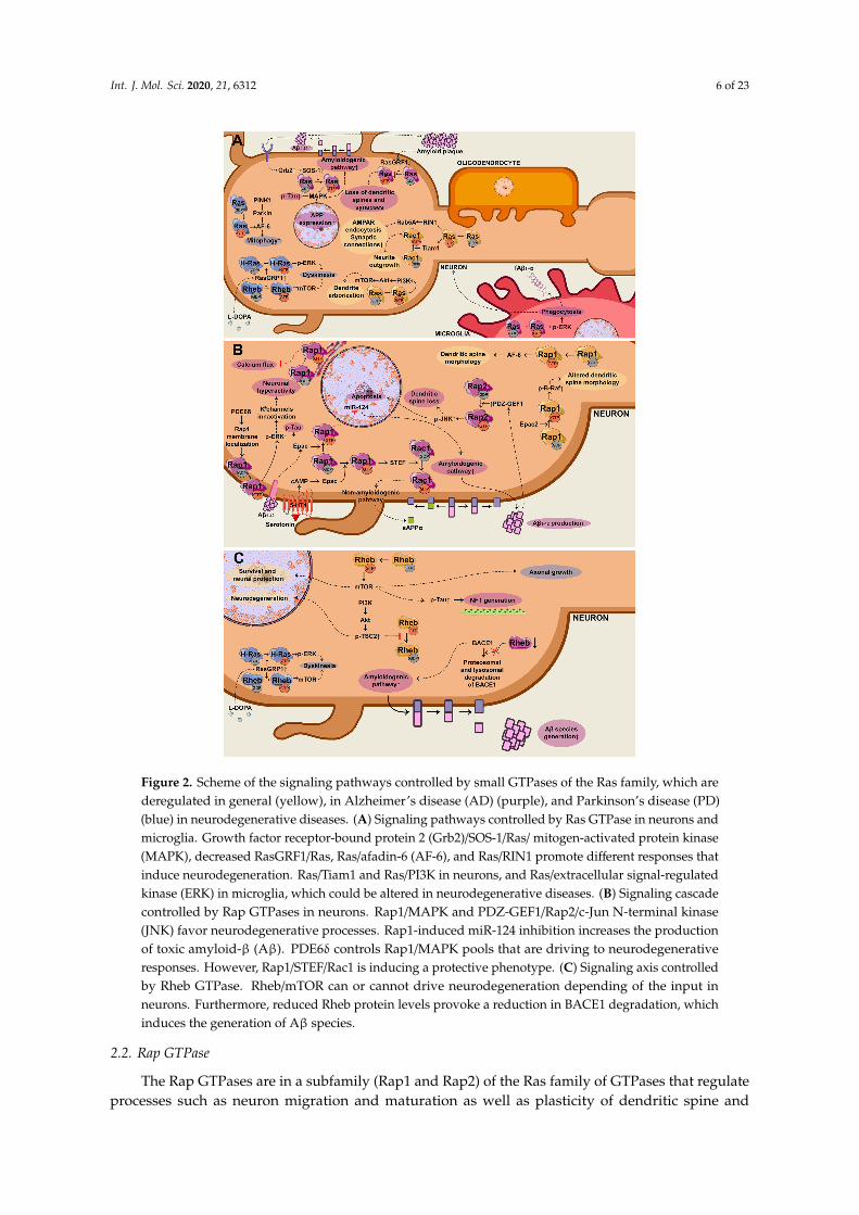
In conclusion, Ras GTPases control the generation of toxic peptides such as Aβ1–42 and pTau viathe MAPK pathway and microglial phagocytosis of fAβ. It also induces dendritic spine and synapseloss. All these are hallmarks of AD. In PD, Ras controls mitophagy and L-DOPA-induced dyskinesia(Figure 2A).
Int. J. Mol. Sci. 2020, 21, 6312 6 of 23Int. J. Mol. Sci. 2020, 21, x FOR PEER REVIEW 6 of 23
Figure 2. Scheme of the signaling pathways controlled by small GTPases of the Ras family, which are deregulated in general (yellow), in Alzheimer’s disease (AD) (purple), and Parkinson’s disease (PD) (blue) in neurodegenerative diseases. (A) Signaling pathways controlled by Ras GTPase in neurons and microglia. Growth factor receptor-bound protein 2 (Grb2)/SOS-1/Ras/ mitogen-activated protein kinase (MAPK), decreased RasGRF1/Ras, Ras/afadin-6 (AF-6), and Ras/RIN1 promote different responses that induce neurodegeneration. Ras/Tiam1 and Ras/PI3K in neurons, and Ras/extracellular signal-regulated kinase (ERK) in microglia, which could be altered in neurodegenerative diseases. (B) Signaling cascade controlled by Rap GTPases in neurons. Rap1/MAPK and PDZ-GEF1/Rap2/c-Jun N-terminal kinase (JNK) favor neurodegenerative processes. Rap1-induced miR-124 inhibition increases the production of toxic amyloid-β (Aβ). PDE6δ controls Rap1/MAPK pools that are driving to neurodegenerative responses. However, Rap1/STEF/Rac1 is inducing a
Figure 2. Scheme of the signaling pathways controlled by small GTPases of the Ras family, which arederegulated in general (yellow), in Alzheimer’s disease (AD) (purple), and Parkinson’s disease (PD)(blue) in neurodegenerative diseases. (A) Signaling pathways controlled by Ras GTPase in neurons andmicroglia. Growth factor receptor-bound protein 2 (Grb2)/SOS-1/Ras/ mitogen-activated protein kinase(MAPK), decreased RasGRF1/Ras, Ras/afadin-6 (AF-6), and Ras/RIN1 promote different responses thatinduce neurodegeneration. Ras/Tiam1 and Ras/PI3K in neurons, and Ras/extracellular signal-regulatedkinase (ERK) in microglia, which could be altered in neurodegenerative diseases. (B) Signaling cascadecontrolled by Rap GTPases in neurons. Rap1/MAPK and PDZ-GEF1/Rap2/c-Jun N-terminal kinase(JNK) favor neurodegenerative processes. Rap1-induced miR-124 inhibition increases the productionof toxic amyloid-β (Aβ). PDE6δ controls Rap1/MAPK pools that are driving to neurodegenerativeresponses. However, Rap1/STEF/Rac1 is inducing a protective phenotype. (C) Signaling axis controlledby Rheb GTPase. Rheb/mTOR can or cannot drive neurodegeneration depending of the input inneurons. Furthermore, reduced Rheb protein levels provoke a reduction in BACE1 degradation, whichinduces the generation of Aβ species.
2.2. Rap GTPase
The Rap GTPases are in a subfamily (Rap1 and Rap2) of the Ras family of GTPases that regulateprocesses such as neuron migration and maturation as well as plasticity of dendritic spine and
Int. J. Mol. Sci. 2020, 21, 6312 7 of 23
synapses [39]. The Rap GTPases are activated by GEFs such as Epac1, Epac2, C3G, RapGEF1,PDZ-GEF1, RA-GEF1, or RAPGEF2.
2.2.1. Rap/MAPK/ERK
Like Ras, Rap functions upstream of the MAPK pathway. The Ras and Rap GTPases have ahighly conserved effector domain in common [40], and they share some effector molecules. WhereasRap1 activates the Raf/MEK/ERK pathway [41], Rap2 activates the c-Jun N-terminal kinase (JNK)pathway [42].
In dendrites, the Epac2-dependent Rap activation results in an increase in phosphorylated B-Raf(p-B-Raf) [43]. Treatment of primary pyramidal neurons with the cAMP analogue that activates Epac,known as 8-(4-Chlorophenylthio)-2′-O-methyladenosine 3′,5′-cyclic monophosphate monosodiumhydrate (8-CPT-cAMP), provokes dendritic spine retraction [43]. Furthermore, the Epac/Rap axismediates synapse structural destabilization, spine shrinkage, and AMPA receptor removal fromspines [43].
Mutations in the EPAC2 gene have been linked to autism [44]. In HEK293, Epac2V646F reducedRap activity and p-B-Raf levels in dendrites. Conversely, Epac2T809S increased Rap activity and p-B-Raflevels. Moreover, both Epac2V646F and Epac2T809S also alter spine morphology [43].
Rap1 also controls neuronal activity via ERK. Phosphodiesterase 6δ (PDE6δ) interacts with Rap1to control MAPK subcellular localization [45]. Thus, PDE6δ functions as a regulator of Rap1 byregulating the MAPK pools that will activate the GTPase. PDE6δ also regulates Rap1 recycling fromthe endomembrane to the plasma membrane [45]. Blockade of the PDE6δ/Rap1 interaction increasesthe presence and activity of Rap1 in the endomembrane, favoring the reduction of neuronal activity.In fact, the Rap1/ERK axis in the plasma membrane phosphorylates and inactivates potassium channelsresponsible for action potentials, resulting in neuronal hyperactivity. In contrast, the Rap1/ERKcomplex suppresses calcium influx through voltage-gated calcium channels (VGCC) and increasesGABAB receptor activity in the endomembrane. Therefore, this regulatory nexus could be a therapeutictarget for treating diseases with neuronal hyperactivity [45], such as the early stages of AD. AD ischaracterized by an increase in intracellular calcium levels, which culminates in elevated excitabilityand, consequently, in neuronal death. Of note, PDE6δ/Rap1 inhibition has proven to partially rescuenormal phenotype in AD [45].
Taking into account that the MAPK pathway is also involved in Tau hyperphosphorylation,inhibition of PDE6δ/Rap1 interaction in the mice models of AD hAPP*PS1 and hTAU reduced ERKcascade activation and Tau phosphorylation in different residues in vivo, which resulted in the rescueof behavioral deficits [45].
2.2.2. Rap2/JNK
Rap2 controls JNK activity, one of the MAPK signaling nodes. JNK family is formed by threemembers, JNK 1–3. While JNK1 and 2 are ubiquitously expressed, JNK3 is mainly expressed in neurons.JNK can directly control apoptosis regulating protein Bcl-2 and c-Jun. Subsequently, JNK was linkedto neurodegenerative diseases [46]. For instance, post-mortem AD brains displayed an increase inphosphorylated JNK [47].
Rap2/JNK is also involved in dendritic spine loss when activated by PDZ-GEF1. 3xTg-AD miceand human post-mortem AD brain samples showed increased protein levels of this GEF. Moreover,the treatment of cultured hippocampal neurons with Aβ oligomers increased PDZ-GEF1 protein levels,which subsequently activated Rap2. In AD, PDZ-GEF1 activates the JNK pathway culminating in adendritic spine loss and cognitive decline [48].
2.2.3. Rap1/AF-6
Like MAPK, Ras and Rap conserved sequence homology determines their sharing of effectormolecule AF-6. Activated Rap1 recruits AF-6 to the plasma membrane, inducing spine neck elongation,
Int. J. Mol. Sci. 2020, 21, 6312 8 of 23
as well as regulating AMPA receptor content in the spines [49]. Furthermore, it is a key molecule inspine formation and synaptogenesis [50].
2.2.4. LRRK2/EPAC-1/Rap1 Pathway
Rap1 controls cell adhesion, polarization, and directional migration; processes that are bestcharacterized in macrophages and microglia. In macrophages, the Rap guanine nucleotide exchangefactor 3 (Rapgef3) gene expression, which codifies for Epac1, is controlled by LRRK2 [51]. LRRK2 is amultifunctional protein. One of its activities, as a serine/threonine kinase, is the phosphorylation—awide range of proteins involved in neuronal plasticity, autophagy, and vesicular trafficking [52–54].Moreover, LRRK2 also regulates Rab family GTPases by phosphorylation [55] and it phosphorylatesAD-related APP [54]. Apart from having a kinase activity domain, LRRK2 also presents a GTPaseactivity domain. The effect of the PD-related pathological mutation (LRRK2G2019S) in the kinaseactivity has been extensively studied, and it is known that LRRK2 mutations result in increased kinaseactivity that induces a neurotoxic effect [56]. In contrast, little attention has been paid to the effects ofthis mutation on the GTPase activity when it is known that the GTPase domain plays an importantrole in the regulation of the kinase activity and its neurotoxic effects [57].
A transcriptomics study showed that LRRK2 reduces Epac1 expression in macrophages [51]. Thus,macrophage migration regulated by LRRK2/Epac1/Rap1 might be one of the possible dysregulatedpathways in PD [51]. Although this axis still needs to be characterized in microglia, Levy andcollaborators suggest that the suppression of Epac1/Rap1 by PD-associated LRRK2 could reduce theability of microglia to migrate to sites of neuronal cell damage in PD [51].
2.2.5. Epac/Rap1/APP Processing
The Epac/Rap1 pathway may present a dual role. On the one hand, it regulates the neuroprotectivecleavage of APP. In fact, α cleavage of APP is induced via the cAMP/Epac/Rap1STEF/Rac1 pathwayactivated by the serotonin 5-HT4 receptor [6]. Rap1 specifically associates with the TSS region ofGEF STEF, which results in the activation of Rac1 GTPase of the Rho family [6]. This culminatesin the secretion of soluble α APP (sAPPα), which has neuroprotective properties and promotesmemory improvement.
On the other hand, Epac/Rap1 can regulate the toxic cleavage of APP. In the presence of Aβ orhypoxia, Epac/Rap1 is involved in miR-124 inhibition [58]. miR-124 regulates BACE1 secretase mRNAexpression and protein levels, which is the enzyme responsible for the β cleavage of APP. Thus, in thepresence of Aβ or hypoxia, miR-124 inhibition provokes an increase in BACE1 levels and, therefore,in the production of toxic Aβ [58]. Additionally, it is important to highlight that AD patients expresslow levels of miR-124 [59].
In a nutshell, Rap regulates both the amyloidogenic and non-amyloidogenic processing of APPand is also responsible for neuronal hyperactivity and dendritic spine loss (Figure 2B).
2.3. Rheb GTPase
Rheb GTPase, another member of the Ras family, is involved in cell cycle regulation, cell growthcontrol [60], autophagy [61], and apoptosis by controlling the interaction of FKBP38 with proteinsBcl-2 and Bcl-XL [62]. Although it shares some domains with other Ras GTPases, such as the effectorand GTP-binding domains or the motif for being prenylated, Rheb regulation is unique amongst themembers of this family. The TSC1/TSC2 GAP complex regulates the Rheb activation/deactivation cycle.Thus, to activate Rheb, the PI3K/Akt/PKB axis phosphorylates TSC2, and in this fashion, inhibits theTSC1/TSC2 GAP complex [63].
Some evidence suggest that Rheb activation potentiates neurodegeneration via the GTPaseassociation to its main effector molecule mTOR. Post-mortem AD brain samples showed reducedTSC2 protein levels, which implicates a continuous active state of Rheb; this suggests that Rhebactivation could play a role in the disease [64]. Another study showed that in post-mortem AD and
Int. J. Mol. Sci. 2020, 21, 6312 9 of 23
PD with dementia (PD/DLB) samples, the detected TSC2 was hyperphosphorylated, probably viaAkt [65]. Because this phosphorylation resulted in GAP inhibition and consequent Rheb activation,this study suggested that this mechanism could trigger neuronal death in various neurodegenerativepathologies such as AD and/or PD [65]. Moreover, as previously explained, simultaneous signaling bythe RasGRP1/Rheb/mTORC1 and RasGRP1/H-Ras/ERK axes could underlie the dyskinesia sufferedby PD patients who have been treated with L-DOPA for a prolonged time [29]. mTOR has also beenshown to induce Tau phosphorylation and its pharmacological inhibition alleviated the pathologyand the behavior deficits in human Tau overexpressing mice. Presently, this data suggests that mTORcould be a therapeutic target for tauopathies [66].
Nevertheless, Rheb/mTOR implication in neurodegeneration is controversial. Other studiesmaintain that Rheb activation, and, therefore, mTOR activation, controls cell survival and protectsfrom neurotoxicity in adult brain [67]. Accordingly, a constitutively active form of Rheb improved thePD phenotype by inducing axonal growth in DA neurons via Akt/mTOR [68]. Moreover, in the ADmouse model 5XFAD, constitutively active Rheb resulted in increased levels of neurotrophic moleculessuch as BDNF and ciliary neurotrophic factor (CNTF), and their respective receptors tropomyosinreceptor kinase B (TrkB) and CNTF receptor α subunit (CNTFRα) [69]. Additionally, a constitutivelyactive form of Rheb inhibited Aβ production and accumulation in the hippocampus of these mice andprevented cognitive impairments [67]. Hence, also Rheb represents a potential therapeutic target forneurodegenerative diseases [70].
Rheb can also modulate signaling cascades independently of mTOR. Recently, Rheb has beendescribed to bind to BACE1 secretase, this being a novel GTPase effector molecule [71]. Rheb-GTPbinds to BACE1 and promotes proteasomal and lysosomal degradation of this secretase in HEK293 [71].In murine primary neuronal cultures, Rheb reduced BACE1 levels, as well as its activity, resulting in adecrease in sAPPβ, APP-CTFβ, Aβ1–40 y Aβ1–42 species. Shahani and collaborators also demonstratedthat post-mortem AD samples showed reduced Rheb protein levels. Taking into account that BACE1levels are increased in AD, Shahani et al. suggested that low Rheb levels could explain the increasedlevels of BACE1 and the consequent Aβ generation [71]. Likewise, low Rheb levels are associated withmemory loss and, therefore, it could be a therapeutic target for pathologies characterized by memoryproblems such as AD [72].
To sum up, Rheb controls the production of Aβ peptides and it improves the PD phenotype byinducing axonal growth (Figure 2C).
3. Small GTPASES of the Rho Family
GTPases of the Rho family are specialized in the regulation of actin cytoskeleton dynamics, althoughthey also control cell cycle progression or transcriptional regulation [3]. The best-characterized GTPasesof this family, from a structural and a functional point of view, are RhoA, Rac1, and Cdc42.
The GEFs that control Rho GTPases activation are divided into two families: Dbl andDOCK [4,69,73]. In the Dbl family, we can find αPIX, Vav1, Tiam-1, Tiam-2/STEF, RasGRF, Sos1,Dbl, RhoGAP, p115RhoGEF, and Trio. In the DOCK family, its members are divided into foursubfamilies depending on the sequence homology and the substrate specificity: DOCK-A (DOCK2, 5,and 180), -B (DOCK3 and 4), -C (DOCK6, 7 and 8), and -D (DOCK9, 10, and 11).
Rho GTPases, as well as their regulators and effector molecules, play an important role inneurodegenerative processes, such as regulating APP processing or dendritic spine loss in AD [17] andoxidative stress and neuroinflammation in PD [18,74]. Furthermore, Rho GTPases are also implicated inother diseases related to the nervous system [12]. For instance, they are involved in amyotrophic lateralsclerosis (ALS) by regulating motor neuron survival and they control Huntingtin protein accumulationin Huntington’s disease [12].
Int. J. Mol. Sci. 2020, 21, 6312 10 of 23
3.1. RhoA
RhoA is the GTPase that gives the name to this family of GTPases, where other closely relatedisoforms such as RhoB and RhoC can be found [2]. RhoA controls the formation of actomyosincontractile fibers, also known as stress fibers.
Changes in RhoA subcellular localization have been linked to neurodegenerative processesoccurring in AD [75]. APPSwe Tg2576 mice express reduced levels of RhoA in synaptic ends andincreased levels in dystrophic neurites [75]. RhoA has also been connected to PD pathology [76,77].The intracellular signaling network controlled by RhoA is complex, being the best characterized effectormolecules Rho-kinase (ROCK) and the formin subfamily Diaphanous (Dia).
3.1.1. RhoA/ROCK
RhoA controls actin cytoskeleton reorganization through phosphorylation carried out by theROCK kinases family resulting in the activation of proteins such as myosin light chain (MLC) or LIMkinase (LIMK), which in turn phosphorylates and activates cofilin.
ROCK kinases have been associated with AD, as they can phosphorylate Tau protein and mediateNFT formation [78]. Studies in post-mortem AD samples showed increased ROCK expressionlevels [79]. Furthermore, ROCK promotes an increase in secreted Aβ1–40 levels and, in turn, increasedAβ levels potentiate ROCK activity, as well as its effector LIMK activity [79]. Henderson andcollaborators proposed that a positive feedback loop between Aβ/RhoA/ROCK/Aβ could be favoringAD progression [79].
The RhoA/ROCK axis has also been linked to AD pathology as it is involved in the retractionof dendritic spines. Aβ peptides induce an increase in actin contractility via Pyk2/RhoGAPGraf1/
RhoA-regulated ROCK2 kinase culminating in dendritic spine retraction [80].In microglia, Aβ peptide requires the RhoA/ROCK axis to induce different responses such
as chemotaxis, cytotoxicity, and inflammatory response [81]. Thus, the RhoA/ROCK signaling inmicroglia could be favoring AD neurodegeneration. Based on this hypothesis, Scheiblich and Bickerdefended the therapeutic potential of RhoA/ROCK inhibitors for treating excessive inflammation andneurodegeneration in CNS microglia [82].
RhoA/ROCK has also been implicated in DA neuron degeneration in PD. Both RhoA and ROCKinhibition presented a therapeutic effect in PD mouse models [76,77]. In addition, RhoA inhibition by C3transferase resulted in a reduction in α-syn (in mRNA and protein levels) in the MN9D neuronal cell line;this suggests RhoA as a potential therapeutic target for synucleopathies [83]. Although Zhou et al. didnot describe the molecular events that led to this α-syn reduction, they observed that RhoA inhibitionprovoked a reduction of the SRF transcription factor in the nucleus, as well as cytosolic retention ofMKL-1 (co-transactivator of SRF that translocates to the nucleus to bind SRF and, therefore, regulatestranscription). Hence, RhoA inhibitory effects could be due to a reduction in transcription [83].
3.1.2. RhoA/NOX
RhoA can also activate the holoenzyme NADPH oxidase (NOX) to generate superoxide ions [84].In the presence of fibrillar Aβ in microglia, RhoA/NOX is activated promoting the production ofsuperoxide species [84]. RhoA controls the superoxide production by regulating the phosphorylationof the p47PHOX subunit of the holoenzyme [84]. Microglial NOX is considered an important source ofoxidative stress that results in AD neuronal cell death [85].
In PD, microglial activation via angiotensin receptor AT1 induces activation of the RhoA/ROCK/
NOX2 axis and subsequent superoxide generation [86]. α-syn 29–40, a α-syn specific peptide, has beendescribed to activate superoxide generation in microglia, which causes DA neuronal damage [87].Later, it was described that extracellular α-syn binds to CD11b integrin in microglia and controls NOX2activation via a RhoA-dependent pathway [88].
Int. J. Mol. Sci. 2020, 21, 6312 11 of 23
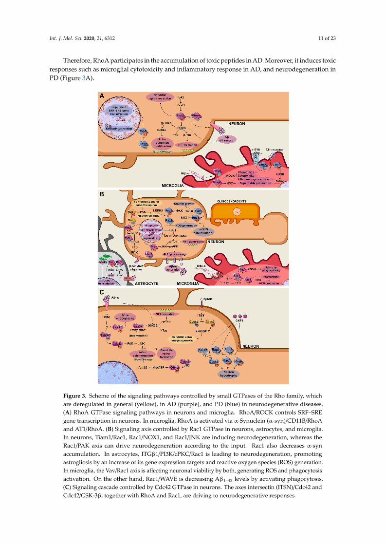
Therefore, RhoA participates in the accumulation of toxic peptides in AD. Moreover, it induces toxicresponses such as microglial cytotoxicity and inflammatory response in AD, and neurodegeneration inPD (Figure 3A).
Int. J. Mol. Sci. 2020, 21, x FOR PEER REVIEW 11 of 23
neuronal damage [87]. Later, it was described that extracellular α-syn binds to CD11b integrin in microglia and controls NOX2 activation via a RhoA-dependent pathway [88].
Therefore, RhoA participates in the accumulation of toxic peptides in AD. Moreover, it induces toxic responses such as microglial cytotoxicity and inflammatory response in AD, and neurodegeneration in PD (Figure 3A).
Figure 3. Scheme of the signaling pathways controlled by small GTPases of the Rho family, whichare deregulated in general (yellow), in AD (purple), and PD (blue) in neurodegenerative diseases.(A) RhoA GTPase signaling pathways in neurons and microglia. RhoA/ROCK controls SRF–SREgene transcription in neurons. In microglia, RhoA is activated via α-Synuclein (α-syn)/CD11B/RhoAand AT1/RhoA. (B) Signaling axis controlled by Rac1 GTPase in neurons, astrocytes, and microglia.In neurons, Tiam1/Rac1, Rac1/NOX1, and Rac1/JNK are inducing neurodegeneration, whereas theRac1/PAK axis can drive neurodegeneration according to the input. Rac1 also decreases α-synaccumulation. In astrocytes, ITGβ1/PI3K/cPKC/Rac1 is leading to neurodegeneration, promotingastrogliosis by an increase of its gene expression targets and reactive oxygen species (ROS) generation.In microglia, the Vav/Rac1 axis is affecting neuronal viability by both, generating ROS and phagocytosisactivation. On the other hand, Rac1/WAVE is decreasing Aβ1–42 levels by activating phagocytosis.(C) Signaling cascade controlled by Cdc42 GTPase in neurons. The axes intersectin (ITSN)/Cdc42 andCdc42/GSK-3β, together with RhoA and Rac1, are driving to neurodegenerative responses.
Int. J. Mol. Sci. 2020, 21, 6312 12 of 23
3.2. Rac GTPase
The Rac GTPases belong to a subfamily of Rho GTPases constituted by four members—Rac1,Rac2, Rac3, and RhoG—where Rac1 is the best studied member. These GTPases control cytoskeletonorganization, gene transcription, and proliferation [3]. Together with Ras and Rho, Rac is considered aprotooncogene, and mutations in these GTPases confer them a constitutive activity in various types oftumors [11]. Apart from participating in tumoral processes, Rac1 plays a key role in development andneuronal survival as well as in neurodegeneration [12].
In AD, Rac1 has been described to increase Aβ levels by transcriptional regulation, as it increasesAPP expression by controlling the −233 to −41 bp positions in the promotor of the APP gene [89].Recently, Borin et al. have reported that a constitutively active form of Rac1 increases APP processing,culminating in a higher content of Aβ1–42 peptides [90]. Thus, Rac1-induced increase in Aβ levelscould be due to an increase in APP expression as well as in its processing. Conversely, Rac1 is alsoinvolved in the reduction of Aβ1–42 levels as it controls the microglial phagocytosis of Aβ1–42 peptidesvia its effector molecule WAVE [91].
3.2.1. Rac1/Toxic Peptide Accumulation
Like RhoA/ROCK, Rac1 also controls Tau phosphorylation in the early stages of AD. Rac1 inducesthe translocation of SET from the nucleus to the cytoplasm [90]. SET, as an inhibitor of Tau phosphatase,blocks Tau dephosphorylation.
In PD, α-syn accumulation is toxic and is responsible for PD symptomatology. Rac1 reducesα-syn accumulation in Caenorhabditis elegans, in the BE(2) M17 human neuroblastoma cell line andDA neuron-like cells derived from iPSCs from PD patients [92]. This can be viewed as a cell survivalmechanism promoted by Rac1 [92].
3.2.2. Rac1/PAK
PAK (p21 activated kinase) is a family of serine/threonine kinases that function as Rac and Cdc42GTPases effector molecules, and they are implicated in the signal transduction emanating from integrinsand tyrosine kinase receptors. Furthermore, PAK has been related to neurodegenerative diseases [93].In a healthy brain, PAK is involved in the regulation of neuron viability, synapse morphology andfunctionality, gene transcription in neurons, microglial motility, microglial immune response, andastrogliosis [94]. In contrast, PAK protein and activity levels are elevated in neurodegenerativedisorders, resulting in a decrease in neuronal cell viability and, therefore, a potentiated neuronaldegeneration in diseases such as AD and PD [94].
Nevertheless, the role of this over-activated Rac1/PAK axis in dendritic spine loss is controversial,as some authors understand that Rac/PAK helps the formation of dendritic spines [95]. Borin et al.claimed that Rac might present a dual role depending on the stage of AD disease [90]. Whereas Rac1 isover-activated in early stages of the disease, in later stages, Rac1 protein levels decrease, which resultsin a dendritic spine loss [90]. Altogether, the role of Rac1/PAK in AD is complex and still needs furtherstudies, considering different factors such as the model used for the study and the stage of the disease.
In PD, wild type LRRK2 binds strongly to Rac and increases its activity. This can be seen as anincrease in the binding of Rac1 to PAK [96]. The LRRK2G2019S mutant phenotype presents neuriteretraction, but in SH-SY5Y cells when Rac1 was co-expressed with LRRK2 G2019S, Rac1 was ableto rescue this retraction [96]. Thus, Chan et al. postulated that pathological mutations in LRRK2could attenuate Rac1 activation, resulting in an actin filament disassembly and, consequently, neuriteretraction [96].
3.2.3. PI3K/PDK/nPKC/Tiam-1/Rac1/Neuronal Cell Death
Rac1 is implicated in Aβ1–42-induced neuronal cell death. In the SN4741 neuronal cell line,Aβ1–42 requires the PI3K/PDK/nPKC axis to promote Tiam-1 phosphorylation and, subsequently,
Int. J. Mol. Sci. 2020, 21, 6312 13 of 23
activate Rac1 GTPase culminating in the apoptosis of SN4741 cells, primary embryonic corticalneurons from rats, as well as in neuronal organotypic cultures of the hippocampus and the entorhinalcortex [97]. Manterola et al. highlighted the central role of Rac1 in transducing the Aβ1–42-mediatedtoxic signaling [97]. Although the signaling cascade inducing neuronal cell death downstream of Rac1is still unknown, the authors suggested that one of the mechanisms could be the Rac1 activation ofNOX and the ensuing generation of reactive oxygen species (ROS).
3.2.4. Rac1/NOX
NOX is also an effector molecule of Rac1. Rac1/NOX axis is also involved in neurodegenerativeprocesses [98,99].
In murine primary microglial cell cultures, fibrillary Aβ (fAβ) activates Rac1/NOX, generatingoxidative stress that affects neuronal viability [98]. Moreover, fAβ binds to different microglial receptors,inducing the activation of Lyn and Syk kinases. These kinases are responsible for phosphorylatingVav, a Rac1 GEF, resulting in Rac1 activation and, subsequently, NOX [98]. The fAβ/Lyn/Syk/Vav/Rac1axis, apart from activating NOX, also induces actin cytoskeleton organization that promotes microglialphagocytosis [98]. In primary astrocytic cultures from mice, Aβ1–42 induces astrogliosis [99], which isan astrocytic hypertrophy that compromises neuronal cell viability. Aβ1–42 induced this astrogliosis byNOX activation via the PI3K/cPKC/Rac1/NOX axis [99].
Rac1 activation and oxidative stress generated by NOX are also involved in PD [74]. Rac1inhibition and NOX1 silencing resulted in a reduction of DNA damage and DA neuronal degenerationin the N27 cell line and DA neurons in the substantia nigra of rats [74].
3.2.5. Rac1/JNK
JNK signaling pathway, together with the p38 and ERK MAPK pathways, are activated in differentstages of AD. JNK activates apoptotic processes [46]. Moreover, it is also able to phosphorylate Tauprotein, favoring the NFT formation, and APP activating its processing [46]. All these processesparticipate in AD progression [46]. On the other hand, JNK could also be a therapeutic target forPD [100]. When studying the Rac1/NOX1 axis in DA neuronal cell death, Choi et al. reported thatRac1 inhibition and NOX1 silencing resulted in a reduction of the apoptotic marker c-Jun, which isphosphorylated by JNK [74]. These results suggest that lower levels of phosphorylated c-Jun could beindicative of a good prognosis for PD.
In conclusion, Rac participates in the accumulation of the three toxic peptides Aβ, pTau, and α-synin AD and PD, respectively. It is also responsible for inducing toxic effects such as apoptosis orROS generation in neurons, phagocytic pathways activation in microglia, and astrogliosis control inastrocytes (Figure 3B).
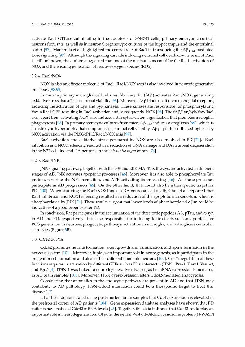
3.3. Cdc42 GTPase
Cdc42 promotes neurite formation, axon growth and ramification, and spine formation in thenervous system [101]. Moreover, it plays an important role in neurogenesis, as it participates in theprogenitor cell formation and also in their differentiation into neurons [102]. Cdc42 regulation of thesefunctions requires its activation by different GEFs such as Dbs, intersectin (ITSN), Prex1, Tiam1, Vav1–3,and Fgd5 [4]. ITSN-1 was linked to neurodegenerative diseases, as its mRNA expression is increasedin AD brain samples [103]. Moreover, ITSN overexpression alters Cdc42-mediated endocytosis.
Considering that anomalies in the endocytic pathway are present in AD and that ITSN maycontribute to AD pathology, ITSN-Cdc42 interaction could be a therapeutic target to treat thisdisease [17].
It has been demonstrated using post-mortem brain samples that Cdc42 expression is elevated inthe prefrontal cortex of AD patients [104]. Gene expression database analyses have shown that PDpatients have reduced Cdc42 mRNA levels [93]. Together, this data indicates that Cdc42 could play animportant role in neurodegeneration. Of note, the neural Wiskott–Aldrich Syndrome protein (N-WASP)
Int. J. Mol. Sci. 2020, 21, 6312 14 of 23
is a Cdc42-specific effector molecule, while PAK, PI5K, or formins are Cdc42-effector molecules sharedwith Rac. All of them are involved in actin cytoskeleton reorganization. It is necessary to understandall these downstream cascades in order to identify potential targets for neurodegeneration.
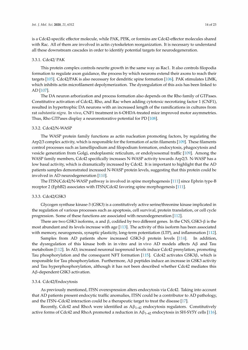
3.3.1. Cdc42/ PAK
This protein complex controls neurite growth in the same way as Rac1. It also controls filopodiaformation to regulate axon guidance, the process by which neurons extend their axons to reach theirtargets [105]. Cdc42/PAK is also necessary for dendritic spine formation [106]. PAK stimulates LIMK,which inhibits actin microfilament depolymerization. The dysregulation of this axis has been linked toAD [107].
The DA neuron arborization and process formation also depends on the Rho family of GTPases.Constitutive activation of Cdc42, Rho, and Rac when adding cytotoxic necrotizing factor 1 (CNF1),resulted in hypertrophic DA neurons with an increased length of the ramifications in cultures fromrat substantia nigra. In vivo, CNF1 treatment in 6-OHDA-treated mice improved motor asymmetries.Thus, Rho GTPases display a neurorestorative potential for PD [108].
3.3.2. Cdc42/N-WASP
The WASP protein family functions as actin nucleation promoting factors, by regulating theArp2/3 complex activity, which is responsible for the formation of actin filaments [109]. These filamentscontrol processes such as lamellipodium and filopodium formation, endocytosis, phagocytosis andvesicle generation from Golgi, endoplasmic reticulum, or endolysosomal traffic [109]. Among theWASP family members, Cdc42 specifically increases N-WASP activity towards Arp2/3. N-WASP has alow basal activity, which is dramatically increased by Cdc42. It is important to highlight that the ADpatients samples demonstrated increased N-WASP protein levels, suggesting that this protein could beinvolved in AD neurodegeneration [110].
The ITSN/Cdc42/N-WASP pathway is involved in spine morphogenesis [111] since Ephrin type-Breceptor 2 (EphB2) associates with ITSN/Cdc42 favoring spine morphogenesis [111].
3.3.3. Cdc42/GSK3
Glycogen synthase kinase-3 (GSK3) is a constitutively active serine/threonine kinase implicated inthe regulation of various processes such as apoptosis, cell survival, protein translation, or cell cycleprogression. Some of these functions are associated with neurodegeneration [112].
There are two GSK3 isoforms, α and β, codified by two different genes. In the CNS, GSK3-β is themost abundant and its levels increase with age [113]. The activity of this isoform has been associatedwith memory, neurogenesis, synaptic plasticity, long-term potentiation (LTP), and inflammation [112].
Samples from AD patients show increased GSK3-β protein levels [114]. In addition,the dysregulation of this kinase both in in vitro and in vivo AD models affects Aβ and Taumetabolism [112]. In AD, increased neuronal isoprenoid levels induce Cdc42 prenylation, promotingTau phosphorylation and the consequent NFT formation [115]. Cdc42 activates GSK3β, which isresponsible for Tau phosphorylation. Furthermore, Aβ peptides induce an increase in GSK3 activityand Tau hyperphosphorylation, although it has not been described whether Cdc42 mediates thisAβ-dependent GSK3 activation.
3.3.4. Cdc42/Endocytosis
As previously mentioned, ITSN overexpression alters endocytosis via Cdc42. Taking into accountthat AD patients present endocytic traffic anomalies, ITSN could be a contributor to AD pathology,and the ITSN–Cdc42 interaction could be a therapeutic target to treat the disease [17].
Recently, Cdc42 and RhoA were identified as Aβ1–42 endocytosis regulators. Constitutivelyactive forms of Cdc42 and RhoA promoted a reduction in Aβ1–42 endocytosis in SH-SY5Y cells [116].
Int. J. Mol. Sci. 2020, 21, 6312 15 of 23
It suggests that the Rho family of GTPases controls Aβ1–42 internalization and that the endocyticpathway could be a target to reduce Aβ1–42.
Conclusively, Cdc42 controls GSK3-mediated pTau and NFT accumulation, and it also controlsdendritic spine formation and Aβ endocytosis. In PD, together with Cdc42, the other Rho familyGTPases regulate neuronal hypertrophy (Figure 3C).
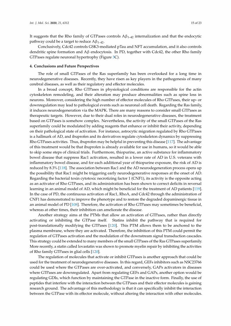
4. Conclusions and Future Perspectives
The role of small GTPases of the Ras superfamily has been overlooked for a long time inneurodegenerative diseases. Recently, they have risen as key players in the pathogenesis of manycerebral diseases, as well as their regulatory and effector molecules.
In a broad concept, Rho GTPases in physiological conditions are responsible for the actincytoskeleton remodeling, and their alteration may produce abnormalities such as spine loss inneurons. Moreover, considering the high number of effector molecules of Rho GTPases, their up- ordownregulation may lead to pathological events such as neuronal cell death. Regarding the Ras family,it induces neurodegeneration via the MAPK. There are many reasons to consider small GTPases astherapeutic targets. However, due to their dual roles in neurodegenerative diseases, the treatmentbased on GTPases is somehow complex. Nevertheless, the activity of the small GTPases of the Rassuperfamily could be modulated by adding reagents that enhance or inhibit their activity, dependingon their pathological state of activation. For instance, astrocytic migration regulated by Rho GTPasesis a hallmark of AD, and ibuprofen and its derivatives regulate cytoskeleton dynamics by suppressingRho GTPases activities. Thus, ibuprofen may be helpful in preventing this disease [117]. The advantageof this treatment would be that ibuprofen is already available for use in humans, so it would be ableto skip some steps of clinical trials. Furthermore, thiopurine, an active substance for inflammatorybowel disease that suppress Rac1 activation, resulted in a lower rate of AD in U.S. veterans withinflammatory bowel disease, and for each additional year of thiopurine exposure, the risk of AD isreduced by 8.3% [118]. The association between Rac1 and the AD neurodegeneration process opens upthe possibility that Rac1 might be triggering early neurodegenerative responses at the onset of AD.Regarding the bacterial toxin cytotoxic necrotizing factor 1 (CNF1), its activity is the opposite actingas an activator of Rho GTPases, and its administration has been shown to correct deficits in reversallearning in an animal model of AD, which might be beneficial for the treatment of AD patients [119].In the case of PD, the continuous activation of Rac1, RhoA, and Cdc42 through the administration ofCNF1 has demonstrated to improve the phenotype and to restore the degraded dopaminergic tissue inan animal model of PD [108]. Therefore, the activation of Rho GTPases may sometimes be beneficial,whereas at other times, their inhibition can ameliorate the disease.
Another strategy aims at the PTMs that allow an activation of GTPases, rather than directlyactivating or inhibiting the GTPase itself. Statins inhibit the pathway that is required forpost-translationally modifying the GTPases [120]. This PTM allows them to be anchored to theplasma membrane, where they are activated. Therefore, the inhibition of this PTM could permit theregulation of GTPases activation and the modulation of the downstream signal transduction cascades.This strategy could be extended to many members of the small GTPases of the Ras GTPases superfamily.More recently, a statin called lovastatin was shown to promote myelin repair by inhibiting the activitiesof Rho family GTPases in glial cells [120].
The regulation of molecules that activate or inhibit GTPases is another approach that could beused for the treatment of neurodegenerative diseases. In this regard, GEFs inhibitors such as NSC23766could be used where the GTPases are over-activated, and conversely, GAPs activators in diseaseswhere GTPases are downregulated. Apart from regulating GEFs and GAPs, another option would beregulating GDIs, which function by maintaining the GTPase in the inactive form. Finally, the use ofpeptides that interfere with the interaction between the GTPases and their effector molecules is gainingresearch ground. The advantage of this methodology is that it can specifically inhibit the interactionbetween the GTPase with its effector molecule, without altering the interaction with other molecules.
Int. J. Mol. Sci. 2020, 21, 6312 16 of 23
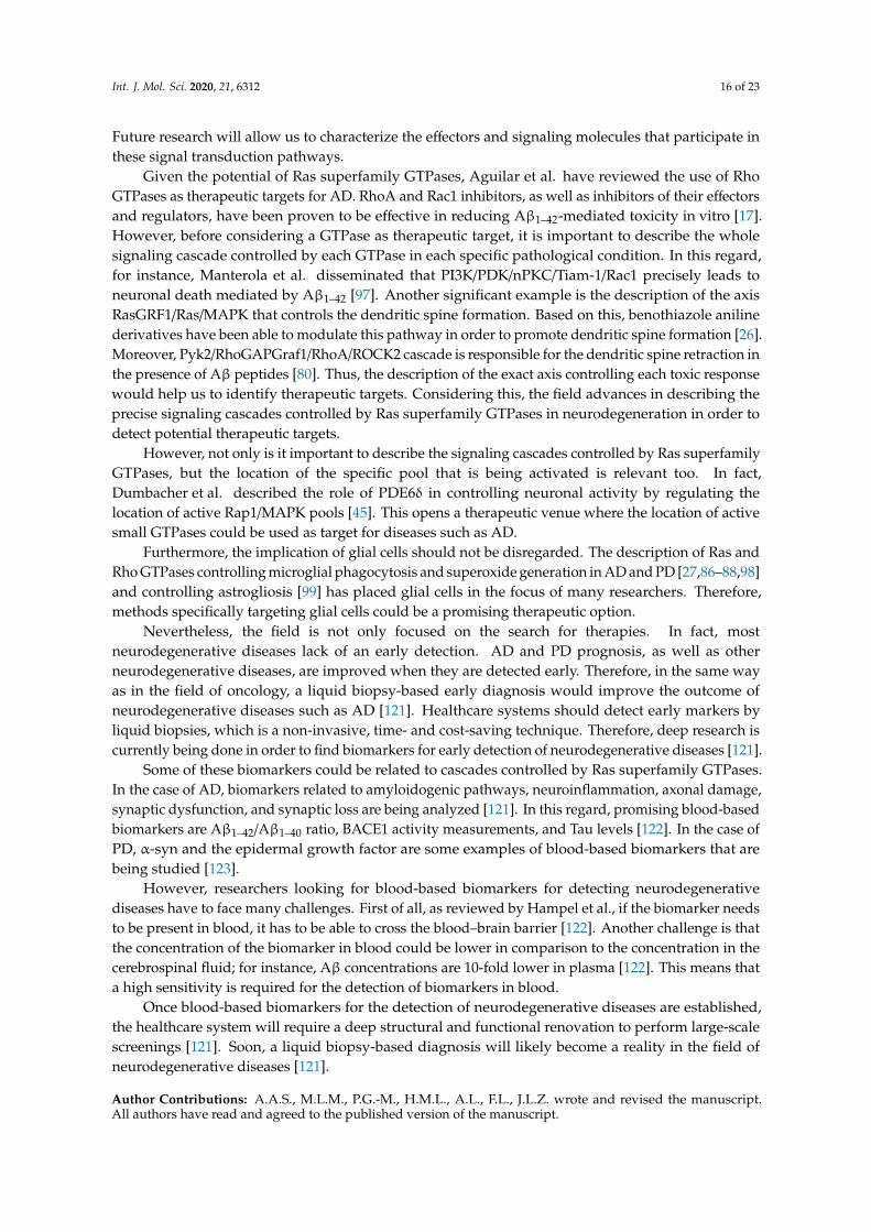
Future research will allow us to characterize the effectors and signaling molecules that participate inthese signal transduction pathways.
Given the potential of Ras superfamily GTPases, Aguilar et al. have reviewed the use of RhoGTPases as therapeutic targets for AD. RhoA and Rac1 inhibitors, as well as inhibitors of their effectorsand regulators, have been proven to be effective in reducing Aβ1–42-mediated toxicity in vitro [17].However, before considering a GTPase as therapeutic target, it is important to describe the wholesignaling cascade controlled by each GTPase in each specific pathological condition. In this regard,for instance, Manterola et al. disseminated that PI3K/PDK/nPKC/Tiam-1/Rac1 precisely leads toneuronal death mediated by Aβ1–42 [97]. Another significant example is the description of the axisRasGRF1/Ras/MAPK that controls the dendritic spine formation. Based on this, benothiazole anilinederivatives have been able to modulate this pathway in order to promote dendritic spine formation [26].Moreover, Pyk2/RhoGAPGraf1/RhoA/ROCK2 cascade is responsible for the dendritic spine retraction inthe presence of Aβ peptides [80]. Thus, the description of the exact axis controlling each toxic responsewould help us to identify therapeutic targets. Considering this, the field advances in describing theprecise signaling cascades controlled by Ras superfamily GTPases in neurodegeneration in order todetect potential therapeutic targets.
However, not only is it important to describe the signaling cascades controlled by Ras superfamilyGTPases, but the location of the specific pool that is being activated is relevant too. In fact,Dumbacher et al. described the role of PDE6δ in controlling neuronal activity by regulating thelocation of active Rap1/MAPK pools [45]. This opens a therapeutic venue where the location of activesmall GTPases could be used as target for diseases such as AD.
Furthermore, the implication of glial cells should not be disregarded. The description of Ras andRho GTPases controlling microglial phagocytosis and superoxide generation in AD and PD [27,86–88,98]and controlling astrogliosis [99] has placed glial cells in the focus of many researchers. Therefore,methods specifically targeting glial cells could be a promising therapeutic option.
Nevertheless, the field is not only focused on the search for therapies. In fact, mostneurodegenerative diseases lack of an early detection. AD and PD prognosis, as well as otherneurodegenerative diseases, are improved when they are detected early. Therefore, in the same wayas in the field of oncology, a liquid biopsy-based early diagnosis would improve the outcome ofneurodegenerative diseases such as AD [121]. Healthcare systems should detect early markers byliquid biopsies, which is a non-invasive, time- and cost-saving technique. Therefore, deep research iscurrently being done in order to find biomarkers for early detection of neurodegenerative diseases [121].
Some of these biomarkers could be related to cascades controlled by Ras superfamily GTPases.In the case of AD, biomarkers related to amyloidogenic pathways, neuroinflammation, axonal damage,synaptic dysfunction, and synaptic loss are being analyzed [121]. In this regard, promising blood-basedbiomarkers are Aβ1–42/Aβ1–40 ratio, BACE1 activity measurements, and Tau levels [122]. In the case ofPD, α-syn and the epidermal growth factor are some examples of blood-based biomarkers that arebeing studied [123].
However, researchers looking for blood-based biomarkers for detecting neurodegenerativediseases have to face many challenges. First of all, as reviewed by Hampel et al., if the biomarker needsto be present in blood, it has to be able to cross the blood–brain barrier [122]. Another challenge is thatthe concentration of the biomarker in blood could be lower in comparison to the concentration in thecerebrospinal fluid; for instance, Aβ concentrations are 10-fold lower in plasma [122]. This means thata high sensitivity is required for the detection of biomarkers in blood.
Once blood-based biomarkers for the detection of neurodegenerative diseases are established,the healthcare system will require a deep structural and functional renovation to perform large-scalescreenings [121]. Soon, a liquid biopsy-based diagnosis will likely become a reality in the field ofneurodegenerative diseases [121].
Author Contributions: A.A.S., M.L.M., P.G.-M., H.M.L., A.L., F.L., J.L.Z. wrote and revised the manuscript.All authors have read and agreed to the published version of the manuscript.
Int. J. Mol. Sci. 2020, 21, 6312 17 of 23
Funding: A.A.S. is a recipient of a predoctoral fellowship (PRE_2017_1_0016) from the Basque Government.M.L.M. is a recipient of a fellowship from Foundation “Jesús de Gangoiti y Barrera”. J.L.Z. was supported by theInstituto de Salud Carlos III (PI18/00207) and the University of Basque Country Grant (US19/04).
Acknowledgments: In memory of our collaborator Prof. Luis Antonio Parada (Institute of Experimental Pathology,CONICET-UNSA, Salta, Argentina).
Conflicts of Interest: The authors declare that they have no conflicts of interest with the contents of this article.All authors qualify for authorship, approved the final version of the manuscript, and agree to be accountable forall aspects of the research in ensuring that questions related to the accuracy or integrity of any part of the studyare appropriately investigated and resolved.
Abbreviations
Aβ: amyloid-β; AD, Alzheimer’s disease; AF-6, afadin-6; ALS, amyotrophic lateral sclerosis; AMPA,α-amino-3-hydroxy-5-methyl-4-isoxazolepropionic acid; APP, amyloid precursor protein; APP-CTFβ, β-secretasecleaved APP-C-terminal fragment; α-syn, α-synuclein, AT1, angiotensin II type 1 receptor; BACE 1, β-siteAPP cleaving enzyme 1; Bcl-2, B-cell lymphoma 2; Bcl-XL, B-cell lymphoma-extra-large; BDNF, brain-derivedneurotrophic factor; cAMP, cyclic adenosine monophosphate; CJD, Creutzfeldt–Jakob disease; CNF1, cytotoxicnecrotizing factor 1; CNS, central nervous system; CNTF, ciliary neurotrophic factor; CNTFRα, CNTF receptor αsubunit; cPKC, classical protein kinase C; DA, dopaminergic; Dbl, diffuse B-cell lymphoma; Dia, diaphanous;DLB, dementia with Lewy bodies; DOCK, dedicator of cytokinesis; Epac, exchange protein activated by cAMP;EphB2, ephrin type-B receptor 2; ERK, extracellular signal-regulated kinase; fAβ, fibrillary Aβ; GABA, gammaamino butyric acid; GAP, GTPase activating protein; GDI, guanine nucleotide dissociation inhibitor; GDP,guanosine diphosphate; GEF, guanine nucleotide exchange factor; Grb2, growth factor receptor-bound protein2; GSK3, glycogen synthase kinase-3; GTP, guanosine triphosphate; GTPase, guanosine triphosphatase; HD,Huntington’s disease; iPSC, induced pluripotent stem cell; ITSN, intersectin; JNK, c-Jun N-terminal kinase;KSR, kinase suppressor of Ras; LB, Lewy body; L-DOPA, L-3,4-dihydroxyphenylalanine; LIMK, LIM kinase;LRRK2, leucine-rich repeat kinase 2; MAPK, mitogen-activated protein kinase; MEK, mitogen-activated proteinkinase kinase; MKL-1, megakaryoblastic leukemia 1; MLC, myosin light chain; mTOR, mammalian targetof rapamycin; NFT, neurofibrillary tangle; NOX, NADPH oxidase; nPKC, novel protein kinase C; PAK, p21activated kinase; PD, Parkinson’s disease; PDE6δ, phosphodiesterase 6δ; PDK, phosphoinositide-dependent kinase;PI3K, phosphatidylinositol 3-kinase; PI5K, phosphatidylinositol-4-phosphate 5-kinase; PINK1, PTEN-inducedkinase 1; PKB, protein kinase B; PS1, presenilin 1; ROCK, Rho-kinase; ROS, reactive oxygen species; sAPPβ,β-secretase-cleaved soluble APP, SRF, serum response factor; TrkB, tropomyosin receptor kinase B; TSC, tuberoussclerosis complex; VGCC, voltage-gated calcium channel; WASP, Wiskott–Aldrich syndrome protein; 3xTg-AD,triple-transgenic mouse model of AD; 8-CPT-cAMP, 8-(4-Chlorophenylthio)-2′-O-methyladenosine 3′,5′-cyclicmonophosphate monosodium hydrate; 5-HT4, 5-hydroxytryptamine receptor 4; 5XFAD, five-familial AD transgenicmouse; 6-OHDA, 6-hydroxydopamine.
References
1. Berridge, M.J. Calcium signalling remodelling and disease. Biochem. Soc. Trans. 2012, 40, 297–309. [CrossRef][PubMed]
2. Goitre, L.; Trapani, E.; Trabalzini, L.; Retta, S.F. The Ras Superfamily of Small GTPases: The Unlocked Secrets.Methods Mol. Biol. 2014, 1120, 1–18. [PubMed]
3. Song, S.; Cong, W.; Zhou, S.; Shi, Y.; Dai, W.; Zhang, H.; Wang, X.; He, B.; Zhang, Q. Small GTPases: Structure,biological function and its interaction with nanoparticles. Asian J. Pharm. Sci. 2019, 14, 30–39. [CrossRef][PubMed]
4. Toma-Fukai, S.; Shimizu, T. Structural insights into the regulation mechanism of small GTPases by GEFs.Molecules 2019, 24, 3308. [CrossRef]
5. Peurois, F.; Peyroche, G.; Cherfils, J. Small GTPase peripheral binding to membranes: Molecular determinantsand supramolecular organization. Biochem. Soc. Trans. 2018, 47, 13–22. [CrossRef]
6. Zaldua, N.; Gastineau, M.; Hoshino, M.; Lezoualc’h, F.; Zugaza, J.L. Epac signaling pathway involves STEF,a guanine nucleotide exchange factor for Rac, to regulate APP processing. FEBS Lett. 2007, 581, 5814–5818.[CrossRef]
7. Singh, V.; Davidson, A.C.; Hume, P.J.; Humphreys, D.; Koronakis, V. Arf GTPase interplay with Rho GTPasesin regulation of the actin cytoskeleton. Small GTPases 2019, 10, 411–418. [CrossRef]
8. Aspenström, P. Activated rho GTPases in cancer—The beginning of a new paradigm. Int. J. Mol. Sci. 2018,19, 3949. [CrossRef]
9. Strassheim, D.; Gerasimovskaya, E.; Irwin, D.; Dempsey, E.C.; Stenmark, K.; Karoor, V. RhoGTPase inVascular Disease. Cells 2019, 8, 551. [CrossRef]
Int. J. Mol. Sci. 2020, 21, 6312 18 of 23
10. Qu, L.; Pan, C.; He, S.M.; Lang, B.; Gao, G.D.; Wang, X.L.; Wang, Y. The ras superfamily of small gtpases innon-neoplastic cerebral diseases. Front. Mol. Neurosci. 2019, 12, 121. [CrossRef]
11. Gan, L.; Cookson, M.R.; Petrucelli, L.; La Spada, A.R. Converging pathways in neurodegeneration, fromgenetics to mechanisms. Nat. Neurosci. 2018, 21, 1300–1309. [CrossRef] [PubMed]
12. Soria Lopez, J.A.; González, H.M.; Léger, G.C. Alzheimer’s disease. Hanb. Clin. Neurol. 2019, 167, 231–255.13. Gallardo, G.; Holtzman, D.M. Amyloid-β and Tau at the Crossroads of Alzheimer’s Disease. Adv. Exp.
Med. Biol. 2019, 1184, 187–203. [PubMed]14. Yang, L.; Mao, K.; Yu, H.; Chen, J. Neuroinflammatory Responses and Parkinson’ Disease: Pathogenic
Mechanisms and Therapeutic Targets. J. Neuroimmune Pharmacol. 2020. [CrossRef]15. Paisán-Ruiz, C.; Lewis, P.A.; Singleton, A.B. LRRK2: Cause, risk, and mechanism. J. Parkinsons. Dis. 2013, 3,
85–103. [CrossRef] [PubMed]16. Pickrell, A.M.; Youle, R.J. The roles of PINK1, Parkin, and mitochondrial fidelity in parkinson’s disease.
Neuron 2015, 85, 257–273. [CrossRef]17. Aguilar, B.J.; Zhu, Y.; Lu, Q. Rho GTPases as therapeutic targets in Alzheimer’s disease. Alzheimer’s Res. Ther.
2017, 9, 97. [CrossRef]18. Hong, L.; Sklar, L.A. Targeting GTPases in Parkinson’s disease: Comparison to the historic path of kinase
drug discovery and perspectives. Front. Mol. Neurosci. 2014, 7, 52. [CrossRef]19. Simanshu, D.K.; Nissley, D.V.; McCormick, F. RAS Proteins and Their Regulators in Human Disease. Cell
2017, 170, 17–33. [CrossRef]20. Clapéron, A.; Therrien, M. KSR and CNK: Two scaffolds regulating RAS-mediated RAF activation. Oncogene
2007, 26, 3143–3158. [CrossRef]21. Kirouac, L.; Rajic, A.J.; Cribbs, D.H.; Padmanabhan, J. Activation of Ras-ERK signaling and GSK-3 by amyloid
precursor protein and amyloid beta facilitates neurodegeneration in Alzheimer’s disease. eNeuro 2017, 4,149–165. [CrossRef] [PubMed]
22. McShea, A.; Zelasko, D.A.; Gerst, J.L.; Smith, M.A. Signal transduction abnormalities in Alzheimer’s disease:Evidence of a pathogenic stimuli. Brain Res. 1999, 815, 237–242. [CrossRef]
23. Pei, J.J.; Braak, H.; An, W.L.; Winblad, B.; Cowburn, R.F.; Iqbal, K.; Grundke-Iqbal, I. Up-regulation ofmitogen-activated protein kinases ERK1/2 and MEK1/2 is associated with the progression of neurofibrillarydegeneration in Alzheimer’s disease. Mol. Brain Res. 2002, 109, 45–55. [CrossRef]
24. Sung, Y.M.; Lee, T.; Yoon, H.; DiBattista, A.M.; Song, J.M.; Sohn, Y.; Moffat, E.I.; Turner, R.S.; Jung, M.;Kim, J.; et al. Mercaptoacetamide-based class II HDAC inhibitor lowers Aβ levels and improves learningand memory in a mouse model of Alzheimer’s disease. Exp. Neurol. 2013, 239, 192–201. [CrossRef]
25. Song, J.M.; Sung, Y.M.; Nam, J.H.; Yoon, H.; Chung, A.; Moffat, E.; Jung, M.; Pak, D.T.S.; Kim, J.; Hoe, H.S.A mercaptoacetamide-based Class II histone deacetylase inhibitor increases dendritic spine density viaRasGRF1/ERK pathway. J. Alzheimer’s Dis. 2016, 51, 591–604. [CrossRef]
26. Cifelli, J.L.; Berg, K.R.; Yang, J. Benzothiazole amphiphiles promote RasGRF1-associated dendritic spineformation in human stem cell-derived neurons. FEBS Open Bio 2020, 10, 386–395. [CrossRef]
27. Chen, M.J.; Ramesha, S.; Weinstock, L.D.; Gao, T.; Ping, L.; Xiao, H.; Dammer, E.B.; Duong, D.D.; Levey, A.I.;Lah, J.J.; et al. Microglial ERK activation is a critical regulator of pro-inflammatory immune responses inAlzheimer’s disease. bioRxiv 2019, 798215.
28. Haskin, J.; Szargel, R.; Shani, V.; Mekies, L.N.; Rott, R.; Lim, G.G.Y.; Lim, K.-L.; Bandopadhyay, R.; Wolosker, H.;Engelender, S. AF-6 is a positive modulator of the PINK1/parkin pathway and is deficient in Parkinson’sdisease. Hum Mol Genet. 2013, 22, 2083–2096. [CrossRef]
29. Eshraghi, M.; Nimrod Ramírez-Jarquín, U.; Shahani, N.; Nuzzo, T.; De Rosa, A.; Swarnkar, S.; Galli, N.;Rivera, O.; Tsaprailis, G.; Scharager-Tapia, C.; et al. RasGRP1 is a causal factor in the development ofl-DOPA-induced dyskinesia in Parkinson’s disease. Sci. Adv. 2020, 6, eaaz7001. [CrossRef]
30. Kumar, V.; Zhang, M.X.; Swank, M.W.; Kunz, J.; Wu, G.Y. Regulation of dendritic morphogenesis byRas-PI3K-Akt-mTOR and Ras-MAPK signaling pathways. J. Neurosci. 2005, 25, 11288–11299. [CrossRef]
31. Vaillant, A.R.; Mazzoni, I.; Tudan, C.; Boudreau, M.; Kaplan, D.R.; Miller, F.D. Depolarization andneurotrophins converge on the phosphatidylinositol 3- kinase-Akt pathway to synergistically regulateneuronal survival. J. Cell Biol. 1999, 146, 955–966. [CrossRef] [PubMed]
Int. J. Mol. Sci. 2020, 21, 6312 19 of 23
32. Jaworski, J.; Spangler, S.; Seeburg, D.P.; Hoogenraad, C.C.; Sheng, M. Control of dendritic arborization by thephosphoinositide-3′-kinase- Akt-mammalian target of rapamycin pathway. J. Neurosci. 2005, 25, 11300–11312.[CrossRef] [PubMed]
33. Xu, F.; Na, L.; Li, Y.; Chen, L. Roles of the PI3K/AKT/mTOR signalling pathways in neurodegenerativediseases and tumours. Cell Biosci. 2020, 10. [CrossRef] [PubMed]
34. Basil, A.H.; Sim, J.P.L.; Lim, G.G.Y.; Lin, S.; Chan, H.Y.; Engelender, S.; Lim, K.-L. AF-6 Protects AgainstDopaminergic Dysfunction and Mitochondrial Abnormalities in Drosophila Models of Parkinson’s Disease.Front. Cell. Neurosci. 2017, 11, 241. [CrossRef]
35. Szíber, Z.; Liliom, H.; Morales, C.O.O.; Ignácz, A.; Rátkai, A.E.; Ellwanger, K.; Link, G.; Szucs, A.; Hausser, A.;Schlett, K. Ras and Rab interactor 1 controls neuronal plasticity by coordinating dendritic filopodial motilityand AMPA receptor turnover. Mol. Biol. Cell 2017, 28, 285–295. [CrossRef]
36. Shirazi Fard, S.; Kele, J.; Vilar, M.; Paratcha, G.; Ledda, F. Tiam1 as a signaling mediator of Nerve GrowthFactor-dependent neurite outgrowth. PLoS ONE 2010, 5, 9647. [CrossRef]
37. Cajanek, L.; Ganji, R.S.; Henriques-Oliveira, C.; Theofilopoulos, S.; Konik, P.; Bryja, V.; Arenas, E. Tiam1Regulates the Wnt/Dvl/Rac1 Signaling Pathway and the Differentiation of Midbrain Dopaminergic Neurons.Mol. Cell. Biol. 2013, 33, 59–70. [CrossRef]
38. Yamauchi, J.; Miyamoto, Y.; Tanoue, A.; Shooter, E.M.; Chan, J.R. Ras activation of a Rac1 exchange factor,Tiam1, mediates neurotrophin-3-induced Schwann cell migration. Proc. Natl. Acad. Sci. USA 2005, 102,14889–14894. [CrossRef]
39. Shah, B.; Püschel, A.W. Regulation of Rap GTPases in mammalian neurons. Biol. Chem. 2016, 397, 1055–1069.[CrossRef]
40. Raaijmakers, J.H.; Bos, J.L. Specificity in Ras and Rap signaling. J. Biol. Chem. 2009, 284, 10995–10999.[CrossRef]
41. Jin, A.; Kurosu, T.; Tsuji, K.; Mizuchi, D.; Arai, A.; Fujita, H.; Hattori, M.; Minato, N.; Miura, O. BCR/ABL andIL-3 activate Rap1 to stimulate the B-Raf/MEK/Erk and Akt signaling pathways and to regulate proliferation,apoptosis, and adhesion. Oncogene 2006, 25, 4332–4340. [CrossRef] [PubMed]
42. Zhu, Y.; Pak, D.; Qin, Y.; Mccormack, S.G.; Kim, M.J.; Baumgart, J.P.; Velamoor, V.; Auberson, Y.P.; Osten, P.;Van Aelst, L.; et al. Rap2-JNK removes synaptic AMPA receptors during depotentiation. Neuron 2005, 46,905–916. [CrossRef]
43. Woolfrey, K.M.; Srivastava, D.P.; Photowala, H.; Yamashita, M.; Barbolina, M.V.; Cahill, M.E.; Xie, Z.;Jones, K.A.; Quilliam, L.A.; Prakriya, M.; et al. Epac2 induces synapse remodeling and depression and itsdisease-associated forms alter spines. Nat. Neurosci. 2009, 12, 1275–1284. [CrossRef]
44. Bacchelli, E.; Blasi, F.; Biondolillo, M.; Lamb, J.A.; Bonora, E.; Barnby, G.; Parr, J.; Beyer, K.S.; Klauck, S.M.;Poustka, A.; et al. Screening of nine candidate genes for autism on chromosome 2q reveals rarenonsynonymous variants in the cAMP-GEFII gene. Mol. Psychiatry 2003, 8, 916–924. [CrossRef]
45. Dumbacher, M.; Van Dooren, T.; Princen, K.; De Witte, K.; Farinelli, M.; Lievens, S.; Tavernier, J.; Dehaen, W.;Wera, S.; Winderickx, J.; et al. Modifying Rap1-signalling by targeting Pde6δ is neuroprotective in models ofAlzheimer’s disease. Mol. Neurodegener. 2018, 13, 50. [CrossRef] [PubMed]
46. Yarza, R.; Vela, S.; Solas, M.; Ramirez, M.J. c-Jun N-terminal kinase (JNK) signaling as a therapeutic target forAlzheimer’s disease. Front. Pharmacol. 2016, 6, 321. [CrossRef] [PubMed]
47. Zhu, X.; Raina, A.K.; Rottkamp, C.A.; Aliev, G.; Perry, G.; Boux, H.; Smith, M.A. Activation and redistributionof c-Jun N-terminal kinase/stress activated protein kinase in degenerating neurons in Alzheimer’s disease.J. Neurochem. 2001, 76, 435–441. [CrossRef] [PubMed]
48. Jang, Y.-N.; Jang, H.; Kim, G.H.; Noh, J.-E. RAPGEF2 mediates oligomeric Aβ-induced synaptic loss andcognitive dysfunction in Alzheimer’s disease. Res. Sq. 2020. [CrossRef]
49. Xie, Z.; Huganir, R.L.; Penzes, P. Activity-dependent dendritic spine structural plasticity is regulated bysmall GTPase Rap1 and its target AF-6. Neuron 2005, 48, 605–618. [CrossRef] [PubMed]
50. Beaudoin, G.M.J.; Schofield, C.M.; Nuwal, T.; Zang, K.; Ullian, E.M.; Huang, B.; Reichardt, L.F. Afadin,A Ras/Rap effector that controls cadherin function, promotes spine and excitatory synapse density in thehippocampus. J. Neurosci. 2012, 32, 99–110. [CrossRef]
51. Levy, D.R.; Udgata, A.; Tourlomousis, P.; Symmons, M.F.; Hopkins, L.J.; Bryant, C.E.; Gay, N.J. Parkinson’sdisease–associated kinase LRRK2 regulates genes required for cell adhesion, polarization, and chemotaxis inactivated murine macrophages. J. Biol. Chem. 2020, 7, jbc.RA119.011842. [CrossRef] [PubMed]
Int. J. Mol. Sci. 2020, 21, 6312 20 of 23
52. Zach, S.; Felk, S.; Gillardon, F. Signal transduction protein array analysis links LRRK2 to Ste20 kinases andPKC zeta that modulate neuronal plasticity. PLoS ONE 2010, 5, e13191. [CrossRef] [PubMed]
53. Gómez-Suaga, P.; Luzón-Toro, B.; Churamani, D.; Zhang, L.; Bloor-Young, D.; Patel, S.; Woodman, P.G.;Churchill, G.C.; Hilfiker, S. Leucine-rich repeat kinase 2 regulates autophagy through a calcium-dependentpathway involving NAADP. Hum. Mol. Genet. 2012, 21, 511–525. [CrossRef] [PubMed]
54. Chen, Z.C.; Zhang, W.; Chua, L.L.; Chai, C.; Li, R.; Lin, L.; Cao, Z.; Angeles, D.C.; Stanton, L.W.; Peng, J.H.; et al.Phosphorylation of amyloid precursor protein by mutant LRRK2 promotes AICD activity and neurotoxicityin Parkinson’s disease. Sci. Signal. 2017, 10, eaam6790. [CrossRef]
55. Seol, W.; Nam, D.; Son, I. Rab GTPases as physiological substrates of LRRK2 kinase. Exp. Neurobiol. 2019, 28,134–145. [CrossRef] [PubMed]
56. Smith, W.W.; Pei, Z.; Jiang, H.; Dawson, V.L.; Dawson, T.M.; Ross, C.A. Kinase activity of mutant LRRK2mediates neuronal toxicity. Nat. Neurosci. 2006, 9, 1231–1233. [CrossRef]
57. Nguyen, A.P.T.; Moore, D.J. Understanding the GTPase activity of LRRK2: Regulation, function, andneurotoxicity. Adv. Neurobiol. 2017, 14, 71–88. [PubMed]
58. Zhang, X.; Huang, X.; Fang, C.; Li, Q.; Cui, J.; Sun, J.; Li, L. miR-124 Regulates the Expression of BACE1 inthe Hippocampus Under Chronic Cerebral Hypoperfusion. Mol. Neurobiol. 2017, 54, 2498–2506. [CrossRef]
59. An, F.; Gong, G.; Wang, Y.; Bian, M.; Yu, L.; Wei, C. MiR-124 acts as a target for Alzheimer’s disease byregulating BACE1. Oncotarget 2017, 8, 114065–114071. [CrossRef]
60. Saucedo, L.J.; Gao, X.; Chiarelli, D.A.; Li, L.; Pan, D.; Edgar, B.A. Rheb promotes cell growth as a componentof the insulin/TOR signalling network. Nat. Cell. Biol. 2003, 5, 566–571. [CrossRef]
61. Sciarretta, S.; Zhai, P.; Shao, D.; Maejima, Y.; Robbins, J.; Volpe, M.; Condorelli, G.; Sadoshima, J. Rheb is acritical regulator of autophagy during myocardial ischemia: Pathophysiological implications in obesity andmetabolic syndrome. Circulation 2012, 125, 1134–1146. [CrossRef] [PubMed]
62. Ma, D.; Bai, X.; Zou, H.; Lai, Y.; Jiang, Y. Rheb GTPase controls apoptosis by regulating interaction of FKBP38with Bcl-2 and Bcl-XL. J. Biol. Chem. 2010, 285, 8621–8627. [CrossRef] [PubMed]
63. Huang, J.; Manning, B.D. A complex interplay between Akt, TSC2 and the two mTOR complexes. Biochem.Soc. Trans. 2009, 37, 217–222. [CrossRef] [PubMed]
64. Ferrando-Miguel, R.; Rosner, M.; Freilinger, A.; Lubec, G.; Hengstschläger, M. Tuberin—A new moleculartarget in Alzheimer’s disease? Neurochem. Res. 2005, 30, 1413–1419. [CrossRef]
65. Habib, S.L.; Michel, D.; Masliah, E.; Thomas, B.; Ko, H.S.; Dawson, T.M.; Abboud, H.; Clark, R.A.; Imam, S.Z.Role of tuberin in neuronal degeneration. Neurochem. Res. 2008, 33, 1113–1116. [CrossRef]
66. Caccamo, A.; Magrì, A.; Medina, D.X.; Wisely, E.V.; López-Aranda, M.F.; Silva, A.J.; Oddo, S. mTOR regulatestau phosphorylation and degradation: Implications for Alzheimer’s disease and other tauopathies. Aging Cell2013, 12, 370–380. [CrossRef]
67. Moon, G.J.; Kim, S.; Jeon, M.-T.; Lee, K.J.; Jang, I.-S.; Nakamura, M.; Kim, S.R. Therapeutic Potential ofAAV1-Rheb(S16H) Transduction Against Alzheimer’s Disease. J. Clin. Med. 2019, 8, 2053. [CrossRef]
68. Nam, J.H.; Leem, E.; Jeon, M.T.; Jeong, K.H.; Park, J.W.; Jung, U.J.; Kholodilov, N.; Burke, R.E.; Jin, B.K.;Kim, S.R. Induction of GDNF and BDNF by hRheb(S16H) Transduction of SNpc Neurons: NeuroprotectiveMechanisms of hRheb(S16H) in a Model of Parkinson’s Disease. Mol. Neurobiol. 2015, 51, 487–499. [CrossRef]
69. Kunimura, K.; Uruno, T.; Fukui, Y. DOCK family proteins: Key players in immune surveillance mechanisms.Int. Immunol. 2020, 32, 5–15. [CrossRef]
70. Moon, G.J.; Shin, M.; Kim, S.R. Upregulation of neuronal Rheb(S16H) for hippocampal protection in theadult brain. Int. J. Mol. Sci. 2020, 21, 2023. [CrossRef]
71. Shahani, N.; Pryor, W.; Swarnkar, S.; Kholodilov, N.; Thinakaran, G.; Burke, R.E.; Subramaniam, S.Rheb GTPase regulates β-secretase levels and amyloid β generation. J. Biol. Chem. 2014, 289, 5799–5808.[CrossRef] [PubMed]
72. Shahani, N.; Huang, W.C.; Varnum, M.; Page, D.T.; Subramaniam, S. Forebrain depletion of Rheb GTPaseelicits spatial memory deficits in mice. Neurobiol. Aging 2017, 50, 134–143. [CrossRef] [PubMed]
73. Gray, J.L.; Delft, F.; Brennan, P.E. Targeting the Small GTPase Superfamily through Their Regulatory Proteins.Angew. Chem. Int. Ed. 2020, 59, 6342–6366. [CrossRef] [PubMed]
74. Choi, D.H.; Cristóvão, A.C.; Guhathakurta, S.; Lee, J.; Joh, T.H.; Beal, M.F.; Kim, Y.S. NADPH oxidase1-mediated oxidative stress leads to dopamine neuron death in Parkinson’s disease. Antioxid. Redox Signal.2012, 16, 1033–1045. [CrossRef]
Int. J. Mol. Sci. 2020, 21, 6312 21 of 23
75. Huesa, G.; Baltrons, A.; Gómez-Ramos, P.; On Morán, A.; García, A.; Hidalgo, J.; Francés, S.; Santpere, G.;Ferrer, I.; Galea, E. Altered Distribution of RhoA in Alzheimer’s Disease and AβPP Overexpressing Mice.J. Alzheimer’s Dis. 2010, 19, 37–56. [CrossRef]
76. Tönges, L.; Frank, T.; Tatenhorst, L.; Saal, K.A.; Koch, J.C.; Va, E.’; Szego, M.; BäHr, M.; Weishaupt, J.H.;Lingor, P. Inhibition of rho kinase enhances survival of dopaminergic neurons and attenuates axonal loss in amouse model of Parkinson’s disease. Brain 2012, 135, 3355–3370.
77. Niu, M.; Xu, R.; Wang, J.; Hou, B.; Xie, A. MiR-133b ameliorates axon degeneration induced by MPP+ viatargeting RhoA. Neuroscience 2016, 325, 39–49. [CrossRef]
78. Amano, M.; Kaneko, T.; Maeda, A.; Nakayama, M.; Ito, M.; Yamauchi, T.; Goto, H.; Fukata, Y.; Oshiro, N.;Shinohara, A.; et al. Identification of Tau and MAP2 as novel substrates of Rho-kinase and myosinphosphatase. J. Neurochem. 2003, 87, 780–790. [CrossRef]
79. Henderson, B.W.; Gentry, E.G.; Rush, T.; Troncoso, J.C.; Thambisetty, M.; Montine, T.J.; Herskowitz, J.H.Rho-associated protein kinase 1 (ROCK1) is increased in Alzheimer’s disease and ROCK1 depletion reducesamyloid-β levels in brain. J. Neurochem. 2016, 138, 525–531. [CrossRef]
80. Lee, S.; Salazar, S.V.; Cox, T.O.; Strittmatter, S.M. Pyk2 signaling through graf1 and rhoA GTPase is requiredfor amyloid-β oligomer-triggered synapse loss. J. Neurosci. 2019, 39, 1910–1929. [CrossRef]
81. Zhang, X.; Ye, P.; Wang, D.; Liu, Y.; Cao, L.; Wang, Y.; Xu, Y.; Zhu, C. Involvement of RhoA/ROCK Signalingin Aβ-Induced Chemotaxis, Cytotoxicity and Inflammatory Response of Microglial BV2 Cells. Cell. Mol.Neurobiol. 2019, 39, 637–650. [CrossRef] [PubMed]
82. Scheiblich, H.; Bicker, G. Regulation of Microglial Phagocytosis by RhoA/ROCK-Inhibiting Drugs. Cell. Mol.Neurobiol. 2017, 37, 461–473. [CrossRef] [PubMed]
83. Zhou, Z.; Kim, J.; Insolera, R.; Peng, X.; Fink, D.J.; Mata, M. Rho GTPase regulation of α-synuclein andVMAT2: Implications for pathogenesis of Parkinson’s disease. Mol. Cell. Neurosci. 2011, 48, 29–37. [CrossRef][PubMed]
84. Moon, M.Y.; Kim, H.J.; Li, Y.; Kim, J.G.; Jeon, Y.J.; Won, H.Y.; Kim, J.S.; Kwon, H.Y.; Choi, I.G.; Ro, E.; et al.Involvement of small GTPase RhoA in the regulation of superoxide production in BV2 cells in response tofibrillar Aβ peptides. Cell. Signal. 2013, 25, 1861–1869. [CrossRef]
85. Wilkinson, B.L.; Landreth, G.E. The microglial NADPH oxidase complex as a source of oxidative stress inAlzheimer’s disease. J. Neuroinflamm. 2006, 3. [CrossRef]
86. Villar-Cheda, B.; Dominguez-Meijide, A.; Joglar, B.; Rodriguez-Perez, A.I.; Guerra, M.J.; Labandeira-Garcia, J.L.Involvement of microglial RhoA/Rho-Kinase pathway activation in the dopaminergic neuron death. Role ofangiotensin via angiotensin type 1 receptors. Neurobiol. Dis. 2012, 47, 268–279. [CrossRef]
87. Wang, S.; Chu, C.H.; Guo, M.; Jiang, L.; Nie, H.; Zhang, W.; Wilson, B.; Yang, L.; Stewart, T.; Hong, J.S.; et al.Identification of a specific α-synuclein peptide (α-Syn 29-40) capable of eliciting microglial superoxideproduction to damage dopaminergic neurons. J. Neuroinflamm. 2016, 13. [CrossRef]
88. Hou, L.; Bao, X.; Zang, C.; Yang, H.; Sun, F.; Che, Y.; Wu, X.; Li, S.; Zhang, D.; Wang, Q. Integrin CD11bmediates α-synuclein-induced activation of NADPH oxidase through a Rho-dependent pathway. Redox Biol.2018, 14, 600–608. [CrossRef]
89. Wang, P.-L.; Niidome, T.; Akaike, A.; Kihara, T.; Sugimoto, H. Rac1 inhibition negatively regulatestranscriptional activity of the amyloid precursor protein gene. J. Neurosci. Res. 2009, 87, 2105–2114.[CrossRef]
90. Borin, M.; Saraceno, C.; Catania, M.; Lorenzetto, E.; Pontelli, V.; Paterlini, A.; Fostinelli, S.; Avesani, A.;Di Fede, G.; Zanusso, G.; et al. Rac1 activation links tau hyperphosphorylation and Aß dysmetabolism inAlzheimer’s disease. Acta Neuropathol. Commun. 2018, 6, 1–17. [CrossRef]
91. Kitamura, Y.; Shibagaki, K.; Takata, K.; Tsuchiya, D.; Taniguchi, T.; Gebicke-Haerter, P.J.; Miki, H.; Takenawa, T.;Shimohama, S. Involvement of Wiskott-Aldrich Syndrome Protein Family Verprolin-Homologous Protein(WAVE) and Rac1 in the Phagocytosis of Amyloid-(1-42) in Rat Microglia. J. Pharmacol. Sci. 2003, 92, 115–123.[CrossRef] [PubMed]
92. Kim, H.; Calatayud, C.; Guha, S.; Fernández-Carasa, I.; Berkowitz, L.; Carballo-Carbajal, I.; Ezquerra, M.;Fernández-Santiago, R.; Kapahi, P.; Raya, Á.; et al. The Small GTPase RAC1/CED-10 Is Essential in MaintainingDopaminergic Neuron Function and Survival Against α-Synuclein-Induced Toxicity. Mol. Neurobiol. 2018,55, 7533–7552. [CrossRef] [PubMed]
Int. J. Mol. Sci. 2020, 21, 6312 22 of 23
93. Chi, J.; Xie, Q.; Jia, J.; Liu, X.; Sun, J.; Deng, Y.; Yi, L. Integrated Analysis and Identification of NovelBiomarkers in Parkinson’s Disease. Front. Aging Neurosci. 2018, 10, 178. [CrossRef] [PubMed]
94. Civiero, L.; Greggio, E. PAKs in the brain: Function and dysfunction. Biochim. Biophys. Acta Mol. Basis Dis.2018, 1864, 444–453. [CrossRef]
95. Impey, S.; Davare, M.; Lasiek, A.; Fortin, D.; Ando, H.; Varlamova, O.; Obrietan, K.; Soderling, T.R.;Goodman, R.H.; Wayman, G.A. An activity-induced microRNA controls dendritic spine formation byregulating Rac1-PAK signaling. Mol. Cell. Neurosci. 2010, 43, 146–156. [CrossRef] [PubMed]
96. Chan, D.; Citro, A.; Cordy, J.M.; Shen, G.C.; Wolozin, B. Rac1 protein rescues neurite retraction caused byG2019s leucine-rich repeat kinase 2 (LRRK2). J. Biol. Chem. 2011, 286, 16140–16149. [CrossRef]
97. Manterola, L.; Hernando-Rodríguez, M.; Ruiz, A.; Apraiz, A.; Arrizabalaga, O.; Vellón, L.; Alberdi, E.;Cavaliere, F.; Lacerda, H.M.; Jimenez, S.; et al. 1–42 β-amyloid peptide requires PDK1/nPKC/Rac 1 pathwayto induce neuronal death. Transl. Psychiatry 2013, 3, e219. [CrossRef]
98. Wilkinson, B.; Koenigsknecht-Talboo, J.; Grommes, C.; Lee, C.Y.D.; Landreth, G. Fibrillar β-amyloid-stimulated intracellular signaling cascades require Vav for induction of respiratory burst and phagocytosisin monocytes and microglia. J. Biol. Chem. 2006, 281, 20842–20850. [CrossRef]
99. Wyssenbach, A.; Quintela, T.; Llavero, F.; Zugaza, J.L.; Matute, C.; Alberdi, E. Amyloid β-induced astrogliosisis mediated by β1-integrin via NADPH oxidase 2 in Alzheimer’s disease. Aging Cell 2016, 15, 1140–1152.[CrossRef]
100. Silva, R.M.; Kuan, C.-Y.; Rakic, P.; Burke, R.E. Mixed lineage kinase-c-jun N-terminal kinase signalingpathway: A new therapeutic target in Parkinson’s disease. Mov. Disord. 2005, 20, 653–664. [CrossRef]
101. Melendez, J.; Grogg, M.; Zheng, Y. Signaling role of Cdc42 in regulating mammalian physiology. J. Biol.Chem. 2011, 286, 2375–2381. [CrossRef]
102. Endo, M.; Druso, J.E.; Cerione, R.A. The two splice variant forms of Cdc42 exert distinct and essentialfunctions in neurogenesis. J. Biol. Chem. 2020, 295, 4498–4512. [CrossRef] [PubMed]
103. Hunter, M.P.; Russo, A.; O’Bryan, J.P. Emerging roles for intersectin (ITSN) in regulating signaling anddisease pathways. Int. J. Mol. Sci. 2013, 14, 7829–7852. [CrossRef] [PubMed]
104. Saraceno, C.; Catania, M.; Paterlini, A.; Fostinelli, S.; Ciani, M.; Zanardini, R.; Binetti, G.; Di Fede, G.;Caroppo, P.; Benussi, L.; et al. Altered Expression of circulating Cdc42 in frontotemporal lobar degeneration.J. Alzheimer’s Dis. 2018, 61, 1477–1483. [CrossRef] [PubMed]
105. Kim, M.D.; Kamiyama, D.; Kolodziej, P.; Hing, H.; Chiba, A. Isolation of Rho GTPase effector pathwaysduring axon development. Dev. Biol. 2003, 262, 282–293. [CrossRef]
106. Ramakers, G.J.A. Rho proteins, mental retardation and the cellular basis of cognition. Trends Neurosci. 2002,25, 191–199. [CrossRef]
107. Salminen, A.; Suuronen, T.; Kaarniranta, K. ROCK, PAK, and Toll of synapses in Alzheimer’s disease.Biochem. Biophys. Res. Commun. 2008, 371, 587–590. [CrossRef]
108. Musilli, M.; Ciotti, M.T.; Pieri, M.; Martino, A.; Borrelli, S.; Dinallo, V.; Diana, G. Therapeutic effects of theRho GTPase modulator CNF1 in a model of Parkinson’s disease. Neuropharmacology 2016, 109, 357–365.[CrossRef]
109. Alekhina, O.; Burstein, E.; Billadeau, D.D. Cellular functions of WASP family proteins at a glance. J. Cell Sci.2017, 130, 2235–2241. [CrossRef]
110. Kitamura, Y.; Tsuchiya, D.; Takata, K.; Shibagaki, K.; Taniguchi, T.; Smith, M.A.; Perry, G.; Miki, H.;Takenawa, T.; Shimohama, S. Possible involvement of Wiskott-Aldrich syndrome protein family in aberrantneuronal sprouting in Alzheimer’s disease. Neurosci. Lett. 2003, 346, 149–152. [CrossRef]
111. Irie, F.; Yamaguchi, Y. EphB receptors regulate dendritic spine development via intersectin, Cdc42 andN-WASP. Nat. Neurosci. 2002, 5, 1117–1118. [CrossRef] [PubMed]
112. Lauretti, E.; Dincer, O.; Praticò, D. Glycogen synthase kinase-3 signaling in Alzheimer’s disease. Biochim.Biophys. Acta Mol. Cell Res. 2020, 1867, 118664. [CrossRef] [PubMed]
113. Lee, S.J.; Chung, Y.H.; Joo, K.M.; Lim, H.C.; Jeon, G.S.; Kim, D.; Lee, W.B.; Kim, Y.S.; Cha, C.I. Age-relatedchanges in glycogen synthase kinase 3β (GSK3β) immunoreactivity in the central nervous system of rats.Neurosci. Lett. 2006, 409, 134–139. [CrossRef] [PubMed]
114. Leroy, K.; Yilmaz, Z.; Brion, J.P. Increased level of active GSK-3β in Alzheimer’s disease and accumulation inargyrophilic grains and in neurones at different stages of neurofibrillary degeneration. Neuropathol. Appl.Neurobiol. 2007, 33, 43–55. [CrossRef] [PubMed]
Int. J. Mol. Sci. 2020, 21, 6312 23 of 23
115. Pelleieux, S.; Picard, C.; Lamarre-Théroux, L.; Dea, D.; Leduc, V.; Tsantrizos, Y.S.; Poirier, J. Isoprenoids andtau pathology in sporadic Alzheimer’s disease. Neurobiol. Aging 2018, 65, 132–139. [CrossRef]
116. Wesén, E.; Jeffries, G.D.M.; Dzebo, M.M.; Esbjörner, E.K. Endocytic uptake of monomeric amyloid-β peptidesis clathrin- A nd dynamin-independent and results in selective accumulation of Aβ(1-42) compared toAβ(1-40). Sci. Rep. 2017, 7, 1–14.
117. Lichtenstein, M.P.; Carriba, P.; Masgrau, R.; Pujol, A.; Galea, E. Staging anti-inflammatory therapy inAlzheimer’s disease. Front. Aging Neurosci. 2010, 2, 142. [CrossRef]
118. Sutton, S.S.; Magagnoli, J.; Cummings, T.; Hardin, J.W. Association between thiopurine medication exposureand Alzheimer’s disease among a cohort of patients with inflammatory bowel disease. Alzheimer’s Dement.Transl. Res. Clin. Interv. 2019, 5, 809–813. [CrossRef]
119. Musilli, M.; Nicolia, V.; Borrelli, S.; Scarpa, S.; Diana, G. Behavioral effects of Rho GTPase modulation in amodel of Alzheimer’s disease. Behav. Brain Res. 2013, 237, 223–229. [CrossRef]
120. Paintlia, A.S.; Paintlia, M.K.; Singh, A.K.; Singh, I. Inhibition of Rho family functions by lovastatin promotesmyelin repair in ameliorating experimental autoimmune encephalomyelitis. Mol. Pharmacol. 2008, 73,1381–1393. [CrossRef]
121. Hampel, H.; Vergallo, A.; Caraci, F.; Claudio Cuello, A.; Lemercier, P.; Vellas, B.; Giudici, K.V.; Baldacci, F.;Hänisch, B.; Haberkamp, M.; et al. Alzheimer Precision Medicine Initiative (APMI). Future avenues forAlzheimer’s disease detection and therapy: Liquid biopsy, intracellular signaling modulation, systemspharmacology drug discovery. Neurophamacology 2020, 108081. [CrossRef] [PubMed]
122. Hampel, H.; O’Bryant, S.E.; Molinuevo, J.L.; Zetterberg, H.; Masters, C.L.; Lista, S.; Kiddle, S.J.; Batrla, R.;Blennow, K. Blood-based biomarkers for Alzheimer disease: Mapping the road to the clinic. Nat. Rev. Neurol.2018, 14, 639–652. [CrossRef] [PubMed]
123. Chahine, L.M.; Stern, M.B.; Chen-Plotkin, A. Blood-based biomarkers for Parkinson’s disease. ParkinsonismRelat. Disord. 2014, 20, 99–103. [CrossRef]
© 2020 by the authors. Licensee MDPI, Basel, Switzerland. This article is an open accessarticle distributed under the terms and conditions of the Creative Commons Attribution(CC BY) license (http://creativecommons.org/licenses/by/4.0/).