Embed Size (px)
Citation preview
Rho GTPases regulate PRK2/PKN2 to control entryinto mitosis and exit from cytokinesis
Anja Schmidt, Joanne Durgan1,Ana Magalhaes1 and Alan Hall1,*
Medical Research Council Laboratory for Molecular Cell Biology, CancerResearch UK Oncogene and Signal Transduction Group, UniversityCollege London, London, UK
Rho GTPases regulate multiple signal transduction path-
ways that influence many aspects of cell behaviour,
including migration, morphology, polarity and cell cycle.
Through their ability to control the assembly and organi-
zation of the actin and microtubule cytoskeletons, Rho and
Cdc42 make several key contributions during the mitotic
phase of the cell cycle, including spindle assembly, spindle
positioning, cleavage furrow contraction and abscission.
We now report that PRK2/PKN2, a Ser/Thr kinase and
Rho/Rac effector protein, is an essential regulator of both
entry into mitosis and exit from cytokinesis in HeLa S3
cells. PRK2 is required for abscission of the midbody at the
end of the cell division cycle and for phosphorylation and
activation of Cdc25B, the phosphatase required for activa-
tion of mitotic cyclin/Cdk1 complexes at the G2/M transi-
tion. This reveals an additional step in the mammalian cell
cycle controlled by Rho GTPases.
The EMBO Journal (2007) 26, 1624–1636. doi:10.1038/
sj.emboj.7601637; Published online 1 March 2007
Subject Categories: signal transduction; cell cycle
Keywords: cell cycle; Cdc25B; Cdk1; PRK; Rho GTPase
Introduction
Members of the Rho family of small GTPases control many
important cellular activities, including the organization of the
actin and microtubule cytoskeletons, cell–cell and cell–matrix
adhesion, polarity, gene expression and cell cycle progression
(Jaffe and Hall, 2005). Guanine nucleotide exchange factors
(GEFs) stimulate the conversion of these molecular switches
from an inactive, GDP-bound conformation to an active, GTP-
bound form, which then interacts with target proteins (effec-
tors) to mediate downstream signaling. Some 85 GEFs for the
Rho family have been identified in the human genome, and
over 60 effectors have been isolated to date for the three best-
characterized members of the family, Rho, Rac and Cdc42
(Schmidt and Hall, 2002; Jaffe and Hall, 2005). With such a
complexity of cellular functions and interacting proteins, it
has proven challenging to dissect individual Rho GTPase
signaling pathways and identify the specific GEFs and effec-
tors involved. Attempts to manipulate Rho GTPase function
by ectopic expression of Rho GTPases, or their GEFs and
effectors, often results in complex phenotypes owing to
activation of several pathways, while dominant-negative
Rho GTPases can potentially interfere with closely related
family members that share the same GEF.
In the cell cycle, Rho, Rac and Cdc42 have each been
shown to promote G1/S progression in a variety of cell types,
but their exact roles have been difficult to pin down
(Yamamoto et al, 1993; Olson et al, 1995). Numerous activ-
ities have been reported, including regulation of cyclin D,
p21Waf1 and p27Kip1 expression at both the transcriptional and
translation levels (Olson et al, 1998; Mettouchi et al, 2001;
Welsh et al, 2001; Coleman et al, 2004). Multiple roles for Rho
GTPases have been described during mitosis and cytokinesis.
A requirement for Rho during cytokinesis was first reported
in sand dollar eggs in 1993 (Mabuchi et al, 1993). Rho also
promotes mitotic cell rounding in HeLa cells and Cdc42 is
required for asymmetric positioning of spindles during zygo-
tic division in Caenorhabditis elegans (Gotta et al, 2001;
Maddox and Burridge, 2003). In addition, there is now
a well-established requirement for a Rho family GTPase in
kinetochore–spindle attachment. In HeLa cells, Cdc42 pro-
motes the attachment of microtubules to the kinetochore,
whereas in Rat-2 fibroblasts, kinetochore attachment is Rho
dependent (Yasuda et al, 2004; Bakal et al, 2005). In HeLa
cells, the Cdc42 effector is mDia3 and the exchange factor is
Ect2 (Yasuda et al, 2004; Oceguera-Yanez et al, 2005). In Rat-2
cells, the target of Rho is mDia1 and the exchange factor is Lfc
(Bakal et al, 2005).
The mechanism by which Rho regulates the contractile
ring during cytokinesis has been studied widely in mamma-
lian cells, Drosophila and C. elegans. Rho is required for the
assembly and ingression of the actomyosin filament ring and
is activated during mitosis by Ect2 in mammalian cells and by
its orthologues Pebble in Drosophila and RhoGEF ect-2 in
C. elegans (Tatsumoto et al, 1999; Shandala et al, 2004;
Glotzer, 2005; Kamijo et al, 2006). Ect2 is phosphorylated
during mitosis by Cdk1 and the Polo-like kinase Plk1, and
then activates Rho at the equatorial region and the cleavage
furrow (Kamijo et al, 2006; Niiya et al, 2006; Nishimura and
Yonemura, 2006). Activated Rho recruits two effector kinases,
citron kinase and Rho kinase, and probably other effectors
such as Diaphanous-related proteins, to assemble the con-
tractile ring, mediate cleavage furrow ingression and promote
abscission (Madaule et al, 1998; Kosako et al, 2000; Eda et al,
2001; Piekny et al, 2005; Gruneberg et al, 2006). Citron kinase
is essential for the development of Drosophila, and in HeLa
cells, siRNA-mediated depletion of citron kinase, but not Rho
kinase, results in the formation of multinucleated cells
(Shandala et al, 2004; Kamijo et al, 2006). However, a citron
kinase knockout mouse shows a relatively mild cytokinesis
phenotype, and it is likely that there is redundancy
in the Rho effector kinases that mediate filament contraction
(Di Cunto et al, 2000).Received: 19 September 2006; accepted: 5 February 2007; publishedonline: 1 March 2007
*Corresponding author. Memorial Sloan-Kettering Cancer Center,1275 York Avenue Box 572, New York, NY 10021, USA.Tel./Fax: þ 1 212 639 2387; E-mail: [email protected] address: Memorial Sloan-Kettering Cancer Center, 1275 YorkAvenue Box 572, New York, NY 10021, USA
The EMBO Journal (2007) 26, 1624–1636 | & 2007 European Molecular Biology Organization |All Rights Reserved 0261-4189/07
www.embojournal.org
The EMBO Journal VOL 26 | NO 6 | 2007 &2007 European Molecular Biology Organization
EMBO
THE
EMBOJOURNAL
THE
EMBOJOURNAL
1624
Given the multiple roles of individual Rho GTPases in the
cell cycle, we have been examining the role of individual
effectors using siRNAi. Here, we report the identification
of the Rho effector protein kinase C-related kinase (PRK2/
PKN2) as a new regulator of both G2/M progression and exit
from cytokinesis.
Results
siRNA-mediated targeting of PRK2 causes inhibition
of cytokinesis
To assess the role of PRK/PKN during the cell cycle, double-
stranded siRNAs targeting each of the three isoforms
(PRK1,2,3/PKN1,2,3) were transfected separately into unsyn-
chronized HeLa S3 cells and the cells were analysed for DNA
content by FACS 72 h later. siRNA for PRK2 (PRK2 A) resulted
in an increase in the percentage of X4n cells compared
with control cells (Figure 1A). siRNA against PRK1/PKN
(Figure 1A) or PRK3/PKN3 (data not shown) had no effect
on cell cycle progression in these cells.
Thirty-two per cent of cells transfected with PRK2 A siRNA
accumulate as binucleated cells (Figure 1B), compared with
less than 5% of control or PRK1 siRNA-transfected cells
(Figure 1C). A second siRNA against PRK2 (PRK2 B) also
showed an accumulation of binucleated cells (Figure 1B and
C). Western blot analysis of whole-cell lysates of siRNA-
transfected cells verified that these siRNAs specifically
reduced PRK2 and PRK1 protein levels, and that PRK2 A
was somewhat more effective than PRK2 B (Figure 1D).
Attempts to rescue the siRNA-induced effect using a full-
length PRK2 construct failed as ectopic expression of PRK2
alone induced a cytokinesis defect (Figure 1E and F).
PRK1 and PRK3 are expressed in HeLa S3 cells and are
closely related to PRK2, and so it was surprising that they
could not compensate for the loss of PRK2 after siRNA
expression (Palmer et al, 1995). However, standardization
of the PRK1 and PRK2 monoclonal antibodies on Western
blots using recombinant PRK1 and PRK2 protein revealed
that HeLa S3 cells contain at least 10-fold more PRK2 than
PRK1 (Figure 1G). It remains a possibility, therefore, that
PRK1 and PRK3 have roles similar to PRK2 in cell cycle (also
see below).
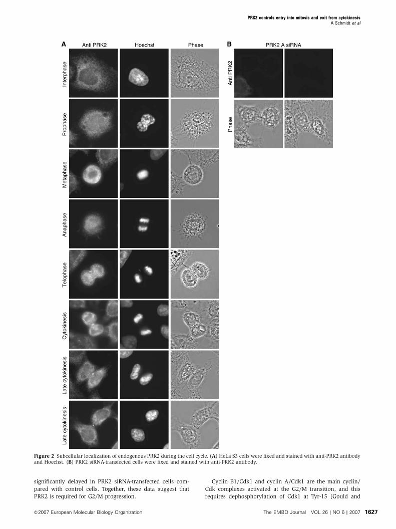
PRK1 and PRK2 localize to the cleavage furrow and the
midbody during cytokinesis
To examine the subcellular localization of PRK, unsynchro-
nized HeLa S3 cells were fixed and stained for endogenous
PRK1 and PRK2 proteins. During interphase, endogenous
PRK2 showed a diffuse distribution in the cytosol
(Figure 2A), which did not change during prophase, meta-
phase or anaphase. During telephase, PRK2 accumulated at
the cleavage furrow and finally became concentrated around
the midbody in cytokinesis. Midbody staining persisted until
late cytokinesis. The cytoplasmic, cleavage furrow and mid-
body staining all disappeared in the PRK2 siRNA-transfected
cells (Figure 2B). Similar, but weaker, staining was seen
for endogenous PRK1 and for overexpressed epitope-tagged
full-length PRK1 (not shown).
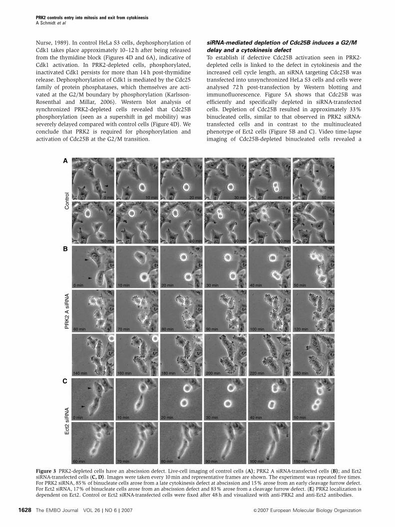
PRK2-depleted cells show a late cytokinesis defect
Activation of Rho by the GEF Ect2 has previously been shown
to control cleavage furrow formation during cytokinesis and
siRNA-mediated depletion of Ect2 results in the formation of
multinucleated cells (Kamijo et al, 2006). To examine if the
cytokinesis defect in PRK2-depleted cells was also at the level
of cleavage furrow formation, cell division was visualized
in PRK2- and Ect2-depleted cells by video time-lapse imaging
(Figure 3A–D). Like control cells, PRK2 siRNA-transfected
cells round up normally and display normal cleavage furrow
ingression and midbody formation (compare Figure 3A and
B). However, PRK2-depleted cells fail to undergo abscission
and the two daughter cells fuse back together resulting in the
formation of binucleated cells. In contrast, in the majority
of Ect2-depleted cells, the cleavage furrow does not fully
ingress, as has been previously reported (Figure 3C) (Kamijo
et al, 2006). We did, however, find that about 15% of Ect2-
depleted cells also showed a late cytokinesis defect, similar
to those observed in the PRK2 siRNA-transfected cells
(Figure 3D). This has also been described by others pre-
viously, and suggests that Ect2 has two functions during
cytokinesis, controlling cleavage furrow formation as well
as cell abscission (Kamijo et al, 2006). Interestingly, in those
Ect2-depleted cells that were able to ingress a cleavage furrow
and form a midbody, PRK2 no longer localized to the mid-
body (Figure 3E). These results suggest that PRK2 is required
for cell abscission during cytokinesis and that PRK2 localiza-
tion to the midbody is dependent on Ect2.
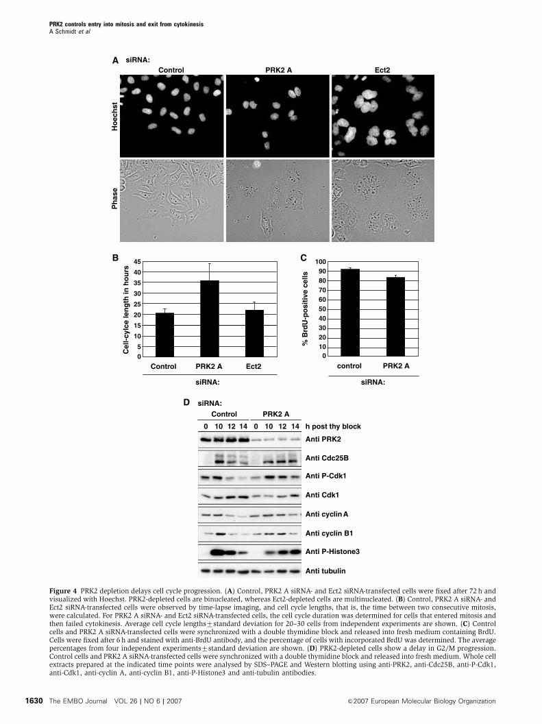
PRK2-depleted cells are delayed in G2/M progression
In the course of analysing PRK2- and Ect2-depleted cells, we
noticed that the majority of Ect2 siRNA-transfected cells were
multinucleated 72 h after transfection, whereas PRK2 siRNA-
transfected cells remained binucleated during this time per-
iod (Figure 4A). This suggests that cell cycle progression is
disturbed specifically in PRK2-knockdown cells. Using video
time-lapse imaging of control, PRK2- and Ect2 siRNA-treated
cells, we find that the cell cycle length of PRK2-depleted
binucleated cells is almost twice as long (36 h) as that of
control cells or Ect2-depleted binucleated cells (21 h)
(Figure 4B). Furthermore, some 30% of PRK2-depleted bi-
nucleated cells underwent apoptosis without further dividing
(not shown). We conclude that PRK2 is required for cell cycle
progression as well as cytokinesis.
To investigate this further, control and PRK2 siRNA-trans-
fected cells were synchronized at the G1/S boundary with a
double thymidine block. Cells were released into fresh med-
ium containing BrdU, fixed 6 h after release and stained with
anti-BrdU antibody as a read-out for DNA synthesis. Both
control cells and PRK2 siRNA-transfected cells showed BrdU
incorporation in 485% of cells, indicating that progression
through S phase is not dependent on PRK2 (Figure 4C). To
monitor the timing of mitosis, thymidine-synchronized con-
trol and PRK2 siRNA-transfected cells were observed under
the microscope at different time points after release. Control
cells started to round up for mitosis 10 h post-release,
whereas the majority of PRK2-depleted cells started to enter
mitosis only after 14 h (not shown), suggesting that PRK2 is
required for entry into mitosis.
To identify the molecular consequences of PRK2 depletion
during G2/M progression, synchronized PRK2 siRNA-trans-
fected cells were harvested at the indicated times and lysates
analysed on Western blots (Figure 4D). The levels of phos-
phorylated Histone 3 (P-Histone3), which accumulates in
prophase, and the degradation of cyclin A and cyclin B1,
PRK2 controls entry into mitosis and exit from cytokinesisA Schmidt et al
&2007 European Molecular Biology Organization The EMBO Journal VOL 26 | NO 6 | 2007 1625

which occurs during prometaphase and metaphase, respec-
tively, were monitored. In control cells, maximal accumula-
tion of P-Histone3 occurs 10 h after release from the
thymidine block (Figures 4D and 6A). In contrast, in PRK2-
depleted cells, P-Histone3 is maximal only after 14 h.
Similarly, the degradation of cyclin A and cyclin B1 was
Con
trol
PR
K2
A
PR
K2
B
PR
K1
siRNA:
Anti PRK2
Anti PRK1
Anti tubulin
D
B siRNA:Control PRK2 A PRK2 B PRK1
Hoe
chst
Pha
seA
05
101520253035
% �
4n
cells
Control PRK2 A PRK1
siRNA: siRNA:
Control PRK2 A PRK2 B PRK1
% B
inuc
leat
e ce
lls
05
10152025303540
C
% B
inuc
leat
e ce
lls
0
10
20
30
40
50
PRK2 mycControl
FE Control PRK2 myc
Hoe
chst
Ant
i myc
G
Anti PRK2Anti PRK1
Gst-PRK1 Gst-PRK2Ex Ex50 25 10 5 50 25 10 5
Gst-PRK1/2PRK2PRK1
Figure 1 Depletion of PRK2 from HeLa S3 cells leads to the formation of binucleated cells. (A) Unsynchronized control, PRK2 A and PRK1siRNA-transfected cells were subjected to FACS analysis and the percentage of X4n DNA cells was determined. The averages7standarddeviation for at least two independent experiments are shown. (B) Unsynchronized control, PRK2 A, PRK2 B and PRK1 siRNA-transfected cellswere fixed after 72 h and stained with Hoechst. PRK2 A and PRK2 B siRNA-transfected cells result in the accumulation of binucleated cells(arrowheads). (C) Unsynchronized control, PRK2 A, PRK2 B and PRK1 siRNA-transfected cells were fixed after 72 h and stained with Hoechst.The percentage of binucleated cells was determined. The averages of at least three independent experiments7standard deviation are shown.(D) Unsynchronized control, PRK2 A, PRK2 B and PRK1 siRNA-transfected cells were lysed after 72 h and cell extracts were analysed byWestern blotting using anti-PRK2, anti-PRK1 and anti-tubulin antibodies. (E) Control and myc PRK2-transfected cells were fixed after 72 h andstained with anti-myc antibody and Hoechst. Overexpression of myc PRK2 results in the accumulation of binucleated cells (arrowheads).(F) Control and myc PRK2-transfected cells were fixed after 72 h, stained with anti-myc antibody and Hoechst, and the percentage ofbinucleated cells determined. The averages of at least three independent experiments7standard deviation are shown. (G) PRK1 protein isexpressed at low levels in HeLa S3 cells. Recombinant Gst-PRK1 and Gst-PRK2 proteins (5–50ng) together with total cell extract (10þ20ml)were analysed by SDS–PAGE and Western blotting using anti-PRK1 and anti-PRK2 antibodies.
PRK2 controls entry into mitosis and exit from cytokinesisA Schmidt et al
The EMBO Journal VOL 26 | NO 6 | 2007 &2007 European Molecular Biology Organization1626
significantly delayed in PRK2 siRNA-transfected cells com-
pared with control cells. Together, these data suggest that
PRK2 is required for G2/M progression.
Cyclin B1/Cdk1 and cyclin A/Cdk1 are the main cyclin/
Cdk complexes activated at the G2/M transition, and this
requires dephosphorylation of Cdk1 at Tyr-15 (Gould and
Anti PRK2 PhaseHoechst
Inte
rpha
seP
roph
ase
Met
apha
seA
naph
ase
Cyt
okin
esis
Tel
opha
seLa
te c
ytok
ines
isLa
te c
ytok
ines
is
B PRK2 A siRNA
Pha
se
A
Ant
i PR
K2
Figure 2 Subcellular localization of endogenous PRK2 during the cell cycle. (A) HeLa S3 cells were fixed and stained with anti-PRK2 antibodyand Hoechst. (B) PRK2 siRNA-transfected cells were fixed and stained with anti-PRK2 antibody.
PRK2 controls entry into mitosis and exit from cytokinesisA Schmidt et al
&2007 European Molecular Biology Organization The EMBO Journal VOL 26 | NO 6 | 2007 1627
Nurse, 1989). In control HeLa S3 cells, dephosphorylation of
Cdk1 takes place approximately 10–12 h after being released
from the thymidine block (Figures 4D and 6A), indicative of
Cdk1 activation. In PRK2-depleted cells, phosphorylated,
inactivated Cdk1 persists for more than 14 h post-thymidine
release. Dephosphorylation of Cdk1 is mediated by the Cdc25
family of protein phosphatases, which themselves are acti-
vated at the G2/M boundary by phosphorylation (Karlsson-
Rosenthal and Millar, 2006). Western blot analysis of
synchronized PRK2-depleted cells revealed that Cdc25B
phosphorylation (seen as a supershift in gel mobility) was
severely delayed compared with control cells (Figure 4D). We
conclude that PRK2 is required for phosphorylation and
activation of Cdc25B at the G2/M transition.
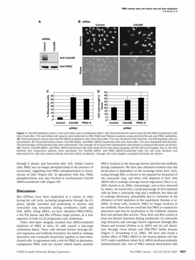
siRNA-mediated depletion of Cdc25B induces a G2/M
delay and a cytokinesis defect
To establish if defective Cdc25B activation seen in PRK2-
depleted cells is linked to the defect in cytokinesis and the
increased cell cycle length, an siRNA targeting Cdc25B was
transfected into unsynchronized HeLa S3 cells and cells were
analysed 72 h post-transfection by Western blotting and
immunofluorescence. Figure 5A shows that Cdc25B was
efficiently and specifically depleted in siRNA-transfected
cells. Depletion of Cdc25B resulted in approximately 33%
binucleated cells, similar to that observed in PRK2 siRNA-
transfected cells and in contrast to the multinucleated
phenotype of Ect2 cells (Figure 5B and C). Video time-lapse
imaging of Cdc25B-depleted binucleated cells revealed a
B
10 min
60 min 70 min 80 min 90 min 100 min 120 min
140 min 180 min 200 min 220 min 280 min
0 min
160 min
PR
K2
A s
iRN
A
C
10 min 20 min 30 min 40 min 50 min0 min
60 min 70 min 80 min 90 min 100 min 150 min
Ect
2 si
RN
A
A
10 min 20 min 30 min 40 min 50 min
20 min 30 min 40 min 50 min
0 min
60 min 70 min 80 min 90 min 100 min 190 min
Con
trol
Figure 3 PRK2-depleted cells have an abscission defect. Live-cell imaging of control cells (A); PRK2 A siRNA-transfected cells (B); and Ect2siRNA-transfected cells (C, D). Images were taken every 10min and representative frames are shown. The experiment was repeated five times.For PRK2 siRNA, 85% of binucleate cells arose from a late cytokinesis defect at abscission and 15% arose from an early cleavage furrow defect.For Ect2 siRNA, 17% of binucleate cells arose from an abscission defect and 83% arose from a cleavage furrow defect. (E) PRK2 localization isdependent on Ect2. Control or Ect2 siRNA-transfected cells were fixed after 48 h and visualized with anti-PRK2 and anti-Ect2 antibodies.
PRK2 controls entry into mitosis and exit from cytokinesisA Schmidt et al
The EMBO Journal VOL 26 | NO 6 | 2007 &2007 European Molecular Biology Organization1628
severe increase in the duration of their cell cycle compared
with control cells (Figure 5D). In addition, around 30% of
Cdc25B-depleted binucleated cells underwent apoptosis with-
out further dividing (not shown). As previously published,
synchronized Cdc25B siRNA-transfected cells showed a delay
in G2/M progression, as judged by a delay in Cdk1 dephos-
phorylation, cyclin A degradation and P-Histone3 accumula-
tion (Figure 6F) (Lindqvist et al, 2005).
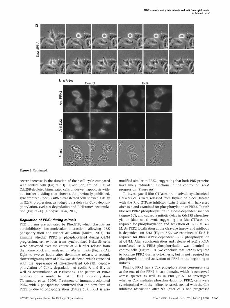
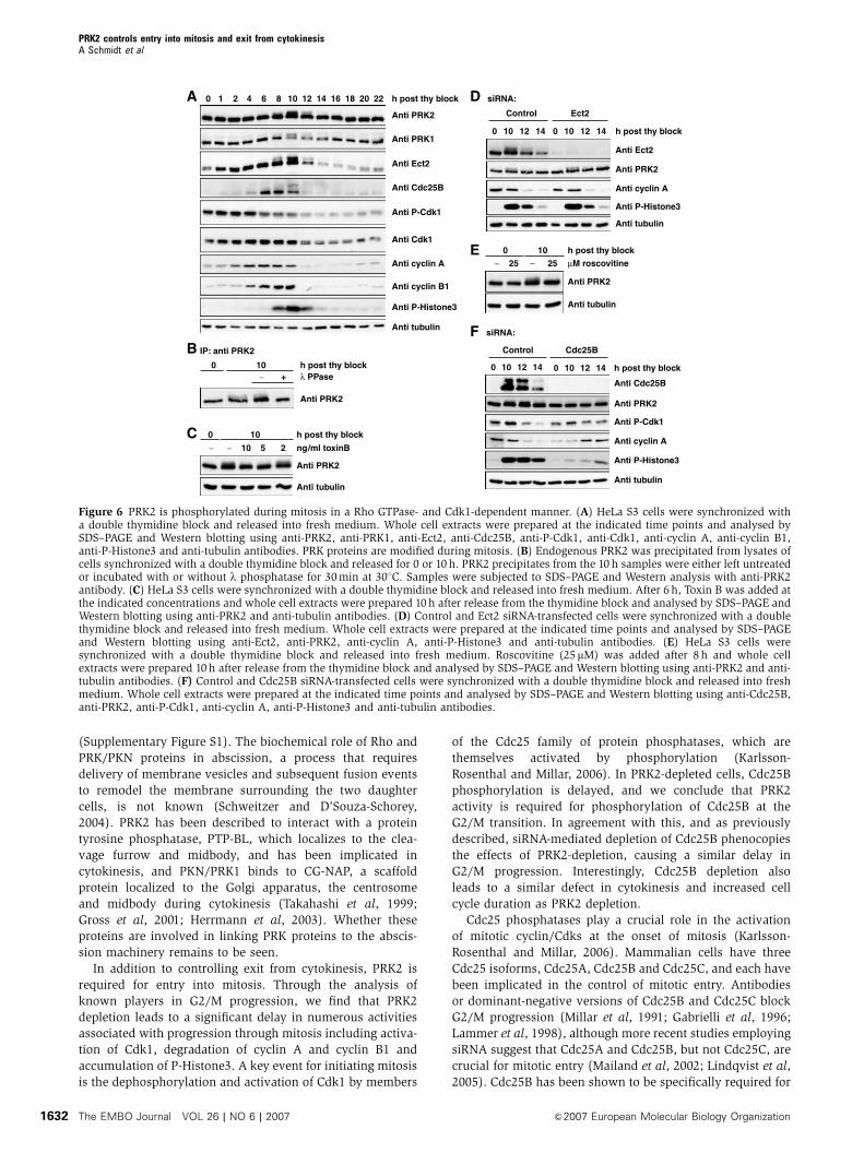
Regulation of PRK2 during mitosis
PRK proteins are activated by Rho.GTP, which disrupts an
autoinhibitory, intramolecular interaction, allowing PRK
phosphorylation and further activation (Mukai, 2003). To
examine whether PRK2 is phosphorylated during G2/M
progression, cell extracts from synchronized HeLa S3 cells
were harvested over the course of 22 h after release from
thymidine block and analysed on Western blots (Figure 6A).
Eight to twelve hours after thymidine release, a second,
slower migrating form of PRK2 was detected, which coincided
with the appearance of phosphorylated Cdc25B, dephos-
phorylation of Cdk1, degradation of cyclin A and B1, as
well as accumulation of P-Histone3. The pattern of PRK2
modification is similar to that of Ect2 phosphorylation
(Tatsumoto et al, 1999). Treatment of immunoprecipitated
PRK2 with l phosphatase confirmed that the new form of
PRK2 is due to phosphorylation (Figure 6B). PRK1 is also
modified similar to PRK2, suggesting that both PRK proteins
have likely redundant functions in the control of G2/M
progression (Figure 6A).
To investigate if Rho GTPases are involved, synchronized
HeLa S3 cells were released from thymidine block, treated
with the Rho GTPase inhibitor toxin B after 6 h, harvested
after 10 h and examined for phosphorylation of PRK2. ToxinB
blocked PRK2 phosphorylation in a dose-dependent manner
(Figure 6C), and caused a mitotic delay in Cdc25B phosphor-
ylation (data not shown), suggesting that Rho GTPases are
required for phosphorylation and activation of PRK2 at G2/
M. As PRK2 localization at the cleavage furrow and midbody
is dependent on Ect2 (Figure 3E), we examined if Ect2 is
required for Rho GTPase-dependent PRK2 phosphorylation
at G2/M. After synchronization and release of Ect2 siRNA-
transfected cells, PRK2 phosphorylation was identical to
control cells (Figure 6D). We conclude that Ect2 is required
to localize PRK2 during cytokinesis, but is not required for
phosphorylation and activation of PRK2 at the beginning of
mitosis.
Finally, PRK2 has a Cdk phosphorylation consensus site
at the end of the PRK2 kinase domain, which is conserved
across species as well as in PRK1/PKN. To investigate
whether Cdk mediates phosphorylation of PRK2, cells were
synchronized with thymidine, released, treated with the Cdk
inhibitor roscovitine after 8 h (after cells had progressed
10 min 20 min 30 min 40 min 50 min
60 min 70 min 80 min 90 min 100 min 120 min
0 min
D
Ect
2 si
RN
A
E
Ant
i PR
K2
Ant
i Ect
2P
hase
Control Ect2siRNA:
Figure 3 Continued.
PRK2 controls entry into mitosis and exit from cytokinesisA Schmidt et al
&2007 European Molecular Biology Organization The EMBO Journal VOL 26 | NO 6 | 2007 1629
A
Control PRK2 AsiRNA:
0 10 12 14 h post thy block0 10 12 14
Anti PRK2
Anti Cdc25B
Anti P-Cdk1
Anti Cdk1
Anti cyclin A
Anti cyclin B1
Anti P-Histone3
Anti tubulin
C
control PRK2 A
% B
rdU
-po
siti
ve c
ells
siRNA:
0
30405060708090
100
2010
B
Control PRK2 A Ect2
Cel
l-cy
lce
len
gth
in h
ou
rs
siRNA:
0
5
10
15
20
25
30
35
45
40
D
Control PRK2 AsiRNA:
Ect2
Ho
ech
stP
has
e
Figure 4 PRK2 depletion delays cell cycle progression. (A) Control, PRK2 A siRNA- and Ect2 siRNA-transfected cells were fixed after 72 h andvisualized with Hoechst. PRK2-depleted cells are binucleated, whereas Ect2-depleted cells are multinucleated. (B) Control, PRK2 A siRNA- andEct2 siRNA-transfected cells were observed by time-lapse imaging, and cell cycle lengths, that is, the time between two consecutive mitosis,were calculated. For PRK2 A siRNA- and Ect2 siRNA-transfected cells, the cell cycle duration was determined for cells that entered mitosis andthen failed cytokinesis. Average cell cycle lengths7standard deviation for 20–30 cells from independent experiments are shown. (C) Controlcells and PRK2 A siRNA-transfected cells were synchronized with a double thymidine block and released into fresh medium containing BrdU.Cells were fixed after 6 h and stained with anti-BrdU antibody, and the percentage of cells with incorporated BrdU was determined. The averagepercentages from four independent experiments7standard deviation are shown. (D) PRK2-depleted cells show a delay in G2/M progression.Control cells and PRK2 A siRNA-transfected cells were synchronized with a double thymidine block and released into fresh medium. Whole cellextracts prepared at the indicated time points were analysed by SDS–PAGE and Western blotting using anti-PRK2, anti-Cdc25B, anti-P-Cdk1,anti-Cdk1, anti-cyclin A, anti-cyclin B1, anti-P-Histone3 and anti-tubulin antibodies.
PRK2 controls entry into mitosis and exit from cytokinesisA Schmidt et al
The EMBO Journal VOL 26 | NO 6 | 2007 &2007 European Molecular Biology Organization1630
through S phase) and harvested after 10 h. Unlike control
cells, PRK2 was no longer phosphorylated in the presence of
roscovitine, suggesting that PRK2 phosphorylation is down-
stream of Cdk1 (Figure 6E). In agreement with this, PRK2
phosphorylation was also blocked in synchronized Cdc25B
siRNA-transfected cells (Figure 6F).
Discussion
Rho GTPases have been implicated in a variety of steps
during the cell cycle, including progression through the G1
phase, spindle assembly and positioning in mitosis, and
contractile ring formation during cytokinesis (Jaffe and
Hall, 2005). Using siRNA, we have identified PRK2/PKN2,
a Ser/Thr kinase and Rho GTPases target protein, as a new
regulator of both G2/M progression and cytokinesis.
Video time-lapse imaging revealed that siRNA-mediated
depletion of PRK2 in HeLa S3 cells caused a late-stage
cytokinesis defect. These cells showed normal cleavage fur-
row ingression and midbody formation, but failed to undergo
abscission and eventually fused back together to form binu-
cleated cells. In agreement with a role for PRK2 in abscission,
endogenous PRK2 (and the closely related family member
PRK1) localizes to the cleavage furrow and then the midbody
during cytokinesis. We have also obtained evidence that this
localization is dependent on the exchange factor Ect2. Ect2,
acting through Rho, is known to be required for formation of
the contractile ring, and HeLa cells depleted of Ect2 with
siRNA fail to undergo cleavage furrow ingression (Yuce et al,
2005; Kamijo et al, 2006). Interestingly, and as first observed
by others, we found that a small percentage of Ect2-depleted
cells do form a contractile ring and a midbody, but then fail
to undergo abscission, presumably due to variations in the
efficiency of Ect2 depletion in the experiment (Kamijo et al,
2006). In those cells, however, PRK2 no longer localizes to
the midbody. From this we conclude that PRK2 is required for
abscission and that its localization to the midbody requires
Ect2 and perhaps Rho activity. Thus, Ect2 and Rho control at
least two distinct functions during cytokinesis: (i) contractile
ring formation and cleavage furrow ingression through Rho
kinase and formins (Piekny et al, 2005) and (ii) cell abscis-
sion through citron kinase and PRK/PKN family kinases
(Figure 7) (Gruneberg et al, 2006). We have also found a
similar effect of PRK2 siRNA in the human glioma cell line
U373 under conditions where Ect2 siRNA produces primarily
multinucleated cells, loss of PRK2 induces binucleated cells
A
C
BControl Cdc25B
Ho
ech
stP
has
e
D
Control Cdc25B
% B
inu
clea
te c
ells
siRNA:
05
101520253035
PRK2 A Control Cdc25B
Cel
l cyl
ce le
ng
th in
ho
urs
siRNA:
05
101520253035
4540
PRK2 A
Co
ntr
ol
siRNA:
Cd
c25B
Anti PRK2
Anti Cdc25B
siRNA:
Figure 5 Cdc25B depletion causes a cell cycle delay and a cytokinesis defect. (A) Unsynchronized control and Cdc25B siRNA-transfected cellswere lysed after 72 h and whole cell extracts were subjected to SDS–PAGE and Western analysis using anti-Cdc25B and anti-PRK2 antibodies.(B) Unsynchronized control and Cdc25B siRNA-transfected cells were fixed after 72 h and visualized with Hoechst. Cdc25B-depleted cells arebinucleated. (C) Unsynchronized control, Cdc25B siRNA- and PRK2 siRNA-transfected cells were fixed after 72 h and visualized with Hoechst.The percentages of binucleated cells were determined. The averages of at least three independent experiments7standard deviation are shown.(D) Control, Cdc25B siRNA- and PRK2 siRNA-transfected cells were observed by time-lapse imaging, and the cell cycle lengths, that is, the timebetween two consecutive mitosis, were calculated. For Cdc25B siRNA- and PRK2 siRNA-transfected cells, the cell cycle duration wasdetermined for cells that entered mitosis and then failed cytokinesis. Average cell cycle lengths7standard deviation are shown.
PRK2 controls entry into mitosis and exit from cytokinesisA Schmidt et al
&2007 European Molecular Biology Organization The EMBO Journal VOL 26 | NO 6 | 2007 1631
(Supplementary Figure S1). The biochemical role of Rho and
PRK/PKN proteins in abscission, a process that requires
delivery of membrane vesicles and subsequent fusion events
to remodel the membrane surrounding the two daughter
cells, is not known (Schweitzer and D’Souza-Schorey,
2004). PRK2 has been described to interact with a protein
tyrosine phosphatase, PTP-BL, which localizes to the clea-
vage furrow and midbody, and has been implicated in
cytokinesis, and PKN/PRK1 binds to CG-NAP, a scaffold
protein localized to the Golgi apparatus, the centrosome
and midbody during cytokinesis (Takahashi et al, 1999;
Gross et al, 2001; Herrmann et al, 2003). Whether these
proteins are involved in linking PRK proteins to the abscis-
sion machinery remains to be seen.
In addition to controlling exit from cytokinesis, PRK2 is
required for entry into mitosis. Through the analysis of
known players in G2/M progression, we find that PRK2
depletion leads to a significant delay in numerous activities
associated with progression through mitosis including activa-
tion of Cdk1, degradation of cyclin A and cyclin B1 and
accumulation of P-Histone3. A key event for initiating mitosis
is the dephosphorylation and activation of Cdk1 by members
of the Cdc25 family of protein phosphatases, which are
themselves activated by phosphorylation (Karlsson-
Rosenthal and Millar, 2006). In PRK2-depleted cells, Cdc25B
phosphorylation is delayed, and we conclude that PRK2
activity is required for phosphorylation of Cdc25B at the
G2/M transition. In agreement with this, and as previously
described, siRNA-mediated depletion of Cdc25B phenocopies
the effects of PRK2-depletion, causing a similar delay in
G2/M progression. Interestingly, Cdc25B depletion also
leads to a similar defect in cytokinesis and increased cell
cycle duration as PRK2 depletion.
Cdc25 phosphatases play a crucial role in the activation
of mitotic cyclin/Cdks at the onset of mitosis (Karlsson-
Rosenthal and Millar, 2006). Mammalian cells have three
Cdc25 isoforms, Cdc25A, Cdc25B and Cdc25C, and each have
been implicated in the control of mitotic entry. Antibodies
or dominant-negative versions of Cdc25B and Cdc25C block
G2/M progression (Millar et al, 1991; Gabrielli et al, 1996;
Lammer et al, 1998), although more recent studies employing
siRNA suggest that Cdc25A and Cdc25B, but not Cdc25C, are
crucial for mitotic entry (Mailand et al, 2002; Lindqvist et al,
2005). Cdc25B has been shown to be specifically required for
A
C
0 1 2 4 6 8 10 12 14 16 18 2220 h post thy block
Anti PRK2
Anti PRK1
Anti Ect2
Anti Cdc25B
Anti P-Cdk1
Anti Cdk1
Anti cyclin A
Anti cyclin B1
Anti P-Histone3
Anti tubulin
DControl Ect2
siRNA:
0 10 12 14 h post thy block0 10 12 14
Anti PRK2
Anti Ect2
E
FB
Anti PRK2
IP: anti PRK2
0 10 h post thy block− + λ PPase
0 10 h post thy block
Anti PRK2
μM roscovitine25 − − 25
Anti tubulin
0 10 h post thy block
ng/ml toxinB− − 10
Anti PRK2
Anti tubulin
5 2
Control
siRNA:
0 10 12 14 h post thy block0 10 12 14
Cdc25B
Anti Cdc25B
Anti PRK2
Anti P-Cdk1
Anti cyclin A
Anti P-Histone3
Anti tubulin
Anti cyclin A
Anti P-Histone3
Anti tubulin
Figure 6 PRK2 is phosphorylated during mitosis in a Rho GTPase- and Cdk1-dependent manner. (A) HeLa S3 cells were synchronized witha double thymidine block and released into fresh medium. Whole cell extracts were prepared at the indicated time points and analysed bySDS–PAGE and Western blotting using anti-PRK2, anti-PRK1, anti-Ect2, anti-Cdc25B, anti-P-Cdk1, anti-Cdk1, anti-cyclin A, anti-cyclin B1,anti-P-Histone3 and anti-tubulin antibodies. PRK proteins are modified during mitosis. (B) Endogenous PRK2 was precipitated from lysates ofcells synchronized with a double thymidine block and released for 0 or 10 h. PRK2 precipitates from the 10 h samples were either left untreatedor incubated with or without l phosphatase for 30min at 301C. Samples were subjected to SDS–PAGE and Western analysis with anti-PRK2antibody. (C) HeLa S3 cells were synchronized with a double thymidine block and released into fresh medium. After 6 h, Toxin B was added atthe indicated concentrations and whole cell extracts were prepared 10h after release from the thymidine block and analysed by SDS–PAGE andWestern blotting using anti-PRK2 and anti-tubulin antibodies. (D) Control and Ect2 siRNA-transfected cells were synchronized with a doublethymidine block and released into fresh medium. Whole cell extracts were prepared at the indicated time points and analysed by SDS–PAGEand Western blotting using anti-Ect2, anti-PRK2, anti-cyclin A, anti-P-Histone3 and anti-tubulin antibodies. (E) HeLa S3 cells weresynchronized with a double thymidine block and released into fresh medium. Roscovitine (25mM) was added after 8 h and whole cellextracts were prepared 10h after release from the thymidine block and analysed by SDS–PAGE and Western blotting using anti-PRK2 and anti-tubulin antibodies. (F) Control and Cdc25B siRNA-transfected cells were synchronized with a double thymidine block and released into freshmedium. Whole cell extracts were prepared at the indicated time points and analysed by SDS–PAGE and Western blotting using anti-Cdc25B,anti-PRK2, anti-P-Cdk1, anti-cyclin A, anti-P-Histone3 and anti-tubulin antibodies.
PRK2 controls entry into mitosis and exit from cytokinesisA Schmidt et al
The EMBO Journal VOL 26 | NO 6 | 2007 &2007 European Molecular Biology Organization1632
the initial activation of cyclinB/Cdk1 at the centrosome,
which triggers G2/M progression and precedes the activation
of nuclear mitotic events (De Souza et al, 2000; Jackman
et al, 2003; Lindqvist et al, 2005). On the other hand,
Cdc25B�/� Cdc25C�/� mice develop normally, suggesting
that Cdc25A can fulfill all Cdc25 protein functions (Ferguson
et al, 2005). Some of these discrepancies are likely due to
overlapping, if not redundant activities and cell type/species
differences.
The regulation of Cdc25 phosphatase activity occurs at
the level of protein expression, subcellular localization and
phosphorylation (Karlsson-Rosenthal and Millar, 2006).
Cdc25 proteins are substrates for many kinases, which can
either activate or inhibit catalytic activity depending on the
site of phosphorylation. In response to environmental stress
or DNA damage, a G2/M checkpoint is induced and Cdc25
proteins are phosphorylated and inhibited by a number of
kinases including Chk1, Chk2 and MAPKAPK-2, thus pre-
venting cyclin/Cdk1 activation and cell cycle progression
(Karlsson-Rosenthal and Millar, 2006). During normal cell
cycle progression, other kinases, including Aurora-A, CK2,
Cdk2 and Cdk1, have been implicated in phosphorylation and
activation of Cdc25 (Karlsson-Rosenthal and Millar, 2006).
Phosphorylation of Cdc25B by Aurora-A on Ser353 correlates
with its centrosomal localization and cyclinB1/Cdk1 activa-
tion, although the in vitro catalytic activity of Cdc25B is not
affected by Aurora-A phosphorylation (Dutertre et al, 2004;
Cazales et al, 2005). siRNA depletion of Aurora-A in HeLa
cells inhibits activation of cyclinB1/Cdk1 at the centrosome
and thus entry into mitosis, and in addition, results in the
formation of binucleated cells and increased apoptosis, simi-
lar to what we observe in PRK2- and Cdc25B-depleted cells
(Hirota et al, 2003; Du and Hannon, 2004; Dutertre et al,
2004). Currently, we can only speculate how PRK2 contri-
butes to Cdc25B activation, but given the similarity of some
of the phenotypes of PRK2- and Aurora-A-depleted cells,
it is possible that PRK2 lies upstream of Aurora-A kinase in
the activation of Cdc25B. Alternatively, PRK2 might act
parallel to Aurora-A and directly phosphorylate Cdc25B to
stimulate its catalytic activity. Consistent with the latter
hypothesis, we have shown that recombinant PRK1 and
PRK2 are both able to phosphorylate CDC25B in vitro
(Supplementary Figure S2). However, it remains to be deter-
mined whether this mechanism is responsible for PRK2-
dependent CDC25B activation in the cell. Interestingly,
PKN/PRK1 has been reported to interact directly with and
phosphorylate Cdc25C in vitro, although in this case, phos-
phorylation seems to inhibit Cdc25C activity and so the
relevance of these in vitro kinase assays to the work described
here is unclear (Misaki et al, 2001).
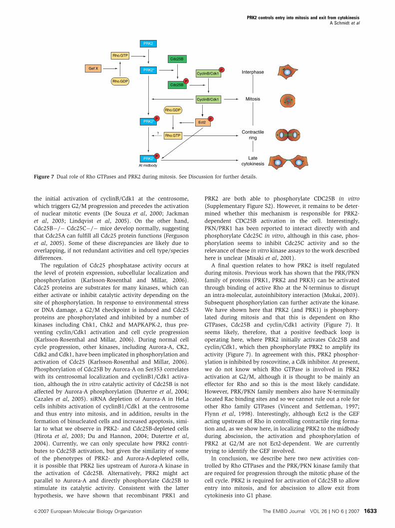
A final question relates to how PRK2 is itself regulated
during mitosis. Previous work has shown that the PRK/PKN
family of proteins (PRK1, PRK2 and PRK3) can be activated
through binding of active Rho at the N-terminus to disrupt
an intra-molecular, autoinhibitory interaction (Mukai, 2003).
Subsequent phosphorylation can further activate the kinase.
We have shown here that PRK2 (and PRK1) is phosphory-
lated during mitosis and that this is dependent on Rho
GTPases, Cdc25B and cyclin/Cdk1 activity (Figure 7). It
seems likely, therefore, that a positive feedback loop is
operating here, where PRK2 initially activates Cdc25B and
cyclin/Cdk1, which then phosphorylate PRK2 to amplify its
activity (Figure 7). In agreement with this, PRK2 phosphor-
ylation is inhibited by roscovitine, a Cdk inhibitor. At present,
we do not know which Rho GTPase is involved in PRK2
activation at G2/M, although it is thought to be mainly an
effector for Rho and so this is the most likely candidate.
However, PRK/PKN family members also have N-terminally
located Rac binding sites and so we cannot rule out a role for
other Rho family GTPases (Vincent and Settleman, 1997;
Flynn et al, 1998). Interestingly, although Ect2 is the GEF
acting upstream of Rho in controlling contractile ring forma-
tion and, as we show here, in localizing PRK2 to the midbody
during abscission, the activation and phosphorylation of
PRK2 at G2/M are not Ect2-dependent. We are currently
trying to identify the GEF involved.
In conclusion, we describe here two new activities con-
trolled by Rho GTPases and the PRK/PKN kinase family that
are required for progression through the mitotic phase of the
cell cycle. PRK2 is required for activation of Cdc25B to allow
entry into mitosis, and for abscission to allow exit from
cytokinesis into G1 phase.
Latecytokinesis
Contractilering
Mitosis
InterphaseCyclinB/Cdk1P
CyclinB/Cdk1
Cdc25B
Cdc25BP
PRK2
PRK2*
PRK2*P
At midbody
PRK2*P
Rho.GTP
Gef X
Ect2P
Rho.GDP
Rho.GTP?
Rho.GDP
Figure 7 Dual role of Rho GTPases and PRK2 during mitosis. See Discussion for further details.
PRK2 controls entry into mitosis and exit from cytokinesisA Schmidt et al
&2007 European Molecular Biology Organization The EMBO Journal VOL 26 | NO 6 | 2007 1633
Materials and methods
Plasmids, siRNAs, antibodies and reagentsPlasmid expressing full-length myc-tagged PRK2 was obtained fromH Mellor (University of Bristol, UK). Gst-PRK1 and Gst-PRK2proteins were from Invitrogen. PRK2 A siRNA was from Ambion(siRNA #781), Ect2 siRNA (50-GAUAAAGGAUGAUCUUGAAUU-30),PRK2 B siRNA (50-GGAGCGCUCUGAUGGACAAUU-30), PRK1 siRNA(Dharmacon Smartpool) and Cdc25B siRNAwere from Dharmacon.Sequence for Cdc25B siRNA, 50-UCCUCCCUGUCGUCUGAAUUU-30,was previously published (Lindqvist et al, 2004). Antibodies wereobtained as follows: monoclonal anti-PRK1 and anti-PRK2 anti-bodies from BD Transduction Laboratories, anti-Ect2 antibodiesfrom H Sillje and E Nigg (MPI, Munich, Germany) and Santa Cruz(C-20), anti-myc 9E10 from S Moss (University College London,UK), anti-P-Histone3 from Upstate, anti-cyclin A (H-432), cyclin B1(GNSI) and Cdc25B (C-20) from Santa Cruz, anti-Cdk1 and P-Cdk1from Cell Signaling, anti-BrdU from Roche Diagnostics and anti-tubulin from Sigma. l phosphatase was obtained from NewEngland Biolabs and thymidine, BrdU and roscovitine were fromSigma. Hoechst 33342 was purchased from Molecular Probes andToxin B from Calbiochem.
Cell culture and transfectionsHeLa S3 cells were maintained in DMEM supplemented with 10%FCS and penicillin/streptomycin (100 IU/ml and 100mg/ml) andincubated at 371C and 5% CO2. U373 cells were maintained in MEMsupplemented with 10% heat-inactivated FCS, 5% non-essentialamino acids, 20mM HEPES and penicillin/streptomycin (100 IU/mland 100 mg/ml) and incubated at 371C and 5% CO2. DNAtransfections were performed using GeneJuiceTM (Novagen) ac-cording to the manufacturer’s specifications. Transfection of siRNAwas performed using OligofectamineTM (Invitrogen). Briefly, HeLaS3 and U373 cells were seeded in six-well plates at a density of 20%and transfected the next day. For transfections, 3ml of Oligofecta-mineTM was incubated with 200 ml of Opti-MEM (Invitrogen) for5min at room temperature. The mixture was added to 30–60pmolsiRNA and further incubated at room temperature for 15min beforeaddition to the cells. The medium was replaced the next day.
Preparation of cell extractsFor preparation of whole cell extracts, cells from six-well plateswere lysed in 150 ml of cold lysis buffer (20mM Tris–HCl (pH 7.5),100mM NaCl, 1% Triton X-100, 12mM b-glycerophosphate, 5mMEGTA, 0.5% deoxycholate, 1mM DTT, 10mM NaF, 1mM Na3VO4,0.1mM PMSF, 20mg/ml aprotinin and leupeptin), mixed with 35mlof 5� SDS protein sample buffer, boiled for 3min, sonicated andanalysed by SDS–PAGE and Western blotting. For immunoprecipita-tion (IP) and l phosphatase treatment, cells were lysed in 600ml ofcold IP buffer (50mM Tris–HCl (pH 8), 150mM NaCl, 1% NonidentP-40, 12mM b-glycerophosphate, 0.5% deoxycholate, 1mMT DTT,10mM NaF, 1mM Na3VO4, 0.1mM PMSF, 20mg/ml aprotinin andleupeptin). Cell debris was pelleted by centrifugation at14 000 r.p.m. for 5min at 41C and lysates were incubated for 2 hat 41C with anti-PRK2 antibody and 20 ml protein G-Sepharose.Immunoprecipitates were collected by centrifugation and washed3� in IP buffer without phosphatase inhibitors and once with lphosphatase buffer containing 2mM MnCl2. Beads were resus-pended in 40 ml of l phosphatase buffer containing 2mMMnCl2 and400U l phosphatase was added and the reaction incubated at 301Cfor 30min. Immunoprecipitated proteins were eluted from thebeads with SDS protein sample buffer and analysed by SDS–PAGEand Western blotting.
ImmunofluorescenceHeLa S3 and U373 cells were plated onto glass coverslips andtransfected as described above. For immunofluorescence analysis ofcells transfected with epitope-tagged plasmids, cells were fixed in4% paraformaldehyde for 15min at room temperature and stained
for the appropriate epitope tags, as previously described (Nobes andHall, 1999). For staining of endogenous proteins, cells were fixedin �201C methanol for 10min and then stained as described. Forstaining with anti-BrdU antibody, cells were fixed in 4% parafor-maldehyde for 15min, incubated with 0.5% Triton X-100 in 0.2MHCl, blocked with 5% BSA in PBS and then stained as described.Fluorescence images were recorded on a CCD camera and processedusing Openlab and Adobe Photoshop software.
Cell synchronizationFor synchronization experiments, cells were seeded in six-wellplates at a density of 20%, transfected as described andimmediately after transfection incubated with 2mM thymidine for24 h. Cells were washed 3� with PBS and released into freshmedium for 8 h, followed by a second round of incubation with2mM thymidine for 15 h, 3� washing with PBS and release intofresh medium (t¼ 0h). Where indicated, BrdU was added afterrelease into fresh medium (t¼ 0h), and Toxin B or roscovitine wasadded 6 and 8h, respectively, after release into fresh medium. Cellswere harvested at the indicated time points and whole cell extractswere prepared as described above.
FACS analysisFor FACS analysis, cells were transfected with siRNA as describedand harvested after 72 h with trypsin, washed twice in PBS andfixed with �201C 70% ethanol for 30min at 41C. Cells were washedtwice with ice-cold PBS, incubated with 6mg/ml propidium iodide,and 10mg/ml RNAse for 30min at room temperature and analysedfor DNA content by flow cytometry on a FACSCalibur (BectonDickinson).
Time-lapse analysisTime-lapse microscopy was carried out on an Axiovert S100equipped with a Qimaging Qicam camera. Images were analysedusing Openlab and Adobe Photoshop software. Cell cycle lengthwas calculated as the time between entry into mitosis (rounding upof cells) and entry into mitosis of the next division cycle.
In vitro kinase assaysIn vitro kinase assays were performed using 250ng of recombinant,human GST-PRK1/PKN1 or GST-PRK2/PKN2 (Invitrogen) andeither 1mg of glutathione S-transferase (GST; Sigma), 0.5mg ofmyelin basic protein (MBP; Sigma) or 1.5mg of Cdc25B (Biomol) asa substrate in 50 ml of the following assay buffer: 12.5mM Tris(pH 7.5), 10mM MgCl2, 1mM EGTA, 0.5mM Na3VO4, 5mMb-glycerophosphate, 2.5mM DTT, 0.01% Triton X-100, 50mM ATPand 5mCi [g-32P]ATP (Amersham). Reactions were initiated with theaddition of ATP, incubated for 30min at 301C and stopped with 25 mlof kinase assay sample buffer: 75% (v/v) 4� LDS sample buffer(Invitrogen), 0.2M dithiothreitol, 4% (v/v) b-mercaptoethanol and5mM EDTA. Samples were boiled at 1001C for 5min, resolved bySDS–PAGE and Coomassie stained/destained. The gel was thenexposed to a Kodak Phosphor Screen K and analysed using thePersonal Molecular Imager System according to the manufacturer’sinstructions (Bio-Rad).
Supplementary dataSupplementary data are available at The EMBO Journal Online(http://www.embojournal.org).
Acknowledgements
We are grateful to Harry Mellor, Herman Sillje and Erich Nigg forreagents, Chris Marshall for help with the RNAi library construc-tion, Derek Davies and Nathalie Signoret for advice on FACSanalysis, Volker Stucke for technical advice and members of thelaboratory for valuable discussion. The work was generouslysupported by a programme grant for Cancer Research UK.
References
Bakal CJ, Finan D, LaRose J, Wells CD, Gish G, Kulkarni S,DeSepulveda P, Wilde A, Rottapel R (2005) The Rho GTP
exchange factor Lfc promotes spindle assembly in early mitosis.Proc Natl Acad Sci USA 102: 9529–9534
PRK2 controls entry into mitosis and exit from cytokinesisA Schmidt et al
The EMBO Journal VOL 26 | NO 6 | 2007 &2007 European Molecular Biology Organization1634
Cazales M, Schmitt E, Montembault E, Dozier C, Prigent C,Ducommun B (2005) CDC25B phosphorylation by Aurora-Aoccurs at the G2/M transition and is inhibited by DNA damage.Cell Cycle 4: 1233–1238
Coleman ML, Marshall CJ, Olson MF (2004) RAS and RHO GTPasesin G1-phase cell-cycle regulation. Nat Rev Mol Cell Biol 5: 355–366
De Souza CP, Ellem KA, Gabrielli BG (2000) Centrosomal andcytoplasmic Cdc2/cyclin B1 activation precedes nuclear mitoticevents. Exp Cell Res 257: 11–21
Di Cunto F, Imarisio S, Hirsch E, Broccoli V, Bulfone A, Migheli A,Atzori C, Turco E, Triolo R, Dotto GP, Silengo L, Altruda F (2000)Defective neurogenesis in citron kinase knockout mice by alteredcytokinesis and massive apoptosis. Neuron 28: 115–127
Du J, Hannon GJ (2004) Suppression of p160ROCK bypasses cellcycle arrest after Aurora-A/STK15 depletion. Proc Natl Acad SciUSA 101: 8975–8980
Dutertre S, Cazales M, Quaranta M, Froment C, Trabut V, Dozier C,Mirey G, Bouche JP, Theis-Febvre N, Schmitt E, Monsarrat B,Prigent C, Ducommun B (2004) Phosphorylation of CDC25B byAurora-A at the centrosome contributes to the G2–M transition.J Cell Sci 117: 2523–2531
Eda M, Yonemura S, Kato T, Watanabe N, Ishizaki T, Madaule P,Narumiya S (2001) Rho-dependent transfer of Citron-kinase to thecleavage furrow of dividing cells. J Cell Sci 114: 3273–3284
Ferguson AM, White LS, Donovan PJ, Piwnica-Worms H (2005)Normal cell cycle and checkpoint responses in mice and cellslacking Cdc25B and Cdc25C protein phosphatases. Mol Cell Biol25: 2853–2860
Flynn P, Mellor H, Palmer R, Panayotou G, Parker PJ (1998) Multipleinteractions of PRK1 with RhoA. Functional assignment of theHr1 repeat motif. J Biol Chem 273: 2698–2705
Gabrielli BG, De Souza CP, Tonks ID, Clark JM, Hayward NK, EllemKA (1996) Cytoplasmic accumulation of cdc25B phosphatase inmitosis triggers centrosomal microtubule nucleation in HeLacells. J Cell Sci 109 (Part 5): 1081–1093
Glotzer M (2005) The molecular requirements for cytokinesis.Science 307: 1735–1739
Gotta M, Abraham MC, Ahringer J (2001) CDC-42 controls earlycell polarity and spindle orientation in C. elegans. Curr Biol 11:482–488
Gould KL, Nurse P (1989) Tyrosine phosphorylation of the fissionyeast cdc2+ protein kinase regulates entry into mitosis. Nature342: 39–45
Gross C, Heumann R, Erdmann KS (2001) The protein kinaseC-related kinase PRK2 interacts with the protein tyrosine phos-phatase PTP-BL via a novel PDZ domain binding motif. FEBS Lett496: 101–104
Gruneberg U, Neef R, Li X, Chan EH, Chalamalasetty RB, Nigg EA,Barr FA (2006) KIF14 and citron kinase act together to promoteefficient cytokinesis. J Cell Biol 172: 363–372
Herrmann L, Dittmar T, Erdmann KS (2003) The protein tyrosinephosphatase PTP-BL associates with the midbody and is involvedin the regulation of cytokinesis. Mol Biol Cell 14: 230–240
Hirota T, Kunitoku N, Sasayama T, Marumoto T, Zhang D, Nitta M,Hatakeyama K, Saya H (2003) Aurora-A and an interactingactivator, the LIM protein Ajuba, are required for mitotic commit-ment in human cells. Cell 114: 585–598
Jackman M, Lindon C, Nigg EA, Pines J (2003) Active cyclin B1-Cdk1 first appears on centrosomes in prophase. Nat Cell Biol 5:143–148
Jaffe AB, Hall A (2005) Rho GTPases: biochemistry and biology.Annu Rev Cell Dev Biol 21: 247–269
Kamijo K, Ohara N, Abe M, Uchimura T, Hosoya H, Lee JS, Miki T(2006) Dissecting the role of Rho-mediated signaling in contrac-tile ring formation. Mol Biol Cell 17: 43–55
Karlsson-Rosenthal C, Millar JB (2006) Cdc25: mechanisms ofcheckpoint inhibition and recovery. Trends Cell Biol 16: 285–292
Kosako H, Yoshida T, Matsumura F, Ishizaki T, Narumiya S, InagakiM (2000) Rho-kinase/ROCK is involved in cytokinesis throughthe phosphorylation of myosin light chain and not ezrin/radixin/moesin proteins at the cleavage furrow. Oncogene 19: 6059–6064
Lammer C, Wagerer S, Saffrich R, Mertens D, Ansorge W, HoffmannI (1998) The cdc25B phosphatase is essential for the G2/M phasetransition in human cells. J Cell Sci 111 (Part 16): 2445–2453
Lindqvist A, Kallstrom H, Karlsson Rosenthal C (2004)Characterisation of Cdc25B localisation and nuclear export duringthe cell cycle and in response to stress. J Cell Sci 117: 4979–4990
Lindqvist A, Kallstrom H, Lundgren A, Barsoum E, Rosenthal CK(2005) Cdc25B cooperates with Cdc25A to induce mitosis but hasa unique role in activating cyclin B1-Cdk1 at the centrosome.J Cell Biol 171: 35–45
Mabuchi I, Hamaguchi Y, Fujimoto H, Morii N, Mishima M,Narumiya S (1993) A rho-like protein is involved in the organisa-tion of the contractile ring in dividing sand dollar eggs. Zygote 1:325–331
Madaule P, Eda M, Watanabe N, Fujisawa K, Matsuoka T, Bito H,Ishizaki T, Narumiya S (1998) Role of citron kinase as a target ofthe small GTPase Rho in cytokinesis. Nature 394: 491–494
Maddox AS, Burridge K (2003) RhoA is required for cortical retrac-tion and rigidity during mitotic cell rounding. J Cell Biol 160:255–265
Mailand N, Podtelejnikov AV, Groth A, Mann M, Bartek J, Lukas J(2002) Regulation of G(2)/M events by Cdc25A through phos-phorylation-dependent modulation of its stability. EMBO J 21:5911–5920
Mettouchi A, Klein S, Guo W, Lopez-Lago M, Lemichez E, WestwickJK, Giancotti FG (2001) Integrin-specific activation of Rac controlsprogression through the G(1) phase of the cell cycle. Mol Cell 8:115–127
Millar JB, Blevitt J, Gerace L, Sadhu K, Featherstone C, Russell P(1991) p55CDC25 is a nuclear protein required for the initiation ofmitosis in human cells. Proc Natl Acad Sci USA 88: 10500–10504
Misaki K, Mukai H, Yoshinaga C, Oishi K, Isagawa T, Takahashi M,Ohsumi K, Kishimoto T, Ono Y (2001) PKN delays mitotic timingby inhibition of Cdc25C: possible involvement of PKN in theregulation of cell division. Proc Natl Acad Sci USA 98: 125–129
Mukai H (2003) The structure and function of PKN, a protein kinasehaving a catalytic domain homologous to that of PKC. J Biochem(Tokyo) 133: 17–27
Niiya F, Tatsumoto T, Lee KS, Miki T (2006) Phosphorylation of thecytokinesis regulator ECT2 at G2/M phase stimulates associationof the mitotic kinase Plk1 and accumulation of GTP-bound RhoA.Oncogene 25: 827–837
Nishimura Y, Yonemura S (2006) Centralspindlin regulates ECT2and RhoA accumulation at the equatorial cortex during cytokin-esis. J Cell Sci 119: 104–114
Nobes CD, Hall A (1999) Rho GTPases control polarity, protrusion,and adhesion during cell movement. J Cell Biol 144: 1235–1244
Oceguera-Yanez F, Kimura K, Yasuda S, Higashida C, Kitamura T,Hiraoka Y, Haraguchi T, Narumiya S (2005) Ect2 and MgcRacGAPregulate the activation and function of Cdc42 in mitosis. J CellBiol 168: 221–232
Olson MF, Ashworth A, Hall A (1995) An essential role for Rho, Rac,and Cdc42 GTPases in cell cycle progression through G1. Science269: 1270–1272
Olson MF, Paterson HF, Marshall CJ (1998) Signals from Ras andRho GTPases interact to regulate expression of p21Waf1/Cip1.Nature 394: 295–299
Palmer RH, Ridden J, Parker PJ (1995) Cloning and expressionpatterns of two members of a novel protein-kinase-C-relatedkinase family. Eur J Biochem 227: 344–351
Piekny A, Werner M, Glotzer M (2005) Cytokinesis: welcome to theRho zone. Trends Cell Biol 15: 651–658
Schmidt A, Hall A (2002) Guanine nucleotide exchange factors forRho GTPases: turning on the switch. Genes Dev 16: 1587–1609
Schweitzer JK, D’Souza-Schorey C (2004) Finishing the job: cyto-skeletal and membrane events bring cytokinesis to an end.Exp Cell Res 295: 1–8
Shandala T, Gregory SL, Dalton HE, Smallhorn M, Saint R (2004)Citron kinase is an essential effector of the Pbl-activated Rhosignalling pathway in Drosophila melanogaster. Development 131:5053–5063
Takahashi M, Shibata H, Shimakawa M, Miyamoto M, Mukai H,Ono Y (1999) Characterization of a novel giant scaffoldingprotein, CG-NAP, that anchors multiple signaling enzymesto centrosome and the golgi apparatus. J Biol Chem 274:17267–17274
Tatsumoto T, Xie X, Blumenthal R, Okamoto I, Miki T (1999)Human ECT2 is an exchange factor for Rho GTPases, phosphory-lated in G2/M phases, and involved in cytokinesis. J Cell Biol 147:921–928
Vincent S, Settleman J (1997) The PRK2 kinase is a potentialeffector target of both Rho and Rac GTPases and regulates actincytoskeletal organization. Mol Cell Biol 17: 2247–2256
PRK2 controls entry into mitosis and exit from cytokinesisA Schmidt et al
&2007 European Molecular Biology Organization The EMBO Journal VOL 26 | NO 6 | 2007 1635
Welsh CF, Roovers K, Villanueva J, Liu Y, Schwartz MA, Assoian RK(2001) Timing of cyclin D1 expression within G1 phase is con-trolled by Rho. Nat Cell Biol 3: 950–957
Yamamoto M, Marui N, Sakai T, Morii N, Kozaki S, Ikai K, ImamuraS, Narumiya S (1993) ADP-ribosylation of the rhoA gene productby botulinum C3 exoenzyme causes Swiss 3T3 cells to accumu-late in the G1 phase of the cell cycle. Oncogene 8: 1449–1455
Yasuda S, Oceguera-Yanez F, Kato T, Okamoto M, Yonemura S,Terada Y, Ishizaki T, Narumiya S (2004) Cdc42 and mDia3regulate microtubule attachment to kinetochores. Nature 428:767–771
Yuce O, Piekny A, Glotzer M (2005) An ECT2-centralspindlincomplex regulates the localization and function of RhoA. J CellBiol 170: 571–582
PRK2 controls entry into mitosis and exit from cytokinesisA Schmidt et al
The EMBO Journal VOL 26 | NO 6 | 2007 &2007 European Molecular Biology Organization1636