Embed Size (px)
Citation preview

IntroductionBy degrading mineralised matrix, multinucleated osteoclastsare crucial for the regulation of bone and calcium homeostasis.In the presence of M-CSF and RANK-L, monocyte-macrophage precursors fuse together to form largemultinucleated cells in vitro (Boyle et al., 2003). Unlike mostnon-transformed cells, cells from monocytic lineage, includingdendritic cells, macrophages and osteoclasts rely onpodosomes to migrate or adhere to a substrate rather than onF-actin stress fibres or focal adhesion plaques. Podosomes areadhesion structures containing a column of a continuous fluxof F-actin surrounded by focal adhesion proteins. In manyrespects, podosomes are similar to focal adhesion plaques asthey are short-lived structures involved in cell migration, andshare major focal adhesion proteins such as integrin, vinculinand talin as well as the signalling protein FAK/Pyk2 (for areview see Linder and Aepfelbacher, 2003). An importantfeature of podosomes is that they are associated withextracellular matrix degradation; they are thought to actively
contribute to tissue invasion and matrix remodelling. Forexample, podosome clusters are found in highly motile andimmature dendritic cells but not in static and mature cells foundin tissues (Burns et al., 2001). These observations underscorethe requirement of podosomes for cell migration. In vitro,osteoclast podosomes acquire a higher degree of self-organisation through the process of maturation. Immatureosteoclasts exhibit clustered podosomes that are subsequentlyarranged into dynamic short-lived rings. Then, these podosomerings expand to the cell periphery to form a stable podosomebelt in mature osteoclasts (Destaing et al., 2003). Thispodosome belt is reminiscent of the sealing zone found inresorbing osteoclasts, which provides tight attachment to boneand seals off the resorption pit where proteases and protons aresecreted to degrade the bone matrix (Vaananen et al., 2000).The molecular mechanisms driving the transition frompodosome clusters or dynamic rings to the podosome belt arepresently unknown. However, recent evidence indicates that, asin focal adhesions and the formation of F-actin structures,microtubules and Rho GTPases are critical for podosome
2901
Osteoclast maturation is accompanied by changes inpodosome patterning, resulting in the formation of aperipheral belt, which requires an intact microtubulenetwork. Here, we report that by inhibiting Rho, thepodosome belt is maintained at the cell periphery despitedepolymerisation of microtubules by nocodazole. Rhoinhibition was correlated to the increase in microtubulestabilisation and microtubule acetylation. Bymicroinjecting activated Rho or its activated effectormDia2 in osteoclasts, we found that the podosome beltwas disrupted and the level of microtubule acetylationdramatically decreased. We further characterised themolecular mechanism responsible for microtubuledeacetylation by co-immunoprecipitation experiments. Wefound that not only was mDia2 coprecipitating with the
recently identified microtubule deacetylase HDAC6 butthat it also activated the microtubule deacetylase activity ofHDAC6 in an in vitro deacetylase assay. Finally, we foundthat during osteoclastogenesis, there is a correlationbetween the increase in microtubule acetylation and thepodosome belt stabilisation and that if Rho is inhibited inthe early stages of osteoclast differentiation, it acceleratesboth microtubule acetylation and podosome beltstabilisation. Altogether, our data reveal a pathway inwhich Rho interferes with the osteoclast maturationprocess by controlling the level of microtubule acetylationand actin organisation through mDIA2 and HDAC6.
Key words: HDAC6, Rho GTPase, mDIA, Microtubule acetylation,Podosomes, Osteoclasts
Summary
A novel Rho-mDia2-HDAC6 pathway controlspodosome patterning through microtubule acetylationin osteoclastsOlivier Destaing1,*,‡, Frédéric Saltel1,*,§, Benoit Gilquin2, Anne Chabadel1, Saadi Khochbin2, Stéphane Ory3,*and Pierre Jurdic1,*,¶
1Laboratoire de Biologie Moléculaire et Cellulaire, UMR 5665 CNRS/ENS, INRA 913, Ecole Normale Supérieure de Lyon, 46, allée d’Italie,69364 Lyon CEDEX 7, France2Laboratoire de Biologie Moléculaire et Cellulaire de la Différenciation, INSERM U309, Institut Albert Bonniot, Faculté de Médecine, 38706 LaTronche Cedex, France3CRBM, CNRS FRE2593, 1919 route de Mende, 34293 Montpellier CEDEX 5, France*These authors contributed equally to this work‡Present address: Boyer Center for Molecular Medicine, Yale School of Medicine, 295 Congress Avenue, 06510 New Haven CT, USA§Present address: CMU-Dpt physiologie cellulaire et métabolisme, 1 rue Michel Servet, 1211 Geneve 4, Switzerland¶Author for correspondence (e-mail: [email protected])
Accepted 6 April 2005Journal of Cell Science 118, 2901-2911 Published by The Company of Biologists 2005doi:10.1242/jcs.02425
Research Article
Jour
nal o
f Cel
l Sci
ence

2902
patterning and assembly (for reviews, see Etienne-Mannevilleand Hall, 2002; Linder and Aepfelbacher, 2003). Similar tofocal adhesions, microtubule plus ends may target podosomes(Kaverina et al., 1999; Evans et al., 2003) and microtubulenetwork integrity has been shown to be crucial for podosomepatterning (Destaing et al., 2003). Forced depolymerisation ofmicrotubules by nocodazole treatment leads to thedestabilisation of isolated podosomes in macrophages or inmacrophage polykaryons (Linder et al., 2000; Ory et al., 2002)and, in osteoclasts, the podosome belt is disrupted. However,in the absence of polymerised microtubules and in contrast tomacrophages, isolated podosomes are still formed inosteoclasts, indicating that in these cells, depolymerisationdisrupts podosome patterning rather than the formation ofpodosomes themselves (Destaing et al., 2003).
Rho GTPases are known to promote F-actin and adhesionstructure rearrangements and appear to be the probablesignalling intermediates between microtubules and F-actin(Etienne-Manneville and Hall, 2003). Microtubuledepolymerisation in fibroblasts promotes stress fibre formationand focal adhesion plaque assembly, which relies on RhoAactivation (Enomoto, 1996; Ren et al., 1999). Although thefunction of Rho GTPases in podosome assembly or patterningis rather unclear, Rho GTPase activity needs to be tightlyregulated to maintain podosome assembly (Linder andAepfelbacher, 2003) and as in fibroblasts, microtubuledynamics regulates Rho GTPase activity (Ory et al., 2002).We recently showed that microtubule repolymerisationrecapitulates the sequence of events that lead to podosome beltformation during osteoclast maturation. This process startswith podosome clustering at the early stage of microtubulerepolymerisation and proceeds to the formation of podosomerings that eventually fuse together to generate the podosomebelt at the cell periphery when microtubules are fully regrown(Destaing et al., 2003). It should be noted that the kinetics ofmicrotubule repolymerisation are faster than the reformation ofthe podosome belt, indicating that not only the dynamics ofmicrotubules are crucial for early events in podosomepatterning (clusters or rings) but also that the microtubulenetwork needs to be in place or stabilised before the podosomebelt can be formed. In cells, there are two pools of dynamicmicrotubules, those that exhibit dynamic instability and havehalf-lives of 5-10 minutes, and stabilised microtubules, whichdo not exhibit dynamic instability and persist for hours (Saxtonet al., 1984; Schulze and Kirschner, 1986; Webster et al.,1987a; Webster et al., 1987b). Stable microtubules accumulatepost-translational modifications including detyrosination oracetylation, and may contribute to specialised functions in cells(Bulinski and Gundersen, 1991; Palazzo et al., 2001b;Rosenbaum, 2000).
Starting with these observations, we decided to investigatethe molecular mechanisms that drive microtubule-dependentpodosome belt stabilisation in osteoclasts and the extent towhich Rho GTPase is involved in this process. Here, we reportthat Rho inhibition prevents podosome belt disruptionfollowing microtubule depolymerisation by nocodazole and,more surprisingly, that Rho inhibition increases the resistanceof microtubules to nocodazole. Checking for microtubule post-translational modifications, we found that stable microtubuleswere acetylated and not detyrosinated. This led us toinvestigate whether Rho was involved in microtubule
acetylation in osteoclasts. We used the fact that the histoneacetylase HDAC6 has recently been described as a microtubuledeacetylase (Hubbert et al., 2002; Matsuyama et al., 2002) andthat the Rho effectors of the mDia family are involved in thecontrol of post-translational modification of microtubules(Palazzo et al., 2001b) as well as the coordination of themicrotubule and actin networks (Watanabe et al., 1999). Thisallowed us to reveal a pathway where activation of Rhopromotes the deacetylation of microtubules through mDia2 andHDAC6 activation. Moreover, we present evidence that thelevel of microtubule acetylation is important for osteoclastfunction.
Materials and MethodsReagentsNocodazole, trichostatin A (TSA) and n-butyrate (Sigma-Aldrich)were used at 2 µM, 3 µM and 5 µM. Human M-CSF and recombinanthuman RANK-L were produced as previously described (Destainget al., 2003). Supernatants were used at a final dilution of 1%corresponding to about 20 ng/ml and 30 ng/ml recombinant M-CSFand RANK-L, respectively. Monoclonal antibody AC40 anti-actin,anti-β-tubulin (clone DM1A) and anti-acetylated tubulin monoclonalantibody 6-11B-1 were from Sigma; anti-β-tubulin (clone N357) fromAmersham Life Science, anti-HA (clone Y11) from Santa CruzBiotechnology and anti-GFP monoclonal antibody was from Rocheand Clontech. Anti-detyrosinated tubulin was a kind gift from DidierJob (CEA, Grenoble, France) and anti-HDAC6 polyclonal was raisedagainst C-terminal peptide (Seigneurin-Berny et al., 2001). F-actindistribution was revealed after incubation with TRITC-conjugatedphalloidin (Molecular Probes). Coverslips were mounted in Prolong®
Antifade (Molecular Probes). Apatite collagen complexes (ACCs)were prepared using a method described previously (Shibutani et al.,2000; Saltel et al., 2004).
Plasmids and constructsGFP-mDIA2 and GFP-mDIA2-∆GBD were from Art Alberts (VanAndel Institute) and have been described previously (Palazzo et al.,2001a). GFP-G14VRho and GFP-G12VRac were gifts from PhilippeFort (CRBM, Montpellier, France). pEGFP-Actin Vector® was fromClontech. TAT-C3 expression vector was a kind gift from Erik Sahaiand was produced as described (Coleman et al., 2001). Vectorsexpressing haemagglutinin (HA)-tagged mHDAC6 and deletionmutants have been described previously (Seigneurin-Berny et al.,2001).
Osteoclast differentiationSpleen cells from six- to eight-week-old male OF1 mice were seededat 2500 cells/mm2 and cultured for 8 days on coverslips indifferentiation medium: α-MEM medium (Life Technologies)containing 10% foetal calf serum (FCS, Hyclone) plus M-CSF andsoluble recombinant RANK-L.
MicroinjectionMouse spleen cell-derived osteoclasts differentiated in vitro onEppendorf CELLocate® coverslips for 7 days in differentiationmedium were transferred to observation medium: α-MEM withoutbicarbonate (Life Technologies) containing 10% foetal calf serum, M-CSF, 20 mM HEPES and soluble recombinant RANK-L. Intranuclearmicroinjections of cDNA (0.2 mg/ml in 0.05 M Tris-HCl, pH 7.4)were carried out at room temperature using Eclipse TE 200 invertedmicroscope (Nikon) with an InjectMan micromanipulator and anEppendorf 5246 microinjector. After injection, cells were further
Journal of Cell Science 118 (13)
Jour
nal o
f Cel
l Sci
ence

2903Microtubule acetylation in osteoclasts
maintained at 37°C and 5% CO2 for 6 hours in differentiation mediumbefore imaging.
Immunoprecipitation and interaction site mappingFor co-immunoprecipitation and interaction site mapping, COS cellswere lysed 24 hours after transfection with Fugene 6® following themanufacturer’s recommendations (Roche). Lysis buffer consists of100 mM HEPES pH 7.9, 6 mM MgCl2, 40% glycerol, 150 mM KCl,0.1% Nonidet P40 and 1 mM dithiothreitol supplemented with aprotease inhibitor cocktail. The lysate was incubated on ice for 20minutes and cleared by centrifugation at 17,000 g for 10 minutesat 4°C. HA-tagged proteins were immunoprecipitated with anti-HA antibody and protein-G sepharose for 2 hours at 4°C.Immunoprecipitated proteins were washed three times in lysis buffer.
Confocal microscopyFor immunofluorescence, cells were fixed in Busson fixation solutionat pH 6.9 (4% paraformaldehyde, 60 mM PIPES, 25 mM HEPES, 20mM EGTA, 2 mM magnesium acetate, 0.05% glutaraldehyde w/v),processed as described (Ory et al., 2000) and imaged with a ZeissLSM 510 microscope using a 63� (NA 1.4) Plan NeoFluor objective.To prevent cross-contamination between fluorochromes, each channelwas imaged sequentially using the multi-track recording modulebefore merging.
Tubulin deacetylase assayCOS cells transfected with 1 µg HDAC6 and/or mDia plasmids werelysed at room temperature for 40 minutes in buffer A (15 mM Tris-HCl, pH 7.4, 15 mM NaCl, 60 mM KCl, 340 mM sucrose, 2 mMEDTA, 0.5 mM EGTA, 0.65 mM spermidine, 1 mM dithiothreitol,0.5% Triton X-100, 50 ng/ml TSA) with a complete protease inhibitorcocktail (Roche Molecular Biochemicals). After centrifugation at17,000 g at 4°C, the supernatant (cytoplasmic extract) was mixed withLaemmli buffer and the extent of tubulin acetylation monitored bywestern blotting, using an anti-acetylated tubulin antibody.
GTP-GTPase affinity precipitation assayThe GST-RBD construct used to evaluate the level of GTP-Rho incell lysates was kindly provided by M. Schwartz (Scripps ResearchInstitute, La Jolla, CA). The activity assay was performed asdescribed (Ren et al., 1999) for GTP-Rho with slight modifications.Briefly, GST-fusion proteins containing the Rho-binding domain(RBD) from mouse Rhotekin (amino acids 7-89) were produced inEscherichia coli BL21 cells. After isopropylthiogalactoside (IPTG)induction, pellets of bacteria were resuspended in lysis buffer (50mM Tris-HCl, pH 8, 2 mM MgCl2, 0.2 mM Na2S2O5, 10% glycerol,20% sucrose, 2 mM DTT, 1 µg/ml each aprotinin, leupeptin andpepstatin) and sonicated. Cell lysates were centrifuged for 20 minutesat 4°C, 45,000 g and the supernatants were incubated withglutathione-coupled sepharose 4B beads (Pharmacia Biotech) for 2hours at 4°C. After three washes with lysis buffer, the amount of
GST-RBD fusion proteins bound to the beads was estimated fromCoomassie Blue-stained SDS gels.
Cells at different stages of the differentiation process were rapidlywashed in ice-cold PBS and proteins were extracted with lysis buffer(50 mM Tris-HCl, pH 7.4, 5 mM MgCl2, 1% Triton-X100, 10%glycerol, 0.5% sodium deoxycholate, 0.1% SDS, 500 mM NaCl and1 µg/ml each leupeptin, pepstatin and aprotinin). Lysates werecentrifuged for 5 minutes at 17,000 g and 4°C, and aliquots from thesupernatant were used to determine total GTPase in the cell lysate. 20µg of bacterially produced GST, GST-RBD fusion proteins bound toglutathione-coupled sepharose beads were added to cell lysates andincubated for 1 hour at 4°C. Beads were washed four times in lysisbuffer and bound proteins were eluted in Laemmli sample buffer.Analyses for bound GTPases by western blotting were performedusing monoclonal antibody 26C4 against RhoA (a generous gift fromJ. Bertoglio, Inserm U461, Paris, France).
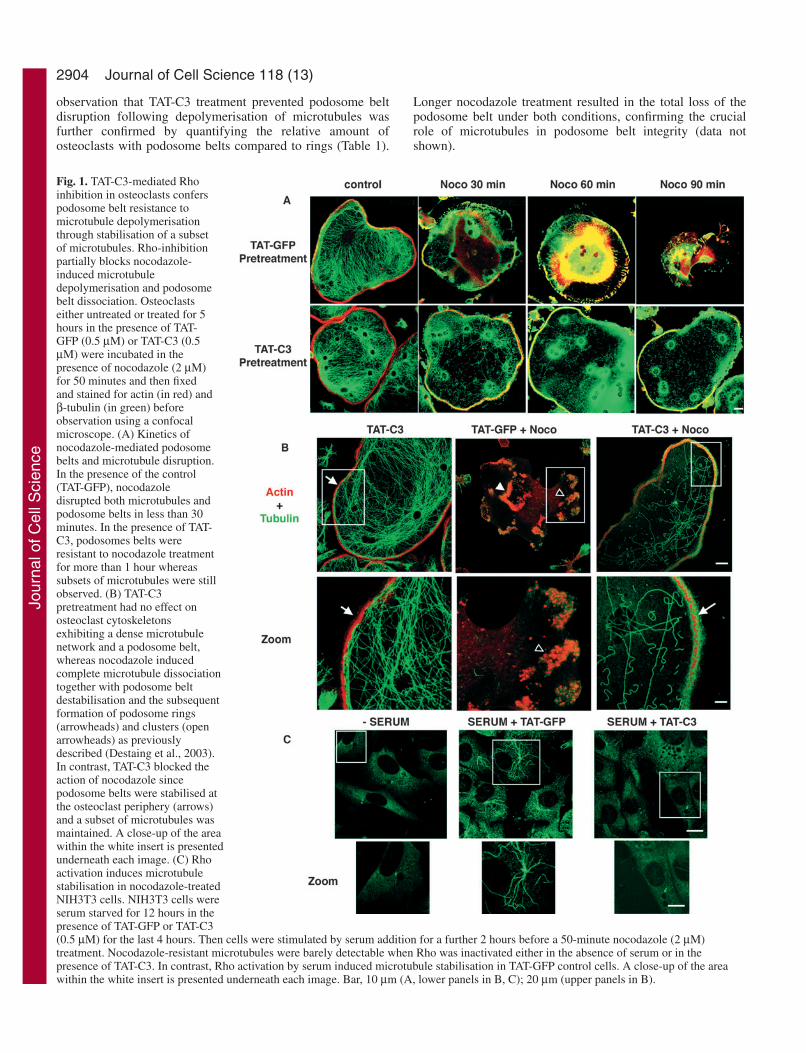
ResultsRho inhibition slows down podosome belt disruptionfollowing microtubule depolymerisationAs microtubule integrity is required for podosome beltformation in mature osteoclasts (Destaing et al., 2003) andthe small GTPase Rho is activated by microtubuledepolymerisation in fibroblasts (Ren et al., 1999) or inmacrophage osteoclast-like polykaryons (Ory et al., 2002), wewondered whether podosome belt destabilisation aftermicrotubule depolymerisation was dependent on Rho activity.To address that question, we used the ability of exoenzyme C3fused to the HIV TAT protein fragment (TAT-C3) to inhibit Rhoactivity quickly (Coleman et al., 2001; Nagahara et al., 1998;Schwarze et al., 1999). We first verified, using an in vitroribosylation assay, that TAT-C3 efficiently inhibited Rho after5 hours in osteoclasts (data not shown). Then, to examine therequirement of Rho activity for podosome belt destabilisationafter microtubule depolymerisation, we maintained theosteoclasts for 5 hours in medium containing either TAT-C3,or TAT-GFP fusion proteins as a control, and treated them withnocodazole for 0, 30, 60 or 90 minutes. The cells were thenfixed and stained for actin and β-tubulin (Fig. 1A). TAT-C3alone neither affected the podosome belt nor the microtubulenetwork (Fig. 1B). However, whereas the podosome beltstarted to be disassembled into podosome clusters after 30minutes of nocodazole treatment, (Fig. 1A,B arrowheads), itremained tightly associated at the cell periphery in TAT-C3treated osteoclasts (Fig. 1B). Longer treatment withnocodazole led to complete disruption of the podosome beltfollowed by cell retraction in control cells, whereas C3treatment drastically delayed podosome belt disassembly aseven after 90 minutes nocodazole treatment, the podosomebelt was maintained at the cell periphery (Fig. 1A). This
Table 1. Percentage of osteoclasts presenting podosome belts compared to podosomes arranged in clusters or rings aftervarious treatments*
TAT-GFPTAT-C3 (5 hours) (5 hours) then
Nocodazole TAT-C3 then nocodazole TAT-GFP nocodazoleControl (50 minutes) (5 hours) (50 minutes) (5 hours) (50 minutes)
% of cells with podosome rings 27 90 15 35 22 95% of cells with podosome belts 73 10 85 65 78 5
*600 osteoclasts were counted for each condition and the results are from five independent experiments.
Jour
nal o
f Cel
l Sci
ence
2904
observation that TAT-C3 treatment prevented podosome beltdisruption following depolymerisation of microtubules wasfurther confirmed by quantifying the relative amount ofosteoclasts with podosome belts compared to rings (Table 1).
Longer nocodazole treatment resulted in the total loss of thepodosome belt under both conditions, confirming the crucialrole of microtubules in podosome belt integrity (data notshown).
Journal of Cell Science 118 (13)
Fig. 1. TAT-C3-mediated Rhoinhibition in osteoclasts conferspodosome belt resistance tomicrotubule depolymerisationthrough stabilisation of a subsetof microtubules. Rho-inhibitionpartially blocks nocodazole-induced microtubuledepolymerisation and podosomebelt dissociation. Osteoclastseither untreated or treated for 5hours in the presence of TAT-GFP (0.5 µM) or TAT-C3 (0.5µM) were incubated in thepresence of nocodazole (2 µM)for 50 minutes and then fixedand stained for actin (in red) andβ-tubulin (in green) beforeobservation using a confocalmicroscope. (A) Kinetics ofnocodazole-mediated podosomebelts and microtubule disruption.In the presence of the control(TAT-GFP), nocodazoledisrupted both microtubules andpodosome belts in less than 30minutes. In the presence of TAT-C3, podosomes belts wereresistant to nocodazole treatmentfor more than 1 hour whereassubsets of microtubules were stillobserved. (B) TAT-C3pretreatment had no effect onosteoclast cytoskeletonsexhibiting a dense microtubulenetwork and a podosome belt,whereas nocodazole inducedcomplete microtubule dissociationtogether with podosome beltdestabilisation and the subsequentformation of podosome rings(arrowheads) and clusters (openarrowheads) as previouslydescribed (Destaing et al., 2003).In contrast, TAT-C3 blocked theaction of nocodazole sincepodosome belts were stabilised atthe osteoclast periphery (arrows)and a subset of microtubules wasmaintained. A close-up of the areawithin the white insert is presentedunderneath each image. (C) Rhoactivation induces microtubulestabilisation in nocodazole-treatedNIH3T3 cells. NIH3T3 cells wereserum starved for 12 hours in thepresence of TAT-GFP or TAT-C3(0.5 µM) for the last 4 hours. Then cells were stimulated by serum addition for a further 2 hours before a 50-minute nocodazole (2 µM)treatment. Nocodazole-resistant microtubules were barely detectable when Rho was inactivated either in the absence of serum or in thepresence of TAT-C3. In contrast, Rho activation by serum induced microtubule stabilisation in TAT-GFP control cells. A close-up of the areawithin the white insert is presented underneath each image. Bar, 10 µm (A, lower panels in B, C); 20 µm (upper panels in B).
Jour
nal o
f Cel
l Sci
ence

2905Microtubule acetylation in osteoclasts
Surprisingly, TAT-C3-treated osteoclasts showed asignificant increase in nocodazole-resistant microtubulescompared to TAT-GFP-treated cells (Fig. 1A,B). Thisobservation contradicts previous published data showing thatmicrotubule stabilisation is induced by Rho activation ratherthan Rho inhibition in NIH3T3 cells (Cook et al., 1998). Toconfirm that our results were not due to experimentaldeficiencies, we observed the microtubule content in NIH3T3cells that were first stimulated by serum to activate Rho, and
then treated with nocodazole in the presence of TAT-C3 orTAT-GFP. As described by others, and in contrast toosteoclasts, Rho activation by serum induced microtubulestabilisation that was otherwise blocked by TAT-C3 (Fig. 1C).We conclude that the effects of Rho inhibition on microtubulestability are cell type specific. It should be noted thatmicrotubule stabilisation by Rho inhibition has also beenfound in astrocytes (S. Etienne-Manneville, personalcommunication).
Rho activity and HDAC6 regulateacetylated microtubule levels inosteoclastsOur previous experiments indicating that Rhoinhibition somehow stabilised microtubulesprompted us to check whether the level of Rhoactivity could modulate post-translationalmodifications (PTMs) of microtubules, asPTMs are often associated with a changein microtubule dynamics and/or stability(Rosenbaum, 2000).
Fig. 2. Rho activity controls the level of tubulinacetylation upstream of HDAC6. (A) Inhibition ofRho by 0.5 µM TAT-C3 for 5 hours induced anaccumulation of acetylated microtubules incomparison to 0.5 µM TAT-GFP used as acontrol. (B) One nucleus per osteoclast wasmicroinjected with either RhoA WT-GFP or aconstitutively activated form of Rho, RhoAV14-GFP expression vectors. Cells were fixed 6 hoursafter microinjection and GFP-expressing cellswere detected by GFP fluorescence using aconfocal microscope. Acetylated tubulin wasdetected by indirect immunofluorescence (green)and F-actin by means of phalloidin-RITC (red)and a close-up of each condition is presented. Inthe presence of RhoA-WT, osteoclasts exhibit thetypical podosome belt and dense networks ofacetylated microtubules. On the other hand,expression of Rho V14-GFP induceddeacetylation of microtubules and disorganisationof podosome belts (arrowhead in close-up area).However, tubulin deacetylation dependent on Rhoactivation was inhibited after treatment with theHDAC6 inhibitor TSA (3 µM) for 1 hour,showing that this enzyme is downstream of Rho.(C,D) HDAC6 is present and active in osteoclasts.Endogenous HDAC6 was easily detected inosteoclasts by western blotting with a polyclonalanti-HDAC6 antibody (C). The deacetylaseactivity of HDAC6 was tested with two drugs:TSA, known to inhibit its activity and sodiumbutyrate, which does not. HDAC6 was indeedactive in osteoclasts as confirmed by greatlyincreased levels of acetylated tubulin in TSA-treated osteoclasts and unchanged levels in thepresence of sodium butyrate compared to thecontrol and to the total amount of β-tubulin (D).Finally, inhibition of Rho by TAT-C3 (for 4hours) in the presence of TSA had no additionaleffect on the increase in acetylated tubulin (C).Bar, 20 µm.
Jour
nal o
f Cel
l Sci
ence

2906
Osteoclasts were incubated for 5 hours with either TAT-GFPas a control or TAT-C3 to inhibit Rho, and processed forconfocal microscopy analysis after staining of their actin andacetylated-microtubule (Ac-MT) cytoskeleton. Whereas theAc-MT level was detectable but low in osteoclasts incubatedwith TAT-GFP fusion protein, it was increased in osteoclastsmaintained in presence of TAT-C3 (Fig. 2A). On the other hand,we never observed changes in detyrosinated microtubules (datanot shown), suggesting that Rho inhibition was promotingmicrotubule acetylation and also that Rho activation shouldtrigger microtubule deacetylation. To test this hypothesis, onenucleus per osteoclast analysed was microinjected with vectorsencoding GFP fused to a wild-type form or a constitutivelyactivated form of RhoA (RhoA-WT, V14-RhoA respectively).Cells were then fixed and Ac-MT levels compared byimmunostaining. GFP-RhoA WT did not significantly affectlevels of Ac-MT whereas V14RhoA promoted a drasticdecrease in the amount of Ac-MT together with disruption ofthe podosome belt (Fig. 2B). Thus, Rho appears to be a keyplayer in regulating levels of Ac-MT in osteoclasts.
It has recently been shown that the histone deacetylase 6(HDAC6) acts as a major microtubule deacetylase that can beinhibited by TSA (Matsuyama et al., 2002). To test whetherHDAC6 could act on the Rho pathway, multinucleatedosteoclasts were microinjected with V14RhoA cDNA andtreated with TSA for 30 minutes. TSA treatment blocked theV14RhoA-mediated deacetylation of microtubules (Fig. 2B,lower panel), indicating that HDAC6 was downstream of Rho.To confirm that microtubule acetylation was increased byHDAC6 or Rho inhibition, we monitored levels of acetylatedtubulin in osteoclast lysates treated with TAT-GFP, TAT-C3 orTSA (Fig. 2C). As expected, whereas levels of tubulin werecomparable between samples, levels of acetylated tubulinincreased an average of 2.5-fold in TAT-C3-treated osteoclastswhen compared to TAT-GFP-treated osteoclasts (mean of fiveindependent experiments). However, the relative increase ofacetylated tubulin was much higher in TSA-treated comparedto C3-treated cells indicating that Rho may partially controltubulin acetylation and/or only affect a subset of microtubules.In addition, in contrast to most HDAC proteins, TSA-sensitiveHDAC6 has been shown to be insensitive to sodium butyrate.To assess whether microtubule deacetylation was dependent onother HDACs in osteoclasts, cells were treated with TSAor sodium butyrate and the amount of acetylated tubulindetermined by western blotting. The amount of acetylatedtubulin did not change in sodium butyrate-treated osteoclastsbut did drastically increase when osteoclasts were treated withTSA (Fig. 2D). Altogether, these experiments indicate that Rhoactivation is able to stimulate microtubule deacetylation andthat HDAC6 is a probable intermediate.
Rho activates HDAC6 via mDia2To further evaluate the role of the Rho pathway in microtubuleacetylation in osteoclasts, we decided to test whether the twobest-characterised Rho effectors, namely ROCK and mDiaproteins, modified levels of Ac-MT in osteoclasts. As theROCK inhibitor Y27632 did not modify levels of Ac-MT inosteoclasts (data not shown), we focused on mDia2. Indeed,mDia2 was a good candidate as it has been shown to beinvolved in microtubule stabilisation and in coordinating
microtubule and actin dynamics (Ishizaki et al., 2001; Palazzoet al., 2001a). We thus microinjected plasmids encoding aconstitutive active mutant of mDia2 fused to GFP (GFP-mDIA2-∆GBD) into osteoclasts (Palazzo et al., 2001a).Consistent with the effect of activated Rho, we found thatactivated mDia2 triggered a drastic decrease in levels of Ac-MT as well as disruption of the podosome belt without any newspecific actin structures being formed (Fig. 3A). As our resultssuggest that both HDAC6 and mDia2 act downstream of Rho,we hypothesised that mDia2 and HDAC6 could interacttogether to regulate tubulin acetylation. To test this hypothesis,we cotransfected HDAC6 with either wild-type mDia2 oractivated mDia2 fused to GFP (GFP-mDia2WT or GFP-mDIA2-∆GBD) in COS cells. As a control, HDAC6 wastransfected with GFP alone. HDAC6 proteins were thenimmunoprecipitated from cleared cell lysates and associatedmDia2 revealed by immunoblotting with anti-GFP antibody.GFP-mDia2WT and GFP-mDIA2-∆GBD were found inHDAC6 immunoprecipitates whereas GFP alone was not (Fig.3B). To gain further insight into which HDAC6 domains wereresponsible for mDia2 binding, we cotransfected deletionmutants of HA-tagged HDAC6 together with GFP-mDia2WT.HDAC6 fragment proteins were immunoprecipitated with HAantibody and associated mDia2 was revealed by anti-GFPimmunoblotting. We detected mDia2 in the DD1 (amino acids85-428) and DD2 (aa 429-824) but not in the C-terminal(aa 825-1149) and N-terminal (aa 1-84) domainimmunocomplexes (Fig. 3C). These results indicate thatHDAC6 interacts with mDia2 in COS cells and that the twodeacetylase domains, DD1 and DD2 are both able to interactwith mDia2. Finally, to determine whether mDia2 was able tostimulate the deacetylase activity of HDAC6 in cells, we usedan in vitro deacetylase assay (Zhang et al., 2003). COS cellswere transfected with either HDAC6 alone, GFP-mDia2 aloneor HDAC6 and GFP-mDia2 together. We then analysed thelevel of acetylated tubulin by western blotting of COS celllysates (Fig. 3D). HDAC6 or GFP-mDia2 alone was not ableto promote deacetylation of tubulin. However, when bothproteins were expressed in cells, the level of acetylated tubulinwas clearly reduced despite comparable amounts of tubulin inthe samples. Moreover, transfecting twice the amount of GFP-mDia2 still reduced the level of acetylated tubulin indicatingthat mDia2 is able to activate HDAC6 deacetylase activity.
Formation of podosome belts in maturing osteoclastscorrelates with microtubule acetylationOur results described above clearly indicated that, inosteoclasts, Rho downregulation is required for podosomebelt stabilisation, microtubule acetylation and stabilisation.We have previously reported that during in vitroosteoclastogenesis, podosome patterning evolves frommicrotubule-independent clusters and rings to microtubule-dependent belts (Destaing et al., 2003). We then reasoned thatosteoclast maturation during osteoclastogenesis should beaccompanied by an increase in microtubule acetylation. Toinvestigate this possibility, we compared the Ac-MT pattern inmacrophages, as well as in immature and mature osteoclastsseeded on glass. In macrophages, Ac-MT staining wasconcentrated in punctate structures resembling centrosomes(Fig. 4A). Immature osteoclasts exhibiting podosome rings
Journal of Cell Science 118 (13)
Jour
nal o
f Cel
l Sci
ence

2907Microtubule acetylation in osteoclasts
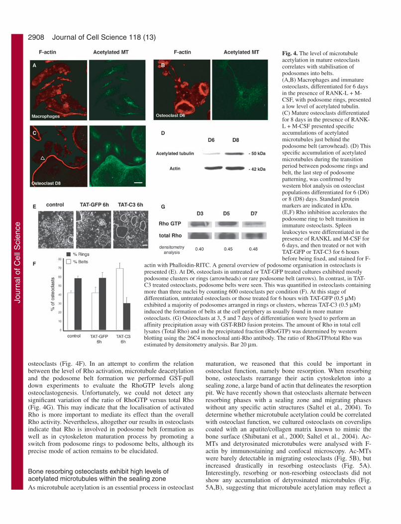
contained only a low level of Ac-MT (Fig. 4B) whereas matureosteoclasts, with a podosome belt, had much higher levels ofAc-MT which were found mostly associated with the actinpodosome belt (Fig. 4C, arrowhead). This was further assessedby comparing acetylated tubulin levels in cultures ofosteoclasts after 6 or 8 days of differentiation containing 40%or 80% respectively, of osteoclasts exhibiting podosome belts(Fig. 4D). We could conclude that microtubule acetylation is
associated with the formation of stable podosome belts duringosteoclastogenesis.
As Rho inhibition is crucial for maintaining the podosomebelt at the cell periphery, we expected that Rho inhibition inimmature osteoclasts (D6) should accelerate the formation ofpodosome belts. Macrophages were differentiated intoosteoclasts and at day 6, TAT-GFP or TAT-C3 was added to thedifferentiation medium for 6 hours. The number of osteoclastswith podosome rings (Fig. 4E, arrowhead) or podosome belts(Fig. 4E, arrows) was then quantified and compared to cellsmaintained in differentiation medium alone. At day 6, only40% of osteoclasts in untreated or TAT-GFP-treated osteoclastsexhibited podosome belts versus 70% in TAT-C3-treated
Fig. 3. mDIA2 mediates the action of Rho at thelevel of acetylated microtubules by interacting withand activating HDAC6. (A) Constitutively activemDia2 blocks tubulin acetylation and podosomebelt formation. One nucleus per osteoclast wasmicroinjected with either GFP or a constitutivelyactivated form of mDIA2, GFP-mDIA2 ∆GBDexpression vectors. 6 hours later, acetylated tubulinwas detected by indirect immunofluorescence(green) and F-actin with phalloidin-RITC (red). InGFP-mDIA2 ∆GBD-expressing osteoclastscompared to GFP-expressing osteoclasts, the Ac-MT level was dramatically decreased and thepodosome belt disrupted. (B) HDAC6 and mDia2interact together. COS cells were either transfectedwith GFP alone, HA-HDAC6, GFP-mDIA2 WT,GFP-mDIA2 ∆GBD vectors or co-transfectedtogether with HDAC6. Transfected HDAC6 ormDia2 were revealed in total cell lysate (TCL) byan anti-HA or an anti-GFP antibody respectively(left panel). HDAC6 was immunoprecipitated withan anti-HA antibody and mDia2-associated proteinswere revealed with an anti-GFP antibody (right
panel). Both the wild type and activatedmDia2 co-precipitated with HA-taggedHDAC6. (C) Mapping of HDAC6 domaininteraction with mDia2. To determine thedomains of HDAC6 implicated in theinteraction with GFP-mDIA2 WT, COS cellswere transfected with GFP-mDIA2 WT andwith the N-terminal domain, the deacetylasedomain 1 (DD1), the deacetylase domain 2(DD2) or the C-terminal domain of HDAC6,using HA-tagged expression vectors. Celllysates of transfected cells wereimmunoprecipitated with a monoclonal anti-HA antibody. (D) mDia2 increases HDAC6deacetylase activity. In a tubulin deacetylasein vitro assay, COS cells were transfectedwith mHDAC6-HA WT, lysed at room
temperature for 15 minutes and the ratioof acetylated tubulin/tubulin monitoredby western blotting. When GFP-mDIA2WT was transfected with mHDAC6-HAWT, this increased the deacetylaseactivity of the enzyme whereas GFP-mDIA2 WT alone had no effect on thelevel of this PTM. Positions of proteinstandards in kDa are indicated on theleft-hand side of blots.
Jour
nal o
f Cel
l Sci
ence
2908
osteoclasts (Fig. 4F). In an attempt to confirm the relationbetween the level of Rho activation, microtubule deacetylationand the podosome belt formation we performed GST-pulldown experiments to evaluate the RhoGTP levels alongosteoclastogenesis. Unfortunately, we could not detect anysignificant variation of the ratio of RhoGTP versus total Rho(Fig. 4G). This may indicate that the localisation of activatedRho is more important to mediate its effect than the overallRho activity. Nevertheless, altogether our results in osteoclastsindicate that Rho is involved in podosome belt formation aswell as in cytoskeleton maturation process by promoting aswitch from podosome rings to podosome belts, although itsprecise mode of action remains to be elucidated.
Bone resorbing osteoclasts exhibit high levels ofacetylated microtubules within the sealing zoneAs microtubule acetylation is an essential process in osteoclast
maturation, we reasoned that this could be important inosteoclast function, namely bone resorption. When resorbingbone, osteoclasts rearrange their actin cytoskeleton into asealing zone, a large band of actin that delineates the resorptionpit. We have recently shown that osteoclasts alternate betweenresorbing phases with a sealing zone and migrating phaseswithout any specific actin structures (Saltel et al., 2004). Todetermine whether microtubule acetylation could be correlatedwith osteoclast function, we cultured osteoclasts on coverslipscoated with an apatite/collagen matrix known to mimic thebone surface (Shibutani et al., 2000; Saltel et al., 2004). Ac-MTs and detyrosinated microtubules were analysed with F-actin by immunostaining and confocal microscopy. Ac-MTswere barely detectable in migrating osteoclasts (Fig. 5B), butincreased drastically in resorbing osteoclasts (Fig. 5A).Interestingly, resorbing or non-resorbing osteoclasts did notshow any accumulation of detyrosinated microtubules (Fig.5A,B), suggesting that microtubule acetylation may reflect a
Journal of Cell Science 118 (13)
Fig. 4. The level of microtubuleacetylation in mature osteoclastscorrelates with stabilisation ofpodosomes into belts.(A,B) Macrophages and immatureosteoclasts, differentiated for 6 daysin the presence of RANK-L + M-CSF, with podosome rings, presenteda low level of acetylated tubulin.(C) Mature osteoclasts differentiatedfor 8 days in the presence of RANK-L + M-CSF presented specificaccumulations of acetylatedmicrotubules just behind thepodosome belt (arrowhead). (D) Thisspecific accumulation of acetylatedmicrotubules during the transitionperiod between podosome rings andbelt, the last step of podosomepatterning, was confirmed bywestern blot analysis on osteoclastpopulations differentiated for 6 (D6)or 8 (D8) days. Standard proteinmarkers are indicated in kDa.(E,F) Rho inhibition accelerates thepodosome ring to belt transition inimmature osteoclasts. Spleenleukocytes were differentiated in thepresence of RANKL and M-CSF for6 days, and then treated or not withTAT-GFP or TAT-C3 for 6 hoursbefore being fixed, and stained for F-
actin with Phalloidin-RITC. A general overview of podosome organisation in osteoclasts ispresented (E). At D6, osteoclasts in untreated or TAT-GFP treated cultures exhibited mostlypodosome clusters or rings (arrowheads) or rare podosome belt (arrows). In contrast, in TAT-C3 treated osteoclasts, podosome belts were seen. This was quantified in osteoclasts containingmore than three nuclei by counting 600 osteoclasts per condition (F). At this stage ofdifferentiation, untreated osteoclasts or those treated for 6 hours with TAT-GFP (0.5 µM)exhibited a majority of podosomes arranged in rings or clusters, whereas TAT-C3 (0.5 µM)induced the formation of belts at the cell periphery as usually found in more matureosteoclasts. (G) Osteoclasts at 3, 5 and 7 days of differentiation were lysed to perform anaffinity precipitation assay with GST-RBD fusion proteins. The amount of Rho in total celllysates (Total Rho) and in the precipitated fraction (RhoGTP) was determined by westernblotting using the 26C4 monoclonal anti-Rho antibody. The ratio of RhoGTP/total Rho wasestimated by densitometry analysis. Bar 20 µm.
Jour
nal o
f Cel
l Sci
ence

2909Microtubule acetylation in osteoclasts
specific post-translational modification of microtubules inactive osteoclasts.
DiscussionWe previously reported that osteoclast differentiation wasaccompanied by a change in podosome patterning. Immatureosteoclasts present dynamic podosome rings and matureosteoclasts contain a stable peripheral podosome belt (Destainget al., 2003). In an attempt to understand the molecularmechanisms underlying podosome patterning, we found thatRho inhibition both delayed podosome belt disruption afternocodazole treatment and accelerated the switch from unstablepodosome rings to a stable peripheral podosome belt, mimickingthe maturation process. We also described for the first time thatduring the differentiation of osteoclasts and their resorptioncycle on mineralised matrix, microtubules became acetylated,and not detyrosinated, indicating that microtubule acetylationmay have a specific function in osteoclasts. Furthermore,stabilisation of the podosome belt was correlated with twoevents: increases in microtubule acetylation and Rho inhibition.This led us to identify a new Rho-dependent pathway in whichthe recently discovered microtubule acetylase HDAC6 (Hubbertet al., 2002; Matsuyama et al., 2002; Zhang et al., 2003) wasactivated by Rho and its effector mDia2. Microinjection of eitheractivated RhoA or mDia2 triggered microtubule deacetylationtogether with podosome belt disruption. On the other hand, Rhoinhibition promoted microtubule acetylation. In vitro assaysconfirmed that mDia2 activates the microtubule deacetylaseactivity of HDAC6 and this may be explained by the presenceof HDAC6 and mDia2 in the same protein complex as revealedby immunoprecipitation experiments. Altogether, our resultsindicate that microtubule acetylation levels are controlled byRho proteins, and that this may be crucial for osteoclastdifferentiation and function.
The fact that, in our study, TAT-C3-mediated Rho inhibitionpromoted podosome belt assembly during osteoclastmaturation is rather surprising. Indeed, exoenzyme C3 has
been reported to disrupt the sealing zone and to blockosteoclast resorption on bone (Zhang et al., 1995; Saltel et al.,2004) as well as disrupting isolated podosomes in macrophagepolykaryons (Chellaiah et al., 2000; Ory et al., 2000). In src-transformed cells, Rho inhibition also disrupted the podosomewhereas activated Rho has been found localised in podosomes(Berdeaux et al., 2004). However, inconsistent with a functionof Rho in podosome formation, and in contrast to Cdc42(Dutartre et al., 1996; Moreau et al., 2003), activated Rho or,interestingly activated mDia, do not lead to typical podosomeformation but rather to their disruption (Burgstaller andGimona, 2004; Chellaiah et al., 2000; Ory et al., 2000)indicating that the Rho activation pathway by itself is notsufficient for podosome formation. However, as revealed byour recent study (Saltel et al., 2004), Rho inhibition inosteoclasts cultured on bone-like substrate led to the loss ofboth their resorptive function and their apico-basal polarity.Interestingly, under these conditions, F-actin reorganised intoa podosome belt, mimicking the actin organisation found inosteoclasts seeded on glass, an organisation never seen inosteoclasts seeded on bone-like substrate in normal conditions.Together with the present study, it indicates that Rho activityis not required for the podosome belt formation. However,despite what we were expecting, we could not observe anyRhoGTP level variation along osteoclastogenesis indicatingthat to get more insight into the function of Rho in such largemultinucleated cells, the subcellular localisation of activatedRho should be investigated. It would be more informative thanthe overall activity measured by pull-down assay. Indeed, weshould expect Rho to be only locally inhibited as its fullinhibition by exoenzyme C3 triggered not only the podosomebelt stabilisation, but also excessive spreading and loss of cellpolarity (Ory et al., 2000; Saltel et al., 2004). This may engagelocal regulation of Rho activity, variations of which may beinsufficient to discriminate it from the overall Rho activity incells. This idea is also supported by the fact that acetylation ofmicrotubules was mainly observed on a subset of microtubuleslocalised in the vicinity of the podosome belt in differentiated
Fig. 5. Formation of the sealingzone in bone resorbingosteoclasts is associated with thedynamic regulations of tubulinacetylation. (A,B) Osteoclastswere differentiated in thepresence of RANK-L + M-CSFfor 8 days, detached, spread onmineralised matrix (ACC,Apatite Collagen Complex)substrate, which mimics dentinslices, fixed and immunostainedwith phalloidin-RITC andmonoclonal anti-acetylated andpolyclonal anti-detyrosinatedtubulin antibodies. These twoosteoclasts are associated with aresorption pit (*). The osteoclastpresented in A is still resorbing,as it exhibits a sealing zone, alarge band of F-actin, and has a large number of acetylated but undetectable detyrosinated microtubules. The osteoclast in B is a migratingosteoclast without a sealing zone and showed neither acetylated nor detyrosinated microtubules. Bar, 20 µm.
Jour
nal o
f Cel
l Sci
ence

2910
osteoclasts. This suggests that a local change in microtubuleproperties is occurring, and consequently, a local change insignalling events leading to microtubule acetylation.
Microtubule acetylation during osteoclast differentiationOur results have shown that podosome belt and sealing zoneformation during osteoclast maturation and bone resorption,respectively, are associated with an increase in tubulinacetylation corresponding to microtubule stabilisation.Interestingly, observations of increases in stable microtubuleshave been made in other differentiated cell types (Bulinski andGundersen, 1991). For example, in muscle cell differentiation,which involves the fusion of myoblasts to form multinucleatedmyotubes, detyrosinated microtubules accumulate inmyogenic precursors shortly before the fusion events.Detyrosinated microtubules are maintained in myotubes butacetylation is only observed at a later stage (Gundersen etal., 1989). Unlike myogenesis (Chang et al., 2002),osteoclastogenesis is not associated with detyrosination ofmicrotubules, as we did not detect any significant changes inthe levels of detyrosinated microtubules. However, as inmyotubes, acetylated microtubules increased at a later stagein mature osteoclasts. These observations indicate that,although PTMs take place on stable microtubules, they arenot necessarily occurring at the same time during thedifferentiation process, suggesting that they are dependent ontheir subcellular micro-environment and do not have the samebiological functions. Differences in the function of post-translational tubulin modifications have been well exemplifiedin Tetrahymena thermophila in which the endogenous β-tubulin gene has been replaced by mutated forms preventingeither acetylation or polyglycylation PTMs. Although therewere no detectable abnormalities when non-acetylatabletubulin was expressed (Gaertig et al., 1995), preventing thepolyglycylation of β-tubulin, in contrast, had an affect oncytokinesis and was lethal (Thazhath et al., 2002).
Although both the underlying molecular mechanisms ofPTM and the function of microtubule acetylation remain to beelucidated, recent reports have highlighted an intriguingproperty of detyrosinated microtubules. In mammalian cells,kinesins have a stronger affinity for detyrosinated tubulin andmay be preferentially recruited on stable detyrosinatedmicrotubules (Gurland and Gundersen, 1995; Kreitzer et al.,1999). Whether a similar function can be attributed toacetylation remains to be seen, but microtubule acetylationis highly regulated in osteoclast resorption function andpodosome patterning.
Rho activates HDAC6 through mDia2 and controlsmicrotubule qualityHDAC6 has been recently characterised as a cytoplasmictubulin deacetylase (Hubbert et al., 2002; Matsuyama et al.,2002; Zhang et al., 2003). We found that Rho inhibitionincreased microtubule acetylation. Conversely, Rho activationas well as microinjection of activated mDia2, promotedmicrotubule deacetylation whereas no changes in the levelsof microtubule acetylation were observed with the ROCKinhibitor Y27632 (data not shown). These results indicate thatmDia2 is the specific Rho effector involved in microtubule
deacetylation. This hypothesis is reinforced by the fact thatmDia2 coprecipitated with and activated HDAC6 in COS cells.Thus, we propose that Rho activates mDia2, which in turnstimulates HDAC6. However, the precise mechanism by whichHDAC6 is activated will require more experiments. Indeed, wenoticed that deacetylation of microtubules was not significantlydifferent when HDAC6 was cotransfected with either theconstitutively activated form of mDia2 (GFP-mDIA2-∆GBD)or its wild-type counterpart. Together with the fact thatmDia2 partially colocalised with HDAC6 on microtubulesin osteoclasts (data not shown), we propose that anmDia2/HDAC6 complex is constitutively formed and that Rhoactivation localises that complex on microtubules to promoteits in vivo deacetylation. Knowing where Rho is activated inthis context is of major importance. Finally, mDia2 bindingrequires the cooperation of the deacetylase domains, DD1 andDD2 of HDAC6, which are also required for HDAC6 to bindtubulin (Zhang et al., 2003). Whether that complex isdependent upon tubulin binding to be assembled and/or activeremains to be answered. Nonetheless, our results highlight therequirement for a fine control of Rho-dependent microtubuleacetylation in osteoclasts. It would be of interest to analyseosteoclast podosome patterning in an HDAC6–/– background.
O.D. and F.S. are recipients of MENRT and FRM grants. We aregrateful to Sandrine Etienne-Manneville for helpful discussions andsharing unpublished data. We wish to thank Art Alberts and KlausAktories for giving us precious reagents and also Jan R. De Mey andEdith Bonnely for stimulating discussions. B.G. is a recipient of a PhDfellowship from the Ligue Nationale Contre le Cancer, Comité del’Isère. We are grateful to Sandrine Curtet-Benitski for technicalassistance. This work was also supported by grants from CNRS(dynamique et réactivité du vivant), INSERM and the Ligue contre leCancer du Rhône.
ReferencesBerdeaux, R. L., Diaz, B., Kim, L. and Martin, G. S. (2004). Active Rho is
localized to podosomes induced by oncogenic Src and is required for theirassembly and function. J. Cell Biol. 166, 317-323.
Boyle, W. J., Simonet, W. S. and Lacey, D. L. (2003). Osteoclastdifferentiation and activation. Nature 423, 337-342.
Bulinski, J. C. and Gundersen, G. G. (1991). Stabilization of post-translational modification of microtubules during cellular morphogenesis.BioEssays 13, 285-293.
Burgstaller, G. and Gimona, M. (2004). Actin cytoskeleton remodelling vialocal inhibition of contractility at discrete microdomains. J. Cell Sci. 117,223-231.
Burns, S., Thrasher, A. J., Blundell, M. P., Machesky, L. and Jones, G. E.(2001). Configuration of human dendritic cell cytoskeleton by RhoGTPases, the WAS protein, and differentiation. Blood 98, 1142-1149.
Chang, W., Webster, D. R., Salam, A. A., Gruber, D., Prasad, A., Eiserich,J. P. and Bulinski, J. C. (2002). Alteration of the C-terminal amino acid oftubulin specifically inhibits myogenic differentiation. J. Biol. Chem. 277,30690-30698.
Chellaiah, M. A., Soga, N., Swanson, S., McAllister, S., Alvarez, U., Wang,D., Dowdy, S. F. and Hruska, K. A. (2000). Rho-A is critical for osteoclastpodosome organization, motility, and bone resorption. J. Biol. Chem. 275,11993-12002.
Coleman, M. L., Sahai, E. A., Yeo, M., Bosch, M., Dewar, A. and Olson,M. F. (2001). Membrane blebbing during apoptosis results from caspase-mediated activation of ROCK I. Nat. Cell Biol. 3, 339-345.
Cook, T. A., Nagasaki, T. and Gundersen, G. G. (1998). Rho guanosinetriphosphatase mediates the selective stabilization of microtubules inducedby lysophosphatidic acid. J. Cell Biol. 141, 175-185.
Destaing, O., Saltel, F., Geminard, J. C., Jurdic, P. and Bard, F. (2003).Podosomes display actin turnover and dynamic self-organization in
Journal of Cell Science 118 (13)
Jour
nal o
f Cel
l Sci
ence
2911Microtubule acetylation in osteoclasts
osteoclasts expressing actin-green fluorescent protein. Mol. Biol. Cell. 14,407-416.
Dutartre, H., Davoust, J., Gorvel, J. P. and Chavrier, P. (1996). Cytokinesisarrest and redistribution of actin-cytoskeleton regulatory components incells expressing the Rho GTPase CDC42Hs. J. Cell Sci. 109, 367-377.
Enomoto, T. (1996). Microtubule disruption induces the formation of actinstress fibers and focal adhesions in cultured cells: possible involvement ofthe rho signal cascade. Cell Struct. Funct. 21, 317-326.
Etienne-Manneville, S. and Hall, A. (2002). Rho GTPases in cell biology.Nature 420, 629-635.
Etienne-Manneville, S. and Hall, A. (2003). Cdc42 regulates GSK-3beta andadenomatous polyposis coli to control cell polarity. Nature 421, 753-756.
Evans, J. G., Correia, I., Krasavina, O., Watson, N. and Matsudaira, P.(2003). Macrophage podosomes assemble at the leading lamella by growthand fragmentation. J. Cell Biol. 161, 697-705.
Gaertig, J., Cruz, M. A., Bowen, J., Gu, L., Pennock, D. G. and Gorovsky,M. A. (1995). Acetylation of lysine 40 in alpha-tubulin is not essential inTetrahymena thermophila. J. Cell Biol. 129, 1301-1310.
Gundersen, G. G., Khawaja, S. and Bulinski, J. C. (1989). Generation of astable, posttranslationally modified microtubule array is an early event inmyogenic differentiation. J. Cell Biol. 109, 2275-2288.
Gurland, G. and Gundersen, G. G. (1995). Stable, detyrosinatedmicrotubules function to localize vimentin intermediate filaments infibroblasts. J. Cell Biol. 131, 1275-1290.
Hubbert, C., Guardiola, A., Shao, R., Kawaguchi, Y., Ito, A., Nixon, A.,Yoshida, M., Wang, X. F. and Yao, T. P. (2002). HDAC6 is a microtubule-associated deacetylase. Nature 417, 455-458.
Ishizaki, T., Morishima, Y., Okamoto, M., Furuyashiki, T., Kato, T. andNarumiya, S. (2001). Coordination of microtubules and the actincytoskeleton by the Rho effector mDia1. Nat. Cell Biol. 3, 8-14.
Kaverina, I., Krylyshkina, O. and Small, J. V. (1999). Microtubule targetingof substrate contacts promotes their relaxation and dissociation. J. Cell Biol.146, 1033-1044.
Kreitzer, G., Liao, G. and Gundersen, G. G. (1999). Detyrosination oftubulin regulates the interaction of intermediate filaments withmicrotubules in vivo via a kinesin-dependent mechanism. Mol. Biol. Cell.10, 1105-1118.
Linder, S. and Aepfelbacher, M. (2003). Podosomes: adhesion hot-spots ofinvasive cells. Trends Cell Biol. 13, 376-385.
Linder, S., Hufner, K., Wintergerst, U. and Aepfelbacher, M. (2000).Microtubule-dependent formation of podosomal adhesion structures inprimary human macrophages. J. Cell Sci. 113, 4165-4176.
Matsuyama, A., Shimazu, T., Sumida, Y., Saito, A., Yoshimatsu, Y.,Seigneurin-Berny, D., Osada, H., Komatsu, Y., Nishino, N., Khochbin,S. et al. (2002). In vivo destabilization of dynamic microtubules by HDAC6-mediated deacetylation. EMBO J. 21, 6820-6831.
Moreau, V., Tatin, F., Varon, C. and Genot, E (2003). Actin can reorganizeinto podosomes in aortic endothelial cells, a process controlled by Cdc42and RhoA. Mol. Cell Biol. 23, 6809-6822.
Nagahara, H., Vocero-Akbani, A. M., Snyder, E. L., Ho, A., Latham,D. G., Lissy, N. A., Becker-Hapak, M., Ezhevsky, S. A. and Dowdy,S. F. (1998). Transduction of full-length TAT fusion proteins intomammalian cells: TAT-p27Kip1 induces cell migration. Nat. Med. 4, 1449-1452.
Ory, S., Munari-Silem, Y., Fort, P. and Jurdic, P. (2000). Rho and Rac exertantagonistic functions on spreading of macrophage-derived multinucleated
cells and are not required for actin fiber formation. J. Cell Sci. 113, 1177-1188.
Ory, S., Destaing, O. and Jurdic, P. (2002). Microtubule dynamicsdifferentially regulates Rho and Rac activity and triggers Rho-independentstress fiber formation in macrophage polykaryons. Eur. J. Cell Biol. 81, 351-362.
Palazzo, A. F., Cook, T. A., Alberts, A. S. and Gundersen, G. G. (2001a).mDia mediates Rho-regulated formation and orientation of stablemicrotubules. Nat. Cell Biol. 3, 723-729.
Palazzo, A. F., Joseph, H. L., Chen, Y. J., Dujardin, D. L., Alberts, A. S.,Pfister, K. K., Vallee, R. B. and Gundersen, G. G. (2001b). Cdc42, dynein,and dynactin regulate MTOC reorientation independent of Rho-regulatedmicrotubule stabilization. Curr. Biol. 11, 1536-1541.
Ren, X. D., Kiosses, W. B. and Schwartz, M. A. (1999). Regulation of thesmall GTP-binding protein Rho by cell adhesion and the cytoskeleton.EMBO J. 18, 578-585.
Rosenbaum, J. (2000). Cytoskeleton: functions for tubulin modifications atlast. Curr. Biol. 10, R801-R803.
Saltel, F., Destaing, O., Bard, F., Eichert, D. and Jurdic, P. (2004).Apatite-mediated actin dynamics in resorbing osteoclasts. Mol. Biol. Cell.15, 5231-5241.
Saxton, W. M., Stemple, D. L., Leslie, R. J., Salmon, E. D., Zavortink, M.and McIntosh, J. R. (1984). Tubulin dynamics in cultured mammaliancells. J. Cell Biol. 99, 2175-2186.
Schulze, E. and Kirschner, M. (1986). Microtubule dynamics in interphasecells. J. Cell Biol. 102, 1020-1031.
Schwarze, S. R., Ho, A., Vocero-Akbani, A. and Dowdy, S. F. (1999). Invivo protein transduction: delivery of a biologically active protein into themouse. Science 285, 1569-1572.
Seigneurin-Berny, D., Verdel, A., Curtet, S., Lemercier, C., Garin, J.,Rousseaux, S. and Khochbin, S. (2001). Identification of components ofthe murine histone deacetylase 6 complex: link between acetylation andubiquitination signaling pathways. Mol. Cell Biol. 21, 8035-8044.
Shibutani, T., Iwanaga, H., Imai, K., Kitago, M., Doi, Y. and Iwayama, Y.(2000). Use of glass slides coated with apatite-collagen complexes formeasurement of osteoclastic resorption activity. J. Biomed. Mater. Res. 50,153-159.
Thazhath, R., Liu, C. and Gaertig, J. (2002). Polyglycylation domain ofbeta-tubulin maintains axonemal architecture and affects cytokinesis inTetrahymena. Nat. Cell Biol. 4, 256-259.
Vaananen, H. K., Zhao, H., Mulari, M. and Halleen, J. M. (2000). The cellbiology of osteoclast function. J. Cell Sci. 113, 377-381.
Webster, D. R., Gundersen, G. G., Bulinski, J. C. and Borisy, G. G. (1987a).Assembly and turnover of detyrosinated tubulin in vivo. J. Cell Biol. 105,265-276.
Webster, D. R., Gundersen, G. G., Bulinski, J. C. and Borisy, G. G.(1987b). Differential turnover of tyrosinated and detyrosinatedmicrotubules. Proc. Natl. Acad. Sci. USA 84, 9040-9044.
Zhang, D., Udagawa, N., Nakamura, I., Murakami, H., Saito, S.,Yamasaki, K., Shibasaki, Y., Morii, N., Narumiya, S., Takahashi, N. etal. (1995). The small GTP-binding protein, rho p21, is involved in boneresorption by regulating cytoskeletal organization in osteoclasts. J. Cell Sci.108, 2285-2292.
Zhang, Y., Li, N., Caron, C., Matthias, G., Hess, D., Khochbin, S. andMatthias, P. (2003). HDAC-6 interacts with and deacetylates tubulin andmicrotubules in vivo. EMBO J. 22, 1168-1179.
Jour
nal o
f Cel
l Sci
ence